Embed Size (px)
Citation preview
© 2012 Karagkiozaki et al, publisher and licensee Dove Medical Press Ltd. This is an Open Access article which permits unrestricted noncommercial use, provided the original work is properly cited.
International Journal of Nanomedicine 2012:7 5327–5338
International Journal of Nanomedicine
Development of a nanoporous and multilayer drug-delivery platform for medical implants
Varvara Karagkiozaki1
Eleftherios Vavoulidis1
Panagiotis G Karagiannidis1
Maria Gioti1
Dimitrios G Fatouros2
Ioannis S Vizirianakis3
Stergios Logothetidis1
1Lab for Thin Films–Nanosystems and Nanometrology, Physics Department, 2Department of Pharmaceutical Technology, 3Laboratory of Pharmacology, School of Pharmacy, Aristotle University of Thessaloniki, Greece
Correspondence: Stergios Logothetidis Aristotle University of Thessaloniki, Physics Department, Laboratory for Thin Films–Nanosystems and Nanometrology, GR-54124, Greece Tel +30 2310 998174 Fax +30 2310 998390 Email [email protected]
Abstract: Biodegradable polymers can be applied to a variety of implants for controlled and
local drug delivery. The aim of this study is to develop a biodegradable and nanoporous poly-
meric platform for a wide spectrum of drug-eluting implants with special focus on stent-coating
applications. It was synthesized by poly(DL-lactide-co-glycolide) (PLGA 65:35, PLGA 75:25)
and polycaprolactone (PCL) in a multilayer configuration by means of a spin-coating technique.
The antiplatelet drug dipyridamole was loaded into the surface nanopores of the platform. Surface
characterization was made by atomic force microscopy (AFM) and spectroscopic ellipsometry
(SE). Platelet adhesion and drug-release kinetic studies were then carried out. The study revealed
that the multilayer films are highly nanoporous, whereas the single layers of PLGA are atomically
smooth and spherulites are formed in PCL. Their nanoporosity (pore diameter, depth, density,
surface roughness) can be tailored by tuning the growth parameters (eg, spinning speed, polymer
concentration), essential for drug-delivery performance. The origin of pore formation may be
attributed to the phase separation of polymer blends via the spinodal decomposition mechanism.
SE studies revealed the structural characteristics, film thickness, and optical properties even of
the single layers in the triple-layer construct, providing substantial information for drug loading
and complement AFM findings. Platelet adhesion studies showed that the dipyridamole-loaded
coatings inhibit platelet aggregation that is a prerequisite for clotting. Finally, the films exhibited
sustained release profiles of dipyridamole over 70 days. These results indicate that the current
multilayer phase therapeutic approach constitutes an effective drug-delivery platform for drug-
eluting implants and especially for cardiovascular stent applications.
Keywords: drug delivery, implants, stents, polymers, spin-coating, atomic force microscopy
One of the fields that has benefited from nanomedicine is the design of new
drug-delivery and -release systems. These novel systems can be combined with
state-of-the-art implant technology and, in turn, give birth to drug-eluting implants
that release therapeutic agents at the site of implantation. One of the most promising
categories of drug-eluting coatings is considered to be nanoporous platforms with pore
sizes less than 0.1 μm. Porosity of such low dimensions contributes to the material’s
high active surface and drug loading, leading to desirable drug release profiles for
each medical application.1,2
A wide range of different materials and fabrication methods have been used to
manufacture nanoporous coatings.3 Biocompatible metals and their alloys, such as
titanium and aluminum, are subjected to a self-ordering porous formation that is
based on electrochemical processes.4 Polymers and especially block copolymers
have been widely used to compose nanoporous drug-delivery platforms aimed at
Dovepress
submit your manuscript | www.dovepress.com
Dovepress 5327
O R I G I N A L R E S E A R C h
open access to scientific and medical research
Open Access Full Text Article
http://dx.doi.org/10.2147/IJN.S31185
International Journal of Nanomedicine 2012:7
clinical applications.5,6 However, their use has been hindered
because of limited biodegradability and slow degradation
rates.7 These properties were found to cause undesirable
side effects, such as hypersensitivity reactions at the site of
implantation, inflammation, and thrombus formation, which
can lead to tissue damage and even implant failure.8
In this study, we designed and developed biodegradable
polymeric matrices in a multilayer configuration character-
ized by a diversity of nanopores for controlled drug loading
and release. Two classes of poly(DL-lactide-co-glycolide)
(PLGA 65:35, PLGA 75:25) and polycaprolactone (PCL)
with different degradation rates constitute the nanolayers
of the platform, and they were deposited by spin coating.
Although this technique has been widely used for the
deposition of thin films for various applications,9,10 to
the best of our knowledge this is the first time that it
has been used for the synthesis of drug-eluting applica-
tions. In particular, novel nanoporous materials with a
variety of nanopore characteristics (depth, density, and
diameter) were manufactured to serve as drug reservoirs
with multiplex loading capacities. The major goals in
designing drug-delivery systems are to control nanopore
size, surface properties, and release of pharmacologically
active agents in order to achieve the site-specific action of
the drug at the nanoporosity therapeutically optimal rate
and dose regimen. The control of the nanoporosity of the
engineered nanomaterials was achieved by the implemen-
tation of highly sensitive techniques, such as atomic force
microscopy (AFM) and spectroscopic ellipsometry (SE) in
correlation with variations in deposition parameters.
These multilayer polymeric nanocoatings may serve as a
drug-eluting platform for a wide spectrum of implants (eg,
orthopedic, cardiovascular, retinal etc). The design of the
platform and the selection of polymers and drugs should be
made in line with the specific medical application. In this
study, the platform was designed for stent-coating needs.
Several drug candidates, such as immune-suppressive
agents, anti-inflammatory, and cellular proliferation inhibi-
tors, have been employed for stent coating and evaluated in
clinical trials. Sirolimus-eluting (Cypher®; Cordis, Miami
Lakes, FL) and paclitaxel-eluting (Taxus®; Boston Scientific,
Natick, MA) stents have been extensively used for coronary
angioplasty.11,12
In our study, dipyridamole (DPM), an antiplatelet drug
known to inhibit clotting,13 was encapsulated into the external
layer of the polymeric matrix and the release of the drug was
monitored over time. We used platelet adhesion studies to
assess their antithrombogenic effect.
MethodsMaterialsDipyridamole was obtained from Sigma-Aldrich (Athens,
Greece). NaCl, KCl, KH2PO
4, and Na
2HPO
4 were obtained
from Merck (Darmstadt, Germany). PLGA with different
lactide:glycolide contents (65:35 with average molecular
weight [Mw] = 40,000–75,000 and 75:25 with average
Mw = 66,000–107,000) and PCL (with average M
w = 48,000–
90,000) were purchased from Sigma-Aldrich. All solutions
were prepared with deionized water.
Fabrication of multilayer filmsThe samples were fabricated by spin coating onto silicon (Si)
and stainless steel substrates inside a nitrogen-filled glovebox.
For the fabrication of polymeric single layers, a solution of
the corresponding polymer was prepared with a total con-
centration of 10 mg mL−1 in chloroform. The solution was
spin coated under various experimental conditions ( rotation
speed = 3–95 × g and spinning time = 18–30 seconds). The
substrates were cleaned prior to spin coating with isopropanol
and methanol and blow dried using N2 flow. Each layer was
left overnight to slow dry any residual solvent left before the
deposition of the next polymeric layer.
Drug-loading studiesA solution of drug (PLGA [65:35] 1:3, weight [w]/w) was
prepared with a total concentration of 13.3 mg mL−1 in
chloroform. The initially added amount of the drug was
1.0 mg. The amount of the drug remaining in the film after
the spin-coating method was determined by washing the
substrate with 3 mL of CHCl3 and measuring the free drug
with a UV-1700 ultraviolet (UV)-visible spectrophotometer
(Shimadzu, Kyoto, Japan) at 292 nm.
Atomic force microscopy for platelet adhesion studiesThe polymeric layers were imaged by an AFM Solver
P-47H (NT-MDT, Moscow, Russia) scanning probe micro-
scope at ambient environmental conditions. For platelet
adhesion studies, human platelet-rich plasma (PRP) was
prepared after the centrifugation of whole blood drawn
by venopuncture (kept in tubes with 3.8% citrate acid)
from healthy donors at 4 × g for 7–10 minutes at room
temperature. The polymeric films were cleaned by N2 flow
and incubated in PRP at room temperature. AFM was then
applied for platelet visualization onto the drug-free triple lay-
ers (PLGA [65:35]–PLGA [75:25]–PCL) and DPM-loaded
ones (PLGA [65:35] + DPM–PLGA [75:25]–PCL) at 1- and
submit your manuscript | www.dovepress.com
Dovepress
Dovepress
5328
Karagkiozaki et al
International Journal of Nanomedicine 2012:7
2-hour intervals in the tapping mode for better image acqui-
sition and avoidance of platelet damage.14 The quantities
that were used for the evaluation of surface roughness of
the multilayer films before and during platelet adhesion
were peak-to-valley (Ry) distance and root-mean-square
roughness (Rq). Ten areas of the samples were chosen
at random to obtain statistical averages of the Rq and R
y
parameters by Student’s t-test.
Spectroscopic ellipsometry studiesThe optical properties of the PLGA and PCL films were
studied with the SE technique using a phase-modulated
spectroscopic ellipsometer (by Horiba/Jobin-Yvon),
covering the extended spectral range from near-infrared
to far UV (1.5–6.5 eV). It has already been established
that SE is a suitable technique for measuring the optical
constants of the materials.15 However, few data have been
reported on the optical characterization of polymeric thin
films using SE. Ellipsometry measures changes in the
reflectance and phase difference between the parallel (Rp)
and perpendicular (Rs) components of a polarized light
beam upon reflection from a material surface. Using the
following equation:
tan .Ψ ∆( ) =ei
R
Rp
s (1)
The intensity ratio of Rp and R
s can be related to the
amplitude ratio (Ψ) and the phase difference (∆) between the
two components of polarized light.15 Because ellipsometry
measures the ratio of two values originating from the same
signal, the data collected are accurate and reproducible.
Moreover, the changes in polarization measured by ellip-
sometry are extremely sensitive to thickness (down to
monolayer level), microstructure, and optical properties of
the film under study.
In vitro drug release studiesThe multilayer polymeric films (n = 7 indicates the number
of substrates coated with the polymeric films) loaded with
dipyridamole were immersed in phosphate buffered saline
(PBS) at 37°C for 70 days in order to determine the drug-
release kinetics. The release studies were conducted in
24-well cell culture clusters (Costar; Corning, Manassas, VA)
containing 1.0 mL of PBS (pH 7.4). Sodium azide (0.05%
w/volume) was added to the medium in order to prevent
bacterial growth. The medium was removed (completely) at
each sampling time (2, 6, and 12 hours and 1, 2, 3, 5, 7, 14,
21, 28, 35, 49, 56, and 70 days) assayed with UV at 292 nm
and fresh medium introduced.
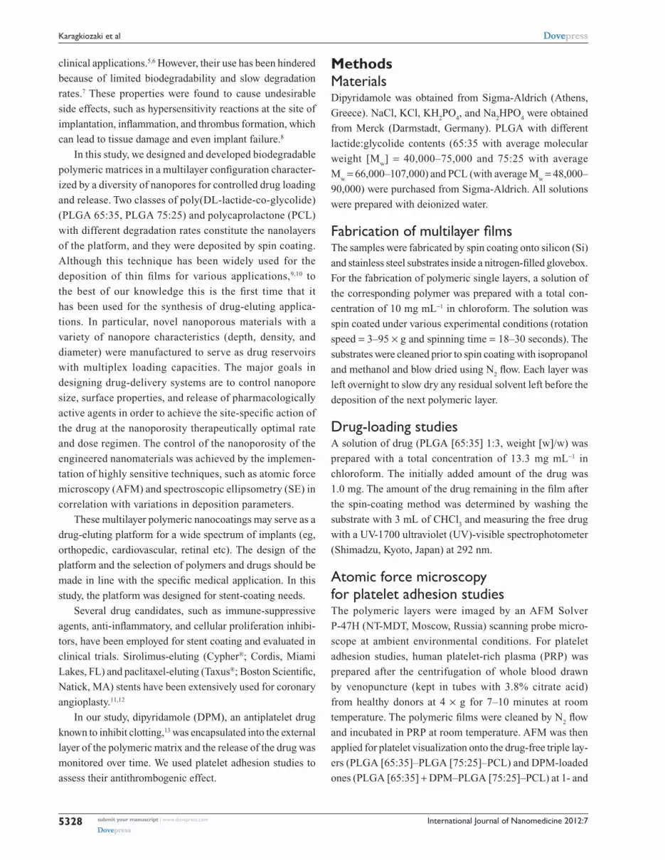
ResultsSurface nanotopography and structural properties of single- and multilayer-polymeric filmsThe surface morphology of the polymeric films was inves-
tigated by AFM. A series of single-layer polymeric films
was first fabricated. Figure 1A illustrates the topography
image of a single-layer film made of PLGA (75:25), while
the topography image of a single-layer film made of PCL is
depicted in Figure 1B. The AFM images revealed that the
PLGA films were atomically smooth with a surface roughness
of 0.2 nm under variable experimental conditions. In contrast
to the PLGA films, the PCL films (Figure 1B) were less
smooth with a surface roughness of ca. 5.5 nm. The surface
topography demonstrated the formation of spherulites with
various sizes (5–40 nm) as an outcome of the crystalliza-
tion process that takes places after thin-film deposition. In
Figure 1E, the corresponding X cross sections of Figure 1B
and C indicate that the holes between the spherulites of the
PCL single layers reaches ca. 8 nm and the pore depth in the
dual layers is ca. 10 nm.
After fabricating single-layer samples, dual-polymeric
layers made of PLGA (as the outer layer) and PCL (as
the inner layer) were constructed. Figure 1C presents the
topography image of a dual-layer polymeric film. In the
dual-layer polymeric films, the formation of nanopores was
evident. Characterization of the pores revealed variation in
diameter (20–170 nm) and in depth (2–17 nm). The pore
density was estimated between 40 and 70 pores/μm2 under
different experimental conditions. Triple-layer polymeric
films composed of PLGA (65:35; outer layer), PLGA
(75:25; intermediate layer), and PCL (inner layer) were then
manufactured. Figure 1D presents the topography image of
a triple-layer film. Nanopores were observed in the triple
layers as well. Hence, the AFM data demonstrated the for-
mation of nanopores with smaller diameter (20–150 nm)
and depth compared to the pores formed onto the surface
of dual-layer polymeric matrices. The pore density of these
triple layers was estimated between 20 and 80 pores/μm2.
The AFM data involving the surface roughness param-
eters and pore characteristics of all layers are presented
at Table 1.
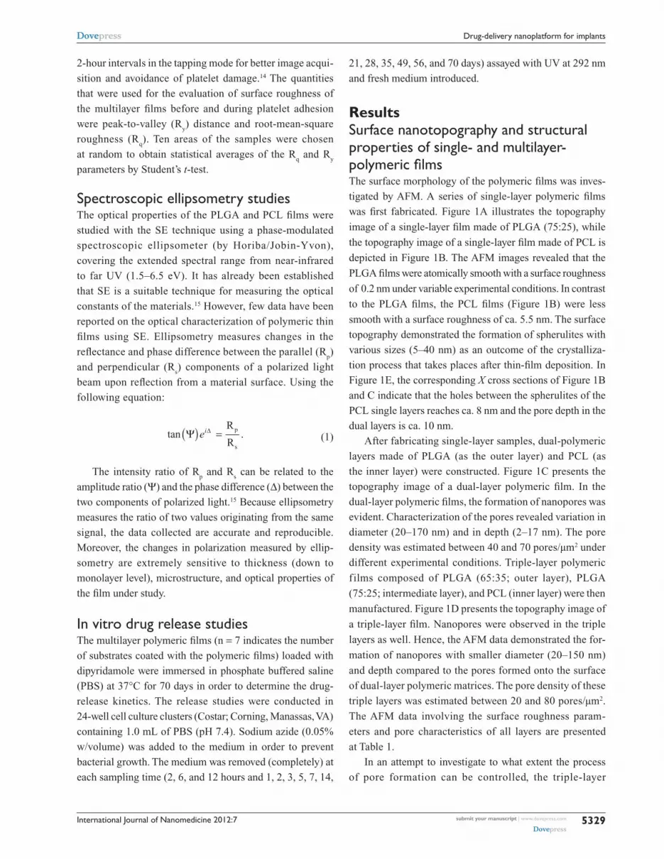
In an attempt to investigate to what extent the process
of pore formation can be controlled, the triple-layer
submit your manuscript | www.dovepress.com
Dovepress
Dovepress
5329
Drug-delivery nanoplatform for implants

International Journal of Nanomedicine 2012:7
films were fabricated under variable experimental conditions:
(a) By altering the spinning speed at 24 × g and 53 × g, two
series of triple-layer films were developed. Figure 2 depicts
the AFM topography images of triple-layer films, spin coated
at 24 × g for 30 seconds and at 53 × g for 30 seconds. Both
samples had a polymer concentration of 10 mg mL−1; (b) by
variations in polymer concentration of the outer layer (5 mg
mL−1 and 10 mg mL−1). Figure 2 shows the topography
images of triple-layer films, one with a polymer concentra-
tion of 10 mg mL−1 and the other with a concentration of
5 mg mL−1. Both samples were spin coated at 24 × g for
30 seconds. These two parameters were correlated with the
0 0.5 1.0 1.5µm
2.0 2.5 0 0.5 1.0 1.5µm
2.0 2.5
0 0.5 1.0 1.5µm
2.0 2.50 0.5 1.0 1.5µm
Distance (µm)
Hei
gh
t (n
m)
2.0 2.5
00.
51.
01.
5
µm
2.0
2.5
00.
51.
01.
5µm
2.0
2.5
00.
51.
01.
5µmn
m
nm
nm
nm
2.0
00.
20.
40.
60.
81.
01.
51.
41.
6
000
24
68
1012
12
34
56
510
1520
2530
3540
45
0.5
1.0
1.5
µm
2.0
2.5
00.5 1.0 1.5
PCL
PLGA (75:25)/PCL
2.00.0 2.5
5
10
15
20
25
30
35
A B
C
E
D
Figure 1 AFM topography images of: (A) PLGA single layer that was spin coated at 53 × g for 30 seconds. (B) PCL single layer that was spin coated at 53 × g for 30 seconds. (C) Dual layer (PLGA [75:25]–PCL) that was spin coated at 53 × g for 30 seconds. (D) Triple layer (PLGA [65:35]–PLGA [75:25]–PCL) that was spin coated at 53 × g for 30 seconds. The scan size is 2.5 μm × 2.5 μm. (E) Corresponding X cross sections of Figure 1B and C that reveal the dimensions of the holes between the spherulites of PCL single layers and the pore sizes of PLGA–PCL dual layers, respectively.Abbreviations: PLGA, poly (DL-lactide-co-glycolide); PCL, polycaprolactone.
submit your manuscript | www.dovepress.com
Dovepress
Dovepress
5330
Karagkiozaki et al
International Journal of Nanomedicine 2012:7
surface roughness, pore depth, diameter, and pore density of
the engineered triple layers (Table 2).
Spectroscopic ellipsometry studies for optical characterization of polymeric layersSE was successfully employed for determining the optical
properties, thickness, and structural characteristics of the
spin-coated polymeric films, either in the form of single-,
dual-, or triple-layer structure. The optical model that was
applied to analyze the measured data consisted of the appro-
priate number of layers for each case. For determining the
optical properties and the thickness of the films, a standard
fitting procedure was applied in which the dielectric func-
tion ε(ω) of the polymeric films was described using the
Tauc–Lorentz (TL) oscillator dispersion equation, where the
imaginary part of the dielectric function is given by
ε ω
ω ω ωω ω ω ω
ω ω20
2
202 2 2 2
1( )
( )
( ), ,=
−− +
⋅ >A C
Cg
g (2)
where ω is the photon energy, ωg the fundamental band gap
energy, A the amplitude of the oscillator, ω0 the Lorentz
resonant energy, C its broadening term, and the real part ε1(ω)
is obtained by the Kramer–Kronig integration.16
The real part of the refractive index (n) and the extinction
coefficient (k) of the complex refractive index are related to
the dielectric function by the following equations:16
ε1 = n2 – k2 (3)
and
ε2 = 2nk (4)
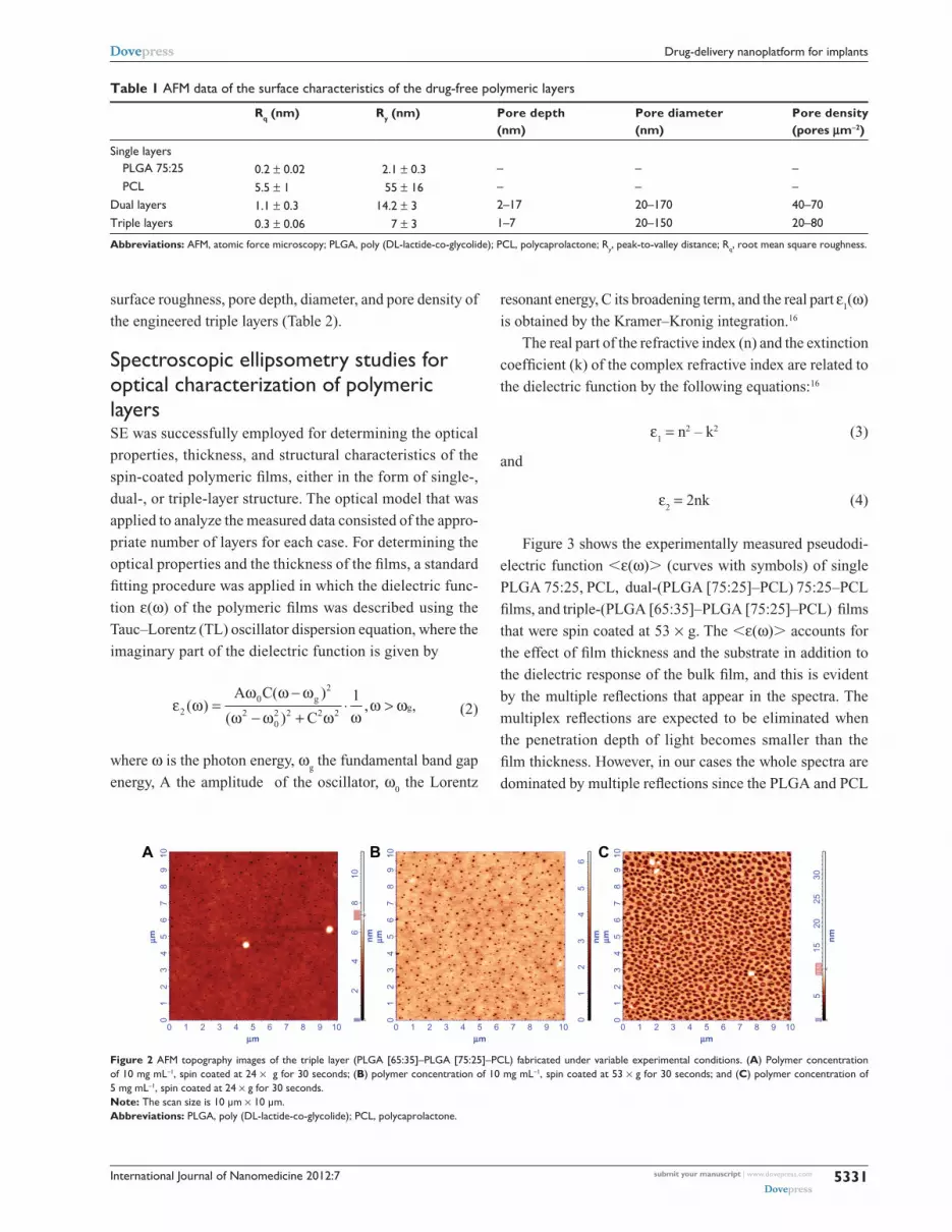
Figure 3 shows the experimentally measured pseudodi-
electric function ,ε(ω). (curves with symbols) of single
PLGA 75:25, PCL, dual-(PLGA [75:25]–PCL) 75:25–PCL
films, and triple-(PLGA [65:35]–PLGA [75:25]–PCL) films
that were spin coated at 53 × g. The ,ε(ω). accounts for
the effect of film thickness and the substrate in addition to
the dielectric response of the bulk film, and this is evident
by the multiple reflections that appear in the spectra. The
multiplex reflections are expected to be eliminated when
the penetration depth of light becomes smaller than the
film thickness. However, in our cases the whole spectra are
dominated by multiple reflections since the PLGA and PCL
Table 1 AFM data of the surface characteristics of the drug-free polymeric layers
Rq (nm) Ry (nm) Pore depth (nm)
Pore diameter (nm)
Pore density (pores μm-2)
Single layers PLGA 75:25 0.2 ± 0.02 2.1 ± 0.3 – – – PCL 5.5 ± 1 55 ± 16 – – –Dual layers 1.1 ± 0.3 14.2 ± 3 2–17 20–170 40–70Triple layers 0.3 ± 0.06 7 ± 3 1–7 20–150 20–80
Abbreviations: AFM, atomic force microscopy; PLGA, poly (DL-lactide-co-glycolide); PCL, polycaprolactone; Ry, peak-to-valley distance; Rq, root mean square roughness.
01
23
45
67
89
10
0 1 2 3 4 5µm
µm
6 7 8 9 10
01
23
45
67
89
10
0 1 2 3 4 5µm
µm
6 7 8 9 10
0 010
.50
515
nm
2025
30
07.
17
24
6
nm
810
01
23
45
6
12
34
56
78
910
0 1 2 3 4 5µm
µmnm
6 7 8 9 10
A B C
Figure 2 AFM topography images of the triple layer (PLGA [65:35]–PLGA [75:25]–PCL) fabricated under variable experimental conditions. (A) Polymer concentration of 10 mg mL−1, spin coated at 24 × g for 30 seconds; (B) polymer concentration of 10 mg mL−1, spin coated at 53 × g for 30 seconds; and (C) polymer concentration of 5 mg mL−1, spin coated at 24 × g for 30 seconds. Note: The scan size is 10 μm × 10 μm.Abbreviations: PLGA, poly (DL-lactide-co-glycolide); PCL, polycaprolactone.
submit your manuscript | www.dovepress.com
Dovepress
Dovepress
5331
Drug-delivery nanoplatform for implants
International Journal of Nanomedicine 2012:7
0
25
50
75
100
−25
−50
−751.0 1.5 2.0 2.5 3.0 3.5
Photon energy (eV)
Pse
ud
od
iele
ctri
c fu
nct
ion
4.0 4.5 5.0 5.5 6.0 6.5 7.0
0
25
50
75
100
−25
−50
−751.0 1.5 2.0 2.5 3.0 3.5
Photon energy (eV)
Pse
ud
od
iele
ctri
c fu
nct
ion
4.0 4.5 5.0 5.5 6.0 6.5 7.0
0
25
50
75
100
−25
−50
−751.0 1.5 2.0 2.5 3.0 3.5
Photon energy (eV)
Pse
ud
od
iele
ctri
c fu
nct
ion
4.0 4.5 5.0 5.5 6.0 6.5 7.0
0
25
50
75
100
−25
−50
−751.0 1.5 2.0 2.5 3.0 3.5
Photon energy (eV)
Pse
ud
od
iele
ctri
c fu
nct
ion
4.0 4.5 5.0 5.5 6.0 6.5 7.0
<ε1(ω)>EXP
<ε2(ω)>EXP
<ε1(ω)>FIT
<ε2(ω)>FIT
<ε1(ω)>EXP
<ε2(ω)>EXP
<ε1(ω)>FIT
<ε2(ω)>FIT
<ε1(ω)>EXP
<ε2(ω)>EXP
<ε1(ω)>FIT
<ε2(ω)>FIT
<ε1(ω)>EXP
<ε2(ω)>EXP
<ε1(ω)>FIT
<ε2(ω)>FIT
PLGA/c-Si PLGA/PCL/c-Si
PLGA 65:35/PLGA 75:25/PCL/c-SiPCL/c-Si
A B
C D
PLGA, d = 117 nm
c-Si
PLGA, d = 112 nm
PCL, d = 58 nm
c-Si
d = 112 nm50% PCL50% voids
PCL, d = 118 nm
c-Sic-Si
d = 29 nmPLGA65:35
d = 44 nmPLGA
d = 61 nmPCL
75:25
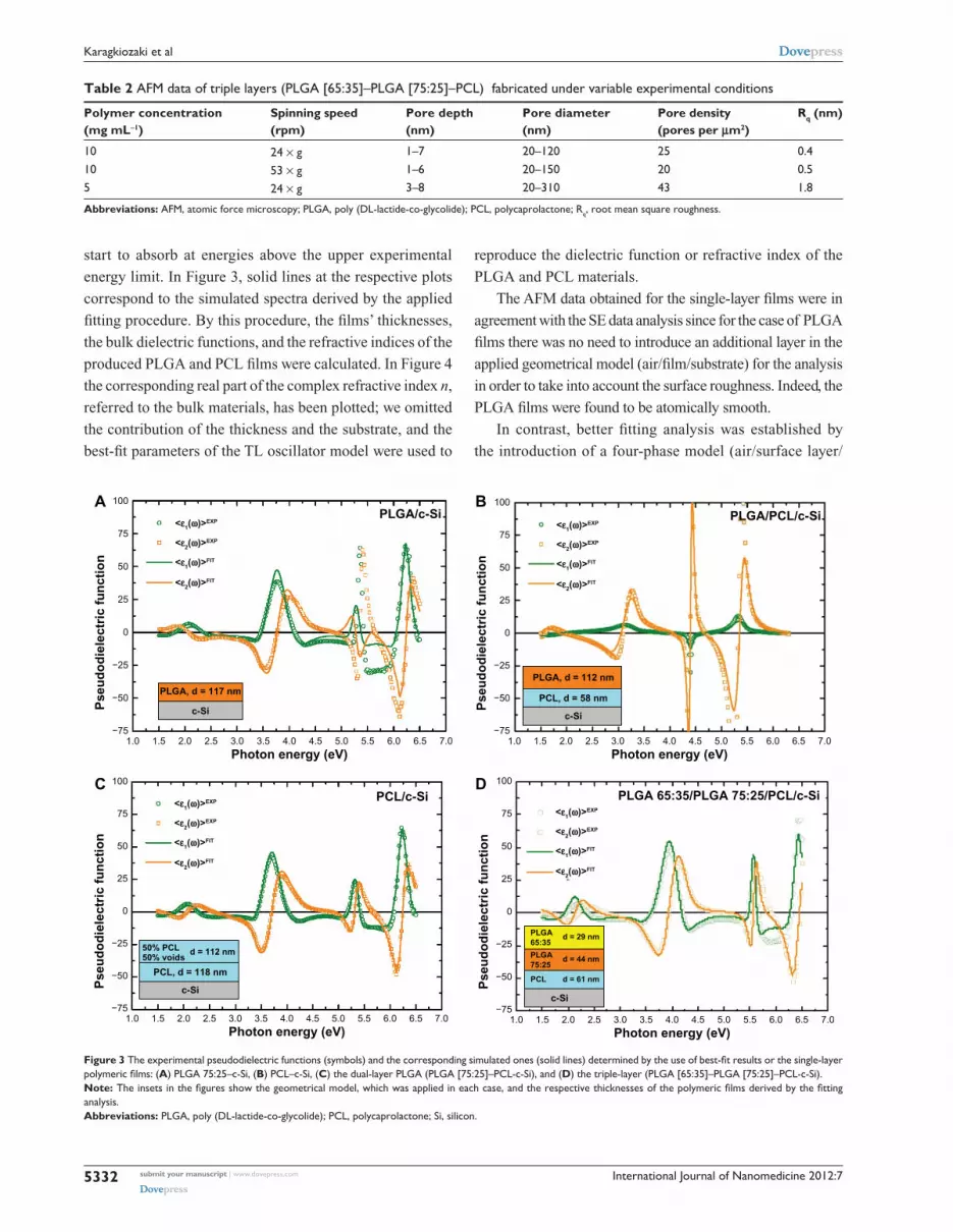
Figure 3 The experimental pseudodielectric functions (symbols) and the corresponding simulated ones (solid lines) determined by the use of best-fit results or the single-layer polymeric films: (A) PLGA 75:25–c-Si, (B) PCL–c-Si, (C) the dual-layer PLGA (PLGA [75:25]–PCL-c-Si), and (D) the triple-layer (PLGA [65:35]–PLGA [75:25]–PCL-c-Si).Note: The insets in the figures show the geometrical model, which was applied in each case, and the respective thicknesses of the polymeric films derived by the fitting analysis.Abbreviations: PLGA, poly (DL-lactide-co-glycolide); PCL, polycaprolactone; Si, silicon.
Table 2 AFM data of triple layers (PLGA [65:35]–PLGA [75:25]–PCL) fabricated under variable experimental conditions
Polymer concentration (mg mL-1)
Spinning speed (rpm)
Pore depth (nm)
Pore diameter (nm)
Pore density (pores per μm2)
Rq (nm)
10 24 × g 1–7 20–120 25 0.410 53 × g 1–6 20–150 20 0.55 24 × g 3–8 20–310 43 1.8
Abbreviations: AFM, atomic force microscopy; PLGA, poly (DL-lactide-co-glycolide); PCL, polycaprolactone; Rq, root mean square roughness.
start to absorb at energies above the upper experimental
energy limit. In Figure 3, solid lines at the respective plots
correspond to the simulated spectra derived by the applied
fitting procedure. By this procedure, the films’ thicknesses,
the bulk dielectric functions, and the refractive indices of the
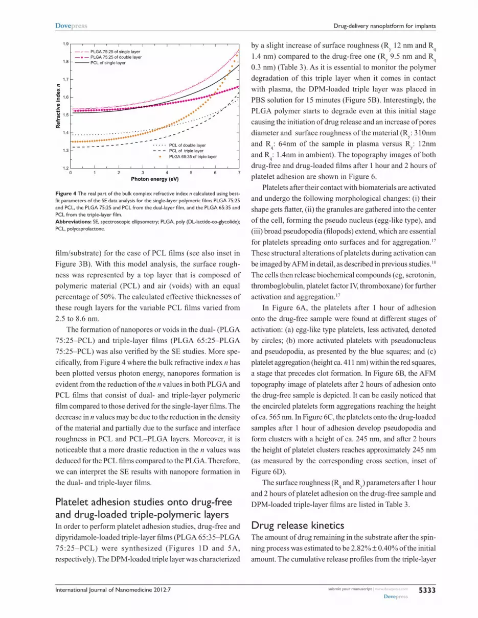
produced PLGA and PCL films were calculated. In Figure 4
the corresponding real part of the complex refractive index n,
referred to the bulk materials, has been plotted; we omitted
the contribution of the thickness and the substrate, and the
best-fit parameters of the TL oscillator model were used to
reproduce the dielectric function or refractive index of the
PLGA and PCL materials.
The AFM data obtained for the single-layer films were in
agreement with the SE data analysis since for the case of PLGA
films there was no need to introduce an additional layer in the
applied geometrical model (air/film/substrate) for the analysis
in order to take into account the surface roughness. Indeed, the
PLGA films were found to be atomically smooth.
In contrast, better fitting analysis was established by
the introduction of a four-phase model (air/surface layer/
submit your manuscript | www.dovepress.com
Dovepress
Dovepress
5332
Karagkiozaki et al
International Journal of Nanomedicine 2012:7
film/substrate) for the case of PCL films (see also inset in
Figure 3B). With this model analysis, the surface rough-
ness was represented by a top layer that is composed of
polymeric material (PCL) and air (voids) with an equal
percentage of 50%. The calculated effective thicknesses of
these rough layers for the variable PCL films varied from
2.5 to 8.6 nm.
The formation of nanopores or voids in the dual- (PLGA
75:25–PCL) and triple-layer films (PLGA 65:25–PLGA
75:25–PCL) was also verified by the SE studies. More spe-
cifically, from Figure 4 where the bulk refractive index n has
been plotted versus photon energy, nanopores formation is
evident from the reduction of the n values in both PLGA and
PCL films that consist of dual- and triple-layer polymeric
film compared to those derived for the single-layer films. The
decrease in n values may be due to the reduction in the density
of the material and partially due to the surface and interface
roughness in PCL and PCL–PLGA layers. Moreover, it is
noticeable that a more drastic reduction in the n values was
deduced for the PCL films compared to the PLGA. Therefore,
we can interpret the SE results with nanopore formation in
the dual- and triple-layer films.
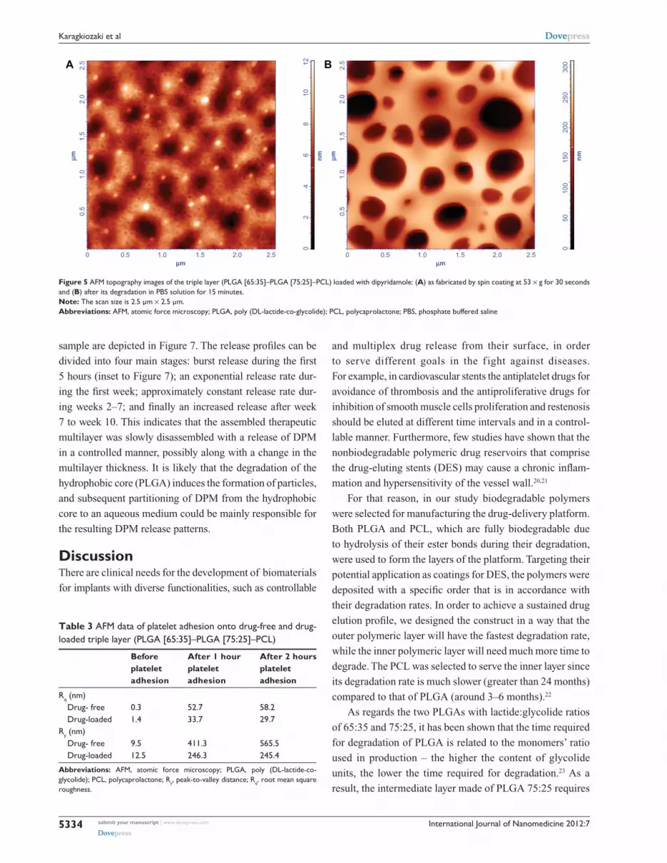
Platelet adhesion studies onto drug-free and drug-loaded triple-polymeric layersIn order to perform platelet adhesion studies, drug-free and
dipyridamole-loaded triple-layer films (PLGA 65:35–PLGA
75:25–PCL) were synthesized (Figures 1D and 5A,
respectively). The DPM-loaded triple layer was characterized
by a slight increase of surface roughness (Ry 12 nm and R
q
1.4 nm) compared to the drug-free one (Ry 9.5 nm and R
q
0.3 nm) (Table 3). As it is essential to monitor the polymer
degradation of this triple layer when it comes in contact
with plasma, the DPM-loaded triple layer was placed in
PBS solution for 15 minutes (Figure 5B). Interestingly, the
PLGA polymer starts to degrade even at this initial stage
causing the initiation of drug release and an increase of pores
diameter and surface roughness of the material (Ry: 310nm
and Rq: 64nm of the sample in plasma versus R
y: 12nm
and Rq: 1.4nm in ambient). The topography images of both
drug-free and drug-loaded films after 1 hour and 2 hours of
platelet adhesion are shown in Figure 6.
Platelets after their contact with biomaterials are activated
and undergo the following morphological changes: (i) their
shape gets flatter, (ii) the granules are gathered into the center
of the cell, forming the pseudo nucleus (egg-like type), and
(iii) broad pseudopodia (filopods) extend, which are essential
for platelets spreading onto surfaces and for aggregation.17
These structural alterations of platelets during activation can
be imaged by AFM in detail, as described in previous studies.18
The cells then release biochemical compounds (eg, serotonin,
thromboglobulin, platelet factor IV, thromboxane) for further
activation and aggregation.17
In Figure 6A, the platelets after 1 hour of adhesion
onto the drug-free sample were found at different stages of
activation: (a) egg-like type platelets, less activated, denoted
by circles; (b) more activated platelets with pseudonucleus
and pseudopodia, as presented by the blue squares; and (c)
platelet aggregation (height ca. 411 nm) within the red squares,
a stage that precedes clot formation. In Figure 6B, the AFM
topography image of platelets after 2 hours of adhesion onto
the drug-free sample is depicted. It can be easily noticed that
the encircled platelets form aggregations reaching the height
of ca. 565 nm. In Figure 6C, the platelets onto the drug-loaded
samples after 1 hour of adhesion develop pseudopodia and
form clusters with a height of ca. 245 nm, and after 2 hours
the height of platelet clusters reaches approximately 245 nm
(as measured by the corresponding cross section, inset of
Figure 6D).
The surface roughness (Rq and R
y) parameters after 1 hour
and 2 hours of platelet adhesion on the drug-free sample and
DPM-loaded triple-layer films are listed in Table 3.
Drug release kineticsThe amount of drug remaining in the substrate after the spin-
ning process was estimated to be 2.82% ± 0.40% of the initial
amount. The cumulative release profiles from the triple-layer
01.2
1.3
1.4
1.5
1.6
1.7
1.8
1.9
1 2 3 4
Photon energy (eV)
Ref
ract
ive
ind
ex n
5 6 7
PLGA 75:25 of single layerPLGA 75:25 of double layerPCL of single layer
PCL of double layerPCL of triple layerPLGA 65:35 of triple layer
Figure 4 The real part of the bulk complex refractive index n calculated using best-fit parameters of the SE data analysis for the single-layer polymeric films PLGA 75:25 and PCL, the PLGA 75:25 and PCL from the dual-layer film, and the PLGA 65:35 and PCL from the triple-layer film.Abbreviations: SE, spectroscopic ellipsometry; PLGA, poly (DL-lactide-co-glycolide); PCL, polycaprolactone.
submit your manuscript | www.dovepress.com
Dovepress
Dovepress
5333
Drug-delivery nanoplatform for implants
International Journal of Nanomedicine 2012:7
sample are depicted in Figure 7. The release profiles can be
divided into four main stages: burst release during the first
5 hours (inset to Figure 7); an exponential release rate dur-
ing the first week; approximately constant release rate dur-
ing weeks 2–7; and finally an increased release after week
7 to week 10. This indicates that the assembled therapeutic
multilayer was slowly disassembled with a release of DPM
in a controlled manner, possibly along with a change in the
multilayer thickness. It is likely that the degradation of the
hydrophobic core (PLGA) induces the formation of particles,
and subsequent partitioning of DPM from the hydrophobic
core to an aqueous medium could be mainly responsible for
the resulting DPM release patterns.
DiscussionThere are clinical needs for the development of biomaterials
for implants with diverse functionalities, such as controllable
and multiplex drug release from their surface, in order
to serve different goals in the f ight against diseases.
For example, in cardiovascular stents the antiplatelet drugs for
avoidance of thrombosis and the antiproliferative drugs for
inhibition of smooth muscle cells proliferation and restenosis
should be eluted at different time intervals and in a control-
lable manner. Furthermore, few studies have shown that the
nonbiodegradable polymeric drug reservoirs that comprise
the drug-eluting stents (DES) may cause a chronic inflam-
mation and hypersensitivity of the vessel wall.20,21
For that reason, in our study biodegradable polymers
were selected for manufacturing the drug-delivery platform.
Both PLGA and PCL, which are fully biodegradable due
to hydrolysis of their ester bonds during their degradation,
were used to form the layers of the platform. Targeting their
potential application as coatings for DES, the polymers were
deposited with a specific order that is in accordance with
their degradation rates. In order to achieve a sustained drug
elution profile, we designed the construct in a way that the
outer polymeric layer will have the fastest degradation rate,
while the inner polymeric layer will need much more time to
degrade. The PCL was selected to serve the inner layer since
its degradation rate is much slower (greater than 24 months)
compared to that of PLGA (around 3–6 months).22
As regards the two PLGAs with lactide:glycolide ratios
of 65:35 and 75:25, it has been shown that the time required
for degradation of PLGA is related to the monomers’ ratio
used in production – the higher the content of glycolide
units, the lower the time required for degradation.23 As a
result, the intermediate layer made of PLGA 75:25 requires
0 0.5 1.0 1.5µm
2.0 2.5
0.5
1.0
1.5
µm2.
02.
5
0 0.5 1.0 1.5µm
2.0 2.5
0.5
02
46
810
12
1.0
1.5
µmnm
050
100
150
200
250
300
nm
2.0
2.5A B
Figure 5 AFM topography images of the triple layer (PLGA [65:35]–PLGA [75:25]–PCL) loaded with dipyridamole: (A) as fabricated by spin coating at 53 × g for 30 seconds and (B) after its degradation in PBS solution for 15 minutes. Note: The scan size is 2.5 μm × 2.5 μm. Abbreviations: AFM, atomic force microscopy; PLGA, poly (DL-lactide-co-glycolide); PCL, polycaprolactone; PBS, phosphate buffered saline
Table 3 AFM data of platelet adhesion onto drug-free and drug-loaded triple layer (PLGA [65:35]–PLGA [75:25]–PCL)
Before platelet adhesion
After 1 hour platelet adhesion
After 2 hours platelet adhesion
Rq (nm) Drug- free 0.3 52.7 58.2 Drug-loaded 1.4 33.7 29.7Ry (nm) Drug- free 9.5 411.3 565.5 Drug-loaded 12.5 246.3 245.4
Abbreviations: AFM, atomic force microscopy; PLGA, poly (DL-lactide-co-glycolide); PCL, polycaprolactone; Ry, peak-to-valley distance; Rq, root mean square roughness.
submit your manuscript | www.dovepress.com
Dovepress
Dovepress
5334
Karagkiozaki et al

International Journal of Nanomedicine 2012:7
02
46
810
1214
1618
20
0 2 4 6 8 10µm
µm
12 14 16 18 20
00 049
0.0
50
50
nm
200
300
400
023
0.0
50
nm
100
150
200
023
4.0
50
nm
nm
100
150
200
100
150
200
250
300
283.
035
040
0
24
68
1012
1416
1820
0 2 4 6 8 10µm
µm
12 14 16 18 20
02
46
810
1214
1618
20
0 2 4 6 8 10µm
µm
12 14 16 18 200
24
68
1012
1416
1820
0 2 4 6 8 10µm
µm12 14 16 18 20
A B
C D
0
100
120
140
160
180
200
220
240
5 10Distance (µm)
Hei
ght (
nm)
15 20
Figure 6 AFM topography images of platelets onto (A and B) drug-free and (C and D) dipyridamole-loaded triple layers (PLGA [65:35]–PLGA [75:25]–PCL). Both samples were spin coated at 53 × g for 30 seconds. (A) drug-free sample after 1 hour of platelet adhesion; (B) drug-free sample after 2 hours of platelet adhesion; (C) drug-loaded sample after 1 hour of platelet adhesion; and (D) drug-loaded sample after 2 hours of platelet adhesion with a typical cross section (inset). Note: The scan size is 20 μm × 20 μm. Abbreviations: AFM, atomic force microscopy; PLGA, poly (DL-lactide-co-glycolide); PCL, polycaprolactone.
0
0
0 10 20 30 40 50 60 70
5
10
15
20
25
30
35
40
0 50 100 150Time (min)
Time (days)
Cu
mu
lati
ve r
elea
se (
%)
Cu
mu
lati
ve r
elea
se (
%)
200 250 300
2
4
6
8
10
12
14
A
B
Figure 7 The dipyridamole cumulative release profiles from the three layer samples.Note: The inset graph depicts the burst effect that occurs during the first 5 hours.
more time to degrade compared to the outer layer made of
PLGA 65:35.
In a fundamental drug-release study of biomaterials,
an uncontrolled drug release occurred when drug adhered
to the outer surface rather than being deposited within the
pores.24 There are difficulties with control of loading and
release of drug from nanometer-sized pores, which is a sig-
nificant challenge due to their small size. Thus, in this study
we describe an experimental procedure for manufacturing
multilayer polymeric thin films with tailored nanoporosity for
drug loading by the implementation of spin coating. It was
noticeable by the data provided by the surface characteriza-
tion techniques AFM and SE that the PLGA–PCL multilayers
were highly nanoporous in contrast to the smooth PLGA and
spherulite-characterized PCL single layers.
The origin of pore formation may be attributed to a
demixing process, which generally involves the spontane-
ous phase separation of polymer blend occurring under
the specific conditions of spin coating. Similar holes or
pores have been reported with a polymer blend phase
separating via a spinodal decomposition mechanism.25
At the initial stages of spin coating, the upper part of the
substrate film was dissolved, forming a blend solution
since chloroform is a common solvent for the selected
polymers. This hypothesis was verified by the SE data
showing that the thickness of the substrate f ilm was
reduced after the deposition of the subsequent film (as
depicted in Figure 3C and D).
submit your manuscript | www.dovepress.com
Dovepress
Dovepress
5335
Drug-delivery nanoplatform for implants

International Journal of Nanomedicine 2012:7
of their functionality as well as for the prediction of their
effectiveness.
In this work we show successful implementation of SE
for determining the thicknesses, nanoporosity and the opti-
cal constants of the spin-coated polymeric films. In regards
to thickness measurements, the SE data revealed (Figure 3C
and D) that the thickness of the substrate film was reduced
after the deposition of the subsequent film due to a spinodal
decomposition mechanism.
Dipyridamole was selected for loading within the
nanopores of the polymeric multilayers to examine their
antiplatelet effect in order to be used as potential coatings
for cardiovascular implants. The ultimate goal was to inhibit
thrombus formation, which may lead to implant failure and
patient complications. The DPM burst effect and release pro-
file during the first week was typical of diffusion-controlled
systems. The third and fourth phase of the constant release
rate presumably involved degradation of PLGA combined
with diffusion of the remaining drug that was more firmly
attached to polymer.
As polymer degradation is an important factor that
determines drug elution, the AFM enables us to image the
degraded DPM-loaded triple-polymeric layer after placing
it in PBS solution for 15 minutes. At this initial stage, an
increase in surface roughness of the drug-loaded layer was
observed due to the degradation of PLGA, the formation of
particles occurred, and there was resultant partitioning of the
drug from PLGA, causing drug release.
Our previous studies have shown that AFM is a useful tool
for imaging platelets in real time, with high precision and
without destroying sensitive cells.27,28 The AFM images of
platelets onto drug-free, triple-layer, polymeric films showed
that the platelets are highly activated, forming pseudopodia
and aggregates. From the AFM data presented in Table 3, it
can be deduced that there was a time-dependent increase of
Rq and R
y parameters, indicative of platelet aggregation. Our
previous studies showed that the process of platelet adhesion
onto nanomaterials that are atomically smooth or have low
nanoroughness is time-dependent, resulting in an increase in
Ry due to platelet aggregation.28,29
In contrast, in the case of the DPM-loaded triple-layer
configuration of the biodegradable polymers, after 1 hour of
platelet adhesion, although the cells develop pseudopodia and
aggregations, the height is lower and remains at the same level
after 2 hours. These findings provide evidence that dipyrida-
mole loaded into the outer layer inhibits the platelet tendency
to form high clusters, which are a prestage of thrombus. More-
over, although the triple-layer drug-loaded films are rougher
In addition, Affrossman et al26 reported that morphology
evolution is strongly depended on the polymer blend ratio
and the film thickness. If the weight content of one compo-
nent in a polymer blend is much higher (about 90%) than
the other component, then small holes are observed. When
this fraction decreases to about 70%, the diameter of the
holes increases and the holes start to coalesce. An analo-
gous behavior is observed in our case comparing Figure 2A
and C; in Figure 2A the concentration of the added PLGA
(65:35) solution is 10 mg mL−1, while in Figure 2C the
concentration is 5 mg mL−1. Since the amount of the added
solution is the same in both cases, the reduction in the film
thickness and the percentage analogy of the two types of
PLGA in the latter case explains its characteristic morphol-
ogy (Figure 2C).
The pore distribution in the nanoporous biomaterials
is a key factor for determination of drug release kinetics
and directional control. By varying the size (ie, diameter,
volume) and number of the porous reservoirs, a range of
therapeutic agent loading levels can be achieved.25 In the
case of DES where the drug targets different sites of the
vessel, it is necessary to control the porosity of the stent
nanocoatings for directional and tailored drug loading
and resultant release. In this study we found that by tun-
ing the spin-coating parameters, the pore distribution and
dimensions, interconnectivity, and density of the materials
can be controlled. The AFM images demonstrated that by
increasing the spinning speed, the pore diameter and surface
roughness were increased, whereas the pore density was
decreased. The pore depth was not found to be affected by
this parameter, as shown in Table 2. Moreover, the decrease
in the polymer concentration of the outer layer of PLGA
(65:35), keeping the rotation speed and spinning time con-
stant, resulted in an increase in pore diameter, pore density,
and surface roughness of the engineered biomaterials (as
presented at Table 3).
In controlled polymeric drug-delivery systems, the drug-
delivery rates are mainly determined by the dynamics of
polymer degradation, which is strongly related to polymer
structure, morphology, and properties. All these can be cor-
related to the optical properties of the polymeric films. The
study of the optical properties and the consequent creation
of a database containing specific optical constants of the
candidate polymeric films for such systems can provide a
powerful tool for a nondestructive and creditable methodol-
ogy to evaluate their performance. Hence, one can consider
that the complete elucidation of the optical response of
the engineered systems is essential for the achievement
submit your manuscript | www.dovepress.com
Dovepress
Dovepress
5336
Karagkiozaki et al
International Journal of Nanomedicine 2012:7
compared to the drug-free films (Ry 12.5 nm versus 9.5 nm and
Rq 1.4 nm versus 0.3 nm, respectively) after 1 hour of platelet
adhesion there is a decrease in surface roughness (as depicted
by the decrease in Ry and R
q values). This flattening of the
surface biomaterials due to platelet adhesion is a phenomenon
that shows how platelets behave toward nanomaterials with
grooves, pores, and other morphologic irregularities.
ConclusionIn this study, a multilayer and biodegradable polymeric
platform with diverse nanopores for drug-eluting implants
was successfully developed. The design of the multilayer
drug-eluting coatings was based on polymer degradation rates
and properties in order to achieve a long-term and controlled
drug elution profile; the spin-coating technique was imple-
mented for this purpose. The spontaneous phase separation
of polymer blends during spin-coating conditions leads to
the creation of nanopores via the spinodal decomposition
mechanism. The formation of nanopores onto the biomaterial
surface with tailored characteristics (pore diameter, depth,
density, surface roughness) was achieved by tuning the
growth parameters.
The complementary AFM and SE studies for determina-
tion of structural characteristics, film thickness, and optical
properties provide essential information for drug-loading
capacities. A case study of dipyridamole loading within
the nanopores and platelet studies for evaluation of their
antiplatelet effect was carried out. It was found that the
dipyridamole-loaded coatings inhibit the platelets tendency
to form high aggregations.
In-parallel, drug release kinetics studies shown the
release profiles of DPM that indicate that the biodegradable
multilayer was slowly degraded with releasing DPM in a
controlled manner. By fine tuning the dipyridamole release
amount in a desired period, it would be possible to prevent
thrombosis and recreate physiologically normal arterial
conditions after DPM release.
AcknowledgmentsThis work was partially supported by NanoArthroxondros
project 09SYN-41-1150.
DisclosureThe authors disclose no conflicts of interest.
References1. Kang H, Kim DJ, Park S, Yoo J, Ryu YS. Controlled drug release
using nanoporous anodic aluminum oxide on stent. Thin Solid Films. 2007;515:5184–5187.
2. Orosz KE, Gupta S, Hassink M, et al. Delivery of antiangiogenic and antioxidant drugs of ophthalmic interest through a nanoporous inor-ganic filter. Mol Vis. 2004;10:555–565.
3. Losic D, Velleman L, Kant K, et al. Self-ordering electrochemistry: a simple approach for engineering nanopore and nanotube arrays for emerging applications. Aust J Chem. 2011;64:294–301.
4. Aninwene GE II, Yao C, Webster TJ. Enhanced osteoblast adhesion to drug-coated anodized nanotubular titanium surfaces. Int J Nanomedicine. 2008;3:257–264.
5. Nuxoll EE, Hillmyer MA, Wang R, Leighton C, Siegel RA. Composite block polymer-microfabricated silicon nanoporous membrane. ACS Appl Mater Interfaces. 2009;1:888–893.
6. Bielecka U, Lutsyk P, Janus K, Sworakowski J, Bartkowiak W. Effect of solution aging on morphology and electrical characteristics of regioregular P3HT FETs fabricated by spin-coating and spray coating. Org Electr. 2011;12:1768–1776.
7. Mansour H, Sohn M, Ghananeem A, DeLuca. P. Materials for pharmaceutical dosage forms: molecular pharmaceutics and controlled release drug delivery aspects. Int J Mol Sci. 2010;11:3298–3322.
8. Hara H, Nakamura M, Palmaz JC, Schwartz RS. Role of stent design and coatings on restenosis and thrombosis. Adv Drug Deliv Rev. 2006;58: 377–386.
9. Karagiannidis P, Kassavetis S, Pitsalidis C, Logothetidis S. Thermal annealing effect on the nanomechanical properties and structure of P3HT:PCBM thin films. Thin Solid Films. 2011;519:4105–4109.
10. Souza F, Lopes K, Nascente P, Leite E. Nanostructured hematite thin films produced by spin-coating deposition solution: Application in water splitting. Solar Energy Mater and Solar Cells. 2009;93:362–368.
11. Acharya G, Park K. Mechanisms of controlled drug release from drug-eluting stents. Adv Drug Deliv Rev. 2006;58:387–340.
12. Garg S, Serruys P. Coronary stents: current status. J Am Coll Cardiol. 2010;56:1–42.
13. Mammen EF. An overview of dipyridamole. Thrombosis Res. 1990;57: 1–3.
14. Müller D, Anderson K. Biomolecular imaging using atomic force microscopy. Trends in Biotech. 2002;20:545–549.
15. Azzam RMA, Boshara N. Ellipsometry and polarized light. In: Azzam RMA, editor. Ellipsometry and Polarized Light. North Holland: Amsterdam; 1977 San Diego.
16. Jellison GE, Modine FA. Parameterization of the optical functions of amorphous materials in the interband region. Appl. Phys. Lett. 1996; 69(3):371–373.
17. Hartwig JH. Platelet structure. In: Michelson AD, editor. Platelets. Academic Press; San Diego 2002:37–45.
18. Karagkiozaki V, Logothetidis S, Kalfagiannis N, et al. AFM probing platelets activation behavior on titanium nitride nanocoatings for biomedical applications. J Nanomedicine. 2009;5:64–72.
19. Bavry A, Kumbhani D, Helton T, Borek P, Mood G, Bhatt D. Late thrombosis of drug-eluting stents: a meta-analysis of randomized clinical trials. Am J Med. 2006;119:1056–1061.
20. Liistro F, Colombo A. Late acute thrombosis after paclitaxel eluting stent implantation. Heart. 2001;86:262–264.
21. Armentano I, Dottori M, Fortunati E, Mattioli S, Kenny JM. Biodegradable polymer matrix nanocomposites for tissue engineering: a review. Polym Degrad Stab. 2010;95:2126–2146.
22. Lu L, Garcia CA, Mikos AG. In vitro degradation of thin poly(DL-lactic-co-glycolic acid) films. J Biomed Mater Res. 1999;46:236–244.
23. Gultepe E, Nagesha D, Sridhar S, Amiji M. Nanoporous inorganic membranes or coatings for sustained drug delivery in implantable devices. Adv Drug Deliv Rev. 2010;62:305–315.
24. Norman J, Desal T. Methods for fabrication of nanoscale topography for tissue engineering scaffolds. Ann Biomed Eng. 2006;34(1):89–101.
25. Affrossman S, Henn G, O’Neill S, Pethrick P, Stamm M. Surface topography and composition of deuterated polystyrene-poly (bromostyrene) blends. Macromolecules 1996;29:5010–5016.
26. Hughes G. Nanostructure-mediated drug delivery. J Nanomedicine. 2005;1:22–30.
submit your manuscript | www.dovepress.com
Dovepress
Dovepress
5337
Drug-delivery nanoplatform for implants
International Journal of Nanomedicine
Publish your work in this journal
Submit your manuscript here: http://www.dovepress.com/international-journal-of-nanomedicine-journal
The International Journal of Nanomedicine is an international, peer-reviewed journal focusing on the application of nanotechnology in diagnostics, therapeutics, and drug delivery systems throughout the biomedical field. This journal is indexed on PubMed Central, MedLine, CAS, SciSearch®, Current Contents®/Clinical Medicine,
Journal Citation Reports/Science Edition, EMBase, Scopus and the Elsevier Bibliographic databases. The manuscript management system is completely online and includes a very quick and fair peer-review system, which is all easy to use. Visit http://www.dovepress.com/ testimonials.php to read real quotes from published authors.
International Journal of Nanomedicine 2012:7
27. Karagkiozaki V, Logothetidis S, Lousinian S, Giannoglou G. Impact of surface electric properties of carbon-based thin films on platelets activation for nano-medical and nano-sensing applications. Int J Nanomedicine. 2008;3:461–469.
28. Karagkiozaki V, Logothetidis S, Laskarakis A, Giannoglou G, Lousinian S. AFM Study of the thrombogenicity of carbon-based coatings for cardiovascular applications. Mater Sci Eng B. 2008;152:16–21.
29. Karagkiozaki V, Logothetidis S, Kassavetis S, Giannoglou G. Nanomedicine for the reduction of the thrombogenicity of stent coatings. Int J Nanomedicine. 2010;5:239–248.
submit your manuscript | www.dovepress.com
Dovepress
Dovepress
Dovepress
5338
Karagkiozaki et al


