Embed Size (px)
Citation preview
US Department of Defense
US Army Research
University of Nebraska - Lincoln Year
Detection of the Superoxide Radical
Anion Using Various Alkanethiol
Monolayers and Immobilized
Cytochrome c
Xiaojun J. Chen Alan C. West
Donald M. Cropek Scott Banta
This paper is posted at DigitalCommons@University of Nebraska - Lincoln.
http://digitalcommons.unl.edu/usarmyresearch/10

Detection of the Superoxide Radical Anion UsingVarious Alkanethiol Monolayers and ImmobilizedCytochrome c
Xiaojun J. Chen,†,‡,§ Alan C. West,† Donald M. Cropek,‡ and Scott Banta*,†
Department of Chemical Engineering, Columbia University, New York, New York, 10027, and U.S. Army EngineerResearch and Development Center, Construction Engineering Research Laboratory (CERL), Champaign, Illinois 61826
The superoxide radical anion (SO) is a critical biomarkerfor monitoring cellular stress responses. ElectrochemicalSO biosensors are frequently constructed through thecovalent immobilization of cytochrome c (Cyt c) onto self-assembled monolayers (SAMs); however, a detailed com-parison of these systems as well as configuration influenceon SO detection is needed to enable robust applications.Two reaction pathways, oxidation of SO by the SAM-modified gold electrode or electron transfer through aprotein and monolayer relay, may be involved during theelectrochemical detection of SO with Cyt c, depending onthe SAM that is used. Although electrodes with SAMsalone can exhibit a high sensitivity and low limit ofdetection (LOD) for the SO, they can suffer from a strongresponse to the presence of interferents such as hydrogenperoxide and ascorbic acid. Electrodes with immobilizedCyt c show decreased sensitivity, but exhibit betterselectivity and resistance to fouling in complex media.Considering the trade-offs between sensitivity, selectivity,and LOD for SO detection, a bioelectrode made with Cytc immobilized on dithiobis(succinimidyl)propionate (DTSP)appears to be the most suitable configuration. In phos-phate buffer, the DTSP/Cyt c electrode has a sensitivityof 410 nA µM-1 cm-2 and an LOD for SO of 73 nM.Results are also presented for the detection of SO in acomplex tissue culture media (MEM) with and withoutserum, and the sensitivity of the DTSP/Cyt c in MEM inthe absence of serum increased to 640 nA µM-1 cm-2.By measuring SO with a DTSP/Cyt c electrode before andafter the addition of a bolus of the superoxide dismutase(SOD) enzyme, the specificity of the SOD enzyme can becombined with the sensitivity of Cyt c system.
Interest in developing methods for detecting the concentrationof the superoxide radical anion (O2
•-) in physiologically relevantenvironments has been increasing, as it is involved in both normalphysiology and disease pathology.1-5 Superoxide radical anion
(SO) is an important reactive oxygen species (ROS) deployed bythe immune system to defend against viral or bacterial attack.For example, SO can be generated during the oxidative burst ofphagocytic cells such as activated macrophages or neutrophils tokill invasive microorganisms.6 SO is also generated by nonph-agocytic cells, and it is involved in signal transduction andregulation. Most cell types can generate a small oxidative burstupon stimulation by growth factors, cytokines, and hormones, andit is thought that the ROS can serve as secondary messengers.2
Oxidative stress is also involved in various pathological conditionsincluding cancer, cardiovascular disease, rheumatoid arthritis,neurological disorders, ischemia/reperfusion, diabetes, and aging.1,2
Robust and reliable detection of SO could improve our under-standing of the pathology and physiology of clinical conditionsthat involve ROS.7-9 However, SO undergoes rapid self andenzymatic disproportionation under normal physiological condi-tions (a half-life in the range of milliseconds to seconds at neutralpH values), leading to low peak concentrations (∼1 × 10-11 M).Thus, selective, sensitive, and fast-response detection methodsare needed to quantify the SO concentrations in a variety of invitro and in vivo systems.
Over the past decades, several detection methods have beendeveloped for SO measurement.10,11 Electrochemical methods forSO quantification are of particular interest because they can beeasily integrated into portable analytical devices. The incorporationof proteins onto bioelectrodes enables the creation of systems thatcan exploit highly evolved biological recognition and catalysis inorder to increase specificity.12 The two main proteins used in theconstruction of SO biosensors are cytochrome c (Cyt c) andsuperoxide dismutase (SOD).13 Cyt c is a ubiquitous protein thatis involved in shuttling electrons during oxidative phosphorylation,but it is not itself an enzyme specific for the conversion of SO.SOD is an enzyme, and it evolved specifically for the rapid
* To whom correspondence should be addressed. E-mail: [email protected]. Phone: (212) 854-7531Fax: (212) 854-3054.
† Columbia University.‡ CERL.§ Present Address: Institute of Bioengineering and Nanotechnology, Singapore.
(1) Droge, W. Physiol. Rev. 2002, 82, 47–95.(2) Valko, M.; Leibfritz, D.; Moncol, J.; Cronin, M. T.; Mazur, M.; Telser, J.
Int. J. Biochem. Cell Biol. 2007, 39, 44–84.
(3) Afanas’ev, I. B. Mol. Biotechnol. 2007, 37, 2–4.(4) Fridovich, I. Annu. Rev. Pharmacol. Toxicol. 1983, 23, 239–257.(5) Mates, J. M. Toxicology 2000, 153, 83–104.(6) Babior, B. M. Am. J. Med. 2000, 109, 33–44.(7) Fabian, R. H.; Kent, T. A. Free Radical Biol. Med. 1999, 26, 355–361.(8) Liu, D.; Sybert, T. E.; Qian, H.; Liu, J. Free Radical Biol. Med. 1998, 25,
298–304.(9) Fabian, R. H.; Perez-Polo, J. R.; Kent, T. A. J. Neurosci. Res. 2000, 60,
795–803.(10) Pontie, M.; Bedioui, F. Analusis 1999, 27, 564–570.(11) Tian, Y.; Mao, L. Q.; Ohsaka, T. Curr. Anal. Chem. 2006, 2, 51–58.(12) Newman, J. D.; Setford, S. J. Mol. Biotechnol. 2006, 32, 249–268.(13) Lisdat, F.; Scheller, F. W. Anal. Lett. 2000, 33, 1–16.
Anal. Chem. 2008, 80, 9622–9629
10.1021/ac800796b CCC: $40.75 2008 American Chemical Society9622 Analytical Chemistry, Vol. 80, No. 24, December 15, 2008Published on Web 11/19/2008
dismutation of SO as part of the cellular antioxidant defensesystem. In fact, this enzyme is considered to display perfectcatalytic behavior as the turnover number is so high that theenzyme approaches mass transfer limitation in solution.14,15
Although many research groups have reported SO biosensorswith immobilized Cyt c or SOD, the results can be difficult tocompare as various immobilization strategies, protein sources, andperformance criteria have been employed. For example, Cytc-based biosensors have been created using a variety of alkanethiolself-assembling monolayers (SAMs) such as the 11-mercaptoun-decanoic acid (MUA)/11-mercaptoundecanol (MU) mixed mono-layer,16 the 3,3′-dithiobissulfosuccinimidylpropionate monolayer,17
and the 3-mercaptopropionic acid (MPA)/3-mercaptoethanol (MP)mixed monolayer.18 SOD-based biosensors have been createdusing various members of the SOD family adsorbed or im-mobilized on cysteine- or MPA-modified electrodes.19-23 Recently,a SOD sensor was made with a mutant enzyme that wasengineered to bind to electrodes.24 Despite these successes,fundamental data comparing the performance of these systemsare lacking.
To be useful for the study of cell physiology, SO biosensorsmust be functional in an environmental milieu of potentialinterferents. In this work, we have explored the responses of goldelectrodes modified with various alkanethiol SAMs in the presenceof SO generated by the xanthine and xanthine oxidase (XOD)reaction. The monolayers were also modified with Cyt c, andcalibration curves in the absence of interferents were determined.The responses of several of the electrodes to relevant interferents,including tissue culture media, are also reported. The dithiobis-(succinimidyl)propionate (DTSP)/Cyt c bioelectrode appears tobe the most suitable design, displaying a high sensitivity and lowlimit of detection (LOD). This sensor can also be operated beforeand after the introduction of a bolus of SOD to confirm responseis due to SO, and this detection scheme utilizes the high sensitivityof Cyt c coupled with the specificity of the SOD enzymaticreaction, which may be optimal for a portable analytical device.
EXPERIMENTAL DETAILSReagents. Cyt c from bovine heart, Cu/Zn SOD from bovine
erythrocytes (SOD, EC 1.15.1.1), XOD (EC 1.17.3.2) from bovinemilk, xanthine, uric acid, L-ascorbic acid, Eagle’s MinimumEssential Media (MEM), dimethyl sulfoxide (DMSO), MPA, MP,MUA, MU, 1-propanethiol, cysteamine, urate, ethanol, and hy-
drogen peroxide (30%) were from Sigma Aldrich (St. Louis, MO).DTSP, N-hydroxysuccinimide (NHS), and 1-ethyl-3-(3-dimethy-laminopropyl) carbodiimide hydrochloride (EDC) were purchasedfrom Pierce (Rockford, IL). All chemicals were of analytical gradeand used as received. The 25 mM sodium phosphate buffer (PB)was prepared with a 25 mM sodium dihydrogen phosphatesolution, adjusted to a pH value of 7.4 with 1 N NaOH. Stocksolutions of 10 mM H2O2 and 1 mM L-ascorbic acid were preparedin PB. A 1 mM xanthine stock solution was freshly prepared beforeeach experiment. Briefly, 7.6 mg of xanthine was dissolved in 1mL of 1 N NaOH, which was diluted to 50 mL using 25 mMsodium dihydrogen phosphate. The pH of this xanthine solutionwas then adjusted with concentrated NaOH to 7.4. All aqueoussolutions were prepared with 18.2 MΩ Millipore water.
Electrode Preparation. Gold electrodes with a diameter of3 mm (Metrohm Ion Analysis, Brinkmann Instruments, Westbury,NY) were polished with emery paper (No. 2000) and thensuccessively polished with 1-µm diamond paste and 0.3-µmalumina paste on a polishing microcloth, followed by ultrasoni-cation in ethanol and deionized water each for 10 min to removeadsorbed alumina particles. The electrodes were then electro-chemically cycled in 0.1 M H2SO4 between -0.2 and 1.5 V at ascan rate of 10 V/s until a typical cyclic voltammogram of a cleangold surface was obtained (∼1000 cycles).
The alkanethiol electrodes (propanethiol, MP, MPA, MPA:MP,and MUA:MU) were prepared by immersing cleaned goldelectrodes in a 10 mM alkanethiol solution in ethanol for 24 h atroom temperature to allow the self-assembled monolayer forma-tion on the gold surface. The mole ratio of MPA to MP (or MUAto MU) was 1:3. The DTSP monolayer electrode was preparedby immersing the gold electrode in a 10 mM DTSP solution inDMSO for 2 h at room temperature, and the cysteamine mono-layer electrode was formed in a 10 mM cysteamine solution indeionized water for 1 h at room temperature. The carboxylterminals on the MPA, MPA:MP. and MUA:MU monolayers wereactivated by incubating in 2 mM EDC and 5 mM NHS for 30 minat room temperature. The DTSP monolayer contains an amine-reactive NHS ester so that no activation is needed.25 Finally, theCyt c electrodes were constructed by incubating the modifiedactivated gold electrodes in 20 µL of 2 mg/mL Cyt c (prepared in25 mM PB) solution for 24 h at 4 °C to covalently link the proteinto the monolayer. After the covalent bonding, the Cyt c electrodeswere named as MPA/Cyt c, MPA:MP/Cyt c, MUA:MU/Cyt c, andDTSP/Cyt c electrodes. These electrodes were stored at 4 °Cwhen not in use.
Apparatus and Electrochemical Measurements. All elec-trochemical measurements were performed in a three-electrodecell with 2-mL volume using Autolab PGSTAT100 (Eco Chemie,The Netherlands). The Cyt c electrodes (or monolayer electrodes)and a platinum wire were used as the working and counterelectrodes, respectively. During electrochemical measurements,the potentials applied to the working electrode were referred toan Ag/AgCl (1 M KCl) reference electrode. The amperometricresponses of the bioelectrodes to SO and interferents wereperformed at various potentials (vs Ag/AgCl, 1 M KCl) in PBnormally at a rotation rate of 1000 rpm (Pine Instruments, Raleigh,
(14) Bertini, I.; Mangani, S.; Viezzoli, M. S. Adv. Inorg. Chem. 1998, 45, 127–250.
(15) Culotta, V. C.; Yang, M.; O’Halloran, T. V. Biochim. Biophys. Acta: Mol.Cell Res. 2006, 1763, 747–758.
(16) Ge, B.; Lisdat, F. Anal. Chim. Acta 2002, 454, 53–64.(17) Chang, S. C.; Pereira-Rodrigues, N.; Henderson, J. R.; Cole, A.; Bedioui,
F.; McNeil, C. J. Biosens. Bioelectron. 2005, 21, 917–922.(18) Gobi, K. V.; Mizutani, F. J. Electroanal. Chem. 2000, 484, 172–181.(19) Ohsaka, T.; Tian, Y.; Shioda, M.; Kasahara, S.; Okajima, T. Chem. Commun.
2002, 990–991.(20) Tian, Y.; Ariga, T.; Takashima, N.; Okajima, T.; Mao, L. Q.; Ohsaka, T.
Electrochem. Commun. 2004, 6, 609–614.(21) Tian, Y.; Mao, L.; Okajima, T.; Ohsaka, T. Anal. Chem. 2002, 74, 2428–
2434.(22) Tian, Y.; Mao, L. Q.; Okajima, T.; Ohsaka, T. Anal. Chem. 2004, 76, 4162–
4168.(23) Tian, Y.; Mao, L. Q.; Okajima, T.; Ohsaka, T. Biosen. Bioelectron. 2005,
21, 557–564.(24) Beissenhirtz, M. K.; Scheller, F. W.; Viezzoli, M. S.; Lisdat, F. Anal. Chem.
2006, 78, 928–935.(25) Darder, M.; Takada, K.; Pariente, F.; Lorenzo, E.; Abruna, H. D. Anal. Chem.
1999, 71, 5530–5537.
9623Analytical Chemistry, Vol. 80, No. 24, December 15, 2008
NC). SO was generated by the enzymatic reaction of xanthineand xanthine oxidase. Concentrations of SO were varied by adding50 µM xanthine into 25 mM PB (pH 7.4) containing varyingamounts of XOD.
Determination of Enzymatic SO Generation Rate. Thegeneration rate of SO in the xanthine-XOD system was deter-mined spectrophotometrically (SpectraMax M2, Molecular De-vices, Sunnyvale, CA) in 800 µL of 25 mM oxygen-saturated PBcontaining 166 µM oxidized Cyt c (ferricytochrome c), 200 µMxanthine, and XOD. The XOD activities in the buffer ranged from5 to 100 mU/mL. In the presence of ferricytochrome c, the SOgeneration rate is proportional to the reduction of ferricytochromec to ferrocytochrome c by SO. The enzymatic SO generation ratecan be determined from the slope of the kinetic curve recordedduring the production of ferrocytochrome c at 550 nm with anextinction coefficient of 28 mM-1 cm-1 (datum provided bySigma) for 1 min. All experiments were performed at roomtemperature (22 ± 1 °C).
RESULTS AND DISCUSSIONIn Situ Generation of SO by the Xanthine and Xanthine
Oxidase System. Because of the short half-life time of SO, thexanthine-XOD system was used to generate steady-state SOconcentrations in neutral solutions. SO is continuously createdby the enzymatic reaction (eq 1) and it quickly undergoes self-disproportionation (eq 2).
xanthine+ 2O2 +H2O98k1
urate+ 2O2•- + 2H+ (1)
2O2•- + 2H+ 98
k2O2 +H2O2 (2)
When xanthine and oxygen are both saturating, the generationrate of SO (eq 1) can be simplified to be proportional to the XODactivity: v1 ) 2k1[XOD]. The self-disproportionation rate of SO (eq2) can be described as v2 ) 2k2[O2
•-]2. By combining the reactions,the accumulation rate of SO in the xanthine-XOD system canbe expressed as
d[O2•-]
dt) v1-v2 ) 2k1[XOD] - 2k2[O2
•-]2 (3)
Under steady state, d[O2•-]/dt ) 0. Thus,
[O2•-]steady )k1
k2[XOD] (4)
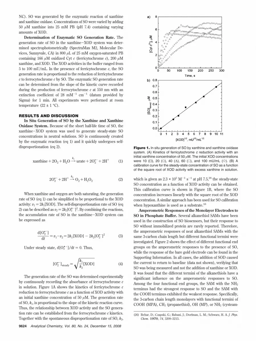
The generation rate of the SO was determined experimentallyby continuously recording the absorbance of ferrocytochrome cin solution. Figure 1A shows the kinetics of ferricytochrome creduction to ferrocytochrome c as a function of XOD activity withan initial xanthine concentration of 50 µM. The generation rateof SO, k1, is proportional to the slope of the kinetic reaction curve.Thus, the relationship between XOD activity and the SO genera-tion rate can be established from the ferrocytochrome c kinetics.Together with the spontaneous disproportionation rate of SO, k2,
which is given as 2.3 × 10⁵ M-1 s-1 at pH 7.5,26 the steady-stateSO concentration as a function of XOD activity can be obtained.This calibration curve is shown in Figure 1B, where the SOconcentration increases linearly with the square root of the XODconcentration. A similar approach has been used for SO calibrationwhen hypoxanthine is used as a substrate.16
Amperometric Responses of the Monolayer Electrodes toSO in Phosphate Buffer. Several alkanethiol SAMs have beenused in the construction of SO biosensors, but their response toSO without immobilized protein are rarely reported. Therefore,the amperometric responses of neat alkanethiol SAMs with thesame 3-carbon chain length but different functional termini wereinvestigated. Figure 2 shows the effect of different functional endgroups on the amperometric responses to the presence of SO,while the response of the bare gold electrode can be found in theSupporting Information. In all cases, the addition of SOD causedthe current to return to baseline (data not shown), verifying thatSO was being measured and not the addition of xanthine or XOD.It was found that the different termini of the alkanethiols have asignificant influence on the amperometric responses to SO.Among the four functional end groups, the SAM with the NH2
terminus had the strongest response to SO and the SAM withthe COOH terminus exhibited the weakest response. Specifically,the 3-carbon chain length monolayers with functional termini ofCOOH (MPA), CH3 (propanethiol), OH (MP), or NH2 (cysteam-
(26) Behar, D.; Czapski, G.; Rabani, J.; Dorfman, L. M.; Schwarz, H. A. J. Phys.Chem. 1970, 74, 3209–3213.
Figure 1. In situ generation of SO by xanthine and xanthine oxidasesystem. (A) Kinetics of ferricytochrome c reduction activity with aninitial xanthine concentration of 50 µM. The initial XOD concentrationswere 10 (0), 20 (O), 40 (∆), 60 (∇ ), and 100 mU/mL (]). (B) Acalibration curve for the steady-state concentration of SO as a functionof the square root of XOD activity with excess xanthine in solution.
9624 Analytical Chemistry, Vol. 80, No. 24, December 15, 2008
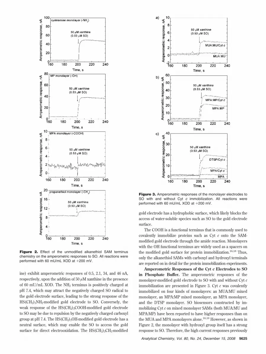
ine) exhibit amperometric responses of 0.5, 2.1, 34, and 46 nA,respectively, upon the addition of 50 µM xanthine in the presenceof 60 mU/mL XOD. The NH2 terminus is positively charged atpH 7.4, which may attract the negatively charged SO radical tothe gold electrode surface, leading to the strong response of theHS(CH2)3NH2-modified gold electrode to SO. Conversely, theweak response of the HS(CH2)3COOH-modified gold electrodeto SO may be due to repulsion by the negatively charged carboxylgroup at pH 7.4. The HS(CH2)3OH-modified gold electrode has aneutral surface, which may enable the SO to access the goldsurface for direct electrooxidation. The HS(CH2)3CH3-modified
gold electrode has a hydrophobic surface, which likely blocks theaccess of water-soluble species such as SO to the gold electrodesurface.
The COOH is a functional terminus that is commonly used tocovalently immobilize proteins such as Cyt c onto the SAM-modified gold electrode through the amide reaction. Monolayerswith the OH functional terminus are widely used as a spacers onthe modified gold surface for protein immobilization.16,18 Thus,only the alkanethiol SAMs with carboxyl and hydroxyl terminalsare reported on in detail for the protein immobilization experiments.
Amperometric Responses of the Cyt c Electrodes to SOin Phosphate Buffer. The amperometric responses of themonolayer-modified gold electrode to SO with and without Cyt cimmobilization are presented in Figure 3. Cyt c was covalentlyimmobilized on four kinds of monolayers: an MUA:MU mixedmonolayer, an MPA:MP mixed monolayer, an MPA monolayer,and the DTSP monolayer. SO biosensors constructed by im-mobilizing Cyt c on mixed monolayer SAMs (both MUA:MU andMPA:MP) have been reported to have higher responses than onthe MUA and MPA monolayers alone.16,18 However, as shown inFigure 2, the monolayer with hydroxyl group itself has a strongresponse to SO. Therefore, the high current responses previously
Figure 2. Effect of the unmodified alkanethiol SAM terminuschemistry on the amperometric responses to SO. All reactions wereperformed with 60 mU/mL XOD at +200 mV.
Figure 3. Amperometric responses of the monolayer electrodes toSO with and without Cyt c immobilization. All reactions wereperformed with 60 mU/mL XOD at +200 mV.
9625Analytical Chemistry, Vol. 80, No. 24, December 15, 2008

observed in the literature can be due to the oxidation of SO bothby the monolayer and by the Cyt c protein. The results in Figure3 indicate that the MPA:MP monolayer electrode has a strongresponse of 19.5 nA to 0.53 µM SO, while the response of theMUA:MU monolayer electrode to 0.53 µM SO is negligible.Generally, the long carbon chain alkanethiols result in betterpassivation of the gold surface after modification.27 This effect islikely the main reason for the different amperometric responsesto SO for the gold electrode modified with MUA:MU and MPA:MP mixed monolayers.
As shown in Figure 3, after immobilizing Cyt c on the SAMs,the bioelectrodes showed higher amperometric responses to 0.53µM SO than was observed for the neat monolayer electrodes. Theimprovement of the Cyt c electrode over the monolayer electrodeis especially obvious for the Cyt c electrodes prepared on MUA:MU mixed monolayer (Figure 3A) and MPA monolayer (Figure3C). Because negligible amperometric responses to SO wereobserved for MUA:MU and MPA monolayer electrodes, theincremental increase in current upon the production of SO in thesolution can be confidently attributed to the direct electron transferfrom the immobilized Cyt c to the gold electrode through themonolayer relay. In contrast, both the MPA:MP/Cyt c electrodeand the MPA:MP electrodes exhibit a large response to the SO(Figure 3B) and thus the benefits of Cyt c incorporation areminimal. DTSP is an amino-reactive NHS ester cross-linker, which,after cleavage of the disulfide bond, has same structure as theNHS-activated MPA upon assembly on the gold electrode. Experi-ments with the neat DTSP monolayer cannot be performed, as itwould react and immobilize the XOD enzyme. Although the twoelectrodes should be similar, as shown in Figure 3C, the DTSP/Cyt c electrode exhibited much higher response to SO than theMPA/Cyt c electrode, which may be due to a higher efficiency ofthe Cyt c covalent immobilization on the DTSP monolayer surface.
Based on the results presented in Figures 2 and 3, two possiblereaction pathways likely occur during the electrochemical detec-tion of SO, i.e., electron transfer through Cyt c and the monolayerrelay (pathway I) and oxidation of superoxide by the SAM-modified gold electrode independent of the Cyt c (pathway II).Reaction pathway I is the main pathway for the electrochemicaldetection of SO on the MUA:MU/Cyt c, MPA/Cyt c, and DTSP/Cyt c electrodes, while reaction pathway II is responsible for thebehavior of SO at bare gold electrodes and those modified withneat SAMs, such as MP, propanethiol, cysteamine, and MPA:MP.For the MPA:MP/Cyt c electrode, both reaction pathways I andII likely contribute to the total amperometric response to the SO.
Figure 4 shows calibration curves (with no backgroundsubtraction) of five different electrode configurations to varyingSO concentrations in 25 mM PB (pH 7.4) at +200 mV, obtainedby varying the XOD activity in the buffer solution. The error barsare based on four independently prepared electrodes. Reproduc-ibility of a single electrode, when exposed multiple times to theSO radical, was also good (not shown). The LOD and sensitivityof each electrode were obtained from Figure 4 and summarizedin Table 1. The LOD is rigorously defined as the baselinemeasurement (intercept) plus three times the standard deviationof the baseline signal in the absence of SO, converted into unitsof superoxide concentration.28 It was found that the MP monolayer
electrode has the highest sensitivity (940 nA µM-1 cm-2) andthe lowest LOD (0.038 µM) for the detection of SO in 25 mM PB(pH 7.4) at +200 mV. As shown in Figure 3A and C, the MUA:MU and MPA monolayers exhibit good passivation for theelectrochemical detection of SO. Attachment of Cyt c to thesemonolayers activates pathway I although performance metrics arenot as good as the MP monolayer. The sensitivity for the MUA:MU/Cyt c electrode (93 nA µM-1 cm-2) was found to be ∼3-foldbetter than what was previously reported for this system using agold needle disk electrode (27.6 nA µM-1 cm-2).16 The effect ofthe inclusion of the MP spacer within the SAM is clearlydemonstrated upon comparison of MPA/Cyt c and MPA:MP/Cytc electrodes. The mixed monolayer electrode has an order ofmagnitude lower LOD and nearly a 5-fold better sensitivity thana monolayer of MPA alone.
The SAM chemical structures of the DTSP/Cyt c and MPA/Cyt c electrodes should be similar, but the former exhibited alower LOD value and higher sensitivity toward SO than the latter,which may be related to better assembly of DTSP on the goldsurface and its higher efficiency for the amide reaction with Cytc. The sensitivity of the DTSP/Cyt c electrode (410 nA µM-1
cm-2) is more than 2 times greater than MPA/Cyt c value and itis almost 5 times greater than the value for the commonly usedMUA:MU/Cyt c electrode. This sensitivity is similar to that of amultilayer (layer-by-layer technique with 6 layers) Cyt c sensormade on a gold wire (398 nA µM-1 cm-2).29
Analysis of Transport Limitations in SO Detection. In orderto understand the effect of the mass-transfer rate of SO to theelectrode surface on the amperometric response of the enzymaticelectrodes, current responses were measured as a function of
(27) Strong, L.; Whitesides, G. M. Langmuir 1988, 4, 546–558.(28) Gonzalez, A. G.; Herrador, M. A. TrAC-Trends Anal. Chem. 2007, 26, 227–
238.
Figure 4. Calibration curves of 5 different electrodes for SO in 25mM PB (pH 7.4) at +200 mV vs Ag/AgCl reference. The differentelectrodes used were MUA:MU/Cyt c (O), MPA/Cyt c (0), DTSP/Cytc (]), MPA:MP/Cyt c (∇ ), and neat MP (∆).
Table 1. LOD and Sensitivity of Different Electrodes forSO Detectiona
electrodes LOD (µM) sensitivity (nA µM-1 m-2)
MUA:MU/Cyt c 0.24 93DTSP/Cyt c 0.073 410MPA/Cytc 0.68 160MPA:MP/Cyt c 0.065 740MP monolayer 0.038 940
a n ) 5 for all measurements.
9626 Analytical Chemistry, Vol. 80, No. 24, December 15, 2008

rotation speed at potentials of 150 and 200 mV for the neat MPmonolayer electrode and the MPA:MP/Cyt c electrode, which arethe two systems with the highest sensitivity values (Table 1).Results are shown in Figure 5 in the form of a Levich-Kouteckyplot. The curves show that mass transfer plays an important rolein the operation of the biosensors. Furthermore, the slopes of thefour lines are nearly the same, as would be expected if the reactionrate is limited by mass transfer of SO to the surface.30
The Levich-Koutecky equation takes the form of
1i) 1
iK+ 1
0.62nFADO2•-
2⁄3 ω1⁄2ν-1⁄6CO2•-
(5)
where all parameters are defined in the Supporting Information.All parameters are known in the second term of the right-handside of eq 5, except the diffusion coefficient and the bulkconcentration of SO. Assuming that the diffusion coefficient ofSO is the same as the measured value for dioxygen in water, DO2
•-
) 2 × 10⁻⁵ cm2 s-1, the bulk concentration CO2•- can be estimated
from the slope of each curve. In the presence of 20 mU/mL XODand 200 µM xanthine, values of 0.24 and 0.25 µM were found at+150 and +200 mV for the MP monolayer electrode, respectively.These values are in good agreement with the SO concentration(0.31 µM) determined by the UV spectrophotometric methodshown in Figure 1B.
Amperometric Responses to SO Radical in Culture Media.Figure 6 shows the amperometric responses of selected electrodes(MP, MPA/Cyt c, MUA:MU/Cyt c, and DTSP/Cyt c) to SO infresh MEM with and without 10% fetal bovine serum (FBS). Theamperometric response of MP electrode to 0.53 µM SO in MEMis 15.5 nA, which is a 50% decrease in the response to the sameamount of SO in 25 mM PB as shown in Figure 1B. In contrast tothe MP electrode, the Cyt c electrodes (MPA/Cyt c, MUA:MU/Cyt c, and DTSP/Cyt c electrodes) have similar amperometricresponses to SO both in MEM and phosphate buffer.
The amperometric responses of all electrodes are negligiblein fresh MEM with the addition of 10% FBS, indicating that theFBS fouls the electrode surface for the electrochemical detection
of SO. The proteins in FBS may nonspecifically adsorb on theelectrode surface, blocking access of the SO to the Cyt c and goldelectrode, leading to this degraded response.
To further understand the responses of the electrodes, theeffects of other interferents were also explored. Figure 7 showsthe comparison of the amperometric responses of four differentelectrode configurations to 0.1 mM H2O2, 20 µM ascorbic acid,0.53 µM SO in PB, and 0.53 µM SO in fresh MEM. The MPelectrode has the highest response to SO in PB, while the DTSP/Cyt c electrode is the most sensitive to SO in fresh MEM. In fact,both the MPA/Cyt c and DTSP/Cyt c electrodes were more
(29) Beissenhirtz, M. K.; Scheller, F. W.; Lisdat, F. Anal. Chem. 2004, 76, 4665–4671.
(30) Bard, A. J.; Faulkner, L. R. Electrochemical Methods: Fundamentals andApplications; John Wiley & Sons, Inc.:New York, 2001.
Figure 5. Levich-Koutecky plots of the MP monolayer and MPA:MP/Cyt c electrodes at various applied potentials (SO was generatedby 20 mU/mL XOD and 200 µM xanthine). MP monolayer electrodeat +200 mV (∆), MPA:MP/Cyt c electrode at +200 mV (2), MPmonolayer electrode at +150 mV (O), and MPA:MP/Cyt c electrodeat +150 mV (b).
Figure 6. Responses of the monolayer electrodes and Cyt c modifiedelectrodes to SO in fresh MEM with and without FBS at an appliedpotential of +200 mV vs Ag/AgCl reference. All reactions wereperformed with 60 mU/mL XOD.
9627Analytical Chemistry, Vol. 80, No. 24, December 15, 2008

sensitive to SO in MEM than in PB. The results in Table 1 indicatethat the MP monolayer electrode may be the best for the detectionof SO in PB without interferents, but it has a very strong responseto ascorbic acid, and its response to SO in fresh MEM drops byalmost 50% of the value in phosphate buffer. Conversely, the Cytc electrodes show better selectivity than the monolayer electrodein more physiologically relevant environments, such as culturemedia. As discussed before, the DTSP/Cyt c and MPA/Cyt celectrodes should be structurally similar, but the results in Figure7 show that the DTSP/Cyt c electrode is more sensitive to everycondition examined as compared to the MPA-based electrode.This is especially clear for the H2O2 results, as it is known thatimmobilized Cyt c can exhibit pseudoperoxidase activity.16 Theincreased sensitivity to H2O2 suggests that the DTSP monolayerenables an increase in Cyt c activity or coverage on the electrodesurface.
The DTSP/Cyt c electrode appears to offer the best tradeoffbetween current response and specificity. Therefore, a calibra-tion curve for this electrode configuration was made in MEMmedia (Figure 8). However, when designing an analytical devicethat will operate in tissue culture media,17,31,32 it may be difficultto obtain a good background signal due to the presence ofinterferents concurrently produced with the SO by cells.Therefore, the calibration curve in MEM was created usingtwo different methods. In method I (Figure 8A), the totalresulting current was measured after the addition of XOD, justas was done for the calibration curves made in PB (Figure 4).In method II, a bolus of SOD enzyme was added after achievinga steady-state SO concentration, and the difference in theamperometric response before and after the SOD addition wasmeasured. This may be a more relevant protocol for themeasurement of SO concentrations in tissue culture samples,as it utilizes the specificity of the SOD enzyme combined withthe high sensitivity of the DTSP/Cyt c electrode. Calibrationcurves for the two methods are shown in Figure 8B, and thesensitivities for these curves are shown in Table 2. Remarkably,the sensitivity of the DTSP/Cyt c electrode in MEM, usingeither method I or method II, is higher than was observed inPB. The sensitivity using method II (740 nA/µM/cm2) is 3times higher than what was recently reported for engineered
SOD that is directly immobilized on a gold wire (230 nA µM-1
cm-2).24 This shows that the specificity of the SOD enzymecan be combined with the high sensitivity of the Cyt celectrodes to create high-performance systems for SO detection.
CONCLUSIONSThe amperometric responses of monolayer electrodes and
Cyt c-modified electrodes to SO were systematically investi-gated. The responses of the monolayer electrodes to SOstrongly depend on the functional termini of the monolayers.SO is easily electrochemically oxidized on the monolayerelectrodes with amino or hydroxyl terminals, while the mono-layers with methyl or carboxyl terminals seem to block theaccess of SO to the gold electrode. Although the MP monolayerelectrode shows high sensitivity and low LOD for SO detectionin PB, the Cyt c electrodes exhibit higher selectivity and betterresistance to fouling in culture media. The DTSP monolayer isa better electron relay than the MPA monolayer after im-mobilization of Cyt c. As a result, the DTSP/Cyt c has the bestsensitivity to SO among the Cyt c electrodes in both PB andMEM. Moreover, the DTSP/Cyt c electrode is likely to be the
(31) Viravaidya, K.; Sin, A.; Shuler, M. L. Biotechnol. Prog. 2004, 20, 316–323.(32) McAuliffe, G. J.; Chang, J. Y.; Glahn, R. P.; Shuler, M. L. Mol. Cell. Biomech.
2008, 5, 119–132.
Figure 7. Comparison of the responses of four electrodes to H2O2,ascorbic acid, SO in PB, and SO in MEM. The steady-stateconcentration of SO was generated by 60 mU/mL XOD and 50 µMxanthine, which results in a concentration of 0.53 µM.
Figure 8. (A) Amperometric response of the DTSP/Cyt c electrodeto SO in MEM media; (B) Calibration curves of 3 different DTSP/Cytc electrodes to SO in MEM at +200 mV vs Ag/AgCl reference(response of the electrodes upon the addition of 50 µM xanthine inthe presence of XOD (method I, 0) and the current drop upon theaddition of 100 U/mL SOD (method II, ∆).
Table 2. Sensitivities of DTSP/Cyt c Bioelectrodes inBuffer and Cell Culture Mediaa
experimental conditions sensitivity (nA/µM · cm2)
phosphate buffer 410MEM (method I) 640MEM (method II) 740
a n ) 5 for measurements in phosphate buffer. n ) 3 for measure-ments in MEM.
9628 Analytical Chemistry, Vol. 80, No. 24, December 15, 2008

most suitable electrode among all of the investigated electrodesfor SO detection in terms of the trade-offs with sensitivity,selectivity, and LOD. By operating the electrode before andafter the addition of the SOD enzyme, the high sensitivity ofthe Cyt c system can be combined with the selectivity of theSOD enzyme to create an optimal configuration for monitoringSO concentrations in physiological environments.
ACKNOWLEDGMENTThis research was supported by the U.S. Army Applied
Research Program. We thank Lt. Col. Robert Bozic for helpfuldiscussions and assistance.
SUPPORTING INFORMATION AVAILABLEAdditional information as noted in text. This material is
available free of charge via the Internet at http://pubs.acs.org.
Received for review April 21, 2008. Accepted October 11,2008.
AC800796B
9629Analytical Chemistry, Vol. 80, No. 24, December 15, 2008

Supplementary Information
Detection of the superoxide radical anion using various alkanethiol
monolayers and immobilized cytochrome c
Xiaojun J. Chen†, ‡, §, Alan C. West†, Donald M. Cropek‡, and Scott Banta†,*
Department of Chemical Engineering, Columbia University, New York, New York
U.S. Army Engineer Research and Development Center, Construction Engineering Research
Laboratory (CERL), Champaign, Illinois
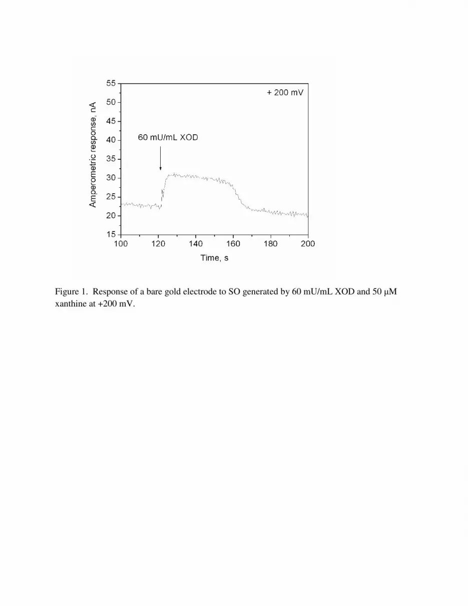
Figure 1. Response of a bare gold electrode to SO generated by 60 mU/mL XOD and 50 µM xanthine at +200 mV.

(A)
(B)
(C)
(D)
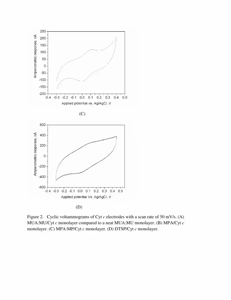
Figure 2. Cyclic voltammograms of Cyt c electrodes with a scan rate of 50 mV/s. (A) MUA:MU/Cyt c monolayer compared to a neat MUA:MU monolayer. (B) MPA/Cyt cmonolayer. (C) MPA:MP/Cyt c monolayer. (D) DTSP/Cyt c monolayer.

LIST OF SYMBOLS
n number of electrons involved for the electron transfer reaction
F Faradaic constant (96,485 C mol-1)
−•2O
C Bulk concentration of O2•- (mol cm-3)
−•2O
D Diffusion coefficient of O2•- (cm2 s-1)
A Surface area of the electrode (cm2)
ik the current in the absence of any mass transfer effects (A cm-2)
ν Kinematic viscosity (cm2 s-1)
ω Angular velocity (rpm or radians per second)