Embed Size (px)
Citation preview

www.elsevier.com/locate/bioelechem
Bioelectrochemistry 6
Direct electrochemistry of heme multicofactor-containing
enzymes on alkanethiol-modified gold electrodes
Elena E. Ferapontovaa,b,*, Lo Gortonb
aGroup of Bioinformatics, Weblab, IT Centre, Voskhod 26a, Novosibirsk 630102, RussiabDepartment of Analytical Chemistry, Lund University, P.O. Box 124, SE-221 00 Lund, Sweden
Received 9 January 2004; received in revised form 31 March 2004; accepted 13 April 2004
Available online 19 August 2004
Abstract
Direct electrochemistry of heme multicofactor-containing enzymes, e.g., microbial theophylline oxidase (ThOx) and d-fructose
dehydrogenase (FDH) from Gluconobacter industrius was studied on alkanethiol-modified gold electrodes and was compared with that of
some previously studied complex heme enzymes, specifically, cellobiose dehydrogenase (CDH) and sulphite oxidase (SOx). The formal
redox potentials for enzymes in direct electronic communication varied for ThOx from �112 to �101 mV (vs. Ag|AgCl), at pH 7.0, and for
FDH from �158 to �89 mV, at pH 5.0 and pH 4.0, respectively, on differently charged alkanethiol layers. Direct and mediated by
cytochrome c electrochemistry of FDH correlated with the existence of two active centres in the protein structure, i.e., the heme and the
pyrroloquinoline quinone (PQQ) prosthetic groups. The effect of the alkanethiols of different polarity and charge on the surface properties of
the gold electrodes necessary for adsorption and orientation of ThOx, FDH, CDH and SOx, favourable for the efficient electrode–enzyme
electron transfer reaction, is discussed.
D 2004 Elsevier B.V. All rights reserved.
Keywords: Theophylline oxidase; d-fructose dehydrogenase; Heme; Direct electron transfer; Alkanethiol self-assembled monolayers
1. Introduction
One of the intriguing phenomena known for the last few
decades is bioelectrocatalysis, with the particular case of
direct electron transfer (DET) between an electrode material
and redox active proteins or protein clusters. The very first
reports on DET with a redox active protein were published
already in 1977 when Eddowes and Hill [1] and Yeh and
Kuwana [2] independently showed that cytochrome c (cyt
c) on bipyridyl-modified gold and tin-doped indium oxide
electrodes, respectively, showed virtually reversible electro-
chemistry, as revealed by cyclic voltammetry (CV). After
that, DET was established for peroxidases [3–5], laccases
1567-5394/$ - see front matter D 2004 Elsevier B.V. All rights reserved.
doi:10.1016/j.bioelechem.2004.04.004
* Corresponding author. Department of Analytical Chemistry, Lund
University, P.O. Box 124, SE-221 00 Lund, Sweden. Tel.: +46 46 222
0103; fax: +46 46 222 4544.
E-mail addresses: [email protected],
[email protected] (E.E. Ferapontova).
[6], ferredoxins [7,8], thus contributing essentially to the
understanding ET mechanisms in these systems. Later, DET
reactions of complex, multicofactor-containing enzymes and
whole cell complexes were probed [7–15]. In parallel to a
successful DET-based bioelectrocatalytic function of com-
plex redox enzymes, DET for a number of enzymes at
electrodes was shown to depend crucially on the belectrodeenvironmentQ. Thus, it was not attained or accompanied by a
loss of enzymatic activity due to enzyme denaturation at the
electrode surfaces, by nonspecific and unstable binding to
the electrode, by random surface orientation or by impeded
internal ET between the multiple redox sites present in the
enzyme, in addition to the electrode–enzyme DET reaction.
Mimicking the biological environment of enzymes by the
modified electrodes is then of particular interest to achieve
DET between the electrode and the enzyme; knowledge of
the redox potentials for the redox active sites present in the
enzyme contributes essentially to the evaluation of the
enzyme function and mechanism [10,11,15]. To provide a
6 (2005) 55–63
E.E. Ferapontova, L. Gorton / Bioelectrochemistry 66 (2005) 55–6356
specific microenvironment at the electrode surface, self-
assembled monolayers (SAMs) of synthetic terminally
functionalised alkanethiols can be used [16–18]. The ability
to manipulate the properties of the interface via the
chemistry of the SAMs of alkanethiols attached onto the
gold electrode surface is unique and unprecedented; these
interfaces can be considered as ideal physiological mem-
brane–mimetic systems for studies of the bioelectrochemical
function of complex membrane and intermembrane redox
enzymes [10,11,15,18,19]. Therewith, the electrostatic and
hydrophilic/hydrophobic interactions between the enzyme
and the modified electrode surface can be easily controlled
by a proper choice of alkanethiols bearing head groups of
different charge and polarity. As a result, a successful
simulation of the molecular surfaces of the biological
partners of the enzyme by the SAM of alkanethiols may
be achieved, which in turn provides the necessary amount/
orientation of the enzyme molecules for DET reaction with
the electrode, as well as a conformation appropriate for its
efficient bioelectrocatalytic function.
In this work, the DET reactions of the heme complex–
cofactor-containing enzymes, specifically, microbial theo-
phylline oxidase (ThOx) and d-fructose dehydrogenase
(FDH) from Gluconobacter industrius, were studied on
alkanethiol-modified gold electrodes to reveal the conditions
for the DET reaction between the electrode and the heme
active site of these enzymes, which may provide a
bioelectrocatalytic function of the enzymes.
ThOx is a complex redox metalloenzyme involved in the
metabolic oxidation of theophylline to form 1,3-dimethyluric
acid. It requires no oxygen in its catalytic action. The
reduced enzyme can then in turn be reoxidized by its natural
redox partner cyt c. Microbial ThOx was isolated by GDS
Technology, in 1988 [20,21]; however, no detailed kinetic
characterization of ThOx, as well as no data on the crystal
structure of ThOx and the number/nature of redox sites/
domains present in the molecule, are still available. As a
consequence, ET reactions of ThOx have not yet been
studied, and just a few works on the electrochemistry of
ThOx were focused on the development of ThOx biosensors
for theophylline, based on spectrophotometrical [20–22] and
electrochemical detection of theophylline oxidation cata-
lysed by ThOx in the presence of cyt c [23] and some
ferrocene mediators [24].
Membrane-bound FDH refers to the pyrroloquinoline
quinone (PQQ)-dependent dehydrogenases, which transfer
electrons from substrates such as fructose to electron
acceptors other than oxygen [25,26]. This multicofactor-
containing enzyme is able to selectively oxidise d-fructose
to 5-keto-d-fructose which is accompanied by the reduc-
tion of its PQQ-cofactor to PQQH2 [27,28]. The natural
electron acceptors of FDH are believed to be cyt c or
ubiquinones [29]. However, both artificial 1e�- and 2e�/
H+-acceptors can be used as electron acceptors as well
[28,30]. When purified from the membranes of G.
industrius, FDH consists of three tightly bound units of
67 kDa (the PQQ-domain), 51 kDa (cyt c-type domain)
and 20 kDa (peptide of unknown function, presumably
banchoringQ the enzyme to the membrane) [28]. The
integrity of the enzyme drastically depends upon the
bconservationQ of the membrane environment, i.e., the
presence of detergents and other stabilisers. Due to its
biotechnological importance, FDH was studied a lot for
biosensor development ([31–34], and references therein),
and there are only a few works on the kinetics of the
catalysis [30]. The structural characteristics of the enzyme
are still not available.
Both enzymes can communicate directly with the
electrode surface [9,35,36]. However, despite the pro-
nounced DET-based bioelectrocatalysis with FDH on carbon
paste electrodes, no direct electrochemistry of the heme
domain or the PQQ-domain of FDH was observed [9]. The
achieved DET reactions of FDH adsorbed under controlled
potential (0.5 V) on bare Pt and Au were correlated with the
redox activity of its PQQ-cofactor [the reported values for
the formal redox potentials of FDH were 80 mV (Ag|AgCl)
at pH 4.5], but not with a redox transformation of the heme
[35]. ThOx demonstrated similar DET-based bioelectrocata-
lytic activity on graphite, but no DET signal from the redox
active sites of ThOx in the absence of the substrate was
shown [36]. Spectroelectrochemical titration of ThOx in an
aldrithiol-modified gold capillary electrode enabled deter-
mination of at least two redox active centres present in
ThOx; however, sluggish electrochemistry of ThOx resulted
in dispersion of the titration data, which deteriorated the
significance of the obtained results [36]. To the best of our
knowledge, no direct voltammetric data are still available on
the redox transformations of the hemes in these two
enzymes.
2. Experimental
2.1. Chemicals and materials
The kit containing stabilised dissolved microbial theo-
phylline oxidase (the activity of 14 U/ml, ThOx) was
obtained from STANBIO Laboratory (Boerne, TX, USA,
Cat. No 2422) and was used as received. d-Fructose
dehydrogenase (d-fructose:[acceptor]5-oxidoreductase; EC
1.1.99.11, FDH) from G. industrius was obtained as a
lyophilised powder additionally containing salts and stabi-
lizing agents (detergent, antioxidant and sugars) to prevent
enzyme inactivation (Sigma, Cat. No F51520; the specific
activity of 471 U/mg of protein) and was used without
further purification. Removal of detergent facilitated the
aggregation of FDH and caused its inactivation [28].
Cytochrome c from horse heart, theophylline and d(�)
fructose were from Sigma (St. Louis, MO, USA) and were
used as received. The buffer components were from Merck
(Darmstadt, Germany). 2-Mercaptoethanol (98%, MC2–
OH), cysteamine (MC2–NH2) and 6-hydroxy-1-hexanethiol

Fig. 1. Absorption spectra of ThOx, (1) totally reduced state upon addition
of a saturated solution of theophylline and (2) totally oxidised state upon
addition of 0.5 mM ferricyanide.
E.E. Ferapontova, L. Gorton / Bioelectrochemistry 66 (2005) 55–63 57
(97%, MC6–OH) were from Sigma-Aldrich (UK). 6-Amino-
1-hexanthiol (MC6–NH2) was from Dojindo Laboratories
(Japan). 18.2 MV Millipore water was used throughout
the work.
2.2. Electrode modification with alkanethiols
Thiol films were prepared by 8 h adsorption from 5 mM
solutions of alkanethiols in absolute ethanol. For mixed
SAM, 5 mM MCx–OH and 5 mM MCx–NH2 in proportion
3:1 v/v were used if not stated otherwise. Modified electrode
surfaces were rinsed thoroughly with water to remove
weakly adsorbed molecule.
2.3. Instrumental procedure
All measurements were performed at ambient temper-
ature: 22F1 8C. Spectroscopic measurements were done
using an UVIKON 930 spectrophotometer (Kontron
Instruments, NorthStar Scientific, Leeds, UK). Cyclic
voltammetry (CV) and differential pulse voltammetry
(DPV) experiments were done in anaerobic solutions using
a potentiostat AUTOLAB PGSTAT 30 (Eco Chemie,
Netherlands) equipped with GPES 4.9 software. An
Ag|AgCl (KClsat) was the reference and a Pt plate was
the auxiliary electrode. For voltammetry, gold disk electro-
des (CHI, USA, A=0.031 cm2) were abraded on emery
paper, successively mechanically polished to a mirror
luster in an alumna slurry, and further electrochemically
polished by cycling in 0.1 M H2SO4 between �0.3 and 1.7
V. Modification of these electrodes with alkanethiol layers
was done as described above. After modification, a Teflon
cap was put on the top part of the electrode, thus forming
a 5-Al volume well-like microcell, with the bottom
representing the electrode surface [15]. Then, a 5-Aldroplet of an enzyme stock solution was dropped into
the micro-cell, and a dialysis membrane was pressed onto
the electrode Teflon cap and fitted tight to the cap surface
with a rubber O-ring. In such a manner, the modified
electrodes were kept for 30 min in working buffer
solutions under a nitrogen flux. Further experiments were
done both with the enzymes entrapped under the mem-
brane and without it. CV and DPV measurements with
ThOx were performed in deaerated 0.01 M phosphate
buffer solution, containing 0.15 M NaCl and 0.1 mM
EDTA (PBS), pH 7.0, and with FDH in 0.1 M citrate–
phosphate buffer system containing 0.1% Triton X-100
(citrate-PBS), in the pH range from 4.0 to 6.0.
3. Results and discussion
Both ThOx and FDH share one and the same property:
they have a heme domain in their molecular structures.
Spectral studies demonstrated that the absorption features of
the heme dominate in the UV/vis spectra of ThOx (Fig. 1),
similarly to those observed with other heme-containing
complex enzymes, such as FDH (cyt c-type heme domain)
[28,30], cellobiose dehydrogenase (CDH, cyt b-type heme
domain) [37,38] or sulphite oxidase (SOx, cyt b5-type heme
domain) [39–42]. The reaction of ThOx with an excess of
theophylline results in spectral changes characteristic of the
heme, from the oxidized state to the fully reduced one (Fig.
1, curve 1). Furthermore, ThOx is able to communicate with
cyt c acting as a natural electron acceptor and completing
the biocatalytic cycle [21,23], or with other electron
acceptors such as ferricyanide. This reaction results in the
full oxidation of the heme of ThOx which can be followed
from the heme spectral changes as well (Fig. 1, curve 2).
Similar spectral variations characteristic for the heme are
shown for FDH upon interaction with fructose and
ferricyanide [30].
To determine the redox potentials for the heme redox
sites of ThOx and FDH, a direct electron exchange between
the electrode and the enzymes should be established. No
direct electrochemistry of the heme domains of these two
enzymes or DET-based bioelectrocatalysis with ThOx or
FDH was attained with bare gold electrodes in the present
work. It is reasonable to suggest that communications of
ThOx and FDH with their biological electron acceptors
should occur through their heme domains, while the
substrate redox conversion takes place in another domain.
In this case, the heme domain serves as a bbuilt-in mediatorQ[9], wiring the electrons between the electron accepting site
and the enzyme biological redox partner [30,38–40,43].
Studies with heme-containing complex enzymes have
demonstrated that their physiological electron acceptor,
specifically, cyt c, can be replaced directly by the electrode
modified with SAMs of properly chosen, terminally
functionalised alkanethiols [4,11,15,18,45]. The same meth-

Fig. 2. CVs of ThOx on a gold electrode modified with a mixture of MC2–
OH/MC2–NH2; scan rates, (1) 10, (2) 20, (3) 30, (4) 50, (5) 100 and (6) 200
mV s�1; pH 7.0. Inset: Amperometric response to 1.68�10�6 M
theophylline at 150 mV. Vertical lines reflect additions of first 0.03�10�6
M and further stepwise addition of 0.33�10�6 M of theophylline.
E.E. Ferapontova, L. Gorton / Bioelectrochemistry 66 (2005) 55–6358
odology was used in our studies of the redox properties of
ThOx and FDH to provide the surface properties of the
electrode appropriate for DET between the electrode and the
heme domain of the enzymes.
CV of ThOx at electrodes modified with MC2–OH,
cysteamine and mixed MC2–OH/cysteamine layers revealed
a strong dependence of the electrochemistry of ThOx on the
nature of the alkanethiol head groups. In fact, only on the
mixed SAMs of MC2–OH and cysteamine that a pro-
nounced direct electrochemistry of ThOx was observed. In
the absence of the substrate, a single pair of redox peaks
with a mean value for the redox potential of �101 mV, at
pH 7.0, was registered (Fig. 2, Table 1). The intensity of the
peak currents depended linearly on the potential scan rate
[46], thus designating the surface electrochemistry of ThOx
on the studied layers. The integration of the peaks gave a
surface coverage of the enzyme, C, close to 1.7 pmol cm�2
of the electrochemically active enzyme, assuming a 1e�
Table 1
Direct electron transfer characteristics of some heme multicofactor-containing enz
Enzyme Electrode modification E0/, mV,
(AgjAgCCDH Phanerochaete
chrysosporium
Cysteamine or
MC3–COOH
�28; �4
pH 3.1,
pH 5.1 [
CDH Humicola insolens Cysteamine �78, pH
SOx chicken liver Cysteamine, MC2–OH �119, p
FDH Gluconobacter industrius MC2–OH �89F1,
FDH Gluconobacter industrius Cysteamine �142.5FMicrobial ThOx MC2–OH �112F2
Microbial ThOx MC2–OH/MC2–NH2 �101F2
transfer process. No redox activity of ThOx was detected on
cysteamine layers, and a significantly decreased amount of
the adsorbed enzyme as well as a negative shift in the redox
potential values were registered on MC2–OH SAMs (Table
1). Thus, a slightly polar/positive character of the hydroxyl-
terminated or mixed hydroxyl-/amine-terminated alkane-
thiols provided adsorption/orientation of ThOx, which is
favourable for DET between the modified gold electrode
and ThOx, similarly to the data obtained previously with
another heme-containing complex enzyme, SOx [15].
On both alkanethiol layers, (–OH)- and mixed (–OH)/
(–NH2)-substituted, ThOx was bioelectrocatalytically
active due to the achieved DET reaction (Fig. 1, inset).
With ThOx, the conversion of theophylline should occur
at the active centre other than the heme, and the heme just
wires electrons to the external electron acceptor, as
supported by spectral studies. Thus, bioelectrocatalytic
activity of ThOx allows us to assume the heme domain of
ThOx as being in DET contact with the modified
electrode. The obtained values for the redox potentials
of the heme in ThOx are expected to correspond to the
biological values because similar experimental conditions
provided close values of the redox potentials of the heme
in SOx determined both by direct electrochemistry and by
titration with mediators (Table 1).
With FDH, a positively charged cysteamine SAM was
required for electrostatic binding of the enzyme to the
electrode surface to achieve pronounced DET (Fig. 3).
Experiments were performed both with FDH entrapped
under the membrane and upon its stripping, with FDH
physically adsorbed onto the electrode surface. In both
cases, a pair of well-defined peaks was observed in DPVs
(Fig. 3a). However, the height of the anodic peak was, in
average, 1.5 times higher than that of the cathodic one; as
well as the peak potential for the reduction process was 9–
15 mV more positive than that for the anodic process,
similarly to the DPV data of Khan et al. [35], obtained with
bare gold electrodes. Upon membrane stripping, the
amount of FDH oxidised in DET did not principally
change, contrary to the amount of the re-reduced enzyme,
which further decreased and constituted around 50% of the
oxidised species. On further scanning, the DPV peaks
ymes
electrode DET
l)
C, pmol cm�2 E0/, mV, redox titration
(AgjAgCl)2 and �81 at
4.2 and 5.6; �42,
29,37]
�44, pH 6.0 [36]
5.0 [38] Not determined
H 7.4 [15] �113, pH 7.0 [33]
pH 4.0 5.7F0.51 Not determined
4, pH 5.0 4.04F0.34 Not determined
, pH 7.0 0.05F0.03 Not determined
, pH 7.0 1.65F0.12 Not determined

Fig. 3. DPV of FDH on a gold electrode modified with cysteamine;
modulation amplitude, 25 mV; modulation time, (1) 50, (2) 70 and (3) 100
ms; step potential, 2 mV; effective scan rate, 10 mV s�1; pH 5.0. Inset:
CVs; scan rate, 30 mV s�1; solid line, FDH itself; dotted line, on addition of
3 mM fructose. All the rest conditions as in the main figure.
E.E. Ferapontova, L. Gorton / Bioelectrochemistry 66 (2005) 55–63 59
successively degraded. It is known that FDH is stable in its
fully reduced state, [PQQH2–Fe2+]: the stabilised state that
is present in the commercial samples. FDH is stable at pH
4.5–6.0; the stability of purified FDH being much
enhanced by the presence of detergent in the enzyme
solution [28]. FDH is readily oxidised, producing the
following states [PQQH2–Fe3+] (oxidation of the heme by
external electron acceptor) and [PQQ–Fe2+] (oxidation of
the reduced PQQ-cofactor, directly or due to internal ET).
All these forms can be reversibly converted into each other
during the catalytic cycle; however, the formation of the
fully oxidised state [PQQ–Fe3+] leads to a bdead-endQ of
biocatalysis, when the totally oxidised enzyme is irrever-
sibly inactivated [30]. The shift of the overall DET reaction
in the oxidative direction, as well as the binverseQ differ-
ence of the peak potentials in DPV, may be interpreted
within the terms of a proceeding internal ET reaction.
Then, during the oxidation process, the heme is oxidised
and bwiresQ oxidation equivalents further to the PQQ-
cofactor, which is in the reduced form in the used
commercial sample. This process implies a stepwise
transfer of 2e�. Upon the reverse re-reduction process,
the internal ET may be impeded, and in this case, only the
oxidised heme is readily reduced, which is just a 1e�
transfer reaction, which roughly corresponds to a twofold
decrease in the peak currents. Therewith, the internal ET
can contribute to the re-reduction process as well. The
formal redox potential estimated as a mean value of the
potential of the redox peaks, both from CV and DPV, was
close to �143 mV, at pH 5.0, whereas the potential for the
PQQ-cofactor redox transformations was shown to be 80
mV at pH 4.5 [35,47]. This allowed us to consider that it
was the heme of FDH that in the initial turn underwent the
DET reaction at the cysteamine-modified gold electrodes.
The surface concentration of adsorbed FDH, calculated
from the anodic peaks in the DPVs assuming a 1e�
reaction [15], was around 4 pmol cm�2, and decreased
twofold if a 2e� transfer was assumed. Upon addition of
fructose, FDH, adsorbed on the cysteamine SAM, was
bioelectrocatalytically active due to DET between the
electrode and the heme domain of the enzyme; however,
the efficiency of catalysis was sufficiently low (Fig. 3b)
compared to the amount of the electrochemically active
enzyme.
The data obtained with FDH on the cysteamine-modified
electrodes correlate well with those previously reported for
the heme domain of flavocytochrome CDH, for which direct
redox activity was achieved with positively charged amine-
and carboxyl-terminated alkanethiols in acidic solutions
(Table 1) [37,44,45]. The similarity between the heme redox
potentials obtained from direct electrochemistry of CDH
and that from its redox titration (Table 1) suggests that the
redox potential for the heme active site in FDH estimated
from voltammograms can be considered as adequate to that
in the naturally existing state of FDH.
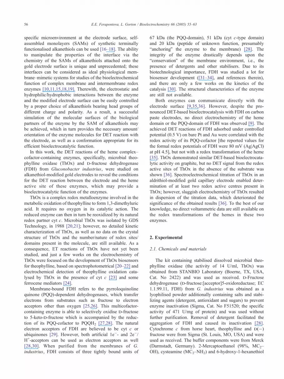
On the (–OH)-terminated alkanethiol SAM, FDH,
entrapped under the membrane, was oxidised in the first
scan. However, no re-reduction of the enzyme and no
pronounced DET signals were observed on the following
potential scans (Fig. 4). Upon addition of fructose, its
catalytic oxidation was observed starting from �120 mV, at
pH 5.0 (Fig. 4, curve 3). bSolutionQ electrochemistry of
FDH was supposed in this case. Variation of the buffer
solution pH provided a variation of the surface charge of
FDH (pI of 4.55 [48]). That resulted at pH 4.0 in a
pronounced direct electrochemistry of FDH on the (–OH)-
terminated SAM (Fig. 4, curve 4), but not in DET-based
bioelectrocatalysis when the substrate was added. The
obtained discrepancy between the redox potentials of FDH
on the cysteamine and MC2–OH SAMs may be due both to
the variation in the pH and the different orientations of the
enzyme at the electrode surface, which correlated with the
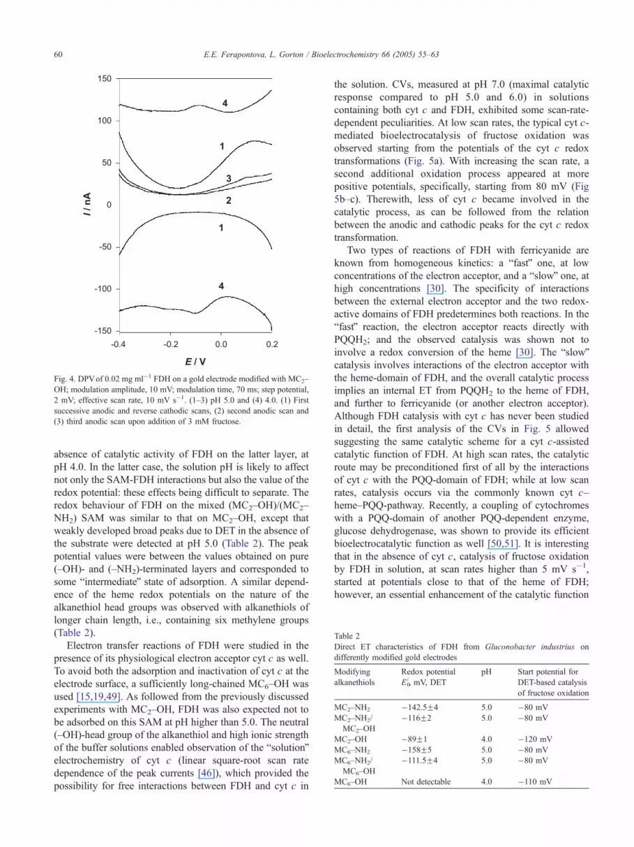
Table 2
Direct ET characteristics of FDH from Gluconobacter industrius on
differently modified gold electrodes
Modifying
alkanethiols
Redox potential
E0/, mV, DET
pH Start potential for
DET-based catalysis
of fructose oxidation
MC2–NH2 �142.5F4 5.0 �80 mV
MC2–NH2/
MC2–OH
�116F2 5.0 �80 mV
MC2–OH �89F1 4.0 �120 mV
MC6–NH2 �158F5 5.0 �80 mV
MC6–NH2/
MC6–OH
�111.5F4 5.0 �80 mV
MC6–OH Not detectable 4.0 �110 mV
Fig. 4. DPVof 0.02 mg ml�1 FDH on a gold electrode modified with MC2–
OH; modulation amplitude, 10 mV; modulation time, 70 ms; step potential,
2 mV; effective scan rate, 10 mV s�1. (1–3) pH 5.0 and (4) 4.0. (1) First
successive anodic and reverse cathodic scans, (2) second anodic scan and
(3) third anodic scan upon addition of 3 mM fructose.
E.E. Ferapontova, L. Gorton / Bioelectrochemistry 66 (2005) 55–6360
absence of catalytic activity of FDH on the latter layer, at
pH 4.0. In the latter case, the solution pH is likely to affect
not only the SAM-FDH interactions but also the value of the
redox potential: these effects being difficult to separate. The
redox behaviour of FDH on the mixed (MC2–OH)/(MC2–
NH2) SAM was similar to that on MC2–OH, except that
weakly developed broad peaks due to DET in the absence of
the substrate were detected at pH 5.0 (Table 2). The peak
potential values were between the values obtained on pure
(–OH)- and (–NH2)-terminated layers and corresponded to
some bintermediateQ state of adsorption. A similar depend-
ence of the heme redox potentials on the nature of the
alkanethiol head groups was observed with alkanethiols of
longer chain length, i.e., containing six methylene groups
(Table 2).
Electron transfer reactions of FDH were studied in the
presence of its physiological electron acceptor cyt c as well.
To avoid both the adsorption and inactivation of cyt c at the
electrode surface, a sufficiently long-chained MC6–OH was
used [15,19,49]. As followed from the previously discussed
experiments with MC2–OH, FDH was also expected not to
be adsorbed on this SAM at pH higher than 5.0. The neutral
(–OH)-head group of the alkanethiol and high ionic strength
of the buffer solutions enabled observation of the bsolutionQelectrochemistry of cyt c (linear square-root scan rate
dependence of the peak currents [46]), which provided the
possibility for free interactions between FDH and cyt c in
the solution. CVs, measured at pH 7.0 (maximal catalytic
response compared to pH 5.0 and 6.0) in solutions
containing both cyt c and FDH, exhibited some scan-rate-
dependent peculiarities. At low scan rates, the typical cyt c-
mediated bioelectrocatalysis of fructose oxidation was
observed starting from the potentials of the cyt c redox
transformations (Fig. 5a). With increasing the scan rate, a
second additional oxidation process appeared at more
positive potentials, specifically, starting from 80 mV (Fig
5b–c). Therewith, less of cyt c became involved in the
catalytic process, as can be followed from the relation
between the anodic and cathodic peaks for the cyt c redox
transformation.
Two types of reactions of FDH with ferricyanide are
known from homogeneous kinetics: a bfastQ one, at low
concentrations of the electron acceptor, and a bslowQ one, athigh concentrations [30]. The specificity of interactions
between the external electron acceptor and the two redox-
active domains of FDH predetermines both reactions. In the
bfastQ reaction, the electron acceptor reacts directly with
PQQH2; and the observed catalysis was shown not to
involve a redox conversion of the heme [30]. The bslowQcatalysis involves interactions of the electron acceptor with
the heme-domain of FDH, and the overall catalytic process
implies an internal ET from PQQH2 to the heme of FDH,
and further to ferricyanide (or another electron acceptor).
Although FDH catalysis with cyt c has never been studied
in detail, the first analysis of the CVs in Fig. 5 allowed
suggesting the same catalytic scheme for a cyt c-assisted
catalytic function of FDH. At high scan rates, the catalytic
route may be preconditioned first of all by the interactions
of cyt c with the PQQ-domain of FDH; while at low scan
rates, catalysis occurs via the commonly known cyt c–
heme–PQQ-pathway. Recently, a coupling of cytochromes
with a PQQ-domain of another PQQ-dependent enzyme,
glucose dehydrogenase, was shown to provide its efficient
bioelectrocatalytic function as well [50,51]. It is interesting
that in the absence of cyt c, catalysis of fructose oxidation
by FDH in solution, at scan rates higher than 5 mV s�1,
started at potentials close to that of the heme of FDH;
however, an essential enhancement of the catalytic function

Fig. 5. CVs of cyt c (6 mg ml�1) and FDH (0.02 mg ml�1) on (MC6–OH)-modified gold electrodes (dotted lines) upon addition of 3 mM fructose (solid lines),
pH 7.0. Scan rate, (a) 2, (b) 20, (c) 30 and (d) 50 mV s�1. Integration of the anodic branches of the voltammograms (solid lines) gives (a) 32.9, (b) 5.3, (c) 4.6
and (d) 3.5 AC.
E.E. Ferapontova, L. Gorton / Bioelectrochemistry 66 (2005) 55–63 61
is observed at potentials of PQQ-redox transformations, i.e.,
more positive than 70 mV (Fig. 6). These are the first results
which suggest an extremely interesting bioelectrocatalytic
scheme for the function of FDH, and further work is in
progress.
To summarise, some parallels can be followed between the
pH optimum for the biological catalytic activity of the studied
enzymes and the properties of the modifying layers. The
lower the pH optimum, a more acidic SAM is necessary to
provide electrostatic interactions between the electrode and
the enzyme, which would favour the DET reaction between
the electrode surface and the heme domain of the enzyme. For
enzymes with pH optima around pH 7.0–7.4, DET-based
Fig. 6. CVs of 0.02 mg ml�1 FDH on (MC6–OH)-modified gold electrodes (1–3)
(2) second and (3) third scans.
bioelectrocatalysis on bprotonatedQ cysteamine layers is
impeded. No DET was observed with ThOx on cysteamine
SAMs, which may result in further efficient bioelectrocata-
lytic function of the enzyme. For the previously studied SOx
on cysteamine layers [15], clear DET was shown, but the
desired orientation of SOx on this SAM was not provided,
which enabled DET-based bioelectrocatalysis [15].
Concerning the enzymes with acidic pH optima, e.g., FDH
and CDH (Table 1), pronounced redox chemistry was
observed from both of them, first of all on the positively
charged SAM of cysteamine, providing the DET-based
bioelectrocatalytic activity of the enzymes at acidic pHs as
well.
upon addition of 3 mM fructose (4), pH 4.0; scan rate, 50 mV s�1. (1) First,

E.E. Ferapontova, L. Gorton / Bioelectrochemistry 66 (2005) 55–6362
4. Conclusions
Complex cofactor-containing enzymes and enzyme com-
plexes embedded in biological membranes play an important
role in the living cell. However, membrane-bound enzymes
need a hydrophobic reaction environment for their catalytic
activity and stability. This impedes electrochemical research
of their function, and special approaches should be used for
their studies. For this purpose, modification of the electrode
surface by a properly charged amine- or hydroxyl-terminated
alkanethiol SAM enables to achieve both adsorption and
orientation of ThOx and FDH favourable for DET between
the electrode and the heme active sites of the enzymes, thus
enabling the determination of the redox potentials of the
hemes and providing DET-bioelectrocatalytic function of the
enzymes. The physiological pH optimum for the catalytic
function of the enzymes seems to predetermine to some
extent the surface charge of the modifying layer required for
an efficient DET reaction.
Acknowledgements
E.E.F. acknowledges the Wenner–Gren Foundations
(WGS) for financial support. The Swedish Research
Council (VR) funded this work.
References
[1] M.J. Eddowes, A.O.H. Hill, Novel method for the investigation of the
electrochemistry of metalloproteins: cytochrome c, J. Chem. Soc.,
Chem. Commun. 21 (1977) 771–772.
[2] P. Yeh, T. Kuwana, Reversible electrode reaction of cytochrome c,
Chem. Lett. 10 (1977) 1145–1148.
[3] F.A. Armstrong, A.M. Lannon, Fast interfacial electron transfer
between cytochrome c peroxidase and graphite electrodes promoted
by aminoglycosides: novel electroenzymic catalysis of hydrogen
peroxide reduction, J. Am. Chem. Soc. 109 (1987) 7211–7212.
[4] R.M. Paddock, E.F. Bowden, Electrocatalytic reduction of hydrogen
peroxide via direct electron transfer from pyrolytic graphite electrodes
to irreversibly adsorbed cytochrome c peroxidase, J. Electroanal.
Chem. 260 (1989) 487–494.
[5] A. Lindgren, T. Ruzgas, L. Gorton, Direct electron transfer of native
and modified peroxidases, in: R. Richard (Ed.), Current Topics in
Analytical Chemistry, vol. 2, Research Trends, Trivandrium, 2001,
pp. 71–94.
[6] A.I. Yaropolov, A.N. Kharibin, J. Emneus, G. Marco-Varga, L.
Gorton, Electrochemical properties of some copper-containing oxi-
dases, Bioelectrochem. Bioenerg. 40 (1996) 49–57.
[7] F.A. Armstrong, Voltammetry of proteins, in: G.S. Wilson (Ed.),
Encyclopedia of Electrochemistry, Bioelectrochemistry, vol. 9, Wiley-
VCH, Weinheim, 2002, pp. 11–29.
[8] F.A. Armstrong, G.S. Wilson, Recent developments in faradaic
bioelectrochemistry, Electrochim. Acta 45 (2002) 2623–2645.
[9] T. Ikeda, Direct redox communication between enzymes and
electrodes, in: F.W. Scheller, F. Schubert, J. Fedrowitz (Eds.),
Frontiers in Biosensorics, Birkh7user Verlag Basel, Switzerland,
1997, pp. 243–266.
[10] K.T. Kinnear, H.G. Monbouquette, Direct electron transfer to
Escherichia coli fumarate reductase in self-assembled alkanethiol
monolayers on gold electrodes, Langmuir 9 (1993) 2255–2257.
[11] S.J. Elliot, A.E. McElhaney, C. Feng, J.H. Enemark, F.A. Armstrong,
Voltammetric study of interdomain electron transfer within sulfite
oxidase, J. Am. Chem. Soc. 124 (2002) 11612–11613.
[12] D.R. Bond, D.R. Lovely, Electricity production by Geobacter
sulfurreducens attached to electrodes, Appl. Environ. Microbiol.
Mar. (2003) 1548–1555.
[13] A.L. Ghindilis, P. Atanasov, E. Wilkins, Enzyme-catalyzed direct
electron transfer. Fundamentals and analytical applications, Electro-
analysis 9 (1997) 661–674.
[14] L. Gorton, A. Lindgren, T. Larsson, F.D. Munteanu, T. Ruzgas, I.
Gazaryan, Direct electron transfer between heme-containing enzymes
and electrodes as basis for third generation biosensors, Anal. Chim.
Acta 400 (1999) 91–108.
[15] E.E. Ferapontova, T. Ruzgas, L. Gorton, Direct electron transfer of
heme- and molybdopterin cofactor-containing chicken liver sulfite
oxidase on alkanethiol-modified gold electrodes, Anal. Chem. 75
(2003) 4841–4850.
[16] H.O. Finklea, Electrochemistry of organized monolayers of thiols
and related molecules on electrodes, Electroanal. Chem. 19 (1996)
109–135.
[17] A. Ulman, Formation and structure of self-assembled monolayers,
Chem. Rev. 96 (1996) 1533–1554.
[18] S. Ferretti, S. Paynter, D.A. Russell, K.E. Sapsford, D.J. Richardson,
Self-assembled monolayers: a versatile tool for the formulation of bio-
surfaces, Trends Anal. Chem. 19 (2000) 530–540.
[19] S. Song, R.A. Clark, E.F. Bowden, M.J. Tarlov, Characterization of
cytochrome c/alkanethiolate structures prepared by self-assembly on
gold, J. Phys. Chem. 97 (1993) 6564–6572.
[20] S.K. Gupta, A.K. Agarwal, A.F. deCastro, A novel enzymatic approach
for serum theophylline measurement, Clin. Chem. 34 (1988) 1267.
[21] A.F. deCastro, S.K. Gupta, A.K. Agarwal, Enzymatic determination of
theophylline, PCT Int. Appl. (1989) 43 pp.
[22] L.M. Vaughan, A. Gotteher, Effect of xanthine-related compounds on
a theophylline assay using theophylline oxidase, Ann. Pharmacother.
26 (1992) 1576–1579.
[23] J. Wang, E. Dempsey, M. Ozsoz, M.R. Smyth, Amperometric enzyme
electrode for theophylline, Analyst 116 (1991) 997–999.
[24] C.J. McNeil, J.M. Cooper, J.A. Spoors, Amperometric enzyme
electrode for determination of theophylline in serum, Biosens.
Bioelectron. 7 (1992) 375–380.
[25] V.L. Davidson, Principles and Applications of Quinoproteins, Marcel
Dekker, New York, 1993.
[26] P.M. Goodwin, C. Anthony, The biochemistry, physiology and
genetics of PQQ and PQQ-containing enzymes, Adv. Microb. Physiol.
40 (1998) 1–80.
[27] Y. Yamada, K. Aida, T. Uemura, Enzymatic studies on the oxidation
of sugar and sugar alcohol: I. Purification and properties of particle-
bound fructose dehydrogenase, J. Biochem. 61 (1967) 636–646.
[28] M. Ameyama, E. Shinagawa, K. Matsyshita, O. Adachi, d-Fructose
dehydrogenase of Gluconobacter industrius: purification, character-
ization, and application to enzymic microdetermination of d-fructose,
J. Bacteriol. 145 (1981) 814–823.
[29] C. Anthony, in: C. Anthony (Ed.), Bacterial Energy Transduction,
Academic Press, San Diego, 1988, pp. 293–294.
[30] J. Marcinkeviciene, G. Johansson, Kinetic studies of the active sites
functioning in the quinohemoprotein fructose dehydrogenase, FEBS
Lett. 318 (1993) 23–26.
[31] G.F. Khan, E. Kobatake, H. Shinohara, Y. Ikariyama, M. Aizawa,
Molecular interface for an activity controlled enzyme electrode and its
application for the determination of fructose, Anal. Chem. 64 (1992)
1254–1258.
[32] K.T. Kinnear, H.G. Monbouquette, An amperometric fructose bio-
sensor based on fructose dehydrogenase immobilized in a membrane
mimetic layer on gold, Anal. Chem. 69 (1997) 1771–1775.
[33] A.S. Bassi, E. Lee, J.-X. Zhu, Carbon paste mediated, amperometric,
thin film biosensor for fructose monitoring in honey, Food Res. Int.
31-2 (1998) 119–127.

E.E. Ferapontova, L. Gorton / Bioelectrochemistry 66 (2005) 55–63 63
[34] M. Stredansky, A. Pizzariello, S. Stredanska, S. Miertus, Determi-
nation of d-fructose in foodstuffs by an improved amperometric
biosensor based on a solid binding matrix, Anal. Commun. 36 (1999)
57–61.
[35] G.F. Khan, H. Shinohara, Y. Ikariyama, M. Aizawa, Electrochemical
behaviour of monolayer quinoprotein adsorbed on the electrode
surfaces, J. Electroanal. Chem. 315 (1991) 263–273.
[36] E. Dock, A. Christenson, L. Gorton, T. Ruzgas, Direct heterogeneous
electron transfer between theophylline oxidase and electrodes,
Biosens. Bioelectron. (in press) (accepted for publication).
[37] T. Larsson, A. Lindgren, T. Ruzgas, Spectroelectrochemical study of
cellobiose dehydrogenase and diaphorase in a thiol-modified gold
capillary in the absence of mediators, Bioelectrochemistry 53 (2001)
243–249.
[38] K. Igarashi, I. Momohara, T. Nishino, M. Samejima, Kinetics of inter-
domain electron transfer in flavocytochrome cellobiose dehydroge-
nase from the white-rot fungus Phanerochaete chrysosporium,
Biochem. J. 365 (2002) 521–526.
[39] R. Hille, The mononuclear molybdenum enzymes, Chem. Rev. 96
(1996) 2757–2816.
[40] H.J. Cohen, I.J. Fridovich, Hepatic sulfite oxidase. The nature and
function of the heme prosthetic groups, J. Biol. Chem. 246 (1971)
367–373.
[41] S.P. Cramer, H.B. Gray, N.S. Scott, M. Barber, K.V. Rajagopalan,
EXAFS and other studies of sulfite oxidase molybdenum, in: W.E.
Newton, S. Otsuka (Eds.), Molybdenum chemistry of biological
significance, Plenum, New York, 1980, pp. 157–168.
[42] E.E. Ferapontova, A. Christenson, A. Hellmark, T. Ruzgas, Spec-
troelectrochemical study of heme- and molybdopterin cofactor-
containing chicken liver sulphite oxidase, Bioelectrochemistry 63
(2004) 49–53.
[43] M.S. Rogers, G.D. Jones, G. Antonioni, M.T. Wilson, M. Brunori,
Electron transfer from Phanerochaete chrysosporium cellobiose
oxidase to equine cytochrome c and Pseudomonas aeruginosa
cytochrome c-551, Biochem. J. 298 (1994) 329–334.
[44] A. Lindgren, T. Larsson, T. Ruzgas, L. Gorton, Direct electron transfer
between the heme of cellobiose dehydrogenase and thiol modified
gold electrodes, J. Electroanal. Chem. 494 (2000) 105–113.
[45] A. Lindgren, L. Gorton, T. Ruzgas, U. Baminger, D. Haltrich, M.
Schqlein, Direct electron transfer of cellobiose dehydrogenase from
various biological origins at gold an graphite electrodes, J. Electroa-
nal. Chem. 496 (2001) 76–81.
[46] A.J. Bard, L.R. Faulkner, Electrochemical methods, Fundamentals
and Applications, Wiley, New York, 1980.
[47] H. Shinohara, G.F. Khan, Y. Ikariyama, M. Aizawa, Electrochemical
oxidation and reduction of PQQ using a conductive polypyrrole-
coated electrode, J. Electroanal Chem. 304 (1991) 75–84.
[48] F.E. Prado, A.R. Sampietro, A method for the determination of
fructose using a single enzyme: production and properties of fructose
dehydrogenase from Gluconobacter indusrius, Biotechnol. Appl.
Biochem. 19 (1994) 361–368.
[49] F.M. Hawkridge, I. Taniguchi, The direct electron transfer reactions of
cytochrome c at electrode surfaces, Comm. Inorg. Chem. 17 (1995)
163–187.
[50] J. Okuda, J. Wakai, K. Sode, The application of cytochromes as the
interface molecule to facilitate the electron transfer for PQQ glucose
dehydrogenase employing mediator type glucose sensor, Anal. Lett.
35 (2002) 1465–1478.
[51] J. Okuda, J. Wakai, N. Yuhashi, K. Sode, Glucose enzyme electrode
using cytochrome b562 as an electron mediator, Biosens. Bioelectron.
18 (2003) 699–704.


