Embed Size (px)
Citation preview

Journal ofMaterials Chemistry B
PAPER
Publ
ishe
d on
02
May
201
3. D
ownl
oade
d by
Lak
ehea
d U
nive
rsity
on
05/0
6/20
13 1
7:06
:12.
View Article OnlineView Journal
aUniversity of Portland, Dept. of Chemistry,
97203, USAbEMSL, Pacic Northwest National LaboratocBattelle Science and Technology Pvt Ltd, 30
Pune, 411057, India. E-mail: Ajay.Karakoti@dGumilyov Eurasian National University, AseNanoscience and Technology Center, Advan
Materials Science and Engineering, Universi
USA
† Electronic supplementary informationmolecular schematic for PVP binding tocarbon 1s, nitrogen 1s, and cerium 3d, CDLS, and the normalized absorptionexposure to H2O2. See DOI: 10.1039/c3tb2
Cite this: DOI: 10.1039/c3tb20204f
Received 12th February 2013Accepted 2nd May 2013
DOI: 10.1039/c3tb20204f
www.rsc.org/MaterialsB
This journal is ª The Royal Society of
Enzyme-free detection of hydrogen peroxide fromcerium oxide nanoparticles immobilized onpoly(4-vinylpyridine) self-assembled monolayers†
James D. Gaynor,ab Ajay S. Karakoti,*c Talgat Inerbaev,d Shail Sanghavi,b
P. Nachimuthu,b V. Shutthanandan,b S. Seale and S. Thevuthasanb
A simple enzyme-free spectrophotometric detection of hydrogen peroxide is demonstrated based on its
colorimetric reaction with oxygen deficient cerium oxide nanoparticles (CNPs). This colorimetric
sensitivity of CNPs towards H2O2 increases significantly with decreasing crystallite size due to an increase
in the surface area as well as the concentration of Ce3+ on the surface. The origin of this colorimetric
reaction was studied using DFT that suggests the adsorption of peroxide and oxygen molecules on ceria
nanoparticles creates new states in the electronic structure leading to transitions absorbing in the visible
region of the electromagnetic spectrum. For detection, a single layer of nanoparticles was immobilized
on transparent microscopic glass slides using self-assembled monolayers (SAMs) of poly(4-vinylpyridine)
(PVP). Cluster-free and uniform immobilization of nanoparticles was confirmed from atomic force
microscopy (AFM) and helium ion microscopy (HIM). UV-Visible absorption measurements showed a
concentration dependent increase in absorbance from immobilized CNPs that were exposed to
increasing concentrations (10–400 mM) of hydrogen peroxide. The immobilized CNPs can be baked at
80 �C after initial use to regenerate the sensor for reuse. The development of a direct, reusable, enzyme-
free and dye-free peroxide sensing technology is possible and can be immediately applied in various
areas, including biomedicine and national security.
Introduction
Peroxides, including hydrogen peroxide (H2O2), are members ofa small group of compounds called reactive oxygen species(ROS).1 Hydrogen peroxide is considered to be an importantanalyte because of its importance in clinical diagnosis andchemical warfare. Biologically, H2O2 is considered to be amarker of oxidative stress,2 a condition closely associated withseveral diseases including cancer,3–5 atherosclerosis,6 Parkin-son's disease,7 Alzheimer's disease,8,9 inammation10 and neu-rodegeneration.11,12 Hydrogen peroxide is also produced as a
5000 N. Willamette Blvd., Portland, OR
ry, Richland, WA 99352, USA
2 Panchshil Technology Park, Hinjewadi,
battelle-india.com
tana, 010008, Republic of Kazakhstan
ced Materials Processing Analysis Center,
ty of Central Florida, Orlando, FL, 32826,
(ESI) available: It contains idealthe glass slide surface, XPS data forNP particle size characterization usingbaseline data for CNP slides before0204f
Chemistry 2013
by-product of several enzymatic reactions that can be used asdiagnostic tools for detection of the onset of various biologicalconditions. Thus direct or indirect detection of hydrogenperoxide is one of the central themes in the design and fabri-cation of various biosensors.13,14 In addition, terrorism hasrecently made direct use of peroxides by implementingperoxide-based materials in home-made explosives, a populartrend among terrorists due to synthetic simplicity and ease ofavailability of chemicals.15 High concentration of hydrogenperoxide in the environment16,17 from its use in industry,18 inatomic power stations19 and in disinfecting agents20 candramatically affect the environment which warrants the detec-tion of residual hydrogen peroxide.
Several strategies have recently been used for the detection ofhydrogen peroxide based on the colorimetric or electrochemicalresponse from the reaction of hydrogen peroxide with dyes21
such as Prussian blue,22 or enzymes such as horseradishperoxidase,23 supported on nanoparticle substrates. Addition-ally, several chemiluminescent24 and uorescent probes fordetecting ROS,25 such as borate-based uorescent probes,26,27
have been described in the literature specically for the detec-tion of hydrogen peroxide. However, most enzymes and dyeshave limited shelf life and long-term instability, are prone tophotobleaching, require lengthy and multiple processing steps,
J. Mater. Chem. B
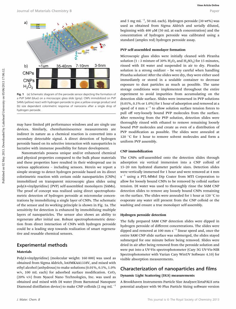
Fig. 1 (a) Schematic diagram of the peroxide sensor depicting the formation ofa PVP SAM (blue) on a microscopic glass slide (grey). CNPs immobilized on PVPSAMs (yellow) react with hydrogen peroxide to give a yellow-orange product and(b) size dependent colorimetric response of nanoceria after a single drop ofhydrogen peroxide.
Journal of Materials Chemistry B Paper
Publ
ishe
d on
02
May
201
3. D
ownl
oade
d by
Lak
ehea
d U
nive
rsity
on
05/0
6/20
13 1
7:06
:12.
View Article Online
may have limited pH performance windows and are single usedevices. Similarly, chemiluminescence measurements areindirect in nature as a chemical reaction is converted into asecondary detectable signal. A direct detection of hydrogenperoxide based on its selective interaction with nanoparticles islucrative with immense possibility for future development.
Nanomaterials possess unique and/or enhanced chemicaland physical properties compared to the bulk phase materialsand these properties have resulted in their widespread use invarious applications – including sensors. Herein we report asimple strategy to detect hydrogen peroxide based on its directcolorimetric reaction with cerium oxide nanoparticles (CNPs)immobilized on transparent microscopic glass slides usingpoly(4-vinylpyridine) (PVP) self-assembled monolayers (SAMs).The proof of concept was realized using direct spectrophoto-metric detection of hydrogen peroxide at micromolar concen-trations by immobilizing a single layer of CNPs. The schematicof the sensor and its working principle is shown in Fig. 1a. Thesensitivity for detection is enhanced by immobilizing multiplelayers of nanoparticles. The sensor also shows an ability toregenerate aer initial use. Robust spectrophotometric detec-tion from direct interaction of CNPs with hydrogen peroxidecould be a leading step towards realization of smart regenera-tive and reusable chemical sensors.
Experimental methodsMaterials
Poly(4-vinylpyridine) (molecular weight: 160 000) was used asobtained from Sigma Aldrich, lot#MKAA1338V, and mixed withethyl alcohol (anhydrous) to make solutions (0.01%, 0.1%, 1.0%w/v, 100 mL each) for adsorbed surface modication. CeO2
(20% v/v) from Nyacol Nano Technologies, Inc. was used asobtained and mixed with DI water (from Barnstead NanopureDiamond distillation device) to make CNP colloids (2 mg mL�1
J. Mater. Chem. B
and 5 mg mL�1, 50 mL each). Hydrogen peroxide (30 wt%) wasused as obtained from Sigma Aldrich and serially diluted,beginning with 400 mM (50 mL at each concentration) and theconcentration of hydrogen peroxide was calibrated using astandard (amplex red) hydrogen peroxide assay.
PVP self-assembled monolayer formation
Microscopic glass slides were initially cleaned with Piranhasolution (1 : 3 mixture of 30% H2O2 and H2SO4) for 15 minutes,rinsed with DI water and suspended in air to dry. Piranhasolution is a strong oxidizer – be very careful when handlingPiranha solution! Aer the slides were dry, they were either usedimmediately or stored in a sealable container to decreaseexposure to dust particles as much as possible. The samestorage conditions were implemented throughout the entireexperiment to avoid impurities from accumulating on thedetection slide surface. Slides were immersed in PVP solutions(0.01%, 0.1% or 1.0%) for 1 hour of adsorption and removed at aspeed of 4 mm s�1 to allow solution surface tension forces topull off very-loosely bound PVP molecules from the surface.Aer removing from the PVP solution, detection slides werethoroughly rinsed with ethanol to remove remaining looselybound PVP molecules and create as even of a distribution ofPVP modication as possible. The slides were annealed at120 �C for 1 hour to remove solvent molecules and form auniform PVP assembly.
CNP immobilization
The CNPs self-assembled onto the detection slides throughadsorption via vertical immersion into a CNP colloid of15–20 nm hydrated diameter particle sizes. Detection slideswere vertically immersed for 1 hour and were removed at 4 mms�1 using a PTL-MMol Dip Coater from MTI Corporation toallow for loosely bound CNPs to be removed by colloid surfacetension. DI water was used to thoroughly rinse the SAM CNPdetection slides to remove any loosely bound CNPs remainingon the surface. The slides were annealed for 1 hour at 120 �C toevaporate any water still present from the CNP colloid or thewashing and ensure a true monolayer self-assembly.
Hydrogen peroxide detection
The fully prepared SAM CNP detection slides were dipped inhydrogen peroxide of different concentrations. The slides weredipped and removed at 100 mm s�1 linear speed and, once theentire SAM CNP slide surface was submerged, the slides stayedsubmerged for one minute before being removed. Slides weredried in air aer being removed from the peroxide solution andwere put into a UV-Vis spectrophotometer (Cary 5G UV-Vis-NIRSpectrophotometer with Varian Cary WinUV Soware 4.10) forvisible absorption measurements.
Characterization of nanoparticles and filmsDynamic Light Scattering (DLS) measurements
A Brookhaven Instruments Particle Size Analyzer/ZetaPALS zetapotential analyzer with 90 Plus Particle Sizing soware version
This journal is ª The Royal Society of Chemistry 2013

Paper Journal of Materials Chemistry B
Publ
ishe
d on
02
May
201
3. D
ownl
oade
d by
Lak
ehea
d U
nive
rsity
on
05/0
6/20
13 1
7:06
:12.
View Article Online
5.20 was used to characterize the particle sizes of the CNPs.Samples were ultrasonicated for 3 minutes before eachmeasurement and the particle count rate was optimized beforeeach trial. Concentration effects were eliminated by taking threemeasurements of each concentration (2 mg mL�1 and 5 mgmL�1) and measuring again aer diluting each sample by afactor of 10.
Helium Ion Microscopy (HIM)
Helium ion microscopy was used to image CNPs immobilizedon the glass slide modied with 1% PVP SAMs. HIM is verysimilar to scanning electron microscopy (SEM) except that ituses He+ as a probe instead of electrons, and has severaladvantages over SEM. For example, the depth of eld is higherin HIM than SEM and the interaction volume is much smallersuch that very high resolution images can be obtained (currentinstrument resolution is 0.35 nm). Furthermore, an electronbeam can be used as a charge neutralizer for insulatingsamples, thus insulating samples such as the CNPs on glasssubstrates can be imaged with a few angstroms of carboncoating or no carbon coating. HIM images were obtained using25 keV helium ions with 5 pA beam current at normal incidence.Secondary electrons were detected using an Everhart–Thornleydetector. For HIM imaging a very thin layer of carbon (<1 nm)was coated using a carbon sputter deposition system as thesamples were completely insulating and charged despite usingan electron neutralizer.
Atomic force microscopy
All atomic force microscopic studies were performed using aDigital Instrument (DI) Nanoscope IIIa Multimode scanningprobe microscope under the Tapping Mode setting. A Tetra 15125 mm length probe tip of 10 nm tip curvature radius was used.DI Nanoscope Imaging soware was used for image processing.All glass slides were cut into 1 cm squares and adhered to thebase of the stage magnet for analysis.
X-ray photoelectron spectroscopy
XPS measurements were performed using a ULVAC PhysicalElectronics VersaProbe Model 5000 Scanning ESCAMicroprobe.The system employed focused monochromatic Al Ka X-rays(1486.7 eV) and a hemispherical section analyzer. The X-raybeam was incident normal to the sample and the photoelectrondetector was at 45� off-normal. All XPS spectra were calibratedusing the adventitious carbon peak at 284.8 eV.
Results and discussion
The size dependent colorimetric response of cerium oxide isillustrated in Fig. 1b wherein the interaction of 2% hydrogenperoxide placed as a drop on microscopic glass slides coatedwith a thick paste of CNPs of various sizes shows a sizedependent color change from the CNPs. It is observed that theintensity of color increases as the size of particles decreasesfrom 35 nm to 5 nm while the larger micron sized cerium oxidedoes not show any appreciable color. To understand the origin
This journal is ª The Royal Society of Chemistry 2013
of this colorimetric response of CNPs upon interaction withhydrogen peroxide PWF-DFT analysis was performed on aCe19O32 cerium oxide nanocluster. This cluster contains 12Ce3+, 7 Ce4+ atoms and 6 oxygen vacancies. It was assumed thathydrogen peroxide can undergo catalytic decomposition viareaction (1) or self-dissociate in aqueous media by eqn (2).
H2O2/H2Oþ 1
2O2 (1)
H2O2 / 2H+ + O22� (2)
Thus the CNPs can adsorb both O2 and O22� molecules on
the surface and the color could originate from the chargetransfer between the cerium and oxygen species. At rst, theground state energy (GSE) of the Ce19O32 nanocluster (Fig. 2a)with one neutral oxygen atom placed near the vertex of themodel CNP (Fig. 2b with one O2) as a function of CNP's Ce3+
vertex ion–O2 distance was calculated. Two local minima inGSE were found and the corresponding congurations wereoptimized to relax the system to these equilibrium positions.Analysis of spin distribution reveals that conguration withshorter O2–CNP distance corresponds to the CNP2+–O2
2� state,i.e. there is a double electron charge transfer from CNP to O2.The equilibrium state with longer CNP–oxygen distance isCNP+–O2
�, i.e. there is a single electron charge transfer fromCNP to O2. In addition, to model treatment of CNPs with H2O2,the O2
2� species were adsorbed to CNP and the resultingsystem was optimized to an equilibrium position. Followingthe optimization of the CNP–O2
2� structure, adsorption of asecond, neutral O2 molecule on the Ce19O32 surface wascarried out and two equilibrium positions for adsorbed oxygenspecies were found as well: one corresponding to both oxygenin the O2
2� state and a second conguration containingoxygen in O2
2� and O2� states. Energy barriers between all
equilibrium states were estimated using a climbing nudgedelastic bands method.28 The energy barrier calculations pre-sented in Fig. 2c show that CNPs exposed to air cannotproduce colorimetric response from electron transferfollowing the adsorption of neutral oxygen alone since (i)formation of O2
2� is not energy favorable when compared withadsorption of O2
� and (ii) the O22� state is separated from the
O2� state by the energy barrier. However, adsorption of the
peroxide anion on CNPs removes both these restrictions forthe adsorption of the neutral oxygen molecule and, following 2electron transfers, is energetically favorable as the calculatedCNP–oxygen binding energy is now 2.18 eV per oxygen speciesand the energy barrier height for second electron transfer isreduced (Fig. 2c).
Briey speaking, O22� species act as a catalyst for transfer of
2 electrons to the accompanying adsorbed neutral oxygenmolecule. The charge transfer following adsorption of peroxideand neutral oxygen on the cerium oxide surface results in a newband structure of CNPs as shown in Fig. 2d (see new bands fromO2pADS) causing a signicant red shi in both direct (O2p–Ce5d) and indirect (O2p–Ce4f) band gaps of CNPs resulting inthe intense orange red coloration.
J. Mater. Chem. B

Fig. 2 (a) Bare and (b) O2 capped Ce19O32 CNPs used in simulations, (c) energy barrier for CNP–O2 (filled squares) and CNP–O2–O22� (open squares), (d) projected
density of states (DOS) for Ce19O32 CNP with adsorbed one neutral and one double negatively charged oxygen species. Green and blue arrows indicate direct andindirect optical electronic transitions for the bare CNP, respectively (magenta and dark yellow arrows correspond to the same transitions for the CNP with adsorbedoxygen species).
Fig. 3 3D views of increasing density of self-assembled CNPs on the surface of (a)0.01% and (b) 1.0% PVP modified microscopic glass slides. Top view of the CNPsimmobilized on (c) 0.1% and (d) 1.0% PVP modified glass slides shows that signifi-cantly higher immobilization of CNPs occurs on glass slides modified with 1.0% PVP.
Journal of Materials Chemistry B Paper
Publ
ishe
d on
02
May
201
3. D
ownl
oade
d by
Lak
ehea
d U
nive
rsity
on
05/0
6/20
13 1
7:06
:12.
View Article Online
In order to make use of the colorimetric response fromadsorption of peroxide, CNPs were immobilized on a micro-scopic glass slide using poly(4-vinylpyridine) (PVP) as a glasssurface modier. PVP has been shown previously29 as auniversal surface modier for immobilization of metal nano-particles. The pyridine rings in a PVP dimer are oppositelyoriented allowing for the binding to a substrate surface and ananoparticle (see ESI 1†). As the dimer scales to a polymer, thisstructure is retained. Achieving such orientations of the PVPmolecule is concentration-dependent.30 At lesser PVP concen-trations (i.e., <0.01%), the conjugated portions of the pyridinering are able to p-bond with the silica surface and there is lesscompetition amongst nitrogen for the silanol binding sites.This results in the polymer orienting parallel to the substratesurface. As PVP concentration increases, the pyridine is lessable to form p-bonds with the surface and competitionincreases amongst nitrogen for silanol binding sites. As morenitrogen bind to the silanol, steric repulsion creates an angledorientation between the polymer, the surface, and othersurrounding polymers. The concentration of PVP was optimizedto obtain optimal orientation of PVP SAMs prior to immobili-zation of CNPs. Vertical dipping of glass slides was chosen forboth PVP SAM formation and CNP immobilization to reduce theobvious agglomeration and deposition of larger unstableparticles to the glass surface that may settle out at the bottomsurface.
Atomic force microscopy (AFM) images in Fig. 3 demonstratethe concentration-dependent orientation of PVP for the 0.01%
J. Mater. Chem. B
and 1.0% PVP modied glass surface. The root mean square(RMS) roughness values decrease from 1.717 to 0.501 nm as thePVP concentration increases from 0.01% to 1.0% suggesting a
This journal is ª The Royal Society of Chemistry 2013

Paper Journal of Materials Chemistry B
Publ
ishe
d on
02
May
201
3. D
ownl
oade
d by
Lak
ehea
d U
nive
rsity
on
05/0
6/20
13 1
7:06
:12.
View Article Online
direct relationship between polymer concentration and itssurface density as shown in Fig. 3a and b. Our studies agree withthe previous reports that the optimal concentration range forachieving oriented PVP SAMs is between 0.01% and 1.0%. X-rayphotoelectron spectroscopy data (Table 1 and ESI 2†) veriedthe adsorption of PVP onto the glass substrate with carbon/nitrogen ratios in similar proportions to that of PVP as theconcentration was increased from 0.01% to 1.0%. This suggeststhat as the PVP concentration increases from 0.01% to 1.0% theC/N ratio approaches the natural monomer's proportion, 7 : 1.Higher C/N proportions at lesser PVP concentrations may beskewed away from PVP's structural ratio due to natural hydro-carbon accumulation on the substrate surface.
Following the SAM formation CNPs were immobilized on the0.1% and 1.0% PVP SAMs modied microscopic glass slides. AsCNPs self-assemble onto PVP-modied glass substrates ofincreasing polymer concentration, it is evident from AFMimages that a more densely packed CNP SAM surface results(Fig. 3c and d). Furthermore, the immobilized CNPs appear to
Table 1 XPS data: atomic concentrations of 1% PVP modified glass substratesbefore and after CNP immobilization
Sample
Atomic%
C N O Si Ce
0.01% PVP 58.05 5.08 26.54 10.34 —1.0% PVP 59.8 7.4 21.3 11.5 —CNPs on PVP SAMs 40.5 2.5 40.9 2.9 13.2CNPs on the unmodied substrate 6.8 — 67.4 24.9 0.9
Fig. 4 Helium ion microscopy images from CNPs immobilized on microscopic glascation depicting the uniform immobilization of CNPs. Inset (c) shows the size distrib
This journal is ª The Royal Society of Chemistry 2013
be dispersed fairly uniformly and in a cluster-free fashion. Thissuggests that the increased surface density of self-assembledCNPs is achievable via PVP surface immobilization using 1.0%PVP as opposed to 0.01% PVP concentration. It should also bementioned that preliminary trials conrm minimal retention ofCNPs occurs on the substrate surface without initial PVP surfacemodication (Table 1). Based on AFM results, CNPs immobi-lized on 1.0% PVP SAMs were selected for obtaining He ionmicroscopy (HIM) images (Fig. 4) that showed uniform immo-bilization of CNPs over the PVP modied glass surface.
Inset Fig. 4c shows the size distribution of immobilizedCNPs calculated by the Image-J analysis of Fig. 4b and showsCNPs with relatively few aggregates or clusters. The size distri-bution is comparable to the size distribution of these nano-particles in aqueous solution obtained from dynamic lightscattering of nanoparticles (ESI 3†). It must be noted that theaqueous suspension of CNPs used for immobilization containsa small fraction of aggregates (as CNPs were not stabilized bysurfactants) that were transferred to the glass slides during theimmobilization. However HIM images clearly show that themajority of nanoparticles were immobilized as individualnanoparticles and that the CNPs were immobilized as a singlelayer instead of multiple stacking layers obtained by polymer/surfactant assisted dip and spin coating of nanoparticles. Theinter nanoparticle separation obtained by such immobilizationis crucial to maximize the surface area available for interactionwith the analyte (H2O2 in the present case). HIM images show avery high number density of nanoparticles (5.4 � 1012 nano-particles per cm2) immobilized on the glass slide calculatedfrom image analysis.
s slides modified with 1% PVP SAMs (a) low magnification and (b) high magnifi-ution of immobilized particles from analysis of (b).
J. Mater. Chem. B
Journal of Materials Chemistry B Paper
Publ
ishe
d on
02
May
201
3. D
ownl
oade
d by
Lak
ehea
d U
nive
rsity
on
05/0
6/20
13 1
7:06
:12.
View Article Online
The immobilization of CNPs on the glass surface was alsoconrmed by the XPS analysis following the immobilization ofCNPs by analysis of the photoemission peak from Ce3d corelevel electrons (ESI 2†). The Ce3d spectrum from immobilizedCNPs shows peaks of cerium in mixed oxidation states con-rming that the immobilization process did not alter theoxidation state of cerium. Atomic composition from XPSquantication shows 13 at% cerium following immobilizationon PVP modied substrates and less than 1 at% of cerium(Table 1) when the glass substrate was not initially PVP modi-ed conrming that PVP SAMs are necessary for CNP immobi-lization. The carbon to nitrogen ratio following immobilizationis skewed from the ideal PVP ratio due to the presence ofadventitious carbon during immobilization of CNPs fromaqueous solutions. Atomic concentration of nitrogen andsilicon shows attenuation of the signal following immobiliza-tion consistent with the presence of a nanoparticle overlayer.
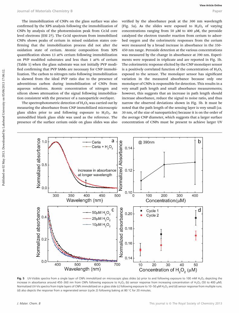
The spectrophotometric detection of H2O2 was carried out bymeasuring the absorbance from CNP immobilized microscopicglass slides prior to and following exposure to H2O2. Anunmodied blank glass slide was used as the reference. Thepresence of the surface cerium oxide on glass slides was also
Fig. 5 UV-Visible spectra from a single layer of CNPs immobilized on microscopicincrease in absorbance around 450–360 nm from CNPs following exposure to H2
Normalized UV-Vis spectra from triple layers of CNPs immobilized on a glass slide (c)(d) also depicts the response from a regenerated sensor (cycle 2) following baking
J. Mater. Chem. B
veried by the absorbance peak at the 300 nm wavelength(Fig. 5a). As the slides were exposed to H2O2 of varyingconcentrations ranging from 50 mM to 400 mM, the peroxidecatalyzed the electron transfer reaction from cerium to adsor-bed oxygen and the colorimetric responses from the ceriumwere measured by a broad increase in absorbance in the 350–450 nm range. Peroxide detection at the various concentrationswas measured by the change in absorbance at 390 nm. Experi-ments were repeated in triplicate and are reported in Fig. 5b.The colorimetric response elicited by the CNPmonolayer sensoris a positively correlated function of the concentration of H2O2
exposed to the sensor. The monolayer sensor has signicantvariation in the measured absorbance because only onemonolayer of CNPs is responsible for detection. This results in avery small path length and small absorbance measurements;however, this suggests that an increase in path length shouldincrease absorbance, reduce the signal to noise ratio, and thusnarrow the observed deviations shown in Fig. 5b. It must benoted that the path length of the sensing layer is very small (ca.30 nm, of the size of nanoparticles) because it is on the order ofthe average CNP diameter, which suggests that a larger surfaceconcentration of CNPs must be present to achieve larger UV
glass slides (a) prior to and following exposure to 100 mM H2O2 depicting theO2 (b) sensor response from increasing concentration of H2O2 (50 to 400 mM).following exposure to 10–50 mMH2O2 and (d) sensor response frommultiple runs.at 80 �C for 20 minutes.
This journal is ª The Royal Society of Chemistry 2013

Paper Journal of Materials Chemistry B
Publ
ishe
d on
02
May
201
3. D
ownl
oade
d by
Lak
ehea
d U
nive
rsity
on
05/0
6/20
13 1
7:06
:12.
View Article Online
absorbance values. This minimizes the absorbance error bymaximizing the signal-to-noise, according to the Lambert–BeerLaw, A ¼ 3bc where A is UV absorbance, 3 is the extinctioncoefficient, b is the path length, and c is the sample concen-tration. A rather high extinction coefficient is characteristic ofCNPs, and because of this CNPs have been used for UVabsorption enhancement.31
In order to improve the detection limit and the signal-to-noise ratio of the monolayer sensor, a multilayered assemblyconsisting of alternate layers of PVP and CNPs was formulated.Fig. 5c shows an enhancement of peroxide detection limit uponintroduction of a multi-layered (triple) assembly of the sensorand a better signal to noise ratio was achieved from multipleruns (Fig. 5d). UV-Vis spectra (ESI 4†) collected from severalmultilayered glass slides show no differences in absorbancedepicting the robustness and repeatability of the immobiliza-tion process. Peroxide must be able to penetrate to all the layersof immobilized CNPs in the triple layer sensor matrix to accountfor the increase in UV absorbance and detection enhancement.In consideration of the Lambert–Beer's law, the increase in pathlength and surface concentration as successive CNP and PVPmonolayers are self-assembled accounts for the measuredincrease in UV absorbance.
Optical studies of CeO2 thin lms, however, suggest that anupper bound on this signal enhancement exists. A previousstudy by Debnath et al. shows that the extinction coefficientdiminishes exponentially as the CeO2 thin lm thickness growsbeyond 150 nm.32 Note the difference between a thin lm andself-assembled monolayers, however. A self-assembled mono-layer construction allows for an increased surface area forreaction and three consecutive SAMs enhance the resulting UVabsorbance; although, there must be a saturation point wheremultiple SAM depositions become a thin lm, in effect. Futurework may elucidate saturation points of the layered assembliesand may possibly identify the limits in UV absorbance formaximizing the sensor's detection abilities.
It is clear from DFT results that the colorimetric response ofCNPs arises from the electron transfer from CNPs to adsorbedoxygen catalyzed by the peroxide. Thus, the sensor could prac-tically be regenerated following desorption of adsorbed oxygenmolecules. Aer initial exposure to H2O2 several triple layerdetection slides were heated at 80 �C for 20 minutes and re-usedto detect H2O2 at 10–50 mM concentrations (Fig. 5d, cycle 2).These results conrm the sensors' ability to be regenerated andused multiple times. The decrease in the response of the sensorin moving from cycle 1 to cycle 2 shows that there is a scope forimprovement and future work will focus on the regenerationand optimization of the number of layers of CNPs that can beimmobilized through this process without compromising thesignal to noise ratio. In addition, the effect of secondary bakingon the oxidation state and redox reversibility of cerium toregenerate the sensor will also be explored.
The selectivity of the sensor towards other reactive oxygenspecies (such as the superoxide and hydroxyl radical) is animportant point to consider. Testing and measuring the selec-tivity of the sensor is outside the scope of the current manu-script. Although it is well known that the CNPs react with
This journal is ª The Royal Society of Chemistry 2013
superoxide radicals, it is clear from the DFT calculationsdescribed earlier in the article that this reaction may not lead toa colorimetric response. The colorimetric response occurs onlywhen a peroxide species is adsorbed on the surface of the CNPs.Additionally, the hydroxyl radical is highly reactive and reactswith almost everything in its path in a diffusion limited reac-tion. It was shown previously by our group using ESR that CNPsdo not react with hydroxyl radicals as the ESR spectrum of thehydroxyl radicals before and aer exposure to the CNPsremained unchanged.33 Thus, the CNP based sensor should behighly selective in its response towards detection of hydrogenperoxide molecules.
Conclusions
The results presented in this paper suggest the viability of anovel hydrogen peroxide sensor that directly utilizes the color-imetric response created from the redox reaction between CNPsand peroxide. PWF-DFT calculations demonstrate that theinherent color change in the system originates from a two-electron transfer process between the CNPs and adsorbedoxygen species following the redox reaction between CNPsand H2O2. AFM, HIM, and XPS together conrm that thedip-coating methodology herein employed is able to constructself-assembled monolayers of poly(4-vinylpyridine) therebyfunctionalizing a glass substrate for immobilization of CNPs.UV-Vis spectrophotometry demonstrates that detection of H2O2
is possible and a triple-layered assembly of subsequent PVP andCNP SAMs signicantly improves H2O2 detection. Furthermore,the sensor can be used multiple times by simply heating atmoderate temperatures. A parallel study by Lin et al. recentlyreported the use of thermal regeneration of CNPs in applica-tions such as biocomputing emphasizing the importance ofregenerative capabilities of CNPs in various applications.Localized heating of immobilized single layers of CNPs coulddrastically reduce the regeneration time in such devices toimprove the reusability and signal detection.34 In addition,since the sensor response is direct and applicable whereverhydrogen peroxide is present, the CNP SAM sensor can beextended to design applications from other enzymatic reactionswhere H2O2 is a by-product, such as for detection of glucose.
Acknowledgements
The research was performed using EMSL, a national scienticuser facility sponsored by the Department of Energy's Office ofBiological and Environmental Research and located at PacicNorthwest National Laboratory and supported by the EMSLIntramural program. J. D. Gaynor was also supported by theDOE Office of Science's SULI undergraduate research program.Additional support from the NSF-NIRT Program supplementedthis research as well.
Notes and references
1 E. Cadenas and K. J. A. Davies, Free Radical Biol. Med., 2000,29, 222.
J. Mater. Chem. B
Journal of Materials Chemistry B Paper
Publ
ishe
d on
02
May
201
3. D
ownl
oade
d by
Lak
ehea
d U
nive
rsity
on
05/0
6/20
13 1
7:06
:12.
View Article Online
2 I. Fridovich, Ann. N. Y. Acad. Sci., 1999, 893, 13.3 D. G. Bostwick, E. E. Alexander, R. Singh, A. Shan, J. Q. Qian,R. M. Santella, L. W. Oberley, T. Yan, W. X. Zhong, X. H. Jiangand T. D. Oberley, Cancer, 2000, 89, 123.
4 K. Ishikawa, Science, 2008, 321, 342.5 C. Polytarchou, M. Hatziapostolou and E. Papadimitriou, J.Biol. Chem., 2005, 280, 40428.
6 T. Shimosato, A. Geddawy, M. Tawa, T. Imamura andT. Okamura, J. Pharmacol. Sci., 2012, 118, 206.
7 J. Emerit, A. Edeas and F. Bricaire, Biomed. Pharmacother.,2004, 58, 39.
8 M. P. Mattson, Nature, 2004, 431, 631.9 P. Sompol, W. Ittarat, J. Tangpong, Y. Chen, I. Doubinskaia,I. Batinic-Haberle, H. M. Abdul, D. A. Buttereld andD. K. S. Clair, Neuroscience, 2008, 153, 120.
10 B. C. Schanen, A. S. Karakoti, S. Seal, D. R. Drake,W. L. Warren and W. T. Self, ACS Nano, 2009, 3, 2523.
11 M. T. Lin and M. F. Beal, Nature, 2006, 443, 787.12 B. Palmieri and V. Sblendorio, Eur. Rev. Med. Pharmacol. Sci.,
2007, 11, 27.13 N. Soh, Anal. Bioanal. Chem., 2006, 386, 532.14 X. Xu, S. Jiang, Z. Hu and S. Liu, NANO, 2010, 4, 4292.15 S. Parajuli and W. J. Miao, Anal. Chem., 2009, 81, 5267.16 P. O. Wennberg, T. F. Hanisco, L. Jaegle, D. J. Jacob,
E. J. Hintsa, E. J. Lanzendorf, J. G. Anderson, R. S. Gao,E. R. Keim, S. G. Donnelly, et al., Science, 1998, 279, 49.
17 W. J. Cooper and R. G. Zika, Science, 1983, 220, 711.18 J. P. Farr, D. S. Steichen and W. L. Smith, Bleaching Agents,
in Kirk-Othmer Encyclopedia of Chemical Technology, Wiley,2003.
J. Mater. Chem. B
19 P. C. Burns, R. C. Ewing and A. Navrotsky, Science, 2012, 335,1184.
20 A. Rudra, N. P. Thacker and S. P. Pande, Environ. Monit.Assess., 2005, 109, 189.
21 D. Lee, S. Khaja, J. C. Velasquez-Castano, M. Dasari, C. Sun,J. Petros,W. R. Taylor andN.Murthy,Nat. Mater., 2007, 6, 765.
22 A. Agrawal, C. Susut, G. Stafford, U. Bertocci, B. McMorran,H. J. Lezec and A. A. Talin, Nano Lett., 2011, 11, 2774.
23 M. Ornatska, E. Sharpe, D. Andreescu and S. Andreescu,Anal. Chem., 2011, 83, 4273.
24 P. Fletcher, K. N. Andrew, A. C. Calokerinos, S. Forbes andP. J. Worsfold, Luminescence, 2001, 16, 1.
25 P. Wardman, Free Radical Biol. Med., 2007, 43, 995.26 E. W. Miller, A. E. Albers, A. Pralle, E. Y. Isacoff and
C. J. Chang, J. Am. Chem. Soc., 2005, 127, 16652.27 W. Oh, Y. S. Jeong, S. Kim and J. Jang, ACS Nano, 2012, 6,
8516–8524.28 G. Henkelman, B. P. Uberuaga and H. Jonsson, J. Chem.
Phys., 2000, 113, 9901.29 S. Malynych, I. Luzinov and G. Chumanov, J. Phys. Chem. B,
2002, 106, 1280.30 K. S. Schmitz, Macromolecules, 2000, 33, 2284.31 N. N. Dao, M. D. Luu, Q. K. Nguyen and B. S. Kim, Adv. Nat.
Sci.: Nanosci. Nanotechnol., 2011, 2, 1–4.32 S. Debnath, M. R. Islam and M. S. R. Khan, Bull. Mater. Sci.,
2007, 30, 315.33 E. G. Heckert, A. S. Karakoti, S. Seal and W. T. Self,
Biomaterials, 2008, 18, 2705–2709.34 Y. Lin, C. Xu, J. Ren and X. Qu, Angew. Chem., Int. Ed., 2012,
51, 1–6.
This journal is ª The Royal Society of Chemistry 2013