Embed Size (px)
Citation preview

lable at ScienceDirect
Biomaterials 30 (2009) 3503–3512
Contents lists avai
Biomaterials
journal homepage: www.elsevier .com/locate/biomateria ls
The antibacterial activity of Magainin I immobilized onto mixed thiolsSelf-Assembled Monolayers
Vincent Humblot a,b,*, Jean-Fabrice Yala c, Pascal Thebault a,b, Kada Boukerma a,b, Arnaud Hequet c,Jean-Marc Berjeaud c,**, Claire-Marie Pradier a,b
a CNRS, UMR CNRS 7197, Laboratoire de Reactivite de Surface, 4 place Jussieu, 75252 Paris Cedex 05, Franceb Universite Pierre et Marie Curie – UPMC Paris VI, Laboratoire de Reactivite de Surface, 4 place Jussieu, 75252 Paris Cedex 05, Francec Universite de Poitiers, Laboratoire de Chimie et Microbiologie de l’Eau – UMR 6008 CNRS, IBMIG – UFR Sciences fondamentales et Appliquees, 40 avenue du Recteur Pineau,86022 Poitiers Cedex, France
a r t i c l e i n f o
Article history:Received 20 January 2009Accepted 17 March 2009Available online 5 April 2009
Keywords:Self-assembled monolayersXPSPM-RAIRSBacteriaMagaininAntibacterial surfaces
* Corresponding author. Universite Pierre et Marieoratoire de Reactivite de Surface, 4 place Jussieu, 75Tel.: þ33 1 4427 5519; fax: þ33 1 4427 6033.** Corresponding author.
E-mail addresses: [email protected] (V. Huuniv-poitiers.fr (J.-M. Berjeaud).
0142-9612/$ – see front matter � 2009 Elsevier Ltd.doi:10.1016/j.biomaterials.2009.03.025
a b s t r a c t
An antibacterial peptide, Magainin I, was covalently bound to a mixed 11-mercaptoundecanoıc acid(MUA) and 6-mercaptohexanol (C6OH) (ratio 1:3) Self-Assembled Monolayer (SAM) on gold surfaces.Each step of the surface functionalization was characterized by Polarization Modulation ReflectionAbsorption InfraRed Spectroscopy (PM-RAIRS) and X-ray Photoelectron Spectroscopy (XPS). The anti-bacterial activity of the anchored Magainin was tested against three Gram-positive bacteria (Listeriaivanovii, Enterococcus faecalis and Staphylococcus aureus), and the results revealed that the adsorbedMagainin I reduced by more than 50% the adhesion of bacteria at the surface, together with the killing ofthe bacteria that nonetheless adhered to the surface. No release of the peptide was observed uponcontact with the bacterial suspension; the activity has proven to be persistent overtime, up to six monthsafter the first use.
� 2009 Elsevier Ltd. All rights reserved.
1. Introduction
Microorganisms have a strong tendency to adhere and grow onsolid surfaces. Once bacteria or fungi are attached to a substratumsurface, a multistep process starts, including binding of mineralcomponents, eventually leading to the formation of a complex,adhering microbial community named ‘‘biofilm’’ [1]. In 2002,Donlan and Costerton [2] defined the biofilm as a microbiallyderived sessile community characterized by cells irreversiblyattached to a substratum, interface or to each other, embedded ina matrix of extracellular polymeric substances that they haveproduced, and exhibiting an altered phenotype with respect togrowth rate and gene transcription. The nature of biofilm structureand the physiological attributes of biofilm organisms offer aninherent resistance to antimicrobial agents, whether these anti-microbial agents are antibiotics, disinfectants, or germicides [2].Established biofilms can tolerate antimicrobial agents at
Curie – UPMC Paris VI, Lab-252 Paris Cedex 05, France.
mblot), jean-marc.berjeaud@
All rights reserved.
concentrations 10–1000 times higher than the concentrationrequired to kill genetically equivalent planktonic bacteria [3].
The pattern of development involves initial attachment toa solid surface, the formation of microcolonies on the surface, andfinally differentiation of microcolonies into exopolysaccharide-encased, mature biofilms [4]. Conceptually, the simplest method forpreventing bacterial colonization and eventual biofilm formationon surfaces is to impregnate the substrate itself with a broad-spectrum antimicrobial agent that impairs bacterial growththrough traditional bactericidal or bacteriostatic mechanisms.Some authors proposed to create a permanently sterile, non-leaching material by covalently functionalizing its surface with anantimicrobial compound [5,6]. Numerous biocidal substances suchas antibiotics [7], quaternary ammonium compounds [6,8,9],phenol derivatives [10], titanium oxide [11], or heavy metals suchas silver and tin derivatives [12–15] have thus been immobilizedonto material surfaces. However, their use is often restricted bytheir potential toxicity.
By comparison with conventional antibiotics, the natural anti-microbial peptides, which are produced by plants, insects,mammalians, as well as microorganisms [16,17] offer severalattractive advantages: numerous have been shown to have a broadspectrum of antimicrobial activity, they rarely promote the rise ofbacterial resistance, and they act at a very low concentration.

V. Humblot et al. / Biomaterials 30 (2009) 3503–35123504
Therefore, the development of antibacterial coatings using thesenatural substances seems to be very attractive. Although the exactmechanisms by which these peptides exert antimicrobial activityare still unclear, it has been suggested that they are able to pene-trate into bacterial membranes and subsequently induce cell lysis[18]. Several recent works report the immobilization of smallpeptides within polyelectrolyte layers [19–21,23] or embedded ina polymeric matrix [22,24]; all these studies demonstrated theantibacterial properties of immobilized peptides, making clear therole of the peptide mobility and accessibility to the microbial cells.
Magainins were discovered from the granular gland of the skinof the African clawed frog Xenopus laevis by Zasloff in 1987 [25].Magainins are two 23-residue peptides, Magainins I and II differingat positions 10 and 22 (Gly-Lys Vs. Lys-Asn, respectively), the moststudied being Magainin II, which exhibits a broad spectrum ofantimicrobial activity against bacteria or fungi. However, in a recentwork Glinel et al., demonstrated that Magainin I, chemically graftedto polymer brushes, displayed a very good activity against Gram-positive bacteria [22]. Magainins are also responsible for theosmotic lysis of protozoa typically in the concentration range of 10–100 mg/mL [25–30]. In contrast, more than 1 mg/mL of Magainin isneeded to lyse mammalian cells, e.g. erythrocytes [31]. Magaininsappear to target the lipid matrix rather than the proteins; this hasbeen suggested by the fact that enantiomeric peptides, composedof D-amino acids, exhibit the same antimicrobial action as thatnaturally occurring with all-L peptides; chiral molecules are thuslikely not involved in the mechanism [26,32]. Moreover, Magaininsinduce permeabilization of artificial lipid bilayers containing acidicphospholipids (vesicles and planar lipid bilayers) [33–37].
In this paper, Self-Assembled Monolayers (SAMs) were elabo-rated in order to immobilize Magainin I on gold surfaces. MagaininI was chosen because it possesses a lysine residue near the C-terminal end (position 21) which enhanced the possibility to obtaina peptide grafted at an extremity thus probably less disturbed in itsactivity. Reactive pure 11-mercaptoundecanoic acid (MUA) layersand mixed SAMs were built by coadsorption of binary mixtures ofthiol 11-mercaptoundecanoic acid (MUA) and 6-mercaptohexanol(C6OH) at a constant fraction MUA:C6OH equal to 25:75 [38,39].When performing Magainin I (MAG) binding on pure MUA layers(100% MUA), the amount of MAG immobilized is identical to the
Au-MUA Au-MUA
Au
S
CHO O
S
HO
S
HO
S
HO
ON
O
O
Au
S
C
S
HO
EDC/NHS20mM/10mM, 1h
Fig. 1. Scheme of the Magainin I immobilization. Step 1: adsorption of thiols’ SAMs, Au–MUcovalent binding of the Magainin I, Au–MUA–MAG.
one obtained with the 25:75 mixed layer. Precedent studies on thebinding of protein A [38] indicate that for equal amounts ofproteins, there efficiency is better on the 25:75 diluted layer thanon pure layer; therefore in this paper we present only the resultsobtained on the 25:75 mixed layer.
Covalent binding of Magainin I (MAG) was achieved by conver-sion of the carboxylic acid tail groups of MUA into esters functions,by reaction with N-hydroxysuccinimide (NHS) in the presence ofcarbodiimide (EDC), followed by reaction with MAG (Fig. 1).
The gold surfaces were characterized step by step, by means ofinfrared spectroscopy (PM-RAIRS) and X-ray photoelectron spec-troscopy (XPS). The antibacterial activity of Magainin I moleculesanchored to the gold surfaces was assessed by bacteria adhesiontests, confocal microscopy and AFM analyses.
2. Experimental
2.1. Chemicals and surface preparation
Magainin I (Gly-Ile-Gly-Lys-Phe-Leu-His-Ser-Ala-Gly-Lys-Phe-Gly-Lys-Ala-Phe-Val-Gly-Glu-Ile-Met-Lys-Ser), 11-mercaptoundecanoıc acid (MUA), 6-mercaptohex-anol (C6OH), N-hydroxysuccinimide (NHS), 1-(3-dimethylaminopropyl)-N0-ethyl-carbodiimide hydrochloride (EDC) were purchased from Sigma Aldrich (Saint-QuentinFallavier, France). All solvents were reagent-grade. Reagents were used withoutany further purification. Experiments were carried out at room temperature if notspecified otherwise.
The surfaces were constituted of glass substrates (11 mm � 11 mm), coatedsuccessively with a 50 Å thick layer of chromium and a 200 nm thick layer of gold,were purchased from Arrandee (Werther, Germany). The gold coated substrateswere annealed in a butane flame to ensure a good crystallinity of the topmost layersand rinsed in a bath of absolute ethanol during 15 min before adsorption.
The substrates were immersed in a binary mixture at 0.01 M (25/75) of MUA(2.5 mM) and C6OH (7.5 mM) in 10 mL of absolute ethanol for 3 h, in order to insurean optimal homogeneity of the adlayer [38,40], and thorough rinsed in ethanol andMilliQ water and dried under a flow of dry nitrogen.
The substrates were treated with a solution of NHS (20 mM) and EDC (10 mM) inultrapure water for 90 min, rinsed in MilliQ water and dried under a flow of drynitrogen.
Immobilization of Magainin I (MAG, 5 mg/L in PBS) on gold surfaces was carriedout by depositing a 150 mL drop of MAG/PBS on the Au-modified substrates at roomtemperature for 2 h. After the immobilization step, the surfaces were vigorouslyrinsed in PBS with agitation, dried under a flow of dry nitrogen.
For each step, 2 series of identical experiments were conducted, one series ofsamples was then characterized by PM-RAIRS and XPS, while the second one wascharacterized by PM-RAIRS and AFM.
act Au-MUA-MAG
O
S
HO
S
HO
HN
Au
S
C O
S
HO
S
HO
S
HO
MAG
MAGAININ5mg/L, 2h
A; Step 2: esterification of the COOH functionalities by NHS/EDC, Au–MUAact; Step 3:

V. Humblot et al. / Biomaterials 30 (2009) 3503–3512 3505
2.2. PM-RAIRS measurements
The gold samples were placed in the external beam of FT-IR instrument (NicoletNexus 5700 FT-IR spectrometer) and the reflected light was focused on a nitrogencooled HgCdTe wide band detector. The infrared spectra were recorded at 8 cm�1
resolution, with co-addition of 128 scans. A ZnSe grid polarizer and a ZnSephotoelastic modulator to modulate the incident beam between p and s polariza-tions (HINDS Instruments, PM90, modulation frequency ¼ 36 kHz) are placed priorto the sample. The detector output is sent to a two-channel electronic device thatgenerates the sum and difference interferograms. Those are processed and undergoFourier transformation to produce the PM-RAIRS signal (DR/R0)¼(Rp� Rs)/(Rpþ Rs).Using a modulation of polarization enabled us to perform rapid analyses of thesample after treatment in various solutions without purging the atmosphere orrequiring a reference spectrum.
2.3. XPS analyses
XPS analyses were performed using a PHOIBOS 100 X-ray photoelectron spec-trometer from SPECS GmbH (Berlin, Germany) with a MgKa X-ray source(hn ¼ 1253.6 eV) operating at P ¼ 1 � 10�10 Torr or less. Spectra were carried outwith a 20 eV pass energy for the survey scan and 10 eV pass energy for the C1s, O1s,S2p and N1s regions. High-resolution XPS conditions have been fixed: ‘‘FixedAnalyser Transmission’’ analyses mode, a 7 � 20 mm entrance slit; leading toa resolution of 0.1 eV for the spectrometer, and an electron beam power of 150 W(12.5 kV and 12 mA). Such a low energy was used to keep the adsorbed layers asintact as possible. A takeoff angle of 90� from the surface was employed for eachsample and binding energies were calibrated against the Au4f binding energy of thegold surface at 84.0 eV. Element peak intensities were corrected by Scofield factors[41], the spectra were fitted using the Casa XPS v.2.3.13 Software (Casa Software Ltd.,UK) and applying a Gaussian/Lorentzian ratio, G/L equal to 70/30. In addition, theerror in the peak fitting process was estimated at 5% or less with respect to theintegration of the raw data.
2.4. AFM characterization
The AFM images of dried surfaces were recorded using a commercial di CaliberAFM microscope from VEECO Instruments Inc. In order to avoid tip and sampledamages, topographic images were taken in the non-contact dynamic mode alsoknown as tapping� mode. Silicon nitride tips (resonance frequency of w280–400 kHz, force constant of 40–80 N/m) have been used. Images were obtained ata constant speed of 2 Hz with a resolution of 512 lines of 512 pixels each. The rawdata were processed using the imaging processing software di SpmLabAnalysis v.7.0.from Veeco Instruments Inc., mainly to correct the background slope between thetip and the gold surface.
2.5. Bacterial strains, media and cultural conditions
The bacterial strains used in this work are Listeria ivanovii Li4pVS2 [42],Enterococcus faecalis III (laboratory collection) and Staphylococcus aureusATCC29213. All strains were grown at 37 �C in Brain Heart Infusion (BHI) broth (BDDIFCO, Le Pont-De-Claix, France). Enumerations of bacteria were conducted on BHIagar (15 g/L) medium.
2.6. Adhesion of bacteria on gold samples
Gold samples were washed successively in 70% ethanol and sterile water thendried in sterile environment. A kappa carrageenan solution (15 g/L) at 50 �C waspoured in a Petri dish and cooled down to 33.5 �C. The sample was then cautiouslydeposited on the surface of the layer with the gold face upwards. After cooling downto room temperature, 105 bacteria in 100 mL were deposited on the gold surface.After 3 h at 37 �C, each sample was washed, three times, with physiological sterilesolution (0.9% NaCl), then transferred into a sterile tube containing 1.5 mL ofphysiological sterile solution, then sonicated 3 min at 51 W. After the gold samplewas removed, the bacteria were pelleted by centrifugation at 13 000 g for 5 min.Then 1.3 mL of supernatant was removed cautiously and the bacteria were re-sus-pended by vortexing. The suspension was then diluted 10 and 100 times; 50 mL ofeach dilution was deposited on agar plates, in duplicate for each dilution, usinga spiral plater WASP (AES, France). Before counting the colonies, the plates wereincubated at 37 �C for 18 h.
2.7. Microscopic analysis
Viability of bacteria attached to the surfaces was evaluated using LIVE/DEAD�
Bacterial Viability Kit (BacLight�). The two BacLight stains, Syto9 and propidiumiodide, dissolved in DMSO, were diluted 1:10 in a NaCl solution (0.085%), providingstock solutions. The stock solutions were kept at �20 �C and protected from light.BacLight mixture was prepared immediately before analysis by mixing 1.5 mL of bothstain stock solutions and diluting to a final 100 mL volume with distilled water.
After incubation with bacterial culture during 3 h, gold samples were washed asdescribed above. 10 mL of BacLight mixture was deposited on the surface of thesample and incubated 10 min in the dark at room temperature prior to microscopicanalysis.
Samples were examined with a confocal FV-1000 station installed on aninverted microscope IX-81 (Olympus, Tokyo, Japan). Images were acquired with anOlympus UplanSapo�60 water, 1.2 NA, objective lens (800� 800 pixels images with0.13 mm/pixel corresponding to Nyquist criteria for optimal sampling). Multiplefluorescence signals were acquired sequentially to avoid cross talk between imagechannels. Fluorophores were excited with the 488 nm line of an argon laser (forSyto9) and the 543 nm line of an HeNe laser (for propidium iodide). The emittedfluorescences were detected through spectral detection channels between 500–530 nm and 555–655 nm, for green and red fluorescence respectively.
2.8. Characterization of magainin I release
Two gold slides, one after MUA grafting (Au–MUA), and one after peptideimmobilization (Au–MUA–MAG), were incubated for 3 h in 1.5 mL of distilled waterat 37 �C under a slight agitation. All the water content was recovered and evaporatedat 37 �C. 10 mL of 0.2% formic acid/5% CH3CN solution was then added to solubilizethe dried residue, possibly containing some peptide. The so-obtained solution wasenriched and separated using a lab-on-a-chip technology (Agilent), and furtherfragmented using an on-line XCT mass spectrometer (Agilent) and the fragmentationdata were interpreted using the data Analysis program (version 3.4 Bruker daltonic).The MS/MS (MS2) data were extracted using Analyst Software 1.4.1, build 1162(Applied Biosystems). Default parameters were used for the generation of peak lists.MS2 peak lists were extracted and compared to the NCBInr protein database restrictedusing the MASCOT Daemon (version 2.1.3) search engine. All searches were performedwith no fixed modification thus allowing carbamidomethylation of cysteins, oxidationof methionins and a maximum of one missed trypsin cleavage. MS2 spectra weresearched with a mass tolerance of 1.2 Da for precursor ions and 0.8 Da for fragmentions, respectively. The same protocol was used with a Magainin I solution (80 mg/mL)as a control.
3. Results
3.1. Surface characterization
3.1.1. PM-RAIRS resultsFig. 1 depicts a schematic representation of the three steps fol-
lowed to construct the antibacterial surface. First, the substrate wasfunctionalized with a monolayer of 11-mercaptoundecanoıc acid/mercaptohexanol, 25/75 respectively (Au–MUA). The acid functionswere then activated into esters (Au–MUAact), in order to let Mag-ainin I (MAG) react via its amino groups (Au–MUA–MAG).
Fig. 2 shows the PM-RAIR spectra recorded after the successivesteps of gold surface functionalization. Spectrum (a) is dominated byan intense nC]O band at 1722 cm�1, characteristic of carboxylicgroups [43,44], showing the presence of the acid-terminated thiol.The nCH band of the CH2 chains [43] are recorded at 2850 and2922 cm�1 suggesting a rather good crystallinity of the mixedmercaptoundecanoic acid þ mercaptohexanol layer [39,45,46].Compared to a SAM elaborated under similar conditions but withMUA alone, the lower intensity of the nC]O band suggests that thedensity of adsorbed mercaptoundecanoic acid is indeed reduced(spectrum not shown) in agreement with previous studies [44]. TheIR absorption, at 1244 cm�1 is attributed to the OH deformation ofthe mercaptohexanol, while the weak, but broad, band at 1455 cm�1
likely includes contributions from the scissor mode of CH2 groupsand from the symmetric COO� stretch; an absorption signal at1555 cm�1, ascribed to the asymmetric COO� stretch vibration,confirms that some acidic groups are deprotonated [39,46].
On spectrum (b), one sees a slight upward shift and increase ofthe nC]O band, to 1752 cm�1, confirming the transformation of acidinto ester terminal groups; two other weak bands appear at 1789and 1822 cm�1 as the signatures of ester groups [47,48] (Fig. 1).These will be reactive towards the NH2 groups present on the MAGmolecule. The binding of MAG is indicated by the appearance ofnew bands on spectrum (c), intense features at 1663 and 1546 cm�1
are ascribed to the amide I and amide II band of the peptidic

0.05
%
PM-IR
RAS
sig
nal
3200 3000 2800 2000 1800 1600 1400 1200 1000
1251
145729
61
285029
22
1244
145515
55
Au-MUA
Au-MUAact
1722
Wavenumber cm-1
c
b
a
Au-MUA-MAG
2922
2850 18
22
1752
1789
2927
2853
1738
1663
1546
Fig. 2. PM-RAIR spectra of the three consecutive steps leading to the immobilization of Magainin I: (a) Au–MUA; (b) Au–MUAact by NHS/EDC; (c) covalent binding of Magainin I, Au–MUA–MAG.
V. Humblot et al. / Biomaterials 30 (2009) 3503–35123506
backbone while the small band at 1738 cm�1 is the C]O stretchingof lateral chain functions and of some hydrolysed ester functions[39]. Weak absorption signals at 1251 and 1457 cm�1 are alsoobserved and can be attributed respectively to the dOH (CH2OH ofthe serine or O]C–OH of the glutamic acid, for instance) and to thesymmetric nCH2 of some functional groups of the lateral chains ofthe molecule [49]. Note the almost complete disappearance of theabsorption signal at 1752 cm�1, suggesting the total consumption ofester groups by reaction with the MAG molecules [48].
3.1.2. XPS resultsXPS analyses after MUA/C6OH immobilization and MAG
bonding provide complementary information. The C1s, N1s, O1s,S2p and Au4f high-resolution regions were recorded, although onlythe N1s, O1s and C1s regions are presented in Fig. 3.
After MUA adsorption, the Au4f7/2 peak is still rather intense; bycomparing with the intensity of the clean gold surface (Au4f7/2)0,the average thicknesses of the Au–MUA and Au–MUA–MAG layerswere estimated to ca. 11 Å and 24 Å, respectively, using thefollowing equation IðAu4f7=2Þ=IðAu4f7=2Þ0 ¼ expð�ðd=lsin qÞÞwhere d is the layer average thickness, q is the takeoff angle fixed at90� in this case, and l is the escape depth of Au4f7/2 electronsthrough an organic layer, calculated at 36 Å using the formulaestablished by Whitesides et al.[50,51].
Adsorption of MAG on the MUA-modified gold surface was alsoshown by the appearance of a nitrogen peak, N1s, centred at400.6 � 0.1 eV, mainly due to the amide functions of the peptide[38,52] (Fig. 3a). The O1s of the Au–MUA surface (Fig. 3b) showsa main contribution at 533.0 � 0.1 eV assigned to the C–OH atoms,and a smaller one at 531.9 � 0.1 eV (C]O) with a ratio of 3.8,suggesting that the expected ratio of MUA over mercaptohexanol inthe adsorbed phase has been reached, almost all MUA thiol groupsbeing protonated; the O1s of the Au–MUA–MAG surface stillpresents two contributions at 533.0 and 532.0 � 0.1 eV, respec-tively assigned to the –OH and to the C]O of the amide bonds [52],with a ratio now close to 1:10, due to the prevailing contribution ofthe peptide backbone. Finally, the S2p peak, with a main contri-bution, S2p3/2, at 162.1 � 0.1 eV [45,53], shows no significant
evolution, either in its position or in its intensity, before and aftergrafting of the MAG (spectra not shown); the addition of sulphur bythe lateral fragments of the MAG (e.g. -Met- fragments) justcompensates the shielding of the thiol contribution.
Fig. 3c shows high-resolution XPS spectra of the C1s core levelpeaks corresponding to Au–MUA and Au–MUA–MAG, respectively.The C1s peak, of the Au–MUA surface, was best fitted with fourcontributions, at 284.9, 286.0, 287.4 and 289.0 � 0.1 eV, corre-sponding respectively to the carbon in C–C, C–H bonds, C ina position of S atoms or in C–C–OH positions, and finally for the lasttwo ones, in COO– and HO–C]O bonds of the acid or carboxylateterminal moieties [43,44].
After MAG adsorption, the carbon relative intensity increased byaround 30% and the C1s peak could still be fitted with fourcomponents, in agreement with previous studies of protein Aadsorption on mixed thiols’ SAMs [38]. The first peak, at the lowestbinding energy (BE) of 285.0� 0.1 eV is assigned to the carbon C–C,C–H; the second and third peaks, at 285.8 and 286.9 � 0.1 eV, areattributed respectively to the C–C–N carbon þ C–OH atoms and tothe carbon atoms in C–C–Oþ Caromatic bonds; and finally, the fourthcontribution at 288.7 � 0.1 eV, includes the C atoms involved inpeptidic bonds, O]C–N and acidic groups (H)O–C]O [38,54]. Theatomic percentages and BE of these contributions are summarizedin Table 1.
3.2. Activity of grafted Magainin
3.2.1. Adhesion tests (PM-RAIRS and AFM)After elaboration and characterization of these new surfaces, the
impact of the grafting of Magainin I on cell adhesion and growthwas evaluated by PM-RAIRS and AFM analyses. 150 mL of L. ivanoviibacterial suspension (1.5 � 106 cfu/mL at 37 �C in fresh BHI underhumid atmosphere) was spread over gold slides functionalizedwith peptide (Au–MUA–MAG) or only with MUA (Au–MUA).Various times of contact were tested in order to assess anddistinguish the anti-adhesion and bactericidal effects of Magainin I;our assumption was that in 30 min, the cells could adhere but notgrow. Indeed, E. faecalis displayed a generation time of about

292 290 288 286 284 282
289,0
285,0
Binding Energy eV
Au-MUA-MAG
286,0
288,7
284,9
285,8286,9
287,4 Au-MUA1000
0 C
PS
C1s
406 402 398 396
2000
CPS
Au-MUA
Au-MUA-MAG
N1s400.6
404 400Binding Energy eV
Binding Energy eV
O1s
5000
CPS
533.0
532.1
Au-MUA-MAG
Au-MUA
533.0 531.9
536 534 532 530 528
a
b
c
Fig. 3. High-resolution XPS spectra of the N1s region in (a), of the O1s region in (b) andof the C1s region in (c), before and after immobilization of Magainin I.
V. Humblot et al. / Biomaterials 30 (2009) 3503–3512 3507
30 min, which was the shortest of the three strains tested (data notshown). At longer times, 3 h for instance, the cells may adhere anddevelop if they are not altered by the surface. In Fig. 4a–d, six bandsare observed for the two times of contact and the two gold samples,some of them being hardly visible after 30 min. Two peaks posi-tioned at 1658 and 1554 cm�1 are ascribed respectively to theamide I and amide II bands of proteins; two other bands at 1456and 1407 cm�1 are also observed and can be attributed respectivelyto nCH2 stretches and nCOO� of fatty acid [55,56]. The two lastbands positioned at 1244 and 1081 cm�1 can be attributedrespectively to phosphate C–O–P of polysaccharides and C–O–C ofcarbohydrates. All these bands are characteristic of bacteria [55–57]and indicated adhesion of bacteria on the modified gold slides.
After 30 min of contact with the bacterial suspension; thespectra of the gold surfaces Au-List and Au–MAG-List, respectively
spectra (a) and (c), show the same profile and the intensity of thebands are very similar. These results also show that the presence ofMAG on the gold slides does not influence the number of adheredbacteria. However, to assess whether the bacteria are alive or not,we have performed AFM analyses directly after drying the samples,and the results are presented in Fig. 5. After 30 min (Fig. 5a and b)one can observe a small amount of bacteria present at the surface,roughly the same number of bacteria for the surface non-grafted, inFig. 5a, or grafted with MAG, in Fig. 5b, thus confirming the PM-RAIRS results presented above. By zooming on a single bacteriaadhered on Au–MUA, the bacteria seems to be ‘alive’ presentinga well defined oval shape in Fig. 5e, while the bacteria that adheredon the Au–MUA–MAG surface looks deteriorated in Fig. 5f, at leastwith a collapse of the cell walls. This tends to show that the pres-ence of Magainin I at the surface drastically alters the integrity ofthe bacteria. As a comparison, direct AFM investigation of cellsurface destruction by bacteriophage was recently performed byDubrovin et al. [58], shows similar changes in the cell surfacetopography; note however that in their study, destructing agentswere in solution (not grafted on a solid support).
In addition, on non-grafted surfaces, the bacteria appear assingle or as doublet, whereas on the MAG-modified surface, onenotices the presence of groups of bacteria, the phenomenon hasalso been reported for Escherichia coli in presence of bacteriophages[58]. This could be explained in one hand by the fact that on MAG-modified surfaces, the bacteria are deteriorated or killed by thepeptide, thus becoming a source of nutrients and consequentlya preferential anchoring site for the still alive bacteria. On the otherhand, Au–MUA–MAG surface is more hydrophobic than Au–MUAsurface because of the amphiphilic structure of the peptide [26,32].Consequently, when the surface was dried prior to AFM analysis,droplets may be formed in which bacteria were concentratedleading to the observed aggregates upon complete drying.
After 3 h of contact between the L. ivanovii and the goldsamples, the PM-RAIRS data (Fig. 4b and d) show two interestingresults. First, one can notice an important increase in intensity ofall IR bands with respect to those observed after 30 min, clearlydemonstrating that more bacteria are present on the surfaces. Thismay be explained by two facts: a longer contact time may allow i)more bacteria to adhere to the surface and ii) enough time to letthe bacteria multiply; Fig. 5c indeed shows some doubletsshowing bacteria not completely detached from each other. Thesecond noticeable result, is that on the Au–MUA–MAG surface, theamount of bacteria is significantly higher, according to an intensityalmost doubled in Fig. 4d compared with the intensity in Fig. 4b.This is, apparently, in contradiction with the expected ‘bacteriakiller’ role of the Magainin I, but can again be explained by theAFM results (Fig. 5d). The groups of bacteria observed after30 min, are now much bigger after 3 h of contact, confirming ourhypothesis that damaged bacteria act as nutrients and attractingsites for alive cells.
3.2.2. Adhered bacteria countingThe antibacterial activity of MAG-functionalized surfaces was
tested against three Gram-positive bacteria, L. ivanovii Li4pVS2, E.faecalis III and S. aureus ATCC29213. Bacterial strains were chosen asrepresenting species often implicated in food contamination(Listeria) [59], food spoilage (Enterococcus) [60] or nosocomialdiseases (Staphylococcus) [61].
A methodology was developed in order to evaluate the impact ofthe grafting of Magainin I on the adhesion of bacteria onto thefunctionalized surfaces. The gold samples were deposited on top ofa carrageenan layer, and once the polysaccharide layer was solid,only the functionalized side of the glass-coated substrate wasaccessible for the bacteria to adhere and not the other parts of the

Table 1Calculated average thickness and percentages of the different components of the C1s peak before and after immobilization of Magainin I on Au–MUA-modified gold surfaces.
Au–MUA Thickness 1.1 nm C–C, C–H (%) Ca, C–OH (%) COO�/HO–C]O(%)Th. Exp. Th. Exp. Th. Exp.69.0 73.7 27.6 16.9 3.4 9.4284.9 eV 286.0 eV 287.4 eV 289.0 eV
Au–MUA–MAG Thickness 2.4 nm C–C, C–H (%) Ca, þ C–C–N (%) C–C–O, Carom. (%) N–C]O, O–C]O (%)Th. Exp. Th. Exp. Th. Exp. Th. Exp.35.8 44.0 25.9 24.9 19.0 18.5 19.4 12.6285.0 eV 285.8 eV 286.9 eV 288.7 eV
V. Humblot et al. / Biomaterials 30 (2009) 3503–35123508
gold sample. Moreover, it was verified that the tested bacteria werenot able to grow on the carrageenan layer.
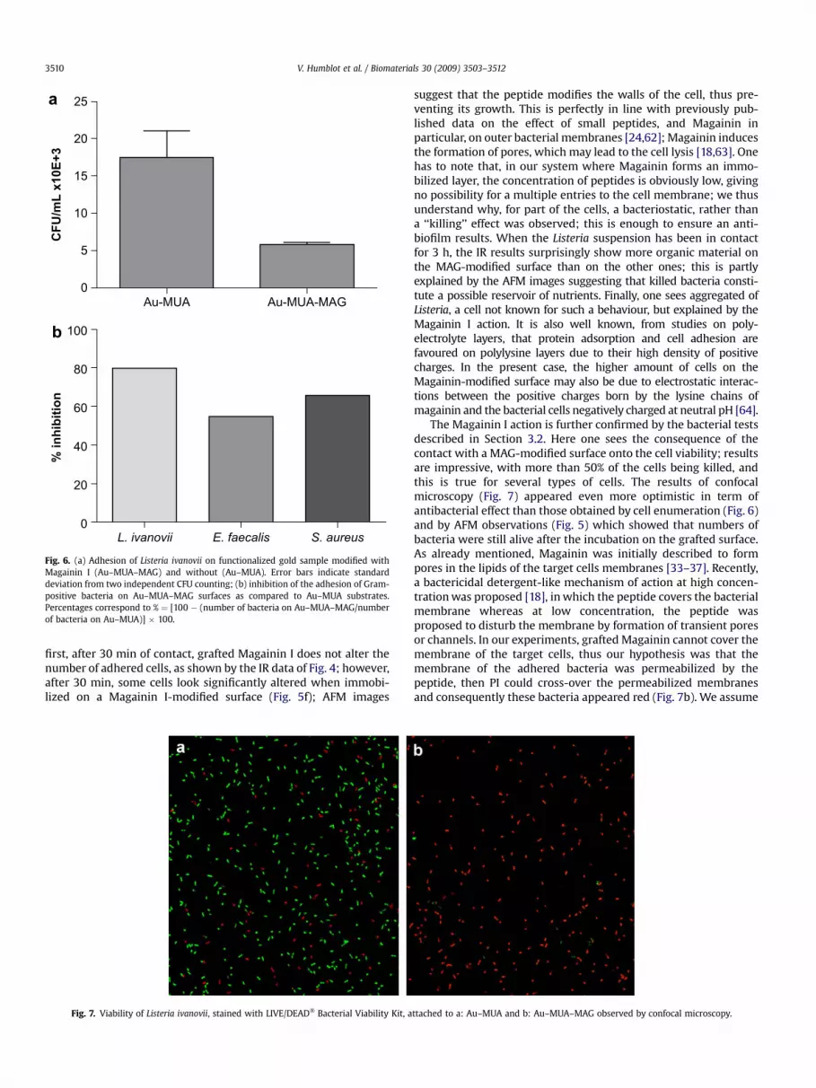
About 105 bacteria were deposited on surfaces modified withMUA (Au–MUA), or with MUA þMagainin I (Au–MUA–MAG). After3 h at 37 �C, adhered cells were detached from the surfaces bysonication before determining the number of CFU per mL. Bothsurfaces, with or without Magainin I were observed by confocalmicroscopy and appeared devoid of any bacterial cells after stainingwith viability dyes (images not shown). As shown in Fig. 6a, thenumber of L. ivanovii cells, which adhered to the surfaces bearingMagainin, is significantly reduced as compared to the surfacewithout peptide. Similar results were obtained with the otherGram-positive bacteria. Fig. 6 shows the percentages of inhibitionof attachment of bacterial cells onto the so-elaborated antibacterialsurfaces as compared to Au–MUA substrates [100 � (number ofbacteria on Au–MUA–MAG surfaces/number of bacteria onAu–MUA surfaces) � 100]. For the three bacterial species, less than50% of adhered bacterial cells were counted when Magainin I wasgrafted to the surface. These results demonstrate that the peptideinhibits the attachment of bacteria to the surfaces significantly.Moreover, reproducibility of the results was checked by usingtwo different sets of functionalized samples and culture batches ofeach strain.
2000 1800 1600 1400 1200 1000Wavenumber cm
-1
Au-List
b 3h
PM-R
AIR
S Si
gnal
0,1
%
1658
1554
1456 14
07
1244
1081
a 30 min
Fig. 4. PM-RAIR spectra of gold samples after contact with a Listeria ivanovii bacterial suspenAu–MUA–MAG surfaces for 30 min (c) and 3 h (d).
3.2.3. Viability of adhered cellsIn order to understand the mode of action of the peptide bonded
to the gold samples against bacteria, the cells attached to thesurfaces were observed using confocal microscopy after dying withBacLight� viability kit. Briefly, the kit contains two fluorescent dyes,Syto9 which stains all bacteria in green, and propidium iodide (PI)which can only cross-over damaged cells membranes and gives redstained cells. Thus, because PI quenches the fluorescent emission ofSyto9, it is assumed that green cells are alive whereas red cells aredead. After an incubation time of 3 h at 37 �C, the majority of L.ivanovii adhered to the surfaces with no Magainin I grafted (Fig. 7a)are green-stained. Only few bacteria appear as red spots on thepicture. On the contrary, on the picture of the MAG-modifiedsurface (Fig. 7b) most bacteria appear as red. The same experimentswere repeated using the other Gram-positive bacteria leading tosimilar results (images not shown). These results indicate that thebacteria first adhered to the surface then were permeabilized by thegrafted peptide which permitted the entry of PI.
3.2.4. Persistence of the activityOne of the objectives of this work was to obtain long shelf-life
antibacterial surfaces. In order to check the persistence of theactivity of the grafted Magainin, each couple of gold samples,
2000 1800 1600 1400 1200 1000Wavenumber cm
-1
1658
PM-R
AIR
S Si
gnal
0,1
%
Au-MAG-List
1554
1456
1407
1244
1081
c 30 min
d 3h
sion at 1.5 � 106 cfu/mL at 37 �C on Au–MUA surfaces for 30 min (a) and 3 h (b) and on

Fig. 5. AFM images of gold samples Au–MUA (a, c) and Au–MUA–MAG (b, d) obtained after 30 min and 3 h, respectively, of contact with a Listeria ivanovii bacterial suspension at1.5 � 106 cfu/mL at 37 �C; imaging conditions: 50 mm � 50 mm, 512 lines, 2 Hz, tapping� mode. (e) Single ‘alive bacteria’ on Au–MUA, and (f) single ‘damaged bacteria’ on Au–MUA–MAG; imaging conditions: 4 mm � 4 mm, 512 lines, 2 Hz, tapping� mode.
V. Humblot et al. / Biomaterials 30 (2009) 3503–3512 3509
Au–MUA and Au–MUA–MAG, was assayed after four periods oftime. Briefly, after the first experiment as described above, the goldsamples were cleaned, dried and stored at 4 �C. This washingprotocol was done after each assay as described below. The sampleswere assayed for antibacterial activity, using the same bacteria andthe same protocol, after one week, then after one month, and finallyonce again after 6 months. The results of the comparison in thenumber of attached bacteria on the surfaces, modified or not withMagainin, are presented in Fig. 8. The inhibition rate is similar for alltested bacteria. It appears to be maximal, between 60 and 70%, forassays 2 and 3. The results showed that the peptide remained activeafter the fourth assay even though the number of viable cellsincreased in the last assay.
In addition, to evaluate the possible release of peptide duringantibacterial tests, we looked for the presence Magainin I in waterafter a 3 h incubation of a functionalized coupon. MS/MS data didnot allow to identify any peptide in the solution, demonstrating theabsence of Magainin release, thus confirming that MAG moleculesare strongly attached onto the thiolated gold surface.
4. Discussion
Magainin I, a small natural peptide was successfully grafted ongold surfaces. A covalent attachment of the molecules was ensuredby a first layer of functionalized thiolates. PM-RAIRS and XPScharacterization of the surface indicates the presence of a 2.4 nmthick organic layer; this is close to the expected thickness of a layerof extended Magainin I peptides, covering the whole surface.
Of course, considering the Magainin I chemical structure whichbears 5 amine groups on its lateral chains, there is likely a lot ofvarious geometries of the immobilized molecules; good or poorantibacterial activity may result from these forms depending ontheir orientation or flexibility for example.
At that point, the main question is the behaviour of sucha functionalized surface in contact with a bacterial solution; inother words, does the so-grafted Magainin I molecules exhibit anti-adhesive or antibacterial effects?
Characterization of the surfaces after being in contact witha Magainin I solution for 30 min or 3 h suggests two main answers:
Au-MUA Au-MUA-MAG0
5
10
15
20
25
CF
U/m
L x
10
E+
3
L. ivanovii E. faecalis S. aureus
0
20
40
60
80
100
% in
hib
itio
n
a
b
Fig. 6. (a) Adhesion of Listeria ivanovii on functionalized gold sample modified withMagainin I (Au–MUA–MAG) and without (Au–MUA). Error bars indicate standarddeviation from two independent CFU counting; (b) inhibition of the adhesion of Gram-positive bacteria on Au–MUA–MAG surfaces as compared to Au–MUA substrates.Percentages correspond to % ¼ [100 � (number of bacteria on Au–MUA–MAG/numberof bacteria on Au–MUA)] � 100.
V. Humblot et al. / Biomaterials 30 (2009) 3503–35123510
first, after 30 min of contact, grafted Magainin I does not alter thenumber of adhered cells, as shown by the IR data of Fig. 4; however,after 30 min, some cells look significantly altered when immobi-lized on a Magainin I-modified surface (Fig. 5f); AFM images
Fig. 7. Viability of Listeria ivanovii, stained with LIVE/DEAD� Bacterial Viability Kit, a
suggest that the peptide modifies the walls of the cell, thus pre-venting its growth. This is perfectly in line with previously pub-lished data on the effect of small peptides, and Magainin inparticular, on outer bacterial membranes [24,62]; Magainin inducesthe formation of pores, which may lead to the cell lysis [18,63]. Onehas to note that, in our system where Magainin forms an immo-bilized layer, the concentration of peptides is obviously low, givingno possibility for a multiple entries to the cell membrane; we thusunderstand why, for part of the cells, a bacteriostatic, rather thana ‘‘killing’’ effect was observed; this is enough to ensure an anti-biofilm results. When the Listeria suspension has been in contactfor 3 h, the IR results surprisingly show more organic material onthe MAG-modified surface than on the other ones; this is partlyexplained by the AFM images suggesting that killed bacteria consti-tute a possible reservoir of nutrients. Finally, one sees aggregated ofListeria, a cell not known for such a behaviour, but explained by theMagainin I action. It is also well known, from studies on poly-electrolyte layers, that protein adsorption and cell adhesion arefavoured on polylysine layers due to their high density of positivecharges. In the present case, the higher amount of cells on theMagainin-modified surface may also be due to electrostatic interac-tions between the positive charges born by the lysine chains ofmagainin and the bacterial cells negatively charged at neutral pH [64].
The Magainin I action is further confirmed by the bacterial testsdescribed in Section 3.2. Here one sees the consequence of thecontact with a MAG-modified surface onto the cell viability; resultsare impressive, with more than 50% of the cells being killed, andthis is true for several types of cells. The results of confocalmicroscopy (Fig. 7) appeared even more optimistic in term ofantibacterial effect than those obtained by cell enumeration (Fig. 6)and by AFM observations (Fig. 5) which showed that numbers ofbacteria were still alive after the incubation on the grafted surface.As already mentioned, Magainin was initially described to formpores in the lipids of the target cells membranes [33–37]. Recently,a bactericidal detergent-like mechanism of action at high concen-tration was proposed [18], in which the peptide covers the bacterialmembrane whereas at low concentration, the peptide wasproposed to disturb the membrane by formation of transient poresor channels. In our experiments, grafted Magainin cannot cover themembrane of the target cells, thus our hypothesis was that themembrane of the adhered bacteria was permeabilized by thepeptide, then PI could cross-over the permeabilized membranesand consequently these bacteria appeared red (Fig. 7b). We assume
ttached to a: Au–MUA and b: Au–MUA–MAG observed by confocal microscopy.

Assay 1 Assay 2 Assay 3 Assay 40
20
40
60
80
100L. ivanovii
E. faecalis
S. aureus
% In
hib
itio
n
Fig. 8. Inhibition of the adhesion of Gram-positive bacteria on the same set of Au–MUA–MAG surfaces at T0 (Assay 1), after one week (Assay 2), one month (Assay 3) and6 months (Assay 4). Samples were washed after each assay. Percentages are defined asfollow: % ¼ [100 � (number of bacteria on Au–MUA–MAG/number of bacteria on Au–MUA)] � 100.
V. Humblot et al. / Biomaterials 30 (2009) 3503–3512 3511
that in the enumeration experiments when the bacteria weredetached from the support and then spread on agar culturemedium, which did not contain Magainin I, the membranes wererepaired and cells were subsequently able to grow again. Inconclusion, the antibacterial activity of the modified surfaces isindeed related to the grafted peptide, and its mode of action seemsto be bacteriostatic, i.e. capable of inhibiting the growth andreproduction of bacteria without killing them at low concentrationor short time of contact.
A question worth being addressed is the possible cytotoxicity ofthis antimicrobial peptide against eukaryotic cells, which of coursewould considerably restrict the fields of applications; this questionhas been addressed by Etienne et al. for chromofungin peptide; notoxicity was evidenced [19,21]. For Magainin, and at least at lowconcentration, no cytotoxicity has ever been made clear [65]; weconsider that the present situation, immobilized magainin ona surface, does correspond to low concentration; this enables us toexpect possible applications in various fields, from food industry tobiomedical instruments for example.
In addition, the persistence of the antibacterial activity overtimehas been tested, and proved to be successful up to six months afterthe first use. Finally, the peptide release during antibacterial testswas checked, after incubation for 3 h of a functionalized coupon; noMagainin I could be detected in solution, demonstrating theabsence of peptide release.
5. Conclusion
Mixed thiolated SAMs constituted of 25% of 11-mercaptounde-canoıc acid (MUA) and 75% 6-mercaptohexanol (C6OH) were builton Au(111) surfaces, on which Magainin I has been successfullygrafted by covalent binding.
The antibacterial activity of the so-immobilized Magainin I hasbeen tested against three Gram-positive bacteria (L. ivanovii, E.faecalis and S. aureus). Less than 50% of the cells remain alive afterbeing in contact with the grafted Magainin I. Confocal microscopyexperiments and AFM imaging have been performed to assay themode of action of the peptides, showing that when Magainin I ispresent at the surface, the bacteria that nonetheless adhered to thesurface are most probably permeabilized thus unable to grow,while bacteria integrity is non-affected on a non-Magainin I func-tionalized surface. A net antibacterial, rather than anti-adhesive,effect of grafted Magainin I was demonstrated. However, it was alsoshown that inhibited bacteria may constitute attractive centres fornew ones.
In the frame of elaborating anti-biofilm surfaces, one has tocombine this antibacterial effect with a sort of continuous flowwhich would remove killed bacteria from the surface. This opensnew ways of protecting surfaces against biofilm growth.
Acknowledgments
This work was financially supported by the PNIR Biofilms CNRSand by the French ANR SURFANBAC No. BLAN06-1_134847.
Confocal microscopy was done at the Confocal MicroscopyFacility of UMR-CNRS 6187 in Poitiers.
We acknowledge Dr. T. Jouenne at the PBS Laboratory FRE-CNRS3101 in ROUEN, for the MS/MS measurements.
Appendix
Figures with essential colour discrimination. Certain figures inthis article, in particular Figures 5 and 7 may be difficult to interpretin black and white. The full colour images can be found in the on-line version, at doi:10.1016/j.biomaterials.2009.03.025.
References
[1] Costerton JW, Cheng KJ, Geesey GG, Ladd TI, Nickel JC, Dasgupta M, et al.Bacterial biofilms in nature and disease. Annu Rev Microbiol 1987;41:435–64.
[2] Donlan RM, Costerton JW. Biofilms: survival mechanisms of clinically relevantmicroorganisms. Clin Microbiol Rev 2002;15(2):167–93.
[3] Lewis K. Riddle of biofilm resistance. Antimicrob Agents Chemother2001;45(4):999–1007.
[4] Costerton JW, Stewart PS, Greenberg EP. Bacterial biofilms: a common cause ofpersistent infections. Science 1999;284(5418):1318–22.
[5] Lewis K, Klibanov AM. Surpassing nature: rational design of sterile-surfacematerials. Trends Biotechnol 2005;23(7):343–8.
[6] Tiller JC, Liao CJ, Lewis K, Klibanov AM. Designing surfaces that kill bacteria oncontact. Proc Natl Acad Sci U S A 2001;98(11):5981–5.
[7] Hanna H, Benjamin R, Chatzinikolaou I, Alakech B, Richardson D, Mansfield P,et al. Long-term silicone central venous catheters impregnated with mino-cycline and rifampin decrease rates of catheter-related bloodstream infectionin cancer patients: a prospective randomized clinical trial. J Clin Oncol2004;22(15):3163–71.
[8] Klibanov AM. Permanently microbicidal materials coatings. J Mater Chem2007;17:2479–82.
[9] Lee SB, Koepsel RR, Morley SW, Matyjaszewski K, Sun Y, Russell AJ. Permanent,nonleaching antibacterial surfaces. 1. Synthesis by atom transfer radicalpolymerization. Biomacromolecules 2004;5(3):877–82.
[10] Chung D, Papadakis SE, Yam KL. Evaluation of a polymer coating containingtriclosan as the antimicrobial layer for packaging materials. Int J Food SciTechnol 2003;38:165–9.
[11] Sunada K, Watanabe T, Hashimoto K. Studies on photokilling of bacteria onTiO2 thin film. J Photochem Photobiol A 2003;156:227–33.
[12] Ahearn DG, May LL, Gabriel MM. Adherence of organisms to silver-coatedsurfaces. J Ind Microbiol 1995;15(4):372–6.
[13] Dowling DP, Donnelly K, McConnell ML, Eloy R, Arnaud MN. Deposition ofanti-bacterial silver coatings on polymeric substrates. Thin Solid Films2001;398:602–6.
[14] Ignatova M, Labaye D, Lenoir S, Strivay R, Jerome R, Jerome C. Immobilizationof silver in polypyrrole/polyanion composite coatings: preparation, charac-terization, and antibacterial activity. Langmuir 2003;19:8971–9.
[15] Omae I. Organotin antifouling paints and their alternatives. Appl OrganometChem 2003;17:81–105.
[16] Hancock RE, Sahl HG. Antimicrobial and host-defense peptides as new anti-infective therapeutic strategies. Nat Biotechnol 2006;24(12):1551–7.
[17] Parisien A, Allain B, Zhang J, Mandeville R, Lan CQ. Novel alternatives toantibiotics: bacteriophages, bacterial cell wall hydrolases, and antimicrobialpeptides. J Appl Microbiol 2008;104(1):1–13.
[18] Bechinger B, Lohner K. Detergent-like actions of linear amphipathic cationicantimicrobial peptides. Biochim Biophys Acta Biomembr 2006;1758(9):1529–39.
[19] Boulmedais F, Frisch B, Etienne O, Lavalle P, Picart C, Ogier JA, et al. Poly-electrolyte multilayer films with pegylated polypeptides as a new type of anti-microbial protection for biomaterials. Biomaterials 2004;25:2003–11.
[20] Etienne O, Gasnier C, Taddei C, Voegel J-C, Aunis D, Schaaf P, et al. Antifungalcoating by biofunctionalized polyelectrolyte multilayered films. Biomaterials2005;26:6704–12.
[21] Etienne O, Picart C, Taddei C, Haikel Y, Dimarcq J, Schaaf P, et al. Multilayerpolyelectrolyte films functionalized by insertion of defensin: a new approach

V. Humblot et al. / Biomaterials 30 (2009) 3503–35123512
to protection of implants from bacterial colonization. Antimicrob AgentsChemother 2004;48(10):3662–9.
[22] Glinel K, Jonas AM, Jouenne T, Leprince J, Galas L, Huck WTS. Antibacterial andantifouling polymer brushes incorporating antimicrobial peptide. BioconjugChem 2009;20(1):71–7.
[23] Guyomard A, De E, Jouenne T, Malandain J-J, Muller G, Glinel K. Incorporationof a hydrophobic antibacterial peptide into amphiphilic polyelectrolytemultilayers: a bioinspired approach to prepare biocidal thin coatings. AdvFunct Mater 2008;18:758–65.
[24] Haynie SL, Crum GA, Doele BA. Antimicrobial activities of amphiphilicpeptides covalently bonded to a water-insoluble resin. Antimicrob AgentsChemother 1995;39:301–7.
[25] Zasloff M. Magainins, a class of antimicrobial peptides from Xenopus skin:isolation, characterization of two active forms, and partial cDNA sequence ofa precursor. Proc Natl Acad Sci U S A 1987;84(15):5449–53.
[26] Bessalle R, Kapitkovsky A, Gorea A, Shalit I, Fridkin M. All-D-magainin:chirality, antimicrobial activity and proteolytic resistance. FEBS Lett1990;274(1–2):151–5.
[27] Chen HC, Brown JH, Morell JL, Huang CM. Synthetic magainin analogues withimproved antimicrobial activity. FEBS Lett 1988;236(2):462–6.
[28] Maloy WL, Kari UP. Structure–activity studies on magainins and other hostdefense peptides. Biopolymers 1995;37(2):105–22.
[29] Matsuzaki K, Sugishita K, Harada M, Fujii N, Miyajima K. Interactions of anantimicrobial peptide, magainin 2, with outer and inner membranes of Gram-negative bacteria. Biochim Biophys Acta 1997;1327(1):119–30.
[30] Zasloff M, Martin B, Chen HC. Antimicrobial activity of synthetic magaininpeptides and several analogues. Proc Natl Acad Sci U S A 1988;85(3):910–3.
[31] Matsuzaki K, Sugishita K, Fujii N, Miyajima K. Molecular basis for membraneselectivity of an antimicrobial peptide, magainin 2. Biochemistry1995;34(10):3423–9.
[32] Wade D, Boman A, Wahlin B, Drain CM, Andreu D, Boman HG, et al. All-D amino acid-containing channel-forming antibiotic peptides. Proc Natl AcadSci U S A 1990;87(12):4761–5.
[33] Cruciani RA, Barker JL, Durell SR, Raghunathan G, Guy HR, Zasloff M, et al.Magainin 2, a natural antibiotic from frog skin, forms ion channels in lipidbilayer membranes. Eur J Pharmacol 1992;226(4):287–96.
[34] Duclohier H, Molle G, Spach G. Antimicrobial peptide magainin I from Xen-opus skin forms anion-permeable channels in planar lipid bilayers. Biophys J1989;56(5):1017–21.
[35] Matsuzaki K, Harada M, Funakoshi S, Fujii N, Miyajima K. Physicochemicaldeterminants for the interactions of magainins 1 and 2 with acidic lipidbilayers. Biochim Biophys Acta 1991;1063(1):162–70.
[36] Matsuzaki K, Harada M, Handa T, Funakoshi S, Fujii N, Yajima H, et al. Mag-ainin 1-induced leakage of entrapped calcein out of negatively-charged lipidvesicles. Biochim Biophys Acta 1989;981(1):130–4.
[37] Vaz Gomes A, de Waal A, Berden JA, Westerhoff HV. Electric potentiation,cooperativity, and synergism of magainin peptides in protein-free liposomes.Biochemistry 1993;32(20):5365–72.
[38] Briand E, Salmain M, Compere C, Pradier C-M. Immobilization of protein A onSAMs for the elaboration of immunosensors. Colloids Surf B Biointerfaces2006;53(1):215–24.
[39] Briand E, Salmain M, Herry JM, Perrot H, Compere C, Pradier C-M. Building ofan immunosensor: how can the composition and structure of the thiolattachment layer affect the immonusensor efficiency? Biosens Bioelectron2006;22:440–8.
[40] Hobara D, Kakiuchi T. Domain structure of binary self-assembled monolayerscomposed of 3-mercapto-1-propanol and 1-tetradecanethiol on Au(111)prepared by coadsorption. Electrochem Commun 2001;3(3):154–7.
[41] Scofield JH. Hartree-Slater subshell photoionization cross-sections at 1254 and1487 eV. J Electron Spectrosc Relat Phenom 1976;8(2):129–37.
[42] Axelsson L, Katla T, Bjornslett M, Eijsink VG, Holck A. A system for heterolo-gous expression of bacteriocins in Lactobacillus sake. FEMS Microbiol Lett1998;168(1):137–43.
[43] Bain CD, Troughton EB, Tao Y-T, Evall J, Whitesides GM, Nuzzo RG. Formulationof monolayer films by spontaneous assembly of organic thiols from solutiononto gold. J Am Chem Soc 1989;111(1):321–35.
[44] Tielens F, Costa D, Humblot V, Pradier C-M. Characterization of w-function-alized undecanethiol mixed self-assembled monolayers on Au(111):a combined polarization modulation infrared reflection–absorption spec-troscopy/X-ray photoelectron spectroscopy/periodic density functional theorystudy. J Phys Chem C 2008;112(1):182–90.
[45] Nuzzo RG, Zegarski BR, Dubois LH. Fundamental studies of the chemisorptionof organosulfur compounds on Au(111) – implications for molecular selfassembly on gold surfaces. J Am Chem Soc 1987;109(3):733–40.
[46] Bertilsson L, Liedberg B. Infrared study of thiol monolayer assemblies on gold:preparation, characterization, and functionalization of mixed monolayers.Langmuir 1993;9(1):141–9.
[47] Duevel RV, Corn RM. Amide and ester surface attachment reactions for alka-nethiol monolayers at gold surfaces as studied by polarization modulationfourier-transform infrared-spectroscopy. Anal Chem 1992;64(4):337–42.
[48] Frey BL, Jordan CE, Kornguth S, Corn RM. Control of the specific adsorption ofproteins onto gold surfaces with poly-L-Lysine monolayers. Anal Chem1995;67(24):4452–7.
[49] Barth A. Infrared spectroscopy of proteins. Biochim Biophys Acta Bioenerg2007;1767(9):1073–101.
[50] Bain CD, Whitesides GM. Attenuation lengths of photoelectrons in hydro-carbon films. J Phys Chem 1989;93(4):1670–3.
[51] Laibinis PE, Bain CD, Whitesides GM. Attenuation of photoelectrons inmonolayers of normal-alkanethiols adsorbed on copper, silver and gold. J PhysChem 1991;95(18):7017–21.
[52] Wirde M, Gelius U, Nyholm L. Self-assembled monolayers of cystamine andcysteamine on gold studied by XPS and voltammetry. Langmuir1999;15(19):6370–8.
[53] Castner DG, Hinds K, Granger DW. X-ray photoelectron spectroscopy sulfur 2pstudy of organic thiol and disulfide binding interactions with gold surfaces.Langmuir 1996;12(21):5083–6.
[54] Minier M, Salmain M, Yacoubi N, Barbes L, Methivier C, Zanna S, et al. Covalentimmobilization of lysosyme on stainless steel. Interface spectroscopic char-acterization and measurement of enzymatic activity. Langmuir2005;21(13):5957–65.
[55] Ngo-Thi NA, Kirschner C, Naumann D. Characterization and identification ofmicroorganisms by FIF-IR microspectrometry. J Mol Struct 2003;661:371–80.
[56] Suci PA, Vrany JD, Mittelman MW. Investigation of interactions between anti-microbial agents and bacterial biofilms using attenuated total reflection Fouriertransform infrared spectroscopy. Biomaterials 1998;19(4–5):327–39.
[57] Bosch A, Serra D, Prieto C, Schmitt J, Naumann D, Yantorno O. Characterizationof Bordetella pertussis growing as biofilm by chemical analysis and FT-IRspectroscopy. Appl Microbiol Biotechnol 2006;71(5):736–47.
[58] Dubrovin EV, Voloshin AG, Kraevsky SV, Ignatyuk TE, Abramchuk SS,Yaminsky IV, et al. Atomic force microscopy investigation of phage infection ofbacteria. Langmuir 2008;24(22):13068–74.
[59] Samelis J, Metaxopoulos J. Incidence and principal sources of Listeria spp. andListeria monocytogenes contamination in processed meats and a meat pro-cessing plant. Food Microbiol 1999;16:465–77.
[60] Giraffa G. Enterococci from foods. FEMS Microbiol Rev 2002;26(2):163–71.[61] Coello R, Charlett A, Ward V, Wilson J, Pearson A, Sedgwick J, et al. Devi-
ce-related sources of bacteraemia in English hospitals – opportunities for theprevention of hospital-acquired bacteraemia. J Hosp Infect 2003;53(1):46–57.
[62] Boman HG. Antibacterial peptides: key components needed in immunity. Cell1991;65:205–7.
[63] Matsuzaki K. Magainins as paradigm for the mode of action of pore formingpolypeptides. Biochim Biophys Acta 1998;1376(3):391–400.
[64] Richert L, Lavalle P, Vautier D, Senger B, Stoltz J-F, Schaaf P, et al. Cell interactionswith polyelectrolyte multilayer films. Biomacromolecules 2002;3(6):1170–8.
[65] Andres E, Dimarcq J-L. Cationic antimicrobial peptides: from innate immunitystudy to drug development. Up date. Med Mal Infect 2007;37:194–9.


