Embed Size (px)
Citation preview
ORIGINAL ARTICLE
Degradation of cyclin A is regulated by acetylation
F Mateo1, M Vidal-Laliena1, N Canela1, L Busino2, MA Martinez-Balbas3, M Pagano2,4,N Agell1 and O Bachs1
1Faculty of Medicine, Department of Cell Biology and Pathology, University of Barcelona, Barcelona, Spain; 2Department ofPathology, NYU Cancer Institute, Smilow Research Center, New York University School of Medicine, New York, NY, USA;3Instituto de Biologıa Molecular de Barcelona, Consejo Superior de Investigaciones Cientıficas, Barcelona, Spain and 4HowardHughes Medical Institute, New York University School of Medicine, New York, NY, USA
Cyclin A accumulates at the onset of S phase, remains highduring G2 and early mitosis and is degraded at prometa-phase. Here, we report that the acetyltransferase P/CAFdirectly interacts with cyclin A that as a consequencebecomes acetylated at lysines 54, 68, 95 and 112. Maximalacetylation occurs simultaneously to ubiquitylation atmitosis, indicating importance of acetylation on cyclinA stability. This was further confirmed by the observationthat the pseudoacetylated cyclin A mutant can beubiquitylated whereas the nonacetylatable mutant cannot.The nonacetylatable mutant is more stable than cyclin AWT (cycA WT) and arrests cell cycle at mitosis. More-over, in cells treated with histone deacetylase inhibitorscyclin A acetylation increases and its stability decreases,thus supporting the function of acetylation on cyclin Adegradation. Although the nonacetylatable mutant cannotbe ubiquitylated, it interacts with the proteins needed forits degradation (cdks, Cks, Cdc20, Cdh1 and APC/C). Infact, its association with cdks is increased and itscomplexes with these kinases display higher activity thancontrol cycA WT–cdk complexes. All these results indicatethat cyclin A acetylation at specific lysines is crucial forcyclin A stability and also has a function in the regulationof cycA-cdk activity.Oncogene (2009) 28, 2654–2666; doi:10.1038/onc.2009.127;published online 1 June 2009
Keywords: acetylation; cyclin A; degradation; P/CAF;ubiquitylation
Introduction
Cell-cycle progression is governed by the family ofcyclin-dependent kinases (cdks) (Morgan, 1997). Theiractivities are regulated by binding to regulatory subunitscalled cyclins, phosphorylation and binding to inhibi-tory proteins (Sherr and Roberts, 1999). During cellcycle, specific pairs of cyclin-cdks are formed and
activated. cdk1 together with cyclins A and B governsG2/M transition. G1 progression is under the control ofcyclin D-cdk4/6. Cyclin E-cdk2 triggers DNA synthesisand cyclin A-cdk2 drives S-phase progression(Malumbres and Barbacid, 2005). Whereas the levelsof most cdks are relatively constant during cell cyclethose of cyclins fluctuate, and in that way, they bind toand activate specific cdks.Cyclin A levels are low during G1 but they increase at
the onset of S phase, when it contributes to thestimulation of DNA synthesis (Resnitzky et al., 1995;Rosenberg et al., 1995). The amount of cyclin A remainshigh after S phase and in early mitosis when, byassociating with cdk1, it drives the initiation ofchromosome condensation and possibly nuclear envel-ope breakdown (Pagano and Draetta, 1991; Furunoet al., 1999; Gong et al., 2007). It is destroyed duringprometaphase by the anaphase-promoting complex/cyclosome (APC/C) by proteasome (den Elzen andPines, 2001). Cyclin B levels rise during G2 and then itbinds to cdk1. This complex promotes the completion ofchromosome condensation and spindle assembly, thusdriving cell-cycle progression until metaphase. Cyclin Bis degraded during metaphase, significantly later thancyclin A (Hagting et al., 2002). Because each cyclin isresponsible for the phosphorylation of a specific subsetof cdk substrates, it is expected that after theirdegradation their specific substrates would be depho-sphorylated. Thus, the ordered destruction of thedifferent cyclins helps to order the sequence of eventsin late mitosis (Bloom and Cross, 2007). In fact, on timedegradation of cyclins A and B is a key event for mitosisprogression and nondegradable mutants of cyclin Acause cell arrest in metaphase, whereas those of cyclin Bblock cells during anaphase (Parry and O’Farrell, 2001;Sullivan and Morgan, 2007).The signals that trigger cyclin A degradation at
prometaphase are still a matter of controversy. Degra-dation is induced by APC/C bound to the targetingsubunit Cdc20 (APC/CCdc20) that is activated by phos-phorylation by cyclin B-cdk1. Cyclin A degradation isspindle-checkpoint independent and thus, it starts assoon as APC/CCdc20 is activated (Geley et al., 2001;den Elzen and Pines, 2001). In contrast, cyclin B1degradation by APC/CCdc20 is sensitive to the spindle-assembly checkpoint. Therefore, at prometaphase
Received 25 November 2008; revised 11 March 2009; accepted 22 April2009; published online 1 June 2009
Correspondence: Professor O Bachs, Department of Cell Biology andPathology, University of Barcelona, Casanova 143, 08036 Barcelona,Spain.E-mail: [email protected]
Oncogene (2009) 28, 2654–2666& 2009 Macmillan Publishers Limited All rights reserved 0950-9232/09 $32.00
www.nature.com/onc
unattached sister chromatids generate signals that allowinhibitory components of the spindle-assembly check-point, such as Mad2, to bind to Cdc20 and block itsability to interact with cyclin B1 (Fang et al., 1998;Sudakin et al., 2001). Moreover, a recent reportindicates that to maintain the cell-cycle arrest inducedby the spindle-assembly checkpoint, Cdc20 must beubiquitylated and degraded (Nilsson et al., 2008). Onlywhen all chromatids are attached to the mitotic spindleat metaphase, the spindle-assembly checkpoint becomesinactivated and then cyclin B1 can be degraded. Thisdifferent behavior of cyclin A and cyclin B degradationby the same APC/C complex indicates that distinctsignals participate in targeting these cyclins for ubiqui-tylation and the subsequent degradation during mitosis(Geley et al., 2001).The association of cyclin A to its cdk partner is
needed for its degradation, suggesting that Cdc20 bindsto cyclin A through an extended motif that includes notonly its N terminus but also its cdk partner (Wolthuiset al., 2008). It has recently been reported that the cyclinA–cdk complex must bind a Cks protein to be degradedin prometaphase and it has been proposed that thecyclin A–cdk–Cks complex is recruited to the phos-phorylated APC by its Cks protein. Then, its attachedCdc20 protein causes cyclin A to be degraded regardlessof whether the spindle checkpoint is active or not(Wolthuis et al., 2008).In general, cyclins have a ‘destruction box’ that is a
sequence recognized by the ubiquitylation machinery todegrade these proteins (Glotzer et al., 1991). Cyclin Aalso has an extended ‘destruction box’ that includes aa47–72 (Klotzbucher et al., 1996). However, to totallyavoid cyclin A ubiquitylation and degradation the first171 aa of cyclin A must be eliminated, revealing that inaddition to the extended ‘destruction box’ moresequences from the N terminus are needed for cyclin Adegradation (Fung et al., 2005).Here we report that cyclin A can be acetylated in vivo
and in vitro by the acetyltransferase P/CAF (p300/CBP-associated factor) at four specific lysine residues locatedin its N terminus. When these residues are substituted byarginines, cyclin A is less ubiquitylated, is much morestable and causes cell-cycle arrest at G2/M. Therefore,our results indicate that acetylation is a critical signal inthe regulation of cyclin A degradation.
Results
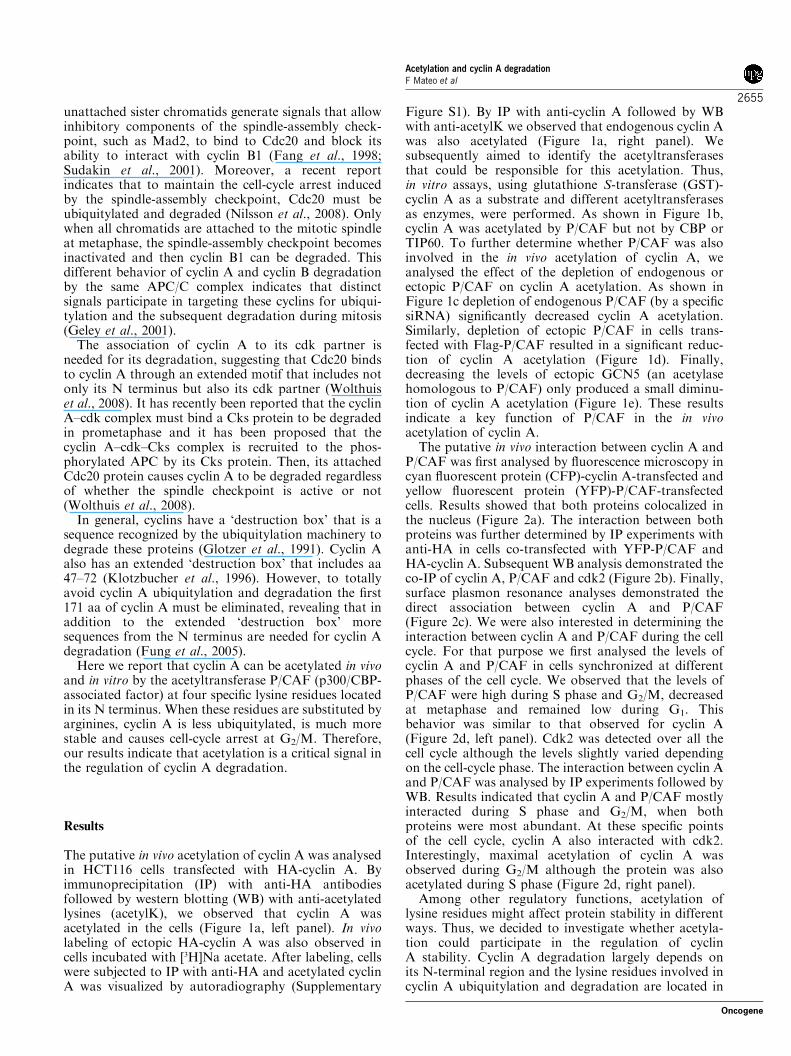
The putative in vivo acetylation of cyclin A was analysedin HCT116 cells transfected with HA-cyclin A. Byimmunoprecipitation (IP) with anti-HA antibodiesfollowed by western blotting (WB) with anti-acetylatedlysines (acetylK), we observed that cyclin A wasacetylated in the cells (Figure 1a, left panel). In vivolabeling of ectopic HA-cyclin A was also observed incells incubated with [3H]Na acetate. After labeling, cellswere subjected to IP with anti-HA and acetylated cyclinA was visualized by autoradiography (Supplementary
Figure S1). By IP with anti-cyclin A followed by WBwith anti-acetylK we observed that endogenous cyclin Awas also acetylated (Figure 1a, right panel). Wesubsequently aimed to identify the acetyltransferasesthat could be responsible for this acetylation. Thus,in vitro assays, using glutathione S-transferase (GST)-cyclin A as a substrate and different acetyltransferasesas enzymes, were performed. As shown in Figure 1b,cyclin A was acetylated by P/CAF but not by CBP orTIP60. To further determine whether P/CAF was alsoinvolved in the in vivo acetylation of cyclin A, weanalysed the effect of the depletion of endogenous orectopic P/CAF on cyclin A acetylation. As shown inFigure 1c depletion of endogenous P/CAF (by a specificsiRNA) significantly decreased cyclin A acetylation.Similarly, depletion of ectopic P/CAF in cells trans-fected with Flag-P/CAF resulted in a significant reduc-tion of cyclin A acetylation (Figure 1d). Finally,decreasing the levels of ectopic GCN5 (an acetylasehomologous to P/CAF) only produced a small diminu-tion of cyclin A acetylation (Figure 1e). These resultsindicate a key function of P/CAF in the in vivoacetylation of cyclin A.The putative in vivo interaction between cyclin A and
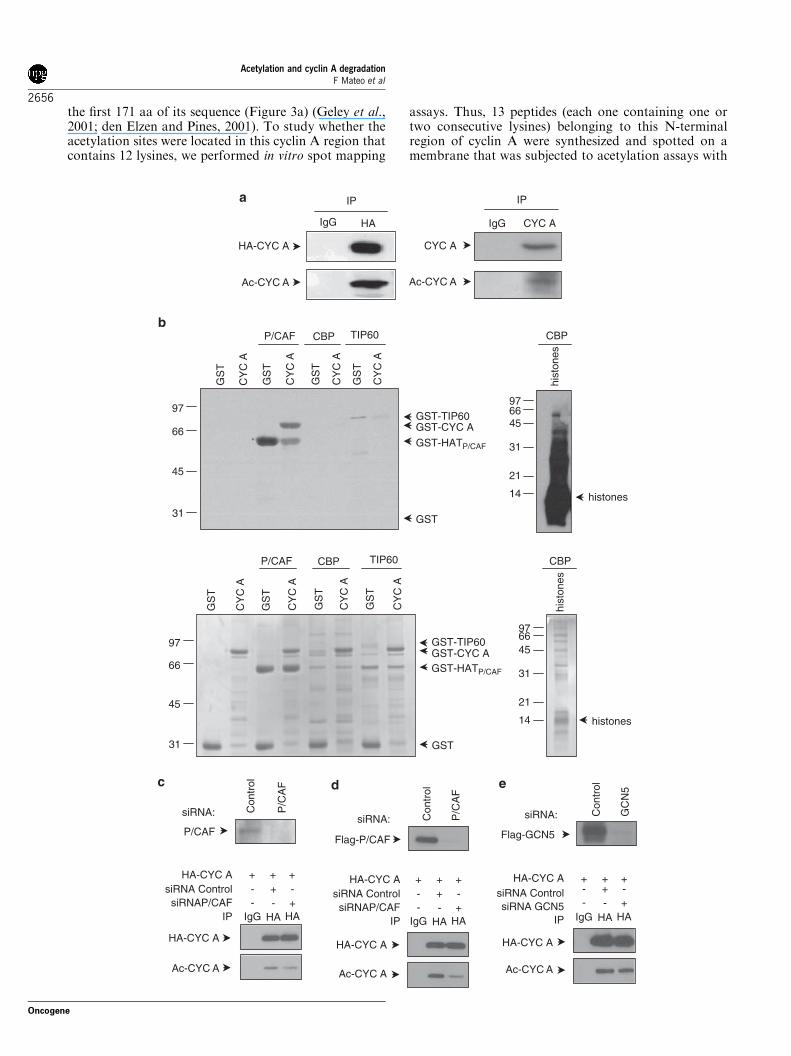
P/CAF was first analysed by fluorescence microscopy incyan fluorescent protein (CFP)-cyclin A-transfected andyellow fluorescent protein (YFP)-P/CAF-transfectedcells. Results showed that both proteins colocalized inthe nucleus (Figure 2a). The interaction between bothproteins was further determined by IP experiments withanti-HA in cells co-transfected with YFP-P/CAF andHA-cyclin A. Subsequent WB analysis demonstrated theco-IP of cyclin A, P/CAF and cdk2 (Figure 2b). Finally,surface plasmon resonance analyses demonstrated thedirect association between cyclin A and P/CAF(Figure 2c). We were also interested in determining theinteraction between cyclin A and P/CAF during the cellcycle. For that purpose we first analysed the levels ofcyclin A and P/CAF in cells synchronized at differentphases of the cell cycle. We observed that the levels ofP/CAF were high during S phase and G2/M, decreasedat metaphase and remained low during G1. Thisbehavior was similar to that observed for cyclin A(Figure 2d, left panel). Cdk2 was detected over all thecell cycle although the levels slightly varied dependingon the cell-cycle phase. The interaction between cyclin Aand P/CAF was analysed by IP experiments followed byWB. Results indicated that cyclin A and P/CAF mostlyinteracted during S phase and G2/M, when bothproteins were most abundant. At these specific pointsof the cell cycle, cyclin A also interacted with cdk2.Interestingly, maximal acetylation of cyclin A wasobserved during G2/M although the protein was alsoacetylated during S phase (Figure 2d, right panel).Among other regulatory functions, acetylation of
lysine residues might affect protein stability in differentways. Thus, we decided to investigate whether acetyla-tion could participate in the regulation of cyclinA stability. Cyclin A degradation largely depends onits N-terminal region and the lysine residues involved incyclin A ubiquitylation and degradation are located in
Acetylation and cyclin A degradationF Mateo et al
2655
Oncogene
the first 171 aa of its sequence (Figure 3a) (Geley et al.,2001; den Elzen and Pines, 2001). To study whether theacetylation sites were located in this cyclin A region thatcontains 12 lysines, we performed in vitro spot mapping
assays. Thus, 13 peptides (each one containing one ortwo consecutive lysines) belonging to this N-terminalregion of cyclin A were synthesized and spotted on amembrane that was subjected to acetylation assays with
IgG
HA-CYC A
Ac-CYC A
IPG
ST
CY
C A
GS
T
CY
C A
GS
T
CY
C A
GS
T
CY
C A
P/CAF
31
45
66
97 GST-TIP60GST-CYC A
GST
GS
T
CY
C A
GS
T
CY
C A
GS
T
CY
C A
GS
T
CY
C A
P/CAF
GST-TIP60GST-CYC A
GST-HATP/CAF
GST-HATP/CAF
GST31
45
66
97
hist
ones
CBP
histones
31
456697
21
14
hist
ones
CBP
31
456697
21
14 histones
IgG
CYC A
Ac-CYC A
IP
siRNA: Con
trol
P/C
AF
Flag-P/CAF
HA-CYC A
Ac-CYC A
siRNA: Con
trol
P/C
AF
P/CAF
siRNA ControlsiRNAP/CAF
--
IgG
HA-CYC A
HA-CYC A
Ac-CYC A
siRNA: Con
trol
GC
N5
Flag-GCN5
siRNA ControlsiRNA GCN5
IP
HA-CYC A
Ac-CYC A
HA CYC A
TIP60CBP
CBP TIP60
HA HA-
-++ + +
+IP
siRNA ControlsiRNAP/CAF
--
IgG
HA-CYC A HA-CYC A
HA HA IgG HA HA-
-++ + + + + +
+
-- -
-++
IP
Acetylation and cyclin A degradationF Mateo et al
2656
Oncogene
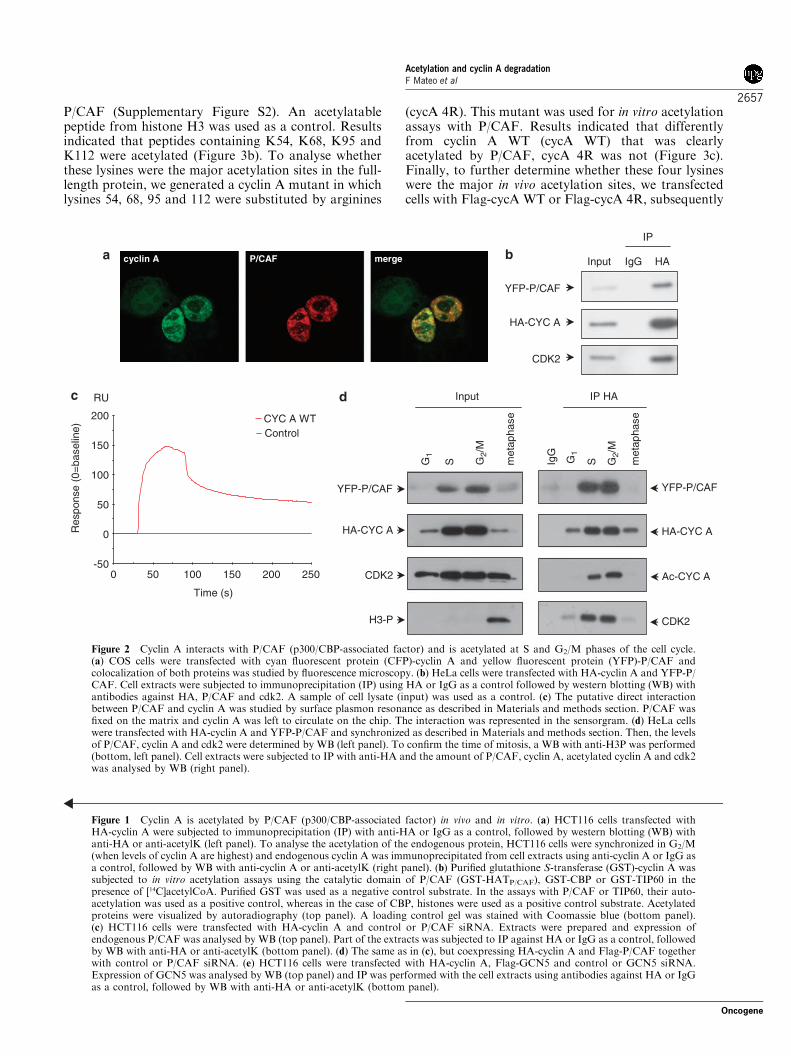
P/CAF (Supplementary Figure S2). An acetylatablepeptide from histone H3 was used as a control. Resultsindicated that peptides containing K54, K68, K95 andK112 were acetylated (Figure 3b). To analyse whetherthese lysines were the major acetylation sites in the full-length protein, we generated a cyclin A mutant in whichlysines 54, 68, 95 and 112 were substituted by arginines
(cycA 4R). This mutant was used for in vitro acetylationassays with P/CAF. Results indicated that differentlyfrom cyclin A WT (cycA WT) that was clearlyacetylated by P/CAF, cycA 4R was not (Figure 3c).Finally, to further determine whether these four lysineswere the major in vivo acetylation sites, we transfectedcells with Flag-cycA WT or Flag-cycA 4R, subsequently
Input
IP
YFP-P/CAF
HA-CYC A
CDK2
cyclin A P/CAF merge
Input
G1
G1
S G2/
M
G2/
M
met
apha
se
IP HA
IgG
HA-CYC A
CDK2
YFP-P/CAF
H3-P
S met
apha
se
Ac-CYC A
HA-CYC A
CDK2
YFP-P/CAF
HAIgG
RU
Time (s)
Res
pons
e (0
=ba
selin
e)
50
200
100
0
-50
150
0 50 200 250100 150
CYC A WT Control
Figure 2 Cyclin A interacts with P/CAF (p300/CBP-associated factor) and is acetylated at S and G2/M phases of the cell cycle.(a) COS cells were transfected with cyan fluorescent protein (CFP)-cyclin A and yellow fluorescent protein (YFP)-P/CAF andcolocalization of both proteins was studied by fluorescence microscopy. (b) HeLa cells were transfected with HA-cyclin A and YFP-P/CAF. Cell extracts were subjected to immunoprecipitation (IP) using HA or IgG as a control followed by western blotting (WB) withantibodies against HA, P/CAF and cdk2. A sample of cell lysate (input) was used as a control. (c) The putative direct interactionbetween P/CAF and cyclin A was studied by surface plasmon resonance as described in Materials and methods section. P/CAF wasfixed on the matrix and cyclin A was left to circulate on the chip. The interaction was represented in the sensorgram. (d) HeLa cellswere transfected with HA-cyclin A and YFP-P/CAF and synchronized as described in Materials and methods section. Then, the levelsof P/CAF, cyclin A and cdk2 were determined by WB (left panel). To confirm the time of mitosis, a WB with anti-H3P was performed(bottom, left panel). Cell extracts were subjected to IP with anti-HA and the amount of P/CAF, cyclin A, acetylated cyclin A and cdk2was analysed by WB (right panel).
Figure 1 Cyclin A is acetylated by P/CAF (p300/CBP-associated factor) in vivo and in vitro. (a) HCT116 cells transfected withHA-cyclin A were subjected to immunoprecipitation (IP) with anti-HA or IgG as a control, followed by western blotting (WB) withanti-HA or anti-acetylK (left panel). To analyse the acetylation of the endogenous protein, HCT116 cells were synchronized in G2/M(when levels of cyclin A are highest) and endogenous cyclin A was immunoprecipitated from cell extracts using anti-cyclin A or IgG asa control, followed by WB with anti-cyclin A or anti-acetylK (right panel). (b) Purified glutathione S-transferase (GST)-cyclin A wassubjected to in vitro acetylation assays using the catalytic domain of P/CAF (GST-HATP/CAF), GST-CBP or GST-TIP60 in thepresence of [14C]acetylCoA. Purified GST was used as a negative control substrate. In the assays with P/CAF or TIP60, their auto-acetylation was used as a positive control, whereas in the case of CBP, histones were used as a positive control substrate. Acetylatedproteins were visualized by autoradiography (top panel). A loading control gel was stained with Coomassie blue (bottom panel).(c) HCT116 cells were transfected with HA-cyclin A and control or P/CAF siRNA. Extracts were prepared and expression ofendogenous P/CAF was analysed by WB (top panel). Part of the extracts was subjected to IP against HA or IgG as a control, followedby WB with anti-HA or anti-acetylK (bottom panel). (d) The same as in (c), but coexpressing HA-cyclin A and Flag-P/CAF togetherwith control or P/CAF siRNA. (e) HCT116 cells were transfected with HA-cyclin A, Flag-GCN5 and control or GCN5 siRNA.Expression of GCN5 was analysed by WB (top panel) and IP was performed with the cell extracts using antibodies against HA or IgGas a control, followed by WB with anti-HA or anti-acetylK (bottom panel).
Acetylation and cyclin A degradationF Mateo et al
2657
Oncogene
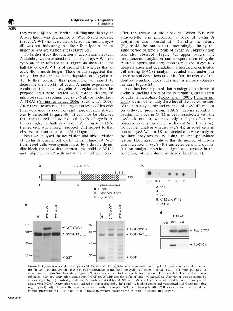
they were subjected to IP with anti-Flag and then cyclinA acetylation was determined by WB. Results revealedthat cycA WT was acetylated whereas the mutant cycA4R was not, indicating that these four lysines are themajor in vivo acetylation sites (Figure 3d).To further study the function of acetylation on cyclin
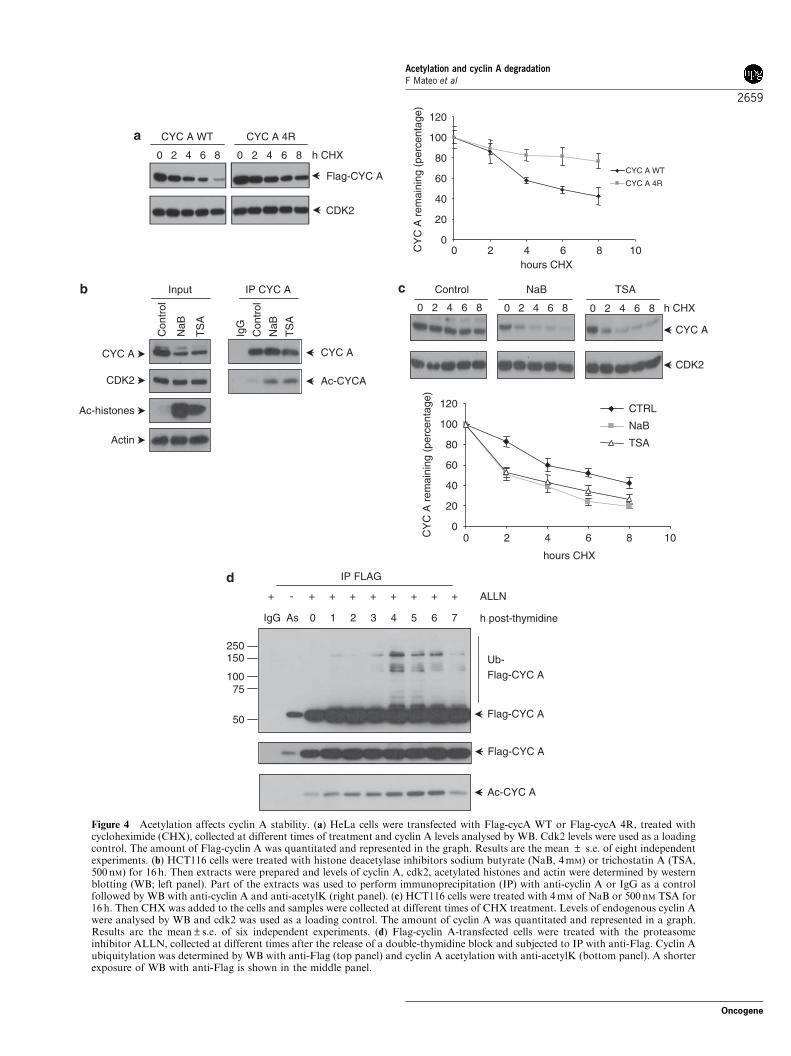
A stability, we determined the half-life of cycA WT andcycA 4R in transfected cells. Figure 4a shows that thehalf-life of cycA WT is of around 6 h whereas that ofcycA 4R is much longer. These results suggested thatacetylation participates in the degradation of cyclin A.To further confirm this possibility, we aimed todetermine the stability of cyclin A under experimentalconditions that increase cyclin A acetylation. For thispurpose, cells were treated with histone deacetylaseinhibitors such as sodium butyrate (NaB) or trichostatinA (TSA) (Abramova et al., 2006; Baek et al., 2006).After these treatments, the acetylation levels of histones(that were used as a control) and those of cyclin A wereclearly increased (Figure 4b). It can also be observedthat treated cells show reduced levels of cyclin A.Interestingly, the half-life of cyclin A in NaB- or TSA-treated cells was strongly reduced (2 h) respect to thatobserved in nontreated cells (6 h) (Figure 4c).Next we analysed the acetylation and ubiquitylation
of cyclin A during cell cycle. Thus, Flag-cycA WT-transfected cells were synchronized by a double-thymi-dine block, treated with the proteasome inhibitor ALLNand subjected to IP with anti-Flag at different times
after the release of the blockade. When WB withanti-acetylK was performed, a peak of cyclin Aacetylation was observed at 4–6 h after the release(Figure 4d, bottom panel). Interestingly, during thesame period of time a peak of cyclin A ubiquitylationwas also observed (Figure 4d, upper panel). Thesimultaneous acetylation and ubiquitylation of cyclinA also supports that acetylation is involved in cyclin Aubiquitylation and degradation. Fluorescence-activatedcell sorting (FACS) analysis revealed that under ourexperimental conditions at 4–6 h after the release of thedouble-thymidine block cells are in mitosis (Supple-mentary Figure S3).As it has been reported that nondegradable forms of
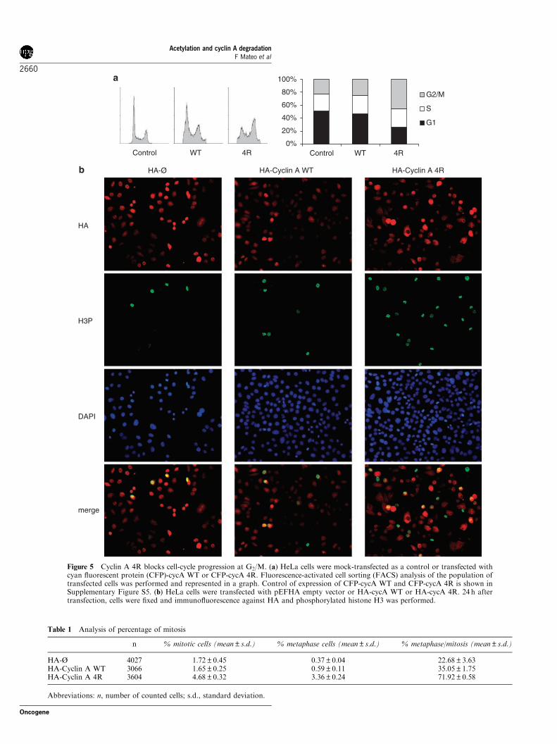
cyclin A (lacking a part of the N terminus) cause arrestof cells in metaphase (Geley et al., 2001; Fung et al.,2005), we aimed to study the effect of the overexpressionof the nonacetylatable and more stable cycA 4R mutanton cell-cycle progression. FACS analysis revealed asubstantial block in G2/M in cells transfected with thecycA 4R mutant, whereas only a slight effect wasobserved in cells transfected with cycA WT (Figure 5a).To further analyse whether cycA 4R arrested cells inmitosis, cycA WT- or 4R-transfected cells were analysedby immunocytochemistry using anti-phosphorylatedhistone H3. Figure 5b shows that the number of mitosiswas increased in cycA 4R-transfected cells and quanti-fication analysis revealed a significant increase in thepercentage of metaphases in these cells (Table 1).
11
CYCLIN A
Lysine residuesD-boxExtended D-boxCyclin box
H3 3 4 7 9
3. K544. K687. K959. K112 and K11311. K112
IP FLAG
IgG WT 4R
Ac-CYCA
Flag-CYCA
1711
1 432
GS
T
CY
C A
WT
CY
C A
4R
31
45
66
97GST-CYC A
GST-HATP/CAF GST-HATP/CAF
GST
GS
T
CY
C A
WT
CY
C A
4R
31
45
66
97GST-CYC A
GST
Figure 3 Cyclin A is acetylated at lysines 54, 68, 95 and 112. (a) Schematic representation of cyclin A lysine residues and domains.(b) Thirteen peptides containing one or two consecutive lysines from the cyclin A fragment including aa 1–171 were spotted on amembrane (see also Supplementary Figure S2). As a positive control, a peptide from histone H3 was added. The membrane wassubjected to in vitro acetylation assays with P/CAF (p300/CBP-associated factor) and [14C]acetylCoA. Acetylation was visualized byautoradiography. (c) Purified glutathione S-transferase (GST)-cycA WT and GST-cycA 4R were subjected to in vitro acetylationassays with P/CAF. Acetylation was visualized by autoradiography (left panel). A loading control gel was stained with Coomassie blue(right panel). (d) HeLa cells were transfected with Flag-cycA WT or Flag-cycA 4R. Cell extracts were subjected toimmunoprecipitation (IP) with anti-Flag followed by western blotting (WB) with anti-Flag and anti-acetylK.
Acetylation and cyclin A degradationF Mateo et al
2658
Oncogene
h CHX
Flag-CYC A
CDK2
0
20
40
60
80
100
120
hours CHX0 2 4 6 8 10
0 2 4 6 8 10
CYC A WT
CYC A 4R
h CHX
CYC A
CDK2
Ac-histones
Con
trol
NaB
TS
A
CYC A
CDK2
Actin
Input
Con
trol
NaB
TS
A
IgG
IP CYC A
CYC A
Ac-CYCA
0
20
40
60
80
100
120
hours CHX
CTRL
NaB
TSA
As 0 1 2 3 4 5 6 7IgG
ALLN
h post-thymidine
Flag-CYC A
Ub-Flag-CYC A
Ac-CYC A
IP FLAG
50
75100
150250
+ - + + + + + + + +
Flag-CYC A
CY
C A
rem
aini
ng (
perc
enta
ge)
CY
C A
rem
aini
ng (
perc
enta
ge)
CYC A 4RCYC A WT
0 2 4 6 8 0 2 4 6 8
0 2 4 6 8 0 2 4 6 8 0 2 4 6 8
Control NaB TSA
Figure 4 Acetylation affects cyclin A stability. (a) HeLa cells were transfected with Flag-cycA WT or Flag-cycA 4R, treated withcycloheximide (CHX), collected at different times of treatment and cyclin A levels analysed by WB. Cdk2 levels were used as a loadingcontrol. The amount of Flag-cyclin A was quantitated and represented in the graph. Results are the mean ± s.e. of eight independentexperiments. (b) HCT116 cells were treated with histone deacetylase inhibitors sodium butyrate (NaB, 4mM) or trichostatin A (TSA,500 nM) for 16 h. Then extracts were prepared and levels of cyclin A, cdk2, acetylated histones and actin were determined by westernblotting (WB; left panel). Part of the extracts was used to perform immunoprecipitation (IP) with anti-cyclin A or IgG as a controlfollowed by WB with anti-cyclin A and anti-acetylK (right panel). (c) HCT116 cells were treated with 4mM of NaB or 500nM TSA for16 h. Then CHX was added to the cells and samples were collected at different times of CHX treatment. Levels of endogenous cyclin Awere analysed by WB and cdk2 was used as a loading control. The amount of cyclin A was quantitated and represented in a graph.Results are the mean±s.e. of six independent experiments. (d) Flag-cyclin A-transfected cells were treated with the proteasomeinhibitor ALLN, collected at different times after the release of a double-thymidine block and subjected to IP with anti-Flag. Cyclin Aubiquitylation was determined by WB with anti-Flag (top panel) and cyclin A acetylation with anti-acetylK (bottom panel). A shorterexposure of WB with anti-Flag is shown in the middle panel.
Acetylation and cyclin A degradationF Mateo et al
2659
Oncogene
0%
20%
40%
60%
80%
100%
Control WT 4RControl WT 4R
G2/M
S
G1
H3P
DAPI
merge
HA
HA-Ø HA-Cyclin A 4RHA-Cyclin A WT
Figure 5 Cyclin A 4R blocks cell-cycle progression at G2/M. (a) HeLa cells were mock-transfected as a control or transfected withcyan fluorescent protein (CFP)-cycA WT or CFP-cycA 4R. Fluorescence-activated cell sorting (FACS) analysis of the population oftransfected cells was performed and represented in a graph. Control of expression of CFP-cycA WT and CFP-cycA 4R is shown inSupplementary Figure S5. (b) HeLa cells were transfected with pEFHA empty vector or HA-cycA WT or HA-cycA 4R. 24 h aftertransfection, cells were fixed and immunofluorescence against HA and phosphorylated histone H3 was performed.
Table 1 Analysis of percentage of mitosis
n % mitotic cells (mean±s.d.) % metaphase cells (mean±s.d.) % metaphase/mitosis (mean±s.d.)
HA-Ø 4027 1.72±0.45 0.37±0.04 22.68±3.63HA-Cyclin A WT 3066 1.65±0.25 0.59±0.11 35.05±1.75HA-Cyclin A 4R 3604 4.68±0.32 3.36±0.24 71.92±0.58
Abbreviations: n, number of counted cells; s.d., standard deviation.
Acetylation and cyclin A degradationF Mateo et al
2660
Oncogene
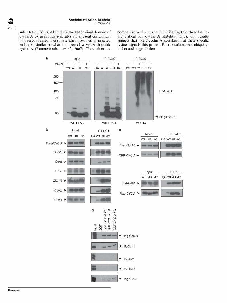
We further explored the possibility that the increasedstability of cycA 4R could be due to defects in itsubiquitylation. Thus, in vivo ubiquitylation assays wereperformed. Cells were transfected with HA-ubiquitinplus Flag-cycA WT or Flag-cycA 4R, then subjected toIP with anti-Flag and finally the ubiquitylation levelswere analysed by WB with anti-Flag and anti-HA.Results indicated that cycA WT was clearly ubiquity-lated whereas ubiquitylation of cycA 4R was stronglyreduced (Figure 6a). To study the function of acetyla-tion in cyclin A ubiquitylation, we analysed the in vivoubiquitylation of Flag-cycA 4Q, a mutant in whichlysines 54, 68, 95 and 112 of cyclin A were substituted byglutamines. This form is considered to be a pseudoace-tylated mutant because of the structure similaritybetween glutamine (Q) and the acetylated-lysine residue(Hecht et al., 1995; Li et al., 2002). Results indicatedthat differently from cycA 4R, the mutant cycA 4Q wasubiquitylated similarly to cycA WT (Figure 6a). Inter-estingly, the half-life of cycA 4Q is shorter than that ofcycA 4R but not as short as that of cycA WT, indicatingthat cycA 4Q can be degraded although not so efficientlyas cycA WT (Supplementary Figure S4). Therefore,these results indicate that K54, K68, K95 and K112 areacetylation sites needed for cyclin A ubiquitylation.We subsequently aimed to analyse the mechanisms
underlying the differential ubiquitylation of cyclin Amutants. It is known that to be degraded cyclin A has toform a cyclin A–cdk–Cks complex that is recruited tothe phosphorylated APC/C by its Cks protein. Cdc20attached to this complex causes cyclin A to be degraded(Wolthuis et al., 2008). Thus, we analysed the interac-tions of Flag-cycA WT, Flag-cycA 4R and Flag-cycA4Q with Cdc20, Cdh1, APC3, Cks1/2, cdk1 and cdk2 inasynchronously growing cells. Results showed that thethree forms of cyclin A interacted with Cdc20 and APC3in a similar manner. However, cycA 4R and cycA 4Qshowed an increased interaction with cdk1, cdk2, Cksand Cdh1 (Figure 6b). The specific interactions of thethree cyclin A forms with Cdc20 and Cdh1 were furtherconfirmed by IP experiments in cells transfected withFlag-Cdc20 or HA-Cdh1 plus each one of cyclin Aforms (Figure 6c). Experiments carried out in cellssynchronized in S phase or G2/M revealed that theinteractions of the different cycA forms with all theseproteins at these specific points are similar to thoseobserved in asynchronously growing cells (data notshown).Finally, we studied the direct interaction of cycA WT,
cycA 4R and cycA 4Q with Cdc20, Cdh1, cdk2 andCks1/2 by pull down using purified proteins. As shown inFigure 6d none of these cyclin A forms directly interactedwith Cks 1/2 and the interaction with Cdc20, Cdh1 andcdk2 was similar in all the three cyclin A forms. Theseresults indicate that the in vivo increased association ofboth cycA mutants with Cdh1 and cdks is not producedby a higher affinity for these proteins but by an unknownmechanism related to the in vivo complexes.The increased in vivo interaction of cycA 4R and cycA
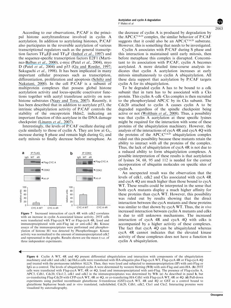
4Q with cdk1 and cdk2 is of particular interest because itmight affect their kinase activity. To analyse this
possibility, we subjected cells transfected with Flag-cycA WT or Flag-cycA 4R to IP with anti-Flag and theassociated cdk activity was determined in the immuno-precipitates. We observed that cycA 4R–cdk complexesdisplay higher kinase activity than that of cycA WT–cdkcomplexes (Figure 7a). When cells were subjected to IPwith anti-cdk2 we also observed an increased associationof cycA 4R with cdk2 respect to that shown by cycAWT, and also a higher kinase activity in the cycA 4R–cdk2 complexes (Figure 7b). Similar results wereobserved with the cycA 4Q mutant (data not shown).So, in addition to the function in cyclin A stability,lysines 54, 68, 95 and 112 also have a function in theregulation of cyclin A interaction with cdk and itsassociated kinase activity.
Discussion
The different timing of cyclin A and cyclin B degrada-tion at mitosis and the diverse sensitivity of these cyclinsto the spindle-assembly checkpoint indicates thatspecific mechanisms target each one of these cyclinsfor degradation (van Leuken et al., 2008). We reporthere that cyclin A acetylation by the acetyltransferaseP/CAF participates in the signaling pathway that targetscyclin A for degradation at early mitosis. Acetylationis a posttranslational modification occurring at theNe-amino group of lysines that might regulate proteinfunctions in many different ways as for instance,protein–protein and protein–DNA interactions andprotein stability. Lately, a number of reports haverevealed that lysine acetylation might act as a directsignal enhancing protein degradation for proteins suchas E2F1 (Galbiati et al., 2005), HIF-1a (Jeong et al.,2002), SV40T antigen (Shimazu et al., 2006) and pRB(Leduc et al., 2006). Moreover, the interplay betweenlysine acetylation and ubiquitylation has been reinforcedby the evidence that at least four acetyltransferases(p300, CBP, P/CAF and TAF1) possess intrinsicubiquitin activating/conjugating or ligase activities(Sadoul et al., 2008).We report here that P/CAF acetylates cyclin A at
lysines K54, K68, K95 and K112 and that these lysinesare the major acetylation sites both in vivo and in vitro.These specific residues are located in the N-terminaldomain of cyclin A that has been involved in thestability of the protein (Wolthuis et al., 2008). In fact,two of these lysines, K54 and K68, were alreadydescribed as important residues for the ubiquitylationand degradation of cyclin A. Specifically, the authorsreported that substitution of K37, K54 and K68 byarginines generates a more stable cyclin A but thismutant was still ubiquitylated (Fung et al., 2005). As ourobservations indicate that lysines K54, K68, K95 andK112 are critical residues for acetylation, this meansthat at least K54 and K68 can be both acetylated andubiquitylated. Thus, it is likely that when these lysinesare acetylated, alternative ubiquitylation sites could beused. It has also been reported that in Drosophila the
Acetylation and cyclin A degradationF Mateo et al
2661
Oncogene
substitution of eight lysines in the N-terminal domain ofcyclin A by arginines generates an unusual enrichmentof overcondensed metaphase chromosomes in injectedembryos, similar to what has been observed with stablecyclin A (Ramachandran et al., 2007). These data are
compatible with our results indicating that these lysinesare critical for cyclin A stability. Thus, our resultssuggest that likely cyclin A acetylation at these specificlysines signals this protein for the subsequent ubiquity-lation and degradation.
IgG WTWT 4R 4Q
ALLN:
IP FLAG
IgG WTWT 4R 4Q
IP FLAG
WT WT 4R 4Q
- - -
Input
Ub-CYCA
Flag-CYC A50
75
100
150
250
HA-Cdh1
Flag-CYC A
Flag-CYC A
Cdc20
Cdh1
IgG WT
IP FLAG
CDK2
CDK1
WT
Input
Cks1/2
APC3
Flag-Cdc20
CFP-CYC A
4Q4QInput IP FLAG
Input IP HA
GS
T-C
YC
A W
T
GS
T-C
YC
A 4
R
Inpu
t
GS
T-C
YC
A 4
Q
GS
T
Flag-Cdc20
HA-Cdh1
HA-Cks1
HA-Cks2
Flag-CDK2
+ + + + + + + + + + +
WB FLAGWB FLAG WB HA
4RWT 4Q4R
4RIgG WT 4Q4R
WT 4Q4R IgG WT 4Q4R
Acetylation and cyclin A degradationF Mateo et al
2662
Oncogene
According to our observations, P/CAF is the princi-pal histone acetyltransferase involved in cyclin Aacetylation. In addition to acetylating histones, P/CAFalso participates in the reversible acetylation of varioustranscriptional regulators such as the general transcrip-tion factors TFIIEb and TFIIF (Imhof et al., 1997) andthe sequence-specific transcription factors E2F1 (Marti-nez-Balbas et al., 2000), c-myc (Patel et al., 2004), myoD (Patel et al., 2004) and p53 (Gu and Roeder, 1997;Sakaguchi et al., 1998). It has been implicated in manyimportant cellular processes such as transcription,differentiation, proliferation and apoptosis (Schiltz andNakatani, 2000). In the cell P/CAF is a subunit ofmultiprotein complexes that possess global histoneacetylation activity and locus-specific coactivator func-tions together with acetyl transferase activity on non-histone substrates (Nagy and Tora, 2007). Recently, ithas been described that in addition to acetylate p53, theintrinsic ubiquitylation activity of P/CAF controls thestability of the oncoprotein Hdm2, indicating animportant function of this acetylase in the DNA damagecheckpoint (Linares et al., 2007).Interestingly, the levels of P/CAF oscillate during cell
cycle similarly to those of cyclin A. They are low at G1,increase during S phase and remain high during G2 andearly mitosis to finally decrease before metaphase. As
the decrease of cyclin A is produced by degradation bythe APC/CCdc20 complex, the similar behavior of P/CAFsuggests that it could also be an APC/CCdc20 substrate.However, this is something that needs to be investigated.Cyclin A associates with P/CAF during S phase and
this interaction is maintained until early mitosis, thenbefore metaphase this complex is disrupted. Concomi-tant to its association with P/CAF, cyclin A becomesacetylated. A more detailed time-course analysis in-dicates that cyclin A acetylation increases at earlymitosis simultaneously to cyclin A ubiquitylation. Allthese data support that acetylation by P/CAF targetscyclin A for its ubiquitylation.To be degraded cyclin A has to be bound to a cdk
subunit that in turn has to be associated with a Cksprotein. This cyclin A–cdk–Cks complex is then recruitedto the phosphorylated APC/C by its Cks subunit. TheCdc20 attached to cyclin A causes cyclin A to bedegraded regardless of the spindle checkpoint beingactive or not (Wolthuis et al., 2008). Thus, a possibilitywas that cyclin A acetylation at these specific lysinesmight be required for the interaction with some of theseproteins of the ubiquitylation machinery. However, theanalysis of the interactions of cycA 4R and cycA 4Q withthe proteins of the APC/CCdc20 ubiquitylation complexruled out this possibility because these mutants retain theability to interact with all the proteins of the complex.Thus, the lack of ubiquitylation of cycA 4R is not due toa reduced ability to form ubiquitylation complexes. Apossible interpretation of these results is that acetylationof lysines 54, 68, 95 and 112 is needed for the correctincorporation of ubiquitin molecules on specific sites ofcyclin A.An unexpected result was the observation that the
levels of cdk1, cdk2 and Cks associated with cycA 4Rand cycA 4Q are much higher than those bound to cycAWT. These results could be interpreted in the sense thatboth cycA mutants display a much higher affinity forthese proteins than cycA WT. However, this possibilitywas ruled out by results showing that the directinteraction between the cycA mutants and these proteinswas similar to that shown by cycA WT. Thus, the in vivoincreased interaction between cyclin A mutants and cdksis due to still unknown mechanisms. The increasedinteraction of cycA 4R and cycA 4Q with cdks isaccompanied by a higher activity of these complexes.The fact that cycA 4Q can be ubiquitylated whereascycA 4R cannot indicates that the elevated kinaseactivity of these complexes does not have a function incyclin A ubiquitylation.
Figure 6 Cyclin A WT, 4R and 4Q present differential ubiquitylation and interaction with components of the ubiquitylationmachinery and cdk1 and cdk2. (a) HeLa cells were transfected with HA-ubiquitin plus Flag-cycA WT, Flag-cycA 4R or Flag-cycA 4Qand treated with the proteasome inhibitor ALLN. Then, they were lysed and subjected to immunoprecipitation (IP) with anti-Flag orIgG as a control. The levels of ubiquitylated cyclin A were determined by western blotting (WB) with anti-Flag and anti-HA. (b) 293Tcells were transfected with Flag-cycA WT, 4R or 4Q, lysed and immunoprecipitated with anti-Flag. The presence of Flag-cyclin A,APC3, Cdh1, Cdc20, Cks1/2, cdk1 and cdk2 in the immunoprecipitates was determined by WB. (c) As described in panel b, butco-transfecting Flag-Cdc20 with CFP-cycA WT, 4R or 4Q, or co-transfecting HA-Cdh1 with Flag-cycA WT, 4R or 4Q. (d) Pull-downexperiments using purified recombinant glutathione S-transferase (GST)-cycA WT, 4R and 4Q or GST as a control bound toglutathione Sepharose beads and in vitro translated, radiolabeled, Cdc20, Cdh1, cdk2, Cks1 and Cks2. Interacting proteins werevisualized by autoradiography.
IP CDK2
CDK2
Flag-CYC A
H1-P
CDK2
Flag-CYC A
H1-P
IP FLAG
0
20
40
60
80
100
rela
tive
units
0
20
40
60
80
100
rela
tive
units
*
CYC A 4RCYC A WT
4RIgG WT 4RIgG WT
CYC A WT CYC A 4R
Figure 7 Increased interaction of cycA 4R with cdk2 correlateswith an increase in cyclin A-associated kinase activity. 293T cellswere transfected with Flag-cycA WT or Flag-cycA 4R, lysed andimmunoprecipitated with anti-Flag (a) or anti-cdk2 (b). Kinaseassays of the immunoprecipitates were performed and phosphor-ylation of histone H1 was detected by PhosphorImager. Kinaseactivity was normalized to the amount of immunoprecipitated cdk2and represented in the graphs. Results shown are the mean±s.e. ofthree independent experiments.
Acetylation and cyclin A degradationF Mateo et al
2663
Oncogene
As a summary, results presented here reveal thatacetylation at specific lysines is a new mechanism thattargets cyclin A for degradation at early mitosis. Inaddition to that, our results also revealed an unexpectednew mechanism for the regulation of cdk activitythat depends on the integrity of four specific lysines ofcyclin A.
Materials and methods
PlasmidscDNA of wild-type cyclin A was cloned into pGEX6P1,pEFHA, pcDNA3-Flag and pECFP-C1 vectors. cycA 4R andcycA 4Q mutants were generated by site-directed mutagenesis.pCX-Flag-P/CAF, pGEX2TKP-HATP/CAF (352–658),pGEX4T2-P/CAF (full length), pGEX2T-CBP andpGEX2T-TIP60 were a kind gift from MA Martınez-Balbas.pcDNA3.1-HA-Cdh1 and Flag-Cdc20 were a kind gift fromM Pagano. pcDNA3-HA-Cks 1 and 2 were a kind gift fromR Wolthuis. P/CAF shRNA and control shRNA werepurchased from Sigma (St Louis, MO, USA).
Antibodies and reagentsAntibodies against cyclin A (H-432), cyclin A (BF-683)-AC(agarose conjugated), cdk2 (M-2), Cdc20 (H175) and Cks1/2(FL-79) were purchased from Santa Cruz Biotechnology (SantaCruz, CA, USA). Anti-Cdh1 (34-2000) was purchased fromZymed (South San Francisco, CA, USA). Anti-acetylated lysines(9441) and anti-phospho-histone H3 (Ser28) (9713) were fromCell Signaling (Danvers, MA, USA). Antibodies against Flag(F7425), HA (H6908) and P/CAF (P7493) were purchased fromSigma, and APC3/Cdc27 (ab10538) from Abcam (Cambridge,UK) Anti-HA 12C5A for immunofluorescence was purchasedfrom Roche (Basel, Switzerland). For IP we used monoclonalanti-HA agarose and monoclonal anti-Flag M2 affinity gelfrom Sigma. [14C]acetylCoA was purchased from PerkinElmer(Waltham, MA, USA). Thymidine, nocodazol, cycloheximide,sodium butyrate and TSA were from Sigma. ALLN was fromCalbiochem (Merck Chemicals Ltd., Nottingham, UK). Fortransfection assays we used Lipofectamine 2000 from Invitrogen(Paisley, UK).
Protein purification and in vitro acetylationProtein expression and purification was performed asdescribed (Canela et al., 2006). Acetylase assays wereperformed as described (Martinez-Balbas et al., 2000). Forcyclin A acetylation assays, 1–10ml of the different acetylases(5000–10 000 c.p.m. activity on histones) were incubated with6 mM of purified GST or GST-cyclin A and 0.02mCi[14C]acetylCoA. For the spot-mapping experiment, the mem-brane was incubated in 3ml of HAT buffer (50mM Tris-HCl(pH 8), 500mM NaCl, 0.1mM EDTA, 5% glycerol, 0.1%Nonidet P-40 (NP-40)) in the presence of GST-HATP/CAF and[14C]acetylCoA, for 30min at 30 1C. Then the membrane waswashed, dried and subjected to autoradiography.
ImmunoprecipitationCells were lysed in RIPA buffer (50mM Tris-HCl (pH 7.5),150mM NaCl, 1% NP-40, 0.5% sodium deoxycholate, 0.1%SDS, 1mM EDTA, 1mM DTT, 1mM PMSF, 0.1mM Na3VO4,0.5 mg/ml aprotinin, 10 mg/ml leupeptin) for 30min on ice.Lysates (0.2–2mg of protein) were incubated with Flag or HAor cyclin A agarose beads for 2 h at 4 1C. After three washes
with RIPA buffer, Laemmli buffer was added to the samplesand they were electrophoresed.
Surface plasmon resonance experimentsThe surface plasmon resonance analysis was performed atroom temperature using a Biacore T100 (Biacore InternationalAB, GE Healthcare, New Jersey, NJ, USA). P/CAF purifiedprotein was immobilized on a carboxymethylated dextransensor chip (CM5) using the amine coupling method asdescribed by the manufacturer. A blank immobilization wasperformed using the same method and was used as thereference surface. Purified full-length cyclin A was diluted inHBS-EP buffer (Biacore International AB) and was injectedover the flow cells at a flow rate of 30ml/min for 60 s. Followinga dissociation time of 120 s, final regeneration of the surface wasperformed with a short pulse of 0.05% (w/v) SDS. The interactionbetween P/CAF and cyclin A was detected and presented as asensorgram by plotting resonance units against time.Cells werefixed with 70% cold ethanol for 2h at 4 1C, washed withphosphate-buffered saline (PBS), and finally incubated with50mg/ml of propidium iodide (Sigma) and 200mg/ml RNase for30min at room temperature. Analysis of DNA content was carriedout in a BD Biosciences FACS Canto II (BD Biosciences,Erembodegen, Belgium). Data were analysed with WinMDI 2.9software (http://facs.scripps.edu/software.html).
ImmunofluorescenceTo detect HA-cyclin A and H3P by immunocytochemistry,cells were grown in coverslips, fixed in 4% paraformaldehyde/PBS for 15min at room temperature, washed in PBS andblocked with 1% BSA, 0.1% Triton X-100 in PBS for 1 h.Then, coverslips were incubated with anti-HA (Roche; 12C5A,mouse monoclonal, 1:200 dilution) and anti-H3P (CellSignaling; 9713, rabbit polyclonal, 1:200 dilution) for 1 h at37 1C in a humidified atmosphere. They were then washed inPBS and incubated for 45min at 37 1C with Alexa-Fluor 594(Invitrogen, A11005, goat anti-mouse, dilution 1:500) andAlexa-Fluor 488 (Invitrogen, Molecular Probes; A11008, goatanti-rabbit, dilution 1:250). After that coverslips were washed,mounted on glass slides with Mowiol (Calbiochem) andanalysed by fluorescence microscopy.
In vivo ubiquitylation assaysCells were transfected with indicated plasmids. At 24 h aftertransfection, cells were replated and treated with 100 mMALLN for 16 h. Then, cells were harvested and subjected toIP as described (Fung et al., 2005).
In vitro protein-binding assayThe GST and GST-cycA WT, 4R and 4Q were expressed inEscherichia coli. These fusion proteins were bound toglutathione Sepharose beads (Glutathione Sepharose 4B; GEHealthcare, Chalfont St Giles, UK) and washed with NETN(20mM Tris-HCl (pH 8), 1mM EDTA (pH 8), 0.5% NP-40,100mM NaCl). Beads were then incubated for 1 h at roomtemperature with 35S-labeled Flag-CDK2, Flag-Cdc20, HA-Cdh1, HA-Cks1 or HA-Cks2 proteins, in vitro transcribed andtranslated using the Promega TnT kit (Promega, Madison, WI,USA). Beads were extensively washed with NETN 150mM
NaCl, and the bound material was analysed by SDS–polyacrylamide gel followed by autoradiography.
Immunoprecipitation and kinase assaysAfter three washes in IP buffer and two in kinase buffer(50mM HEPES (pH 7.4), 2.5mM EGTA, 10mM MgCl2)immunoprecipitates were resuspended in a final volume of
Acetylation and cyclin A degradationF Mateo et al
2664
Oncogene
30ml of kinase buffer containing 15mM ATP, 10mCi of [32P]ATP,2mM dithiothreitol and 2mg of histone H1 for 30min at 30 1C.Reactions were stopped by the addition of Laemmli buffer.Samples were then electrophoresed on 12% SDS–PAGE andthen stained with Coomassie blue and dried. The radioactivityassociated to the gels was detected with a PhosphorImager(BIO-RAD laboratories, Hercules, CA, USA).
Conflict of interest
The authors declare no conflict of interest.
Acknowledgements
This research was supported by grants SAF2006-05212and SAF2007-60491 from the Ministerio de Educacion yCiencia of Spain and RETICS RD06/0020/0010 fromthe Instituto de Salud Carlos III. It was also supported bygrants from the National Institutes of Health (R01-GM57587,R37-CA76584, and R21-CA125173) and the MultipleMyeloma Research foundation to Michele Pagano. MichelePagano is an Investigator with the Howard Hughes MedicalInstitute.
References
Abramova MV, Pospelova TV, Nikulenkov FP, Hollander CM,Fornace Jr AJ, Pospelov VA. (2006). G1/S arrest induced by histonedeacetylase inhibitor sodium butyrate in E1A + Ras-transformedcells is mediated through down-regulation of E2F activity andstabilization of beta-catenin. J Biol Chem 281: 21040–21051.
Baek SY, Kim SR, Bae MK, Hwang JW, Kim JS, Choi YH et al.(2006). Trichostatin A increases the thermosensitivity of humanglioblastoma A172 cells. Neurosci Lett 396: 230–234.
Bloom J, Cross FR. (2007). Multiple levels of cyclin specificity in cell-cycle control. Nat Rev Mol Cell Biol 8: 149–160.
Canela N, Orzaez M, Fucho R, Mateo F, Gutierrez R, Pineda-LucenaA et al. (2006). Identification of an hexapeptide that binds to asurface pocket in cyclin A and inhibits the catalytic activity of thecomplex cyclin-dependent kinase 2-cyclin A. J Biol Chem 281:35942–35953.
den Elzen N, Pines J. (2001). Cyclin A is destroyed in prometaphaseand can delay chromosome alignment and anaphase. J Cell Biol
153: 121–136.Donzelli M, Squatrito M, Ganoth D, Hershko A, Pagano M, Draetta
GF. (2002). Dual mode of degradation of Cdc25 A phosphatase.EMBO J 21: 4875–4884.
Fang G, Yu H, Kirschner MW. (1998). Direct binding of CDC20protein family members activates the anaphase-promoting complexin mitosis and G1. Mol Cell 2: 163–171.
Fung TK, Yam CH, Poon RY. (2005). The N-terminal regulatorydomain of cyclin A contains redundant ubiquitination targetingsequences and acceptor sites. Cell Cycle 4: 1411–1420.
Furuno N, den Elzen N, Pines J. (1999). Human cyclin A is requiredfor mitosis until mid prophase. J Cell Biol 147: 295–306.
Galbiati L, Mendoza-Maldonado R, Gutierrez MI, Giacca M. (2005).Regulation of E2F-1 after DNA damage by p300-mediatedacetylation and ubiquitination. Cell Cycle 4: 930–939.
Geley S, Kramer E, Gieffers C, Gannon J, Peters JM, Hunt T.(2001). Anaphase-promoting complex/cyclosome-dependent proteo-lysis of human cyclin A starts at the beginning of mitosis and isnot subject to the spindle assembly checkpoint. J Cell Biol 153:137–148.
Glotzer M, Murray AW, Kirschner MW. (1991). Cyclin is degraded bythe ubiquitin pathway. Nature 349: 132–138.
Gong D, Pomerening JR, Myers JW, Gustavsson C, Jones JT, HahnAT et al. (2007). Cyclin A2 regulates nuclear-envelope breakdownand the nuclear accumulation of cyclin B1. Curr Biol 17: 85–91.
Gu W, Roeder RG. (1997). Activation of p53 sequence-specific DNAbinding by acetylation of the p53 C-terminal domain. Cell 90:595–606.
Hagting A, den Elzen N, Vodermaier HC, Waizenegger IC, Peters JM,Pines J. (2002). Human securin proteolysis is controlled by thespindle checkpoint and reveals when the APC/C switches fromactivation by Cdc20 to Cdh1. J Cell Biol 157: 1125–1137.
Hecht A, Laroche T, Strahl-Bolsinger S, Gasser SM, Grunstein M.(1995). Histone H3 and H4 N-termini interact with SIR3 and SIR4proteins: a molecular model for the formation of heterochromatin inyeast. Cell 80: 583–592.
Imhof A, Yang XJ, Ogryzko VV, Nakatani Y, Wolffe AP, Ge H.(1997). Acetylation of general transcription factors by histoneacetyltransferases. Curr Biol 7: 689–692.
Jeong JW, Bae MK, Ahn MY, Kim SH, Sohn TK, Bae MH et al.(2002). Regulation and destabilization of HIF-1alpha by ARD1-mediated acetylation. Cell 111: 709–720.
Klotzbucher A, Stewart E, Harrison D, Hunt T. (1996). The‘destruction box’ of cyclin A allows B-type cyclins tobe ubiquitinated, but not efficiently destroyed. EMBO J 15:3053–3064.
Leduc C, Claverie P, Eymin B, Col E, Khochbin S, Brambilla E et al.(2006). p14ARF promotes RB accumulation through inhibition ofits Tip60-dependent acetylation. Oncogene 25: 4147–4154.
Li M, Luo J, Brooks CL, Gu W. (2002). Acetylation of p53 inhibits itsubiquitination by Mdm2. J Biol Chem 277: 50607–50611.
Linares LK, Kiernan R, Triboulet R, Chable-Bessia C, Latreille D,Cuvier O et al. (2007). Intrinsic ubiquitination activity of PCAFcontrols the stability of the oncoprotein Hdm2. Nat Cell Biol 9:331–338.
Malumbres M, Barbacid M. (2005). Mammalian cyclin-dependentkinases. Trends Biochem Sci 30: 630–641.
Martinez-Balbas MA, Bauer UM, Nielsen SJ, Brehm A, Kouzarides T.(2000). Regulation of E2F1 activity by acetylation. EMBO J 19:662–671.
Morgan DO. (1997). Cyclin-dependent kinases: engines, clocks, andmicroprocessors. Annu Rev Cell Dev Biol 13: 261–291.
Nagy Z, Tora L. (2007). Distinct GCN5/PCAF-containing complexesfunction as co-activators and are involved in transcription factorand global histone acetylation. Oncogene 26: 5341–5357.
Nilsson J, Yekezare M, Minshull J, Pines J. (2008). The APC/Cmaintains the spindle assembly checkpoint by targeting Cdc20 fordestruction. Nat Cell Biol 10: 1411–1420.
Pagano M, Draetta G. (1991). Cyclin A, cell cycle control andoncogenesis. Prog Growth Factor Res 3: 267–277.
Parry DH, O’Farrell PH. (2001). The schedule of destruction of threemitotic cyclins can dictate the timing of events during exit frommitosis. Curr Biol 11: 671–683.
Patel JH, Du Y, Ard PG, Phillips C, Carella B, Chen CJ et al. (2004).The c-MYC oncoprotein is a substrate of the acetyltransferaseshGCN5/PCAF and TIP60. Mol Cell Biol 24: 10826–10834.
Ramachandran V, Matzkies M, Dienemann A, Sprenger F. (2007).Cyclin A degradation employs preferentially used lysinesand a cyclin box function other than Cdk1 binding. Cell Cycle 6:171–181.
Resnitzky D, Hengst L, Reed SI. (1995). Cyclin A-associated kinaseactivity is rate limiting for entrance into S phase and is negativelyregulated in G1 by p27Kip1. Mol Cell Biol 15: 4347–4352.
Rosenberg AR, Zindy F, Le Deist F, Mouly H, Metezeau P, Brechot Cet al. (1995). Overexpression of human cyclin A advances entry intoS phase. Oncogene 10: 1501–1509.
Sadoul K, Boyault C, Pabion M, Khochbin S. (2008). Regulation ofprotein turnover by acetyltransferases and deacetylases. Biochimie
90: 306–312.
Acetylation and cyclin A degradationF Mateo et al
2665
Oncogene
Sakaguchi K, Herrera JE, Saito S, Miki T, Bustin M, Vassilev A et al.(1998). DNA damage activates p53 through a phosphorylation-acetylation cascade. Genes Dev 12: 2831–2841.
Schiltz RL, Nakatani Y. (2000). The PCAF acetylase complexas a potential tumor suppressor. Biochim Biophys Acta 1470:M37–M53.
Sherr CJ, Roberts JM. (1999). CDK inhibitors: positive and negativeregulators of G1-phase progression. Genes Dev 13: 1501–1512.
Shimazu T, Komatsu Y, Nakayama KI, Fukazawa H, Horinouchi S,Yoshida M. (2006). Regulation of SV40 large T-antigen stability byreversible acetylation. Oncogene 25: 7391–7400.
Sudakin V, Chan GK, Yen TJ. (2001). Checkpoint inhibitionof the APC/C in HeLa cells is mediated by a complex ofBUBR1, BUB3, CDC20, and MAD2. J Cell Biol 154:925–936.
Sullivan M, Morgan DO. (2007). Finishing mitosis, one step at a time.Nat Rev Mol Cell Biol 8: 894–903.
van Leuken R, Clijsters L, Wolthuis R. (2008). To cell cycle, swing theAPC/C. Biochim Biophys Acta 1786: 49–59.
Wolthuis R, Clay-Farrace L, van Zon W, Yekezare M, Koop L,Ogink J et al. (2008). Cdc20 and Cks direct the spindle checkpoint-independent destruction of cyclin A. Mol Cell 30: 290–302.
Supplementary Information accompanies the paper on the Oncogene website (http://www.nature.com/onc)
Acetylation and cyclin A degradationF Mateo et al
2666
Oncogene


