Embed Size (px)
Citation preview
1
SA
TY
AJI
T
mA
Yo
r
cont
ents
ManageMent 3
IntroductIon 4
researchreports Biochemistry, Biophysics and Bioinformatics 9 Cellular Organization and Signaling 45 Genetics and Development 65 Neurobiology 85 Adjunct and Visiting Faculty Members 108 Publications 109
talksandMeetIngs Seminars 120 Colloquia 127 Meetings and Workshops 129
acadeMIcactIvItIes Overview 143 Programs 144 Degrees 145 Courses 146 Lectures and Visits 149 Trainees 153 MSc Wild Life Program 156
honorsandawards 158
support Research Facilities 161 Administration, Finance and Technical Services 162 The Central Imaging and Flow Cytometry Facility 164
people Academic Personnel 168 Support Personnel 175 Thanking a Dean... 176 Quality of Life at NCBS 177
Such Treasure and Rich Merchandize 179
NCBS_AR_05_07.indd 1 7/10/08 5:31:51 PM
NCBS_AR_05_07.indd 2 7/10/08 5:31:54 PM
3
SA
TY
AJI
T
mA
Yo
r
man
agem
ent
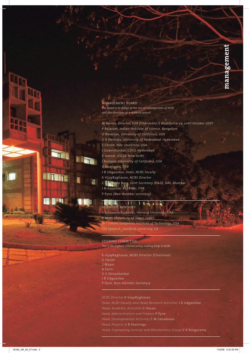
ManageMentBoardThe Board is in charge of the overall management of NCBSand also functions as a research council
MBarma,Director, TIFR (Chairman); sBhattacharya, until October 2007pBalaram, Indian Institute of Science, BangaloreuBanerjee,University of California, USAgrdesiraju, University of Hyderabad, Hyderabadsghosh,Yale University, USAJgowrishankar,CDFD, HyderabadsJameel,ICGEB, New DelhiJkuriyan, University of California, USAvrodrigues, TIFRJBudgaonkar,Dean, NCBS Facultykvijayraghavan, NCBS DirectorcvanandaBose,Joint Secretary (R&D), DAE, MumbaiJnkayarkar,Registrar, TIFR
ppyne(Non-member secretary)
scIentIfIcadvIsorssartavanis-tsakonas,Harvard University, USAkMori, University of Tokyo, JapanMIsimon,California Institute of Technology, USAJimspudich, Stanford University, CA
steerIngcoMMItteeThis is the highest, internal policy-making body in NCBS
kvijayraghavan,ncBsdirector(chairman)ghasansMayorasaringvshivashankarJBudgaonkarppyne,non-membersecretary
NCBS DirectorkvijayraghavanDean, NCBS Faculty and Head, Research ActivitiesJBudgaonkarHead, Academic ActivitiesghasanHead, Administration and Finance ppyneHead, Developmental ActivitiestMsahadevanHead, ProjectsuBpoornimaHead, Engineering Services and Maintenance Groupvrrengasamy
NCBS_AR_05_07.indd 3 7/10/08 5:31:55 PM

Viewed from any perspective, the scientific scene in today’s India is a churning of resources and ideas
whose implementation makes for foggy weather and stormy seas. The number of new institutions
being announced and plans being made is mind-boggling compared to the sparse fare of the past.
The National Centre for Biological Sciences (NCBS) is a small, relatively insulated, institution that has
grown slowly, adding a few carefully chosen groups each year. Should we join the expedition and risk
sinking or lie low and simply miss the boat?
The critics of change, and there are many, have a point. When they view the transformations taking
place they uniformly ask: Where are the people who will lead this change, where are the scientists
who will staff all the new places coming up? In any given area, they argue, India has but a handful
of researchers. To grow too fast, do the wrong things or to spread one’s time and effort too thin
is suicidal. on the other hand, others argue that it will be suicidal not to grasp the opportunities
for growth and change that are available now, that it is part of the adventure to nurture and
find the human resources (as people are nowadays called) for the task. It is rather interesting to
have alternative ways of suicide as one’s only choices! In the event, this is a good time to look at
NCBS’s past and discuss what we should do in interacting with our environment. There are many
responsibilities we could take and these come with opportunities and potential problems. We need
to see through the fog if we are to avoid the rocks of trying things the wrong way, trying too many
things, trying the wrong things or, equally risky, trying nothing.
over the sixteen years since NCBS’s birth and growth to about 25 groups, its simple core values have
been its strength. These include a rigorous tenure-track system and the academic freedom to take
one’s science forward without having directions imposed. We have chosen to hire the best and have
not grown by excluding some areas or focusing on others. our research areas range from the study of
the physics and chemistry of macromolecules to ecological sciences. This breadth and, crucially, the
diverse student and postdoctoral community that it brings with it, have created an environment that
is stimulating, creative, irreverent, questioning and one that is constantly renewed. These features
Driving, and responding to, change
NCBS_AR_05_07.indd 4 7/10/08 5:31:56 PM

5
SA
TY
AJI
T
mA
Yo
r
are necessary, but are not sufficient explanation for past successes or for assuring future ones. All of
what is good about NCBS today is also a consequence of our perception of a few years ago on how
to recognize ‘good’ science. These perceptions should not be a constant: If we were to retain today
the perceptions of yesterday in making our choices for the future we are very likely to make serious
mistakes. The way we have started and grown has allowed us to become good. To become excellent,
simply having more of the same will not work, except to retain the ability to choose in the context of
what is currently the best anywhere.
What do we need to do to position us for success in the coming fifteen years? There are tantalizing
possibilities. our forthcoming new laboratories allow us to expand our Young Investigator Programme
with the aim to nurture a population of dynamic and constantly renewed researchers. Till now, we
had little choice but to have these talented people find themselves a longer-term home at the end of
their tenure. The growth of new institutions in India now allows them to cast their net wider. NCBS
hopes to interact with these new institutions to formally embed interactions that allow movement
of investigators. Closer to home, the entry into Bangalore of the Department of Biotechnology (DBT)
in a direct manner offers a range of new possibilities: The DBT is starting a new stem-cell institute
(SCI) close to NCBS and we have been asked to incubate this laboratory and define the nature of
our interactions in a memorandum of association. If formulated well and implemented better, this
affords an unprecedented opportunity for symbiosis leading to the development of a critical mass of
very high quality life sciences researchers in Bangalore. The DBT is keen that the SCI, in addition to
its mandate, serve as a home for several satellite centres whose focus will be by no means limited
to stem cell research. Interacting in this venture thus provides both NCBS and SCI with invigorating
possibilities in all of biology: We can share intellectual and material resources and we can increase
the size, diversity and capability of our community while constantly improving quality. This allows
us to address scientific problems in new ways and develop a niche advantage or an approach that
allows us to be truly innovative. In addition to working to develop these new institutional structures
in a variety of ways, NCBS has the opportunity to expand on two of its relatively newer strengths: We
dire
ctor
’s r
epor
t
NCBS_AR_05_07.indd 5 7/10/08 5:31:57 PM
have been fortunate to have developed an excellent band of theoreticians and experimentalists who use
physics, chemistry and computational approaches to study biology. Another group has emerged in what
can be broadly called ecological sciences and conservation biology. It is clear that these groupings are
poised to attract even more excellent researchers. It is just this sort of growth – in an environment of cell
and molecular biology – that can open the possibilities of transformation into excellence.
As always, the generalities of discussion about the future need to be moored in the reality of what we
practice. Since our last report we had many changes in our research groups. V. Sriram joins the Department
of Biological Sciences (DBS) at TIFr as a faculty member. Sriram’s success as a young investigator and his
recruitment by DBS is exemplary and we hope the Young Investigator Programme will grow and populate
more places with such excellent researchers. Kaustubh rau, another Young Investigator, has decided to
leap into a start-up company. This is the kind of move which will inspire many talented young scientists as
entrepreneurship is stimulated and grows in our environment. We welcome Deepak Nair and Sanjay Sane
who both study the relationship between structure and function, albeit at very diverse scales and in very
different ways. mukund Thattai has metamorphosed from Young Investigator to faculty member. mahesh
Sankaran, who studies the mechanism that governs changing ecosystems, joins us from Leeds later this
year. A very warm welcome to all and a fond farewell to Sriram and Kaustubh.
Finally, the answer to the question in the first paragraph is simple: If we set sail in a vessel with a good hull
and a team that can handle fair weather and foul, we can contribute to, and be part of, a great expedition
without sinking and becoming history. NCBS seems fully capable of doing just this (the former, not the
latter)! In any event, with marine metaphors abounding, we perhaps need a marine station too: Any takers
to start one?
k.vijayraghavan
ncBsdirector
NCBS_AR_05_07.indd 6 7/10/08 5:31:59 PM

7
SA
TY
AJI
T
mA
Yo
r
NCBS_AR_05_07.indd 7 7/10/08 5:32:01 PM

Bioch emistry, Biophysics & Bioi n formatics
NCBS_AR_05_07.indd 8 7/10/08 5:32:02 PM

10 Yamuna Krishnan | 14 M.K. Mathew | 18 Deepak T. Nair | 20 Mrinalini Puranik | 24 Kaustubh R. Rau
28 G.V. Shivashankar | 32 R. Sowdhamini | 36 Mukund Thattai | 40 Jayant B. Udgaonkar
research reportsBioch emistry, Biophysics & Bioi n formatics
NCBS_AR_05_07.indd 9 7/10/08 5:32:04 PM

Selected publications
Structure and dynamics of nucleic acids
Y A M U N A K R I S H N A N
Bionanotechnology aims to learn from nature – to understand the structure and function
of biological devices and to utilise nature’s solutions in advancing science and engineering.
Evolution has produced an overwhelming number and variety of biological devices that function
at the nanoscale or molecular level. my laboratory’s central theme is one of ‘translational biology’,
which involves taking a biological device, component or concept out of its cellular context
and harnessing its function in a completely new setting such as in materials or diagnostics.
our current research involves understanding the structure and dynamics of unusual forms
of DNA and translating this knowledge to create DNA-based nanodevices for applications in
bionanotechnology.
Structural DNA nanotechnology is an emerging field that uses the base-complementarity
design principle of DNA to create ordered superstructures from a set of DNA sequences that self-
assemble into regular, well-defined topologies on the nanoscale. With a diameter of 2 nm and a
helical periodicity of 3.5 nm, the DNA double helix is inherently a nanoscale object. The specificity
of Watson-Crick base pairing endows oligonucleotides with unique and predictable recognition
capabilities. This makes DNA an ideal nanoscale construction material. Understanding and
thereby controlling structure and dynamics in DNA is thus key to realizing its potential as a
nanoscale building block for device applications of structural DNA nanotechnology. These DNA
nanodevices may function as rigid scaffolds in 1D, 2D or 3D. They could also function as switches
or transducers, undergoing controlled nanomechanical motion, by exhibiting a conformational
change in response to a stimulus. We create such DNA-based nanodevices for both materials
applications as well as high-performance ‘custom’ biosensors that intercept biochemical signals,
thereby interrogating and reporting on cellular processes such as endocytosis.
Ghodke, H. B., Krishnan, R., Vignesh, K., Kumar, G.V.P., Narayana, C. and Krishnan, Y. (2007). The I-tetraplex building block: rational Design and Controlled Fabrication of robust 1D DNA Scaffolds via non-Watson Crick self assembly. Angewandte Chemie International Edition, 46, 2646-2649.
Modi, S., Wani, A. H. and Krishnan, Y.(2006). A PNA-DNA hybrid i-motif – Implications for sugar-sugar contacts in i-motif tetramerization. Nucleic Acids Research, 34, 4354-4363.
Pitchiaya, S. and Krishnan, Y. (2006). First Blueprint, now Bricks - DNA as construction material on the nanoscale. Chemical Society Reviews, 35, 1111-1121.
structuraldnananotechnology:B-dnaisusedas a rigid rod on the nanoscale to constructarchitectures of well-defined topology in 1d,2dand3d.whatdrivestetramerizationinthei-motif?
[email protected] . in
NCBS_AR_05_07.indd 10 7/10/08 5:32:04 PM

11
1whatdrivestetramerizationinthei-motif?
Souvik Modi and Saikat Chakraborty
The i-motif is a four-stranded nucleic acid structure formed from C-rich sequences and consists of two
parallel stranded duplexes formed from hemi-protonated cytosines that intercalate (Figure 1). one
of the prevailing issues regarding DNA i-motifs has been in understanding why intercalation should
occur at all. In a natural DNA i-motif, duplex intercalation results in a remarkably narrow groove that
positions two negatively charged DNA backbones extremely close, resulting in greater electrostatic
repulsion. Sugar-sugar contacts along the backbones of both strands that flank the narrow grooves
of the i-motif have been implicated in tetramer stabilization although evidence for the contrary
also exists. We have shown that hybrid i-motifs are an excellent platform to probe interactions in
the narrow groove. It is the only method that can identify molecular interactions that promote or
‘positively regulate’ i-motif formation. All other methods have identified only destabilisers of i-motifs.
our work has shown that (i) sugar-sugar contacts in i-motifs are not a consequence of, but possibly
drive tetramerization (ii) 2'oH steric clash in the narrow groove is not as destabilizing as perceived
(iii) i-motif intercalation topology is highly sensitive to narrow groove interactions and (iv) there is a
subtle specificity encoded even in the sugar-sugar contacts.
2ani-motifbaseddnananoswitchasanintracellularphbiosensor
Vidhya Rangaraju and Souvik Modi
We have used the i-motif as a means to bring about large scale conformational changes in designed
nucleic acid assemblies to make nanoswitches. This DNA nanodevice has an in-built mechanism
for i-motif formation, activated by a proton input that creates a force that compels the device to
undergo a large scale conformational change, thus functioning as a proton-sensitive switch. The
real-time performance of the nanoswitch as a sensor for mapping spatio-temporal pH changes has
been demonstrated in vitro and on cell surfaces. We are currently using the switch as an intracellular
pH biosensor by targeting it to endocytic vesicles through the transferrin receptor pathway and
mapping spatiotemporal pH changes. Should this be successful, it would be the first example of an
artificially designed DNA nanomachine performing a custom task in a cellular environment. We are
also investigating pH-driven conformational switches of other types of DNA-based assemblies.
Collaborator: Satyajit Mayor, NCBS
figure 1. schematic representation of atetramolecular i-motif formed from d(c4a2).Cytosine nucleobases are indicated in blue, and the adenine bases in pink. Notice the two parallel stranded C+-C duplexes, one in grey, the other in orange, intercalating to yield two wide grooves and two highly narrow minor grooves.
YA
mU
NA
Kr
ISH
NA
N
11
NCBS_AR_05_07.indd 11 7/10/08 5:32:04 PM
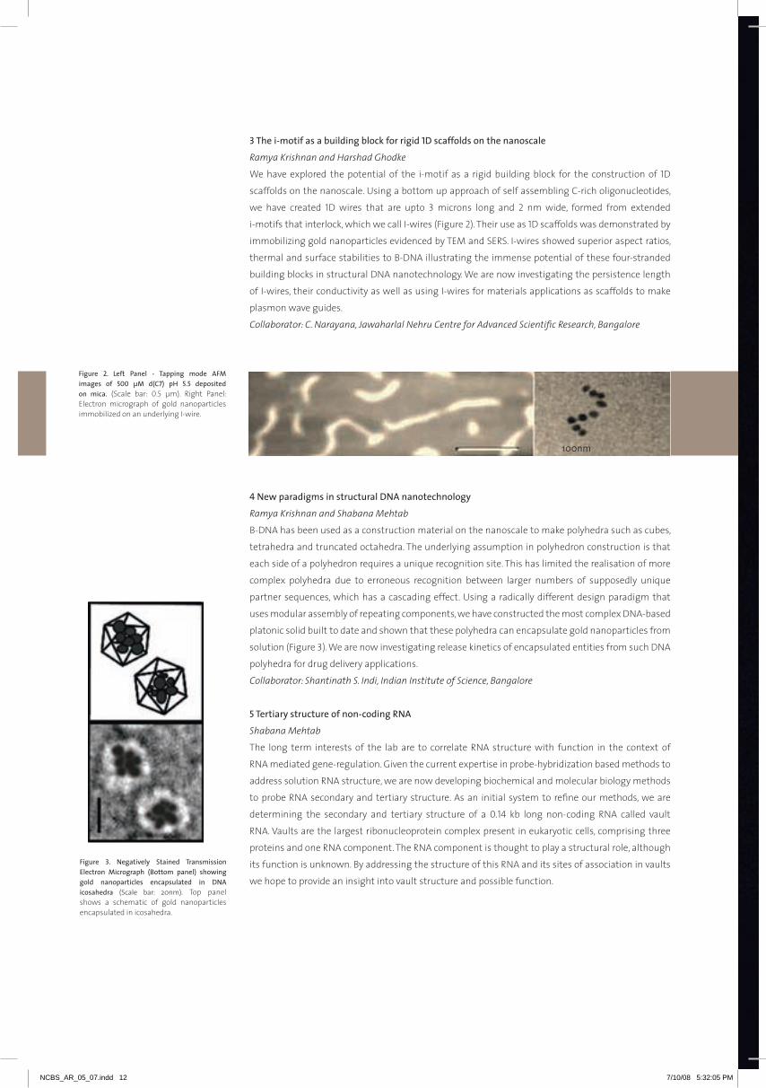
3thei-motifasabuildingblockforrigid1dscaffoldsonthenanoscale
Ramya Krishnan and Harshad Ghodke
We have explored the potential of the i-motif as a rigid building block for the construction of 1D
scaffolds on the nanoscale. Using a bottom up approach of self assembling C-rich oligonucleotides,
we have created 1D wires that are upto 3 microns long and 2 nm wide, formed from extended
i-motifs that interlock, which we call I-wires (Figure 2). Their use as 1D scaffolds was demonstrated by
immobilizing gold nanoparticles evidenced by TEm and SErS. I-wires showed superior aspect ratios,
thermal and surface stabilities to B-DNA illustrating the immense potential of these four-stranded
building blocks in structural DNA nanotechnology. We are now investigating the persistence length
of I-wires, their conductivity as well as using I-wires for materials applications as scaffolds to make
plasmon wave guides.
Collaborator: C. Narayana, Jawaharlal Nehru Centre for Advanced Scientific Research, Bangalore
figure 2. left panel - tapping mode afMimages of 500 µM d(c7) ph 5.5 depositedon mica. (Scale bar: 0.5 µm). right Panel: Electron micrograph of gold nanoparticles immobilized on an underlying I-wire.
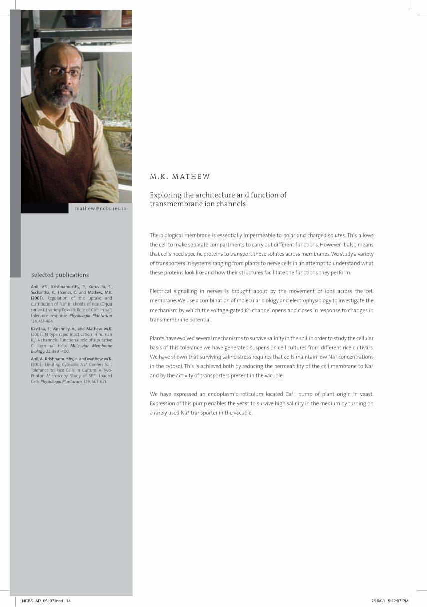
figure 3. negatively stained transmissionelectron Micrograph (Bottom panel) showinggold nanoparticles encapsulated in dnaicosahedra (Scale bar: 20nm). Top panel shows a schematic of gold nanoparticles encapsulated in icosahedra.
100nm
4newparadigmsinstructuraldnananotechnology
Ramya Krishnan and Shabana Mehtab
B-DNA has been used as a construction material on the nanoscale to make polyhedra such as cubes,
tetrahedra and truncated octahedra. The underlying assumption in polyhedron construction is that
each side of a polyhedron requires a unique recognition site. This has limited the realisation of more
complex polyhedra due to erroneous recognition between larger numbers of supposedly unique
partner sequences, which has a cascading effect. Using a radically different design paradigm that
uses modular assembly of repeating components, we have constructed the most complex DNA-based
platonic solid built to date and shown that these polyhedra can encapsulate gold nanoparticles from
solution (Figure 3). We are now investigating release kinetics of encapsulated entities from such DNA
polyhedra for drug delivery applications.
Collaborator: Shantinath S. Indi, Indian Institute of Science, Bangalore
5tertiarystructureofnon-codingrna
Shabana Mehtab
The long term interests of the lab are to correlate rNA structure with function in the context of
rNA mediated gene-regulation. Given the current expertise in probe-hybridization based methods to
address solution rNA structure, we are now developing biochemical and molecular biology methods
to probe rNA secondary and tertiary structure. As an initial system to refine our methods, we are
determining the secondary and tertiary structure of a 0.14 kb long non-coding rNA called vault
rNA. Vaults are the largest ribonucleoprotein complex present in eukaryotic cells, comprising three
proteins and one rNA component. The rNA component is thought to play a structural role, although
its function is unknown. By addressing the structure of this rNA and its sites of association in vaults
we hope to provide an insight into vault structure and possible function.
NCBS_AR_05_07.indd 12 7/10/08 5:32:05 PM

13
SA
TY
AJI
T
mA
Yo
rY
aM
un
ak
rIs
hn
an
NCBS_AR_05_07.indd 13 7/10/08 5:32:07 PM
Selected publications
Exploring the architecture and function of transmembrane ion channels
M . K . M A T H E W
The biological membrane is essentially impermeable to polar and charged solutes. This allows
the cell to make separate compartments to carry out different functions. However, it also means
that cells need specific proteins to transport these solutes across membranes. We study a variety
of transporters in systems ranging from plants to nerve cells in an attempt to understand what
these proteins look like and how their structures facilitate the functions they perform.
Electrical signalling in nerves is brought about by the movement of ions across the cell
membrane. We use a combination of molecular biology and electrophysiology to investigate the
mechanism by which the voltage-gated K+-channel opens and closes in response to changes in
transmembrane potential.
Plants have evolved several mechanisms to survive salinity in the soil. In order to study the cellular
basis of this tolerance we have generated suspension cell cultures from different rice cultivars.
We have shown that surviving saline stress requires that cells maintain low Na+ concentrations
in the cytosol. This is achieved both by reducing the permeability of the cell membrane to Na+
and by the activity of transporters present in the vacuole.
We have expressed an endoplasmic reticulum located Ca++ pump of plant origin in yeast.
Expression of this pump enables the yeast to survive high salinity in the medium by turning on
a rarely used Na+ transporter in the vacuole.
Anil, V.S., Krishnamurthy, P., Kuruvilla, S., Sucharitha, K., Thomas, G. and Mathew, M.K. (2005). regulation of the uptake and distribution of Na+ in shoots of rice (Oryza sativa L.) variety Pokkali: role of Ca2+ in salt tolerance response Physiologia Plantarum 124, 451-464.
Kavitha, S., Varshney, A., and Mathew, M.K. (2005). N type rapid inactivation in human KV1.4 channels: Functional role of a putative C- terminal helix Molecular Membrane Biology, 22, 389 -400.
Anil, A., Krishnamurthy, H. and Mathew, M.K. (2007). Limiting Cytosolic Na+ Confers Salt Tolerance to rice Cells in Culture: A Two-Photon microscopy Study of SBFI Loaded Cells Physiologia Plantarum, 129, 607-621.
[email protected] . in
NCBS_AR_05_07.indd 14 7/10/08 5:32:07 PM

15
1deducingthemachineryunderlyingvoltage-dependentopeningofk+channels
Sanjeev Upadhyay
Voltage-gated potassium channels are among the most intensely studied proteins today. They are
tetrameric proteins with each subunit consisting of six transmembrane segments and contributing a
re-entrant “Pore-loop” to the pore (Figure 1). Crystal structures of both 2-Transmembrane (2-Tm) and 6-
Tm channels together with a number of atomic level models provide a starting point to elucidate the
mechanism that underlies the transduction of membrane potential changes into channel opening
and closing.
We had earlier observed that a leucine heptad repeat motif is highly conserved in voltage-gated
channels, and that mutations in these leucines lead to unexpectedly large shifts in the voltage
range of Shaker channels. We argued that such large voltage shifts implied a role in the transduction
machinery and sought sites elsewhere on the protein where the leucines could interact.
We have undertaken an iterative modelling and mutagenesis exercise to arrive at models for the
open and closed states of the channel, and to elucidate the mechanism by which electric field-driven
movements in the voltage sensor result in channel opening. In order to do this, we have first evaluated
the available models and crystal structures in terms of their ability to explain the consequences of
conservative substitutions of hydrophobic residues in the sensor domain. Having settled on the KV1.2
crystal structure as being close to the open state, we then modelled a closed state based on available
mutagenesis data, and fine tuned the model based on additional mutagenesis. Finally, we propose a
series of conformational changes leading from the closed state to an activated, but non-conducting
state, followed by cooperative channel opening (Figure 2).
figure 1. schematic representation of a k+-channelmonomer. The fourth transmembrane segment, S4 (red), is the primary voltage-sensing unit. The S5-S6 stretch (yellow box) contributes to the aqueous pore, with S6 forming the inner lining. Channel opening requires movement of S6. Note that the functional channel is a tetramer.
figure 2. proposed mechanism of channelgating. Top panel: schematic showing S4 sensing helices in red and S6 pore lining helices in blue. The kink in the open state is based on published EPr data. Lower panel: Atomic models based on the KV1.2 crystal structure for the open state. Surface representation for the channel models are presented, with an overlay of the S4 and S6 helices shown as cylinders. Three states are indicated: red – Closed; Yellow – Activated; Green – open. Left panel shows the Closed to Activated transition; right panel shows the Activated to open state transition.
m K
mA
TH
EW
NCBS_AR_05_07.indd 15 7/10/08 5:32:09 PM

2regulationofk+channeltrafficking
Hyder Usman
Voltage-gated K+-channels have been shown to play roles in cell proliferation and cell death as well as
in development. As such, control of their surface expression is critical and several regulatory molecules
participate in fine-tuning the expression and functionality of channels at the cell surface. one such
regulator, KCNrG (potassium channel regulator), is a potential tumor suppressor, and has been
shown to reduce whole-cell K+ currents on overexpression in tumor cell lines. This could, in principle,
be brought about by reducing transcription or translation of channel protein, by limiting the amount
of protein that reaches the surface or by blocking channels already at the plasma membrane.
We have demonstrated that KCNrG is localized in the Endoplasmic reticulum as an oligomer.
Expression of KCNrG does not affect overall amounts of channel protein in cells indicating that
transcription and translation are unaffected. Nor does it affect endocytosis or exocytosis of unrelated
proteins or of bulk fluid establishing the specificity of the KCNrG-KV1 regulation. KCNrG interacts
with the T1 domain of KV1 family channels and retains these channels in the Er. This reduces surface
expression by about a third in mammalian cells (Figure 3). Analysis of whole cell currents of channels
expressed in Xenopus oocytes shows that co-expression of KCNrG results in reduction of whole-cell
currents by about 30%, consistent with the reduction in surface expression. The channels that do
reach the surface have biophysical properties (voltage dependence of activation and inactivation as
well as rates of inactivation) that are identical to channels expressed without KCNrG (Figure 4).
our data indicates that KCNrG regulates surface expression by specifically retaining KV1 channel
proteins in the Er.
3Mechanismslimitingna+levelsinthecytosolofricecells
Veena Anil
We have tested the text book statement that survival of plant cells subjected to saline stress correlates
with low levels of cytosolic Na+. The technology for monitoring Na+ levels in specific cell compartments
non-invasively in real time is currently restricted to isolated cells and protoplasts. We have monitored
figure 4. kv1.4 expressed in Xenopus oocytes.a: Voltage dependence of currents evoked on depolarizing Xenopus oocytes expressing KV1.4 channels with or without KCNrG. b: Voltage dependence of channel opening for the same data set.
figure 3. surface expression of kv1.4 channelprotein. The protein was tagged with YFP and the yellow colour indicates total protein in the cell. A myc tag was introduced in an extracellular loop. Anti-myc antibodies label protein on the surface and is shown in red. Top panel: KV1.4 co-expressed with KCNrG. Lower panel: KV1.4 expressed alone.
NCBS_AR_05_07.indd 16 7/10/08 5:32:11 PM
17
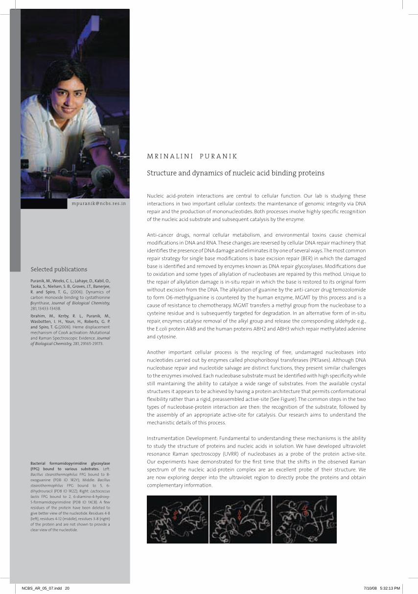
cytosolic Na+ with the sodium-specific dye SBFI, using two-photon microscopy to establish that the
dye is localized exclusively in the cytosol (Figure 5). We have contrasted suspension cells derived
from the sensitive rice cultivar Jaya to those from the salt-tolerant Pokkali. We find that Pokkali cells
maintain low cytosolic Na+ even when medium Na+ is raised to 250 mm, whereas Jaya is unable to
do so (Figure 5). We have, for the first time, manipulated cytosolic Na+ levels and demonstrated that
survival of rice cells in suspension culture is inversely correlated with this parameter, over a range of
cytosolic Na+ concentrations spanning two orders of magnitude (Figure 6). This finding holds for both
Jaya and Pokkali cells indicating that differences in their tolerance to salt is determined in large part
by their ability to regulate cytosolic Na+.
We have estimated the plasma membrane permeability to Na+ and find that while Jaya cells have a
permeability in the range reported for other glycophytes, that of Pokkali is over an order of magnitude
lower – in the range reported for halophytes. This is the largest variation in plasma membrane
permeability reported within a species to date. In addition, Pokkali cells accumulate Na+ in their large
vacuoles more efficiently than Jaya cells do.
4heterologousexpressionofplanttransportersinyeast
Veena Anil
We have expressed an Er-located Ca++-ATPase, ACA2 from Arabidopsis thaliana, in a yeast strain lacking
most endogenous Ca++ transporters. The strain is hypersensitive to saline stress due to the elimination
of the Ca++-dependent phosphatase, calcineurin B. Expression of ACA2 relieves this hypersensitivity.
We have followed the transient rise and decay of cytosolic Ca++ in response to saline stress. The
clearance of Ca++ from the cytosol is very slow in the yeast strain lacking Ca++ transporters, but is
normal in the transformant expressing ACA2. Further, whereas wild type yeast removes Na+ entering
the cytosol by pumping it out across the plasma membrane, the transformant accumulates Na+ in
the vacuole, using the pre-vacuolar Na+/H+ - antiporter, Nhx1. Nhx1 does not normally play a role in
the salt-tolerance response of wild type yeast and its activation involves a novel signal transduction
mechanism.
figure 5. a: cytosolic na+ levels in Jaya andpokkalisuspensioncells. SBFI-loaded cells were exposed to salt stress for an hour followed by two-photon excitation of the dye at 730 and 780 nm. Fluorescence emission at 515nm was ratiometrically analyzed (730F/780F) to estimate cytosolic Na+.
figure 6. correlation between cell viabilityandcytosolicna+concentrations. Cell viability was estimated by trypan blue staining, while cytosolic Na+ was determined using SBFI and 2-photon microscopy.
m K
mA
TH
EW
NCBS_AR_05_07.indd 17 7/10/08 5:32:12 PM

Selected publications
Structural biology and macromolecular crystallography
D E E PA K T . N A I R
Life may be described as a complex chemical reaction where a multitude of bio-molecules
interact and carry out their functions with great precision. The shape of these molecules is
crucial to their proper functioning. This is especially true of a majority of proteins, where a linear
polypeptide folds to attain a characteristic three-dimensional shape that enables it to possess
a specific bioactivity. Usually, for a given protein, a unique three dimensional arrangement of
a subset of its atoms in space allows the protein to perform a specific chemical function. X-
ray crystallography has traditionally provided valuable information regarding the shape of the
molecule and the spatial arrangement of its constituent atoms. Along with Nmr, it is one of the
two techniques that can provide a snapshot of the structure of the bio-molecule in its functional
as well as inactive state.
The primary step in the process of protein crystallography is the purification of protein to high
homogeneity to aid crystallization. X-ray diffraction data are collected from the protein crystals.
Using mathematical tools, an electron density map of the contents of the crystal is computed. A
chemical model of the corresponding protein is built into this map with constant qualitative and
quantitative monitoring of the agreement between the model and the diffraction data. The final
model is subjected to stringent computational tests to ensure its validity and then analyzed in
the context of available biochemical and genetic data. Inferences drawn regarding the chemical
function of the molecule from the deduced structure are also confirmed using biochemical and
genetic methods.
Using macromolecular crystallography and other biophysical methods, we aim to understand
the molecular mechanisms of viral infection by the members of the genus Flavivirus. Around
80 flaviviruses have been identified and many of them are responsible for diseases in humans.
These include DENV (Dengue fever virus), JEV (Japanese encephalitis virus) and YFV (yellow fever
virus) – which cause severe diseases and a large number of fatalities annually all over the world.
The flaviviruses are usually transmitted to humans through arthropod vectors. Infection by DENV,
JEV and YFV occurs primarily through mosquitoes. The virus particles are enveloped and about
40-50 nanometre in size with an icosahedral capsid. The flavivirus genome is represented by a
single-stranded, capped rNA molecule which is a monocistronic mrNA with a single long open
reading frame. Translation of the viral genome encodes a 370-kDa polyprotein precursor, which
Nair, D.T., Johnson, R. E., Prakash, S., Prakash, L. and Aggarwal, A. K.(2005).rev1 employs a novel mechanism of DNA synthesis using a protein template. Science, 309, 2219-2222.
Nair, D.T., Johnson, R. E., Prakash, S., Prakash, L. and Aggarwal, A. K. (2004). replication by human DNA polymerase-iota occurs by Hoogsteen base- pairing. Nature, 430, 377-380.
Nair, D. T., Johnson, R. E., Prakash, L., Prakash, S. and Aggarwal, A. K. (2008) Protein-template-directed synthesis across an acrolein-derived DNA adduct by yeast rev1 DNA polymerase. Structure. 16:239-245.
[email protected] . in
NCBS_AR_05_07.indd 18 7/10/08 5:32:12 PM

19
is processed by host and viral proteases to yield three structural proteins (C, m, and E) and seven non-
structural proteins (NS1, NS2A, NS2B, NS3, NS4A, NS4B, and NS5). The viral serine protease is formed by
the N-terminal domain of the NS3 protein and a cofactor peptide within the NS2B protein. In addition
the NS3 protein also possesses a rNA helicase activity towards the C-terminus to aid replication. The
viral rNA-dependent rNA polymerase (rdrp) activity present in the NS5 protein is responsible for
replication of the viral genome. Also, NS5 has a methyltransferase activity for capping the progeny
rNA at the 5’ end. It has been hypothesized that the non-structural proteins associate with presently
unknown host cofactors to form complexes where replication takes place. The copies of the progeny
rNA generated by these replication complexes are then packaged into the nucleocapsid and the
virions undergo maturation in the endoplasmic reticulum and the Golgi complex. After maturation,
the virions are released by vesicle fusion. overall, there has to occur a precise interplay between viral
macromolecules and host factors to ensure successful completion of the replicative cycle. We aim to
try and understand at a structural level how the different enzymatic activities of the NS3 and NS5
proteins are coordinated to ensure proper replication of the viral rNA.
DE
EP
AK
T N
AIr
figure 1.the figures shows how the exocyclicpdg adduct is accommodated in a region intheactivesiteoftheY-familydnapolymeraserev1. It is a stereo view of the rev1 surface near the PdG adduct. The surface is colored according to a spectrum of hydrophobicity, where dark red corresponds to maximum hydrophobicity and dark blue corresponds to maximum hydrophilicity. DNA is shown in stick representation and the relevant water molecules are shown as magenta spheres. The figure shows that the displayed region of the active site is shaped optimally to accommodate the PdG adduct.
NCBS_AR_05_07.indd 19 7/10/08 5:32:12 PM
Selected publications
Structure and dynamics of nucleic acid binding proteins
M R I N A L I N I P U R A N I K
[email protected] . inNucleic acid-protein interactions are central to cellular function. our lab is studying these interactions in two important cellular contexts: the maintenance of genomic integrity via DNA repair and the production of mononucleotides. Both processes involve highly specific recognition of the nucleic acid substrate and subsequent catalysis by the enzyme.
Anti-cancer drugs, normal cellular metabolism, and environmental toxins cause chemical modifications in DNA and rNA. These changes are reversed by cellular DNA repair machinery that identifies the presence of DNA damage and eliminates it by one of several ways. The most common repair strategy for single base modifications is base excision repair (BEr) in which the damaged base is identified and removed by enzymes known as DNA repair glycosylases. modifications due to oxidation and some types of alkylation of nucleobases are repaired by this method. Unique to the repair of alkylation damage is in-situ repair in which the base is restored to its original form without excision from the DNA. The alkylation of guanine by the anti-cancer drug temozolomide to form o6-methylguanine is countered by the human enzyme, mGmT by this process and is a cause of resistance to chemotherapy. mGmT transfers a methyl group from the nucleobase to a cysteine residue and is subsequently targeted for degradation. In an alternative form of in-situ repair, enzymes catalyse removal of the alkyl group and release the corresponding aldehyde e.g., the E.coli protein AlkB and the human proteins ABH2 and ABH3 which repair methylated adenine and cytosine.
Another important cellular process is the recycling of free, undamaged nucleobases into nucleotides carried out by enzymes called phosphoribosyl transferases (PrTases). Although DNA nucleobase repair and nucleotide salvage are distinct functions, they present similar challenges to the enzymes involved. Each nucleobase substrate must be identified with high specificity while still maintaining the ability to catalyze a wide range of substrates. From the available crystal structures it appears to be achieved by having a protein architecture that permits conformational flexibility rather than a rigid, preassembled active-site (See Figure). The common steps in the two types of nucleobase-protein interaction are then: the recognition of the substrate, followed by the assembly of an appropriate active-site for catalysis. our research aims to understand the mechanistic details of this process.
Instrumentation Development: Fundamental to understanding these mechanisms is the ability to study the structure of proteins and nucleic acids in solution. We have developed ultraviolet resonance raman spectroscopy (UVrr) of nucleobases as a probe of the protein active-site. our experiments have demonstrated for the first time that the shifts in the observed raman spectrum of the nucleic acid-protein complex are an excellent probe of their structure. We are now exploring deeper into the ultraviolet region to directly probe the proteins and obtain complementary information.
Puranik, M., Weeks, C. L., Lahaye, D., Kabil, O., Taoka, S., Nielsen, S. B., Groves, J.T., Banerjee, R. and Spiro, T. G., (2006). Dynamics of carbon monoxide binding to cystathionine βsynthase, Journal of Biological Chemistry, 281, 13433-13438.
Ibrahim, M., Kerby, R. L., Puranik, M., Wasbotten, I. H., Youn, H., Roberts, G. P. and Spiro, T. G.(2006). Heme displacement mechanism of CooA activation: mutational and raman Spectroscopic Evidence. Journal of Biological Chemistry, 281, 29165-29173.
Bacterial formamidopyrimidine glycosylase(fpg) bound to various substrates. Left: Bacillus stearothermophilus FPG bound to 8-oxoguanine (PDB ID 1r2Y); middle: Bacillus stearothermophilus FPG bound to 5, 6- dihydrouracil (PDB ID 1r2Z); right: Lactococcus lactis FPG bound to 2, 6-diamino-4-hydroxy-5-formamidopyrimidine (PDB ID 1XC8). A few residues of the protein have been deleted to give better view of the nucleotide. residues 4-8 (left), residues 4-12 (middle), residues 3-8 (right)of the protein and are not shown to provide a clear view of the nucleotide.
NCBS_AR_05_07.indd 20 7/10/08 5:32:13 PM

21
1 Molecular mechanism of multiple substrate specificty of the base excision repair enzyme
formamidopyrimidineglycosylase(fpg)
Namrata Jayanth and Biakdik Guite
oxidation of DNA by reactive oxygen species produced during the normal metabolism of the cell leads
to covalent modification of nucleobases. These modified nucleobases are mutagenic in nature and their
presence is implicated in cancer, neurodegenerative disorders and aging. A well-known, major product
of DNA oxidation in cells is 8-oxoguanine. The deleterious effects of 8-oxoguanine are combated by
formamidopyrimidine glycosylase (FPG), one of the key enzymes in the base excision repair pathway
(Figure 1). FPG is highly specific in distinguishing 8-oxoguanine from its normal counterpart, guanine,
which differs by only two atoms. In addition to 8-oxoguanine, FPG also recognizes and excises other
modifications of guanine, uracil and cytosine. This promiscuity is characteristic of glycosylases and
is hypothesized to originate in the conformational flexibility of their active-sites. Indeed, the crystal
structures of FPG from different organisms in free and substrate bound forms (opposite page) reveal
considerable conformational changes in the protein structure upon the binding of each type of
substrate.
Elucidation of the precise mechanism of the recognition of a damaged nucleobase, formation of
the active-site and subsequent catalysis requires an understanding of the nucleobase-protein
interaction in solution. We are studying the nature of FPG-DNA interaction using UVrr. As a first step,
we have studied the structure of free 8-oxoguanine in solution using UVrr. raman spectra obtained
with laser excitation at 260 nm of guanine and 8-oxoguanine show remarkably different relative
intensities indicative of the difference in the structures of the electronic excited states responsible
for the absorption at 260 nm in the two molecules. We have shown that UVrr can be used as a
base-specific probe to distinguish between guanine and 8-oxoguanine (Figure 2). The contentious
structure of 8-oxoguanine is unequivocally established to be that of the diketo tautomer in solution
from our experiments, quantum chemical calculations and normal mode analysis. Having established
the specific raman signatures of the oxidized base, we are now using the nucleobase as a probe of the
FPG active-site. Experiments involve probing the complex of FPG with 8-oxoguanine containing DNA.
2on-siterepairofalkylateddnabyE. colialkBanditshumananalogues
Nirmala O. and Silja Poulose
Alkylating chemicals are widely used as chemotherapeutic drugs in the treatment of cancer, e.g.,
carmustine, temozolomide, etc. S-adenosylmethionine present in cells is also a potential methylating
agent. These and other alkylating agents in the environment damage DNA by adding alkyl groups to
nucleobases. Alkylation of nucleobases alters their hydrogen bonding properties leading to cytotoxicity
and mutagenicity. Similar modifications can be expected to occur in rNA nucleobases as well.
E. coli AlkB (Figure 3) and its human homologues ABH2 and ABH3 bring about direct damage reversal
of alkylated single and double stranded DNA and rNA in a reaction that requires Fe(II) as a cofactor,
a-ketoglutarate as co-substrate and oxygen (Figure 4). AlkB enzyme repairs alkylation lesions such
as N1-methyladenine (1meA) and N3-methylcytosine efficiently and N1-methylguanine and N3-
methylthymine with lower efficiency. Apart from these small lesions, AlkB removes larger alkyl
groups such as ethyl and propyl groups and exocyclic ethano adducts like 1,N6-ethanoadenine and
figure1.theBaseexcisionrepairpathway.
figure2.ultra-violetresonanceramanspectraofgMpanditsoxidizedform,8-oxoguanosine,in water, ph 7.0, obtained with 260 nmexcitationwavelength.
mr
INA
LIN
I P
Ur
AN
IK
NCBS_AR_05_07.indd 21 7/10/08 5:32:14 PM

3,N4-ethanocytosine. These lesions are formed only in single stranded DNA during replication and
transcription as these sites are involved in base pairing between complementary strands in a double
stranded DNA. How does AlkB accommodate and catalyze both small and large lesions?.
The crystal structure of substrate-bound AlkB solved recently by Hunt and coworkers (Nature 2006)
provided detailed insight into the mechanism of AlkB action. The Fe(II)-oxoglutarate dioxygenase
core matched other superfamily members but the trinucleotide alkylated substrate showed a unique
fold designed to hold the 1meA in correct orientation. A conformationally flexible ‘lid’ was found that
completes the assembly of the active-site pocket when the substrate binds to AlkB. The transport of
molecular oxygen, essential for the reaction appears to be through a tunnel connected to the active
site. The crystals were prepared and examined in the absence of molecular oxygen to prevent the
catalytic reaction. However, when oxygen was provided, the reaction was found to be inefficient in
the crystal. This suggests the possibility that the structure in solution is different and that protein
dynamics may modulate the chemistry. These crystal structures have thrown open several interesting
questions about the action of AlkB, the role of protein dynamics and the assembly of the active site
pocket for different substrates. We will now address these for the first time using resonance raman
spectroscopic approaches developed in our laboratory.
3 understanding the different substrate specificities of human and Plasmodium falciparum
hypoxanthineguaninephosphoribosyltransferase(hgprt)
Spriha Gogia
Nucleotide synthesis in humans is carried out by the de novo synthesis pathway, where nucleotides are
synthesized from small molecules present in the cell, and the salvage pathway which involves recycling
of previously metabolized intermediates, e.g. purines. Some protozoan parasites like Plasmodium
falciparum, the malarial parasite, possess only the salvage pathway for nucleotide synthesis. Inhibitors
for this pathway would block nucleotide synthesis and are, thus, a target for chemotherapy against
diseases caused by these parasites. The enzyme HGPrT that is a part of the purine salvage pathway
is one such therapeutic target. our current interest is to elucidate the catalytic mechanism of this
enzyme and to understand differences between the human and Plasmodium enzymes.
For these studies, we have developed a first application of UVrr spectroscopy to observe the
nucleotide specifically while it is bound to the enzyme. The human and malarial enzymes exhibit
differing substrate specificities with the respect to xanthine. The human as well as Plasmodium
figure 3. crystal structure of alkB (pdB file:2fd8) in anaerobic conditions showing thefe(II),nucleobaseand2-oxoglutarate(2og)andmethylated trinucleotide d(t-mea-t) bound toit.
figure 4. Mechanism of alkylation damagereversal by alkB enzyme in the presenceof the fe(II) co-factor and the co-substrate(2-oxoglutarate) with consumption of onemolecule of oxygen and release of carbondioxideandcorrespondingaldehyde.
NCBS_AR_05_07.indd 22 7/10/08 5:32:14 PM

23
falciparum enzymes catalyze the conversion of hypoxanthine and guanine into their nucleotide
counterparts inosine monophosphate and guanosine monophosphate, respectively (Figure 5).
However, the malarial enzyme catalyses the conversion of xanthine to xanthosine monophosphate as
well while the human enzyme does not. The crystal structures currently available do not explain this
difference between the two enzymes (Figure 6). With an aim to understand the origin of this substrate
specificity, we have examined the end-product complexes of huHGPrT and pfHGPrT spectroscopically.
our current experiments of the enzyme-end-product complexes demonstrate that the environment
of the nucleobase in the two enzymes has subtle differences. raman spectra of the nucleotides bound
to the enzymes show shifts that are found to correlate with the published crystal structures of the
enzymes. We plan to carry out experiments with various available site mutants of the enzyme to
elucidate the origin of substrate specificity and mechanism of catalysis.
While the raman shifts observed experimentally will provide a qualitative measure of the protein-
nucleotide interaction, we are using a computational approach to obtain a quantitative interpretation
of the observed shifts. Currently, we are carrying out ab initio and density functional theoretical
calculations using co-ordinates from the available crystal structure of the enzyme to simulate
the protein-nucleotide interaction. The computed structure of the free base (from HF/DFT) will be
compared with the structure of the base in the active-site of the protein. The changes in structure
due to interaction with the amino acids of the protein will be correlated with observed changes in the
corresponding raman spectra. We will further extend these studies to model other substrates and
end-products for which crystal structures are not yet available.
Collaborator: Hemalatha Balaram, Jawaharlal Nehru Centre for Advanced Scientific Research,
Bangalore
figure 6. crystal structures of human hgprtwith bound gMp, (PDBID IHmP) left; and P. falciparum HGPrT bound to the transition state inhibitor immucillin, right. (PDBID 1CJB)
figure 5. the proposed catalytic mechanismof the human enzyme. The last rate limiting step of the product binding to the enzyme by incubating the enzyme with a large excess of the product was studied with UVrr.
mr
INA
LIN
I P
Ur
AN
IK
NCBS_AR_05_07.indd 23 7/10/08 5:32:15 PM
Selected publications
Cellular responses to fluid forces
K A U S T U B H R R A U
Cells are exposed to fluid forces in a variety of physiological or man-made scenarios. our lab is
studying how cells sense and respond to these forces in two different contexts. In the first one, we
examine how laser pulses cause damage in tissues in which the damage happens by an explicitly
physical mechanism. This question is primarily of interest because of the widespread use of
lasers in surgery and medicine. moreover, increasingly laser microbeams are also being used in
cell biology for transfection and microsurgery. In both these areas an improved understanding of
the laser-cell interaction is critical for the continued development of laser microbeams in biology.
We have primarily addressed this question by conducting time-resolved imaging studies on the
nanosecond - microsecond timescales.
In the second area of research we study fluid-flow sensing of endothelial cells. The apical
membrane of endothelial cells is made up of a polymer brush layer that is termed the glycocalyx.
It is now known that the glyocalyx is the primary sensor of blood flow in endothelial cells. This
fluid sensing is an important physiological function necessary for vasodilation of blood vessels
and its mechanism remains a central question in vascular biology. our group is using different
microscopy techniques to image this membrane layer and determine if there is any organization
inherent to its protein constituents.
Cherian, A.V. and Rau, K.R. (2008). Pulsed laser-induced damage in 3D cell cultures: time-resolved imaging of physical effects and biological response. Journal of Biomedical Optics, 13, 024009.
Hellman, A.N., Rau, K.R., Yoon, H. H. and Venugopalan, V.(2008). Biophysical response to laser microbeam-induced cell lysis and molecular delivery. Journal of Biophotonics, 1, -24-35, 2008.
Rau, K.R., Quinto-Su, P. A., Hellman, A.N. and Venugopalan, V. (2006). Pulsed laser microbeam-induced cell lysis: time-resolved imaging and analysis of hydrodynamic effects. Biophysical Journal, 91, 317-329.
[email protected] . in
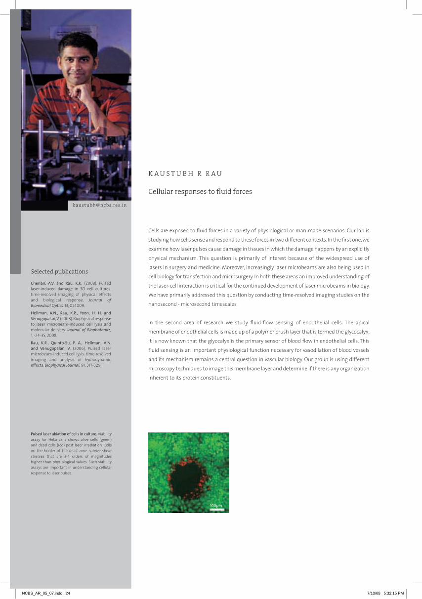
pulsedlaserablationofcellsinculture. Viability assay for HeLa cells shows alive cells (green) and dead cells (red) post laser irradiation. Cells on the border of the dead zone survive shear stresses that are 3-4 orders of magnitudes higher than physiological values. Such viability assays are important in understanding cellular response to laser pulses.
100µm
NCBS_AR_05_07.indd 24 7/10/08 5:32:15 PM

25
1lasermicrobeamsfortissueablationandcellularmicromanipulation
Anoop Cherian
Pulsed laser-induced tissue ablation: Time-resolved imaging and acute biological effects
Nanosecond laser surgery with pulse energies in the mJ range is now part of standard clinical practice.
Highly focused pulsed lasers (laser microbeams) with energies in the µJ range are also increasingly
being used for tissue micro dissection, targeted cell lysis and transfection and cell microsurgery. There
is however limited understanding of the physical and biological damage mechanisms of pulsed lasers.
models of cell damage based on experimental data will prove useful in development and refinement
of laser based tools for surgery and biotechnology. In the present work we have studied laser induced
damage in 3D cell cultures and ex-vivo samples of rat corneas using time-resolved imaging and
biological assays. Previously we had shown that time-resolved imaging of laser induced cell lysis could
provide great insights into the dynamics of the damage process and also generate quantitative data
for calculation of shear forces experienced by cells. The imaging system we have designed is capable
of capturing fast events from nano- to micro-seconds with < 1 µm spatial resolution in thick tissue
samples. These events have not been studied at such high spatial resolution before and our system
can provide a detailed understanding of these processes. Figure 1 shows the dynamics of tissue
ablation as captured on our time-resolved system. A cavitation bubble is generated after a single laser
pulse is focused into the tissue sample (in this case rat cornea). The bubble expansion and collapse
is captured along with the cellular deformation that it induces. In Figure 2 we observe the biological
aftermath of the cavitation induced damage. Surprisingly we observe that cells that experienced
large deformations due to cavitation bubble expansion were viable.
μ μ
μμμμ
μ μ μ μ
μμμμ
μ μ
μμ μμ
figure 1. time resolved imaging of laser-induceddamageintheratcornealepithelium.The time-point at which the image is taken is given in the upper left corner. Cavitation bubble growth and collapse can be clearly visualized along with cellular deformation at the bubble rim. Scale bar = 50 µm.
figure 2. Biological response of cornealepitheliumafterlaser-inducedablation. Phase-contrast imaging of control (a and c) and ablated (b and d) regions. Confocal fluorescence microscopy of epithelium damage at different depths visualized after actin staining (e-h). Viability of cells at ablation sites visualized en-face (i, l) and in cross-section ( j, k) by actin (green) and propidium iodide (red) staining.
KA
US
TU
BH
r r
AU
NCBS_AR_05_07.indd 25 7/10/08 5:32:16 PM
2laserinducedcelltransfectionandmicrosurgery
G. Nageswara Rao
The use of laser microbeams for cell transfection and microsurgery has been amply demonstrated
by several groups. However to date it has not become an established technique mainly due to the
numerous parameters that have to be controlled and also due to limited understanding of the
damage caused by laser microbeams to cells. We are attempting to standardize this technique for
both these applications. Since the group already possesses a detailed understanding of laser-induced
damage processes (see above), we can choose the correct parameters for successful transfection or
microsurgery but minimize cell death. In the current setup we can deliver 355 or 532 nm, 6 ns focused
laser pulses for irradiation of cells or organisms. organelles of interest tagged with fluorescent markers
can be viewed using epifluorescence, brought to the laser spot and ablated within few seconds.
Experiments on this setup in collaboration with different groups at NCBS involve transfection of
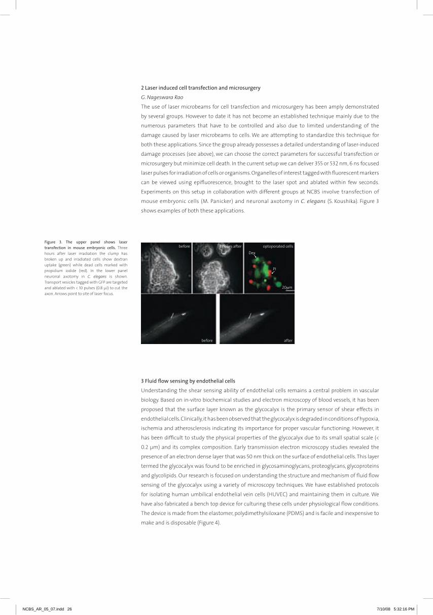
mouse embryonic cells (m. Panicker) and neuronal axotomy in C. elegans (S. Koushika). Figure 3
shows examples of both these applications.
3fluidflowsensingbyendothelialcells
Understanding the shear sensing ability of endothelial cells remains a central problem in vascular
biology. Based on in-vitro biochemical studies and electron microscopy of blood vessels, it has been
proposed that the surface layer known as the glycocalyx is the primary sensor of shear effects in
endothelial cells. Clinically, it has been observed that the glycocalyx is degraded in conditions of hypoxia,
ischemia and atherosclerosis indicating its importance for proper vascular functioning. However, it
has been difficult to study the physical properties of the glycocalyx due to its small spatial scale (<
0.2 µm) and its complex composition. Early transmission electron microscopy studies revealed the
presence of an electron dense layer that was 50 nm thick on the surface of endothelial cells. This layer
termed the glycocalyx was found to be enriched in glycosaminoglycans, proteoglycans, glycoproteins
and glycolipids. our research is focused on understanding the structure and mechanism of fluid flow
sensing of the glycocalyx using a variety of microscopy techniques. We have established protocols
for isolating human umbilical endothelial vein cells (HUVEC) and maintaining them in culture. We
have also fabricated a bench top device for culturing these cells under physiological flow conditions.
The device is made from the elastomer, polydimethylsiloxane (PDmS) and is facile and inexpensive to
make and is disposable (Figure 4).
μ
figure 3. the upper panel shows lasertransfection in mouse embryonic cells. Three hours after laser irradiation the clump has broken up and irradiated cells show dextran uptake (green) while dead cells marked with propidium iodide (red). In the lower panel neuronal axotomy in C. elegans is shown. Transport vesicles tagged with GFP are targeted and ablated with < 10 pulses (0.8 µJ) to cut the axon. Arrows point to site of laser focus.
NCBS_AR_05_07.indd 26 7/10/08 5:32:16 PM

27
4spatialorganizationintheendothelialglycocalyx
Amit Sharma and P. Senthil
We are studying two membrane proteins that form part of the glycocalyx namely syndecan-1 and
CD44 to determine their organization within the membrane and their role in fluid flow sensing. We
will be conducting hetero-FrET using fragmented antibodies raised against Syndecan-1 and CD44. GFP
fusion constructs of syndecan-1 and CD44 are also being used for transient transfection of endothelial
cells. These will be used in homo-FrET experiments to determine if these proteins are organized in
domains. Transmission electron microscopy of human umbilical vein is also being undertaken to
visualize the glycocalyx and identify its components.
5physicalpropertiesofsyndecan-1studiedbyatomicforceMicroscopy
P. Senthil and Lokanath Sai
We are using biochemical protocols to isolate the heparin sulfate proteogylcan syndecan-1 from
endothelial cell surfaces. We wish to study the physical properties of this molecule to understand
its role in mechanotransduction. To this end we are attempting to view syndecan-1 molecules using
Atomic Force microscopy. Currently we have standardized protocols for isolating proteoglycans
from endothelial cell membranes. Attempts are underway to purify this fraction to isolate intact
syndecan-1 molecules. We have also optimized protocols for viewing single molecules of hyaluronic
acid, a common extracellular polysaccharide using AFm. An example of membrane protein isolation
and AFm imaging is shown in Figure 5.
figure 5. proteoglycan isolation fromendothelialcellmembranesasseenaftersds-page electrophoresisandsilverstaining (left).afM image (right) of the purified fractionshowslargeproteinclumps.
KA
US
TU
BH
r r
AU
figure4.Bench-topflowchamberforculturingand exposing endothelial cells to fluid flow. (a) Device with persistaltic pump attached in incubator. (b) Close up of the device fabricated in silicone (PDmS). (c) Endothelial cells prior to fluid flow exposure. (d) Same area as (c) post 24 hours flow. Endothelial cells show shape elongation and alignment in the direction of flow.
NCBS_AR_05_07.indd 27 7/10/08 5:32:18 PM
Selected publications
Cellular architecture of genome regulation
G . V . S H I VA S H A N K A R
[email protected] . in
Cellular function, during development and disease, is controlled by changing patterns of
gene expression. recent evidence shows that the spatio-temporal organization of a gene
and its interaction with the transcription apparatus within the crowded 3D architecture of
the cell nucleus is vital to orchestrating gene regulation. Notably, mechanical cues are found
to alter gene transcription, cellular differentiation in culture, and developmental programs in
organisms, suggesting a strong link between cellular architecture and information control.
Thus, understanding design principles of the physical coupling between cellular architecture
to transcription control on the nanoscale is of immense importance. recent progress in high
resolution live-cell imaging combined with optical spectroscopy and biomechanics methods
developed in many laboratories including ours have provided a new paradigm in understanding
chromatin structure and function. We use multi-disciplinary approaches (combining methods
in soft-condensed matter physics, nanoscience and biology), to probe the mechanistic basis of
the spatio-temporal organization of chromatin and its coupling to transcription control at single
gene/cluster resolution during cellular differentiation and development. With this approach
we hope to understand the spatial code underlying chromatin assembly during differentiation
and its implications for cellular transcription control and memory within living cells. Controlled
physical perturbations of such a code may then provide possibilities to engineer (as we intend)
gene regulation in diverse developmental contexts.
Banerjee, B., Bhattacharya, D. and Shivashankar, G.V. (2006). Chromatin structure exhibits spatio-temporal heterogeneity within the cell nucleus. Biophysical Journal, 91, 2297-2303.
Bhattacharya, D., Mazumder, A., Miriam, S.A. and Shivashankar, G.V. (2006). EGFP-tagged core and linker histones diffuse via distinct mechanisms within living cells. Biophysical Journal, 91, 2326-2336.
Mazumder, A. and Shivashankar, G.V. (2007). Gold-nanoparticle-assisted laser perturbation of chromatin assembly reveals unusual aspects of nuclear architecture within living cells. Biophysical Journal, 93, 2209-2216.
NCBS_AR_05_07.indd 28 7/10/08 5:32:18 PM

29
1designprinciplesunderlyingthe3dorganizationofgenetranscriptionwithinlivingcells
Shovamayee Maharana, V. Ramya and R.Indulaxmi
recent evidence suggests an intimate link between higher-order chromatin assembly and
transcriptional hubs, where genes appear to interact with the transcription apparatus to regulate
their expression within a living cell. In order to understand the design principles that underlie gene
transcription, we have developed live-cell imaging assays to visualize transcription factories (TF) and
candidate gene loci (Figure-1). Single-particle tracking analysis of TF reveals their ATP-dependent
dynamic organization. TF dynamics were cell-cycle dependent and hindered by specific histone
deacetylase inhibitors which decondense chromatin assembly. Upstream sites of a candidate gene
were marked with tandem 96 lac operator repeats enabling us to visualize the position of the gene
loci. Live cell imaging of the gene locus exhibited confined diffusion in the repressed state, while
gene activation resulted in increased mobility. Collectively these experiments, using live-cell imaging
and chromatin capture assays, will probe how a transcription factor, a factory and gene loci/clusters
temporally organize within the 3D architecture of the living cell nucleus to control transcription and
cellular memory.
2tracking epigenetic plasticity in higher-order chromatin assembly during cellular differentiation
Shefali Talwar and Soumya Gupta
In this project, we study how epigenetic plasticity in higher-order chromatin assembly impinges on
differential gene expression programs during cellular differentiation. Experiments in our laboratory
and others have revealed that histone and other nuclear proteins that compact higher order
chromatin assembly are highly mobile within the cell nucleus (Figure-2). Further, recent evidence has
suggested that chromatin assembly is highly plastic in undifferentiated cells, arising due to hyper-
dynamic histone proteins. Using fluorescence recovery after photo-bleaching (FrAP) and fluorescence
correlation spectroscopy (FCS) methods, we observed that there exist distinct diffusive mechanisms
for core and linker histone proteins. Intriguingly, the mobility of histone proteins were found to be
differentially altered resulting in the transition from a plastic to frozen chromatin organization
during cellular differentiation in both mouse embryonic stem cells and during Drosophila embryo
development. We are now studying the dynamic reorganization of higher-order chromatin assembly
and its coupling to transcription control of lineage specific genes within single living cells in two
functional contexts: haematopoietic stem-cell differentiation to T-cell lineage and during T-cell
development.
Collaborator: Apurva Sarin, NCBS
3understandingthecouplingbetweenhigher-orderchromatinassemblyandthepre-stressedcell
nucleus
Nisha Ramdas and Aprotim Mazumder
In living cells, the nucleus is balanced by cytoplasmic architectural elements to provide an appropriate
size and shape that is conjectured to be important in defining genome function. Nuclear size within
the cytoplasmic context is larger as compared to isolated nuclei suggesting the existence of a
mechanical pre-stressed state. We show that the organization of chromatin assembly is a balance of
physical forces; outward forces arising due to their entropic nature given the length of the genome
and due to cytoplasmic components and inward forces driven by histone tail-tail interactions and
other nuclear proteins that condense the genome into metaphase chromosomes. In order to test if the
pre-stressed state of the nucleus is coupled to higher-order chromatin assembly, we developed a laser
G V
SH
IVA
SH
AN
KA
r
figure 1. live-cell imaging combined withsingle-particletrackinganalysis
a. Incorporation of fluorescently-labeled UTP molecules marks transcription compartments within the nucleus.
b. Tandem repeats of lac-operator sites bound with fluorescent lac-repressor was used to visualize gene loci
c. The interface of the program used to quantitate transcription factory or gene loci dynamics in live cells.
figure2.translationalandrotationaldiffusionof egfp-tagged histone proteins correlateswiththeirfunctionality.
a,b. Fluorescence recovery after photobleaching (FrAP) experiments reveal core histones to be stably bound to the chromatin, while linker histones are more dynamic, as envisaged by the recovery in the bleach spots at indicated time-points.
c. Fluorescence anisotropy maps of the same proteins in live HeLa cells expressing H2B-EGFP.
NCBS_AR_05_07.indd 29 7/10/08 5:32:19 PM
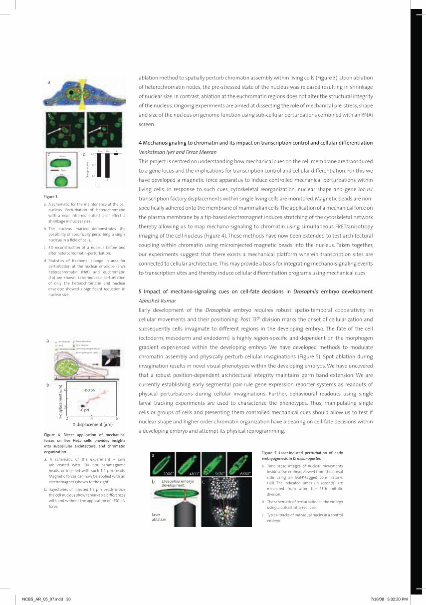
ablation method to spatially perturb chromatin assembly within living cells (Figure 3). Upon ablation
of heterochromatin nodes, the pre-stressed state of the nucleus was released resulting in shrinkage
of nuclear size. In contrast, ablation at the euchromatin regions does not alter the structural integrity
of the nucleus. ongoing experiments are aimed at dissecting the role of mechanical pre-stress, shape
and size of the nucleus on genome function using sub-cellular perturbations combined with an rNAi
screen.
4Mechanosignalingtochromatinanditsimpactontranscriptioncontrolandcellulardifferentiation
Venkatesan Iyer and Feroz Meeran
This project is centred on understanding how mechanical cues on the cell membrane are transduced
to a gene locus and the implications for transcription control and cellular differentiation. For this we
have developed a magnetic force apparatus to induce controlled mechanical perturbations within
living cells. In response to such cues, cytoskeletal reorganization, nuclear shape and gene locus/
transcription factory displacements within single living cells are monitored. magnetic beads are non-
specifically adhered onto the membrane of mammalian cells. The application of a mechanical force on
the plasma membrane by a tip-based electromagnet induces stretching of the cytoskeletal network
thereby allowing us to map mechano-signaling to chromatin using simultaneous FrET/anisotropy
imaging of the cell nucleus (Figure 4). These methods have now been extended to test architectural
coupling within chromatin using microinjected magnetic beads into the nucleus. Taken together,
our experiments suggest that there exists a mechanical platform wherein transcription sites are
connected to cellular architecture. This may provide a basis for integrating mechano-signaling events
to transcription sites and thereby induce cellular differentiation programs using mechanical cues.
5 Impact of mechano-signaling cues on cell-fate decisions in Drosophila embryo development
Abhishek Kumar
Early development of the Drosophila embryo requires robust spatio-temporal cooperativity in
cellular movements and their positioning. Post 13th division marks the onset of cellularization and
subsequently cells invaginate to different regions in the developing embryo. The fate of the cell
(ectoderm, mesoderm and endoderm) is highly region-specific and dependent on the morphogen
gradient experienced within the developing embryo. We have developed methods to modulate
chromatin assembly and physically perturb cellular invaginations (Figure 5). Spot ablation during
invagination results in novel visual phenotypes within the developing embryos. We have uncovered
that a robust position-dependent architectural integrity maintains germ band extension. We are
currently establishing early segmental pair-rule gene expression reporter systems as readouts of
physical perturbations during cellular invaginations. Further, behavioural readouts using single
larval tracking experiments are used to characterize the phenotypes. Thus, manipulating single
cells or groups of cells and presenting them controlled mechanical cues should allow us to test if
nuclear shape and higher-order chromatin organization have a bearing on cell-fate decisions within
a developing embryo and attempt its physical reprogramming.
μ
figure3.
a. A schematic for the maintenance of the cell nucleus. Perturbation of heterochromatin with a near infra-red pulsed laser effect a shrinkage in nuclear size.
b. The nucleus marked demonstrates the possibility of specifically perturbing a single nucleus in a field of cells.
c. 3D reconstruction of a nucleus before and after heterochromatin-perturbation.
d. Statistics of fractional change in area for perturbation at the nuclear envelope (Env), heterochromatin (Het) and euchromatin (Eu) are shown. Laser-induced perturbation of only the heterochromatin and nuclear envelope showed a significant reduction in nuclear size.
μ
μ
figure 4. direct application of mechanicalforces on live hela cells provides insightsinto subcellular architecture, and chromatinorganization.
a. A schematic of the experiment – cells are coated with 100 nm paramagnetic beads, or injected with such 1-2 µm beads. magnetic forces can now be applied with an electromagnet (shown to the right).
b. Trajectories of injected 1-2 µm beads inside the cell nucleus show remarkable differences with and without the application of ~150 pN force.
figure 5. laser-induced perturbation of earlyembryogenesisinD. melanogaster.
a. Time lapse images of nuclear movements inside a live embryo, viewed from the dorsal side using an EGFP-tagged core histone, H2B. The indicated times (in seconds) are measured from after the 13th mitotic division.
b. The schematic of perturbation in the embryo using a pulsed infra-red laser.
c. Typical tracks of individual nuclei in a control embryo.
NCBS_AR_05_07.indd 30 7/10/08 5:32:20 PM
31
SA
TY
AJI
T
mA
Yo
r
NCBS_AR_05_07.indd 31 7/10/08 5:32:22 PM

Selected publications
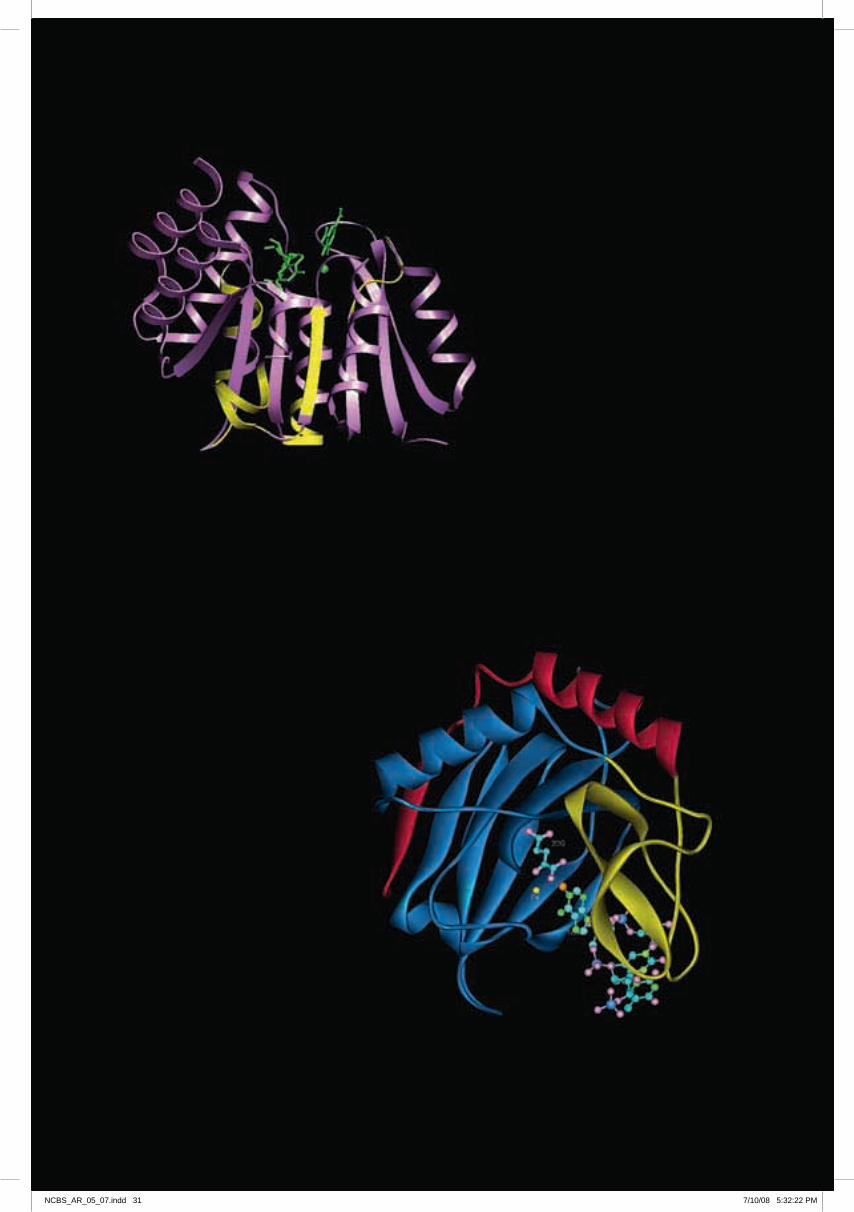
Computational approaches to protein science
R . S O W D H A M I N I
[email protected] . in
Proteins are related to each other at different levels in a hierarchical manner – some of them
are closely-knit into homologous families wherein they look similar in every way: sequence,
structure and biological function. There are more distant relationships where the proteins may
have diverged extensively, reflected as low sequence identity but still are remarkably similar and
perform similar biological function: these are grouped as superfamilies. There are many other
protein domains that happen to share similar folds and could be unrelated in sequence and
function. The homologous family level is trivial to identify in a predictive sense and are “no-
brainers”, but the superfamily level relationships are hard to reliably predict given mere sequence
information and hence are more challenging. We are interested in the generation of structural
bioinformatics algorithms that can perform well at distant relationships amongst proteins to
apply in biological problems.
Tripathi, P.L. and Sowdhamini, R. (2006). Cross genome comparisons of serine proteases in Arabidopsis and rice. BMC Genomics, 7,200.
Pugalenthi, G., Shameer, K., Srinivasan, N. and Sowdhamini, R. (2006). HArmoNY: a web-server for the assessment of protein structures Nucleic Acids Research, 34, W143-W146.
Pugalenthi G, Suganthan, P.N., Sowdhamini, R. and Chakrabarti, S. (2007). Smotif: A server for structural motifs in proteins. Bioinformatics, 23, 637-638.
structural motifs as fold signatures inproteindomains
NCBS_AR_05_07.indd 32 7/10/08 5:32:22 PM

33
1applicationofsensitivesequencesearchproceduresforgenome-widesurveys
Lokesh P. Tripathi and R. Sowdhamini
Early statistics of mass protein structure and function prediction on genome sequences indicated
that nearly 40% of the gene products appear to be globular but have surpassed computational tools
to be associated with a known protein family such that function prediction was possible. Till today,
this statistics holds true. In the past, we have applied sensitive algorithms to recognize putative
members of protein families and superfamilies (Bhaduri and Sowdhamini, 2003; 2005; metpally
and Sowdhamini, 2005) in whole genomes to improve function prediction. We have now extended
genome-wide survey for serine proteases in two plants species, representative of dicots and monocots,
to perform cross-genome survey of the extent of homology in different serine proteases. Although
the genome sizes are different, we find nearly equal numbers of different types of serine proteases
(Tripathi and Sowdhamini, 2006). We find that the serine carboxypeptidases, largely identified in plants
and associated with secondary metabolism, for instance, suggest extensive duplication subsequent to
monocot-dicot divergence and possible functional redundancy for these enzymes (Figure 1a). Despite
two distinct clades (I and II), the presence of rice-only and Arabidopsis-only subclades suggests early
diversification of these members followed by rapid species-specific expansion. The signal peptidases
in the two species fall into two clusters (Figure 1b) with high sequence identities: the type I proteases
that contain Ser-Lys catalytic dyad are found in prokaryotes, chloroplasts and in mitochondria,
whereas the type II proteases that contain Ser-His catalytic dyad are found in Er and also observed
in archaea. Rhomboids are one of the largest membrane-bound serine protease families containing
seven Tm helices and are conserved in several species. Phylogenetic analysis of Arabidopsis and rice
rhomboid-like proteins (Figure 1c) reveals presence of several gene clusters that share low pairwise
sequence identity with each other, though the members within a cluster share significant sequence
similarity with each other. multiple clusters suggest polyphyletic origin, perhaps some acquired by
lateral gene transfer and have bacterial origin. members of Clade I and II correspond to rHo and
PErL subfamilies. members of Clade IV and VII contain an additional ubiquitin-like domain and the
clustering patterns suggest that this domain architecture had evolved much before monocot-dicot
divergence. Nevertheless, it illustrates that additional domains and domain architecture of genes
provide important information about the overall function of the gene products.
2sensitiveapproachesforenhancedfunctionassociation
2a. Sensitive sequence search strategies
Kumar Gaurav, Sandhya Sankaran and R. Sowdhamini
The availability of known members of protein families enables the construction of records of the
position, order and spacing of conserved regions or motifs in protein families. We have developed a
neural-network driven procedure, called FASSm, which can objectively score new sequences for their
match to pre-recorded motifs of existing protein families (Gaurav et al., 2005). This method will be
especially useful for detecting newer domains that have undergone evolutionary changes like circular
permutation and discontinuity in domains from classical members. In addition, we have developed
an approach, called CASCADE PSI-BLAST, that employs multiple generations of PSI-BLAST searches
figure 1. genome-wide survey of plant serineproteases and cross-genome surveys acrossArabidopsisandricegenomes.
a. serine carboxypeptidases
b. signal peptidases and
c. rhomboid family. Putative serine proteases across both genomes are multiply aligned using CLUSTALX and clustered using PHYLIP (Thompson et al., 1994) and the alignments were exported to Phylip package for representing the Neighbor-Joining tree. Elements marked with red and blue colour represent putative members in Arabidopsis and rice genomes, respectively. Bootstrap values between 50-60% are represented by an asterix, circles represent bootstrap values from 60%-80% while bootstrap values >80% are represented by rectangles. major clusters in the claudograms are marked (please see text for discussion on the clustering patterns). A section of the alignment corresponding to the active site residues are shown for the major clades in the case of signal peptidases (Figure 1b).
r S
oW
DH
Am
INI
NCBS_AR_05_07.indd 33 7/10/08 5:32:22 PM
(Figure 2a) to discover newer and distant relationships between protein families that are hard to
establish (Sandhya et al., 2005). This method is also made available in the public domain in the
form of a webserver (rana et al., 2006).
Collaborators: N. Srinivasan and Rana Bhadra, Indian Institute of Science, Bangalore and Nitin
Gupta, Trainee, Indian Institute of Technology, Kanpur
2b. Structure prediction of unassigned regions in proteins
Chandrashekar Reddy, Manonmani Arunachalam, Meenakshi Babu and R. Sowdhamini
There are often large regions of globular proteins that are not associated to pre-existing
domains. We propose an approach to fill-in these gaps in domain assignments (Figure 2b) where
we examine unassigned regions of considerable length, sequence conservation and predicted
structural content to accumulate sequence homologues. We then examine the domain
architecture of individual homologous sequences using Hidden markov model searches against
PFAm database to extrapolate possible newer domains into the gene of interest. We show
that these indirect connections can enable better domain assignments for nearly 25% of the
unassigned regions that we had examined amongst the genes that contain adenylate cyclase
domains (Chandrashekar et al., 2006). The realisation of connections to pre-existing domains
can have direct consequences in better understanding of biological function of gene products.
3proteinstructurevalidation
Pugalenthi, Ganesan, Shameer Khader and R. Sowdhamini
Subsequent to homology modeling of protein domains, there could be errors in the entire protein
model or in different parts of the model like loop regions. These could be non-trivial errors that
are introduced owing to poor sequence identity between the query and template structure. A
structure validation procedure was earlier developed, called HArmoNY, that is also provided
as a webserver (Pugalenthi et al., 2006), to obtain a retrospective picture of the quality of the
model and checks the compatibility between structure and sequence. The local environments
of individual residues in a protein structure are assessed for its quality by considering structural
parameters like secondary structure, hydrogen bonding and solvent burial. Gross errors in a
structure could suggest misfolds and the method is equally efficient at detecting local errors in
protein modeling that might reside in loop regions.
Collaborator: N. Srinivasan, Indian Institute of Science, Bangalore
4structuralMotifsinproteindomainsuperfamilies
Anirban Bhaduri, Pugalenthi Ganesan and R. Sowdhamini
In order to recognise the correct connections, it is equally important to ensure that the interior
of the fold or the protein core is preserved. We had earlier reported that spatially interacting
conserved regions of the protein domains are useful drivers to improve sensitivity as well as
figure 2. sensitive sequence searchapproaches for recognizing connectionsbetween pre-existing protein domainfamilies.
a. Schematic representation of two protein families in sequence space to illustrate the effectiveness of CASCADE PSI-BLAST approach (Sandhya et al., 2005) in connecting distantly related protein families. Each ellipse represents a family in sequence space with their classical family members shown as stars within the ellipse. Added stars represent intermediate sequences obtained as hits during different generations of PSI-BLAST that could be useful in relating the two families. PSI-BLAST runs in every generation are performed with stringent E-value thresholds to avoid false positives and drifts.
b. Schematic representation of indirect domain finding techniques (Chandrashekar et al., 2006). In many gene products, large regions (more than 70 residues) are noticed where no assignment has been recorded to sequence domain family database (Bateman et al., 2000). It is possible to assign sequence domains by additionally accumulating information about the sequence homologues and their domain architectures. Such an unassigned region or linker is shown in black and co-existing domains labeled as A and B. Sequence homologues to the linker suggest the presence of an additional domain X (marked by green colour).
NCBS_AR_05_07.indd 34 7/10/08 5:32:23 PM

35
figure 3. a flow-chart explaining theworkflow of sMotIf server to recognizestructural templates of a protein or aprotein superfamily. A protein structural entry is submitted as input either as sequence or PDB code. Where possible, the server internally recognizes other structural members of the protein superfamily. After best superposition and considering structural environments such as solvent accessibility and hydrogen bonding, structurally conserved regions or ‘structural templates’ are recognized and mapped on the structures. Additional structural parameters such as intermotif torsion angles and distances are also calculated.
r S
oW
DH
Am
INI
specificity (Bhaduri et al., 2004). We have now developed a webserver, SmoTIF, which automatically
identifies structural motifs and examines structural properties (Pugalenthi et al., 2007). Starting from
a crude sequence alignment of structural entries, a rigid-body superposition leads to obtaining a more
reliable structure-based sequence alignment. With further accumulation of sequence homologues,
one can examine amino acid conservation and examine structural parameters critically. These are then
objectively coded to provide scores to different regions of the protein fold that can in turn enable the
automatic recognition of structural motifs. These motifs can be projected on the structure and further
analysed for inter-motif distances and angles (Figure 3). Such structural analyses can be helpful to
experimental biologists in designing the expression or characterization of protein domains and in
generating point mutants. Such analyses can facilitate computational biologists in ascertaining if the
structurally conserved regions have been carefully preserved in alignments and in modelling.
Collaborators: Saikat Chakrabarti, National Institutes of Health, USA; P. N. Suganthan, Nanyang
Technological University, Singapore and Ravishankar, R., Trainee, Anna University, Chennai
NCBS_AR_05_07.indd 35 7/10/08 5:32:23 PM

Selected publications
The dynamics and evolution of living networks
M U K U N D T H A T T A I
[email protected] . inWe interested in networks. A network is a group of entities, connected by a web of interactions,
whose collective behavior can be remarkably complex. In living systems, networks arise in various
contexts, across many scales. Physical networks of specific amino-acid contacts can generate an
attractive potential between proteins. The set of transient and permanent protein complexes
in a cell is abstractly represented as a protein interaction network. The uptake and efflux of
material by a cell, and the movement of cargo between various intracellular compartments,
defines a traffic network. Cells use signalling and regulatory networks to control gene expression,
metabolism, and traffic, in response to external conditions. And groups of cells can influence
one-another, as occurs by means of chemical and electrical signals in a neuronal network.
In every sense, these are truly living networks. Each network is a dynamic object, whose state
changes as a function of time; moreover, the structure of a network itself, its connectivity and
topology, can vary over evolutionary timescales. Together, these aspects define the two questions
on which we focus. First, we use quantitative experiments, coupled with mathematical and
computational models, to study the dynamical properties of living networks. Second, we use
genomic and protein sequence data to probe the evolutionary history of these systems. We find
that, across scales and contexts, the ‘network idea’ is a broad and powerful framework within
which the complexity of biological systems can be usefully organized and studied.
Dabholkar, S. and Thattai, M. (2007). Brainstorming Biology. IET Synthetic Biology, 1, 17-20.
Thattai, M., Burak, Y. and Shraiman, B.I. (2007). The origins of specificity in polyketide synthase protein interactions. PloS Computational Biology, 3, e186.
A dash of color reveals how specificity arises in a protein interaction network. Polyketides are pharmaceutically important biochemicals, synthesized in bacteria by giant multi-protein chains of polyketide synthase proteins. In order to be functional, these chains must be correctly ordered. When the N- and C-terminal sticky ends of each protein are colored according to a phylogenetic clustering, the interaction specificity code emerges: pairs of compatible sticky ends are invariably of the same color.
NCBS_AR_05_07.indd 36 7/10/08 5:32:24 PM

37
1theoriginsofspecificityinthepolyketidesynthaseproteininteractionnetwork
Polyketides, a diverse group of heteropolymers with antibiotic and antitumor properties, are
assembled in bacteria by multi-protein chains of modular polyketide synthase (PKS) proteins. Specific
protein-protein interactions determine the order of proteins within a multi-protein chain, and thereby
the order in which chemically distinct monomers are added to the growing polyketide product. We
investigated the evolutionary and molecular origins of protein interaction specificity. We focussed on
the short, conserved N- and C-terminal docking domains that mediate interactions between modular
PKS proteins. our analysis revealed a hieararchical interaction specificity code. PKS docking domains
are descended from a single ancestral interacting pair, but have split into three phylogenetic classes
that are mutually non-interacting. Specificity within one such compatibility class is determined by
a few key residues, which can be used to define compatibility sub-classes. A single PKS system can
use docking domain pairs from multiple classes, as well as domain pairs from multiple sub-classes
of any given class. The termini of individual proteins are frequently shuffled, but docking domain
pairs straddling two interacting proteins are linked as an evolutionary module. The hierarchical and
modular organization of the specificity code is intimately related to the processes by which bacteria
generate new PKS pathways.
Collaborators: Y. Burak and B.I. Shraiman, Kavli Institute for Theoretical Physics, UCSB, USA and Andrei
Lupas, Max Planck Institute for Developmental Biology, Germany
2co-evolvingresiduesandphysicalinteractions
our goal was to identify residue pairs that co-evolved between interacting proteins. This task is
complicated by the fact that sequence datasets are typically small and non-uniformly sampled,
therefore dominated by spurious correlations. To overcome this problem, we developed a new
algorithm called CroSS (correlated residues of statistical significance) which uses both interaction
and non-interaction data to identify significant pairings between residues. The algorithm takes two
inputs (multiple sequence alignment of proteins, and experiemental data about protein interactions)
and identifies residue pairs involved in interaction specificity. We applied CroSS to analyze the
interaction between polyketide synthase (PKS) proteins. The analysis picked out only eight significant
residues, seven of which were involved in pairwise physical interactions in an Nmr structure of a PKS
interaction complex. This provided strong confirmation that CroSS was able to sensitively pick out
co-evolving residues.
Collaborators: Y. Burak and B.I. Shraiman, Kavli Institute for Theoretical Physics, UCSB, USA
figure 1. co-evolving residues in pks proteininteractions.
a. We refer to pks protein interaction domains as heads (red) and tails (green). The CroSS matrix runs over head residues (vertical) and tail residues (horizontal). Bright entries represent significant co-evolution.
b/c. Structure of the head-tail interaction complex.
d/e/f. We zoom in on the eight significant residues identified in Figure 2a; remarkably, seven of these are involved in pairwise physical interactions.
mU
KU
ND
TH
AT
TA
I
NCBS_AR_05_07.indd 37 7/10/08 5:32:24 PM
3thedesignprinciplesofrapidgeneticswitches
Sugat Dabholkar
We used a simple mathematical model to investigate the factors that primarily determine the rapidity
of response of a genetic switch. Specifically, we explored the case in which a cell switches its gene
expression level between two distinct states, in response to a sudden change in the concentration of
an extracellular biochemical. We defined a dimensionless measure of rapidity, and used optimization
techniques to generate networks with the most rapid response possible given various biochemical
constraints. We found that the key determinants of rapidity were protein lifetime, maximal and
minimal transcription rates, and the ratio of induced to uninduced expression levels. our analysis
suggests that the promoter transfer function (the rate of transcription as a function of regulatory
inputs) might be optimized for its dynamical properties, rather than its steady-state behavior.
We next applied our rapidity analysis to experimental data, investigating the time-dependence of
transcription rates at an Escherichia coli promoter as extracellular conditions were shifted between
various discrete states. Specifically, we measured the concentration of a chromosomally integrated
copy of gfp, under the control of the lac promoter. Extracellular conditions were defined by various
combinations of positive and negative regulators of Plac. We measured GFP expression over several
hours using fluorescence microscopy of single cells, and avoided cell-density dependent effects
by using a novel serial dilution assay. We found that the different regulators elicited very different
dynamical responses and that, in terms of rapidity, there was a distinct asymmetry between the way
a promoter turned on versus the way it turned off.
4Modellingtheresponseofneuronsintheratolfactorybulbtoodormixtures
The aim of the experiment was to investigate the response of neurons in the rat olfactory bulb to
mixtures of odors. The rat was presented with two pure odors, designated A and B, as well as various
mixtures of these. The odor was presented in pulses, flanked by periods in which the rat responded
to air alone. The responses of single neurons were recorded using tetrodes; simultaneously, the
rat’s respiratory phase was recorded. Typically, every neuron responded with a characteristic firing
pattern as a function of respiration phase, with the pattern being different between the odor and
air presentations, but uniform within each presentation. This function varied smoothly as the
figure2.Quantifyingtherapidityofageneticswitch.
a. A change in external conditions, a(t), triggers a change in the internal state of a cell, b(t). The score z measures how rapidly the cell reaches its final state.
b. We used a chromosomal copy of gfp driven by Plac, and grew cells in media containing various combinations of inducers lactose, TmG, and IPTG, as well as negative regulators glucose and fructose.
c/d. Promoter activity measured in terms of reporter accumulation, when IPTG is added to glucose medium (c: G→GI) or removed from it (d: GI→G).
NCBS_AR_05_07.indd 38 7/10/08 5:32:25 PM

39
odor mixture was varied between pure A and pure B, but it could not be represented as a simple
superposition of the response to pure odors. To capture this behavior, we developed the following
model: the neuron receives both activating and inhibiting inputs, with contributions from the air, odor
A, and odor B; these contributions are summed in a concentration dependent manner, and passed
through a sigmoidal non-linearity to give the neural output. A strong feature of this model is that
the response to any mixture of odors is completely determined by the response to the individual
components. The model was able to accurately account for the diverse responses of the 23 neurons
investigated.
Collaborators: A.G. Khan and U.S. Bhalla, NCBS
5thelivingnetworksworkshop
The aim of the Living Networks workshop is to design, construct, and validate genetic networks in
living bacterial cells. This fits into the wider framework of synthetic biology, an engineering discipline
that seeeks to achieve the routine, rapid, reproducible design and fabrication of biological networks.
our workshop begins with multidisciplinary brainstorming sessions, in which participants design
networks having various dynamical properties. The actual network construction, achieved by standard
molecular biology techniques, is contracted to a company. Finally the participants experimentally
test their constructs for the desired behaviors. We present our results at iGEm, mIT’s International
Genetically Engineered machines Competition. In 2007 our entry picked up one of the top awards at
this prestigious international event.
figure 3. response of neurons to odormixtures.
a. The response to odor (as a function of respiration phase, vertical) is shown as the odor is varied from pure A to pure B (horizontal); color represents firing rate. model predictions (right panel) closely match the observations (left panel).
b. The model predicts the response to odor mixtures given the response to pure odors, shown here as line graphs. [Figure courtesy A.G. Khan.]
figure4.Meetafewnetworks.
a. Synchronization of bacterial cell cycles. We used a cell cycle-dependent promoter to drive the vibrio quorum sensing system. oscillatory synthesis of autoinducer in principle allows one cell to influence another’s progression through the cell cycle.
b. X-Y regulation of bacterial chemotaxis. We used a host strain that was deficient in the serine and aspartate receptors, Tsr and Tar. These receptors were expressed from IPTG- and aTc- inducible promoters, so the sensitivity of a cell to various chemical gradients could be tuned.
c. Converting a transient stimulus to a persistent response. The E. coli SoS promoter produces a burst of transcription in response to UV-induced DNA damage. Coupling this to the positive-feedback lac system should allow this burst to be converted into a persistent expression of GFP.
mU
KU
ND
TH
AT
TA
I
NCBS_AR_05_07.indd 39 7/10/08 5:32:26 PM

Selected publications
How do proteins fold, unfold and misfold?
J AY A N T B . U D G A O N K A R
To fold into the unique structure that enables it to function in the cell, a polypeptide chain must
condense as well as turn, bend, coil, loop and twist itself in a very precise manner. The protein
folding problem is to understand how structure develops as a protein folds. An understanding
of the mechanism of protein folding will contribute to a better understanding of the other facet
of the protein folding problem, which is how to predict the functional structure of a protein
from the amino-acid sequence that specifies it. The protein folding problem is a long-standing,
unsolved puzzle in biology, whose solution will have obvious practical biotechnological as well
as medical applications. In particular, the improper folding of many proteins, and the consequent
aggregation of mis-folded proteins into fibrils, are characteristic features of many neuro-
degenerative diseases as well as of the prion diseases.
my laboratory uses several small proteins, including barstar, monellin, thioredoxin, the SH3 domain
of the PI3-kinase, a-synuclein, and the mouse prion protein as archetypical model proteins for
studying how proteins fold, unfold and mis-fold. We also study how correct folding is assisted by
the chaperone GroEL. We use the tools of protein engineering and physical biochemistry. These
include diverse optical spectroscopic methods, including time-resolved fluorescence methods, as
well as nuclear magnetic resonance spectroscopy and mass spectrometry methods. In order to
study folding, we typically use millisecond mixing methods to initiate refolding, as well as faster
mixing methods with sub-millisecond resolution.
Highlights of our recent work on protein folding and unfolding include (1) the demonstration of
competing pathways for the folding of monellin, (2) the demonstration that different regions of
barstar acquire structure at slightly different rates during the major folding reaction suggesting
that structure formation in different regions may not be synchronized, and (3) the demonstration
that the initial collapse of the polypeptide chain during folding, is a gradual barrier-less transition,
in which specific structure is nevertheless formed. Highlights of our recent work on protein
misfolding and aggregation include (1) the demonstration that the transformation of soluble
oligomers of barstar into amyloid protofibrils occurs in multiple steps, (2) the demonstration
that the mouse prion protein can form amyloid protofibrils and fibrils rapidly under suitable
conditions, and (3) the demonstration that two-chain monellin can form amyloid protofibrils
and fibrils by different mechanisms under different aggregation conditions.
Kumar, S., Mohanty, S., and Udgaonkar, J.B. (2007). mechanism of formation of amyloid protofibrils of barstar from soluble oligomers: evidence for multiple steps and lateral association coupled to conformational conversion. Journal of Molecular Biology, 367, 1186-1204.
Sinha, K. and Udgaonkar, J.B. (2007). Dissection of the specific and non-specific components of the initial folding reaction of barstar by multi-site FrET measurements. Journal of Molecular Biology, 370, 385-405.
Patra, A.K. and Udgaonkar, J.B. (2007) Characterization of the folding and unfolding reactions of single-chain monellin: evidence for multiple intermediates and competing pathways. Biochemistry, 46, 11727-11743.
[email protected] . in
pathwaysofproteinfolding
NCBS_AR_05_07.indd 40 7/10/08 5:32:27 PM

41
1specificstructureisacquiredinacontinuousstructuraltransitionduringtheinitialfoldingreactions
ofbarstar
Kalyan Sinha
To determine whether the initial folding reactions of proteins, which occur in the microsecond
time domain, are barrier-limited, all-or-none transitions, or barrier-less gradual transitions, and to
determine whether specific structure is formed in either case, has been a major challenge in protein
folding studies. Eleven different intra-molecular distances were measured in the product of the
microsecond folding reactions of barstar, as were the changes in these distances during its formation
from completely unfolded protein. It is seen that these distances not only change in a gradual manner
during this transition, but also that the changes in these different distances are not synchronized
with each other. The site-specific characterization of the initial folding reactions of barstar therefore
indicates that these reactions are barrier-less. Nevertheless, secondary structure can form during
these folding reactions.
A detailed analysis of the initial contraction of the polypeptide chain during folding indicates that there
are two components to the contraction. A non-specific component is seen for all distances measured.
This arises from the change in solvent, which is used to initiate folding, and can be predicted from the
response of completely unfolded protein to a change in solvent conditions. A specific component is
seen for some but not all intra-molecular distances. This appears to lead to the formation of specific
structure.
A microsecond mixing device has been built to study the initial ultra-fast folding reactions. Intra-
molecular distances are seen to contract at rates exceeding 10,000 s-1. more specific structure appears
to form at a rate of 4000 s-1. The observation that the slower rate is independent of denaturant
concentration is strongly indicative of a barrier-less transition.
2cooperativityofthemajorfoldingreactionofbarstar
Santosh Kumar Jha
To ascertain whether structure formation is synchronized across different regions of a protein during
its major folding reaction has been a difficult endeavor. To obtain such information at the individual
residue side-chain level for the major millisecond folding reaction of barstar, a cysteine thiol labeling
methodology, coupled to mass spectrometric detection, was used to determine the rates of side-chain
burial at 10 different sites on the protein. The major folding reaction could be shown to be multi-step,
with different regions appearing to fold at different rates. The earliest intermediate populated at a
few milliseconds of folding has been shown to be a loosely compact, but still completely solvated
form.
figure 1. asynchronous contraction ofintramoleculardistancesduringtheformationof the early millisecond intermediate fromcompletelyunfoldedproteinduringthefoldingof barstar. The results suggest that multiple pathways of folding may be operative.
JAY
AN
T B
UD
GA
oN
KA
r
NCBS_AR_05_07.indd 41 7/10/08 5:32:27 PM

3Multipleintermediatesandpathwaysforthefoldingandunfoldingofsingle-chainmonellin
Ashish Kumar Patra
To establish the existence of competing pathways for the folding or unfolding of a protein, which do
not arise from slow isomerization events in the starting unfolded or folded state, is of great interest
because these pathways may represent alternative sequences of structural events describing the
formation or dissolution of structure. Such competing pathways have now been demonstrated
in the case of single-chain monellin, a small intensely sweet protein. The roles of partially folded
intermediate structures in determining the kinetic partitioning events responsible for the competing
pathways have been studied.
4foldingandunfoldingofanapparenttwo-statefolder
Ajazul Hamid Wani
To dissect a complex protein folding or unfolding reaction into simpler steps, it is important to
identify and characterize the folding intermediates that define the folding or unfolding pathways. It
is not easy to make out whether the folding pathway of a two-state folding protein, which appears
to fold without detectable formation of intermediates, is the same under different folding conditions.
The SH3 domain of the PI3 kinase appears to be a two-state folder, but spectroscopic changes during
unfolding occur in more than one step. The differences observed in the transition state structures for
folding in guanidine hydrochloride and urea, suggest that the folding pathways are different in the
two denaturants.
5collapseofanunfoldedprotein
Amrita Sekhar
To understand structural transitions in an unfolded protein is vital for understanding how the protein
folds. FrET methods are being used to study polypeptide chain contraction in the natively-unfolded
protein, a-synuclein, in conditions where no structure is formed, as well as in conditions that lead to
partial structure formation.
6foldingofadimericprotein
Aghera Nilesh
To obtain an understanding of how multimeric proteins fold and assemble is an important component
of protein folding studies. Two-chain monellin is being used as a model protein to study the assembly
of polypeptide chains during folding. Assembly is being studied in the absence as well as in the
presence of the folding chaperone GroEL.
7Mechanismofamyloidprotofibrilformationbybarstar
Santosh Kumar and Subhendu Mohanty
To delineate the steps leading to the formation of amyloid protofibrils and fibrils is an important
element in protein aggregation studies. The use of multiple structural probes has allowed a detailed
study of the formation of amyloid protofibrils of barstar from soluble oligomers. Aggregation has
been shown to occur in multiple steps and along multiple pathways. It appears that the conformation
transition leading to an enhancement of β-strand structure occurs at the same time that single-
stranded protofibrils associate laterally and become multi-stranded.
Cysteine scanning mutagenesis is being used to investigate systematically the participation of
different residue positions in protofibril formation. It appears that the kinetics of aggregation are
particularly sensitive to changes at only a few specific residue positions.
8Mechanismofformationofamyloidprotofibrilsandfibrilsbythemouseprionprotein
Shweta Jain
Aggregation of the cellular prion protein leads to neuronal degeneration in the brain. In vitro conditions
have been established, in which the mouse prion protein forms first amyloid protofibrils and then
figure 2. Mechanism of formation ofamyloid protofibrilsby barstar. The soluble 16mer (A form) grows to first form higher order oligomers and then to form critical oligomers. The critical oligomers associate laterally to form amyloid protofibrils. Conformational conversion appears to occur simultaneously with lateral association.
NCBS_AR_05_07.indd 42 7/10/08 5:32:28 PM

43
fibrils. The mechanism of prion protein aggregation is being studied. The aggregation process has
been shown to be exquisitely sensitive to solvent conditions.
9Mechanismofamyloidfibrilformationbytwo-chainmonellin.
Shmilona Sarangi
Two-chain monellin has been shown to form amyloid protofibrils and fibrils via a nucleation-dependent
polymerization mechanism. The critical concentration for aggregation has been determined. multiple
steps that involve multiple conformational changes have been delineated. Small changes in solvent
conditions seem to affect the mechanism of the aggregation process.
10aggregationofa-synucleininneuronalcells
Vishal
Deposits of the protein a-synuclein in Lewy bodies, in specific dopaminergic neurons in the brain are a
characteristic feature of Parkinson’s disease. To understand the steps leading to the formation of such
protein inclusions, the aggregation of a-synuclein in a cultured neuronal cell line is being studied.
Collaborator: M.M. Panicker, NCBS
figure 3. amyloid protofibrils and fibrilsof the mouse prion protein. AFm images show the curly protofibrils early during the aggregation process (left). Longer fibrils are seen later during the aggregation process (right).
figure 4. expression of a-synuclein indopaminergic neurons. An a-synuclein-EGFP fusion protein was expressed in SHSY-5Y cells. The bright blobs of GFP fluorescence appear to indicate inclusions of the fusion protein in the cell, whose nucleus has been photo-bleached.
figure5.Microsecondmixingsetup
JAY
AN
T B
UD
GA
oN
KA
rμm
NCBS_AR_05_07.indd 43 7/10/08 5:32:28 PM
NCBS_AR_05_07.indd 44 7/10/08 5:32:31 PM

research reports
46 Sudhir Krishna | 50 K.S. Krishnan | 54 Satyajit Mayor | 58 Apurva Sarin | 61 V. Sriram
cellu lar organ ization an d signali ng
NCBS_AR_05_07.indd 45 7/10/08 5:32:33 PM
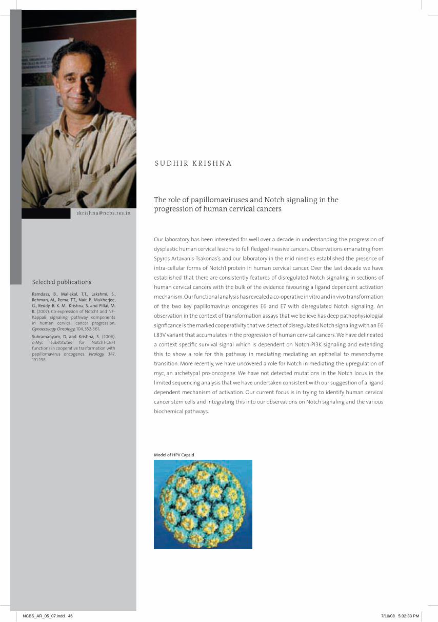
Selected publications
The role of papillomaviruses and Notch signaling in the progression of human cervical cancers
S U D H I R K R I S H N A
Ramdass, B., Maliekal, T,T., Lakshmi, S., Rehman, M., Rema, T.T., Nair, P., Mukherjee, G., Reddy, B. K. M., Krishna, S. and Pillai, M. R. (2007). Co-expression of Notch1 and NF-KappaB signaling pathway components in human cervical cancer progression.Gynaecology Oncology,104, 352-361.
Subramanyam, D. and Krishna, S. (2006). c-myc substitutes for Notch1-CBF1 functions in cooperative trasformation with papillomavirus oncogenes. Virology, 347, 191-198.
[email protected] . in
our laboratory has been interested for well over a decade in understanding the progression of
dysplastic human cervical lesions to full fledged invasive cancers. observations emanating from
Spyros Artavanis-Tsakonas’s and our laboratory in the mid nineties established the presence of
intra-cellular forms of Notch1 protein in human cervical cancer. over the last decade we have
established that there are consistently features of disregulated Notch signaling in sections of
human cervical cancers with the bulk of the evidence favouring a ligand dependent activation
mechanism. our functional analysis has revealed a co-operative in vitro and in vivo transformation
of the two key papillomavirus oncogenes E6 and E7 with disregulated Notch signaling. An
observation in the context of transformation assays that we believe has deep pathophysiologial
signficance is the marked cooperativity that we detect of disregulated Notch signaling with an E6
L83V variant that accumulates in the progression of human cervical cancers. We have delineated
a context specific survival signal which is dependent on Notch-PI3K signaling and extending
this to show a role for this pathway in mediating mediating an epithelial to mesenchyme
transition. more recently, we have uncovered a role for Notch in mediating the upregulation of
myc, an archetypal pro-oncogene. We have not detected mutations in the Notch locus in the
limited sequencing analysis that we have undertaken consistent with our suggestion of a ligand
dependent mechanism of activation. our current focus is in trying to identify human cervical
cancer stem cells and integrating this into our observations on Notch signaling and the various
biochemical pathways.
Modelofhpvcapsid
NCBS_AR_05_07.indd 46 7/10/08 5:32:33 PM

47
1notchactivationupregulatesMyc
Deepa Subramanyam
Using the transformation system devised by A. rangarajan to analyse the cooperative transformation
of the two key papillomavirus oncogenes E6 and E7 from the high risk virus type 16 along with activated
alleles of Notch1, we analysed what effector mechanisms might operate downstream of Notch
signaling. We first noted that c-myc can cooperate with E6 and E7 analogous to activated Notch1.
Intriguingly, in the context of Notch1 mediated transformation, using dominant negative blockers of
c-myc function, we were able to retard the transforming process. This led us to evaluate as to whether
activated Notch1 was able to upregulate myc levels. We have previously shown that Notch activation
of the PI3K-Akt pathway relies on a CBF1 independent process, the core effector mechanism in this
signaling pathway. Here, we noted the existence of CBF1 binding sites in the upstream regulatory
region of the c-myc gene and were able to show that expression of activated Notch1 upregulates
c-myc in a manner dependent on CBF1. Cumulatively, activated Notch1 seems to phenocopy ras and
utilize c-myc in a CBF1 independent and dependent manner respectively to mediate transformation.
2signalintegrationofthenotchpathwayinhumancervicalcancer
Deepa Subramanyam, Eric Vivien, Sweta Srivastava and Mousita Kamarkar
“Context” is of enormous importance in defining the events that occur during the process of
oncogenesis. However, both the mechanisms and the processes that should be used to study this
phenomenon are presently poorly understood. one of the potential intersection points of extra-
cellular regulation of Notch function is through mediating the stability. The Notch protein is targeted
for degradation through many still poorly understood mechanisms and one of the putative processes
is through the SCF adaptor protein Fbw7. We have been following up an interesting observation where
we noted that the inhibition of Notch mediated cooperative transformation in “in vitro” assays with
Fbw7 can be reversed by growth factors. We are currently in the process of establishing the molecular
mechanisms that mediate this process through a combination of functional studies, extensive
mutagenesis of the Notch allele and mass spec based techniques.
3cellularinvasioninhumancervicalcancer
Bharathi Ramdass, Tessy Maliekal and Pradip Nair
This work stemmed from a multi-centric collaborative programme grant that was funded by the
Department of Biotechnology. Features of disregulated Notch and NF- kB signaling have been noted in
independent studies. Here we set out to determine on a large scale the veracity of these observations
SU
DH
Ir K
rIS
HN
A
NCBS_AR_05_07.indd 47 7/10/08 5:32:34 PM

and also determined the possible cross-talk between these two pathways. We established in the
large study that there were parallel features of both Notch and NF-k activation. Using a combination
of in viro analysis, we are able to Notch activation functions as an upstream regulator of NF-k in
this context. In addition, our data uncovers some interesting observation about the relationship of
Notch signaling to cell cycle regulators and the p53 pathway extending some of our earlier insights
generated by P. Nair and colleagues.
Collaborators: V. Giri, Kidwai Memorial Institute of Oncology and R. Pillai, Rajiv Gandhi Centre for
Biotechnology
4howdohumancervicalcancersinvade?
Sweta Srivastava
rhoGTPases such as rhoC has been linked to invasive tumor progression. However, the upstream
regulators of rhoC are poorly understood. Here, we investigate the role of rhoC in the context of
invasive human cervical cancers. We initially demonstrate that increasing rhoC activity increases
motility, invasion and in vivo tumor formation in xenografts of cervical carcinoma cell lines. We
then examined the relationship to the Notch-PI3K axis; a pathway that we have suggested earlier
is involved in driving the invasive phase of human cervical cancers. We show that inhibition of PI3K,
Notch or rho GTPases blocks the induction of EmT, a phenomenon that is central to invasive tumor
progression. Further, inhibition of either PI3K- Akt axis or Notch activity reduces the activity of rhoC.
In addition to extending the role of rhoC to invasive cervical cancers, these results position rhoC
downstream of Notch-PI3K signaling in the context of these cancers.
5huntingforhumancervicalcancerstemcells
Tessy Maliekal, Jeevisha Bajaj and Eric Vivien
We are in the process of characterizing human cervical tumours for their resident stem cells and
integrating that information into our models of Notch signaling.
NCBS_AR_05_07.indd 48 7/10/08 5:32:36 PM
49
SA
TY
AJI
T
mA
Yo
r
NCBS_AR_05_07.indd 49 7/10/08 5:32:38 PM

Selected publications
Cell biology of the synapse
K . S . K R I S H N A N
Hanumae Gowd, K., Krishnan, K.S. and Balaram, P. (2007). Identification of conus amadis disulfide isomerase: minimum sequence length of peptide fragment necessary for protein annotation. Molecular BioSystems 3, 554-566.
Rikhy, R., Kamat, S., Ramagiri, S., Sriram, V. and Krishnan, K.S. (2007). mutations in dynamin-related protein result in gross changes in mitochondrial morphology and affect synaptic vesicle recycling at the Drosophila neuromuscular junction. Genes Brain Behaviour 6, 42-53.
Understanding the cellular basis of animal behavior is broadly the research aim of my group.
The brain is essentially responsible for behavior and is composed of a large number of neurones
and glia, over 100 billion of them in humans and a million in Drosophila. Synapses are specialized
junctions through which the cells of the nervous system signal to each other and to muscles
or glands and control other systems of the body. Synapses allow the neurons of the central
nervous system to form interconnected circuits that underlie perception,emotions and thought.
most of the syanpses in the central nervous system are chemical synapses. At these synapses
neurotransmitter substances are released into the synaptic cleft by exocytic fusion of the
membrane bound vesicles and subsequently the vesicular machinery is recycled and repacked
with neurotransmitter. The cell biology of the vesicle cycle and its molecular underpinnings are
not only conserved in different organisms but in different cellular contexts. We use genetic, cell
biological and molecular approaches in Drosophila to delineate mechanisms of vesicle cycling
and trafficking. Acute temperature sensitive paralytic mutants have been of particular value
in this approach. Endosomal trafficking studies in primary cell cultures derived from animals
carrying conditional mutations has turned out to be insightful for understanding principles
of membrane trafficking in general as well. Little is known about the mechanisms of general
anaesthesia. We have been able to input a promising new approach to this area in the form of
mutations that render Drosophila resistant to the effects of inhalational anaesthetics.
We have initiated a major project to identify and characterise toxic components of venoms of a
large number of carnivorous marine snails of the genus Conidae. We hope to exploit this unique
resource to mine for prospectve neuroactive compounds and therapeutuc agents.
[email protected] . in
Selected publications
NCBS_AR_05_07.indd 50 7/10/08 5:32:38 PM

51
K S
Kr
iSh
na
n
1 Genetics and dell biology of the synapse
Riddhi Majumder, Anil Limaye, Debjani Bhar
Our recent work in this area is aimed at elucidating the role of Stoned protein in dynamin dependent
endocytic process. at the whole animal level, our data indicates that Stoned enhances the eye-
scar phenotype of shibire flies and this results from the inhibition of dynamin-dependent trans-
endocytosis of signaling ligands in the eye-disc. Our observations in larval hemocytes suggest that
Stoned and Eps15 regulate clathrin mediated dynamin-dependent endocytotic process, specifically by
catalyzing the recruitment of clathrin to pits at early stages of membrane retrieval. in order to further
understand the regulation of receptor mediated endocytosis by tyrosine kinase, the role of Cdk5/p35
was studied.
homozygous combinations were made with shibire alleles namely ts1, ts2, and ck2. initial experiments
to check paralysis profiles suggest that the p35 null causes enhancement of shibire paralysis,
implicating a role for Cdk5 mediated phophorylation in synaptic vesicle recycling. Surface accessibility
assays in isolated hemocytes were done using p35 mutant and transgenes of Cdk5 driven by collagen-
Gal4. The clearance of surface receptors is adversely affected in p35 mutant background. We also
observe that p35 mutants affect the temperature dependent paralysis phenotype of shibire flies. at
the cellular level, abrogation of p35 or Cdk5 genetically or by pharmacological reagents adversely
affected receptor internalization in hemocytes. Cultured embryonic neurons from mutant and wild
type Drosophila are being analyzed as a prospective system for study of vesicle recycling.
Collaborator: S. Mayor, NCBS
2 Anaesthetic response in Drosophila
Ludwin Pinto
Several genetic loci that seems to be involved in the response of Drosophila to general anesthetics
have been identified. These include GaBa receptor subtypes, channel proteins and several molecules
crucial for maintaining membrane potential and synaptic function. The genetic interaction amongst
these many loci is presently being assessed.
3 Peptides of therapeutic value from marine cone snails
Hanumae Gowd, Benjamin Franklin and Santhana Ramasamy
a previously unexplored source of drugs, the Conus species of marine snails with a repertoire of
toxic peptides (referred to as conotoxins/conopeptides), are now being looked at as a mine of
Figure 1. Scanning electron micrographs of shibire (b) and shibire stoned double (c) showing eye scar compared to wild type(a). Shibire(e) and double mutant (f)show perturbation of Boss ligand uptake in r7 cells of imaginal disc suggesting a defect in endocytosis. at cellular level in larval hemocytes in mutant combinations (h,i) receptor endocytosis(mBSa) is perturbed compared to control(g).
NCBS_AR_05_07.indd 51 7/10/08 5:33:47 PM
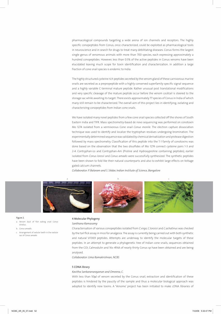
pharmacological compounds targeting a wide arena of ion channels and receptors. The highly
specific conopeptides from Conus, once characterized, could be exploited as pharmacological tools
in neuroscience and in search for drugs to treat many debilitating diseases. Conus forms the largest
single genus of venomous animals with more than 700 species, each expressing approximately a
hundred conopeptides. however, less than 0.5% of the active peptides in Conus venoms have been
elucidated leaving much scope for toxin identification and characterization. in addition a large
fraction of cone snail species is endemic to india.
The highly structured cysteine rich peptides secreted by the venom gland of these carnivorous marine
snails are secreted as a prepropeptide with a highly conserved superfamily-specific signal sequence
and a highly variable C-terminal mature peptide. rather unusual post translational modifications
and very specific cleavage of the mature peptide occur before the venom cocktail is steered to the
storage sac while awaiting its target. There exists approximately 77 species of Conus in india of which
many still remain to be characterized. The overall aim of this project lies in identifying, isolating and
characterizing conopeptides from indian cone snails.
We have isolated many novel peptides from a few cone snail species collected off the shores of South
Eastern india and TiFr. Mass spectrometry-based de novo sequencing was performed on conotoxin
Mo 1274 isolated from a vermivorous Cone snail Conus monile. The electron capture dissociation
technique was used to identify and localize the tryptophan residues undergoing bromination. The
experimentally determined sequence was validated by chemical derivatization and protease digestion
followed by mass spectrometry. Classification of this peptide into the T-1 family of conotoxins was
done based on the observation that the two disulfides of Mo 1274 connect cysteine pairs 1-3 and
2-4. Contryphan-Lo and Contryphan-am (Proline and hydroxyproline containing peptides), earlier
isolated from Conus lorosii and Conus amadis were successfully synthesized. The synthetic peptides
have been shown to fold like their natural counterparts and also to exhibit large effects on Voltage
gated calcium channels.
Collaborator: P. Balaram and S. Sikdar, Indian Institute of Science, Bangalore
4 Molecular Phylogeny
Santhana Ramasamy
Characterisation of various conopeptides isolated from C.virgo, C.loroisii and C.achatinus was checked
by the tail flick assay in mice for analgesia. The assay is currently being carried out with both synthetic
and natural Vi1369 peptides. attempts are underway to identify the molecular targets of these
peptides. in an attempt to generate a phylogenetic tree of indian cone snails, sequences obtained
from the COi, Calmodulin and 16s rrna of nearly thirty Conus sp have been obtained and are being
analysed.
Collaborator: Uma Ramakrishnan, NCBS
5 CDNA library
Kavitha Sankaranarayanan and Omeena, C.
With less than 50µl of venom secreted by the Conus snail, extraction and identification of these
peptides is hindered by the paucity of the sample and thus a molecular biological approach was
adopted to identify new toxins. a ‘Venome’ project has been initiated to make cDna libraries of
Figure 2.
a. Venom duct of fish eating snail Conus striatus.
b. Conus amadis.
c. arrangement of radular teeth in the radular sac of Conus amadis
a b c
NCBS_AR_05_07.indd 52 7/10/08 5:33:47 PM
53
K S
Kr
iSh
na
n
cone venoms from indian Conus species. The procedure involved cDna preparation and subsequent
amplification using superfamily specific signal sequence primers. Transcriptome studies on
C.achatinus, C.virgo and C.monile have been performed and ~10 such Conotoxin sequences of the M
and O superfamily have been obtained so far. Electrophysiological characterization of the natural as
well as synthetic conopeptides by looking for an effect on in-vivo (expressed in Xenopus laevis eggs)
ion channels/receptors, or on invertebrate neurons (Drosophila) is in progress.
Collaborators: M.K.Mathew, NCBS, P. Balaram and Kalyan Dewan, Indian Institute of Science, Bangalore
6 Biodiversity
Benjamin Franklin
Taxonomy, species composition, geographical range, distribution, habitat and habits of Conidae
off Tamil nadu coast (india) have been studied. This resulted in the identification of 4 new species,
thereby increasing the number of Conus species of india from 77 to 81. With a long term study of the
distribution of Conidae along the entire indian coast, with its diverse habitats, these additional new
species could be identified thereby paving way to the isolation of even more novel conopeptides. a
morphology and morphometric analysis of radular teeth from 22 species of Conus snails (collected
from the East and West coast of india) resulted in the development of a simplified species identification
method based on tooth morphology. Partial characterization of Conus araneosus venom was done
using rP- hPLC, ESi-MS and MaLDi-TOF-TOF. Biological assays were carried out for 5 peptides from
C.araneosus to study analgesic and aggression scores in mouse and rats. a sleep inducing peptide has
been identified from the venom of C.araneosus.
Currently we have an inventory of nearly fifty toxin peptides at various stages of identification,
characterization and chemical synthesis.
Collaborators: Anthony Fernando and Olivia Fernando, Annamalai University and P. Balram, Indian
Institute of Science, Bangalore
NCBS_AR_05_07.indd 53 7/10/08 5:33:47 PM

Selected publications
The broad aim of my laboratory is to provide an understanding of the molecular mechanisms of endocytosis in metazoan cells. in this regard we have undertaken to develop a framework to understand principles of membrane organization and deformation in living cells. For scientists interested in studying phenomena at the cellular scale, principles from the physical and chemical sciences provide a powerful paradigm in framing questions about the mechanisms of movement of molecules and organelles inside cells. Consequently, we have taken a multi-disciplinary approach, combining biology with theoretical physics and synthetic chemistry, to address the main theme of our research at nCBS.
We have developed new microscopy techniques to study nanometer scale organization of cellular components. Using fluorescence microscopy we have explored sorting properties and endocytic pathways of a variety of molecules, including membrane proteins, lipids and lipid-tethered proteins, both in living cells in tissue culture and in vivo. Our method of choice is to study the organization and dynamics of fluorescently-tagged molecules at different scales in living cells; from the nanometer scale in specialized domains in cell membranes to the micron scale prevalent in mapping endocytic pathways or in specialized regions of the cell surface.
We are now involved in several lines of inquiry, some continuing and others more recently configured. These include: i) continuing studies on understanding structure and function of membrane rafts, in collaboration with Prof. Madan rao at raman research institute, Bangalore; ii) continuing studies on understanding the mechanism of dynamin-independent endocytosis using specific targeted molecular perturbations coupled with high resolution microscopy and single-molecule biochemistry; iii) genome-wide rnai-based screening of genes involved in dynamin-dependent and independent forms of endocytosis in Drosophila cells; iv) functional genomics of different endocytic pathways in the context of their role in a metazoan system capable of genetic manipulation, in collaboration with Prof. KS Krishnan, TiFr, Mumbai; v) exploring pathways of antigen presentation for loading onto MhC class ii, and endocytic mechanisms that modulate the presence of long-term cell surface resident proteins.
The trajectory of this work has led us to explore the fine structure of the plasma membrane, providing for the first time an in vivo picture of lipidic assemblies challenging existing notions of membrane rafts, an understanding of the cellular basis for the establishment of developmental gradients, and novel pathways for antigen presentation, long-term cell surface resident protein modulation.
Mechanisms of endocytosis in metazoan cells
[email protected] . in
S A T Y A J I T M AY O R
Scales of organization in metazoans, integrating biology with physics and chemistry. Starting at the scale of molecules, molecular players interact to generate larger scale structures in the context of cells (e. g. membrane rafts). These in turn participate to create processes at the cellular scale (e. g. pathways of endocytosis). Several endocytic pathways function to regulate phenomenon at the next level of organization (e. g. information transfer at neuronal synapses, morphogen gradients to specify tissue patterns or control cell signalling outputs in the larger multicellular organism). Focussing on the mechanism of a basic cellular process, namely endocytosis, studies in my laboratory using functional genomics are aimed at integrating different scales of organization in biology.
Mayor, S. and Pagano, R.E. (2007). Pathways of clathrin-independent endocytosis. Nature Reviews Molecular Cell Biology, 8, 603-612.
Chaudhry, A., Das, S.R., Jameel, S., George, A., Bal, V., Mayor, S. and Rath, S. (2007). hiV-1 nef protein triggers endocytosis of immune costimulatory proteins CD80 and CD86. Cell Host and Microbe, 1, 37-49.
Altman, D., Goswami, D., Hasson, T., Spudich, J.A. and Mayor, S. (2007). Precise positioning of myosin Vi on endocytic vesicles in vivo. PLoS Biology, 5, e210.
Sarasij, R.C., Mayor, S. and Rao, M. (2007). Chirality-induced budding: a raft-mediated mechanism for endocytosis and morphology of caveolae? Biophysical Journal, 92, 3140-3158.
NCBS_AR_05_07.indd 54 7/10/08 5:33:48 PM

55
1 Structure and function of membrane rafts
Sameera Bilgrami, Debanjan Goswami, Subhashri Ghosh and Riya Raghupathy
Our concept of the structure and organization of the membrane of cells has undergone major
revisions from the time of the fluid mosaic model proposed by Singer and nicolson. instead of
considering the membrane as a ‘two-dimensional oriented solution of integral proteins in a viscous
phospholipid bilayer’, both protein and lipid components of the bilayer are anisotropically arranged in
the lateral and vertical directions. in living cells, it has been difficult to visualize these heterogeneities.
Lateral heterogeneities variously called membrane rafts or domains, have been particularly difficult
to visualize, and the most compelling evidence for such structures in the undifferentiated plasma
membrane of cells is functional. With the help of theoretical (condensed matter) physicist Prof.
Madan rao, we are probing the architecture of these lateral heterogeneities and the basis for
their formation. These studies have required the development of new tools of biophysics to study
membrane heterogeneities in situ, and a new physico-chemical understanding of the properties of
membranes in living cells.
From an operational perspective we have focussed on understanding how an exoplasmic lipid-tethered
protein [Glycosylphosphatidylinositol (GPi)-anchored proteins] is distributed in cell membranes.
This has obvious implications for understanding the structure and function of lipidic assemblies in
membranes. Our work provides a fundamentally different picture of the structure and formation
of lipid rafts, summarized in the adjoining Figure 1. We believe that these approaches will provide
an understanding of how local membrane composition and physical properties of membranes are
maintained and regulated in living cells.
Collaborator: Madan Rao, Raman Research Institute, Bangalore
2 Mechanism of endocytosis of GPI-anchored proteins via a clathrin- and dynamin-independent,
Cdc42-regulated endocytic pathway
Rahul Chadda and Sudha Kumari
GPi-anchored proteins are a large class of proteins of extreme diversity in structure and function;
10% of all membrane proteins are tethered to the membrane via a GPi-anchor. Several receptors and
pathogenic molecules depend on the presence of the GPi-anchor for their function. For example, the
main mammalian folate receptor requires the GPi-anchor for efficient delivery of folate into cells.
Understanding the nature of sorting of GPi- anchored proteins thus, has implications for physiology
and pathology of a large fraction of membrane proteins.
We had discovered that GPi-anchored proteins are sorted at the cell surface of mammalian cells into a
Cdc42-regulated, clathrin and dynamin-independent endocytic pathway that is also a major pathway
for the internalization of the fluid-phase into most cells (Figure 2). We have termed this pathway,
the GPi-anchored protein enriched early endosomal (GEEC) pathway. Changing specific lipid levels in
cells affects the sorting of the GPi-anchored proteins in endosomes, and at the cell surface, altering
the efficiency of endocytic uptake. These data and others indicate that GPi-anchored proteins may
exist as pre-sorted microdomains or rafts in membranes of endosomes which in turn are responsible
Figure 1. Actively generated spatial and temporal organization of raft components: Data from our laboratory regarding physical characteristics of nanoscale clusters of GPi-anchored proteins in living cells show that these structures are small, dynamic, and co-exist with monomers; they are also distributed in a non-random fashion on the surface of cells. These structures may be actively induced to form large-scale stable ‘rafts’. These studies provide a different picture of membrane rafts where GPi-anchored proteins form nano-scale lipid assemblies in specific regions of the plasma membrane which in turn may determine local lipid composition. Black circles, GPi-anchored proteins; red and pink circles, non-raft associated lipids; yellow circles, raft-associated lipids; green, cholesterol. Scale bar ~ 5 nm.
Sa
TY
aJi
T M
aY
Or
NCBS_AR_05_07.indd 55 7/10/08 5:33:48 PM
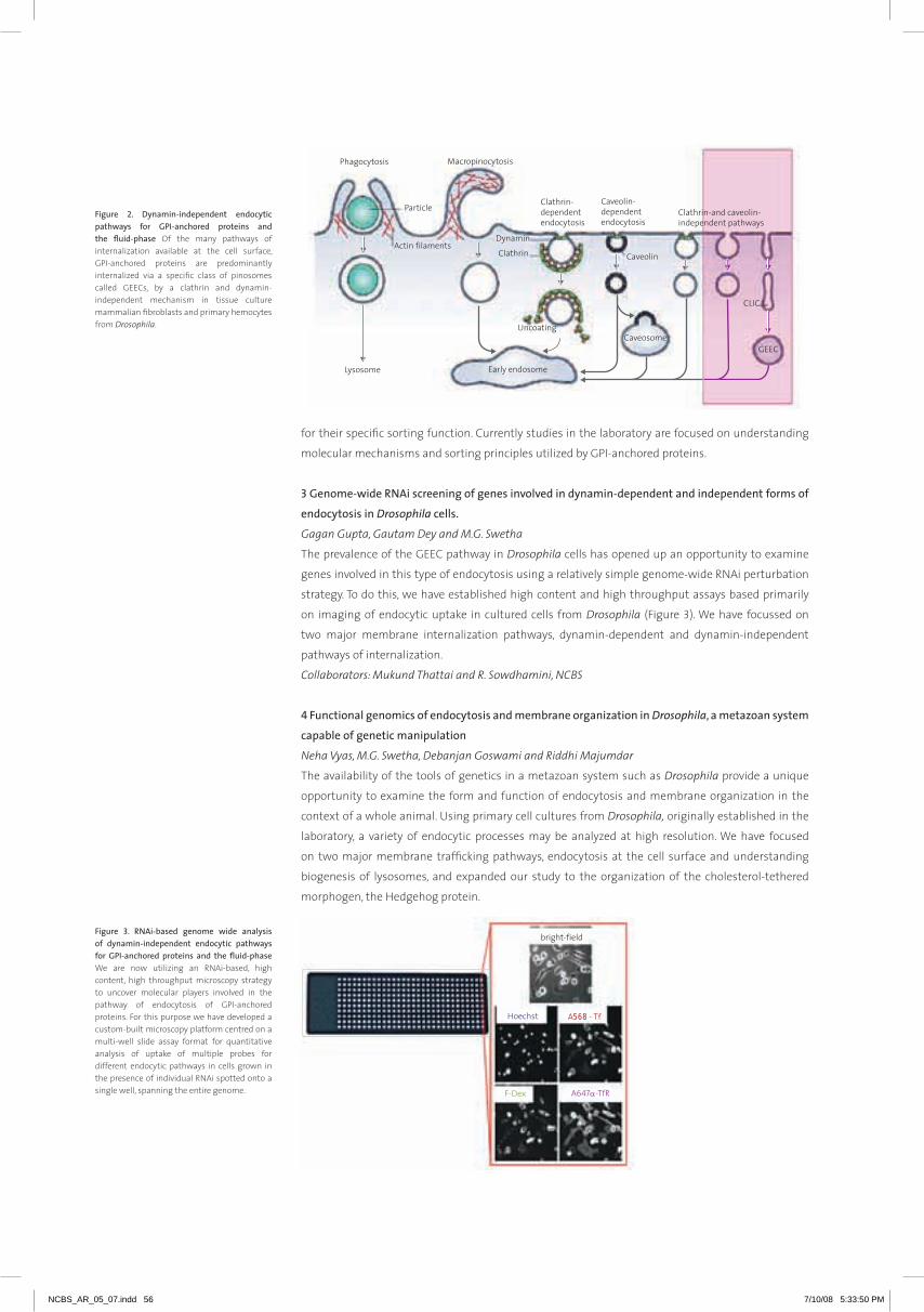
Figure 3. RNAi-based genome wide analysis of dynamin-independent endocytic pathways for GPI-anchored proteins and the fluid-phase We are now utilizing an rnai-based, high content, high throughput microscopy strategy to uncover molecular players involved in the pathway of endocytosis of GPi-anchored proteins. For this purpose we have developed a custom-built microscopy platform centred on a multi-well slide assay format for quantitative analysis of uptake of multiple probes for different endocytic pathways in cells grown in the presence of individual rnai spotted onto a single well, spanning the entire genome.
568
Figure 2. Dynamin-independent endocytic pathways for GPI-anchored proteins and the fluid-phase Of the many pathways of internalization available at the cell surface, GPi-anchored proteins are predominantly internalized via a specific class of pinosomes called GEECs, by a clathrin and dynamin-independent mechanism in tissue culture mammalian fibroblasts and primary hemocytes from Drosophila.
for their specific sorting function. Currently studies in the laboratory are focused on understanding
molecular mechanisms and sorting principles utilized by GPi-anchored proteins.
3 Genome-wide RNAi screening of genes involved in dynamin-dependent and independent forms of
endocytosis in Drosophila cells.
Gagan Gupta, Gautam Dey and M.G. Swetha
The prevalence of the GEEC pathway in Drosophila cells has opened up an opportunity to examine
genes involved in this type of endocytosis using a relatively simple genome-wide rnai perturbation
strategy. To do this, we have established high content and high throughput assays based primarily
on imaging of endocytic uptake in cultured cells from Drosophila (Figure 3). We have focussed on
two major membrane internalization pathways, dynamin-dependent and dynamin-independent
pathways of internalization.
Collaborators: Mukund Thattai and R. Sowdhamini, NCBS
4 Functional genomics of endocytosis and membrane organization in Drosophila, a metazoan system
capable of genetic manipulation
Neha Vyas, M.G. Swetha, Debanjan Goswami and Riddhi Majumdar
The availability of the tools of genetics in a metazoan system such as Drosophila provide a unique
opportunity to examine the form and function of endocytosis and membrane organization in the
context of a whole animal. Using primary cell cultures from Drosophila, originally established in the
laboratory, a variety of endocytic processes may be analyzed at high resolution. We have focused
on two major membrane trafficking pathways, endocytosis at the cell surface and understanding
biogenesis of lysosomes, and expanded our study to the organization of the cholesterol-tethered
morphogen, the hedgehog protein.
NCBS_AR_05_07.indd 56 7/10/08 5:33:50 PM
57
Our aim is, firstly, to relate alteration in cellular trafficking pathways to mutations in specific genes in
the hope of uncovering molecular machinery behind these basic cellular processes, and secondly, to
relate alterations in trafficking processes to perturbations in integrative processes in the whole animal
such as those involved in establishing developmental gradients and control of intracellular signaling.
at the level of membrane protein organization and its functional implications we have studied
the organization of the hedgehog (hh) morphogen. These studies have suggested a hierarchical
organization of a membrane protein, from the nano to the micron-scale. This organization contributes
to distinct functionality of a diffusible morphogen. We have found that hh forms nanoscale clusters,
determined by specific residues in its protein domain, and these clusters in turn organize to larger
scale structures that participate in transporting the hh protein over many cell diameters.
Collaborator: K.S. Krishnan, NCBS
5 Understanding pathways of antigen presentation for loading onto MHC class II and endocytic
mechanisms that modulate the cell surface presence of long-term resident surface proteins
Sudha Kumari
antigen presentation is an essential function of the immune system in its ability to deal with infections.
in this regard the presentation of soluble antigen to cells of the immune system has a central role.
We have been studying processes of antigen presentation which utilize cytosolic pathways of antigen
generation and MhC Class ii restricted presentation. We succeeded in identifying intracellular sites of
this pivotal event in antigen presentation, after providing the antigen to antigen presenting cells (aPC)
in different ways – as free peptides, or as a fusion constructs. This pathway is likely to be extremely
important in mechanisms of generating tolerance of CD4+ T-cells to self-antigens.
in addition to our ongoing studies on antigen presentation, we have used the cells of the immune
system to study how cell surface levels of long-term resident membrane proteins may be acutely
modulated. here we have focused on: a) the role of the hiV Nef protein on the immune co-stimulatory
molecules, CD8O and CD86; b), endogenous mechanisms by which the acetylcholine receptor is
modulated by autoimmune antibodies and ligands.
Collaborators: Satyajit Rath, National Institute of Immunology, New Delhi and Francisco Barrantes,
INIBIB, Bahia-Blanca, Argentina
Sa
TY
aJi
T M
aY
Or
NCBS_AR_05_07.indd 57 7/10/08 5:33:50 PM

Selected publications
[email protected] . in
The regulation of cell number is critical for the normal development of multicellular organisms.
Molecular mechanisms regulating cellular death processes are largely evolutionarily conserved
in metazoans. as in several other systems, the regulated depletion of cells is critical for the
maintenance of immune system homeostasis. in the mammalian immune system, the bulk of
circulating T-cells activated as a consequence of encounter with antigen are fated to die [via
a programmed pathway of death or apoptosis] with only a small subset of antigen-reactive
cells set aside to generate immune memory. Thus, expansion and reduction in cell number are
recurring events in the immune system. Several aspects of this process can be recapitulated in
vitro and offer an attractive model system to investigate the molecular underpinnings of cellular
homeostatic mechanisms. activated T-cell apoptosis is regulated by the Bcl-2 family, comprising
both pro- and anti-apoptotic proteins. reactive oxygen species are also key intermediates of
T-cell death. Specifically, we are probing signaling events that regulate pro-death cascades; the
interactions of the latter with cellular survival machinery and the consequences of this for T-cell
function. Some of our recent experiments are described in the sections that follow.
Mechanisms of apoptosis
A P U R VA S A R I N
Srivastava, S., Banerjee, H., Chaudhry, A., Khare, A., Sarin, A., George, A., Bal, V., Durdik, J.M. and Rath, S. (2007). apoptosis-inducing factor (aif) regulates death in peripheral T cells. Journal of Immunology, 179, 797-803.
Goyal, G., Fell, B., Sarin, A., Youle, R. J. and Sriram, V. (2007). role of mitochondrial remodeling in programmed cell death in Drosophila melanogaster. Developmental Cell, 12, 807-816.
Parikh, N., Koshy, C., Dhayabharan, V., Perumalsamy, L.R., Sowdhamini, R. and Sarin, A. (2007). The n-terminus and alpha-5, alpha-6 helices of the pro-apoptotic protein Bax, modulate functional interactions with the anti-apoptotic protein Bcl-xL. BMC Cell Biology, 8,16-
Geetha, B., Divya, P., Sade, H., Gunasekharan, V., Rangarajan, A. and Sarin, A. (2006). Evidence for a role for notch signaling in the cytokine-dependent survival of activated T-cells. Journal of Immunology, 177, 5041-5050.
Selected publications
Peripheral T-cell cycle
NCBS_AR_05_07.indd 58 7/10/08 5:33:51 PM
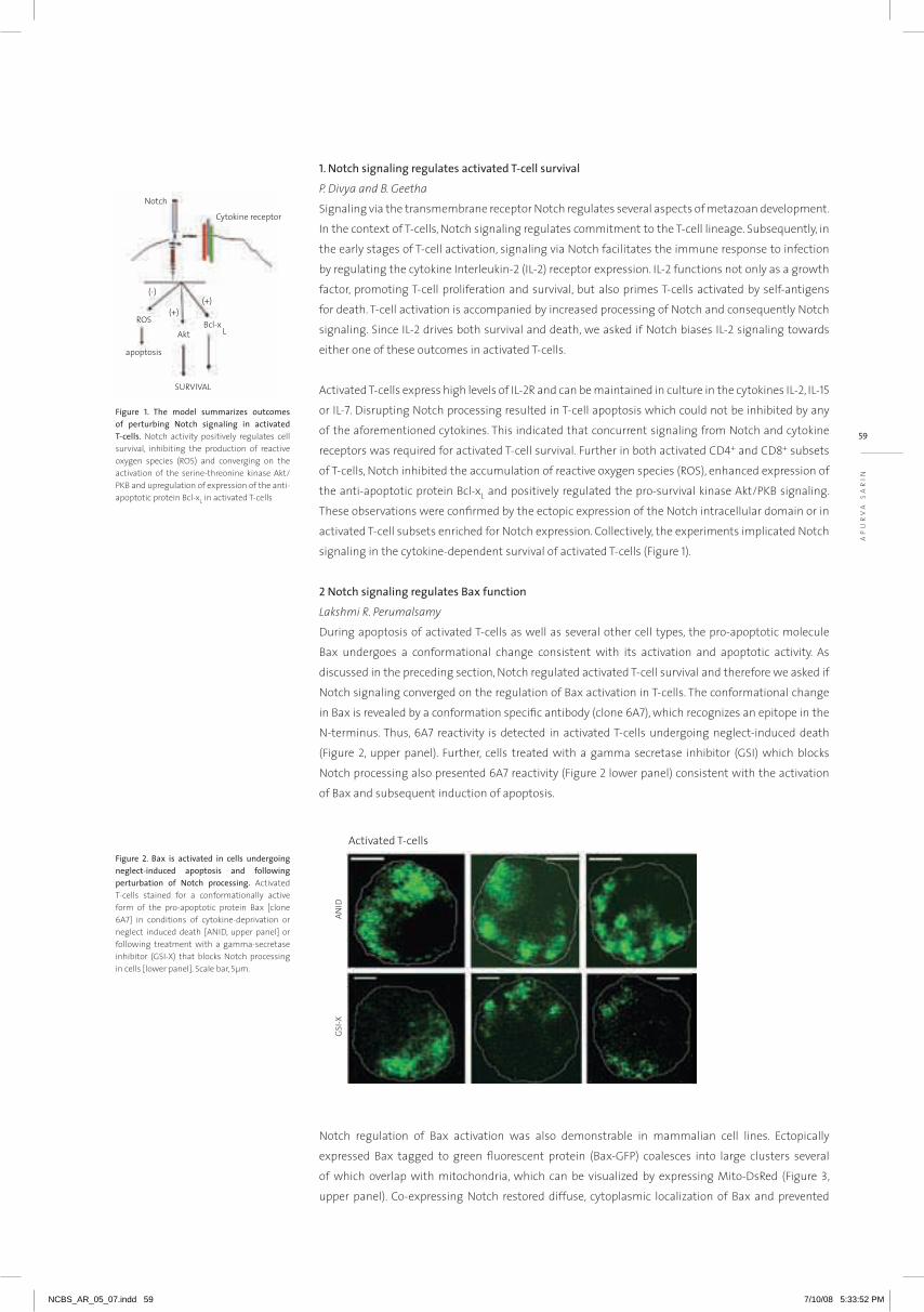
59
aP
Ur
Va
Sa
rin
1. Notch signaling regulates activated T-cell survival
P. Divya and B. Geetha
Signaling via the transmembrane receptor notch regulates several aspects of metazoan development.
in the context of T-cells, notch signaling regulates commitment to the T-cell lineage. Subsequently, in
the early stages of T-cell activation, signaling via notch facilitates the immune response to infection
by regulating the cytokine interleukin-2 (iL-2) receptor expression. iL-2 functions not only as a growth
factor, promoting T-cell proliferation and survival, but also primes T-cells activated by self-antigens
for death. T-cell activation is accompanied by increased processing of notch and consequently notch
signaling. Since iL-2 drives both survival and death, we asked if notch biases iL-2 signaling towards
either one of these outcomes in activated T-cells.
activated T-cells express high levels of iL-2r and can be maintained in culture in the cytokines iL-2, iL-15
or iL-7. Disrupting notch processing resulted in T-cell apoptosis which could not be inhibited by any
of the aforementioned cytokines. This indicated that concurrent signaling from notch and cytokine
receptors was required for activated T-cell survival. Further in both activated CD4+ and CD8+ subsets
of T-cells, notch inhibited the accumulation of reactive oxygen species (rOS), enhanced expression of
the anti-apoptotic protein Bcl-xL and positively regulated the pro-survival kinase akt/PKB signaling.
These observations were confirmed by the ectopic expression of the notch intracellular domain or in
activated T-cell subsets enriched for notch expression. Collectively, the experiments implicated notch
signaling in the cytokine-dependent survival of activated T-cells (Figure 1).
2 Notch signaling regulates Bax function
Lakshmi R. Perumalsamy
During apoptosis of activated T-cells as well as several other cell types, the pro-apoptotic molecule
Bax undergoes a conformational change consistent with its activation and apoptotic activity. as
discussed in the preceding section, notch regulated activated T-cell survival and therefore we asked if
notch signaling converged on the regulation of Bax activation in T-cells. The conformational change
in Bax is revealed by a conformation specific antibody (clone 6a7), which recognizes an epitope in the
n-terminus. Thus, 6a7 reactivity is detected in activated T-cells undergoing neglect-induced death
(Figure 2, upper panel). Further, cells treated with a gamma secretase inhibitor (GSi) which blocks
notch processing also presented 6a7 reactivity (Figure 2 lower panel) consistent with the activation
of Bax and subsequent induction of apoptosis.
notch regulation of Bax activation was also demonstrable in mammalian cell lines. Ectopically
expressed Bax tagged to green fluorescent protein (Bax-GFP) coalesces into large clusters several
of which overlap with mitochondria, which can be visualized by expressing Mito-Dsred (Figure 3,
upper panel). Co-expressing notch restored diffuse, cytoplasmic localization of Bax and prevented
Figure 1. The model summarizes outcomes of perturbing Notch signaling in activated T-cells. notch activity positively regulates cell survival, inhibiting the production of reactive oxygen species (rOS) and converging on the activation of the serine-threonine kinase akt/PKB and upregulation of expression of the anti-apoptotic protein Bcl-xL in activated T-cells
Figure 2. Bax is activated in cells undergoing neglect-induced apoptosis and following perturbation of Notch processing. activated T-cells stained for a conformationally active form of the pro-apoptotic protein Bax [clone 6a7] in conditions of cytokine-deprivation or neglect induced death [aniD, upper panel] or following treatment with a gamma-secretase inhibitor (GSi-X) that blocks notch processing in cells [lower panel]. Scale bar, 5µm.
NCBS_AR_05_07.indd 59 7/10/08 5:33:52 PM

its accumulation at mitochondria (Figure 3, lower panel). Similarly inhibition of Bax activation and
apoptosis was also observed in cells expressing a processed (activated) form of the notch intracellular
domain. Taken together, these and experiments demonstrated that notch signaling inhibited Bax
activation and oligomerization, thereby regulating a key antagonist of activated T-cell survival.
3 The serine-threonine kinase Akt and Bcl-xL regulate Bax function by distinct mecahnisms
Neha Parikh, Lakshmi R. Perumalsamy and D. Vaigundan
Bax function is negatively regulated by mechanisms that promote its retention in the cytosol
and by anti-apoptotic molecules such as Bcl-2 and Bcl-xL that antagonize Bax function at the
mitochondrion. The Bax n-terminus had been implicated in its localization to mitochondria and
the triggering of cytotoxicity. While, the Bh3 domain regulates interactions within the family, it
is becoming increasingly apparent that regions other than the Bh3 contribute to Bax function. in
recent experiments we showed that the Bh3 and TM1 domains in Bax are adequate for association
with Bcl-xL and localization to mitochondria respectively, but an intact n-terminus played a non-
redundant role in the regulation of Bax cytotoxicity by Bcl-xL. Earlier work from our laboratory had
shown that the kinases akt and MEK target a region in the Bax n-terminus to ensure its cytoplasmic
sequestration in T-cells. Thus, we asked if Bcl-xL and akt regulate Bax via similar mechanisms as Bax-
induced apoptosis is inhibited by the serine-threonine kinase akt. a site-directed substitution mutant
of Bax with alanine substitutions of the Ser/Thr residues (T14, S15, S16) in the n-terminus (Bax-ala3)
was refractory to inhibition by constitutively active Ca-akt (Figure 4a). interestingly, the modification
in the n-terminus did not interfere with Bcl-xL inhibition of Bax-ala3 induced apoptosis (Figure 4b)
giving the first indications that the two anti-apoptotic pathways target Bax function via distinct
mechanisms. Furthermore, as shown in Figure 4c, Ca-akt did not change the cellular distribution of
Bax-ala3.
Figure 4. Akt/PKB and Bcl-xL regulate Bax function by distinct mechanisms. a & b, apoptosis triggered by expressing the recombinant proteins indicated in the panels. c, active-akt does not regulate the subcellular distribution of the Bax mutant, Bax-ala3. hEK cells expressing Bax-ala-3GFP and Dsred2-Mito in the absence [upper panel] or presence of akt [lower panel]. Scale Bar 10µm.
Figure 3. Ectopically expressed full-length Notch [Notch-FL] inhibits Bax function. recombinant Bax-GFP clusters around mitochondria [marked with the protein Dsred2-Mito] in apoptotic cells. When co-expressed with notch-FL, Bax-GFP is largely evenly distributed in the cytoplasm.
NCBS_AR_05_07.indd 60 7/10/08 5:33:53 PM
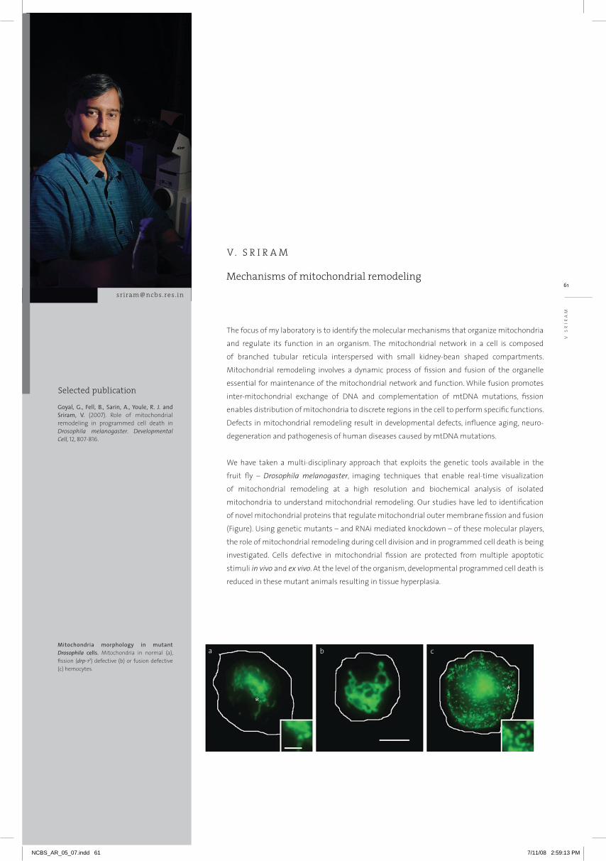
61
The focus of my laboratory is to identify the molecular mechanisms that organize mitochondria
and regulate its function in an organism. The mitochondrial network in a cell is composed
of branched tubular reticula interspersed with small kidney-bean shaped compartments.
Mitochondrial remodeling involves a dynamic process of fission and fusion of the organelle
essential for maintenance of the mitochondrial network and function. While fusion promotes
inter-mitochondrial exchange of DNA and complementation of mtDNA mutations, fission
enables distribution of mitochondria to discrete regions in the cell to perform specific functions.
Defects in mitochondrial remodeling result in developmental defects, influence aging, neuro-
degeneration and pathogenesis of human diseases caused by mtDNA mutations.
We have taken a multi-disciplinary approach that exploits the genetic tools available in the
fruit fly – Drosophila melanogaster, imaging techniques that enable real-time visualization
of mitochondrial remodeling at a high resolution and biochemical analysis of isolated
mitochondria to understand mitochondrial remodeling. Our studies have led to identification
of novel mitochondrial proteins that regulate mitochondrial outer membrane fission and fusion
(Figure). Using genetic mutants – and RNAi mediated knockdown – of these molecular players,
the role of mitochondrial remodeling during cell division and in programmed cell death is being
investigated. Cells defective in mitochondrial fission are protected from multiple apoptotic
stimuli in vivo and ex vivo. At the level of the organism, developmental programmed cell death is
reduced in these mutant animals resulting in tissue hyperplasia.
Mechanisms of mitochondrial remodeling
V . S R I R A M
[email protected] . in
Goyal, G., Fell, B., Sarin, A., Youle, R. J. and Sriram, V. (2007). Role of mitochondrial remodeling in programmed cell death in Drosophila melanogaster. Developmental Cell, 12, 807-816.
Mitochondria morphology in mutant Drosophila cells. Mitochondria in normal (a), fission (drp-12) defective (b) or fusion defective (c) hemocytes.
Selected publication
V S
RIR
AM
NCBS_AR_05_07.indd 61 7/11/08 2:59:13 PM
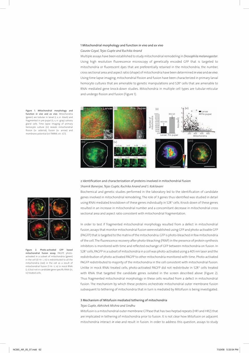
Figure 1. Mitochondrial morphology and function in vivo and ex vivo. Mitochondria (green) are tubular in larval (i, v, vi -black) and fragmented in pre-pupal (ii, v, vi -gray) salivary gland cells. Time lapse imaging of primary hemocyte culture (iii) reveals mitochondrial fission (iv- asterisk), fusion (iv- arrow) and membrane potential (vii-TMrM; viii- JC1).
1 Mitochondrial morphology and function in vivo and ex vivo
Gaurav Goyal, Tejas Gupte and Ruchika Anand
Multiple assays have been established to study mitochondrial remodeling in Drosophila melanogaster.
Using high resolution fluorescence microscopy of genetically encoded GFP that is targeted to
mitochondria or fluorescent dyes that are preferentially retained in the mitochondria, the number,
cross sectional area and aspect ratio (shape) of mitochondria have been determined in vivo and ex vivo.
Using time lapse imaging, mitochondrial fission and fusion have been characterized in primary larval
hemocyte cultures that are amenable to genetic manipulations and S2r+ cells that are amenable to
rnai mediated gene knock-down studies. Mitochondria in multiple cell types are tubular-reticular
and undergo fission and fusion (Figure 1).
2 Identification and characterization of proteins involved in mitochondrial fusion
Shamik Banerjee, Tejas Gupte, Ruchika Anand and S. Kokilavani
Biochemical and genetic studies performed in the laboratory led to the identification of candidate
genes involved in mitochondrial remodeling. The role of 3 genes thus identified was studied in detail
using rnai mediated knockdown of these genes individually in S2r+ cells. Knock-down of these genes
resulted in an increase in mitochondrial number and a concomitant decrease in mitochondrial cross
sectional area and aspect ratio consistent with mitochondrial fragmentation.
in order to test if fragmented mitochondrial morphology resulted from a defect in mitochondrial
fusion, assays that monitor mitochondrial fusion were established using GFP and photo-activable GFP
(PaGFP) that is targeted to the matrix of the mitochondria. GFP is photo-bleached in few mitochondria
of the cell. The fluorescence recovery after photo-bleaching (FraP) in the presence of protein synthesis
inhibitors is monitored with time and reflected exchange of GFP between mitochondria on fusion. in
S2r+ cells, PaGFP in a subset of mitochondria in a cell was photo-activated using a 405 nm laser and the
redistribution of photo-activated PaGFP to other mitochondria monitored with time. Photo-activated
PaGFP redistributed to majority of the mitochondria in the cell consistent with mitochondrial fusion.
Unlike in mock rnai treated cells, photo-activated PaGFP did not redistribute in S2r+ cells treated
with rnai that targeted the candidate genes isolated in the screen described above (Figure 2).
Thus fragmented mitochondrial morphology in these cells resulted from a defect in mitochondrial
fusion. The mechanism by which these proteins orchestrate mitochondrial outer membrane fusion
subsequent to tethering of mitochondria that in turn is mediated by Mitofusin is being investigated.
3 Mechanism of Mitofusin mediated tethering of mitochondria
Tejas Gupte, Abhishek Mishra and Sindhu
Mitofusin is a mitochondrial outer membrane GTPase that has two heptad repeats (hr1 and hr2) that
are implicated in tethering of mitochondria prior to fusion. it is not clear how Mitofusin on adjacent
mitochondria interact in vivo and result in fusion. in order to address this question, assays to study
Figure 2. Photo-activated GFP based mitochondrial fusion assay. PaGFP, photo-activated in a subset of mitochondria (green) in the cell (0 hr- i, iii) is redistributed to all the mitochondria (red) in the cell as a result of mitochondrial fusion (1 hr- ii, iv) in mock rnai (i, ii) but not in candidate gene specific rnai (iii, iv) treated cells.
m
NCBS_AR_05_07.indd 62 7/10/08 5:33:54 PM

63
Mitofusin function in cell cultures have been developed. a bioinfomatic approach was taken to identify
candidate amino acids in the heptad repeats of Mitofusin (Marf) essential for either the parallel or the
anti-parallel interaction of the coiled-coil domains. Using PCr based site directed mutagenesis; we are
testing experimentally if the hr1 and hr2 domains in different Marf molecules interact in a parallel or
an anti-parallel orientation during the process of mitochondrial fusion.
Collaborators: R. Sowdhamini and S. Ambika (NCBS)
4 Mechanism of mitochondrial inheritance during cell division
Tejas Gupte and Arun Kumar
The mechanism by which mitochondria are partitioned into daughter cells during cell division is
poorly understood. it has been recently suggested that mitochondrial function plays a critical role in
regulating mitosis. The objective is to characterize mitochondrial segregation and its function during
mitosis. Using mitochondria targeted fluorescent proteins, changes in mitochondrial dynamics and
morphology have been determined during cell division in S2r+ cells. Preliminary studies show that
mitochondria undergo dramatic fission during pro-metaphase and subsequently fuse post cytokinesis.
Studies on the role that changes in mitochondrial morphology play during cell division are underway
using rnai mediated knockdown of molecular players that regulate mitochondrial fission and fusion
(Figure 3).
5 Role of mitochondrial remodeling during programmed cell death
Gaurav Goyal and Nagaraju Dhanyasi
Mitochondrial remodeling is essential for Drosophila cells to undergo death. Developmental
programmed cell death stimuli in vivo and multiple apoptotic stimuli ex vivo induce mitochondrial
fragmentation upstream of caspase activation. Unlike genotoxic stress, a lipid cell death mediator
induced an increase in mitochondrial contiguity prior to fragmentation of the mitochondria (Figure 4).
Using genetic mutants of drp-1, we find that Drp-1 not only regulates mitochondrial fission in normal
cells, but mediates mitochondrial fragmentation during programmed cell death. Mitochondria in drp-
1 mutants fail to fragment, resulting in hyperplasia of tissues in vivo and protection of cells from
multiple apoptotic stimuli ex vivo. We are currently investigating the functional role of mitochondrial
fragmentation and increase in mitochondrial contiguity in programmed cell death. The concentration
of mitochondrial calcium and aTP in the cell is measured for this purpose.
Collaborators: Richard J Youle, NINDS, NIH, USA and Apurva Sarin, NCBS
6 Analysis of mitochondrial distribution in cells
in collaboration with Madan rao and abhishek Chaudhary at raman research institute, we are
involved in the design of a coarse grain model of mitochondrial distribution in cells that is based on
experimentally measurable parameters. This work is aimed at uncovering possible rules that underlie
mitochondrial organization and function in the cell at a macroscopic scale and aimed at a subsequent
analysis at a microscopic scale of mitochondrial organization.
Figure 3. Mutant Marf is defective in mitochondrial tethering. Over-expression of Mitofusin (Marf)-GFP mutant (ii) results in reduced clumping (tethering) of mitochondria (red) unlike what is observed when wild-type Marf-GFP is over-expressed (i).
Figure 4. Mitochondrial remodeling during cell death. Developmental programmed cell death drives mitochondrial fragmentation in the pre-pupal mid-gut (ii). The average mitochondrial cross sectional area is indicated. in ex vivo preparations, etoposide induces mitochondrial (green) fragmentation (iii) while lipids induce extensively tubular mitochondrial morphology (iv) prior to fragmentation and cell death.
V S
rir
aM
NCBS_AR_05_07.indd 63 7/10/08 5:33:54 PM

NCBS_AR_05_07.indd 64 7/10/08 5:33:59 PM

Research Reports
66 Gaiti Hasan | 70 Suhel Quader | 72 Uma Ramakrishnan | 76 Veronica Rodrigues | 79 Michael Bate | 80 K. VijayRaghavan
GEN ETICS aN d dEvElopmENT
NCBS_AR_05_07.indd 65 7/10/08 5:34:02 PM

Selected publications
gait [email protected] . in
research in my group addresses systemic and cellular consequences of changes in intracellular
calcium levels in multicellular organisms. We are specifically interested in the second messenger
inositol 1,4,5-trisphosphate (insP3) and its receptor – the insP3 receptor. This protein exists on
the membranes of intracellular calcium stores and performs the dual function of a receptor
for insP3 and a channel for calcium release. When insP3 is generated in the cell, in response to
an external stimulus, it binds to the insP3 receptor and releases calcium from internal stores.
We address insP3 receptor function in the model organism Drosophila using genetic, molecular,
cellular, electrophysiological and behavioral methods.
Inositol 1,4,5-trisphosphate signaling in cellular and systemic physiology
G A I T I H A S A N
Intracellular Ca2+ levels in isolated Drosophila neurons loaded with a calcium sensitive fluorescent dye (Fluo-4).
Nair, S., Agrawal, N. and Hasan, G. (2007). homeostasis of glutamate neurotransmission is altered in Drosophila inositol 1,4,5-trisphosphate receptor mutants. Invert Neuroscience, 7,137-147.
Banerjee, S., Joshi, R., Venkiteswaran, G., Agrawal, N., Srikanth, S., Alam, F. and Hasan, G. (2006). Compensation of inositol 1,4,5-trisphosphate receptor function by altering Sarco-endoplasmic reticulum calcium aTPase activity in the Drosophila flight circuit. Journal of Neuroscience, 26, 8278-8288.
Srikanth, S., Banerjee, S. and Hasan, G. (2006). Ectopic expression of a Drosophila insP3r channel mutant has dominant-negative effects in vivo. Cell Calcium, 39, 187-196.
Banerjee, S. and Hasan, G. (2005). The insP3 receptor: its role in neuronal physiology and neurodegeneration. Bioessays, 27, 1035-1047.
NCBS_AR_05_07.indd 66 7/10/08 5:34:02 PM

67
Figure 2. Mutants in dgq and plcß21c enhance itpr mutant phenotypes.
Figure 1. Suppression of itpr mutant phenotypes by a Serca mutant allele, Kum170.
Compensation of Inositol 1,4,5-trisphosphate receptor function by altering Sarco-endoplasmic
reticulum calcium ATPase activity in the Drosophila flight circuit
Santanu Banerjee, Rohit Joshi and Gayatri Venkiteswaran
normal flight in Drosophila requires insP3r activity in aminergic interneurons during pupal
development. By altering intracellular Ca2+ levels through genetic means, we have shown that
signaling through the insP3r is required at multiple steps for generating the neural circuit required in
air-puff stimulated Drosophila flight. Decreased Ca2+ release in aminergic neurons during development
of the flight circuit can be compensated by reducing Ca2+ uptake from the cytosol to intracellular
stores. however, this mode of increasing intracellular Ca2+ is insufficient for maintenance of flight
patterns over time periods necessary for normal flight. Our study suggests that processes such as
maintenance of wing posture and formation of the flight circuit require insP3 receptor function at a
slow time scale and can thus be modulated by altering levels of cytosolic Ca2+ and insP3. in contrast
maintenance of flight patterns probably requires fast modulation of Ca2+ levels, where the intrinsic
properties of the insP3r play a pivotal role.
InsP3R function in Drosophila is downstream of Gq and PLC
Santanu Banerjee and Neha Agrawal
insP3 in vertebrates is generated in two cellular contexts, either by the activation of PLCβ or PLCγ.
interestingly, in Drosophila none of the itpr mutant phenotypes were enhanced by reducing PLCγ
activity suggesting that this arm of the insP3 pathway maybe non-functional in invertebrates.
From genetic interaction studies with newly generated mutants for Gq and existing mutants for
PLCβ (plcβ21C alleles) we propose that activation of the insP3r is primarily through Gq∝ and PLCβ in
Drosophila. all itpr mutant phenotypes, except for the maintenance of flight patterns, are enhanced in
dgq; itpr double mutants and further enhanced in itpr, dgq and plcß21C triple mutants.
itprwc361/wc703 dgq221c/+; itprwc361/wc703
dgq221c/+;/plc21cP319; itprwc361/wc703
Ga
iTi
ha
Sa
n
NCBS_AR_05_07.indd 67 7/10/08 5:34:03 PM
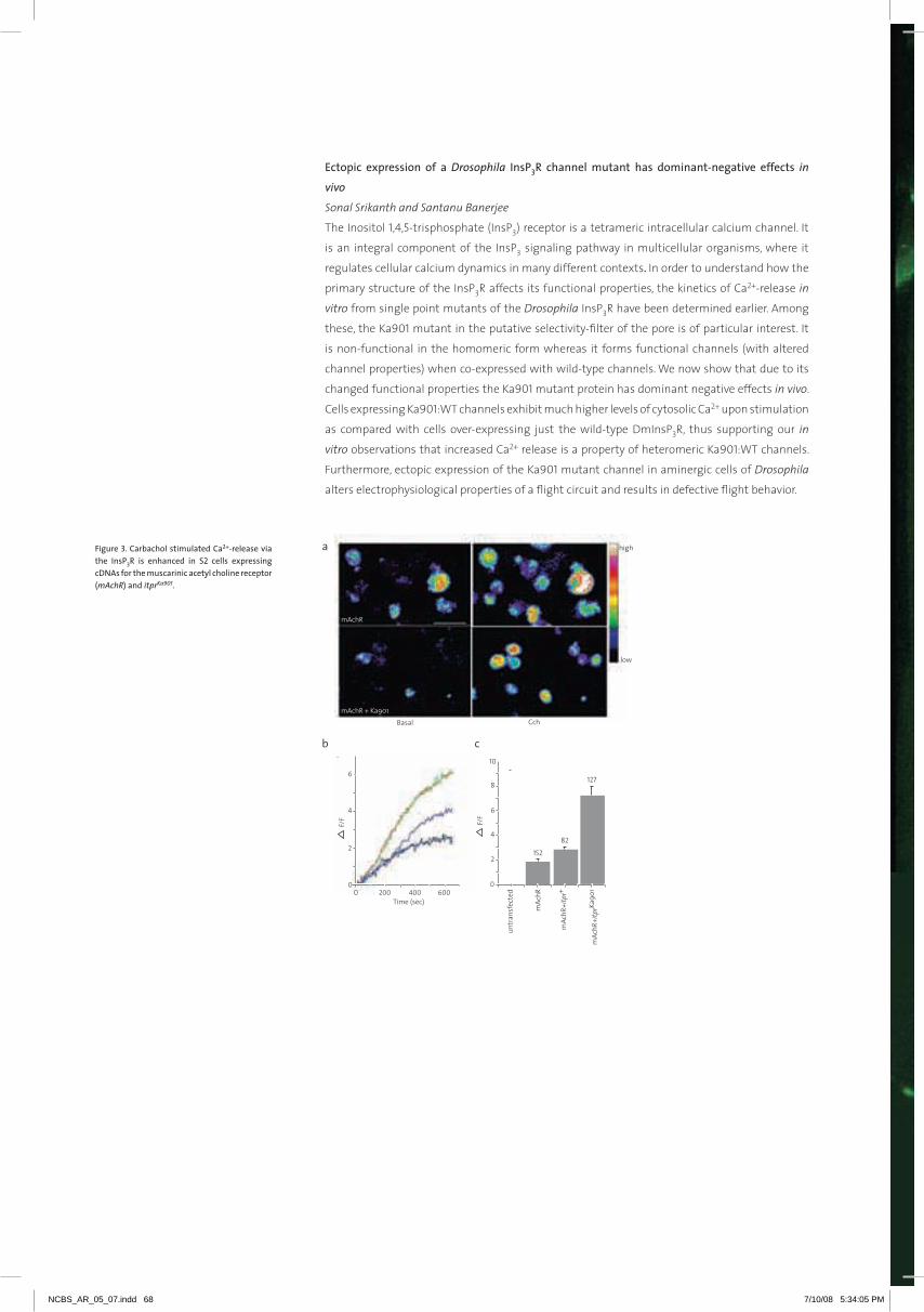
Ectopic expression of a Drosophila InsP3R channel mutant has dominant-negative effects in
vivo
Sonal Srikanth and Santanu Banerjee
The inositol 1,4,5-trisphosphate (insP3) receptor is a tetrameric intracellular calcium channel. it
is an integral component of the insP3 signaling pathway in multicellular organisms, where it
regulates cellular calcium dynamics in many different contexts. in order to understand how the
primary structure of the insP3r affects its functional properties, the kinetics of Ca2+-release in
vitro from single point mutants of the Drosophila insP3r have been determined earlier. among
these, the Ka901 mutant in the putative selectivity-filter of the pore is of particular interest. it
is non-functional in the homomeric form whereas it forms functional channels (with altered
channel properties) when co-expressed with wild-type channels. We now show that due to its
changed functional properties the Ka901 mutant protein has dominant negative effects in vivo.
Cells expressing Ka901:WT channels exhibit much higher levels of cytosolic Ca2+ upon stimulation
as compared with cells over-expressing just the wild-type DminsP3r, thus supporting our in
vitro observations that increased Ca2+ release is a property of heteromeric Ka901:WT channels.
Furthermore, ectopic expression of the Ka901 mutant channel in aminergic cells of Drosophila
alters electrophysiological properties of a flight circuit and results in defective flight behavior.
Figure 3. Carbachol stimulated Ca2+-release via the InsP3R is enhanced in S2 cells expressing cDNAs for the muscarinic acetyl choline receptor (mAchR) and itprKa901.
NCBS_AR_05_07.indd 68 7/10/08 5:34:05 PM
69
Sa
TY
aJi
T
Ma
YO
r
NCBS_AR_05_07.indd 69 7/10/08 5:34:06 PM

Welbergen, J.A. and Quader, S. (2006). Mother guarding: how offspring may influence the extra-pair behaviour of their parents. Proceedings of the Royal Society, B: Biological Sciences, 273, 2363–2368.
Quader, S. (2005). Elaborate nests in a weaverbird: a role for female choice? Ethology, 111, 1073-1088.
Quader, S. (2005). Mate choice and its implications for conservation and management. Current Science, 89, 1220-1229.
Selected publications
Organisms show a fascinating variety of traits that allow them to survive and reproduce in nature.
how do these adaptations work, and why are they so variable? To answer these questions one
must understand evolution, because traits evolve over generations based on a trade-off between
benefits and costs. Equally, one must understand ecology, because it is on the ecological stage
that the evolutionary play is enacted.
My particular interests include understanding the evolutionary ecology of conflict between
individuals, both within species and between species. Conflicts of interest arise when an
individual behaves in a way that is not optimal for others. i have studied conflicts within family-
groups, and currently work on conflicts between brood parasites and their hosts. Conflicts of
interest are also widespread when males and females make reproductive decisions. Sexual
selection, the framework within which such decisions are studied, remains an interest of mine.
i am also interested in the application of evolutionary thinking (adaptation, trade-offs, conflicts of
interests, life-history strategies) to ecological questions: What are the population consequences
of environmental change? how do inducible defences affect population trajectories? What are
the causes and consequences of invasions by non-native species into new environments?
My approach in tackling these questions is to combine theory with empirical work. Formal
modelling focusses hypotheses and predictions, and forces unstated assumptions into the
open; empirical tests of predictions lead to refined models of how the world works. i intend that
members of my lab work in a diversity of systems: terrestrial and aquatic, plant and animal. We
conduct observations and experiments in the field; and, where greater control is required, in
artificial environments, mimicking natural conditions as closely as possible.
Evolutionary ecology and environmental conservation
S U H E L Q U A D E R
[email protected] . in
NCBS_AR_05_07.indd 70 7/10/08 5:34:06 PM

71
S U H E L Q U A D E R
SU
hE
L Q
Ua
DE
r
1 Antagonistic co-evolution: brood parasites and their hosts
This work focusses on the strategies shown by avian brood parasites and the counter-strategies of
their hosts. Brood parasites lay their eggs in the nests of other species, thereby taking advantage
of the parental efforts of the hosts. What explains the large variation in virulence of avian brood
parasites? What adaptations make brood parasites successful at their deception? how do parasitic
chicks manage to manipulate their foster parents into providing for them? and, on the other hand,
what defences do hosts have against brood parasites?
2 Individual adaptations and population ecology: mosquito behaviour and population dynamics
Overall population change results from variation in the survival and reproduction of individuals. The
more we understand this variation, the better we will be able to predict future population change.
For this, we need to study the evolved tactics of individuals to survive and reproduce, and how these
tactics are influenced by the environment. When predation risk and competition vary in space, how do
female mosquitoes decide where to lay their eggs? how do larvae adjust their behavioural and life-
history tactics? Can we incorporate this information into our models to better understand population
dynamics?
3 Causes and consequences of invasions: the ecology and evolution of invasive species
Species that have successfully colonised new environments provide a wonderful opportunity to study
the evolutionary process of adaptation, and the ecological process of competition and its consequences.
What makes invasive species successful: release from ecological constraints (pathogens, predators,
competitors), or rapid adaptation? What effects do invasives have on native species, and through what
mechanisms? Does a detailed understanding of the evolution and ecology of problem species allow
us to design better control measures?
Two crow (above) and two koel eggs from the same nest
Lantana: an invasive shrub
The behavior of female mosquitoes influences the dynamics of their populations.
NCBS_AR_05_07.indd 71 7/10/08 5:34:07 PM
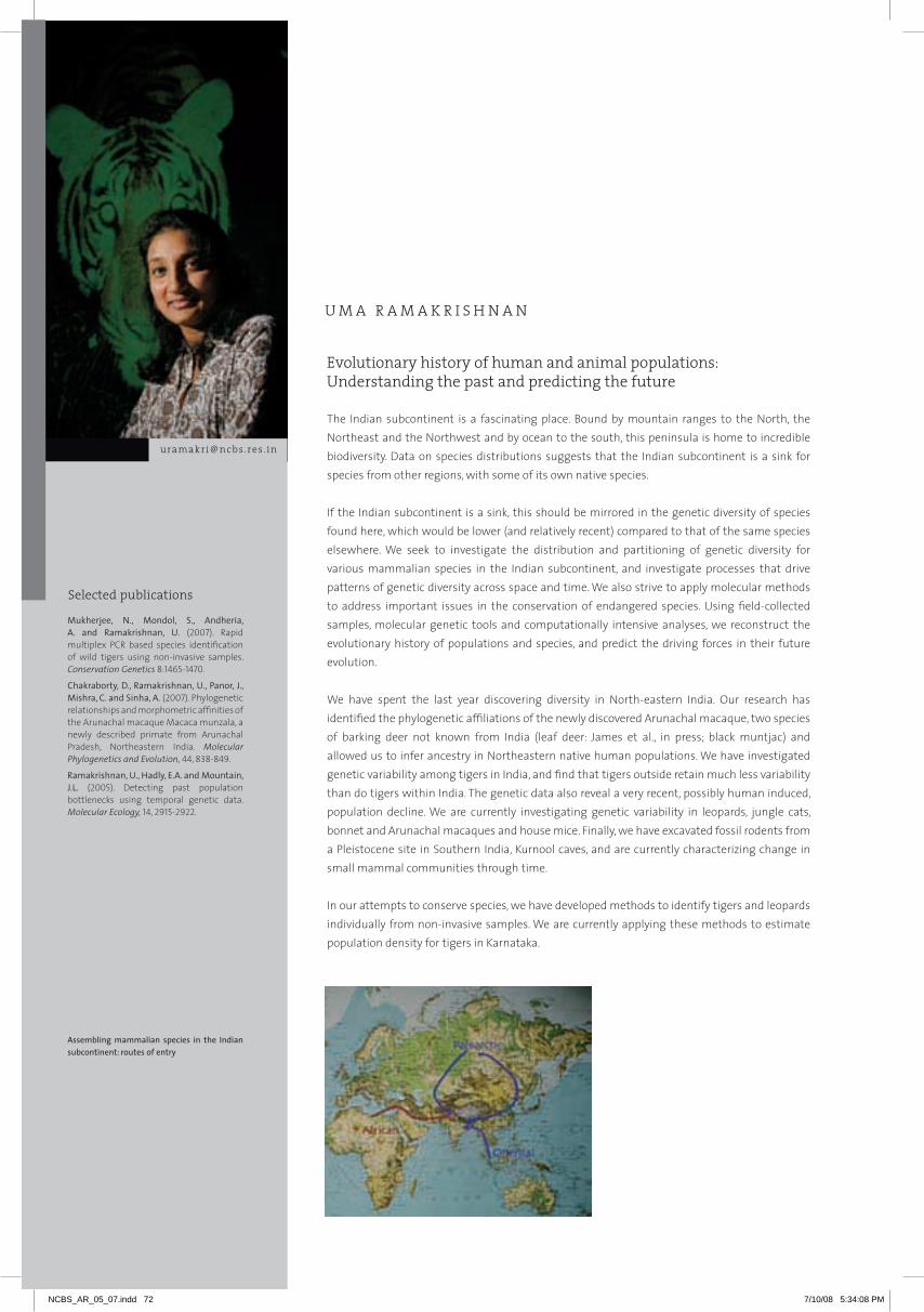
The indian subcontinent is a fascinating place. Bound by mountain ranges to the north, the northeast and the northwest and by ocean to the south, this peninsula is home to incredible biodiversity. Data on species distributions suggests that the indian subcontinent is a sink for species from other regions, with some of its own native species.
if the indian subcontinent is a sink, this should be mirrored in the genetic diversity of species found here, which would be lower (and relatively recent) compared to that of the same species elsewhere. We seek to investigate the distribution and partitioning of genetic diversity for various mammalian species in the indian subcontinent, and investigate processes that drive patterns of genetic diversity across space and time. We also strive to apply molecular methods to address important issues in the conservation of endangered species. Using field-collected samples, molecular genetic tools and computationally intensive analyses, we reconstruct the evolutionary history of populations and species, and predict the driving forces in their future evolution.
We have spent the last year discovering diversity in north-eastern india. Our research has identified the phylogenetic affiliations of the newly discovered arunachal macaque, two species of barking deer not known from india (leaf deer: James et al., in press; black muntjac) and allowed us to infer ancestry in northeastern native human populations. We have investigated genetic variability among tigers in india, and find that tigers outside retain much less variability than do tigers within india. The genetic data also reveal a very recent, possibly human induced, population decline. We are currently investigating genetic variability in leopards, jungle cats, bonnet and arunachal macaques and house mice. Finally, we have excavated fossil rodents from a Pleistocene site in Southern india, Kurnool caves, and are currently characterizing change in small mammal communities through time.
in our attempts to conserve species, we have developed methods to identify tigers and leopards individually from non-invasive samples. We are currently applying these methods to estimate population density for tigers in Karnataka.
Evolutionary history of human and animal populations: Understanding the past and predicting the future
U M A R A M A K R I S H N A N
Mukherjee, N., Mondol, S., Andheria, A. and Ramakrishnan, U. (2007). rapid multiplex PCr based species identification of wild tigers using non-invasive samples. Conservation Genetics 8:1465-1470.
Chakraborty, D., Ramakrishnan, U., Panor, J., Mishra, C. and Sinha, A. (2007). Phylogenetic relationships and morphometric affinities of the arunachal macaque Macaca munzala, a newly described primate from arunachal Pradesh, northeastern india. Molecular Phylogenetics and Evolution, 44, 838-849.
Ramakrishnan, U., Hadly, E.A. and Mountain, J.L. (2005). Detecting past population bottlenecks using temporal genetic data. Molecular Ecology, 14, 2915-2922.
Assembling mammalian species in the Indian subcontinent: routes of entry
[email protected] . in
Selected publications
NCBS_AR_05_07.indd 72 7/10/08 5:34:08 PM

73
U M A R A M A K R I S H N A N
UM
a r
aM
aK
riS
hn
an
1 Discovering biodiversity in northeastern India
Debapriyo Chakraborty and Jiffy James
The eastern himalaya includes habitats as diverse as broadleaf forest, alpine meadows and grasslands.
although this region is an important part of international conservation efforts, the rugged terrain
and extreme weather conditions make work here very difficult. Over the last couple of years, we have
applied molecular approaches to the discovery of biodiversity in this species rich region.
a new species of primate, the arunachal macaque Macaca munzala, was described from northeastern
india in 2005. Based on its appearance and distribution, the arunachal macaque was hypothesized to
be closely related to M. assamensis and M. thibetana. Molecular analyses in our laboratory established
the distinct identity of the species and indicate a time of origin of c. 0.48 mya. Surprisingly, maternally
inherited markers reveal close phylogenetic affinities with the Southern indian bonnet macaque (M.
radiata), while the Y chromosome revealed shared ancestry with the geographically closer assamese
and thibetan macaques, possibly mediated by ancestral male introgression. Morphometric analyses
reiterate similarity between M. munzala and its neighbors, presumably resulting from convergent
evolution under similar ecological conditions. We are continuing research on the arunachal macaque,
and are investigating population structure and demographic history using mitochondrial Dna and
microsatellite variation.
The leaf deer Muntiacus putaoensis was discovered in Myanmar and its presence in eastern arunachal
Pradesh, india, was subsequently confirmed based on morphometric analyses of two skulls collected
from local hunters. however, ambiguity remained regarding several other partial skulls and dried skin
samples collected during subsequent surveys. We developed molecular genetic analyses that can
unambiguously identify muntjac species. analyses of the collected skins revealed the presence of
three muntjac species in arunachal Pradesh: the indian muntjac, the leaf deer and the black muntjac.
These results increased the muntjac diversity of the indian subcontinent from one (only the common
muntjac) to three species. Further, it doubled the size of the leaf deer’s east-west range. Our discovery
of the black muntjac in this region are surprising given this species is previously known only from
eastern China. Phylogenetic analyses revealed that the indian population is reciprocally monophyletic
with the Chinese population. Given genetic differences and current geographical isolation between
these populations, we suggest that the indian population of the black muntjac be considered a
management unit. Our research highlights the importance of molecular tools in facilitating the
discovery of cryptic biodiversity as well as in determining conservation status of populations in such
understudied regions.
Collaborators: Anindya Sinha, National Institute of Advanced Studies and Aparajita Datta, National
Conservation Foundation, Bangalore
181 0 164 202
191185
181 168
248
224
173155
91170
117286
225184
165163
161158
126115
9280
5857
4539
20
115
159
184
266
286
184 227
227 181 180 173
17692
7
0.1
62
0.92
83
1.00
0.93
0.99
0.890.52
1.00
0.560.92
7997
98
1.00
1.00
64
1.00
0.77
871.00
92
97
zj 12
zj 13ah 19
ah 20
zj 8zj 11
hz 6zj 9
haplo 1haplo 8
nmd 02nmd 05
nmd 01nmd 03
nmd 04M. muntjak nmd 06M. muntjak nmd 10M. muntjak nmd 08M. muntjak nmd 07M. muntjak nmd 09
M. muntjak*M. reevesi*
M. reevesi micrurus*E. cephalophus*
C. eldi*
haplo 9
haplo 2
haplo 3haplo 4haplo 5
haplo 6
haplo 7
zj 16zj 14
70
95
M.crinifrons *
M.crinifrons(China)
M.crinifrons(Arunachal Pradesh)
Figure 1. Gene tree and haplotype network (based on 293 bp from control region) for back muntjacs from Arunachal Pradesh and China. Values at nodes represent Bayesian posterior probabilities (above) and the maximum parsimony bootstrap values (below).
NCBS_AR_05_07.indd 73 7/10/08 5:34:09 PM
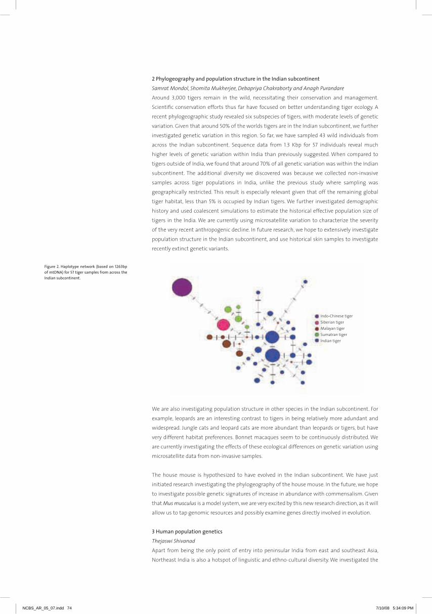
2 Phylogeography and population structure in the Indian subcontinent
Samrat Mondol, Shomita Mukherjee, Debapriya Chakraborty and Anagh Purandare
around 3,000 tigers remain in the wild, necessitating their conservation and management.
Scientific conservation efforts thus far have focused on better understanding tiger ecology. a
recent phylogeographic study revealed six subspecies of tigers, with moderate levels of genetic
variation. Given that around 50% of the worlds tigers are in the indian subcontinent, we further
investigated genetic variation in this region. So far, we have sampled 43 wild individuals from
across the indian subcontinent. Sequence data from 1.3 Kbp for 57 individuals reveal much
higher levels of genetic variation within india than previously suggested. When compared to
tigers outside of india, we found that around 70% of all genetic variation was within the indian
subcontinent. The additional diversity we discovered was because we collected non-invasive
samples across tiger populations in india, unlike the previous study where sampling was
geographically restricted. This result is especially relevant given that off the remaining global
tiger habitat, less than 5% is occupied by indian tigers. We further investigated demographic
history and used coalescent simulations to estimate the historical effective population size of
tigers in the india. We are currently using microsatellite variation to characterize the severity
of the very recent anthropogenic decline. in future research, we hope to extensively investigate
population structure in the indian subcontinent, and use historical skin samples to investigate
recently extinct genetic variants.
We are also investigating population structure in other species in the indian subcontinent. For
example, leopards are an interesting contrast to tigers in being relatively more adundant and
widespread. Jungle cats and leopard cats are more abundant than leopards or tigers, but have
very different habitat preferences. Bonnet macaques seem to be continuously distributed. We
are currently investigating the effects of these ecological differences on genetic variation using
microsatellite data from non-invasive samples.
The house mouse is hypothesized to have evolved in the indian subcontinent. We have just
initiated research investigating the phylogeography of the house mouse. in the future, we hope
to investigate possible genetic signatures of increase in abundance with commensalism. Given
that Mus musculus is a model system, we are very excited by this new research direction, as it will
allow us to tap genomic resources and possibly examine genes directly involved in evolution.
3 Human population genetics
Thejaswi Shivanad
apart from being the only point of entry into peninsular india from east and southeast asia,
northeast india is also a hotspot of linguistic and ethno-cultural diversity. We investigated the
Figure 2. Haplotype network (based on 1263bp of mtDNA) for 57 tiger samples from across the Indian subcontinent.
NCBS_AR_05_07.indd 74 7/10/08 5:34:09 PM

75
UM
a r
aM
aK
riS
hn
an
demographic history and possible genetic relationships between northeast indian tribal populations
and the contentious role of northeastern india in populating the indian subcontinent using detailed
population genetic analysis of data from 650 individuals from seven tribes from arunachal Pradesh and
Sikkim in the northeast. Genetic data included mitochondrial hVS1 sequences from these individuals
and published data (over 1400 sequences) from other tribes in Meghalaya and other parts of this
region. Our results reveal population growth among these tribes, contrary to most hunter-gatherer
groups elsewhere across the world, and that ethno-religious tradition could be a significant isolating
factor in geographically proximate populations. Using coalescent simulations we investigated
divergence times between these tribes and those from peninsular india and southeastern China. Our
simulations reveal that the northeast indian tribes could have colonized the region relatively recently,
having a more recent shared ancestry with Chinese tribes than peninsular indian tribes. We propose
that for human populations the northeast is best viewed as a sink rather than a corridor or barrier to
gene flow.
Collaborator: B.R. Rao, Anthropological Survey of India
4 Phylochronology: What was India like in the past?
Indrani Suryaprakash and Krishnapriya Tamma
in February of 2007, we excavated a cave site close to Kurnool, in andhra Pradesh, Southern india.
Our initial explorations reveal that the materials we excavated represent a rich assemblage of small
mammal fauna from this region. We are now in the process of dating layers in the excavated remains,
cleaning and identifying the recovered small mammals jaws. We are also testing the feasibility
of using the material we have recovered as a source of ancient Dna. Longer-term, we hope to use
excavation materials like these to investigate how changes in climate have impacted the ecology and
evolution of small mammal communities in the past in the indian subcontinent.
Collaborators: Elizabeth Hadly, Stanford University and Anthony Barnosky, UC Berkeley, USA
5 Conservation genetics Samrat Mondol
The survival of tigers in the indian subcontinent is threatened. in the recent years, non-invasive genetic
samples like scat have served as an invaluable tool to study the population status of rare and elusive
species. We have developed genetic methods to identify species, gender and individuals. We are
currently working with scat samples collected in a mark-recapture framework to estimate population
density in Bandipur national Park. Bandipur is a good site to test our methods, as Dr. Karanth and
others at WCS have studied this population for many years, and have independent estimates of
population density. in future research, we hope to extend the geographical scope of our research to
the Western ghats in Karnataka. We hope to combine genetic and ecological research to investigate
tiger habitat preference, tiger density in key regions, and connectivity between different parts of this
landscape. We believe that such research will be critical for the conservation of tigers.
Collaborator: Ullas Karanth, Wildlife Conservation Society
Figure 3. Log likelihood surfaces for two-population models including (a) Arunachal-Meghalaya (b) Arunachal-China and (c) Arunachal-Peninsular India. Likelihood is plotted as a function of divergence time and number of migrants per generation. Effective size is assumed to be 1,000 for both populations.
Figure 4. The picture shows modern (above) and ancient (from excavation, below) rodent jaws.
NCBS_AR_05_07.indd 75 7/10/08 5:34:10 PM

My laboratory has focused on trying to understand how the olfactory system is constructed
to detect and respond to odorant stimuli at high sensitivity. We are interested in deciphering
mechanisms that ensure integrity of circuits within adult animals. What are the changes that
must occur within individual neurons to maintain homeostasis and how do these properties
change to allow plasticity? The Drosophila olfactory system presents several advantages that
allows us to address these issues. The circuit which is composed of sensory neurons, local
interneurons, projection neurons and modulatory neurons is anatomically well defined and
is amenable to a range of genetic strategies available in the fly (Figure below). Odor quality is
largely represented as a 3-dimensional spatial code in the brain and the basic cell types and
underlying cellular organization is well described. Building upon this knowledge, we are now
investigating how this collection of neurons function to result in behavior.
Developmental neurobiology of the olfactory system
V E R O N I C A R O D R I G U E S
[email protected] . in
Roy, B., Singh, A.P., Shetty, C., Chaudhary, V., North, A., Landraf, M., VijayRaghavan, K. and Rodrigues, V. (2007). Metamorphosis of an identified serotonergic neuron in the Drosophila olfactory system. Neural Development, 2, 20.
The olfactory circuit in Drosophila indicating classes of neurons as well as (in green) P(Gal4) lines that mark them.
Selected publication
NCBS_AR_05_07.indd 76 7/10/08 5:34:11 PM

77
V E R O N I C A R O D R I G U E S
VE
rO
niC
a r
OD
riG
UE
S
1 Spontaneous neural activity is necessary for maintenance of adult olfactory circuits
Albert Chiang
The mechanisms that underlie maintenance of adult circuits are not well understood in any system.
The role of neural activity during development is better studied in the vertebrate olfactory system,
where activity along with odorant receptor identity, specifies the combination of adhesive and
repulsive cues that result in the maturation of the sensory map.
We used a conditional system to express any of a panel of neural-activity blockers in the adult
Drosophila olfactory neurons and observed a disruption of axonal terminals within antennal lobe
glomeruli (Figure 1). To understand how activity maintains neural elements, we searched for molecules
whose levels are regulated in an activity-dependent manner and identified the homophilic adhesion
molecules Fas2, n-Cadherin and neuroglian. in the presynaptic terminals of olfactory receptor
neurons, where synaptic function is blocked, Fas2 and neuroglian are upregulated while n-Cadherin
is downregulated. Our results suggest that the maintenance of sensory inputs to the glomerular map
is an active process in vivo, requiring neural activity during adulthood.
in order to link activity to cell adhesion molecules, we have initiated a screen using UaS-rnai lines
kindly provided by ryu Ueda at the national institute of Genetics, Japan and by Barry Dickson at the
research institute of Molecular Pathology, Vienna. We have obtained some candidate genes whose
‘knock-down’ in sensory neurons or in glial cells results in synapse withdrawal or degeneration. These
are being investigated currently.
Collaborator: K. VijayRaghavan, NCBS
2 Re-modeling of an identified serotonergic neuron during pupal life is regulated by activity and
Wingless signaling
Bidisha Roy and Ajeet P. Singh
it is likely that many of the mechanisms employed during metamorphosis of neurons are shared
with those involved in adult plasticity and may also provide knowledge about neural degeneration.
We therefore decided to examine molecular mechanisms that control the re-modeling of a wide-field
serotonergic neuron in the Drosophila olfactory pathway which undergoes dramatic changes from
larva to adult.
Modification of a larval neuron into its adult-specific form is regulated by steroid hormone receptors,
neuronal activity, signalling through the Wingless pathway and Ephrins (Figure 2). These studies
establish a cellular system for studying how extrinsic cues interact with autonomous signals to
pattern complex cellular architecture. Parallel studies aimed at deciphering the function of this
neuron will link anatomy to the development of olfactory behaviour.
Collaborator: K. VijayRaghavan, NCBS
Figure 1. Chronic blockage of activity in a subset of olfactory neurons in the adult leads to their degeneration. Glomeruli within the adult antennal lobe are visualized by staining using mabnc82. a subset of ~50 neurons marked by Or47b-Gal4>GFP project to a single glomerulus in the antennal lobe (a). Co-expression of tetanus toxin in these neurons for six days after eclosion results in degeneration. Scale bar=20µm
Figure 2. A single neuron from a pair of contralaterally innervating serotonergic deutocerebral neurons (CSD). The arborization of pre-synaptic terminals in the contralateral lobe is affected in mutant conditions.
NCBS_AR_05_07.indd 77 7/10/08 5:34:11 PM

3 Mechanisms of Short-term and Long-term Habituation in the Drosophila olfactory pathway
Sudeshna Das, Avni Gandhi and Somdatta Karnik
We have devised behavioral paradigms that allow us to study two forms of non-associative
memory which we term Long Term habituation (LTh) and Short Term habituation (STh). in LTh,
newly eclosed flies are exposed for several days to the odor of benzaldehyde or to Carbon dioxide.
The normal repulsive response to these stimuli is severely diminished and recovers only after
eight to ten days. STh, on the other hand is more robustly measured in larvae where five minute
pre-exposure to odorants results in a drop in response which recovers within an hour. We are
dissecting the cellular basis of these behavioral observations using P(Gal4) lines that allow us to
manipulate the properties of individual cells within the circuit.
Electrophysiological studies and functional imaging techniques from several laboratories
suggest that local interneurons serve to integrate information among different glomeruli
suggesting that these could be the substrates for generation of LTh and STh. We have initiated
an analysis using the mosaic analysis with repressible cell marker (MarCM) method to study
the lineage and architecture of individual local interneurons (Ln) (Figure 3). The presence of a
Or-GFP fusion constructs in the background of the Gal4 line that marks Ln allows us to trace
arborizations to individual identified glomeruli (Figure 3b).
Collaborators: Abhijit Das, TIFR and Mani Ramaswami, Trinity College, Dublin
4 Studies on olfactory development in a social ant Campotonus sericeus
Social insects, notably ants and bees, exhibit extreme forms of behavioral plasticity with different
members of the community performing very distinct and complex tasks. There are several
reported examples of chemical cues triggering diverse behavior patterns that can be modified
within short timescales. We have initiated experiments to study the development and function
of the olfactory system in the ant Campotonus sericeus which relies heavily on olfaction for its
lifestyle.
antibodies against Drosophila synaptic proteins label synapses in the ant brain and allowed us
to visualize the organization of the olfactory lobe in different castes (Figure 4). Unlike that in
flies, the olfactory lobe of ants shows sexual dimorphism – the male has a macroglomerulus,
presumably involved in detection of sex pheromones and about 75 smaller glomeruli. The
worker on the other hand, has about 175 glomeruli of similar size and the queen possesses 115
glomeruli.
Figure 3. A single local interneuron ramifies extensively within the antennal lobe to innervate all glomeruli. The inset shows a single glomerulus marked with Or47b-CD2 (red) allows visualization of Ln arbors within a single glomerulus.
Figure 4. Ant brains were stained against antibodies to Synapsin (green) and anti-Serotonin (red). The glomeruli in the antennal lobe (aL) are easily visualized. arrowheads indicate the mushroom bodies.
NCBS_AR_05_07.indd 78 7/10/08 5:34:12 PM
MiC
ha
EL
Ba
TE
c [email protected] . in
Developmental genetic analysis of locomotor mechanisms
M I C H A E L B A T E
We are interested in understanding the genetic and developmental origins of the patterned
movements that underlie all behavior. We use Drosophila for our studies of emerging behavior,
since Drosophila provides us with a wealth of genetic tools and reagents with which to study the
development and function of the nervous system. We focus our investigation on the embryo
because this is the developmental stage when neural circuits begin to mature and the coordinated
outputs that drive movement first appear. Our aims at this stage are 1) to identify genes essential
for the normal development of movement 2) to identify elements of the neural circuitry underlying
locomotor movements 3) to investigate the emergence of function in the motor system and how this
is controlled.
We have completed our first level of genetic analysis by showing that patterns of movement that are
specific to particular body regions are specified by the local expression of hox genes (Dixit et al 2008).
Loss of hox gene function leads to a loss of the normal pattern of local movement; gain of function
leads to the ectopic expression of movements characteristic of a different body region. We conclude
that hox genes regulate the region specific differentiation of neural networks that drive coordinated
movement and a major challenge now is to translate this finding into a deeper understanding of how
such control is exerted at a cellular and molecular level. at the same time a major screen aimed at
identifying components of the motor circuitry that are essential for specific patterns of movement
has been carried out using a loss of function approach. The results of this screen are promising: a large
number of lines that are either paralysed or show defective patterns of behavior when tetanus toxin is
targeted to specific subsets of neurons has been identified. Many of the neuronal subsets identified in
this way are relatively small and restricted and should allow us to catalogue and manipulate essential
components of both locomotor circuitry and its upstream control.
This work is complemented by and interacts with work in Cambridge that is particularly concerned
with the progressive emergence of coordinated movement in late embryogenesis. This work indicates
an essential role for neural activity in the proper development of coordinated movement. This finding
is reflected at a cellular level by the discovery of an apparently homeostatic mechanism that regulates
the growth of postsynaptic elements of motor circuitry in response to the level of synaptic input they
receive. We conclude that the developing motor circuitry shows an unexpected degree of plasticity
that allows for compensation in the face of naturally occurring variability.
Primary affiliation: Department of Zoology, University of Cambridge, UK
adjunct faculty
79
NCBS_AR_05_07.indd 79 7/10/08 5:34:12 PM
Selected publications
[email protected] . in
Nerves, muscles and the development of behaviour
K . V I J AY R A G H AVA N
Our laboratory is interested in understanding the mechanisms underlying development and
assembly of the network-comprising nerves, muscles and tendons – responsible for the ability
of animals to move from one place to another by crawling, walking or flying. Our approach
is to analyze this problem – using genetic and molecular tools – by first understanding the
mechanisms that specify the birth of motor neurons, their morphogenesis and relationship
to their input neurons and target muscles. This cellular analysis has begun to yield results on
the developmental genetic basis underlying the construction of hardware. We are, in parallel,
addressing which aspects of the network are essential for function and, importantly, the ways
in which function is robust, and resilient to perturbations in structure and development. These
experiments have required the careful development of assays of locomotor behaviour. alterations
in behaviour upon induced changes in gene expression in sensory-or motor-neurons are then
measured to understand how ‘function’ is built into the network during development.
Mondal, K., Dastidar, A.G., Singh, G., Madhusudhanan, S., Gande, S.L., VijayRaghavan, K. and Varadarajan, R. (2007). Design and isolation of temperature-sensitive mutants of Gal4 in yeast and Drosophila. Journal of Molecular Biology, 370, 939-950.
Roy, B., Singh, A.P., Shetty, C., V. Chaudhary, V., North, A., Landraf, M., VijayRaghavan, K. and Rodrigues, V. (2007). Metamorphosis of an identified serotonergic neuron in the Drosophila olfactory system. Neural Development, 2, 20.
Dixit R., VijayRaghavan, K. and Bate, M. (2007). hox genes and the regulation of movement in Drosophila. Developmental Neurobiology, 68, 309-331.
Selected publications
NCBS_AR_05_07.indd 80 7/10/08 5:34:13 PM
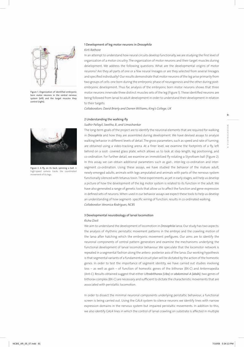
81
K . V I J AY R A G H AVA N
K V
iJa
Yr
aG
ha
Va
n
1 Development of leg motor neurons in Drosophila
Kirti Rathore
in an attempt to understand how neural circuits develop functionally, we are studying the first level of
organization of a motor circuitry: The organization of motor neurons and their target muscles during
development. We address the following questions: What are the developmental origins of motor
neurons? are they all parts of one or a few neural lineages or are they selected from several lineages
and specified individually? Our results demonstrate that motor neurons of the leg arise primarily from
two groups of cells: one born during the embryonic phase of neurogenesis and the other during post-
embryonic development. Thus far, analysis of the embryonic born motor neurons shows that three
motor neurons innervate three distinct muscles sets of the leg (Figure 1). These identified neurons are
being followed from larval to adult development in order to understand their development in relation
to their targets.
Collaborators: David Brierly and Darren Williams, King’s College, UK
2 Understanding the walking-fly
Sudhir Pallayil, Swetha, B., and Umashankar
The long-term goals of the project are to identify the neuronal elements that are required for walking
in Drosophila and how they are assembled during development. We have devised assays to analyze
walking behavior in different levels of detail. The gross parameters, such as speed and rate of turning.
are obtained using a video-tracking arena. at a finer level, we examine the footprints of a fly, left
behind on a soot- covered glass plate, which allows us to look at step length, leg positioning, and
co-ordination. For further detail, we examine an immobilized fly rotating a Styrofoam ball (Figure 2).
in this assay, we can obtain additional parameters such as gait-, inter-leg co-ordination and inter-
segment co-ordination. Using these assays, we have studied the behavior of the mature adult,
newly emerged adults, animals with legs amputated and animals with parts of the nervous system
functionally silenced with tetanus toxin. These experiments, as yet in early stages, will help us develop
a picture of how the development of the leg motor system is related to its function in the adult. We
have also generated a range of genetic tools that allow us to affect the function and gene-expression
in defined sets of neurons. When used in our behavior assays we expect these tools to help us develop
an understanding of how segment- specific wiring of function, results in co-ordinated walking.
Collaborator: Veronica Rodrigues, NCBS
3 Developmental neurobiology of larval locomotion
Richa Dixit
We aim to understand the development of locomotion in Drosophila larva. Our study has two aspects:
the analysis of rhythmic peristaltic movement patterns in the embryo and the crawling motion of
the larva after hatching which the embryonic movement prefigures. Our aims are to identify the
neuronal components of central pattern generators and examine the mechanisms underlying the
functional development of larval locomotor behaviour. We speculate that the locomotor network is
repeated in a segmental fashion along the antero- posterior axis of the larva. Our working hypothesis
is that segmental variants of a fundamental circuit plan will be dictated by the action of the homeotic
genes. in order to test the importance of segment identity, we have carried out studies involving
loss – as well as gain – of function of homeotic genes of the bithorax (BX-C) and antennapedia
(ant-C). results obtained suggest that either Ultrabithorax (Ubx) or abdominal-A (abdA), two genes of
bithorax complex (BX-C) are necessary and sufficient to dictate the characteristic movements that are
associated with peristaltic locomotion.
in order to dissect the minimal neuronal components underlying peristaltic behaviour, a functional
screen is being carried out. Using the GAL4 system to silence neurons we identify lines with narrow
expression domains in the nervous system but impaired peristaltic movements. in addition to this,
we also identify GAL4 lines in which the control of larval crawling on substrate is affected in multiple
Figure 1. Organization of identified embryonic born motor neurons in the central nervous system (left) and the target muscles they control (right).
Figure 2. A fly, on its back, spinning a ball. a high-speed camera tracks the coordinated movement of its legs.
NCBS_AR_05_07.indd 81 7/10/08 5:34:13 PM
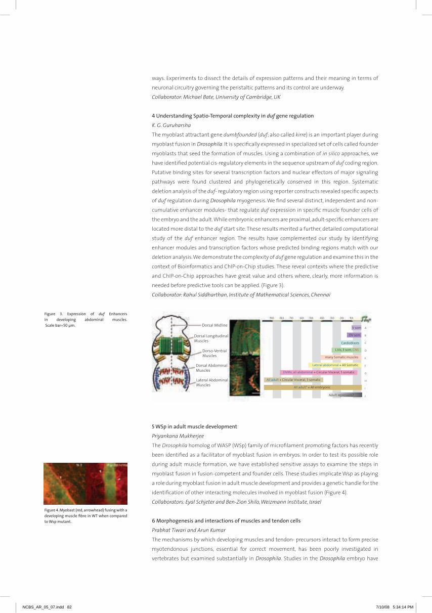
ways. Experiments to dissect the details of expression patterns and their meaning in terms of
neuronal circuitry governing the peristaltic patterns and its control are underway.
Collaborator: Michael Bate, University of Cambridge, UK
4 Understanding Spatio-Temporal complexity in duf gene regulation
K. G. Guruharsha
The myoblast attractant gene dumbfounded (duf; also called kirre) is an important player during
myoblast fusion in Drosophila. it is specifically expressed in specialized set of cells called founder
myoblasts that seed the formation of muscles. Using a combination of in silico approaches, we
have identified potential cis-regulatory elements in the sequence upstream of duf coding region.
Putative binding sites for several transcription factors and nuclear effectors of major signaling
pathways were found clustered and phylogenetically conserved in this region. Systematic
deletion analysis of the duf- regulatory region using reporter constructs revealed specific aspects
of duf regulation during Drosophila myogenesis. We find several distinct, independent and non-
cumulative enhancer modules- that regulate duf expression in specific muscle founder cells of
the embryo and the adult. While embryonic enhancers are proximal, adult-specific enhancers are
located more distal to the duf start site. These results merited a further, detailed computational
study of the duf enhancer region. The results have complemented our study by identifying
enhancer modules and transcription factors whose predicted binding regions match with our
deletion analysis. We demonstrate the complexity of duf gene regulation and examine this in the
context of Bioinformatics and ChiP-on-Chip studies. These reveal contexts where the predictive
and ChiP-on-Chip approaches have great value and others where, clearly, more information is
needed before predictive tools can be applied. (Figure 3).
Collaborator: Rahul Siddharthan, Institute of Mathematical Sciences, Chennai
5 WSp in adult muscle development
Priyankana Mukherjee
The Drosophila homolog of WaSP (WSp) family of microfilament promoting factors has recently
been identified as a facilitator of myoblast fusion in embryos. in order to test its possible role
during adult muscle formation, we have established sensitive assays to examine the steps in
myoblast fusion in fusion-competent and founder cells. These studies implicate Wsp as playing
a role during myoblast fusion in adult muscle development and provides a genetic handle for the
identification of other interacting molecules involved in myoblast fusion (Figure 4).
Collaborators: Eyal Schjeter and Ben-Zion Shilo, Weizmann Institute, Israel
6 Morphogenesis and interactions of muscles and tendon cells
Prabhat Tiwari and Arun Kumar
The mechanisms by which developing muscles and tendon- precursors interact to form precise
myotendonous junctions, essential for correct movement, has been poorly investigated in
vertebrates but examined substantially in Drosophila. Studies in the Drosophila embryo have
Figure 3. Expression of duf Enhancers in developing abdominal muscles. Scale bar=50 µm.
Figure 4. Myobast (red, arrowhead) fusing with a developing muscle fibre in WT when compared to Wsp mutant.
NCBS_AR_05_07.indd 82 7/10/08 5:34:14 PM

83
K V
iJa
Yr
aG
ha
Va
n
shown that stripe expressing tendon- precursors and developing muscles interact through the EGF-
signaling pathway. Explorations in the embryo and in the adult have suggested that hh and Wnt
signaling, respectively, could have a role in the matching of specific muscle- tendon- interactions.
We are examining identified signaling pathways in adult muscle- tendon interaction in Drosophila.
We use stripe-Gal4 to perturb selected molecules in tendon cells. Perturbation of the Wnt signaling
myotubes pathway in muscle attachment sites affects the migration of myotubes towards their
attachment sites. inhibition of EGFr signaling in tendon cells using dominant negative receptor
affects tendon cell projections.
We have marked muscles and tendons with differential labels and, in this background, have started
a large scale- screen of UaS-rnai lines (obtained from niG Japan) to identify molecules in the
establishment of myo- tendonous junctions. We use MhC-Gal4 and stripe-Gal4 to express the rnai
lines in muscle and tendon cells respectively. The approach is to look for the lethality and any other
easily scorable phenotype first and then to examine their role in detail. We have picked up several
interesting molecules, which show either lethality or flight defects, and these are being examined in
detail.
7 The protein complex map of the Drosophila proteome
Aijaz Noor, Aijaz Parray, Venkateswara Reddy Ontedddu and K. G. Guruharsha
Differences in complexity between organisms are not completely dependent on the number of genes
in the genome but rather on how these genes interact in the cellular and organismal environment.
Thus, understanding how gene products interact and indeed the rules that govern such interactions is
an essential element in dissecting the cellular function of biological systems. Multi-protein complexes
carry out most cellular processes. The identification and composition of such complexes is crucial for
understanding how the ensemble of expressed proteins (the Proteome) is organized into functional
units. Proteins interact with each other to build up networks that govern cellular functions. Defining
these networks is an essential part of gaining a molecular understanding of how cellular behavior
dictates the development and physiology of an organism. The goal of this project is to establish
a complete protein interaction map of the Drosophila proteome based on the isolation and Mass
spectrometric analysis of purified protein complexes associated with individually tagged proteins.
The project involves a systematic retrieval and analysis of cellular protein complexes in sufficient
quantities for biochemical analysis of their components. This will determine the first complete
metazoan proteome map beyond the level of binary protein interactions. We are generating stable
transgenic fly lines that can express tagged proteins in regulated manner to understand protein
complexes in different cellular/ tissue contexts. The results we obtain from these studies will not
only benefit the Drosophila community but will be of fundamental interest and practical use to a very
broad spectrum of researchers.
Collaborators: Spyros Artavanis-Tsakonas, Harvard Medical School, USA; Susan Celniker and Mark
Stapleton, Berkeley Drosophila Genome Project, USA; L.S. Shashidhara, Centre for Cellular and Molecular
Biology, Hyderabad and Cellzome, Heidelberg, Germany
NCBS_AR_05_07.indd 83 7/10/08 5:34:14 PM

NCBS_AR_05_07.indd 84 7/10/08 5:34:22 PM

85
Sa
TY
aJi
T
Ma
YO
rResearch Reports
86 Upinder S. Bhalla | 90 Sumantra Chattarji | 94 Sandhya P. Koushika | 98 Mitradas M. Panicker | 102 Sanjay P. Sane
104 Obaid Siddiqi
N Eu RobIoloGy
NCBS_AR_05_07.indd 85 7/10/08 5:34:25 PM
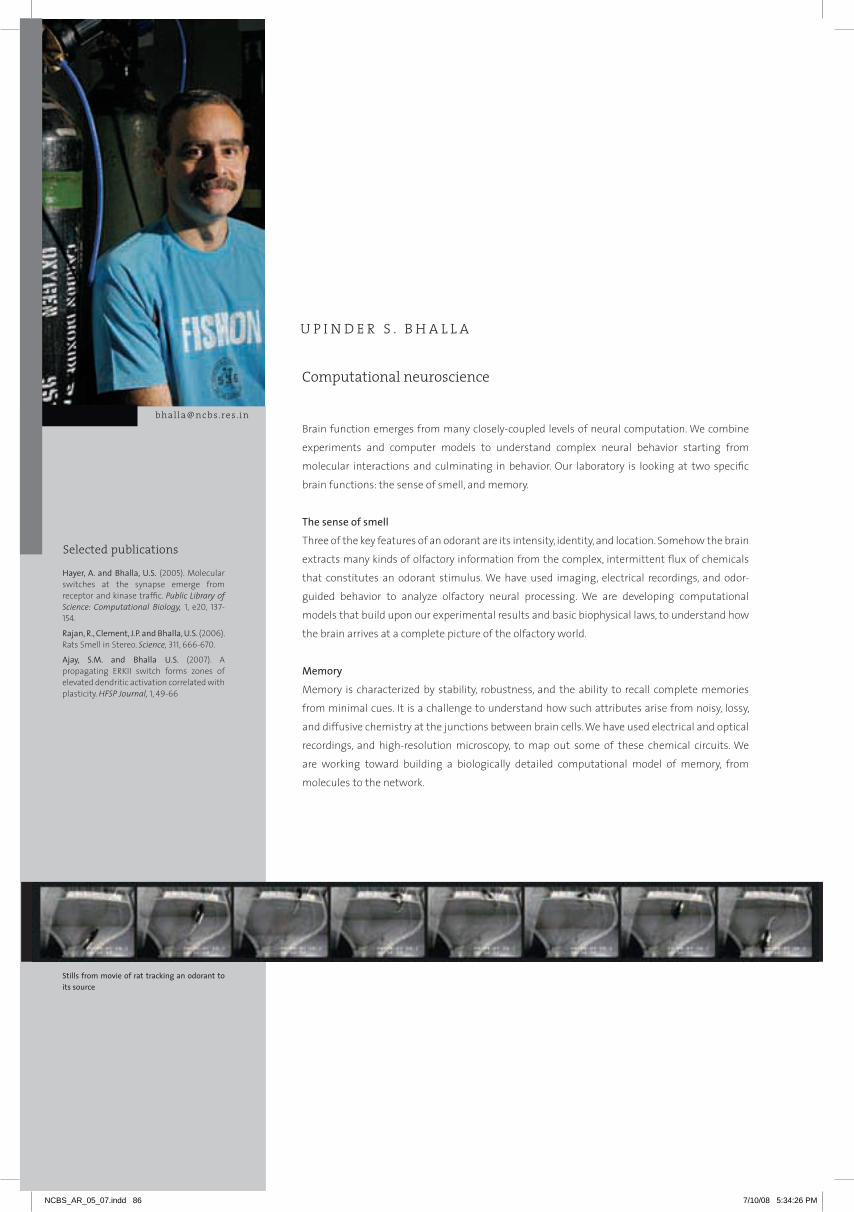
Selected publications
Brain function emerges from many closely-coupled levels of neural computation. We combine
experiments and computer models to understand complex neural behavior starting from
molecular interactions and culminating in behavior. Our laboratory is looking at two specific
brain functions: the sense of smell, and memory.
The sense of smell
Three of the key features of an odorant are its intensity, identity, and location. Somehow the brain
extracts many kinds of olfactory information from the complex, intermittent flux of chemicals
that constitutes an odorant stimulus. We have used imaging, electrical recordings, and odor-
guided behavior to analyze olfactory neural processing. We are developing computational
models that build upon our experimental results and basic biophysical laws, to understand how
the brain arrives at a complete picture of the olfactory world.
Memory
Memory is characterized by stability, robustness, and the ability to recall complete memories
from minimal cues. it is a challenge to understand how such attributes arise from noisy, lossy,
and diffusive chemistry at the junctions between brain cells. We have used electrical and optical
recordings, and high-resolution microscopy, to map out some of these chemical circuits. We
are working toward building a biologically detailed computational model of memory, from
molecules to the network.
Computational neuroscience
U P I N D E R S . B H A L L A
[email protected] . in
Hayer, A. and Bhalla, U.S. (2005). Molecular switches at the synapse emerge from receptor and kinase traffic. Public Library of Science: Computational Biology, 1, e20, 137-154.
Rajan, R., Clement, J.P. and Bhalla, U.S. (2006). rats Smell in Stereo. Science, 311, 666-670.
Ajay, S.M. and Bhalla U.S. (2007). a propagating ErKii switch forms zones of elevated dendritic activation correlated with plasticity. HFSP Journal, 1, 49-66
Stills from movie of rat tracking an odorant to its source
Selected publications
NCBS_AR_05_07.indd 86 7/10/08 5:34:26 PM

87
UP
inD
Er
S B
ha
LL
a
1 Smell
a. Physiology
Adil G. Khan, K. Parthasarathy, Raghav Rajan, James P. Clement and Ashesh Dhawale
We study how brain activity encodes three key dimensions of an odorant stimulus: intensity, identity
and location. Using electrical recordings, we have measured rat olfactory bulb responses to odorant
stimuli under a wide range of concentrations and in mixtures. We have identified a likely encoding
scheme that accurately describes the firing patterns of 80% of recorded neurons to any combination
and concentration of two odorants. This may account for intensity and identity encoding. We have
also identified laterality in response properties that rapidly and accurately predicts the location of
odorants. This may provide the third dimension, that of odor location, in the olfactory bulb. To go from
single-unit to network-level encoding, we have built an in-vivo 2-photon system for optically recording
from many cells simultaneously.
b. Behavior
Raghav Rajan, Urvashi Raheja and K. Parthasarathy
rats are excellent at tracking odors, such as food, to their source. By studying rats trained to push
sensors depending on odor direction, we have shown that rats can locate odors using stereo cues.
They compare the intensity and time of arrival in each nostril, to determine the location of the odor
source. They can identify multiple odors and respond in a different way to each. We find that rats can
do this in under 200 msec, comparable to the reaction time of a trained athlete. Do they use this
ability in nature? We have trained rats to navigate toward an odor source in a laminar air-flow arena
to test the contribution of stereo and rapid odor processing in natural odor-location strategies.
c. Models
Subhasis Ray, Niraj Dudani, Rinaldo D’Souza and U.S. Bhalla
The cellular and network physiology of the olfactory bulb is among the best characterized in the brain.
With this data we propose that we can predict the emergence of representations of odor identity,
intensity, and location from the first principles of cellular biophysics. We are building one of the
largest-ever simulations of any brain structure, where we plan to model the entire olfactory bulb with
particular attention to biophysical detail. This model will be calibrated against our own and other
data, and will run on our 260-node supercomputer.
2 Memory
a. Physiology
Sriram M. Ajay and Dhanya Parameshwaran
how do memory-triggered signals spread through the neuron? We have used electrical recordings
and immunohistochemistry in the hippocampal slice to map out the extent and mechanisms of this
spread. We examined a series of possible models by comparing computer predictions with experiments,
to show that both synaptic and voltage-gated calcium influx contributes to the activity spread. We are
also approaching the issue of spatial extent of memory signals from the network perspective, using
optical recordings and multiple stimulus points to read out many synaptic responses in parallel.
b Models
Pragati Jain, Arnold Hayer, Priyamvada Rajasethupathy and U.S. Bhalla
Complex chemical circuits mediate the subcellular steps that give rise to memory. One mechanism
for this is when chemical circuits act like memory switches, which can stably persist in either of two
states (on or off).
While computationally analyzing the movement of molecules in the synapse, we unexpectedly
discovered that such a switch emerged from the regulated movement of neurotransmitter receptors.
We showed that this switch could retain its state for over a year despite molecular noise, diffusion
and turnover. We are modeling the synaptic activity-dependent control of protein synthesis through
Figure 1. Image of neurons from hippocampus taken with custom-built 2-photon microscope
Figure 2. Photograph of supercomputer cluster used by the lab for simulations.
Figure 3. Activity of ERKII in hippocampal CA1 neurons
NCBS_AR_05_07.indd 87 7/10/08 5:34:27 PM

the mTOr pathway, as another likely mechanism leading to synaptic state changes. Finally,
in a collaboration with naren ramakrishnan at Virginia Tech, we are systematically exploring
chemical space to find such bistable switches.
3 Computational tools
a. Simulation databases
G.V. Harsha Rani, Niraj Dudani, Poorvi Kaushik, Subhasis Ray and U.S. Bhalla
The brain is too complex to understand unaided. We have developed an extensive set of tools and
on-line resources to model the brain. The Database of Quantitative Cellular Signaling (DOQCS)
was one of the first databases of the chemical systems in the brain, and we have enhanced its
capabilities to include multiple file formats (Matlab and SBML) as well as improved searches. The
DOQCS database is now twinned with the BioModels database at EBi. We are also developing a
way to generally specify experiments and their outcomes, to drive models in a database and test
how accurate their results are.
b. Simulation software
Subhasis Ray, Niraj Dudani and U.S. Bhalla
The challenging goal of modeling brain function requires the use of powerful computers, and
new kinds of software that can model at many levels of detail and do so transparently from
laptops to supercomputers. The Messaging Object Oriented Simulation Environment, MOOSE,
is a new simulator that supports this. MOOSE is backward compatible with the old GEnESiS
simulator, but is a complete redesign with much greater power and usability. MOOSE is designed
to easily plug in optimized numerical engines to perform very fast computations, while retaining
transparent parallelization. MOOSE works with modern scripting languages like Python and
handles standard XML-based data formats for model specification.
Collaborators: Z. Mainen, Cold Spring Harbor Laboratory; Nicolas Le Novere, European Bioinformatics
Institute, Hinxton; Dennis Bray, Cambridge University; Naren Ramakrishnan, Virginia Tech; Ravi
Iyengar, Mount Sinai School of Medicine, New York and Parag Chandragupta, Computational
Research Labs, Pune
Figure 4. Simulation of wave-like activity propagation of ERKII during plasticity in hippocampal neurons.
Figure 5. MOOSE simulator block diagram
User interfaces
External I/O
Messaging Object-Oriented Simulation Environment
Databases
Modelfiles
SBW,othermodel
systems
Shell
Model
Start123
Stop
Parsers
input
Solvers
Multiprocessor schedulerInter-node messaging
Simulationentities, egmolecules
To other nodes
NCBS_AR_05_07.indd 88 7/10/08 5:34:29 PM

89
Sa
TY
aJi
T
Ma
YO
r
NCBS_AR_05_07.indd 89 7/10/08 5:34:30 PM
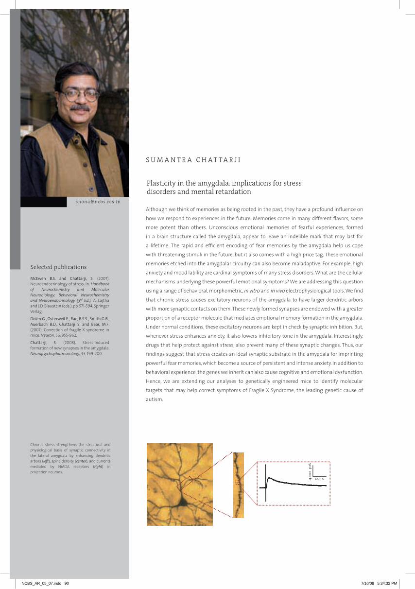
Selected publications
although we think of memories as being rooted in the past, they have a profound influence on
how we respond to experiences in the future. Memories come in many different flavors, some
more potent than others. Unconscious emotional memories of fearful experiences, formed
in a brain structure called the amygdala, appear to leave an indelible mark that may last for
a lifetime. The rapid and efficient encoding of fear memories by the amygdala help us cope
with threatening stimuli in the future, but it also comes with a high price tag. These emotional
memories etched into the amygdalar circuitry can also become maladaptive. For example, high
anxiety and mood lability are cardinal symptoms of many stress disorders. What are the cellular
mechanisms underlying these powerful emotional symptoms? We are addressing this question
using a range of behavioral, morphometric, in vitro and in vivo electrophysiological tools. We find
that chronic stress causes excitatory neurons of the amygdala to have larger dendritic arbors
with more synaptic contacts on them. These newly formed synapses are endowed with a greater
proportion of a receptor molecule that mediates emotional memory formation in the amygdala.
Under normal conditions, these excitatory neurons are kept in check by synaptic inhibition. But,
whenever stress enhances anxiety, it also lowers inhibitory tone in the amygdala. interestingly,
drugs that help protect against stress, also prevent many of these synaptic changes. Thus, our
findings suggest that stress creates an ideal synaptic substrate in the amygdala for imprinting
powerful fear memories, which become a source of persistent and intense anxiety. in addition to
behavioral experience, the genes we inherit can also cause cognitive and emotional dysfunction.
hence, we are extending our analyses to genetically engineered mice to identify molecular
targets that may help correct symptoms of Fragile X Syndrome, the leading genetic cause of
autism.
Plasticity in the amygdala: implications for stress disorders and mental retardation
S U M A N T R A C H A T T A R J I
[email protected] . in
McEwen B.S. and Chattarji, S. (2007). neuroendocrinology of stress. in: Handbook of Neurochemistry and Molecular Neurobiology: Behavioral Neurochemistry and Neuroendocrinology (3rd Ed.), a. Lajtha and J.D. Blaustein (eds.), pp. 571-594, Springer Verlag.
Dolen G., Osterweil E., Rao, B.S.S., Smith G.B., Auerbach B.D., Chattarji S. and Bear, M.F. (2007). Correction of fragile X syndrome in mice. Neuron, 56, 955-962.
Chattarji, S. (2008). Stress-induced formation of new synapses in the amygdala. Neuropsychopharmacology, 33, 199-200.
Chronic stress strengthens the structural and physiological basis of synaptic connectivity in the lateral amygdala by enhancing dendritic arbors (left), spine density (center), and currents mediated by nMDa receptors (right) in projection neurons.
Selected publications
800
600
400
200
0
-200
-400
pA
0.5 0.4 0.3 0.2 0.1 0.0
s
NCBS_AR_05_07.indd 90 7/10/08 5:34:32 PM

91
SU
Ma
nT
ra
Ch
aT
Ta
rJi
1 Impact of chronic stress on excitatory neurons in the lateral amygdala
Aparna Suvrathan, Harini Laxminarasimhan, Rajnish Rao and K. Manish Sharma
Using whole-cell patch-clamp recordings from pyramidal neurons in the lateral amygdala (La), we
are examining the electrophysiological properties of spines formed by chronic immobilization stress
(CiS, 2h/day for 10 days). We find that stress amplifies the ratio of nMDa to aMPa receptor mediated
excitatory post-synaptic currents (EPSCs). Further, the ratio of coefficient of variation (CV) is smaller in
stress-treated cells when the CV of evoked aMPa and nMDa EPSCs is compared in the same cell. This
decrease in CV ratio in stressed neurons is only due to a decrease in the CV of nMDa-EPSC, reflecting
a larger number of nMDa receptor synapses contributing to the EPSC. analysis of mixed miniature
EPSCs with aMPa and nMDa components also suggests that the increase in evoked nMDa currents is
due to addition of nMDa receptors to newly formed spines that do not contain aMPars. Thus, chronic
stress appears to create nMDa-only or “silent synapses” in the La, which in turn could enhance their
capacity for subsequent potentiation. indeed, nMDa receptor-dependent long-term potentiation
(LTP), a synaptic plasticity mechanism for learning and memory, is amplified after chronic stress. We
are now studying the molecular mechanisms underlying these cellular changes.
2 Stress-induced modulation of fear memory and its in vivo encoding in single neurons
Supriya Ghosh and Anupratap Tomar
Stress induced formation of silent synapses and enhancement of LTP is expected to amplify the
functional output of the La. We use Pavlovian auditory fear conditioning to examine the extent to
which stress modulates learning and memory of emotional events. The same pairing of auditory
tones with weak footshock that only elicits moderate levels of freezing in unstressed rats causes
significantly greater freezing in previously stressed rats (Figure 1a). hence, stress-induced generation of
silent synapses may shift amygdalar cells to a state where their capacity for subsequent potentiation
is enhanced, thereby creating an ideal synaptic substrate for affective disorders. These studies,
however, are limited by the fact that function is inferred from analysis at the cellular and behavioral
levels without any online readout of dynamic changes in neuronal activity in the intact animal. To
bridge this gap, we are recording extracellular action potential firing simultaneously from multiple La
neurons in awake, behaving rats. Such single-unit recordings from La neurons show robust increases
in tone responses after fear conditioning (Figure 1b), thereby enabling us to monitor how neural
encoding of fear memories in the amygdala is modulated during and after stressful experiences of
varying duration and intensity. These in vivo multi-electrode recordings are being extended to study
the impact of stress on “place cells” in the hippocampus.
Figure 1. (a) Stress enhances consolidation of memory formed after auditory fear conditioning. (b) Fear conditioning induces increase in tone-evoked spike firing in lateral amygdala neurons, monitored using multi-channel single-unit recording system in behaving animals.
NCBS_AR_05_07.indd 91 7/10/08 5:34:32 PM
3 Delayed effects of a single episode of stress on synaptic inhibition
Aparna Suvrathan
While the above results suggest a role for stress-induced modulation of excitatory synaptic
transmission in enhancing fear, inhibitory synaptic transmission also regulates functional
output of the amygdala. hence, we are analyzing the relationship between stress, anxiety and
synaptic inhibition in the La. The same chronic stress that enhances anxiety causes a reduction
in GaBaergic inhibitory currents in La principal neurons. Strikingly, this inverse relation between
anxiety and synaptic inhibition emerges even when animals are exposed to a single 2-hour
episode of stress. although this brief stress has no significant effect on anxiety a day later, it leads
to higher anxiety 10 days later. Strikingly, there is a significant reduction in the mean amplitude
and frequency of miniature GaBaergic inhibitory postsynaptic currents (miPSCs) in La principal
neurons only 10 days, but not a day, after acute stress. Thus, gradual suppression of synaptic
inhibition in the La parallels the slow build-up of anxiety even after a brief stressor.
4 From animal models to therapeutic strategies and back
Rajnish Rao, Anuparatp Tomar, Aparna Suvrathan, Shilpa Ravinder, Shobha Anil and Anup Pillai
Our results on the delayed effects of acute stress have led to animal models that capture various
aspects of emotional disorders, such as Post-Traumatic Stress Disorder (PTSD), in humans.
Treatment with the anxiolytic diazepam before acute stress prevents reduction in synaptic
inhibition, spinogenesis, and enhanced anxiety 10 days after acute stress. Strikingly, diazepam
given after acute stress also reverses the decrease in synaptic inhibition 10 days later. Further,
corticosterone treatment preceding acute stress blocks the eventual increase in anxiety and
La spine-density. Conversely, in adrenalectomized animals acute stress leads to even greater
anxiety, which can be attenuated with corticosterone treatment. These animal studies are
consistent with clinical reports showing that cortisol treatment reduces the cardinal symptoms
of PTSD. Since stress and emotional trauma have been implicated in depressive disorders, we are
also analyzing the synaptic and behavioral effects of antidepressants, such as fluoxetine and
tianeptine, on the amygdala.
5 Negative versus positive experiences
Ruchi Malik
Environmental enrichment (EE) – consisting of increased exercise, social interactions and
exposure to objects that encourage exploration – is known to have beneficial effects in the
cortex and hippocampus of the rodent brain, e.g. enhanced dendritic arbors, spine density, LTP,
and learning. Paradoxically, we observed the same effects in the amygdala, not as a result of
a positive experience like EE, but after an aversive experience like chronic stress. This raises an
intriguing question – how would the amygdala respond to EE? if EE elicits cellular changes in
the amygdala similar to the hippocampus, then our earlier results predict EE to enhance anxiety
and fear learning. But one expects positive experiences to reduce fear and anxiety. On the other
hand, if EE does reduce fear and anxiety, would it induce cellular changes in the amygdala that
are different from the hippocampus? indeed, we find that EE decreases amygdala-dependent
auditory fear conditioning, as well as anxiety. But, EE does not enhance spine density in the La.
in contrast, the same EE enhances hippocampal spine density and contextual fear conditioning.
Thus, plasticity elicited by a positive experience also appear to be different in the amygdala.
6 Reversal of symptoms of Fragile X Syndrome
Aparna Suvrathan and Sonal Kedia
Fragile X Syndrome (FXS), the most commonly inherited form of mental retardation and the
leading genetic cause of autism, is caused by transcriptional silencing of the FMR1 gene. although
moderate to severe cognitive impairment is a key feature of FXS, patients also display severe
problems related to anxiety, attention deficit, and hyperactivity. The Fmr1-knockout (KO) mouse
is a powerful animal model for investigating symptoms of human FXS. We have examined two
NCBS_AR_05_07.indd 92 7/10/08 5:34:32 PM
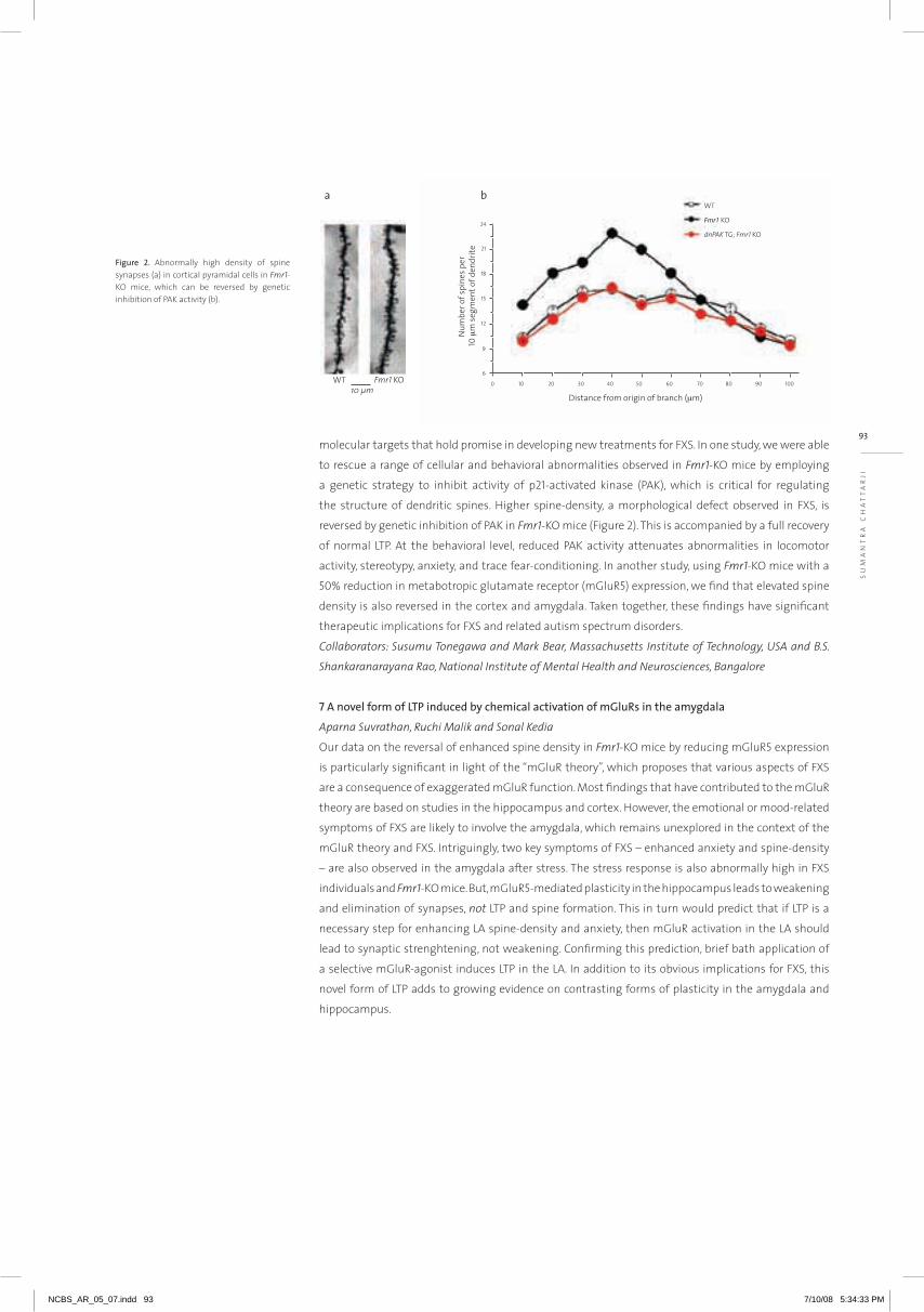
93molecular targets that hold promise in developing new treatments for FXS. in one study, we were able
to rescue a range of cellular and behavioral abnormalities observed in Fmr1-KO mice by employing
a genetic strategy to inhibit activity of p21-activated kinase (PaK), which is critical for regulating
the structure of dendritic spines. higher spine-density, a morphological defect observed in FXS, is
reversed by genetic inhibition of PaK in Fmr1-KO mice (Figure 2). This is accompanied by a full recovery
of normal LTP. at the behavioral level, reduced PaK activity attenuates abnormalities in locomotor
activity, stereotypy, anxiety, and trace fear-conditioning. in another study, using Fmr1-KO mice with a
50% reduction in metabotropic glutamate receptor (mGlur5) expression, we find that elevated spine
density is also reversed in the cortex and amygdala. Taken together, these findings have significant
therapeutic implications for FXS and related autism spectrum disorders.
Collaborators: Susumu Tonegawa and Mark Bear, Massachusetts Institute of Technology, USA and B.S.
Shankaranarayana Rao, National Institute of Mental Health and Neurosciences, Bangalore
7 A novel form of LTP induced by chemical activation of mGluRs in the amygdala
Aparna Suvrathan, Ruchi Malik and Sonal Kedia
Our data on the reversal of enhanced spine density in Fmr1-KO mice by reducing mGlur5 expression
is particularly significant in light of the “mGlur theory”, which proposes that various aspects of FXS
are a consequence of exaggerated mGlur function. Most findings that have contributed to the mGlur
theory are based on studies in the hippocampus and cortex. however, the emotional or mood-related
symptoms of FXS are likely to involve the amygdala, which remains unexplored in the context of the
mGlur theory and FXS. intriguingly, two key symptoms of FXS – enhanced anxiety and spine-density
– are also observed in the amygdala after stress. The stress response is also abnormally high in FXS
individuals and Fmr1-KO mice. But, mGlur5-mediated plasticity in the hippocampus leads to weakening
and elimination of synapses, not LTP and spine formation. This in turn would predict that if LTP is a
necessary step for enhancing La spine-density and anxiety, then mGlur activation in the La should
lead to synaptic strenghtening, not weakening. Confirming this prediction, brief bath application of
a selective mGlur-agonist induces LTP in the La. in addition to its obvious implications for FXS, this
novel form of LTP adds to growing evidence on contrasting forms of plasticity in the amygdala and
hippocampus.
μ
Fmr1
μ
Figure 2. abnormally high density of spine synapses (a) in cortical pyramidal cells in Fmr1-KO mice, which can be reversed by genetic inhibition of PaK activity (b).
SU
Ma
nT
ra
Ch
aT
Ta
rJi
NCBS_AR_05_07.indd 93 7/10/08 5:34:33 PM

Selected publications
The lab is in engaged in understanding the regulation of axonal transport of cargoes that are
targeted to synapses. axonal transport carries a variety of cargoes from the cell body to the
synapse and from the synapse to the cell body, known respectively as anterograde and retrograde
transport. Transport along the axonal highway takes place using molecular motors that move
on microtubule tracks by hydrolyzing aTP. Most kinesin family motors move to the plus-end of
microtubules and thus carry cargo to the synapse while dynein motors carry cargo to the minus-
end of microtubules and thus away from the synapse. Transport in neurons is an essential
process. This has been demonstrated, for instance, by progressive motor neuropathies such as
human CMT2A observed in patients carrying mutations in motor protein genes.
neuronal transport is very complex and its regulation is poorly understood. To gain further
insights, we study axonal transport of certain cargoes carried by Kinesin-3/UNC-104 and
Kinesin-i/UnC-116, two well-characterized kinesin family motor proteins. Kinesin-3/UNC-104
is known to transport synaptic vesicle proteins. Kinesin-i/UnC-116 is known to transport
mitochondria and to influence transport of synaptic vesicle proteins. Presence of both these
cargoes at synapses is very important for neuronal function. We visualize synaptic vesicles using
synaptic vesicle proteins fused to GFP and mitochondria using a matrix targeted GFP. These
markers report the localization of both synaptic vesicles and mitochondria not only at synapses
but also along the axon. We broadly address the following questions: (1) Cargo recognition by the
UnC-104 motor, (2) regulation of mitochondrial and synaptic vesicle transport by Kinesin-i and
its cargo adaptors. We address these questions using a combination of genetic, live imaging and
molecular techniques using the C. elegans model.
Regulation of axonal transport
S A N D H Y A P K O U S H I K A
[email protected] . in
Koushika, S.P. (2007). ‘JiP’ing along the axon: the complex roles of JiPs in transport. BioEssays (in press).
image of worm showing fluorescent mitochondria in mechanosensory neurons
NCBS_AR_05_07.indd 94 7/10/08 5:34:33 PM

95
Sa
nD
hY
a P
KO
US
hiK
a
1 Cargo recognition by the Kinesin-3/UNC-104 motor
Jitendra Kumar
UnC-104 is a major synaptic vesicle motor in multiple model systems. This protein binds vesicles
through its Ph-domain. The lipid binding Ph domain of UnC-104 has been shown to be essential
for function. Further, an orthologous Ph-domain with the same in vitro lipid binding specificity does
not fully restore UnC-104 function in vivo, suggesting a role for other proteins. We therefore began
studying how the UnC-104 Ph domain recognizes cargo. We identified an unc-104 allele, (e1265) which
has a single point mutation D1497N in the Ph domain that severely affects the locomotion of the
mutant animals.
To determine how a single amino acid change could dramatically alter function, we built a structural
model of the UnC-104 Ph domain. On docking the reported substrate PiP2 onto this model, we
observed that the highest probability of binding the lipid was in the region juxtaposed to D1497
(Figure 1). To further understand the role of this lesion we isolated intragenic suppressors of e1265
and sequenced the alleles obtained. We identified two intragenic suppressors within the Ph domain
that restored locomotion to the e1265 allele. These intragenic suppressors have two compensatory
changes in the Ph domain, namely either an M1540i lesion or an R1501Q lesion. The M1540i lesion was
independently isolated multiple times. This residue acts at a distance of over 20Å and could mediate
suppression effects through intermediate residues. The R1501Q is present in the same loop as D1497n
and probably mediates its suppression effects locally. These suppressors also largely restore transport
in the shorter motor neurons as seen by recovery of synaptic vesicle markers in synapses. however in
longer mechanosensory neurons the D1497n M1540I or D1497n R1501Q alleles only partially restore
transport. Live imaging of GFP::raB-3, a synaptic vesicle cargo marker, shows that anterograde flux in
mechanosensory neurons is significantly reduced in unc-104(e1265) animals and is partially restored
in the intragenic suppressor alleles (Figure 2). reports in literature suggest that cargo binding may
influence the velocity profile of UnC-104. We are about to test whether the D1497n lesion and the
other suppressor alleles affect the in vivo motility properties of the UnC-104 motor. in summary
we have identified a new amino acid that we think influences UnC-104 Ph domain function by
modulating its ability to bind cargo.
We are also interested in other molecules that act along with UnC-104 to enable cargo recognition.
That other proteins play roles in the ability of the Ph domain to bind cargo has been demonstrated by in
vitro biochemistry showing reduced binding of the UnC-104 Ph domain to vesicles devoid of proteins.
We have taken a genetic approach to identify such molecules by isolating extragenic suppressors
of the e1265 allele. Characterization thus far has demonstrated that each such suppressor acts in
neurons to partially restore transport. We are further characterizing these extragenic suppressors for
allele-specificity by testing whether they restore locomotion to an unc-104 allele with a motor domain
mutation or restore viability to an unc-104 null allele. The suppressors that act only on the e1265 allele
Figure 1. A structural model of the UNC-104 PH-domain showing the region of maximum probability of docking PI(4,5)P2 [arrow] which is near the D1497 residue.
8
7
6
5
4
3
2
1
0
wild type e1265 e1265 tb 120 (M15401)
Anterograde
Retrograde
e1265 tb 107 (R1501Q)
Num
ber o
f syn
aptic
vesic
les
Figure 2. Flux of synaptic vesicles reduced in unc-104(e1265) animals and is partially restored in the intragenic suppressors.
NCBS_AR_05_07.indd 95 7/10/08 5:34:35 PM

are of greatest interest in identifying molecules that act along with the UnC-104 Ph-domain in the
synaptic vesicle transport pathway.
Collaborator: R. Sowdhamini, NCBS.
2 Regulation of synaptic vesicle transport by the Kinesin-I adaptor complex
Bikash Chandra Choudhury
The Kinesin-i motor is known to influence synaptic vesicle transport. The Kinesin-i motor is a
heteroteramer that consists of two heavy chains (KhC) and two light chains (KLC). The heavy chains
bind microtubules and hydrolyze aTP while the light chains enable binding to certain cargoes.
The role of Kinesin-i in synaptic vesicle transport requires a multi-protein adaptor complex found
associated with the Kinesin-i motor (Figure 3). The main adaptor that links the Kinesin-i motor to its
cargo is JiP3, a c-JUn kinase interacting protein. Members of the JiP family are JnK cascade kinase
scaffolding proteins. They function as Kinesin-i cargo adapters and play complex roles in transport.
For instance unc-16, the JiP3 orthologue in C. elegans, has been shown to function in an unc-104
bypass pathway. in unc-104; unc-16 double mutants certain synaptic vesicle proteins are able to exit
the cell body and weakly suppress the locomotor defects of unc-104 mutants. it has been proposed
that these effects are mediated by binding vesicular cargoes to alternate motors. Our current data
suggest that in unc-16 mutants there is a significant reduction in the average anterograde velocity
of GFP::raB-3 marked vesicles with only small effects on retrograde transport. The observed change
in velocity profiles supports the hypothesis that certain synaptic vesicle proteins are transported by
alternate motors only in the anterograde pathway. We are also assessing whether the unc-16 bypass
pathway acts on all or a subset of synaptic vesicle proteins. We are also examining the role of other
known members of the Kinesin-i adaptor complex (e.g. UNC-14) in the unc-104 bypass pathway.
3 Regulation of mitochondrial transport by Kinesin-I and its adaptor complex
Guruprasad Reddy, Swathi Reddy and S. Mohan
The Kinesin-i motor is known to transport mitochondria in other model systems. We tested whether
the Kinesin-i motor and other associated molecules known to transport vesicular cargoes influence
mitochondrial transport (Figure 3). as expected the lack of Kinesin-i motor greatly reduced the
number of mitochondria in axons. Members of the complex such as unc-14 and unc-51 also reduced
number of mitochondria in axons. Surprisingly, we found that lack of the vesicular adaptor UNC-16/
JiP3 dramatically increases the number of mitochondria (Figure 4). in unc-16, Kinesin-i motor double
mutant animals the number of mitochondria in axons remains high. Thus we see that UNC-16/JiP3
has roles independent of the Kinesin-i motor in regulating numbers of mitochondria in axons (Figure
4). On live imaging of transport in unc-16 animals we observed a decrease in average retrograde
velocity but no effect on anterograde velocity. Thus the effects of unc-16 on regulating mitochondrial
numbers in axons may occur through retrograde transport in a Kinesin-i motor independent pathway.
For at least one other member of the Kinesin-i adaptor complex we did not find such Kinesin-i motor
independent roles. The unc-51 adaptor Kinesin-i motor double mutants did not show any change in
mitochondrial numbers in axons when compared with Kinesin-i motor mutants alone. Some Kinesin-i
motor independent effects of unc-16 could occur through JnK stress signaling pathways. Therefore
we are testing the possible role of JnK cascade kinases in altering mitochondrial numbers in axons.
Figure 3. Schematic representation the Kinesin-I motor and cargo adaptor complex formed by UNC-16, UNC-14 and UNC-51. UNC-76 is likely to act with the JIP protein UNC-16 to activate the motor.
Figure 4. UNC-16 acts in a Kinesin-I motor independent pathway to regulate mitochondrial numbers in axons.
- +
KHC/UNC-116
KLC-2
UNC-76
UNC-69DLK-1?JKK-1
JNK-1
UNC-16
UNC-14
UNC-51
NCBS_AR_05_07.indd 96 7/10/08 5:34:37 PM
97
Sa
TY
aJi
T
Ma
YO
r
NCBS_AR_05_07.indd 97 7/10/08 5:34:38 PM
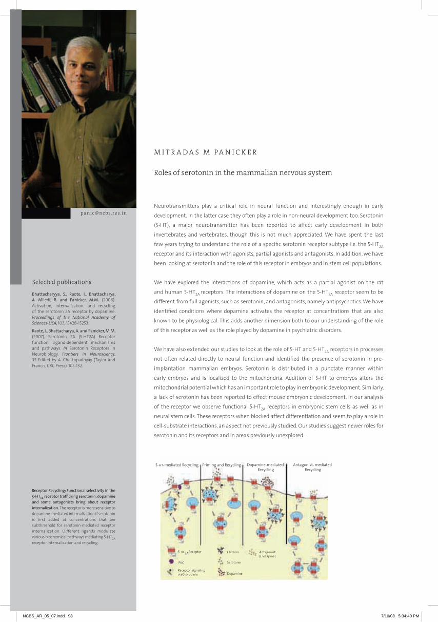
Roles of serotonin in the mammalian nervous system
M I T R A D A S M PA N I C K E R
[email protected] . inneurotransmitters play a critical role in neural function and interestingly enough in early
development. in the latter case they often play a role in non-neural development too. Serotonin
(5-hT), a major neurotransmitter has been reported to affect early development in both
invertebrates and vertebrates, though this is not much appreciated. We have spent the last
few years trying to understand the role of a specific serotonin receptor subtype i.e. the 5-hT2a
receptor and its interaction with agonists, partial agonists and antagonists. in addition, we have
been looking at serotonin and the role of this receptor in embryos and in stem cell populations.
We have explored the interactions of dopamine, which acts as a partial agonist on the rat
and human 5-hT2a receptors. The interactions of dopamine on the 5-hT2a receptor seem to be
different from full agonists, such as serotonin, and antagonists, namely antipsychotics. We have
identified conditions where dopamine activates the receptor at concentrations that are also
known to be physiological. This adds another dimension both to our understanding of the role
of this receptor as well as the role played by dopamine in psychiatric disorders.
We have also extended our studies to look at the role of 5-hT and 5-hT2a receptors in processes
not often related directly to neural function and identified the presence of serotonin in pre-
implantation mammalian embryos. Serotonin is distributed in a punctate manner within
early embryos and is localized to the mitochondria. addition of 5-hT to embryos alters the
mitochondrial potential which has an important role to play in embryonic development. Similarly,
a lack of serotonin has been reported to effect mouse embryonic development. in our analysis
of the receptor we observe functional 5-hT2a receptors in embryonic stem cells as well as in
neural stem cells. These receptors when blocked affect differentiation and seem to play a role in
cell-substrate interactions, an aspect not previously studied. Our studies suggest newer roles for
serotonin and its receptors and in areas previously unexplored.
Bhattacharyya, S., Raote, I., Bhattacharya, A. Miledi, R. and Panicker, M.M. (2006). activation, internalization, and recycling of the serotonin 2a receptor by dopamine. Proceedings of the National Academy of Sciences-USA, 103, 15428-15253.
Raote, I., Bhattacharya, A. and Panicker, M.M. (2007). Serotonin 2a (5-hT2a) receptor function: Ligand-dependent mechanisms and pathways. In Serotonin receptors in neurobiology. Frontiers in Neuroscience, 35 Edited by a. Chattopadhyay (Taylor and Francis, CrC Press). 105-132.
Receptor Recycling: Functional selectivity in the 5-HT2A receptor trafficking serotonin, dopamine and some antagonists bring about receptor internalization. The receptor is more sensitive to dopamine-mediated internalization if serotonin is first added at concentrations that are subthreshold for serotonin-mediated receptor internalization. Different ligands modulate various biochemical pathways mediating 5-hT2a receptor internalization and recycling.
Selected publications
NCBS_AR_05_07.indd 98 7/10/08 5:34:40 PM

99
MiT
ra
Da
S M
Pa
niC
KE
r
1 Role of dopamine and mechanisms of internalization and ‘priming’ of the 5-HT2A receptor
Samarjit Bhattacharyya, Ishier Raote and Aditi Bhattacharya
neurotransmitters have their specific and cognate receptors and most studies are directed to
understanding these specific interactions. interactions between neurotransmitter systems are often
seen but these are usually indirect. Serotoninergic and dopaminergic interactions are well known and
are particularly important in the study of psychosis. antipsychotics and antidepressants largely target
the receptors or the ligand of these two systems. The partial activation of the 5-hT2a receptor by
dopamine has been known for some time but the mechanism and its physiological relevance has not
been understood. We have now established that though dopamine and serotonin activate the 5-hT2a
receptor the intracellular pathways associated with the activation are overlapping but different. in
addition, we have identified conditions where these interactions would be physiologically relevant. We
have discovered that if concentrations of serotonin sub-threshold for the internalization of the 5-hT2a
receptor are applied, the receptor becomes 10-fold more sensitive to dopamine. We have termed this
‘priming’ and this effect is maintained only transiently. The temporal order in which the ligands are
applied is also critical and suggests a condition in which these two neurotransmitters may function
like an anD gate. These results should help in understanding the role of these ligands and the 5-hT2a
receptor in behavior and also generate fresh clinical approaches in treating psychiatric disorders. More
recently, in collaboration with Dr. Subeer Majumdar, nii, new Delhi, we have made transgenic mice
that express the rat 5-hT2a –EGFP receptor which will be used to study the receptor both in the brain
as well as in other tissues.
Collaborator: Sabeer Majumdar, National Institute of Immunology, Delhi
2 Examining the differences between human and rat 5-HT2A receptor subtypes
Aditi Bhattacharya and Shobhana Shankar
Most studies on the 5-hT2a receptor have used the rat receptor as a model to study ligand interactions,
drug binding as well as the second messenger pathways activated by the receptor. Our initial studies
have also focused on the rat homolog. More recently, we have begun work on the human homolog and
observed that there are subtle but perhaps critical differences in its interaction with agonists, partial
agonists and antagonists. Since this may have important implications in clinical approaches we have
examined the human homolog in greater detail. We have observed that the human receptor exhibits
increased sensitivity to serotonin and also seems to have an increased repertoire of intracellular
pathways. its sensitivity to dopamine is also higher. Site-specific mutagenesis studies have helped to
identify key residues that are involved in these interactions. in addition, we have been working with
a naturally occurring variant of this receptor that occurs with high frequency the human population.
We have noticed that there are differences in the manner in which this variant receptor interacts with
ligands and this could lead us to control receptor effects and activity better.
Figure 1. Time series of dopamine-mediated internalization of rat 5-HT2A receptor in HEK293 cells
Figure 2. Time series of serotonin-mediated internalization of human 5-HT2A receptor in HEK293 cells
NCBS_AR_05_07.indd 99 7/10/08 5:34:43 PM

3 Role of 5-HT2A receptors in cell adhesion
Basudha Basu and Rupam Chowdhury
Serotonin and its receptors have been known to play an important role in neural systems. its role in
non-neural development and function outside the nervous system are not as widely appreciated.
not surprisingly, the role of 5-hT2a receptors in areas outside the nervous system has also not been
extensively studied. We had in the last few years noticed that overexpression of the 5-hT2a receptor
seems to affect cell-substrate adhesion in hEK 293 cells. The adhesion seems to be decreased by
receptor-specific antagonists and adhesion is increased on application of serotonin. We have now
determined that cytoskeletal changes that accompany changes in adhesion can be regulated
by serotonin through the 5-hT2a receptor and that the major second messenger pathway, which
is associated with protein kinase C, does not play a role in the maintenance of adhesion. Similar
roles seem to be played by the 5-hT2a receptor in neural stem adhesion and embryonic stem cell
differentiation in in vitro experiments. We have identified a number of genes that are downregulated
on inhibiting the 5-hT2a receptor in in vitro studies. Extending these studies to in vivo models, we find
that the same genes are downregulated in the brain on exposure to an antagonist of the receptor.
These studies indicate that antagonists to the 5-hT2a receptor may bring about changes in a new and
unexpected manner and changes in the adhesion properties may play a part in the changes brought
about by such ligands.
Collaborator: Sanjeev Galande, National Centre for Cell Science, Pune
4 Characterization and differentiation of embryonic stem cells
Imtiaz Zafar, Basudha Basu, Tejaswini Sharangdhar, Deepika Kaveri, Praveen Kumar, Sushmita Saha and
Sucharita Sen
Mammalian embryonic stem cells serve both as valuable models systems for developmental biology
as well as substrates for cell replacement therapies. We have been looking at the differentiation of
embryonic stem cells towards neural lineages. a report on the presence of serotonin, a neurotransmitter
in mouse embryonic stem cells, led us to look for serotonin in human embryonic stem cells and also
its receptors. We find that serotonin is present and distributed in a punctate pattern in both human
and embryonic stem cells and both isoforms of tryptophan hydroxylase are present, indicating that
serotonin is synthesized in ES cells. in addition, we find that a few of the serotonin receptor subtypes
are also expressed in mammalian ES cells. We have established that one of the receptor subtypes,
i.e. 5-hT2a, is functional and blocking this receptor affects the differentiation of ES cells. We are
presently exploring the mechanism by which serotonin and its receptors affect ES cell growth and
differentiation.
5 Role of serotonin in pre-implantation embryos
Basudha Basu
The presence of serotonin in mammalian ES cells led us to look for its presence in earlier stages in
development, namely pre-implantation embryos. Pre-implantation mouse embryos were examined
at various stages for serotonin using immunocytochemistry as well as multi-photon microscopy.
The latter technique allows us to visualize 5-hT in live embryos. We find that serotonin is present in
early embryos including oocytes and that it is distributed in punctate structures. in pre-implantation
embryos, both TPh2, the neural isoform of tryptophan hydroxylase and the serotonin transporter are
present. in addition, we find that serotonin is localized to mitochondria in embryos and can increase
mitochondrial potential. Lack of serotonin and changes in mitochondrial potential have both been
known to disrupt embryonic development. We are now determining the mechanism by which
serotonin affects mitochondrial potential.
Collaborators: Sudipta Maiti, TIFR, Mumbai and V. Sriram, NCBS
Figure 3. Neurospheres generated from the mouse brain used as a model to study cell-substratum adhesion
Figure 4. Multiphoton imaging of serotonin localization in mouse embryos (a) Two-cell stage (b) Eight-cell stage c. Blastocyst
a
b
c
NCBS_AR_05_07.indd 100 7/10/08 5:34:43 PM
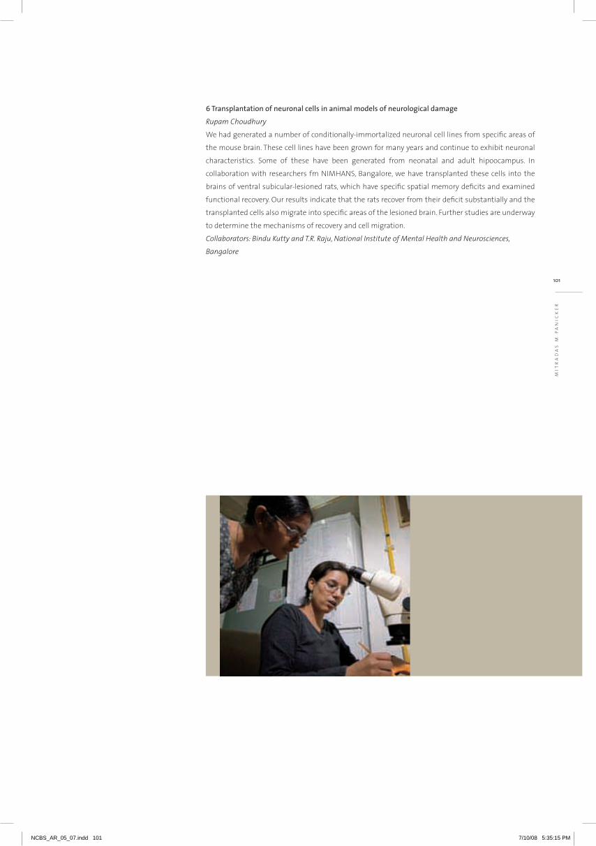
101
MIT
RA
DA
S M
PA
NIC
KE
R
6 Transplantation of neuronal cells in animal models of neurological damage
Rupam Choudhury
We had generated a number of conditionally-immortalized neuronal cell lines from specific areas of
the mouse brain. These cell lines have been grown for many years and continue to exhibit neuronal
characteristics. Some of these have been generated from neonatal and adult hipoocampus. In
collaboration with researchers fm NIMHANS, Bangalore, we have transplanted these cells into the
brains of ventral subicular-lesioned rats, which have specific spatial memory deficits and examined
functional recovery. Our results indicate that the rats recover from their deficit substantially and the
transplanted cells also migrate into specific areas of the lesioned brain. Further studies are underway
to determine the mechanisms of recovery and cell migration.
Collaborators: Bindu Kutty and T.R. Raju, National Institute of Mental Health and Neurosciences,
Bangalore
NCBS_AR_05_07.indd 101 7/10/08 5:35:15 PM
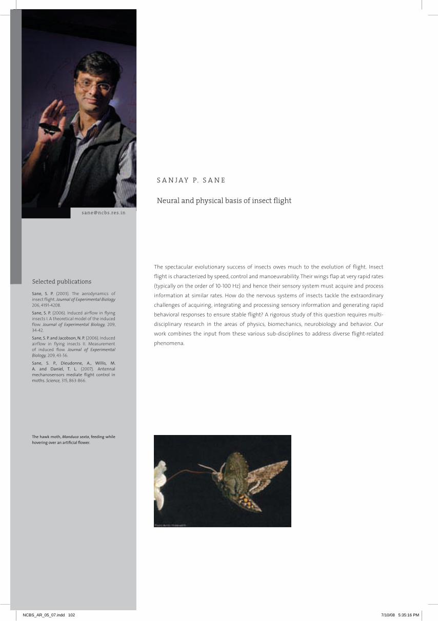
Selected publications
Neural and physical basis of insect flight
S A N J AY P. S A N E
[email protected] . in
The spectacular evolutionary success of insects owes much to the evolution of flight. Insect
flight is characterized by speed, control and manoeuvrability. Their wings flap at very rapid rates
(typically on the order of 10-100 Hz) and hence their sensory system must acquire and process
information at similar rates. How do the nervous systems of insects tackle the extraordinary
challenges of acquiring, integrating and processing sensory information and generating rapid
behavioral responses to ensure stable flight? A rigorous study of this question requires multi-
disciplinary research in the areas of physics, biomechanics, neurobiology and behavior. Our
work combines the input from these various sub-disciplines to address diverse flight-related
phenomena.
Sane, S. P. (2003). The aerodynamics of insect flight. Journal of Experimental Biology 206, 4191-4208.
Sane, S. P. (2006). Induced airflow in flying insects I. A theoretical model of the induced flow. Journal of Experimental Biology, 209, 34-42.
Sane, S. P. and Jacobson, N. P. (2006). Induced airflow in flying insects II. Measurement of induced flow. Journal of Experimental Biology, 209, 43-56.
Sane, S. P., Dieudonne, A., Willis, M. A. and Daniel, T. L. (2007). Antennal mechanosensors mediate flight control in moths. Science, 315, 863-866.
The hawk moth, Manduca sexta, feeding while hovering over an artificial flower.
Selected publications
NCBS_AR_05_07.indd 102 7/10/08 5:35:16 PM

103
SA
NJA
Y S
AN
E
1 The physical basis of insect flight
On the physical front, how do the flapping wings of insects generate sufficient aerodynamic forces to
make flight possible and how do insects modulate these forces to determine their flight trajectories
when chasing territorial interlopers, locating odor sources or finding mates? To address this question,
we built a dynamically scaled robotic flapper (affectionately called Robofly) to study the basic fluid
dynamical principles that make flapping flight of insects fundamentally distinct from fixed-wing
flight of airplanes. From these studies, we were able to show that the high lift generated by flapping
wings can be sustained over the entire duration of the wing stroke, unlike their fixed wing counterpart
which tends to stall. This study resulted in a semi-empirical model that could predict the instantaneous
forces generated by a flapping wing performing any arbitrary kinematic pattern.
Currently, we are extending these techniques to study the effect of wing flexibility on aerodynamic
force generation. With increase in size of the insect, their wings tend to show greater flexibility with
direct consequences to their ability to generate aerodynamic forces. This study will also extend into
the biomaterial aspects of wing flexibility to address how wing flexibility may influence longevity of
the wing structure through the typical lifetime of an animal. In addition, we are also studying the
gross flows around insect bodies during flight and trying to understand how these flows influence
various aspects of insect physiology such as thermoregulation, odor detection etc. Apart from the
obvious biological implications of these studies, these results will also help in development of
optimally performing wings for micromechanical robotic insects. These aspects are being studied in
close collaboration with roboticists and engineers, at the University of Delaware.
2 The neural basis of flight behavior
On the neurobiological front, how do flying insects use various sensors distributed over their body to
acquire information about their environment and how is this information used in stabilizing their
flight? We have recently begun investigating this question with specific focus on the role of antennae
in acquisition and processing of self-generated and externally-generated sensory cues that mediate
flight control in hawk moths. Our behavioral studies revealed that antennae play a very crucial role in
mediating flight control: insects lacking the distal segment of their antennae (i.e. flagella) are unable
to fly in a coordinated manner. However, when the flagellar integrity is restored, their flight ability can
be rescued. Our electrophysiological studies show that individual mechanosensory neurons at the
base of the antennae are tuned to vibratory motion of the antennae and capable of transducing cues
essential for flight control. Visualization of the underlying neural circuitry reveals that a bulk of the
antennal mechanosensory information is relayed to an area of the brain called the AMMC (Antennal
Motor and Mechanosensory Centre), before it is transmitted to the flight motor units. Thus, antennal
mechanosensors are involved in mediating flight control in insects.
Our studies will endeavour to throw light on the evolution of antennal diversity among insects in
relation to their flight mechanics, as well as the specific roles of the antenna and the AMMC in flight
control. Ongoing research from my laboratory also addresses flight under natural circumstances.
We will study how the antennal mechanosensory modality combines with visual inputs to enable
flight in moths and butterflies. We are keen to diversify this study into the area of flight energetics,
thermoregulation and behavioral ecology of live, migrating insects.
Figure 2. Antennal Anatomy.
Figure 1. Air flow measurements around a flying insect.
NCBS_AR_05_07.indd 103 7/11/08 2:59:13 PM

Selected publications
Genetic analysis of chemosensory perception
O B A I D S I D D I Q I
Olfactory behavior in Drosophila is partly inborn and partly acquired. Our group is studying
ontogeny of olfactory behavior and odor learning ability in the fly. The report by Sarit Pati Goswami
et al. shows that newly born larvae are attracted by low concentration of ethyl acetate within
minutes of eclosion irrespective of the odor environment, but sensitivity to odorants increases
with age. Aversion to high concentration takes several hours to develop. In the previous report,
we had shown that the multiphasic learning retention curves, in the third instar larvae can be
decomposed by a simple graphic procedure into three distinct phases; short term memory (STM),
middle term memory (MTM) and long term memory (LTM). We now show that these phases
correspond to exactly STM, MTM and LTM as revealed by study of learning mutants and effect of
anesthetics and other memory ablating treatments.
Tuhin Subhra Chakraborty et al. describe changes in the sensitivity of chemoreceptors that
accompany imaginal conditioning. They find that odor imprinting increases the affinity of the
chemoreceptor to several different ligands, all acting on the same receptor but as Jawaid Ahsan
shows, there is no indication of transcriptional regulation. The first effect of odor imprinting is
thus, an enhancement of peripheral sensitivity.
Type III sensillae contain two receptors Or22a and Or85b. The A neuron projects to the glomerulus DM2. Recordings from the sensillum permit us to study the effect of conditioning on the olfactory pathway described in this report.
Odor
[email protected] . in
Selected publications
Chakraborty, T.S., Prabhakar, S., Kumar, S. and Siddiqi, O. (2006). Neural correlates of imaginal conditioning in Drosophila melanogaster. 4th IBRO-FAONS Congress, Hong Kong.
Chakraborty, T.S., Prabhakar, S., Mahapatra, S., Goswami, S.P., Kumar, S. and Siddiqi, O. (2007). Increased sensitivity of olfactory receptor neurons correlate to imaginal conditioning in Drosophila melanogaster. Society for Neuroscience, San Diego, USA.
Or22a
DM2
NCBS_AR_05_07.indd 104 7/10/08 5:35:17 PM

105
1 Early development of olfactory response in Drosophila larvae
Sarit Pati Goswami, Sunil Prabhakar, Latha Murugesan and Agila Somasundaram
The olfactory responses of Drosophila imago undergo distinctive changes after eclosion. When the flies
are exposed to certain odorants, attraction towards these chemicals increase. This phenomenon called
‘Imaginal conditioning’ has been described in these reports. We are now studying the development of
olfactory behavior in the larva.
Larval response can be analyzed by tracking individual larvae in an odor gradient. Single larvae are placed
at the centre of a petri dish and their movement towards an odor source is tracked videographically
(Figure 1c). Analysis of these tracks provides information about several features of the response.
Freshly eclosed larvae have an inborn attraction towards ethyl acetate (EA) (Figure 1a). Their movements
are however, disoriented and wayward and sensitivity to the odorant is low. With age, the ability to
orient towards the odor source improves and sensitivity increases (Figure 2a). Newly born larvae,
cultured on cornmeal or a synthetic medium with or without EA, show the same attractive or aversive
orientation to the odor source. 20 hour old larvae, exposed to 10-4 dilution of EA however, tend to move
away from concentrations of the odorant higher than 10-4 (Figure 2c).
2 Olfactory memory in the larvae of Drosophila melanogaster
Mohammed Bin Abu Baker, Malavika Murugan, Dushyant Mishra, Mukund Thattai and Obaid Siddiqi
We are studying the memory of aversive conditioning in Drosophila larvae. The learning retention
curves obtained from repetitive electroshock training are polyphasic and can be analyzed by a simple
graphic procedure, originally due to Scatchard, into three monophasic component each decaying
exponentially at a fixed rate. The three component of memory curve, short term (STM), middle term
(MTM) and long term (LTM) display distinct properties in kinetics of their formation and decline (Abu
Baker et al, NCBS report 2005). One might ask whether the dissociation of polyphasic memory curve
into distinct components STM, MTM and LTM is merely an algebraic manipulation or reveals something
real about memory. We present evidence that STM, MTM and LTM obtained by our analysis correspond
exactly to components of larval memory determined by independent experiments.
OB
AID
SID
DIq
I
Figure 1. Movement of newly eclosed larvae towards 10-3 EA. Larva starts at S, the centre of the plate; E is the end of the track. The displacement vector SE represents individual responses. The odor source is placed along the x-axis. The average displacement 1/N (∑Dx) towards the source is a measure of attraction. The divergence angle, θ, between the displacement vector and abscissa 1/N (∑θ), provides a measure of avoidance. (N=20)
Figure 2. Development of attraction and aversion. Larvae were cultured on complete media (2a). Newly born larvae are less responsive to the odorant (EA) compared to 20 hr old larvae. Larvae are cultured on a synthetic medium (2b and 2c). Larvae conditioned with 10-4 EA (CR) and unconditioned larvae (UCR) show attraction at birth (2b), but develop an avoidance response at concentrations higher than 10-4 when 20 hr old (2c).
dx
E
S
a b c
x-di
spla
cem
ent
Dive
rgen
ce (d
egre
es)
Dive
rgen
ce (d
egre
es)
1.11.00.90.80.70.60.50.40.30.20.10.0
-6 -5 -50
20
40
60
80
100
-4 -3 -2 -1 -4 -3 -2 -6 -5 -4 -3 -2-0.1
Control Control
newly born larve
20 hr old larvae
Log dilution (EA) Log dilution (EA)Log dilution (EA)
CRUCRCornmeal
Control0
20
40
60
80
100
CRUCRCornmeal
2a 2b 2c
NCBS_AR_05_07.indd 105 7/10/08 5:35:18 PM
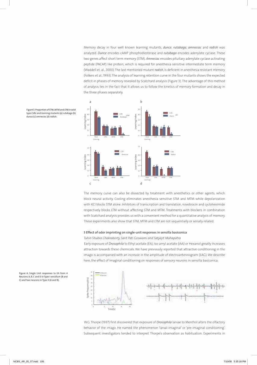
Memory decay in four well known learning mutants, dunce, rutabaga, amnesiac and radish was
analyzed. Dunce encodes cAMP phosphodiesterase and rutabaga encodes adenylate cyclase. These
two genes affect short term memory (STM). Amnesiac encodes pituitary adenylate cyclase activating
peptide (PACAP) like protein, which is required for anesthesia sensitive intermediate term memory
(Waddell et. al., 2000). The last mentioned mutant radish, is deficient in anesthesia resistant memory
(Folkers et al., 1993). The analysis of learning retention curve in the four mutants shows the expected
deficit in phases of memory revealed by Scatchard analysis (Figure 3). The advantage of this method
of analysis lies in the fact that it allows us to follow the kinetics of memory formation and decay in
the three phases separately.
The memory curve can also be dissected by treatment with anesthetics or other agents, which
block neural activity. Cooling eliminates anesthesia sensitive STM and MTM while depolarization
with KCl blocks STM alone. Inhibitors of transcription and translation, novobiocin and cycloheximide
respectively blocks LTM without affecting STM and MTM. Treatments with blockers in combination
with Scatchard analysis provides us with a convenient method for a quantitative analysis of memory.
These experiments also show that STM, MTM and LTM are not sequentially or serially related.
3 Effect of odor imprinting on single-unit responses in sensilla basiconica
Tuhin Shubra Chakraborty, Sarit Pati Goswami and Satyajit Mahapatra
Early exposure of Drosophila to Ethyl acetate (EA), Iso-amyl acetate (IAA) or Hexanol greatly increases
attraction towards these chemicals. We have previously reported that attractive conditioning in the
imago is accompanied with an increase in the amplitude of electroantennogram (EAG). We describe
here, the effect of imaginal conditioning on responses of sensory neurons in sensilla basiconica.
W.G. Thorpe (1937) first discovered that exposure of Drosophila larvae to Menthol alters the olfactory
behavior of the imago. He named the phenomenon ‘larval-imaginal’ or ‘pre-imaginal conditioning’.
Subsequent investigators tended to interpret Thorpe’s observation as habituation. Experiments in
Figure 3. Proportion of STM, MTM and LTM in wild type CsBz and learning mutants (a) rutabaga (b) dunce (c) amnesiac (d) radish.
Figure 4. Single Unit responses to EA from 4 Neurons A, B, C and D in Type I sensillum (B and C) and two neurons in Type II (A and B).
5050
40
30
20
10
0
Lear
ning
Inde
x
CsBz
Amnesiac 40
30
20
10
0
Lear
ning
Inde
x
CsBz
Radish
50
40
30
20
10
0
Lear
ning
Inde
x CsBz
Rutabaga2080
50
40
30
20
10
0Le
arni
ng In
dex
TotalLearning
LTM MTM STM TotalLearning
LTM MTM STM
CsBz
Dunce M11
a b
c d
TotalLearning
LTM MTM STM TotalLearning
LTM MTM STM
NCBS_AR_05_07.indd 106 7/10/08 5:35:18 PM
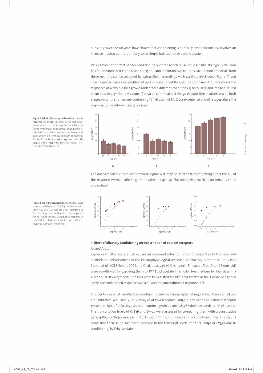
OB
AID
SID
DIq
I
our group over several years have shown that conditioning is primarily post-eclosion and involves an
increase in attraction. It is unlikely to be simple habituation or desensitization.
We examined the effect of odor conditioning on three selected Basiconic sensilla. The type I sensillum
has four neurons A, B, C and D and the type II and III contain two neurons each. Action potentials from
these neurons can be analyzed by extracellular recordings with capillary electrodes (Figure 4) and
dose response curves in conditioned and unconditioned flies, can be compared. Figure 5 shows the
responses of 4-day old flies grown under three different conditions; i) both larva and imago cultured
on an odorless synthetic medium; ii) larva on cornmeal and imago on odor-free medium and iii) both
stages on synthetic medium containing 10-4 dilution of EA. Odor experience at both stages alters the
response to five different acetate esters.
The dose-response curves are shown in Figure 6. It may be seen that conditioning alters the Km of
the response without affecting the maximal response. The underlying mechanism remains to be
understood.
4 Effect of olfactory conditioning on transcription of odorant receptors
Jawaid Ahsan
Exposure to Ethyl acetate (EA) causes an increased attraction in conditioned flies to this odor and
a correlated enhancement in the electrophysiological response of olfactory receptor neurons (See
Rashid et al, NCBS Report 2005 and Chakraborty et al, this report). The adult flies (0 to 12 hours old)
were conditioned by exposing them to 10-4 Ethyl acetate in an odor free medium for four days in a
12/12 hours day night cycle. The flies were then tested for 10-5 Ethyl acetate in the T maze behavioral
assay. The conditioned response was 0.80 and the unconditioned response 0.25
In order to see whether olfactory conditioning involves transcriptional regulation, I have carried out
a quantitative Real Time RT-PCR analysis of two receptors Or83b, a non-canonical odorant receptor
present in 70% of olfactory receptor neurons, synthetic and Or59b which responds to Ethyl acetate.
The transcription levels of Or83b and Or59b were assessed by comparing them with a constitutive
gene rpII140 (RNA polymerase II 140kD subunit) in conditioned and unconditioned flies. The results
show that there is no significant increase in the transcript levels of either Or83b or Or59b due to
conditioning by Ethyl acetate.
Spik
e/50
0ms
Odors
30
25
20
15
10
5
0EA EV IV EB IB
Spik
e/50
0ms
Odors
30
25
20
15
10
5
0EA EV IV EB IB
Spik
e/50
0ms
Odors
30
25
20
15
10
5
0EA EV IV EB IB
Figure 5. Effect of larval growth medium on the response of imago: (a) Both larvae and adult were cultured on odorless synthetic medium, (b) larvae were grown on cornmeal and adult were cultured on synthetic medium, (c) larvae and adult grown on synthetic medium containing 10-4 EA. As can be seen, odor experience at both stages alters response towards odors. Test odors EA, EV, IV, EB and IB
Figure 6. Odor induced responses. The flies were induced with EA at 10-4 for 3 days and tested with Ethyl Valerate (EV) and Iso amyl Valerate (IV). Conditioning reduces threshold and apparent Km for all chemicals. Conditioned response is denoted in black lines while unconditioned response is shown in red lines.
a b c
107
NCBS_AR_05_07.indd 107 7/10/08 5:35:19 PM

A z i m S u r A n i (S I R DORABJ I TATA PROFESSOR OF TH E TI FR)Primary affiliation: University of Cambridge, [email protected] Surani is the Mary Marshall and Arthur Walton Professor of Physiology and Reproduction, and a member of the Physiology Department of the University of Cambridge. His current research interests are towards understanding how germ cells are specified and made. At NCBS he collaborates with the groups of G.V. Shivashankar, K. VijayRaghavan, M.M. Panicker and with the group of Maneesha Inamdar at the Jawaharlal Nehru Centre for Advanced Scientific Research. Azim has been actively involved in conducting practical research workshops on stem cells in Bangalore.
J A m e S A S p u d i c hPrimary affiliation: Stanford University, [email protected] Spudich works on the molecular basis of motility. He has been a regular visitor to NCBS, spending several weeks to months each year and bringing his students and postdocs with him. He interacts with the Mayor and Rao labs, amongst others. His recent collaborative work has been on the function of Myosin VI. This myosin has been studied in both a monomeric and a dimeric form in vitro. Since the functional characteristics of the motor are dramatically different for these two forms, it is important to understand whether myosin VI heavy chains are brought together on endocytic vesicles. The Mayor and Spudich groups have used fluorescence anisotropy measurements to detect fluorescence resonance energy transfer between identical fluorophores (homoFRET) resulting from myosin VI heavy chains being brought into close proximity. They observed that, when associated with clathrin-mediated endocytic vesicles, myosin VI heavy chains are precisely positioned to bring their tail domains in close proximity. They show that on endocytic vesicles, myosin VI heavy chains are brought together in an orientation that previous in vitro studies have shown causes dimerization of the motor. Their results are therefore consistent with vesicle-associated myosin VI existing as a processive dimer, capable of its known trafficking function.
F r A n c i S c o B A r r A n t e S Primary affiliation: Universidad Nacional del Sur, [email protected] J Barrantes, is Professor at the UNESCO Chair of Biophysics and Molecular Neurobiology, Director of the Institute of Biochemical Research and currently the Head of the Scientific and Technological Research Centre – Argentinian Res. Council - in Bahia Blanca, Argentina. As a member of the Academy of Sciences for the Developing World (TWAS), and more recently as Adjunct Faculty, Prof. Barrantes has frequently visited NCBS and engaged in teaching in various local and international courses and conducted collaborative research with Prof. Satyajit Mayor and his group, also involving prolonged visits of his students to NCBS. The ongoing joint project involves the study of the nicotinic acetylcholine receptor stability at the cell membrane and its internalization. This receptor is a key molecule involved in the propagation of signals in the central nervous system and peripheral synapses. It is involved in functions such as consciousness, attention, and memory; alteration of those functions gives rise to various invalidating pathologies such as Alzheimer’s disease, Parkinson’s disease, and schizophrenia, making this family of receptors very important drug targets. The collaborative research with Prof. Mayor has disclosed a hitherto unknown slow-down endocytic mechanism operative in the muscle-type acetylcholine receptor, distinct from the canonical caveolin- and clathrin-dependent pathways. A similar mechanism may be operative in the brain in the case of neuronal receptors, and constitute a modulatory process involved in synaptic plasticity.
adjunct and visiting professors
NCBS is fortunate to have excellent interactions with colleagues whose primary appointment is at other institutions in India and abroad. Typically, adjunct faculty members have established research programmes at NCBS either independently or in collaboration with NCBS investigators. Madan Rao and Michael Bate have been around, happily for us, for long enough that their reports have moved into their respective ‘areas’. We also have Visiting Professors whose collaborative interactions are no less intense and who are regular visitors to NCBS. The more recent ones in this category are Teymuras Kurzchalia from the Max-Planck Institute for Cell Biology and Genetics, Dresden; Vivek Malhotra from UC San Diego and CRG Barcelona and L. Mahadevan from Harvard. We look forward to exciting interactions with them.
NCBS_AR_05_07.indd 108 7/10/08 5:35:20 PM
109
SA
TY
AJI
T
MA
YO
R
PUBLICATIONS110 Biochemistry, Biophysics and Bioinformatics | 113 Cellular Organization and signaling | 115 Genetics and
Development | 117 Neurobiology
NCBS_AR_05_07.indd 109 7/11/08 2:59:14 PM

Alva, V., Divya, S.P. and Sowdhamini, R. (2007). COILCHECK: A interactive server for the analysis of interface regions in coiled coils. Protein and Peptide Letters (In press).
Anil, V.S., Krishnamurthy, P., Kuruvilla, S., Sucharitha, K., Thomas, G. and Mathew, M.K. (2005). Regulation of the uptake and distribution of Na+
in shoots of rice (Oryza sativa) variety Pokkali: Role of Ca2+ in salt tolerance response. Physiologia Plantarum, 124, 451-464.
Anil, V., Krishnamurthy, H. and Mathew, M.K. (2007). Limiting cytosolic Na+ confers salt tolerance to rice cells in culture: A two-photon microscopy study of SBFI loaded cells. Physiologia Plantarum, 129, 607-50.
Anil, V., Krishnamurthy, H. and Mathew, M.K. (2007). Na+ Efflux across the plasma membrane contributes to salt tolerance of rice cells in culture. Proceedings of the Indian National Science Academy, 73, 43-50.
Banerjee, B., Bhattacharya, D. and Shivashankar, G.V. (2006). Chromatin assembly exhibits spatio-temporal heterogeneity within the cell nucleus. Biophysical Journal, 91, 2297-2303.
Bhaduri, A. and Sowdhamini, R. (2005). Genome-wide survey of prokaryotic O-protein phosphatases. Journal of Molecular Biology, 352, 736-752.
Bhaduri, A. and Sowdhamini, R. (2006). Domain architectural census of eukaryotic gene products containing O-protein phosphatases. Gene, 366, 246-255.
Bhadra, R., Sandhya, S., Abhinandan, K.R., Chakrabarti, S., Sowdhamini, R. and Srinivasan, N. (2006). Cascade PSI-BLAST web server: A remote homology search tool for relating protein domains. Nucleic Acids Research, 34, W143-W146.
+Bhaduri, A. and Sowdhamini, R. (2007). O-protein phosphatases in signal transduction pathways. In: New Trends in Bioinformatics, P. Shanmugavel (ed), 40-58.
+Bhaduri, A. and Sowdhamini, R. (2006). Diversity of phosphatases: Study in bacterial and archael genomes. In: Mathematical Biology: Recent trends Peeyush Chandra and B.V. Rathish Kumar (Eds). Anamaya Publishers, New Delhi, India.
Bhattacharya, D., Mazumder, A., Anne, M. and Shivashankar, G.V. (2006). Core and linker histones diffuse via distinct mechanisms within living cells. Biophysical Journal, 91, 2326-2336.
Bhattacharya, D., Banerjee, B., Sarin, A. and Shivashankar, G.V. (2007). Trichostatin-A induces differential changes in histone dynamics and expression in HeLa cells. Biochemical and Biophysical Research Communications, 363, 263-268.
Blaise, M., Bhattacharyya, D., Sowdhamini, R. and Pradhan, N. (2005). Structural consequences of D481N/K483q mutation at glycine binding site of NMDA ionotropic glutamate receptors: A molecular dynamics study. Journal of Biomolecular Structure and Dynamics, 22, 399–410.
@Blaise, M.C., Sowdhamini, R. and Pradhan, N. (2005). Comparative analysis of different competitive antagonists interaction with NR2A and NR2B subunits of N-methyl-D-aspartate (NMDA) ionotropic glutamate receptor. Journal of Molecular Modeling, 11, 489-502.
Chakrabarti, S., Anand, P., Bharadwaj, N., Pugalenthi, G. and Sowdhamini, R. (2005). SCANMOT: Searching for similar sequences using simultaneous scan of multiple sequence motifs. Nucleic Acids Research, 33, 274-276.
Chakrabarti, S., Manohari, G., Pugalenthi, G. and Sowdhamini, R. (2006). SSToSS: sequence structural templates of single-member superfamilies. In Silico Biology, 6, 0033.
Chandrashekar, C., Manonmani, A., Babu, M. and Sowdhamini, R. (2006). Enhanced structure prediction of gene products containing adenylyl cyclase. In Silico Biology, 6, 0029.
Dabholkar, S. and Thattai, M. (2007). Brainstorming Biology. IET Synthetic Biology, 1, 17-20.
Deepalakshmi, P.D. (2006). Top-down approach of completely sequencing a 4.9 kDa recombinant peptide using quadrupole time-of-flight mass spectrometry, Rapid Communications in Mass Spectrometry, 20, 3747-3754.
Gadkari, R., Sandhya, S., Sowdhamini, R. and Dighe, R.R. (2007). The antigen binding sites of various hCG monoclonal antibodies show homology to different domains of LH receptor. Molecular and Cellular Endocrinology, 260, 23-32.
Gaurav, K., Gupta, N. and Sowdhamini, R. (2005). FASSM: Enhanced function association in whole genome analysis using sequence and structural Motifs. In Silico Biology, 5, 40.
Gavory, G., Symmons, M.F., Krishnan-Ghosh, Y., Klenerman, D. and Balasubramanian, S. (2006). Structural analysis of the catalytic core of human telomerase RNA by FRET and molecular modeling. Biochemistry, 45, 13304-13311.
Biochemistry, Biophysics and Bioinformatics
publications
NCBS_AR_05_07.indd 110 7/10/08 5:35:22 PM

Ghodke, H.B., Krishnan, R., Vignesh, K., Kumar, G.V.P., Narayana, C. and Krishnan, Y. (2007). The I-Tetraplex building block: Rational design and controlled fabrication of robust 1D DNA scaffolds through non-watson-crick interactions. Angewandte Chemie International Edition, 46, 2646-2649.
Gowri, V.S. and Sandhya, S. (2006). Recent trends in remote homology detection: An Indian Medley. Bioinformation, 3, 94-96.
Hellman, A.N., Rau, K.R., You, H.H., Bae, S., Palmer, J.F., Phillips, K.S., Allbritton, N.L. and Venugopalan, V. (2007). Laser-induced mixing in microfluidic channels. Analytical Chemistry, 79, 4484-4492.
@Hellman, A.N., Rau, K.R., You, H.H. and Venugopalan, V. (2007). Biophysical response to pulsed laser microbeam-induced cell lysis and molecular delivery. Journal of Biophotonics, DOI:10.1002/jbio.200710010.
Ibrahim, M., Kerby, R.L., Puranik, M., Wasbotten, I.H., Youn, H., Roberts, G.P. and Spiro, T.G. (2006). Heme displacement mechanism of CooA activation: Mutational and Raman spectroscopic evidence. Journal of Biological Chemistry, 281, 29165.
Jha, S.K. and Udgaonkar, J.B. (2007). Exploring the cooperativity of the fast folding reaction of a small protein using pulsed thiol labeling and mass spectrometry. Journal of Biological Chemistry, 282, 37479-37491.
Kavitha, S., Varshney, A. and Mathew, M.K. (2005). N type rapid inactivation in human Kv1.4 channels: Functional role of a putative C-terminal helix. Molecular Membrane Biology, 22, 389-400.
Khajamohiddin, S., Babu, P.S., Chakka, D., Merrick, M., Bhaduri, A., Sowdhamini, R. and Siddavattam, D. (2006). A novel meta-cleavage product hydrolase from flavobacterium sp. ATCC27551. Biochemical and Biophysical Research Communications, 351, 675-681.
Khader, S. and Sowdhamini, R. (2007). IWS: Integrated web server for protein sequence and structure analysis. Bioinformation, 2, 86-90.
Krishnan-Ghosh, Y., Stephens, E. and Balasubramanian, S. (2005). PNA forms an i-motif. Chemical Communications, 5278-5280.
Krishnan-Ghosh, Y., Whitney, A.M. and Balasubramanian, S. (2005). Dynamic covalent chemistry on self-templating PNA oligomers; Formation of a bimolecular PNA quadruplex. Chemical communications, 3068-3070.
Krishna, S., Banerjee, B., Ramakrishnan, T.V. and Shivashankar, G.V. (2005). Origins and implications of long-tailed distributiosn in gene expression. Proceedings of the National Academy of Sciences of USA, 102, 4771–4776.
Kumar, S., Mohanty, S.K. and Udgaonkar, J.B. (2007). Mechanism of formation of amyloid protofibrils of barstar from soluble oligomers: Evidence for multiple steps and lateral association coupled to conformational conversion Journal of Molecular Biology, 367, 1186-1204.
Mazumder, A. and Shivashankar, G.V. (2007). Gold-nanoparticle-assisted laser perturbation of chromatin assembly reveals unusual aspects of nuclear architecture within living cells. Biophysical Journal, 93, 2209-2216
Metpally, R.R. and Sowdhamini, R. (2005). Genome-wide survey of G protein-coupled receptors in Tetraodon nigroviridis. BMC Evolutionary Biology 5, 41.
Metpally, R.R. and Sowdhamini, R. (2005). Cross-genome phylogenetic analysis of human and Drosophila G-protein-coupled receptors: Application to functional annotation of orphan receptors. BMC Genomics, 6, 106.
Metpally, R.R., Vigneshwar, R. and Sowdhamini, R. (2007). Genome inventory and analysis of nuclear hormone receptors in Tetraodon nigriviridis. Journal of Biological Sciences, 32, 43-50.
Modi, S., Wani, A.H. and Krishnan, Y. (2006). The PNA-DNA hybrid I-motif: Implications for sugar-sugar contacts in i-motif tetramerization. Nucleic Acids Research, 34, 4354-4363.
Mukhopadhyay, S., Nayak, P.K., Udgaonkar, J.B. and Krishnamoorthy, G. (2006). Characterization of the formation of amyloidprotofibrils from barstar by mapping residue-specific fluroscence dynamics. Journal of Molecular Biology, 358, 935-942.
Nagapriya, K.S., Raychaudhuri, A.K. and Shivashankar, G.V. (2007). Thermal fluctuations in histone during denaturation. Journal of Nanoscience Nanotechnology, 7, 2125-2128.
Patra, A.K. and Udgaonkar, J.B. (2007). Characterization of the folding and unfolding reactions of single-chain monellin: Evidence for multiple intermediates and competing pathways. Biochemistry, 46, 11727-11743.
Parikh, N., Koshy, C., Dhayabharan, V., Perumalsamy, L.R., Sowdhamini, R. and Sarin, A. (2007). The N-terminus and alpha-5, alpha-6 helices of the pro-apoptotic protein Bax, modulate functional interactions with the anti-apoptotic protein Bcl-xL. BMC Cell Biology, 8, 16.
Pitchiaya, S. and Krishnan, Y. (2006). First Blueprint, now bricks: DNA as construction material on the nanoscale. Chemical Society Reviews, 35, 1111-1121.
Pradeep, L. and Udgaonkar, J.B. (2006). Diffusional barrier in the unfolding of a small protein. Journal of Molecular Biology, 366, 1016-1028.
Pugalenthi, G., Archunan, G. and Sowdhamini, R. (2005). DIAL: A web-based server for the automatic identification of structural domains in proteins. Nucleic Acids Research, 30, W130-132.
Pugalenthi, G., Bhaduri, A. and Sowdhamini, R. (2005). GenDiS: Genomic distribution of protein structural superfamilies. Nucleic Acids Research, 33, 252-255.
@Pugalenthi. G., Bhaduri, A. and Sowdhamini, R. (2006). iMOTdb – a comprehensive collection of spatially interacting motifs in proteins. Nucleic Acids Research, 34, D285-286.
@Pugalenthi, G., Shameer, K., Srinivasan, N. and Sowdhamini, R. (2006). HARMONY: A web-server for the assessment of protein structures. Nucleic Acids Research, 34, W231-234.
Pugalenthi, G., Suganthan, P.N., Sowdhamini, R. and Chakrabarti, S. (2007). SMotif: A server for structural motifs in proteins, Bioinformatics, 23, 637-638.
PU
BL
ICA
TIO
NS
111
NCBS_AR_05_07.indd 111 7/10/08 5:35:23 PM

Pugalenthi, G., Ke Tang, Suganthan, P.N., Archunan, G. and Sowdhamini, R. (2007). A machine learning approach for the identification of odorant binding proteins from sequence-derived properties. BMC Bioinformatics (In press).
Pugalenthi, G., Suganthan, P.N., Sowdhamini, R. and Chakrabarti, S. (2007). MegaMotifBase: A database of structural motifs in protein families and superfamilies. Nucleic Acids Research (In press).
Puranik, M., Weeks, C.L., Lahaye, D., Kabil, O., Taoka, S., Nielsen, S.B., Groves, J.T., Banerjee, R. and Spiro, T.G. (2006). Dynamics of carbon monoxide binding to cystathionine-synthase. Journal of Biological Chemistry, 281, 13433-13438.
Rau, K.R., quinto-Su, P.A., Hellman, A.N. and Venugopalan, V. (2006). Pulsed laser microbeam-induced cell lysis: Time-resovled imaging and analysis of hydrodynamic effects. Biophysics Journal, 91, 317-329.
@Reddy, C.C.S., Shameer, K., Bernard, O. and Sowdhamini, S. (2007). PURE: A web server for querying the relationship between pre-existing domains and unassigned regions in proteins Nature Protocols, DOI: 10.1038/nprot. 486.
Roopa, T. and Shivashankar, G.V. (2006). Direct measurement of chromatin fluidity using optical trap modulation force spectroscopy. Biophysical Journal, 91, 4632-4637.
Sandhya, S., Chakrabarti, S., Abhinandan, K.R., Sowdhamini, R. and Srinivasan, N. (2005). Assessment of a rigorous transitive profile based search method to detect remotely similar proteins. Journal of Biomolecular Structure and Dynamics, 23, 283-298.
+Saxena, A.M., Udgaonkar, J.B. and Krishnamoorthy, G. (2005). Protein dynamics and protein folding dynamics revealed by time-resolved fluorescence. In Fluorescence Spectroscopy in Biology (eds. Hof, M., Hutterer, R. and Fidler, V.) Springer.
Saxena, A.M., Udgaonkar, J.B. and Krishnamoorthy, G. (2005). Protein dynamics control proton transfer from bulk solvent to protein interior: A case study with green fluorescent protein. Protein Science, 14, 1787-1799.
Saxena, A.M., Udgaonkar, J.B. and Krishnamoorthy, G. (2006). Characterization of intra-molecular distances and site-specific dynamics in chemically unfolded barstar: Evidence for denaturant-dependent non-random structure. Journal of Molecular Biology, 359, 174-189.
Saxena, A.M., Krishnamoorthy, G., Udgaonkar, J.B. and Periasamy, N. (2007). Identification of intermediate species in protein-folding by quantitative analysis of amplitudes in time-domain fluorescence spectroscopy. Journal of Chemical Sciences, 119, 61-69.
Shah, P.K., Tripathi, L.P., Jensen, L.J., Gahnim, M., Mason, C., Furlong, E.E., Rodrigues, V., White, K.P., Peer Bork, P. and Sowdhamini, R. (2007). Enhanced function annotations for Drosophila serine proteases: A case study for systematic annotation of multi-member gene families. Gene (In press).
Sinha, K.K. and Udgaonkar, J.B. (2005). Dependence of the size of the initially collapsed form during the refolding of barstar on denaturant concentration: Evidence for a continuous transition. Journal of Molecular Biology, 353, 704-718.
Sinha, K.K. and Udgaonkar, J.B. (2007). Dissecting the non-specific and specific components of the initial folding reacting of barstar by multi-site FRET measurements. Journal of Molecular Biology, 370, 385-405.
Sinha, D., Sastry, S. and Shivashankar, G.V. (2006). Probing mRNA conformational heterogeneity using single-moleculefluorescence anisotropy, Applied Physics Letters, 88, 3901-3903.
Soni, G.V., Brar, L., Meeran, F., Raychaudhuri, A.K. and Shivashankar, G.V. (2007). Distinct levels in the nanoscale organization of DNA-histone complex revealed by its mechanical unfolding. Appied Physics Letters, 90, 163904-3.
Sundara Baalaji, N., Mathew, M.K. and Krishnaswamy, S. (2006). Functional assay of S.typhi OmpC using reconstituted large unilamellar vesicles: A general method for characterization of outer membrane proteins. Biochimie, 88, 1419–1424.
Thangudu, R.R., Vinayagam, A., Pugalenthi, G., Manonmani, A., Offmann, B. and Sowdhamini, R. (2005). Native and modelled disulphide bonds in proteins: Knowledge-based approaches towards structure prediction of disulphide-rich polypeptides. Proteins: Structure Function and Bioinformatics, 58, 866-879.
Thattai, M., Burak, Y. and Shraiman, B.I. (2007). The origins of specificity in polyketide synthase protein interactions. PLoS Computational Biology, 3, e186.
Tripathi, L.P. and Sowdhamini, R. (2006). Cross-genome comparisons of serine proteases in Arabidopsis and rice. BMC Genomics, 7, 200.
Tripathi, L.P. and Sowdhamini, R. (2007). Genome-wide survey of protein families and superfamilies. Biobytes (In press).
Vijayalakshmi, M., Shivashankar, G.V. and Sowdhamini, R. (2007). Simulations of SIN mutations and histone variants in human nucleosomes reveal altered protein-DNA and core histone interactions. Journal of Biomolecular Structure and Dynamics, 25, 207-218.
Wani A. and Udgaonkar, J.B. (2006). HX-ESI-MS and optical studies of the unfolding of thioredoxin indicate stabilization of a partially unfolded, aggregation-competent intermediate at low pH. Biochemistry, 45, 11226-11238.
*Review +Book chapter #Others @Web publication
NCBS_AR_05_07.indd 112 7/10/08 5:35:23 PM

Altman, D., Goswami, D., Hasson, T., Spudich, J.A. and Mayor, S. (2007). Precise positioning of myosin VI on endocytic vesicles in vivo. PLoS Biology, 5, e210.
Ayyub, C., Sen, A., Gonsalves, F., Kishan, B., Bhandari, P., Shashidhara, L.S., Krishna, S. and Rodrigues, V. (2005). Cullin-5 plays multiple roles in cell fate specification and synapse formation during Drosophila development. Developmental Dynamics, 232, 865-875.
Benjamin Franklin, S., Fernando, A., Chalke, B.A. and Krishnan, K.S. (2007). Radular morphology of Conus (Gastropoda: Caenogastropoda: Conidae) from India. Molluscan Research, 27, 111-122.
Bhattacharya, D., Banerjee, B., Sarin, A. and Shivashankar, G.V. (2007). Trichostatin-A induces differential changes in histone dynamics and expression in HeLa cells. Biochemical and Biophysical Research Communications, 363, 263-268.
Chadda, R., Howes, M.T., Plowman, S.J., Hancock, J.F., Parton, R.G. and Mayor, S. (2007). Cholesterol-sensitive Cdc42 activation regulates actin polymerization for endocytosis via the GEEC pathway. Traffic, 8, 702-717.
Chaudhry, A., Das, S.R., Hussain, A., Mayor, S., George, A., Bal, V., Jameel, S. and Rath, S. (2005). The Nef protein of HIV-1 induces loss of cell surface costimulatory molecules CD80 and CD86 in APCs. Journal of Immunology, 175, 4566-4574.
Chaudhry, A., Das, S.R., Jameel, S., George, A., Bal, V., Mayor, S. and Rath, S. (2007). HIV-1 Nef protein triggers endocytosis of immune costimulatory proteins CD80 and CD86. Cell Host and Microbe, 1, 37-49.
Geetha, B., Divya, P., Sade, H., Gunasekharan, V., Rangarajan, A. and Sarin, A. (2006). Evidence for a role for Notch signaling in the cytokine-dependent survival of activated T-cells. Journal of Immunology, 177, 5041-5050.
Gowd, K.H., Sabareesh, V., Sudarslal, S., Iyengar, P., Franklin, B., Fernando, A., Dewan, K., Ramaswami, M., Sarma, S.P., Sikdar, S., Balaram, P. and Krishnan, K.S. (2005). Novel peptides of therapeutic promise from Indian Conidae. Annals of New York Academy of Sciences, 1056, 462-473.
Gowd, K.H., Krishnan, K.S. and Balaram, P. (2007). Identification of Conus amadis disulfide isomerase: Minimum sequence length of peptide fragment necessary for protein annotation. Molecular BioSystems, 3, 554-566.
Goyal, G., Fell, B., Sarin, A., Youle, R.J. and Sriram, V. (2007). Role of mitochondrial remodeling in programmed cell death in Drosophila melanogaster. Developmental Cell, 12, 807-816.
Kalia, M., Kumari, S., Chadda, R., Hill, M.M., Parton, R.G. and Mayor, S. (2006). Arf6-independent GPI-anchored protein-enriched early endosomal compartments fuse with sorting endosomes via a Rab5/phosphatidylinositorl-3’ Kinase-dependent machinery. Molecular Biology of the Cell, 17, 3689-3704.
Karmakar, S., Raghunathan, V. A. and Mayor, S. (2005). Phase behaviour of dipalmitoyl phosphatidylcholine (DPPC)-cholesterol membranes. Journal of Physics: Condensed Matter, 17, 1177–1182.
Kirkham, M., Fujita, A., Chadda, R., Nixon, S. J., Kurzchalia, T. V., Sharma, D.K., Pagano, R. E., Hancock, J. F., Mayor, S. and Parton, R. G. (2005). Ultrastructural identification of uncoated caveolin-independent early endocytic vehicles. Journal of Cell Biology, 168, 465-476.
Malhotra, V. and Mayor, S. (2006). Cell biology: The golgi grows up. Nature, 441, 939-940.
Mandal, A., Ramakrishnan, M., Ramasamy, S., Sabareesh, V., Openshaw, M.E., Krishnan, K.S. and Balaram, P. (2007). De novo sequencing of T-superfamily conotoxins from Conus virgo: Pyroglutamic acid identification, leucine/isoleucine discrimination and disulfide arrangement by MALDI mass spectrometry. Journal of American Society for Mass spectrometry, 18, 1396-404.
#Mayor, S. (2005). ACEing GPI release. Nature Structural and Molecular Biology, 12, 107-108.
Mayor, S. (2005). Coming home. Nature, 436, 488-489.
+Mayor, S., Viola, A., Stan, R.V. and del Pozo, M.A. (2006). Flying kites on slippery slopes at Keystone. In: Symposium on Lipid Rafts and Cell Function, EMBO Reports, 7, 1089-1093.
Mayor, S. and Pagano, R.E. (2007). Pathways of clathrin-independent endocytosis. Nature Reviews in Molecular and Cellular Biology, 8, 603-612.
Nair, P. (2005). Epidermal growth factor receptor family and its role in cancer progression. Current Science, 88, 890-898.
Cellular Organization and Signaling
publications113
PU
BL
ICA
TIO
NS
NCBS_AR_05_07.indd 113 7/10/08 5:35:25 PM

Nair, S.S., Nilsson, C.L., Emmett, M.R., Schaub, T.M., Gowd, K.H., Thakur, S.S., Krishnan, K.S., Balaram, P. and Marshall, A.G. (2006). De novo sequencing and disulfide mapping of a bromotryptophan-containing conotoxin by fourier transform ion cyclotron resonance mass spectrometry. Analytical Chemistry, 78, 8082-8088.
+Nair, S.S., Emmett, M.R., Nilsson, C.L., Krishnan, K.S., Balaram, P. and Marshall, A.G. (2006). FT-ICR MS-based structural characterization of a novel conotoxin from C. monile: Determination of bromotryptophan by use of electron capture dissociation. 54th American Society of Mass Spectrom. Annual Conference on Mass Spectrometry and Allied Topics, Seattle, WA, May 27-June 2.
Parikh, N., Koshy, C., Dhayabharan, V., Perumalsamy, L.R., Sowdhamini, R. and Sarin, A. (2007). The N-terminus and alpha-5, alpha-6 helices of the pro-apoptotic protein Bax, modulate functional interactions with the anti-apoptotic protein Bcl-xL BMC Cell Biology, 8, 16.
Rajagopal, D., Bal, V., Mayor, S., George, A. and Rath, S. (2006). A role for the Hsp90 molecular chaperone family in antigen presentation to T lymphocytes via major histocompatibility complex class II molecules. European Journal of Immunology, 36, 828-841.
Ramdass, B., Maliekal, T,T., Lakshmi, S., Rehman, M., Rema, P., Nair, P., Mukherjee, G., Reddy, B.K.M., Krishna, S. and Pillai, M.R. (2007). Co-expression of Notch1 and NF-KappaB signaling pathway components in human cervical cancer progression. Gynaecology Oncology, 104, 352-361.
Rikhy, R., Kamat, S., Ramagiri, S., Sriram, V. and Krishnan, K.S. (2007). Mutations in dynamin-related protein result in gross changes in mitochondrial morphology and affect synaptic vesicle recycling at the Drosophila neuromuscular junction. Genes Brain and Behaviour, 6, 42-53.
Rao, M. and Mayor, S. (2005). Use of Forster’s resonance energy transfer microscopy to study lipid rafts. Biochimica et Biophysica Acta, 1746, 221-333.
Sabareesh, V, Gowd, K.H., Ramasamy, P., Sudarslal, S., Krishnan, K.S., Sikdar, S.K. and Balaram, P. (2007). Characterization of contryphans from Conus loroisii and Conus amadis that target calcium channels. Peptides, 27, 2647-2654.
Sanyal, S., Consoulas, C., Kuromi, H., Basole, A., Mukai, L., Kidokoro, Y., Krishnan, K.S. and Ramaswami, M. (2005). Analysis of conditional paralytic mutants in Drosophila Sarco-Endoplasmic Reticulum calcium ATPase reveals novel mechanisms regulating membrane excitability, Genetics, 169, 737-750.
Sarasij, R.C., Mayor, S. and Rao, M. (2007). Chirality-induced budding: A raft-mediated mechanism for endocytosis and morphology of caveolae? Biophysical Journal, 92, 3140-3158.
+Sharma, P., Varma, R. and Mayor, S. (2006). Biophysical characterization of lipid rafts. In: Lipid Rafts and Caveolae (eds. Christopher Fielding). Wiley-VCH, Verlag Gmbh & Co., KGaA, Weinheim. 278 pages.
Siddhartha, S. P., Senthil Kumar, G., Sudarslal, S., Iengar, P., Ramasamy, P., Sikdar, S.K., Krishnan, K.S. and Balaram, P. (2005). Solution structure of Am2766, a highly hydrophobic delta conotoxin from conus amadis that inhibits inactivation of brain voltage gated sodium channels, Chemistry and Biodiversity, 2, 535-556.
Sriram, V., Krishnan, K.S. and Mayor, S. (2006). Biogenesis of lysosomes. Proceedings of the Indian National Science Academy. 72, 19-42.
Subramanyam, D. and Krishna, S. (2006). c-Myc substitutes for Notch1-CBF1 functions in cooperative transformation with papillomavirus oncogenes. Virology, 347, 191-198.
Srivastava, S., Banerjee, H., Chaudhry, A., Khare, A., Sarin, A., George, A., Bal, V., Durdik, J.M. and Rath, S. (2007). Apoptosis-inducing factor (Aif) regulates death in peripheral T cells. Journal of Immunology, 179, 797-803.
+Varma, R. and Mayor, S. (2006). Homo-FRET microscopy to investigate molecular-scale organization in living cells. In: Cell Imaging (eds. David Stephens.) Methods Express Series, Scion Publishing Ltd, UK. 301.
Veeraraghavalu, K., Subbaiah, V. K., Srivastava, S., Chakrabarti, O., Syal, R. and Krishna, S. (2005). Complementation of HPV-16 E6/E7 by Jagged1 specific Notch1-PI3K signaling involves pleiotropic oncogenic functions independent of CSL activation. Journal of Virology, 79, 7889-7898.
*Review +Book chapter #Others @Web publication
NCBS_AR_05_07.indd 114 7/10/08 5:35:25 PM

Ayyub, C., Sen, A., Gonsalves, F., Kishan, B., Bhandari, P., Shashidhara, L.S., Krishna, S. and Rodrigues, V. (2005). Cullin-5 plays multiple roles in cell fate specification and synapse formation during Drosophila development. Developmental Dynamics, 232, 865-875.
*Banerjee S. and Hasan, G. (2005), The InsP3 receptor: Its role in neuronal physiology and neurodegeneration. Bioessays, 27, 1035-1047.
Banerjee, S., Joshi, R., Venkiteswaran, G., Agrawal, N., Srikanth, S., Alam, F. and Hasan, G. (2006). Compensation of Inositol 1,4,5-trisphosphate receptor function by altering Sarco-endoplasmic reticulum calcium ATPase activity in the Drosophila flight circuit. Journal of Neuroscience, 26, 8278-8288.
Belle, E., Ramakrishnan, U., Mountain, J.L. and Barbujani, G. (2006). Serial coalescent simulations suggest a weak genealogical relationship between Etruscans and modern Tuscans. Proceedings of the National Academy of Sciences of USA, 103, 8012-8017.
Chakraborty, D., Ramakrishnan, U., Panor, J., Mishra, C. and Sinha, A. (2007). Phylogenetic relationships and morphometric affinities of the Arunachal macaque Macaca munzala, a newly described primate from Arunachal Pradesh, Northeastern India. Molecular Phylogenetics and Evolution, 44, 838-849.
Dixit, R., VijayRaghavan, K. and Bate, M. (2007). Hox genes and the regulation of movement in Drosophila. Developmental Neurobiology, 68, 309-331
Dutta, D., Shaw, S., Maqbool, T., Pandya, H. and VijayRaghavan, K. (2005). Drosophila heartless acts with heartbroken/Dof in muscle founder differentiation. PLoS Biology, 3, e337.
+Dutta, D. and VijayRaghavan, K. (2005). Metamorphosis and the formation of the adult musculature. In: Muscle Development in Drosophila. (ed. Helen Sink), Landis Publishers, 1-18.
Guruharsha, K.G. and VijayRaghavan, K. (2005). Discrimination in partnership: Compartment-specific interactions of Hox proteins. Journal of Genetics, 83, 223–226.
Hatton-Ellis, E., Ainsworth, C., Sushama, Y., Wan, S., VijayRaghavan, K. and Skaer, H. (2007). Genetic regulation of patterned tubular branching in Drosophila. Proceedings of the National Academy of Sciences of USA, 104, 169-174.
James, J., Ramakrishnan, U. and Datta, A. (2007). Molecular evidence for the occurrence of the leaf deer Muntiacus putaoensis in Arunachal Pradesh, north-east India. Conservation Genetics (In press).
Mondal, K., Dastidar, A.G., Singh, G., Madhusudhanan, S., Gande, S.L., VijayRaghavan, K. and Varadarajan, R. (2007). Design and isolation of temperature-sensitive mutants of Gal4 in yeast and Drosophila. Journal of Molecular Biology, 370, 939-950.
Mondal, K., VijayRaghavan, K. and Varadarajan, R. (2007). Design and utility of temperature-sensitive Gal4 mutants for conditional gene expression in Drosophila. Fly, 1, 282-286.
Maqbool, T., Soler, C., Jagla, T., Daczewska, M., Lodha, N., Palliyil, S., VijayRaghavan, K. and Jagla, K. (2006). Shaping leg muscles in Drosophila: Role of ladybird, a conserved regulator of appendicular myogenesis. PLoS ONE, e122.
Mukherjee, N., Mondol, S., Andheria., A. and Ramakrishnan, U. (2007). Rapid multiplex PCR based species identification of wild tigers using non-invasive samples, Conservation Genetics, 8, 1465-1470.
Nair, S., Agrawal, N. and Hasan, G. (2007). Homeostasis of glutamate neurotransmission is altered in Drosophila inositol 1,4,5-trisphosphate receptor mutants. Invert Neuroscience, 7, 137-147.
#Rodrigues, V. (2005). Prescience and critical thought: The life and science of Jose Campos-Ortega. Journal of Genetics, 84, 217-222.
Roy, B., Singh, A.P., Shetty, C., V. Chaudhary, V., North, A., Landraf, M., VijayRaghavan, K. and Rodrigues, V. (2007). Metamorphosis of an identified serotonergic neuron in the Drosophila olfactory system. Neural Development, 2, 20.
Sen, A., Shetty, C., Jhaveri, D. and Rodrigues, V. (2005). Distinct types of glial cells populate the Drosophila antenna. BMC Developmental Biology, 5, 25-36.
Sharma, P., Asztalos, Z., Ayyub, C., De Bruyne, M., Doran, A.J., Gomex-Hernandez, A., Keane, J., Killeen, J., Kramer, S., Madhavan, M., Roe, H., Sherkhane, P.D., Siddiqi, K., Silva, E., Carlson, J.R., Goodwin, S.F., Heisenberg, Krishnan, K., Kyriacou, C.P., Patridge, L., Riesgo-Escovar, J., Rodrigues, V., Tully T. and O’Kane, C.J. (2005). Isogenic autosomes to be applied in optimal screening for novel mutants with viable phenotypes in Drosophila melanogaster. Journal of Neurogenetics, 19, 57-85.
Genetics and Development
publications
115
PU
BL
ICA
TIO
NS
NCBS_AR_05_07.indd 115 7/10/08 5:35:25 PM

Shah, P.K., Tripathi, L.P., Jensen, L.J., Gahnim, M., Mason, C., Furlong, E.E., Rodrigues, V., White, K.P., Peer Bork, P. and Sowdhamini, R. (2007). Enhanced function annotations for Drosophila serine proteases: A case study for systematic annotation of multi-member gene families. Gene (In press).
Srikanth, S., Banerjee, S. and Hasan, G. (2006). Ectopic expression of a Drosophila InsP3R channel mutant has dominant-negative effects in vivo. Cell Calcium, 39, 187-196.
Tishkoff, S.A., Gonder, M.K., Henn., B.M., Mortensen, H., Fernandopulle, F., Gignoux, C., Godfrey, L., Nyambo, T.B., Underhill, P.A., Ramakrishnan, U., Reed, F.A. and Mountain, J.L. (2007). History of click-speaking populations of Africa inferred from mtDNA and Y chromosome variation. Molecular Biology and Evolution, 24, 2180-2195.
van Tuinen, M., O’Keefe, K., Ramakrishnan, U. and Hadly, E. (2007). Fire and ice: Genetic structure of the uinta fround squirrel (Spermophilus armatus) across the yellowstone hotspot. Molecular Ecology (In press).
VijayRaghavan, K. (2006). Reading Waddington today. Journal of Genetics, 85, 99.
VijayRaghavan, K. (2006). Punnett and duck genetics. Journal of Genetics, 85, 1.
Wahne, K., D’Souza, K. Sen, J., Rikhy, R., Rodrigues, V. and Rao, B.J. (2006). Expression pattern of Drosophila Translin and behavioral analyses of the mutant. European Journal of Cell Biology, 86, 173-186.
*Review +Book chapter #Others @Web publication
NCBS_AR_05_07.indd 116 7/10/08 5:35:26 PM

*Ajay, S.M. and Bhalla, U.S. (2006). Synaptic plasticity in vitro and in silico: insights into an intracellular signaling maze. Physiology, 21, 289-296.
Ajay, S.M. and Bhalla, U.S. (2007). A propagating ERKII switch forms zones of elevated dendritic activation correlated with plasticity. HFSP Journal, 1, 49-66.
Bennur, S., Rao, B.S.S., Pawlak, R., Strickland, S., McEwan, B.S. and Chattarji, S. (2006). Stress-induced spine loss in the medical amygdala is mediated by tissue-plasminogen activator. Neuroscience, 144, 8-16.
Bhattacharyya, S., Raote, I., Bhattacharya, A., Miledi, R. and Panicker, M.M. (2006). Activation, internalization, and recycling of the serotonin 2A receptor by dopamine. Proceedings of the National Academy of Sciences of USA, 103, 15428-15253.
Cannon, R.C., Gewaltig, M.O., Bhalla, U.S., Cornelis, H., Gleeson, P., Hines, M.L., Howell, F.W., Muller, E., Stiles, J.R. Wils, S. and De Schutter, E. (2007). Interoperability of neuroscience modeling software: Current status and future directions. Neuroinformatics, 5.
Dolen G., Osterweil E., Rao, B.S.S., Smith, G.B., Auerbach B.D., Chattarji, S. and Bear, M.F. (2007). Correction of fragile X syndrome in mice. Neuron, 56, 955-962.
Govindarajan, A., Rao, B.S.S., Nair, D., Trinh, M., Mawjee, N., Tonegawa, S. and Chattarji, S. (2006). Transgenic brain-derived neurotrophic factor expression causes both anxiogenic and antidepressant effects. Proceedings of the National Academy of Science of USA, 103, 13208-13213.
*HarshaRani, G.V., Vayttaden, S.J. and Bhalla, U.S. (2005). Electronic data sources for kinetic models of cell signaling. Journal of Biochemistry, 137, 653–657.
Hayer, A. and Bhalla, U.S. (2005). Molecular switches at the synapse emerge from receptor and kinase traffic. PLoS: Computational Biology 1, e20, 2005.
Hayashi, M.L., Rao, B.S.S., Seo J-S., Choi H-S., Dolan, B.M., Choi S-Y., Chattarji, S. and Tonegawa, S. (2007). Inhibition of p21-activated kinase rescues symptoms of fragile X syndrome in mice. Proceedings of the National Academy of Sciences of USA, 104, 11489-494.
Le Novere, N., Finney, A., Hucka, M., Bhalla, U.S., Campagne, F., Collado-Vides, J., Crampin, E., Halstead, M., Klipp, E., Mendes, P., Nielsen, P., Sasuro, H., Shapiro, B., Snoep, J.L., Spence, H.G. and Wanner, B.L. (2005). Minimal information Requested in the Annotation of biochemical Models (MIRIAM). Nature Biotechnology, 23, 1509-1515.
+McEwen B.S. and Chattarji, S. (2007). Neuroendocrinology of stress. In: Handbook of Neurochemistry and Molecular Neurobiology: Behavioral Neurochemistry and Neuroendocrinology (3rd Ed.), A. Lajtha and J.D. Blaustein (eds.), pp. 571-594, Springer Verlag.
Mitra, R., Jadhav, S., McEwen, B.S., Vyas, A. and Chattarji, S. (2005). Stress duration modulates the spatiotemporal patterns of spine formation in the basolateral amygdala. Proceedings of the National Academy of Science of USA, 102, 9371-9376.
Mitra, R., Vyas, A., Chatterjee, G. and Chattarji, S. (2005). Chronic-stress induced modulation of different states of anxiety-like behavior in female rats. Neuroscience Letters, 383, 278-283.
Mohanty, S.K., Sharma, M., Panicker, M.M., and Gupta, P.K. (2005). Controlled induction, enhancement, and guidance of neuronal growth cones by use of line optical tweezers. Optics Letters, 30, 2596-2598.
Narayanan, R., Narayan, A. and Chattarji, S. (2005). A probabilistic framework for region-specific remodeling of dendrites in three-dimensional neuronal reconstructions. Neural Computation, 17, 75-96.
Pawlak, R., Rao, B.S.S., Melchor, J.P., Chattarji, S., McEwan, B. and Strickland, S. (2006). Tissue plasminogen activator and plaminogen mediate stress induced decline of neuronal and cognitive functions in the hippocampus. Proceedings of the National Academy of Sciences of USA, 102, 18201-18206.
Rajan, R., Clement, J.P. and Bhalla, U.S. (2006). Rats smell in stereo. Science, 311, 666-670.
*Rajasethupathy, P., Vayttaden, S.J. and Bhalla, U.S. (2005). Systems modeling: a pathway to drug discovery. Current Opinion in Chemical Biology, 9, 400-406.
Raote, I., Bhattacharya, A. and Panicker, M.M. (2007). Serotonin 2A (5-HT2A) receptor function: Ligand-dependent mechanisms and pathways. In: Serotonin Receptors in Neurobiology, Frontiers in Neuroscience Vol 35 Edited by A. Chattopadhyay (Taylor and Francis, CRC Press).
Titus, A.D., Rao, B.S.S., Harsha, H.N., Ramkumar, K., Srikumar, B.N., Singh, S.B., Chattarji, S. and Raju, T.R. (2007). Hypobaric hypoxia-induced dendritic atrophy of hippocampal neurons is associated with cognitive impairment in adult rats. Neuroscience, 145, 265-78.
Vyas, A., Jhadav, S. and Chattarji, S. (2006). Prolonged behavioral stress enhances synaptic connectivity in the basolateral amygdala. Neuroscience, 143, 387-393.
Neurobiology
publications
*Review +Book chapter #Others @Web publication
117
PU
BL
ICA
TIO
NS
NCBS_AR_05_07.indd 117 7/10/08 5:35:27 PM
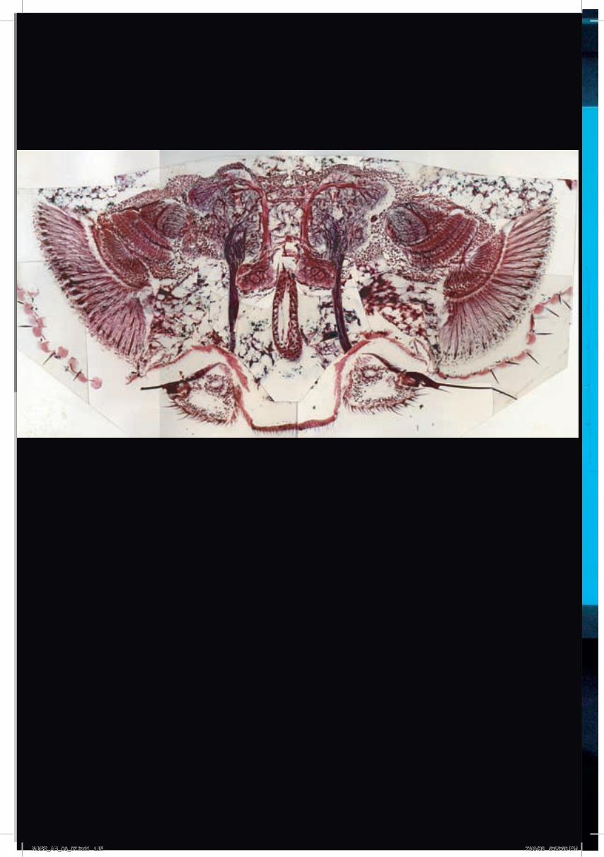
Selected publications
NCBS_AR_05_07.indd 118 7/10/08 5:35:30 PM
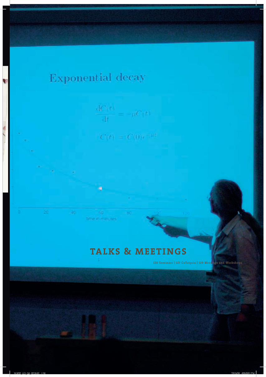
119
SA
TY
AJI
T
MA
YO
R
120 Seminars | 127 Colloquia | 129 Meetings and Workshops
talks & meeti ngs
NCBS_AR_05_07.indd 119 7/10/08 5:35:31 PM

seminars
Revathi Ananthakrishnan University of California at Davis, USA 4.4.05 Modeling the structural response of a eukaryotic cell in the optical stretcher.
Elisha Moses Weizmann Institute, Israel 5.4.05 Propagation, information flow and coding in linear neuronal cultures and 15.4.05 Organization and function in Molecular Motor Assemblies.
Sanjeev Kumar Gupta Abramson Family Cancer Research Institute, USA 25.4.05 Role of HtrA2/Omi in mitochondrial homeostasis and cell death.
Samrat Mukhopadhyay Tata Institute of Fundamental Research, India 6.5.05 Unraveling the complexity of protein aggregation leading to amyloid fibrillation.
Ajitkumar Parayil Singapore-MIT Alliance, Singapore 23.5.05 Biochemical and biomimetic approaches to unravel the biomineralization process.
J Krishnan The John Hopkins University, USA 31.5.05 Mathematical and computation modeling of the eukaryotic cell compass.
Subramanian Ramaswamy University of Iowa, USA 3.6.05 Random walks in 3-D space.
Sajith Dass University of Koln, Germany 6.6.05 The role of Cornichon (Cni) during axis formation in Drosophila.
S Vijaylakshmi Indian Institute of Technology, India 13.6.05 Studies on physiological and genetic aspects of riboflavin over production by the hemiascomycete fungi Eremothecium ashbyii and Ashbya gossypi.
R Nagaraj Centre for Cellular and Molecular Biology, India 22.6.05 Structures and activities of peptides that species across the evolutionary scale use to counter bacterial invasion.
Vaijayanti Gupta National Institutes of Health, USA 29.6.05 X-Chromosome dosage compensation in Drosophila germ cells.
Pritha Ray Stanford University, USA 18.7.05 Imaging molecular events in living animals: A multimodality fusion gene approach.
Marien De Bruyne Universität Berlin, Germany 15.7.05 Olfaction in Drosophila from voltage to mileage.
Suresh Kumar Balasubramanian Max-Planck Institute of Developmental Biology, Germany 25.7.05 Variety is the spice of life: Natural variation in flowering responses of Arabidopsis thaliana.
Irimpan Mathews Stanford University, USA 27.7.05 Functional studies of two potential antibacterial targets.
J Srividhya Indian Institute of Technology, India 27.7.05 Modeling dynamical systems with delay differential equations.
Brigitte Quenete Ecole Supérieure de Physique et Chemie Industrielle, France 1.8.05 Neural networks as models of the olfactory pathway and as tools for signal processing.
Subramaniah Ramaswamy University of Iowa, USA 2.8.05 Very short introduction to protein x-ray crystallography.
Shwetal Patel The University of Texas Southwestern Medical Center, USA 10.8.05 Mechanistic Modeling of DNA Repair Pathways.
Rajiv Singh University of California at Davis, U.S.A 12.8.05 Prions and amyloids: Recent developments and open questions.
Wiebe E Bijker Maastricht University, The Netherlands 16.8.05 Politics of modern biology – Role for the precautionary principle.
K S Manilal University of Calicut, India 19.8.05 Hortus Malabaricus – a 17th century botanical publication that changed the political history of India.
Shiv Pillai MGH Cancer Center, Harvard Medical School, USA 22.8.05 A novel cell surface acetylation/ deacetylation paradigm for the regulation of lymphocyte development.
Sidhartha Goyal Princeton University, USA 24.8.05 Growth regulation in E. coli under nitrogen limitation.
Suman Lata Johann Wolfgang Goethe-University, Germany 26.8.05 (1) High affinity multivalent chelators for reversible modification and organization of Histidine-tagged proteins. (2) Ligand induced Type I interferon receptor assembling on model membranes.
U J McMahan Stanford University School of Medicine, USA 2.9.05 What electron microscope tomography is telling us about synaptic transmission of the nerve impulse.
Sanjay Puri Jawaharlal Nehru University, India 2.9.05 Kinetics of Phase Transitions.
Erdem Karatekin Institut de Biologie Physico-Chimique, France 5.9.05 Secretory vesicle dynamics in the subplasmalemma: interactions with the cytoskeleton and the cell membrane, and relationship to exocytosis.
Ashutosh Sharma Indian Institute of Technology, India 14.9.05 Enigma and aesthetics of small things: Forces and patterns.
Satyavani Vemparala University of Pennsylvania, USA 20.9.05 Computational studies of chemical and biological systems: a molecular dynamics perspective.
Naweed I Naqvi National University of Singapore, Singapore 27.9.05 HARD TALK: Thigmotropism and initiation of fungal pathogenesis.
Uttam Surana Institute of Molecular and Cell Biology, Singapore 30.9.05 Taming the spindle and restraining the chromosomes.
NCBS_AR_05_07.indd 120 7/10/08 5:35:33 PM

SE
MIN
AR
S
Sudhindra B Sant Twin Technologies Inc, USA 4.10.05 Anti-microbial silver films as therapeutic agents for burn wound dressings.
Janaki Balakrishnan Instituut Lorentz for Theoretical Physics Universiteit Leide, The Netherlands 4.10.05 Self tuning towards an instability in a noisy nonlinear system: how the ear can detect whispers.
Srinivas Krishnagopal Centre for Advanced Technology, India 10.12.05 Tunable Femtosecond X-ray lasers: What’s in it for biologists.
Satyajit Rath National Institute of Immunology, India 12.10.05 The Lineage-specific semantics of protein function: Apoptosis-inducing factor (Aif) and the T cell lineage.
Arun Sripati Johns Hopkins University, USA 21.10.05 What does the hand tell the brain? A model for mechanoreceptive afferent responses.
Bhavin Khatri University of Leeds, UK 26.10.05 Visco-elastic Force Spectra of Single Biomolecules.
Viji M Draviam Massachusetts Institute of Technology, USA 4.11.05 Probing the role of kinetochore proteins in chromosome segregation.
Hugo Bellen Howard Hughes Medical Institute Program in Developmental Biology USA 10.11.05 Genetic Dissection of neurotransmitter release.
Madan Anup University of Iowa, USA 14.11.05 Epigenetic regulation of gene expression in Glioblastoma Multiforme.
Christoph Hergersberg Biosciences GE Global Research, USA 18.11.05 Seeing is believing – on the road to personalized medicine.
Radhamany Anandalakshmi Yale University, USA 21.11.05 At the cross-roads of plant defense and RNA silencing.
Andre Fiala Lehrstuhl fur Genetik und Neurobiologie Theodor-Boveri--Institut, Germany 28.11.05 Optical calcium imaging of neuronal activity in the Drosophila brain.
Mahendra Sonawane Max-Planck Institute for Developmental Biology, Germany 28.11.05 Breaches in the barrier: Genetic analysis of cellular adhesion in the zebrafish epidermis.
Albert Libchaber Rockfeller University, USA 30.11.05 A tour of experimental approaches in molecular biology.
Margaret Buckingham Pasteur Institute, France 5.12.05 The role of Pax-genes in the formation and regeneration of skeletal muscle.
Krishna Rajarathnam University of Texas, USA 5.12.05 Chemokines - Dr. Jekyll or Mr. Hyde? Structural Basis of how ‘good’ chemokines turn ‘bad’.
Thomas L Schwarz Harvard Medical School, USA 6.12.05 Membrane traffic to and at the synapse.
Manush S M Central Institute of Fisheries Education, India 7.12.05 Stress responses in Macrobrachium rosenbergii and its amelioration.
Yacine Graba Universitaire de Luminy, France 9.12.05 Hox, transcription and morphogenesis in development and evolution.
Lakshmikanth Gandikota Stanford University School of Medicine, USA 12.12.05 Molecular understanding of cytokinesis: Role of the mitotic kinesin like protein Kif12.
Anthony P Sinai University of Kentucky College of Medicine, USA 13.12.05 Rewiring of the host NFKB signaling cascade by the protozoan parasite Toxoplasma gondii.
M Prabha National Institute of Mental Health and Neuro Sciences, India 15.12.05 A comparative biochemical study on hydrolytic enzymes in normal human brain (postmortem), brain tumors and in their derived cell lines.
Seema Agarwal Yale University, USA 16.12.05 Modulation of NFKB pathway.
Sanjay Sane University of Washington, USA 16.12.05 How insects fly: A systems perspective.
Jayanta Mukhopadhyay Rutgers University, USA 19.12.05 Chemical genetics of bacterial RNA polymerase.
Karuna Sampath The National University of Singapore, Singapore 19.12.05 Specification of the dorsal axis in zebrafish.
Vinod Subramaniam University of Twente, The Netherlands 19.12.05 Nanoscale characterization of protein aggregation.
Richard S Mann Columbia University Medical Center, USA 20.12.05 Hox genes, Hox cofactors, and the control of appendage development in Drosophila.
Chiara Zurzolo Traffic Membranaire et Pathogenese Institut Pasteur, France 20.12.05 Role of lipid microdomains in GPI anchored protein sorting and prion folding.
Ramanuj Dasgupta NYU Cancer Center, USA 21.12.05 Functional genomic analysis of the Wnt/Wingless signaling pathway.
Arko Ghosh ETH-Zurich, Switzerland 21.12.05 Specificity of plasticity? - in adult CNS injury.
Madhavi Krishnan Institute of Biophysics, Germany 21.12.05 Fluidics in Biology.
Chandrajit Lahiri Bose Institute, India 22.12.05 Studies on a thiosulfate oxidation gene locus in a facultative sulfur lithotrophic bacterium.
Vinay Tergaonkar Institute of Molecular and Cell Biology, Singapore 23.12.05 Regulation of NFKB function.
Rupasri Ain University of Kansas School of Medicine, USA 26.12.05 Decoding the cell cell communication at the maternal-fetal interface.
Avinash R Shenoy Indian Institute of Science, India 27.12.05 Cyclic AMP, nucleotide and phosphodiesterases: A new beginning in mycobacteria.
Shahid S Siddiqui University of IIIinois, USA 28.12.05 Metazoan motor models: Kinesins in worms and man.
Pritinder Kaur Peter Maccallum Cancer Centre, Australia 3.1.06 Identification and biological characterization of the epidermal stem cells of skin.
Ronald Vale University of California at San Francisco, USA 3.1.06 Imaging signal transduction pathways.
Maya Cesari University of Reunion Islands, France 4.1.06 Ongoing work in Reunion on adipocyte and on cyclotron project.
Mayank Mehta Brown University, USA 5.1.06 Role of oscillations in sequence learning.
121
NCBS_AR_05_07.indd 121 7/10/08 5:35:33 PM

Jonaki Sen Harvard Medical School, USA 9.1.06 Patterning in the retina: The tale of two signaling molecules.
Aurnab Ghose Harvard Medical School, USA 9.1.06 Receptor tyrosine phosphatases in neuronal morphogenesis.
Padmanabhan University of Murcia, Spain 10.1.06 Transcriptional factors in Myxococcus xanthus development and light response: novel modules, novel architecture.
Amitabha Bandyopadhyay Harvard Medical School, USA 11.1.06 Role of tissue interactions and signaling molecules in skeletal differentiation.
Chandrajit Bajaj University of Texas at Austin, USA 12.1.06 Modeling synaptic transmission at the neuro muscular junction with spatial realism.
Patricia Bassereau PhysicoChimie Curie Institut, France 27.1.06 Model membranes for probing membrane deformations by molecular motors or proteins.
Michael Klein University of Pennsylvania, USA 30.1.06 Classical and quantum computational studies in chemical biology: From Enzymes to membranes and membrane-bound species.
Arulandu Arockiasamy Texas A&M University, USA 13.2.06 Snapshots of P1 Phage SAR-endolysin Lyz activation.
Gopal C Kundu National Center for Cell Science,India 14.2.06 Osteopontin, a member of SIBLING gene family: Role in cell signaling and tumor progression.
Rajat Varma Skirball Institute for Biomolecular Medicine USA 15.2.06 The T cell receptor complex dynamics in the immunological synapse.
Ludger Johannes Laboratoire Trafic et Signalisation Institut Curie, France 20.2.06 Shiga toxin transport on clathrin-dependent and clathrin-independent tracks.
Dennis Bray University of Cambridge, UK 24.2.06 Signaling In a molecular jungle: insights from bacterial chemotaxis.
Nivedita Chatterjee Johannes Gutenberg University, Germany 27.2.06 Gilal cells in health and disease – exploring the depths of the cerebral iceberg.
Rajnish P Rao Indian Institute of Science, India 28.2.06 Embryo-endometrial expression of leukemia inhibitory factor in the golden hamster.
Vivek Malhotra University of California at San Diego, USA 14.2.06, 27.2.06, 2.3.06 and 9.3.06 (1) Making a transport vesicle (2) Genome-wide screen for identification of protein secretion machinery (3) Partitioning of cellular compartments during cell division (4) Novel cell cycle specific checkpoints related to organelle partitioning.
Kavitha Damodaran Bharathiar University, India 10.3.06 Adsorption dynamics on the removal of dyes, phenol and chlorophenols from wastewater by activated carbon prepared from an agricultural solid waste: Coir pith.
Zulfiqar Ahmad University of Rochester Medical Center, USA 27.3.06 Molecular mechanism of ATP Synthase: Identification of phosphate binding residues.
Deepak T Nair Mount Sinai School of Medicine, USA 31.3.06 DNA polymerases lota and Rev1: The relation between structure, biochemical properties and function.
Quasar Saleem Padiath NCBS, India 18.4.06 When too much is too little: Lamin B1 duplications cause autosomal dominant leukodystrophy.
Suresh Subramani University of California at San Diego, USA 19.4.06 Receptor shuttling and dynamics during peroxisome biogenesis and a peroxisomal quality-control pathway.
Tom Albright The Salk Institute, USA 28.4.06 In your mind’s eye: neural basis of visual associative memory.
Mahua Ghosh The National Institute of Environmental Sciences, USA 8.5.06 Structural studies of non-specific nuclease and its interaction with specific inhibitor.
Sathya R Sriram Johns Hopkins Medical Institutions, USA 10.5.06 Inactivation of parkin by c-Abl-mediated tyrosine phosphorylation: implications in Parkinson’s disease.
Suchitra Derebail National Institute of Child Health and Human Development, USA 11.5.06 Effects of point mutations in the HIV-1 capsid protein on vital processes of the viral replication cycle.
Balaji J Weil Medical College of Cornell University, USA 23.5.06 1,2,3...Infinity: Endocytosis followed from single vesicle events to action potential trains.
Sudeshna Kar National Cancer Institute, USA 30.5.06 When friends turn to foes: Conversion of benign, commensal bacteria to invasive pathogens by chromosome re-modeling.
Marc A McDowall Waters, UK 6.6.06 quantitative Proteomics.
Deepak Dasgupta Saha Institute of Nuclear Physics, India 15.6.06 Effect of small transcription inhibitors upon chromatin structures.
Carlos Fernandes Cardiff and Lisben Universities, UK and Portugal 14.6.06 Mitochondrial phylogeography of genets (Carnivora, Viverridae): Implications for Afro-Asian biogeograph.
Satish Devadas University of Medicine and Dentistry of New Jersey, USA 26.6.06 T helper cells and the immune response.
Suhel Quader Cambridge University, UK 28.6.06 Models, molecules and meta-analyses: Explorations in evolutionary ecology.
Robert J Deschenes Medical College of Wisconisn, USA 29.6.06 Protein palmitoylation and the subcellular trafficking of Ras family GTPases.
Anuradha Ray University of Pittsburgh School of Medicine, USA 3.7.06 Cross-talk between the Notch and TGF-ß pathways in immunosuppression in the respiratory tract.
Ashwin A Seshia University of Cambridge, UK 7.7.06 Micro-electro-mechanical interfaces for biochemical sensing.
Inderpreet Sur Karolinska Institute, Sweden 10.7.06 Role of NFKB and Kruppel like factor5 (Klf5) in skin development and carcinogenesis.
Mamta Fuloria Wake Forest University School of Medicine, USA 17.7.06 Cytochrome P450 metabolites: Role in the modulation of neonatal pulmonary vascular tone.
Praveen Sethupathy University of Pennsylvania, USA 18.7.06 A combined computational-experimental approach toward the discovery of micro RNA targets.
Ashish Lal National Institute of Ageing, USA 24.7.06 Ribonucleoprotien complexes governing gene expression in response to genotoxic stress.
NCBS_AR_05_07.indd 122 7/10/08 5:35:34 PM

Madhusudhan Katti California State University, USA 25.7.06 Birds in the city: Avian ecology in an urbanizing world.
Suvendranath Bhattacharyya Friedrich Meischer Institute for Biomedical Research, Switzerland 26.7.06 Reversibility of microRNA-mediated translational repression and mRNA P-Body localization in stressed human cells.
Rajini Rao The Johns Hopkins University School of Medicine, USA 31.7.06 Novel ion transporters in endomembranes: From yeast to human.
Uttara Sengupta The Johns Hopkins University, USA 1.8.06 Assembly of L3-L6-ribosomal RNA Complex in B. stearothermophilus.
Manju Bansal Indian Institute of Science, India 7.8.06 Role of structure and stability in promoter activity of genomic DNA: their application to promoter identification.
Abdul Abbas University of California at San Francisco, USA 14.8.06 Immunological self-tolerance and why it fails.
Sanjeev Kumar Tiwari Indian Institute of Technology, India 17.8.06 Pulsed laser deposition of Si nanocrystalline thin films and Random laser action in microcrystalline ZnO.
Arnab Mukhopadhyay University of Massachusetts Medical School, USA 21.8.06 Molecular mechanisms of aging and fat storage regulation using C. elegans.
Ramanuj Dasgupta NYU School of Medicine, USA 28.8.06 Dissecting the Wnt/Wingless signaling pathway using RNAi and proteomic approaches.
Srikala Raghavan Columbia University, USA 30.8.06 Uncovering the role of integrin beta 1 and its interacting proteins in skin epidermis.
Deepa Shankar University of California, USA 1.9.06 CREB: An unindicted co-conspirator in leukemogenesis.
Anil Kumar Indian Institute of Technology, India 1.9.06 Designer functional biopolymers.
N Earanna University of Agricultural Sciences, India 6.9.06 Characterization of Azotobacter chroococcum strains isolated from the agroclimatic zones of Karnataka and their influences on growth and biomass of Adhatoda vasica Nees.
Gourishankar Rajshekhar Aland National Chemical Laboratory, India 11.9.06 Aminoethyl - (a,a-dimethyl) glycyl PNAs : Synthesis and interaction studies with DNA/RNA and gold nanoparticles.
Parantu Shah Yale University School of Medicine, USA 21.9.06 Data mining for function annotation.
Ivan Aranh University of Mysore, India 25.9.06 Studies on functional morphology and ultrastructure of epididymis and vas deferens in the lizard, Mabuya carinata.
Prasad N G Queens University, Canada 27.9.06 Sex and the single genome: Evolutionary cost and consequences of gender.
Prakash Arumugam University of Oxford, USA 29.9.06 Insights into function and regulation of cohesin’s ATPase activity.
Maithreyi Narasimha Tata Institute of Fundamental Research, India 4.10.06 Cellular reorganization and cell adhesion: through the looking glass, brightly.
Pramod P University of Bayreuth, Germany 4.10.06 Understanding cell mechanics: from axonal instabilities to oscillating cells.
Indulaxmi Radhakrishnan University of Kerala, India 9.10.06 Towards deciphering the virulence mechanism of M. tuberculosis.
Riad Mohamed Riad Genetic Engineering and Biotechnology Research Institute, Egypt 10.10.06 Biotechnology of probiotics, promising and challenging.
Jayajit Das Massachusetts Institute of Technology, USA 13.10.06 Early and late signaling events during T cell activation.
Radhika O Nagarkar and Rohan A. Hule University of Delaware, USA 2.11.06 Sequence-structure relationships in self-assembling ß-hairpin peptide hydrogels.
Carmen Coelho NCBS India 2.11.06 How does one stop growing? Can developmental genetics tell me? said
Alice.
Urmila Maitra The University of Texas, USA 13.11.06 A novel mode of mammary specific MMTV and cellular gene regulation by the transcription factor CDP.
Jyotsna Dhawan Centre for Cellular and Molecular Biology, India 15.11.06 Control of quiescence in muscle stem cells.
Manjunatha R Kini National University of Singapore, Singapore 16.11.06 Research on venoms and toxins: Fascinating future.
Imran Siddiqi Centre for Cellular and Molecular Biology, India 17.11.06 Meiotic chromosome organization in plants.
Monika Vig Harvard Medical School, USA 22.11.06 CRACing the calcium entry mechanism in non-excitable cells.
Adish Dani Harvard University, USA 23.11.06 MHCs of the vomeronasal organ.
David Cahen Weizmann Institute of Science, Israel 24.11.06 Solid state electronic charge transport across molecules - From simple alkyls to proteins.
Jayanta Mukhopadhyay Rutgers University, USA 27.11.06 Transription: Structure and mechanism.
Sateesh Natarajan University of Comenius, Slovakia 29.11.06 System for the evaluation of horizontal gene transfer in the gastrointestinal tract.
Suzanne L Ziegenhorn Northwestern University Graduate School, USA 1.12.06 Regulation of cubitus interruptus by the N-Terminal regulatory domain.
Jim Schwob Tufts University School of Medicine, USA 4.12.06 Taking poetic license: Neurogenesis and its regulation in the mammalian olfactory epithelium.
Elisha Moses Weizmann Institute, Israel 4.12.06 Networks within and without the brain.
Karina Meiri Tufts University School of Medicine,USA 4.12.06 GAP-43 and the role of lipid-raft mediated signaling platforms during patterning of the nervous system.
Tsvi Tlusty Weizmann Institute of Science, Israel 5.12.06 Statistical mechanics approach to molecular recognition as an information channel.
Gilad Haran Weizmann Institute of Science, Israel 6.12.06 Single-molecule protein folding.
Chantal Vaury Institut National de la Santé et de la Recherche Médicale, France 8.12.06 A specific RNA silencing machinery triggers endogenous retroviruses from Drosophila melanogaster and prevents the genome from their mutational threat.
123
SE
MIN
AR
S
NCBS_AR_05_07.indd 123 7/10/08 5:35:34 PM

Arjun Guha University of California at San Francisco, USA, 9.12.06 Genesis of the adult tracheal system in Drosophila: The birth and formation of the Dorsal Air Sacs.
Iqbal Hamza University of Maryland, USA, 12.12.06 Notes from the underground: Specification of heme trafficking pathways in C. elegans.
Debleena Dey Indian Institute of Chemical Biology, India 13.12.06 Interplay of PKC isoforms in fatty acid induced impairment of insulin signaling.
Thomas Lecuit Institute de Biologie du Development del, France 14.12.06 Regulation of cell surface mechanics underlying tissue morphogenesis.
Shantanu Chowdhury Institute of Genomics and Integrative Biology, India 14.12.06 Telomere-like motifs in a genome wide regulatory role.
Madhavi Krishnan Biotechnological Center, TU Dresden, Germany 19.12.06 Spontaneous extension of DNA in a two-dimensional fluidic nanoslit: anomalous electrostatics of confined charged objects.
Gopal Gopinathrao Reactome, USA 20.12.06 Reactome – a knowledge of biological processes.
Aathavan Karunakaran University of California at Berkeley, USA 21.12.06 Single molecule studies of the mechanism of a viral DNA packaging motor.
Vidya T N C Indian Institute of Science, India 22.12.06 Social organization, population genetic structure and phylogeography of the Asian elephant.
Rama Ranganathan University of Texas Southwestern Medical Center, USA 26.12.06 The evolutionary design of proteins.
Sandeep Krishna Neils Bohr Institute, Denmark 28.12.06 Modeling UV-induced mutagenesis in the E. coli SOS response.
Anirvan Sengupta Rutgers University, USA 29.12.06 Dynamical modeling in genetic networks, with segment polarity as an example.
Saikat Mukhopadhyay Brandeis University, USA 29.12.06 Mechanisms of neuron-specific primary cilia formation in C. elegans.
Tony Hyman Max Planck Institute, Germany 1.1.07 Cell polarity and cell division in the early C. elegans embryo.
Ramanujan Hegde National Institute of Child Health and Development, USA 3.1.07 Regulation of protein translocation in health and diseases.
Kausalya Murthy University of Massachusetts, USA 8.1.07 Multiple roles of microtubules during cytokinesis in mammalian cells.
Vinodh Narayanan Arizona State University, USA 9.1.07 Neurobiology of genetic disorders.
Meghnad G Joshi Shivaji University, India 12.1.07 Evaluation of hepatocurative properties of aqueous extracts of Ricinus communis leaves on CC14 induced hepatotoxicity in male albino rats.
Ravi Korisettar Karnatak University, India 16.1.07 Geoarchaeology of the limestone cave areas in the Kurnool sub-basin, Andhra Pradesh.
J E Davies University of Toronto, Canada 17.1.07 Mesenchymal cells and tissue regeneration.
V V N Ravikishore Catholic University, Belgium 18.1.07 Photoluminescence and electroluminescence of Tris (8-Hydroxyquinolinato) aluminum.
Anindya Bagchi Cold Spring Harbor, USA 19.1.07 An in vivo screen identifies CHD5 as a novel tumor suppressor at human 1p36.
Pramod Vallurupalli University of Toronto, Canada 22.1.07 NMR studies of protein and RNA dynamics.
Yasushi Hiromi National Institute of Genetics, Japan, 22.1.07 Intra-axonal patterning: its mechanism and implications.
Kakoli Bose Tufts University School of Medicine, USA 29.1.07 Structure-based design of an inhibitor of papillomavirus E2 protein.
Gunasekaran Singaravelu Gwangju Institute of Science and Technology, South Korea 30.1.07 Functions of calcineurin and calumenin in the behaviour and development of C. elegans.
Vimlesh Kumar Smurfit Institute of Genetics, Ireland 2.2.07 Actin links synapse organization and synaptic plasticity.
Janki Rangatia Cancer Research, UK 5.2.07 Chronic myelogenous leukemia: Model for cancer initiation, maintenance and progression.
Dale Sanders University of York, UK 6.2.07 Calcium-permeable channels and cell signaling in plants.
Howard Reizman University of Geneva, Switzerland 9.2.07 Sterols and sphingolipids: An intimate functional relationship in biological membranes.
James Caffrey Invitrogen Corporation, USA 9.2.07 Vector NTI advanced for sequence analysis and cloning.
Rashna Bhandari The Johns Hopkins School of Medicine, USA 13.2.07 Protein pyrophosphorylation: a novel post-translational event.
Kaushik Choudhuri University of Oxford, UK 14.2.07 Size based segregation at cell-cell interfaces: A novel mechanism of signal transduction.
Teymuras Kurzchalia Max Planck-Institute of Molecular Cell Biology and Genetics, Germany, 15.2.07 Attempts to develop chemical reverse genetics.
S Gopalan Sampathkumar The Johns Hopkins University, USA 16.2.07 Metabolic engineering of glycosylation pathways for cell adhesion, stem cell differentiation and cancer drug development.
Odity Mukherjee Washington University, USA 21.2.07 Molecular genetics of neurodegenerative dementias.
Gautam Karan University of Utah, USA 26.2.07 A potential mouse model for macular degeneration.
Venkatesh Ramakrishnan Max Planck Institute for Biophysical Chemistry, Germany 2.3.07 Structural analysis of a transactivation domain-cofactor complex.
NCBS_AR_05_07.indd 124 7/10/08 5:35:35 PM

Raghunand R Tirumalai The Johns Hopkins School of Medicine, USA 5.3.07 How to crack the code of latency: Dissecting the mechanisms of growth and cell division in Mycobacterium tuberculosis.
Marcos Gonzalez Gaitan University of Geneva, Switzerland 6.3.07 Endocytosis and morphogenetic signalling: development, signaling, cell biology and physics.
Anindya Sinha National Institute of Advanced Studies, India 6.3.07 When is a species a species? The tail and other stories of the Arunachal macaque, a newly discovered Indian primate.
Diana M E Otto Wellcome Trust Biocentre, UK 7.3.07 Development of a high throughout expression system for testing type I membrane proteins on glycoarrays.
Travis Thomson McGill University, Canada 7.3.07 Analysis of tudor and its role in Drosophila germ cell formation.
Teymuras Kurzchalia Max-Planck-Institute of Molecular Cell Biology and Genetics, Germany 8.3.07 Role of sterols in C. elegans.
Paul Lasko McGill University, Canada 9.3.07 Bicaudal-C and post-transcriptional gene regulation in the Drosophila germ line.
Kavitha P G Rajiv Gandhi Centre for Biotechnology, India 12.3.07 Genetic variation in wild gingers (Zingiberaceae) for resistance to pythium infection and analysis of differentially expressed transcripts in hosts with difference levels of pathogen response.
Giora Simchen The Hebrew University of Jerusalem, Israel 13.3.07 New genes affecting meiosis and sporulation in yeast.
Ramnarayanan, R. Indian Institute of Technology, India 26.3.07 Problems common to energy and biology.
Venkatesh Ramakrishnan Max Planck Institute for Biophysical Chemistry, Germany 2.4.07 Structural analysis of a transactivation domain-cofactor complex.
Pradeepkumar P I University of Illinois at Urbana-Champaign, USA 6.4.07 Nucleic acids as drugs and as catalysts.
Shubha Tole Tata Institute of Fundamental Research, India 11.4.07 Cell fate and migration in the embryonic brain: Creating the cortex, assembling the amygdala.
Shyam Unniraman Yale University of School of Medicine, USA 12.4.07 C-> U = A/t mutations: Understanding the spread of mutations during antibody diversification.
Sanjeev Galande National Centre for Cell Science, India 16.4.07 The third dimension of gene regulation: It’s all in the looping.
Heinrich Reichert University of Basel, Switzerland 19.4.07 Development and evolution of the brain: Insights from insects.
Heinrich Reichert University of Basel, Switzerland 20.4.07 Evolution of brain pattern.
Hari Dass The Institute of Mathematical Sciences, India 23.4.07 Photosynthesis and foundations of quantum mechanics.
Rakesh K Mishra Centre for Cellular and Molecular Biology, India 24.4.07 Repetitive RNA as a component of nuclear matrix.
Eva Mandelkow Max Plank Institute of Structural Biology, Germany 1.5.07 Tau protein: Role in neurite outgrowth, axonal transport and Alzheimer’s disease.
Pushpalatha Prasad University of Delhi, India 7.5.07 Molecular genetic correlates of diabetic chronic renal insufficiency.
George Chandy University of California at Irvine, USA 10.5.07 Kv1.3 channels are a therapeutic target for T cell-mediated autoimmune diseases.
Anjali Shiras University of Pune, India 11.5.07 Are multipotent brain tumor stem cells (btscs) involved in glioma progression?
Ganesh Bagler Centre for Cellular and Molecular Biology, India 21.5.07 Assortative mixing in protein contact networks and protein folding kinetics.
Aditya Singh Johns Hopkins Medical School, USA 31.5.07 Stochastic and spatio-temporal modeling in systems biology.
Smita Agrawal University of California at Berkley, USA 11.6.07 Notch1 receptor signaling in adult neural stem cell differentiation.
Prashant Raghavan Baylor College of Medicine, USA 15.6.07 Chromatin regulation by MEP-1 and a NuRD-like complex in C. elegans.
Arvind Kumar University of Texas Southwestern Medical Center, USA 2.7.07 Epigenetic mechanisms in animal models of addiction and depression.
Vincenz Pirrotta Université de Geneve, Switzerland 6.7.07 Polycomb complexes and genomic programming.
Catherine Hanni Universite Claude Bernard Lyon, France 11.7.07 Ancient DNA: an access to the past genetic diversity of species and populations.
V Sundaresan University of California at Davis, USA 17.7.07 Developmental mechanisms in plants: Genes, genomes and conflicts.
Trevor Young University of British Columbia, Canada 18.7.07 How mood stabilizers work: refining their metabolic and cytoprotective properties.
Madhusudhan M S University of California at San Francisco, USA 20.7.07 Genome wide prediction of interactions between proteins.
Vivek Malhotra University of California at San Diego, USA 20.7.07 Mechanism of conventional and unconventional protein secretion.
Prahlad T Ram University of Texas M.D. Anderson Cancer Center, USA 23.7.07 Systems biology approach to understanding and targeting signaling networks in cancer.
Chetana Sachidanandan Massachusetts General Hospital and Harvard Medical School, USA 30.7.07 Sushi and wasabi in the lab: Chemical genetics in zebrafish.
John H Ipsen University of Southern Denmark, Denmark 8.8.07 Managing free-energy barriers in nuclear pore transport.
Radhika Madhavan Georgia Institute of Technology, USA 10.8.07 Functional plasticity and spatiotemporal dynamics of cortical networks.
John Kuriyan University of California at Berkeley, USA 13.8.07 Allosteric mechanisms in the activation of Ras and EGF Receptor at the membrane.
Niloufer Gillan Irani The Ohio State University, USA 14.8.07 Cellular and molecular aspects of the transport and sequestration of anthocyanins in maize and
Arabidopsis.
125
SE
MIN
AR
S
NCBS_AR_05_07.indd 125 7/10/08 5:35:36 PM

Pramod Pullarkat University of Bayreuth, Germany 20.8.07 Understanding cell mechanics: from axonal instabilities to oscillating cells.
Prithi Rajan The Burnham Institute for Medical Research, USA 21.8.07 Stem cells as in vitro models of complex CNS diseases.
Anil Kumar Challa The Ohio State University, USA 27.8.07 Zebrafish story: The round about way.
Ron Vale University of California, USA 31.8.07 Kinesin – A two decade journey of studying a microtubule-based protein. 7.9.07 Dynein-A new journey to resolve the mechanism of one of nature’s largest molecules machines. 14.9.07 T cell signaling: Dynamics of signaling molecules and their organization into protein microdomains.
Utpal Nath Indian Institute of Science, India 3.9.07 Controlling an expanding surface: Genetic regulation of leaf shape and size.
Narasimhan Sudarsan Yale University, USA 10.9.07 Genetic regulation by riboswitches.
Andrej Shevchenko Max Plank Institute of Molecular Cell Biology and Genetics, Germany 10.9.07 Proteomics within the genomic realm and far beyond.
Dominik Schwudke Max Plank Institute of Molecular Cell Biology and Genetics, Germany 11.9.07 Mass spectrometry for lipidomics.
Gayathri Subramanyam National University of Singapore, Singapore 11.9.07 Biochemical approaches to investigate biomineralization of magnesium calcite and aragonite.
Jayakrishnan Nandakumar Cornell University / Memorial Sloan-Kettering Cancer Center, USA 21.9.07 Discrimination of RNA versus DNA by an RNA ligase and distinct modes of substrate recognition by DNA ligases.
Mahesh Sankaran University of Leeds, UK 24.9.07 Species diversity and ecosystem functioning in a changing world.
Vishwesha Guttal The Ohio State University, USA 4.10.07 Catastrophic regime shifts in ecological systems: Theory and observation.
Ajay Narendra Australian National University, Australia 15.10.07 Sensory adaptations in ants.
Jacob Stierle University of Konstanz, Germany 16.10.07 Representation of imperfect odor mixtures in projection neurons of the honeybee.
Himanshu Sinha European Molecular Biology Laboratory, Germany 22.10.07 High-resolution mapping of quantitative trait loci in yeast.
Alice Harmon University of Florida, USA 29.10.07 Substrates of calcium-dependent protein kinases.
Chris Abell University of Cambridge, UK 31.10.07 Enzyme mechanism and inhibition.
Amit Grover Institute of Genomics and Integrative Biology, India 5.11.07 Molecular analysis of genes involved in zinc and copper ion resistance in Mycobacterium smegmatis.
Radhakrishnan E K Rajiv Gandhi Centre for Biotechnology, India 12.11.07 Genomics of type III polyketide synthases in Zingiberaceae.
Arindam P Chowdhury Indian Institute of Technology at Mumbai, India 12.11.07 Visualizing the dynamics of individual DNA mismatch repair proteins in action.
Sreeganga Chandra Yale School of Medicine, USA 14.11.07 Presynaptic mechanisms of neurodegeneration.
Sorab N Dalal Tata Memorial Centre, India 16.11.07 Regulation of checkpoint control and neoplastic progression by 14-3-3 proteins and their ligands.
Sanjeev Das Massachusetts General Hospital, USA 19.11.07 Live or let die: The role of Hzf in p53-mediated genotoxic stress response.
Judith Klumperman Utrecht University Medical Center, The Netherland 29.11.07 Imaging lysosome dynamics.
Michel Semeriva Institut de Biologie du Developpement de Marseille-Luminy, France 3.12.07 Functional cardiogenesis in Drosophila.
Dmitri A Petrov Stanford University, USA 3.12.07 Evidence of extensive adaptation in the Drosophila and human lineages.
Sudip Mondal Indian Institute of Science, India 4.12.07 Development and optimization of a microchip PCR system using fluorescence detection.
Meher Prakash California Institute of Technology, USA 5.12.07 Fluctuations of observables in single enzymes.
Bertrand Pain INRA, France 10.12.07 Molecular control of pluripotency and germ line competency in embryonic stem cells derived from different species.
Eric Meyer Laboratoire de Genetique Moleculaire Ecole Normale Superieure, France 11.12.07 RNA-based mechanisms of non-Mendelian inheritance in ciliates.
Anupam Madhukar University of Southern California, USA 11.12.07 Abiotic-Biotic interface: Where engineering meets medicine.
Randi Hagerman University of California at Davis, USA 12.12.07 Fragile X syndrome and FXTAS: A family affair.
Dennis Bray University of Cambridge, UK 12.12.07 Computer-based analysis of bacterial chemotaxis.
Patrick D’Silva Indian Institute of Science, India 13.12.07 Hsp70 molecular chaperones: Beyond protein folding.
Pankaj Dhonukshe Utrecht University, The Netherlands 17.12.07 Cell polarity and vesicle trafficking in plants.
P D Deepalaxmi NCBS, India 27.12.07 Protein sequencing by mass spectrometry.
NCBS_AR_05_07.indd 126 7/10/08 5:35:37 PM

Colloquia
CO
LL
Oq
UIA
Physical sciences colloquium series This on-going monthly colloquium series are intended to
provide exposure to areas of modern science other than biology. This series was coordinated by G.V. Shivashankar and R. Sowdhamini.
< Elisha Moses Weizmann Institute, Israel 15.4.05 Propagation, information flow and coding in linear neuronal cultures.
< Srikanth Ramaswamy University of Iowa, USA 3.6.05 Random
walks in 3-D space. < T V Ramakrishnan Banares Hindu University, India 2.8.05
Encounters with phenomena in condensed matter: Soft and hard.
< Rajiv Singh University of California at Davis, USA 12.8.05
Prions and Amyloids: Recent developments and open questions.
< Sanjay Puri Jawaharlal Nehru University, India 2.9.05 Kinetics
of phase transitions. < Albert Libchaber Rockefeller University, USA 2.12.05 The
artificial cell: progress and problems. < Shiraz Minwalla Tata Institute of Fundamental Research,
India 3.8.06 The Gravity-Gauge theory correspondence. < Hari Dass Institute of Mathematical Sciences, India 15.6.06
Do you need to understand general relativity to understand gravitation?
< Rajaram Nityananda National Centre for Radio Astrophysics,
Tata Institute of Fundamental Research, India 8.1.07 From seeing to believing: Images, inversion, information, inference.
< Leon Sanche Universite de Sherbrooke, Canada 19.1.07
Interaction of low energy electrons with DNA and application to radiotherapy.
< L Mahadevan Harvard University, USA 16.8.07 Macromolecular assemblies: Order, disorder, statics and dynamics.
< Debashish Chowdhury Indian Institute of Technology, Kanpur, India 31.8.07 Molecular motor traffic on nucleic acid tracks.
< Naba K Mondal Tata Institute of Fundamental Research, India
12.11.07 India-based neutrino observatory (INO) < L Mahadevan Harvard University, USA 27.12.07 Soft hydraulics:
Physics and physiology.
Science, history and philosophy lecture series This is an inter-disciplinary lecture series coordinated by Indira
Chowdhury. < Alex McKay The Wellcome Trust Centre for the History of
Medicine University College, UK 12.12.05 Centering the periphery: The introduction of biomedicine into the Indo-Tibetan Himalayas, 1870-1970.
< Ashish Ganju India 3.11.06 Architecture in India. < Sridhar Rajagopalan Educational Initiative, India 8.11.06
Understanding students learning. < Mahesh Rangarajan Jadavpur University, India 20.8.07
Engaging with nature in a developing country: Jawaharlal Nehru, Indira Gandhi and nature.
Public lectures In these lectures we are trying to focus on some of the diverse
areas of research in the Life Sciences and how they relate to our world view. This lecture series is coordinated by S. Mayor and
K. VijayRaghavan.
< Wiebe E Bijker Maastricht University, Netherlands 16.8.05 Politics of modern biology: Role for the precautionary principle.
127
NCBS_AR_05_07.indd 127 7/10/08 5:35:38 PM
< K S Manilal University of Calicut, India 19.8.05 Hortus Malabaricus: A 17th century Botanical publication that changed the political history of India.
< Asiya Siddiqi India 30.9.05 Tata’s partner Kahandas and the
friendship of Premchund. < Ramachandran Guha India 21.10.05 The challenge of
contemporary history. < Michael Berridge The Babraham Institute, UK 24.11.05 Spatial
and temporal aspects of calcium signalling. < Chris Darwin University of Sussex, UK 30.12.05 Space and the
sparsity of speech. < Akeel Bilgrami Columbia University, USA 6.1.06 A radical
ambiguity in scientific rationality. < Torsten Wiesel Rockefeller University, USA 17.5.06 Do we learn
to see? The role of nature and nurture in brain development. < Yingy Qian University of California, USA 5.7.06 Investment
and infrastructure: How China did it? < Bruce Pike McGill University, Canada 27.11.06 Form and
function – quantitative imaging of brain anatomy and physiology.
< Tim Bliss National Institute for Medical Research, UK 25.1.07 The future of the past: New perspectives on the neural basis of memory.
< Tom Blundell University of Cambridge, UK 26.2.07 Structural biology and drug discovery: Opportunities and challenges.
< Elizabeth Hadly Stanford University, USA 16.2.07 Population
response to climatic change as revealed by ancient DNA. < James Rothman Columbia University, USA 5.3.07 Membrane
fusion: A basic principle of synaptic transmission, hormonal secretion and cell division.
Biology colloquium
In these lectures, speakers provide broad perspectives on their area of research and the marvels of modern biology to the general public.
< Hugo Bellen Baylor College of Medicine, USA 11.11.05 The senseless and Gfi1 transcription factors in fly and mouse nervous system development, oncogenesis and ataxia.
< Joe Gindhart University of Richmond, USA 2.4.07 Intracellular transport and motor proteins.
< Rakesh K Mishra Center for Cellular and Molecular Biology,
India 23.4.07 Epigenetic cellular memory. < Antonio Garcia-Bellido Universidad Autonoma de Madrid,
Spain 9.7.07 Genetic control of size + shape in the Drosophila wing.
< Noah Rosenberg University of Michigan, USA 17.8.07 A
worldwide survey of human genotypic and haplotypic variation.
< Ron Vale University of California, USA 17.9.07 The mitotic
spindle – Understanding a complex self-assembling structure using whole genome RNAi and high resolution imaging.
NCBS_AR_05_07.indd 128 7/10/08 5:35:39 PM

129
SA
TY
AJI
T
MA
YO
R
indo-uK Stem ceLL WorKShop
April 4 – 11, 2005 Scientific Coordinators: K VijayRaghavan NCBS and Azim Surani The Wellcome Trust/Cancer Research, UK
The Indo-UK Networking Fund (managed by the Department of Science & Technology India and Royal Society UK) and the British Foreign Office’s Global Opportunities Fund supported this workshop. It was co- ordinated by the Science & Innovation team, British High Commission India.
This workshop brought together leading Indian and British stem cell scientists to share latest developments and identify areas for collaborative research. The participants were mainly young scientists, doctoral students and post-doctoral fellows. The programme included a workshop on handling stem cells.
P Andrews University of Sheffield, UK Human embryonic stem cells: Self-renewal and adaptation to culture. Surface markers of human ES and EC cells.
M Arthur British High Commissioner Indo-UK partnership. D Balasubramanian L.V. Prasad Eye Institute, India Stem cell research at various centres in India – an overview. M Caldwell Brain Repair Centre, UK Human Neural stem cells: prospects for transplantation in neurodegenerative
disease. A Clarke Cardiff University, UK Investigating the functional requirement for tumour suppressor genes through
conditional deletion in the mouse. Addressing the nature of the intestinal stem cell niche using transgenics. J Dhawan Centre for Cellular and Molecular Biology, India Stem cells in adult skeletal muscle. R Eiges The Alexander Silberman Institute of Life Sciences, Israel Human ES cells. N Lenka National Centre for Cell Sciences, India Embryonic Stem Cells: An elegant developmental model in exploring
cardiomyogenesis and neurogenesis. S Mani National Brain Research Centre, India Differentiation of murine and human embryonic stem cells into
neurons - does it parallel development in vivo? J Martin Centre for Cardiovascular Biology & Medicine University College, UK The use of a model of rat myocardial
ischaemia to demonstrate the efficacy of human stem cell transfection in improved function of the myocardium. Problems in the translation of basic research to clinical use in the application of stem cells to cardiac repair.
S Minger King’s College, UK Therapeutic application of human embryonic stem cells. R Morini Asia International Policy Section, Royal Society Role of the Royal Society. A Mukhopadhyay National Institute for Immunology, India Ex vivo expansion of mouse HSC, and its characterization.
Potential for transdifferentiation of HSC. D Natarajan National Institute of Medical Research,UK Isolation of Enteric nervous system progenitors from
embryonic and postnatal gut: developing new therapies for human Hirschsprung’s disease.
meetings and Workshops
NCBS has an active Meetings and Workshops program. The development of this program has its genesis in our realization that students at NCBS, as well as those at other scientific institutions in India, benefit tremendously from an exposure to the best international science. The meetings and workshops also play a major role in exposing Indian science at its best to visitors from outside the country. The program has been coordinated by A. Sarin, V. Rodrigues and the NCBS Meetings and Workshops team of Pradip Pyne and Nidhi Srivastava.
NCBS_AR_05_07.indd 129 7/10/08 5:35:40 PM

P Scotting University of Nottingham, UK Fate choice in the early ectoderm. Cancer stem cells and brain tumours. P Seshagiri Indian Institute of Science, India Blastocyst development and differentiation: Potential use of EGFP-ES-
cells. A Surani Wellcome Trust / Cancer Research, UK Germ line, stem cells and epigenetic programming. S Totey KMC Manipal Academy of Higher Education, India Hunting for pluripotent stem cells in the adult body. H Wheadon University of Ulster, UK Molecular signals involved in leukaemic transformation by the tyrosine kinase
fusion protein Tel/PDGFbetaR. V Wilson University of Edinburgh, UK Preimplantation development, ES cells and the origin of stem cells in the
embryo. Existence of a novel stem cell type necessary for development of the embryonic anteroposterior axis.
dBt-ncBS cAncer meet
August 6-7, 2005 Scientific Coordinators: Sudhir Krishna NCBS and Inder Verma Salk Institute, USA This meeting was sponsored by the Department of Biotechnology, Govt. of India. The meeting had 24 young
investigators present their work in short talks. The idea was to exhibit the diversity that is required to generate good cancer biology and therapeutics. In addition, the mix of basic and clinical sciences was a precursor to initiatives in biology and medicine.
P Bhattacharya Indian Statistical Institute, India Genetic variations in immunological and cell-cycle regulatory factors that influence susceptibility to Cervical Cancer in Indian women.
R Chadda NCBS Studying the mechanism of endocytosis of GPI-anchored proteins. P Chaturvedi Tata Memorial Hospital, India Oral cancer control in India: Simple solution for a complex problem. A Chaudhry National Institute of Immunology, India The Nef protein of HIV-1 induces loss of cell surface
costimulatory molecule CD80 and CD86 in APCs. P Dandekar Tata Memorial Hospital, India Making Holy herbs work: A randomized trial evaluating radio-protective
effect of Ocimum sanctum (Tulalsi). G Harsha NCBS Enhancer modules for muscle diversity. G Jinesh Rajiv Gandhi Centre for Biotechnology, India Role of Smad3 in filopodia formation in HeLa cells. N Kalra International Centre for Genetic Engineering and Biotechnology, India Cooperation between Hepatitis B virus
‘X’ protein and cellular oncoprotein c-Myc in development of Hepatocellular carcinoma. A Kotnis Advanced Centre for Treatment, Research and Education in Cancer, India Multiple primary cancers: Nature,
nurture or both? P Kumar Indian Institute of Science, India DNA Repair in Mycobacteria. M Mallik Banares Hindu University, India Misexpression of the non-coding hsrù gene in Drosophila has pleiotropic
effects. K Mukherjee Indian Institute of Chemical Biology, India Biological role of newly induced 9-O-acetylated
sialoglycoproteins in survival of lymphoblasts of acute leukemia. S Pal Indian Institute of Technology, India Functional genomics of tumor development N Parameswaran National Institute of Immunology, India Lack of ICAM-1 on APCs during T cell priming leads to poor
generation of central memory cells. L Pavithra National Centre for Cell Science, India Cycling “D”own cancer by tumor suppressor SMAR1. R Prashanth and V Rameshrao Centre for Cellular and Molecular Biology, India Studies on growth, differentiation and
cancer using Drosophila as a model system. L Radhakrishnan Rajiv Gandhi Centre for Biotechnology, India Molecular epidemiology of HPV and cervical cancer in
India. H Rangaswami National Centre for Cell Science, India Nuclear Factor Inducing Kinase: A Key Regulator in osteopontin
induced Matrix Metalloproteinase-9 activation and melanoma progression. D Rao Advanced Centre for Treatment, Research and Education in Cancer, India Down-regulation of CD3a chain in oral
cancer patients: T cells left in a lurch? D Sethi National Institute of Immunology, India Understanding the structural basis for degenerate specificity in
molecular recognition.
NCBS_AR_05_07.indd 130 7/10/08 5:35:40 PM

D Subramanyam NCBS Stromal cells sustain Notch signaling in human epithelial cancers. H Sultana Centre for Cellular and Molecular Biology, India Conserved non-coding sequences as novel regulatory
elements in development and disease. B V Venugopala Reddy Advanced Centre for Treatment, Research and Education in Cancer, India Cell surface
glycosylation and tumor cell invasion. N Wajapeyee Indian Institute of Science, India Drug resistance: Old vice and new players.
AnnuAL DROSOPHILA meet
June 6-7, 2005 Scientific Coordinator: V Sriram NCBS
This meeting was conducted with the idea of getting the growing Drosophila community to exchange ideas and reagents in a relatively informal setting. Post doctoral fellows and graduate students from different institutes came together to discuss their work. The emphasis of the discussion was also on the new methods and techniques they utilized.
6th iBro SchooL oF neuroScience, ASiA-pAciFic reGion
August 8-20, 2005 Scientific Coordinator: Sumantra Chattarji NCBS
This hands-on school involved coursework and laboratory sessions and attended by a mix of neuroscience students and post-doctoral fellows from both Indian and Asian countries.
U S Bhalla NCBS Single neuron biophysics. M K Mathew NCBS Membrane structure and channels. M M Panicker NCBS Molecular neurobiology. A Luthi Friedrich Miescher Institute for Biomedical Research, Switzerland Neural substrates of fear conditioning:
Mechanisms of synaptic plasticity in the lateral amygdala. G J Quirk Ponce School of Medicine, Puerto Rico Learning not to fear: Prefrontal-amygdala interactions in extinction of
conditioned fear. T W Soong Yong Loo Lin School of Medicine, Singapore Alternative splicing diversifies calcium channel structure and
function.
internAtionAL WorKShop on ‘moLecuLAr phYSioLoGY oFintrAceLLuLAr cALcium SiGnALinG’
November 30 - December 4, 2005 Scientific Coordinators: Gaiti Hasan, Veronica Rodrigues NCBS and Raghu Padinjat Babraham Institute, UK
This international meeting drew together 21 speakers from 6 countries along with a number of selected participants mainly from India. The meeting was timed to coincide with recent rapid advances in the area of calcium signaling triggered by emerging post-genomic technologies. In addition there were a number of excellent contributions from younger scientists drawn from the registered participants. In summary, this meeting drew together a number of emerging threads on the molecules that mediate the effects of calcium in cellular physiology
R Burgoyne University of Liverpool, UK Neuronal calcium sensor proteins: Calcium sensors and regulators of diverse cellular functions.
M J Berridge Babraham Institute, UK Remodeling the calcium signalsome. D Clapham Harvard University, USA TRP channels.
ME
ET
ING
S A
ND
WO
RK
SH
OP
S
131
NCBS_AR_05_07.indd 131 7/10/08 5:35:40 PM

A Fiala Theodor-Boveri-Institut, Germany Optical calcium imaging of neuronal activity in the Drosophila brain. G Hasan NCBS Mutants in the InsP3 receptor reveal differential modes of intracellular Ca2+ signaling. R Hardie Cambridge University, UK Phototransduction and adaptation in Drosophila: A tale of two messengers. R Lewis Stanford University, USA Defining the role of STIM in store-operated calcium entry. A Millar Hong Kong University of Science & Technology, Hong Kong Ca2+ transients during early zebrafish
development: a possible function in embryonic cell adhesion. K Mikoshiba University of Tokyo, Japan IP3 receptor/Calcium channel: structure and its role in cell function. C Montell The Johns Hopkins University School of Medicine, USA TRP channels: Mediators of sensory signaling. O Peterson University of Liverpool, UK Intracellular Ca2+ release mechanisms: physiology and pathology. A Rao Harvard University, USA genome-wide Drosophila RNAi screen identifies novel regulators of the Ca /
calcineurin/ NFAT signaling pathway. K V S Rao International Centre for Genetic Engineering and Biotechnology, India Regulation of the strength of
receptor signaling. V Rodrigues NCBS The heterotrimeric G protein subunit dGq mediates olfactory transduction in Drosophila
melanogaster. T Schwarz Harvard University, USA Motors for mitochondria and for building a synapse. T Shuttleworth University of Rochester, USA ARC channels and their role in agonist-activated Ca2+ entry. N Spitzer University of California at San Diego, USA Calcium spikes specify neurotransmitter expression and
matching receptor selection. K Stortkuhl Yale University, USA Odorant detection and signal transduction: the first step. C Taylor University of Cambridge, UK More jobs for IP3 receptors. K W Yau The Johns Hopkins University School of Medicine, USA Ca2+ and olfactory transduction signaling.
indo-dAniSh Stem ceLL WorKShop
February 21-22, 2006 Scientific Coordinators: S Krishna NCBS and A Srivastava Christian Medical College, India
This was a workshop run with a view to promote interactions between scientists and physicians from Denmark and India in the field of stem cells. The workshop covered issues ranging from basic aspects of stem cell biology to regulatory issues in this field. NCBS and Christian Medical College, Vellore, jointly conducted this workshop in collaboration with Department of Biotechnology, Government of India, New Delhi.
N Agarwal NCBS Investigating the cellular basis of Drosophila itpr mutant phenotypes. D Bhartiya National Institute for Research in Reproductive Health, India Derivation of human and non - human
primate ES cell lines - Our initiatives. H C Bisgaard University of Southern Denmark, Denmark Transcriptional networks in stem/progenitor cell driven liver
regeneration. S Dadke Manipal Hospital, India Regulation of insulin signaling by protein tyrosine phosphatase 1B (PTP 1B). J Dhawan Centre for Cellular and Molecular Biology, India quiescence and muscle stem cell function V Geeta L.V. Prasad Eye Institute, India Limbal stem cells for corneal repair. G Gupta NCBS Functional genomic screen to uncover mechanisms of endocytosis. M Hansson University of Southern Denmark, Denmark Nodal-induced formation of endoderm from embryonic stem
cells A A Hardikar National Centre for Cell Science, India Human islet derived progenitor cells for cure of diabetes. J H Nielsen University of Southern Denmark Regulation of the beta cell mass: Possible implications for the treatment
of diabetes. P Hokland University of Southern Denmark Molecular characterization of immature blast cells in AML – impact on
patient.
NCBS_AR_05_07.indd 132 7/10/08 5:35:42 PM

C H Jensen University of Southern Denmark Antibodies as a tool in stem cell research. V Kale National Centre for Cell Science, India Identification of biochemical pathways involved in hematopoietic stem
cell proliferation. M Kassem University of Southern Denmark Basic biology to clinical application. Current status on 4 human ES Cell
lines, produced in Denmark. M Kassem University of Southern Denmark and A Srivastava Christian Medical College, India GMP requirements for
cell processing. N Lenka National Centre for Cell Science, India Intrinsic control and lineage diversification during embryonic stem cell
differentiation. V Madhuri Christian Medical College, India Cultured chondrocytes for treating physeal arrests. V Mathews Christian Medical College, India Expansion and clinical applications. S Nityanand Sanjay Gandhi Post Graduate Institute of Medical Sciences, India Mesenchymal stem cells in cardiac
regeneration. N Parikh NCBS The Bax N-terminus regulates inhibition of Bax induced apoptosis by the anti-apoptotic protein Bcl-xL. R K Murthy Christian Medical College, India Stem cell therapy for traumatic brain injury. B S Ramakrishna Christian Medical College, India Role in inflammatory bowel disease. S K Sarin G.B. Pant Hospital, India Stem cells therapy in liver failure. H D Schroeder University of Southern Denmark, Denmark Booster therapy: Evaluation of some factors for effects on
muscle regeneration. A Sharma Department of Biotechnology, India Stem cell research in India: The role of DBT. A Srivastava Christian Medical College, India Stem Cell research in India: Overview and introduction to the workshop. A Suvrathan NCBS Behaviorally-induced synaptic changes in the amygdala. J Zimmer University of Southern Denmark, Denmark Stem cell research in Denmark: Organization, research training
and regulations. Neural stem cells: Dopaminergic differentiation, and ex vivo neural stem cell test models.
the FirSt AnnuAL LiVinG netWorKS JAmBoree
June 3, 2006 Scientific Coordinator: Mukund Thattai NCBS
The aim of the Living Networks workshop is to design, construct, and validate genetic networks in living bacterial cells. This fits into the wider framework of synthetic biology, and engineering disciplines that seeks to achieve the routine, rapid, reproducible design and fabrication of biological networks. Our workshop began with multidisciplinary brainstorming sessions, in which participants designed networks having various dynamical properties. The actual network construction, achieved by standard molecular biology techniques, was contracted to a company. The participants experimentally tested their constructs for the desired behaviors. Our results were presented at iGEM, MIT’s annual Genetically Engineered Machines competition.
Balaji, P Divya, Krithiga, G Ramakrishnan and R Prasad NCBS Monitoring long-term effects of DNA damage in single cells.
A Bhat, N Agarwal, V Mishra Strand Genomics, India The promise and perils of open-source biological engineering. S P Dabholkar, S Gogia, A Sethy, T Sharangdhar and A Suvrathan NCBS Synchronizing cell cycles in a bacterial
population. V Devaiah Alternative Law Forum, India A legal framework for open-source biology. G Jentsch University of Gottingen, USA M Thattai NCBS and S Bhattacharyya Indian Institute of Science, India
Computational tools for synthetic biology. K Tandon and S Thutupalli GE Research, India Industrial applications of synthetic biology. M Thattai NCBS What is synthetic biology? The many successes of synthetic biology. What next? Working towards
the Second Annual Living Networks Jamboree.
133
ME
ET
ING
S A
ND
WO
RK
SH
OP
S
NCBS_AR_05_07.indd 133 7/10/08 5:35:43 PM
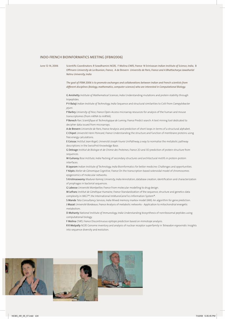
INDO-FRENCH BIOINFORMATICS MEETING (IFBM2006)
June 12-14, 2006 Scientific Coordinators: R Sowdhamini NCBS, F Molina CNRS, France N Srinivasan Indian Institute of Science, India, B Offmann University de La Reunion, France, A de Brevern Universite de Paris, France and A Bhattacharya Jawaharlal Nehru University, India
The goal of IFBM 2006 is to promote exchanges and collaborations between Indian and French scientists from
different disciplines (biology, mathematics, computer sciences) who are interested in Computational Biology.
G Anishetty Institute of Mathematical Sciences, India Understanding mutations and protein stability through tripeptides.
P V Balaji Indian Institute of Technology, India Sequence and structural similarities to CstII from Campylobacter jejuni.
P Barbry University of Nice, France Open-Access microarray resources for analysis of the human and mouse transcriptomes (from mRNA to miRNA).
P Benech Parc Scientifique et Technologique de Luminy, France Predict search: A text mining tool dedicated to decipher data issued from microarrays.
A de Brevern Universite de Paris, France Analysis and prediction of short loops in terms of a structural alphabet. C Chipot Université Henri Poincaré, France Understanding the structure and function of membrane proteins using
free energy calculations. E Coissac Institut Jean Roget, Université Joseph Fourie UniPathway, a way to normalise the metabolic pathway
descriptions in the SwissProt Knowledge Base. G Deleage Institut de Biologie et de Chimie des Proteines, France 2D and 3D prediction of protein structure from
sequences. M Guharoy Bose Institute, India Packing of secondary structures and architectural motifs in protein-protein
interfaces. B Jayaram Indian Institute of Technology, India Bioinformatics for better medicine: Challenges and opportunities. F Képès Atelier de Génomique Cognitive, France On the transcription-based solenoidal model of chromosomes
epigenomics of molecular networks. S Krishnaswamy Madurai Kamraj University, India Annotation, database creation, identification and characterization
of prophages in bacterial sequences. G Labesse Université Montpellier, France From molecular modelling to drug design. M Lefranc Institut de Génétique Humaine, France Standardization of the sequence, structure and genetics data
complexity in IMGT®, the International ImMunoGeneTics Information System®. S Mande Tata Consultancy Services, India Mixed memory markov model (4M): An algorithm for gene prediction. J Mazat Université Bordeaux, France Analysis of metabolic networks - Application to mitochondrial energetic
metabolism. D Mohanty National Institute of Immunology, India Understanding biosynthesis of nonribosomal peptides using
computational biology. F Molina CNRS, France Discontinuous epitope prediction based on mimotope analysis. R R Metpally NCBS Genome inventory and analysis of nuclear receptor superfamily in Tetraodon nigroviridis: Insights
into sequence diversity and evolution.
NCBS_AR_05_07.indd 134 7/10/08 5:35:45 PM
H A Nagarajaram Centre for DNA Fingerprinting and Diagnostics, India Microsatellite Polymorphism in Prokaryotic genomes: Implications on genome plasticity and evolution.
C Nguyen Université d’Aix-Marseille II, France Biologists’ point of view to questions for which answers with transcriptome analysis? B Offmann Université de La Reunion, France Bridges and blocks: A taste of protein structure analysis in Indo-French
collaboration. S Pérès Université Bordeaux, France Structural analysis of metabolism. O Poch Institut de Génétique et de Biologie Moléculaire et Cellulaire, France Towards efficient data exploitation and
knowledge discovery in the information era. O Radulescu Institute for Mathematical Research, France New qualitative approaches in molecular biology. R Sathyapriya Indian Institute of Science, India Structure networks of aminoacyl tRNA synthetases: Insights into
amino acid and tRNA recognition. R Sowdhamini NCBS Putative functional attributes to serineproteases in Drosophila genome. D M Salunke National Institute of Immunology, India Molecular mimicry: Structural basis and physiological
implications. N Srinivasan Indian Institute of Science, India Social domains: An investigation on preferred combinations of Tethered
Protein Domain Families. M Tyagi Université de La Réunion, France Structural alphabets, from protein structure description to comparison.
NCBS-JNCASR-HARVARD SYMPOSIUM ON “INTERDISCIPLINARY MATERIALS SCIENCE, COMPUTATION AND BIOLOGY”
August 11-13, 2006 Scientific Coordinators: G V Shivashankar NCBS, Srikanth Sastry Jawaharlal Nehru Centre for Advanced Scientific Research, India and L Mahadevan Harvard University, USA
Speakers at this meeting were from Harvard University and from various institutions in Bangalore. The scientific program was divided broadly among sessions on: Soft and hard matter science, Active materials and physiology, Molecular and cellular biophysics, and Computation and systems biology. The first session was held at JNC and three sessions were held at NCBS.
B Bagchi Indian Institute of Science, India Dynamics of a binary supercooled liquid with orientational degree of freedom.
H Balaram Jawaharlal Nehru Centre for Advanced Scientific Research, India Codon bias, amino acid composition and functional annotation of Plasmodium falciparum proteins.
R Bandopadhyay Raman Research Institute, India Experimental studies of the slow dynamics in aging clay suspensions.
N Chandrabhas Jawaharlal Nehru Centre for Advanced Scientific Research, India Light, nanoparticles, materials: A Raman study.
S Chattarji NCBS “Good plasticity” in a “bad neighborhood”: Silent synapses speak up in the amygdala. C Dasgupta Indian Institute of Science, India Dynamic heterogeneity from the structure of glassy free energy minima. V D’Souza Harvard University, USA Structural studies of RNA protein complexes involved in retroviral replication. R Govindrajan Jawaharlal Nehru Centre for Advanced Scientific Research, India Gravity-free hydraulic jumps and metal
femtocups. Y Hatwalne Raman Research Institute, India Escape configuration lattice near the nematic-isotropic transition: Tilt
analogue of blue phases. Y Krishnan NCBS First blueprint, Now bricks - DNA’s new incarnation. T K Kundu Jawaharlal Nehru Centre for Advanced Scientific Research, India Chromatin dynamics link to disease and
therapeutics. L Mahadevan Harvard University, USA Materials and physiology: Shape, flow and movement. P Maiti Indian Institute of Science, India Structure and dynamics of DNA-dendrimer complexation: Free energy
landscape and DNA sequence dependence. S Mayor NCBS Active organization of nanoscale clusters of GPI-anchored proteins in living cell membranes:
Implications for rafts and endocytosis. V Mootha Harvard University, USA Genomic approaches to human diabetes.
135
ME
ET
ING
S A
ND
WO
RK
SH
OP
S
NCBS_AR_05_07.indd 135 7/10/08 5:35:45 PM

V N Murthy Harvard University, USA Using light to probe neural circuits. S Narasimhan Jawaharlal Nehru Centre for Advanced Scientific Research, India Ab initio investigations of low
dimensional systems. K S Narayan Jawaharlal Nehru Centre for Advanced Scientific Research, India Soft matter electronics and
optoelectronics. R Pandit Indian Institute of Science, India Drag reduction by polymer additives in decaying, homogeneous, isotropic
turbulence. S Pati Jawaharlal Nehru Centre for Advanced Scientific Research, India quantum magnetism, optical and transport
properties. M Puranik NCBS Biophysical and computational studies of DNA damage and repair mechanisms. S Ramaswamy Indian Institute of Science, India Active particles: Rheology, coating flows and granular monolayers. M Rao RRI and NCBS Active membranes: transport, shape deformations and chemical networks. S Sastry Jawaharlal Nehru Centre for Advanced Scientific Research, India Gelation, phase separation and dynamical
arrest in fluids. S K Sikdar Indian Institute of Science, India How does epileptic activity affect molecular, cellular and neuronal
network properties? A Sood Indian Institute of Science, India Spatio-temporal chaotic dynamics in the flow of soft matter. G V Shivashankar NCBS Spatio-temporal organization of the chromatin assembly and function within living cells. G Subramanian Jawaharlal Nehru Centre for Advanced Scientific Research, India Hydrodynamic interactions in
suspensions of E.coli. N Surolia Jawaharlal Nehru Centre for Advanced Scientific Research, India Plasmodium Biology – A systems biology
approach. M Thattai NCBS Engineering and encoding living networks. J Udgaonkar NCBS Collapse and cooperativity in protein folding. R Varadarajan Indian Institute of Science, India Design and isolation of temperature-sensitive mutants in E. coli, yeast
and Drosophila. U Waghmare Jawaharlal Nehru Centre for Advanced Scientific Research, India Electronic structure, bonding and
properties of materials. D Weitz Harvard University, USA Soft materials research: Crystals, glasses and gels.
THE SECOND BANGALORE STEM CELL COURSE AND WORKSHOPNovember 20 - December 3, 2006. Scientific Coordinators: Maneesha Inamdar JNC, M M Panicker and K VijayRaghavan NCBS
The course was conducted both at NCBS and JNC and it aimed to bring together international and Indian stem cell researchers to share latest developments and provide “hands-on” training in embryonic stem cell manipulation. This course was two weeks of laboratory training on the culture, assay and differentiation of human and mouse embryonic stem cells and embryonic carcinoma cells. In addition, the course included research seminars and lectures. Participants were restricted to 12 in number.
P Andrews University of Sheffield, UK Genetic instability and adaptation of human ES cells. Surface antigen markers of human ES cells.
B Blum The Hebrew University of Jerusalem, Israel Spontaneous and induced differentiation of human embryonic stem cells.
J Dhawan Centre for Cellular and Molecular Biology, India Transcriptomes – methods and caveats. P Gokhale University of Sheffield, UK Genetic manipulation of human ES cells. The international stem cell initiative. A Hardikar National Centre for Cell Science, India Human pancreatic progenitor cells for diabetes cure. J Jackson Rajiv Gandhi Centre for Biotechnology, India Notch dependent and independent activation of Hes-1 during
stem cell proliferation. A Khanna Reliance Life, India Regulatory mechanisms governing differentiation of embryonic stem cells.
NCBS_AR_05_07.indd 136 7/10/08 5:35:50 PM

S Khosla Center for DNA Fingerprinting and Diagnostics, India Allele-specific chromatin organization within the imprinted mouse neuronatin gene.
S Kumar Centre for Cellular and Molecular Biology, India Structural and functional analysis of Casein Locus. N Lenka National Centre for Cell Science, India Restoration of functional activities in Parkinsonian rats by ES cell-
derived dopaminergic neurons. R Lovell-Badge National Institute for Medical Research, UK Basic mouse embryology. Sox genes and stem cells. M R S Rao Jawaharlal Nehru Centre for Advanced Scientific Research, India Chromatin remodeling during mammalian
germ cell differentiation. A Rangarajan Indian Institute of Science, India Stem cells, cancer and cancer stem cells. G Stacey Stem Cell Bank, UK Considerations for the use of stem cells in therapy. The role of stem cell banks in stem
cell research. A Urbach The Hebrew University of Jerusalem, Israel Modeling of human genetics diseases using human embryonic
stem cells. G Vemuganti L.V. Prasad Eye Institute, India Limbal stem cells. H Wheadon University of Ulster, UK The use of embryonic stem cells as a model to study leukaemia: Haemopoietic
differentiation of ES cells.
THE THIRD BANGALORE BENNY SHILO COURSE IN DEVELOPMENTAL BIOLOGY December 25 – January 4, 2007 Scientific Coordinator: K VijayRaghavan NCBS The focus of this course was to connect molecular and cellular pathways to the understanding of the mechanisms
that underlie animal development. Topics in this course covered range from the early development and patterning of the embryo, the patterning of specific tissues and their morphogenesis, the analysis of signaling and regulatory mechanisms, and the development of behaviour.
The total number of participants in the course was restricted to 40. Lectures were combined with a few practical demonstrations and the study of research papers in work- groups followed by discussions with the teachers.
S Cohen The European Molecular Biology Laboratory, Germany MicroRNA functions. | Morphogen gradients and disc patterning. | Growth control in imaginal discs.
T Hyman Max Planck Institute of Molecular Cell Biology and Genetics, Germany Cell Polarity and Cell Division in the early C. elegans embryo.
M Inamdar Jawaharlal Nehru Centre f or Advanced Scientific Research, India Introduction to mouse development. Z Paroush, The Hebrew University of Jerusalem, Israel Segmentation of the Drosophila embryo: i) Interpreting a
morphogen gradient into positional information and cell identity. ii) Transcription factors and the regulation of gene expression in development. Is groucho a two-faced co-repressor?
B Podbilewicz Technion, Israel General Introduction to C. elegans (Overview: lineage, apoptosis, genetics, RNAi). | Cell fusion and epidermal morphogenesis (embryonic development). | Evolution of vulva development (postembryonic development). | Neuronal arborization (larval-adult development). | Why and how do cells fuse?
P Rørth The European Molecular Biology Laboratory, Germany Cell migration and guidance mechanisms: Guiding border cell migration.
E Raz Gottingen, Germany Looking at zebrafish embryos. Introduction to germ cell development in invertebrates and vertebrates. | Zebrafish as a model organism in developmental biology. | Looking at zebrafish embryos. | Molecular and cellular mechanisms controlling germ cell migration in zebrafish.
R Ranganathan University of Texas, USA Signaling networks. E Schejter Weizmann Institute, Israel Stem cell vs. cystoblast cell fates. | Oocyte selection. | Oocyte postioning within
the egg-chamber. | Bicoid morphogen gradient. | The WASp-Arp2/3 actin polymerization machinery is essential for myoblast fusion in Drosophila.
A Sengupta Rutgers University, USA Dynamical modeling in genetic networks, with segment polarity as an example. B Shilo Weizmann Institute, Israel Regulation of EGFR signaling in Drosophila development by intracellular
trafficking. | Determination of dorso-ventral polarity during Drosophila embryogenesis : Determination of dorsal fates by the BMP pathway. | Determination of dorso-ventral polarity during Drosophila embryogenesis : Determination of ventral fates by the EGFR pathway.
V Sriram, NCBS Role of mitochondrial remodeling in programmed cell death K VijayRaghavan NCBS The development of behavior. | Developmental biology and genetic tools used to study
development. T Volk Weizmann Institute, Israel Early neurogenesis: Neuroblast selection and the involvement of Notch signaling
and asymmetric cell division. | Late neurogenesis: Neuronal pathfinding and the activity of Robo, Slit and Netrins. | Alternative splicing mechanisms and the control of glia and tendon cell maturation.
137
ME
ET
ING
S A
ND
WO
RK
SH
OP
S
NCBS_AR_05_07.indd 137 7/10/08 5:35:50 PM
NCBS ANNUAL RESEARCH TALKS
Feb 18-20, 2007 Scientific Coordinator: Jayant Udgaonkar, NCBS
U S Bhalla Cells, smells and memories: Towards whole-system models. S Chattarji Known and unknown unknowns in the amygdala. G Hasan The InsP3 receptor and neuronal calcium homeostasis. S P Koushika Transport in neurons: Early stories from C. elegans. K S Krishnan Venomous peptides. S Krishna Notch ligands: The inter phase with stem cells, cancers and the immune system. Y Krishnan Fourplay with DNA: I-tetraplexes and structural DNA Nanotechnology. M K Mathew Moving ions in a Salty World. S Mayor Mechanisms of Endocytosis in Eukaryotic Cells. M M Panicker The 5-HT2A receptor, its ligands and possible roles. M Puranik Purines as intrinsic probes of the protein active-site. S Quader Strategies and arms-races: Evolutionary ecology in the wild. M Rao Principles of organization in biological systems. U Ramakrishnan Genetic time travel in South Asia: Using modern and ancient genetic variation to infer population
history. K Rau Laser induced damage mechanisms in tissue: insights from time resolved imaging and hydrodynamic
modeling. V Rodrigues Activity dependent mechanisms in the development and maintenance of neuronal circuits. V Sriram Mechanisms of mitochondrial remodeling. A Sarin Ulysses Pact: T-cell activation and the emergence of memory. G V Shivashankar Spatio-temporal organization of chromatin assembly and transcription control within living cells O Siddiqi Olfactory learning and memory in Drosophila melanogaster. R Sowdhamini Computational approaches to protein science. M Thattai The dynamics and evolution of living networks. J B Udgaonkar How do proteins misfold? K VijayRaghavan Assembling the elements required for walking
FROM SUSHI AND THE SNAIL: A JOURNEY FROM MOLECULES TO CIRCUITS AND BEHAVIOR THROUGH CELLULAR TRAFFIC
February 23-25, 2007 Scientific Organizers: Veronica Rodrigues, Satyajit Mayor NCBS and M Ramaswami University of Arizona, USA
U Banerjee University of California, USA Blood relations. M Bate University of Cambridge, UK From muscles to neurons - getting to grips with behaviour. E Buchner Universitat Wurzburg, Germany Synapses and circuits in olfactory conditioning: Novel proteins and new
techniques. W Chia Temasek Life Sciences Laboratory, Singapore Asymmetric division and the control of cell proliferation. K S Krishnan NCBS Genetic studies of inhalational anesthesia. T Kurzalia Max Planck Institute, Germany Why we all need cholesterol? J Manjrekar M.S. University of Baroda, India What, if anything, are prions good for? M K Mathew NCBS Transporting a transporter - the Voltage-Gated K+ Channel. S Mayor NCBS Stoned and Comatose in TIFR. T Oliveria University of Utah, USA Using deadly cone snails to learn drug design and fill the drug pipeline. M Ramaswami University of Arizona, USA The snail, sushi, and the snail. M Rao NCBS and RRI, India Dynamics of Trafficking. S Sharma Indian Institute of Science, India NMR studies of Conotoxins. M Singh University of Mysore, India Rules for spatial distribution patterns of wild mammals. O Siddiqi NCBS Learn how to smell the sushi and snails. V Sriram NCBS Role of mitochondrial remodeling in Drosophila programmed cell death. T Venkatesh City University, USA A novel regulatory network work for glial differentiation in Drosophila. J B Udgaonkar NCBS Cooperativity in protein folding.
NCBS_AR_05_07.indd 138 7/10/08 5:35:50 PM

WORKSHOP IN CONSERVATION GENETICS AND MOLECULAR METHODS
April 9-12, 2007 Scientific Coordinator: Uma Ramakrishnan NCBS
This workshop was conducted for scientists/managers at the Department of Forests, Bhutan. The workshop aimed to familiarize participants with how genetic data can be used for conservation and management of species. The workshop included lectures and practical demonstrations. The subject material was divided into genetic investigations at the levels of the species, the population and the individual.
Y V Bhatnagar NCF NCF’s collaboration with NCD, WWF-B and NCBS. S Mondal and S Velumani NCBS Wet lab: Check gel for species identification, individual identification and sexing. S Mondal Wet lab: DNA extraction from faecal material. Computer lab: Designing primers for snow leopards.Case
studies lecture: Individual identification and sexing of tigers from the wild. S Mukherjee NCBS Scats and diet analysis. Case studies lecture: Methods for species identification: Challenges of
species id for small carnivores. U Ramakrishnan NCBS The basics: Introduction to genetics and molecular biology. How do we identify species
using molecular tools? Wet lab: Basics of DNA extraction and PCR. Phylogenetics and the building of trees. Basics of population genetics and individual identification.
U Ramakrishnan and S Mondal NCBS Wet lab: PCR for species identification/discrimination for tiger, leopard. U Ramakrishnan NCBS What does a basic molecular lab need? Brainstorming and planning for the future. The need
for molecular methods in conservation in the Bhutanese context. A Sinha NCF and NIAS, India Plenary lecture: A new species of macaque from Arunachal Pradesh, India. J J Vadakkam and Debopriyo NCBS and NCF Computer lab: Basic bioinformatics: Why do we need it and how do we
do it? J J Vadakkam NCBS and NCF Case studies lecture: Methods for species identification: Discovering cryptic species in
Namdapha. S Velumani NCBS Case studies lecture: Individual identification of leopards.
THE LIVING NETWORKS JAMBOREE
July 14, 2007 Scientific coordinator: Mukund Thattai NCBS
V Gautam Delhi University, India The assembly: Parts lists and biobricks. S More IISER, India and S Kundu, St. Stephen’s, India The computational tools: Analyzing images, exploring data. N Rai Indian Institute of Technology, India The system: Bacterial quorum sensing. K Ramakumar Indian Institute of Technology, India The experiments: Understanding a network by characterizing its
parts. M Thattai NCBS The model: Differential equations and input-output maps.
INDO-AUSTRALIAN WORKSHOP ON “RAMAN SPECTROSCOPY AND IMAGING FOR UNDERSTANDING MOLECULAR PROCESSES IN LIVE CELLS”.
August 22-23, 2007 Scientific coordinators: Mrinalini Puranik NCBS, Bayden R Wood and John Beardall Monash University, Australia Siva Umapathy Indian Institute of Science, India
This workshop brought together experts in ultraviolet resonance Raman spectroscopy and Raman imaging of live cells to understand molecular processes and structures.
C C A Bernard Monash University, Australia Understanding CNS autoimmunity: From animal models to MS patients. J Beardall Monash University, Australia New approaches to old problems: applications of biospectroscopy to answer
biological questions in algae and plants. P Heraud Monash University, Australia Investigations of MS-like diseases using vibrational micro spectroscopy. Using
vibrational spectroscopy to probe environmental change in phytoplankton.
139
ME
ET
ING
S A
ND
WO
RK
SH
OP
S
NCBS_AR_05_07.indd 139 7/10/08 5:35:51 PM

P Lay University of Sydney, Australia Vibrational spectroscopic imaging of breast cancers and single cells. S Maiti Tata Institute of Fundamental Research, India Looking at the body’s own chromophores. D McNaughton Monash University, Australia Vibrational spectroscopic imaging for biological systems. K Menon Monash University, Australia Molecular mechanisms involved in the breakdown of myelin: Implication for
neurodegenerative diseases. S Mayor NCBS Heterogeneity of membrane composition in living cells. C Narayana Jawaharlal Nehru Centre for Advanced Scientific Research, India Surface enhanced Raman spectroscopy
for interaction studies and diagnostics in biological systems. M M Panicker NCBS A role for serotonin in early mammalian development – an imaging and functional study. M Puranik NCBS Resonance Raman spectroscopy of protein ligands as a probe of their amino acid environment. A Rangarajan Indian Institute of Science, India Stem cells, cancer and imaging. K Shelly Monash University, Australia Characterising nutrient stress in Chlorella emersonii (Chlorophyta) using
nutrient-induced fluorescence transients and biospectroscopy. G V Shivashankar NCBS Functional imaging of chromatin organization and transcription control within living cells. B R Wood Monash University, Australia Resonance Raman Spectroscopy in malaria research. | Molecular
characterization of oocyte maturation using FTIR synchrotron spectroscopy and FTIR imaging.
NCBS AND JST-ICORP MEETING ON SPATIAL AND TEMPORAL MAPPING OF THE MOLECULAR ORGANIZATION IN MEMBRANES OF LIVING CELLSNovember 20-22, 2007 Scientific Coordinators: Akihiro Kusumi Kyoto University, Japan and Satyajit Mayor NCBS
This meeting explored the landscape of the cell membrane from the single molecule to the mesoscale, to understand how mechanical and compositional properties of the cell membrane are regulated, and in turn could influence gene expression.
W Cho University of Illinois, USA Spatiotemporal regulation of cellular processes by lipids and lipid-binding proteins. T Fujiwara Kyoto University, Japan Compartmentalized movement of lipids in the cell membrane as revealed by
single molecule techniques. G Gupta NCBS Genomics. S Kumari and D Goswami NCBS Membrane organization and endocytosis. A Kusumi Kyoto University, Japan Mechanism for raft-based signal transduction. A Mazumder and F M Hameed NCBS Mechano-chemical signaling. S Mayor NCBS Functional rafts in living cell membranes. M Rao NCBS Physical principles of organization in biological systems. S Sivaramakrishnan Stanford University, USA Myosin VI and its role in cellular endocytic pathway. G V Shivashankar NCBS Spatio-temporal organization of chromatin assembly and transcription control within living
cells. J A Spudich Stanford University, USA Myosin VI and the basis of reverse directionality. K Suzuki Kyoto University, Japan Mechanism for Raft-based signal transduction as studied by single molecule
tracking. Raft and non-raft molecules undergo very similar diffusion in the time scales between 25 microseconds and 2.5 seconds.
M Thattai NCBS The dynamics and evolution of living networks. R Vale University of California at San Francisco, USA Protein-protein networks based plasma membrane
microdomains in T-Cells.
NCBS_AR_05_07.indd 140 7/10/08 5:35:51 PM

ADVANCED COURSE ON MOLECULAR MODELING AND PROTEIN-LIGAND INTERACTIONSDecember 14-18, 2007 Scientific Coordinator: R Sowdhamini NCBS This workshop was dedicated to the particular area of computational biology. There was an interesting series of talks
about the theory of protein-ligand interactions, along with special lectures on biological perspectives as well. The talks were accompanied by ample amount of hands-on experience (practical sessions) which was led by a team of software engineers from VLife Sciences, Pune.
V N Balaji Jubliant Biosystems, India Docking studies in the active site of GSK-3. N Chandra Indian Institute of Science, India Characterization of molecular recognition : Protein-Ligand Interface. M Puranik NCBS Quantum mechanics of modeling ligands. K Seshadri Astra Zeneca, India Assessment of protein druggability by structural features: Application to enzyme. R Sowdhamini NCBS Protein-ligand interactions: Locks-and-keys? R Sowdhamini NCBS Protein databank and Cambridge small molecule databases. N Srinivasan Indian Institute of Science, India Modeling pentraxins and inferring ligand specificities. L Tripathi NCBS Softwares employed for docking. P Xavier Avesthagen. India Structure-based drug design and its applications in modern drug discovery research.
WORKSHOPS ORGANIZED ELSEWHERE
Evolution of Development Mechanisms Mahabaleswar, India January 21-26, 2005 Scientific Coordinators: K VijayRaghavan and V Rodrigues
Genes, Development and the Emergence of Behaviour Trieste, Italy April 25 – May 13, 2005 Scientific Coordinators: K VijayRaghavan, M Bate and V Rodrigues
8th IBRO School of Neuroscience – Asia-Pacific Region: Brain Storming in Bombay Tata Institute of Fundamental Research, Mumbai September 02-10, 2006 Scientific Coordinators: V Rodrigues, V Vaidya, R Mallick and S Tole
Ecological Modeling Nature Conservation Foundation, Mysore, India October 18-19, 2007 Scientific Coordinators: S Quader and K Isvaran
141
Me
eT
InG
S A
nD
WO
RK
Sh
OP
S
NCBS_AR_05_07.indd 141 7/10/08 5:36:54 PM

143 Overview | 144 Programs | 145 Degrees | 146 Courses | 149 Lectures and Visits
153 Trainees | 156 MSc Wild Life Program
AcAdemic Activiti es
NCBS_AR_05_07.indd 142 7/11/08 2:59:15 PM
The academic community in nCBS has diversified further over the past two years with the addition of several international scholars to almost all our academic programs. Our graduate programs leading to PhD degrees come in two flavours. Many of our students join for a PhD after a Masters degree in a basic science discipline or after a Bachelors degree in an applied science discipline like engineering or Medicine. We also have students joining for an Integrated MS-PhD program after a Bachelors degree in basic sciences. In addition, we have a few students working towards a Masters by research. All our students undertake a rigorous coursework programme and participate in research leading to a thesis to be submitted for evaluation. Through a separate entrance test we will now also admit students for a Masters program in Wildlife Biology & Conservation, in collaboration with Centre for Wildlife Studies, Bangalore.
The majority of students register with TIFR at Mumbai. however, for several years we have had an excellent relationship with the Manipal Academy for higher education (MAhe) and students working at nCBS can continue to register with either MAhe or Mysore University if needed. This is particularly so for students working on externally-funded PhDs, like those on grants and with their own fellowships from agencies such as CSIR, DBT or ICMR. We continue our close ties with our former hosts, the Indian Institute of Science and our current hosts, the University of Agricultural Sciences. new links are being built through Memoranda of Understanding with several other institutions in India and abroad.
Apart from students working towards academic degrees, we have a comparable number of young folk working on projects funded by external agencies. A smaller population of enthusiastic Post Doctoral Fellows brings with them both the expertise and the perspectives of their respective graduate programmes from all over the country and abroad. More transiently, we have an almost continuous stream of trainees visiting from all over the country and carrying out short 6-8 week projects at nCBS, getting some exposure to biological research here and spreading the excitement on their return to their home institutions. We also host a limited number of Masters students for their MSc thesis projects usually carried out over a 6-month period. With the recent nCBS/harvard-MIT student exchange program in place we have begun hosting undergraduates from these internationally renowned institutions for a period of three months every summer. Similar student exchange programs with other international Universities, including the University of Cambridge, are planned. A large number of scientists both national and international and students from all over the country participate in our Annual Symposia.
Intellectual diversity is as important for us as cultural variety and both are critical for driving interdisciplinary research. We have an active iBio (integrative Biology) program in collaboration with the Raman Research Institute and enthusiastic interactions with chemists and physicists from several institutions. We are keen to foster relations with medical institutions and to this end have initiated several joint programs for research with faculty at the Christian Medical College, Vellore. With the energy of youth and the vigorous admixture of inputs from diverse backgrounds, the Centre is kept in a ferment, receptive of new ideas and striving to build on accomplishments.
Gaiti hasan
Head, Academic Activities
overview
NCBS_AR_05_07.indd 143 7/10/08 5:36:57 PM

Programs
PhD Program
The national Centre for Biological Sciences has academic programs leading to PhD and Integrated PhD degrees. The PhD program at nCBS accepts candidates with a Masters in any basic science discipline or a basic degree in any applied science such as Medicine, engineering, Pharmacy and Veterinary Science. The Integrated PhD program takes on students with an excellent academic record at the BSc level and who are strongly motivated to pursue a career in research.
Interdisciplinary Research Program
iBio is a interdisciplinary program that seeks to apply the experimental and theoretical tools of the physical sciences to the study of challenging biological problems. The program provides a stimulating research environment, with dynamic young faculty, state-of-the-art equipment, and international exposure through conferences, workshops, and visiting scientists from around the world. The iBio faculty at nCBS includes chemists, physicists, neuroscientists, and computational biologists. exceptional candidates with strong backgrounds in the physical sciences, including experimental and theoretical physicists, chemists, mathematicians, computer scientists, and engineers are eligible to enter the iBio PhD and postdoctoral programs.
We also encourage talented medical students with an interest in biomedical research to pursue a PhD degree at nCBS.
The academic programs at nCBS include basic and advanced courses, but emphasise research. The programs are full-time and students often put in over 12 hours of work in a day between course work and a research program. A qualifying examination is held during the second year of each of these programs to decide on whether a student is eligible to go on to work toward a PhD. Students register with the Tata Institute of Fundamental Research.
Advertisements for applications to all programs appear in leading national newspapers in September/October each year, for a nation-wide written test conducted in mid December. Past academic performance, letters of recommendation from current/former teachers, performance in the written test, and individual write ups in the applications are considered in short listing candidates for the interview held at the end of May/ beginning of June. The new academic year begins in August.
Visiting Students/Training Program
The training program at nCBS is open to interested and motivated pre-doctoral candidates and usually comprises M.Sc. students between their first and second years. Many students also do their Masters projects here. engineering and medical students are also encouraged to apply.
Applications are accepted all through the year. Individual faculty members choose the students they will accommodate in their laboratories.
Many of the Centre’s trainees find their research experience invaluable. Perhaps the most tangible benefits of this program are manifest in the number of such trainees who are enrolled for a degree in the best research laboratories within the country and outside.
Teacher Trainees
In addition to training fresh graduates, nCBS also has an active Teacher Training program wherein college and university teachers carry out research leading to a PhD degree. It is hoped that such programs will improve the interaction between universities and research institutes in a mutually beneficial manner. Our recruitment to this program is extremely informal. A teacher who is interested contacts a faculty member at nCBS whose research interests most closely match his/her own. On the faculty member’s recommendation, the candidacy is evaluated by a committee on a case-by-case basis.
Postdoctoral Positions
nCBS has an active postdoctoral (visiting) fellow training program. Postdoctoral fellows usually work with one of the existing groups. Fellowships are tenable for one to three years. Interested individuals are encouraged to write to the head, Academic Activities with their curriculum vitae and the names of at least three referees. Applications will be accepted throughout the year. Decisions on award of postdoctoral fellowships are made twice a year, in February and August.
In addition to nCBS-funded Fellowships, some grants also fund Post-doctoral and Junior Research Fellow positions. Appointments to these positions are made as and when they become available, and applications are accepted through the year.
For further information about these programs, e-mail us at:Postdoc: [email protected] | PhD.: [email protected] | Training: [email protected] further details about admissions/positions are available at: http://www.ncbs.res.in
NCBS_AR_05_07.indd 144 7/10/08 5:36:58 PM

degrees
PhD< Sriram M Ajay Electrophysiological and biochemical analysis of temporal tuning in a hippocampal
synaptic network (Guide: U S Bhalla)
< Santanu Banerjee Investigating the role of InsP3 receptor gene in Drosophila flight (Guide: G hasan)
< Arjun Guha The role of Dynamin in endocytosis in primary cultured cells in Drosophila (Guide: S Mayor)
< Debkanya Dutta Molecular and cellular mechanisms of founder cell function in adult Drosophila myogenesis (Guide: K VijayRaghavan)
< Ashwini H Godbole Functional characterization of VDAC: A conserved element of programmed cell death pathways (Guide: M K Mathew)
< N Parikh Regulation of localization and function of the pro-apoptotic protein Bax (Guide: A Sarin)
< Raghav Rajan Odour localisation by rats (Guide: U S Bhalla)
< M Raghuprasad Rao Genome analysis of receptor superfamilies (Guide : R Sowdhamini)
< B S Sharath Experience dependant plasticity: A study of structure-function relationships in the hippocampus and amygdala (Guide: S Chattarji)
< Gautam Vivek Soni Understanding physical interactions leading to structure-function dependence in biological systems (Guide : G V Shivashankar)
< Deepa Subramanyam Epithelial-stromal cell cross-talk maintains notch signaling in cancer (Guide: S Krishna)
< Y Sushama Role of the gene brinker in antero-posterior patterning of Drosophila Malpighian tubules (Guide: K VijayRaghavan)
< Rajat R Varma Exploring the organization of GPI-anchored proteins in living cells (Guide: S Mayor)
MSc< Sugat Dabholkar Development of techniques to probe bacterial genetic networks (Guide: M Thattai)
< Kumar Gaurav Biological sequence analysis for protein annotation based on motif descriptors (Guide: R Sowdhamini)
< Girija Goyal Mechanism of HID induced apoptosis in Drosophila melanogaster (Guide: A Sarin)
AC
AD
eM
IC A
CT
IVIT
IeS
145
NCBS_AR_05_07.indd 145 7/10/08 5:36:58 PM

courses
NCBS offers courses at the basic and advanced levels. At the basic level we offer courses in Biochemistry and Biophysics; Cell Biology; Neurobiology; Immunology; Molecular Biology and Genetics, Developmental Biology and Physical Methods.
As in the past, our students attend basic and advanced courses at the Indian Institute of Science in the Departments of Biochemistry, Ecological Sciences, Microbiology and Cell Biology, Molecular Reproduction, Development and Genetics, Physics and Molecular Biophysics Unit. Our students also attend courses offered by Jawaharlal Nehru Centre for Advanced Scientific Research and University of Agricultural Sciences, Bangalore.
Courses offered at NCBS
neurobiology Basic/August 2005 S Chattarji and U S Bhalla
Cell Biology Basic/August 2005 S Mayor
Diffusion for Biologists Basic/August 2005 K Banerjee
Developmental Biology Basic/August 2005 K VijayRaghavan and M Inamdar
Conservation genetics: Advanced/August 2005 U RamakrishnanTheory and from theory to practice
DnA Dyamics: Transposition Advanced/December 2005 G Simchenand Recombination
Systems and Synthetic Biology Advanced/January 2006 M Thattai
Scientific Writing Advanced/January 2006 G hyde
Molecular spectroscopy Advanced/February 2006 M Puranik and K Rau and imaging: A survey of conceptsand techniques
Membrane Trafficking Advanced/February 2006 S Mayor and V Malhotra
Membrane Biophysics Advanced/March 2006 M K Mathew
Immunology Basic/August 2006 S Krishna and S Pillai
Biophysical Chemistry Basic/August 2006 J B Udgaonkar and M K Mathew
NCBS_AR_05_07.indd 146 7/10/08 5:37:00 PM

Developmental Biology Basic/August 2006 K VijayRaghavan and M Inamdar
Bioinformatics Basic/August 2006 R Sowdhamini
Genetics Basic/ August 2006 S P Koushika and U Ramakrishnan
Scientific Writing Advanced/August 2006 G hyde
Cellular Biophysics Advanced/September 2006 G V Shivashankar
Benny Shilo Course in Advanced/December 2006 B Shilo and S CohenDevelopmental Biology
Simulations in Biology Advanced/January 2007 U S Bhalla and M Thattai
Intracellular Transport Advanced/April 2007 S P Koushika
Physical Biochemistry Basic/August 2007 J B Udgaonkar and M K Mathew
Cell Biology Basic/August 2007 S Mayor
Chemical Biology Basic/September 2007 Y Krishnan
Developmental Biology Basic/September 2007 K VijayRaghavan and M Inamdar
Molecular Biology Basic/September 2007 M M Panicker
Short courses offered elsewhere S P Koushika | One module in molecular genetics Indian Institute of Science, India (April - May, 2005).
R Sowdhamini | history and biological impact of protein structure determination Reunion University, France (April, 2007)
Courses attended elsewhereN Agrawal | neural systems and behavior, Marine Biological Laboratory, USA (June – August, 2006).
J Bajaj, B C Choudhary, S Gupta and Vishal | Molecular oncology, Indian Institute of Science, India (January – May, 2006).
J Bajaj and M G Swetha | human molecular genetics, Indian Institute of Science, India (January – May, 2006).
A Bhattacharya | IBRO-ISn neuroscience School, The National University of Singapore, Singapore (June, 2006).
N K Chakraborty and J Kumar | Molecular genetics, Indian Institute of Science, India (January – May, 2006).
B C Choudhary, L Harini, K Rathore, S Gupta, T Gupte, T S Sharangdhar, S Velumani and Vishal | Statistical methods, University of Agricultural Sciences, India (October, 2005).
P D Deepalakshmi | QToF Ultima API small molecule operator training course, Waters, UK (June, 2005) | Mass spectrometry for proteomics, Munnar, India (January, 2006).
A Dasgupta | DnA-protein interaction, regulation of gene expression, nanobiology, Indian Institute of Science, India (August – December, 2007).
A Dasgupta and M N Modi | Protein structure, folding and design, Indian Institute of Science, India (January – May, 2007).
A Dasgupta and A Nilesh | Introduction to biophysical chemistry, Indian Institute of Science, India (August – December, 2007).
Dhanya P | neural networks, Indian Institute of Science, India (January – May, 2006).
147
AC
AD
eM
IC A
CT
IVIT
IeS
NCBS_AR_05_07.indd 147 7/10/08 5:37:00 PM
Dhanya P | Okinawa computational neuroscience course, Japan (June – July, 2006).
Divya P S | Computational approaches to drug discovery, Indian Institute of Science, India (January – May, 2006).
Divya P S and S Kedia | Molecular oncology, Indian Institute of Science, India (January – December, 2007).
Divya P S, N Kumar and P Raychaudhuri | Molecular spectroscopy and structure, Indian Institute of Science, India (August – December, 2005).
S P Goswami, S Gupta and M Sehgal | Molecular basis of signal propagation and synaptic transmission in neurons, Indian Institute of Science, India (January – December, 2007).
S Jain and S Kumar | Introduction to biophysical chemistry, Indian Institute of Science, India (August – December, 2005)
N Jayanth | human molecular genetics, Indian Institute of Science, India (January – May, 2007).
K V Iyer, A Kumar and A Sharma | Biological physics, Indian Institute of Science, India (January – May, 2007).
N Jayanth and P Raychaudhuri | Conformational and structural aspects of biopolymers, Indian Institute of Science, India (August - December, 2006).
Kavitha S | IBRO-VLTP course in neuroscience, National Institute of Mental Health and Neuro Sciences, India (August – September, 2005) | Symposium on the emerging trends in neurosciences and the Annual Meeting of the Indian Academy of neurosciences, National Institute of Mental Health and Neuro Sciences, India (December, 2005) | 4th Congress of Federation of Asian-Osceanian neuroscience Societies, Hong Kong (november – December, 2006) | 9th IBRO School of neurosciences, Hong Kong (December, 2006).
J Kumar and S Kumar | General biochemistry, Indian Institute of Science, India (August – December, 2005)
A Nilesh | Proteomics, Indian Institute of Science, India (January – December, 2007).
A Purandare, T Shivanand and S Velumani | Population and quantitative genetics, Jawaharlal Nehru Centre for Advanced Scientific Research, India (August - December, 2006).
U Raheja and S Velumani | evolutionary biology, Indian Institute of Science, India (January – May, 2006).
G Ramakrishnan | elements of structural biology, Indian Institute of Science, India (January 2007).
G Ramakrishnan, A Sharma, K V Iyer and A Kumar | Quantum mechanics, Indian Institute of Science, India (August - December, 2006).
S Ray | high performance computing (August – December, 2006), Parallel programming, cognition and machine learning (January – June, 2007), Indian Institute of Science, India
S Ravinder, P Raychaudhuri, U Raheja and K Rathore | Molecular basis of signal propagation and synaptic transmission in neurons, Indian Institute of Science, India (January – May, 2006).
P Raychaudhuri | Introduction to biophysical chemistry, Indian Institute of Science, India (August - December, 2006).
S Sarangi | Molecular spectroscopy and structure, Indian Institute of Science, India (August - December, 2006).
A Sharma | Cell biology, Indian Institute of Science, India (August – December, 2007),
M Sehgal and T Shivanand | Basic quantitative tools in biology, Jawaharlal Nehru Centre for Advanced Scientific Research, India (August – December, 2007).
A Sethy | Introduction to biophysical chemistry (August – December, 2005), Parallel programming and data analysis and visualization (January – May, 2006), Indian Institute of Science, India.
T Shivanand | Population and quantitative genetics, Jawaharlal Nehru Centre for Advanced Scientific Research, India (August – December, 2007).
A Tomar | Indo-German Lecture workshop on Behavioral neurobiology, National Institute of Mental Health and Neuro Sciences, India (September, 2006). | IBRO – Visiting Lecture Training Program in neuroscience, National Institute of Mental Health and Neuro Sciences, India (August, 2005)
S Velumani | Mathematics and statistics for biologists, Indian Institute of Science, India (January – May, 2006).
A H Wani | Advanced molecular biology, Jawaharlal Nehru Centre for Advanced Scientific Research, India (August – December, 2005)
NCBS_AR_05_07.indd 148 7/10/08 5:37:02 PM
Lectures and visits
J Bajaj Notch-Wnt cross-talk in hematopoietic stem cells. International Conference of the Stem Cell Research Forum of India, India (January, 2007).
Sumantra Chattarji Synaptic basis of anxiety in the amygdala 1st Symposium of Molecular & Cellular Cognition Society - Asia, Japan (September, 2005) • Effects of stress on the amygdala: from cells to behavior Max-Planck Institut, Germany (September, 2005) • Effects of tianeptine on stress-induced plasticity in the amygdala The 4th ENCEPHALE Congress, France (January, 2006) • The amygdala and affective symptoms of stress disorders GSK International Symposium on Neurodegeneration and the Neurobiology of Cognition, Singapore (February, 2006) • Affective symptoms of Fragile X Syndrome and synaptic plasticity in the amygdala Banbury Symposium on Fragile X Syndrome - Basic Mechanisms and Treatment Implications, Cold Spring Harbor Laboratory, USA (March, 2006); 10th International Fragile X Conference, The National Fragile X Foundation, USA (July, 2006) • Effects of stress on the amygdala: “good plasticity” in a “bad neighborhood” Frontiers in Neuroscience, Stanford University, USA (May, 2006) • From memories to molecules and back Indo-German Workshop on Behavioral Neurobiology, India (September, 2006) • The amygdala and emotional symptoms of stress disorders Wellcome Trust Senior Fellows Meeting, U.K. (October, 2006) • Emotional symptoms of stress disorders and neuroplasticity in the amygdala 7th Annual National Conference of the Indian Association of Private Psychiatry, India (November, 2006); 59th Annual National Conference of the Indian Psychiatric Society, India (January, 2007) • Silent synapses speak up in the amygdala 2nd Indo-American Frontiers of Science Symposium, U.S. National Academy of Sciences, USA (January,2007); UK-Asia Pacific Developmental Biology Network Meeting on “Development and the Emergence of Function in the Nervous System”, RIKEN Center for Developmental Biology, Japan (February, 2007) • Fragile X mental retardation protein and spine plasticity in the amygdala. Banbury Symposium on Fragile X Syndrome & Mechanisms of Synaptic Translation, Cold Spring Harbor Laboratory, USA (April, 2007) • “Good plasticity” in a “bad neighborhood”: implications for stress disorders Distinguished Lecturer Series, Mood & Anxiety Disorders Program, National Institute of Mental Health, NIH,USA (July, 2007); Invited Speaker, Gordon Research Conference, “The Amygdala in Health and Disease”, USA (August, 2007). • The impact of experiences on the brain: implications for innovation and creativity. Oracle Corporation, India (August, 2007) • Modulation of fear and anxiety: “good plasticity” in a “bad neighborhood” 2nd International Symposium of the Molecular & Cellular Cognition Society – Asia, Japan (September, 2007) • The changing brain: How visual and other behavioral experiences influence the brain. Plenary Lecture, 45th Annual Meeting of the International Society for Clinical Electrophysiology of Vision, India (September, 2007) • Effects of chronic stress on the amygdala & its prevention Invited Lecture, 5e Workshop International Neuroplasticité, France (September, 2007) • Impact of stress on the amygdala University of Lausanne, Switzerland (October, 2007) • Effects of behavioral stress on cells and synapses of the amygdala University of Zurich, Switzerland (October, 2007) • Stress, anxiety and the amygdala: postcards from a bad neighborhood Friedrich Miescher Institute, Switzerland (October, 2005) • Genetic rescue of symptoms of fragile X syndrome in mice. 2nd International Conference on Early Intervention in Mental Retardation, India (Decmeber, 2007)
P D Deepalakshmi Attempting to sequence a full length protein by low resolution mass spectrometry. Sir William Dunn School of Pathology, UK (July, 2005). • Top-down approach of sequencing proteins using Qtof. 10th ISMAS Triennial Symposium on Mass Spectrometry, USA (January, 2006). • From genomics to proteomics. Avinashilingam Institute for Home Science and Higher Education for Women, Coimbatore (February, 2006). • Top-down sequencing and characterisation of large peptides by quadrupole time-of-flight mass sectrometry. Indian Institute of Science, India (November, 2006).
149
AC
AD
eM
IC A
CT
IVIT
IeS
NCBS_AR_05_07.indd 149 7/10/08 5:37:03 PM

A K Ghani Uncovering underlying input functions in rat olfactory bulb neural responses: The central role of respiration phase locked firing. Salk Institute, USA (February, 2007).
G Hasan InsP3 signaling: Acute or chronic? Brandeis University, USA (October, 2005). • The InsP3 receptor and calcium homeostasis in neuronal function. Annual Meeting of the Society for Biological Chemists, Central Drug Research Institute, India (November, 2005). • Genetic interactions of InsP3 receptor mutants reveal differential modes of intracellular calcium signaling. International Workshop on Molecular Physiology of intracellular calcium signaling at Coorg, India (December, 2005). • Co-ordination of the Drosophila flight circuit by the Inositol 1,4,5-trisphosphate receptor. 8th NBNI meeting, Japan (July, 2006). • The Inositol 1,4,5-trisphosphate receptor and neuronal calcium homeostasis. Frontiers of Science invited lecture, Bangalore University, India (February, 2007). • Calcium homeostasis and Drosophila flight. University of Cambridge, UK (April, 2007). • Calcium homeostasis and Drosophila flight ~V when multiple wrongs make a right. National Brain Research Centre, India (June, 2007).
S P Koushika Motoring along neurons to make and clean up the synapse: A view from C. elegans. Indian Institute of Science, India (July, 2005). • Indian Institute of Technology, India (November, 2005). • Motoring along neurons to clean up the synapse: A C. elegans perspective. SBC Meeting at Lucknow, India (November, 2005). ISDB Meeting at Pune, India (March, 2006). • Mitochondrial transport in C. elegans neurons. Indo-US Mitochondrial Meeting, Centre for Cellular and Molecular Biology, India (January, 2007). • Model organisms and axonal transport motors. Traffic workshop at TIFR, India (March, 2007). • Cell biology of retrograde transport. Anna University, India (April, 2007).
K S Krishnan Genetics of synaptic vesicle recycling. Gulbarga University, India (August, 2006). • Cone snail venoms. Guwahati University, India (November, 2006). • Brain basics. Meerut University, India (February, 2007). • Biodiversity. St. Stephen’s College, India (May, 2007).
Y Krishnan First blueprint, now bricks – DNA reinvented. DBS Annual Talks, Tata Institute of Fundamental Research, India (August, 2005). Monsoon Meeting, SCRAC, ACTREC (August, 2005). • Creating novel functional nanostructures using Four-stranded DNA. Workshop on Healthcare and Nanotechnology, Shanmugha Arts, Science, Technology & Research Academy, India (May, 2005). • Unusual nucleic acid structures through the eyes of human telomerase RNA. Bioblooms 2006. MS Ramaiah Institute of Technology, India (March, 2006). • Structural DNA Nanotechnology – DNA as construction material on the nanoscale. Nanomaterials: An emerging area of nanotechnology. Dr Ambedkar Institute of Technology, India (April, 2006.). • DNA as construction material on the nanoscale. Mid Year Meeting, Indian Academy of Sciences, India (July, 2006). • First blueprint, now bricks – DNA is construction material on the nanoscale. Building futures. First Indo-UK Nanotechnology Conference at Kolkata, India (November, 2006). • First Blueprint, now Bricks – DNA is construction material on the nanoscale. St. Joseph’s College, India (December, 2006). • Stringing quartets: Fourstranded DNA nanowires: Nucleation, aggregation and growth. Jawaharlal nehru Centre for Advanced Scientific Research, India (January, 2007). • Fourplay with DNA: I-tetraplexes and structural DNA nanotechnology. University of Cambridge, UK (March, 2007). • Alternative approaches to structural DNA nanotechnology. 1st International Symposium of Nano Medicine - from basic to applications, Japan (April, 2007). The XVth Conversation, USA (June, 2007). Arizona State University, USA (June, 2007). New design paradigms in structural DNA nanotechnology. Stanford University, USA (June, 2007).
M K Mathew Architecture and choreography in a membrane protein: The Voltage-Gated K+-channel. Centre for Cellular and Molecular Biology, India (April, 2005). • Form and function in a transmembrane protein: The Voltage - Gated K+-channel. Indian Institute of Technology at Chennai, India (July, 2005). • Ion transport mechanisms in salt tolerance: A rice story. Centre for Cellular and Molecular Biology, India (December, 2005). • The Voltage Dependent Anion Channel of mitochondria: A conserved element of cell death pathways in plants and animals. Central University, India (April, 2006). • Tunneling through the biological membrane: The architecture of a Voltage-Gated Ion Channel. School of Life Sciences, Central University, India (April, 2006). • Ion transport systems and salt tolerance in plants: A rice story. School of Life Sciences, Central University, India (April, 2006). • Salt tolerance: A rice story. Planetarium, Bangalore (June, 2006). • Form and function in a membrane protein: The Voltage-Gated K+-channel. Bharatiar University, India (October, 2006). • Transducing voltage sensing into channel opening: Cooperativity in a K+-channel. Society for Biological Chemists, India (February, 2007).
M Puranik Dynamics of CO binding to heme proteins. Discussion meeting on Applications of Spectroscopy, Bangalore, India (February, 2005). • Protein and nucleic acid structure and dynamics from resonance Raman spectroscopy. Max Planck Institute for Biophysical Chemistry, Germany (September, 2005). • Mechanisms of DNA damage and repair. Department of Physics and Astronomy. University of Aalborg, Denmark (October, 2005). • Dynamics of CO binding to heme
NCBS_AR_05_07.indd 150 7/10/08 5:37:03 PM

proteins. Indo-Japan Symposium, Bangalore, India (February, 2006). • Biological applications of Raman spectroscopy. Discussion meeting on applications of Free Electron Lasers, Goa, India (March, 2006). • Biophysical and computational studies of DNA damage and repair mechanisms. NCBS-JNCASR-Harvard Symposium on “Interdisciplinary Materials Science, Computation and Biology”, Bangalore, India (August, 2006). • Nanosecond dynamics during protein-ligand interaction. Manipal University, India (October, 2006). • Ultraviolet resonance Raman markers for protein-nucleic acid interaction. Asian Spectroscopy Conference, Bangalore, India (January, 2007). • Resonance Raman spectroscopy of protein ligands as a probe of their amino acid environment. Indo-Australia Meeting on ‘Raman imaging to understand processes in live cells’ at Bangalore, India (August, 2007). • Ultraviolet resonance Raman markers for damaged DNA. LS Symposium, Bhabha Atomic Research Centre, India (December, 2007). • Quantum mechanical modeling of ligands. Advanced course on Molecular Modeling and Protein-Ligand Interactions at Bangalore, India. (December, 2007).
S Quader Sexual size dimorphism and variance in reproductive success. University of Cambridge, UK (March, 2005). • Sexual selection and the evolution of sexual size dimorphism in birds: A comparative analysis. Indian Institute of Science, India (August, 2005). • Setting spatial priorities for conservation. University of Aberdeen, UK (October, 2006). • Prioritising conservation across species’ ranges. Royal Society for the Protection of Birds, UK (October, 2006).
U Ramakrishnan Genetic insights into the evolution of humans and culture, science and spirituality. Bangalore University, India (August, 2005). • Multi-locus approaches to understanding the history of human populations. Annual meeting of the Anthropological Survey of India, India (March, 2006). • Evolution in action: What ancient DNA tells us about the past. Asia Pacific International Molecular Biology Network Annual meeting, Kuala Lumpur, Malaysia (September, 2006). • Genetic time travel: What ancient DNA tells us about the past. Frontiers of Science, Irvine. (November, 2006). • Genetic time travel: Understanding the past using ancient and modern DNA. National University of Singapore, Singapore (March, 2007). Workshop in Molecular Ecology, Coorg, India (March, 2007). • Indian tigers retain more than their share of genetic variation. Population genetics and Animal conservation, India (September, 2007). • Discovering cryptic biodiversity in the Eastern Himalayas using molecular tools and phylogenetic approaches. Conservation genetics, India (September, 2007). • Genetic time travel in South Asia. Cardiff University, USA (October, 2007).
K R Rau Pulsed laser-induced damage in rat corneas: Time-resolved Imaging of physical effects and acute biological response. Conference on Frontiers in Optics, USA (September, 2007).
V Rodrigues Neural basis of behavioral modification in Drosophila. Institute of Molecular and Cell Biology, Singapore (September, 2005). • Change and stability in olfactory circuits. Tamasek Life Science Laboratories, Singapore (September, 2005).
A Sarin Intramolecular interactions in Bax regulate inhibition by Bcl-2/Bcl-xl, Annual meeting of the Society of Biological Chemists, Lucknow, India. (November, 2005). • Pathways regulating activated T-cell survival. Molecular Insights into Digestive Disorders, Fourth International Winter Symposium, Christian Medical College, India. (December, 2005). • Dying to remember – regulated deletion of activated T-cells and the implications for immune memory. Annual Talks, Tata Institute of Fundamental Research, India (August, 2006).
G V Shivashankar Functional organization of chromatin assembly within living cells. Annual Talks, Tata Institute of Fundamental Research, India (August, 2006).
R Sowdhamini Enhanced Prediction of substrate specificity and accurate function association to gene products by the study of conserved motifs. Genome Institute of Singapore, Singapore (September, 2005). • Protein structural databases in BIOINFO 2005. Asian Bioinformatics meeting, Busan, Korea (September, 2005). • Genomic distribution of protein domain superfamilies Navodaya School, India (October,2005). • Structural motifs in protein domain superfamilies. Central Drug Research Institute, India (November, 2005) • Computational approaches to structure prediction of proteins. Shanmugha Arts, Science, Technology & Research Academy, India (January, 2006). • Structural domains and domain excursions in proteins. National Workshop on Application of Bioinformatics in Molecular & Structural Biology, Bose Institute, India (February, 2006). • Integration of structural bioinformatics and gene expression for enhanced function prediction of gene products. CSIR-NSFC workshop on Genome Informatics between India and China, Institute of Genomics and Integrative Biology, India (June, 2006). • Genome-wide survey of O-protein phosphatases. Laboratory of Molecular Biology, UK (August 2006). INCOB 2006 satellite meeting on Computational Insights into Biological Systems, Indian Institute of Science, India (December, 2006). • Computational approaches to protein science. John Innes Centre, UK (August, 2006). • Integrated sequence databases and clustering of functional groups. University College, Ireland
151
AC
AD
eM
IC A
CT
IVIT
IeS
NCBS_AR_05_07.indd 151 7/10/08 5:37:03 PM
(August, 2006). • Domain architectures of genes containing O-protein phosphatases model organisms. 36th National Seminar on Crystallography, University of Madras, India (January, 2007). • O-protein phosphatases in signal transduction pathways. One-day Bioinformatics meeting, Bharathiar University, India (February, 2007). • Cross-genome comparisons of plant serine proteases. Structural Biology symposium, Centre for Cellular and Molecular Biology, India (February, 2007) • Structural motifs in protein superfamilies Indian Institute of Technology, Delhi (March, 2007). • Genome-wide survey of plant serine proteases. Reunion University, France (April, 2007). • Structurally conserved and variant regions in protein domain superfamilies: Themes emerging from evolutionary divergence. Albany conference: The 15th Conversation, USA (June, 2007). • Cross-genome comparisons of plant serine proteases. Genome Science Center, Japan (October, 2007).
V Sriram Study of membrane remodeling using genetic model system - Drosophila melanogaster Indian Institute of Technology (June, 2005). • Study of mitochondrial remodeling using a genetic model system Drosophila melanogaster. DBS Annual talks, Tata Institute of Fundamental Research, India (August, 2005). International workshop on molecular physiology of intracellular calcium signaling, Coorg (December, 2005). • Mechanisms of mitochondrial remodeling. Christian Medical College, India (February, 2006). Centre for Cellular and Molecular Biology, India (October, 2006). • Study of mitochondrial remodeling using genetic model system- Drosophila melanogaster. Shanmuga Arts, Science, Technology and Research Academy, India (December, 2006). • Role of mitochondrial fission and fusion during programmed cell death. Indian Academy of Sciences, India (July, 2007) • Mechanisms of mitochondrial remodeling. Annual meeting of the Indian Society for Developmental Biologists, Agra, India (October, 2007).
M Thattai The persistence of cellular memory. Indian Institute of Technology, Chennai, India (September, 2005). • Anticipation and response in cell signaling. International Conference on Systems Biology, Harvard Medical School, USA (October, 2005). • Gene networks in theory and practice. Indo-US Frontiers in Engineering Symposium, Agra, India (March, 2006). • General Motors Research Meeting, Bangalore India (March, 2006). University of California at Berkeley, USA (July, 2006). • Encoding evolvability: The hierarchical language of polyketide synthase protein interactions. University of California at Berkeley, USA (July, 2006). University of Tuebingen, Germany (September, 2006). • Gene networks in theory and practice. John F. Welch Technology Centre, Bangalore, India (September, 2006). Indian Institute of Science, India (October, 2006). Unilever Research Centre at Bangalore, India (October, 2006). • Synchronizing bacterial cell cycles using a synthetic genetic network. International Genetically Engineered Machines Competition, Massachusetts Institute of Technology, USA (November, 2006). • The origins of specificity in polyketide synthease protein interactions. Harvard University, USA (November, 2006). National Chemical Laboratories, India (November, 2006). Computational Insights into Biological Systems Meeting, Indian Institute of Science, India (December, 2006). • How to build and test a genetic network in six weeks. Shaastra engineering Festival, Indian Institute of Technology, Chennai, India (October, 2007).
J B Udgaonkar How do proteins fold, unfold and misfold? National Symposium on molecules interaction and design: A biophysical perspective organized by the Indian Biophysical Society, Bose Institute, India (January, 2006). • Collapse and cooperativity in protein folding. Asia-Pacific workshop on Biological Physics, Singapore (July, 2006). International Conference on Structure and Dynamics, Indian Institute of Chemical Biology, India (December, 2006). • Cooperativity in protein folding. National Symposium on 21st Century Research in Biochemistry & Biophysics, University of Kalyani, India (February, 2007)
K VijayRaghavan Drosophila as a model system for molecular genetics. Indian Institute of Science, India (April, 2005). • Patterning muscle founders in adult Drosophila by the autonomous action of Hox genes. International Society of Developmental Biology Meeting, Australia (September, 2005). • Creating, building and sustaining world- class institutions. National Symposium on Competitiveness and Preparedness of India in S&T in the coming decades: Challenges, opportunities and strategies, National Institute of Advanced Studies, India (October, 2005). • Maggots and the mechanisms of making movement. 74th Annual General Body Meeting of the Society of Biological Chemists, Central Drug Research Institute, India (November, 2005). • Treating human disease and understanding fly development: Divergent paths linked by genetics and cell-biology. Christian Medical College, India (February, 2006). • Genes, neurons and the development of locomotion. National Science Day celebration, University of Hyderabad, India (February, 2006). • Frontier areas in biotechnology. Maharani Lakshmi Ammanni College, India (September, 2006). • Genes and the development of behaviour. Strand Life Sciences, India (October, 2006). • Development of locomotive ability. Indian Institute of Science, India (October 2006). • Human Genomics. 4th NIAS-DST Training Program on Multidisciplinary perspectives on Science & Technology, National Institute of Advanced Studies, India (November, 2006). • Molecular marker techniques and gene gun use in crop improvement. University of Agricultural Sciences, India (January, 2007).
E Vivien Notch1 signalization pathway in human epithelial cancer. Reva Institute of Management, India (March, 2007).
NCBS_AR_05_07.indd 152 7/10/08 5:37:04 PM
trainees
Biochemistry, Biophysics and Bioinformatics
Priya Sivaramakrishnan Stella Maris College, Chennai • Krishna Kumar Indian Institute of Technology, Mumbai • Jai Prakash Indian Institute of Science, Bangalore • Ankur Jain Indian Institute of Technology, Kharagpur • Himanshu Sharma Indian Institute of Technology, Kharagpur • K C Sushma Bangalore University, Bangalore • R Vidhya Centre for Biotechnology Anna University, Chennai • R Sandeep Anna University, Chennai • Vinay Ramabhadran Anna University, Chennai • Nimish Gupta Indian Institute of Technology, Chennai • R Krishnan Anna University, Chennai • Pankaj Barah Hindustan College of Arts & Science, Tamil Nadu • P Sethuramasundaram Anna University, Chennai • R Premraj Anna university, Chennai • Asha Narayan Sharma Anna university, Chennai • M Padmanabhan Anna university, Chennai • Prabuddha Bansal Indian Institute of Technology, Chennai • Peeyush Birla Institute of Technology and Sciences, Pilani • Aakash Basu Indian Institute of Technology, Kanpur • Sidhartha Goyal Princeton University, USA • Reshma Padmini Shetty Massachusetts Institute of Technology, USA • Manash Shankar Chatterjee Visveswaraiah Technological University, Belgaum • P J Gregor Bharathiyar University, Coimbatore • Rakesh Mishra Anna University, Chennai • Sujitha Mary Bharathiar University Coimbatore • Srinivas Ramachandran Anna University, Chennai • Anchal Chandra Vellore Institute of Technology, Tamil Nadu • Arthi Deiva Anna University, Chennai • Akshay Surendra Shanmugha Arts, Science, Technology & Research Academy University, Tamil Nadu • Vinay Ramachandran Anna University, Chennai • Soumik Basuray Madurai Kamaraj University, Madurai • M N Kiran Kumar Mysore University, Mysore • A Yamini Anna University, Chennai • Harsha N Baver Amrita Vishwa Vidyapeetham, Kerala • R. Hayagreevan Anna University, Chennai • Garrit Jenstck University of Gottingen, Germany • Shabana Mehtab Shaik Sri Krishna Devaraya Unviersity, Anantapur • Venkatessh Moktali Vellore Institute of Technology, Tamil Nadu • A Sharmila Bharathidasan University, Coimbatore • M C Ritika Rajiv Gandhi University of Health Sciences, Bangalore • S Saranya Anna University, Chennai • Sandeep Kumar Indian Institute of Technology, Kanpur • Angika Basant University of Delhi, Delhi • Sayantan Chatterjee National Institute of Technology, Surathkal • Ashish Pancholi Indian Institute of Technology, Kanpur • Nutan Guru Nanak Deo University, Panjab • S Jagadish Anna University, Chennai • M Abishek Anna University, Chennai • Pradeep Lal Indian Institute of Technology, Chennai • Antony Augustin Madurai Kamaraj University, Chennai • R Kalpana Shanmugha Arts, Science, Technology & Research Academy University, Tamil Nadu • Ramya Krishnan Cochin University of Science and Technology, Cochin • L R Veena Vishveswaraiah Technological University, Bangalore • V Pavan Kumar Reddy Bangalore University, Bangalore • Manish Kumar SBSPG Institute, UP • V Ramya Anna University, Chennai • Rohan Mitra Bangalore University, Bangalore • C Omeena Bangalore University, Bangalore • Animesh Bhattacharya Madurai Kamaraj University, Madurai • G Muthukumar Alagappa University, Tamil Nadu • P Soumya University of Mysore, Mysore • Elizabeth Bannon Drew University, USA • P Sreenivasula Reddy Bangalore University, Bangalore • S Vinod Kumar Bharathiyar University, Coimbatore • Shreyasi Thakur University of Pune, Pune • Nivedh Jayanth Bangalore University, Bangalore • Asifkhan Bharathiar University, Coimbatore • G Kavitha Anna University, Chennai • Paras Chopra Delhi University, Delhi • Nitya Ramkumar Vellore Institute of Technology, Tamil Nadu • S Radhika Bharathiar University, Tamil Nadu • Gaurav Jain Vellore Institute of Technology, Tamil Nadu • P Mithun Madurai Kamaraj University, Madurai • P Nagarajan Bharathiar University, Tamil Nadu • Ratnadeep Mukherjee Bangalore University, Bangalore • Shipra Gupta Vellore Institute of Technology, Tamil Nadu • Divya Varma Amrita Vishwa Vidyapeetham, Kerala • R Sharanya Madras University, Chennai • S Harish University of Madras, Chennai • Chaitanya Gokhale Sikkim Manipal University, Gangtok • R Vijeta University of Mysore, Mysore • Chaitra S Rao University of Mysore, Mysore • Angelina John Bangalore University, Bangalore • M Senthil Kumar Anna University, Chennai • Gourab Chatterjee Indian Institute of Technology, Kharagpur • Afreen Ferdoash Indian Institute of Technology, Kharagpur • Krishna Ramkumar Indian Institute of Technology, Mumbai • Varun Sreenivasan University of Mumbai, Mumbai • Raashi Sreenivasan Anna University, Chennai • Parwathi Shankara Menon Sheffield Hallam University, UK • Nimi Goapalakrishnan Cochin University of Science & Technology, Cochin • Bhaskar Bhushan Delhi University, Delhi • P G Sai Srinivas Indian Institute of Technology, Chennai • S Sivaraman Bharathiyar University, Coimbatore • Sunaina Surana Calcutta University, Kolkata • Shashanka Sekhar Kundu Delhi University, Delhi • Navneet Rai Indian Institute of Technology, Mumbai • Sushant Nivasrao More Indian Institute of Science Education and Research, Pune • Aahana Nibidita Ganguly University of Delhi, Delhi • Nirmala Ogirala Bharathidasan University, Chennai • Vini Gautam University of Delhi, Delhi • Manav Singh Birla Institute of Technology & Science, Pilani • Shaik Naseer Pasha Bangalore University, Bangalore • K Gayathri Anna University, Chennai • Sonali Bhattacharjee Bangalore University, Bangalore • S Renuka Annamalai University, Chennai • Aparna Sunil Sherlekar Manipal University, Manipal • H Kitdorlang Dkhar The Maharaja Sayajirao University of Baroda, Gujarat • A Gopi Bangalore University, Bangalore • Gunja Bansal Charan Singh University, Meerut • Ashvini Kumar Dubey University of Mysore, Mysore • R Sai Sudha Shamugha Arts Science, Technology & Rersearch Academy, Tamil Nadu • B Poornima Shamugha Arts Science, Technology & Rersearch Academy, Tamil Nadu • S Marudachalam Bharathiar University, Kerala • Vinay Ganapathy Banaras Hindu University, Varanasi
153
AC
AD
eM
IC A
CT
IVIT
IeS
NCBS_AR_05_07.indd 153 7/10/08 5:37:05 PM

Cellular Organization and Signaling
P V Durga Prasad Indian Institute of Technology, Guwahati • K V Mithun Indian Institute of Technology, Mumbai • Shah Sagar Nem Chand Indian Institute of Technology, Mumbai • Laxmikant Vashishta Bangalore University, Bangalore • G Lavanya Anna Universtiy, Chennai • K Mithra Anna University, Chennai • Varsha Pattu University of Madras, Chennai • Supriya Madhok University of Delhi, New Delhi • P Lakshmi Revathi Birla Institute of Technology and Sciences, Pilani • Anurag Sharma University of Rajasthan, Jaipur • Caroline Koshy Mahatma Gandhi University, Kerala • Kalicharan Patra Sambalpur University, Orissa • Anshul Kudal Visveswaraiah Technological University, Belgaum • R K Pavan Bangalore University, Bangalore • Jatin Narula Indian Institute of Technology Madras, Chennai • Arnab Mukherjee Indian Institute of Technology, Chennai • N Sabarish Anna Universtiy, Chennai • S. Preethi Anna University, Chennai • G Devikala Anna University, Chennai • S Gandadhara Reddy Sri Krishnadevaraya University, Andhra Pradesh • V Srividhya Visweshwaraiah Institute of Technology, Bangalore • B Rekha University of Mysore, Mysore • A Arunkumar Dr. M.G.R. Medical University, Chennai • G Hemasundar Mohan Indian Institute of Technology, Kharagpur • Megha Gautam Devi Ahilya Vishwavidyalaya, Indore • E Sudharshan Madras University, Chennai • Tandrika Chattopadhyay Delhi University, Delhi • V Krithika Shanmugha Arts, Science, Technology & Research Academy, Tamil Nadu • M Rajesh Indian Institute of Technology, Chennai • Andrea Jonas Harvard University, USA • R Srividya Birla Institute of Technology and Science, Pilani • V Shruthi Birla Institute of Technology and Science, Pilani • Dity Sen Banaras Hindu Universtiy, Varanasi • T V Krishna Priya Vishveswaraiah Technological University, Belgaum • T Sindhu Vishveswaraiah Technological University, Belgaum • S Kokilavani Alagappa University, Tamil Nadu • Shilpashree Balakrishnan Indian Institute of Technology, Mumbai • Jyothi Satish Chandra University of California, USA • S R Ramadevi Indian Institute of Technology, Mumbai • K Ram Anna University, Chennai • Anubhav Shukla Shanmugha Arts, Science, Technology & Research Academy, Tamil Nadu • A Ismaeel Mohamed Anna University, Chennai • Nina Sabu Amrita Vishwa Vidyapeetham, Kerala • Sruti Srivatsan Birla Institute of Technology & Science, Pilani • Sharat Bharat Varma Anna University, Chennai • P Kavitha University of Madras, Chennai • Krithika Anbazhagan Bangalore University, Bangalore • Madhav Sankunny University of Madras, Chennai • Aishwarya Griselda Jacob Anna University, Chennai • K M Saravana Kumar Indian Institute of Technology, Kharagpur • L Aparna Anna University, Chennai • N Saranya Nandini Anna University, Chennai • S Sankaranarayanan Anna University, Chennai • S Suresh Anna University, Chennai • Soundarya Iyer Banaras Hindu University, Varanasi • Ankur Gupta Indian Institute of Technology, Kharagpur • L Ramya PSG College of Technology, Coimbatore • Shiney Mahajan Panjab University, Panjab • Arvind Kothandaraman Anna University, Chennai • K Arun Kumar Sri Sathya Sai institute of Higher Learning, Puttaparthy • Jayshree Khanikar Anna University, Chennai • Abhishek Kumar Mishra University of Mysore, Mysore • K Balachandra Bangalore University, Bangalore • Yonit Lavin Harvard University, UK • N Manjula Anna University, Chennai • R Sridevi Birla Institute of Technology & Science, Pilani • Rituparna Mondal Bangalore University, Bangalore • Saranya Ravi Mount Holyoke College, USA • Ashwat Visvanathan Madurai Kamaraj University, Madurai • H V Goutham University of Mysore, Mysore • T SindhuVisveswaraiah Technological University, Belgaum • Megha Garg Panjab University, Panjab • Mohite Ganesh Maruti University of Pune, Pune • Ravish Rashpa Panjab University, Panjab • Anupama Ambika A Cochin University of Science & Technology, Cochin • Radhika S Raheja Vellore Institute of Technology, Tamil Nadu • Lileena Maria Johnson University of Madras, Chennai
Genetics and Development
R V Silpa PSG College of Technology, Coimbatore • Vinita Vasanth Anna University, Chennai • Amritha Ramakrishnan Anna University, Chennai • Vibha Chattoo Dharm Singh Desai Institute of Technology, Gujarat • Aditya Saxena, University of Madras, Chennai • Archana Murali University of Madras, Chennai • Kalyani Lal H.N.B Garhwal University, Srinagar • Thejaswi Shivanand University of Mysore, Mysore • D. Senthil Kumar Alagappa University, Tamil Nadu • Md. Farhan Alam A.P.S. University, Patna • Lakshmi Venugopal Vellore Institute of Technology, Tamil Nadu • Shilpa Harshan Cochin University of Science & Technology, Cochin • Ann Maria Cochin University of Science & Technology, Cochin • Jiffy James Vadakkan Manipal University, Manipal • Pramod Kumar Madhav Institute of Technology & Sciences, Gwalior • Tulip Nuwal Anna University, Chennai • Nidhi Singhal Anna University, Chennai • Poojitha Sitaram Anna University, Chennai • N Divya Anna University, Chennai • Abhishek Jayant Kulkarni Banaras Hindu University, Varanasi • Shantala A Hari Dass Vellore Institute of Technology, Tamil Nadu • S Nithya University of Madras, Chennai • S N Navya Shree Kuvempu University, Bhadravathi • Navneet A Vasistha University of Delhi, Delhi • Jazzier Sultan Parram University of Kashmir, Kashmir • T S Bhuvanesih Anna University, Chennai • Gudu Basha Shaik Bharathidasan University, Tamil Nadu • Zahoor Ahmad Bangalore University, Bangalore • Budhaditya Chowdhury Visva Bharati Universtiy, West Bengal • Kumar Vishal Madurai Kamaraj University, Madurai • S Chellam Gayathri Anna university, Chennai • Swathi Krishnan Anna University, Chennai • Akila Sridhar Vellore Institute of Technology, Tamil Nadu • J Michael Joseph Alagappa University, Tamil Nadu • A Madhan Kumar Anna University, Chennai • Sandeep Pawar Madras University, Chennai • Syed Mubarak Hussain University of Kashmir, Srinagar • Rozario Ilaventhan T Anna University, Chennai • Chua Ying Shi Physilia Ngee Ann Polytechnic, Singapore • Chia Kai Lin Ngee Ann Polytechnic, Singapore • Palvi Korgaonkar University of Mumbai, Mumbai • S Subhaashini Anna University, Chennai • Nimesh Jain Indian Institute of Technology, Chennai • B Swathi University of Madras, Chennai • P P Devika Sharanya University of
NCBS_AR_05_07.indd 154 7/10/08 5:37:05 PM

Madras, Chennai • Megan L Srinivas Harvard University, USA • Neha Dewan TERI University, Delhi • Ambika Kamath Delhi University, Delhi • Melanie M Smith University of Pennsylvania, USA • Sahil Mahajan University of Madras, Chennai • Chandrima Home Saurashtra University, Gujarat • Meghna Rajendra Thakkar Dr. D.Y. Patil Vidyahpeeth, Mumbai • T V Krishnapriya Visveswaraiah Technological University, Belgaum • Nikhil Abraham Rajiv Gandhi University of Health Sciences, Bangalore • Bhagawat S Chandrasekar Alagappa University,Chennai • V N Nithu Prihya Anna University, Chennai • Usharani Parida Vellore Institute of Technology, Vellore • Rashmit Kaur Vellore Institute of Technology, Vellore
Neurobiology
Sakshi Lalitkumar Goenka St. Xaviers College, Gujarat • Bhumika S Shah St. Xaviers College, Gujarat • Sadhana Sridharan Visveswaraiah Technological University, Belgaum • Abhishek Chatterjee Calcutta University, Kolkata • V Archana Anna University, Chennai • S Preethi Anna University, Chennai • P Aravindakshna Birla Institute of Technology & Science, Pilani • Guruprasad Reddy Sure Himachal Pradesh University, Shimla • A. Shajahan Bharathidasan University, Tamil nadu • Gargi Khare Massachusetts Institute of Techonology, USA • P Vasudeva Sri Venkateswara University, Tirupathi • Divya Sekhar Vellore Institute of Technology, Tamil Nadu • Sherin Mary Kurian Mahatma Gandhi University, Kerala • Shaili N Johri The Maharaja Sayajirao University, Gujarat • Sarit Pati Goswami Shanmugha Arts Science Technology & Research Academy, Tamil Nadu • Dushyant Mishra Vellore Institute of Technology, Tamil Nadu • Shiva Kumar Vellore Institute of Technology, Tamil Nadu • Meera Jayan Bharathiar University, Kerala • Satyajit Mahapatra Anna University, Chennai • H R Dharni Kumar Anna University, Chennai • Agila Somasundaram Anna University, Chennai • K Shilali University of Mysore, Mysore • Praveen Kumar University of Mysore, Mysore • Deepika Kaveri Bangalore University, Bangalore • G Thirupugal Madurai Kamraj University, Madurai • Kailash Tiwari Devi Ahilya Vishwavidyalaya, Indore • H S Sanjay Visveswaraiah Technological University, Belgaum • Seby John Rajiv Gandhi University of Health Sciences, Bangalore • Rashmi Sarniak Indian Institute of Technology Bombay, Mumbai • L Charanya Delhi University, New Delhi • Nisha Sambamurthy Anna University, Chennai • Sucharita Sen Banaras Hindu University, Varanasi • Suma Jaini Indian Institute of Technology, Kharagpur • Gopika G Nair Vellore Institute of Technology, Tamil Nadu • S Shobhana Anna University, Chennai • S Deepitha Anna University, Chennai • Saranya Ravi Bangalore University, Bangalore • Rajshri Joshi Delhi University, New Delhi • Samayita Das Visva Bharati University, West Bengal • Anjana Khanduri Vellore Institute of Technology, Tamil Nadu • P Sanjay Kumar Periyar University, Tamil Nadu • S Karthik Visveswaraiah Technological University, Belgaum • Debashis Banerjee Bangalore University, Bangalore • Malavika Murugan Vellore Institute of Technology, Tamil Nadu • M Latha Vellore Institute of Technology, Tamil Nadu • Hemant Kakkar Vellore Institute of Technology, Tamil Nadu • Nisha Venugopal Amrita Vishwa vidyapeetham, Kerala • Swathi Devireddy Sri Venkateshwara University, Tirupati • Nivin Scaria Varghese Al-Azhar College of Arts and Science, Kerala • Rishabh Kasliwal Indian Institute of Technology, Mumbai • Sucheta Kulkarni University of Pune • Joanna Chiang Bangalore University, Bangalore • Jyoti Khanna Trinity College, Spain • Robiya Joseph Calicut University, Kerala • P A Aswathy Bharathiyar University, Coimbatore • Kevin Y Zhang Harvard College, UK • Shafquat Azim Aligarh Muslim University, Varanasi • A S Asfa Anna University, Chennai • Atulya Iyengar University of Iowa, USA • Vishnu Anand Anna University, Chennai • Anupriya Srivastava Vellore Institute of Technology, Tamil Nadu • Koneru Sneha Latha Birla Institute of Technology & Science, Pilani • Marta Castellano Palomino University of VIC, Canada • G S Srinivas University of Delhi, Delhi • N Sundar Ram Anna University, Chennai • Sunjay Jude Fernandes Bangalore University, Bangalore • Monique Coersmeyer Ruhr-University, Germany • Neha Joshi Osmania University, Hyderabad • Shikha Panjab University, Panjab • Priya P Selvam Periyar University, Kerala • I Jennifer Sinthiya Bharathidasan University, Coimbatore • L Ramya Anna University, Chennai • N Srilakshmi Vellore Institute of Technology, Tamil Nadu • M K Renuga Devi Vellore Institute of Technology, Tamil Nadu • Madhuvika Murugan Vellore Institute of Technology, Tamil Nadu • P R Sudharshan Vellore Institute of Technology, Tamil Nadu
Research Facility
Thomas Antony Choolackal Madras University, Chennai
155
AC
AD
eM
IC A
CT
IVIT
IeS
NCBS_AR_05_07.indd 155 7/10/08 5:37:06 PM

Honors An d AwArds
msc in wildlife Biology and conservation
Flying dragons: what do we know about them? Very little at the moment; but this will change once Dipti Humraskar finishes her Master’s research. Her work on the ecology and behaviour of these amazing little gliding lizards of the Western Ghats is soon to begin. Dipti is one of 15 students in the second batch of the MSc course in Wildlife Biology & Conservation, run jointly by a consortium of organizations and housed at NCBS. She and her classmates are busy getting ready for the last semester of their program, in which each will design and execute a research study and write a dissertation based on it. Why do leopards kill livestock and what can be done to mitigate the resulting conflict with humans? What are the causes of mortality of hatchling sea turtles emerging from their nests? Do rodents alter the population dynamics of rainforest trees by carrying away and burying seeds? What explains variation in the gut parasites of Gir lions? What are the habitat requirements of Gangetic dolphins? These are some of the questions that will be addressed by students of the 2006–2008 batch. They come from a diversity of backgrounds: Medicine, Veterinary Science, English, Geography, Zoology and Psychology. But they share a deep passion for nature and the environment, and are preparing to fan out across India to do their projects: from Rajasthan to Arunachal Pradesh, and from Kerala to Himachal Pradesh.
The earth’s biodiversity is declining rapidly because of changes in the environment at global and local scales. Faced with this decline, it has become critical for us to understand patterns of biodiversity and natural resources, the ecological and evolutionary processes they depend on, their value to humans, their role in ecosystems, the causes underlying their decline, and ways to halt and reverse this decline. The “wildlife course” (as it is known for short) teaches enthusiastic students from all over the country how to address these issues. The dissertation projects follow one and a half years of coursework on the philosophy of science, quantitative methods, conservation genetics, ecological principles, animal behaviour, the social sciences and much more.
Sprinkled in between courses are field trips to a variety of natural areas to give the students hands-on experience in designing research projects and in practicing field techniques. Field trips always generate humorous anecdotes: about an instructor’s bright magenta leech-proof socks, about malfunctioning GPS units causing a wild-goose chase in Periyar, about a student’s uncanny ability to smell elephants at 400 metres… But it’s certainly not all fun and games – there are real hazards associated with fieldwork, including poisonous plants, ticks, leeches and snakes; being mock-charged by an elephant in Nagarahole is not an experience the second batch is likely to forget soon.
The course was started in 2004 by the Centre for Wildlife Studies (CWS), the National Institute for Advanced Studies (NIAS), the Manipal Academy for Higher Education (MAHE), and NCBS. Students are taught by faculty from these organizations as well as by guest faculty from across the country. The course has attracted great interest, with nearly 300 candidates applying for the first batch and nearly 500 for the second. The 15 students of the first batch (which ended in 2006) have now dispersed; some have joined PhD programs and others are working for wildlife and conservation NGOs. Since they graduated, they have published papers in leading journals in the field, and have won awards at international conferences for their dissertation research.
The success of the first two batches and the growing importance of this field of biology have prompted NCBS to take a larger role. From the third batch (beginning in August 2008), the course will be run by NCBS (with continuing financial and academic support from CWS), and the degree awarded by TIFR. With this, NCBS formally brings under its umbrella training and research in wildlife, ecology, and the environment; critical areas for continued human well-being in a rapidly changing world.
Suhel Quader
NCBS_AR_05_07.indd 156 7/10/08 5:37:09 PM

157
SA
TY
AJI
T
MA
YO
R
Honors An d AwArds
NCBS_AR_05_07.indd 157 7/10/08 5:37:11 PM

Honors
U S Bhalla Department of Atomic energy Science Research Council – Outstanding Research Investigator award (2005) | Board member, International Society for Systems Biology (2006) | Shanti Swarup Bhatnagar Award (2007)
S Chattarji Research Award from the Fragile X Research Foundation (FRAXA), USA (2005) | Appointed to the editorial Board of “neural Plasticity” (2006) | elected to the Council of the Molecular and Cellular Cognition Society (MCCS) (2006) | elected to serve as the Secretary of MCCS-Asia (2006) | Awarded Indo-Swiss Bilateral Research Initiative Fellowship (2006) | Appointed to the editorial Board of “Journal of neurophysiology” (2007) | Appointed to serve as Review editor for “Frontiers in Systems neuroscience” (2007) | Distinguished Lecturer, Mood & Anxiety Disorders Program, national Institute of Mental health, nIh, USA (2007)
G Hasan elected Fellow of the Indian national Science Academy (2005) | elected Fellow of the Indian Academy of Sciences (2006)
Y Krishnan Associate of the Indian Academy of Sciences (2005) | Innovative Young Biotechnologist Award, Department of Biotechnology, Government of India (2007). | Award from the British Council, India (2007)
S Mayor S Mayor and Aki Kusumi were awarded ICORP grant by the JST for the years 2005-2010 for studying membrane mechanisms. | Appointed to the editorial Board of “Traffic”(2005) | Appointed to the Reviewing Board of “Molecular Biology of the Cell” (2005) | J C Bose Fellowship (2007)
M Puranik Max Planck-India Fellow (2004-2008) | Innovative Young Biotechnologist Award, Department of Biotechnology, Government of India (2007)
S Quader editor, Forktail, The Journal of Asian Ornithology. | Member of the Red List Index working group under the Biodiversity Assessments sub-committee of the Species Survival Commission Steering Committee of the IUCn. | Appointed to the editorial Board of “Current Conservation”
U Ramakrishnan Research Fellow, Wildlife Conservation Society, India (2005) | Kavli Frontiers of Science Fellow, national Academy of Sciences (2006)
K R Rau Fogarty International Research Collaboration Award from nIh, USA (2005-2008)
V Rodrigues J C Bose Fellowship (2007)
A Sarin national Bioscience award for Career Development, Department of Biotechnology, Government of India (2005-2006)
V Sriram Appointed Associate of the Indian Academy of Sciences (2006) | Innovative Young Biotechnologist Award, Department of Biotechnology, Government of India (2007)
| Young Scientist Award, Indian national Science Academy (2007)
P Sharma Awarded the InSA Young Scientist Medal (2005)
O Siddiqi Padma Vibhushan (2006) | honorary Degree DSc (honoris causa) from Jamia Millia Islamia (2006) | honorary Degree DSc (honoris causa) from Indian Institute of Technology, Mumbai (2006)
R Sowdhamini national Woman Bioscientist award under the Young Category (2006) | DBT national Bioscience Career Development Award (2007) | Appointed to the editorial Board of “Journal of Biomolecular Structure and Dynamics” | Appointed Associate editor in “Bioinformation Journal” | Visiting Professorship offered to work in Laboratoire Biologie Genetique Moleculaire (2007) | Visiting Professorship in Bharathidasan University (2007)
G V Shivashankar BM Birla Science Prize (2006)
M Thattai Associate, Indian Academy of Sciences (2005-2009) | Max Planck-India Fellow (2004-2008)
J B Udgaonkar J C Bose Fellowship (2007)
K VijayRaghavan J C Bose Fellowship (2006)
NCBS_AR_05_07.indd 158 7/10/08 5:37:12 PM

N Agrawal howard A Schneiderman endowed Scholarship, Surdna Foundation and International Brain Research Organization Scholarship to attend the neural Systems and Behavior course at Marine Biological Laboratory, USA (2006)
A Bhattacharya IBRO/ISn travel grant to attend the neurochemistry School at the national University of Singapore, Singapore (2006) | The Tokuji Ikenaka prize for the best poster presentation at the 7th Biennial meet of the Asia-Pacific Society for neurochemistry, Singapore (2006)
T S Chakraborty Journal of experimental Biology, Company of Biologist Travel Award to work in Chun-Fang Wu’s laboratory in University of Iowa, USA (2006) | IBRO-FAOnS Travel Award to present poster at 4th FAOnS Congress, hong Kong (2006) | CCSTDS Travel Award to present poster at the Annual Meeting of Society for neuroscience, San Diego, USA (2007)
A Chiang IBRO and DBT travel awards for an oral presentation in the “Synapses: From molecules to circuits and behavior” meeting at Cold Spring harbor Laboratory, USA (2007)
R Dixit Received a fellowship from the Society for neuroscience and International Brain Research Organization to attend the “neurobiology of Drosophila course” at Cold Spring harbor Laboratory, USA (2005) | Awarded travel fellowship from the Society for neuroscience to attend the Annual meeting of the Society (2006) | Fellowship from the Company of Biologists and Sarojini Damodaran Fellowship to visit Michael Bate’s laboratory at University of Cambridge, UK (2005)
D Dutta nCBS-DBS Best Publication Award (2005)
Gayathri, V N Jayanth, A Suvrathan, Best poster awards in the nCBS Annual Review talks (February, 2007)N Vyas and MSc wildlife students
S K Jha 1st prize for poster presentation in biological sciences at “10th ISMAS Triennial International Symposium on Mass Spectrometry”, India (2006)
A G Khan IBRO Travel Fellowship to attend FenS Forum in Vienna, Austria (July, 2006) | IBRO studentship to work in Cold Spring harbor Laboratory, USA (2007)
S Kavitha IBRO Fellowship to attend the 9th IBRO School at hong Kong (2006)
T T Maliekal Award from Department of Science & Technology, SeRC Fast Track Scheme for young scientists (2005)
M N Modi Best 1st year Integrated PhD student award (2006)
S Maharana Best 1st year PhD student award (2006)
R Rajan Fellowship from CnS 2005 and from the Department of Science and Technology, India for attending CnS 2005, USA. | Best poster presentation award at CnS 2005, USA.
S Ravinder Best 1st year Integrated PhD student award (2005)
B Roy Sarojini Damodaran Fellowship to visit erich Buchner’s laboratory, University of Wuerzburg, Germany (2005)
K Sridevi TAA-Zita Lobo memorial award for the best thesis in Biological/Chemical Sciences, TIFR (2005)
M G Swetha Best 1st year PhD student award (2005)
A H Wani 2nd prize for poster presentation in biological sciences at “10th ISMAS Triennial International Symposium on Mass Spectrometry”, India (2006)
S Ziegenhorn national Science Foundation International Research Fellowship, USA (2007-2009)
Awards
hO
nO
RS
An
D A
WA
RD
S
159
NCBS_AR_05_07.indd 159 7/10/08 5:37:12 PM
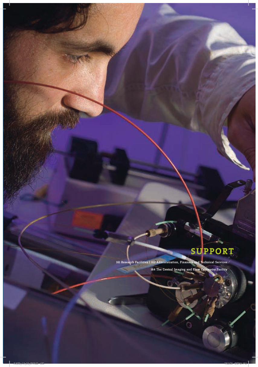
161 Research Facilities | 162 Administration, Finanace and Technical Services
164 The Central Imaging and Flow Cytometry Facility
su PPort
NCBS_AR_05_07.indd 160 7/10/08 5:37:14 PM

research facilities
In the next few years, nCBS will have a new research building. existing laboratory space will more than double in size, and it will allow nCBS to grow to a critical mass of more than forty faculty members and investigators. Ample space will become available to initiate or augment existing centralized research facilities. These will include a transgenic animal facility as well as a spectroscopy and mass spectrometry facility. Imaging, computing and the library will have new space to expand into.
Much effort has gone into planning the new building by our project and engineering staff. Intensive discussions with scientists at nCBS and elsewhere, as well as visits to laboratories in other countries, were part of the planning process. A new architect was selected, and a plan has been finalized for a new modern-looking building juxtaposed next to our now ten year old castle-like building. Major highlights of the design of the new building include shared rather than individual laboratory space, and a built-in flexibility that will allow easy reorganization for any new activity, for many years to come.
The past two years have seen the establishment of a genuinely world-class central imaging and flow facility (CIFF), largely due to the perseverance of Drs Mayor and Shivashankar. The range of optical and force microscopy imaging, as well as cell flow capabilities, now available at nCBS, matches or surpasses that available at most biology departments anywhere else in the world.
In this context, it is important to appreciate the efforts of the people who manage our evolving centralized research facilities. Dr Krishnamoorthy of the CIFF, Dr Mohan of the Animal Facility, Dr Deepalakshmi of the Mass Spectrometry Facility, and Dr Ain of the budding Transgenic Animal Facility play specialized roles that enable students to carry out their experiments in many different and innovative ways.
Major reorganization of existing laboratories has also occurred over the past year, with the joining of a Young Investigator, Dr Quader as well as two new faculty members, Drs nair and Sane. In addition, two joint faculty members, Drs Rodrigues and Krishnan transferred their research activities fully to nCBS from TIFR, Mumbai. Administration and engineering staff moved from occupying laboratory space to a swanky new building. Staff members of the Research Services and engineering Services, the Project office, as well as of the Administration, ensured that the reorganization of laboratory space occurred with relatively little disruption to ongoing activities. As the centre has continued to grow, with now 25 independent research groups, it has become necessary to share responsibilities in more formal ways. Dr Mayor became Chairman of our engineering and Maintenance Services Committee, and now oversees the functioning of an activity critical to the efficient functioning of the centre. Dr Bhalla became Chairman of the Research Services Committee, and supervises the functioning of our research support staff, which enables efficiency in our research endeavors.
Jayant UdgaonkarDean and Head, Research facilities
Artist representation of the laboratory building. Construction starting soon.
161
SU
PP
OR
T
NCBS_AR_05_07.indd 161 7/10/08 5:37:14 PM

What is loosely called the ‘administration’ at nCBS is really the mesh of services that range from engineering and architecture, laboratory maintenance, campus services, new developments and the core finance and general administration required for all of these. The efficient functioning of the centre is a consequence of all of us interacting as a team. each major area has its head, who reports to the nCBS Director. As head of Administrator and Finance, this report only touches on most aspects non-academic functioning and is intended as an overview.
During this period, we have constructed an extension to our existing academic and research block building, set up a state-of-the-art imaging and flow facility and established new laboratories for our scientists. Along with this, we have handled a range of administrative and financial functions ranging from recruitment of faculty, staff, research scholars, financial budget & accounts and have managed our ever growing meetings and workshop programme.
If managing our growth over the last five years is viewed as being, at least, modestly successful we are also aware that every success had its challenges. We have viewed every challenge in a positive manner and have negotiated them in an innovative way.
The chart below summarises our financial position for last five fiscal years (April 02 - March 07):
As can be seen from the table, we have continued to spend a major portion of our budget on research. Our salary budget constitutes around 9 percent of our total budget and has remained at this level for the last five years. Part of the reason is that to be operationally effective and cost efficient, we outsource our routine maintenance and support services. however in doing so, we ensure that the contractual obligations are fulfilled with quality.
We have continued to receive a significant portion of our budgetary support from extra Mural Grants. Our scientists have been extraordinarily successful in generating national and international funding. From Rs.36 million in 2002-03 we have been able to raise our extra mural funding to the level of Rs.125 million in 2006-07. The strong external support that we have obtained speaks highly about the drive of our various research groups.
To facilitate effective research, in addition to an excellent administration and finance group, we have a highly dedicated and competent engineering Services & Maintenance Group. The role of this group has been crucial, as they have been instrumental in construction of our Mandara hostel, extension Block to the current Academic and Research Block, Central Imaging and Flow Facility and modification of various laboratories for the scientists during the last five years. The engineering Services and Maintenance Group have ensured that these facilities are constructed in an aesthetic manner. Currently, this group is concentrating its efforts on construction of a state-of-the-art laboratory building for scientists who
Administration Finance and engineering services
Salaries
Operational Expenses
Research & Development
Construction
46%
36%
9%9%
52%
30%
10% 8%
41%
37%
10%12%
11%3%
42%
44%50%
30%
8%12%
2002-03 2003-04 2004-05 2005-06 2006-07
NCBS_AR_05_07.indd 162 7/10/08 5:37:15 PM

0
20
40
60
80
100
120
140
160
180
2002-03 2003-04 2004-05 2005-06 2006-07
Students and Research Fellows on GrantsPost Doctoral FellowsScientific and Technical StaffAdministrative StaffFacultyYoung InvestigatorsMeetings, Symposium, Workshop & Public Lectures
would be carrying out research in frontier areas of biology and other inter-disciplinary areas. Along with this, a new dining-cum-sports complex at the present campus and a housing complex at Chikkabommasandra is also being planned. It is expected that the construction will be completed by the middle of 2010.
Given the growth that we have experienced during the last five years as service providers (Administration, Accounts, Purchase and engineering Services) we have strived to adopt the best practices that enable us to provide ‘just-in-time’ service to our scientific community to facilitate their research pursuits. In the coming years we also hope to pay substantial attention to ensuring that the best employee welfare and benefits programme are put in place. This and a robust mechanism of feedback from employees and other comments in general will be a major goal. We will continue to function in an innovative manner that will enable us to harness our available resources in an efficient manner.
nCBS during the next coming years is headed for a phase that will create a lasting impression in the scientific world. One thing we know for sure that, the last five years has been the most interesting phase and the future holds surprises. We need to create a capacity to respond to such surprises. We view the modest success that we have met in the past as not our destination but a journey. In this journey we are committed to the idea of creating value and will endeavour to build a set of institutional values. We are certain that these institutional values will help all in the long run.
Before I end, I would like to sincerely thank all my colleagues for their support. The support of my colleagues have helped us to tide over the difficult times and testing phases in a successful manner. I am confident that all my colleagues will continue to do the same.
Pradip PyneHead, Administration and Finance
Our growth over the last five years is summarised below:
163
SU
PP
OR
T
NCBS_AR_05_07.indd 163 7/10/08 5:37:16 PM

Many research programs at nCBS rely heavily on the latest methods in bio-imaging and flow cytometry. The Central Imaging and Flow Cytometry Facility (CIFF), with its state-of-the-art equipment, has been set up to meet these needs. Although conceptualized in 2005, it was fully realized in 2006-2007 through national and international funds generated by nCBS faculty. The CIFF not only caters to our institutional needs but also to researchers from all over India and abroad. Further, we train individual researchers to operate microscopes and flow cytometers at the CIFF. To this end, the CIFF offers regular training programs for new users and it currently has more than 90 well-trained users.
nCBS has also formed strong links with several well-known manufacturers of high-end microscopes and flow cytometers. This, in turn, has enabled these manufacturers to station several imaging systems at the CIFF. The open usage system at the CIFF coupled with its strong training program make it worthwhile for these manufacturers to demonstrate the versatility and performance of their instruments to potential buyers, while they serve the imaging needs of nCBS. These facilities are briefly described below. Additional details on workshops and training programs conducted at these facilties, along with a list of research publications from these, are available at http://www.ncbs.res.in/researchinfra/flowcytometry.htm
NCBS-Olympus micro-imaging centre (NOMIC) This centre was created in association with the Olympus Corporation, Japan. Olympus has installed six
high-end fluorescence microscopes at nCBS for two years, which will be used for conducting imaging workshops and microscopy courses.
NCBS-zeISS advanced imaging and microscopy training platform A similar understanding has been reached with Carl ZeISS, Germany where an advanced imaging
and microscopy platform has been installed in the CIFF. Carl ZeISS has located its LSM 5 LIVe system as well as the LSM 510 Meta at nCBS. These imaging platforms will also be part of microscopy training programs and workshops.
BD-NCBS centre for excellence in flow cytometry This centre, which has been set up in association with Becton Dickinson, India, has led to the
installation of a high-speed sorter, the FACS Aria, for user-training and workshops.
Facility Manager: h Krishnamurthy CIFF staff: B S Srinag, B Smitha and S Gopal Faculty Coordinators: S Mayor and A Sarin
the central imaging and Flow cytometry Facility (ciFF)
NCBS_AR_05_07.indd 164 7/10/08 5:37:19 PM

Transgenic and knockout mice facility Transgenic and knockout mice are a key resource in cell and developmental biology. nCBS has now
reached a size where several faculty members have steady, yet small, requirements for their respective programs. Generating these mice require technologies that are well-established. Yet, implementing them in an efficient and sustainable manner would require nCBS to establish the necessary infrastructure and technical expertise. To this end, nCBS has signed an agreement with the RIKen Cell and Developmental Biology (CDB) Institute at Kobe, Japan. The CDB mouse facility, led by Dr. Shin Aizawa, has agreed to make several transgenic and knockout mice for nCBS each year. In addition, CDB is working closely with nCBS to establish our mouse facility and provide technical advice.
Facility Manager: Rupasri Ain Faculty Co-ordinators: K VijayRaghavan and S Chattarji Staff: S Uma and S Srinivasan
Drosophila stock maintenance and screening facility The availability of the complete annotated sequence of the Drosophila genome coupled with the
invention of a number of elegant genetic techniques has made the fruit fly a more attractive model system than ever before. It is the system of choice, not only for the investigation of a variety of basic cellular principles, but also for the more applied areas of clinically driven research, drug testing and pharmacology. There is a resurgence of interest in carrying out large scale, genome wide, screens to identify genes important for understanding mechanisms in cell biology, neuroscience and the development of behavior. The Drosophila centre maintains a large number of mutant strains and transgenic lines. In addition, the facility offers a microinjection service for the generation of transgenic lines. The facility aims to hold frequent meetings, discussion sessions and co-ordinate with other research groups to facilitate large scale genetic screens.
Staff: S G Gajendra Faculty Co-ordinator: V Rodrigues
Technology centre for nanobiosystems (TCNB) The TCnB was created through generous funding obtained from the nanoScience and Technology
Initiative of the Department of Science and Technology, India in 2005. The primary aim of TCnB is to augment infrastructure capabilities at nCBS to study nano-scale architecture and phenomena in living systems in real time, with a focus on developing new bio-inspired nanodevices. These devices and technologies are likely to be deployed in the visualization of sub-cellular and cellular processes at temporal and spatial resolution previously unachievable. Additional details on the wide range of exciting research and technology emerging from this centre is available at: www.ncbs.res.in
Faculty Co-ordinators: G V Shivashankar, S Mayor, Y Krishnan and K VijayRaghavan
Cell-based high content screening facility new molecular players in cellular processes are being identified worldwide through large-scale screens.
Given its strengths in imaging, nCBS is perfectly positioned to establish, in an academic setting, high content high throughput (hChT) imaging-based screens. The Technology Centre for nanoBiosystems has created a screening facility that will establish the hChT approach initially through genome-wide RnAi screens for cellular scale phenomena of interest at nCBS. The know-how from the establishment of such screens will subsequently be adapted for hChT approaches for screening small-molecule libraries.
Facility Manager: Gagan Gupta Staff: S Krishnamoorthy and K Ramesh Kumar Faculty Coordinators: S Mayor, G V Shivashankar and Y Krishnan
165
core Facilities
SU
PP
OR
T
NCBS_AR_05_07.indd 165 7/10/08 5:37:19 PM

PeoPLe
NCBS_AR_05_07.indd 166 7/10/08 5:37:21 PM

167
SA
TY
AJI
T
MA
YO
R
PeoPLe168 Academic Personnel | 175 Support Personnel | 177 Quality of Life at NCBS
NCBS_AR_05_07.indd 167 7/10/08 5:37:22 PM

Research Groups (as on December, 2007)Academic personnel
Biochemistry, Biophysics and BioinformaticsYamuna Krishnan Structure and dynamics of nucleic acids
Visiting fellow: Gourishankar R Aland, until July 2007PhD students: Dhiraj Devidas Bhatia Saikat Chakraborty Souvik Modi Sonali Saha Suruchi SharmaGraduate trainee: S Chamundeeswari, until January 2006Junior research fellows: Shruti K Gowda Jyothsna Gunturu, until April 2006 Ramya Krishnan, until September 2007 Shabana Mehtab Vidhya Rangaraju, until June 2007 Poulami Sengupta M K Mathew Exploring the architecture and function of transmembrane ion channels
Visiting fellows: Kavitha P G Veena S Anil, until December 2007 PhD students: Anirban Baral Sanjeev Kumar Pinky Raychaudhuri hyder UsmanJunior research fellow: Amit Kumar Sachan Students registered elsewhere: Sam Kuruvilla K Pannaga Susan M Philip Lakshmana Reddy D L Deepak T Nair Structural biology and macromolecular crystallography
PhD student: Amit Sharma Mrinalini Puranik Structure and dynamics of biomolecules
PhD students: Spriha Gogia namrata Jayanth Gopakumar Ramakrishnan, until July 2007Teacher trainee: erix A Milan GraceGraduate trainees: C Megha, until January 2006 Parvati S Menon, until november 2007 G Jonah Unnatha nesson, until September 2006 Silja Poulose, until november 2007Junior research fellows: Shreya M Ankolekar, until July 2006 Mandar Satish Paingakar, until June 2005 Shagolsem Lenin Singh Kaustubh Rau Mechanisms of damage by laser pulses to single cells and tissue
PhD student: Amit Sharma, until May 2007Graduate trainee: G D Rajesh, until July 2006Junior research fellows: Anoop V Cherian, until October 2007 nimi Gopalakrishnan Arcot Raghupathi Lokanathan G nageswara Rao P Senthil G V Shivashankar Cellular architecture of genome regulation
Visiting fellows: V V n Ravi Kishore, until May 2007 Indulakshmi Radhakrishnan Bindu Radhamany, until July 2005
NCBS_AR_05_07.indd 168 7/10/08 5:37:23 PM

PhD students: Bidisha Banerjee, until July 2007 Feroz Meeran hameed Abhishek Kumar K Venkatesan Iyer Shovamayee Maharana Aprotim Mazumder M Padmanabhan Deepak Kumar Sinha, until October 2007 Gautam V Soni, until January 2006 Shefali TalwarGraduate trainees: Annie Miriam, until July 2007 V Ramya Junior research fellow: Kaushik Ghosh, until February 2006Students registered elsewhere: Dipanjan Bhattacharya T Roopa M Vijayalakshmi R Sowdhamini Computational approaches to protein science
PhD students: P S Divya Anirban Bhaduri, until April 2005 M Raghuprasad Rao, until September 2006 C Chandrasekar Reddy Sandhya Sankaran Lokesh P TripathiMSc student: Kumar Gaurav, until September 2005Junior research fellows: K harini, until December 2006 Caroline Koshy, until January 2007 A Manonmani, until April 2005 K Manjunath, until February 2007 P nagarajan Agnel Praveen, until March 2007 G Pugalenthi Aswathy Sebastian, until July 2006 Ambika Subramaniam Chengala Sairam Swamy, until December 2005 Ramakrishnan Vigneshwar, until november 2005Students registered elsewhere: K Kanagarajadurai P K Shameer P Sundaramurthy Mukund Thattai The dynamics and evolution of living networks
PhD student: Sugat P Dabholkar, until July 2007 Rohini RamdasJunior research fellows: S Vivek Raj Senthil, until April 2007 Jayant B Udgaonkar How do proteins fold, unfold and misfold?
Visiting fellows: n earanna Rupali A Gadkari, until november 2006 Subhendu K Mohanty, until July 2007PhD students: Amrita Dasgupta Shweta Jain Santosh Kumar Jha Megha Kishore Santosh Kumar K Aghera nilesh Ashish Kumar Patra Gayathri Ramachandran Shmilona Sarangi Amrita Sekhar Kalyan K Sinha Vishal Ajazul hamid Wani Junior research fellows: K Durga, until August 2007 K C Vishwanatha
Pe
OP
Le
169
NCBS_AR_05_07.indd 169 7/10/08 5:37:23 PM

Cellular Organization and SignalingSudhir Krishna The role of papillomaviruses and Notch signaling in the progression of human cervical cancers
Visiting fellows: R Bharathi, until April 2006 Tessy Thomas Maliekal Sweta Srivastava Deepa Subramanyam, until July 2006 Eric VivienPhD students: Jeevisha Bajaj P Chitra Azham Roohi, until April 2006 Megha SehgalJunior research fellows: Ajay Abraham V Krishna Chaitanya Mausita Karmakar T Senthil Kumar, until February 2006 Michael Rahman, until November 2006
K S Krishnan Cell biology of the synapse
Visiting fellows: S Kavitha M Santhana Ramasamy K A Subramanian, until October 2005PhD students: Hanumae Gowd Riddhi Majumdar Junior research fellows: M Deepa Jayaseelan Benjamin Franclin, until June 2007 Amitabha Mazumdar, until December 2005 C Omeena Saravanan Palani Shobha V Ramagiri, until December 2005 Suneel A Reddy Y Saikumari Supriya Syal, until July 2005 Manisha A Tole Satyajit Mayor Mechanisms of endocytosis in metazoan cells
Visiting fellows: Gagan Gupta, until July 2007 Niloufer Gillan Irani Manjula Kalia, unti April 2007 Indranil Mitra, until April 2006PhD students: H L Anupama Sameera Bilgrami, until October 2007 Rahul Chadda Subhashri Ghosh Debanjan Goswami Sudha Kumari Suvrajit Saha M G Swetha MSc students: Gautam Dey Abhijit Kale, until August 2005 A Vijay Subba Rao, until May 2007Junior research fellows: Revu Alexander Brojo Kishore Chowdhury, until January 2007 Jayanthi Menon Sindhu A Menon Garima Singhal Nina Sabu Teresa Thulasi Warrier, until May 2006 Students registered elsewhere: Riya Raghupathy Neha Vyas
NCBS_AR_05_07.indd 170 7/11/08 2:59:17 PM

Madan Rao Physical principles of organization in biological systems(Adjunct faculty member)
Junior fellow: V S Gayathri Apurva Sarin Mechanisms of apoptosis
Visiting fellows: G Aparna, until October 2007 Manjula NagalaPhD students: Soumya Gupta Neha Parikh, until June 2007 Lakshmi Revathy PerumalsamyMSc student: Girija Goyal, until July 2006Junior research fellows: Megha Garg B Geetha, until September 2005 Ganesh Maruti Mohite Varsha Pattu, until January 2006 Ravish Rashpa S Uma, until January 2007 Ashwath VishwanathanStudents registered elsewhere: P Divya D Vaigundan V Sriram Mechanisms of mitochondrial remodeling
PhD students: Ruchika Anand Swagata Dey Pankaj Kumar Vijaykant Dubey Gaurav Goyal Tejas M GupteGraduate trainee: Shamik BanerjeeJunior research fellows: Kondadi Arunkumar Nagaraju Dhanyasi Ram Kannan Kokilavani S
Genetics and DevelopmentGaiti Hasan Inositol 1,4,5-trisphosphate signaling in cellular and systemic physiology
Visiting fellows: Santanu Banerjee, until May 2006 Debleena Dey Mamta Fuloria Sonal Patel, until November 2005 Suzanne Lynn ZiegenhornPhD students: Neha Agrawal Gayatri V Prakash V Subramanyam, until July 2005Junior research fellows: Nikhil Abraham V Bhaskar, until July 2005 Sandeep Grover, until January 2007 Satish Kumar D Senthil Kumar Nisha Padmanabhan Veena S Patil, until December 2005 Rashmi N PrasadStudents registered elsewhere: Manivannan S Shalima Nair, until July 2007 Suhel Quader Evolutionary ecology and environmental conservation
Project associate: Rashid Hasnain Raza Graduate trainee: Kedar Suresh Champhekar
171
PE
OP
LE
NCBS_AR_05_07.indd 171 7/11/08 2:59:17 PM

Uma Ramakrishnan Evolutionary history of human and animal populations: Understanding the past and predicting the future
Visiting fellows: Kavita Isvaran, until April 2007 Shomitha Mukherjee S RamaswamyPhD students: Anagh Purandare Thejaswi Shivanand S Velumani, until november 2007Junior research fellows: Jiffy James, until november 2007 Bhairavi Swaminathan Vidhya Ranganathan, until May 2006Graduate trainees: Jeevan Karloss, until March 2006 Krishnapriya TammaStudents registered elsewhere: Debapriyo Chakraborty Samrat Mondol Veronica Rodrigues Developmental neurobiology of the olfactory system
Visiting fellow: R C Sarasij, until February 2006Junior research fellows: T S Bhuvaneish, until April 2007 Varun Chaudhary, until August 2005 Sudeshna Das Aditi Deshpande, until December 2006 Abu Farhan Syed Mubarak hussain Utham Kashyap Abhilasha S Kumar, until August 2005 Kiran Kumar, until May 2007 Amit Kundnani, until May 2007 Archana Murali, until August 2006 Darshana narayanan, until May 2006 Tulip nuwal, until August 2006 hetal Pandya, until May 2005 Rashi Priya Vijay Kumar Ranka, until August 2006 Michelle R Rebello, until March 2006 Ashlesha Rodrigues, until October 2006 Sahithya Y R, until Sept. 2007 Aditya Saxena, until May 2006 Gudubasha Shaik, until June 2007 Ashish Shukla, until August 2005 Arvind Suresh S Vinayaka, until July 2007Students registered elsewhere: Pinky Kain Sonia Sen Keshava Subramanya K VijayRaghavan Nerves, muscles and the development of behaviour
Visiting fellows: T Sajith Dass, until november 2007 S VijayalakshmiPhD students: Albert Chiang Richa Dixit S Krithiga, until April 2007 Kirthi Rathore Bidisha Roy Pushpam Kumar Sinha, until June 2005Junior research fellows: Rajaguru Aradhya, TC Sajesh Abu, until March 2006 A M Barve, until May 2005 Ankita Chadda Kitdorlang Dkhar Swathi Krishnan Arun Kumar
NCBS_AR_05_07.indd 172 7/10/08 5:37:27 PM

Neha Lodha, until December 2006 Ann Maria Priyankana Mukherjee Aijaz ul Noor Aeijaz Sultan Parry Narendra Solanki, until June 2007 G S Sreeranjini, until May 2005 Akila Sridhar Sudhir P Swetha B M Umashankar MStudents registered elsewhere: Guruharsha K G, until October 2007 O Venkateswara Reddy
NeurobiologyUpinder S Bhalla Computational Neuroscience
Visiting fellows: Sriram M Ajay, until May 2006 Radhika Madhavan Raghav Rajan, until August 2006PhD students: Dhanya P Ashesh Dhawale Niraj Dudani Priyanka Gupta Adil Ghani Khan Mehrab N Modi Urvashi Raheja Subhasis Ray Anmol Sethi, until December 2006Junior research fellows: Prasoon Agarwal, until September 2006 Abhishek Banerjee, until July 2006 James Premdoss Clement, until May 2006 Raamesh Deshpande Rinaldo R D’Souza, until July 2006 Shaista Hussain, until November 2005 Pragathi Jain, until September 2006 Poorvi Kaushik, until May 2007 Pradeep Kumar, until July 2004 Priyamvada Rajasethupathy, until June 2005 G V Harsha Rani Rajnish Ranjan, until September 2005 B M Ravikumar, until March 2004 Vinay Sutrawe, until July 2007 Karthika Valli, until February 2004 Karan Vasudeva, until December 2003 Adithya Vasudevan, until March 2004 Sharat J Vayttaden, until August 2005 Sanal Viswanathan, until March 2005Student registered elsewhere: K Parthasarathy Sumantra Chattarji Plasticity in the amygdala: implications for stress disorders and mental retardation
Visiting fellows: Ganesh Bagler Rajnish P RaoPhD students: Roy Thomas Daniel Supriya Ghosh Swati Gupta L Harini Sonal Kedia Ruchi Malik Anup G Pillai Shilpa Ravinder Sharath B S, until September 2005 K Manish Sharma Aparna SuvrathanJunior research fellows: Shobha Anil Kumar Amrita Kuthiala, until August 2005 Deepti Rao, until July 2005 Anand Karthik Sharma, until July 2005Student registered elsewhere: Anupratap Tomar
173
PE
OP
LE
NCBS_AR_05_07.indd 173 7/11/08 2:59:17 PM
Sandhya P Koushika Genetic approaches to understand axonal transport
Visiting fellows: Nivedita Chatterjee, until June 2007 Eva M RomareoPhD students: Niloy Kumar Chakraborty, until September 2007 Bikash Chandra Choudhary Jitendra KumarGraduate trainees: Swati Devidreddy Swetha Mohan, until July 2007 Dharnikumar Rajasekar, until February 2007Junior research fellows: Jaffar Mohd. Bhat Shaili Johri, until January 2007 Rajshri Joshi, until August 2007 Sucheta S KulkarniStudent registered elsewhere: Guruprasad Reddy Sure Mitradas M Panicker Gene regulation in the mammalian nervous system
Visiting fellows: M S Geetha, until June 2005 Odity MukherjeePhD students: Basudha Basu, until July 2006 Aditi Bhattacharya Ishier Raote Tejaswini S SharangdharJunior research fellows: Meenakshi Balaraman, until May 2006 Rupam Choudhury S P Suresh Kannan, until January 2006 Deepika Kaveri Ashita P Magal, until May 2006 Sushmita Saha, until August 2006 Aparna Shah Imtiaz Zafar Sanjay Sane Neural and physical basis of insect flight
PhD students: Anand Krishnan Nitesh SaxenaGraduate trainee: Janani Subramanian Obaid Siddiqi Genetic analysis of chemosensory perception
Junior research fellows: Dushyant Mishra, until April 2007 Sunil Prabhakar, until June 2006 K J StanleyStudents registered elsewhere: Jawaid Ahsan Mohammed Bin Abu Baker Tuhin S Chakraborty Sarit Pati Goswami Junior research fellows with collaborators
With R Varadarajan, IISc P Shaik Syed Ali, until August 2006 C Sandhanakrishnan, until August 2006 With Maneesha Inamdar, JNC M Harsha, until May 2005
NCBS_AR_05_07.indd 174 7/11/08 2:59:18 PM

Scientific Services
Prasanta Kumar BaruahVeerana BentoorA Charles, until February, 2006Avinash D ChinchureS DevakumarSourabh n Gaikwad, until February, 2007Prem Chand GautamR JayaprakashP KothandanP C Kottureswara, until July 2006S Senthil Selva Kumar, until June, 2007G h MohanS naganand, until February 2007G Aswatha narayanaP P RanjithM Saravanan, until July, 2006Pranesh Shantaram, until July 2007S UmashashiK Vasudeva, until Janury 2007h S Venkataramana
FacilitiesRupasri AinP D DeepalakshmiS Gajendra, until October 2005Sneha GopalR Kanimozhi, until June 2007T Shabana Khanum, until May 2007h KrishnamurthyS KrishnamoorthySasirekha KrishnanK Ramesh KumarGayathri ManjunathSanchari RoyB SmithaB S SrinagLavanya SivashanmugamSharanya SrinivasanS Uma
Technical Servicesh M BasavarajaChakrapaniBheeman GoudaSatyanarayan Guntur, until December 2005Mallesh n. harogeri, until March 2007
support personnel
h V harshaBasavaraj JalihalT S Govindaraju, until June 2005M n Krishnaiah, until May 2007Bhavani R KulkarniP B Mahadeva KumarS Sujeeth Kumar, until March 2006Ravindra MunshiK M nagaraj, until August 2007P V narayana RaoT natarajanM Ananda PrakashU B PoornimaT Rajath, until September 2007D Ravikiran, until February 2007h R Revanna, until October 2005B M SathishkumarK S SavithaKhalendar Sharief, until February 2006Abdul K SharrifR M Somashekara, until September 2005Jaishree M Somwanchi, until July 2006S Sujeeth Kumar, until March 2006Sidhartha S SwainSeethalakshmi Shivaswamy, until May 2006K S Shivananda Swamy
ADMInISTRATIOn AnD FInAnCe
Administrative OfficersPradip PyneB S Ramamurthy, until October 2006K V RamanathanS Ashok RaoT M SahadevanS B Saraswathin n ShanthakumaryPurushottam V SuryaraoShaju Varghese
Consultants/Medical Officersh V ChandralekhaA V R Murthy, until May 2005Vishwanath n Patil, until July 2005R Padmanabhan, until July, 2007P h PrasadS RajagopalanG Prakash Rao, until April 2006V R Rengasamy
Meetings and WorkshopsPapiya Bhattacharya, until August 2006nidhi SrivastavaReena Shrivastav
Administration, Accounts, Secretarial and Auxiliary staffM Amalanathan, until June 2006A Anandarajn S Kannan Kasturi
C Prashanth MurthySeetharam naikR n nagarajP Lakshmi PriyaK S nirmalaA n Rajendran RamaprasadMaithly RameshP M RathnakaranR n Shalinin Sujathah R UmaV S Shailaja, until January 2007S V Swethah R UmaS VenkataramaniK S Vishalakshi
Trainees/Project AssistantsSavitha J Balakrishna, until December 2006S R ChetanaVidhya V Dixit, until August 2007Amit Gaikwad, until December 2005J S Gayatrie Geetha, until March 2007M S GunasheelanK V hamsaveniSuraiya hussain, until June 2006S Kavithanazia Kauser, until December 2005Jyothi Khanna, until June 2007S Girish KumarB S Vasanth Kumar, until December 2005V V LakshmiM Latha, until March 2006K R Madhu, until May 2007S Madhu, until February 2006n MalathiS n Manjula, until June 2007n MithraK Lakshmi narasimha, until June 2006K R Padma, until October 2007R Pallavi, until June 2006J Poornima, until May 2007R n PratibhaM Pushpalatha, until June 2005K RakshithaJ RamyaVibha Rao, until April 2007A RathnaC S Sangeetha, until June 2006M SaravananC G Seema, until July 2007S ShanthalakshmiB n ShobhaV Sindhu, until July 2007M R Smitha, until May 2006A Shwetha, until February 2007Praveen Tiwari, until november 2005V UdayaM Veena, until December 2005nishu Verma, until June 2007
175
Pe
OP
Le
NCBS_AR_05_07.indd 175 7/10/08 5:37:31 PM

Professor Jayant Udgaonkar steps down as Dean from March 31, 2008 after over 11 years on the job. Jayant, whose science has been extraordinary in quality and in volume for several years, would like to devote more time to his laboratory.
Jayant joined us before NCBS formally came into being. It can rightly be said that while the concept of NCBS required Obaid Siddiqi, transforming this into reality required Jayant Udgaonkar. Jayant has been the pillar of robust good-sense and best-practice and the pivot around which all aspects of NCBS have grown. Our hiring of faculty and staff, administrative structure, buildings, architecture, landscape: indeed every facet has relied on him to set the standards for both innovation and correctness. This afforded several of us the invaluable luxury of being sloppy, speculative and irresponsible. We could always explore freely, in the full confidence that our moral and scientific pivot would not allow us to stray and translate a poorly-formulated whim or fancy of the day into unwise action. Jayant thus developed a paradigm for a structure that could be innovative, yet structured. NCBS owes him a great debt – he would typically say that he was just doing his job – and we are fortunate that we will continue to have him advise and participate in our forthcoming ventures.
Satyajit Mayor takes over as our new Dean. Jitu has become a world-leader in his area of research in cell biology. NCBS may lay claims to have nurtured him, and be pleased, but this is a man who would have done great work anywhere and no place could have stopped him (we didn’t try). In addition to his science, Jitu has taken the leadership to establish one of the best imaging and cell-sorting facilities in the world and is now following this up to establish a genetic screening facility of a similar quality. His science, his leadership and his judgement ensure that, in him, we have a Dean of the highest quality.
thanking a dean and welcoming another
NCBS_AR_05_07.indd 176 7/10/08 5:37:36 PM

The NCBS campus is lucky to have open green spaces, many trees, bushes (w-free), flowering plants, migrating butterflies, geckos, bees and bats. We are located in the postgraduate campus of the University of Agricultural Sciences, which has generously leased us 20 acres. Our visitors and us have a pleasant, quiet and green environment to work and relax. We have terrace cafeterias where coffee, snacks and light meals are served and birthdays celebrated. Several handy chalkboards have made these cafeterias centers for discussion. Some of these discussions have recently crystallized into a student-run symposium exploring different areas in biology. A larger (soon to be expanded) canteen serves full meals from breakfast to dinner. The canteen is located near a pond fed by harvested rainwater and clean water from treated sewage. The pond-side is where we gather for dinner after on-campus symposia and public lectures. We have an amphitheater that serves as an interactive place for tea after scientifically exciting seminars. Facilities for tennis, basketball, volleyball, badminton, table tennis and a gym are all available. We provide transport to all members of the community into the ever-expanding Bangalore city. We also help our staff and faculty with their commute to work. We have excellent accommodation for students and post-doctoral fellows. We accommodate some faculty on campus and the rest in comfortable apartments and houses close to campus. We do need to expand our campus housing for faculty as well as our excellent administrative and technical staff. In the near future we hope to expand our hostel, sports facilities and housing to keep pace with our scientific growth.
One major factor that enhances the quality of life on campus is our on-campus child-care and after-school program. This facility aims to be a complete one-stop solution for child-care for children from the ages of 1 to 10 years. The campus environment provides space, lush greenery, a park and a sand pit for children to play and learn. Over the years, our teachers, staff, several visiting consultants, parents and exceptional institutional support have helped develop the childcare center into an excellent facility. The day-to-day affairs of the facility are coordinated by several experienced teachers. The NCBS community at large provides a rich learning environment. We have several artists, wildlife lovers, storytellers and musicians on campus who informally participate in various activities at the child care center. We have age appropriate fun and educational activities for infants, toddlers, pre-school and school going children. Current efforts are geared towards developing a stimulating after-school program supplemented with off-campus activities such as swimming, volunteer work and music in addition to homework and examination-related studies. We also undertake field trips to various locations such as parks, science museums, circuses and dance concerts. Good childcare is a major factor that achieves a supportive workplace for working parents. We aim to provide a facility that recognizes the needs of working parents while providing children with a nurturing environment.
Quality of life at ncBs:what we have and where we aim to be
LIF
e A
T n
CB
S
177
NCBS_AR_05_07.indd 177 7/10/08 5:37:40 PM

Selected publications
NCBS_AR_05_07.indd 178 7/10/08 5:37:44 PM

The stunning and innovative exhibit “Such Treasure and Rich Merchandize” at the National Center For Biological Sciences/TIFR held in early 2008 brought together science, art and social history in a compelling fashion. The exhibit began with a large walk-through drop down of an Indian Ficus tree from John Gerard’s Herbal and ended with a list of 50 single molecule drugs in use today isolated from botanicals. Beautiful botanical illustrations of Indian plants in european books published from the 16th and 17th centuries, maps, and cityscapes of Indian port cities were used to trace the story of how the search for spices led to the discovery of a wealth of information on Indian plants, their medicinal uses available in India and the importance of that knowledge to science and medicine in europe and also to the development of botany as a science.
Woven into the narrative were excerpts from Shakespeare, the Portuguese poet Camoens, Milton and other writers of the period and a marvelous image of the Indian elephant “Hanno” by the renaissance master Raphael, evocatively represented on a variety of materials such as glass, stone and fabric. You could walk through a herbal garden based on many of the plants described in the volumes or browse through a digital flipbook of the book in focus – the Hortus Malabaricus. This exhibit offered something for the scientist, artist, social historian and everyone interested in the history of India.
The exhibit was curated by Annamma Spudich, Scholar in Residence at NCBS. Annamma Spudich is a cell biologist (Ph.D., Stanford University) with a life long interest in the history of Indian scientific traditions in the natural sciences. In 2003 Dr. Spudich curated the exhibit “From Forreine Places All the Varietie of Herbes” at The Cantor Center for Visual Arts, Stanford University. The exhibition at the NCBS and related collateral was designed and coordinated by Sarita Sundar and her team at Trapeze, a multi-disciplinary design consultancy and studio. The design team worked with the curator to reproduce and present material from various libraries and collections throughout the world and present an informative and novel look at the history of east-West interaction in the pre-colonial period, focused on the natural sciences.
such treasure and rich merchandize
179
eX
hIB
ITIO
n
NCBS_AR_05_07.indd 179 7/10/08 5:37:49 PM

NCBS_AR_05_07.indd 180 7/10/08 5:37:51 PM


