Embed Size (px)
Citation preview
Epidemiol. Infect. (1994), 113, 321-334 321Copyright © 1994 Cambridge University Press
Clonal analysis and virulence of Australian isolates ofStreptococcus suis type 2
C. G. MWANIKI, I. D. ROBERTSON, D. J. TROTT, R. F. ATYEO,B. J. LEE AND D. J. HAMPSON*
School of Veterinary Studies, Murdoch University, Murdoch, Western Australia6150, Australia
(Accepted 9 May 1994)
SUMMARYMultilocus enzyme electrophoresis was used to divide 124 Australian isolates of
Streptococcus suts type 2 into 17 electrophoretic types (ETs). Isolates in ET 1 werethe most frequent cause of disease amongst Western Australian pigs, but isolatesof ET 8 were more commonly associated with disease in other Australian states.Multiple isolates from 10 of 19 farms all belonged to the same ET, whilst isolatesfrom the other farms belonged to between 2 and 4 different ETs. Some isolatescould be differentiated further by DNA restriction endonuclease analysis, whilstothers with the same restriction pattern were located in different, but closely-related ETs. Fourteen isolates were tested for their virulence in mice. Most causeddisease if given in high numbers, but isolates in ET 1 were virulent at lower doserates. This virulent clone also was distinguished by the fact that 80% of isolatesproduced extracellular factor (EF).
INTRODUCTION
Streptococcus suis can cause a variety of clinical problems in young pigs,especially meningitis, pneumonia, septicaemia and arthritis [1-3]. The bacterialspecies has been divided into 29 capsular serotypes, designated 1 to 28, and type1/2 [4-6]. Of these, serotype 2 is the most important causing disease in pigs inmost parts of the world [7, 8], although serotype 7 predominates in Scandinavia[6, 9]. Despite the association of these bacteria with disease, they may also berecovered from the nasal cavities and tonsils of healthy pigs [10-12].
Outbreaks of disease may follow periods of stress, such as when weaned pigs aremoved and mixed, or are overcrowded. The build-up of fumes from slurry,combined with poor ventilation, also predisposes to disease [13]. Outbreaks alsomay occur following the introduction of new pigs to a herd [14]. It is presumedthat these are healthy carrier animals which bring new virulent strains into theherd, and that these strains then spread to susceptible pigs. Strains of S. suis type2 with differing degrees of virulence to pigs have been described [14, 15]. Virulentstrains produced a muramidase-released protein (MRP) and, particularly, anextracellular factor (EF) [16, 17]. Strains that produced a higher molecular weight
* Correspondence and requests for reprints to: Dr D. J. Hampson.

322 C. G. MWANIKI AND OTHERS
form ofEF (EF*) were of intermediate virulence in newborn germ-free pigs, whilststrains lacking both proteins did not induce any signs of disease [17].The species S. suis is genetically homogeneous as assessed by levels of DNA
sequence homology between 13 strains [18]. The use of multilocus enzymeelectrophoresis (MEE) to examine a much larger collection of S. suis isolates ofvarious serotypes has, however, shown the species to be relatively diverse [19]. Forinstance 31 isolates of serotype 2 had a mean genetic diversity of 0-264, and weredivided into 6 electrophoretic types (ETs), representing 3 clonal groups. Geneticdifferences between strains of S. suis type 2 also have been demonstrated usingDNA restriction endonuclease analysis (REA), and ribotyping with an Escherichiacoli rDNA probe [20, 21]. These techniques have also been used to study thedistribution of strains of the bacteria amongst pigs during outbreaks of S. suis-associated disease.The purpose of this study was to determine the diversity of Western Australian
(WA) isolates of S. suis type 2, and the association of specific strains with clinicalproblems on piggeries. MEE was used as it is applicable to studies of bacterialpopulations [22], and is a useful technique for analysing isolates of S. suis [19].Results were compared with those previously obtained for other Australian strainsof S. suis type 2 [19]. In addition REA was used to help clarify the epidemiologyof the infections. The virulence of isolates in different genetic groups was assayedin mice, and correlated with the presence or absence of MRP and EF in Westernblot analysis of culture supernatants, using specific monoclonal antisera.
MATERIALS AND METHODS
Bacterial isolatesThe isolates of S. suis type 2 (n = 124) were from 4 main sources (Table 1).
Source 1 comprised 67 isolates collected during a survey of pigs from 19 WesternAustralian (WA) piggeries (designated A to S respectively). The isolates were fromthe tonsils or lungs of healthy pigs following their slaughter at local abattoirs,except for 2 isolates from farm S which were obtained by swabbing the nasalcavities of 2 healthy grower pigs. Between 2 and 5 isolates were examined fromslaughtered pigs from each herd, with an average of 3-4 isolates per herd. Source2 comprised 15 isolates recovered from 6 litter-mates on farm Q. These animalswere followed as a cohort from weaning to slaughter, with nasal swabs taken fromeach pig at 1, 2, 3 and 4 months of age, and swabs from the nose, tonsils and lungstaken following slaughter at 6 months. Source 3 comprised 11 isolates from WApigs clinically affected with streptococcal meningitis. These were received fromN. B. Buller of the Veterinary Diagnostic Microbiology Laboratory, WADepartment of Agriculture, South Perth, or were isolated from dead pigssubmitted for diagnosis to the Murdoch University Veterinary Hospital. Theseanimals came from 4 farms (R to U respectively). The 4th source was 31 strainsof S. suis type 2 studied previously [19], and included isolates from pigs in SouthAustralia (n = 22), Tasmania (n = 3), Victoria (n = 2), and New South Wales(n = 2). Seventeen were from pigs with meningitis or septicaemia, and 12 wereisolated from the tonsils of healthy animals at slaughter. One isolate was recoveredin Australia from the meninges of a human, and the other was a porcine referencestrain from the State Serum Institute, Copenhagen, Denmark.

Analysis of Streptococcus suis type 2
Table 1. Electrophoretic types of Streptococcus suis type 2 isolated fromA ustralian pigs
Electrophoretictype WA piggeries* Other statest
1 G(3); 1(3); J(3); L(1); SA(1); SA(1)TM(2); N(3); R(3); R(6)$;S(2)$; U(1)$
2 TAS(1)t3 Q(3)4 C(2)5 S(1)6 TAS(1)T; NSW(2)$7 Q(1); R(2) SA(6); SA(2)T8 A(3); C(1); D(3); E(3); SA(4); SA(7$);
F(3); H(3); L(2); P(3); TAS(1)$; VIC(2)$Q(14); T(1)T
9 M(3)10 B(3); L(1)11 H(1) SA(1)12 K(2); R(1)T13 K(1); O(1)14 O(1)15 S(2)16 S(3)17 S(2)
* Twenty-one piggeries in WNestern Australia, designated A to IU. Numbers in parentheses arethe number of isolates.
t SA. South Australia. TAS, Tasmania. NSW, New South Wales, VIC, Victoria.i Indicates isolates recovered from pigs with disease. All the other isolates are from healthy
pigs. ET 1 also contained an isolate from a human with meningitis, and ET 2 contained a Danishporcine reference strain.
Bacterial isolation and characterizationSwabs were taken from the live and the slaughtered pigs as described previously
[23]. These were used to inoculate medium incorporating 3 9% (w/v) Columbiaagar base (Oxoid), 1-5% (w/v) streptococcal selective antibiotic supplement(Oxoid), and 5% (v/v) specific sheep antiserum raised against a South Australianreference strain of S. suis type 2 [24]. Plates were incubated aerobically for18-24 h at 37 °C, and then for 24 h at 4 'C. Colonies of S. suis type 2 were identifiedby the presence of bright broad haloes of immunoprecipitation around them whenviewed by indirect light in semi-darkness [25, 26]. Specificity of the method wasconfirmed by testing representatives of each batch of plates with S. suis isolatesof serotypes 1, 3-9, 13, 15, 18, 20, and untypable isolates of S. suis [19], as well aswith Enterococcus faecalis, S. pyogenes, S. equi, S. zooepidemicus, S. bovis, and agroup G streptococcus.
Positive colonies were subcultured to (i) Columbia agar containing 5 % (v/v)defibrinated ovine blood (SBA plates), and incubated overnight at 37 'C, and (ii)the selective agar, which in this case contained specific sheep antiserum to S. suistype 1. Colonies that reacted positively on both antiserum plates were recorded as
being type 1/2, and were not investigated further.Small alpha-haemolytic colonies of Gram positive cocci growing on the SBA
plates were confirmed as being S. suis by testing for absence of growth on 6-5%
323

C. G. MWANIKI AND OTHERS
9MI11121314151617
I ...I0 02 04 06 0.8
Genetic distance
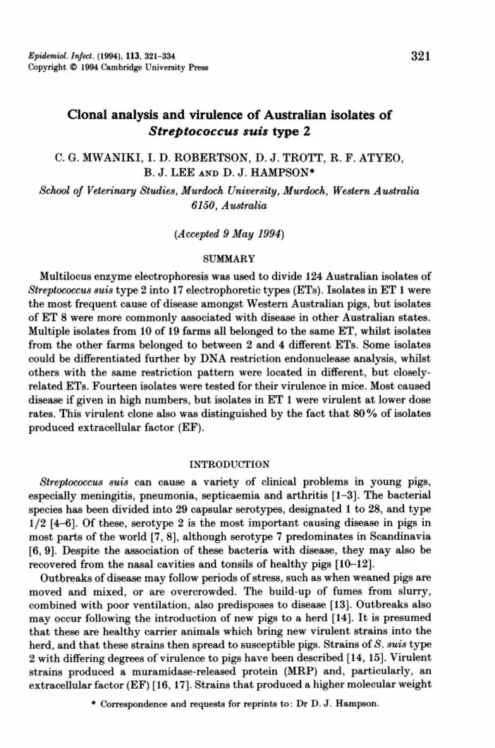
Fig. 1. Phenogram of genetic distance (expressed as percent fixed allelic differences)among 17 ETs, containing 124 isolates of S. suis type 2, clustered by the unweightedpair group method with averages (UPGMA) strategy.
NaCl agar, a negative Voges-Proskauer test, and production of acid in eithertrehalose or salicin broths or both [27]. The serotype of selected isolates from eachof the major genetic groups was subsequently confirmed using the tubeprecipitation and/or the capsular swelling reaction tests [27].
Multilocus enzyme electrophoresis (MEE)The technique has been described [19]. Briefly, bacterial cells were grown
overnight in 500 ml of Todd-Hewitt broth (Oxoid), harvested by centrifugation,washed in phosphate buffered saline (PBS; pH 7 2), centrifuged again, andresuspended in sonication buffer (40 mM-K2HPO4, 10 mM-L-cysteine HCl, 5 jtg/mlbovine serum albumen, 3 mm dithiothrietol, pH 7 5). They were lysed by fourcycles of sonication, clarified by centrifugation, and the supernatant collected.These lysates were subjected to electrophoresis in 11F4% horizontal starch gels,and the electrophoretic mobilities of the following 16 enzymes were determined bystaining for specific enzyme activity: Leucyl-tyrosine peptidase (LT), glucose-6-phosphate dehydrogenase (G6P), mannose-6-phosphate isomerase (MPI), leucyl-glycine peptidase (LGG), nucleoside phosphorylase-1 (NP), a-naphthyl esterase(EST), leucyl-proline peptidase (LP), glutamate dehydrogenase (GDH), phospho-glucomutase (PGM), lactate dehydrogenase (LDH), 6-phosphogluconate de-hydrogenase (6PG), nucleoside phosphorylase-2 (NSP), adenylate kinase (ADK),glyceraldehyde-3-phosphate dehydrogenase (GI1), alanine dehydrogenase (ALD),and phosphoglucose isomerase (PGI). The gel and electrode buffers used were tris-citrate (pH 8-0).
Mobility variants of the enzymes were interpreted as the products of differentalleles at the corresponding enzyme locus. Groups of one of more isolates with thesame alleles at all loci were referred to as being an electrophoretic type (ET).
324

Analysis of Streptococcus suis type 2
Table 2. DNA restriction endonuclease analysis (REA) patterns of 33 Australianisolates of S. suis type 2 belonging to different electrophoretic types (ETs)
Health REAIsolates* statust ETI pattern§S9, S10, U1 d 1 1R1-R3 h 1 2R6-R11 d 1 3C4-5 (SA) d 8 4F2 h 8 5F1, F3 h 8 6N3 h 8 713 (TAS) d 2 8St h 5 9S2, S3 h 15 10S4-S6 h 16 10S7, S8 h 17 10R4, R5 h 7 11C7-32 (SA) h 7 12BI h 10 13R12 d 12 1401 h 13 1402 h 14 14
* TAS, Tasmania; SA, South Australia. All the other isolates originated from WesternAustralia.
t d, diseased pig; h, healthy pig.t ETs in MEE analysis, shown in Fig. 1.§ Following digestion with HaeIIJ. Patterns were not obtained for 53 other isolates examined.
Genetic diversity (h) at each enzyme locus was calculated as h = (1- XPi2) (n/n- 1),where Pi is the frequency of the ith allele and n is the number of ETs or isolatesin the sample [28]. Total genetic diversity (H) was calculated as the mean of h overall loci. Genetic distances between ETs were calculated as the proportion of fixedloci at which dissimilar alleles occurred, and the unweighted pair-group method ofarithmetic averages (UPGMA) clustering fusion strategy was used to create aphenogram (Fig. 1) to show relationships between isolates [29].
DNA restriction endonuclease analysis (REA)REA was based on techniques described for S. suis [20, 21]. Attempts were
made to obtain DNA banding patterns from 83 of the WA isolates, and 3 otherAustralian isolates (Table 2). The bacteria were grown overnight in 100 mlTodd-Hewitt broth, harvested by centrifugation at 10000 g, and washed twice inPBS (pH 7 2). The pellet was resuspended in 750 jdl of 250 mM-Tris HCl, pH 8-0,containing 10 jug/ml freshly prepared lysozyme, and was incubated at 37 °C for1 h, with occasional shaking. The tube was centrifuged at 15000 g for 3 min, thesupernatant removed, and the pellet resuspended in 750 #1 of TE buffer (50 mM-Tris, 10 mM-EDTA, pH 8 0). The tube was frozen at -20 °C, thawed, and 150 /ulof pre-warmed STEP buffer (0 5% SDS, 50 mM-Tris HCl, pH 7-5, 40 mM-EDTA)added, followed by proteinase K to a concentration of 1 mg/ml. The tube wasmixed by inversion and incubated at 50 °C for 1 h. 500 jul of phenol-chloroformwas added and the tube repeatedly mixed by gentle inversion until an emulsionformed. Following centrifugation at 5000 g for 5 min, the upper aqueous layer was
12 HYG 113
325

326 C. G. MWANIKI AND OTHERS
removed and re-extracted with phenol-chloroform. After centrifugation, the upperaqueous layer was transferred to a new tube, and 90 1ul of 3 M sodium acetate andI ml of isopropanol added. After mixing, the DNA was spooled out, rinsed brieflyin 70% ethanol, dried, and dissolved in 250 ,1 of TE buffer. The concentration ofthe dissolved DNA was determined by measuring absorbance at 260 nm [30].15 ,tg DNA was then digested with 20 units of HaeIII (Boehringer Mannheim) in10% medium salt buffer for 6 h at 37 'C. Digested DNA was subjected toelectrophoresis for 21 h at 30 V in horizontal 0-7% agarose gels using TBEelectrophoresis buffer (45 mM-Tris borate, 1 mM-EDTA, pH 8-0). Phage lambdaDNA predigested with HindIII and HindIII +EcoR1 (Boehringer Mannheim) wasused as a molecular mass marker. Gels were stained by soaking in ethidiumbromide (0'5 mg/ml), and were photographed under u.v. light. Where distinctbanding patterns could not be obtained after 2-5 attempts, isolates were recordedas being not susceptible to digestion with HaeIII. DNA banding patterns wereassessed visually without knowledge of results obtained by other methods.Characteristic patterns were given a numeral, and DNAs with similar patternswere subjected to electrophoresis on the same gel to check for differences.
Virulence testingThree-week-old outbred female Swiss ARC Albino SPF mice (Animal Resources
Centre, Perth, Western Australia) were used to test the virulence of 14 selectedisolates (the first 14 isolates in Table 3). This procedure was approved by theMurdoch University Animals Experimentation and Ethics Committee, and wasmonitored by the University's Animal Welfare Officer.
Six of the isolates were recovered from clinical cases of S. suis type 2 infectionsin pigs, and 8 were recovered from healthy pigs at slaughter. Eight of the isolatestested belonged to ET 1, 4 to ET 8, and 2 to ET 12 (Fig. 1). The mice were housedin 14 groups of 7, in individual mouse boxes. Six mice were inoculated initiallywith 2 x 10⁸ colony forming units (c.f.u.), which had been resuspended from earlylog-phase culture in Todd-Hewitt broth into 0 1 ml of sterile PBS, by theintraperitoneal route; the 7th mouse acted as an in-contact control. One weeklater the surviving animals were challenged with 4 x 10⁸ c.f.u. of the same strainin 0-2 ml PBS, and a week later survivors were challenged with 1 x 10⁹ c.f.u. in a0 5 ml volume. Surviving mice were killed 1 week later, and swabs were taken fromthe heart blood and cultured for S. suis type 2. Throughout the experiment themice were observed twice daily, and any clinical signs recorded.
Analysis for MRP and EF productionThe production of muramidase-release protein (MRP) and extracellular factor
(EF) by the 14 isolates tested for virulence in mice, and another 17 isolates (Table3), was assayed [16, 17]. Overnight cultures in Todd-Hewitt broth were diluted1:10 in fresh broth, and incubated at 37 'C for 4 h. The cultures were centrifugedat 4000 g for 15 min, and the supernatants analysed by sodium dodecylsulfate-polyacrylamide gel electrophoresis (SDS-PAGE) in 8% separating gels,and by Western immunoblotting, using a mini-Protean II electrophoresis unit andtransblot cell (Bio-Rad Laboratories, Richmond, Calif.). A control culturesupernatant from a Dutch strain, 4005,which was positive for MRP and EF, and

Analysis of Streptococcus suis type 2
Table 3. Production of muramidase release protein (MRP) and extracellular factor(EF) by 31 isolates S. suis type 2
Isolate*S9S10U1R6I1LINiJiD2E2Q5V4TiKlR12C5-16 (SA)II13 (TAS)C5DR31-V315 (TAS)C7-25 (SA)D7-42 (SA)04B3C8-39 (SA)C6-13 (SA)14 (TAS)C4-5 (SA)Q6QiDR41-4DR27-T
Statustddddhhhhhhhdhdddhhdhdhhhdddhhhh
ETt111111118888121212456779108888
13141516
MRP§
+++
EF§
+++
* The first 14 isolates (S9-R12)) were tested for virulence in mice, and S9, Ul, I1, LI and Nicaused disease at 2 x 10⁸ c.f.u. when administered intraperitoneally. SA, South Australia; TAS,Tasmania. All other isolates from Western Australia.
t Disease status of pig from which the isolate was recovered. d, diseased; h, healthy.t ET, electrophoretic type in Fig. 1.§ +, protein present in the supernatant; -, protein absent; x , higher molecular mass band
present (EF*).11 Isolate from a case of meningitis in a human.
which was supplied by Dr Uri Vecht, DLO-Central Veterinary Institute, Lelystad,The Netherlands, was also included on the gels. Following electrophoresis, gelswere stained using a silver staining kit (Bio-Rad Laboratories). Proteins thattransferred to nitrocellulose membranes in Western blots were blocked for 1 h in5% (w/v) non-fat milk powder, and then incubated with a 1: 1 mixture of mouseanti-MRP monoclonal antibody (Mab) (1 13 mg/ml) and anti-EF Mab (8-4 mg/ml),each in a 1:200 dilution [16, 17], provided by Dr Vecht. After washing, themembranes were incubated with a 1: 2000 dilution of a 1: 1 mixture of goat anti-mouse IgG and goat anti-mouse IgM, both conjugated with horseradish peroxidase(Bio-Rad laboratories). After washing, antibodies were localized by adding the
12-2
327

C. G. MWANIKI AND OTHERSsubstrate 3-3'-diaminobenzidine tetrahydrochloride dihydrate (Bio-RadLaboratories).
RESULTSMEE
The enzymes LDH, ADK, GP1 and ALD were monomorphic whilst the other12 enzymes were polymorphic, with 2-4 alleles. The average number of alleles perlocus was 2-69. The 124 isolates were divided into 17 ETs, with a mean geneticdiversity per locus of 0 397, or 0-283 when the number of isolates in each ET wasconsidered. Two broad genetic diversions were observed (Fig. 1). ETs 1-11contained 111 (89-5 %) of the 124 isolates, separated from ETs 12-17 at a geneticdistance of 0-467.The WA isolates were distributed in 15 of the ETs, but not in ETs 2 or 6 (Table
1). The Danish isolate and the other Australian isolates of serotype 2, from ourprevious study [19], were located in ETs 1, 2, 6, 7, 8 and 11. These ETscorresponded respectively to ETs 33, 34, 50, 51, 53 and 54 in our previous study[19]. Overall, there were 50 isolates in ET 8. ET 1 contained 30 isolates, ET 7 11isolates, and the other ETs between 1 and 4 isolates.
Isolates in ET 8 were common in WA (39% of isolates), where they made up 35of the 82 (43 %) isolates from healthy pigs, but only 1 of the 11 (9%) isolates fromdiseased animals. Isolates in this ET also were common in the other states (14 of29 porcine isolates; 47 %), but here they were more frequently recovered fromdiseased pigs (10 of 17; 59%) than from healthy animals (4 of 12; 33%). Incontrast, isolates in ET 1 were common inWA (29% of 93 isolates), but were muchless so in the other states (2 of 29; 7 %). In WA they made up 82% (9 of 11) of theclinical isolates, but only 22% (18 of 82) of those from healthy pigs.
Isolates were collected from healthy pigs on 19 farms, and on 11 of these allbelonged to the same ET. Five farms had isolates belonging to 2 ETs, 2-3 ETs,whilst the 8 isolates from farm S belonged to 4 ETs. In the cohort study on farmQ, 9 isolates from 5 of 6 pigs belonged to ET 8 (as did 3 isolates collected earlierin the abattoir survey). In the sixth pig, however, 2 isolates were of ET 8, 3 ET3, and 1 ET 7. Of the 11 isolates recovered from diseased pigs in WA (Table 1), 9belonged to ET 1, and 1 each to ETs 8 and 12. Isolates belonging to these ETs werealso recovered from healthy WA pigs. The 17 isolates from diseased pigs in otherAustralian states belonged to ETs 1(1), 2(1), 6(3), 7(2) and 8(10); those fromhealthy pigs were of ETs 1(1), 7(6), 8(4) and 11 (1) (Table 1).
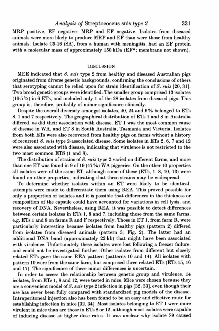
REAFourteen different REA patterns (Fig. 2) were identified for 33 isolates, using
the enzyme HaeIII (Table 2). Patterns could not be obtained for 53 other isolatesas these either failed to lyse adequately, yielded poor quality DNA, or the DNAwas refractory to HaeIII.The 12 belonging to ET 1 in MEE analysis were divided into three REA
patterns. Notably, 3 nasal isolates from healthy pigs on farm R had a differentpattern from 6 clinical isolates from diseased pigs on the same property. The latterhad an additional band at approximately 22 kb. Five isolates belonging to ET 8were divided into 4 REA patterns. In contrast 7 isolates, from farm S, all had
328

Analysis of Streptococcus suis type 21 2 3 4 5 6 7 8
kb
23 1 -
21.2-9.4 -
20 -
14-
231 -
21 2-9.4 -
44 -
329
9 10 11 12 13 14 15 16
1-6 -
Fig. 2. Restriction fragment patterns of DNA from isolates of S. suis type 2. Samplesdigested with HaeJII and separated by electrophoresis in 07% agarose. Fourteenpatterns were recognized, and representative isolates from each of these are presentedin ascending order in lanes 2-8, and 10-16. Lanes: 1 and 9, A DNA size markers; 2,isolate S9; 3, isolate RI; 4, isolate R6; 5, isolate C4-5 (SA); 6, isolate F2; 7, isolate F3;8, isolate L3; 10, isolate 13 (TAS); 11, isolate SI; 12, isolate S5; 13, isolate R4; 14,isolate C7-32 (SA); 15, isolate Bi; 16, isolate R12.
REA pattern No. 10, but were separated by MEE into the closely related ETs 15,16 and 17. Similarly, another 3 isolates with REA pattern No. 14 belonged to theclosely related ETs 12, 13 and 14.
Virulence testingFollowing the first challenge, some mice (mean 1P7 of 6) inoculated with isolates
Ut, It and LI died, and S. suis type 2 was isolated from these. Other miceinoculated with these cultures, as well as S9 and NI, also developed clinical signs

C. G. MWANIKI AND OTHERS
1 2 3 4 5 6 7 8 9 10
- 80 kDa
- 49.5 kDa
11 12 13 14 15 16 17 18
- MRP- EF
Fig. 3. Western blots of cell culture supernatants of the 14 isolates of S. suis type 2 thatwere used to infect mice. Membranes probed with a 1:1 mixture of mouse anti-MRPand anti-EF Mab (supplied by U. Vecht). Lane 1, positive control supernatant(MRP+ ve; EF + ve), supplied by U. Vecht; Lane 2, isolate S9; lane 3, isolate S10; lane4, isolate Ul; lane 5, isolate R6; lane 6, isolate I1; lane 7; isolate LI; lane 8, isolateNI; lane 9, isolate J1; lanes 10 and 11, prestained molecular mass markers (low range,Bio-Rad Laboratories); lane 12, isolate D; lane 13, isolate E2; lane 14, isolate Q5V4;lane 15, isolate T1; lane 16, isolate K; lane 17, isolate R 12; lane 18, positive controlsupernatant.
(10 of 25 mice), which included lack of grooming, lethargy and incoordination. Allfive isolates belonged to ET 1, although only S9 and Ul originated from diseasedpigs. Following the second challenge, with a higher inoculum, only those miceexposed to isolate LI developed disease. Two of these 4 animals died, and the other2 showed clinical signs. After the 3rd inoculation, deaths occurred in 12 of the 14groups, although not in mice challenged with isolates S9 or Ki. Forty-one of 77mice died (53%), and 23 showed clinical signs (30%).
MRP and EF productionProduction of MRP and EF by the 31 isolates is recorded in Table 3. Specific
bands in Western blots using the Mabs are shown in Fig. 3 for the 14 isolates testedfor virulence in mice. Three phenotypes were recorded: MRP and EF positive;
330
MRP--EF -
80 kDa -
49-5 kDa -
Analysis of Streptococcus suis type 2 331MRP positive, EF negative; MRP and EF negative. Isolates from diseasedanimals were more likely to produce MRP and EF than were those from healthyanimals. Isolate C5-16 (SA), from a human with meningitis, had an EF proteinwith a molecular mass of approximately 150 kDa (EF*; membrane not shown).
DISCUSSION
MEE indicated that S. suis type 2 from healthy and diseased Australian pigsoriginated from diverse genetic backgrounds, confirming the conclusions of othersthat serotyping cannot be relied upon for strain identification of S. suis [20, 31].Two broad genetic groups were identified. The smaller group comprised 13 isolates(10O5 %) in 6 ETs, and included only 1 of the 28 isolates from diseased pigs. Thisgroup is, therefore, probably of minor significance clinically.
Despite the overall diversity amongst isolates, 40, 24 and 9% belonged to ETs8, 1 and 7 respectively. The geographical distribution of ETs 1 and 8 in Australiadiffered, as did their association with disease. ET 1 was the most common causeof disease in WA, and ET 8 in South Australia, Tasmania and Victoria. Isolatesfrom both ETs were also recovered from healthy pigs on farms without a historyof recurrent S. suis type 2-associated disease. Some isolates in ETs 2, 6, 7 and 12were also associated with disease, indicating that virulence is not restricted to thetwo most common ETS (1 and 8).The distribution of strains of S. suis type 2 varied on different farms, and more
than one ET was found in 9 of 19 (47 %) WA piggeries. On the other 10 propertiesall isolates were of the same ET, although some of these (ETs, 1, 8, 10, 13) werefound on other properties, indicating that these strains may be widespread.To determine whether isolates within an ET were likely to be identical,
attempts were made to differentiate them using REA. This proved possible foronly a proportion of isolates and it is possible that differences in the thickness orcomposition of the capsule could have accounted for variations in cell lysis, andrecovery of DNA. Nevertheless, using REA, it was possible to detect differencesbetween certain isolates in ETs 1, 8 and 7, including those from the same farms,e.g. ETs 1 and 8 on farms R and F respectively. Those in ET 1, from farm R, wereparticularly interesting because isolates from healthy pigs (pattern 2) differedfrom isolates from diseased animals (pattern 3; Fig. 2). The latter had anadditional DNA band (approximately 22 kb) that might have been associatedwith virulence. Unfortunately these isolates were lost following a freezer failure,and could not be investigated further. Other isolates from different but closelyrelated ETs gave the same REA pattern (patterns 10 and 14). All isolates withpattern 10 were from the same farm, but comprised three related ETs (ETs 15, 16and 17). The significance of these minor differences is uncertain.
In order to assess the relationship between genetic group and virulence, 14isolates, from ETs 1, 8 and 12, were tested in mice. Mice were chosen because theyare a convenient model ofS. suis type 2 infection in pigs [32, 33], even though theiruse has never been fully compared with standardized pig models of the disease.Intraperitoneal injection also has been found to be an easy and effective route forestablishing infection in mice [32, 34]. Most isolates belonging to ET 1 were morevirulent in mice than are those in ETs 8 or 12, although most isolates were capableof inducing disease at higher dose rates. It was unclear why isolate S9 caused

C. G. MWANIKI AND OTHERSdisease at 2 x 101 c.f.u., but not following challenge with 1 x 10⁹ c.f.u. A protectiveimmunity may have developed after the mice received the initial doses, but asimilar effect was not seen with other isolates. Three of the 5 most virulent isolateswere recovered from the tonsils of healthy pigs at slaughter, showing that healthypigs may harbour virulent strains.Vecht and colleagues [16, 17] demonstrated that isolates of S. suis type 2,
virulent to newborn germ-free pigs, released MRP and EF into the culturemedium. Isolates that produced both MRP and EF*, a higher molecular weightform of EF, were of intermediate virulence, whilst isolates not producing eitherform did not induce disease. In this study, 8 of the isolates produced MRP, but notEF or EF*; this phenotype has not previously been described. Six isolates fromET 1 tested in mice produced both MRP and EF, and, in concordance with resultsobtained in pigs [16, 17], 4 were virulent at relatively low doses. Neverthelessisolate II, also in ETI, did not produce MRP or EF, but was virulent at low doserates. Other isolates, lacking either or both proteins, were virulent at higher doserates. MRP and EF, therefore, are not essential as virulence determinants in miceinoculated by the intraperitoneal route. They may be important in the earlyestablishment of infection in natural cases of disease in pigs, however, since 6 of13 isolates from diseased pigs produced MRP and EF, and 4 produced MRP,whilst only 2 of 18 isolates from healthy pigs produced MRP and EF, and 4produced MRP. Although most isolates from healthy pigs were MRP and EFnegative, some that were potentially more virulent (MRP and EF positive) werecarried by healthy animals in certain of the WA herds that infrequently sufferfrom S. suis-associated disease. The absence of disease in these herds may beassociated with superior management practices, such as provision of goodventilation and low stocking densities.
Isolate C5-16 (SA) recovered from the meninges of a human produced an EF-like molecule (EF*) with a higher molecular mass than that produced by ourporcine isolates (approximately 150 kDa, compared to 110 kDa). Similar material(EF*) has been described by Vecht and colleagues [17] in strains isolated fromhumans, although in relatively few porcine isolates. These results suggest thatEF* may be involved in specific aspects of the pathogenesis of the infection inhumans.
All the isolates producing EF belonged to the closely related ETs 1 and 2 (8 of10 isolates analysed from these ETs were EF positive), and 9 of the 10 isolatesproduced MRP. In contrast only 7 of the 21 (33%) isolates from other ETs thatwere examined produced MRP, including only 2 of the 8 isolates from ET 8 (4 ofwhich were from diseased pigs). ETs 1 and 2, which could be distinguished by thepresence of allele 1 for esterase and allele 4 for glutamate dehydrogenase, appearedto be more virulent than other ETs. Production of EF and MRP by these isolatestherefore may only be a marker for this virulent clone, rather than the proteinsthemselves necessarily being determinants of virulence. This possibility was
suggested by Vecht and colleagues [17]. They pointed out that, in order todetermine the function of the proteins, it was necessary to obtain isogenic strainsthat varied in their production of the proteins, and to test these strains forvirulence in pigs. Currently, irrespective of the function of EF, screening for itsproduction is a means of detecting isolates belonging to the virulent clone
332

Analysis of Streptococcus suis type 2 333represented by ETs 1 and 2. Isolates from other clones that do not produce EFhowever may also be capable of causing disease.
ACKNOWLEDGEMENTS
This study was supported by grants from the Western Australian Pig IndustryCompensation Fund, and from the Australian Pig Research and DevelopmentCorporation. C. G. M. was in receipt of a Postgraduate Scholarship from theAustralian International Development Assistance Bureau. Dr Uri Vecht gen-erously donated the monoclonal antibodies against MRP and EF, and providedconstructive comment on the manuscript.
REFERENCES
1. Lamont MH, Edwards PT, Windsor RS. Streptococcal meningitis in pigs: results of a fiveyear survey. Vet Rec 1980; 107: 467-9.
2. Sanford SE, Higgins R. Streptococcal diseases. In: Leman A et al., eds., Diseases of swine,7th ed. Ames, USA: Iowa State University Press, 1992: 588-90.
3. Windsor RS, Elliot SD. Streptococcal infections in young pigs. IV. An outbreak ofstreptococcal meningitis in weaned pigs. J Hyg 1975; 75: 68-78.
4. Gottschalk M, Higgins R, Jacques M, Mittal KR, Henrichsen J. Description of 14 newcapsular types of Streptococcus sui8. J Clin Microbiol 1989; 27: 2633-6.
5. Gottschalk M, Higgins R, Jacques M, Beaudoin M, Hendrichsen J. Characterisation of sixnew capsular types (23 through 28) of Streptococcus suis. J Clin Microbiol 1991; 29: 2590-4.
6. Perch B, Pedersen KB, Hendrichsen J. Serology of capsulated streptococci pathogenic forpigs: six new serotypes of Streptococcus suis. J Clin Microbiol 1983; 17: 993-6.
7. Alexander TJL. Streptococcus suis: an update. Pig Vet Soc Proc 1991; 27: 50-60.8. Touil F, Higgins R, Nadeau M. Isolation of Streptococcus suis from diseased pigs in Canada.
Vet Microbiol 1988; 17: 171-7.9. Sihovenen L, Kurl DN, Hendrichsen J. Streptococcus suis isolated from pigs in Finland.
Acta Vet Scand 1988; 29: 9-13.10. Clifton-Hadley FA, Alexander TJL. The carrier site and carrier rate of Streptococcus suis
type 2 in pigs. Vet Rec 1980; 107: 40-1.11. Clifton-Hadley FA, Alexander TJL, Enright MR, Guise J. Monitoring herds for
Streptococcus suis type 2 by sampling tonsils of slaughter pigs. Vet Rec 1984; 115: 562-4.12. Robertson ID, Blackmore DK. Prevalence of Streptococcus suis types 1 and 2 in domestic
pigs in Australia and New Zealand. Vet Rec 1989; 124: 391-4.13. Clifton-Hadley FA. Streptococcal meningitis in pigs. Pig News Info 1989; 10: 9-12.14. Clifton-Hadley FA, Alexander TJL, Enright MR, Lindsay HJ. Monitoring herds for
Streptococcus suis type 2: cross-reactions and variations in virulence. Proc Int Pig Vet SocCong. Barcelona, Spain, 1986: 359.
15. Vecht U, Arends JP, van der Molen EJ, van Leengoed LAMG. Differences in virulencebetween two strains of Streptococcus suis type II after experimental induced infection ofnewborn germ-free pigs. Am J Vet Res 1989; 50: 1037-43.
16. Vecht U, Wisselink HJ, Jellema MlL, Smith HE. Identification of two proteins associatedwith virulence of Streptococcus sui.s type 2. Infect Immun 1991; 59: 3156-62.
17. Vecht U, Wisselink HJ, van Dijk JE, Smith HE. Virulence of Streptococcus suis type 2strains in newborn germfree pigs depends on phenotype. Infect Immun 1992; 60: 550-6.
18. Klipper-Balz R. Schleifer KH. Streptococcus suits sp. nov., nom. rev. Int J Svst Bacteriol1987: 37: 160-2.
19. Hampson DJ, Trott DJ, Clarke IL, Mwaniki CG, Robertson ID. Population structure ofAustralian isolates of Streptococcus suis. J Clin Microbiol 1993; 31: 2895-900.
20. Beaudoin M. Harel J, Higgins R. Gottschalk M, Frenette M, Maclnnes JI. Molecularanalysis of isolates of Streptococcus suis capsular type 2 by restriction-endonuclease-digestedDNA separated on SDS-PAGE and by hybridization with an rDNA probe. J Gen Microbiol1992; 138: 2639-45.

334 C. G. MWANIKI AND OTHERS
21. Mogollon JD, Pijoan C, Murtaugh MP, Kaplan EL, Collins JE, Cleary PP. Characterisationof prototype and clinically defined strains of Streptococcus suis by genomic fingerprinting.J Clin Microbiol 1990; 28: 2462-6.
22. Selander RK, Caugant DA, Ochmann H, Musser JM, Gilmour MN, Wittam TS. Methods ofmultilocus enzyme electrophoresis for bacterial population genetics and systematics. ApplEnviron Microbiol 1986; 51: 873-84.
23. Robertson ID, Blackmore DK, Hampson DJ, Fu ZF. A longitudinal study of naturalinfection of piglets with Streptococcus suis types 1 and 2. Epidemiol Infect 1991; 107:119-26.
24. Davies PR, Ossowicz CJ. Evolution of methods used for detecting Streptococcus suis type2 in tonsils, and investigation of the carrier state in pigs. Res Vet Sci 1991; 50: 190-4.
25. Clifton-Hadley FA, Alexander TJL, Enright MR. Diagnosis of Streptococcus suis type 2infection in pigs. Pig Vet Soc Proc 1985; 14: 27-34.
26. Moreau A, Higgins R, Bigras-Poulin M, Nadeau M. Rapid detection of Streptococcus suisserotype 2 in weaned pigs. Am J Vet Res 1989; 50: 1667-71.
27. Higgins R, Gottschalk M. An update on Streptococcus suis identification. J Vet Diagn Invest1990; 2: 249-52.
28. Nei M. Estimates of average heterozygosity and genetic distance from a small number ofindividuals. Genetics 1978; 89: 583-90.
29. Sneath PHA, Sokal RR. Numerical taxonomy, San Francisco: WH Freeman, 1993: 573.30. Sambrook J, Fritsch EF, Maniatis T. Molecular cloning: a laboratory manual. Cold Spring
Harbor: Spring Harbor Laboratory Press, 1989.31. Mogollon JD, Pijoan C, Murtaugh MP, Collins JE, Cleary PP. Identification of epidemic
strains of Streptococcus suis by genomic fingerprinting. J Clin Microbiol 1991; 29: 782-7.32. Robertson ID, Blackmore DK. Experimental studies on the comparative infectivity and
pathogenicity of Streptococcus suis type 2. II. Porcine and human isolates in laboratoryanimals. Epidemiol Infect 1990; 105: 479-84.
33. Williams AE, Blackmore WF, Alexander TJL. A murine model of Streptococcus suis type2 meningitis in the pig. Res Vet Sci 1988; 45: 394-9.
34. Kebede M, Chengappa MM, Stuart JG. Isolation of characterisation of temperature-sensitive mutants of Streptococcus suis: efficacy trial of the mutant vaccine in mice. VetMicrobiol 1990; 22: 249-57.


