Embed Size (px)
Citation preview

OIE Terrestrial Manual 2022 1
C H A P T E R 3 . 1 . 4 .
B R U C E L L O S I S ( I N F E C T I O N W I T H B . A B O R T U S , B . M E L I T E N S I S A N D B . S U I S )
SUMMARY
Description of the disease: Brucellosis is the generic name used for the animal and human infections caused by several species of the genus Brucella, mainly Brucella abortus, B. melitensis and B. suis. Infection with Brucella in cattle is usually caused by B. abortus, less frequently by B. melitensis, and occasionally by B. suis. Brucella melitensis is the main causative agent of infection with Brucella in sheep and goats. Brucella melitensis and B. abortus may also infect other species, including camels. Infection with Brucella in pigs is due to B. suis biovars 1–3, but the disease caused by biovar 2 differs in its host range and its limited geographical distribution. In some areas, B. suis infection has become established in wild pigs. Clinically, infection with Brucella in animals is characterised by one or more of the following signs: abortion, infertility, retained placenta, orchitis, epididymitis and, rarely, arthritis, with excretion of the organisms in uterine discharges, milk, urine and semen. Unequivocal diagnosis depends on the isolation of Brucella from abortion material, udder secretions or from tissues removed at post-mortem. Brucella abortus, B. melitensis and B. suis are highly pathogenic for humans, and potentially contaminated tissues, cultures and materials must be handled under appropriate containment conditions.
Detection of the agent: Indication of Brucella is provided by the demonstration of Brucella-like organisms in abortion material or vaginal discharge using modified acid-fast staining, and is considered presumptive, especially if supported by serological tests. Polymerase chain reaction (PCR) methods are additional means for detection of the presence of Brucella DNA in a sample. Whenever possible, Brucella spp. should be isolated by culturing samples from uterine discharges, aborted fetuses, udder secretions or selected tissues, such as lymph nodes and male and female reproductive organs. Species and biovars can be identified by phage lysis, and by cultural, biochemical and serological tests. PCR can provide the basis of complementary identification and typing methods based on specific genomic sequences.
Serological and cellular immunity tests: Serological tests indicate exposure to Brucella species, but cannot identify the aetiological agent to the species level. The buffered Brucella antigen tests (rose bengal test and buffered plate agglutination test), the complement fixation test, the enzyme-linked immunosorbent assays (ELISA) or the fluorescence polarisation assay, are suitable tests for screening of herds/flocks and individual small ruminants, camelids and bovines (cattle and buffaloes). However, no single serological test is appropriate in each animal species and all epidemiological situations, and some of these tests are not adequate for diagnosing brucellosis in pigs. Therefore, the reactivity of samples that are positive in screening tests should be assessed using an established confirmatory or complementary strategy. The indirect ELISA or milk ring test performed on bulk milk samples is effective for screening and monitoring dairy cattle. The brucellin skin test can be used in unvaccinated ruminants, camels and swine as either a screening or a confirmatory herd test when positive serological reactors occur in the absence of obvious risk factors.
Requirements for vaccines and diagnostic biologicals: Brucella abortus strain 19 and B. melitensis strain Rev.1 remain the reference vaccines for the control of Brucella infections in cattle and in sheep and goats, respectively, with which any other vaccines should be compared. Both should be prepared from adequately derived seed cultures. The rough B. abortus strain RB51 vaccine has also become the official vaccine for prevention of B. abortus infection in cattle in some countries. No suitable vaccines exist for the control of Brucella infection in swine. Brucellin preparations must be free of smooth lipopolysaccharide, and antigens for serological tests must be prepared from smooth
Chapter 3.1.4. – Brucellosis (infection with Brucella abortus, B. melitensis and B. suis)
2 OIE Terrestrial Manual 2022
B. abortus strain 1119-3 or 99 and, in the case of indirect ELISA, from smooth B. melitensis strain 16M as well. Vaccines and brucellin preparations must comply with relevant standards.
A. INTRODUCTION
Brucellosis is the generic name used for the animal and human infections caused by several species of the genus Brucella, mainly Brucella abortus, B. melitensis, B. suis and B. canis. Infection of sheep with B. ovis is described separately in Chapter 3.8.7 Ovine epididymitis (Brucella ovis).
Causal pathogens: Genetic and immunological evidence indicates that all members of the Brucella genus are closely related. Nevertheless, based on relevant differences in host preference and epidemiology displayed by the major variants, as well as molecular evidence of genomic variation, the International Committee on Systematics of Prokaryotes, Subcommittee on the Taxonomy of Brucella took a clear position in 2005 on a return to pre-1986 Brucella taxonomic opinion; the consequences of this statement imply the re-approval of the six classical Brucella nomenspecies with their corresponding recognised biovars, although both opinions remain valid. The classical names related to the six Brucella nomenspecies are validly published in the Approved Lists of Bacterial Names, 1980, and the designated type strains are attached to these validly published names: B. abortus, B. melitensis, B. suis, B. neotomae, B. ovis and B. canis. The first three of these are subdivided into biovars based on cultural and serological properties (see Tables 2 and 3). Strains of Brucella have been isolated from marine mammals and classified into two new species: B. ceti and B. pinnipedialis (Foster et al., 2007). A new species, named B. microti, was also isolated from the common vole (Microtus arvalis) as well as from foxes, soil and frogs raised for human consumption in Europe (Scholz et al., 2008). Novel isolates from human breast implant infection, from baboons that had delivered stillborn offspring, and from foxes have also been described, although the natural reservoir of these isolates remains uncertain. While limited isolates of each new type have been described, they have been formally published as the tenth, eleventh, and twelfth Brucella species, B. inopinata, B. papionis and B. vulpis respectively (Scholz et al., 2010; 2016; Whatmore et al., 2014). Finally, various strains isolated from rodents, foxes reptiles, fish and frogs were characterised as atypical Brucella strains distinct from the currently described species. They have not yet been approved as new Brucella species.
Brucella is a member of the Brucellaceae family, in the order Rhizobiales, class Alphaproteobacteria. It shows close genetic relatedness to some plant pathogens and symbionts of the genera Agrobacterium and Rhizobium, as well as animal pathogens (Bartonella) and opportunistic or soil bacteria (e.g. Ochrobactrum).
1. Description of the disease
1.1. Infection with Brucella in cattle
Infection with Brucella in cattle is usually caused by biovars (bv.) of Brucella abortus. In some countries, particularly in southern Europe, Africa and western Asia, where cattle are kept in close association with sheep or goats, infection can also be caused by B. melitensis (Verger, 1985). Occasionally, B. suis may cause infections in cattle. The disease is global in distribution but a number of countries are considered free from both B. abortus and B. melitensis. For up-to-date information, consult OIE WAHIS interface1.
Young animals and non-pregnant females usually show no signs of the disease. Following infection with B. abortus or B. melitensis, pregnant adult females develop a placentitis usually resulting in abortion between the fifth and ninth month of pregnancy. Even in the absence of abortion, profuse excretion of the organism occurs in the placenta, fetal fluids and vaginal discharges. The mammary gland and associated lymph nodes may also be infected, and organisms may be excreted in the milk. Colostrum originating from infected dams is a source of infection in the newborn population. Subsequent pregnancies are usually carried to term, but uterine and mammary infection recurs, with reduced numbers of organisms in afterbirth products and milk. In acute infections, the organism is present in most major body lymph nodes. Adult male cattle may develop orchitis/epididymitis and brucellosis may be a cause of infertility in both sexes. Brucella abortus can be shed in semen, seminal fluid and urine. Hygromas, usually involving leg joints, are a common manifestation of brucellosis in some tropical countries and may be the only obvious indicator of infection; the hygroma fluid is often infected with Brucella.
1 https://www.woah.org/en/what-we-do/animal-health-and-welfare/disease-data-collection/
Chapter 3.1.4. – Brucellosis (infection with Brucella abortus, B. melitensis and B. suis)
OIE Terrestrial Manual 2022 3
1.2. Infection with Brucella in sheep and goats
Infection with Brucella in sheep and goats (excluding B. ovis infection) is primarily caused by B. melitensis. Sporadic infections caused by B. abortus or B. suis have been observed in sheep and goats, but such cases are extremely rare. Infection with Brucella in sheep and goats is widespread although a number of countries are believed to be free from the agent. For up-to-date information, consult OIE WAHIS interface. Pathologically and epidemiologically, B. melitensis infection in sheep and goats is very similar to B. abortus infection in cattle. In most circumstances, the primary routes of transmission of Brucella are the placenta, fetal fluids and vaginal discharges expelled by infected ewes and goats at and up to several months following abortion or parturition. Shedding of Brucella is also common in udder secretions and semen, and Brucella may be isolated from various tissues, such as lymph nodes from the head, spleen and organs associated with reproduction (uterus, epididymides and testes), and from arthritic lesions (Alton et al., 1988).
1.3. Infection with Brucella in pigs
Infection with Brucella in pigs is primarily caused by biovars 1, 2 or 3 of B. suis. Sporadic infections caused by B. abortus or B. melitensis have been also observed in pigs, but such cases are rare. The disease occurs in many countries where pigs are raised. Generally, the prevalence is low, but in some regions, such as South America and south-east Asia, the prevalence may be much higher. Porcine brucellosis may be a serious but presently unrecognised problem in some countries. Brucella suis bv. 1 infection has been reported from feral pigs in some of the southern states of the United States of America (USA), in parts of Australia and several other countries in Oceania. In these countries, a number of human infections have been reported from people who hunt and handle material taken from feral pigs. The disease is generally transmitted by consumption of feed contaminated by birth or abortion products and uterine discharges. Pigs will instinctively eat aborted fetuses and placental membranes. Transmission during copulation also occurs frequently, and B. suis excretion in semen has implications for those practising artificial insemination. In pigs, as in ruminants, after the initial bacteraemia, B. suis colonises the reproductive tract of either sex. In females, placentas and fetuses are invaded, while in males, invasion occurs in one or more of the following: testes, prostate, epididymides, seminal vesicles or bulbo–urethral glands. In males the lesions, which are most often unilateral, start with a hyperplasia that may progress to abscess formation; the final stage is characterised by sclerosis and atrophy. The most common manifestation of brucellosis in female pigs is abortion, occurring at any time during pregnancy, but most frequently between day 50 and 110 of gestation. Vaginal discharge is not often evident, and, in chronically infected herds, infertility rather than abortion is the most relevant clinical sign of the disease. In males, brucellosis is more likely to be persistent, with lesions in the genital tract often leading to interference with sexual activity, which can be temporary or permanent. The boar may excrete Brucella in the semen without any apparent abnormality in the sex organs or interference with sexual activity. In both sexes, arthritis may occur in various joints, there may be swollen joints and tendon sheaths, lameness and, occasionally, posterior paralysis or spondylitis. A significant proportion of both male and female pigs will recover from the infection, often within 6 months, but many will remain permanently infected (Olsen et al., 2012).
Infection caused by B. suis bv. 2 differs from infection caused by bv. 1 and bv. 3 in its host range, distribution, and in pathogenicity. Historically, the geographical distribution of B. suis bv. 2 has been in a broad range between Scandinavia and the Balkans. The prevalence in wild boars appears to be high throughout continental Europe (EFSA, 2009). In outbreaks in Europe, wild boars were implicated as the source of transmission of bv. 2 to outdoor reared pigs, and are considered as the main wild reservoir of this infection (EFSA, 2009). Brucella suis bv. 2 causes miliary lesions, particularly in reproductive tissues, that often become purulent. To date, B. suis bv. 2 has rarely been reported as the cause of human brucellosis. However, B. suis bv. 2 infections have been reported in immuno-compromised hunters, who had been extensively exposed through gutting or skinning boars or hares. Moreover, rare cases of B. suis bv. 2 infection without clinical signs have been reported in Europe in cattle or sheep exposed to infected wild boars.
1.4. Infection with Brucella in other domestic, captive–wild or wild species
Infection with B. abortus or B. melitensis has been reported in the one-humped camel (Camelus dromedarius) and the two-humped camel (C. bactrianus), as well as in the South American camelids: llama (Lama glama), alpaca (Vicugna pacos), guanaco (Lama guanicoe), and vicuña (Vicugna vicugna), and is related to contact with large and small ruminants infected with B. abortus or B. melitensis.
Chapter 3.1.4. – Brucellosis (infection with Brucella abortus, B. melitensis and B. suis)
4 OIE Terrestrial Manual 2022
In addition, brucellosis has been observed in the domestic buffalo (Bubalus bubalis), American and European bison (Bison bison and B. bonasus, respectively), yak (Bos grunniens), elk/wapiti (Cervus canadensis, sika deer (C. nippon), African buffalo (Syncerus caffer) and various antelope species. The clinical manifestations of brucellosis in these animals are similar to those seen in cattle, sheep and goats.
Brucella melitensis infection in wild ruminants may occur when these species are in close contact with sheep and goats in enzootic areas. The manifestations of brucellosis in these animals are similar to those in cattle or sheep and goats. However, in several wild ruminant species (e.g. chamois [Rupicapra rupicapra], Alpine ibex [Capra ibex] and the Iberian wild goat [Capra pyrenaica]), purulent or calcified arthritis and orchitis as well as uveitis and neurological signs have been reported. These species are considered as dead-end carriers, and the disease usually disappears naturally as soon as Brucella infection has been eradicated from domestic livestock, unless anthropogenic effects take place. Nevertheless three reservoirs are currently described in wild ruminants: B. abortus in bison in the Yellowstone area of North America, B. melitensis in alpine ibex and B. abortus in wood bison in Wood Buffalo National Park in Canada. There have also been sporadic reports of B. melitensis isolation from dogs, especially from contact with infected sheep or goats, or ingestion of placenta or aborted fetuses.
There are two different types of epidemiological situation with regard to B. suis infection in other non-porcine species. In the first case, B. suis infection occurs in animals that are not the natural host of the particular infection through the ingestion of contaminated materials or by co-habitation with infected natural hosts. For example, Arctic foxes and wolves may contract B. suis bv. 4 from reindeer; dogs and rodents, such as rats and mice, may acquire other B. suis biovars by cohabitation with infected hosts; cattle and horses may become infected by cohabitation or interaction with infected swine. The infecting bacteria are invariably the well-defined biovars of the natural host species. In the second case, wildlife species are natural hosts for B. suis or B. suis-like infections. One example is the so-called murine brucellosis of the Commonwealth of Independent States (CIS) and the Baltic countries, where small rodents are infected with B. suis bv. 5.
In addition to wild boar, the European hare (Lepus europaeus) is also considered to be a reservoir for B. suis bv. 2 and has been implicated as a possible source of transmission to domestic livestock. The disease in the European hare is characterised by the formation of nodules, varying in size from that of a millet seed to a cherry or even larger; these often become purulent. Such nodules may occur in almost any location, sometimes subcutaneously or intramuscularly, in the spleen, liver or lung and in the reproductive organs of either sex. The body condition of the hare may be unaffected. Other species may also become infected by cohabitation with B. suis bv. 2 infected swine, wild boars or hares. Gutting or skinning wild boars in cattle sheds could be a route of transmission to cattle.
Brucella suis bv. 4 causes a serious zoonotic disease in wild or domesticated reindeer or caribou (Rangifer tarandus and its various subspecies) throughout the Arctic region, including Siberia, Canada and Alaska. Rangifer tarandus is very susceptible to B. suis infection, which causes fever, depression and various local signs, such as abortion, retained placentas, metritis, sometimes with blood-stained discharge, mastitis, bursitis and orchitis. Transmission to humans may be by direct contact or through consumption of raw milk and other inadequately heated products from reindeer, bone marrow in particular.
1.5. Zoonotic risk and biosafety requirements
Some Brucella species, most notably B. melitensis, B. abortus, B. suis and, probably to a lesser extent B. canis are readily transmissible to humans, causing acute febrile illness – undulant fever – which may progress to a more chronic form and can also produce serious complications affecting the musculo-skeletal, cardiovascular, and central nervous systems. Precautions should be taken to prevent human infection. Infection is acquired by the oral, respiratory, or conjunctival routes. Ingestion of raw milk products constitutes the main risk to the general public where the disease is endemic. There is an occupational risk to veterinarians, abattoir workers and farmers who handle infected animals/carcasses and aborted fetuses or placentas. Brucellosis is also one of the most easily acquired laboratory infections, and all laboratory manipulations with live cultures or potentially infected/contaminated material must be performed at an appropriate biosafety and containment level determined by biorisk analysis (see Chapter 1.1.4 Biosafety and biosecurity: Standard for managing biological risk in the veterinary laboratory and animal facilities). Specific recommendations have been made for the biosafety precautions to be observed with Brucella-infected materials (for further details see Alton et al., 1988;
Chapter 3.1.4. – Brucellosis (infection with Brucella abortus, B. melitensis and B. suis)
OIE Terrestrial Manual 2022 5
Joint FAO/WHO Expert Committee on Brucellosis, 1986; WHO, 1953; WHO, 2004; Chapter 1.1.3 Transport of biological materials).
B. DIAGNOSTIC TECHNIQUES
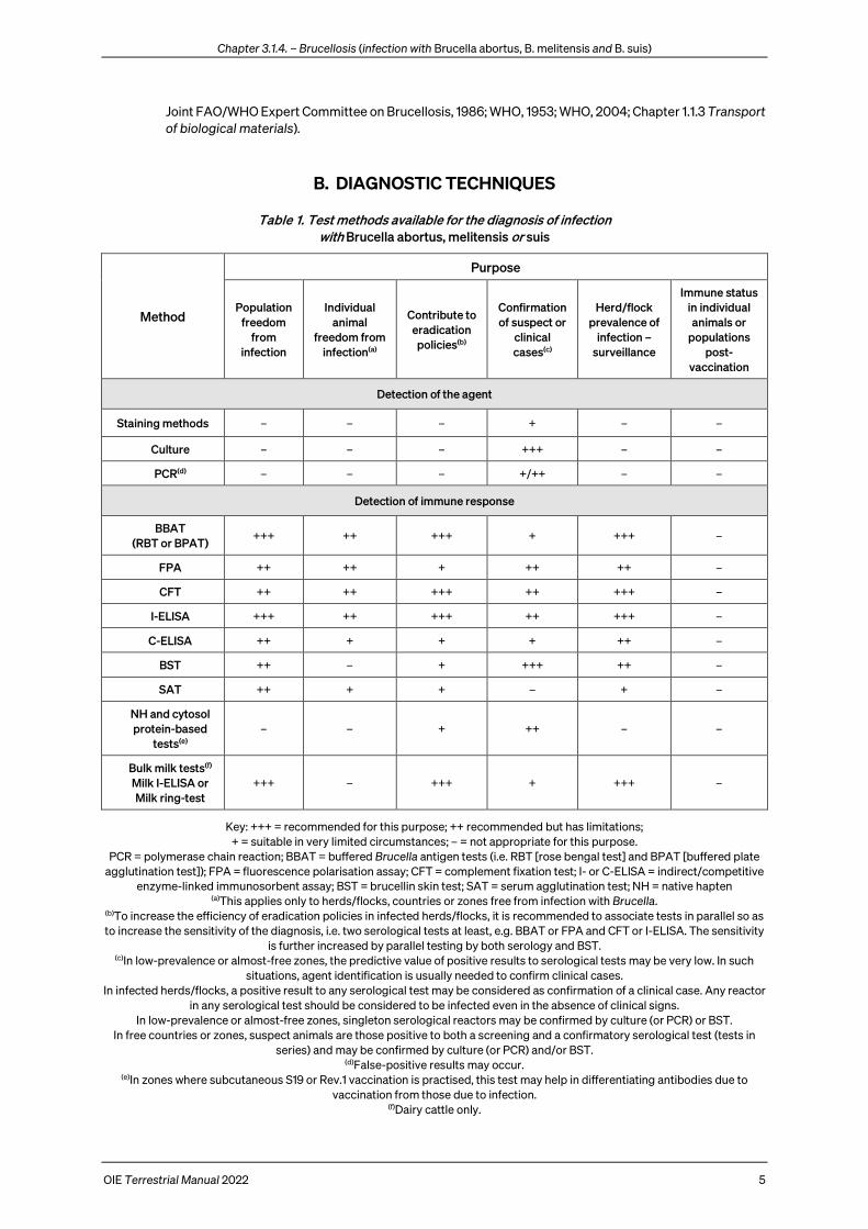
Table 1. Test methods available for the diagnosis of infection with Brucella abortus, melitensis or suis
Method
Purpose
Population freedom
from infection
Individual animal
freedom from infection(a)
Contribute to eradication
policies(b)
Confirmation of suspect or
clinical cases(c)
Herd/flock prevalence of
infection – surveillance
Immune status in individual animals or
populations post-
vaccination
Detection of the agent
Staining methods – – – + – –
Culture – – – +++ – –
PCR(d) – – – +/++ – –
Detection of immune response
BBAT (RBT or BPAT)
+++ ++ +++ + +++ –
FPA ++ ++ + ++ ++ –
CFT ++ ++ +++ ++ +++ –
I-ELISA +++ ++ +++ ++ +++ –
C-ELISA ++ + + + ++ –
BST ++ – + +++ ++ –
SAT ++ + + – + –
NH and cytosol protein-based
tests(e) – – + ++ – –
Bulk milk tests(f) Milk I-ELISA or Milk ring-test
+++ – +++ + +++ –
Key: +++ = recommended for this purpose; ++ recommended but has limitations; + = suitable in very limited circumstances; – = not appropriate for this purpose.
PCR = polymerase chain reaction; BBAT = buffered Brucella antigen tests (i.e. RBT [rose bengal test] and BPAT [buffered plate agglutination test]); FPA = fluorescence polarisation assay; CFT = complement fixation test; I- or C-ELISA = indirect/competitive
enzyme-linked immunosorbent assay; BST = brucellin skin test; SAT = serum agglutination test; NH = native hapten (a)This applies only to herds/flocks, countries or zones free from infection with Brucella.
(b)To increase the efficiency of eradication policies in infected herds/flocks, it is recommended to associate tests in parallel so as to increase the sensitivity of the diagnosis, i.e. two serological tests at least, e.g. BBAT or FPA and CFT or I-ELISA. The sensitivity
is further increased by parallel testing by both serology and BST. (c)In low-prevalence or almost-free zones, the predictive value of positive results to serological tests may be very low. In such
situations, agent identification is usually needed to confirm clinical cases. In infected herds/flocks, a positive result to any serological test may be considered as confirmation of a clinical case. Any reactor
in any serological test should be considered to be infected even in the absence of clinical signs. In low-prevalence or almost-free zones, singleton serological reactors may be confirmed by culture (or PCR) or BST.
In free countries or zones, suspect animals are those positive to both a screening and a confirmatory serological test (tests in series) and may be confirmed by culture (or PCR) and/or BST.
(d)False-positive results may occur. (e)In zones where subcutaneous S19 or Rev.1 vaccination is practised, this test may help in differentiating antibodies due to
vaccination from those due to infection. (f)Dairy cattle only.
Chapter 3.1.4. – Brucellosis (infection with Brucella abortus, B. melitensis and B. suis)
6 OIE Terrestrial Manual 2022
All cases of abortion as well as orchitis in cattle, sheep and goats, camels and pigs, should be considered as suspected brucellosis and should be investigated through the herd/flock history and submission of specimens for laboratory testing. The clinical signs are not pathognomonic and unequivocal diagnosis of Brucella infections can be made only by the isolation and identification of Brucella, but in situations where bacteriological examination is not practicable, diagnosis must be based on molecular or immunological methods.
1. Detection of the agent
Classically a bacterial culture is identified as Brucella by growth characteristics (see Section B.1.3 Identification and typing) although unequivocal identification as Brucella is now possible through various molecular approaches.
All samples from suspect cases should be cooled (4°C) immediately after they are taken, and transported to the laboratory by the most rapid means. If they are to spend more than 12 hours in transit, all samples apart from vaginal swabs, should be frozen (–20°C). On arrival at the laboratory, samples that are not to be cultured immediately should be frozen (Alton et al., 1988). In all cases, the shorter the shipment and storage time, the higher is the probability of Brucella isolation, especially in cases where the initial amount of Brucella is low in the sample. No specific transport medium has been demonstrated to improve Brucella survival in animal samples.
1.1. Staining methods
Brucella are coccobacilli or short rods measuring from 0.6 to 1.5 µm long and from 0.5 to 0.7 µm wide. They are usually arranged singly, and less frequently in pairs or small groups. The morphology of Brucella is fairly constant, except in old cultures where pleomorphic forms may be evident. Brucella are nonmotile. They do not form spores; pili or true capsules are not produced. Brucella are Gram negative and usually do not show bipolar staining. They are resistant to decolourisation by weak acids and thus stain red by the Stamp’s modification of the Ziehl–Neelsen’s method (Alton et al., 1988). With this method, in smears of organs or biological fluids previously fixed with heat or ethanol, Brucella organisms stain red against a blue background. A fluorochrome or peroxidase-labelled antibody conjugate-based technique could also be used. The presence of intracellular, weakly acid-fast organisms of Brucella morphology or immuno-specifically stained organisms is presumptive evidence of brucellosis. However, these methods are not feasible or have a low sensitivity in milk and dairy products where Brucella are often present in small numbers, and interpretation is frequently impeded by the presence of fat globules. Care must be taken as well in the interpretation of positive results in the Stamp’s method because other organisms that cause abortions, e.g. Chlamydia abortus or Coxiella burnetii, may be difficult to differentiate from Brucella organisms in these preparations. The results, whether positive or negative, should be confirmed by culture.
Polymerase chain reaction (PCR) methods can also be used to demonstrate the agent in various biological samples (Bricker, 2002; Whatmore & Gopaul, 2011), but the sensitivity of these approaches may be low with respect to classical bacteriology because of limitations around sample volume. Molecular tests may detect infection where poor sample storage means bacteria are no longer viable.
1.2. Collection of samples and culture
Bacteriological isolation is slow, expensive and cumbersome, but it should be performed whenever possible to confirm the disease and to determine the Brucella species involved. It also enables emerging epidemiological approaches such as high throughput sequencing to be applied. Although often considered not sensitive, it can be very effective when the type and number of samples, their adequate storage, amount seeded and the culture media used are optimised.
1.2.1. Basal media
Direct isolation and culture of Brucella are usually performed on solid media. This is generally the most satisfactory method as it enables the developing colonies to be isolated and recognised clearly. Such media also limit the establishment of non-smooth mutants and excessive development of contaminants. However, the use of liquid media may be recommended for voluminous samples or for the purpose of enrichment. A wide range of commercial dehydrated basal media is available, e.g. Brucella medium base, tryptose (or trypticase)–soy agar (TSA). The addition of 2–5% bovine or equine serum is necessary for the growth of strains such as B. abortus bv. 2, and many laboratories systematically add serum to basal media, such as blood agar base or Columbia agar, with excellent results. Other satisfactory media, such as serum–dextrose agar (SDA) or glycerol–dextrose agar, can be used (Alton et al., 1988). SDA is usually preferred for

Chapter 3.1.4. – Brucellosis (infection with Brucella abortus, B. melitensis and B. suis)
OIE Terrestrial Manual 2022 7
observation of colonial morphology. A non-selective, biphasic medium, known as Castañeda’s medium, is recommended for the isolation of Brucella from blood and other body fluids or milk, where enrichment culture is advised. Castañeda’s medium is used because brucellae tend to dissociate in broth medium, interfering with biotyping by conventional bacteriological techniques.
1.2.2. Selective media
All the basal media mentioned above can be used for the preparation of selective media. Appropriate antibiotics are added to suppress the growth of organisms other than Brucella.
The most widely used selective medium is the modified Farrell’s medium (FM) (Stack et al., 2002), added to 1 litre of agar: polymyxin B sulphate (5000 units = 5 mg); bacitracin (25,000 units = 25 mg); natamycin (50 mg); nalidixic acid (5 mg); nystatin (100,000 units); vancomycin (20 mg). A corresponding freeze-dried antibiotic supplement is available commercially. However, nalidixic acid and bacitracin, at the concentration used in FM, have inhibitory effects on some B. abortus, B. melitensis and B. suis strains. Accordingly, the simultaneous use of FM and the less selective Thayer–Martin’s modified (mTM) culture media has been considered the strategy of choice for Brucella primary isolation from field veterinary samples. However, the mTM is not translucent because of the haemoglobin contained as a basal component, being thus unsuitable for the direct observation of colonial morphology, probably the most practical procedure for the presumptive identification of Brucella (Alton et al., 1988).
A selective and translucent culture medium (named CITA) was formulated by De Miguel et al. (2011). For its preparation, blood agar base is used as a basal component, supplemented with 5% sterile calf serum and containing vancomycin (20 mg/litre), colistin methanesulfonate (7.5 mg/litre), nitrofurantoin (10 mg/litre), nystatin (100,000 International Units [IU)]/litre), and amphotericin B (4 mg/litre). This antibiotic mixture can be prepared as follows: weigh vancomycin, colistin and nystatin in the same 50 ml sterile container, then rehydrate the mixture with 10 ml of a 1:1 solution of absolute methanol in sterile purified water. Weigh then nitrofurantoin in a sterile tube and dissolve it with 1 ml of 0.1 M NaOH solution (sterilised previously by filtration through a 0.22 µm filter). Finally, weigh 10 mg of amphotericin B in a 20 ml sterile container and dissolve with 1 ml dimethyl sulphoxide. Once fully dissolved (5–10 minutes are required), add 9 ml of 10 mM sterile phosphate-buffered saline (PBS) (pH=7.2 ± 0.2). The final concentration of amphotericin B would be 1 mg/ml; a total of 4 ml of this solution are required for 1 litre of medium. The remaining Amphotericin B suspension can be kept at 5°C ± 3°C for several days for further uses. This CITA medium inhibits most contaminant microorganisms but allows simultaneously the growth of all Brucella species and is more sensitive than both mTM and Farrell’s media for isolating all smooth Brucella species from field samples, being thus the selective medium of choice for overall Brucella isolation, although the maximal diagnostic sensitivity is obtained using both FM and CITA simultaneously (De Miguel et al., 2011).
A modified Brucella selective medium (named MBS) has been developed to select B. abortus strains, including the RB51 vaccine strain more effectively than previously described media but needs further validation studies to confirm performance (Her et al., 2010).
Contrary to the situation with several B. abortus biovars as well as B. ovis, the growth of B. melitensis or B. suis is not dependent on an incubating atmosphere containing 5–10% CO2 (Table 2), but such a CO2 enriched-atmosphere is optimal for the culture of all Brucella.
As the number of Brucella organisms is likely to be lower in milk, colostrum and some tissue samples than in abortion material, enrichment can be advisable. In the case of milk, results can be improved by centrifugation and culture from both the cream and the pellet, but strict safety measures should be implemented in this case to avoid aerosols. A more practical way to increase the sensitivity of milk culture while avoiding the risks of centrifugation is increasing the number of both FM and CITA culture plates per milk sample tested (two plates per udder quarter should be a minimum), each plate being inoculated with ca. 0.5 ml of milk. Enrichment can be carried out in liquid medium consisting of serum–dextrose broth, tryptose broth or trypticase–soy broth (TSB) or Brucella broth supplemented with an antibiotic mixture of at least amphotericin B (1 µg/ml), and vancomycin (20 µg/ml) (all final concentrations). The enrichment medium should be incubated at 37°C ± 2°C in air supplemented with 5–10% (v/v) CO2 for up to 6 weeks, with weekly subcultures on to solid FM and CITA selective media. If preferred, a biphasic system of
Chapter 3.1.4. – Brucellosis (infection with Brucella abortus, B. melitensis and B. suis)
8 OIE Terrestrial Manual 2022
solid and liquid selective medium in the same bottle (Castañeda’s method) may be used to minimise subculture. A selective biphasic medium composed of the basal Castañeda’s medium with the addition of the following antibiotics to the liquid phase, is sometimes recommended for isolation of Brucella in milk (quantities are per litre of medium): polymyxin B (sulphate) (6000 units = 6 mg); bacitracin (25,000 units = 25 mg); natamycin (50 mg); nalidixic acid (5 mg); amphotericin B (1 mg); vancomycin (20 mg); D-cycloserine (100 mg).
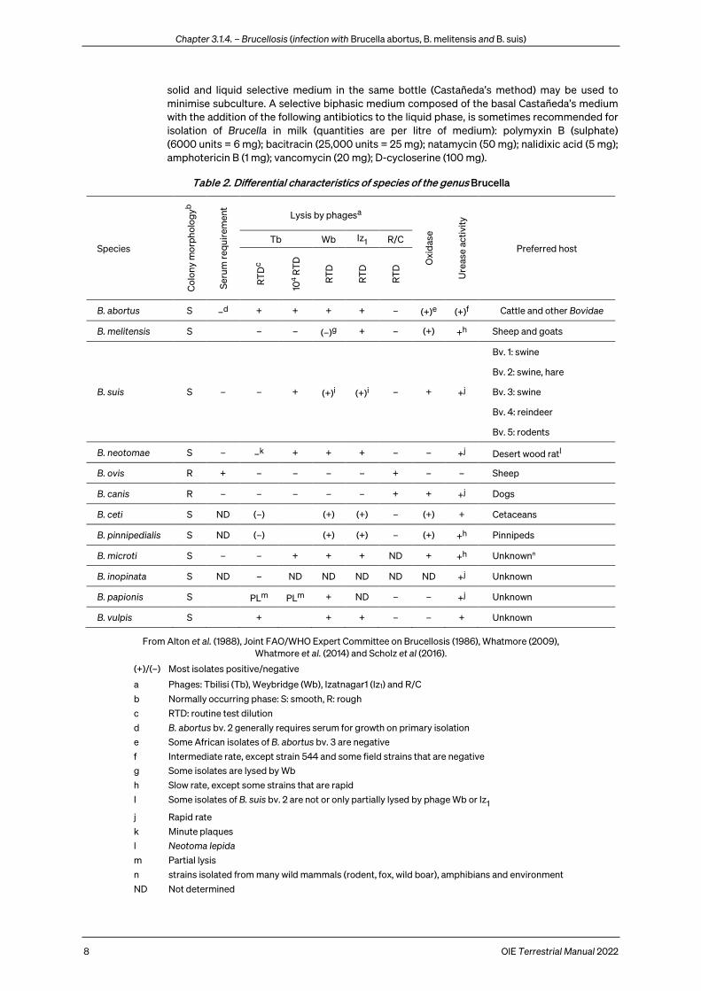
Table 2. Differential characteristics of species of the genus Brucella
Species
Col
ony
mor
pho
log
yb
Ser
um r
equi
rem
ent
Lysis by phagesa
Oxi
das
e
Ure
ase
acti
vity
Preferred host Tb Wb Iz1 R/C
RT
Dc
104 R
TD
RT
D
RT
D
RT
D
B. abortus S –d + + + + – (+)e (+)f Cattle and other Bovidae
B. melitensis S – – (–)g + – (+) +h Sheep and goats
B. suis S – – + (+)i (+)i – + +j
Bv. 1: swine
Bv. 2: swine, hare
Bv. 3: swine
Bv. 4: reindeer
Bv. 5: rodents
B. neotomae S – –k + + + – – +j Desert wood ratl
B. ovis R + – – – – + – – Sheep
B. canis R – – – – – + + +j Dogs
B. ceti S ND (–) (+) (+) – (+) + Cetaceans
B. pinnipedialis S ND (–) (+) (+) – (+) +h Pinnipeds
B. microti S – – + + + ND + +h Unknownn
B. inopinata S ND – ND ND ND ND ND +j Unknown
B. papionis S PLm PLm + ND – – +j Unknown
B. vulpis S + + + – – + Unknown
From Alton et al. (1988), Joint FAO/WHO Expert Committee on Brucellosis (1986), Whatmore (2009), Whatmore et al. (2014) and Scholz et al (2016).
(+)/(–) Most isolates positive/negative
a Phages: Tbilisi (Tb), Weybridge (Wb), Izatnagar1 (Iz1) and R/C
b Normally occurring phase: S: smooth, R: rough
c RTD: routine test dilution
d B. abortus bv. 2 generally requires serum for growth on primary isolation
e Some African isolates of B. abortus bv. 3 are negative
f Intermediate rate, except strain 544 and some field strains that are negative
g Some isolates are lysed by Wb
h Slow rate, except some strains that are rapid
I Some isolates of B. suis bv. 2 are not or only partially lysed by phage Wb or Iz1
j Rapid rate
k Minute plaques
l Neotoma lepida
m Partial lysis
n strains isolated from many wild mammals (rodent, fox, wild boar), amphibians and environment
ND Not determined
Chapter 3.1.4. – Brucellosis (infection with Brucella abortus, B. melitensis and B. suis)
OIE Terrestrial Manual 2022 9
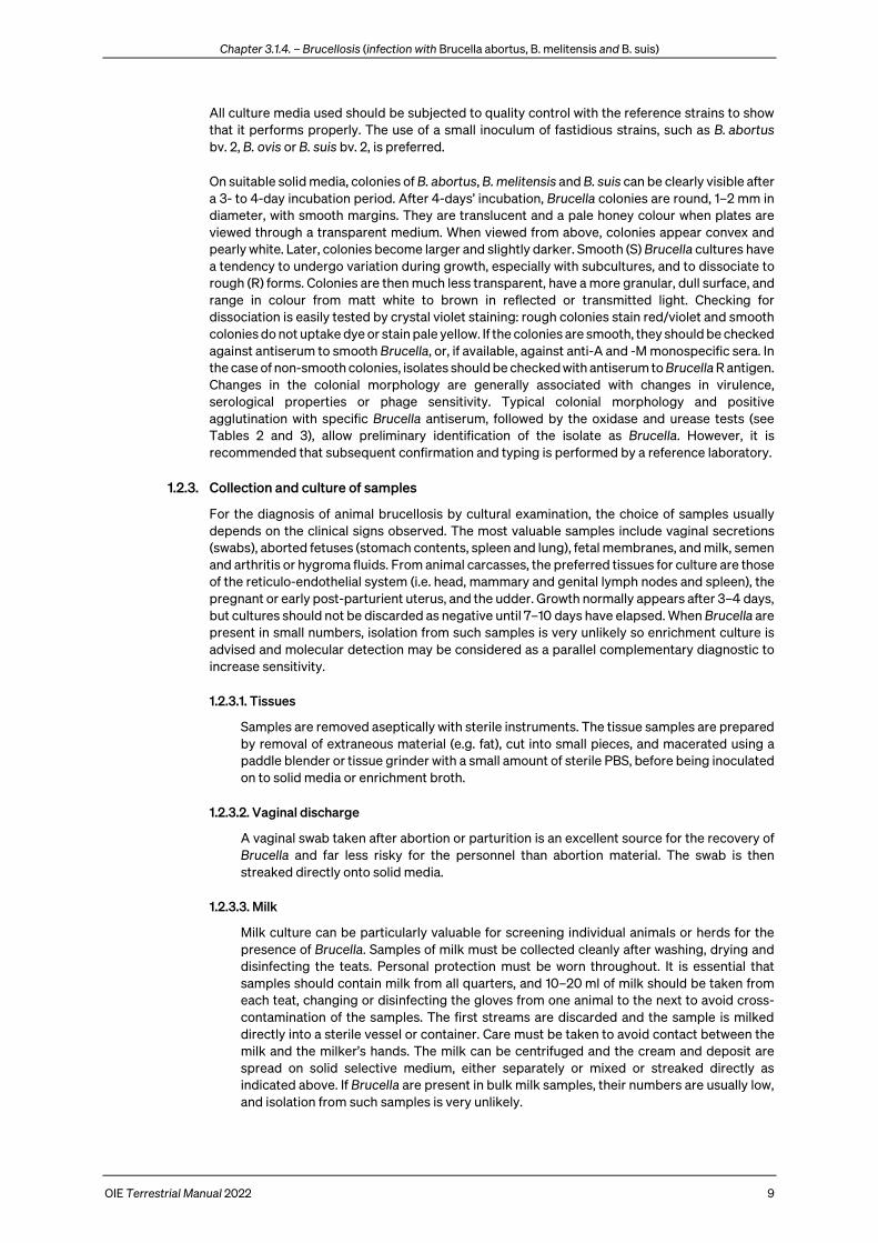
All culture media used should be subjected to quality control with the reference strains to show that it performs properly. The use of a small inoculum of fastidious strains, such as B. abortus bv. 2, B. ovis or B. suis bv. 2, is preferred.
On suitable solid media, colonies of B. abortus, B. melitensis and B. suis can be clearly visible after a 3- to 4-day incubation period. After 4-days’ incubation, Brucella colonies are round, 1–2 mm in diameter, with smooth margins. They are translucent and a pale honey colour when plates are viewed through a transparent medium. When viewed from above, colonies appear convex and pearly white. Later, colonies become larger and slightly darker. Smooth (S) Brucella cultures have a tendency to undergo variation during growth, especially with subcultures, and to dissociate to rough (R) forms. Colonies are then much less transparent, have a more granular, dull surface, and range in colour from matt white to brown in reflected or transmitted light. Checking for dissociation is easily tested by crystal violet staining: rough colonies stain red/violet and smooth colonies do not uptake dye or stain pale yellow. If the colonies are smooth, they should be checked against antiserum to smooth Brucella, or, if available, against anti-A and -M monospecific sera. In the case of non-smooth colonies, isolates should be checked with antiserum to Brucella R antigen. Changes in the colonial morphology are generally associated with changes in virulence, serological properties or phage sensitivity. Typical colonial morphology and positive agglutination with specific Brucella antiserum, followed by the oxidase and urease tests (see Tables 2 and 3), allow preliminary identification of the isolate as Brucella. However, it is recommended that subsequent confirmation and typing is performed by a reference laboratory.
1.2.3. Collection and culture of samples
For the diagnosis of animal brucellosis by cultural examination, the choice of samples usually depends on the clinical signs observed. The most valuable samples include vaginal secretions (swabs), aborted fetuses (stomach contents, spleen and lung), fetal membranes, and milk, semen and arthritis or hygroma fluids. From animal carcasses, the preferred tissues for culture are those of the reticulo-endothelial system (i.e. head, mammary and genital lymph nodes and spleen), the pregnant or early post-parturient uterus, and the udder. Growth normally appears after 3–4 days, but cultures should not be discarded as negative until 7–10 days have elapsed. When Brucella are present in small numbers, isolation from such samples is very unlikely so enrichment culture is advised and molecular detection may be considered as a parallel complementary diagnostic to increase sensitivity.
1.2.3.1. Tissues
Samples are removed aseptically with sterile instruments. The tissue samples are prepared by removal of extraneous material (e.g. fat), cut into small pieces, and macerated using a paddle blender or tissue grinder with a small amount of sterile PBS, before being inoculated on to solid media or enrichment broth.
1.2.3.2. Vaginal discharge
A vaginal swab taken after abortion or parturition is an excellent source for the recovery of Brucella and far less risky for the personnel than abortion material. The swab is then streaked directly onto solid media.
1.2.3.3. Milk
Milk culture can be particularly valuable for screening individual animals or herds for the presence of Brucella. Samples of milk must be collected cleanly after washing, drying and disinfecting the teats. Personal protection must be worn throughout. It is essential that samples should contain milk from all quarters, and 10–20 ml of milk should be taken from each teat, changing or disinfecting the gloves from one animal to the next to avoid cross-contamination of the samples. The first streams are discarded and the sample is milked directly into a sterile vessel or container. Care must be taken to avoid contact between the milk and the milker’s hands. The milk can be centrifuged and the cream and deposit are spread on solid selective medium, either separately or mixed or streaked directly as indicated above. If Brucella are present in bulk milk samples, their numbers are usually low, and isolation from such samples is very unlikely.

Chapter 3.1.4. – Brucellosis (infection with Brucella abortus, B. melitensis and B. suis)
10 OIE Terrestrial Manual 2022
Table 3. Differential characteristics of the biovars of Brucella species
Species
Bio
var
CO
2 re
qui
rem
ent
H2S
pro
duc
tion
Growth on dyesa Agglutination with monospecific sera
Reference strain
Thi
onin
Bas
ic
Fuc
hsin
A M R Strain ATCC NCTC
B. abortus
1 (+)b + – + + – – 544 23448 10093
2 (+)b + – – + – – 86/8/59 23449 10501
3c (+)b + + + + – – Tulya 23450 10502
4 (+)b + – (+) – + – 292 23451 10503
5 – – + + – + – B3196 23452 10504
6c – (–) + + + – – 870 23453 10505
9 +/– + + + – + – C68 23455 10507
B. melitensis
1 – – + + – + – 16M 23456 10094
2 – – + + + – – 63/9 23457 10508
3 – – + + + + – Ether 23458 10509
B. suis
1 – + + (–) + – – 1330 23444 10316
2 – – + – + – – Thomsen 23445 10510
3 – – + + + – – 686 23446 10511
4 – – + (–) + + – 40 23447 11364
5 – – + – – + – 513 ND 11996
B. neotomae – + –d – + – – 5K33 23459 10084
B. ovis + – + (–) – – + 63/290 25840 10512
B. canis – – + (–) – – + RM6/66 23365 10854
B. ceti (–) – (+) (+) + (–) – B1/94
BCCN 94-74 ND 12891
B. pinnipedialis (+) – + (+) (+) (–) – B2/94
BCCN 94-73 ND 12890
B. microti – – + + (–) (+) – CCM4915
BCCN 07-01 CAPM 6434
ND ND
B. inopinata – + + + – +e BO1
BCCN 09-01 CAPM 6436
ND ND
B. papionis – – – – + – – F8/08-60
CIRMBP 0958 ND 13660
B. vulpis – – + + + – F60
BCCN 09-2 DSM 101715
ND ND
From Alton et al. (1988), Joint FAO/WHO Expert Committee on Brucellosis (1986), Whatmore (2009), Whatmore et al. (2014), and Scholz et al. (2016).
(+)/(–) Most isolates positive/negative
a Dye concentration in serum dextrose agar: 20 µg/ml
b Usually positive on primary isolation
c For more certain differentiation of bv. 3 and 6, thionin at 40 µg/ml is used in addition: bv. 3 = +, bv. 6 = –
d Growth at a concentration of 10 µg/ml thionin
e Weak agglutination
ND Not determined

Chapter 3.1.4. – Brucellosis (infection with Brucella abortus, B. melitensis and B. suis)
OIE Terrestrial Manual 2022 11
1.2.3.4. Dairy products
Dairy products, such as cheese, should be cultured on the media described above. As these materials are likely to contain small numbers of organisms, enrichment culture with selective media is advised. Samples need to be carefully homogenised before culture, after they have been ground in a tissue grinder or macerated and pounded in a ‘paddle blender or an electric blender with an appropriate volume (avoiding over-dilution) of sterile PBS. Superficial strata (rind and underlying parts) and the core of the product should be cultured. As brucellae grow, survive or disappear quite rapidly, their distribution throughout the different parts of the product varies according to the local physico-chemical conditions linked to specific process technologies.
1.2.3.5. Arthritis/hygroma fluids – abscesses content
Such samples must be collected aseptically and spread directly on solid selective media.
All the above samples should be cooled (4–10°C) immediately after sampling and transported to the laboratory in the fastest way. Otherwise, the samples should be frozen to avoid viability losses. On arrival at the laboratory, milk and tissue samples and other biological liquids should be frozen if they are not to be cultured immediately.
1.2.3.6. Blood culture
Culture from blood can be attempted although bacteraemia in livestock animals is generally considered short lived or intermittent so the approach is not widely used. Direct culturing of anti-coagulant treated blood (sodium citrate or heparin, except EDTA [ethylene diamine tetra-acetic acid]) may be performed on selective or non-selective agar to obtain results in a shorter time (4–7 days; Alton, 1988). Alternatively an appropriate non-selective media, such as TSB, can be inoculated with blood and subcultured at weekly intervals onto selective Farrell’s media for up to 4 weeks.
1.2.3.7. Animal passage
Although used historically, use of laboratory animals should be avoided unless absolutely necessary, but may sometimes provide the only means of detecting the presence of Brucella, especially when samples have been shown to be heavily contaminated or are likely to contain a low number of Brucella organisms. Animal inoculation may be intravenously or intraperitoneally in mice or intra-muscularly, subcutaneously or intraperitoneally in guinea-pigs. This work must be carried out under appropriate biosafety conditions as outlined in chapter 1.1.4. The spleens of inoculated animals are cultured at 7 days (mice) or 3–6 weeks (guinea-pigs) after inoculation. Serum samples can be collected by intra-cardiac puncture before necropsy from guinea-pigs and subjected to buffered Brucella antigen tests (BBAT); a positive serological result is highly suggestive of brucellosis (Alton et al., 1988).
1.3. Identification and typing
Any colonies showing the characteristic Brucella morphology should be examined using a Gram-stained-smear. As the serological properties, dyes and phage sensitivity are usually altered in the non-smooth phases, attention to the colonial morphology is essential in the typing tests described below. The recommended methods for observing colonial morphology are Henry’s method by obliquely reflected light, the acriflavine test described by Braun & Bonestell, or White & Wilson’s crystal violet method of staining colonies (Alton et al., 1988).
Identification of Brucella organisms to species and biovar level can be carried out by a combination of the following tests: organism morphology after Gram or Stamp’s staining, direct observation of colonial morphology, growth characteristics, urease and oxidase tests, and the slide agglutination test with a polyclonal anti-Brucella serum. Species and biovar identification requires elaborate tests (such as phage lysis and agglutination with anti-A, -M or -R monospecific sera), the performance of which should be left to reference laboratories with accredited expertise in these methods. The simultaneous use of several phages e.g. Tbilisi (Tb), Weybridge (Wb), Izatnagar1 (Iz1) and R/C provides a phage-typing system that, in experienced hands, allows a practical identification of the Brucella species. However, several characteristics, for example added CO2 requirement for growth, production of H2S (detected by lead
Chapter 3.1.4. – Brucellosis (infection with Brucella abortus, B. melitensis and B. suis)
12 OIE Terrestrial Manual 2022
acetate papers), and growth in the presence of basic fuchsin and thionin, are revealed by routine tests that can be performed in moderately equipped non-specialised laboratories (see Tables 2 and 3). While the technique remains useful particularly at the species level, the value of the biovar designations as epidemiological markers is increasingly questioned as, notably in the case of B. melitensis and some B. abortus biovars, molecular evidence has shown biovars do not correspond to meaningful genetic divisions.
MALDI-TOF (matrix assisted laser desorption ionisation time of flight) is increasingly used in diagnostic microbiology and has been applied to identification of Brucella. While application of MALDI-TOF appears effective for genus identification the close genetic relationship among Brucella species, and limitations in commercial database coverage, has not yet enabled robust and unambiguous discrimination of Brucella species.
For the maintenance of B. abortus, B. melitensis or B. suis strains as well as for sending them to a reference laboratory for typing, it is essential that only smooth colonies be selected. Cultures may be maintained for short periods at 5°C ± 3°C, but for longer periods they should be lyophilised or stored in a screw-capped tube at a temperature ≤ –16°C in tryptose broth with 15% (v/v) glycerol. For shipment, cultures should be lyophilised and sealed in ampoules packed in screw-capped canisters or subcultured onto appropriate nutrient agar slopes contained in screw-capped bottles. The strains could also be sent suspended in transport media (e.g. Amies), but this could cause dissociation.
For transporting Brucella cultures, the caps of the bottles or canisters should be screwed tightly down and sealed with PVC (polyvinyl chloride) tapes. Bottles should be wrapped in absorbent paper or cotton wool, sealed in polyethylene bags and packed into a rigid container (triple packaging) in accordance with the requirements of the International Air Transport Association (IATA) for shipping dangerous goods (IATA, 2021). These regulations are summarised in Chapter 1.1.2 Collection, submission and storage of diagnostic specimens and Chapter 1.1.3 Transport of biological materials, and they must be followed.
1.4. Nucleic acid recognition methods
The PCR, including the real-time format, provides an additional means of detection and identification of Brucella sp. (Bricker, 2002; Lopez-Goni et al., 2011; Ocampo-Sosa et al., 2005; Whatmore & Gopaul, 2011). Despite the high degree of DNA homology within the genus Brucella, several historical molecular methods including PCR, PCR restriction fragment length polymorphism (RFLP) and Southern blot, allowed, to a certain extent, the differentiation of Brucella species and some of their biovars (for a review see Bricker, 2002; Moreno et al., 2002; Whatmore & Gopaul, 2011).
The first species-specific multiplex PCR assay for the differentiation of Brucella was described by Bricker & Halling (1994). The assay, named AMOS-PCR, was based on the polymorphism arising from species-specific localisation of the insertion sequence IS711 in the Brucella chromosome, and comprised five oligonucleotide primers that could identify without differentiating B. abortus bv. 1, 2 and 4 but could not identify B. abortus bv. 3, 5, 6, and 9. Modifications to the assay have been introduced over time to improve performance, and additional strain-specific primers were incorporated for identification of the B. abortus vaccine strains, and other biovars and species (Ocampo-Sosa et al., 2005). A multiplex PCR assay (Bruce-ladder) has been proposed for rapid and simple one-step identification of Brucella (Lopez-Goni et al., 2011). The major advantage of this assay over previously described PCRs is that it can identify and differentiate in a single step most Brucella species as well as the vaccine strains B. abortus strain 19 (S19), B. abortus RB51 and B. melitensis Rev.1. In contrast to other PCRs, Bruce-ladder is also able to detect DNA from B. neotomae, B. pinnipedialis and B. ceti. An update to the original Bruce-ladder PCR protocol has been described. This updated version (Bruce-ladder v2.0), that has been validated in several laboratories, is also able to discriminate between B. suis and B. canis, and allows the differentiation of B. microti as does a modification reported by Kang et al. (2011). Similarly, another updated multiplex PCR assay (Suis-ladder), has been developed for fast and accurate identification of B. suis strains at the biovar level (Lopez-Goni et al., 2011).
Alternative approaches allowing identification of all Brucella species, B. suis biovars and vaccine strains based on single nucleotide polymorphism (SNP) discrimination by either primer extension or real-time PCR or the ligase-chain-reaction have been described. These tests are rapid, simple, unambiguous, and based on a robust population genetic analysis that helps ensure the species/biovar specificity of markers used (Whatmore & Gopaul, 2011).
Chapter 3.1.4. – Brucellosis (infection with Brucella abortus, B. melitensis and B. suis)
OIE Terrestrial Manual 2022 13
A number of other methods adding useful epidemiological information have also been described and are widely used. These include multilocus sequencing schemes (Whatmore & Foster, 2021) and several typing schemes based on the use of MLVA (multiple locus variable number of tandem repeats analysis) (Le Fleche et al., 2006; Scholz & Vergnaud, 2013; Whatmore & Foster, 2021). Depending on the particular markers chosen, these methods allow isolates to be identified at species level and provide epidemiological information at the subspecies level. Finally, whole genome sequence-based approaches are rapidly becoming tools for routine surveillance and outbreak detection for several infectious diseases including brucellosis. Through various analytical approaches being developed, including SNP-based approaches or gene-by-gene comparison through core-genome multilocus sequence typing (cgMLST), it is already clear that whole genome sequence approaches will offer the ultimate level of epidemiological precision. These tools will become more widely accessible with time and ultimately greatly inform understanding of local and international brucellosis epidemiology.
1.5. Identification of vaccine strains
Vaccine strains B. abortus S19, B. melitensis Rev.1 and B. abortus RB51 may be identified using specific PCRs (Kang et al., 2011; Lopez-Goňi et al., 2011), or by their growth characteristics in culture.
Brucella abortus S19 has the typical properties of a bv. 1 strain of B. abortus, but does not require CO2, does not grow in the presence of benzyl-penicillin (3 µg/ml = 5 IU/ml), thionin blue (2 µg/ml), or i-erythritol (1 mg/ml) (all final concentrations), and presents a high L-glutamate use (Alton et al., 1988).
Brucella melitensis strain Rev.1 has the typical properties of a bv. 1 strain of B. melitensis, but develops smaller colonies on solid media, does not grow in the presence of basic fuchsin, thionin (both at 20 µg/ml) or benzyl-penicillin (3 µg/ml), but does grow in the presence of streptomycin at 2.5 or 5 µg/ml (5 IU/ml) (Alton et al., 1988).
Brucella abortus strain RB51 can be distinguished from its B. abortus biovar 1 smooth counterparts by its rough morphology and growth in presence of rifampicin (250 µg per ml of media).
2. Serological tests
No single serological test is appropriate in all epidemiological situations and all animal species; all tests have limitations especially when testing individual animals. Consideration should be given to all factors that impact on the relevance of the test method and test results to a specific diagnostic interpretation or application. In epidemiological units where vaccination with smooth Brucella is practised, and depending on the vaccination method (dose/route) used, positive serological reactions may be expected among the vaccinated animals because of antibodies cross-reacting with wild strain infection. Moreover, a number of bacteria, in particular Yersinia enterocolitica O:9, may induce antibody responses that cause false positive serological reactions (FPSR) in brucellosis tests, impeding accurate serological diagnosis. FPSR may occur in all animal species at variable rates according to the time and the region.
The serum agglutination test (SAT) is generally regarded as being unsatisfactory for the purposes of international trade. The complement fixation test (CFT) is more specific than the SAT, and has also a standardised system of unitage, but can be impacted by anti-complementary activity. The diagnostic performance characteristics of some enzyme-linked immunosorbent assays (ELISAs) and the fluorescence polarisation assay (FPA) are comparable with or better than that of the CFT, and as they are technically simpler to perform and more robust, their use may be preferred. The diagnostic performance of these tests has been compared in cattle, small ruminants and swine.
For the control of brucellosis at the national or local level, BBATs (the rose bengal test [RBT] and the buffered plate agglutination test [BPAT]), ELISA and FPA, are considered as suitable screening tests. Depending on the purpose of testing, positive reactors could be retested using a suitable confirmatory or complementary method.
In other species, for example, buffaloes (Bubalus bubalis), American and European bison (Bison bison, Bison bonasus), yak (Bos grunniens), elk/wapiti (Cervus elaphus), camels (Camelus bactrianus and C. dromedarius), and South American camelids, Brucella sp. infection follows a course similar to that in cattle. The same serological procedures may be used for these animals, but each test should be validated for its fitness in the corresponding animal species.
Chapter 3.1.4. – Brucellosis (infection with Brucella abortus, B. melitensis and B. suis)
14 OIE Terrestrial Manual 2022
2.1. Reference sera
OIE reference standards are those against which all other standards are compared and standardised. These reference standards are all available to national reference laboratories and should be used to establish secondary or national standards against which working standards can be prepared and used in the diagnostic laboratory for daily routine use.
These sera have been developed and designated by OIE as International Standard Sera2. The use of these reagents promotes international harmonisation of diagnostic testing and antigen standardisation:
i) For RBT, CFT, SAT and milk ring test (MRT), OIE International Standard Serum (OIEISS, previously named the WHO Second International standard anti-Brucella abortus Serum; WHO, 1953) is used. This serum is of bovine origin and contains 1000 IU (SAT) and 1000 ICFTU (international complement fixation test units).
ii) For indirect ELISA (I-ELISA), competitive or blocking ELISA (C-ELISA) and FPA in cattle, three OIE ELISA Standard Sera are available for use. These are also of bovine origin and consist of a strong positive (OIEELISASPSS), a weak positive (OIEELISAWPSS) and a negative (OIEELISANSS) standard.
iii) For I-ELISA, C-ELISA and FPA in sheep and goats, the International standard anti-Brucella melitensis Serum (ISaBmS) is used (McGiven et al., 2011).
iv) For I-ELISA, C-ELISA and FPA in pigs, there is no OIE International Standard serum available at present. However, an EU standard for porcine I-ELISA and C-ELISA, EUPigBSS, is available3.
2.2. Production of antigens
Brucella abortus strain 99 (Weybridge) (S99)3 or B. abortus strain 1119-3 (USDA) (S1119-3)4 should always be used for the production of antigens for the BBATs, SAT, CFT and FPA. These B. abortus strains can be also used as a source of soluble antigen extracts (smooth lipopolysaccharide [S-LPS] or O-polysaccharide [OPS]) for the ELISAs or the Native Hapten tests, but B. melitensis strain 16M is also suitable for such a purpose. It should be emphasised that antigen made with any of the two B. abortus or B. melitensis 16M strains is used to test for any infections due to smooth Brucella species.
The strains must be completely smooth and should not auto-agglutinate in saline and 0.1% (w/v) acriflavine. They must be pure cultures and conform to the characteristics of CO2-independent strains of B. abortus bv. 1 or B. melitensis bv. 1. The original seed cultures should be propagated to produce a seed lot that must conform to the properties of these strains, and should be preserved by lyophilisation or by freezing in liquid nitrogen.
For antigen production, the seed culture is used to inoculate a number of potato-infusion agar slopes that are then incubated at 37°C ± 2°C for 48 hours. SDA and TSA, to which 5% equine or new-born calf serum or 0.1% yeast extract may be added, are satisfactory solid media provided a suitable seed is used as recommended above. The growth is checked for purity, resuspended in sterile PBS, pH 6.4, and used to seed layers of potato-infusion agar or glycerol-dextrose agar in Roux flasks. These are then incubated at 37°C ± 2°C for 72 hours with the inoculated surface facing down. Each flask is checked for purity by Gram staining samples of the growth, and the organisms are harvested by adding 50–60 ml phenol saline (0.5% phenol in 0.85% sodium chloride solution) to each flask. The flasks are gently agitated, the suspension is decanted, and the organisms are killed by heating at 80°C for 90 minutes. Following a viability check, the antigen is stored at 5°C ± 3°C.
Alternatively, the cells may be produced by batch or continuous culture in a fermenter, using a liquid medium containing (per litre of purified water) D-glucose (30 g), a high-grade peptone (30 g), yeast extract (Difco) (10 g), sodium dihydrogen phosphate (9 g) and disodium hydrogen phosphate (3.3 g). The initial pH is 6.6 (± 0.2), but this tends to rise to pH 7.2 (± 0.2) during the growth cycle. Care should be taken to check batches of peptone and yeast extract for capacity to produce good growth without formation of abnormal or dissociated cells. Vigorous aeration and stirring is required during growth, and adjustment to pH 7.2 (± 0.2) by the addition of sterile 0.1 M HCl may be necessary. The seed inoculum is prepared as
2 Obtainable from the OIE Reference Laboratory for brucellosis in the United Kingdom (see online list of OIE Reference
Laboratories: https://www.woah.org/en/what-we-offer/expertise-network/reference-laboratories/#ui-id-3). 3 Obtainable from the OIE Reference Laboratory for brucellosis in France. 4 Obtainable from the United States Department of Agriculture (USDA), National Veterinary Services Laboratories (NVSL)
1800 Dayton Road, Ames, Iowa, United States of America.
Chapter 3.1.4. – Brucellosis (infection with Brucella abortus, B. melitensis and B. suis)
OIE Terrestrial Manual 2022 15
described above. The culture is incubated at 37°C ± 2°C for 48 hours. Continuous culture runs can be operated for much longer periods, but more skill is required to maintain them. In-process checks should be made on the growth from either solid or liquid medium to ensure purity, an adequate viable count and freedom from dissociation to rough forms. Cells for use in the preparation of all antigens should be checked for purity and smoothness at the harvesting stage.
The culture is harvested by centrifugation to deposit the organisms, which are resuspended in phenol saline. The organisms are killed by heating at 80°C for 90 minutes and are stored at 5°C ± 3°C. They must form stable suspensions in physiological saline solutions and show no evidence of auto-agglutination. A viability check must be performed on the suspensions and no growth must be evident after 10 days of incubation at 37°C ± 2°C. The packed cell volume (PCV) of the killed suspensions can be determined by centrifuging 1 ml volumes in Wintrobe tubes at 3000 g for 75 minutes.
2.3. Buffered Brucella antigen tests (BBAT)
2.3.1. Rose bengal test
This test is a simple spot agglutination test using antigen stained with rose bengal and buffered to a low pH, 3.65 ± 0.05 (Morgan et al., 1969).
2.3.1.1. Antigen production
Antigen for the RBT is prepared by depositing killed B. abortus S99 or S1119-3 cells by centrifugation at 23,000 g for 10 minutes (or 14,000 g for 40 minutes) at 5°C ± 3°C, and uniformly resuspending in sterile phenol saline (0.5%) at the rate of 1 g to 22.5 ml. (Note: if sodium carboxymethyl cellulose is used as the sedimenting agent during preparation of the cell concentrate, insoluble residues must be removed by filtering the suspension through an AMF-CUNO Zeta-plus prefilter [Type CPR 01A] before staining.) In case of production of cells in liquid media (e.g. in bioreactor), it is possible to concentrate the bacterial suspension (up to 40 times) before centrifugation, using a tangential flow filtration system (TFFS) with 0.1 µm cassette and doing three washes (1:3 ratio) with sterile phenol saline (0.5%). To every 35 ml of this suspension, 1 ml of 1% (w/v) rose bengal (Cl No. 45440) in sterile purified water is added, and the mixture is stirred for 2 hours at room temperature. The mixture is filtered through sterile cotton wool, and centrifuged at 10,000 g to deposit the stained cells, which are then uniformly resuspended at the rate of 1 g cells to 7 ml of diluent (21.1 g of NaOH dissolved in 353 ml of sterile phenol saline, followed by 95 ml of lactic acid, and adjusted to 1056 ml with sterile phenol saline). The colour of this suspension should be an intense pink and the supernatant of a centrifuged sample should be free of stain; the pH should be 3.65 ± 0.05. After filtration through cotton wool, the suspension is filtered twice through a Sartorius No. 13430 glass fibre prefilter, adjusted to a PCV of approximately 8%, pending final standardisation against serum standardised against the OIEISS, and stored at 5°C ± 3°C in the dark. The antigen should be stored as recommended by the manufacturer. It should not be frozen.
2.3.1.2. Antigen standardisation
When used in the standard test procedure, the RBT antigen should give a clearly positive reaction with 1/45 dilution, but not 1/55 dilution, of the OIEISS diluted in 0.5% phenol saline or normal saline.
Additional checks may be performed with the ISaBmS. The highest dilution (in negative goat serum) of this standard that must give a positive result and the lowest dilution (in negative goat serum) that must simultaneously give a negative result have been established at 1/16 and 1/200, respectively (McGiven et al., 2011).
It is also be advisable to compare the reactivity of new and previously standardised batches of antigen using a panel of well-defined reference sera.
However the above standardisation against the OIEISS is probably a cause of the reduced sensitivity of some RB antigen batches for diagnosing B. melitensis infection in small ruminants and of the discrepancies with the CFT (Blasco et al., 1994a). When testing small ruminants, the discrepancies with the CFT can be minimised by using three volumes of serum and one volume of antigen (e.g. 75 μl and 25 μl, respectively) in place of an equal
Chapter 3.1.4. – Brucellosis (infection with Brucella abortus, B. melitensis and B. suis)
16 OIE Terrestrial Manual 2022
volume of each as mentioned in the standard test procedure. However, this modification of the RBT should not be recommended for testing cattle and pig sera.
2.3.1.3. Test procedure
i) Bring the serum samples and antigen to room temperature (22°C ± 4°C); only sufficient antigen for the day’s tests should be removed from the refrigerator.
ii) Place 25–30 µl of each serum sample on a white tile, enamel or plastic plate, or in a WHO haemagglutination plate.
iii) Shake the antigen bottle well, but gently, and place an equal volume of antigen near each serum spot.
iv) Immediately after the last drop of antigen has been added to the plate, mix the serum and antigen thoroughly (using a clean glass or plastic rod for each test) to produce a circular or oval zone approximately 2 cm in diameter.
v) The mixture is agitated gently for 4 minutes at room temperature (22°C ± 4°C) on a rocker or three-directional agitator (if the reaction zone is oval or round, respectively).
vi) Read for agglutination immediately after the 4-minute period is completed. Any visible coloured agglutination is considered to be a positive reaction. A control serum that gives a minimum positive reaction should be tested before each day’s tests are begun to verify the sensitivity of test conditions.
The RBT is very sensitive. However, like all other serological tests, it could sometimes give a positive result in cattle because of B. abortus S19 vaccination or FPSR. The same phenomenon occurs in small ruminants or pigs affected by FPSR and in small ruminants vaccinated with B. melitensis Rev.1. Therefore positive reactions should be investigated using suitable confirmatory or complementary strategies (including epidemiological investigation). Conversely, false-negative reactions occur rarely. Nevertheless RBT appears to be adequate as a screening test for detecting infected herds or to guarantee the absence of infection in brucellosis-free herds or flocks.
2.3.2. Buffered plate agglutination test
2.3.2.1. Antigen production
Antigen for the BPAT is prepared from B. abortus S1119-3 according to the procedure described by Angus & Barton (1984).
Two staining solutions are required: brilliant green (2 g/100 ml) and crystal violet (1 g/100 ml) both certified stains dissolved in purified water. Once prepared, the two solutions should be stored separately for a period of 24 hours, and then mixed together in equal volumes in a dark bottle and stored in a refrigerator for a period of not less than 6 months before use. The mixed stain may only be used between 6 and 12 months after initial preparation.
Buffered diluent is prepared by slowly dissolving sodium hydroxide (150 g) in 3–4 litres of sterile phenol saline. Lactic acid (675 ml) is added to this solution, and the final volume is adjusted to 6 litres by adding sterile phenol saline. The pH of the solution should be 3.65 ± 0.05.
Brucella abortus S1119-3 packed cells are diluted to a concentration of 250 g/litre in phenol saline; 6 ml of stain is added per litre of cell suspension, and the mixture is shaken thoroughly before being filtered through sterile absorbent cotton. The cells are centrifuged at 10,000 g at 5°C ± 3°C, and the packed cells are then resuspended at a concentration of 50 g/100 ml in buffered diluent (as described above). This mixture is shaken thoroughly for 2 hours, and is then further diluted by the addition of 300 ml of buffered diluent per 100 ml of suspended cells (i.e. final concentration of 50 g packed cells/400 ml buffered diluent). The mixture is stirred at room temperature for 20–24 hours before the cell concentration is adjusted to 11% (w/v) in buffered diluent. This suspension is stirred overnight before testing. Pending final quality control tests, the antigen is stored at 5°C± 3°C until required for use. The antigen should not be frozen.
Chapter 3.1.4. – Brucellosis (infection with Brucella abortus, B. melitensis and B. suis)
OIE Terrestrial Manual 2022 17
The pH of the buffered plate antigen should be 3.70 ± 0.03 and the pH of a serum–antigen mixture at a ratio of 8:3 should be 4.02 ± 0.04. The 11% stained-cell suspension should appear blue–green. Each batch of buffered plate antigen should be checked by testing at least 10 weakly reactive sera and comparing the results with one or more previous batches of antigen. If possible, the antigen batches should be compared with the standard antigen prepared by the NVSL, USDA (see footnote 5 for address). There is, however, no international standardisation procedure established for use with either the OIEISS or with the ISaBmS.
2.3.2.2. Test procedure
i) Bring the serum samples and antigen to room temperature (22°C ± 4°C); only sufficient antigen for the day’s tests should be removed from the refrigerator.
ii) Shake the sample well. Place 80 µl of each serum sample on a glass plate marked in 4 × 4 cm squares
iii) Shake the antigen bottle well, but gently, and place 30 µl of antigen near each serum spot.
iv) Immediately after the last drop of antigen has been added to the plate, mix the serum and antigen.
v) Thoroughly (using a clean glass or plastic rod for each test) to produce a circular zone approximately 3 cm in diameter.
vi) After the initial mixing, the plate should be rotated three times in a tilting motion to ensure even dispersion of the reagents, and then incubated for 4 minutes in a humid chamber at ambient temperature.
vii) The plate should be removed and rotated as above, and then returned for a second 4-minute incubation.
viii) Read for agglutination immediately after the 8-minute period is completed. Any visible reaction is considered to be positive. A control serum that gives a minimum positive reaction should be tested before each day’s tests are begun to verify the sensitivity of test conditions.
Like the RBT, the test is very sensitive in cattle, especially for detection of vaccine-induced antibody, and positive samples should be retested using a confirmatory or complementary test(s). False-negative reactions may occur, usually due to prozoning, which may be overcome by diluting the serum or retesting after a given time. While the BPAT has been extensively used with apparent good results in small ruminants and pigs in some countries, its diagnostic value in these species has not been reported at international level.
2.4. Complement fixation test
The CFT is widely used but it is complex to perform, and requires good laboratory facilities and adequately trained staff to accurately titrate and maintain the reagents. There are numerous variations of the CFT in use, but this test is most conveniently carried out in a microtitre format. Warm or cold fixation may be used for the incubation of serum, antigen and complement: either 37°C ± 2°C for 30 minutes or 5°C ± 3°C for 14–18 hours. A number of factors affect the choice of the method: anti-complementary activity in serum samples of poor quality is more evident with cold fixation, while fixation at 37°C ± 2°C increases the frequency and intensity of prozones, and a number of dilutions must be tested for each sample.
Several methods have been proposed for the CFT using different concentrations of fresh or preserved sheep red blood cells (SRBCs) (a 2%, 2.5% or 3% suspension is usually recommended) sensitised with an equal volume of rabbit anti-SRBC serum diluted to contain several times (usually from two to five times) the minimum concentration required to produce 100% lysis of SRBCs in the presence of a titrated solution of guinea-pig complement. The latter is independently titrated (in the presence or absence of antigen according to the method) to determine the amount of complement required to produce either 50% or 100% lysis of sensitised SRBCs in a unit volume of a standardised suspension; these are defined as the 50% or 100% haemolytic unit of complement/minimum haemolytic dose (C’H or MHD50 or C’H or MHD100), respectively. It is generally recommended to titrate the complement before each set of tests, a
Chapter 3.1.4. – Brucellosis (infection with Brucella abortus, B. melitensis and B. suis)
18 OIE Terrestrial Manual 2022
macromethod being preferred for an optimal determination of C’H50. Usually, 1.25–2 C’H100 or 5–6 C’H50 are used in the test.
Barbital (veronal) buffered saline is the standard diluent for the CFT. This is prepared from tablets available commercially; otherwise it may be prepared from a stock solution of sodium chloride (42.5 g), barbituric acid (2.875 g), sodium diethyl barbiturate (1.875 g), magnesium sulphate (1.018 g), and calcium chloride (0.147 g) in 1 litre of purified water, diluted by the addition of four volumes of 0.04% gelatine solution before use. However, this buffer contains barbituric derivatives that are no longer available in several countries. Satisfactory results may be also obtained with a barbituric-free solution of sodium chloride 0.85% containing calcium and magnesium, prepared by adding 1 ml of a stock solution of 1 M magnesium chloride and 0.3 M calcium chloride (anhydrous MgCl2: 9.5 g CaCl2: 3.7 g; purified water: up to 100 ml) (stored in small amounts at 5°C ± 3°C) to 1 litre of saline solution (Alton et al., 1988). The pH is critical and must be strictly adjusted to 7.35 (± 0.05). The replacement of the veronal buffer by this barbituric-free buffer has been validated in the OIE Brucellosis Reference Laboratory in France5.
2.4.1. Antigen production
Numerous variations of the test exist but, whichever procedure is selected, the test must use an antigen that has been prepared from an approved smooth strain of B. abortus, such as S99 or S1119-3, and standardised against the OIEISS. Antigen for the CFT can be prepared following specialised procedures (Alton et al., 1988) or a whole cell antigen can be used after diluting the stock suspension such that the PCV of the concentrated antigen suspension for CFT is approximately 2% before standardisation against the OIEISS.
2.4.2. Antigen standardisation
The antigen should be standardised to give 50% fixation at a dilution of 1/200 of the OIEISS and must also show complete fixation at the lower serum dilutions, because too weak (or too strong) a concentration of antigen may not produce 100% fixation at the lower dilutions of serum. When two dilutions of antigen are suitable, the more concentrated antigen suspension must be chosen in order to avoid prozone occurrence. The appearance of the antigen, when diluted 1/10 must be that of a uniform, dense, white suspension with no visible aggregation or deposit after incubation at 37°C ± 2°C for 18 hours. It must not produce anti-complementary effects at the working strength for the test. The antigen is stored at 5°C ± 3°C and should not be frozen.
2.4.3. Test procedure (example)
The undiluted test sera and appropriate working standards should be inactivated for 30 minutes in a water bath at 60°C ± 2°C. If previously diluted with an equal volume of veronal buffered saline, these sera could be inactivated at 58°C ± 2°C for 50 minutes. Usually, only one serum dilution is tested routinely (generally 1/4 or 1/5 depending on the CF procedure chosen), but serial dilutions are recommended for trade purposes and when clinical signs have been reported in order to detect prozone.
Using standard 96-well microtitre plates with round (U) bottoms, the technique is usually performed as follows:
i) Volumes of 25 µl of diluted inactivated test serum are placed in the well of the first, second and third rows. The first row is an anti-complementary control for each serum. Volumes of 25 µl of CFT buffer are added to the wells of the first row (anti-complementary controls) to compensate for lack of antigen. Volumes of 25 µl of CFT buffer are added to all other wells except those of the second row. Serial doubling dilutions are then made by transferring 25 µl volumes of serum from the third row onwards; 25 µl of the resulting mixture in the last row are discarded.
ii) Volumes of 25 µl of antigen, diluted to working strength, are added to each well except in the first row.
iii) Volumes of 25 µl of complement, diluted to the number of units required, are added to each well.
5 Obtainable from the OIE Reference Laboratory for brucellosis in France.
Chapter 3.1.4. – Brucellosis (infection with Brucella abortus, B. melitensis and B. suis)
OIE Terrestrial Manual 2022 19
iv) Control wells containing:
a) diluent only,
b) complement + diluent,
c) antigen + complement + diluent,
are set up to contain 75 µl total volume in each case.
A control serum that gives a minimum positive reaction should be tested in each set of tests to verify the sensitivity of test conditions.
v) The plates are incubated at 37°C ± 2°C for 30 minutes or at 5°C ± 3°C overnight, and a volume (25 or 50 µl according to the technique) of sensitised SRBCs is added to each well. The plates are re-incubated at 37°C ± 2°C for 30 minutes.
vi) The results are read after the plates have been centrifuged at 1000 g for 10 minutes at 5°C ± 3°C or left to stand at 5°C ± 3°C for 2–3 hours to allow unlysed cells to settle. The degree of haemolysis is compared with standards corresponding to 0, 25, 50, 75 and 100% lysis. The absence of anti-complementary activity is checked for each serum in the first row.
vii) Standardisation of results of the CFT
A unit system that is based on the OIEISS exists for the standardisation of results. This serum contains 1000 ICFTU per ml. If this serum is tested in a given method and gives a titre of, for example 200 (50% haemolysis), then the factor for an unknown serum tested by that method can be found from the formula: 1000 × 1/200 × titre of test serum = number of ICFTU of antibody in the test serum per ml. The OIEISS contains specific IgG; national standard sera should also depend on this isotype for their specific complement-fixing activity. Difficulties in standardisation arise because different techniques selectively favour CF by different immunoglobulin isotypes. It is recommended that any country using the CFT on a national scale should obtain agreement among the different laboratories performing the test to use the same method in order to obtain the same level of sensitivity. To facilitate comparison between countries, results should always be expressed in ICFTUs, calculated in relation to those obtained in a parallel titration with a standard serum, which in turn may be standardised against the OIEISS.
viii) Interpretation of the results: Sera giving a titre equivalent to 20 ICFTU/ml or more are considered to be positive.
Animals that have been vaccinated with B. abortus S19 or B. melitensis Rev.1 between 3 and 6 months are usually considered to be infected if the sera give positive fixation at a titre of 30 or greater ICFTU/ml when the animals are tested at an age of 18 months or older.
This procedure is an example, other volumes and quantities of reagents could be chosen provided that the test is standardised against the OIEISS as described above and the results expressed in ICFTU/ml.
The CFT is usually very specific but less sensitive than RBT and ELISA, particularly in the case of swine, as swine complement interacts with guinea-pig complement to produce a pro-complementary activity that reduces the sensitivity. Thus, the CFT has a reduced sensitivity for diagnosing B. suis infection, is not capable of eliminating the FPSR problem, and can be recommended only as a complementary test in swine. Moreover, like most serological tests, the CFT can be positive in ruminants after B. abortus S19 or B. melitensis Rev.1 vaccination and it is not specific enough in presence of FPSR. Therefore, CFT results should be investigated using suitable confirmatory or complementary strategies.

Chapter 3.1.4. – Brucellosis (infection with Brucella abortus, B. melitensis and B. suis)
20 OIE Terrestrial Manual 2022
2.5. Enzyme-linked immunosorbent assays
2.5.1. Indirect ELISA
Numerous variations of the I-ELISA have been described for cattle, small ruminants, camelids and pigs employing different antigen preparations, antiglobulin-enzyme conjugates, and substrate/ chromogens. Brucella abortus strain 99 (Weybridge) (S99)3 or B. abortus strain 1119-3 (USDA) (S1119-3)4 should be used for production of these antigens, but B. melitensis strain 16M can also be suitable for such a purpose. Several commercial I-ELISAs using whole cell, S-LPS or the OPS as antigens that have been validated in extensive field trials are available and are in wide use. Nevertheless, the technique used and the interpretation of results must have been validated in accordance with the principles laid down in Chapter 1.1.6 Principles and methods of validation of diagnostic assays for infectious diseases. The OPS antibody epitopes from these strains have also been exactly replicated through chemical synthesis and successfully used as antigens in ELISA, but these assays await full validation.
2.5.1.1. Test standardisation of the I-ELISA (EU, 2008; McGiven et al., 2011)
2.5.1.1.1. Infection with Brucella in cattle
i) A 1/2 pre-dilution of the OIEELISAWPSS or a 1/16 pre-dilution of the OIEELISASPSS made up in a negative bovine serum (or in a negative pool of bovine sera) must give a positive reaction;
and
ii) A 1/8 pre-dilution of the OIEELISAWPSS or a 1/64 pre-dilution of the OIEELISASPSS made up in a negative bovine serum (or in a negative pool of bovine sera) must give a negative reaction;
and
iii) The OIEELISANSS must always give a negative reaction.
2.5.1.1.2. Infection with Brucella in sheep and goats:
i) A 1/64 pre-dilution of the ISaBmS made up in a negative goat serum (or in a negative pool of goat sera) must give a positive reaction;
and
i) A 1/750 pre-dilution of the ISaBmS made up in a negative goat serum (or in a negative pool of goat sera) must give a negative reaction;
and
i) The above-mentioned negative goat serum (or negative pool of goat sera) must always give a negative reaction.
2.5.1.1.3. Infection with Brucella in pigs and camelids:
i) In the absence of appropriate international standard sera the test should be duly validated and the cut-off established in the test population with appropriate validation techniques (see chapter 1.1.6).
The I-ELISAs that use S-LPS or OPS as antigens are highly sensitive for the detection of anti-Brucella antibodies in cattle, small ruminants and pigs, but are not capable of fully resolving the problem of differentiating between antibodies resulting from B. abortus S19 and B. melitensis Rev.1 vaccination. The B. abortus RB51 vaccine may also interfere in S-LPS-based I-ELISAs.
Positive reactions should be investigated using suitable confirmatory or complementary strategies as for BBAT and CFT.
Using I-ELISA standardised against OIE standard sera described above, the diagnostic sensitivity should be equal to or greater than that of the BBATs (RBT/BPAT) or the CFT in the testing of

Chapter 3.1.4. – Brucellosis (infection with Brucella abortus, B. melitensis and B. suis)
OIE Terrestrial Manual 2022 21
infected cattle, small ruminants and pigs. However, the specificity would usually be lower (EFSA, 2006; 2009; Greiner et al., 2009; Gusi et al., 2019).
The problem of FPSR may be reduced but not fully resolved, in pigs in particular, by performing I-ELISAs using extracts from rough strains of Brucella. Most FPSR are a result of cross reaction with the OPS portion of the S-LPS molecule; cross-reaction among core epitopes is less frequent but does exist. The use of chaotropic I-ELISAs or procedures using heterologous extracts or Brucella cytosolic proteins as antigens does not resolve this problem, at least in cattle (Munoz et al., 2005). Moreover, in the context of FPSR, the most specific diagnostic procedure remains the brucellin skin test (see Section B.3.1 below).
Monoclonal, polyclonal antiglobulin or protein G or AG enzyme conjugates may be used depending on availability and performance requirements. A monoclonal antibody (MAb) specific for the heavy chain of bovine IgG1 may provide some improvement in specificity at the possible cost of some loss of sensitivity while a protein G or AG enzyme conjugate may provide a reagent useful for testing a variety of mammalian species.
Several commercial I-ELISAs are available. Some protocols are less sensitive or less specific than others; therefore results obtained from different assays are not always comparable. I-ELISA for diagnosing anti-Brucella antibodies in small ruminants and pigs is essentially the same as that described for cattle, but the cut-off should have been properly established for these species using the appropriate validation techniques (see chapter 1.1.6), and, moreover, I-ELISA for sheep and goats should be standardised against the ISaBmS (McGiven et al., 2011).
Whatever the I-ELISA format used:
i) A positive and a negative control are included in each plate. OD (optical density) ranges to be obtained with these two controls must be established to define the criteria for validating each plate results. The OD of the positive control is the one with which the OD of each test serum is compared to establish the final result (negative or positive).
ii) An additional positive serum (internal control) must be included in each plate to validate the repeatability of the test from plate to plate and from day to day.
2.5.2. Competitive ELISA
Several variations of the C-ELISA, using S-LPS or OPS as antigens, have been described for cattle, small ruminants, camelids and pigs employing different antiglobulin-enzyme conjugates, substrate or chromogens and antigens prepared from different smooth Brucella strains. Nevertheless, the technique used and the interpretation of results must have been validated in accordance with the principles laid down in chapter 1.1.6.
2.5.2.1. Test standardisation of the C-ELISA (EU, 2008; McGiven et al., 2011)
2.5.2.1.1. Infection with Brucella in cattle:
i) A 1/2 pre-dilution of the ELISAWPSS or a 1/16 pre-dilution of the OIEELISASPSS made up in a negative bovine serum (or in a negative pool of bovine sera) must give a positive reaction;
and
ii) A 1/8 pre-dilution of the OIEELISAWPSS or a 1/64 pre-dilution of the OIEELISASPSS made up in a negative bovine serum (or in a negative pool of bovine sera) must give a negative reaction;
and
iii) The OIEELISANSS must always give a negative reaction.
2.5.2.1.2. Infection with Brucella in sheep and goats:
i) A 1/8 pre-dilution of the ISaBmS made up in a negative goat serum (or in a negative pool of goat sera) must give a positive reaction;
Chapter 3.1.4. – Brucellosis (infection with Brucella abortus, B. melitensis and B. suis)
22 OIE Terrestrial Manual 2022
and
ii) a 1/300 pre-dilution of the ISaBmS made up in a negative goat serum (or in a negative pool of goat sera) must give a negative reaction;
and
iii) the above-mentioned negative goat serum (or negative pool of goat sera) must always give a negative reaction.
2.5.2.1.3. Infection with Brucella in pigs:
i) In the absence of an international standard serum for porcine brucellosis, the test should be duly validated and the cut-off established in the test population with appropriate validation techniques (see chapter 1.1.6).
Several commercial C-ELISAs are available. Some protocols are less sensitive or less specific than others, therefore results obtained from different assays are not always comparable. The cut-off should have been properly established using the appropriate validation techniques (see chapter 1.1.6).
The C-ELISA using an MAb specific for one of the epitopes of the Brucella sp. OPS has been shown in cattle, sheep and swine to have usually, but not always, higher specificity but lower sensitivity than the BBAT or I-ELISA (Munoz et al., 2005; 2012; Nielsen et al., 1995; Praud et al., 2012; Stack et al., 1999).
The C-ELISA may reduce but not fully eliminate the reactions caused by antibodies produced in response to vaccination. It is highly probable that the improved specificity is due to a reduction in sensitivity of the C-ELISA compared with BBAT and I-ELISA. Therefore, the results from C-ELISA reactions should not be considered in isolation but alongside suitable confirmatory or complementary strategies as for RBT, BBAT, CFT and I-ELISA.
The choice of MAb and its more or less high and unique specificity and affinity will have a distinct influence on the diagnostic performance characteristics of the assay. As with any MAb-based assays, the universal availability of the MAb or the hybridoma must also be considered with respect to international acceptance and widespread use.
Whatever the C-ELISA format used:
i) A positive and a negative control are included in each plate. OD ranges to be obtained with these two controls must be established to define the criteria for validating each plate results. The OD of the positive control is the one with which the OD of each test serum is compared to establish the final result (negative or positive).
ii) An additional positive serum (internal control) must be included in each plate to validate the repeatability of the test from plate to plate and from day to day.
2.6. Fluorescence polarisation assay
The FPA is a simple technique for measuring antigen/antibody interaction. It is a homogeneous assay in which analytes are not separated and it is therefore very rapid. However, unlike another homogeneous method (e.g. RBT) a blank/background read is required for each sample before adding the antigen. Thus it is a two-step assay.
For the diagnosis of brucellosis, a small molecular weight fragment (average 22 kD) of the OPS of B. abortus strain B. abortus 1119-3 S-LPS is labelled with fluorescein isothiocyanate (FITC) and used as the antigen. Once the blank/background read is performed (2–3 minutes), this antigen is added to diluted serum and a measure of the antibody content is obtained in about 2 minutes after the addition of antigen using a fluorescence polarisation analyser (FPM) (Nielsen et al., 1996).
Chapter 3.1.4. – Brucellosis (infection with Brucella abortus, B. melitensis and B. suis)
OIE Terrestrial Manual 2022 23
2.6.1. Antigen production (example)
OPS from 5 g dry weight (or 50 g wet weight) of B. abortus S1119-3 is prepared by adding 400 ml of 2% (v/v) acetic acid, autoclaving the suspension for 15 minutes at 121°C and removing the cellular debris by centrifugation at 10,000 g for 10 minutes at 5°C ± 3°C. The supernatant solution is then treated with 20 g of trichloroacetic acid to precipitate any proteins and nucleic acids. The precipitate is again removed by centrifugation at 10,000 g for 10 minutes at 5°C ± 3°C. The supernatant fluid is dialysed against at least 100 volumes of purified water and freeze dried; 3 mg of OPS are dissolved in 0.6 ml of 0.1 M sodium hydroxide (4 g NaOH/litre) and incubated at 37°C ± 2°C for 1 hour, followed by the addition of 0.3 ml of FITC isomer 1 at a concentration of 100 mg/ml in dimethyl sulphoxide and a further incubation at 37°C ± 2°C for 1 hours. The conjugated OPS is applied to a 1 × 10 cm column packed with DEAE (diethylaminoethyl) Sephadex A 25 equilibrated in 0.01 M phosphate buffer, pH 7.4 ± 0.2. The first fraction (after 10–15 ml of buffer) is bright green, after which the buffer is switched to 0.1 M phosphate, pH 7.4 ± 0.2. This results in the elution of 10–15 ml of buffer followed by 25–40 ml of green fluorescent material. The latter material is the antigen used in the FPA. Antigen preparation may be scaled up proportionally.
The amount of antigen used per test is determined by diluting the material derived above until a total fluorescence intensity of 250,000–300,000 is achieved using the FPM.
The antigen can be stored as a liquid for several years at 5°C ± 3°C in a dark bottle or it may be freeze-dried in dark bottles. Labelled antigen may be obtained from a limited number of commercial sources.
2.6.2. Test standardisation of the FPA (EU, 2008; McGiven et al., 2011)
2.6.2.1. Infection with Brucella in cattle
i) the OIEELISAWPSS and OIEELISASPSS consistently give a positive reaction;
and
ii) a 1/8 pre-dilution of the OIEELISAWPSS or a 1/64 pre-dilution of the OIEELISASPSS made up in a negative bovine serum (or in a negative pool of bovine sera) must give a negative reaction;
and
iii) the OIEELISANSS must always give a negative reaction.
2.6.2.2. Infection with smooth Brucella in sheep and goats
i) a 1/16 pre-dilution of the ISaBmS made up in a negative goat serum (or in a negative pool of goat sera) must give a positive reaction;
and
ii) a 1/200 pre-dilution of the ISaBmS made up in a negative goat serum (or in a negative pool of goat sera) must give a negative reaction;
and
iii) the above-mentioned negative goat serum (or negative pool of goat sera) must always give a negative reaction.
2.6.2.3. Infection with Brucella in pigs
i) In the absence of international standard serum for porcine brucellosis the test should be duly validated and the cut-off established in the test population with appropriate validation techniques (see chapter 1.1.6).
The diagnostic sensitivity and specificity of the FPA for bovine brucellosis are almost identical to those of the C-ELISA. The FPA for diagnosing anti-Brucella antibodies in small ruminants and swine is essentially the same as that described for cattle, but the cut-off should have been properly established for these species using the appropriate validation techniques (see chapter
Chapter 3.1.4. – Brucellosis (infection with Brucella abortus, B. melitensis and B. suis)
24 OIE Terrestrial Manual 2022
1.1.6), and the test should be standardised against the corresponding international Standards, as mentioned above.
The FPA is capable of reducing but not fully eliminating the reactions due to residual antibody produced in response to vaccination (Nielsen et al., 1996). Moreover, the specificity of FPA in FPSR conditions is currently unknown in cattle and small ruminants, but it has been clearly shown that it does not resolve the FPSR problem in swine (Praud et al., 2012).
Accordingly, like all other serological tests, positive reactions should be investigated using suitable confirmatory and/or complementary strategies.
2.6.3. Test procedure (Nielsen et al., 1996)
The FPA can be performed in glass tubes or a 96-well plate format.
Bovine sera are diluted 1/10 for the plate test or 1/100 for the tube test.
Sheep and goat as well as porcine sera are diluted 1/10 for the plate test or 1/25 (goat and porcine) and 1/40 (sheep) for the tube test.
The diluent used is 0.01 M Tris (1.21 g), containing 0.15 M sodium chloride (8.5 g), 0.05% Igepal CA630 (500 µl) (formerly NP40), 10 mM EDTA (3.73 g) per litre of purified water, pH 7.2 ± 0.2 (Tris buffer).
An initial reading to assess light scatter is obtained with the FPM after mixing. Suitably labelled titrated antigen is added, mixed and a second reading is obtained in the FPM about 2 minutes later.
A reading (in millipolarisation units, mP) over the established threshold level is indicative of a positive reaction.
A typical threshold level is 90–100 mP units, however, the test should be standardised locally against the corresponding OIE reference standard sera (as mentioned above). A strong positive, a weak positive and a negative working standard serum (standardised against the above-mentioned OIE reference standard sera) should be included.
2.6.3.1. Example for bovine sera
i) 1 ml of diluent is added to a 10 × 75 mm borosilicate glass tube followed by 10 µl of serum or 20 µl of EDTA-treated blood. For the 96-well format, 20 µl of serum is added to 180 µl of buffer. It is important to mix well. A reading is obtained on the FPM to determine light scatter.
ii) A volume of antigen, which results in a total fluorescence intensity of 250–300 × 103, is added to the tube and mixed well. This volume will vary from batch to batch, but is generally in the range of about 10 µl. A second reading is obtained on the FPM after incubation at room temperature (22°C ± 4°C) for approximately 2 minutes for serum and 15 seconds for EDTA-treated blood.
iii) A reading above the predetermined threshold is indicative of a positive reaction.
2.7. Serum agglutination test (cattle only)
The SAT has been used with success for many years in surveillance and control programmes for bovine brucellosis, particularly in northern Europe. Its specificity is significantly improved with the addition of EDTA to the antigen (MacMillan & Cockrem, 1985).
The antigen represents a bacterial suspension of B. abortus strain 99 or B. abortus strain 1119-3 in phenol saline (NaCl 0.85% [w/v] and phenol at 0.5% [v/v]). Formaldehyde must not be used. Antigens may be delivered in the concentrated state provided the dilution factor to be used is indicated on the bottle label. EDTA may be added to the antigen suspension to 5 mM final test dilution to reduce the level of false-positive results. Subsequently the pH of 7.2 ± 0.2 must be readjusted in the antigen suspension.

Chapter 3.1.4. – Brucellosis (infection with Brucella abortus, B. melitensis and B. suis)
OIE Terrestrial Manual 2022 25
The OIEISS contains 1000 IUs of agglutination. The antigen should be prepared without reference to the cell concentration, but its sensitivity must be standardised in relation to the OIEISS in such a way that the antigen produces either 50% agglutination with a final serum dilution of 1/600 to 1/1000 or 75% agglutination with a final serum dilution of 1/500 to 1/750. It may also be advisable to compare the reactivity of new and previously standardised batches of antigen using a panel of defined sera.
The test is performed either in tubes or in microplates. The mixture of antigen and serum dilutions should be incubated for 16–24 hours at 37°C ± 2°C. If the test is carried out in microplates, the incubation time can be shortened to 6 hours. At least three dilutions must be prepared for each serum in order to refute prozone negative responders. Dilutions of suspect serum must be made in such a way that the reading of the reaction at the positivity limit is made in the median tube (or well for the microplate method).
Interpretation of results: The degree of Brucella agglutination in a serum must be expressed in IU per ml. A serum containing 30 or more IU per ml is considered to be positive.
2.8. Native hapten and cytosol protein-based tests (ruminants only)
In cattle, native hapten tests6 are highly specific in B. abortus S19 vaccination contexts, and have been used successfully in combination with the RBT as a screening test. The optimal sensitivity (close to that of CFT but significantly lower than that of RBT and S-LPS based I-ELISAs) is obtained in a reverse radial immunodiffusion (RID) system in which the serum diffuses into a hypertonic gel containing the polysaccharide, but the double gel diffusion assay is also useful (Munoz et al., 2005). Calves vaccinated subcutaneously with the standard dose of B. abortus S19 at 3-5 months of age are negative usually by 2 months after vaccination, and adult cattle vaccinated subcutaneously 4–5 months previously with the reduced dose of B. abortus S19 do not give positive reactions unless the animals become infected and shed the vaccine strain in their milk. The conjunctival vaccination (both in young and adults) reduces significantly the time to obtain a negative response in native hapten tests. A characteristic of the native hapten tests is that a positive result correlates with Brucella shedding as shown in experimentally and in naturally infected cattle (Jones et al., 1980). In case of FPSR caused by Yersinia enterocolitica O:9 and FPSR of unknown origin in cattle, gel precipitation tests using native hapten or Brucella cytosol proteins usually give negative results (Munoz et al., 2005).
These native hapten tests are also of interest in sheep and goats as they are very specific for discriminating the serological responses of infected animals (positive) from those induced in B. melitensis Rev.1 vaccinated animals (usually negative after a given time post-vaccination). The optimal diagnostic sensitivity (around 90%) is obtained in the double gel diffusion or RID tests for sheep and goats, respectively.
2.9. Milk tests
An efficient means of screening dairy herds is by testing milk from the bulk tank. When a positive test result is obtained, all cows contributing milk should be blood tested. The milk I-ELISA is a sensitive and specific test, and is particularly valuable for testing large herds. The MRT is a suitable alternative if the milk I-ELISA is not available. However, the MRT is not suitable in milk from small ruminants.
2.9.1. Milk I-ELISA (cattle and sheep and goats only)
As with the serum I-ELISA, many variations of the milk I-ELISA are possible. Several commercial I-ELISAs are available that have been validated in extensive field trials and are in wide use. In the interests of international harmonisation, the three OIE ELISA Standard Sera should be used by national reference laboratories to check or standardise a particular test for use in cattle.
2.9.1.1. Test standardisation of the Milk I-ELISA in dairy cattle
The milk I-ELISA for cattle should be standardised such that the OIE ELISA strong positive standard when diluted 1/125 in negative serum and further diluted 1/10 in negative milk consistently tests positive (EU, 2008).
6 The detailed procedure can be obtained from the brucellosis Laboratory, Centro de Investigación y Tecnología
Agroalimentaria / Gobierno de Aragón, Avenida Montaňana 930, 50059 Zaragoza, Spain.

Chapter 3.1.4. – Brucellosis (infection with Brucella abortus, B. melitensis and B. suis)
26 OIE Terrestrial Manual 2022
Bulk milk samples are generally tested at much lower dilutions than sera, i.e. undiluted to 1/2 to 1/10 in diluent buffer, with the remainder of the assay being similar to that described for serum.
FPSR may be observed in milk I-ELISA, but usually less frequently than with blood tests.
The I-ELISA for diagnosing anti-Brucella antibodies in sheep or goat milk is essentially the same as that described for cattle, but the cut-off should be properly established for these species using the appropriate validation techniques (see chapter 1.1.6). However there are no international standardisation recommendations of the milk I-ELISA against the corresponding ISaBmS.
2.9.2. Milk ring test (cattle only)
In lactating cattle, the MRT can be used for screening herds for brucellosis.
In large herds (> 100 lactating cows), the sensitivity of the test becomes less reliable. The MRT may be adjusted to compensate for the dilution factor from bulk milk samples from large herds. The samples are adjusted according to the following formula: herd size < 150 animals use, 1 ml bulk milk; 150–450 animals, use 2 ml milk sample, 451–700 animals, use 3 ml milk sample.
False-positive reactions may occur in cattle vaccinated less than 4 months prior to testing, in samples containing abnormal milk (such as colostrum) or in cases of mastitis. Therefore, it is not recommended to use this test in very small farms where these problems have a greater impact on the test results.
2.9.2.1. Antigen production
MRT antigen is prepared from concentrated, killed B. abortus S99 or S1119-3 cell suspension, grown as described previously. It is centrifuged at, for example, 23,000 g for 10 minutes at 5°C ± 3°C, followed by resuspension in haematoxylin-staining solution. Various satisfactory methods are in use; one example is as follows: 100 ml of 4% (w/v) haematoxylin (Cl No. 75290) dissolved in 95% ethanol is added to a solution of ammonium aluminium sulphate (5 g) in 100 ml of purified water and 48 ml of glycerol; 2 ml of freshly prepared 10% (w/v) sodium iodate is added to the solution. After standing for 30 minutes at room temperature (22°C ± 4°C), the deep purple solution is added to 940 ml of 10% (w/v) ammonium aluminium sulphate in purified water. The pH of this mixture is adjusted to 3.1 ± 0.2, and the solution must be aged by storage at room temperature in the dark for 45–90 days.
Before use, the staining solution is shaken and filtered through cotton wool. The packed cells are suspended in the staining solution at the rate of 1 g per 30 ml stain, and held at room temperature (22°C ± 4°C) for 48 hours (some laboratories prefer to heat at 80°C for 10 minutes instead). The stained cells are then deposited by centrifugation, and washed three times in a solution of sodium chloride (6.4 g), 85% lactic acid (1.5 ml) and 10% sodium hydroxide (4.4 ml) in 1.6 litres of purified water, final pH 3.0 ± 0.2. The washed cells are resuspended at the rate of 1 g in 27 ml of a diluent consisting of 0.5% phenol saline, adjusted to pH 4.0 ± 0.2 by the addition of 0.1 M citric acid (approximately 2.5 ml) and 0.5 M disodium hydrogen phosphate (approximately 1 ml) and maintained at 5°C± 3°C for 24 hours. The mixture is filtered through cotton wool, the pH is checked, and the PCV is determined and adjusted to approximately 4%.
2.9.2.2. Antigen standardisation
The antigen should be standardised against the OIEISS so that a 1/500 dilution is positive and 1/1000 dilution is negative. The sensitivity of the new batch should be compared as well with a previously standardised batch using a panel of samples of varying degrees of reaction prepared by diluting a positive serum in milk.
The antigen should be stored as recommended by the manufacturer but usually at 5°C ± 3°C.
Chapter 3.1.4. – Brucellosis (infection with Brucella abortus, B. melitensis and B. suis)
OIE Terrestrial Manual 2022 27
The pH of the antigen should be 3.5 (± 0.2) and its colour should be dark blue. A little free stain in the supernatant of a centrifuged sample is permissible. When diluted in milk from a brucellosis-free animal, the antigen must produce a uniform coloration of the milk layer with no deposit and no coloration of the cream layer.
2.9.2.3. Test procedure
The test is performed on bulk tank milk samples. If necessary, samples could be pre-treated with preservative (0.1% formalin or 0.02% bronopol) for 2–3 days at 5°C ± 3°C prior to use.
i) Bring the milk samples and antigen to room temperature (22°C ± 4°C); only sufficient antigen for the day’s tests should be removed from the refrigerator.
ii) Gently shake the antigen bottle well.
iii) The test is performed by adding 30–50 µl of antigen to a 1–2 ml volume of whole milk (the volume of milk may be increased for bulk samples from larger herds – see above).
iv) The height of the milk column in the tube must be 20–25 mm. The milk samples must not have been frozen, heated, subjected to violent shaking or stored for more than 72 hours.
v) The milk/antigen mixtures are normally incubated at 37°C ± 2°C for 1 hour, together with positive and negative working standards. However, overnight incubation at 5°C± 3°C increases the sensitivity of the test and allows for easier reading.
vi) A strongly positive reaction is indicated by formation of a dark blue ring above a white milk column. Any blue layer at the interface of milk and cream should be considered to be positive as it might be significant, especially in large herds.
vii) The test is considered to be negative if the colour of the underlying milk exceeds that of the cream layer.
viii) When the MRT is adjusted for large herd sizes (2 or 3 ml of milk used), 0.1 ml of pooled negative cream is added to the test tube and is followed by 30–50 µl of the ring test antigen. After mixing, the test is incubated and read in the same manner as the unadjusted MRT. The negative pooled cream is collected from the separation of composite, unpasteurised milk from a brucellosis negative herd of 25 or more cows.
2.10. Serological tests in wildlife
Serological investigations in wild species are usually carried out for screening purposes. In these particular circumstances, adequate specificity is of paramount importance. The RBT can be recommended as a general purpose diagnostic test in all wildlife species. The CFT could also be recommended for such purpose, but the selection of the complement inactivation temperature and the cut-off titres have not been properly documented in all wildlife species. Both tests require the use of high quality serum samples that are not easy to obtain in wildlife studies. When poor quality serum samples are tested in both RBT and CFT, the results are frequently uninterpretable. The I- and C-ELISAs appear to be useful for epidemiological sero-surveys in wild animals as both are generally more reliable than both RBT and CFT, and, moreover, can be used with poor quality and haemolysed sera (Stack et al., 1999). Another advantage of the ELISAs is that if serum is not available, it is possible to test meat juice samples. Attention must be paid to the conjugate used in the I-ELISA as it must have a satisfactory affinity for the corresponding antibody isotypes of the wild species under study. However, in wild species, the interpretation of ELISA results may be problematic, due to the lack of validation studies. Whenever possible, the cut-off of ELISAs should be properly established for the particular species using the appropriate validation techniques (see chapter 1.1.6). Nevertheless, where positive or doubtful serological results are found, a bacteriological investigation should be conducted, when possible, to clarify the diagnosis.
Chapter 3.1.4. – Brucellosis (infection with Brucella abortus, B. melitensis and B. suis)
28 OIE Terrestrial Manual 2022
3. Tests for cellular immunity
3.1. Brucellin skin test
An alternative immunological assay is the brucellin skin test, which can be used for screening unvaccinated herds, provided that a purified (free of S-LPS) and standardised antigen preparation is used. The brucellin skin test has a very high specificity, such that serologically negative unvaccinated cattle that are positive reactors to the brucellin test should be regarded as infected animals (Pouillot et al., 1997). The brucellin skin test also has a high sensitivity for the diagnosis of B. melitensis infection in small ruminants and, in the absence of vaccination, is considered one of the most specific diagnostic tests.
Brucellin was developed for use in ruminants, but is also effective for confirming the disease at the herd level in pigs. Field trials have also shown its good sensitivity in Brucella-infected pigs (Dieste-Perez et al., 2014). Also, results of this test may aid the interpretation of serological reactions thought to be FPSR due to infection with cross-reacting bacteria, as FPSR affected animals always give negative results in the skin test (Dieste-Perez et al., 2014; Pouillot et al., 1997). Preliminary studies have suggested that brucellin can be used as a confirmatory diagnostic test in camels (Khalafalla et al., 2020).
Animals vaccinated with B. melitensis Rev.1, B. abortus S19 or RB51 can give positive results in this test for years (Pouillot et al., 1997; De Massis et al., 2005; 2015). Therefore this test cannot be recommended either as the sole diagnostic test or for the purposes of international trade in areas where Brucella vaccines are used. Moreover, not all infected animals react, therefore this test alone cannot be recommended as an individual diagnostic test or for the purposes of international trade. However, due to its high specificity and its adequate sensitivity at the herd or flock level, it can be recommended for herd/flock surveillance in brucellosis-free areas.
It is essential to use a standardised, defined brucellin preparation that does not contain S-LPS antigen, as this may provoke nonspecific inflammatory reactions or interfere with subsequent serological tests.
Although the brucellin test is probably the most specific indirect assay for diagnosing brucellosis (in unvaccinated animals), the final diagnosis should not be made solely on the basis of positive intradermal reactions given by a few animals in the herd, and should be supported by a complementary diagnostic test. The intradermal inoculation of brucellin induces a temporary anergy in the cellular immune response, at least in some animal species (Blasco et al., 1994b). Therefore an interval of 6 weeks is generally recommended between two tests on the same animal.
3.1.1. Test procedure
3.1.1.1. Infection with Brucella in cattle
i) A volume of 0.1 ml of brucellin (2000 Units/ml) is injected intra-dermally into the caudal fold, the skin of the flank, or the side of the neck.
ii) The test is read after 48–72 hours.
iii) The skin thickness at the injection site is measured with vernier callipers before injection and at re-examination.
iv) A strong positive reaction is easily recognised by local swelling and induration. However, borderline reactions require careful interpretation. Skin thickening of 1.5–2 mm would be considered as a positive reaction.
3.1.1.2. Infection with Brucella in sheep and goats
i) A volume of 0.1 ml of brucellin (2000 Units/ml) is injected intra-dermally into the lower eyelid.
ii) The test is read after 48 hours.
iii) Any visible or palpable reaction of hypersensitivity, such as an oedematous reaction leading to an elevation of the skin or thickening of the eyelid (≥ 2 mm), should be interpreted as a positive reaction.

Chapter 3.1.4. – Brucellosis (infection with Brucella abortus, B. melitensis and B. suis)
OIE Terrestrial Manual 2022 29
3.1.1.3. Infection with Brucella in pigs
As a diagnostic agent in pigs, 0.1 ml of the allergen suspension (2000 Units/ml) is injected intra-dermally into the skin at the base of the ear or preferably, next to the base of the tail. The latter appears more practical and less hazardous. The reaction is assessed by visual inspection and palpation of the inoculated area after 48 hours and a positive reaction is characterised by erythema of non-pigmented skin and an oedematous swelling. In some cases, there may also be some haemorrhage/necrosis.
3.2. Interferon gamma release assay
The interferon gamma release assay (IGRA) involves stimulation of lymphocytes in whole blood with a suitable antigen such as brucellin. The resulting gamma interferon production is detected through a capture ELISA. This test could be useful in the discrimination of brucellosis from FPSR but more specific antigens are still needed and the protocol needs to be standardised and properly validated in the different animal species and epidemiological conditions. For the moment, a fully validated protocol is not available.
C. REQUIREMENTS FOR VACCINES AND DIAGNOSTIC BIOLOGICALS
1. Vaccines
As mentioned previously, brucellosis is one of the most easily acquired laboratory infections, and strict safety precautions should be observed. Laboratory manipulation of live cultures of Brucella, including vaccine strains, is hazardous and must be performed at an appropriate biosafety and containment level determined by biorisk analysis (see Chapter 1.1.4). The S19, RB51 and Rev.1 vaccines have some virulence for humans, and a hazard warning should be included on the label of the final containers. Medical advice should be sought in the event of accidental inoculation or exposure (see Section C.1.2.3.2.3 Precautions) (Ashford et al., 2004; Joint FAO/WHO Expert Committee on Brucellosis, 1986; USDA, 2003).
1.1. Background
1.1.1. Brucella abortus strain 19 vaccine
A widely used vaccine for the prevention of brucellosis in cattle is B. abortus S19, which remains the reference vaccine with which any other vaccines must be compared. It is used as a live vaccine and is normally given to female calves aged between 3 and 6 months as a single subcutaneous dose of 5–8 × 10¹⁰ viable organisms. A reduced dose of from 3 × 10⁸ to 5 × 10⁹ organisms can be administered subcutaneously to adult cattle, but some animals can develop persistent antibody titres and may abort and excrete the vaccine strain in the milk. Alternatively, the vaccine can be administered to cattle of any age as either one or two doses of 5 × 10⁹ viable organisms, given by the conjunctival route. This vaccination procedure induces protection against both B. abortus (Nicoletti et al., 1978) and B. melitensis (Jimenez de Bagües et al., 1991) without a persistent antibody response and reduces the risks of abortion and excretion in milk when vaccinating adult cattle.
Brucella abortus S19 vaccine induces good immunity to moderate challenge by virulent B. abortus or B. melitensis organisms. The vaccine must be prepared from USDA-derived seed (see footnote 5 for address) and each batch must be checked for purity (absence of extraneous microorganisms), viability (live bacteria per dose) and smoothness (determination of dissociation phase). Seed lots for B. abortus S19 vaccine production should be regularly tested for residual virulence and immunogenicity in mice.
Control procedures for this vaccine are detailed in Section C.1.2.2.3 In-process controls.
1.1.2. Brucella abortus strain RB51 vaccine
Since 1996, B. abortus strain RB51 has become the official vaccine for prevention of brucellosis in cattle in several countries. However there is a disagreement over the protective performance of strain RB51 compared with strain S19 in cattle (Moriyon et al., 2004). Each country uses slightly
Chapter 3.1.4. – Brucellosis (infection with Brucella abortus, B. melitensis and B. suis)
30 OIE Terrestrial Manual 2022
different methods to apply this vaccine. In the USA (a country that was almost free of bovine brucellosis before RB51 was introduced), calves are vaccinated subcutaneously between the ages of 4 and 12 months with 1–3.4 × 10¹⁰ viable organisms. Vaccination of cattle over 12 months of age is carried out only under authorisation from the State or Federal Animal Health Officials, and the recommended dose is 1–3 × 10⁹ viable organisms (USDA, 2003). In other countries, it is recommended to vaccinate cattle as calves (4–12 months of age) with a 1–3.4 × 10¹⁰ dose, with revaccination from 12 months of age onwards with a similar dose to elicit a booster effect and increase immunity.
However, it has been reported that full doses of B. abortus strain RB51 when administered intravenously in cattle induce severe placentitis and placental infection in most vaccinated cattle, and that there is excretion in milk in a relevant number of vaccinated animals. Field experience also indicates that it can induce abortion and increased perinatal mortality if applied to pregnant cattle. These observations have led to the recommendation to avoid vaccination of pregnant cattle with B. abortus RB51. One way to reduce the side effects of B. abortus RB51 is to reduce the dose. When using the reduced dose of this vaccine (1 × 10⁹ colony-forming units [CFU]), on late pregnant cattle, no abortions or placentitis lesions have been reported, but the vaccine strain can be shed by a significant proportion of vaccinated animals. However, this reduced dose does not protect against B. abortus when used as a vaccine in calves, and the protection against B. abortus is only moderate when used as an adult vaccine. The protection conferred by B. abortus RB51 against B. melitensis infection in cattle is unknown.
Control procedures for this vaccine are detailed in Section C.1.2.2.3.
1.1.3. Brucella melitensis strain Rev.1 vaccine
It is not infrequent to isolate B. melitensis in cattle in countries with a high prevalence of this infection in small ruminants (Verger, 1985). There has been some debate on the protective efficacy of B. abortus S19 against B. melitensis infection in cattle, but there is published evidence proving that this vaccine is able to control B. melitensis in cattle. It has been hypothesised that B. melitensis Rev.1 should be a more effective vaccine than B. abortus S19 in these conditions. However there is very little information related to this issue (Joint FAO/WHO Expert Committee on Brucellosis, 1986), and no experiments have been reported showing the efficacy of Rev.1 against B. melitensis infection in cows. Moreover, the safety of Rev.1 vaccine is practically unknown in cattle. Accordingly, until the safety of Rev.1 in cattle of different physiological status and efficacy studies against B. melitensis under strictly controlled conditions are performed, this vaccine should not be recommended for use in cattle.
Brucella melitensis Rev.1 is the most widely used vaccine for the prevention of brucellosis in sheep and goats, and, despite its drawbacks, remains the reference vaccine with which any other vaccines should be compared. By contrast, the rough B. abortus RB51 vaccine is not effective against B. melitensis infection in sheep. The Rev.1 vaccine is used as a freeze-dried suspension of live B. melitensis Rev.1 strain for the immunisation of sheep and goats. It should be given to lambs and kids aged between 3 and 5 months as a single subcutaneous or conjunctival inoculation, 5 months being the upper time limit to minimise the antibody response to make this vaccination compatible with further serological testing. No matter the inoculation route, the standard dose must be between 0.5 × 10⁹ and 2.0 × 10⁹ viable organisms. The reduced doses confer a significantly lower protection than the standard doses, and should not be recommended for vaccinating sheep and goats. The subcutaneous vaccination induces long-lasting serological responses, causing strong interferences in serological tests and should not be recommended for use in combined eradication programmes. However, when this vaccine is administered conjunctivally at the standard dose, it produces a similar protection without inducing a persistent antibody response, thus facilitating the application of eradication programmes combined with vaccination. Care must be taken when using B. melitensis Rev.1 vaccine to avoid the risk of contaminating the environment or causing human infection. In many developing countries and endemic areas, vaccination of the whole population has to be considered as the best option for the control of the disease (Blasco, 1997). However, Rev.1 vaccine is known to often cause abortion and excretion in milk when animals are vaccinated during pregnancy, either with a full or reduced dose (Blasco, 1997). These side-effects are considerably reduced when adult animals are vaccinated conjunctivally (full dose) during lambing/kidding, lactation or before mating. Therefore, when mass vaccination is the only means of controlling the disease, a vaccination
Chapter 3.1.4. – Brucellosis (infection with Brucella abortus, B. melitensis and B. suis)
OIE Terrestrial Manual 2022 31
campaign should be recommended using the standard dose of Rev.1 administered by the conjunctival route when the animals are not pregnant or during the late lambing/kidding and pre-breeding season (Blasco, 1997).
The subcutaneous vaccination of young animals and the vaccination of adult animals, even at reduced doses, may lead to long-term persistence of vaccinal antibodies in a significant proportion of Rev.1 vaccinated animals that creates serious interferences in the serological diagnosis of brucellosis. As indicated above, conjunctival vaccination minimises these problems (particularly when the upper limit of age for vaccination is 5 months) and thus it is the method of choice for combined eradication programmes. Therefore, the serological diagnosis of brucellosis should take into account the vaccinal state of the flock and the overall frequency distribution of antibody titres detected in the group of animals tested.
Control procedures for this vaccine are detailed in Section C.1.2.2.3.
1.1.4. Vaccination in pigs
Attempts have been made to develop a suitable vaccine to immunise pigs against B. suis, but none has been found fully effective. The live vaccine strain B. suis strain 2 (S2), produced in China (People’s Rep. of) by serial transfer of a virulent B. suis biovar 1 strain isolated from swine origin, has been widely used in that country (in pigs and other species), but efficacy data against B. suis infection under strictly controlled conditions are not available.
It is also reported that B. abortus strain RB51 vaccine is ineffective for the protection of swine against exposure to B. suis (Stoffregen et al., 2006).
1.1.5. Vaccination in other species
In wild species, differences in vaccine effects in wild and domestic species have been observed, particularly in the American bison (Bison bison) and elk (Cervus canadensis) regarding B. abortus (National Academies of Sciences, 2020) and in Alpine ibex using Rev.1 vaccination (Ponsart et al., 2019). The Rev.1 study suggests that in ibex, the Rev.1 vaccine strain persists longer at higher bacteriological levels (CFUs) than in goats. Given this discrepancy, it is very difficult to assess vaccine efficacy in wild species based on experiments done in domestic species. Injection route, type and dose of vaccines should be systematically assessed including an infection challenge.
1.2. Outline of production and requirements
1.2.1. Characteristics of the seed
1.2.1.1. Biological characteristics of the master seed
Brucella abortus S19 original seed for vaccine production must be obtained from the USDA (see footnote 5 for address), and used to produce a seed lot that is preserved by lyophilisation or by freezing at liquid nitrogen temperature. The properties of this seed lot must conform to those of a pure culture of a CO2-independent B. abortus bv. 1 that is also sensitive to benzyl-penicillin (3 µg [5 IU]/ml), thionin blue (2 µg/ml) and i-erythritol (1 mg/ml), and that displays minimal pathogenicity for guinea-pigs.
Brucella abortus RB51 original seed for vaccine production is available commercially. It is also obtainable from the USDA (see footnote 5 for address). Brucella abortus RB51 has the normal properties of a bv. 1 strain of B. abortus, but is 100% in the rough phase and does grow in the presence of rifampicin (250 µg/ml).
Brucella melitensis strain Rev.1 original seed for vaccine production can be obtained commercially. A European reference Rev.1 strain that possesses the characteristics of the Rev.1 original seed is also obtainable from the OIE Reference Laboratory for brucellosis in France. Strain Rev.1 must conform to the characteristics of B. melitensis bv. 1, except that it should grow more slowly. Additionally, when incubated in air (atmospheres containing CO2 alter the results) at 37°C± 2°C, it should grow on agar containing streptomycin (2.5 µg/ml), and it should be inhibited by the addition to a suitable culture medium of sodium benzyl-penicillin (3 µg [5 IU]/ml), thionin (20 µg/ml) or basic fuchsin (20 µg/ml).

Chapter 3.1.4. – Brucellosis (infection with Brucella abortus, B. melitensis and B. suis)
32 OIE Terrestrial Manual 2022
PCR and molecular techniques have been used to further characterise the S19, RB51 or Rev.1 vaccines (see Section B.1.4).
The specific requirements for S19 and Rev.1 vaccine production recommend that each seed lot (i.e. the culture used to inoculate medium for vaccine production) should be no more than three passages removed from an original seed culture and that the harvest of a vaccine lot should be no more than three passages from a seed lot or an original seed. The original seed culture should always be checked for the absence of dissociation before use. The recommended method for preparing seed material is given in Alton et al. (1988).
1.2.1.2. Quality criteria
Brucella abortus S19 and RB51 as well as B. melitensis Rev.1 master seeds should be checked for purity, identity and, where appropriate, smoothness or roughness. S19 and Rev.1 seed lots must also conform to the characteristics of residual virulence and immunogenicity in mice of the original seed.
1.2.1.2.1. Purity
Tests for purity and freedom from contamination of biological materials may be found in Chapter 1.1.9 Tests for sterility and freedom from contamination of biological materials intended for veterinary use.
1.2.1.2.2. Safety
The S19 and Rev.1 vaccines show reduced virulence, but should keep a minimal virulence to be efficient (see Section C.1.2.1.2.3 Potency). However a safety test is not routinely done. If desired, when a new manufacturing process is started and when a modification in the innocuousness of the S19 and Rev.1 vaccine preparations is expected, it may be performed on cattle (S19) and sheep and goats (Rev.1). This control should be done as follows: the test uses 12 female calves or sheep/goats respectively, aged 4–6 months. Six young females are injected with one or three recommended doses. Each lot of six young females are kept separately. All animals are observed for 21 days. No significant local or systemic reaction should occur. If, for a given dose and route of administration, this test gives good results on a representative batch of the vaccine, it does not have to be repeated routinely on seed lots or vaccine lots prepared with the same original seed and with the same manufacturing process. A safety test on S19/Rev.1 vaccine may also be performed in guinea-pigs. Groups of at least ten animals are given intramuscular injections of doses of vaccine diluted in PBS, pH 7.2 ± 0.2, to contain 5 × 10⁹ viable organisms. The animals should show no obvious adverse effects and there must be no mortality.
If this safety test has been performed with good results on a representative seed lot or batch of the test vaccine, it does not have to be repeated routinely on other vaccine lots prepared from the same seed lot and using the same manufacturing process.
If a safety test for RB51 is desired, 8- to 10-week-old female Balb/c mice can be injected intraperitoneally with 1 × 10⁸ CFUs and the spleens cultured at 6 weeks post-inoculation. Spleens should be free from RB51 and the mice should not develop anti-OPS antibodies.
1.2.1.2.3. Potency
i) S19 vaccine
An S19 vaccine is effective if it possesses the characteristics of the S19 original strain, i.e. if it is satisfactory with respect to identity and smoothness. Moreover, it should have been produced with a given seed lot with adequate immunogenicity and residual virulence (Grillo et al., 2000).
a) Identity
Brucella abortus present in the vaccine is identified by suitable morphological, serological and biochemical tests and by culture: B. abortus S19 has the normal properties of a B. abortus bv. 1 strain of, but does not require CO2 for growth, does
Chapter 3.1.4. – Brucellosis (infection with Brucella abortus, B. melitensis and B. suis)
OIE Terrestrial Manual 2022 33
not grow in the presence of benzyl-penicillin (3 µg/ml = 5 IU/ml), thionin blue (2 µg/ml), and i-erythritol (1 mg/ml) (all final concentrations). PCR and molecular techniques have been described to identify the S19 vaccine strain (see Section B.1.4).
b) Smoothness (determination of dissociation phase)
The S19 vaccine reconstituted in purified water is streaked across six agar plates (serum–dextrose agar or trypticase–soy agar (TSA) with added serum 5% [v/v] or yeast extract 0.1 % [w/v) in such a manner that the colonies will be close together in certain areas, while semi-separated and separated in others. Slight differences in appearance are more obvious in adjacent than widely separated colonies. Plates are incubated at 37°C ± 2°C for 5 days and examined by obliquely reflected light (Henry’s method) before and after staining (three plates) with crystal violet (White & Wilson’s staining method).
Appearance of colonies before staining: S colonies appear round, glistening and blue to blue–green in colour. R colonies have a dry, granular appearance and are dull yellowish–white in colour. Mucoid colonies (M) are transparent and greyish in colour and can be distinguished by their slimy consistency when touched with a loop. Intermediate colonies (I), which are the most difficult to classify, have an appearance intermediate between S and R forms: they are slightly opaque and more granular than S colonies.
Appearance of colonies after staining with crystal violet: S colonies do not take up the dye. Dissociated colonies (I, M, or R) are stained various shades of red and purple and the surface may show radial cracks. Sometimes a stained surface film slips off a dissociated colony and is seen adjacent to it.
The colony phase can be confirmed by the acriflavine agglutination test (Alton et al., 1988). S colonies remain in suspension, whereas R colonies are agglutinated immediately and, if mucoid, will form threads. Intermediate colonies may remain in suspension or a very fine agglutination may occur.
At least 99% of cells in seed lots should be in the smooth phase.
c) Residual virulence (50% persistence time or 50% recovery time) (Grillo et al., 2000; Pouillot et al., 2004)
1) Prepare adequate suspensions of either the B. abortus S19 seed lot to be tested (test vaccine) and the S19 original seed culture (as a reference strain). For this, harvest 24–48 hours’ growth of each strain in sterile buffered saline solution (BSS: NaCl 8.5 g; KH2PO4 1.0 g; K2HPO4 2.0 g; purified water 1000 ml; pH 6.8 ± 0.2) and adjust the suspension in BSS to 109 CFU/ml using a spectrophotometer (0.170 OD when read at 600 nm). The exact number of CFU/ml should be checked afterwards by plating serial tenfold dilutions on to adequate culture medium (blood agar base or TSA are recommended).
2) Inject subcutaneously 0.1 ml (108 CFU/mouse) of the suspension containing the test vaccine into each of 32 female CD1 mice, aged 5–6 weeks. Carry out, in parallel, a similar inoculation in another 32 mice using the suspension containing the S19 reference strain. The original seed S19 strain, which has been shown satisfactory with respect to immunogenicity and/or residual virulence, can be obtained from USDA (see footnote 5 for address).
3) Kill the mice by cervical dislocation, in groups of eight selected at random 3, 6, 9 and 12 weeks later.
4) Remove the spleens and homogenise individually and aseptically with a glass grinder (or in adequate sterile bags with a paddle blender) in 1 ml of sterile BSS.
Chapter 3.1.4. – Brucellosis (infection with Brucella abortus, B. melitensis and B. suis)
34 OIE Terrestrial Manual 2022
5) Spread each whole spleen suspension in toto onto several plates containing a suitable culture medium and incubate in standard Brucella conditions for 5–7 days (lower limit of detection: 1 bacterium per spleen). An animal is considered infected when at least 1 CFU is isolated from the spleen.
6) Calculate the 50% persistence time or 50% recovery time (RT50) by a statistical method specifically developed for RT50 calculations 7. For this, determine the number of cured mice (no colonies isolated in the spleen) at each slaughtering point time (eight mice per point) and calculate the percentage of cured accumulated mice over time, by the Reed and Muench method (see ref. cited in Grillo et al., 2000). The function of distribution of this percentage describes a sigmoid curve, which must be linearised for calculating the RT50 values, using the computerised PROBIT procedure of the statistical package.
7) Compare statistically the parallelism (intercept and slope) between the distribution lines obtained for both tested and reference S19 strains using the software specifically designed for this purpose. Two RT50 values can be statistically compared exclusively when they come from parallel distribution lines. If parallelism does not exist, the residual virulence of the tested strain should be considered inadequate, and discarded for vaccine production.
8) If the parallelism is confirmed, compare statistically the RT50 values obtained for both tested and reference S19 strains using the software specifically designed for this purpose. To be accepted for vaccine production, the RT50 obtained with the tested strain should not differ significantly from that obtained with the reference S19 strain (RT50 and confidence limits are usually around 7.0 ± 1.3 weeks).
The underlying basis of the statistical procedure for performing the above residual virulence calculations have been described in detail (see ref. cited in Grillo et al., 2000). Alternatively, the statistical calculations described in steps 6 to 8 above can be avoided by an easy-to-use specific HTML-JAVA script program (Rev2) available free from the OIE Reference Laboratory in France (Pouillot et al., 2004).
If this test has been performed with good results on a representative seed lot, it does not have to be repeated routinely on other seed lots and vaccine batches prepared from the same seed strain and using the same manufacturing process.
d) Immunogenicity in mice (Grillo et al., 2000)
This test uses three groups of six female CD1 mice, aged 5–7 weeks that have been selected at random.
1) Prepare and adjust spectrophotometrically the vaccine suspensions as indicated above.
2) Inject subcutaneously a suspension containing 105 CFU (in a volume of 0.1 ml/mouse) of the vaccine to be examined (test vaccine) into each of six mice of the first group.
3) Inject subcutaneously a suspension containing 105 CFU of live bacteria of a reference S19 vaccine into each of six mice of the second group. The third group will serve as the unvaccinated control group and should be inoculated subcutaneously with 0.1 ml of BSS.
4) The exact number of CFU inoculated should be checked afterwards by plating serial tenfold dilutions onto adequate culture medium (blood agar base or TSA are recommended).
7 To obtain details of the software contact the OIE Reference Laboratory for brucellosis in France.
Chapter 3.1.4. – Brucellosis (infection with Brucella abortus, B. melitensis and B. suis)
OIE Terrestrial Manual 2022 35
5) All the mice are challenged 30 days after vaccination (and immediately following 16 hours starvation), intraperitoneally with a suspension (0.1 ml/mouse) containing 2 × 10⁵ CFU of B. abortus strain 544 (CO2-dependent), prepared, adjusted and retrospectively checked as above.
6) Kill the mice by cervical dislocation 15 days later.
7) Each spleen is excised aseptically, the fat is removed, and the spleen is weighed and homogenised.
8) Alternatively, the spleens can be frozen and kept at ≤ –16°C for 24 hours to 7 weeks.
9) Each spleen is homogenised aseptically with a glass grinder (or in adequate sterile bags in Stomacher) in nine times its weight of BSS, pH 6.8 ± 0.2 and three serial tenfold dilutions (1/10, 1/100 and 1/1000) of each homogenate made in the same diluent. Spread 0.2 ml of each dilution by quadruplicate in agar plates and incubate two of the plates in a 10% CO2 atmosphere (allows the growth of both vaccine and challenge strains) and the other two plates in air (inhibits the growth of the B. abortus 544 CO2-dependent challenge strain), both at 37°C± 2°Cfor 5 days.
10) Colonies of Brucella (B. abortus 544) should be enumerated on the dilutions corresponding to plates showing fewer than 300 CFU. When no colony is seen in the plates corresponding to the 1/10 dilution, the spleen is considered to be infected with five bacteria. These numbers of Brucella per spleen are first recorded as X and expressed as Y, after the following transformation: Y = log (X/log X). Mean and standard deviation, which are the response of each group of six mice, are then calculated.
11) The conditions of the control experiment are satisfactory when: i) the response of unvaccinated mice (mean of Y) is at least of 4.5; ii) the response of mice vaccinated with the reference S19 vaccine is lower than 2.5; and iii) the standard deviation calculated on each lot of six mice is lower than 0.8.
12) Carry out the statistical comparisons (the least significant differences test is recommended) of the immunogenicity values obtained in mice vaccinated with the S19 strain to be tested with respect to those obtained in mice vaccinated with the reference vaccine and in the unvaccinated control group. The test vaccine would be satisfactory if the immunogenicity value obtained in mice vaccinated with this vaccine is significantly lower than that obtained in the unvaccinated controls and, moreover, does not differ significantly from that obtained in mice vaccinated with the reference vaccine (for detailed information on this procedure, see the OIE Reference Laboratory in France).
If this test has been performed with good results on a representative seed lot, it does not have to be repeated routinely on other seed lots and vaccine batches prepared from the same seed strain and with the same manufacturing process.
ii) RB51 vaccine
An RB51 vaccine seed lot must possess the characteristics of the RB51 original strain, i.e. if it is satisfactory with respect to identity, roughness and potency.
a) Identity
The reconstituted RB51 vaccine should not contain extraneous microorganisms. Brucella abortus present in the vaccine is identified by suitable morphological, serological and biochemical tests and by culture: B. abortus RB51 has the normal properties of a B. abortus bv. 1 strain, but is 100% in the rough phase and does grow in the presence of rifampicin (250 µg/ml). PCR and molecular techniques have been described to further characterise the RB51 vaccine strain (see Section B.1.4).
Chapter 3.1.4. – Brucellosis (infection with Brucella abortus, B. melitensis and B. suis)
36 OIE Terrestrial Manual 2022
b) Roughness (determination of dissociation phase)
The same technical procedures indicated for S19 vaccine (see Section C. 1.2.1.2.3.i S19 vaccine above) have to be applied for RB51. 100% of the RB51 cells must be in the rough phase. Additionally, for RB51, all colonies should be negative on dot-blot assays with MAbs specific for the OPS antigen.
c) Potency
As dosage (CFU) of the master seed was correlated to protection as part of registration of RB51 for use cattle in the USA, in-vivo potency tests are not routinely conducted for serials of the RB51 vaccine. In the USA, plate counts of viable organisms have been approved and used as a measure of potency (this approach is identical to the potency test for S19 vaccine in the USA (USDA, 2003). Rough vaccines for brucellosis have been discussed in some detail (Moriyon et al., 2004).
iii) Rev.1 vaccine
A Rev.1 vaccine is efficient if it possesses the characteristics of the Rev.1 original strain, i.e. if it is satisfactory with respect to identity and smoothness. Moreover, it should have been produced with a given seed lot with adequate immunogenicity, and residual virulence (Grillo et al., 2000).
a) Identity
Brucella melitensis present in the vaccine is identified by suitable morphological, serological and biochemical tests and by culture: when incubated in air at 37°C ± 2°C, Rev.1 strain is inhibited by addition to the suitable culture medium of 3 µg (5 IU) per ml of sodium benzyl-penicillin, thionin (20 µg/ml) or basic fuchsin (20 µg/ml); the strain grows on agar containing 2.5 µg per ml of streptomycin. PCR and molecular techniques have been described to further characterise the Rev.1 vaccine strain (see Section B.1.4).
b) Smoothness (determination of dissociation phase)
The same technical procedures indicated for S19 vaccine (Section C. 1.2.1.2.3.i above) have to be applied for Rev.1.
Sometimes, slight and difficult to observe differences can be seen in the size of Rev.1 colonies. The small colonies (1–1.2 mm in diameter) are typical for Rev.1, but larger Rev.1 colonies can appear depending on the medium used, the amount of residual moisture in the incubator atmosphere, and the presence or absence of CO2. The frequency of variation in colony size occurs normally at a ratio of 1 large to 103 small colonies. Both Rev.1 variants are of the S (smooth) type. To avoid an increase in this colony size variation along successive passages, it is important to always select small colonies for preparation of seed lots.
At least 99% of cells in seed lots should be in the smooth phase.
c) Residual virulence (50% persistence time or 50% recovery time) (Grillo et al., 2000)
The same technical procedures indicated for RT50 calculation of S19 vaccine (see above) have to be applied for Rev.1, except that B. abortus S19 seed lot to be tested (test vaccine) and the S19 original seed culture (used as a reference strain), respectively, are replaced by the corresponding B. melitensis Rev.1 seed lot to be tested (test vaccine) and the B. melitensis Rev.1 original seed culture as the reference strain. For the reference original Rev.1 strain, RT50 and confidence limits are around 7.9 ± 1.2 weeks. A given Rev.1 vaccine seed lot or batch should keep similar residual virulence to be acceptable.
Chapter 3.1.4. – Brucellosis (infection with Brucella abortus, B. melitensis and B. suis)
OIE Terrestrial Manual 2022 37
If this test has been done with good results on a representative seed lot, it does not have to be repeated routinely on seed lots and vaccine batches prepared from the same seed strain and with the same manufacturing process.
d) Immunogenicity in mice
The same technical procedures indicated for immunogenicity calculation of S19 vaccine (see above) have to be applied for Rev.1, except that B. abortus S19 seed lot to be tested (test vaccine) and the B. abortus S19 original seed culture (used as a reference strain), respectively, are replaced by the corresponding B. melitensis Rev.1 seed lot to be tested (test vaccine) and the B. melitensis Rev.1 original seed culture as the reference strain.
Conditions of the control experiment are satisfactory when: i) the response in unvaccinated mice (mean of Y) is at least of 4.5; ii) the response in mice vaccinated with the reference Rev.1 vaccine is lower than 2.5; and iii) the standard deviation calculated on each lot of six mice is lower than 0.8.
If this test has been done with good results on a representative seed lot, it does not have to be repeated routinely on seed lots and vaccine batches prepared from the same seed strain and with the same manufacturing process.
1.2.1.3. Validation as a vaccine
Numerous independent studies have confirmed the value of S19 as a vaccine for protecting cattle from brucellosis, and it has been the vaccine used (in combination with serological testing and culling) to eradicate bovine brucellosis in most currently free countries. The organism behaves as an attenuated strain when given to sexually immature cattle. In rare cases, when standard doses are applied subcutaneously, it may produce localised infection in the genital tract particularly in males. For this reason, vaccination of males is contra-indicated. Antibody responses persisting for 6 months or longer are likely to occur in a substantial proportion of cattle that have been vaccinated subcutaneously with the standard dose as adults. Some of the cattle vaccinated subcutaneously as calves may later develop arthropathy, particularly of the femoro-tibial joints. The vaccine is safe for most animals if administered to calves between 3 and 6 months of age. It may also be used in adult animals at a reduced dose, with the advantage of reducing the diagnostic interferences. It produces lasting immunity to moderate challenge with virulent B. abortus strains, but the precise duration of this immunity is not well known. The length of protection of this vaccine against B. melitensis infection in cattle is also unknown. The vaccine strain is stable and reversion to virulence is extremely rare. It has been associated with the emergence of i-erythritol-using strains when inadvertently administered to pregnant animals. The organism behaves as an attenuated strain in mice, and even large inocula are rapidly cleared from the mice tissues.
Reports from both experimental challenge studies and field studies remain controversial as far as the value of B. abortus strain RB51 in protecting cattle from brucellosis is concerned (see above). The organism is attenuated in calves but can cause safety problems in adults. Brucella abortus strain RB51 contains minimally expressed OPS but there is no serological conversion in both standard RBT and CFT in vaccinated animals. In addition, it has also been claimed that RB51 does not induce detectable antibodies, using current OPS-based testing procedures (USDA, 2003). However, the presence of common core epitopes in both smooth and rough Brucella does not allow always the antibody response to RB51 to be distinguished from that induced by field smooth Brucella strains, no matter which S-LPS or OPS-based ELISA is used (Gusi et al., 2019). The efficacy of RB51 against Brucella infection in cattle is controversial (Moriyon et al., 2004), but it is claimed that it protects against moderate challenge with virulent B. abortus, although the precise duration of this protection is unknown. The efficacy of this vaccine against B. melitensis infection in cattle is also unknown. The vaccine is very stable and no reversion to smoothness has been described in vivo or in vitro. The organism behaves as an attenuated strain in a variety of animals including mice where it is rapidly cleared from the tissues.

Chapter 3.1.4. – Brucellosis (infection with Brucella abortus, B. melitensis and B. suis)
38 OIE Terrestrial Manual 2022
Numerous independent studies have confirmed the value of B. melitensis strain Rev.1 as a vaccine for protecting sheep and goats from brucellosis. Its virulence is unchanged after passage through pregnant sheep and goats. However, abortions and excretion in milk may result when the Rev.1 vaccine is inoculated into pregnant ewes and goats. The vaccine-induced abortions are not avoided using reduced doses and doses as low as 106, used either subcutaneously or conjunctivally in pregnant animals, have been proven to induce abortions and milk excretion of the vaccine strain (Blasco, 1997).
1.2.2. Method of manufacture
1.2.2.1. Procedure
Production of Brucella live vaccines is based on the seed-lot system described above (Section B.2.2) for BBAT and CFT antigens.
For the production of S19 vaccine, the procedures described above can be used, except that the cells are collected in PBS, pH 6.3 ± 0.2, and deposited by centrifugation or by the addition of sodium carboxymethyl cellulose at a final concentration of 1.5 g/litre. The yield from one fermenter run or the pooled cells from a batch of Roux flask cultures that have been inoculated at the same time from the same seed lot constitutes a single harvest. More than one single harvest may be pooled to form a final bulk, which is used to fill the final containers of a batch of vaccine. Before pooling, each single harvest must be checked for purity, cell concentration, dissociation and identity. A similar range of tests must be done on the final bulk, which should have a viable count of between 8 and 24 × 109 CFU/ml. Adjustments in concentration are made by the addition of PBS for vaccine to be dispensed in liquid form, or by the addition of stabiliser for lyophilised vaccine. If stabiliser is to be used, loss of viability on lyophilisation should be taken into account, and should not be in excess of 50%. The final dried product should not be exposed to a temperature exceeding 35°C during drying, and the residual moisture content should be 1–2%. The contents must be sealed under vacuum or dry nitrogen immediately after drying, and stored at 5°C ± 3°C.
The production process for B. abortus strain RB51 is very similar to the one used for S19.
For the production of B. melitensis strain Rev.1 vaccine, the procedures described above for antigens (Alton et al., 1988) can be used except that the cells are collected in a freeze-drying stabiliser and deposited by centrifugation. The yield from one fermenter run or the pooled cells from a batch of Roux flask cultures inoculated on the same occasion from the same seed lot constitutes a single harvest. More than one single harvest may be pooled to form the final bulk that is used to fill the final containers of a batch of vaccine. Before pooling, each single harvest must be checked for purity, cell concentration, dissociation and identity. The volume of the final bulk is adjusted by adding sufficient stabiliser so that a dose contains an appropriate number of viable organisms. After adjusting the cell concentration of the final bulk, tests for identity, dissociation and absence of contaminating organisms are conducted (see below).
1.2.2.2. Requirements for ingredients
Strains should be cultured in a suitable medium.
Brucella abortus S19 and B. melitensis strain Rev.1 for vaccine production is grown on medium free from serum or other animal products, under conditions similar to those described above for B. abortus S99 or S1119-3 (Alton et al., 1988). The phenol saline is replaced by a freeze-drying stabiliser.
Brucella abortus strain RB51 follows similar culture methods.
Serum–dextrose agar, and trypticase–soy agar, to which 5% serum or 0.1% yeast extract may be added, are among the solid media that have been found to be satisfactory for propagating the Rev.1 strain (Alton et al., 1988). However, Rev.1 strain does not grow well on potato agar and generally needs 3–5 days to grow.
Chapter 3.1.4. – Brucellosis (infection with Brucella abortus, B. melitensis and B. suis)
OIE Terrestrial Manual 2022 39
For all vaccines, the organisms are not killed but are stored at 5°C ± 3°C while quality control examinations are carried out as described below.
For preparation of the lyophilised vaccine, a stabiliser containing 2.5% casein digest, e.g. Tryptone (Oxoid), 5% sucrose and 1% sodium glutamate, dissolved in purified water and sterilised by filtration is recommended.
Antimicrobial preservatives must not be used in live B. abortus strain S19 or RB51 and B. melitensis strain Rev.1 vaccines.
1.2.2.3. In-process controls
Brucella abortus S19 and RB51 as well as B. melitensis Rev.1 vaccines should be checked for purity, identity and, where appropriate, smoothness or roughness during preparation of the single harvests. The cell concentration of the bulks should also be checked. This can be done by opacity measurement, but a viable count must be performed on the final filling lots. The identity of these should also be checked by agglutination tests with antiserum to either Brucella-A, -R or -M antigen respectively.
The viable count of the final containers should not be less than the recommended doses (see above).
For S19 and Rev.1, at least 99% of cells in seed lots and 95% of cells in final lots should be in the smooth phase, while 100% of the RB51 cells must be in the rough phase. Additionally, for RB51, all colonies should be negative on dot-blot assays with monoclonal antibodies specific for the OPS antigen.
For S19 and Rev.1, the immunogenicity and the residual virulence (50% persistence time or 50% recovery time) in the mice model should also be determined on representative seed lots. If these tests have been done with good results on a representative seed lot, it does not have to be repeated routinely on seed lots and vaccine batches prepared from the same seed strain and with the same manufacturing process.
1.2.2.4. Final product batch tests
With freeze-dried vaccine, the control tests should be conducted on the vaccine reconstituted in the form in which it will be used (same diluent).
1.2.2.4.1. Purity
Tests for purity and freedom from contamination of biological materials may be found in chapter 1.1.9.
1.2.2.4.2. Identity
See Section C.1.2.1.2.3 Potency.
1.2.2.4.3. Safety
See Section C.1.2.1.2.2 Safety.
1.2.2.4.4. Batch potency
i) Potency
For S19 and Rev.1 vaccines, potency can also be determined on the final lyophilised product. The procedure is as described above (identity; smoothness; residual virulence and immunogenicity checks; see Section C.1.2.1.2.3.). If residual virulence and immunogenicity checks have been performed with good results on a representative batch of the test vaccine, they do not have to be repeated routinely on other vaccine lots prepared from the same seed lot and using the same manufacturing process.
Chapter 3.1.4. – Brucellosis (infection with Brucella abortus, B. melitensis and B. suis)
40 OIE Terrestrial Manual 2022
ii) Enumeration of live bacteria
Batches should also be checked for the number of viable organisms. The same procedure may be applied for the S19, Rev.1 and RB51 vaccine batches. Inoculate each of at least five plates of tryptose, serum-dextrose or other suitable agar medium with 0.1 ml of adequate dilutions of the vaccine spread with a sterile glass, wire or plastic spreader. CFU per vaccine volume unit are enumerated.
Suitable CFU counts are the following:
S19:
a) 0.5–1 × 10¹¹ CFU (standard dose; subcutaneous route);
b) 0.5–5 × 10⁹ CFU (reduced dose; subcutaneous route);
c) 5 × 10⁹ CFU (reduced dose; conjunctival route).
Rev.1:
a) 0.5–2 × 10⁹ CFU (standard dose, subcutaneous or conjunctival route).
RB51:
a) 1–3.4 × 10¹⁰ CFU (standard dose; subcutaneous route).
1.2.3. Requirements for regulatory approval
1.2.3.1. Manufacturing process
For regulatory approval of the vaccine, all relevant details concerning manufacture of the vaccine and quality control testing (see above) should be submitted to the authorities. This information should be provided from three consecutive vaccine batches with a volume of not less than 1/3 of the typical industrial batch volume.
1.2.3.2. Safety requirements
1.2.3.2.1. Target and non-target animal safety
For the potential side-effects of the Brucella vaccines according to the status of the animals, see Section C.1.1. Background.
1.2.3.2.2. Reversion-to-virulence
Brucella abortus S19 and B. melitensis Rev.1 vaccines prepared from seed stock from appropriate sources are stable in characteristics, provided that the in-process and batch control requirements described above are fulfilled, and show no tendency to reversion to virulence.
Brucella abortus strain RB51 has shown no tendency to revert to virulent smooth organisms after many passages in vitro or in vivo. This is probably due to the nature and place of the mutations found in this strain. Brucella abortus strain RB51, despite carrying, among other unknown mutations, an IS711-disrupted wboA (a putative glycosyl-tranferase gene), accumulates low amounts of cytoplasmic M-like OPS.
1.2.3.2.3. Precautions
Brucella abortus S19 and RB51, as well as B. melitensis Rev.1, although attenuated strains, are still capable of causing disease in humans. Accordingly cell cultures and suspensions must be handled under appropriate conditions of biohazard containment (see chapter 1.1.4). Reconstitution and subsequent handling of the reconstituted vaccine should be done with care to avoid accidental injection or eye or skin contamination. Vaccine residues and injection equipment should be decontaminated with a suitable disinfectant (phenolic, iodophor or aldehyde formulation) at recommended concentration. Medical advice should

Chapter 3.1.4. – Brucellosis (infection with Brucella abortus, B. melitensis and B. suis)
OIE Terrestrial Manual 2022 41
be sought in the event of accidental exposure. The efficacy of the antibiotic treatment of infections caused by S19, RB51 or Rev.1 in humans has not been fully established. However, it must be reiterated that, while the S19 strain carries no particular antibiotic resistance compared with other Brucella field strains, Rev.1 and RB51 strains are respectively streptomycin- and rifampicin-resistant.
Vaccine should be identified as pathogenic for vaccinators. Manufacturers should provide adequate warnings that medical advice should be sought in the case of self-injection or exposure to vaccine (including aerosols) with warnings included on the product label/leaflet so that the vaccinator is aware of any danger.
1.2.3.3. Efficacy requirements
Potency can also be determined on the final batch, but if safety/efficacy tests have been performed with good results on a representative seed lot or a batch of the test vaccine, it does not have to be repeated routinely on other vaccine lots prepared from the same seed lot and using the same manufacturing process.
1.2.3.4. Duration of immunity
Vaccinating calves with a full dose of S19 vaccine is considered to give long-lasting immunity, and subsequent doses are not recommended. However, there is scanty evidence for this and revaccination within 6–12 months could be advisable in endemic areas.
The duration of immunity induced by RB51 vaccine in cattle is unknown, whatever the dose applied and the age at vaccination. Revaccination within 6–12 months has been proposed for boosting immunity in endemic areas.
It is accepted that subcutaneous or conjunctival vaccination with standard doses of Rev.1 confers a solid and durable immunity in sheep and goats. However, growing field evidence shows that the immunity conferred declines with time, and revaccination within 6–12 months could be advisable in endemic areas.
1.2.3.5. Stability
Brucella abortus S19 and B. melitensis Rev.1 vaccines prepared from seed stock from appropriate sources are stable in characteristics, provided that the in-process and batch control requirements described above are fulfilled, and show no tendency to reversion to virulence. The lyophilised vaccine shows a gradual loss of viable count, but should retain its potency for the recommended shelf life. Allowance for this phenomenon is normally made by ensuring that the viable count immediately following lyophilisation is well in excess of the minimum requirement. Maintenance of a cold chain during distribution of the vaccine will ensure its viability.
Brucella abortus strain RB51 has shown no tendency to revert to virulent smooth organisms after many passages in vitro or in vivo. This is probably due to the nature and place of the mutations found in this strain. Brucella abortus strain RB51, despite carrying, among other unknown mutations, an IS711-disrupted wboA (a putative glycosyl-tranferase gene), accumulates low amounts of cytoplasmic M-like OPS.
2. Diagnostic biologicals: brucellin
2.1. Background
Brucellin-INRA is an LPS-free extract from rough B. melitensis B115, and a single inoculation of this preparation does not provoke formation of antibodies reactive in BBAT, CFT or ELISAs. However, as this rough strain contains Brucella OPS sugars in the cytosol extract, the repeated inoculation of brucellin could elicit antibodies, interfering with other diagnostic tests. For this reason, cytosolic protein extracts have been obtained from rough B. abortus mutants, defective in genes strictly necessary to synthesise perosamine, and therefore unable to generate OPS antibody response in sheep.
Chapter 3.1.4. – Brucellosis (infection with Brucella abortus, B. melitensis and B. suis)
42 OIE Terrestrial Manual 2022
2.2. Outline of production and requirements
2.2.1. Characteristics of the seed
2.2.1.1. Biological characteristics of the master seed and quality criteria
Production of brucellin-INRA is based on a seed-lot system as described for antigens and vaccines. The original seed B. melitensis strain B115 for brucellin production5 should be propagated to produce a seed lot, which should be preserved by lyophilisation or freezing at liquid nitrogen temperature. It should conform to the properties of a pure culture of a rough strain of B. melitensis and must not produce smooth Brucella LPS. It should produce reasonable yields of a mixture of protein antigens reactive with antisera to smooth and rough Brucella strains.
2.2.1.2. Quality criteria
Brucella melitensis B115 seed should be checked for purity.
Tests for purity and freedom from contamination of biological materials may be found in chapter 1.1.9.
2.2.1.3. Validation as an in-vivo diagnostic reagent
Laboratory and field studies in France have confirmed that brucellin-INRA is safe, non-toxic and specific in action. The preparation contains 50–75% proteins, mainly of low molecular weight, and 15–30% carbohydrate. It does not contain S-LPS antigens, does not provoke inflammatory responses in unsensitised animals, and it is not in itself a sensitising agent. After a single inoculation, it does not induce detectable antibodies in the standard serological tests for brucellosis. More than 90% of small ruminants infected with B. melitensis manifest delayed hypersensitivity to brucellin-INRA at some stage. The preparation is not recommended as a diagnostic agent for individual animals, but can be useful when used for screening herds. It is given to small ruminants in 100-µg doses by the intradermal route, and provokes a local delayed hypersensitivity reaction visible at 48–72 hours in sensitised animals. Positive reactions can be given by vaccinated as well as by infected animals (Pouillot et al., 1997).
2.2.2. Method of manufacture (Alton et al., 1988)
2.2.2.1. Procedure and requirements for ingredients
Brucellin is produced from B. melitensis strain B115 according to Alton et al., 1988.
2.2.2.2. In-process control
The crude brucellin extract should be checked for sterility after acetone extraction, to ensure killing of Brucella cells, and again at the end of the process to check possible contamination. The pH and protein concentration should be determined, and control tests (as described in Section C.2.2.2.3 below) should be performed on the bulk material before filling the final containers.
2.2.2.3. Final product batch tests
i) Sterility
Brucellin preparations should be checked for sterility as described in chapter 1.1.9.
ii) Safety
Brucellin preparations should also be checked for abnormal toxicity. Doses equivalent to 20 cattle doses (2 ml) should be injected intraperitoneally into a pair of normal guinea-pigs that have not been exposed previously to Brucella organisms or their antigens. Five normal mice are also inoculated subcutaneously with 0.5 ml of the brucellin (2000 U/ml) to be examined. Animals are observed for 7 days, and there should be no local or generalised reaction to the injection.
Chapter 3.1.4. – Brucellosis (infection with Brucella abortus, B. melitensis and B. suis)
OIE Terrestrial Manual 2022 43
Dermo-necrotic capacity is examined by intradermal inoculation of 0.1 ml of the product to be tested into the previously shaved and disinfected flank of three normal albino guinea-pigs that have not been exposed previously to Brucella organisms or their antigens. No cutaneous reaction should be observed.
Absence of allergic and serological sensitisation is checked by intradermal inoculation of three normal albino guinea-pigs, three times every 5 days, with 0.1 ml of a 1/10 dilution of the preparation to be examined. A fourth similar injection is given, 15 days later, to the same three animals and to a control lot of three guinea-pigs of the same weight that have not been injected previously. The animals should not become seropositive to the standard tests for brucellosis (RBT, CFT) when sampled 24 hours after the last injection, and should not develop delayed hypersensitivity responses.
iii) Batch potency
The potency of brucellin preparations is determined by intradermal injection of graded doses of brucellin into guinea-pigs that have been sensitised by subcutaneous inoculation of 0.5 ml of reference brucellin8 in Freund’s complete adjuvant from 1 to 6 months previously (the use of a live Brucella strain, for example Rev.1 strain, is possible provided that it produces the same level of sensitisation). The erythematous reactions are read and measured at 24 hours and the titre is calculated by comparison with a reference brucellin9. This method is only valid for comparing brucellin preparations made according to the same protocol as the sensitising allergen. Initial standardisation of a batch of allergen and the sensitisation and titration in ruminants is described (Alton et al., 1988).
2.2.3. Requirements for regulatory approval
2.2.3.1. Manufacturing process
For regulatory approval of the brucellin, all relevant details concerning manufacture of the product and quality control testing (see above) should be submitted to the regulatory authorities in accordance with their requirements. This information should be provided from three consecutive product batches with a volume of not less than 1/3 of the typical industrial batch volume.
2.2.3.2. Safety requirements
i) Target and non-target animal safety
No side-effects of the brucellin have ever been reported in animals.
ii) Reversion-to-virulence
Not applicable.
iii) Precautions
Brucellin is not toxic. Nevertheless it may provoke severe hypersensitivity reactions in sensitised individuals who are accidentally exposed to it. Care should be taken to avoid accidental injection or mucosal contamination. Used containers and injection equipment should be carefully decontaminated or disposed of by incineration in a suitable disposable container.
Brucellin should be identified as potentially harmless for practitioners. Manufacturers should provide adequate warnings that medical advice should be sought in the case of self-injection or mucosal exposure with warnings included on the product label/leaflet so that the practitioner is aware of any danger.
8 An EU reference brucellin (2000 Units/ml) is obtainable from the OIE Reference Laboratory for brucellosis in France. 9 The statistical procedure can be obtained from the OIE Reference Laboratory for brucellosis in France.

Chapter 3.1.4. – Brucellosis (infection with Brucella abortus, B. melitensis and B. suis)
44 OIE Terrestrial Manual 2022
2.2.3.3. Efficacy requirements
Potency must be determined on the final product. The procedure is as described above.
2.2.3.4. Duration of sensitivity
Duration of sensitivity is uncertain. Individual animals vary considerably in the degree of hypersensitivity manifested to brucellin. Animals in the very early stages of infection, or with long-standing infection, may not manifest hypersensitivity to intradermal injection.
2.2.3.5. Stability
The freeze-dried preparation retains full potency for several years. The liquid commercial preparation should retain potency for the recommended shelf-life.
REFERENCES
ALTON G.G., JONES L.M., ANGUS R.D. & VERGER J.M. (1988). Techniques for the Brucellosis Laboratory. Institut National de la Recherche Agronomique, Paris, France.
ANGUS R.D. & BARTON C.E. (1984). The production and evaluation of a buffered plate antigen for use in a presumptive test for brucellosis. Dev. Biol. Stand., 56, 349–356.
ASHFORD D.A., DI PIETRA J., LINGAPPA J., WOODS C., NOLL H., NEVILLE B., WEYANT R., BRAGG S.L., SPIEGEL R.A., TAPPERRRO J. & PERKINS B.A. (2004). Adverse events in humans associated with accidental exposure to the livestock brucellosis vaccine RB51. Vaccine, 22, 3435–3439.
BLASCO J.M. (1997). A review on the use of B. melitensis Rev 1 vaccine in adult sheep and goats. Prev. Vet. Med., 31, 275–283.
BLASCO J.M., GARIN-BASTUJI B., MARÍN C.M., GERBIER G., FANLO J., JIMÉNEZ DE BAGÜES M.P. & CAU C. (1994a). Efficacy of different rose bengal and complement fixation antigens for the diagnosis of Brucella melitensis infection in sheep and goats. Vet. Rec., 134, 415–420.
BLASCO J.M., MARÍN C., JIMÉNEZ DE BAGÜÉS M.P. & BARBERÁN M. (1993). Efficacy of Brucella suis strain 2 vaccine against Brucella ovis in rams. Vaccine, 11, 1291–1294. doi: 10.1016/0264-410x(93)90097-h. PMID: 8296481
BLASCO J.M., MARÍN C.M, JIMÉNEZ DE BAGÜES M.P., BARBERAN M., HERNANDEZ A., MOLINA L., VELASCO J., DIAZ R. & MORIYÓN I. (1994b). Evaluation of allergic and serological tests for diagnosing Brucella melitensis infection of sheep. J. Clin. Microbiol., 32, 1835–1840.
BRICKER B.J. (2002). PCR as a diagnostic tool for brucellosis. Vet. Microbiol., 90, 435–446.
BRICKER B.J. & HALLING S.M. (1994) Differentiation of Brucella abortus bv. 1, 2, and 4, Brucella melitensis, Brucella ovis, and Brucella suis bv. 1 by PCR. J. Clin. Microbiol., 32, 2660–2666.
DE MASSIS F., ATZENI M., CALISTRI P., DI GIANNATALE E., FERRI N., MARCHI E., MARTUCCIELLO A. & TITTARELLI M. (2015). A diagnostic protocol to identify water buffalo (Bubalus bubalis) vaccinated with Brucella abortus strain RB51 vaccine. Vet. Ital., 51, 99–105. doi:10.12834/VetIt.472.2296.3
DE MASSIS F., GIOVANNINI A., DI EMIDIO B., RONCHI G.F., TITTARELLI M., DI VENTURA M., NANNINI D.& CAPORALE V. (2005). Use of the complement fixation and brucellin skin tests to identify cattle vaccinated with Brucella abortus strain RB51. Vet. Ital., 41, 291–299.
DE MIGUEL M.J., MARÍN C.M., MUÑOZ P.M., DIESTE L., GRILLÓ M.J. & BLASCO J.M. (2011). Development of a selective culture medium for primary isolation of the main Brucella species. J. Clin. Microbiol., 49, 1458–1463.
Chapter 3.1.4. – Brucellosis (infection with Brucella abortus, B. melitensis and B. suis)
OIE Terrestrial Manual 2022 45
DIESTE-PEREZ L., BLASCO J.M., DE MIGUEL M.J., MARÍN C.M., BARBERÁN M., CONDE-ÁLVAREZ R., MORIYÓN I., MUÑOZ P.M. (2014). Performance of skin tests with allergens from B. melitensis B115 and rough B. abortus mutants for diagnosing swine brucellosis. Vet. Microbiol., 168, 161–168.
EUROPEAN FOOD SAFETY AUTHORITY (EFSA). (2006). Opinion of the Scientific Panel on Animal Health and Welfare (AHAW) on a request from the Commission concerning Brucellosis Diagnostic Methods for Bovines, Sheep, and Goats. EFSA J., 432, 1–44. https://doi.org/10.2903/j.efsa.2007.432.
EUROPEAN FOOD SAFETY AGENCY (EFSA) (2009). Scientific Opinion of the Panel on Animal Health and Welfare (AHAW) on a request from the Commission on porcine brucellosis (Brucella suis). EFSA J., 1144, 1–112.
EUROPEAN UNION (2008). Commission Decision of 10 December 2008 amending Annex C to Council Directive 64/432/EEC and Decision 2004/226/EC as regards diagnostic tests for bovine brucellosis (notified under document number C [2008] 7642) (Text with EEA relevance) (2008/984/EC). Official Journal of the European Union, L 352/38-45.
FOSTER G., OSTERMAN B.S., GODFROID J., JACQUES I. & CLOECKAERT A. (2007). Brucella ceti sp. nov. and Brucella pinnipedialis sp. nov. for Brucella strains with cetaceans and seals as their preferred hosts. Int. J. Syst. Evol. Microbiol., 57, 2688–2693.
GREINER M., VERLOO D. & DE MASSIS F. (2009). Meta-analytical equivalence studies on diagnostic tests for bovine brucellosis allowing assessment of a test against a group of comparative tests. Prev. Vet. Med., 92, 373–381.
GRILLO M.J., BOSSERAY N. & BLASCO J.M. (2000). In vitro markers and biological activity in mice of seed lot strains and commercial Brucella melitensis Rev.1 and Brucella abortus B19 vaccines. Biologicals, 28, 119–127.
GUSI A.M., BERTU W.J., JESÚS DE MIGUEL M., DIESTE-PÉREZ L., SMITS H.L., OCHOLI R.A., BLASCO J.M., MORIYÓN I. & MUÑOZ P.M. (2019). Comparative performance of lateral flow immunochromatography, iELISA and Rose Bengal tests for the diagnosis of cattle, sheep, goat and swine brucellosis. PLoS Negl. Trop. Dis., 13, e0007509.
HER M., CHO D.H., KANG S.I., CHO Y.S., HWANG I.Y., BAE Y.C., YOON H., HEO Y.R., JUNG S.C. & YOO H. (2010). The development of a selective medium for the Brucella abortus strains and its comparison with the currently recommended and used medium Diagn. Microbiol. Infect. Dis., 67, 15–21.
INTERNATIONAL AIR TRANSPORT ASSOCIATION (IATA) (2021). Dangerous Goods Regulations Manual https://www.iata.org/en/publications/dgr/
JIMENEZ DE BAGÜES M.P., MARÍN C. & BLASCO J.M. (1991). Effect of antibiotic therapy and strain 19 vaccination on the spread of Brucella melitensis within an infected dairy herd. Prev. Vet. Med., 11, 17–24.
JOINT FOOD AND AGRICULTURE ORGANIZATION OF THE UNITED NATIONS (FAO)/WORLD HEALTH ORGANIZATION (WHO) EXPERT
COMMITTEE ON BRUCELLOSIS (1986). Technical Report Series 740, Sixth Report. WHO, Geneva, Switzerland.
JONES L.M., BERMAN D.T., MORENO E., DEYOE B.L., GILSDORF M.J., HUBER J.D. & NICOLETTI P.L. (1980). Evaluation of a radial immunodiffusion test with polysaccharide B antigen for diagnosis of bovine brucellosis. J. Clin. Microbiol., 12, 753–760.
KANG S.I., HER M., KIM J.W., KIM J.Y., KO K.Y., HA Y.M. & JUNG S.C. (2011). Advanced multiplex PCR assay for differentiation of Brucella species. Appl. Environ. Microbiol., 77, 6726–6728.
KHALAFALLA A.I., RASHID J., KHAN R.A., ALAMIN K.M., BENKHELIL A., DE MASSIS F., CALISTRI P., GIOVANNINI A., KHAN I.A., AL
HOSANI M.A. & AL MUHAIRI S.S. (2020). Preliminary Comparative Assessment of Brucellergene Skin Test for Diagnosis of Brucellosisin Dromedary Camels Camelus dromedarius. Vector Borne Zoonotic Dis., 20, 412–417.
LE FLECHE P., JACQUES I., GRAYON M., AL DAHOUK S., BOUCHON P., DENOEUD F., NÖCKLER K., NEUBAUER H., GUILLOTEAU L.A. &
VERGNAUD G. (2006). Evaluation and selection of tandem repeat loci for a Brucella MLVA typing assay. BMC Microbiol., 6, 9.

Chapter 3.1.4. – Brucellosis (infection with Brucella abortus, B. melitensis and B. suis)
46 OIE Terrestrial Manual 2022
LOPEZ-GONI I., GARCÍA–YOLDI D., MARÍN C.M., DE MIGUEL M.J., BARQUERO-CALVO E., GUZMÁN-VERRI C., ALBERT D. & GARIN-BASTUJI B. (2011). New Bruce-ladder multiplex PCR assay for the biovar typing of Brucella suis and the discrimination of Brucella suis and Brucella canis. Vet Microbiol., 154, 152–155.
MCGIVEN J., TAYLOR A., DUNCOMBE L., SAYERS R., ALBERT D., BANAI M., BLASCO J.M., ELENA S., FRETIN D., GARIN-BASTUJI B., MELZER F., MUÑOZ P.M., NIELSEN K., NICOLA A., SCACCHIA M., TITTARELLI M., TRAVASSOS DIAS I., WALRAVENS K. & STACK J. (2011).The first International Standard anti-Brucella melitensis Serum. Rev. sci. tech. Off. int. Epiz., 30, 809–819.
MACMILLAN A.P. & COCKREM D.S. (1985). Reduction of non–specific reactions to the Brucella abortus serum agglutination test by the addition of EDTA. Res. Vet. Sci., 38, 288–291.
MORENO E., CLOECKAERT A. & MORIYON I. (2002). Brucella evolution and taxonomy. Vet. Microbiol., 90, 209–227.
MORGAN W.J.B., MACKINNON D.J., LAWSON J.R. & CULLEN G.A. (1969). The rose bengal plate agglutination test in the diagnosis of brucellosis. Vet. Rec., 85, 636–641.
MORIYON I., GRILLÓ M.J, MONREAL D., GONZALEZ D., MARÍN C.M., LÓPEZ-GOÑI I., MAINAR-JAIME R.C., MORENO E. & BLASCO J.M. (2004). Rough vaccines in animal brucellosis: structural and genetic basis and present status. Vet. Res., 35, 1–38.
MUNOZ P.M., BLASCO J.M., ENGEL B., DE MIGUEL M.J., MARÍN C.M., DIESTE L., MAINAR-JAIME R.C. (2012). Assessment of performance of selected serological tests for diagnosing brucellosis in pigs. Vet. Immunol. Immunopathol., 146, 150–158
MUNOZ P.M., MARIN C.M., MONREAL D., GONZALES D., GARIN–BASTUJI B., DIAZ R., MAINAR–JAIME R.C., MORIYON I. & BLASCO J. (2005). Efficacy of several serological tests and antigens for the diagnosis of bovine brucellosis in the presence of false positive serological results due to Yersinia enterocolitica O:9. Clin. Diagn. Lab. Immunol., 12, 141–151.
NATIONAL ACADEMIES OF SCIENCES, ENGINEERING AND MEDICINE (2020). Revisiting Brucellosis in the Greater Yellowstone Area. Washington, DC: The National Academies Press. https://doi.org/10.17226/24750.
NICOLETTI P. (1990). Vaccination against Brucella. Adv. Biotech. Processes, 13, 147–168.
NICOLETTI P, JONES L M & BERMAN D.T. (1978). Comparison of the subcutaneous and conjunctival route of vaccination with Brucella abortus strain 19 vaccine in adult cattle. J. Am. Vet. Med. Assoc., 173, 1450–1456.
NIELSEN K., GALL D., JOLLEY M., LEISHMAN G., BALSEVICIUS S., SMITH P., NICOLETTI P. & THOMAS F. (1996). A homogenous fluorescence polarisation assay for detection of antibody to Brucella abortus. J. Immunol. Methods, 195, 161–168.
NIELSEN K., KELLY L., GALL D., NICOLETTI P. & KELLY W. (1995). Improved competitive enzyme immunoassay for the diagnosis of bovine brucellosis. Vet. Immunol. Immunopathol., 46, 285–291.
OCAMPO–SOSA A.A., AGÜERO–BALBÍN J. & GARCÍA–LOBO J.M. (2005). Development of a new PCR assay to identify Brucella abortus biovars 5, 6 and 9 and the new subgroup 3b of biovar 3. Vet. Microbiol., 110, 41–51.
OLSEN S.C., GARIN-BASTUJI B., BLASCO J.M., NICOLA A.M., SAMARTINO L. (2012). Swine Brucellosis. In: Diseases of Swine, Zimmerman J.J., Karriker A.L., Ramirez A., Schwartz K.J. & Stevenson G.W. eds, 10th Edition, John Wiley & Sons, USA, 697–708.
PONSART C., RIOU M., LOCATELLI Y., JACQUES I., FADEAU A., JAY M., SIMON R., PERROT L., FREDDI L., BRETON S., CHAUMEIL T., BLANC B., ORTIZ K., VION C., RIOULT D., QUÉMÉRÉ E., SARRADIN P., CHOLLET J.Y., GARIN-BASTUJI B. & ROSSI S. (2019). Brucella melitensis Rev.1 vaccination generates a higher shedding risk of the vaccine strain in Alpine ibex (Capra ibex) compared to the domestic goat (Capra hircus). Vet. Res., 50, 100.
POUILLOT R., GARIN–BASTUJI B., GERBIER G., COCHE Y., CAU C., DUFOUR B. & MOUTOU F. (1997). The brucellin skin test as a tool to differentiate false positive serological reactions in bovine brucellosis. Vet. Res., 28, 365–374.
Chapter 3.1.4. – Brucellosis (infection with Brucella abortus, B. melitensis and B. suis)
OIE Terrestrial Manual 2022 47
POUILLOT R., GRILLÓ M.J., ALABART J.L., GARIN–BASTUJI B. & BLASCO J.M. (2004). Statistical procedures for calculating the residual virulence of Brucella abortus strain 19 (S19) and Brucella melitensis strain Rev.1 vaccines in mice: theoretical basis and practical applications. Rev. sci. tech. Off. int. Epiz., 22, 1051–1063.
PRAUD A., GIMENEZ O., ZANELLA G., DUFOUR B., POZZI N., ANTRAS V., MEYER L. & GARIN-BASTUJI B. (2012). Estimation of sensitivity and specificity of five serological tests for the diagnosis of porcine brucellosis. Prev. Vet. Med., 104, 94–100.
SCHOLZ H.C., HUBALEK Z., SEDLACEK I., VERGNAUD G., TOMASO H., AL DAHOUK S., MELZER F., KÄMPFER P., NEUBAUER H., CLOECKAERT A., MAQUART M., ZYGMUNT M.S., WHATMORE A.M., FALSEN E., BAHN P., GÖLLNER C., PFEFFER M., HUBER B., BUSSE
H.J. & NÖCKLER K. (2008). Brucella microti sp. nov., isolated from the common vole Microtus arvalis. Int. J. Syst. Evol. Microbiol., 58, 375–382.
SCHOLZ H.C., NÖCKLER K., GÖLLNER C., BAHN P., VERGNAUD G., TOMASO H., AL DAHOUK S., KÄMPFER P., CLOECKAERT A., MAQUART M., ZYGMUNT M.S., WHATMORE A.M., PFEFFER M., HUBER B., BUSSE H.J. & DE B.K. (2010). Brucella inopinata sp. nov., isolated from a breast implant infection. Int. J. Syst. Evol. Microbiol., 60, 801–808.
SCHOLZ H.C., REVILLA-FERNÁNDEZ S., DAHOUK S.A., HAMMERL J.A., ZYGMUNT M.S., CLOECKAERT A., KOYLASS M., WHATMORE
A.M., BLOM J., VERGNAUD G., WITTE A., AISTLEITNER K. & HOFER E. (2016). Brucella vulpis sp. nov., isolated from mandibular lymph nodes of red foxes (Vulpes vulpes). Int. J. Syst. Evol .Microbiol., 66, 2090–2098.
SCHOLZ H.C. & VERGNAUD G. (2013). Molecular characterisation of Brucella species. Rev. Sci. Tech., 32, 149–162.
STACK J.A., HARRISON M. & PERRETT L.L (2002). Evaluation of a selective medium for Brucella isolation using natamycin. J. Appl. Microbiol., 92, 724–728.
STACK J.A., PERRETT L.L., BREW S.D. & MACMILLAN A.P. (1999). C–ELISA for bovine brucellosis suitable for testing poor quality samples. Vet. Rec., 145, 735–736.
STOFFREGEN W.C., OLSEN S.C., BRICKER B.J. (2006). Parenteral vaccination of domestic pigs with Brucella abortus strain RB51. Am. J. Vet. Res., 67, 1802–1808.
UNITED STATES DEPARTMENT OF AGRICULTURE (USDA), ANIMAL AND PLANT HEALTH INSPECTION SERVICES (APHIS) (2003). Availability of an Environmental Assessment for Licensing of Brucella abortus Vaccine, Strain RB–51, Live Culture. Federal Register, 18 Feb 2003, 68, 7761.
VERGER J.M. (1985). B. melitensis infection in cattle. In: Brucella melitensis, Plommet M. & Verger J.M., eds. Martinus Nijhoff Publ., Dordrecht, Netherlands, 197–203.
VERGER J.M., GRAYON M., ZUNDEL E., LECHOPIER P. & OLIVER-BERNARDIN V. (1995). Comparison of the efficacy of Brucella suis strain 2 and Brucella melitensis Rev.1 live vaccines against a Brucella melitensis experimental infection in pregnant ewes. Vaccine, 13, 191–196.
WHATMORE A.M. (2009). Current understanding of the genetic diversity of Brucella, an expanding genus of zoonotic pathogens. Infect. Genet. Evol., 9, 1168–1184.
WHATMORE A.M. & FOSTER J.T. (2021). Emerging diversity and ongoing expansion of the genus Brucella. Infect. Genet. Evol., 16, 104865.
WHATMORE A.M. & GOPAUL K.K. (2011). Recent advances in molecular approaches to Brucella diagnostics and epidemiology. In: Brucella: Molecular Microbiology and Genomics, López-Goñi I. & O’Callaghan D., eds, Caister Academic Press, Norfolk, UK, 57–88.
WHATMORE A.M., DAVISON N., CLOECKAERT A., AL DAHOUK S., ZYGMUNT M.S., BREW S.D., PERETT L.L., KOYLASS M.S., VERGNAUD G., QUANCE C., SCHOLZ H.C., DICK E.J. Jr, HUBBARD G. & SCHLABRITZ-LOUTSEVITCH N.E. (2014). Brucella papionis sp. nov. isolated from baboons (Papio spp.). Int. J. Syst. Evol. Microbiol., 64, 4120–4128.
Chapter 3.1.4. – Brucellosis (infection with Brucella abortus, B. melitensis and B. suis)
48 OIE Terrestrial Manual 2022
WORLD HEALTH ORGANIZATION (1953). WHO Technical Report Series No. 68. Sixth report of the WHO Expert Committee on Biological Standardization. WHO, Geneva, Switzerland.
WORLD HEALTH ORGANIZATION (2004). WHO Laboratory Biosafety Manual, Third Edition. WHO, Geneva, Switzerland.
XIE X. (1986). Orally administrable brucellosis vaccine: Brucella suis strain 2 vaccine. Vaccine, 4, 212–216.
ZHU L., FENG Y., ZHANG G., JIANG H., ZHANG Z., WANG N., DING J. & SUO X. (2016). Brucella suis strain 2 vaccine is safe and protective against heterologous Brucella spp. infections. Vaccine, 34, 395–400. doi: 10.1016/j.vaccine.2015.09.116. Epub 2015 Nov 25. PMID: 26626213.
* * *
NB: There are OIE Reference Laboratories for brucellosis (Brucella abortus, B. melitensis and B. suis) (please consult the OIE Web site for the most up-to-date list:
https://www.woah.org/en/what-we-offer/expertise-network/reference-laboratories/#ui-id-3). Please contact OIE Reference Laboratories for any further information on
diagnostic tests, reagents and vaccines for these Brucella agents
NB: BOVINE BRUCELLOSIS FIRST ADOPTED IN 1990. BRUCELLOSIS IN SHEEP, GOATS AND SWINE FIRST ADOPTED IN 1991. CHAPTER
FIRST ADOPTED WITH CURRENT TITLE IN 2016. MOST RECENT UPDATES ADOPTED IN 2022.