Embed Size (px)
Citation preview
Annals of Botany 78 : 317–324, 1996
Changes in Apical Morphology during Floral Initiation and Reproductive
Development in Quinoa (Chenopodium quinoa Willd.)
DANIEL BERTERO, DIEGO MEDAN and A. J. HALL
Departamento de EcologıUa, Facultad de AgronomıUa, Uni�ersidad de Buenos Aires, A�. San MartıUn 4453,
(1417) Buenos Aires, Argentina
Received: 11 September 1995 Accepted: 7 March 1996
A numerical scale for identifying main apex morphological development stages from vegetative to open flower inquinoa (Chenopodium quinoa Willd.) has been developed, using SEM photographs and stereomicroscope observations.The scale accounts for the different patterns of development found in the two inflorescence types known in the species(glomerulate and amaranthiform). Eight stages are described for the glomerulate inflorescence, and seven for theamaranthiform inflorescence. Development of the apical meristem ends with the formation of an apical flower in theglomerulate type, and is interrupted by the appearance of a cap-like structure in the amaranthiform type inflorescence.This structure has not been observed in other Chenopodium species. A terminal flower is formed in all flower-bearingsecond-order axes in the glomerulate inflorescence; and the formation of a cap is repeated for the apical meristemsof second-order axes in the amaranthiform inflorescence. Differentiation of axillary meristems progresses basipetallyat a constant rate of 0±21 nodes °Cd−" (base temperature 6±4 °C) for the glomerulate inflorescence (variety Baer I) andin two stages for the amaranthiform inflorescence (variety Amarilla de Maranganı!) : an initial faster period with a rateof 0±28 nodes °Cd−" (base temperature 3±7 °C) in the upper nodes and a second, slower one, with a progression rateof 0±07 nodes °Cd−" in lower nodes. A description of the distribution of the grain-bearing glomeruli on the matureinflorescence is given. #1996 Annals of Botany Company
Key words : Quinoa, Chenopodium quinoa, floral initiation, inflorescence ontogeny.
INTRODUCTION
Scales describing apical meristem ontogeny in transition tothe reproductive phase are useful not only for understandingmorphogenetic aspects of development, but also as a basefor physiological research, breeding, and decision-takingconcerning agricultural management. Chenopodium quinoaWilld. is a pseudocereal that has recently attracted attentionas a commercial crop because of its nutritional qualities(protein content and amino acid balance characteristics)and adaptabitily to marginal agricultural conditions (Risiand Galwey, 1984). Originating in the Andean Region, it iscultivated from southern Colombia (Region of Narin4 o,latitude 2° N) to Southern Chile (Mt. Cochrane, latitude47° S).
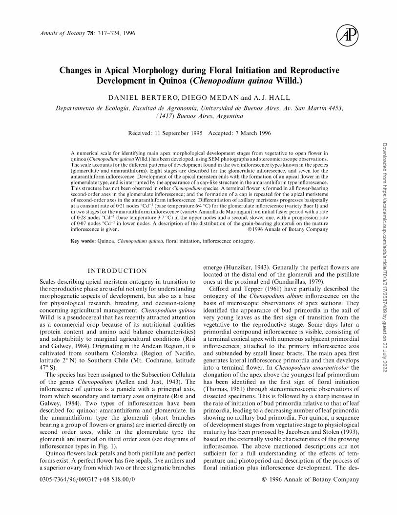
The species has been assigned to the Subsection Cellulataof the genus Chenopodium (Aellen and Just, 1943). Theinflorescence of quinoa is a panicle with a principal axis,from which secondary and tertiary axes originate (Risi andGalwey, 1984). Two types of inflorescences have beendescribed for quinoa: amaranthiform and glomerulate. Inthe amaranthiform type the glomeruli (short branchesbearing a group of flowers or grains) are inserted directly onsecond order axes, while in the glomerulate type theglomeruli are inserted on third order axes (see diagrams ofinflorescence types in Fig. 1).
Quinoa flowers lack petals and both pistillate and perfectforms exist. A perfect flower has five sepals, five anthers anda superior ovary from which two or three stigmatic branches
emerge (Hunziker, 1943). Generally the perfect flowers arelocated at the distal end of the glomeruli and the pistillateones at the proximal end (Gandarillas, 1979).
Gifford and Tepper (1961) have partially described theontogeny of the Chenopodium album inflorescence on thebasis of microscopic observations of apex sections. Theyidentified the appearance of bud primordia in the axil ofvery young leaves as the first sign of transition from thevegetative to the reproductive stage. Some days later aprimordial compound inflorescence is visible, consisting ofa terminal conical apex with numerous subjacent primordialinflorescences, attached to the primary inflorescence axisand subtended by small linear bracts. The main apex firstgenerates lateral inflorescence primordia and then developsinto a terminal flower. In Chenopodium amaranticolor theelongation of the apex above the youngest leaf primordiumhas been identified as the first sign of floral initiation(Thomas, 1961) through stereomicroscopic observations ofdissected specimens. This is followed by a sharp increase inthe rate of initiation of bud primordia relative to that of leafprimordia, leading to a decreasing number of leaf primordiashowing no axillary bud primordia. For quinoa, a sequenceof development stages from vegetative stage to physiologicalmaturity has been proposed by Jacobsen and Stolen (1993),based on the externally visible characteristics of the growinginflorescence. The above mentioned descriptions are notsufficient for a full understanding of the effects of tem-perature and photoperiod and description of the process offloral initiation plus inflorescence development. The des-
0305-7364}96}09031708 $18.00}0 # 1996 Annals of Botany Company
Dow
nloaded from https://academ
ic.oup.com/aob/article/78/3/317/2587489 by guest on 22 July 2022
318 Bertero et al.—Floral Ontogeny in Quinoa
F. 1. Inflorescence types. A, Glomerulate inflorescence; B,amaranthiform inflorescence.
T 1. Stages in de�elopment of the apical meristem ofglomerulate and amaranthiform inflorescence types
Glomerulate AmaranthiformStage inflorescence Stage inflorescence
G0 vegetative A0 vegetativeG1 early reproductive A1 early reproductiveG2 exposed apical bud A2 exposed apical budG3 beginning floral A3 transition to
differentiation cap stageG4 beginning ovary A4 cap stage
formationG5 ovary wall partly A5 cap elongating
covering ovule laterallyG6 ovule covered A6 scar stageG7 differentiation of A7 anthesis
stigmatic branchesG8 anthesis
criptions given by Thomas (1961) and Gifford and Tepper(1961) lack a characterization of the later stages in floraldevelopment, and use of the Gifford and Tepper scalerequires the preparation of sections of the apex; the scale ofJacobsen and Stolen is based on the characteristics of thegrowing inflorescence but does not account for the organdifferentiation sequence in the apical meristem and axillarybuds of the inflorescence.
The objectives of this work were to describe the sequenceof changes in apical morphology from vegetative to anthesisstages in this species, to characterize these events taking intoaccount the differences observed between inflorescencetypes, and to generate a quantitative scale with stages thatcan be easily identified in stereomicroscopic observations attwo levels : main apex and inflorescence.
MATERIALS AND METHODS
Source of material and en�ironmental conditions
Two quinoa varieties : Baer I (glomerulate type panicle) andAmarilla de Maranganı! (amaranthiform type), were cul-
tivated under optimal irrigation and nitrogen fertilizationon a silty clay loam (Vertic Argiudol) soil at the Faculty ofAgronomy, University of Buenos Aires, Argentina(34° 35« S, 58° 29« W) in a complete block design with tworeplications. Plant density was 30 plants m−#. Data from twosowings (Jan. 1994, mean daily temperature 23±6 °C, meanphotoperiod 13±8 h, mean total incident radiation 21±2 MJm−# d−") ; and Feb. 1995, (mean daily temperature 20±8 °C,meanphotoperiod 12±1 h,meandaily total incident radiation15±3 MJ m−# d−") were used for these observations. Baer I isa cultivar originated in Southern Chile (Temuco, 38° 45« S,72° 40« W), and Amarilla de Maranganı! originates fromCuzco, Peru (Cuzco, 13° 32« S, 71° 57« W).
Specimen preparation
Samples were taken every 2 d from emergence toflowering, dissected and observed through a stereo-microscope to determine floral stage on fresh material. Toprovide a graphical support to the description generated inthis way part of the sampled specimens were subjected toScanning Electron Microscopy (SEM). These specimenswere fixed in FAA, dehydrated in a graded acetone series(70, 80, 90, 100%), critical-point dried, mounted on SEMstubs using conductive paint, dissected to expose the apicalmeristem and sputter-coated with gold}palladium.
Data collection
Two aspects of the pattern of floral development werestudied. In the 1994 experiment the sequence of devel-opmental stages of the main apex was established throughstereomicroscopic observations of fresh material and SEM.In the 1995 experiment, the progression of development (asreflected by the achievement of a particular floral stage)from the apical meristem downwards through theinflorescence was followed using the scale previouslyestablished, through steromicroscopic observations on freshmaterial. Three plants were randomly sampled from eachreplication in both experiments. Floral stage was determinedas the time when 50% of the plants sampled reached thatstage. The patterns of development at both apex andinflorescence levels were expressed on a thermal time basis ;computed as the accumulation of mean daily temperatures(maximumminimum}2) over a base temperature (Ritchieand Ne Smith, 1991). Base temperatures were estimated foreach variety by plotting the rate of development towardsflowering (the inverse of the duration in days from emergenceto flowering) �s. average temperature from the period(Summerfield et al., 1991), using data from anotherexperiment which included seven sowing dates from Jul.1992 to Feb. 1993. A base temperature of 6±4 °C wascalculated for Baer I and 3±7 °C for Amarilla de Maranganı!.
At physiological maturity, five plants per inflorescencetype were sampled for each replication in the 1995experiment to analyse the distribution of the grain-bearingglomeruli on the inflorescence and number of grainsglomerule.
Dow
nloaded from https://academ
ic.oup.com/aob/article/78/3/317/2587489 by guest on 22 July 2022
Bertero et al.—Floral Ontogeny in Quinoa 319
RESULTS
Apical meristem de�elopmental scale
Eight floral stages were identified for the apical meristem inthe glomerulate and seven for the amaranthiforminflorescence, as summarized in Table 1. In glomerulateinflorescences, apical meristem development ends with theformation of a terminal flower; in amaranthiforminflorescences development of a terminal flower is in-terrupted and diverted toward the formation of a capstructure. Flower formation in this inflorescence occursonly in lateral meristems.
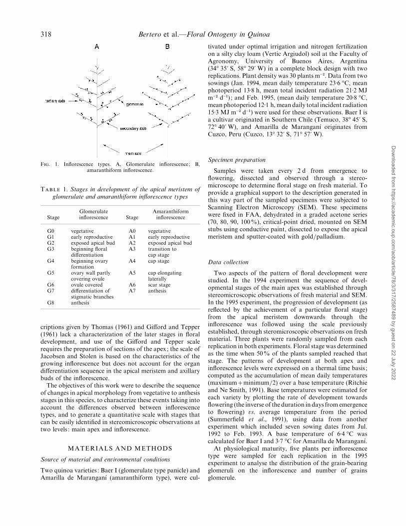
F. 2. Apical development in the glomerulate inflorescence; stages G0–G5. A, Vegetative (G0); B, early reproductive (G1); C, exposed apex (G2);D, start of differentiation of the terminal flower (G3); E, beginning of gynoecium differentiation (G4); F, ovary wall partially covering ovularprimordium (G5). a, Apex; ab, axillary buds; g, gynoecial primordium; lp, leaf primordium; o, ovular primordium; ow, ovary wall ; s, sepal
primordium; st, stamen primordium; t, thecae. Bars¯ 0±1 mm.
Description of stages
I.Glomerulate inflorescence. Apicalmeristem developmentin this type of inflorescence progresses up to the formationof a terminal flower. In both apical and axillary buds,differentiation progresses basipetally. Axillary buds growgiving origin to second order axes, and an hermaphroditeflower is formed at the apical meristems of these rami-fications. From these second-order axes third-order axes areformed. Flower-bearing glomeruli are supported by thesethird-order axes. The main stages of the apical meristemdevelopment observed in the glomerulate inflorescence areas follows.
G0. Vegetative: in this stage, the apical dome is Dow
nloaded from https://academ
ic.oup.com/aob/article/78/3/317/2587489 by guest on 22 July 2022
320 Bertero et al.—Floral Ontogeny in Quinoa
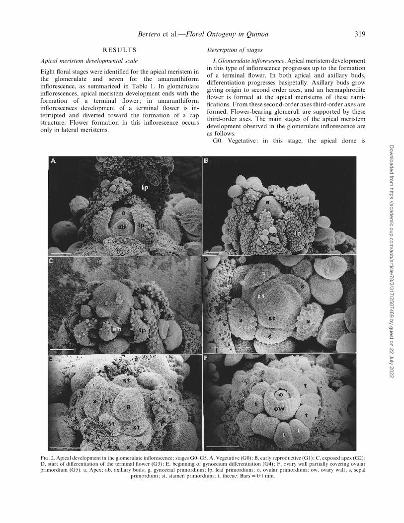
F. 3. Apical development in the glomerulate inflorescence; stages G6–G8. A, Ovary wall fully developed (G6); B, onset of differentiation ofstigmatic branches (G7); C, pistillate flower (G8); D, perfect flower (G8); E, detail of pistillate flower showing rudimentary anthers. g,
Gynoecium; t, thecae; ps, pollen sacs ; ra, rudimentary anthers ; s, sepals ; sb, stigmatic branches; st, stamen. Bars¯ 0±1 mm.
hemispherical, and appears fully covered by leaf primordia.These primordia hide subjacent bud primordia (Fig. 2A).
G1. Early reproductive: the first sign of the transitiontowards flowering is an increase in the rate of growth of theapical meristem with respect to leaf primordia that leads tothe emergence of the apical dome from among the leafprimordia (Fig. 2B).
G2. Exposed apex: the rate of axillary bud growthincreases in relation to that of leaf primordia and thusbecome visible (Fig. 2C).
G3. Beginning of the differentiation of the terminalflower: the apex assumes a pentagonal shape owing to theappearance of five sepal primordia (Fig. 2D).
G4. Beginning of gynoecium differentiation: the apexexpands to form a globose body, around which a rim soondevelops (the future ovary wall) encircling a small dome (thefuture solitary ovule) (Fig. 2E). The sepal and stamenprimordia are now clearly distinguished around the ovary.
G5. Ovary wall partially covering the ovular primordium,which in fresh material is distinguished by a more intensegreen colour (Fig. 2F). The two thecae of each stamen areclearly distinguishable.
G6. Ovary wall almost fully developed, ovular pri-mordium no longer visible (Fig. 3A).
G7. Onset of differentiation of stigmatic branches. Fourprimordial stigmatic branches are apparent at this stage
Dow
nloaded from https://academ
ic.oup.com/aob/article/78/3/317/2587489 by guest on 22 July 2022
Bertero et al.—Floral Ontogeny in Quinoa 321
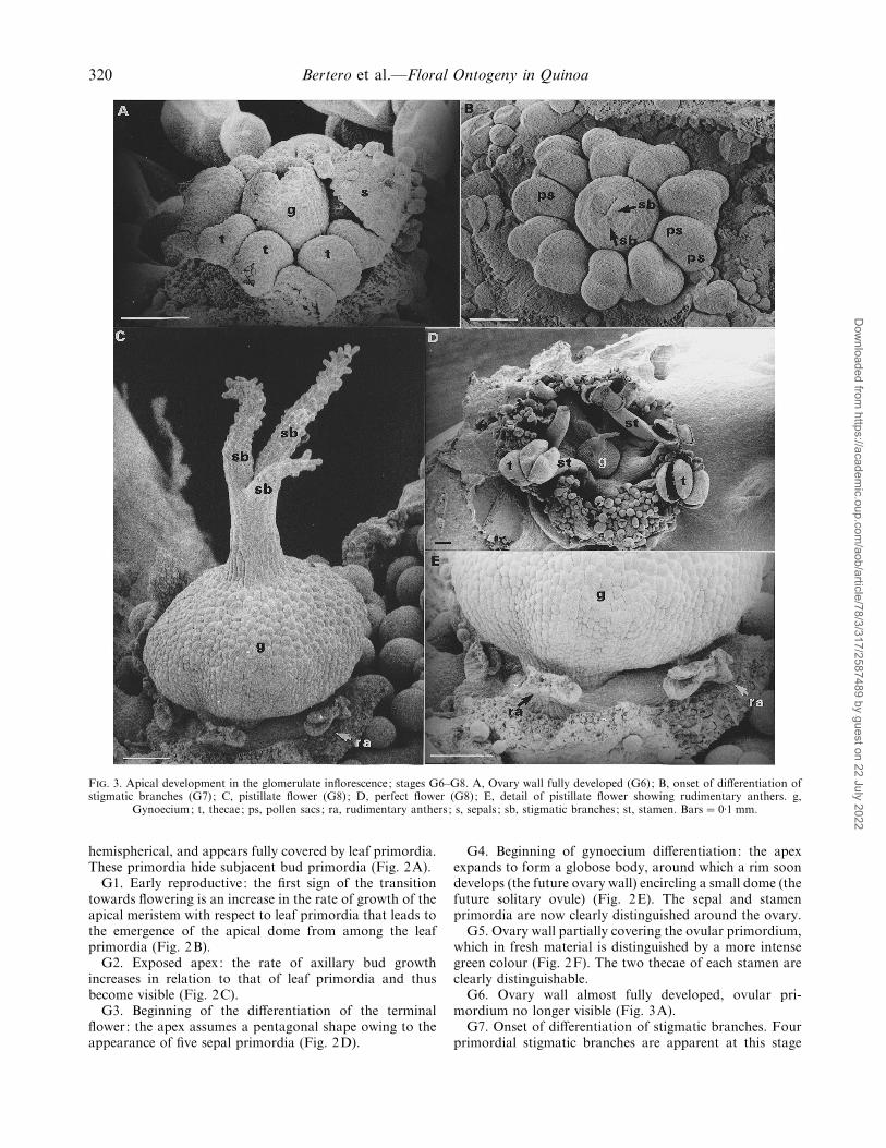
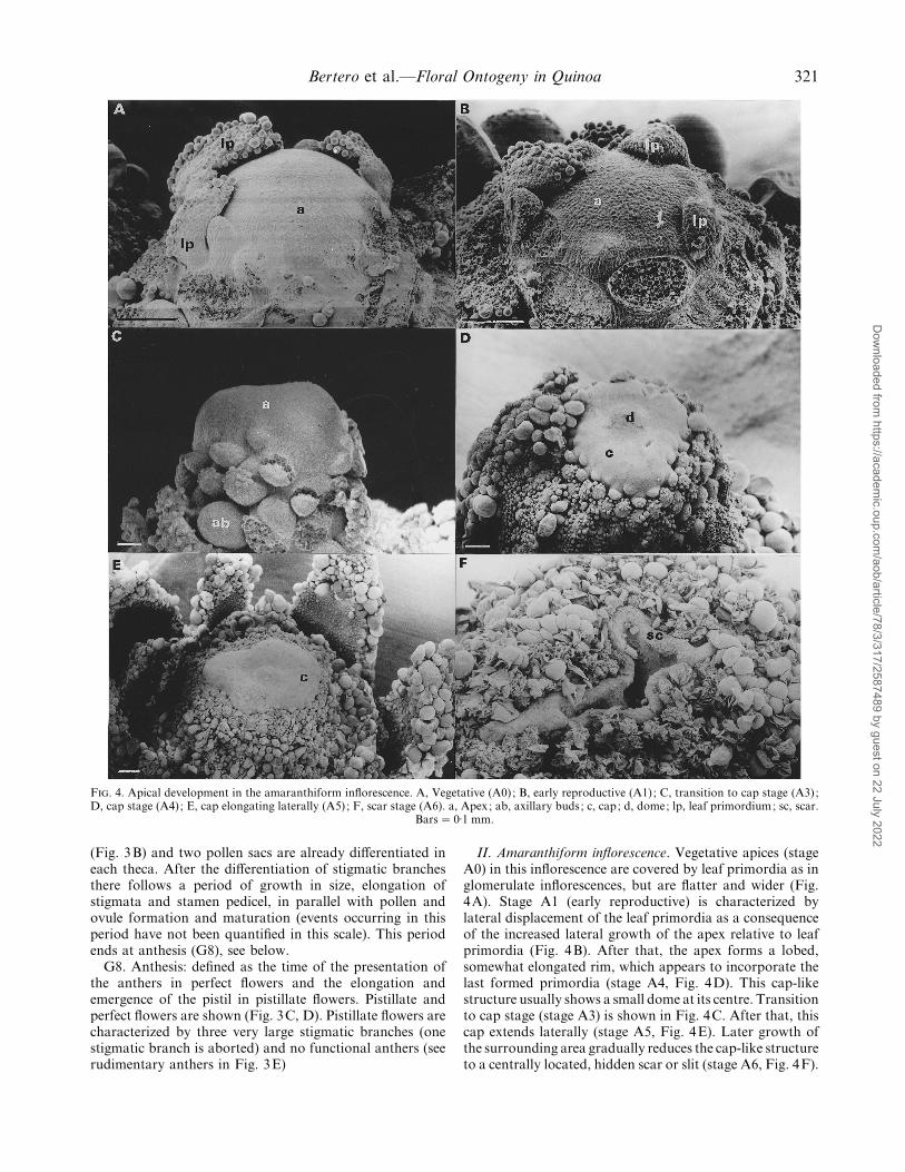
F. 4. Apical development in the amaranthiform inflorescence. A, Vegetative (A0) ; B, early reproductive (A1) ; C, transition to cap stage (A3) ;D, cap stage (A4) ; E, cap elongating laterally (A5) ; F, scar stage (A6). a, Apex; ab, axillary buds; c, cap; d, dome; lp, leaf primordium; sc, scar.
Bars¯ 0±1 mm.
(Fig. 3B) and two pollen sacs are already differentiated ineach theca. After the differentiation of stigmatic branchesthere follows a period of growth in size, elongation ofstigmata and stamen pedicel, in parallel with pollen andovule formation and maturation (events occurring in thisperiod have not been quantified in this scale). This periodends at anthesis (G8), see below.
G8. Anthesis: defined as the time of the presentation ofthe anthers in perfect flowers and the elongation andemergence of the pistil in pistillate flowers. Pistillate andperfect flowers are shown (Fig. 3C, D). Pistillate flowers arecharacterized by three very large stigmatic branches (onestigmatic branch is aborted) and no functional anthers (seerudimentary anthers in Fig. 3E)
II. Amaranthiform inflorescence. Vegetative apices (stageA0) in this inflorescence are covered by leaf primordia as inglomerulate inflorescences, but are flatter and wider (Fig.4A). Stage A1 (early reproductive) is characterized bylateral displacement of the leaf primordia as a consequenceof the increased lateral growth of the apex relative to leafprimordia (Fig. 4B). After that, the apex forms a lobed,somewhat elongated rim, which appears to incorporate thelast formed primordia (stage A4, Fig. 4D). This cap-likestructure usually shows a small dome at its centre. Transitionto cap stage (stage A3) is shown in Fig. 4C. After that, thiscap extends laterally (stage A5, Fig. 4E). Later growth ofthe surrounding area gradually reduces the cap-like structureto a centrally located, hidden scar or slit (stage A6, Fig. 4F).
Dow
nloaded from https://academ
ic.oup.com/aob/article/78/3/317/2587489 by guest on 22 July 2022
322 Bertero et al.—Floral Ontogeny in Quinoa
1000
10
0200
Thermal time from emergence (°Cd)
Ape
x sc
ore 8
6
4
2
400 600 800
A
1000
10
0200
8
6
4
2
400 600 800
B
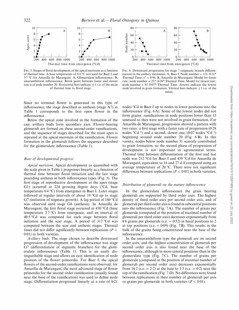
F. 5. Stages of floral development of the apical meristem as a functionof thermal time. A base temperature of 6±4 °C was used for Baer I and3±7 °C for Amarilla de Maranganı!. A, Glomerulate inflorescence; B,amaranthiform inflorescence. Break point between faster and slowerrate is at node number 20. Horizontal bars indicate ³1 s.e. of the mean
of thermal time to floral stage.
Since no terminal flower is generated in this type ofinflorescence, the stage described as anthesis (stage A7) inTable 1 corresponds to the first open flower in theinflorescence.
Below the apical zone involved in the formation of thecap, axillary buds form secondary axes. Flower-bearingglomeruli are formed on these second-order ramifications,and the sequence of stages described for the main apex isrepeated at the apical meristem of the ramification. Flowerformation in the glomeruli follows the sequence describedfor the glomerulate inflorescence (Table 1).
Rate of de�elopmental progress
Apical meristem. Apical development as quantified withthe scale given in Table 1 progresses linearly as a function ofthermal time between floral initiation and the last stagepreceding anthesis in both inflorescence types (Fig. 5). Thefirst stage of reproductive development at the apex (stageG1) occurred at 224 growing degree days (°Cd, basetemperature 6±4 °C) from emergence in Baer I. Later stagesfollowed at regular intervals of about 19 °Cd up to stageG7 (initiation of stigmata growth). A lag period of 180 °Cdwas observed until stage G8 (anthesis). In Amarilla deMaranganı!, the first floral stage occurred at 430 °Cd (basetemperature 3±7 °C) from emergence, and an interval of40±5 °Cd was computed for each stage between floralinitiation and the scar stage. A period of 317 °Cd wascomputed between the scar and anthesis stages. Thermaltimes did not differ significantly between replications (P!0±01) in both varieties.
Axillary buds. The stage chosen to describe downwardprogression of development of the inflorescence was stageG7 (differentiation of stigmatic branches) for the glom-erulate inflorescence (Table 1). This is an easily dis-tinguishable stage and allows an easy identification of nodeposition of the flower primordia. For Baer I, the apicalflowers of the second-order ramifications were observed; forAmarilla de Maranganı!, the most advanced stage of flowerprimordia for the second order ramification (usually foundnear the base of the ramification) was used to define nodestage. Differentiation progressed linearly at a rate of 0±21
1000
80
0200
Thermal time from emergence (°Cd)
Nod
e n
um
ber
60
40
20
400 600 800
A
1000
80
0200
60
40
20
400 600 800
B
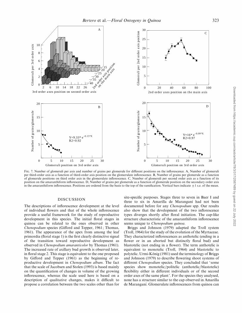
F. 6. Downward progression for stage 7 (stigmatic branch differen-tiation) in the axillary meristems. A, Baer I. Node number¯ 121–0±21*Thermal Time; r#¯ 0±96. B, Amarilla de Maranganı!. Model for fasterrate ; node number¯ 237–0±28* Thermal Time. Model for slower rate ;node number¯ 81–0±07* Thermal Time. Arrows indicate the lowestnode involved in grain formation. Vertical bars indicate ³1 s.e. of the
mean.
nodes °Cd in Baer I up to nodes in lower positions into theinflorescence (Fig. 6A). Some of the lowest nodes did notform grains : ramifications in node positions lower than 15senesced so they were not involved in grain formation. ForAmarilla de Maranganı!, progression showed a pattern withtwo rates : a first stage with a faster rate of progression (0±28nodes °Cd−") and a second, slower one, (0±07 nodes °Cd−")beginning around node number 20 (Fig. 6 B). In thisvariety, nodes below node number 31 scarcely contributedto grain formation, so the second phase of progression ofdevelopment is not important in agronomical terms.Thermal time between differentiation of the first and lastnode was 212 °Cd for Baer I and 439 °Cd for Amarilla deMaranganı!, equivalent to 16 and 27 d if compared using anaverage temperature of 20 °C. There were not significantdifferences between replications (P! 0±01) in both varieties.
Distribution of glomeruli on the mature inflorescence
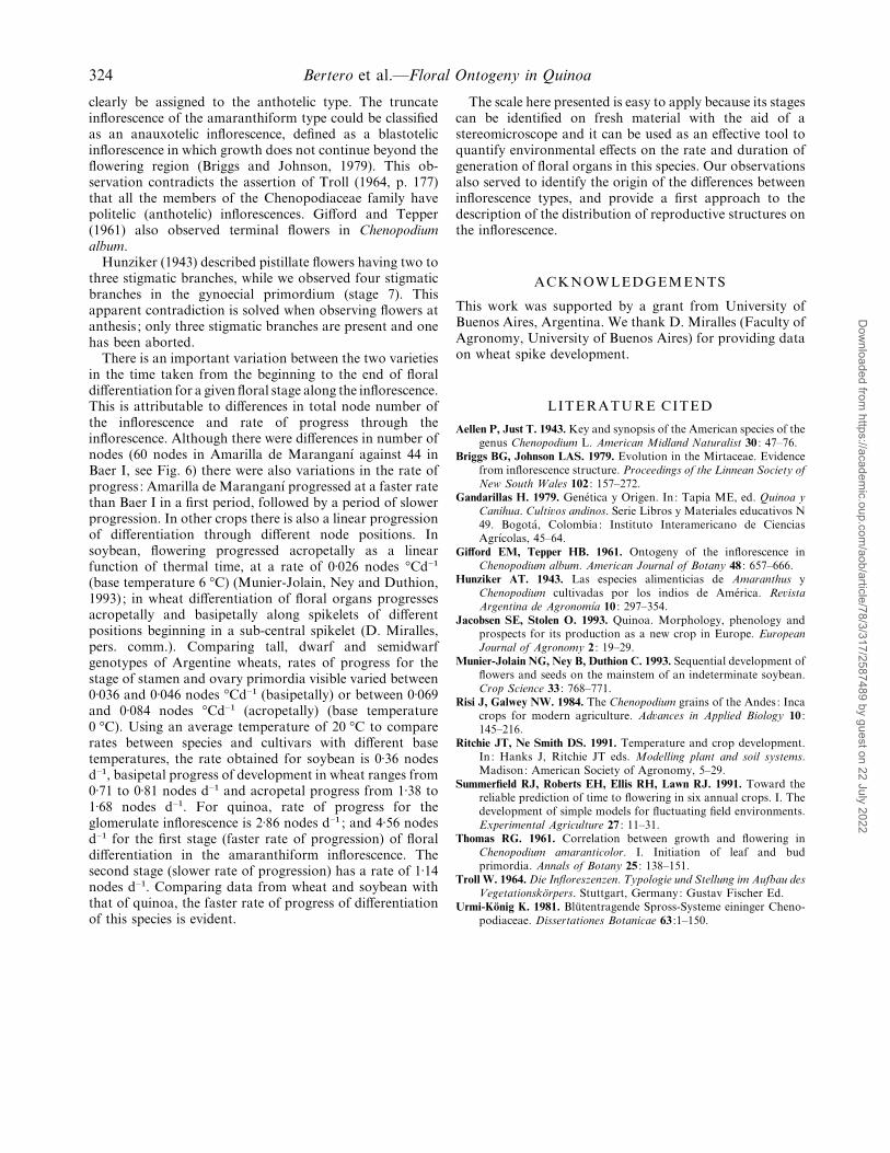
In the glomerulate inflorescence the grain bearingglomeruli are supported by third order axes. The highestdensity of third order axes per second order axis, and ofglomeruli per third order axis is found in subcentral positionsinto the inflorescence (Fig. 7A). The number of grains perglomerule (computed at the position of maximal number ofglomeruli per third order axis) decreases exponentially from8±6 grains per glomerule (s.e.¯ 0±52) in basal positions to 2at top positions (s.e.¯ 0±09) (Fig. 7B). This results in thebulk of the grains being concentrated near the base of theinflorescence.
In the amaranthiform type the glomeruli are on secondorder axes, and the highest concentration of glomeruli persecond order axis is also found near the base of theinflorescence, although in more central positions than in theglomerulate type (Fig. 7C). The number of grains perglomerule (computed at the position of maximal number ofglomeruli per second order axis) decreases exponentiallyfrom 16±2 (s.e.¯ 2±2) at the base to 1±3 (s.e.¯ 0±2) near thetop of the ramification (Fig. 7 D). No differences were foundbetween replications in their number of glomeruli per axisor grains per glomerule in both varieties (P! 0±01).
Dow
nloaded from https://academ
ic.oup.com/aob/article/78/3/317/2587489 by guest on 22 July 2022
Bertero et al.—Floral Ontogeny in Quinoa 323
30
20
0Glomeruli position on 3rd order axis
Nu
mbe
r of
gra
ins/
glom
eru
le
5
15
10
10 205 15 25
Y=9.33* e–0.15*X
R2=0.92
B
30
20
0Glomeruli position on 3rd order axis
Nu
mbe
r of
gra
ins/
glom
eru
le
5
15
10
10 205 15 25
Y=16* e–0.09*X
R2=0.97
D
100
30
02nd order axes position on the main axis
Glo
mer
uli
per
2n
d or
der
axis
pos
itio
n
5
15
10
20 6040 80
C
20
25
26
10
02
3rd order axes position on second order axis
Glo
mer
uli
per
3rd
ord
er a
xis
2
6
4
10 226 14
A
18
8
4035
3025
2015
2nd
orde
r axi
s pos
ition
on th
e mai
n ax
is
F. 7. Number of glomeruli per axis and number of grains per glomerule for different positions on the inflorescence. A, Number of glomeruliper third-order axis as a function of third-order axis position on the glomerulate inflorescence. B, Number of grains per glomerule as a functionof glomerule positions on third order axis in the glomerulate inflorescence. C, Number of glomeruli per second order axis as a function of itsposition on the amaranthiform inflorescence. D, Number of grains per glomerule as a function of glomerule position on the secondary order axisin the amaranthiform inflorescence. Positions are ordered from the basis to the top of the ramification. Vertical bars indicate ³1 s.e. of the mean.
DISCUSSION
The descriptions of inflorescence development at the levelof individual flowers and that of the whole inflorescenceprovide a useful framework for the study of reproductivedevelopment in this species. The initial floral stages inquinoa can be related to the ones observed in otherChenopodium species (Gifford and Tepper, 1961; Thomas,1961). The appearance of the apex from among the leafprimordia (floral stage 1) is the first clearly distinctive signalof the transition toward reproductive development asobserved in Chenopodium amaranticolor by Thomas (1961).The increased rate of axillary bud growth is observed later,in floral stage 2. This stage is equivalent to the one proposedby Gifford and Tepper (1961) as the beginning of re-productive development in Chenopodium album. The factthat the scale of Jacobsen and Stolen (1993) is based mainlyon the quantification of changes in volume of the growinginflorescence, whereas the scale used here is based on adescription of qualitative changes, makes it difficult topropose a correlation between the two scales other than for
site-specific purposes. Stages three to seven in Baer I andthree to six in Amarilla de Maranganı! had not beendocumented before for any Chenopodium spp. Our resultsalso show that the development of the two inflorescencetypes diverges shortly after floral initiation. The cap-likestructure characteristic of the amaranthiform inflorescenceseems unique to Chenopodium quinoa.
Briggs and Johnson (1979) adapted the Troll system(Troll, 1964) for the study of the evolution of the Myrtaceae.They characterized inflorescences as anthotelic (ending in aflower or in an aborted but distinctly floral bud) andblastotelic (not ending in a flower). The term anthotelic isequivalent to monotelic (Troll, 1964) and blastotelic topolytelic. Urmi-Ko$ nig (1981) used the terminology of Briggsand Johnson (1979) to describe flowering shoot systems ofdifferent Chenopodium species. They concluded that ‘somespecies show monotelic}politelic (anthotelic}blastotelic)flexibility either in different individuals or of the secondorder axes of the same plant ’. For the species they analysed,none has a structure similar to the cap observed in Amarillade Maranganı!. Glomerulate inflorescences from quinoa can
Dow
nloaded from https://academ
ic.oup.com/aob/article/78/3/317/2587489 by guest on 22 July 2022
324 Bertero et al.—Floral Ontogeny in Quinoa
clearly be assigned to the anthotelic type. The truncateinflorescence of the amaranthiform type could be classifiedas an anauxotelic inflorescence, defined as a blastotelicinflorescence in which growth does not continue beyond theflowering region (Briggs and Johnson, 1979). This ob-servation contradicts the assertion of Troll (1964, p. 177)that all the members of the Chenopodiaceae family havepolitelic (anthotelic) inflorescences. Gifford and Tepper(1961) also observed terminal flowers in Chenopodiumalbum.
Hunziker (1943) described pistillate flowers having two tothree stigmatic branches, while we observed four stigmaticbranches in the gynoecial primordium (stage 7). Thisapparent contradiction is solved when observing flowers atanthesis ; only three stigmatic branches are present and onehas been aborted.
There is an important variation between the two varietiesin the time taken from the beginning to the end of floraldifferentiation for a given floral stage along the inflorescence.This is attributable to differences in total node number ofthe inflorescence and rate of progress through theinflorescence. Although there were differences in number ofnodes (60 nodes in Amarilla de Maranganı! against 44 inBaer I, see Fig. 6) there were also variations in the rate ofprogress : Amarilla de Maranganı! progressed at a faster ratethan Baer I in a first period, followed by a period of slowerprogression. In other crops there is also a linear progressionof differentiation through different node positions. Insoybean, flowering progressed acropetally as a linearfunction of thermal time, at a rate of 0±026 nodes °Cd−"
(base temperature 6 °C) (Munier-Jolain, Ney and Duthion,1993) ; in wheat differentiation of floral organs progressesacropetally and basipetally along spikelets of differentpositions beginning in a sub-central spikelet (D. Miralles,pers. comm.). Comparing tall, dwarf and semidwarfgenotypes of Argentine wheats, rates of progress for thestage of stamen and ovary primordia visible varied between0±036 and 0±046 nodes °Cd−" (basipetally) or between 0±069and 0±084 nodes °Cd−" (acropetally) (base temperature0 °C). Using an average temperature of 20 °C to comparerates between species and cultivars with different basetemperatures, the rate obtained for soybean is 0±36 nodesd−", basipetal progress of development in wheat ranges from0±71 to 0±81 nodes d−" and acropetal progress from 1±38 to1±68 nodes d−". For quinoa, rate of progress for theglomerulate inflorescence is 2±86 nodes d−" ; and 4±56 nodesd−" for the first stage (faster rate of progression) of floraldifferentiation in the amaranthiform inflorescence. Thesecond stage (slower rate of progression) has a rate of 1±14nodes d−". Comparing data from wheat and soybean withthat of quinoa, the faster rate of progress of differentiationof this species is evident.
The scale here presented is easy to apply because its stagescan be identified on fresh material with the aid of astereomicroscope and it can be used as an effective tool toquantify environmental effects on the rate and duration ofgeneration of floral organs in this species. Our observationsalso served to identify the origin of the differences betweeninflorescence types, and provide a first approach to thedescription of the distribution of reproductive structures onthe inflorescence.
ACKNOWLEDGEMENTS
This work was supported by a grant from University ofBuenos Aires, Argentina. We thank D. Miralles (Faculty ofAgronomy, University of Buenos Aires) for providing dataon wheat spike development.
LITERATURE CITED
Aellen P, Just T. 1943. Key and synopsis of the American species of the
genus Chenopodium L. American Midland Naturalist 30 : 47–76.
Briggs BG, Johnson LAS. 1979. Evolution in the Mirtaceae. Evidence
from inflorescence structure. Proceedings of the Linnean Society of
New South Wales 102 : 157–272.
Gandarillas H. 1979. Gene! tica y Origen. In: Tapia ME, ed. Quinoa y
Canihua. Culti�os andinos. Serie Libros y Materiales educativos N
49. Bogota! , Colombia: Instituto Interamericano de Ciencias
Agrı!colas, 45–64.
Gifford EM, Tepper HB. 1961. Ontogeny of the inflorescence in
Chenopodium album. American Journal of Botany 48 : 657–666.
Hunziker AT. 1943. Las especies alimenticias de Amaranthus y
Chenopodium cultivadas por los indios de Ame! rica. Re�ista
Argentina de AgronomıUa 10 : 297–354.
Jacobsen SE, Stolen O. 1993. Quinoa. Morphology, phenology and
prospects for its production as a new crop in Europe. European
Journal of Agronomy 2 : 19–29.
Munier-Jolain NG, Ney B, Duthion C. 1993. Sequential development of
flowers and seeds on the mainstem of an indeterminate soybean.
Crop Science 33 : 768–771.
Risi J, Galwey NW. 1984. The Chenopodium grains of the Andes : Inca
crops for modern agriculture. Ad�ances in Applied Biology 10 :
145–216.
Ritchie JT, Ne Smith DS. 1991. Temperature and crop development.
In: Hanks J, Ritchie JT eds. Modelling plant and soil systems.
Madison: American Society of Agronomy, 5–29.
Summerfield RJ, Roberts EH, Ellis RH, Lawn RJ. 1991. Toward the
reliable prediction of time to flowering in six annual crops. I. The
development of simple models for fluctuating field environments.
Experimental Agriculture 27 : 11–31.
Thomas RG. 1961. Correlation between growth and flowering in
Chenopodium amaranticolor. I. Initiation of leaf and bud
primordia. Annals of Botany 25 : 138–151.
Troll W. 1964. Die Infloreszenzen. Typologie und Stellung im Aufbau des
VegetationskoX rpers. Stuttgart, Germany: Gustav Fischer Ed.
Urmi-Ko$ nig K. 1981. Blu$ tentragende Spross-Systeme eininger Cheno-
podiaceae. Dissertationes Botanicae 63 :1–150.
Dow
nloaded from https://academ
ic.oup.com/aob/article/78/3/317/2587489 by guest on 22 July 2022


