Embed Size (px)
Citation preview

www.elsevier.com/locate/yviro
Virology 341 (20
Bioluminescence imaging of vaccinia virus: Effects of interferon on
viral replication and spread
Kathryn E. Lukera, Martha Hutchensb, Tracey Schultza, Andrew Pekoszc, Gary D. Lukera,d,*
aDepartment of Radiology, University of Michigan Medical School, Ann Arbor, MI 48109-0648, USAbGraduate Program in Immunology, University of Michigan Medical School, Ann Arbor, MI 48109-0648, USA
cDepartment of Molecular Microbiology, Department of Pathology and Immunology, Washington University School of Medicine, St. Louis, MO 63110, USAdDepartment of Microbiology and Immunology, University of Michigan Medical School, Ann Arbor, MI 48109-0648, USA
Received 12 May 2005; returned to author for revision 23 June 2005; accepted 30 June 2005
Available online 10 August 2005
Abstract
Whole animal imaging allows viral replication and localization to be monitored in intact animals, which provides significant advantages
for determining viral and host factors that determine pathogenesis. To investigate effects of interferons on spatial and temporal progression of
vaccinia infection, we generated recombinant viruses that express firefly luciferase or a monomeric orange fluorescent protein. These viruses
allow vaccinia infection to be monitored with bioluminescence or fluorescence imaging, respectively. The recombinant viruses were not
attenuated in vitro or in vivo relative to a control WR virus. In cell culture, reporters could be detected readily by 4 h post-infection, showing
that these viruses can be used as early markers of infection. The magnitude of firefly luciferase activity measured with bioluminescence
imaging in vitro and in vivo correlated directly with increasing titers of vaccinia virus, validating imaging data as a marker of viral infection.
Replication of vaccinia was significantly greater in mice lacking receptors for type I interferons (IFN I R�/�) compared with wild-type mice,
although both genotypes of mice developed focal infections in lungs and brain after intranasal inoculation. IFN I R�/� mice had greater
dissemination of virus to liver and spleen than wild-type animals even when mortality occurred at the same time point after infection.
Protective effects of type I interferons were mediated primarily through parenchymal cells rather than hematopoietic cells as analyzed by
bone marrow transplant experiments. Collectively, our data define a new function for type I interferon signaling in systemic dissemination of
vaccinia and validate these reporter viruses for studies of pathogenesis.
D 2005 Elsevier Inc. All rights reserved.
Keywords: Vaccinia; Bioluminescence imaging; Interferon
Smallpox was eradicated as a human disease before
molecular tools were available to analyze the cellular and
molecular effects of various viral proteins and the host
immune response to infection. As a result, the complex
interactions between virus and host immunity that determine
pathogenesis of poxvirus infection remain incompletely
defined. Because of the threat of smallpox as a potential
agent of bioterrorism, there is renewed emphasis on
developing new techniques and reagents to investigate viral
0042-6822/$ - see front matter D 2005 Elsevier Inc. All rights reserved.
doi:10.1016/j.virol.2005.06.049
* Corresponding author. Department of Radiology, University of Mich-
igan Medical School, 1150 West Medical Center Drive, 9301 MSRB III,
Ann Arbor, MI 48109-0648, USA.
E-mail address: [email protected] (G.D. Luker).
and host determinants of disease in relevant animal models
(Harrison et al., 2004). It is expected that identifying and
characterizing specific viral and host mediators of patho-
genesis will lead to new strategies for vaccination against
poxvirus infection and novel targets for therapy. These are
key areas of research because of the risk for complications
associated with the existing smallpox vaccine (Wollenberg
and Engler, 2004) and limitations of available drugs to treat
infections with poxviruses (Ortiz et al., 2005).
Vaccinia virus was used as the vaccine to eradicate
smallpox as a human disease, and vaccinia now is established
as the model virus for poxvirus biology and disease (Harrison
et al., 2004). Intranasal infection of mice with vaccinia
reproduces spread of smallpox through the upper respiratory
05) 284 – 300

K.E. Luker et al. / Virology 341 (2005) 284–300 285
tract, which is believed to be the typical route of infection
(Breman and Henderson, 2002). Vaccinia replicates in the
respiratory tract and lungs and then can disseminate systemi-
cally to visceral organs. Systemic infection may be cleared or
progress to death, depending on factors such as viral
inoculum, mouse age, and variations in immune response
(Turner, 1967a; Williamson et al., 1990).
Interferons (IFN) are one of the key mediators of host
innate immunity to vaccinia virus and other viruses. There
are two major classes of IFNs produced in response to viral
infection: type I and type II. Type I IFNs, which in mice and
humans include multiple isotypes of IFN-a and a single
isotype of IFN-h, are secreted by most cells in response to
viral infection (Samuel, 1998). Production of type II IFN
(IFN-g) is restricted to activated CD4+ and CD8+ T
lymphocytes, natural killer (NK) cells, and natural killer T
(NKT) cells (Farrar and Schreiber, 1993; Fujii et al., 2002).
Type I and II IFNs bind and signal through distinct
heterodimeric receptors (IFN I R and IFN II R, respec-
tively), each of which is expressed normally on all nucleated
cells (Stark et al., 1998). In response to viral infection,
specific isotypes of type I IFNs (IFN-h and IFN-a4) are
secreted early in the host immune response. These IFNs
bind to the type I IFN receptor and subsequently induce
other subtypes of type I IFNs, as well as type II IFN, thereby
establishing a positive feedback loop for amplifying anti-
viral effects of both types of IFNs (Marie et al., 1998; Sato
et al., 1998, 2000; Taniguchi and Takaoka, 2001).
Both type I and II IFN are important mediators of host
immunity to vaccinia infection. Mice lacking receptors for
type II IFN (IFN II R�/�) are more susceptible to vaccinia
infection than wild-type animals, and deletion of receptors
for type I IFN (IFN I R�/�) diminishes resistance to vaccinia
to an even greater extent than IFN II R�/� mice (Huang et
al., 1993; van den Broek et al., 1995). Combined deficiency
of both type I and type II IFN receptors (IFN I/II R�/�)
produces an additive phenotype of susceptibility to vaccinia,
as demonstrated by higher titers of virus in lungs after
intranasal infection (van den Broek et al., 1995). Collec-
tively, these previous studies emphasize the key functions of
interferons in the immune response to vaccinia and indicate
that type I interferon has a quantitatively greater impact than
type II IFN on replication of vaccinia in vivo.
Vaccinia virus encodes a variety of proteins that inhibit
IFN and its mediators, including soluble binding proteins
for types I and II IFN and antagonists of the PKR pathway
of anti-viral defense (reviewed in Katze et al., 2002).
Viruses lacking these modulators of IFN are attenuated in
vivo, further demonstrating the importance of IFN in host
defense to vaccinia (Brandt and Jacobs, 2001; Sroller et al.,
2001; Verardi et al., 2001). In cultured cells, the PKR
antagonists E3L and K3L have been identified as key
determinants of the host range for vaccinia (Langland and
Jacobs, 2002). However, the extent to which IFN regulates
dissemination of vaccinia to various organs and tissues in
vivo has not been established.
Research by our laboratory and others demonstrates
several significant advantages of investigating viral-host
pathogenesis with imaging (Cook and Griffin, 2003; Luker
et al., 2002). Spatial and temporal progression of infection
can be quantified in the same animals, identifying animal-
to-animal variations in viral replication and dissemination
and host immunity. Imaging greatly increases data obtained
about viral pathogenesis in living mice compared with other
global assays of disease progression, such as weight loss or
external signs of disease. Imaging data also allow relative
amounts of viral replication in various anatomic sites to be
quantified over time. These data otherwise require sacrifice
of animals at multiple time points for determinations of viral
titers in excised organs and tissues. Importantly, imaging
techniques can identify unexpected sites or patterns of viral
infection that could be missed if organs are not collected or
if entire organs are analyzed for viral titers (Luker et al.,
2003).
To exploit the advantages of imaging for studies of
vaccinia pathogenesis, we generated a recombinant vacci-
nia virus that expresses firefly luciferase (FL) to enable
bioluminescence imaging (BLI) of vaccinia virus infection
in living mice. We also engineered a recombinant vaccinia
virus that expresses a monomeric orange fluorescent
protein (mKO) to facilitate analyses of infected tissues
by microscopy. Unlike many reporter viruses for vaccinia,
these recombinant viruses were constructed without delet-
ing any viral genes (Blasco and Moss, 1991). This
strategy allows our reporter viruses to reproduce repli-
cation and dissemination of wild-type virus in cultured
cells and mice. We used these recombinant viruses to
investigate to what extent innate immunity mediated
through interferons regulates replication and dissemination
of vaccinia. Our research demonstrated that type I
interferons limit replication of vaccinia virus and that the
protective effects of type I IFN are mediated primarily
through parenchymal rather than hematopoietic cells. We
also determined that type I IFN regulates dissemination of
vaccinia virus to specific tissues following intranasal
inoculation.
Results
Bioluminescent and fluorescent reporter vaccinia viruses for
imaging
Recombinant vaccinia viruses typically are constructed
by replacing a viral gene, such as thymidine kinase, with a
heterologous protein. While this strategy facilitates selec-
tion of recombinant viruses, replacement of thymidine
kinase is known to attenuate vaccinia virus in mice (Buller
et al., 1985). Even disruption of a potential open reading
frame in the viral genome may attenuate vaccinia and limit
the use of the recombinant virus for studies of pathogenesis
(Coupar et al., 2000).

K.E. Luker et al. / Virology 341 (2005) 284–300286
We used an alternative strategy based on repair of a
mutant virus to engineer vaccinia viruses with reporter
genes for imaging. The system is based on a WR strain of
vaccinia that has a mutation in vp37, a gene for a protein in
the outer viral membrane that is essential for efficient cell-
to-cell spread. The recombination vector repairs vp37 and
inserts a foreign gene driven by a strong synthetic vaccinia
virus p7.5 early/late promoter (Blasco and Moss, 1995).
This system enabled us to produce vaccinia viruses that
express either firefly luciferase (Vac-FL) or a monomeric
orange fluorescent protein (Vac-mKO) for bioluminescence
imaging or identification of infected cells, respectively
(Karasawa et al., 2004). We also constructed a virus that
repaired the vp37 gene without insertion of a reporter gene
as a marker rescue virus. The marker rescue virus differs
from wild-type WR only by insertion of the p 7.5 early/late
promoter into the genome.
To validate the virulence of the reporter viruses used in
this study, we quantified growth of Vac-FL and Vac-mKO
relative to the marker rescue virus and wild-type WR in a
multi-step growth assay. Vero cells were infected with
various viruses at an MOI of 0.1, and viral titers were
quantified for 3 days post-infection. The kinetics of viral
replication did not differ among the four viruses, demon-
strating that the reporter viruses were not attenuated in vitro
(Fig. 1). Sizes of plaques were comparable among the
recombinant viruses and repaired virus, while the parental
mutant virus did not form plaques within 3 days (data not
shown).
Because attenuation of vaccinia viruses only may be
apparent in vivo, we compared Vac-FL with the marker
rescue WR vaccinia virus in an intranasal model of infection
in mice. WT mice were infected with 1 � 106 pfu of Vac-FL
or marker rescue WR virus. Bioluminescence imaging was
performed daily on mice infected with Vac-FL, and weights
of animals in both groups were measured on each day post-
Fig. 1. Replication of Vac-FL and Vac-mKO in vitro. Vero cells were
infected with Vac-FL, Vac-mKO, marker rescue virus, or wild-type WR at
an MOI of 0.1. Cells were harvested every 24 h after infection for
determinations of viral titers by plaque assay. Each data point is the mean of
three independent determinations. Error bars represent SEM. Data are
representative of two independent experiments.
infection. Infection with Vac-FL or the marker rescue virus
had comparable weight loss over time, and mice were
euthanized on day 7 post-infection after losing 30% of
initial body weight (Fig. 2A). We quantified viral titers in
lung, brain, and spleen from mice on days 3 and 7 post-
infection (Figs. 2B and C). These organs were selected as
sites of local infection (lung) and disseminated disease
(brain and spleen), respectively. Titers of Vac-FL and
marker rescue virus did not differ in these organs on either
day. Collectively, these data indicate that insertion of FL or
the bioluminescence imaging protocol did not attenuate
vaccinia or its pathogenicity in mice.
Having established that expression of FL did not attenuate
the recombinant virus, we determined the extent to which
bioluminescence imaging of luciferase activity could be used
to quantify replication of Vac-FL in vivo. WT mice were
infected intranasally with 1 � 106 pfu Vac-FL, and mice
were euthanized immediately after bioluminescence imaging
on day 7 post-infection. Animals were dissected rapidly to
verify sites of FL activity identified by in vivo imaging.
Excised organs were imaged within 5 min after euthanizing
each mouse to quantify photon flux, and plaque assays then
were performed on these same organs. Relative to in vivo
imaging, ex vivo imaging of bioluminescence in excised
organs has potential limitations, including changes in cell
metabolism and oxygenation, loss of perfusion, and alter-
ations in intracellular concentration of luciferin. In part, these
limitations can be overcome by maintaining a consistent time
interval between euthanasia of mice and imaging. We
observed a direct correlation between relative values for
photon flux and viral titers in lung, brain, liver, and spleen
(Fig. 3). These data demonstrate that bioluminescence
imaging data for FL can be used as a reporter for relative
amounts of Vac-FL in various anatomic sites.
Detection of reporter genes in cultured cells
We determined the kinetics of FL activity following
infection of Vero cells with Vac-FL at an MOI of 5.
Luciferase activity from Vac-FL was quantified by imaging
at various times through 24 h post-infection, and these same
samples also were analyzed for viral titers. Bioluminescence
was slightly above background levels at 1 h post-infection
(Fig. 4A). FL activity continued to increase at subsequent
time points, reaching a level that was 4 logs above
uninfected cells by 24 h. Viral titers decreased initially,
corresponding to entry of virus into cells and the start of
replication. Titers of Vac-FL at 24 h were approximately 15-
fold greater than the input inoculum of virus. These data
show that the synthetic early/late promoter in Vac-FL allows
rapid production of the FL imaging reporter and a large
dynamic range for quantifying vaccinia infection.
We further characterized the FL reporter by determining
the limits of bioluminescence imaging for detecting Vac-FL
in cultured cells. Monolayers of Vero cells in 6-well plates
were infected with Vac-FL at various pfu, and photon flux

Fig. 2. Pathogenesis and replication of Vac-FL in mice. 129 Ev/Sv mice (WT) were infected with 1 � 106 pfu of Vac-FL or marker rescue WR by intranasal
(i.n.) inoculation (n = 8 mice per virus). (A) Mice were weighed daily to monitor overall progression of disease. Data are presented as mean T SEM percent loss
of initial weight. (B) Viral titers in lung, brain, and spleen at days 3 and 7 post-infection were quantified by plaque assay (n = 4 mice per time point). Data are
shown as mean T SEM of vaccinia virus pfu/g tissue. The lower limits of detection for the plaque assay were 30 pfu/g tissue.
K.E. Luker et al. / Virology 341 (2005) 284–300 287
from FL was quantified 12 h post-infection. FL activity was
detectable above background levels using an input of 30 pfu
per well, although viral replication would have occurred in
the interval between infection and the measurement of
bioluminescence. The lower limits for detecting Vac-FL in
vitro and in vivo with bioluminescence imaging will vary
based on factors including numbers of virions per unit
volume of cells or tissues and overlying materials that
absorb light. Nevertheless, this result provides a guideline
for the sensitivity of bioluminescence imaging for detecting
Vac-FL in cultured cells.
We also assayed the time course of expression of mKO
by Vac-FL over time as described for Vac-FL. After
infection at an MOI of 5, flow cytometry showed that
approximately 17% of cells expressed mKO after 1 h. The
percentage of orange cells increased to approximately 40%
by 4–12 h post-infection and increased to more than 70% at
24 h post-infection (Fig. 4B). While the percentages of
orange cells increased over time, the mean fluorescence
intensity from the population of orange cells was similar in
samples acquired between 1 and 24 h post-infection. By
comparison, titers of Vac-mKO decreased immediately post-
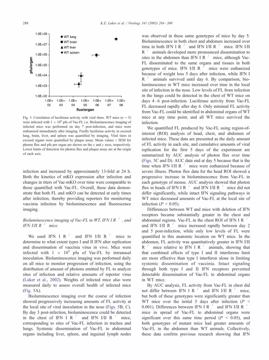
Fig. 3. Correlation of luciferase activity with viral titers. WT mice (n = 5)
were infected with 1 � 106 pfu of Vac-FL i.n. Bioluminescence imaging of
infected mice was performed on day 7 post-infection, and mice were
euthanized immediately after imaging. Firefly luciferase activity in excised
lung, brain, liver, and spleen was quantified by imaging. Viral titers in
excised organs were quantified by plaque assay. Mean values T SEM for
photon flux and pfu per organ are shown on the x and y axes, respectively.
Lower limits of detection for photon flux and plaque assay are at the origin
of each axis.
K.E. Luker et al. / Virology 341 (2005) 284–300288
infection and increased by approximately 13-fold at 24 h.
Both the kinetics of mKO expression after infection and
changes in titers of Vac-mKO over time were comparable to
those quantified with Vac-FL. Overall, these data demon-
strate that both FL and mKO can be detected at early times
after infection, thereby providing reporters for monitoring
vaccinia infection by bioluminescence and fluorescence
imaging.
Bioluminescence imaging of Vac-FL in WT, IFN I R�/�, and
IFN I/II R�/� mice
We used IFN I R�/� and IFN I/II R�/� mice to
determine to what extent types I and II IFN alter replication
and dissemination of vaccinia virus in vivo. Mice were
infected with 1 � 105 pfu of Vac-FL by intranasal
inoculation. Bioluminescence imaging was performed daily
on all mice to monitor progression of infection, using the
distribution of amount of photons emitted by FL to analyze
sites of infection and relative amounts of reporter virus
(Luker et al., 2002). Weights of infected mice also were
measured daily to assess overall health of infected mice
(Fig. 5A).
Bioluminescence imaging over the course of infection
showed progressively increasing amounts of FL activity at
the local site of viral inoculation in the nose (Figs. 5B, C).
By day 3 post-infection, bioluminescence could be detected
in the chest of IFN I R�/� and IFN I/II R�/� mice,
corresponding to sites of Vac-FL infection in trachea and
lungs. Systemic dissemination of Vac-FL to abdominal
organs including liver, spleen, and inguinal lymph nodes
was observed in these same genotypes of mice by day 5.
Bioluminescence in both chest and abdomen increased over
time in both IFN I R�/� and IFN I/II R�/� mice. IFN I/II
R�/� animals developed more pronounced dissemination to
sites in the abdomen than IFN I R�/� mice, although Vac-
FL disseminated to the same organs and tissues in both
genotypes of mice. IFN I/II R�/� mice were euthanized
because of weight loss 5 days after infection, while IFN I
R�/� animals survived until day 6. By comparison, bio-
luminescence in WT mice increased over time in the local
site of infection in the nose. Low levels of FL from infection
in the lungs could be detected in the chest of WT mice on
days 4–6 post-infection. Luciferase activity from Vac-FL
FL decreased rapidly after day 6. Only minimal FL activity
from Vac-FL could be identified in abdominal organs of WT
mice at any time point, and all WT mice survived the
infection.
We quantified FL produced by Vac-FL using region-of-
interest (ROI) analysis of head, chest, and abdomen of
infected mice. These data are presented as the daily amount
of FL activity in each site, and cumulative amounts of viral
replication for the first 5 days of the experiment are
summarized by AUC analysis of photon flux over time
(Figs. 5C and D). AUC data end at day 5 because that is the
time when IFN I/II R�/� mice were euthanized because of
severe illness. Photon flux data for the head ROI showed a
progressive increase in bioluminescence from Vac-FL in
each genotype of mouse. AUC analysis showed that photon
flux in heads of IFN I R�/� and IFN I/II R�/� mice did not
differ significantly, while intact IFN signaling pathways in
WT mice decreased amounts of Vac-FL at the local site of
infection (P < 0.05).
Differences between WT and mice with deletion of IFN
receptors became substantially greater in the chest and
abdominal regions. Vac-FL in the chest ROI of IFN I R�/�
and IFN I/II R�/� mice increased rapidly between day 2
and 5 post-infection, while only low levels of FL were
quantified in this anatomic location on WT mice. In the
abdomen, FL activity was quantitatively greater in IFN I/II
R�/� mice relative to IFN I R�/� animals, showing that
that combined effects of type I and II interferons limit
are more effective than type I interferon alone in limiting
systemic dissemination of vaccinia. Intact signaling
through both type I and II IFN receptors prevented
detectable dissemination of Vac-FL to abdominal organs
in WT mice.
By AUC analysis, FL activity from Vac-FL in chest did
not differ between IFN I R�/� and IFN I/II R�/� mice,
but both of these genotypes were significantly greater than
WT mice over the initial 5 days after infection (P <
0.001). Differences between IFN I R�/� and IFN I/II R�/�
mice in spread of Vac-FL to abdominal organs were
significant over this same time period (P < 0.05), and
both genotypes of mutant mice had greater amounts of
Vac-FL in the abdomen than WT animals. Collectively,
these data confirm previous research showing that IFN

Fig. 4. Kinetics of reporter gene expression from Vac-FL and Vac-mKO. Vero cells were infected with Vac-FL (A) or Vac-mKO (B) at an MOI of 5. Luciferase
activity or fluorescence in infected cells was quantified at various times post-infection by bioluminescence imaging (A) or flow cytometry (B), respectively (n =
3 for Vac-FL and n = 2 for Vac-mKO per time point). Viral titers also were quantified in infected cells at these same time points. Values are mean T SEM for
photon flux (A). Histograms from infected samples are shown after subtraction of the histogram for uninfected cells (B). Numeric values for mean fluorescence
intensity, percent orange cells, and pfu/ml at each time point are tabulated. Data for Vac-FL are representative of 2 independent experiments, while results for
Vac-mKO are from a single experiment.
K.E. Luker et al. / Virology 341 (2005) 284–300 289
signaling limits replication of vaccinia virus in lungs (van
den Broek et al., 1995). Type I IFN confers most IFN-
mediated host immunity to vaccinia virus, although
combined functions of types I and II IFN further restrict
systemic spread of vaccinia. In addition, our results show
that IFN signaling regulates dissemination of vaccinia
virus to intraabdominal organs including liver and spleen.
Because of the predominant function of type I IFN
receptors in vaccinia infection, we focused our subsequent
studies on IFN I R�/� and WT mice.

K.E. Luker et al. / Virology 341 (2005) 284–300290
Vac-FL produces focal infections in lungs and brain
Bioluminescence imaging in living mice showed focal
areas of increased luciferase activity in lungs rather than a
uniform distribution of Vac-FL. This pattern of infection
was detected in WT and IFN I R�/� mice. To verify this
finding, we rapidly removed lungs of infected mice
immediately after in vivo imaging at various times between
1 and 7 days post-infection and imaged luciferase activity in
excised lungs. This strategy exploits the fact that bio-
luminescence from luciferase can be detected in tissues after
Fig. 5. Bioluminescence imaging of Vac-FL in WT, IFN I R�/�, and IFN I/II R�/
FL i.n. (A) Weights of animals are expressed as mean values T SEM for percent
the graph when animals were euthanized because of disease severity. (B) Biolu
depicted with a pseudocolor scale using red as the highest and blue as the lowe
images with the minimum value at background noise of the instrument and a un
yellow arrow, lymph node. (C) Luciferase activity from Vac-FL was quantified
values for photon flux T SEM. (D) AUC analyses of photon flux for each genoty
*P < 0.05; **P < 0.001.
euthanizing animals. Bioluminescence could not be detected
in excised lungs 1 day after intranasal infection (data not
shown). On subsequent days, specimens from WT and IFN I
R�/� mice both showed focal areas of luciferase activity
from Vac-FL in lungs (Figs. 6A and B). The amount of
luciferase activity increased over time and spread to
adjacent lung tissue. However, the multifocal pattern of
Vac-FL persisted on all subsequent days, which is consistent
with aspiration of Vac-FL administered intranasally.
We analyzed microscopic sites of vaccinia infection in
lungs by infecting WT and IFN I R�/� mice with 1 � 106 or
� mice. Mice (n = 5 per genotype) were infected with 1 � 106 pfu of Vac
initial weight for each genotype of mouse. Weights for a genotype end on
minescence imaging of mice infected with Vac-FL. Luciferase activity is
st levels of photon flux, respectively. The pseudocolor scale is set for al
iform level of maximum photon flux. White arrow, lung; red arrow, liver
by ROI analysis of the head, chest, and abdomen. Data represent mean
pe of mouse over the first 5 days after infection. Error bars represent SEM
-
l
;
.

Fig. 5 (continued ).
K.E. Luker et al. / Virology 341 (2005) 284–300 291
1 � 105 pfu, respectively, of Vac-mKO. Infected lungs were
removed daily between 1 and 4 days after intranasal infection.
Fluorescence from Vac-mKO infection initially could be
detected on day 2 following infection. Sites of viral infection
localized predominantly to bronchioles in the lung, as
evidenced by fluorescence microscopy and confirmed by
hematoxylin and eosin staining of adjacent sections (Figs.
6C–E). An inflammatory infiltrate composed primarily of
mononuclear cells was identified adjacent to an infected
bronchiole. Similar to bioluminescence data, Vac-mKO
produced a multifocal pattern of infection with areas of
uninfected lung positioned between discontiguous sites of
infection. Collectively, these data show that intranasal
infection with vaccinia produces multiple foci of infection
in lungs and suggest that the infection may begin in
bronchiolar epithelium.
Ex vivo imaging of brains removed from infected WT
and IFN I R�/� mice repeatedly demonstrated that luciferase
activity was restricted to a focal area in the anterior, inferior
aspect of the frontal lobes (Fig. 7A). This localized area of
bioluminescence in the brain was not detectable in living
mice because of the high levels of Vac-FL in the
nasopharynx and sinuses of mice following intranasal
inoculation. To confirm this pattern of vaccinia infection
in the brain, we used intranasal inoculation of Vac-mKO.
Fluorescence microscopy of tissue sections from brain
K.E. Luker et al. / Virology 341 (2005) 284–300292
showed focal orange fluorescence in the same distribution
identified by bioluminescence imaging (Fig. 7B). Focal
infection of the frontal lobes following intranasal inocu-
lation of vaccinia potentially could be due to an inability of
Fig. 6. Focal vaccinia infection in lungs. IFN I R�/�mice were infected with
1� 105 pfu Vac-FL or Vac-mKO i.n. (A) Bioluminescence image obtained 4
days post-infection shows focal areas of increased luciferase activity in lungs
(arrows). Infection also is present in the spleen (asterisk). (B) Ex vivo image
of lungs demonstrates corresponding focal areas of Vac-FL infection. (C)
Fluorescence microscopy (4� objective) shows Vac-mKO localized primar-
ily to bronchioles. The arrow shows a bronchiole that was examined at higher
magnification (40� objective) in panel D. (D) Hematoxylin and eosin
staining shows a predominantly mononuclear cell infiltrate adjacent to the
bronchiole of a WT mouse infected with Vac-FL. (E) A lung specimen from
an uninfected mouse is shown for comparison.
virus to infect other parts of the brain. We investigated this
possibility by infecting IFN I R�/� mice with 1 � 104 pfu
Vac-FL by direct intracranial injection of virus into the left
parietal lobe. Following direct injection of virus, Vac-FL
spread to other parts of the brain within 2 days, as monitored
by in vivo and ex vivo bioluminescence imaging (Figs. 7C
and D). These data show that focal infection of the anterior,
inferior aspects of the frontal lobes after intranasal infection
is not caused by resistance of other parts of the brain to
vaccinia infection.
Vaccinia infection is limited to a greater extent by type I IFN
receptors on parenchymal cells relative to hematopoietic
cells
Studies in WT and IFN I R�/� mice showed that
signaling through type I IFN receptors was essential for
limiting local replication of vaccinia and subsequent
systemic dissemination. To determine the extent to which
type I IFN limits vaccinia infection through effects on
hematopoietic cells versus parenchymal cells in organs and
tissues, we established chimeric mice by transferring bone
marrow between WT and IFN I R�/� mice. We also
transplanted bone marrow from WT to WT animals as a
control. Mice were infected with 1 � 105 pfu Vac-FL i.n. 6
weeks after bone marrow transplantation. As additional
controls, age-matched WT and IFN I R�/� mice that had not
undergone bone marrow transplantation were infected with
the same inoculum of Vac-FL. Viral replication and
dissemination were monitored daily with bioluminescence
imaging, and data for photon flux were analyzed by AUC
analysis for the 7 days that all animals survived.
AUC analysis of photon flux in the head, which is
dominated by Vac-FL in the intranasal site of infection,
showed that IFN I R�/� mice had significantly higher
luciferase activity from Vac-FL than the other groups for
days 1 to 7 post-infection (P < 0.05; Fig. 8A). Comparable
amounts of bioluminescence were quantified in heads of
IFN I R�/� mice transplanted with WT bone marrow, IFN I
R�/� to WT transplants, and WT to WT mice. WT mice had
the lowest calculated value for head AUC (P < 0.05).
Differences among groups of mice were larger for AUC data
calculated from images of the chest and abdomen. In the
chest, AUC for photon flux was highest in the WT to IFN I
R�/� transplant and IFN I R�/� mice, while AUC in the

Fig. 7. Focal vaccinia infection in brain. (A) Ex vivo bioluminescent image
of the ventral surface of the brain shows focal bioluminescence (arrow) in
an IFN I R�/� 7 days post-infection with 1 � 106 pfu Vac-FL i.n. (B)
Hematoxylin and eosin staining of a section obtained from the ventral
surface of the frontal lobe (4� objective). The circled area in panel B was
examined at higher magnification (20� objective) for Vac-mKO by
fluorescence microscopy on an immediately adjacent section. (C) Bio-
luminescence image of IFN I R�/� mouse injected intracranially with 1 �104 pfu Vacl-FL shows dissemination of virus throughout the brain on day 2
post-infection. (D) Ex vivo bioluminescent image of the brain of the same
mouse removed immediately after in vivo imaging.Fig. 8. Vac-FL infection in WT and IFN I R�/� bone marrow transplant
mice. WT, WT bone marrow to WT, WT bone marrow to IFN I R�/�,
IFN I R�/� bone marrow to WT, and IFN I R�/� mice (n = 5–6 per
condition) were infected with 1 � 105 pfu Vac-FL i.n. Bioluminescence
imaging was performed daily, and replication and spread of Vac-FL were
quantified by ROI analysis. AUC data for days 1–7 post-infection are
presented for defined ROIs in head, chest, and abdomen. Error bars
represent SEM. *P < 0.05; **P < 0.01; #P < 0.001.
K.E. Luker et al. / Virology 341 (2005) 284–300 293
abdomen was greatest for IFN I R�/� mice (P < 0.05). AUC
data in chest and abdomen for WT to IFN I R�/� mice and
untransplanted IFN I R�/� animals were significantly
greater than all other mice (P < 0.001). IFN I R�/� to
WT transplant mice were more susceptible to vaccinia
infection than WT mice as quantified by AUC of photon
flux in the chest and abdomen (P < 0.01). AUC values in
these sites also were significantly greater in WT to WT
transplant mice as compared with WT animals (P < 0.01).
The number of days that groups of mice survived after
intranasal infection corresponded with AUC data for photon
flux from Vac-FL. IFN I R�/� mice with WT bone marrow
and IFN I R�/� mice were euthanized on day 7 post-
infection because of weight loss of approximately 30% (Fig.
8B). IFN I R�/� to WT transplant mice and WT to WT mice
survived through day 9. WT mice lost approximately 12%
of body weight by day 7 post-infection, but these animals all
regained weight and survived. Collectively, these data show
that protective effects of type I IFN against vaccinia virus
are mediated to a quantitatively greater extent through
parenchymal tissues as compared with hematopoietic cells.

K.E. Luker et al. / Virology 341 (2005) 284–300294
Lethal infection with Vac-FL produces comparable amounts
of virus in lungs and brain of WT and IFN I R�/� mice
Data from in vivo experiments showed that signaling
through type I IFN receptors is an essential component of
host immunity to vaccinia virus. When infected with the
same input pfu, Vac-FL replicated to a greater extent in IFN I
R�/� mice than WT animals. Notably, systemic dissem-
ination of virus to anatomic sites including liver, spleen, and
lymph nodes was significantly more extensive in IFN I R�/�
mice. Potentially, this result could be caused by a function of
type I IFN to limit dissemination of vaccinia to these organs
and tissues independent of amounts of viral replication.
Alternatively, it was possible that type I IFN signaling
produced a relative barrier to systemic dissemination of
vaccinia, and comparable spread of virus beyond the lungs
would occur in WT mice infected with a lethal inoculum of
V-FL.
To distinguish between these possible effects of signaling
through type I IFN receptors, we infected IFN I R�/� and
WT mice with amounts of virus that produced comparable
progression to lethality in both groups. Intranasal infection
of WT and IFN I R�/� mice with 1 � 106 and 9 � 104 pfu of
Vac-FL, respectively, produced comparable weight loss in
each genotype of mouse (Fig. 9A). Both groups of animals
were euthanized because of disease severity at 7 days after
infection.
Bioluminescence imaging showed that luciferase activity
quantified in head regions of WT mice was greater than IFN
I R�/� animals, consistent with the greater input inoculum
of Vac-FL (Figs. 9B and E). Luciferase activity from Vac-FL
in the chest was comparable at all times after infection, and
total AUC for photon flux in the chest did not differ
significantly between the two genotypes (Figs. 9C and E).
By comparison, spread of Vac-FL to abdominal organs and
tissues was significantly greater in IFN I R�/� mice relative
to WT animals (P < 0.01), even when viral replication in the
chest and the overall weight loss associated with infection
were the same (Figs. 9D and E).
We used ex vivo imaging of excised organs and quan-
tification of viral titers on day 7 post-infection to validate
data from in vivo imaging. Luciferase activity and viral titers
in spleen and liver of IFN I R�/� mice were approximately
10-fold greater than those quantified in WT animals (P <
0.001; Figs. 9F and G). Amounts of virus in blood were
slightly higher in WT mice, although these differences were
not statistically significant. These data for viral titers in blood
suggest that lack of receptors for type IFN did not limit
dissemination of virus from lungs under conditions in which
Fig. 9. Type I IFN signaling affects dissemination of Vac-FL to liver and spleen wi
infected i.n. with 1 � 106 and 9 � 104 pfu of Vac-FL, respectively, to produc
mortality. (A) Data for animal weights are presented as mean values T SEM f
quantified from bioluminescence imaging data on days 1–7 post-infection in head
in head, chest, and abdomen ROIs for each genotype of mouse. (F) Photon flux
detection for titers. *P < 0.01; **P < 0.001.
overall progression of disease was comparable (Fig. 9G). By
comparison, bioluminescence and titers of Vac-FL in lung
and brain did not differ significantly on day 7 post-infection,
which is the time when both genotypes of mice were
euthanized because of the severity of disease (Figs. 9F and
G). These data imply that viral infection and associated
pathology in lungs and/or brain may be key determinants of
lethality, independent of dissemination of virus to other
organs and tissues.
Discussion
In vivo studies of poxviruses in the integrated physiology
of a living animal are essential to understanding the cellular
and molecular determinants of pathogenesis. Conventional
studies of viral-host pathogenesis are based on infecting
large numbers of animals and euthanizing subsets of
animals at various time points to collect organs to determine
localization and titers of virus. This approach requires large
numbers of animals and precludes direct correlations
between viral replication and the outcome of disease in
individual animals. In addition, anatomic localization of
infection within an organ and/or dissemination of virus to
unexpected sites may be missed by harvesting selected
organs and titering total amounts of virus. Previous studies
by our laboratory and others have demonstrated that
imaging can be used to investigate viral infection in mouse
models and identify new aspects of pathogenesis in vivo. To
enable imaging studies of vaccinia virus, we have developed
new recombinant viruses with fluorescent or bioluminescent
reporter proteins. A key feature of these recombinant
vaccinia viruses is that they are not attenuated in vitro or
in vivo, making these viruses important new tools for
analyzing host–pathogen interactions in vaccinia infection.
Vac-FL and Vac-mKO allow complementary approaches
for imaging vaccinia infection. Vac-FL exploits the sensi-
tivity of firefly luciferase to allow sensitive detection of virus
in vitro and in vivo. Luciferase activity from Vac-FL is
directly proportional to titers of vaccinia virus, allowing rela-
tive differences in amounts of virus in various anatomic sites
to be quantified with bioluminescence imaging. This reporter
virus builds upon previous work with recombinant vaccinia
viruses that express firefly luciferase. Rodriguez et al. con-
structed a virus with firefly luciferase replacing the thymidine
kinase gene in vaccinia and showed that as few as 10�6 pfu
per cell could be detected in homogenates of cells 18 h after
infection (Rodriguez et al., 1988). This virus also provided
sensitive detection of vaccinia in homogenates of infected
thout affecting overall survival. WT and IFN I R�/� mice (n = 5 each) were
e comparable rates of overall infection as determined by weight loss and
or percent initial weight on each day post-infection. (B–D) Photon flux
(B), chest (C), and abdomen (D). (E) AUC data summarizing photon flux
and (G) viral titers in excised organs. 30 pfu/g tissue is the lower limits of

K.E. Luker et al. / Virology 341 (2005) 284–300 295
tissues from mice. The current Vac-FL has the advantage of
retaining full virulence in vivo, and it likely provides greater
sensitivity for detecting viruses because the firefly luciferase
in Vac-FL has improved expression in mammalian cells.
Previous studies with transgenic mice that express firefly
luciferase also demonstrate a high correlation among pre-
sence of luciferase, bioluminescence produced in vivo, and
luciferase activity in homogenates of excised tissues (Zhang

K.E. Luker et al. / Virology 341 (2005) 284–300296
et al., 2001). Collectively, these data support the use of
luciferase activity produced by Vac-FL as a sensitive reporter
of vaccinia virus in living mice and for in vitro assays.
Vac-mKO enables facile detection of anatomic sites
infected with vaccinia using fluorescence imaging. Using an
orange fluorescent protein shifts fluorescence from the
reporter away from autofluorescence that can limit detection
of the commonly used GFP reporter in tissues. The
relatively high quantum yield of fluorescence from mKO
also enhances detection of Vac-mKO in tissue specimens
(Karasawa et al., 2004). While we have used Vac-mKO to
detect vaccinia in tissue sections, the favorable properties of
this fluorescent reporter should enhance detection of Vac-
mKO for future studies of the immune response to vaccinia
with intravital two-photon microscopy. In addition, it should
be feasible to construct a reporter virus that maintains the
virulence of wild-type WR while expressing both FL and a
fluorescent protein either as separate proteins or as a fusion
protein. We currently are developing this type of recombi-
nant vaccinia virus to enable macroscopic and microscopic
imaging of infection in the same animal.
We have used these reporter viruses to investigate effects
of IFN on pathogenesis of vaccinia virus in mice. In
particular, we focused on functions of type I IFN in host
resistance to vaccinia. Type I IFN confers an approximately
10-fold reduction in the amount of virus necessary to
produce lethal infection in mice. These data confirm the
established functions of type I IFN in limiting replication of
vaccinia and other viruses. However, we have determined
that type I IFN also affects dissemination of virus from the
respiratory system to systemic sites. Even when infected
with amounts of virus that produce comparable progression
of overall disease, IFN I R�/� mice have significantly
higher amounts of virus in liver and spleen than WT
animals at the time of death. Because total amounts of
vaccinia virus in blood are similar between both genotypes
of mice, these data suggest that type I IFN affects the
intrinsic susceptibility of cells within liver and spleen to
infection with vaccinia. However, additional studies with
isolated cultures of various types of primary cells from
these organs will be needed to separate effects of dissem-
ination from intrinsic susceptibility of cells to vaccinia. Our
imaging data with IFN I/II R�/� mice also showed similar
spread of Vac-FL to liver and spleen after intranasal
infection, although levels of bioluminescence in these
organs were higher in IFN I/II R�/� animals relative to
IFN I R�/� animals. Collectively, these data demonstrate a
key function of type I IFN for dissemination of vaccinia
virus to liver and spleen.
Our results with vaccinia viruses are consistent with
other recent studies that demonstrate a function of IFN
pathways in dissemination of viruses. For example,
coxsackievirus 3B replicates selectively in the heart of
wild-type mice without causing lethality. IFN I R�/� mice
infected with coxsackievirus 3B have comparable titers of
virus in the heart, but greatly enhanced spread of virus to
the liver causes death in these mutant mice (Wessely et
al., 2001). Ryman et al. demonstrated that Sindbis virus
disseminated to macrophage-dendritic cells in various
organs in IFN I R�/� mice, while these sites were not
infected with Sindbis in WT mice (Ryman et al., 2000).
Similarly, we have shown that absence of type I IFN
signaling permits HERPES simplex virus type I to spread
from a mucosal site of infection to liver and spleen
(Luker et al., 2003).
Despite the function of type I IFN to limit spread of
vaccinia virus to systemic organs such as liver and spleen,
our data suggest that vaccinia infection in lungs and/or brain
may be more important determinants of mortality in mice.
When the input inoculum of vaccinia virus was adjusted to
produce comparable overall progression of disease as
monitored by weight loss and time to death, WT and IFN
I R�/� mice had equal amounts of vaccinia virus in lung and
brain, as quantified by bioluminescence and viral titer.
Although these data do not allow us to distinguish the
relative contributions of viral replication versus immune-
mediated damage, our results imply that lethality is due to
pathology in the lungs and/or brain.
The combination of in vivo imaging and fluorescence
microscopy shows that vaccinia produces focal infection in
lungs and brain of infected mice. Focal infection in lungs
could be anticipated based on patterns of aspiration of liquids
into dependent portions of anesthetized animals (Franquet et
al., 2000). However, localization of vaccinia to the anterior,
inferior aspects of the frontal lobes of the brain after
intranasal inoculation was unexpected. This site of vaccinia
infection in the brain is adjacent to the olfactory bulbs,
suggesting that the mechanism of neuroinvasion was direct
spread of virus from the nasal mucosa via the olfactory
system. The potential for vaccinia to enter the brain by direct
cell-to-cell spread rather than hematogenous dissemination is
supported by previous research with vaccinia and cowpox
(Martinez et al., 2000; Turner, 1967b). By histology, cowpox
antigens were detected in perineural fibroblasts adjacent to
the olfactory nerve, and viral antigens extended to the
leptomeninges surrounding the olfactory tract. A variety of
other viruses, including influenza A and vesicular stomatitis
virus, also appear to enter the brain directly along the
olfactory nerves (Aronsson et al., 2003; Plakhov et al., 1995).
By comparison, Sindbis virus and Venezuelan equine
encephalitis virus are believed to spread from blood to the
nasal neuroepithelium before entering the brain (Charles et
al., 1995; Cook and Griffin, 2003). A more detailed study of
the kinetics and microscopic localization of vaccinia in the
nervous systemwill be necessary to establish definitively that
spread through the olfactory route is a mechanism of
neuroinvasion by vaccinia.
We used a bone marrow transplant approach to establish
the extent to which signaling through receptors for type I
IFN in parenchymal cells contributes to the innate immune
response to vaccinia infection. IFN I R�/� mice transplanted
with WT bone marrow were significantly more susceptible

K.E. Luker et al. / Virology 341 (2005) 284–300 297
to Vac-FL than WT mice that received bone marrow from
IFN I R�/� mice. However, type I IFN signaling in both the
parenchymal and hematopoietic compartments was neces-
sary for mice to have normal immunity to vaccinia. Our
results are consistent with similar studies showing that
innate immune responses activated by toll receptors in both
parenchymal tissues and hematopoietic cells are essential
for normal responses to bacterial and viral pathogens (Sato
and Iwasaki, 2004; Schilling et al., 2003). Collectively,
these data demonstrate the critical function of parenchymal
cells in the IFN-mediated immune response to vaccinia
infection and emphasize the interrelated functions of IFN in
hematopoietic and parenchymal cells.
Interestingly, the bone marrow transplant experiments
also showed that WT to WT transplant mice were more
susceptible to vaccinia infection than WT animals that did
not undergo bone marrow transplantation. Potentially, this
result may be due to residual damage to parenchymal tissues
and endothelium as a result of irradiation used to prepare for
bone marrow transplantation (Giaid et al., 2003). However,
previous studies suggest that irradiation may alter immune
control of pathogens. Following total body gamma-irradi-
ation, mice become susceptible to pulmonary infection with
cytomegalovirus because of defective responses of T
lymphocytes (Reddehase et al., 1985). Mice that have
undergone total body irradiation and bone marrow trans-
plantation also are more susceptible to infection with
Pseudomonas aeruginosa after intratracheal inoculation of
the organism. Greater replication of Pseudomonas aerugi-
nosa was associated with impaired phagocytosis of alveolar
macrophages, reduced levels of specific cytokines, and
lower levels of selected integrins (Ojielo et al., 2003). These
data suggest that specific immune defects caused by the
procedure of bone marrow transplantation impair the host
response to vaccinia infection. This susceptibility is relevant
to development of improved vaccines against smallpox and
their possible use in the general population. Mechanisms
that cause the increased susceptibility of bone marrow
transplant recipients to vaccinia investigation are under
investigation in our laboratory.
While the current study demonstrates numerous advan-
tages of imaging for investigating vaccinia pathogenesis in
vivo, it also shows limitations of the present technology.
Current instruments for bioluminescence imaging provide
two-dimensional images. This limitation may be overcome
in part by obtaining images from several different projec-
tions to localize sites of bioluminescence in three-dimen-
sional space. However, the focal infection of the brain was
obscured by high amounts of Vac-FL in the nasopharynx
and paranasal sinuses of mice following intranasal inocu-
lation of virus. We anticipate that this type of problem with
bioluminescence imaging will be substantially improved as
three-dimensional imaging systems become available com-
mercially (Wang et al., 2004).
In summary, we have established two new reporter
viruses for imaging vaccinia virus pathogenesis. Impor-
tantly, the method used to engineer these viruses does not
attenuate vaccinia in vitro and in vivo, and these viruses can
be engineered further to delete specific target genes of
interest. The imaging reporters validated in this study
enabled us to detect unexpected patterns of vaccinia
infection in vivo and establish effects of type I IFN on
viral dissemination. Beyond the role of imaging demon-
strated in the current study, other research has shown that
BLI and magnetic resonance imaging (MRI) also can be
used to monitor responses of defined populations of immune
cells in vivo (Cao et al., 2004; Dubey et al., 2003; Edinger et
al., 2003; Kircher et al., 2003). We anticipate that combined
imaging studies of vaccinia infection and the host immune
response to the virus will enhance greatly in vivo studies of
pathogenesis and facilitate testing of candidate vaccines and
anti-viral compounds in vivo.
Materials and methods
Cells
Vero cells were cultured in DMEM medium with 10%
heat-inactivated fetal bovine serum, 1% l-glutamine, and
0.1% penicillin–streptomycin in a 5% CO2 incubator at
37 -C.
Recombinant viruses
Recombinant viruses were constructed in vRB12, a
WR strain of vaccinia with deletion of the F13L gene for
vp37 (Blasco and Moss, 1991). The RB21 transfer vector
is designed to insert a foreign gene and restore a
functional vp37 protein following homologous recombi-
nation with vRB12 (Blasco and Moss, 1995). To prepare
the transfer plasmid for firefly luciferase (FL), we
digested FL from pGL3 Basic (Promega) with XbaI and
blunted with Klenow. pGL3 basic then was digested with
NheI and ligated to the NheI and SmaI sites in pRB21.
Monomeric orange fluorescent protein (mKO; Medical and
Biological Laboratories, Ltd.) was digested with EcoRI and
HindIII, respectively, and ligated to the corresponding sites
in pRB21.
2 � 105 Vero cells were plated in 35 mm dishes and
infected 1 day later with vRB12 at an MOI of 1. One hour
post-infection, cells were transfected with pRB21 contain-
ing FL or mKO, respectively. Transfection with pRB21
alone was used to rescue vp37 and produce a control virus
containing the early/late promoter in pRB21 but lacking a
reporter gene. Transfections were performed with Lipofect-
amine (Invitrogen) or Fugene 6 (Roche) according to the
manufacturer’s instructions. Cells were harvested 2 days
post-infection by scraping, and three cycles of freeze-thaw
were used to release viruses. Recombinant viruses were
prepared by infecting Vero cells as described previously
(Blasco and Moss, 1995). Recombinant viruses containing

K.E. Luker et al. / Virology 341 (2005) 284–300298
FL (Vac-FL) were selected based on restoration of normal
plaque formation 2 days post-infection, while recombinants
with mKO (Vac-mKO) were identified by fluorescence
microscopy. Viruses were plaque-purified three times before
preparing viral stocks (Earl et al., 1998).
Mouse strains
All mice were in a pure 129 Ev/Sv background (referred
to as wild type). Immunocompetent wild-type (WT) mice
were purchased from Taconic (Germantown). Mice deficient
in type I IFN receptors (IFN I R�/�) or type I and II IFN
receptors (IFN I/II R�/�) were bred at the University of
Michigan (Muller et al., 1994). Experiments were per-
formed with adult mice. Mice were 6–10 weeks old for all
studies except for the bone marrow transplant experiment, in
which mice were 7 weeks old at the time of transplantation
and 13 weeks old when infected. There were no effects of
age on susceptibility to vaccinia infection among the various
cohorts of adult mice. Both male and female mice were used
in all experiments except that only males were used for bone
marrow transplantation. Numbers of male and female mice
in various experimental groups were matched, although we
did not observe a difference between sexes of mice in
susceptibility to vaccinia infection.
Viral replication in cells
Vero cells were plated at 100,000 cells per well in 6-well
plates for multi-step growth of various viruses at an MOI of
0.1. Cells in triplicate wells were harvested for plaque assays
at various time points after infection by scraping cells into
culture medium. To quantify bioluminescence from Vac-FL
over time, Vero cells were plated at 400,000 cells per well in
35 mm dishes and infected with Vac-FL at an MOI of 5.
Bioluminescence from FL was quantified by adding 150 Ag/ml d-luciferin (Promega) to culture medium 10 min before
imaging dishes of cells with a cryogenically cooled CCD
camera system (IVIS, Xenogen Corporation). Triplicate
samples were imaged at various time points after infection.
Viral titers in infected cells were analyzed by plaque assay.
Bioluminescence was quantified by manually defined
region-of-interest (ROI) analysis, and data were expressed
as photon flux. The relationship between Vac-FL pfu and
bioluminescence was performed by infecting Vero cells
(400,000 cells per well in 6-well plates) with various
amounts of Vac-FL between 30 and 1 � 105 pfu. Bio-
luminescence was quantified by imaging as described above.
Fluorescence from Vac-mKO over time was measured by
infecting 35 mm dishes of Vero cells (400,000 cells per
dish) with Vac-mKO at an MOI of 5. Duplicate wells were
trypsinized to release cells for analysis of fluorescence by
flow cytometry at various times post-infection as shown in
the figure (FACScalibur, BD Biosciences). Mean fluores-
cence intensity was quantified at each time point. Data for
percent orange cells were analyzed by subtracting the
histogram for uninfected cells from the histograms obtained
from samples at various time points post-infection. Dupli-
cate wells infected in parallel were harvested for determi-
nations of viral titers by plaque assay.
Virus titration
Cells harvested from experiments in tissue culture were
lysed by three cycles of freezing and thawing prior to plaque
assays. Organs and tissues excised from mice at various
time points after infection were manually homogenized as
described previously (Luker et al., 2003). Liver, spleen,
brain, and lungs were homogenized in 3 ml of medium,
while lymph nodes and trachea were collected in 0.5 ml of
medium. Whole blood was obtained by cardiac puncture
and diluted with 1 ml of culture medium. Samples were
frozen and thawed three times prior to plaque assays.
Viral titers in cells and organs were assayed by serial
dilution on Vero cells plated at 400,000 cells per well in 6-
well plates. Plaque assays were performed according to a
standard protocol (Earl et al., 1998), and plaques were
counted 3 days after infection.
Mouse infection
All animal procedures were approved by the University
of Michigan University Committee on Use and Care of
Animals. For intranasal infection with vaccinia viruses,
mice were anesthetized with subcutaneous injection of
ketamine (100 mg/kg) and xylazine (15 mg/kg). Mice were
inoculated intranasally with 20 Al of virus diluted in
DMEM. Anesthetized mice were shaved with clippers to
decrease absorption and scattering of light for biolumines-
cence imaging. Weights of animals were determined
immediately prior to infection and then daily throughout
the course of each infection.
Bone marrow transplantation
Bone marrow was harvested from femora and tibiae of
donor mice and resuspended at a concentration of 2 � 107
cells per milliliter in sterile PBS. Recipient mice were
irradiated with a total of 11 Gy divided into two equal
doses separated by 3 h. Mice received 4 � 106 bone
marrow cells suspended in 200 Al PBS injected via a 30 g
needle into the retroorbital venous plexus. Mice were
housed in autoclaved cages and received autoclaved food
and water after transplantation. Experiments were per-
formed 6 weeks after transplantation of bone marrow.
Control mice that received PBS alone died approximately
10 days after irradiation.
Microscopic analysis of organs
Selected organs were excised from mice and frozen in
OCT compound. Lungs were inflated with 1 ml of a solution
K.E. Luker et al. / Virology 341 (2005) 284–300 299
containing 1% low melting point agarose in phosphate-
buffered saline immediately before removal. 6 Am frozen
sections were prepared from various tissues and fixed in 4%
paraformaldehyde. For fluorescence microscopy of Vac-
mKO, sections were incubated in 10 mM glycine for 10 min
to quench background fluorescence. Frozen sections adja-
cent to the corresponding fluorescence section were stained
with hematoxylin and eosin.
Bioluminescence imaging
Bioluminescence imaging was performed with a cry-
ogenically cooled CCD camera (IVIS) as described pre-
viously (Luker et al., 2002). ROIs corresponding to the
head, chest, and abdomen of infected mice were used to
quantify bioluminescence as photon flux. For some experi-
ments, mice were euthanized immediately after imaging,
and selected organs were dissected rapidly and imaged ex
vivo within 5 min to quantify bioluminescence on the
imaging system (Luker et al., 2003). Bioluminescence from
these organs was quantified by ROI analysis, and back-
ground bioluminescence was measured from the same size
ROI positioned adjacent to each organ. Background
subtracted photon flux was calculated for each organ. These
same organs were then analyzed for viral titers by plaque
assay.
Statistics
Pairs of data points were analyzed by t test for statisti-
cally significant differences (P < 0.05). Statistics for area-
under-the-curve analyses were performed with commer-
cially available software (Graphpad, Prism).
Acknowledgments
The authors thank Bernard Moss for providing vRB12
and pRB21 and reviewing the manuscript. The authors also
thank Myria Petrou and Bradley Foerster for help with
animal experiments. Research was supported in part by NIH
grant P50-CA093990-04. Support for imaging experiments
was provided by NIH R24CA083099 for the University of
Michigan Small Animal Imaging Resource.
References
Aronsson, F., Robertson, B., Ljunggren, H., Kristensson, K., 2003. Invasion
and persistence of the neuroadapted influenza virus A/WSN/33 in the
mouse olfactory system. Viral. Immunol. 16 (3), 415–423.
Blasco, R., Moss, B., 1991. Extracellular vaccinia virus formation and cell-
to-cell virus transmission are prevented by deletion of the gene
encoding the 37,000-Dalton outer envelope protein. J. Virol. 65 (11),
5910–5920.
Blasco, R., Moss, B., 1995. Selection of recombinant vaccinia viruses on
the basis of plaque formation. Gene 158 (2), 157–162.
Brandt, T., Jacobs, B., 2001. Both carboxy- and amino-terminal domains of
the vaccinia virus interferon resistance gene, E3L, are required for
pathogenesis in a mouse model. J. Virol. 75 (2), 850–856.
Breman, J., Henderson, D., 2002. Diagnosis and management of smallpox.
N. Engl. J. Med. 346 (17), 1300–1308.
Buller, R., Smith, G., Cremer, K., Notkins, A., Moss, B., 1985. Decreased
virulence of recombinant vaccinia virus expression vectors is
associated with a thymidine kinase-negative phenotype. Nature 317,
813–815.
Cao, Y., Wagers, A., Beilhack, A., Dusich, J., Bachman, M., Negrin, R.,
Weissman, I., Contag, C., 2004. Shifting foci of hematopoiesis during
reconstitution from single stem cells. Proc. Natl. Acad. Sci. U.S.A. 101
(1), 221–226.
Charles, P., Walters, E., Margolis, F., Johnston, R., 1995. Mechanism of
neuroinvasion of Venezuelan equine encephalitis virus in the mouse.
Virology 208 (2), 662–671.
Cook, S., Griffin, D., 2003. Luciferase imaging of a neurotropic viral
infection in intact animals. J. Virol. 77 (9), 5333–5338.
Coupar, B., Oke, P., Andrew, M., 2000. Insertion sites for recombinant
vaccinia virus construction: effects on expression of a foreign protein.
J. Gen. Virol. 81, 431–439.
Dubey, P., Su, H., Adonai, N., Du, S., Rosato, A., Braun, J., Gambhir, S.,
Witte, O., 2003. Quantitative imaging of the T cell antitumor response
by positron-emission tomography. Proc. Natl. Acad. Sci. U.S.A. 100
(3), 1232–1237.
Earl, P., Cooper, N., Wyatt, L., Moss, B., 1998. Preparation of cell cultures
and vaccinia virus stocks. In: Ausubel, F.M., Brent, R., Kingston, R.E.,
Moore, D.D., Seidman, J.G., Smith, J.A., Struhl, K. (Eds.), Current
Protocols in Molecular Biology. John Wiley and Sons, Inc., pp.
16.16.1–16.16.11.
Edinger, M., Cao, Y., Verneris, M., Bachman, M., Contag, C., Negrin, R.,
2003. Revealing lymphoma growth and the efficacy of immune cell
therapies using in vivo bioluminescence imaging. Blood 101 (2),
640–648.
Farrar, M., Schreiber, R., 1993. The molecular cell biology of interferon-
gamma and its receptor. Annu. Rev. Immunol. 11, 571–611.
Franquet, T., Gimenez, A., Roson, N., Torrubia, S., Sabate, J., Perez, C.,
2000. Aspiration diseases: findings, pitfalls, and differential diagnosis.
Radiographics 20 (3), 673–685.
Fujii, S., Shimizu, K., Kronenberg, M., Steinman, R., 2002. Prolonged IFN-
gamma-producing NKT response induced with alpha-galactosylcera-
mide-loaded DCs. Nat. Immunol. 3, 867–874.
Giaid, A., Lehnert, S., Chehayeb, B., Chehayeb, D., Kaplan, I., Shenouda,
G., 2003. Inducible nitric oxide synthase and nitrotyrosine in mice with
radiation-induced lung damage. Am. J. Clin. Oncol. 26 (4), e67–e72.
Harrison, S., Alberts, B., Ehrenfeld, E., Enquist, L., Fineberg, H.,
Mcknight, S., Moss, B., O’Donnell, M., Ploegh, H., Schmid, S., Walter,
K., Theriot, J., 2004. Discovery of antivirals against smallpox. Proc.
Natl. Acad. Sci. U.S.A. 101 (31), 11178–11192.
Huang, H., Hendriks, W., Althage, A., Hemmi, S., Bluethmann, H.,
Kamijo, R., Vilcek, J., Zinkernagel, R., Aguet, M., 1993. Immune
response in mice that lack the interferon-gamma receptor. Science 259
(5102), 1742–1745.
Karasawa, S., Araki, T., Nagai, T., Mizuno, H., Miyawaki, A., 2004. Cyan-
emitting and orange-emitting fluorescent proteins as a donor/acceptor
pair for fluorescence resonance energy transfer. Biochem. J. 381 (Pt. 1),
307–312.
Katze, M., Yupeng, H., Gale, M., 2002. Viruses and interferon: a fight for
supremacy. Nat. Rev., Immunol. 2, 675–687.
Kircher, M., Allport, J., Graves, E., Love, V., Josephson, L., Lichtman, A.,
Weissleder, R., 2003. In vivo high resolution three-dimensional imaging
of antigen-specific cytotoxic T-lymphocyte trafficking to tumors.
Cancer Res. 63 (20), 6838–6846.
Langland, J., Jacobs, B., 2002. The role of the PKR-inhibitory genes, E3L
and K3L, in determining vaccinia virus host range. Virology 299 (1),
133–141.
Luker, G., Bardill, J., Prior, J., Pica, C., Piwnica-Worms, D., Leib, D., 2002.

K.E. Luker et al. / Virology 341 (2005) 284–300300
Noninvasive bioluminescence imaging of herpes simplex virus type 1
infection and therapy in living mice. J. Virol. 76 (23), 12149–12161.
Luker, G., Prior, J., Song, J., Pica, C., Leib, D., 2003. Bioluminescence
imaging reveals systemic dissemination of HSV-1 in the absence of
interferon receptors. J. Virol. 77, 11082–11093.
Marie, I., Durbin, J., Levy, D., 1998. Differential viral induction of distinct
interferon-alpha genes by positive feedback through interferon regu-
latory factor-7. EMBO J. 17, 6660–6669.
Martinez, M., Bray, M., Huggins, J., 2000. A mouse model of aerosol-
transmitted orthopoxviral disease: morphology of experimental aerosol-
transmitted orthopoxviral disease in a cowpox virus-BALB/c mouse
system. Arch. Pathol. Lab. Med. 124 (3), 362–377.
Muller, U., Steinhoff, L., Reis, S., Hemmi, S., Pavlovic, J., Zinkernagel, R.,
Aguet, M., 1994. Functional role of type I and type II interferons in
antiviral defense. Science 264, 1918–1921.
Ojielo, C., Cooke, K., Mancuso, P., Standiford, T., Olkiewicz, K., Clouthier,
S., Corrion, L., Ballinger, M., Towes, G., Paine, R.r., Moore, B., 2003.
Defective phagocytosis and clearance of Pseudomonas aeruginosa in
the lung following bone marrow transplantation. J. Immunol. 171 (8),
4416–4424.
Ortiz, A., Justo, P., Sanz, A., Merlero, R., Caramelo, C., Guerrero, M.,
Strutz, F., Muller, G., Barat, a., Egido, J., 2005. Tubular cell
apoptosis and cidofovir-induced acute renal failure. Antivir. Ther. 10
(1), 185–190.
Plakhov, I., Arlund, E., Aoki, C., Reiss, C., 1995. The earliest events in
vesicular stomatitis virus infection of the murine olfactory neuro-
epithelium and entry of the central nervous system. Virology 209 (1),
257–262.
Reddehase, M., Weiland, F., Munch, K., Jonjic, S., Luske, A., Koszinowski,
U., 1985. Interstitial murine cytomegalovirus pneumonia after irradi-
ation: characterization of cells that limit viral replication during
established infection of the lungs. J. Virol. 55 (2), 264–273.
Rodriguez, J., Rodriguez, D., Rodriguez, J., McGowan, E., Esteban, M.,
1988. Expression of the firefly luciferase gene in vaccinia virus: a
highly sensitive gene marker to follow virus dissemination in tissues of
infected animals. Proc. Natl. Acad. Sci. U.S.A. 85 (5), 1667–1671.
Ryman, K., Klimstra, W., Nguyen, K., Biron, C., Johnston, R., 2000.
Alpha/beta interferon protects adult mice from fatal Sindbis virus
infection and is an important determinant of cell and tissue tropism.
J. Virol. 74 (7), 3366–3378.
Samuel, C., 1998. Reoviruses and the interferon system. Curr. Top.
Microbiol. Immunol. 223, 125–145.
Sato, A., Iwasaki, A., 2004. Induction of antiviral immunity requires Toll-
like receptor signaling in both stromal and dendritic cell compartments.
Proc. Natl. Acad. Sci. U.S.A. 101 (46), 16274–16279.
Sato, M., Hata, N., Asagiri, M., Nakaya, T., Taniguchi, T., Tanaka, N.,
1998. Positive feedback regulation of type I IFN genes by the IFN-
inducible transcription factor IRF-7. FEBS Lett. 425, 112–116.
Sato, M., Suemori, H., Hata, M., Asagiri, K., Ogasawara, K., Nakao, K.,
Nakaya, T., Katsuki, S., Noguchi, S., Tanaka, N., Taniguchi, T.,
2000. Distinct and essential roles of transcription factors IRF-3 and
IRF-7 in response to viruses for IFN-alpha/beta gene induction.
Immunity 13, 539–548.
Schilling, J., Martin, S., Hung, C., Lorenz, R., Hultgren, S., 2003. Toll-like
receptor 4 on stromal and hematopoietic cells mediates innate resistance
to uropathogenic Escherichia coli. Proc. Natl. Acad. Sci. U.S.A. 100
(7), 4203–4208.
Sroller, V., Ludvikova, V., Maresova, L., Hainz, P., Nemeckova, S., 2001.
Effect of IFN-receptor gene deletion on vaccinia-virus virulence. Arch.
Virol. 146, 239–249.
Stark, G., Kerr, I., Williams, B., Silverman, R., Schreiber, R., 1998. How
cells respond to interferons. Annu. Rev. Biochem. 67, 227–264.
Taniguchi, T., Takaoka, A., 2001. A weak signal for strong responses:
interferon-alpha/beta revisited. Nat. Rev., Mol. Cell. Biol. 2, 378–386.
Turner, G., 1967a. Respiratory infection of mice with vaccinia virus. J. Gen.
Virol. 1, 339–402.
Turner, G., 1967b. Respiratory infection of mice with vaccinia virus. J. Gen.
Virol. 1, 399–402.
van den Broek, M., Muller, U., Huang, S., Aguet, M., Zinkernagel, R.,
1995. Antiviral defense in mice lacking both alpha/beta and gamma
interferon receptors. J. Virol. 69 (8), 4792–4796.
Verardi, P., Jones, L., Aziz, F., Ahmad, S., Yilma, T., 2001. Vaccinia virus
vectors with an inactivated-interferon receptor homolog gene (B8R) are
attenuated in vivo without a concomitant reduction in immunogenicity.
J. Virol. 75, 11–18.
Wang, G., Li, Y., Jiang, M., 2004. Uniqueness theorems in bioluminescence
tomography. Med. Phys. 31 (8), 2289–2299.
Wessely, R., Klingel, K., Knowlton, K., Kandolf, R., 2001. Cardioselective
infection with coxsackievirus B3 requires intact type I interferon
signaling: implications for mortality and early viral replication.
Circulation 103 (5), 756–761.
Williamson, J., Reith, R., Jeffrey, L., Arrand, J., Mackett, M., 1990.
Biological characterization of recombinant vaccinia viruses in mice
infected by the respiratory route. J. Gen. Virol. 71, 2761–2767.
Wollenberg, A., Engler, R., 2004. Smallpox, vaccination and adverse
reactions to smallpox vaccine. Curr. Opin. Allergy Clin. Immunol. 4 (4),
271–275.
Zhang, W., Feng, J., Harris, S., Contag, P., Stevenson, D., Contag, C., 2001.
Rapid in vivo functional analysis of transgenes in mice using whole
body imaging of luciferase expression. Transgenic Res. 5, 423–434.