Embed Size (px)
Citation preview

Behavioral reactivity to stress:
Amplification of stress-induced noradrenergic activation elicits a
galanin-mediated anxiolytic effect in central amygdala
Habibeh Khoshboueia, Marco Cecchia, Stephanie Dovea, Martin Javorsb, David A. Morilaka,*aDepartment of Pharmacology, University of Texas Health Science Center at San Antonio, 7703 Floyd Curl Drive, San Antonio, TX 78229-3900, USA
bDepartment of Psychiatry, University of Texas Health Science Center at San Antonio, 7703 Floyd Curl Drive, San Antonio, TX 78229-3900, USA
Received 6 March 2001; received in revised form 15 June 2001; accepted 25 June 2001
Abstract
Brain norepinephrine (NE) modulates many aspects of the stress response. The interaction between NE and neuropeptides such as galanin,
with which it is closely associated and which may be released from noradrenergic terminals under conditions of high activity, has not been well
studied. We therefore investigated the modulatory effects of galanin in the central nucleus of the amygdala (CeA) on behavioral responsivity to
stress when activation of the noradrenergic system was amplified using the adrenergic autoreceptor antagonist yohimbine (2.5 mg/kg ip). Either
immobilization stress or yohimbine alone had anxiogenic effects on rat behavior in the elevated plus maze. However, yohimbine pretreatment
before stress produced a paradoxical anxiolytic response, which we hypothesized was attributable to galanin release in CeA. Microdialysis
verified that yohimbine amplified NE release in CeA during immobilization stress, and also showed that whereas there was no detectable
change in galanin release in CeA during stress alone, there was an increase during immobilization stress after yohimbine pretreatment. Bilateral
administration of the galanin antagonist M40 into CeA before stress blocked the anxiolytic influence of yohimbine pretreatment. Exogenous
galanin mimicked the anxiolytic effect of yohimbine pretreatment, and this too was blocked by M40. These results suggest that amplifying the
noradrenergic response to stress can recruit galanin release in CeA, which buffers the anxiety-like behavioral response to acute stress. The
balance between noradrenergic and peptidergic neurotransmission may be modified by prior stress, drug treatment or genetic variability, and
may represent a novel target for treatment of stress-related neuropsychiatric disorders. D 2002 Elsevier Science Inc. All rights reserved.
Keywords: Stress; Anxiety; Galanin; Central amygdala; Norepinephrine; Neuropeptide; Yohimbine
1. Introduction
The central nucleus of the amygdala (CeA) is important in
conditioned fear and modulating affective responses to stress
(Davis and Shi, 1999; Gray, 1993; LeDoux, 1998), and
neurons in the CeA are activated by acute immobilization
stress (Henke and Ray, 1992; Honkaniemi et al., 1992).
Activation of the amygdala in conscious animals elicits
behavioral and autonomic effects indicative of anxiety-like
responses (Feldman and Wiedenfeld, 1998; Goldstein et al.,
1996; Moller et al., 1997), suggesting that the CeA repre-
sents a site of convergence for stress-responsive and anxiety-
mediating neural systems. Thus, afferents to the amygdala
that modulate its activity during stress may influence the
expression of stress-induced anxiety-like behavior.
The ascending noradrenergic neurotransmitter system is
activated by stress (Morilak et al., 1987a,b; Pacak et al.,
1993), and provides a dense innervation of the extended
amygdala (Moore and Bloom, 1979). Norepinephrine (NE)
modulates many behavioral, autonomic and endocrine com-
ponents of the stress response (Palkovits et al., 1999;
Southwick et al., 1999) and may have a specific role in
modulating anxiety and fear (Charney et al., 1987, 1992).
Pharmacological agents which increase firing of noradre-
nergic neurons and release of NE in the limbic forebrain
modulate fear-and anxiety-like behaviors associated with
stress (Charney et al., 1992; Grant et al., 1988; Hatfield et
al., 1999). Systemic administration of the adrenergic a2
autoreceptor antagonist yohimbine induces release of NE in
target regions such as hypothalamus and hippocampus
(Tjurmina et al., 1999), and elicits behaviors consistent with
0091-3057/02/$ – see front matter D 2002 Elsevier Science Inc. All rights reserved.
PII: S0091 -3057 (01 )00683 -9
* Corresponding author. Tel.: +1-210-567-4174; fax: +1-210-567-4303.
E-mail address: [email protected] (D.A. Morilak).
www.elsevier.com/locate/pharmbiochembeh
Pharmacology, Biochemistry and Behavior 71 (2002) 407–417
anxiety and fear (Handley and Mithani, 1984; Johnston and
File, 1989).
In addition to NE, noradrenergic neurons also co-localize
and presumably co-release other neurotransmitters, one of
which is the neuropeptide galanin (Levin et al., 1987).
Galanin is found in a number of limbic brain regions
important for emotionality. Immunohistochemical studies
have demonstrated galanin immunoreactivity in noradrener-
gic neurons of the locus coeruleus (Hokfelt et al., 1998) and
terminals in the amygdala (Skofitsch and Jacobowitz, 1985).
It is possible that GAL has a role in the process of stress
adaptation in the noradrenergic system, as expression of
prepro-GAL mRNA in LC is increased in response to
chronic social stress or reserpine administration (Austin et
al., 1990; Holmes et al., 1995). In addition to providing
afferents to the amygdala, galanin-synthesizing neurons are
also present within CeA and other limbic forebrain regions
(Melander et al., 1988), and these neurons may themselves
be targets of noradrenergic innervation (Kozicz, 1999; Ryan
and Gundlach, 1996). These local galaninergic neurons are
also responsive to stress, as prepro-galanin mRNA expres-
sion is increased in CeA following restraint stress (Sweerts
et al., 1999).
Release of neuropeptide transmitters appears to require
higher levels of neuronal activity than small-molecule
neurotransmitters with which they are co-localized. For
example, NE release exhibits a relatively linear relationship
to electrical activity, but a much higher firing rate, or a
pattern of burst-firing, is necessary to recruit release of
galanin (Consolo et al., 1994; Hokfelt et al., 1995; Lundberg
and Hokfelt, 1983). Thus, it is possible that co-release of
galanin from noradrenergic terminals may be recruited
during stress to modulate the effects of NE in target
structures such as CeA, but only when high levels of
noradrenergic activation are induced.
Little is known regarding the role of galanin in modu-
lating fear and anxiety. Central administration of galanin
has been shown to decrease anxiety in rats, measured as an
increase in punished drinking (Bing et al., 1993). How-
ever, increased anxiety has also been reported by the same
group (Moller et al., 1999). Thus, given the importance of
the noradrenergic system in modulating acute behavioral
reactivity to stress, and the convergent innervation of CeA
by these two neurotransmitters, it would be informative to
address the possible involvement of galanin in modulating
behavioral responses to acute stress when activation of the
noradrenergic system is sufficient to recruit galanin release.
Thus, in the present study, we investigated the potential
role of galanin neurotransmission in the CeA in modulat-
ing acute behavioral reactivity to stress, after amplifying
the stress-induced activation of the noradrenergic system
by systemic pretreatment with yohimbine. Behavioral
reactivity in this study was defined as the induction of
anxiety-like responses, measured as a proportional reduc-
tion in open-arm exploration on the elevated plus maze
following acute immobilization stress. Portions of this
work have been presented in abstract form (Khoshbouei
et al., 2000).
2. Materials and methods
2.1. Animals
One hundred ninety-two adult male Sprague–Dawley
rats (Harlan, Indianapolis), weighing approximately 225–
250 g upon arrival, were housed in groups of three in a
room adjacent to the testing rooms. They were maintained
on 12/12-h light/dark cycle (lights on at 07:00 h). Experi-
ments were conducted between 09:00 and 14:00 h, during
the light portion of the cycle. Food and water were
available ad libitum. Rats were handled 5 min daily for
one week prior to intiation of experiments. All experi-
mental procedures were reviewed and approved by the
Institutional Animal Care and Use Committee of the
University of Texas Health Science Center at San Antonio,
and were consistent with NIH guidelines for the care and
use of laboratory animals.
2.2. Experiment 1: Modulation of behavioral reactivity to
acute immobilization stress by selective amplification of the
noradrenergic stress response
Sixty rats were randomly assigned to four groups,
comprising two drug pretreatment conditions and two stress
conditions: vehicle/unstressed controls, vehicle/stressed,
yohimbine/unstressed and yohimbine/stressed. Each rat
was given an intraperitoneal injection of either saline
vehicle or yohimbine (2.5 mg/kg in a volume of 1.0 ml/kg),
and returned to the home cage for 20 min. They were then
transported in a cage containing bedding from their home
cage to an adjacent laboratory room where unstressed
animals remained undisturbed, and stressed animals were
subjected to 5 min immobilization stress.
2.2.1. Immobilization stress
Immobilization stress was applied according to published
procedures (Mamalaki et al., 1992; Pacak et al., 1993, 1995)
with slight modification. Briefly, animals were placed
supine on top of a flat, plastic rack large enough to securely
support the entire body (26� 13 cm). The limbs were taped
gently but securely to the rack with medical adhesive tape,
and strips of tape were placed across the animals neck and
back of the head to prevent excessive head movements.
Care was taken to avoid applying undue pressure on the
limbs with the tape, and the animals were monitored
constantly throughout the procedure. After 5 min of immob-
ilization, the animals were removed and returned to the
holding cage, with home bedding, for 15 min. Unstressed
animals were left undisturbed in the cage for an equivalent
amount of time. Immediately following the post-stress
recovery period (and 40 min after systemic yohimbine or
H. Khoshbouei et al. / Pharmacology, Biochemistry and Behavior 71 (2002) 407–417408
vehicle pretreatment), animals were tested on the elevated
plus-maze.
2.2.2. Elevated plus-maze
Stress-induced anxiety-like behavioral reactivity was
measured using the elevated plus-maze according to pub-
lished procedures (Handley and Mithani, 1984; Pellow et
al., 1985), with minor modification as indicated. The maze
(AccuScan Instruments) consisted of four white plastic
arms, 10� 50 cm, oriented in the shape of a cross, inter-
secting at a 10� 10 cm central platform. Two arms situated
opposite each other were enclosed by walls 48 cm high
(‘‘closed arms’’). The remaining two ‘‘open arms’’ had no
walls, but were fitted with a 0.5-cm clear plastic rim around
the edge to prevent animals falling off (Fernandes and File,
1996). The elevation of the maze was 75 cm from the floor.
Dual infrared sensor beams positioned at the entry to each
arm interfaced with a PC card controlled by Plus-Maze
Software (AccuScan). To start the 5-min trial, rats were
placed onto the center platform facing the junction of an
open and closed arm. An arm entry was counted only when
the first sensor beam was broken, then the second beam,
positioned further in the arm, was also broken, and finally
the first beam was released. Thus, an animal had to enter
completely into an arm to trigger the required sequence.
After 5 min, the program was terminated, the animal was
removed, and the maze was washed and dried thoroughly.
Data collected from each trial included the number of
entries and time spent in the central platform, number of
open arm entries, number of closed arm entries, total time
spent in open arms and total time spent in closed arms. From
these data, the open/total ratios (OTR) for both Time and
Entries, defined as the proportion of open arm exploration
relative to total exploration in all arms (open/open + closed)
were calculated as indicators of anxiety-like responses.
Lower OTR values indicate reduced open-arm exploration,
interpreted as an increase in anxiety, and higher OTR
indicates a reduction in anxiety-like behavior. To monitor
nonspecific locomotor effects, the number of entries into
closed arms was taken as an indicator of general locomotor
activity independent of anxiety (File et al., 1993). Any
animal falling or jumping from the maze during the 5-min
test was excluded from analysis.
2.2.3. Measurement of NE release in CeA by microdialysis
To verify that systemic pretreatment with yohimbine
amplified activation of the noradrenergic system by immob-
ilization stress, a separate group of 12 animals were used for
NE microdialysis. Rats were anesthetized (cocktail of ket-
amine 43 mg/ml, acepromazine 1 mg/ml, xylazine 8.6 mg/
ml, given in a dose of 0.8 ml/kg im, with 25% supplement
administered as needed) and placed in a stereotaxic frame. A
guide cannula (CMA/12) was implanted with the tip posi-
tioned above the CeA using the following coordinates: AP
� 2.5 mm, ML + 3.9 mm, DV � 7.0 mm relative to bregma.
The cannula was anchored to the skull with four jeweler
screws and acrylic dental cement. After the surgery the
animals were housed singly. Microdialysis was performed
5–7 days after the surgery. On the day of the experiment, a
microdialysis probe (CMA/12) with 2 mm of active mem-
brane was inserted into the guide cannula. The probe
extended 2 mm beyond the tip of the guide cannula, placing
it in the CeA. The probe was perfused with artificial
cerebrospinal fluid (aCSF; 147 mM NaCl, 2.5 mM KCl,
1.3 mM CaCl2, 0.9 mM MgCl2, pH 7.4) at a flow rate of
2 ml/min. After a 2-h equilibration period, four baseline
samples were collected. Sample collection time was 30 min,
resulting in sample volume of 60 ml. Animals were then
subjected to 30-min immobilization stress, during which the
stress sample was collected. The rats were then released and
returned to their cages, and four recovery samples were
collected. For 6 of the rats, yohimbine was administered
systemically (2.5 mg/kg ip) 30 min prior to the immobiliza-
tion period. Following all experiments, placement of the
dialysis probe within the CeA was verified histologically.
Concentration of NE in the dialysate samples was meas-
ured by HPLC with coulometric detection (ESA Coulochem
2 detector) using Waters Millenium software. Dihydroxy-
benzoic acid was used as an internal standard, and NE was
quantified against a calibration curve ranging from 0.5 to
50 pg. Detection limit for NE, defined as a signal-to-noise
ration of 3:1, was approximately 1.5 pg/sample.
2.2.4. Measurement of galanin release in CeA
by microdialysis
To determine whether systemic pretreatment with
yohimbine prior to immobilization stress induced galanin
release in CeA, a separate group of 18 animals were used
for galanin microdialysis. The procedures were identical to
those described above for NE microdialysis, with minor
modification as follows. Two guide cannulae (CMA/12)
were implanted bilaterally above the CeA. On the day of
the experiment, a microdialysis probe (CMA/12) with
2 mm of active membrane was inserted into each of the
guide cannulae, extending 2 mm beyond the tips of the
guides, placing them in the CeA. The probes were
perfused with aCSF containing 2% BSA and 0.5% Baci-
tracin (Consolo et al., 1994) at a flow rate of 1.8 ml/min.
Sample collection time was 30 min, and dialysate collected
from both probes was pooled into a single sample for each
time point. All samples were frozen immediately and
stored at � 80 �C until assayed. After a 2-h equilibration
period, 4 baseline samples were collected, then animals
were subjected to 30-min immobilization stress, during
which one stress sample was collected. The rats were then
released and returned to their cages, and three recovery
samples were collected. For 12 of the rats, yohimbine was
administered systemically (2.5 mg/kg ip) 30 min prior to
the immobilization period, while the remaining rats
received systemic vehicle injections. Following all experi-
ments, placement of the dialysis probes within the CeA
was verified histologically. Concentration of galanin in the
H. Khoshbouei et al. / Pharmacology, Biochemistry and Behavior 71 (2002) 407–417 409

dialysate samples was measured by radioimmunoassay
using a commercially available kit (Peninsula, Belmont,
CA). Samples were measured in two separate assays.
Detection limits for galanin in the two assays, defined at
� 98% of total binding in each respective standard curve,
were 0.7 and 1.2 pg/sample. Intra- and interassay coef-
ficients of variation were less than 7% and 8%, respect-
ively. In vitro recovery of galanin through the dialysis
probes, determined by collecting samples as described
above from probes immersed in a 1-nM solution of
GAL, was approximately 2.1%.
2.3. Experiment 2: The role of galanin in the CeA in
modulating the behavioral response to stress after
amplification of noradrenergic activation by yohimbine
To assess the behavioral effects of local drug micro-
injection into CeA prior to stress, 102 rats were randomly
assigned to 7 groups, defined by the systemic drug pretreat-
ment condition (vehicle or yohimbine), the drugs adminis-
tered by local microinjection into CeA prior to stress
exposure (vehicle, the galanin antagonist M40 [1.0 or
4.0 nmol], galanin alone [1.0 nmol] or M40 followed by
galanin), and the stress condition (5 min immobilization
stress or unstressed). The doses of drugs administered into
the amygdala were initially determined from previously
published experiments (Crawley et al., 1993; McDonald
and Crawley, 1996).
Rats weighing 275–280 g at the time of surgery were
anesthetized and placed in a stereotaxic apparatus as above.
Guide cannulae, consisting of two lengths of 23 gauge
stainless steel tubing (Small Parts), were implanted bilat-
erally, so as to position the tips 1.0 mm above the CeA
(coordinates from bregma: AP� 2.5 mm, ML± 3.9 mm,
DV� 7.2 mm). The guide cannulae were fitted with 30
gauge obdurators, and anchored to the skull with four
jewelers screws and acrylic dental cement. Following all
surgical preparations, animals were housed individually for
5–7 days prior to testing.
On the day of the experiment, the obdurators were
removed and replaced with 30 gauge stainless steel injectors
(Small Parts) extending 1.0 mm beyond the tip of the guide
cannulae, placing them in the CeA. Each rat was given an
intraperitoneal injection of either vehicle (1.0 ml/kg) or
yohimbine (2.5 mg/kg in 1.0 ml/kg), then returned to the
home cage for 20 min. The microinjection cannulae were
connected by PE-10 polyethylene tubing to a Hamilton
syringe mounted on a syringe pump. Twenty min after
systemic pretreatment with vehicle or yohimbine, bilateral
microinjections were made into CeA of vehicle (sterile
distilled water), the galanin antagonist M40 (1 or 4 nmol,
American Peptide), galanin (1 nmol, American Peptide) or
M40 followed 3 min later by galanin. All drugs were
injected in a volume of 0.5 ml at a rate of 0.5 ml/min.
Following completion of the microinjections, cannulae were
left in place for 3 min before withdrawing.
Unstressed rats were returned to their cages immediately
after microinjections, and stressed rats were subjected to
5 min immobilization stress exactly as described above.
After termination of the stress, the stressed animals were
returned to their cages for 15 min, then tested as described
above on the elevated plus-maze. Unstressed animals
remained undisturbed in their cages for an equivalent time
after microinjection before testing.
After each experiment, bilateral microinjections (0.5 ml)of 1% Evans Blue dye were made through the cannulae.
Anatomical localization of the injection sites were deter-
mined histologically after counterstaining the sections with
Cresyl violet. Cases in which one or both injections sites
were located outside the CeAwere eliminated from analyses.
2.4. Data analyses
For all experiments, data were analyzed by ANOVA,
with significance determined at P < .05. For the micro-
dialysis experiments, two-way ANOVA (Group�Time)
were used, with repeated measures over time. Where
ANOVA indicated significant main effects or interactions,
post-hoc comparisons were made using Fisher’s PLSD test.
Any rats that fell or jumped from the plus-maze during the
5-min behavioral test, or any rats in which histological
examination determined that injection sites or probe place-
ment fell outside the CeA, were eliminated from further
analysis a priori.
3. Results
3.1. Experiment 1: Modulation of behavioral reactivity to
acute immobilization stress by selective amplification of the
noradrenergic stress response
In all experiments with the elevated plus-maze, we found
that open-arm exploratory behavior as indicated by both
OTR for time and OTR for entries always varied in the same
direction and in response to the same manipulations. How-
ever, we also found that OTR for time was generally the
more sensitive, consistent and reliable measure. While in all
cases the OTR for entries showed the same pattern of
response as time, the variability also tended to be greater
for entries, and some effects that clearly achieved signific-
ance for time failed to reach statistical significance for
entries (P < .06–.07). Thus, we have presented only the
analyses of OTR for time.
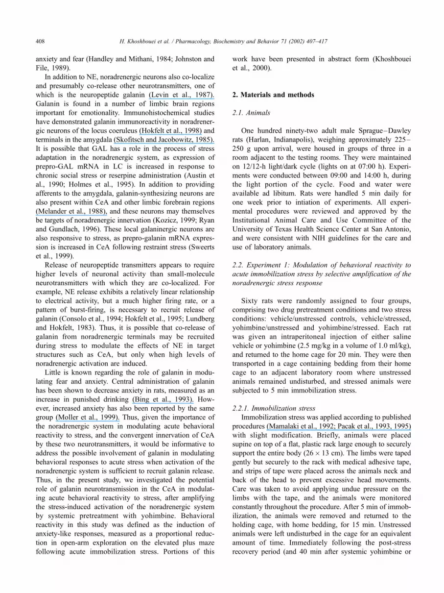
3.1.1. Effects of stress and yohimbine on anxiety-like
behavior on the elevated plus-maze
In the first experiment testing the effects of stress and
yohimbine, applied independently and in combination,
ANOVA revealed a significant treatment effect on OTR
for Time (F3,37 = 3.793, P < .05). Subsequent post hoc
analyses indicated that 5-min immobilization stress induced
H. Khoshbouei et al. / Pharmacology, Biochemistry and Behavior 71 (2002) 407–417410
a significant decrease in OTR for time, indicative of the
expected anxiogenic effect of a mild acute stress exposure
(Fig. 1). An anxiogenic effect similar to that induced by
acute stress was also seen following systemic yohimbine
administration (Fig. 1). However, when these two anxio-
genic stimuli were combined, there was a significant
increase in OTR to a level that was not different from that
of untreated, unstressed control rats (Fig. 1). This indicates
that yohimbine pretreatment reversed the anxiogenic
response to acute stress. In no case was there any significant
change in locomotor activity, measured by the number of
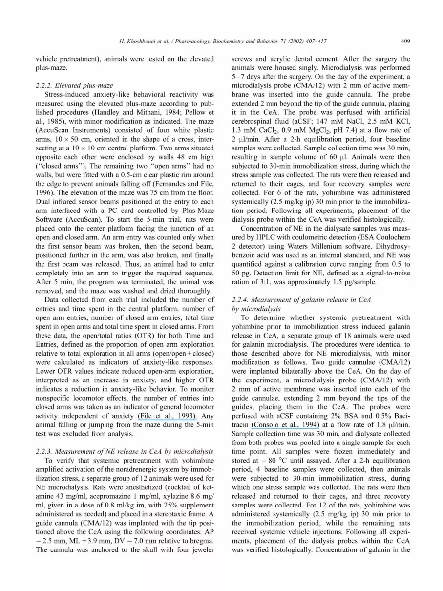
closed arm entries (F3,37 = 1.816, n.s., Fig. 2).
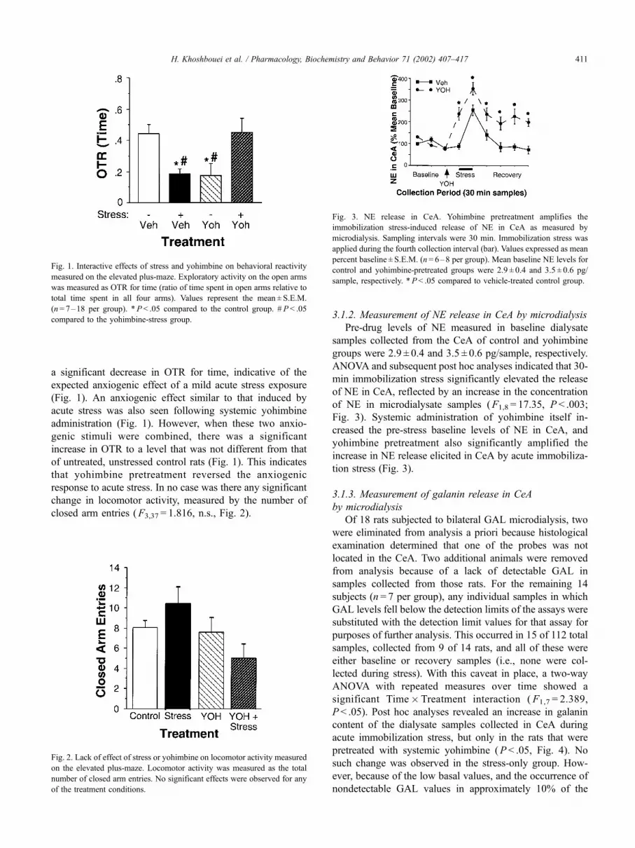
3.1.2. Measurement of NE release in CeA by microdialysis
Pre-drug levels of NE measured in baseline dialysate
samples collected from the CeA of control and yohimbine
groups were 2.9 ± 0.4 and 3.5 ± 0.6 pg/sample, respectively.
ANOVA and subsequent post hoc analyses indicated that 30-
min immobilization stress significantly elevated the release
of NE in CeA, reflected by an increase in the concentration
of NE in microdialysate samples (F1,8 = 17.35, P < .003;
Fig. 3). Systemic administration of yohimbine itself in-
creased the pre-stress baseline levels of NE in CeA, and
yohimbine pretreatment also significantly amplified the
increase in NE release elicited in CeA by acute immobiliza-
tion stress (Fig. 3).
3.1.3. Measurement of galanin release in CeA
by microdialysis
Of 18 rats subjected to bilateral GAL microdialysis, two
were eliminated from analysis a priori because histological
examination determined that one of the probes was not
located in the CeA. Two additional animals were removed
from analysis because of a lack of detectable GAL in
samples collected from those rats. For the remaining 14
subjects (n = 7 per group), any individual samples in which
GAL levels fell below the detection limits of the assays were
substituted with the detection limit values for that assay for
purposes of further analysis. This occurred in 15 of 112 total
samples, collected from 9 of 14 rats, and all of these were
either baseline or recovery samples (i.e., none were col-
lected during stress). With this caveat in place, a two-way
ANOVA with repeated measures over time showed a
significant Time�Treatment interaction (F1,7 = 2.389,
P < .05). Post hoc analyses revealed an increase in galanin
content of the dialysate samples collected in CeA during
acute immobilization stress, but only in the rats that were
pretreated with systemic yohimbine (P < .05, Fig. 4). No
such change was observed in the stress-only group. How-
ever, because of the low basal values, and the occurrence of
nondetectable GAL values in approximately 10% of the
Fig. 1. Interactive effects of stress and yohimbine on behavioral reactivity
measured on the elevated plus-maze. Exploratory activity on the open arms
was measured as OTR for time (ratio of time spent in open arms relative to
total time spent in all four arms). Values represent the mean ± S.E.M.
(n= 7–18 per group). *P < .05 compared to the control group. # P < .05
compared to the yohimbine-stress group.
Fig. 2. Lack of effect of stress or yohimbine on locomotor activity measured
on the elevated plus-maze. Locomotor activity was measured as the total
number of closed arm entries. No significant effects were observed for any
of the treatment conditions.
Fig. 3. NE release in CeA. Yohimbine pretreatment amplifies the
immobilization stress-induced release of NE in CeA as measured by
microdialysis. Sampling intervals were 30 min. Immobilization stress was
applied during the fourth collection interval (bar). Values expressed as mean
percent baseline ± S.E.M. (n= 6–8 per group). Mean baseline NE levels for
control and yohimbine-pretreated groups were 2.9 ± 0.4 and 3.5 ± 0.6 pg/
sample, respectively. *P < .05 compared to vehicle-treated control group.
H. Khoshbouei et al. / Pharmacology, Biochemistry and Behavior 71 (2002) 407–417 411
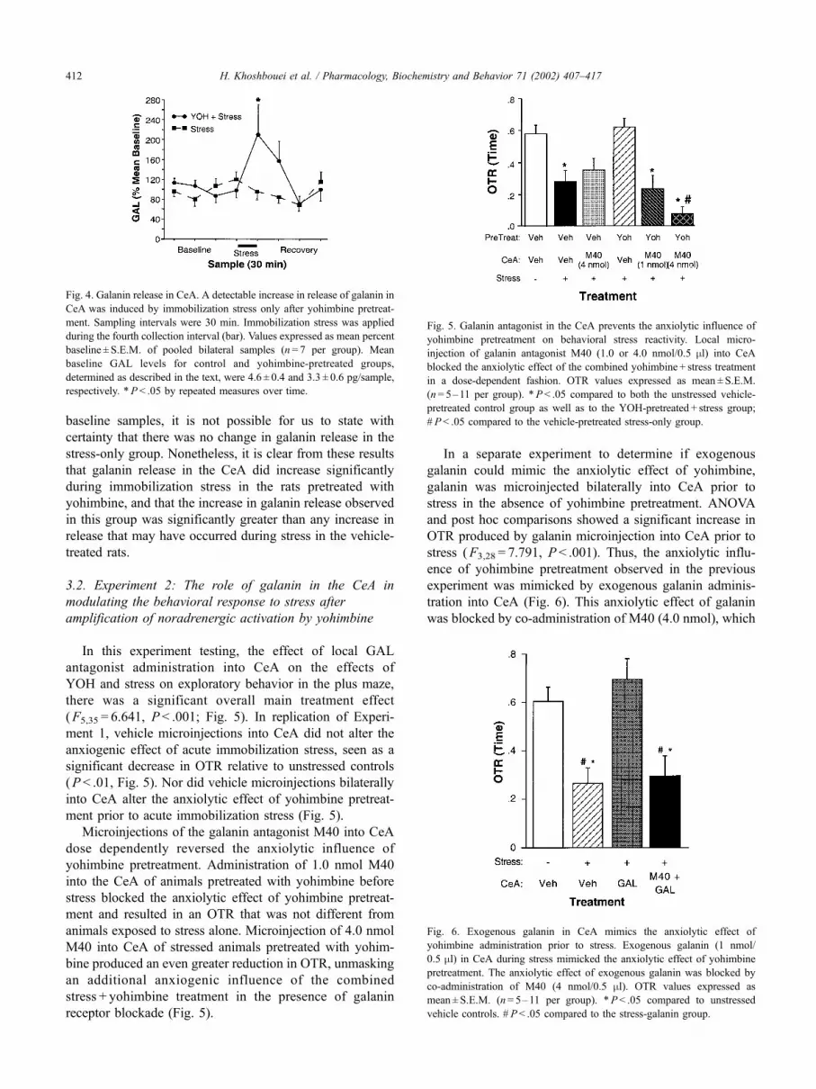
baseline samples, it is not possible for us to state with
certainty that there was no change in galanin release in the
stress-only group. Nonetheless, it is clear from these results
that galanin release in the CeA did increase significantly
during immobilization stress in the rats pretreated with
yohimbine, and that the increase in galanin release observed
in this group was significantly greater than any increase in
release that may have occurred during stress in the vehicle-
treated rats.
3.2. Experiment 2: The role of galanin in the CeA in
modulating the behavioral response to stress after
amplification of noradrenergic activation by yohimbine
In this experiment testing, the effect of local GAL
antagonist administration into CeA on the effects of
YOH and stress on exploratory behavior in the plus maze,
there was a significant overall main treatment effect
(F5,35 = 6.641, P < .001; Fig. 5). In replication of Experi-
ment 1, vehicle microinjections into CeA did not alter the
anxiogenic effect of acute immobilization stress, seen as a
significant decrease in OTR relative to unstressed controls
(P < .01, Fig. 5). Nor did vehicle microinjections bilaterally
into CeA alter the anxiolytic effect of yohimbine pretreat-
ment prior to acute immobilization stress (Fig. 5).
Microinjections of the galanin antagonist M40 into CeA
dose dependently reversed the anxiolytic influence of
yohimbine pretreatment. Administration of 1.0 nmol M40
into the CeA of animals pretreated with yohimbine before
stress blocked the anxiolytic effect of yohimbine pretreat-
ment and resulted in an OTR that was not different from
animals exposed to stress alone. Microinjection of 4.0 nmol
M40 into CeA of stressed animals pretreated with yohim-
bine produced an even greater reduction in OTR, unmasking
an additional anxiogenic influence of the combined
stress + yohimbine treatment in the presence of galanin
receptor blockade (Fig. 5).
In a separate experiment to determine if exogenous
galanin could mimic the anxiolytic effect of yohimbine,
galanin was microinjected bilaterally into CeA prior to
stress in the absence of yohimbine pretreatment. ANOVA
and post hoc comparisons showed a significant increase in
OTR produced by galanin microinjection into CeA prior to
stress (F3,28 = 7.791, P < .001). Thus, the anxiolytic influ-
ence of yohimbine pretreatment observed in the previous
experiment was mimicked by exogenous galanin adminis-
tration into CeA (Fig. 6). This anxiolytic effect of galanin
was blocked by co-administration of M40 (4.0 nmol), which
Fig. 4. Galanin release in CeA. A detectable increase in release of galanin in
CeA was induced by immobilization stress only after yohimbine pretreat-
ment. Sampling intervals were 30 min. Immobilization stress was applied
during the fourth collection interval (bar). Values expressed as mean percent
baseline ± S.E.M. of pooled bilateral samples (n= 7 per group). Mean
baseline GAL levels for control and yohimbine-pretreated groups,
determined as described in the text, were 4.6 ± 0.4 and 3.3 ± 0.6 pg/sample,
respectively. *P < .05 by repeated measures over time.
Fig. 5. Galanin antagonist in the CeA prevents the anxiolytic influence of
yohimbine pretreatment on behavioral stress reactivity. Local micro-
injection of galanin antagonist M40 (1.0 or 4.0 nmol/0.5 ml) into CeA
blocked the anxiolytic effect of the combined yohimbine + stress treatment
in a dose-dependent fashion. OTR values expressed as mean ± S.E.M.
(n= 5–11 per group). *P< .05 compared to both the unstressed vehicle-
pretreated control group as well as to the YOH-pretreated + stress group;
# P < .05 compared to the vehicle-pretreated stress-only group.
Fig. 6. Exogenous galanin in CeA mimics the anxiolytic effect of
yohimbine administration prior to stress. Exogenous galanin (1 nmol/
0.5 ml) in CeA during stress mimicked the anxiolytic effect of yohimbine
pretreatment. The anxiolytic effect of exogenous galanin was blocked by
co-administration of M40 (4 nmol/0.5 ml). OTR values expressed as
mean ± S.E.M. (n= 5–11 per group). *P< .05 compared to unstressed
vehicle controls. # P< .05 compared to the stress-galanin group.
H. Khoshbouei et al. / Pharmacology, Biochemistry and Behavior 71 (2002) 407–417412

restored the post-stress OTR to a level that was no different
from that after stress with vehicle injection in CeA (Fig. 6).
Administration of M40 alone into CeA in the absence of
yohimbine pretreatment had no effect on the anxiogenic
effect of stress (Fig. 6), suggesting an absence of an endog-
enous galanin-mediated anxiolytic effect when stress-
induced activation of the noradrenergic system was not
amplified by yohimbine.
None of the drug treatments in either of these micro-
injection experiments produced any change in general
locomotor activity as measured by number of closed arm
entries in the plus-maze (F5,35 = 1.696 and F3,28 = 1.414,
respectively, both n.s., data not shown). Histological ana-
lyses verified that the injection sites were centered bilat-
erally in the CeA (Fig. 7). Any cases in which one or both
injections sites were located outside the CeA were elimi-
nated from analyses. This resulted in elimination of eight
cases (not included in the total animal count).
4. Discussion
In this study, we investigated a possible role for galanin
in modulating a behavioral–affective component of the
stress response in the CeA when stress-induced activation
of the noradrenergic system had been amplified by prior
administration of systemic yohimbine. In the first experi-
ment, we demonstrated that acute immobilization stress and
systemic administration of yohimbine both induced anxiety-
like reductions in open-arm exploration on the elevated plus
maze when administered by themselves. The anxiogenic
effect of yohimbine is consistent with theories suggesting a
role for NE in stress, arousal and anxiety (Aston-Jones et al.,
1991; Charney et al., 1987). However, when these two
stimuli were combined, i.e., when the noradrenergic
response to immobilization stress was amplified by pretreat-
ing with yohimbine, the anxiogenic effect of the acute
stressor was attenuated. It may seem paradoxical at first
that combining two anxiogenic stimuli induced an anxio-
lytic response, and we attempted to resolve this paradox by
hypothesizing that the combination of anxiogenic stimuli
recruited a new anxiolytic mechanism in the CeA that was
not evoked by either stimulus alone, namely the release of
galanin. Using microdialysis, we verified that yohimbine
pretreatment not only amplified stress-induced release of
NE in the CeA, but release of the neuropeptide galanin was
also evoked in the CeA in this condition.
In the second experiment, we microinjected the galanin
antagonist M40 into CeA to test the hypothesis that galanin
participated in the anxiolytic effect observed when animals
were given yohimbine prior to stress. Local bilateral admin-
istration of M40 into the CeA prior to stress completely
blocked, in a dose-dependent fashion, the anxiolytic influ-
ence of yohimbine pretreatment on the behavioral response
to acute stress. Administration of exogenous galanin into the
CeA was itself capable of inducing an anxiolytic effect
during stress that mimicked yohimbine pretreatment, and
this too was blocked by M40. Therefore, we concluded that
the release of galanin in CeA was responsible in large part
for the anxiety-attenuating effects of yohimbine treatment
given prior to stress.
The galanin-mediated attenuation of stress-induced anxi-
ety-like behavioral responses in the CeA was not evident
when immobilization stress was applied alone, as M40
administration had no effect in this condition. Rather, the
anxiolytic influence of galanin was only manifest when the
impact of the stressor, and subsequent activation of the
noradrenergic system, was amplified by yohimbine pretreat-
ment. One possible source of this galanin release could have
been from the noradrenergic terminals innervating the CeA,
i.e., GAL that is co-localized with NE. More than 80% of
noradrenergic neurons in the locus coeruleus also contain
galanin (Melander et al., 1986). It has been shown both in
vitro and in vivo that co-localized neuropeptides are
released preferentially at higher levels of neuronal activity
than those required for classical small-molecule neurotrans-
mitters such as NE (Bartfai et al., 1988; Consolo et al.,
1994; Hokfelt, 1991). Thus, whereas NE may be preferen-
Fig. 7. Histological verification of injection site localization in CeA. At left is a representative section through the amygdala, stained with Cresyl violet,
showing the extent of distribution of a bilateral microinjection (0.5 ml) of 1% Evans Blue dye made after the completion of an experiment through the same
cannulae used for drug administration. The injections were both centered in the CeA, as portrayed in the schematic illustration at right, adapted from plate 27 of
the atlas of Swanson (1992). CeA: central amygdala; LHA: lateral hypothalamic area; CP: caudate–putamen.
H. Khoshbouei et al. / Pharmacology, Biochemistry and Behavior 71 (2002) 407–417 413

tially released from noradrenergic afferent terminals innerv-
ating the amygdala with low-to-moderate levels of activa-
tion, yohimbine pretreatment may have amplified the stress-
induced activation of the NE system sufficiently to recruit
co-release of galanin from the noradrenergic terminals,
resulting in the anxiolytic effect observed.
This suggestion was supported by the results of our
microdialysis experiment, which showed the stress-induced
release of galanin in CeA to be greater after yohimbine
pretreatment. It is necessary to exercise caution in conclud-
ing that there was no galanin released in the absence of
yohimbine pretreatment, as the galanin levels measured in
baseline samples were very close to the sensitivity limits of
the radioimmunoassay for GAL. Thus, even though we did
not detect any increase in galanin in the dialysate samples
collected during stress without yohimbine pretreatment, it is
possible that a slight increase could have occurred and
remained below detection. Nonetheless, we can conclude
with confidence that stress-induced release of galanin in
CeAwas significantly greater following yohimbine pretreat-
ment than it was in the absence of yohimbine.
Alternatively, it is possible that some or all of the galanin
released in CeA may not have originated from noradrenergic
terminals. The amygdala itself contains a number of gal-
anin-synthesizing neurons, as well as galanin receptors
(Melander et al., 1988; Ryan and Gundlach, 1996; Skofitsch
and Jacobowitz, 1985; Waters and Krause, 2000). Both
chronic and acute restraint stress have been shown to
increase pre-pro-galanin mRNA expression in the CeA
(Sweerts et al., 2000), indicating that galaninergic neurons
within CeA are stress-responsive. Thus, a second possible
source of galanin released in CeA by the combination of
yohimbine and stress may have been the galanin-containing
neurons intrinsic to the CeA. Noradrenergic afferent termi-
nals have been shown to contact galanin-positive targets in
the bed nucleus of the stria terminalis (Kozicz, 1999), a
component of the extended amygdala closely related to
CeA. It is thus possible that noradrenergic afferents also
contact galaninergic targets in CeA, and that NE activates
these intrinsic galanin-synthesizing neurons in response to
stress only when a certain level of noradrenergic transmis-
sion has been reached, such as that occurring when the
stress is preceded by yohimbine pretreatment.
Regardless of the source of galanin release in CeA, it
reduced the magnitude of the anxiogenic response to stress,
but only when a sufficient level of activity had been achieved
in the noradrenergic system innervating the CeA. Moreover,
when the anxiolytic influence of galanin was blocked by the
highest dose of M40, an additive anxiogenic effect of the
combined yohimbine plus stress treatment was revealed. As
might be expected from the combination of two anxiogenic
stimuli, the OTR in this condition was significantly lower
than that following stress alone, suggesting that galanin
release in the CeA had masked the additional anxiogenic
effect of yohimbine that was evident when the drug was
given alone.
The galanin-mediated attenuation of behavioral reactivity
to stress observed in this study may represent a form of
negative feedback regulation of noradrenergic modulation
of the stress response. Yohimbine pretreatment, in addition
to inducing galanin release, also increased stress-induced
activation of NE release, as would be expected following a2
adrenergic autoreceptor blockade. NE has been strongly
implicated in behavioral arousal and anxiety associated with
stress (Aston-Jones et al., 1991; Charney et al., 1987;
Redmond, 1987). Thus, it seems likely that the anxiogenic
component of yohimbine pretreatment that was masked by
galanin may be attributable to a noradrenergic mechanism.
In this way, once the stress-induced activation of the
noradrenergic system is elevated sufficiently, GAL release
is recruited in CeA, which then acts in turn to buffer the
behavioral reactivity and anxiogenic responses associated
with the elevated noradrenergic activity.
Few previous studies have addressed a possible role for
galanin in the modulation of anxiety-like behaviors, and
these studies have generated inconsistent results (Bing et al.,
1993; Moller et al., 1999). An anxiolytic effect, measured as
an increase in punished drinking, was reported after intra-
cerebroventricular administration of galanin (Bing et al.,
1993). By contrast, in another experiment conducted by the
same group, administration of galanin into the amygdala
produced an apparent anxiogenic response on the same test,
while no effect was observed on the elevated plus maze
(Moller et al., 1999). A number of factors may account for
the inconsistencies between these experiments, and also for
the lack of effect on the plus maze compared to the clear
effects on exploratory behavior in the plus maze that were
observed in the present study. First, intracerebroventricular
administration of drugs is likely to affect many brain
regions, perhaps eliciting different or opposing responses.
Also, there are many tests of anxiety-like behavior in rats,
and depending on the nature of the experimental manipu-
lations, they can generate very different results (File, 1995;
Spear and File, 1996). Galanin has been implicated in a
variety of appetitive, nociceptive and memory-related pro-
cesses (Crawley, 1999; Hokfelt et al., 1999), and the
punished drinking test involves aspects of all of these,
including thirst, consummatory behavior, motivation, con-
ditioning, and pain perception, as well as anxiety (Menard
and Treit, 1999). Thus, the behavioral effects observed in
this test after intracerebral administration of galanin may not
have been related exclusively to changes in anxiety. In
addition, the results obtained on any presumed measure of
anxiety can be very sensitive to differences in testing,
housing and handling conditions (File and Fluck, 1994).
Such factors may have accounted for some differences
between the results of these previous experiments and the
present study. Finally, an important aspect of the present
study is that we explicitly addressed a possible role for GAL
in modulating stress-induced behavioral reactivity, meas-
ured as a change in exploratory behavior following an acute
stress exposure, rather than examining possible effects of
H. Khoshbouei et al. / Pharmacology, Biochemistry and Behavior 71 (2002) 407–417414
GAL on baseline exploratory behavior, as was done in the
previous experiments. In the present study, exogenous
galanin administered into CeA was shown to buffer behav-
ioral reactivity to acute stress, manifest as an attenuation of
the stress-induced reduction in open-arm exploration in the
plus maze. However, in the previously cited experiments, no
stressor was applied. Thus, the effects tested in these experi-
ments were on baseline behavior, and baseline levels of
anxiety may have been sufficiently low that exogenously
administered galanin had no effect. Nevertheless, in the
current study, stress-induced release of endogenous galanin
in the CeA following yohimbine pretreatment exerted a
clear and consistent anxiolytic effect.
The interaction between NE activity and the neuropep-
tide GAL demonstrated in this experiment raises the pos-
sibility of noradrenergic interaction with other peptide
neurotransmitters with which it is closely associated, such
as NPY. NPY is also co-localized extensively with NE,
though less in LC and more in medullary noradrenergic cell
groups than GAL (Sawchenko et al., 1985; Zardetto-Smith
and Gray, 1995). NPY has been shown to modulate behav-
ioral responsivity to fear and stress (Heilig et al., 1994).
Specifically, administration of NPY into CeA produced
anxiolytic effects (Heilig et al., 1993), and selective block-
ade of NPY-Y1 receptors in CeA decreased open arm
exploration on the elevated plus maz (Heilig et al., 1994;
Wahlestedt et al., 1993). Consistent with a possible NPY-
noradrenergic interaction, NPY attenuated the anxiety-like
reduction in social interaction that was possibly attributable
to noradrenergic supersensitivity following DSP4-induced
denervation (Kask et al., 2000). These observations,
together with the results of the present experiment, suggest
that, depending on the nature of the stressor and the
response elicited, the subset of noradrenergic neurons that
are activated, and the degree to which this system is
activated by specific stressors, a variety of potential mod-
ulatory interactions in the CeA could occur involving NE,
NPY and GAL. Regulation or modification of these inter-
actions may provide for considerable flexibility, specificity
and plasticity in the modulatory effects exerted by activation
of the noradrenergic system in response to stress.
In summary, the results of these experiments suggest that
amplification of the noradrenergic response to stress can
recruit the release of galanin in CeA, which then acts to
buffer the anxiogenic effects of NE. Thus, the net behavioral
response to stress, and the nature of the modulatory influ-
ence exerted by the noradrenergic system on behavioral
stress reactivity will ultimately depend on the overall level
of activation of this system, and the resulting balance
between NE and peptide neurotransmission. This balance,
in turn, may be subject to regulation or modification as a
result of prior stress exposure, regulatory changes in the
reactivity or sensitivity of the noradrenergic system, chronic
drug treatment, or even genetic variability. Dysregulation of
the normal interaction between NE and galanin in the
amygdala may thereby contribute to stress-related neuro-
psychiatric disorders such as depression, post-traumatic
stress disorder or other anxiety disorders. Further, the
interaction between NE and galanin may represent a pos-
sible mechanism by which genetic predisposition may
confer a differential vulnerability to stress, and may also
represent a novel target for future therapeutic strategies
aimed at treating stress-related psychiatric disorders.
Acknowledgments
We thank Ms. Lauren Scholz and Ms. Erin Worthy for
expert technical assistance. This work was supported by
research grants from the National Institute of Mental Health
(MH53851 and MH60118).
References
Aston-Jones G, Chiang C, Alexinsky T. Discharge of noradrenergic locus
coeruleus neurons in behaving rats and monkeys suggests a role in
vigilance. Prog Brain Res 1991;88:501–20.
Austin MC, Cottingham SL, Paul SM, Crawley JN. Tyrosine hydroxylase
and galanin mRNA levels in locus coeruleus neurons are increased
following reserpine administration. Synapse 1990;6:351–7.
Bartfai T, Iverfeldt K, Fisone G, Serfozo P. Regulation of the release
of coexisting neurotransmitters. Ann Rev Pharmacol Toxicol 1988;
28:285–310.
Bing O, Moller C, Engel JA, Soderpalm B, Heilig M. Anxiolytic-like actions
of centrally administered galanin. Neurosci Lett 1993;164:17–20.
Charney DS, Woods SW, Goodman WK, Heninger GR. Neurobiological
mechanisms of panic anxiety: biochemical and behavioral correlates of
yohimbine-induced panic attacks. Am J Psychiatry 1987;144:1030–6.
Charney DS, Woods SW, Krystal JH, Nagy LM, Heninger GR. Noradre-
nergic neuronal dysregulation in panic disorder: the effects of intra-
venous yohimbine and clonidine in panic disorder patients. Acta
Psychiatr Scand 1992;86:273–82.
Consolo S, Baldi G, Russi G, Civenni G, Bartfai T, Vezzani A. Impulse
flow dependency of galanin release in vivo in the rat ventral hippo-
campus. Proc Natl Acad Sci USA 1994;91:8047–51.
Crawley J. The role of galanin in feeding behavior. Neuropeptides 1999;
33:369–75.
Crawley JN, Robinson JK, Langel U, Bartfai T. Galanin receptor antago-
nists M40 and C7 block galanin-induced feeding. Brain Res 1993;
600:268–72.
Davis M, Shi C. The extended amygdala: are the central nucleus of amyg-
dala and the bed nucleus of the stria terminalis differentially involved in
fear versus anxiety? Ann NY Acad Sci 1999;877:281–91.
Feldman S, Wiedenfeld J. The excitatory effects of the amygdala on hypo-
thalamo–pituitary– adrenocortical responses are mediated by hypo-
thalamic norepinephrine, serotonin, and CRF-41. Brain Res Bull 1998;
45:389–93.
Fernandes C, File S. The influence of open arm ledges and maze experience
in the elevated plus-maze. Pharmacol Biochem Behav 1996;54:31–40.
File S. Animal models of different anxiety states. Adv Biochem Psycho-
pharmacol 1995;48:93–113.
File S, Fluck E. Handling alters habituation and response to stimulus
change in the holeboard. Pharmacol Biochem Behav 1994;49:449–53.
File S, Zangrossi HJ, Viana M, Graeff F. Trial 2 in the elevated plus-
maze: a different form of fear? Psychopharmacology (Berlin) 1993;
111:491–4.
Goldstein LE, Rasmusson AM, Bunney BS, Roth RH. Role of the amyg-
dala in the coordination of behavioral, neuroendocrine, and prefrontal
H. Khoshbouei et al. / Pharmacology, Biochemistry and Behavior 71 (2002) 407–417 415
cortical monoamine responses to psychological stress in the rat.
J Neurosci 1996;16:4787–98.
Grant S, Huang Y, Redmond DJ. Behavior of monkeys during opiate with-
drawal and locus coeruleus stimulation. Pharmacol, Biochem Behav
1988;30:13–9.
Gray TS. Amygdaloid CRF pathways: role in autonomic, neuroendocrine,
and behavioral responses to stress. Ann NYAcad Sci 1993;697:53–60.
Handley SL, Mithani S. Effects of alpha-adrenoceptor agonists and antag-
onists in a maze-exploration model of ‘‘fear’’-motivated behaviour.
Naunyn-Schmiedeberg’s Arch Pharmacol 1984;327:1–5.
Hatfield T, Spanis C, McGaugh J. Response of amygdalar norepinephrine to
footshock and GABAergic drugs using in vivo microdialysis and HPLC.
Brain Res 1999;835:340–5.
Heilig M, McLeod S, Brot M, Heinrichs SC, Menzaghi F, Koob GF, Britton
KT. Anxiolytic-like action of neuropeptide Y: mediation by Y1 receptors
in amygdala, and dissociation from food intake effects. Neuropsycho-
pharmacology 1993;8:357–63.
Heilig M, Koob GF, Ekman R, Britton KB. Corticotropin-releasing factor
and neuropeptide Y: role in emotional integration. Trends Neurosci
1994;17:80–5.
Henke PG, Ray A. Stress ulcer modulation by limbic system structures. Acta
Physiol Hung 1992;80:117–25.
Hokfelt T. Neuropeptides in perspective: the last ten years. Neuron
1991;7:867–79.
Hokfelt TGM, Castel M-N, Morino P, Zhang X, Dagerlind A. General over-
view of neuropeptides. In: Bloom FE, Kupfer DJ, editors. Psychophar-
macology: the fourth generation of progress. New York: Raven Press,
1995. pp. 483–92.
Hokfelt T, Xu Z, Shi T, HolmbergK, ZhangX. Galanin in ascending systems.
Focus on coexistence with 5-hydroxytryptamine and noradrenaline. Ann
NYAcad Sci 1998;863:252–63.
Hokfelt T, Broberger C, Diez M, Xu Z, Shi T, Kopp J, Zhang X, Holmberg
K, Landry M, Koistinaho J. Galanin and NPY, two peptides with multi-
ple putative roles in the nervous system. Horm Metab Res 1999;31:
330–4.
Holmes PV, Blanchard DC, Blanchard RJ, Brady LS, Crawley JN. Chronic
social stress increases levels of preprogalanin mRNA in the rat locus
coeruleus. Pharmacol Biochem Behav 1995;50:655–60.
Honkaniemi J, Kainu T, Ceccatelli S, Rechardt L, Hokfelt T, Pelto-Huikko
M. Fos and jun in rat central amygdaloid nucleus and paraventricular
nucleus after stress. NeuroReport 1992;3:849–52 (Oct.).
Johnston A, File S. Yohimbine’s anxiogenic action: evidence for noradre-
nergic and dopaminergic sites. Pharmacol Biochem Behav 1989;
32:151–6.
Kask A, Eller M, Oreland L, Harro J. Neuropeptide Y attenuates the effect
of locus coeruleus denervation by DSP-4 treatment on social behaviour
in the rat. Neuropeptides 2000;34:58–61.
Khoshbouei H, Cecchi M, Morilak DA. Amplification of the noradrenergic
response to stress elicits galanin-mediated anxiolytic effects in central
amygdala that counteract the anxiogenic influence of norepinephrine.
Soc Neurosci Abstr 2000;26:1154.
Kozicz T. Synaptic interactions between galanin and axon terminals immu-
nopositive for tyrosine hydroxylase and dopamine b-hydroxylase in the
bed nucleus of the stria terminalis in the rat. Soc Neurosci Abstr 1999;
25:2220.
LeDoux J. Fear and the brain: where have we been and where are we
going? Biol Psychiatry 1998;44:1229–38.
Levin MC, Sawchenko PE, Howe PRC, Bloom SR, Polak JM. The organ-
ization of galanin-immunoreactive inputs to the paraventricular nucleus
with special reference to their relationship to catecholaminergic affer-
ents. J Comp Neurol 1987;261:562–82.
Lundberg JM, Hokfelt T. Coexistence of peptides and classical neurotrans-
mitters. Trends Neurosci 1983;6:325–33.
Mamalaki E, Kvetnansky R, Brady LS, Gold PW, Herkenham M. Repeated
immobilization stress alters tyrosine hydroxylase, corticotropin-releas-
ing hormone and corticosteroid receptor messenger ribonucleic acid
levels in rat brain. J Neuroendocrinol 1992;4:689–99.
McDonald M, Crawley J. Galanin receptor antagonist M40 blocks galanin-
induced choice accuracy deficits on a delayed-nonmatching-to-position
task. Behav Neurosci 1996;110:1025–32.
Melander T, Hokfelt T, Rokaeus A, Cuello AC, Oertel WH, Verhofstad A,
Goldstein M. Coexistence of galanin-like immunoreactivity with cate-
cholamines, 5-hydroxytryptamine, GABA and neuropeptides in the rat
CNS. J Neurosci 1986;6:3640–54.
Melander T, Kohler C, Nilsson S, Hokfelt T, Brodin E, Theodorsson E,
Bartfai T. Autoradiographic quantitation and anatomical mapping of125I-galanin binding sites in the rat central nervous system. J Chem
Neuroanat 1988;1:213–33.
Menard J, Treit D. Effects of centrally administered anxiolytic compounds
in animal models of anxiety. Neurosci Biobehav 1999;23:591–613.
Moller C, Wiklund L, Sommer W, Thorsell A, Heilig M. Decreased ex-
perimental anxiety and voluntary ethanol consumption in rats follow-
ing central but not basolateral amygdala lesions. Brain Res 1997;
760:94–101.
Moller C, Sommer W, Thorsell A, Heilig M. Anxiogenic-like action of
galanin after intra-amygdala administration in the rat. Neuropsycho-
pharmacology 1999;21:507–12.
Moore RY, Bloom FE. Central catecholamine neuron system: anatomy and
physiology of the norepinephrine and epinephrine systems. Ann Rev
Neurosci 1979;2:113–68.
Morilak DA, Fornal CA, Jacobs BL. Effects of physiological manipulations
on locus coeruleus neuronal activity in freely moving cats: I. Thermo-
regulatory challenge. Brain Res 1987a;422:17–23.
Morilak DA, Fornal CA, Jacobs BL. Effects of physiological manipulations
on locus coeruleus neuronal activity in freely moving cats: II. Cardio-
vascular challenge. Brain Res 1987b;422:24–31.
Pacak K, Palkovits M, Kvetnansky R, Fukuhara K, Kopin IJ, Goldstein DS.
Effects of single or repeated immobilization on release of norepinephrine
and its metabolites in the central nucleus of the amygdala in conscious
rats. Neuroendocrinology 1993;57:623–33.
Pacak K, McCarty R, Palkovits M, Kopin I, Goldstein D. Effects
of immobilization on in vivo release of norepinephrine in the bed
nucleus of the stria terminalis in conscious rats. Brain Res 1995;
688:242–6.
Palkovits M, Baffi J, Pacak K. The role of ascending neuronal pathways in
stress-induced release of noradrenaline in the hypothalamic paraventric-
ular nucleus of rats. J Neuroendocrinol 1999;1:529–39.
Pellow S, Chopin P, File SE, Briley M. Validation of open: closed arm
entries in an elevated plus-maze as a measure of anxiety in the rat.
J Neurosci Methods 1985;14:149–67.
Redmond DE. Studies of the nucleus locus coeruleus in monkeys and hy-
potheses for neuropsychopharmacology. In: Meltzer HY, editor. Psycho-
pharmacology: the third generation of progress. New York: Raven Press,
1987. pp. 967–75.
Ryan M, Gundlach A. Localization of preprogalanin messenger RNA in rat
brain: identification of transcripts in a subpopulation of cerebellar Pur-
kinje cells. Neuroscience 1996;70:709–28.
Sawchenko PE, Swanson LW, Grzanna R, Howe PRC, Bloom SR, Polak
JM. Colocalization of neuropeptide Y immunoreactivity in brainstem
catecholaminergic neurons that project to the paraventricular nucleus of
the hypothalamus. J Comp Neurol 1985;241:138–53.
Skofitsch G, Jacobowitz DM. Immunohistochemical mapping of galanin-
like neurons in the rat central nervous system. Peptides 1985;6:509–46.
Southwick S, Bremner J, Rasmusson A, Morgan CR, Arnsten A, Charney D.
Role of norepinephrine in the pathophysiology and treatment of post-
traumatic stress disorder. Biol Psychiatry 1999;46:1192–204.
Spear L, File S. Methodological considerations in neurobehavioral
teratology. Pharmacol Biochem Behav 1996;55:455–7.
Swanson LW. Brain maps: structure of the rat brain. Amsterdam:
Elsevier, 1992.
Sweerts B, Jarrott B, Lawrence A. Expression of preprogalanin mRNA
following acute and chronic restraint stress in brains of normotensive
and hypertensive rats. Brain Res Mol Brain Res 1999;69:113–23.
Sweerts B, Jarrott B, Lawrence A. Acute and chronic restraint stress: effects
H. Khoshbouei et al. / Pharmacology, Biochemistry and Behavior 71 (2002) 407–417416
on [125I]galanin binding in normotensive and hypertensive rat brain.
Brain Res 2000;873:318–29.
Tjurmina OA, Goldstein DS, Palkovits M, Kopin IJ. a2-Adrenoceptor-
mediated restraint of norepinephrine synthesis, release, and turnover
during immobilization in rats. Brain Res 1999;826:243–52.
Wahlestedt C, Pich EM, Koob GF, Heilig M. Modulation of anxiety and
neuropeptide Y–Y1 receptors by antisense oligodeoxynucleotides.
Science 1993;259:528–31.
Waters S, Krause J. Distribution of galanin-1, -2 and -3 receptor messen-
ger RNAs in central and peripheral rat tissues. Neuroscience 2000;
95:265–71.
Zardetto-Smith AM, Gray TS. Catecholamine and NPY efferents from the
ventrolateral medulla to the amygdala in the rat. Brain Res Bull
1995;38:253–60.
H. Khoshbouei et al. / Pharmacology, Biochemistry and Behavior 71 (2002) 407–417 417