Embed Size (px)
Citation preview
ltcpdNwdm
ARCHIVAL REPORT
FK506 Binding Protein 5 Shapes StressResponsiveness: Modulation of NeuroendocrineReactivity and Coping BehaviorChadi Touma, Nils Christian Gassen, Leonie Herrmann, Joyce Cheung-Flynn, Dominik R. Büll,Irina A. Ionescu, Jan-Michael Heinzmann, Alana Knapman, Anna Siebertz, Anna-Mareike Depping,Jakob Hartmann, Felix Hausch, Mathias V. Schmidt, Florian Holsboer, Marcus Ising, Marc B. Cox,Ulrike Schmidt, and Theo Rein
Background: The Hsp90 cochaperone FK506 binding protein 5 (FKBP5) is an established regulator of the glucocorticoid receptor (GR), andnumerous genetic studies have linked it to stress-related diseases such as major depression or posttraumatic stress disorder. However,translational studies including genetic animal models are lacking.
Methods: Mice deficient of FKBP5 were generated and analyzed in comparison with wildtype littermates. They were subjected to severaltest paradigms characterizing their emotionality, stress reactivity, and coping behavior as well as hypothalamus-pituitary-adrenal axisfunction and regulation. Moreover, protein expression of GR and FKBP5 was determined in different brain structures 8 days after stressexposure. The combined dexamethasone/corticotropin-releasing hormone test was performed both in mice and healthy human subjects ofdifferent FKBP5 genotypes. The GR function was evaluated by reporter gene assays.
Results: Under basal conditions, deletion of FKBP5 did not change exploratory drive, locomotor activity, anxiety-related behavior, stress-coping, or depression-like behavior. After exposure to different acute stressors of sufficient intensity, however, it led to a more active copingbehavior. Moreover, loss of FKBP5 decreased hypothalamus-pituitary-adrenal axis reactivity and GR expression changes in response tostressors. In mice and humans, the FKBP5 genotype also determined the outcome of the dexamethasone/corticotropin-releasing hormonetest.
Conclusions: This study in mice and humans presents FKBP5 as a decisive factor for the physiological stress response, shaping neuroen-docrine reactivity as well as coping behavior. This lends strong support to the concept emerging from human studies of FKBP5 as important
factor governing gene– environment interactions relevant for the etiology of affective disorders.l(
tsfdcHG
aarertghtcsemHtfb
Key Words: Dex/CRH test, emotionality, FKBP51, HPA axis, stress-coping behavior, stress reactivity
I n the recent years, polymorphisms in the FK506 binding protein5 (FKBP5), also referred to as FKBP51, have emerged as one ofthe most important and intriguing associations with stress-re-
ated phenotypes and diseases such as major depression and post-raumatic stress disorder (1–7). FKBP5 was originally identified asomponent of the progesterone receptor-chaperone heterocom-lex (8). Its role in stress regulation was first conjectured uponiscovery of elevated FKBP5 levels in squirrel monkeys (9,10). Theseew World primates exhibit glucocorticoid resistance togetherith markedly elevated levels of plasma cortisol but lack signs ofetrimental glucocorticoid excess (11–13). The decreased hor-one-binding affinity of their glucocorticoid receptor (GR) (12) was
From the Research Group of Psychoneuroendocrinology (CT, J-MH, AK),Chaperone Research Group (NCG, TR), Research Group of MolecularPsychotraumatology (LH, DRB, IAI, US), Research Group of MolecularPsychology (AS, A-MD, MI), Research Group of Chemical Genomics (FHa),and the Research Group of Neurobiology of Stress (MVS, JH), Max PlanckInstitute of Psychiatry (FHo), Munich, Germany; Department of Surgery(JC-F), Vanderbilt University Medical Center, Nashville, Tennessee; andthe Department of Biological Sciences (MBC), University of Texas at ElPaso, El Paso, Texas.
Authors US and TR contributed equally to this work.Address correspondence to Theo Rein, Ph.D., Max Planck Institute of Psychi-
atry, Chaperone Research Group, Kraepelinstrasse 2-10, MunichD-80804 Germany; E-mail [email protected].
cReceived Apr 28, 2011; revised Jul 16, 2011; accepted Jul 19, 2011.
0006-3223/$36.00doi:10.1016/j.biopsych.2011.07.023
ater largely attributed to the inhibitory action of FKBP5 on GR14 –17).
FKBP5 possesses peptidylprolyl isomerase activity (18) and fea-ures a domain for interaction with the central chaperone heathock protein (Hsp) 90. Although interaction with Hsp90 is essentialor its inhibitory action on GR, peptidylprolyl isomerase activity isispensable (16). Thus, according to the current mechanistic con-ept, FKBP5 competes with other proteins for access to thesp90-GR heterocomplex, thereby interfering with the action ofR-stimulatory factors, in dependency of its expression levels (19).
The well-documented influence of FKBP5 on GR function serveds rationale for its inclusion as one of the candidates in the genessociation study that first associated FKBP5 polymorphisms withesponse to antidepressant treatment (1). Over decades, amplevidence accumulated for an essential role of GR function in stress-elated psychiatric disorders such as major depression and post-raumatic stress disorder (20 –22). In general, elevated levels oflucocorticoids in response to stressful life events constitute aealthy adaptive reaction, provided this response is balanced and
ransient. The hypothalamus-pituitary-adrenal (HPA) axis is a keyontrol system to balance hormonal and behavioral responses totressors. A hallmark of HPA axis regulation is the negative feedbackxerted by glucocorticoids via GR on the secretion of stress hor-ones (20). Substantial evidence suggests that this attenuation ofPA axis activity, which is an integral part of the adaptive response
o stressors and challenges, is often impaired in patients sufferingrom major depression (23,24). For example, major depression haseen repeatedly shown to be associated with elevated levels of
irculating glucocorticoids, decreased responsiveness to dexa-BIOL PSYCHIATRY 2011;xx:xxx© 2011 Society of Biological Psychiatry
lnoruarc
tbsrHcdFC
maswtas
E
m
B
aattwb(b
H
mhwpbdda
bbrCaCnd
D
wddfgpaMwsft
swtb
E
cma
T
bfst
2 BIOL PSYCHIATRY 2011;xx:xxx C. Touma et al.
methasone (Dex) suppression and increased adrenocortical re-sponse to stimulation with corticotropin-releasing hormone (CRH)in the combined Dex/CRH test (20,24). Although the exact physio-ogical consequences of prolonged glucocorticoid elevation areot fully understood (25), it is intriguing that successful treatmentf depression mostly goes along with a normalization of HPA axis
eactivity. Moreover, remitted individuals with incompletely atten-ated HPA axis overdrive have a higher risk of relapse (26). Thesend other observations led to the formulation of the corticosteroideceptor hypothesis of depression, which stipulates a link betweenorticosteroid receptor dysfunction and major depression (23).
This hypothesis is further supported by animal experiments (27).For example, mice with impaired GR function due to transgenicantisense RNA expression exhibit neuroendocrine characteristicssimilar to those observed in major depression, including a hyperac-tive HPA axis (28). Antidepressant treatment counteracted thesealterations (29). In addition, acquired deficit of forebrain GR pro-duces depression-like phenotypes in behavioral and physiologicalstress reactivity, which were normalized by antidepressant treat-ment (30).
Given the established role of GR and its regulatory protein FKBP5in stress-related disorders and associated endophenotypes, we un-dertook a molecular, neuroendocrine, and behavioral characteriza-tion of Fkbp5�/� mice under basal conditions as well as in responseo stressors. We considered the latter point particularly important,ecause the ability of an organism to respond and the way it re-ponds to stressors have been accepted as crucial factors in stress-elated disorders (20). Deletion of Fkbp5 resulted in alterations ofPA axis reactivity and feedback regulation, induced more activeoping behavior, and impacted on the expression changes of GR 8ays after stress exposure. In similarity to the findings in mice,KBP5 genotypes in humans also altered the outcome of the Dex/RH test.
Methods and Materials
Details of all experimental procedures are provided in Supple-ment 1. All work was in accord with accepted standards of humanecare and use of experimental animals and approved by the appro-priate local authority.
Cell Culture and Reporter Gene AssaysConditions for cultivating cells, reporter gene assays, and plas-
mid details have been described previously (19,31–34). Briefly,ouse embryonic fibroblast (MEF) cells derived from knockout (KO)
nd wildtype (WT) animals were cultured in medium containingteroid-free serum for 24 hours before transfection. Plasmids usedere a steroid responsive luciferase reporter and expression vec-
ors for GR and Fkbp5. The following day, cells were exposed to Dexnd harvested for protein extraction and luciferase activities mea-ured 1 day later.
xperimental Animals and Housing ConditionsAll mice were derived from heterozygous matings. Animals ho-
ozygous for the Fkbp5 KO or WT alleles were used for the experi-ments. The animal housing and experimental rooms were main-tained under standard laboratory conditions. Commercial mousediet and water were available ad libitum. In all experiments, youngadult males were used (10 –16 weeks of age). At least 2 weeksbefore each experiment, mice were single-housed and habituatedto the experimental room to avoid transportation and dominance
hierarchy effects. ewww.sobp.org/journal
asal Behavioral PhenotypingFkbp5�/� and Fkbp5�/� littermates were tested in paradigms
ssessing anxiety-related behavior, exploratory drive, locomotorctivity, stress-coping, and depression-like behavior (35). The bat-ery of tests consisted of the open-field test, the elevated plus-mazeest, the dark-light box test, and the forced swim test (FST). All testsere performed as described previously (36,37) in the order listedetween 9:00 AM and 12:00 AM with an inter-test interval of 48 hours
i.e., 1 day rest between the tests, as recommended for successiveehavioral testing) (38).
PA Axis Function and RegulationNeuroendocrine stress reactivity (37) was assessed in the same
ice that had been characterized in the previously described be-avioral test battery by subjecting them to a “stress reactivity test” 1eek after behavioral testing. Briefly, the stress reactivity test com-rises a 15-min restraint period and blood sampling immediatelyefore and after stress exposure as well as 75 min thereafter, foretermination of corticosterone levels. The test was performeduring the first hours of the light phase when corticosterone levelsre at the trough of the circadian glucocorticoid rhythm (39,40).
In the Dex/CRH test (41)—assessing HPA axis functions—alood sample was taken 7 days before the actual test to obtain aasal reference value (“untreated”). On the day of testing, the mice
eceived an IP injection of Dex at 9:00 AM, followed by an injection ofRH at 3:00 PM (.15 mg/kg). Blood samples were collected immedi-tely before CRH injection (“after Dex” value) and 30 min later (“afterRH” value). Two independent Dex/CRH tests were performed withaïve mice, with either a relatively high (2 mg/kg) or low (.05 mg/kg)ose of Dex.
ex/CRH Test and Genotyping in Human SubjectsHealthy subjects between 20 and 44 years of age (33 men, 32
omen; mean age 27.4 years, SD 6.5, all Caucasians from Germanescent) without any history of psychiatric or severe somatic disor-ers, verified by standardized clinical interviews, were recruited
rom webpage advertising and local notice boards. Participantsave written informed consent after all study details were ex-lained. The study protocol was approved by the ethical committeet the Medical Department of the Ludwig-Maximilians-Universityunich, Germany. Subjects received 1.5 mg Dex orally at 11 PM andere injected with human CRH (100 �g) at 3 PM the next day. Blood
amples for determination of cortisol levels were drawn 1 day be-ore Dex treatment, immediately before CRH injection, and 30 minhereafter.
FKBP5 genotyping was performed with pyrosequencing. Theingle nucleotide polymorphism rs1360780 was selected, becausee found it associated with FKBP5 expression changes (1). Geno-
ype call rate was 100%, and no deviation from the Hardy-Wein-erg-Equilibrium was observed (p � .358).
ffects of Acute Stressors on Coping BehaviorIn order to investigate the effects of acute stress exposure on the
oping behavior of Fkbp5�/� and Fkbp5�/� mice, several experi-ents were performed with stressors of different intensity and
ddressing the response of the animals in the FST 24 hours later.
issue Protein Extraction and ImmunoblottingMice were sacrificed under basal conditions 8 days after the last
ehavioral test or the Dex/CRH tests, and total protein was extractedrom different brain regions. Immunoblots were performed as de-cribed with minor modifications (16). Briefly, equal amounts of pro-ein were separated by sodium dodecyl sulfate polyacrylamide gel
lectrophoresis, transferred to a membrane, probed with suitable pri-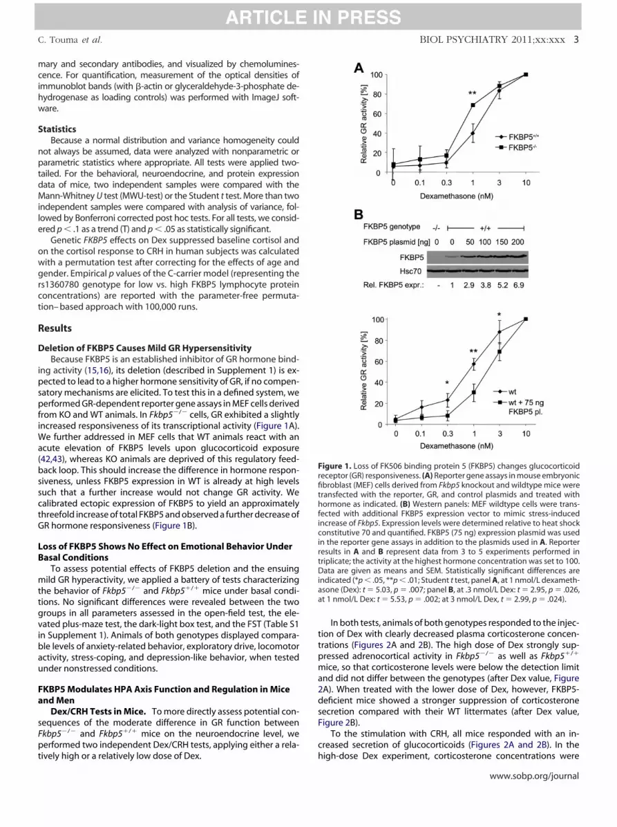
owgrct
tgvibau
Fa
sF
ttpma2dsF
c
FrfithficirtDiaa
C. Touma et al. BIOL PSYCHIATRY 2011;xx:xxx 3
mary and secondary antibodies, and visualized by chemolumines-cence. For quantification, measurement of the optical densities ofimmunoblot bands (with �-actin or glyceraldehyde-3-phosphate de-hydrogenase as loading controls) was performed with ImageJ soft-ware.
StatisticsBecause a normal distribution and variance homogeneity could
not always be assumed, data were analyzed with nonparametric orparametric statistics where appropriate. All tests were applied two-tailed. For the behavioral, neuroendocrine, and protein expressiondata of mice, two independent samples were compared with theMann-Whitney U test (MWU-test) or the Student t test. More than twoindependent samples were compared with analysis of variance, fol-lowed by Bonferroni corrected post hoc tests. For all tests, we consid-ered p � .1 as a trend (T) and p � .05 as statistically significant.
Genetic FKBP5 effects on Dex suppressed baseline cortisol andn the cortisol response to CRH in human subjects was calculatedith a permutation test after correcting for the effects of age andender. Empirical p values of the C-carrier model (representing the
s1360780 genotype for low vs. high FKBP5 lymphocyte proteinoncentrations) are reported with the parameter-free permuta-ion– based approach with 100,000 runs.
Results
Deletion of FKBP5 Causes Mild GR HypersensitivityBecause FKBP5 is an established inhibitor of GR hormone bind-
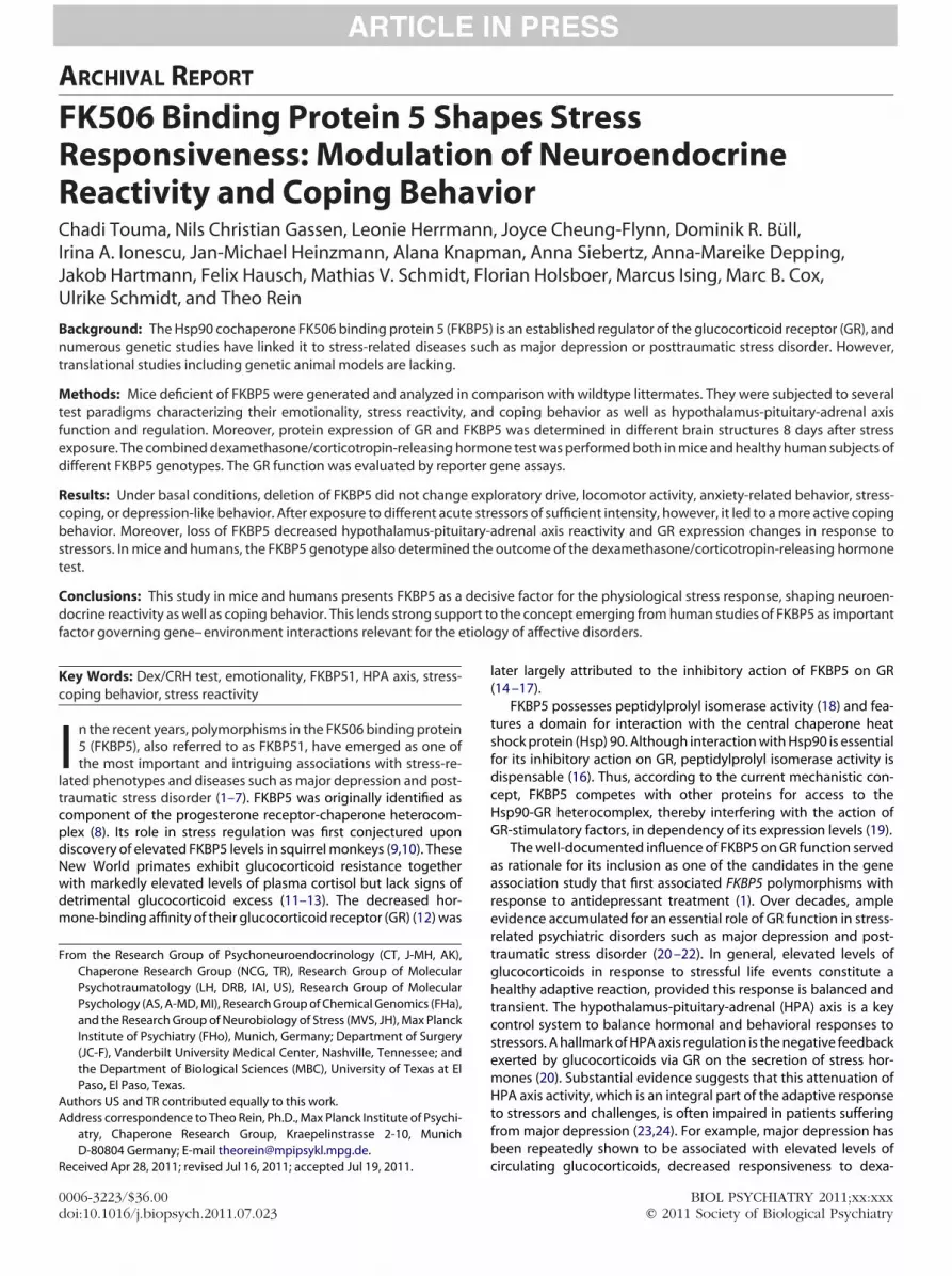
ing activity (15,16), its deletion (described in Supplement 1) is ex-pected to lead to a higher hormone sensitivity of GR, if no compen-satory mechanisms are elicited. To test this in a defined system, weperformed GR-dependent reporter gene assays in MEF cells derivedfrom KO and WT animals. In Fkbp5�/� cells, GR exhibited a slightlyincreased responsiveness of its transcriptional activity (Figure 1A).We further addressed in MEF cells that WT animals react with anacute elevation of FKBP5 levels upon glucocorticoid exposure(42,43), whereas KO animals are deprived of this regulatory feed-back loop. This should increase the difference in hormone respon-siveness, unless FKBP5 expression in WT is already at high levelssuch that a further increase would not change GR activity. Wecalibrated ectopic expression of FKBP5 to yield an approximatelythreefold increase of total FKBP5 and observed a further decrease ofGR hormone responsiveness (Figure 1B).
Loss of FKBP5 Shows No Effect on Emotional Behavior UnderBasal Conditions
To assess potential effects of FKBP5 deletion and the ensuingmild GR hyperactivity, we applied a battery of tests characterizingthe behavior of Fkbp5�/� and Fkbp5�/� mice under basal condi-ions. No significant differences were revealed between the tworoups in all parameters assessed in the open-field test, the ele-ated plus-maze test, the dark-light box test, and the FST (Table S1
n Supplement 1). Animals of both genotypes displayed compara-le levels of anxiety-related behavior, exploratory drive, locomotorctivity, stress-coping, and depression-like behavior, when testednder nonstressed conditions.
KBP5 Modulates HPA Axis Function and Regulation in Micend Men
Dex/CRH Tests in Mice. To more directly assess potential con-equences of the moderate difference in GR function betweenkbp5�/� and Fkbp5�/� mice on the neuroendocrine level, we
performed two independent Dex/CRH tests, applying either a rela-
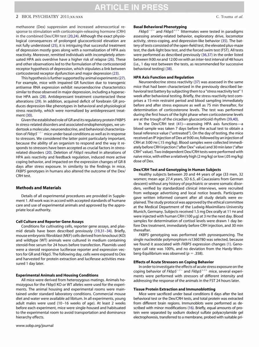
tively high or a relatively low dose of Dex. hIn both tests, animals of both genotypes responded to the injec-ion of Dex with clearly decreased plasma corticosterone concen-rations (Figures 2A and 2B). The high dose of Dex strongly sup-ressed adrenocortical activity in Fkbp5�/� as well as Fkbp5�/�
ice, so that corticosterone levels were below the detection limitnd did not differ between the genotypes (after Dex value, FigureA). When treated with the lower dose of Dex, however, FKBP5-eficient mice showed a stronger suppression of corticosteroneecretion compared with their WT littermates (after Dex value,igure 2B).
To the stimulation with CRH, all mice responded with an in-reased secretion of glucocorticoids (Figures 2A and 2B). In the
igure 1. Loss of FK506 binding protein 5 (FKBP5) changes glucocorticoideceptor (GR) responsiveness. (A) Reporter gene assays in mouse embryonicbroblast (MEF) cells derived from Fkbp5 knockout and wildtype mice wereransfected with the reporter, GR, and control plasmids and treated withormone as indicated. (B) Western panels: MEF wildtype cells were trans-
ected with additional FKBP5 expression vector to mimic stress-inducedncrease of Fkbp5. Expression levels were determined relative to heat shockonstitutive 70 and quantified. FKBP5 (75 ng) expression plasmid was used
n the reporter gene assays in addition to the plasmids used in A. Reporteresults in A and B represent data from 3 to 5 experiments performed inriplicate; the activity at the highest hormone concentration was set to 100.ata are given as means and SEM. Statistically significant differences are
ndicated (*p � .05, **p � .01; Student t test, panel A, at 1 nmol/L dexameth-sone (Dex): t � 5.03, p � .007; panel B, at .3 nmol/L Dex: t � 2.95, p � .026,t 1 nmol/L Dex: t � 5.53, p � .002; at 3 nmol/L Dex, t � 2.99, p � .024).
igh-dose Dex experiment, corticosterone concentrations were
www.sobp.org/journal
tan
dslh
(rc39
C
RH: p
4 BIOL PSYCHIATRY 2011;xx:xxx C. Touma et al.
only slightly elevated 30 min after CRH injection (nearly reachingthe levels observed before Dex treatment) and did not differ be-tween Fkbp5�/� and Fkbp5�/� mice (after CRH value, Figure 2A). Inhe low-dose Dex experiment, however, the CRH injection inducedrobust increase of corticosterone concentrations, which was sig-ificantly less pronounced in Fkbp5�/� mice than in their Fkbp5�/�
Figure 2. The Fkbp5 genotype affects hypothalamus-pituitary-adrenal axis fuCRH) testing. (A, B) Combined Dex/CRH test in mice. Corticosterone respelatively high (2 mg/kg, panel A) or relatively low (.05 mg/kg, panel B)oncentrations were measured in male Fkbp5 knockout (KO) and wildtype (W0 min after CRH injection (after CRH). Data are given as box plots showing m0% percentiles (whiskers). Statistical differences between the groups are in
each time point (panel A, high-dose Dex, Mann-Whitney U test (MWU-test), NRH: U � 35, p � .256; panel B, low-dose Dex, MWU-test, NKO�12, NWT � 12
.001). (C) Stress reactivity testing. Plasma corticosterone concentrations immas 75 min after termination of the stressor (recovery) in male Fkbp5 KO andMWU-test, NKO � 11, NWT � 13, initial: U � 34, p � .028, reaction: U � 14, p �cortisol response to the Dex/CRH test combining a pharmacological suppresubsequent stimulation with CRH (100 �g) in healthy human subjects. Plasm23) and in noncarriers (TT) (n � 7) of the FKBP5 single nucleotide polymorphDex) and 30 min after CRH injection (after CRH). Data are given as meansindicated (p � .1 n.s., **p � .01) above the columns for each time pogender-corrected residuals, untreated: p � .101, after Dex: p � .004, after C
littermates (after CRH value, Figure 2B). (
www.sobp.org/journal
The corticosterone concentrations measured under basal con-itions at 3:00 PM 1 week before the Dex/CRH test did not differignificantly between Fkbp5�/� and Fkbp5�/� mice either in theow-dose Dex experiment (untreated value, Figure 2B) or in theigh-dose Dex experiment (untreated value, Figure 2A).
Stress Reactivity Testing in Mice. To further test the reactivity
n as revealed by stress reactivity and Dex/corticotropin-releasing hormoneto a pharmacological suppression of adrenocortical activity with either aof Dex and a subsequent stimulation with CRH. Plasma corticosterone
ce 1 week before (untreated) and 6 hours after Dex treatment (after Dex) andns (lines in the boxes), 25% and 75% percentiles (boxes), as well as 10% anded (p � .1 nonsignificant (n.s.), *p � .05, ***p � .001) above the columns for10, NWT � 10, untreated: U � 57, p � .386, after Dex: U � 34.5, p � .103, aftereated: U � 40, p � .450, after Dex: U � 28, p � .011, after CRH: U � 14, p �tely before (initial) and after (reaction) a 15-min restraint (RS) period as wellice. Data and statistical differences are given as described above (panel C,recovery: U � 36, p � .040). (D) Dex/CRH test in humans. Graph depicts theof adrenocortical activity with a relatively low dose of Dex (1.5 mg) and a
tisol concentrations were measured in C allele carriers (CC) (n � 34; CT, n �s1360780 1 day before (untreated) and 16 hours after Dex treatment (afterSEM. Statistical differences between the genotypes (recessive model) areermutation tests with 100,000 permutations, performed with age- and� .898). Other abbreviations as in Figure 1.
nctioonsedoseT) miedia
dicatKO �, untredia
WT m.001,ssiona corism rand
int (p
and recovery) of the HPA axis in a more physiological paradigm, we
aivdoCD
S
f
wtFah3bs
omt
seae(
pr(Fset
D
opbiiacddaigeDfot
mtocelbHFsftasagea(tdHm
dfiGwhavlr
C. Touma et al. BIOL PSYCHIATRY 2011;xx:xxx 5
used a 15-min restraint period as moderate psychological stressor.Compared with Fkbp5�/� mice, plasma corticosterone concentra-tions were significantly lower in Fkbp5�/� mice at all three timepoints (Figure 2C) (i.e., immediately before [initial value], directlyafter [reaction value], and 75 min after termination of the stressor[recovery value]).
Dex/CRH Test in Humans. To relate the findings on HPAxis function and regulation in our mouse model to the situation
n humans, we performed the Dex/CRH test in 65 genotypedolunteers screened for absence of mental and severe physicalisorders during lifetime. Individuals whose genotype previ-usly had been associated with lower levels of FKBP5 (rs1360780-carriers) reacted with a stronger suppression of cortisol afterex treatment (Figure 2D). Although this finding is consistent
with the difference observed in mice, we did not observe agenotype-dependent difference in overall cortisol levels afterstimulation with CRH.
FKBP5�/� Mice Exhibit More Active Coping Behavior Aftertress Exposure
Stressors of different intensity were used to investigate the ef-ects of acute stress exposure on the coping behavior of Fkbp5�/�
and Fkbp5�/� mice in the FST 24 hours later. In a first experiment,e exposed mice to a 15-min restraint stressor, returned them to
heir home cage for 15 min, and then subjected them to a 6-minST. In this FST, Fkbp5�/� animals showed a trend toward morective coping with the aversive situation (i.e., more swimming be-avior was observed compared with their WT littermates) (FigureA). The second FST, which was performed 24 hours after the com-ined restraint and first FST stressor, revealed more pronouncedtatistically significant behavioral differences (Figure 3B). This ex-
periment was also replicated with an independent batch of ani-mals, yielding very similar results (Table S2 in Supplement 1).
In a second experiment with reduced overall stress intensity, theFkbp5�/� and Fkbp5�/� animals were subjected to the 15-minrestraint stressor, but without FST testing shortly afterward. In thisexperiment, no differences in coping behavior between the twogroups were observed during the FST performed 24 hours afterstress exposure (Figure 3C).
In another experiment with increased stress intensity, Fkbp5�/�
and Fkbp5�/� mice were subjected to a prolonged restraint periodf 60 min and tested in the FST 24 hours later. The FKBP5-deficientice again exhibited more swimming and less floating behavior
han WT animals (Figure 3D) (i.e., showed more active coping).Confirming our assumption that 60-min restraint constitutes a
tronger stressor than 15-min restraint, the prolonged restraintlicited a higher increase of corticosterone concentrations immedi-tely after releasing the mice. Importantly, the genotypic differ-nces in HPA axis reactivity were similar in both experiments
Figures 2C and 3E), with FKBP5-deficient mice showing signifi-cantly lower endocrine stress reactivity.
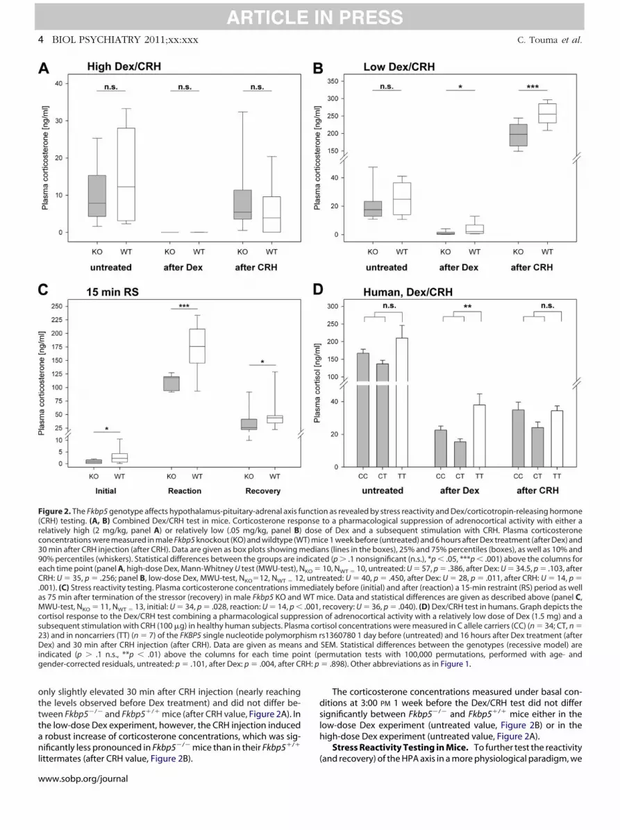
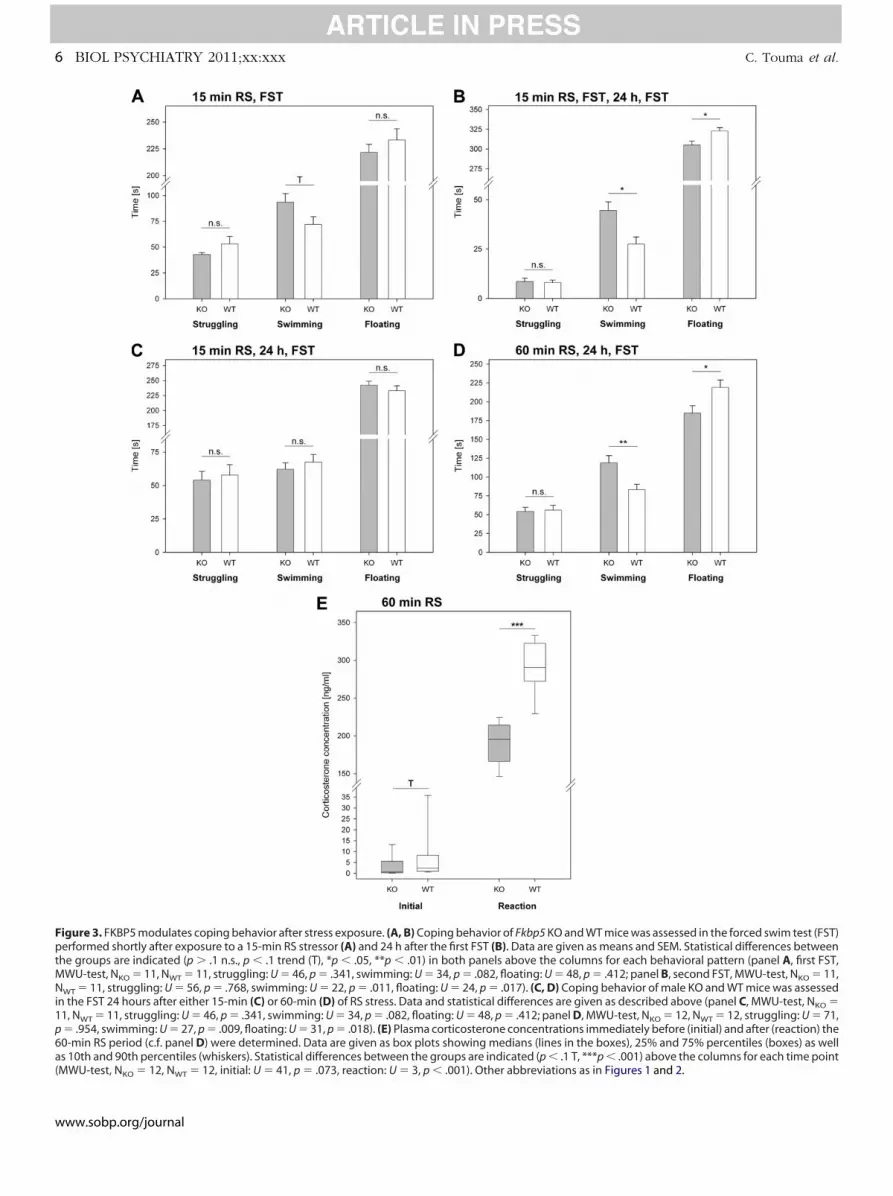
Deletion of FKBP5 Impacts on Stress-Induced Changes ofProtein Expression
We compared the expression levels of GR between WT and KOanimals 8 days after exposure to the Dex/CRH tests (Figure 2) orafter the combined exposure to the restraint (15 min or 60 min) andFST (Figure 3). In the Dex/CRH tests, only the experiment with thelower dose of Dex produced a difference between the genotypes,with the WT animals exhibiting lower levels of GR expression in thehippocampus, prefrontal cortex, and cerebellum (Figures 4A and B).
For the combined restraint and FST exposure, only the experi-
ment with 60-min restraint resulted in a significant change of hip- tocampal GR expression, where WT but not KO animals exhibitededuced GR expression compared with unstressed control animalsFigure 4C). In the WT mice of this experiment, we also assessedKBP5 protein levels in the hippocampus and observed a small butignificant decrease of FKBP5 expression eight days after stressxposure (Figure 4D). Similar results were obtained for the prefron-al cortex and the cerebellum (data not shown).
iscussion
It has been recognized over the years that decisive for the devel-pment of stress-related diseases such as major depression is not aarticular traumatic event per se or a particular genetic endowmentut the interplay of genetic and environmental factors. Therefore, it
s crucial for increasing our understanding of affective disorders tonvestigate how individual differences in stress responsivenessrise and by which mechanisms an organism is able to cope withhallenges and stressful situations. We report here that Fkbp5 geneeletion results in a mild GR hypersensitivity, changes in neuroen-ocrine reactivity and feedback regulation of the HPA axis, a morective coping behavior after exposure to stressors, and alterations
n the stress-induced changes of GR expression. Similarly, the FKBP5enotypes in humans shown to be associated with different FKBP5xpression levels turned out to be linked to the outcome of theex/CRH test. The functional link between FKBP5 and GR most likely
orms the basis for these effects, although the possibility exists thatther important actions of FKBP5 (19,44 – 46) might contribute to
he observed endophenotypes.In general, gene KO strategies carry the risk of compensatory
olecular reactions that compromise detection of protein func-ion. Because FKBP5 acts on GR via its interaction with Hsp90 (16),ther inhibitory factors competing for Hsp90 binding could in prin-iple take over the function of attenuating GR activity (19). Ourxperiments, however, indicate a more sensitive GR in Fkbp5-de-
eted MEF cells (Figure 1A), which was also supported by hormoneinding experiments in brain extracts (not shown). Thus, othersp90 cochaperones apparently did not compensate for the loss ofKBP5 (e.g., by decreasing FKBP4) (Figure 1B). An enhanced GRensitivity in animals devoid of FKBP5 is the most likely explanationor the changes in HPA axis settings observed here. This includeshe lower levels of basal morning corticosterone (Figures 2 and 3E)nd particularly the reduced secretion of corticosterone in re-ponse to pharmacological or psychological stressors (Figures 2Cnd 3E). This interpretation is corroborated by a recent report of aain of function GR knock-in mouse (47). In that study, the endog-nous GR was replaced by the human GRM604L mutant, which isctivated at lower glucocorticoid concentrations than wildtype GR48). Those mice were phenotypically normal under basal condi-ions—with birth weight, postnatal growth, and development in-istinguishable from WT littermates— but exhibited changes inPA axis activity (47), in similarity to our findings in the Fkbp5�/�
ice presented here.Our observation that the Dex/CRH test revealed a genotype
ifference only at the lower dose of Dex might seem surprising atrst (Figure 2). This dose-dependency most likely reflects the shift ofR responsiveness toward lower concentrations of hormone,hich was observed here (Figure 1) and elsewhere (15,16). Theigher dose of Dex that did not produce a genotype effect presum-bly amounted to a saturating concentration of hormone that acti-ates GR irrespectively of the FKBP5 status. In contrast, the fortyfold
ower dose of Dex that exposed a genotype effect presumablyesulted in sub-saturating hormone concentration, as evidenced by
he less pronounced suppression of corticosterone (Figure 2), awww.sobp.org/journal
MN
1p
6 BIOL PSYCHIATRY 2011;xx:xxx C. Touma et al.
Figure 3. FKBP5 modulates coping behavior after stress exposure. (A, B) Coping behavior of Fkbp5 KO and WT mice was assessed in the forced swim test (FST)performed shortly after exposure to a 15-min RS stressor (A) and 24 h after the first FST (B). Data are given as means and SEM. Statistical differences betweenthe groups are indicated (p � .1 n.s., p � .1 trend (T), *p � .05, **p � .01) in both panels above the columns for each behavioral pattern (panel A, first FST,
WU-test, NKO � 11, NWT � 11, struggling: U � 46, p � .341, swimming: U � 34, p � .082, floating: U � 48, p � .412; panel B, second FST, MWU-test, NKO � 11,WT � 11, struggling: U � 56, p � .768, swimming: U � 22, p � .011, floating: U � 24, p � .017). (C, D) Coping behavior of male KO and WT mice was assessed
in the FST 24 hours after either 15-min (C) or 60-min (D) of RS stress. Data and statistical differences are given as described above (panel C, MWU-test, NKO �1, NWT � 11, struggling: U � 46, p � .341, swimming: U � 34, p � .082, floating: U � 48, p � .412; panel D, MWU-test, NKO � 12, NWT � 12, struggling: U � 71,� .954, swimming: U � 27, p � .009, floating: U � 31, p � .018). (E) Plasma corticosterone concentrations immediately before (initial) and after (reaction) the
60-min RS period (c.f. panel D) were determined. Data are given as box plots showing medians (lines in the boxes), 25% and 75% percentiles (boxes) as well
as 10th and 90th percentiles (whiskers). Statistical differences between the groups are indicated (p � .1 T, ***p � .001) above the columns for each time point(MWU-test, NKO � 12, NWT � 12, initial: U � 41, p � .073, reaction: U � 3, p � .001). Other abbreviations as in Figures 1 and 2.www.sobp.org/journal
sewlt
aeenoGdeeb
o(sdodleb
Fmmltosa
itc
lpgbD
s 1 an
C. Touma et al. BIOL PSYCHIATRY 2011;xx:xxx 7
condition in which the dependency of GR activity on FKBP5 expres-sion is most pronounced (15,16). Notably, the differences we ob-erved in response to the lower dose of Dex between FKBP5-xpressing and nonexpressing mice are paralleled in humans,here the genotype previously shown to be associated with lower
evels of FKBP5 (1) also exhibited lower levels of cortisol after Dexreatment (Figure 2).
Expression changes of genes relevant to the function of the HPAxis have been frequently described to be associated with stressxposure—in particular stressful events during sensitive periods ofarly development (49). For example, in rats, lower levels of mater-al care led to decreased GR expression in the hippocampus of theffspring (50). Maternal separation led to decreased expression ofR in the hippocampus and forebrain (51,52), apparently in a time-ependent fashion, because it is preceded by an increase of GRxpression (51). Exposure of pregnant rats to Dex also caused GRxpression changes in the offspring, which was dependent on therain region and duration/timing of treatment (53). In humans, a
postmortem analysis of suicide victims revealed an association ofchildhood abuse with decreased hippocampal GR expression (54).Because there is more evidence linking GR expression to psycho-pathological conditions (20,55), there are intense research efforts
Figure 4. FKBP5 modulates stress-induced changes of protein expression. (Aand prefrontal cortex (PFC) were determined 8 days after completion of the cevels (panel A: high-dose Dex, panel B: low-dose Dex). Brains from seven maerformed in triplicates. Depicted bands on the immunoblots represent samraphs above. Data are given as means and SEM. Statistical differences betrain region (panel A, high-dose Dex, Student t test, NKO � 7, NWT � 7, HIP: tex, Student t test, NKO � 7, NWT � 7, HIP: t � 1.76, p � .008, CER: t � 1.98, p
were determined in six Fkbp5 KO and six WT littermates 8 days after the stretwo FST or 60-min RS followed by one FST on the next day or unstressed coImmunoblot experiments were performed in triplicates, and GR signals wereblot depicted shows 12 adjacent bands with 2 bands representative for eachof variance revealed significant group differences for treatment [F(2,30) � 14factors was not significant (p � .150). (D) FKBP5 protein levels were determisame procedure. Blots depicted show two adjacent immunoblot bands eachsignificant group differences for treatment [F(2,15) � 6.7, p � .009]. Significrespective columns (*p � .05, ***p � .001). Other abbreviations as in Figure
aiming at understanding the mechanisms governing programming o
f GR. Epigenetic mechanisms are very likely to contribute54,56,57), and our findings of a genotypic difference in GR expres-ion after stress exposure (Figure 4) put FKBP5 forward as a candi-ate to contribute to stress-induced GR expression changes. Ourbservation that the genotypic difference was dependent on theose/intensity of the pharmacological/psychological stressor most
ikely relates to the hormone responsiveness of GR, where differ-nces were exposed at sub-saturating concentrations of steroidsut barely at concentrations above and below.
It is worth mentioning that our study was limited to male mice.emales were not included, due to methodological difficulties ofonitoring the estrous cycle in a noninvasive manner, which isandatory to avoid systematically biasing the results. Neverthe-
ess, it will be important to address a potential gender difference inhe effect of FKBP5 in preclinical models, because prevalence ratesf major depression are higher in women (58). Actually, one clinicaltudy suggests a gender difference in the FKBP5-genotype associ-tion with major depression (59).
The effect of FKBP5 deletion on HPA axis parameters discussedn the preceding text can be interpreted as a compensatory reac-ion to altered GR sensitivity restoring an adequate balance of re-eptor and hormone and therefore should not have further physi-
R protein levels in the bilaterally pooled hippocampi (HIP), cerebellum (CER),ned Dex/CRH tests (cf. Figure 2) by immunoblotting and normalized to actince for each genotype and condition were extracted, and immunoblots werefrom six individual mice, corresponding to the conditions displayed in thethe groups are indicated (*p � .05, **p � .01) above the columns for each
1, p � .161, CER: t � .765, p � .215, PFC: t � .717, p � .343; panel B, low-dose4, PFC: t � 1.85, p � .007). (C) GR protein levels in the bilaterally pooled HIPeriments presented in Figures 3B and 3D (i.e. either 15-min RS followed by
subjects [six mice of each genotype left undisturbed in their home cages]).alized to glyceraldehyde-3-phosphate dehydrogenase (GAPDH) levels. Theimental group, respectively. Statistical analysis employing two-way analysis
.001] and Fkbp5 genotype [F(1,30) � 7.8, p � .009]. The interaction of boththe same six WT animals used for determination of GR in panel C with the
constitute representative examples. One-way analysis of variance revealedfferences in the pairwise Bonferroni post hoc tests are indicated above thed 2.
, B) Gombile miples
ween� .65� .03ss expntrolnorm
exper.4, p �ned in
andant di
logical consequences. Accordingly, we did not find genotype-
www.sobp.org/journal

airitFnor
ao
orapgc
1
1
1
1
1
1
1
1
1
1
2
2
2
2
2
2
2
2
2
2
3
8 BIOL PSYCHIATRY 2011;xx:xxx C. Touma et al.
dependent behavioral differences under basal conditions.However, by deleting Fkbp5, the animals are deprived not simply of
static factor that calibrates GR activity but also of the ultra-shortntracellular feedback loop, a peculiar hallmark of molecular stressegulation linking FKBP5 and GR. This feedback loop leads to anncrease of FKBP5 upon GR activation (60,61), thereby limiting ex-ended, excessive activation of GR. The lack of this feedback loop inkbp5�/� animals might be the basis for our observation that phe-otypic differences in coping behavior during the FST are exposednly if preceded by stressors of sufficient strength (Figure 3). Very
ecently, this was similarly shown for anxiety-related behavior (62),further corroborating the relevance of the Fkbp5�/� mouse model.
In this respect, it is intriguing that several studies in humansttest to the concept that FKBP5 is decisive for the developmentf stress-related mental disorders only in combination with trau-
matic events (2,63– 65). We hypothesize that, taken together withur results in mice, the reported polymorphisms are linked— di-
ectly or indirectly—to the ultra-short feedback loop connecting GRnd FKBP5 as a crucial component of stress regulation. Among theossible mechanisms, genetic polymorphisms might change pro-ramming of FKBP5 expression, because it has been shown re-ently that FKBP5 is amenable to epigenetic regulation (43). In
conclusion, our findings translate clinical observations on FKBP5into animal-based experimental systems. This should encouragemore detailed studies testing the hypothesis that genotype-depen-dent reaction of FKBP5 expression to stressful life events deter-mines the risk for development of affective disorders, ultimatelyleading to improved treatment regimes.
This study was supported by the Max Planck Society and the HorstKübler foundation, Bad Ragaz. Drs. Holsboer and Rein are co-inventorsof the following pending patent application: FKBP5: a novel target forantidepressant therapy (International publication number: WO 2005/05450). We are indebted to Sergej Asmus, Bozidar Novak, KathrinHafner, Lisa Tietze, and Albin Varga for technical assistance. The otherauthors report no biomedical financial interests or potential conflictsof interest.
Supplementary material cited in this article is available online.
1. Binder EB, Salyakina D, Lichtner P, Wochnik GM, Ising M, Putz B, et al.(2004): Polymorphisms in FKBP5 are associated with increased recur-rence of depressive episodes and rapid response to antidepressanttreatment. Nat Genet 36:1319 –1325.
2. Binder EB, Bradley RG, Liu W, Epstein MP, Deveau TC, Mercer KB, et al.(2008): Association of FKBP5 polymorphisms and childhood abuse withrisk of posttraumatic stress disorder symptoms in adults. JAMA 299:1291–1305.
3. Ising M, Depping AM, Siebertz A, Lucae S, Unschuld PG, Kloiber S, et al.(2008): Polymorphisms in the FKBP5 gene region modulate recoveryfrom psychosocial stress in healthy controls. Eur J Neurosci 28:389 –398.
4. Lekman M, Laje G, Charney D, Rush AJ, Wilson AF, Sorant AJ, et al. (2008):The FKBP5-gene in depression and treatment response—an associationstudy in the Sequenced Treatment Alternatives to Relieve Depression(STAR*D) Cohort. Biol Psychiatry 63:1103–1110.
5. Koenen KC, Saxe G, Purcell S, Smoller JW, Bartholomew D, Miller A, et al.(2005): Polymorphisms in FKBP5 are associated with peritraumatic dis-sociation in medically injured children. Mol Psychiatry 10:1058 –1059.
6. Willour VL, Chen H, Toolan J, Belmonte P, Cutler DJ, Goes FS, et al. (2008):Family-based association of FKBP5 in bipolar disorder. Mol Psychiatry14:261–268.
7. Yehuda R, Cai G, Golier JA, Sarapas C, Galea S, Ising M, et al. (2009): Geneexpression patterns associated with posttraumatic stress disorder fol-
lowing exposure to the World Trade Center attacks. Biol Psychiatry 66:708 –711.www.sobp.org/journal
8. Smith DF, Albers MW, Schreiber SL, Leach KL, Deibel MRJ (1993): FKBP54,a novel FK506-binding protein in avian progesterone receptor com-plexes and HeLa extracts. J Biol Chem 268:24270 –24273.
9. Reynolds PD, Roveda KP, Tucker JA, Moore CM, Valentine DL, ScammellJG (1998): Glucocorticoid-resistant B-lymphoblast cell line derived fromthe Bolivian squirrel monkey (Saimiri boliviensis boliviensis). Lab AnimSci 48:364 –370.
0. Reynolds PD, Ruan Y, Smith DF, Scammell JG (1999): Glucocorticoidresistance in the squirrel monkey is associated with overexpression ofthe immunophilin FKBP51. J Clin Endocrinol Metab 84:663– 669.
1. Bamberger CM, Schulte HM, Chrousos GP (1996): Molecular determi-nants of glucocorticoid receptor function and tissue sensitivity to glu-cocorticoids. Endocr Rev 17:245–261.
2. Chrousos GP, Renquist D, Brandon D, Eil C, Pugeat M, Vigersky R, et al.(1982): Glucocorticoid hormone resistance during primate evolution:Receptor-mediated mechanisms. Proc Natl Acad Sci U S A 79:2036 –2040.
3. Chrousos GP, Loriaux DL, Tomita M, Brandon DD, Renquist D, AlbertsonB, et al. (1986): The new world primates as animal models of glucocorti-coid resistance. Adv Exp Med Biol 196:129 –144.
4. Denny WB, Valentine DL, Reynolds PD, Smith DF, Scammell JG (2000):Squirrel monkey immunophilin FKBP51 is a potent inhibitor of gluco-corticoid receptor binding. Endocrinology 141:4107– 4113.
5. Riggs DL, Roberts PJ, Chirillo SC, Cheung-Flynn J, Prapapanich V, Rata-jczak T, et al. (2003): The Hsp90-binding peptidylprolyl isomeraseFKBP52 potentiates glucocorticoid signaling in vivo. EMBO J 22:1158 –1167.
6. Wochnik GM, Rüegg J, Abel GA, Schmidt U, Holsboer F, Rein T (2005):FK506-binding proteins 51 and 52 differentially regulate dynein inter-action and nuclear translocation of the glucocorticoid receptor in mam-malian cells. J Biol Chem 280:4609 – 4616.
7. Scammell JG, Denny WB, Valentine DL, Smith DF (2001): Overexpressionof the FK506-binding immunophilin FKBP51 is the common cause ofglucocorticoid resistance in three New World primates. Gen Comp Endo-crinol 124:152–165.
8. Pirkl F, Buchner J (2001): Functional analysis of the Hsp90-associatedhuman peptidyl prolyl cis/trans isomerases FKBP51, FKBP52 and Cyp40.J Mol Biol 308:795– 806.
9. Schülke JP, Wochnik GM, Lang-Rollin I, Gassen NC, Knapp RT, Berning B,et al. (2010): Differential impact of tetratricopeptide repeat proteins onthe steroid hormone receptors. PLoS One 5:e11717.
0. De Kloet ER, Joels M, Holsboer F (2005): Stress and the brain: Fromadaptation to disease. Nat Rev Neurosci 6:463– 475.
1. Heim C, Nemeroff CB (2001): The role of childhood trauma in the neu-robiology of mood and anxiety disorders: Preclinical and clinical studies.Biol Psychiatry 49:1023–1039.
2. Charney DS, Manji HK (2004): Life stress, genes, and depression: Multiplepathways lead to increased risk and new opportunities for intervention.Sci STKE 2004:re5.
3. Holsboer F (2000): The corticosteroid receptor hypothesis of depres-sion. Neuropsychopharmacology 23:477–501.
4. Ising M, Holsboer F (2006): Genetics of stress response and stress-re-lated disorders. Dialogues Clin Neurosci 8:433– 444.
5. Wolkowitz OM, Burke H, Epel ES, Reus VI (2009): Glucocorticoids. Mood,memory, and mechanisms. Ann N Y Acad Sci 1179:19 – 40.
6. Zobel AW, Yassouridis A, Frieboes RM, Holsboer F (1999): Prediction ofmedium-term outcome by cortisol response to the combined dexa-methasone-CRH test in patients with remitted depression. Am J Psychi-atry 156:949 –951.
7. Touma C (2011): Stress and affective disorders: Animal models elucidat-ing the molecular basis of neuroendocrine-behavior interactions. Phar-macopsychiatry 44:S15–S26.
8. Pepin MC, Pothier F, Barden N (1992): Impaired type II glucocorticoid-receptor function in mice bearing antisense RNA transgene. Nature355:725–728.
9. Pepin MC, Govindan MV, Barden N (1992): Increased glucocorticoidreceptor gene promoter activity after antidepressant treatment. MolPharmacol 41:1016 –1022.
0. Boyle MP, Brewer JA, Funatsu M, Wozniak DF, Tsien JZ, Izumi Y, et al.(2005): Acquired deficit of forebrain glucocorticoid receptor produces
depression-like changes in adrenal axis regulation and behavior. ProcNatl Acad Sci U S A 102:473– 478.
3
3
3
3
3
3
3
4
4
4
4
4
4
4
4
4
4
5
5
5
5
5
5
5
5
5
5
6
6
6
6
6
6
C. Touma et al. BIOL PSYCHIATRY 2011;xx:xxx 9
31. Wochnik GM, Young JC, Schmidt U, Holsboer F, Hartl FU, Rein T (2004):Inhibition of GR-mediated transcription by p23 requires interactionwith Hsp90. FEBS Lett 560:35–38.
2. Abel A, Wochnik G, Rüegg J, Rouyer A, Holsboer F, Rein T (2002): Activityof the glucocorticoid receptor in G2 and mitosis. Mol Endocrinol 16:1352–1366.
33. Schmidt U, Wochnik GM, Rosenhagen MC, Hartl FU, Holsboer F, Rein T(2003): Essential role of the unusual DNA binding motif of BAG-1 forinhibition of the glucocorticoid receptor. J Biol Chem 278:4926 – 4931.
4. Herr AS, Tsolakidou AF, Yassouridis A, Holsboer F, Rein T (2003): Antide-pressants differentially influence the transcriptional activity of the glu-cocorticoid receptor in vitro. Neuroendocrinology 78:12–22.
5. Cryan JF, Holmes A (2005): The ascent of mouse: advances in modellinghuman depression and anxiety. Nat Rev Drug Discov 4:775–790.
6. Varadarajulu J, Lebar M, Krishnamoorthy G, Habelt S, Lu J, Bernard W, I, etal. (2011): Increased anxiety-related behaviour in Hint1 knockout mice.Behav Brain Res 220:305–311.
7. Touma C, Bunck M, Glasl L, Nussbaumer M, Palme R, Stein H, et al. (2008):Mice selected for high versus low stress reactivity: A new animal modelfor affective disorders. Psychoneuroendocrinology 33:839 – 862.
8. Paylor R, Spencer CM, Yuva-Paylor LA, Pieke-Dahl S (2006): The use ofbehavioral test batteries, II: effect of test interval. Physiol Behav 87:95–102.
9. Buijs RM, Kalsbeek A (2001): Hypothalamic integration of central andperipheral clocks. Nat Rev Neurosci 2:521–526.
0. Touma C, Fenzl T, Ruschel J, Palme R, Holsboer F, Kimura M, et al. (2009):Rhythmicity in mice selected for extremes in stress reactivity: Behav-ioural, endocrine and sleep changes resembling endophenotypes ofmajor depression. PLoS One 4:e4325.
1. Ising M, Horstmann S, Kloiber S, Lucae S, Binder EB, Kern N, et al. (2007):Combined dexamethasone/corticotropin releasing hormone test pre-dicts treatment response in major depression—a potential biomarker?Biol Psychiatry 62:47–54.
2. Scharf SH, Liebl C, Binder EB, Schmidt MV, Muller MB (2011): Expressionand regulation of the Fkbp5 gene in the adult mouse brain. PLoS One6:e16883.
3. Lee RS, Tamashiro KL, Yang X, Purcell RH, Harvey A, Willour VL, et al.(2010): Chronic corticosterone exposure increases expression and de-creases deoxyribonucleic acid methylation of Fkbp5 in mice. Endocrinol-ogy 151:4332– 4343.
4. Jinwal UK, Koren J, III, Borysov SI, Schmid AB, Abisambra JF, Blair LJ, et al.(2010): The Hsp90 cochaperone, FKBP51, increases Tau stability andpolymerizes microtubules. J Neurosci 30:591–599.
5. Pei H, Li L, Fridley BL, Jenkins GD, Kalari KR, Lingle W, et al. (2009): FKBP51affects cancer cell response to chemotherapy by negatively regulatingAkt. Cancer Cell 16:259 –266.
6. Romano S, D’Angelillo A, Pacelli R, Staibano S, De LE, Bisogni R, et al.(2010): Role of FK506-binding protein 51 in the control of apoptosis ofirradiated melanoma cells. Cell Death Differ 17:145–157.
7. Zhang J, Ge R, Matte-Martone C, Goodwin J, Shlomchik WD, Mamula MJ,et al. (2009): Characterization of a novel gain of function glucocorticoidreceptor knock-in mouse. J Biol Chem 284:6249 – 6259.
8. Zhang J, Simisky J, Tsai FT, Geller DS (2005): A critical role of helix 3-helix5 interaction in steroid hormone receptor function. Proc Natl Acad Sci US A 102:2707–2712.
9. Lupien SJ, McEwen BS, Gunnar MR, Heim C (2009): Effects of stressthroughout the lifespan on the brain, behaviour and cognition. Nat Rev
Neurosci 10:434 – 445.0. Liu D, Diorio J, Tannenbaum B, Caldji C, Francis D, Freedman A, et al.(1997): Maternal care, hippocampal glucocorticoid receptors, and hypo-thalamic-pituitary-adrenal responses to stress. Science 277:1659 –1662.
1. Navailles S, Zimnisky R, Schmauss C (2010): Expression of glucocorticoidreceptor and early growth response gene 1 during postnatal develop-ment of two inbred strains of mice exposed to early life stress. DevNeurosci 32:139 –148.
2. Ladd CO, Huot RL, Thrivikraman KV, Nemeroff CB, Plotsky PM (2004):Long-term adaptations in glucocorticoid receptor and mineralocorti-coid receptor mRNA and negative feedback on the hypothalamo-pitu-itary-adrenal axis following neonatal maternal separation. Biol Psychia-try 55:367–375.
3. Welberg LA, Seckl JR, Holmes MC (2001): Prenatal glucocorticoid pro-gramming of brain corticosteroid receptors and corticotrophin-releas-ing hormone: Possible implications for behaviour. Neuroscience 104:71–79.
4. McGowan PO, Sasaki A, D’Alessio AC, Dymov S, Labonte B, Szyf M, et al.(2009): Epigenetic regulation of the glucocorticoid receptor in humanbrain associates with childhood abuse. Nat Neurosci 12:342–348.
5. Webster MJ, Knable MB, O’Grady J, Orthmann J, Weickert CS (2002):Regional specificity of brain glucocorticoid receptor mRNA alterationsin subjects with schizophrenia and mood disorders. Mol Psychiatry7:985–94, 924.
6. Weaver IC, Champagne FA, Brown SE, Dymov S, Sharma S, Meaney MJ, etal. (2005): Reversal of maternal programming of stress responses inadult offspring through methyl supplementation: Altering epigeneticmarking later in life. J Neurosci 25:11045–11054.
7. Weaver IC, Cervoni N, Champagne FA, D’Alessio AC, Sharma S, Seckl JR,et al. (2004): Epigenetic programming by maternal behavior. Nat Neuro-sci 7:847– 854.
8. Parker G, Brotchie H (2010): Gender differences in depression. Int RevPsychiatry 22:429 – 436.
9. Lavebratt C, Aberg E, Sjoholm LK, Forsell Y (2010): Variations in FKBP5and BDNF genes are suggestively associated with depression in a Swed-ish population-based cohort. J Affect Disord 125:249 –255.
0. Hubler TR, Scammell JG (2004): Intronic hormone response elementsmediate regulation of FKBP5 by progestins and glucocorticoids. CellStress Chaperones 9:243–252.
1. Paakinaho V, Makkonen H, Jaaskelainen T, Palvimo JJ (2010): Glucocor-ticoid receptor activates poised FKBP51 locus through long-distanceinteractions. Mol Endocrinol 24:511–525.
2. Attwood BK, Bourgognon JM, Patel S, Mucha M, Schiavon E, SkrzypiecAE, et al. (2011): Neuropsin cleaves EphB2 in the amygdala to controlanxiety. Nature 473:372–375.
3. Xie P, Kranzler HR, Poling J, Stein MB, Anton RF, Farrer LA, et al. (2010):Interaction of FKBP5 with childhood adversity on risk for post-traumaticstress disorder. Neuropsychopharmacology 35:1684 –1692.
4. Roy A, Gorodetsky E, Yuan Q, Goldman D, Enoch MA (2010): Interactionof FKBP5, a stress-related gene, with childhood trauma increases the riskfor attempting suicide. Neuropsychopharmacology 35:1674 –1683.
5. Zimmermann P, Brückl T, Nocon A, Pfister H, Binder EB, Uhr M, et al. (inpress): Interaction of variants in the FKBP5 gene and adverse life eventsin predicting the first depression onset: Results from a ten-year prospec-tive community study. Am J Psychiatry. doi:10.1176/appi.ajp.2011.
10111577.www.sobp.org/journal


