Embed Size (px)
Citation preview

Barrett's Esophagus
High cancer-risk groups, Cardiovascular co-morbidity
and interaction with Helicobacter pylori
5

Coverfoto: Geysir - Haukadal Iceland by RAX, Iceland
Publication of this thesis was financially sponsored by: Jansen-Cilag Iceland, Jansen-Cilag The Netherlands, AstraZeneca Iceland Is gratefully acknowledged
All rights are reserved. No parts of this publication may be reproduced, stored in a retrieval system, or transmitted in any form or by any means, mechanically, by photocopying, recording, or otherwise, without the written permission of the author.
Printed by: Svansprent, Iceland
6
Barrett's Esophagus
High cancer-risk groups, cardiovascular co-morbidity
and interaction with Helicobacter pylori
Barrett-oesophagus
Groepen met een grote lmns op carcinoom, bijkomende hart- en vaatziekten
en interacties met Helicobactel' pylori
Proefschrift
ter verkrijging van de graad van doctor aan de Erasmus Universiteit Rotterdam
op gezag van de Rector Magnificus
Prof,ddr, J,H. van Bemmel
en volgens besluit van het College voor Promoties. De openbare verdediging zal plaatsvinden op
woensdag 15 mei 2002 om 15.45 uur door
Sunna Gudlaugsdottir
geboren te Reykjavik, Usland
7

Promotiecommissie
Promotor: Prof. J.H.P. Wilson
Overige leden: Prof. dr. E.J. Kuipers Prof. dr. B.H.Ch. Stricker Prof. dr. H.W.Tilanus
8

In 111emOl)' of 1I1y father To my mother
To Snorri and the children
9

10
CHAPTER 1
1.1 1.2 1.3 1.4 1.5 1.6 1.7 1.8
1.8.1 1.8.2
1.9 1.10
1.1 0.1 1.10.2
l.ll 1.12
CHAPTER 2
CHAPTER 3
CHAPTER 4
CHAPTER 5
CHAPTER 6
CHAPTER 7
7.1 7.2 7.3 7.4
7.4.1 7.4.2
7.5 7.6
CHAPTER 8
8.1 8.2
CHAPTER 9
9.1 9.2 9.3
CONTENTS GENERAL INTRODUCTION AND OUTLINE OF THE THESIS 13
Background 14 History 14 The definition of Barrett's esophagus 16 Why does Barrett's esophagus develop? 16 How long does it take for Barrett's esophagus to develop? 17 Relationship between Barrett's esophagus and adenocarcinoma 17 Natural history of Barrett's esophagus 18 Helicobacter pylori and gastro-esophageal reflux disease 20 Virulence of H. pylori strains 20 Acid production. 21 Treatment of BE and risks oflong term acid suppression 23 Surveillance of BE patients 25 Features supporting the need for endoscopic cancer surveillance 25 PITF ALLS in endoscopic cancer surveillance 26 Aims of the thesis 28 References 29
A MAJORITY OF PATIENTS WITH BARRETT'S OESOPHAGUS ARE UNLIKELY TO BENEFIT FROM ENDOSCOPIC CANCER SURVEILLANCE 37
CARDIOVASCULAR RISK FACTORS AND HISTORY OF MYOCARDIAL INFARCTION IN PATIENTS WITH BARRETT'S ESOPHAGUS 63
HYPERTENSION IS A FREQUENT CO-MORBIDITY IN PATIENTS WITH REFLUX OESOPHAGITIS OR BARRETT'S OESOPHAGUS BUT NOT IN NONULCER DYSPEPSIA. 81
THE PREVALENCE OF CAGA STRAINS OF HELICOBACTER PYLORI IN NONULCER DYSPEPSIA, REFLUX OESOPHAGITIS AND BARRETT'S OESOPHAGUS. 101
PROLONGED USE OF PROTON PUMP INHIBITORS, CAGA STATUS AND THE OUTCOME OF HELICOBACTER PYLORI GASTRITIS. 115
GENERAL DISCUSSION AND FUTURE PERSPECTIVES 133
Introduction 134 The main findings of this thesis can be summarised as follows. 136 Methodological considerations and General interpretation of study results 138 Closing remarks and future perspectives 143 Endoscopic cancer surveillance for subgroups of known BE patients 143 Population screening for subgroups in the general population 146 Targets for future research on BE include 150 References 152
SUMMARY - SAMENVATTING 155
Summary 157 Samenvatting 165
ADDENDUM 173
Danbvoord 175 About the author 177 List of abbreviations used in this thesis 179
II

12

Chapter 1 General introduction and outline of the thesis
13
1.1 Background
Barrett's esophagus (BE) has come to be regarded as an impOliant
premalignant condition (1). In recent years the incidence of adenocarcinoma of the
esophagus and of the gastric cardia has risen dramatically (2). Analyses of cancer
incidence data fi'om nine areas of the United States revealed steadily rising rates of
adenocarcinomas of the esophagus and gastric cardia from 1976 to 1987. The
increases among men in this period ranged from 4% to 10% per year, and exceeded
those of any other type of cancer (2). The rate of esophageal adenocarcinoma in
Denmark has increased eightfold over a 20-yr period (3). In the Netherlands the rate
of cardiacarcinoma has increased 35% (4), and since the early 90's mainly (Barrett's
related) adenocarcinomas ofthe esophagus have been on the rise (Ph.D. thesis
"Adenocarcinoma of the gastro-oesophageal junction - From gene to clinic" pages
71-73, B.P.L Wijnhoven, 2001). The reasons for this alarming rise are not clear but
many believe it to be a consequence of gastroesophageal-reflux disease (GERD) and
BE. This has led to an increased interest in research on BE and GERD.
1.2 History
Barrett's esophagus is named after Norman Barrett who described the
condition in 1950, but the first known description of islets of ectopic gastric mucosa
in the esophagus was in 1805 by Schmidt (5). One century later, in 1906, Tileston
attracted attention to peptic ulceration in esophagus, as a rare entity (6). In 1950
Norman Barrett reviewed the literature and described the columnar epithelium lined
esophagus(5). He suggested that this represented a congenital short esophagus. Only
when gastric-type mucosa was in islands, surrounded on all sides by normal
squamous epithel, could it be a part of the esophagus itself. He distinguished between
14
peptic-type ulcer and other esophageal ulcers. The peptic ulcers were thought to either
arise in the islands of ectopic gastric mucosa or as a result of secretion of acid by such
islands. Barrett failed to take into account the absence of the musculature and
peritoneal covering of the stomach in his theory of a congenital short esophagus (5).
In the same period LOliat-Jakob described the same condition, which he named
endobrachyesophagus (7). Allison and Johnstone in 1953 showed that anatomically
and funtionally the columnar lined esophagus was a paJi of the esophagus, although
they still considered this a congenital condition (8). Strong suppOli for an acquired
cause was published during the next decade. In 1970 Bremner and co-workers (9)
repOlied that in dogs with an incompetent lower esophageal sphincter (LES), removal
of the squamous epithelium was followed by replacement with columnar epithelium
whereas the squamous epithelium recovered in dogs with a competent LES. This was
chiefly evident when there was also an induction of gastric hypersecretion in the dog
with histamine injections(9). These workers postulated a replacement with mucosa
obtained from the gastric or junctional mucosa in persistent GERD. Although the
pathogenesis of BE is probably a multifactorial process (10), it is now generally
accepted the most important factor is chronic severe (duodeno)gastroesophageal
reflux disease (11, 12). (13). Most impOliant evidence for this includes case repOlis of
patients who underwent endoscopy and biopsy before and after developing columnar
lined esophagus(l3, 14).
Congenital islands of ectopic gastric mucosa however do occur and are found
in up to 10% of individuals undergoing endoscopy (15). These so called "inlet
patches" occur principally in the cervical esophagus and are mostly surrounded by
normal squamous epithelium (15,16).
15
1.3 The definition of Barrett's esophagus
The definition of BE has with time evolved from a macroscopic, usually
endoscopic definition (17) to a histologic definition(18, 19). It is generally agreed that
the BE of interest as a premalignant condition is a histological diagnosis (19-21) of
incomplete intestinal metaplasia (lM) or specialized columnar epithelium that
resembles intestinal mucosa with goblet cells between columnar cells with a flat or
viIIiform surface (22, 23). The glands do not contain parietal cells or peptic cells, but
esophageal mucous glands are present in the submucosa (24)
This has led to the modern definition of BE, which is: an endoscopically
visible segment of columnar mucosa, which on biopsy demonstrates intestinal
metaplasia.
1.4 Why does Barrett's esophagus develop?
Barrett's esophagus is probably a late but most severe complication of chronic
GERD (11,12, 14). Only about 5% of patients with chronic symptoms ofGERD have
BE at endoscopy and 1M in a biopsy sample. Patients with BE tend to have more
severe reflux disease (10,25) with greater impairment ofLES function and
esophageal body motility compared to GERD patients without BE( I 0, 26). There may
be a genetic predisposition to the development of reflux in families of patients with
BE (27-29) and esophageal adenocarcinoma (30). For uncomplicated reflux
esophagitis, environmental factors such as increasing body mass index appear to be
more important (30). Other envirol1111ental factors could theoretically also playa role
(smoking, alcohol, fatty foods, chocolate, large meals, bedtime snacks and drugs that
lowers LES pressure) although this has not been proven (30). The relationship with H.
pylori infection is discussed below.
16
In patients with BE, the composition ofthe refluxed juice is different from that
of GERD patients without BE. Patients with reflux of both gastric and duodenal juice,
that are potentially carcinogenic(such as bile salts), have a higher prevalence of BE
than do those who reflux gastric juice alone (26, 31). There are indications that age of
onset, duration of symptoms, and occurrence of complications of GERD (reflux
esophagitis, stricture and ulceration) may be markers of increased risk of BE (32).
Patients with esophageal atresia seem to be at risk for developing BE (33). This is also
true for achalasia, both after esophagomyotomy (34) (34) and without any surgical
intervention (35, 36).
1.5 How long does it take for Barrett's esophagus to develop?
Progression of BE with migration of columnar epithelium up to the esophagus
as a response to chronic GERD was accepted for many years, in keeping with the
suggestion of Bremner et.a\. (9). However, evidence fr0111 the Mayo Clinic has
suggested that BE may develop to its full extent fairly rapidly (37). In a recent study
in which ambulatory 24 hour esophageal pH monitoring was used to assess the extent
of oesophageal acid exposure, it was found that the length of BE correlates with the
duration of acid exposure (38).
1.6 Relationship between Ban'ett's esophagus and adenocarcinoma
The earliest repOli ofthe association of BE with adenocarcinoma was
described in the early 1960s (39). More recent studies report the risk of
adenocarcinoma in BE to be 30-125 x higher than in the normal population (40-45). A
recent meta-analysis suggested an esophageal cancer incidence rate of 0.5% per year
as a reasonable estimate (46).
17
1.7 Natural history of Barrett's esophagus
There appear to be subtypes of BE which do not progress to adenocarcinoma.
The factors that initiate changes to an adenocarcinoma, the natural history, and the
biological behavior of BE are not well known or understood. It is thought to depend
on sequence of genetic alterations that give affected cells growth advantages (47).
Before these cells become malignant the genetic changes cause morphologic variation
known as dysplasia (48). A major risk factor for adenocarcinoma of the esophagus is
dysplasia in IM (48-51). The temporal course of histological progression of dysplasia
in surveyed patients supports the theory that adenocarcinoma in BE develops through
stages of increasing severity of dysplasia (43, 52). The time intervals during the
development ofIM, dysplasia and subsequent transition to carcinoma is not known,
but any segment ofIM is capable of undergoing dysplastic change and ultimately of
becoming a focus of adenocarcinoma (21,53). However, the same morphological
alterations can also be seen in non-neoplastic tissue as a reaction to injury, which
makes it difficult for a pathologist to be celiain of neoplastic changes in BE solely on
morphological criteria. The diagnosis is only clear when there is evidence of invasion.
High-grade dysplasia in a histologic sample is often associated with the presence of
adenocarcinoma if resection is performed (54, 55). Cumulative cancer risk in focal
high-grade dysplasia is lower than if diffuse high-grade dysplasia is present (54, 56).
However, a recent follow-up study suggests that high grade dysplasia may regress and
have a more benign course than earlier reported. (57). Schnell and co-workers
reported a 5-year cumulative cancer incidence of only 9% and made it clear that
progression to cancer may take years (57).
Even though we are technically able to identify high risk cases, only about 5%
of the current cases of esophageal adenocarcinoma occur in patients previously
18
known to have BE (58). Since there are no population based studies on BE the actual
prevalence on BE is not known. A majority of cases (94%) of BE remain
unrecognized in the general population (59). Some studies have also repOlied that
only a small fraction of patients with BE actually die from adenocarcinomas (40-42,
60). Taken together, this means that endoscopic cancer surveillance of known cases of
BE would neither influence the overall survival rate of patients known to have BE nor
have a impact on the rising incidence of adenocarcinoma in BE. And, there is no
evidence in the literature that patients with BE benefit from endoscopic surveillance.
However, using both clinical information fi'om the BE patient and histological
information on the BE we can look for high risk groups. The BE group known to have
a high risk of adenocarcinoma are: middle-aged men of Caucasian origin (2, 61),
patients with u1ceration( 42) or stricture formation (62) in a BE, or in relation to
smoking, alcohol(63) and obesity (64). The length of the BE segment is a potential
risk factor in that there appears to be an increased risk with increasing length of the
BE segment (65). Significantly greater esophageal bilirubin exposure has been
demonstrated in those with dysplasia or early cancer (66). One study has reported that
the widespread use ofLES-relaxing drugs may have contributed to the increasing of
esophageal adenocarcinoma(67). The combination of achalasia and adenocarcinoma
in BE has also been pUblished(68). In recent years the possible interaction of H. pylori
(H. pylori) colonization with GERD and its consequences has received more attention
and will be discussed below.
19
1.8 Helicobacter pylori and gastro-esophageal reflux disease
Recently, clinicians have become increasingly aware that H pylori infection
may have beneficial effects for the human host, with potential preventive effects on
the development of GERD and its complications such as BE and adenocarcinoma of
the distal esophagus. Evidence includes the observation in the Western world of
opposing time trends in peptic ulcer disease and distal gastric cancer, which are
decreasing, and reflux oesophagitis and its outcome and cardia cancer, which are
increasing (69). The decrease in the first two is at least partially explained by a
decrease in H. pylori infection (69).
H. pylori colonisation affects gastric physiology (70, 71) by injuring the
mucosa. Epithelial damage plays a key role in the induction and establishment of
disease during long-term of H. pylori colonization by providing essential nutrients.
Ammonia production of H. pylori plays a role in epithelial damage and lowers gastric
acidity (72). Different H pylori strains and the distribution of H pylori-associated
gastritis also determine the effects on acid secretion (72), which later on may playa
role in the development to atrophic gastritis.
Clinical epidemiological studies suggest that the severity of gastritis depends
mainly on the virulence of H pylori strain(73). The greater the virulence of a
pm1icular strain, the greater the epithelial damage. The epithelial damage probably is
a factor in the prevention of GERD by reducing acid production and by leading to
destruction of glands and replacement by fibrosis and/or metaplasia (74).
1.8.1 Virulence of H. pylori strains (73)
1) Expression ofYacuolating cytotoxin A (YacA) is a marker for more virulent
strains, with higher cytotoxicity and with a relation to peptic ulcer disease. The
relationship with atrophic gastritis and gastric carcinoma is less clear. VacA
20
expression is found in only 50% of H. pylori strains, even though the gene
encoding for this cytotoxin (vacA) is present in all strains.
2) Strains carrying the 120 kD cytotoxin-associated-gene A protein (CagA positive
phenotype) are more virulent than CagA negative strains and are associated with
higher levels of gastric inflammation and greater intramucosal IL-8 production.
They are found in around 60% of infected subjects in the Western population. The
CagA gene is a 30 kb marker for a pathogenicity island, containing several genes
including picA and picB. The virulence of CagA positive strains essentially
depends on expression of picA and picB. Nearly all H. pylori isolates fi'om
patients with peptic ulcers, atrophic gastritis and gastric cancer are CagA positive
(75-77).
3) IceA, is a novel gene independent of the CagA and vacA status. Its transcription is
induced by contact with epithelium. At present the function of this gene is not
fully understood. In a recent report an association was found between peptic
ulcers and the presence of iceA 1 containing strains.
1.8.2 Acid production.
Hyperacidemia is important in pathophysiology of both duodenal ulcer disease
and GERD. Patients with duodenal ulcer tend to have an increased maximal acid
output and an increased parietal cell mass due to hypergastrinemia (78-80). The same
category are at risk of getting GERD. However, when corpus gastritis is present,
hypergastrinemia does not lead to increased acid production. EI-Serag et al
demonstrated a 54% reduced risk for reflux esophagitis in duodenal ulcer patients
compared to controls in subjects with corpus gastritis (74). The H. pylori colonization
rate was similar in both groups. H. pylori possesses factors capable of inhibiting
parietal cells (81, 82), while host cytokines, such as TNF-alpha and IL-l 13, resulting
21
from chronic inflammation, also inhibit parietal cell function (75). It has also been
found that the prevalence of H pylori and mainly the more pathogenic form - CagA
positive H pylori - is lower in BE than in the rest of the population. Vicari et al
prospectively compared the prevalence of CagA serum antibodies in 153 patients with
GERO with that in 57 controls, who underwent upper endoscopy for other reasons
(83). There was no difference in the carriage ofCagA-positive H pylori-strains
between controls (46%) and non-erosive GERO patients (41 %), but a progressive
decrease in the prevalence of antibodies was observed in patients with more severe
complications ofGERO, such as erosive GERO (31%), Barrett's esophagus (13%)
and Barrett's with dysplasia or adenocarcinoma (0%)(83). Similarly, a retrospective
study reported that infection with CagA-positive strains was associated with a reduced
risk for esophageal and cardia adenocarcinomas (OR 0.4; 95% confidence interval
0.2-0.8), whereas the risk for non-cardiac gastric cancer was not affected by CagA
status (84). There is an inverse association between carcinoma in the cardia and lower
oesophagus and colonisation with CagA positive H pylori. However, there was little
association with CagA negative strains of H pylori for either cancer site (OR 1.0 and
1.1 respectively)(84). In one small study the results suggested that eradication of
CagA serotype H pylori was associated strongly with subsequent development of
esophagitis (85). These observations provide indirect support for the hypothesis that
severe corpus inflammation, being linked to CagA positive organisms, has a
protective effect on the entire spectrum of reflux disease through inhibition of acid
secretion.
22
1.9 Treatment of BE and risks of long term acid suppression
No single therapy (surgical or medical) currently has been shown to be
superior to others in the treatment of patients with BE. Many cases of progression to
cancer in spite of adequate acid suppression with proton pump inhibitors (PPI) or
successful surgical antireflux procedures have been documented (86). Neveliheless, it
has become customary for patients diagnosed as having BE to be treated with
powerful suppressors of acid secretion such as PPI (87). The reason for this are the
following.
1) PPls are superior to any other medical treatment in the healing of severe
esophagitis, including esophageal strictures and ulcers, and in maintaining patients
with severe forms of esophagitis in remission (88). In recent years doubt regarding
the ability oftherapy to promote regression of BE once it is formed has risen.
This may in part be due to fact that the efficacy ofPPT's has been shown to be
impaired in BE patients (89). Nevertheless, at present, regression of metaplastic
mucosa with adequate acid suppressant therapy appears to be a real but
uncommon occurance (89, 90). Acid suppressive therapy needs to be powerful
and continous enough to completely abolish any acid pulses in order to effectively
diminish cell proliferation and promote cellular differentiation (89, 91-93). The
only therapy potentially powerful enough is treatment with PPJ's(94). Regression
after fundoplication alone has not been convincingly demonstrated (95).
2) PPI's are also theoretically important in nove\, nonsurgical experimental
treatments for BE. Removal of Barrett's metaplastic mucosa by laser,
photodynamic therapy or multipolar electrocoagulation is intended to allow
subsequent restoration of the mucosal surface under low acid load to squamous
23
epithelium(86). Long term studies of such approaches are now being performed,
but as yet none has been proven to be ideal (96-98). Complete regression of BE
occurs in a minority of patients, primarily in those with no hiatal hernia and
shorter segments of BE. UnfOliunately 1M may persist under the newly developed
squamous layer (99). Although patients with high-grade dysplasia and
intramucosal adenocarcinoma on biopsy who do not have an endoscopically
visible lesion are unlikely to have lymphatic metastases, 7% do have submucosal
invasion. Thus, even in these very early tumors, treatment directed only at the
mucosa may be inadequate (20). Given the risk of malignant transformation in
BE, there is continuing competition between different ablation techniques.
Careful data from much larger populations will be needed before ablation reaches
the stage of broad clinical application.
Treating all patients with BE with PPJ's has its controversial sides and leads to
an apparent paradox in the management of the H pylori positive BE patient. In most
duodenal ulcer patients, antral gastritis predominates but corpus gastritis often
develops during maintenance therapy with acid suppressive drugs (79). It has been
suggested if acid production is impaired, for example because of use of acid
suppressive drugs or the presence of atrophic gastritis with loss of parietal cells (100),
it would consequently possible bring the development of gastric cancer (10 1).
Recentely, a large Japanese study has helped to consolidate the notion that H. pylori
infection is associated with gastric cancer. Those infected patient with severe gastric
atrophy, corpus-predominant gastritis, and intestinal metaplasia were at higher risk for
gastric cancer, not those with duodenal ulceration (102). Although there is insufficient
evidence at present that long-term proton pump inhibition will indeed increase the risk
24
of distal stomach cancer (103), there is consensus that patients who are to be given
PPI for long periods should be tested for the presence of H pylori and given
antibacterial treatment if found to be positive (104). However, if H pylori is
eradicated, its inhibitory effects on parietal cells are lost (105, 106), and the increased
parietal cell mass may result in a larger volume oftitrable acid after normal meal
stimulation, and a decrease in the efficacy of proton pump inhibitors (107). This
would operate in favour of more reflux symptoms and reflux oesophagitis (107, 108),
and moreover could predispose to adenocarcinoma in the esophagus and cardia (109).
More reflux symptoms has particularly been observed in patients with duodenal ulcer.
Labenz et al published the first study in 1997 concerning the hazard of reflux
esophagitis following eradication of H. pylori in 460 patients with duodenal ulcer
within the next 3 years (107).
1.10 Surveillance of BE patients
The issue of endoscopic surveillance of the BE epithelium is controversial.
Consensus surveillance strategies have not yet been clearly established, despite
general acceptance ofthe histological sequence of the development of dysplasia and
subsequent transition to carcinoma in BE.
1.10.1 Features supporting the need for endoscopic cancel' surveillance
• Since there is no proven method of primary prevention, endoscopy has a crucial
role in detecting affected patients and is the reason why some advocate that
everyone with endoscopically obvious BE, even shOlier than 3 cm in length,
should be included in a surveillance programme. More than 96% of responding
gastroenterologists in one study recommended endoscopic cancer surveillance
(ECS) for BE (110).
25
.. ECS has the potential to detect malignancy at an early and curable stage and
thereby reduce mOliality from esophageal adenocarcinoma (43, 52).
.. Symptoms are unreliable as guides to successful control of reflux (111, 112). The
hardest symptom to control is regurgitation and there is concern that continued
reflux of altered gastric contents, patiicularly bile acids in their nonpolar form,
may contribute to progression of BE (66).
.. Individuals with histologically proven IM at the cardia regio are not included in
most surveillance programs because ofthe low risk of carcinoma (113). However,
patients with reflux symptoms and irregular zona sen'ata might be an exception
(114).
1.10.2 PITFALLS in endoscopic cancel' surveillance
.. Identification of patients at risk. Most of the adenocarcinomas diagnosed in the
esophagus occur in individuals not known to have BE(3). ECS in known BE
patients will therefore not help in reducing cancer mOliality in the whole
population.
.. Sampling error. Even with multiple biopsies, a degree of sampling error exists,
because dysplasia seems to arise in multiple mucosal pits of Barrett's epithelium
simultaneously and may only be present focally (54).
.. lnterobservational cancer. Adenocarcinoma can apparently develop within the
space of several months. lfthe cancer is allowed to invade into the submucosa,
50% of these patients will have lymphatic metastases, thereby negating the
purpose of surveillance.
26
• Patient compliance. During long-term follow-up the dropout rate of the BE
population in ECS is high. In one study only 54% had one follow-up examination
(50). Another study showed similar results (115).
• Pathologist. There are interobserver discrepancies among pathologists in
identification of dysplasia, especially when differentiating low-grade dysplasia
from reactive epithelial alterations caused by reflux esophagitis (116).
• High grade dysplasia (HOD). Surgical treatment of all patients with high grade
dysplasia has been advocated (117-121), because the group of patients with HOD
or early carcinoma in the BE has a high chance of cure following resection.
However, the treatment stratigies for HOD might be changing. Recently intensive
endoscopic follow-up has been advocated for HGD patients instead of an
immediate surgical procedure by one group (57).
• Co-morbidity. Most patients known to have BE do not die from adenocarcinoma
of the esophagus( 40-42, 60). Most patients with BE would therefore not benefit
from endoscopic cancer surveillance, which has also been observed in one
observational study (115).
The premalignant potential of Barrett's esophagus and the dramatic increase in
incidence of adenocarcinoma ofthe esophagus in the Western world are matters of
concern (2, 3). At present, there is a lack of sufficient evidence to support endoscopic
cancer surveillance for all patients with BE(3). The studies described in this thesis
were initiated to clarify some areas of uncertainty such as: Whether H. pylori
infection should be sought and treated iffound in BE. Whether an explanation can be
found for the increased mortality from cardiovascular diseases in follow-up studies of
patients with BE and whether it would-be possible to identify subgroup of individuals
27

with BE who might benefit fi'om endoscopic cancer surveillance or a low risk of
cancer expectancy.
Endoscopic and histological features of BE at initial diagnosis can be
predictive of risk of progression to cancer (51). The diagnostic reliability of
gastroscopy and histological sampling is fundamental if surveillance protocols are to
be proposed. This is even more impOliant if we are to depend only on the index scopy
to determine who should be followed. Then errors are not acceptable (122).
1.11 Aims ofthe thesis
To derive guidelines to select patients with known BE who would be likely to
benefit from endoscopic cancer surveillance.
2 To find factors likely to be the reason for the lower life-expectancy in the BE
population, paying particular attention to cardiovascular co-morbidity.
3 To determine the prevalence of H. pylori in the Dutch GERD population, paying
particular attention to the BE population and the virulence factor CagA positive H.
pylori infections.
4 To determine the risk oflong-term maintenance treatment with proton pump
inhibitors.
28
1.12 References
1. McDonald GB, Brand DL, Thorning DR. Multiple adenomatous neoplasms arising in columnarlined (Barrett's) esophagus. Gastroenterology 1977;72(6): 1317-21.
2. Blot WJ, Devesa SS, Kneller RW, Fraumeni JF, Jr. Rising incidence of adenocarcinoma of the esophagus and gastric cardia. Jama 1991 ;265(10): 1287-9.
3. Bytzer P, Christensen PB, Damkier P, Vinding K, Seersholm N. Adenocarcinoma of the esophagus and Barrett's esophagus: a populationbased study. Am J GastroenteroI1999;94(1):86-91.
4. Craanen ME, Dekker W, Blok P, Ferwerda J, Tytgat GN. Time trends in gastric carcinoma: changing patterns oftype and location. Am J Gastroenterol 1992;87(5):572-9.
5. Barrett NR. Chronic peptic ulcer of the oesophagus and 'oesophagitis'. Br J Surg 1950;38:175-182.150
6. Tileston W. Peptic ulcer of the oesophagus. Am J Med Sci 1906;132:240-65. 7. Lortat-Jacob JL. L'endo-brachy-oesophage. Ann ChiI' 1957;11:1247-54 8. Allison PR, Johnstone AS. The oesophagus lined with gastric mucous
membrane. Thorax 1953;8:87-101. 9. Bremner CG, Lynch VP, Ellis FH, Jr. Barrett's esophagus: congenital or
acquired? An experimental study of esophageal mucosal regeneration in the dog. Surgery 1970;68( 1 ):209-16.
10. Coel11'aad M, Masclee AA, Straathof JW, Ganesh S, Griffioen G, Lamers CB. Is Barrett's esophagus characterized by more pronounced acid reflux than severe esophagitis? Am J GastroenteroI1998;93(7):1068-72.
11. Borrie J, Goldwater L. Columnar cell-lined esophagus: assessment of etiology and treatment. A 22 year experience. J Thorac Cardiovasc Surg 1976;71(6):825-34.
12. Hamilton SR, Yardley JH. Regnerative of cardiac type mucosa and acquisition of Barrett mucosa after esophagogastrostomy. Gastroenterology 1977;72(4 Pt 1):669-75.
13. Mossberg SM. The columnar-lined esophagus (Barrett syndrome)--an acquired condition? Gastroenterology 1966;50(5):671-6.
14. Halvorsen JF, Semb BK. The "Barrett syndrome" (the columnarlined lower oesophagus): an acquired condition secondary to reflux oesophagitis. A case repOli with discussion of pathogenesis. Acta ChiI' Scand 1975; 141 (7):683-7.
15. Borhan-Manesh F, Farnum JB. Incidence of heterotopic gastric mucosa in the upper oesophagus. Gut 1991 ;32(9):968-72.
16. Van Asche C, Rahm AE, Jr., Goldner F, Crumbaker D. Columnar mucosa in the proximal esophagus. Gastrointest Endosc 1988;34(4):324-6.
17. Spechler SJ, Goyal RK. Barrett's esophagus. N Engl J Med 1986;315(6):362-71.
18. Weinstein WM, Ippoliti AF. The diagnosis of Barrett's esophagus: goblets, goblets, goblets. Gastrointest Endosc 1996;44(1):91-5.
19. Riddell RH. The biopsy diagnosis of gastroesophageal reflux disease, "carditis," and Barrett's esophagus, and sequelae of therapy. Am J Surg PathoI1996;20(Suppll):S31-50.
29
20. DeMeester SR, DeMeester TR. The diagnosis and management of Barrett's esophagus. Adv Surg 1999;33:29-68.
2l. Clark GW, Smyrk TC, Burdiles P, Hoeft SF, Peters IH, Kiyabu M, et al. Is Barrett's metaplasia the source of adenocarcinomas ofthe cardia? Arch Surg 1994;129(6):609-14.
22. Haggitt RC, Reid Bl, Rabinovitch PS, Rubin CEo Barrett's esophagus. Correlation between mucin histochemistry, flow cytometry, and histologic diagnosis for predicting increased cancer risk. Am I Pathol 1988; l31 (1 ):53-6l.
23. Levine DS, Rubin CE, Reid Bl, Haggitt RC. Specialized metaplastic columnar epithelium in Barrett's esophagus. A comparative transmission electron microscopic study. Lab Invest 1989;60(3):418-32.
24. Goldman MC, Beckman RC. Barrett syndrome 1960: 104-110. 25. D'Onofi'io Y, Bovero E, Iaquinto G. Characterization of acid and alkaline
reflux in patients with Barrett's esophagus. G.O.S.P.E. Operative Group for the study of Esophageal Precancer. Dis Esophagus 1997; I 0(1): 16-22; discussion 22-3.
26. Oberg S, Ritter MP, Crookes PF, Fein M, Mason Rl, Gadensytatter M, et al. Gastroesophageal reflux disease and mucosal injury with emphasis on ShOlisegment Barrett's esophagus and duodenogastroesophageal reflux. J Gastrointest Surg 1998;2(6):547-53; discussion 553-4.
27. Crabb DW, Berk MA, Hall TR, Conneally PM, Biegel AA, Lehman GA. Familial gastroesophageal reflux and development of Barrett's esophagus. Ann Intern Med 1985;103(1):52-4.
28. Trudgill NJ, Kapur KC, Riley SA. Familial clustering of reflux symptoms. Am J Gastroenterol 1999;94(5): 1172-8.
29. Romero Y, Locke GR, 3rd. Is there a GERD gene? Am I Gastroenterol 1999;94(5):1127-9.
30. Romero Y, Cameron AJ, Locke GR, 3rd, Schaid Dl, Slezak 1M, Branch CD, et al. Familial aggregation of gastroesophageal reflux in patients with Barrett's esophagus and esophageal adenocarcinoma. Gastroenterology 1997;113(5): 1449-56.
31. Liron R, Parrilla P, Martinez de Haro LF, Ortiz A, Robles R, Lujan lA, et al. Quantification of duodenogastric reflux in Barrett's esophagus. Am J Gastroenterol 1997;92(1 ):32-6.
32. Eisen GM, Sandler RS, Murray S, Gottfi'ied M. The relationship between gastroesophageal reflux disease and its complications with Barrett's esophagus. Am I Gastroenterol 1997;92(1):27-3l.
33. Krug E, Bergmeijer IH, Dees I, de Krijger R, Mooi WI, Hazebroek FW. Gastroesophageal reflux and Barrett's esophagus in adults born with esophageal atresia. Am I Gastroenterol 1999;94(10):2825-8.
34. Agha FP, Keren DF. Barrett's esophagus complicating achalasia after esophagomyotomy. A clinical, radiologic, and pathologic study of70 patients with achalasia and related motor disorders. I Clin Gastroenterol 1987;9(2):232-7.
35. Lee FI, BellaIY SV. Barrett's esophagus and achalasia. A case repOli. J Clin Gastroenterol 1991; 13(5):559-61.
36. Sprung DJ, Gibb SP. Barrett's esophagus in a patient with achalasia. Am J Gastroenterol 1985;80(5):330-3.
30
37. Cameron AJ, Lomboy CT. Barrett's esophagus: age, prevalence, and extent of columnar epithelium. Gastroenterology 1992; 1 03(4): 1241-5.
38. Fass R, Hell RW, Garewal HS, Mmiinez P, Pulliam G, Wendel C, et al. Correlation of oesophageal acid exposure with Barrett's oesophagus length. Gut 2001;48(3):310-3.
39. Adler RH. The lower esophagus lined by columnar epithelium. J Thorac Cardiovasc Surg 1963;45:13-44 ..
40. Spechler SJ, Robbins AH, Rubins HB, Vincent ME, Heeren T, Doos WG, et al. Adenocarcinoma and Barrett's esophagus. An overrated risk? Gastroenterology 1984;87(4):927-33.
4l. Cameron AJ, Ott BJ, Payne WS. The incidence of adenocarcinoma in columnar-lined (Barrett's) esophagus. N Engl J Med 1985;313(14):857-9.
42. Van del' Veen AH, Dees J, Blankensteijn ill, Van Blankenstein M. Adenocarcinoma in Barrett's oesophagus: an overrated risk. Gut 1989;30(1): 14-8.
43. Hameeteman W, Tytgat GN, HouthoffHJ, van den Tweel JG. Barrett's esophagus: development of dysplasia and adenocarcinoma. Gastroenterology 1989;96(5 Pt 1):1249-56.
44. Atkinson M. Barrett's oesophagus--to screen or not to screen? Gut 1989;30(1 ):2-5.
45. Iftikhar SY, James PD, Steele RJ, Hardcastle JD, Atkinson M. Length of Barrett's oesophagus: an impOliant factor in the development of dysplasia and adenocarcinoma. Gut 1992;33(9): 1155-8.
46. Shaheen NJ, Crosby MA, Bozymski EM, Sandler RS. Is there publication bias in the repOliing of cancer risk in Barrett's esophagus? Gastroenterology 2000;119(2):333-8.
47. Souza RF, Meltzer SJ. The molecular basis for carcinogenesis in metaplastic columnar-lined esophagus. Gastroenterol Clin NOlih Am 1997;26(3):583-97.
48. Schmidt HG, Riddell RH, Walther B, Skinner DB, Riemann JF. Dysplasia in Barrett's esophagus. J Cancer Res Clin Oncol 1985; II 0(2): 145-52.
49. Heatley RV, Guillou PG. Barrett's oesophagus--a ray of hope. Eur J Gastroenterol Hepatol 1997;9(9):873-5.
50. Ferraris R, Bonelli L, Conio M, Fracchia M, Lapertosa G, Aste H. Incidence of Barrett's adenocarcinoma in an Italian population: an endoscopic surveillance programme. Gruppo Operativo per 10 Studio delle Precancerosi Esofagee (GOSPE). Eur J Gastroenterol Hepatol 1997;9(9):881-5.
5l. Weston AP, Badr AS, Hassanein RS. Prospective multivariate analysis of clinical, endoscopic, and histological factors predictive of the development of Barrett's multifocal high-grade dysplasia or adenocarcinoma. Am J Gastroenterol 1999;94(12):3413-9.
52. van Sandick JW, van Lanschot JJ, Kuiken BW, Tytgat GN, Offerhaus GJ, ObeJiop H. Impact of endoscopic biopsy surveillance of Barrett's oesophagus on pathological stage and clinical outcome of Barrett's carcinoma. Gut 1998;43(2):216-22.
53. Clark GWB, Ireland AP, Peters JH, Chandrasoma P, DeMeester TR, Bremner CG. ShoJi-Segment Barrett's Esophagus: A Prevalent Complication of Gastroesophageal Reflux Disease With Malignant Potential. J Gastrointest Surg 1997;1(2):113-122.
31
54. Cameron Al, Carpenter HA. Barrett's esophagus, high-grade dysplasia, and early adenocarcinoma: a pathological study. Am 1 Gastroenterol 1997;92( 4):586-9l.
55. Zaninotto G, Parenti AR, Ruol A, Costantini M, Merigliano S, Ancona E. Oesophageal resection for high-grade dysplasia in Barrett's oesophagus. Br 1 Surg 2000;87(8): 1102-5.
56. Buttar NS, Wang KI(, Sebo TJ, Riehle DM, Krishnadath KK, Lutzke LS, et al. Extent of high-grade dysplasia in barrett's esophagus correlates with risk of adenocarcinoma. Gastroenterology 2001 ;120(7): 1630-9.
57. Schnell TG, Sontag SJ, Chejfec G, Aranha G, Metz A, O'Connell S, et al. Long-term nonsurgical management of barrett's esophagus with high-grade dysplasia. Gastroenterology 2001; 120(7): 1607 -19.
58. Blot W J, Devesa SS, Fraumeni JF, Jr. Continuing climb in rates of esophageal adenocarcinoma: an update. Jama 1993;270(11): 1320.
59. Cameron Al, Zinsmeister AR, Ballard Dl, Carney lA. Prevalence of columnar-lined (Barrett's) esophagus. Comparison of population-based clinical and autopsy findings. Gastroenterology 1990;99(4):918-22.
60. van der Burgh A, Dees 1, Hop WC, van Blankenstein M. Oesophageal cancer is an uncommon cause of death in patients with Barrett's oesophagus. Gut 1996;39(1 ):5-8.
61. Caygill CP, Reed PI, 10hnston Bl, Hill Ml, Ali MH, Levi S. A single centre's 20 years' experience of columnar-lined (Barrett's) oesophagus diagnosis. Eur 1 Gastroenterol HepatoI1999;11(12):1355-8.
62. Spechler Sl, Sperber H, Doos WG, Schimmel EM. The prevalence of Barrett's esophagus in patients with chronic peptic esophageal strictures. Dig Dis Sci 1983;28(9):769-74.
63. Menke-Pluymers MB, Hop WC, Dees J, van Blankenstein M, Tilanus HW. Risk factors for the development of an adenocarcinoma in columnar-lined (Barrett) esophagus. The Rotterdam Esophageal Tumor Study Group. Cancer 1993;72(4): 1155-8.
64. Brown LM, Swanson CA, Gridley G, Swanson GM, Schoenberg JB, Greenberg RS, et al. Adenocarcinoma of the esophagus: role of obesity and diet. 1 Nat! Cancer Inst 1995;87(2): 1 04-9.
65. Rudolph RE, Vaughan TL, Storer BE, Haggitt RC, Rabinovitch PS, Levine DS, et al. Effect of segment length on risk for neoplastic progression in patients with Barrett esophagus. Ann Intern Med 2000;132(8):612-20.
66. Stein HJ, Kauer WK, Feussner H, Siewert JR. Bile reflux in benign and malignant Barrett's esophagus: effect of medical acid suppression and nissen fundoplication. J Gastrointest Surg 1998;2(4):333-4l.
67. Lagergren J, Bergstrom R, Adami HO, Nyren O. Association between medications that relax the lower esophageal sphincter and risk for esophageal adenocarcinoma. Ann Intern Med 2000; 133(3):165-7 5.
68. Ellis FH, Jr., Gibb SP, Balogh K, Schwaber JR. Esophageal achalasia and adenocarcinoma in Barrett's esophagus: a repOli of two cases and a review of the literature. Dis Esophagus 1997;10(1):55-60.
69. el-Serag HB, Sonnenberg A. Opposing time trends of peptic ulcer and reflux disease. Gut 1998;43(3):327-33.
70. Blaser MJ. Hypotheses on the pathogenesis and natural history ofHelicobacter pylori-induced inflammation. Gastroenterology 1992; 1 02(2):720-7.
32
71. Blaser MJ. Ecology ofHelicobacter pylori in the human stomach. J Clin Invest 1997; I OO( 4):759-62.
72. Calam J, Gibbons A, Healey ZV, Bliss P, Arebi N. How does Helicobacter py lori cause mucosal damage? Its effect on acid and gastrin physiology. Gastroenterology 1997;113(6 Suppl):S43-9; discussion S50.
73. van Doorn LJ, Figueiredo C, Sanna R, Plaisier A, Schneeberger P, de Boer W, et al. Clinical relevance of the cagA, vacA, and iceA status ofHelicobacter pylori. Gastroenterology 1998;115(1):58-66.
74. EI-Serag HE, Sonnenberg A, Jamal MM, Inadomi JM, Crooks LA, Feddersen RM. Corpus gastritis is protective against reflux oesophagitis. Gut 1999;45(2): 181-5.
75. Beales IL, Crabtree IE, Scunes D, Covacci A, Calam J. Antibodies to CagA protein are associated with gastric atrophy in Helicobacter pylori infection. Eur J Gastroenterol Hepatol 1996;8(7):645-9.
76. Weel JF, van del' Hulst RW, Gerrits Y, Roorda P, Feller M, Dankert 1, et al. The interrelationship between cytotoxin-associated gene A, vacuolating cytotoxin, and Helicobacter pylori-related diseases. 1 Infect Dis 1996; 173(5): 1171-5.
77. Parsonnet 1, Friedman GD, Orentreich N, Vogelman H. Risk for gastric cancer in people with CagA positive or CagA negative Helicobacter pylori infection. Gut 1997;40(3):297-30l.
78. Moss SF, Cal am 1. Acid secretion and sensitivity to gastrin in patients with duodenal ulcer: effect of eradication of Helicobacter pylori. Gut 1993 ;34(7):888-92.
79. Kuipers El, Uyterlinde AM, Pena AS, Hazenberg HI, Bloemena E, Lindeman 1, et al. Increase of Helicobacter pylori-associated corpus gastritis during acid suppressive therapy: implications for long-term safety. Am J Gastroenterol 1995 ;90(9): 1401-6.
80. McColl KE, el-Omar E, Gillen D. Interactions between H. pylori infection, gastric acid secretion and anti-secretory therapy. Br Med Bull 1998;54(1): 121-38.
81. Huang YY, Nguyen PV, Abel T, Kandel ER. Long-lasting forms of synaptic potentiation in the mammalian hippocampus. Learn Mem 1996;3(2-3):74-85.
82. Beil W, Birkholz C, Wagner S, Sewing KF. Interaction of Helicobacter pylori and its fatty acids with parietal cells and gastric H+/K(+)-ATPase. Gut 1994;35(9):1176-80.
83. Vicari JJ, Peek RM, Falk GW, Goldblum JR, Easley KA, Schnell J, et al. The seroprevalence of cagA-positive Helicobacter pylori strains in the spectrum of gastroesophageal reflux disease. Gastroenterology 1998;115(1):50-7.
84. Chow WH, Blaser Ml, Blot WJ, Gammon MD, Vaughan TL, Risch HA, et al. An inverse relation between cagA+ strains of Helicobacter pylori infection and risk of esophageal and gastric cardia adenocarcinoma. Cancer Res 1998;58(4):588-90.
85. Rokkas T, Ladas SD, TriantafYllou K, Liatsos C, Petridou E, Papatheodorou G, et al. The association between CagA status and the development of esophagitis after the eradication ofHelicobacter pylori. Am J Med 2001 ;110(9):703-7.
86. Sampliner RE. Ablative therapies for the columnar-lined esophagus. Gastroenterol Clin North Am 1997;26(3):685-94.
33
87. Bell NJ, Burget D, Howden CW, Wilkinson J, Hunt RH. Appropriate acid suppression for the management of gastro-oesophagealreflux disease. Digestion 1992;51(Suppll):59-67.
88. Klinkenberg-Knol EC, Nelis F, Dent J, Snel P, Mitchell B, Prichard P, et al. Long-term omeprazole treatment in resistant gastroesophageal reflux disease: efficacy, safety, and influence on gastric mucosa. Gastroenterology 2000;118( 4):661-9.
89. Peters FT, Kuipers EJ, Ganesh S, Sluiter WJ, Klinkenberg-Knol EC, Lamers CB, et al. The influence of Helicobacter pylori on oesophageal acid exposure in GERD during acid suppressive therapy. Aliment Pharmacol Ther 1999; 13(7):921-6.
90. Sharma P, Morales TG, Bhattacharyya A, Garewal HS, Sampliner RE. Squamous islands in Barrett's esophagus: what lies underneath? Am J Gastroenterol 1998;93(3):332-5.
91. Katzka DA, Castell DO. Successful elimination of reflux symptoms does not insure adequate control of acid reflux in patients with Barrett's esophagus. Am J Gastroenterol 1994;89(7):989-91.
92. Fitzgerald RC, Ommy MB, Triadafilopoulos G. Dynamic effects of acid on Barrett's esophagus. An ex vivo proliferation and differentiation model. J Clin Invest 1996;98(9):2 I 20-8.
93. Peghini PL, Katz PO, Castell DO. Ranitidine controls nocturnal gastric acid breakthrough on omeprazole: a controlled study in normal subjects. Gastroenterology 1998; I 15(6): I 335-9.
94. Vela MF, Camacho-Lobato L, Srinivasan R, Tutuian R, Katz PO, Castell DO. Simultaneous Intraesophageal Impedance and pH Measurement of Acid and Nonacid Gastroesophageal Reflux: Effect of Omeprazole. Gastroenterology 2001; 120(7): 1599-606.
95. Csendes A, Braghetto I, Burdiles P, Puente G, Korn 0, Diaz JC, et al. Longterm results of classic antireflux surgery in 152 patients with Barrett's esophagus: clinical, radiologic, endoscopic, manometric, and acid reflux test analysis before and late after operation. Surgery 1998;123(6):645-57.
96. Gossner L, Stolte M, Sroka R, Rick K, May A, Hahn EG, et al. Photodynamic ablation of high-grade dysplasia and early cancer in Barrett's esophagus by means of 5-aminolevulinic acid. Gastroenterology 1998; 114(3):448-55.
97. Overholt BF, Panjehpour M, Haydek IM. Photodynamic therapy for Barrett's esophagus: follow-up in 100 patients. Gastrointest Endosc 1999;49(1): 1-7.
98. Schulz H, Miehlke S, Antos D, Schentke KU, Vieth M, Stolte M, et al. Ablation of Barrett's epithelium by endoscopic argon plasma coagulation in combination with high-dose omeprazole. Gastrointest Endosc 2000;51 (6):659-63.
99. Krishnadath KK, Wang KK, Taniguchi K, Sebo TJ, ButtaI' NS, Anderson MA, et al. Persistent genetic abnormalities in Barrett's esophagus after photodynamic therapy. Gastroenterology 2000; I 19(3):624-30.
100. Kuipers EJ, Lundell L, Klinkenberg-Knol EC, Havu N, Festen HP, Liedman B, et al. Atrophic gastritis and Helicobacter pylori infection in patients with reflux esophagitis treated with omeprazole or fundoplication. N Engl J Med 1996;334(16): 1 018-22.
101. Cats A, Meuwissen SG, Forman D, Craanen ME, Kuipers EJ. Helicobacter pylori: a true carcinogen? Eur J Gastroenterol Hepatol 1998;10(6):447-50.
34
102. Uemura N, Okamoto S, Yamamoto S, Matsumura N, Yamaguchi S, Yamakido M, et ai. Helicobacter pylori infection and the development of gastric cancer. N Engl J Med 2001 ;345:784-9.
103. Lundell L, Miettinen P, Myrvold HE, Pedersen SA, Thor K, Andersson A, et ai. Lack of effect of acid suppression therapy on gastric atrophy. Nordic Gerd Study Group. Gastroenterology 1999;117(2):319-26.
104. Malfeliheiner P, Megraud F, O'Morain C, Bell D, Bianchi Porro G, Deltenre M, et ai. Current European concepts in the management of Helicobacter pylori infection--the Maastricht Consensus RepOJi. The European Helicobacter Pylori Study Group (EHPSG). Eur J Gastroenterol HepatoI1997;9(1):1-2.
105. Verdu EF, Armstrong D, Fraser R, Viani F, Idstrom JP, Cederberg C, et ai. Effect ofHelicobacter pylori status on intragastric pH during treatment with omeprazole. Gut 1995;36(4):539-43.
106. Labenz J, Tillenburg B, Peitz U, Idstrom JP, Verdu EF, Stolte M, et ai. Helicobacter pylori augments the pH-increasing effect of omeprazole in patients with duodenal ulcer. Gastroenterology 1996; 1 10(3):725-32.
107. Labenz J, Blum AL, Bayerdorffer E, Mehling A, Stolte M, Borsch G. Curing Helicobacter pylori infection in patients with duodenal ulcer may provoke reflux esophagitis. Gastroenterology 1997;112(5): 1442-7.
108. Sacca L, Fazio S. Cardiac performance: growth hormone enters the race. Nat Med 1996;2(1):29-3 I.
109. Labenz J, Malfeliheiner P. Helicobacter pylori in gastro-oesophageal reflux disease: causal agent, independent or protective factor? Gut 1997;41(3):277-80.
110. Gross CP, Canto MI, Hixson J, Powe NR. Management of Barrett's esophagus: a national study of practice patterns and their cost implications. Am J GastroenteroI1999;94(12):3440-7.
111. Ouatu-Lascar R, Triadafilopoulos G. Complete elimination of reflux symptoms does not guarantee normalization of intraesophageal acid reflux in patients with Barrett's esophagus. Am J Gastroenterol 1998;93(5):711-6.
112. Costantini M, Crookes PF, Bremner RM, Hoeft SF, Ehsan A, Peters JH, et a1. Value of physiologic assessment of foregut symptoms in a surgical practice. Surgery 1993;114(4):780-6; discussion 786-7.
113. Sharma P, Sampliner RE. Short segment Barrett's esophagus and intestinal metaplasia of the cardia--it's not all symantics!!! Am J Gastroenterol 1998;93( 11 ):2303-4.
114. de Mas CR, I(ramer M, Seifeli E, Rippin G, Vieth M, Stolte M. ShOJi Barrett: prevalence and risk factors. Scand J Gastroenterol 1999;34(11): 1065-70.
115. Macdonald CE, Wicks AC, Playford RJ. Final results from 10 year COhOli of patients undergoing surveillance for Barrett's oesophagus: observational study. Bmj 2000;321(7271):1252-5.
116. Reid BJ, Haggitt RC, Rubin CE, Roth G, Surawicz CM, Van Belle G, et ai. Observer variation in the diagnosis of dysplasia in Barrett's esophagus. Hum Pathol 1988; 19(2): 166-78.
117. Rusch VW, Levine DS, Haggitt R, Reid BJ. The management of high grade dysplasia and early cancer in Barrett's esophagus. A multidisciplinary problem. Cancer 1994;74(4):1225-9.
118. Hamilton SR, Smith RR. The relationship between columnar epithelial dysplasia and invasive adenocarcinoma arising in Barrett's esophagus. Am J Clin PathoI1987;87(3):301-12.
35

119. Pera M, Trastek VF, Carpenter HA, Allen MS, Deschamps C, Pairolero PC. Barrett's esophagus with high-grade dysplasia: an indication for esophagectomy? Ann Thorac Surg 1992;54(2):199-204.
120. Levine DS, Haggitt RC, Blount PL, Rabinovitch PS, Rusch VW, Reid BJ. An endoscopic biopsy protocol can differentiate high-grade dysplasia from early adenocarcinoma in Barrett's esophagus. Gastroenterology 1993; 1 05(1 ):40-50.
121. Edwards MJ, Gable DR, Lentsch AB, Richardson JD. The rationale for esophagectomy as the optimal therapy for Barrett's esophagus with high-grade dysplasia. Ann Surg 1996;223(5):585-9; discussion 589-91.
122. Lambert R. Barrett's oesophagus: better left alone? Eur J Gastroenterol HepatoI2001;13(6):627-30.
36
Chapter 2 A majority of patients with Barrett's oesophagus are unlikely to benefit from endoscopic cancer surveillance
SUlUla Gudlaugsdottir l,2, Mark V. Blankenstein2
, Jan Dees2 and lH. Paul Wilson l
1) Dept. OfInternal Medicine, University Hospital Rotterdam, The Netherlands 2) Dept. Of Gastroenterology and Hepatology, University Hospital Rotterdam,
The Netherlands
European JO/l/'l1al o/Gastroenterology & Hepatology 2001, Vol 13 No 6, 639-645
37
Abstract
Backgroulld. Endoscopic cancer surveillance has been advocated for patients with
Barrett's oesophagus. However, only a small minority of patients dies from
adenocarcinoma in Barrett's oesophagus. It has been calculated that endoscopic
cancer surveillance will only add to the quality of life of individuals in whom the
incidence of adenocarcinoma in Barrett's oesophagus is greater than 11200 patients
years.
Objective. To determine the proportion of a consecutive cohOli of patients, in whom
Barrett's oesophagus was diagnosed over a 5-year period, likely to benefit fi'om
endoscopic cancer surveillance.
Metltods. All patients who had died during the observation period or were over 75
years old and those with diseases likely to impair survival were excluded. Next, all
patients in whom the risk of developing adenocarcinoma in Barrett's oesophagus fell
below 1/200 patients-year were excluded (including all women, all men under the age
of 60 and all men with Barrett's oesophagus of < 3 cm). Patients with dysplasia of any
degree and/or presence of an ulcer or stricture in Barrett's oesophagus were
reinstated.
Results. Of 335 adult patients diagnosed with Barrett's oesophagus but without
adenocarcinoma or high-grade dysplasia, 75 had died fi'om unrelated causes, 47 had
other diseases limiting survival and 59 were over 75 years old. After exclusion of all
women, all men with Barrett's oesophagus of < 3 cm and all men under 60 years old,
15 patients were left. However, 32 were reinstated because of risk factors and another
38
five because of insufficient data, resulting in 52 of the original 335 patients (15.5'%)
being eligible for endoscopic cancer surveillance.
Conclusion. This study suggests that less than 20% of patients identified with
Barrett's oesophagus at routine endoscopy would benefit from endoscopic cancer
surveillance. Prospective surveillance programmes should be limited to patients with
an increased cancer risk and a good health profile.
Introduction
Barrett's oesophagus is a complication of gastro-oesophageal reflux, in which the
distal oesophagus is lined by columnar epithelium [1]. It is found in about one of
every 100 oesophageal endoscopic examinations [2J. The prevalence increases with
age, and the absolute prevalence among the population also appears to be on the
increase [2--4J. Specialized columnar epithelium, also called intestinal metaplasia, is
the most common type of mucosa in adults with Barrett's oesophagus, and one that
carries an increased risk of adenocarcinoma [5]. The dramatic rise in the incidence of
adenocarcinoma in Barrett's oesophagus over the past 20 years [6J has led to an
increased interest in this condition and to proposals for endoscopic cancer
surveillance in patients with Barrett's oesophagus. The incidence of adenocarcinoma
in past studies ranges from 1146 to 11441 patients-year [7--30J (Table 1).
39
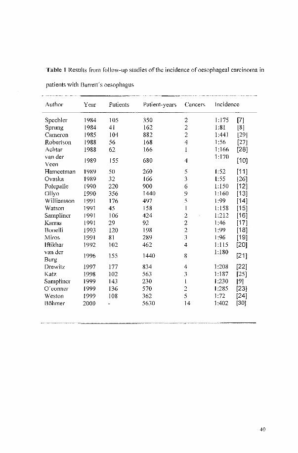
Table 1 Results from follow-up studies of the incidence of oesophageal carcinoma in
patients with Barrett's oesophagus
Author Year Patients Patient-years Cancers Incidence
Spechler 1984 105 350 2 1 :175 [7] Sprung 1984 41 162 2 1:81 [8] Cameron 1985 104 882 2 1:441 [29] Robelison 1988 56 168 4 1:56 [27] Achtar 1988 62 166 1 1 :166 [28] van der
1989 155 680 4 1 :170
[10] Veen Hameetman 1989 50 260 5 1:52 [11] Ovaska 1989 32 166 3 1:55 [26] Pol epa lie 1990 220 900 6 1:150 [12] Ollyo 1990 356 1440 9 1 :160 [13] Williamson 1991 176 497 5 1:99 [14] Watson 1991 45 158 1 1: 158 [15] Sampliner 1991 106 424 2 1 :212 [16] Karras 1991 29 92 2 1:46 [17] Bonelli 1993 120 198 2 1:99 [18] Miros 1991 81 289 3 1:96 [19] lftikhar 1992 102 462 4 1: 115 [20] van del'
1996 155 1440 8 1:180
[21] Burg Drewitz 1997 177 834 4 1:208 [22] Katz 1998 102 563 3 1 :187 [25] Sampliner 1999 143 230 I 1 :230 [9] O'conner 1999 136 570 2 1:285 [23] Weston 1999 108 362 5 1:72 [24] Bohmer 2000 5630 14 1:402 [30]
40
As only 5% of patients with Barrett's oesophagus are known as such to the medical
profession [3], endoscopic cancer surveillance would have a negligible impact on the
rising incidence of adenocarcinoma in Barrett's oesophagus in the population. On the
other hand, endoscopic cancer surveillance in patients with Barrett's oesophagus is
suppolied by the finding that, with this surveillance, a higher proportion of cases of
high-grade dysplasia or early carcinoma are detected than are seen in symptomatic
patients [11,31,32]. However, it is questionable whether life-long endoscopic cancer
surveillance will, in fact, significantly influence the survival of patients with Barrett's
oesophagus, as only a small fraction of these patients actually die from
adenocarcinomas [7,10,21,29]. The majority die from unrelated causes, mainly
cardio-pulmonaJY diseases [21]. Endoscopic cancer surveillance of these patients
would have caused much discomfort but would have saved few, if any, lives.
However, it is possible that surveillance might be effective in subgroups of patients
[33]. It therefore seemed rational to test the concept of endoscopic cancer surveillance
against the recommendation for surveillance as defined by the World Health
Organization (WHO) [34] and, subsequently, to define subgroups with an above
average risk of developing adenocarcinoma who are also free of diseases likely to
impair survival or hinder treatment. Obviously, the concept of increased, and
therefore also decreased, risk needed some standard. Provenzale and coworkers
calculated that patients with Barrett's oesophagus and an expected incidence of
adenocarcinoma below 11200 patients-year would not benefit from endoscopic cancer
surveillance [35]. On this basis, guidelines were drawn up for selecting patients who
were both fit for and likely to benefit from endoscopic cancer surveillance. These
guidelines comprised medical factors likely to impair survival, protective factors
which made an incidence in excess of 11200 patients-year unlikely and risk factors
41
which mandated endoscopic cancer surveillance. By applying these guidelines to all
patients with Barrett's oesophagus, identified over a 5-year period at our endoscopy
unit, the proportion of these patients who could potentially have benefited from
endoscopic cancer surveillance was established.
Materials and methods
All cases of Barrett's oesophagus in patients over the age of 18 years diagnosed
between January I 1992 and December 31 1996 were identified from computerized
endoscopy records. Patients in whom a carcinoma or high-grade dysplasia in Barrett's
oesophagus was found were excluded. Barrett's oesophagus was defined as an
endoscopic diagnosis of columnar epithelium lining the distal tubular oesophagus,
usually more than 3 in length. We also included shOJier segments of Barrett's
oesophagus because the endoscopic diagnosis of short-segment Barrett's oesophagus is
associated with a high prevalence of intestinal-type colunmar epithelium (significantly
higher than that found in oesophagogastric junctions that appear apparently normal)
[36]. There is also a known cancer risk in shOJi-segment Barrett's oesophagus [37].
Information on the age, sex and length of the Barrett's oesophagus segment was
obtained fi'om the endoscopic database. Other data, including dysplasia and other
medical disorders in these patients, were obtained fi'om the case records.
All patients with Barrett's oesophagus meeting these criteria were subjected to a
process of elimination on the basis of a number of factors considered relevant to their
suitability for endoscopic cancer surveillance.
42
Factors influencing the suitability for endoscopic cancer surveillance (Table 2).
Men of Caucasian origin with Barrett's oesophagus have been found in most studies to
have a risk of developing adenocarcinoma that is greater than 1/200 per year
[4,6,31,38,39]. The risk is higher for Caucasian men than for women or Black people
[31,38,39]. There were no Black patients in our study group.
Barrett's oesophagus of 3 cmlength or more was chosen as an indication for
endoscopic cancer surveillance, as the tendency to malignancy increases with
increasing Barrett's oesophagus length [10,20,21,24,38].
Negative prognostic factOl's leading to exclusion ji'OIII endoscopic callcer
surveillance
1. Concurrent diseases likely to impair survival or effective treatment, including
malignancies, severe cardio-pulmonary disease and other diseases expected to limit
life expectancy to less than 5 years.
2. Advanced age: the mean age at the endoscopic diagnosis of Barrett's
oesophagus was about 63--65 years, which is very similar to that at which
adenocarcinoma is diagnosed [2]. In our own hospital the mean age at diagnosis of
adenocarcinoma in Barrett's oesophagus patients was 67 years (52--81 years) [21].
The degree of dysplasia and the likelihood of malignancy in Barrett's oesophagus rise
with age [11]. On the other hand, surveillance programs have not always been offered
to the elderly [33]. Age by itself should not be a limiting factor for endoscopic cancer
43

surveillance but, because competing causes of death rise with age [40], a cut-off point
needs to be drawn. For this study we chose 75 years of age as this cut-off point.
3. Finally, all patients dying within the 5-year period fi'om unrelated diseases
were considered unlikely to have benefited from endoscopic cancer surveillance.
44

Protective factors leading to exclusion frol1l endoscopic cancer surveillance
In the absence of risk factors (see below) the following factors were considered to be
associated with an incidence of adenocarcinoma in Barrett's oesophagus ofless than
11200 patients-year:
1. Women: The male to female ratio reported for adenocarcinoma in Barrett's
oesophagus ranges between 3: 1 and 8: 1 [6,30,38]. This ratio was confirmed in a large
population of institutionalized, intellectually handicapped individuals who do not
smoke or drink alcohol, thus confirming the protective effect of being female [30]. hl
two observational studies, the incidence of adenocarcinoma in women with Barrett's
oesophagus ranged between 1/294 and 1/981 patients-year. After correction for age,
these incidences were not significantly different [30; personal communication, CJ
Bohmer, 2000] and fell far Sh01i of the 1/200 patients-year criterion.
2. Youth: As adenocarcinoma in Barrett's oesophagus practically never occurs
below the age of 40, endoscopic cancer surveillance below this age would appear
futile [21,30]. However, even above this age the incidence remains low, not meeting
the 11200 patients-year criterion until the age of 65 in men. As the onset of high-grade
dysplasia will generally occur years before symptomatic cancer, 60 years of age was
taken as a cut-off point.
3. ShOJi-segment Barrett's oesophagus: Although it may be a risk factor for very
distal carcinomas [41], the prevalence of short-segment Barrett's oesophagus in the
population coming to endoscopy is so high (18%) [42] that the incidence of
adenocarcinoma in these patients must be far lower than 11200 patients-year. This was
45
confirmed in our own observational study in which no adenocarcinomas were found
in Barrett's oesophagus ofless than 8 cm in length [10,21]. Short-segment Barrett's
oesophagus is not seen as an indication for surveillance [43].
Risk factors that override protective factors fI1U/l1ecessitate el1doscopic cal1cer
surveillal1ce
1. Dysplasia distinguishes Barrett's oesophagus patients particularly at risk of
developing cancer in the relatively near future [11,24], while high-grade dysplasia is
often a marker of coexistent cancer and therefore often considered an end point in
endoscopic cancer surveillance [5]. The degree of dysplasia increases with the age
and length of Barrett's oesophagus [20,44] and the time of follow-up [11,45].
2. The presence of ulcerations and/or strictures has been found to be an important
predictor of malignancy [1,10,21,33].
Riskfactors which were not taken il1to accoul1t
1. Reflux of duodenal juice has been repOlied to be associated with a
significantly higher rate of severe oesophagitis and Barrett's oesophagus [46].
Although not proven, it may be a risk factor for developing adenocarcinoma in
Barrett's oesophagus. However, duodenal juice reflux is not routinely tested.
2. Smoking and high alcohol intake are associated with Barrett's oesophagus
carcinoma, the relationship being more pronounced for tobacco than for alcohol
[7,38,47]. However, this relationship is not very strong, therefore smoking is much
46
more likely to induce other causes of death before the onset of adenocarcinoma in
Barrett's oesophagus [21,30].
3. Obesity has been shown to entail an increased risk of oesophageal
adenocarcinoma [48], while raw fruit, vegetable and fibre intake decreases this risk
[49).
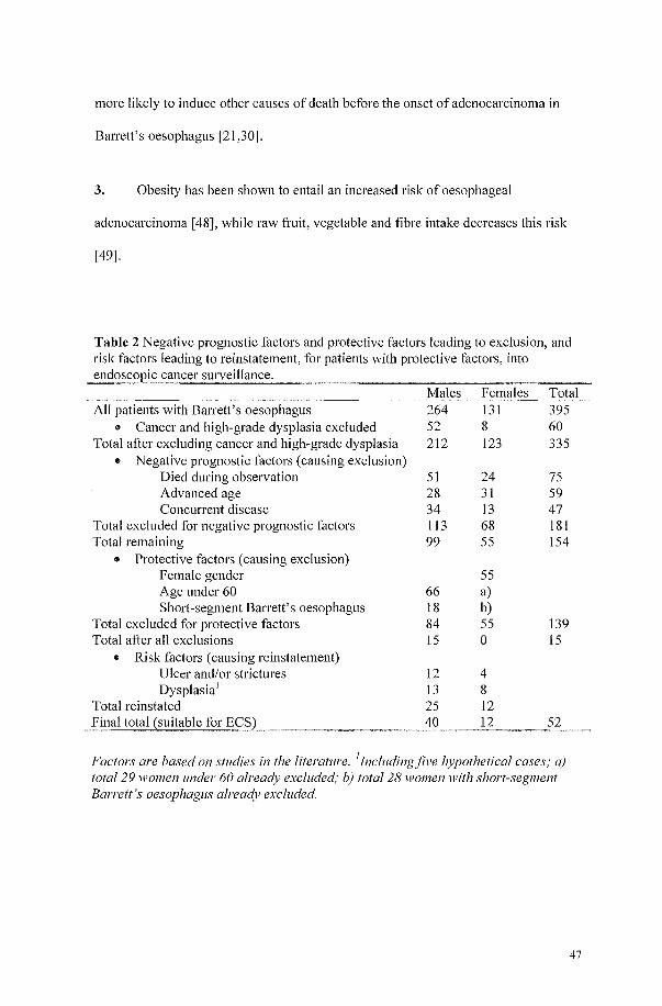
Table 2 Negative prognostic factors and protective factors leading to exclusion, and risk factors leading to reinstatement, for patients with protective factors, into
Males Females Total All patients with Barrett's oesophagus 264 131 395
.. Cancer and high-grade dysplasia excluded 52 8 60 Total after excluding cancer and high-grade dysplasia 212 123 335 .. Negative prognostic factors (causing exclusion)
Died during observation 51 24 75 Advanced age 28 31 59 Concurrent disease 34 13 47
Total excluded for negative prognostic factors 113 68 181 Total remaining 99 55 154
.. Protective factors (causing exclusion) Female gender 55 Age under 60 66 a) Short-segment Barrett's oesophagus 18 b)
Total excluded for protective factors 84 55 139 Total after all exclusions 15 0 15
.. Risk factors (causing reinstatement) Ulcer and/or strictures 12 4 Dysplasia! 13 8
Total reinstated 25 12 40 12
Factors are based on studies in the literature. ilncludingjive hypothetical cases; a) total 29 women under 60 already excluded; b) total 28 women1l'ith short-segment Barrett's oesophagus already excluded.
47
Results
During the 5-year period of studied, 75 patients had died from reasons other than
oesophageal carcinoma and would therefore not have benefited fi'om surveillance.
The remaining 260 patients (99 women and 161 men) formed the study cohort. Of
these, 76% were outpatients and 24% inpatients. There was no significant different in
age or sex between outpatients and inpatients.
A progressive process of exclusion from endoscopic cancer surveillance was
implemented, producing the following results. Twenty-eight men and 31 women over
the age of75 were excluded fi'om surveillance on the grounds of age, leaving 201
patients. Of these, 47 (34 men and 13 women) had other diseases (see Table 3), which
limited survival, leaving 154 patients (99 men and 55 women).
48
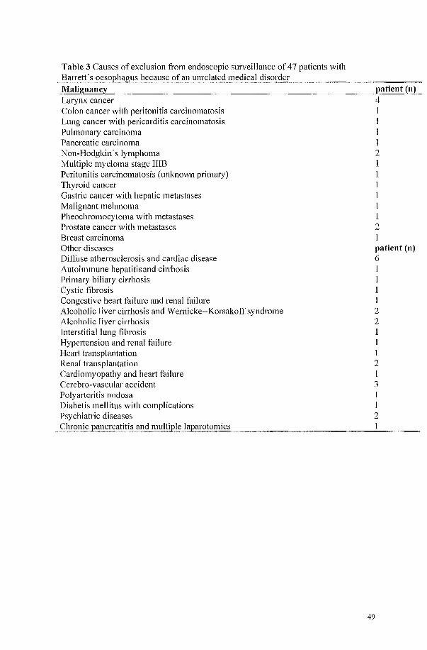
Table 3 Causes of exclusion from endoscopic surveillance of 47 patients with Barrett's oesophagus because of an unrelated medical disorder
Malignancy Larynx cancer Colon cancer with peritonitis carcinomatosis Lung cancer with pericarditis carcinomatosis Pulmonmy carcinoma Pancreatic carcinoma Non-Hodgkin's lymphoma Multiple myeloma stage IIIB Peritonitis carcinomatosis (unknown primmy) Thyroid cancer Gastric cancer with hepatic metastases Malignant melanoma Pheochromocytoma with metastases Prostate cancer with metastases Breast carcinoma Other diseases Diffuse atherosclerosis and cardiac disease Autoimmune hepatitis and cirrhosis Primary biliary cirrhosis Cystic fibrosis Congestive heali failure and renal failure Alcoholic liver cirrhosis and Wernicke--Korsakoff syndrome Alcoholic liver cirrhosis Interstitial lung fibrosis Hypeliension and renal failure Heart transplantation Renal transplantation Cardiomyopathy and heart failure Cerebro-vascular accident Polymieritis nodosa Diabetis mellitus with complications Psychiatric diseases Chronicpancreatitis and multiple laparotomies
patient (n) 4 I I I I 2 1 1 1 1 I 1 2 1 patient (n) 6 1 1 1 1 2 2 1 I 1 2 I 3 I 1 2 1
49
The women were excluded, leaving 99 men. Of these remaining 99 individuals (all
men), 66 were under the age of 60 and, fi'om the remaining 33 patients, there were 18
with short-segment Barrett's oesophagus. Only 15 had Barrett's oesophagus segments
of 3 cm or longer and therefore remained eligible for endoscopic cancer surveillance.
Histology was available in 89% of Barrett's oesophagus segments of3 cm or longer
but not available in about 50% of short-segment Barrett's oesophagus.
In the eliminated groups there were eight women and eight men with dysplasia; in
addition, four women and 12 men had ulcers or strictures. These patients, totaling 32,
were reinstated because of these risk factors, leaving a total of 47 patients with an
average age of 54.5 years who were eligible for endoscopic cancer surveillance.
However, based on the prevalence of dysplasia and because no histology was
available in 11% of patients with Barrett's oesophagus and about 50% of patients with
shOli-segment Barrett's oesophagus, we estimated that an extra five patients should
have been reinstated. This resulted in a final total of 52 patients eligible for
endoscopic cancer surveillance out of the original 335 (15.5%).
50
Discussion
Only about 5% of the current cases of oesophageal adenocarcinoma occur in patients
already known to have Barrett's oesophagus [50]. The seemingly obvious solution to
the challenge of the rapid rise in the incidence of adenocarcinoma in Barrett's
oesophagus is endoscopic population screening for Barrett's oesophagus. However,
both practical problems in providing endoscopic services and failure to meet the
WHO 'principles of early disease detection', specifically principles 2, 6, 8 and 9 [34],
are powerful arguments against population screening (Table 4).
A number of experimental treatments for Barrett's oesophagus have been launched,
such as removal of Barrett's metaplastic mucosa by laser, photodynamic therapy or
multipolar electrocoagulation and subsequent restoration ofthe mucosal surface under
low acid load to squamous epithelium. However, none of these treatments has, as yet,
been proven to be feasible or safe in long-term studies [51--53].
On the other hand, endoscopic cancer surveillance in cases of Barrett's oesophagus
identified at routine endoscopy to detect dysplasia or early carcinoma does not violate
any of the WHO principles, although principles 6 and 9 may again present some
problems. Here, the most important principle -- an acceptable treatment for patients
with recognized disease[34] -- can be met.
51
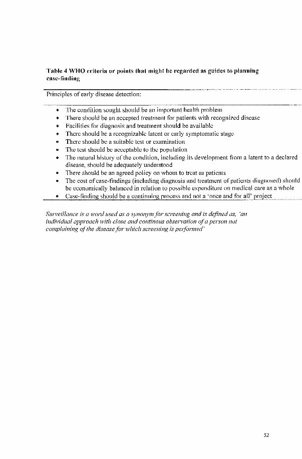
Table 4 WHO criteria or points that might be regarded as guides to planning case-finding
Principles of early disease detection:
.. The condition sought should be an important health problem
.. There should be an accepted treatment for patients with recognized disease
.. Facilities for diagnosis and treatment should be available
.. There should be a recognizable latent or early symptomatic stage
.. There should be a suitable test or examination
.. The test should be acceptable to the population
.. The natural history of the condition, including its development from a latent to a declared disease, should be adequately understood
II There should be an agreed policy on whom to treat as patients II The cost of case-findings (including diagnosis and treatment of patients diagnosed) should
be economically balanced in relation to possible expenditure on medical care as a whole .. Case-finding should be a continuing process and nota 'once~and for all' project
Surveillance is a word used as a synonym for screening and is defined as, 'an individual approach with close and continous obsel1 lation of a person not complaining of the disease for which screening is pel/ormed'
52
Oesophagectomy for high-grade dysplasia or early cancer detected by surveillance is
often curative and therefore should improve long-term survival [5,39,54], although
this advantage would be partly counterbalanced by mortality from oesophagectomy.
However, both laser-ablation and photodynamic therapy may provide effective
treatment for those patients with high-grade dysplasia or early cancer who are
unsuitable or unwilling to undergo surgery [51--53]. If endoscopic cancer surveillance
Qfknown cases of Barrett's oesophagus is acceptable, the question remains whether it
should be applied universally. In view of the fact that the great majority of patients
with Barrett's oesophagus die from um-elated diseases [21], it became obvious that
endoscopic cancer surveillance should be limited to a sub-group combining a
relatively high cancer risk with an otherwise good life expectancy.
The aim of the present study was to define guidelines for identifying this sub-group
and, by applying these guidelines, to establish the propOliion of patients with Banett's
oesophagus, diagnosed at routine endoscopy in our unit over a 5-year period, who
would have been eligible for endoscopic cancer surveillance. This was achieved by
taking into account that the clinical, endoscopic and histological features of Barrett's
oesophagus patients at initial diagnosis are probably predictive of cancer risk [24], and
by retrieving data on all patients diagnosed with Barrett's oesophagus over this
period. Patients in whom high-grade dysplasia or adenocarcinoma in Barrett's
oesophagus was found at the inclusion endoscopy were excluded. The remaining
patients were subjected to a process of elimination, first those with poor prognosis,
then those with a low cancer risk. Finally, those found at the inclusion endoscopy to
have an increased cancer risk on the basis of previously defined risk factors were
reinstated. In no case were data other than those fr0111 the inclusion endoscopy taken
53
into account. The poor prognosis category included all those who had died during the
5-year recruitment period fi'om causes unrelated to adenocarcinoma in Barrett's
oesophagus. They would obviously not have benefited fi'om endoscopic cancer
surveillance but would have been exposed to needless discomfort. Patients with
diseases unrelated to Barrett's oesophagus but likely to preclude long-term survival
were also excluded. It could be argued that in a university hospital this category could
be larger than in a general hospital. On the other hand, in this retrospective study the
prevalence of mild or as yet asymptomatic cardio-pulmonary disease is likely to have
been underestimated, while cardiopulmonary disease constitutes the major cause of
death in patients with Barrett's oesophagus [21].
The last group in the poor prognosis category were patients over the age of 75 years.
In the light of recent insight into the operability of fit elderly patients and newer non
surgical treatment options, this exclusion criterion could be questioned. However, in
view of the slow progression fi'om high-grade dysplasia to symptomatic cancer, it
seems unlikely that even non-surgical eradication of high-grade dysplasia would add
to the life expectancy of these patients.
The low cancer risk group was less simple to define. Refraining from treating patients
too ill or too fi'ail to survive treatment for a meaningful period is a generally accepted
principle. However, where to draw the line in screening or surveillance ofheaIthy,
low-risk individuals is a contentious issue. The repOlied risk of developing
adenocarcinoma in Barrett's oesophagus varies from 1146 to 11441 patients-year of
follow-up [7--30,35]. The higher incidences may be the result of overestimation in
smaller studies, which have received undue attention in the literature because of
publication bias [55]. This risk ( or incidence) of adenocarcinoma in patients with
54
Barrett's oesophagus is of obvious impOliance as it determines the number of
endoscopies -- with their attendant risks, discomfOli and expense -- needed to identifY
one case of high-grade dysplasia or early cancer. Although the precise cancer risk
remains unclear, Shaheen et al. [55] suggest that an oesophageal cancer incidence rate
of 0.5% per year might be a reasonable estimate. If both length and quality oflife are
considered, it has been calculated that ifthe incidence of cancer is less than 0.5%
(11200 patients-year), the preferred strategy is no surveillance [35]. The same author
recently published a revised study assuming a cancer incidence of 11227 patients-year
[56]. She concluded that endoscopic cancer surveillance once every 5 years increases
life expectancy by 0.1 years at a cost of $9800 per annum, resulting in a quality
adjusted life year of$98.000. In view ofthis excessive cost (by European standards),
we decided to stay with the 1/200 patients-year limit for endoscopic cancer
surveillance. In practice, the low cancer risk category included all women, men under
the age of 60 and patients with sholi-segment Barrett's oesophagus without dysplasia.
The exclusion of women was based on the fact that the incidence of adenocarcinoma
in Barrett's oesophagus in women was found to be no higher than 11294 patients-year
[21,30]. This is suppOlied by the ratio of incidence of adenocarcinoma in Barrett's
oesophagus in men and women, which ranges between 3: 1 and 8: 1 [6,38]. In
principle, Black people should also be excluded on this basis; there were however no
Black patients in our Barrett's oesophagus population. Similarly, in men the 1/200
patients-year limit was not exceeded before the age of 60 [21,30]. In principle, healthy
men with Barrett's oesophagus on reaching the age of 60 could be enrolled in
endoscopic cancer surveillance.
Although it is now thought likely that most, if not all, cases of adenocarcinoma of the
gastric cardia and the distal oesophagus arise from short-segment Barrett's
55
oesophagus [57], this condition is so common that it was considered to be associated
with a cancer incidence far short of 11200 patients-year. However, these exclusions on
the basis of low cancer risk were reversed in the presence of one or more factors
associated with an increased risk of cancer in Barrett's oesophagus, which could be
identified at routine initial endoscopy. These factors include dysplasia other than
high-grade dysplasia and an active or healed ulcer (resulting in a stricture) in Barrett's
oesophagus [5,7,31,33]. Heavy smoking, which is the only other prognostic factor
consistently associated with an increased risk of endoscopic cancer surveillance, was
considered to be a self-defeating prognostic sign as these patients are much more
likely to die from cardio-pulmonary disease than from cancer.
At the final analysis of 335 patients qualifYing for entry into the study, only 52
(15.5'%) could have benefited from endoscopic cancer surveillance. Assuming a 100%
compliance with endoscopic cancer surveillance, these 52 patients, with an average
age of 54.5 years, would, on reaching the age of75, together have completed almost
1100 years under surveillance. Assuming an incidence of adenocarcinoma in Barrett's
oesophagus of 1/112 patients-year for males over 45 years [21], slightly less than
1100 annual endoscopies would have discovered 10 cancer cases or, approximately,
one cancer case would be discovered per 110 endoscopies. A crude estimate can be
made as to the cost. Upper-gastrointestinal endoscopy with histology costs about
300 Euros. The cost of identifYing one case of adenocarcinoma in Barrett's
oesophagus is therefore about 33&th;600 Euros. High-grade dysplasia, being
diagnosed a few years earlier, would be somewhat cheaper. In both cases, however,
these amounts are considerably in excess of the costs of identifYing one cancer in
current public screening programs.
56
In conclusion, this study suggests that only 15.5% of patients identified as having
Barrett's oesophagus at routine upper-gastrointestinal endoscopy would benefit fi'om
endoscopic cancer surveillance. This benefit would arise from early, pre-symptomatic
diagnosis of high-grade dysplasia or adenocarcinoma in Barrett's oesophagus giving a
greater chance of successful treatment in individuals. However, such surveillance is
unlikely to reduce the death rate fi'om oesophageal adenocarcinomas in the general
population.
Rational selection, based on clinical, endoscopic and histological features in Barrett's
oesophagus patients at initial endoscopy, should prevent futile surveillance in those
unlikely to develop cancer or unfit for treatment.
Until long-term prospective follow-up studies have been performed, we recommend
limiting surveillance to high-risk, physically fit patients as defined by guidelines used
in this study (Table 2).
57
References
Naef AP, Savmy M, Ozzello L. Columnar-lined lower esophagus: an acquired lesion with malignant predisposition. Report on 140 cases of Barrett's esophagus with 12 adenocarcinomas. J Thome Cardiovase Slirg 1975; 70:826--835.
2 Cameron AJ, Lomboy CT. Barrett's esophagus: age, prevalence, and extent of columnar epithelium. Gastroenterology 1992; 103: 1241-- I 245.
3 Cameron AJ, Zinsmeister AR, Ballard DJ, Carney JA. Prevalence of columnarlined (Barrett's) esophagus. Comparison of population-based clinical and autopsy findings. Gastroenterology 1990; 99:918--922.
4 Caygill CPJ, Reed PI, Johnston BJ, Hill MJ, Ali MH, Sassoon L. A single centre's 20 years' experience of columnar-lined (Barrett's) oesophagus diagnosis. EliI' J Gastroenterol Hepatol1999; 11: 1355--1358.
5 Hamilton SR, Smith RR. The relationship between columnar epithelial dysplasia and invasive adenocarcinoma arising in Barrett's esophagus. Am J Clin Patho11987; 87:301--312.
6 Blot WJ, Devesa SS, Kneller RW, Fraumeni JF. Rising incidence of adenocarcinoma of the esophagus and gastric cardia. Jama 1991; 265: 1287--1289.
7 Spechler SJ, Robbins AH, Rubins HB, Vincent ME, Heeren T, Doos WG et al. Adenocarcinoma and Barrett's esophagus. An overrated risk? Gastroenterology 1984; 87:927--933.
8 Spmng OJ, Ellis FH, Gibb SP. Incidence of adenocarcinoma in Barrett's esophagus. [Abstract]. Am J Gastroentero/1984; 79:817.
9 Sampliner RE, Sharma P, Garewal HS. The incidence of adenocarcinoma in Bsrrett's esophaguas - 1/230 patient years of follow-up [Abstract]. Am J Gastroentero/1999; 94:2599.
10 Van del' Veen AH, Dees J, Blankensteijn JD, van Blankenstein M. Adenocarcinoma in Barrett's oesophagus: an overrated risk. Gilt 1989; 30: 14--18.
11 Hameeteman W, Tytgat GNJ, HouthoffHJ, van den Tweel JG. Barrett's esophagus: development of dysplasia and adenocarcinoma. Gastroenterology 1989; 96: 1249--1256.
12 Polepalle SC, McCallum R W. Barrett's esophagus. Current assessment and future perspectives. Gastroenterol Clin North Am 1990; 19:733--744.
13 Ollyo 18, Lang F, Fontolliet Ch, Krayenbuhl F, Monnier Ph, Savary M et aT. Barrett's esophagus: surveillance follow-up by oesophagoscopy, colonoscopy or panendoscopy? [Abstract]. Gastroenterology 1990; 98:A I 00.
14 Williamson WA, Ellis FH, Gibb SP, Shahian DM, Aretz HT, Heatley OJ et al. Barrett's esophagus. Prevalence and incidence of adenocarcinoma. Arch intel'11 Med 1991; 151:2212--2216.
15 Watson RGP, POIier KG, Sloan JM. Incidence of adenocarcinoma in Barrett's oesophagus and an evaluation of endoscopic surveillance. EliI' J Gastroenterol Hepatol1991; 3:159--162.
16 Sampliner R, Mackel C, Fenneliy MB, Garewal HS. Prospective incidence of cancer in Barrett's esophagus [Abstract]. Gastroenterology 1991; 100:AI53.
58
17 Karras DJ, Michalos A. Importance of endoscopic surveillance in Barrett's esophagus [Abstract]. Gastroenterology 1991; 1 OO:A95.
18 Bonelli L. Barrett's esophagus: results of a multicentric survey. G.O.S.P.E. (Gruppo Operativo per 10 Studio delle Precancerosi Esofagee). Endoscopy 1993; 25:652--654.
19 Miros M, Kerlin P, Walker N. Only patients with dysplasia progress to adenocarcinoma in Barrett's oesophagus. Gut 1991; 32:1441--1446.
20 Iftikhar SY, James PD, Steele RJ, Hardcastle JD, Atkinson M. Length of Barrett's oesophagus: an impOliant factor in the development of dysplasia and adenocarcinoma. Gut 1992; 33:1155--1158.
21 van del' Burgh A, Dees J, Hop WC, van Blankenstein M. Oesophageal cancer is an uncommon cause of death in patients with Barrett's oesophagus. Gut 1996; 39:5--8.
22 Drewitz DJ, Sampliner RE, Garewal HS. The incidence of adenocarcinoma in Barrett's esophagus: a prospective study of 170 patients followed 4.8 years. Am J Gastroenterol1997; 92:212--215.
23 O'COlmor JB, Falk GW, Richter JE. The incidence of adenocarcinoma and dysplasia in Barrett's esophagus: repOli on the Cleveland Clinic Barrett's Esophagus RegistJy. Am J Gastroenterol1999; 94:2037--2042.
24 Weston AP, Badr AS, Hassanein RS. Prospective multivariate analysis of clinical, endoscopic, and histological factors predictive of the development of Barrett's muItifocal high-grade dysplasia or adenocarcinoma. Am J Gastroenterol1999; 94:3413--3419.
25 Katz D, Rothstein R, Schned A, Dunn J, Seaver K, Antonioli D. The development of dysplasia and adenocarcinoma during endoscopic surveillance of Barrett's esophagus. Am J Gastroel1tel'011998; 93:536--541.
26 Ovaska J, Miettinen M, Kivilaakso E. Adenocarcinoma arising in Barrett's esophagus. Dig Dis Sci 1989; 34: 1336--1339.
27 Robertson CS, Maybeny JF, Nicholson DA, James PD, Atkinson M. Value of endoscopic surveillance in the detection of neoplastic change in Barrett's oesophagus. Br J SUl'g 1988; 75:760--763.
28 Achkar E, Carey W. The cost of surveillance for adenocarcinoma complicating Barrett's esophagus. Am J Gastroenterol1988; 83:291--294.
29 Cameron AJ, Ott BJ, Payne WS. The incidence of adenocarcinoma in columnarlined (Barrett's) esophagus. N Engl J Med 1985; 313:857--859.
30 Bohmer CJ, Hop WCJ, Meuwissen SGM, van Blankenstein M. Carcinoma of Barrett's esophaguus, do smoking and alcohol really matter [Abstract]. Gastroenterology 2000; 118:A459.
31 Wright T A, Gray MR, Morris AI, Gilmore IT, Ellis A, Smart HL et al. Cost effectiveness of detecting Barrett's cancer. Gut 1996; 39:574--579.
32 van Sandick JW, van Lanschot JJB, Kuiken BW, Tytgat GNJ, Offerhaus GJA, Obetop H. Impact of endoscopic biopsy surveillance of Barrett's oesophagus on pathological stage and clinical outcome of Barrett's carcinoma. Gut 1998; 43:216--222.
33 Macdonald CE, Wicks AC, Playford RJ. Final results from 10-year COhOli of patients undergoing surveillance for Barrett's oesophagus: observational study. BMJ2000; 321:1252--1255.
34 Wilson JM, Jungner YG. Principles and practice oj screeningjor disease. Geneva: World Health Organization; 1968. (Public health papers no. 34).
59
35 Provenzale D, Kemp AI, Arora S, Wong lB. A guide for surveillance of patients with Barrett's esophagus. Am J Gastroenterol 1994; 89:670--680.
36 Pereira AD, Suspiro A, Chaves P, Saraiva A, Gloria L, Mendes de Almeida JC, Leit et al. Short segments of Barrett's epithelium and intestinal metaplasia in normal appearing oesophagogastric junctions: the same or two different entities? Gut 1998; 42:659--662.
37 Clark GW, Smyrk TC, Hoeft SF, Burdiles P, Dreuw B, Crookes PF. Is the length of Barrett's mucosa related to the prevalence of complications and adenocarcinoma in Barrett's esophagus [Abstract]. Gastroenterology 1993; 104:A393.
38 Menke-Pluymers MBE, Hop WCJ, Dees J, van Blankenstein M, Tilanus HW. Risk factors for the development of an adenocarcinoma in columnar-lined (Barrett) esophagus. The Rotterdam Esophageal Tumor Study Group. Cancel' 1993; 72:1155--1158.
39 Streitz JM Jr, Andrews CW Jr, Ellis FH JI'. Endoscopic surveillance of Barrett's esophagus. Does it help? JThorac Cardiovasc Surg 1993; 105:383--388.
40 Mackenbach JP, Kunst AE, Lautenbach H, Oei YB, Bijlsma F. Competing causes of death: a death certificate study. J Clin Epidemiol1997; 50: 1 069--1077.
41 Mendes de Almeida IC, Chaves P, Pereira AD, Altorki NK Is Barrett's esophagus the precursor of most adenocarcinomas of the esophagus and cardia? A biochemical study. Ann Surg 1997; 226:725--735.
42 Spechler SJ, Zeroogian JM, Antonioli DA, Wang HR, Goyal RK. Prevalence of metaplasia at the gastro-oesophageal junction. Lancet 1994; 344: 1533--1536.
43 Nandurkar S, Talley NJ. Barrett's esophagus: the long and the short of it. Am J Gastroentero/1999; 94:30--40.
44 Gopal DV, Faigel DO, Magaret N, Fennerty JI.1B, Sampliner RE, Garewal HS et al. Risk factors for dysplasia in patients with Barrett's esophagus (BE): results from a multicenter consOliium [Abstract]. Gastroenterology 1999; 116:AI75.
45 Cameron AJ, Carpenter HA. Barrett's esophagus, high-grade dysplasia, and early adenocarcinoma: a pathological study. Am J Gastroentero/1997; 92:586--591.
46 Attwood SEA, DeMeester TR, Bremner CG, Barlow AP, Hinder RA. Alkaline gastroesophageal reflux: implications in the development of complications in Barrett's columnar-lined lower esophagus. Surge!)' 1989; 106:764--770.
47 Gray MR, Donnelly RJ, KingsnOlih AN. The role of smoking and alcohol in metaplasia and cancer risk in Barrett's columnar lined oesophagus. Gut 1993; 34:727--731.
48 Brown LM, Swanson CA, Gridley G, Swanson GM, Schoenberg JB, Greenberg RS et al. Adenocarcinoma of the esophagus: role of obesity and diet. J Natl Cancerinst 1995; 87: 104--109.
49 Chow WH, Blot W J, Vaughan TL, Risch HA, Gammon MD, Stanford JL et at. Body mass index and risk of adenocarcinomas of the esophagus and gastric cardia. J Natl Cancerinst 1998; 90: 150--155.
50 Blot WJ, Devesa SS, Fraumeni JF Jr. Continuing climb in rates of esophageal adenocarcinoma: an update. Jama 1993; 270: 1320.
51 Van Laethem JL, Cremer M, Peny MO, Delhaye M, Deviere 1. Eradication of Barrett's mucosa with argon plasma coagulation and acid suppression: immediate and mid-term results. Gut 1998; 43:747--751.
60

52 Ackroyd R, Brown NJ, Davis MF, Stephenson TJ, Marcus SL Stoddard CJ et al. Photodynamic therapy for dysplastic Barrett's oesophagus: a prospective, double blind, randomised, placebo controlled trial. Gilt 2000; 47:612--617.
53 van den Boogert J, van Hillegersberg R, Siersema PO, de Bruin RWF, Tilanus HW. Endoscopic ablation therapy for Barrett's esophagus with high-grade dysplasia: a review. Am J Gastroentero/1999; 94: 1153--1160.
54 Pera M, Trastek VF, Carpenter HA, Allen MS, Deschamps C, Pairolero Pc. Barrett's esophagus with high-grade dysplasia: an indication for esophagectomy? Ann Thome Su/'g 1992; 54: 199--204.
55 Shaheen NJ, Crosby MA, Bozymski EM, Sandler RS. Is there publication bias in reporting of cancer risk in Barrett's Esophagus? Gastroenterology 2000; 119:333--338.
56 Provenzale 0, Schmitt C, Wong JB. Barrett's esophagus: a new look at surveillance based on emerging estimates of cancer risk. Am J Gastroenterol 1999; 94:2043--2053.
57 Rudolph RE, Vaughan TL, Storer BE, Haggitt RC, Rabinovitch PS, Levine OS et al. Effect of segment length on risk for neoplastic progression in patients with Barrett Esophagus. Ann/ntem Med 2000; 132:612--620.
61

62
Chapter 3 Cardiovascular risk factors and history of myocardial infarction in patients with Barrett's esophagus
Sunna Gudlaugsdottirl,2, Jacqueline eM Witteman3
, Frank JA van Rooi/, Jan Dees2,
Theo Stijnen3, JH Paul Wilson I.
1. Dept. OfInternal Medicine, University Hospital Rotterdam, The Netherlands 2. Dept. Of Gastroenterology and Hepatology, University Hospital Rotterdam, The
Netherlands 3. Institute of Epidemiology and Biostatistics, Erasmus University Rotterdam, The
Netherlands
Submitted
PreliminGlJl results Qfthis study were presented at the Netherlands society of Gastroenterology meeting in Veldhoven, the Netherlands in March 2000, and published in the form of an abstract in the European Journal of Gastroenterology and Hepatology - December 2000.
63
Abstract
Backgrou11d. In a previous follow-up study of patients with Barrett's esophagus we
found an excess mortality of 50% in the Barrett's Esophagus (BE) population, one
third because of cardiovascular diseases. As a part of an epidemiological study on the
prevalence of Helicobactel' pylori in a BE population, we studied verified history of
myocardial infarction and cardiovascular risk factors and in BE patients compared to
controls.
Methods. Forty-seven patients aged 55-79 years with upper gastrointestinal
complaints in whom BE was diagnosed at routine endoscopy were examined. A
standard questionnaire including age, sex, verified history of myocardial infarction,
and its risk factors was used. Blood pressure, body mass index (kg/m2), total- and
HDL cholesterol were measured. Data from a population-based prospective cohort
study of the same age range and living in the same area were used for comparison.
Results. Mean blood pressure was higher and hypertension significantly more
frequently present in the BE population than in the control group. Mean body mass
index was higher, although not significantly so. Obesity (BMI ~30 kg/m2) was
significantly more often observed in the BE group. The prevalences of smoking and
hypercholesterolemia were not different. A validated history of myocardial infarction
was significantly more frequent in the BE population.
Conclusio11. This study suggests that BE diagnosed in patients with upper
gastrointestinal complaints is associated with an increased prevalence of hype liens ion,
obesity and a history of myocardial infarction. If true, this should be taken into
account when designing or evaluating surveillance studies of BE.
64
Introduction
Barrett's esophagus (BE) is a complication of gastro-esophageal reflux disease [1]. It
is found in about I of every 100 endoscopic examinations [2], and is a risk factor for
adenocarcinoma of the esophagus. Because of the dramatically rising incidence of
adenocarcinoma in the esophagus in the past decade[3], several groups have proposed
endoscopic biopsy surveillance for BE patients to detect malignancy at an early and
curable stage[4, 5]. However, doubt has been cast on the effectiveness of endoscopic
biopsy surveillance as few patients diagnosed as having BE actually die from
adenocarcinoma ofthe esophagus [6-9]. In a follow-up study of 166 patients from this
hospital in whom the diagnosis BE had been established between 1973 - 1986 during
a mean follow-up of9.3 years (amounting to 1440 patient years) 8 patients developed
esophageal cancer at various times, giving one case in 180 patient years [6]. Seventy
nine patients had died, one-third because of coronary heaJi disease (CHO). This
mortality was 50% above the expected mOliality in an age/sex matched control
popUlation. In only 2 cases was esophageal cancer the cause of death [6]. These
results raised the hypothesis that BE patients have an increased risk of CHD.
The aim of this study was to determine the prevalence of risk factors for CHD and
prevalence of myocardial infarction in patients with BE compared to the general
elderly population. The study was a paJi of epidemiological analysis on prevalence of
He/icobacterpy/ori in BE patients.
65
Methods
For this study we used BE patients between 55-80 years of age. The invitation was
from January 1998 to August 1999. The computerised endoscopic and medical data of
all patients with BE found at routine endoscopy in the University Hospital Rotterdam
in the years 1992 to 1996 were scrutinised to recruit patients with BE. Information on
the length of BE and histological definition of a biopsy sample from the BE was
noted. Patients were included if the BE met the definition of columnar mucosa in the
tubular esophagus found at endoscopy with histological proof of intestinal metaplasia
(IM) [10]. Ifthe patient had no upper gastrointestinal malignancies at endoscopy, he
or she was sent a standard questionnaire and informed consent form together with a
request to participate. 0f335 BE patients without adenocarcinoma, a total of 212 (137
men and 75 women) where alive, in the age-range 18 - 80 years, and were sent an
invitation. One hundred and nine reacted to the invitation with agreement to
participate. The study subjects were given an appointment for an
esophagogastroduodenoscopy (EGD) with biopsy to confirm the diagnosis of BE. If
the patient refused a new EDG we used previous data for information about the length
of the Barrett's segment and histology. FOliy-seven patients were eligible in the age
category 55-79 years with proven BE.
Weight and height were mostly self-repOlied in the standard questionnaire and
checked at the outpatient visit. If it was not known or not reported in the standard
questiommire, height and weight were measured with the patient wearing indoor
clothes. At the outpatient visit a non-fasting blood sample was taken for laboratory
tests. We also asceliained by a simple check-list the following demographic
characteristics of the study cohort: age, gender, residence, ethnicity, date of first
diagnosis of BE and date oflast endoscopy. A standard questionnaire filled in at home
66
about the presence of CHD and its risk factors, and list of current medication use was
collected at the outpatient visit. Fifteen BE patients reported taking anti-hypertensive
medication. Two did not report what they used. Most fi'equently used as treatment for
hypeliension were ACE inhibitors (n=7), followed by beta-blockers (n=5) and
calcium channel antagonists (n=3). Three patients were on combination treatment.
The questionnaire included the following questions; 1) Have you ever had a
myocardial infarction? 2) Have you ever had hypeliension? 3) Do you use medication
for hypertension? If answered with "yes" the medications listed were checked and
verified. 4) Have you ever smoked? 5) Do you still smoke? 6) Have you never
smoked?) 7) Do you use medication for high cholesterol? If answered with "yes" it
was verified in the patients-list of medication. 8) Are cardiovascular diseases known
in your family, and if answered yes, we later checked by telephone to determine if it
was CHD and in which family member. Only those with known first-degree family
member with CHD were noted as having positive family history ofCHD and used as
such for comparison. 9) Are you known with diabetes mellitus? Body mass index was
calculated as weight (kg)/height(m)2, and obesity was defined as ::0:30 kg/m2. Blood
pressure was measured once after 5 minutes rest in sitting position at the end ofthe
outpatient visit by a mercury sphygmomanometer. The diastolic reading was taken at
the level when sounds disappear (Korotkoffphase V). Blood was taken for
determination of serum total and high-density lipoprotein (HDL) cholesterol
concentrations by an automated enzymatic procedure. All cholesterol determinations
were performed at the Clinical Chemistry Laboratory of the University Hospital in
Rotterdam, which is the coordinator ofthe Dutch National Cholesterol
Standardisation Program.
67
In the BE group all self-reported infarcts were verified in our own hospital documents
or through information from the general practitioner.
A reference population was derived from a population-based prospective cohort study
on chronic diseases in the elderly; the Rotterdam Study. The rationale of the
Rotterdam Study has been described extensively elsewhere [11]. In shOlt, all residents
ofthe suburb Ommoord of Rotterdam over 55 years of age were invited to patticipate
in the study. Sixty seven percent from the whole group participated in both the home
interview and two visits to the research centre.
The baseline phase of the study comprised a home interview and two visits to the
research centre and was conducted from 1990 to 1993. Height and weight were
measured with participants wearing light clothes and without shoes. Information on
current health status, medical history, cUl'l'ent medication use, smoking behaviour and
family history was obtained using a computerised questionnaire during the home
interview. We used the following questions for the current study; 1) Have you ever
had a myocardial infarction? 2) Have you ever had hypeltension? 3) Do you use
medication for the indication hypeltension? 4) Have you ever smoked? 5) Do you still
smoke? 6) Have you never smoked? 7) Are your mother, father or siblings known
with MI? 8) Do you use medication for DM. Body mass index (BMI) was calculated
as weight (kg)/height(m)2, and used to evaluate obesity defined as >30 kg/m2. At the
research centre, the systolic and diastolic blood pressures, were measured twice on the
right arm in all subjects in a sitting position by a mercury manometer (random zero
sphygmomanometer). The diastolic reading was taken at the level when sounds
disappear (Korotkoff phase V). In the CUl1'ent study we used only the first
measurement for reasons of comparison. A non-fasting blood sample was taken for
68
determination of serum total and high-density lipoprotein (HDL) cholesterol
concentrations by an automated enzymatic procedure. All cholesterol measurements
were performed in the laboratory ofthe Depmiment of Epidemiology and Biostatistics
(Erasmus University Medical School), which participated in the Dutch National
Cholesterol Standardisation Program.
Subjects were asked to take all medication they were currently using to the research
center for registration. Medication for hypercholesterolemia and hypeliension were
obtained from that notification
Hypercholesterolemia was defined in both studies as serum cholesterol of more than 8
mmollL or treatment with cholesterol lowering medication or both.
The definition for hypeliension used in both studies was the presence of at least one
of the following factors: systolic blood pressure measured ~ 160mmHg, diastolic
blood pressure measured ~ 95 mmHg, or use of antihypeliensive medication for the
indication to lower the blood pressure. According to the Sixth Report of the Joint
National Committee on Prevention, Detection, Evaluation, and Treatment of High
Blood Pressure (JNC VI) and World Health Organization defined and classified
hypertension in adults, this blood pressure limit is mild to moderate hypertension [12,
13]. Self-reported myocardial infarction in the Rotterdam study was verified by
hospital discharge data, written information from the subject's general practitioner or
electrocardiogram measurements.
The medical ethical committee ofthe Erasmus University Medical Center Rotterdam
approved the studies. Written informed consent was obtained from all pmiicipants.
69
Statistical analysis
From all subjects between 55 years and 80 years of age fi'om the Rotterdam study we
selected 3 reference subjects per Barrett patient, matched for gender and age in strata
of 5 years. This resulted in 144 subjects in the reference group.
Differences in continuous variables between the BE group and the controls were
analysed by using the Student's t-test and differences in categorical variables by using
the chi-square test. A two-sided p-value < 0.05 was considered statistically
significant.
Results
The mean age of BE patients was 67.4 years (range 55 -79). The gender distribution
was 35 male (72.9%), all of Caucasian origin. All were offered repeat EDG, and 90%
(42 of 47) underwent this procedure. The mean length of Barrett's mucosa in the
enrolled group was 4.0 cm (range 0.5 - 10 cm). All included cases had intestinal
metaplasia in a biopsy sample. In total 9 patients (19%) had dysplasia. There were 6
cases of mild dysplasia (13%) and 3 cases of high-grade dysplasia (6%). The mean
length of BE segment with mild dysplasia was 3.9 cm (range 3-6 cm), but for high
grade dysplasia the length was 8.3 cm (range 5-10 cm).
Cardiovascular risk factors in both groups are present in table 1. A significantly
higher mean blood pressure was present in the BE patients than in the controls
(p<O.O I). Hypertension was also found more frequently in the BE group than in the
controls (71.7% vs 33.6%, p <0.001). Individuals in the BE group received treatment
for hypertension more often than the control group (43.8% vs 31.4% respectively),
but the difference was not significant. When the groups were compared on the single
70
question "have you ever had hypertension? 56.3% answered "yes" in the BE
population compared to 28.9% in the control group (p = 0.001). Hypercholesterolemia
defined as a serum cholesterol of more than 8 mmollL or treatment with cholesterol
lowering medication or both, resulted in prevalence of hypercholesterolemia that did
not differ between the two groups (table 1). Mean total cholesterol was lower in the
BE group than in the control group (5.95 vs 6.64; P =0.001), but on the other hand
treatment for high cholesterol was significantly more frequent in the BE group (18.8%
vs 3.5%, respectively, p <0.001). Mean body mass index was higher in BE patients
than in controls, but the difference did not reach statistic significance. However,
obesity (8Ml ~30kg/m2) was significantly higher in the BE group (18.3% vs 5.6%,
respectively, p <0.01). Smoking habits were the same in both groups. The prevalence
of diabetes mellitus was somewhat higher in the Barrett's population (8.3%) than in
the control group (5.6%) but this was not significant. In the BE group the prevalence
of seif-repOlied history with subsequent validation of myocardial infarction was
27.1 % and in the control population it was 13.4% (p=0.03). A histOlY of eHD in the
first-degree relatives, was found significantly more often in the Barrett's population
than in the control group, (65.0% vs 36.1 % respectively, p <0.05).
71
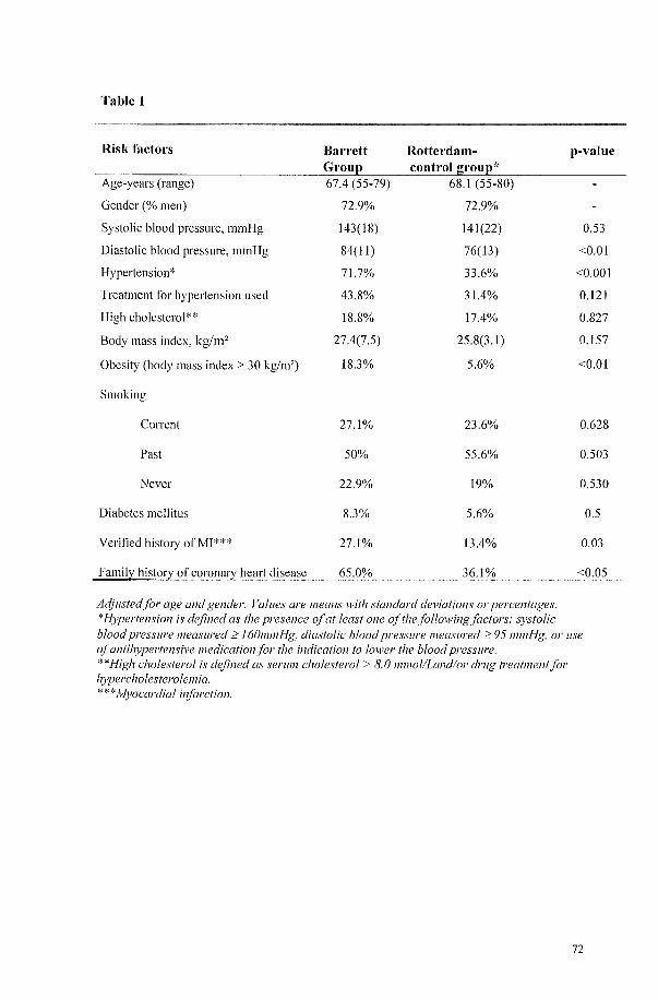
Table 1
Risk factors Barrett Rotterdam- p-value Grou~ control grou~*
Age-years (range) 67.4 (55-79) 68.1 (55-80)
Gender (% men) 72.9% 72.9%
Systolic blood pressure, mmHg 143(18) 141(22) 0.53
Diastolic blood pressure, mmHg 84(11) 76( 13) <0.01
Hypeltension* 71.7% 33.6% <0.001
Treatment for hypertension used 43.8% 31.4% 0.121
High cholesterol** 18.8% 17.4% 0.827
Body mass index, kg/m2 27.4(7.5) 25.8(3.1) 0.157
Obesity (body mass index ;:0: 30 kg/m2) 18.3% 5.6% <0.01
Smoking
Current 27.1% 23.6% 0.628
Past 50% 55.6% 0.503
Never 22.9% 19% 0.530
Diabetes mellitus 8.3% 5.6% 0.5
Verified histmy ofMI*** 27.1% 13.4% 0.03
Family histm} of coronary heart disease 65.0% 36.1% <0.05
Adjusted for age and gender. Values are means with standard deviations 01' percentages. *Hypertensiol1 is defined as the presence of at least one ofthefollowingfactors: systolic blood pressure measured ~ 160mmHg, diastolic blood pressure measured ~ 95 mmHg, 01' use of antihypertensive medication for the indication to 100per the blood pressure. **High cholesterol is defined as serum cholesterol> 8.0 m1l101iLandior drug treatment for hypercholesterolelliia. ***Myocardial illfarction.
72
Discussion
The present study is the first to examine the prevalence of verified history of
myocardial infarction in BE patients. It was found to be significantly more frequent in
the BE population than the control group. We also looked at risk factors for CHD.
Blood pressure was significantly higher and hypertension was significantly more
frequently present in the BE population than in controls. Obesity (BMI ~30kg/m2)
was significantly more prevalent in the BE group .. Hypercholesterolemia or
treatment for hypercholesterolemia and the prevalence of smoking and diabetes
mellitus was not significantly different between the groups.
Before interpreting the data several methodological issues need to be
discussed. The invitation for paJiicipation in this study was not to detect CHD in BE
patients but to look for prevalence of Helicobacter pylori infection. The invitation
included a notice that we would like to assess general health status of participants
with a standard questionnaire at the same time. Therefore, it is not likely that the
response of BE patients is influenced by the presence of history of myocardial
infarction or CHD risk factors. Although BE can be found at any age [2], we limited
the analyses to the older population (255 years) to enable a comparison with a sample
of the Rotterdam Study [11]. We thought this age-limit was reasonable because BE is
most often diagnosed within this age-range. Mean age at diagnosis of BE is 63 years
in subjects without carcinoma and 64 years in patients with adenocarcinoma
(range30-89)[2]. The diagnosis of BE in the patients included in this study was
originally made by diagnostic endoscopy, almost always performed for upper
gastrointestinal complaints. All patients with BE diagnosed in the above-mentioned
period willing to paJiicipate were included in the study, also those who had only come
for diagnostic endoscopy and had not been followed as outpatients. This group
73
represents thus the great majority of patients now known to have BE, as screening
upper GI endoscopy is not generally performed in an otherwise healthy population.
The cohort ofthe Rotterdam Study was considered a valid control group because
participants were derived from the general population and living in the same area. The
majority of subjects in both groups were Caucasians. However, there was a difference
in period of measurements 1990 to 1993 in the Rotterdam group versus 1998 to 1999
in the BE group. Treatment of hypercholesterolemia in the elderly has increased in the
Netherlands. In 1990-1993 treatment for hypercholesterolemia was used in 2.8% of
subjects between 60 and 80 years participating in the Rotterdam study, while in 1997-
1999 this percentage is 14.8%. Therefore the difference in frequency of cholesterol
lowering medication use can be explained by the time difference in data collection
between the groups.
The comparability of the two sets of data was achieved by only using the same
items from the standard questionnaire of the reference group and the BE population
on presence of CHD and its risk factors. For myocardial infarction we verified self
repOlied histories in both the BE and the reference population.
Our study, shows a higher prevalence of hypeltension and treatment for
hypeltension in the BE group than the reference Rotterdam population. It could be
argued that the difference in prevalence of those factors was due to selection bias as
the BE group, being hospital outpatients, might conceivably be a group requiring
more fi'equent medical attention. This would increase the chance of detection of
hypeltension, myocardial infarction, hypercholesterolemia and BE and therefore
treatment of both hypeltension and hypercholesterolemia. However, the higher blood
pressure level in the BE group than in the controls, despite the more frequent use of
antihypertensive medication, is difficult to explain purely by selection bias. The
74
current study might have two limitations with respect to blood pressure measurement.
Firstly, we cannot exclude the possibility that the higher levels in the BE group are
due to the circumstances of measurement (white coat effect) [14], as the blood
pressure was measured by physician in the BE study and by a research assistant in the
Rotterdam study. However, the phenomenon of white coat hypertension, possibly
refers to a health care institute hypeliension or office hypertension phenomenon,
because it depends on multiple factors and does not correlate with the pressure
response to the doctor [13]. If this is true, it should imply that the isolated office
hypeliension, which might also be the earliest manifestation of real hypertension [15],
is evenly impOliant in both cases and the reference group. Secondly, since an
individual person's blood pressure can vary substantially, a single measurement might
not accurately represent a person's average, or usual, blood pressure level. It is likely
to lead to overestimation of the prevalence of hypertension. However, by depending
only on the first measurement in both cases and controls we believe the comparison
between the groups is made trustworthy.
In the BE group, weight and height were often self-repOlied, in contrast to the
Rotterdam study. Self-repOliing tends to give 5-7% lower BMf's than measured
BMI's, which tend to be more biased in older and overweight groups [16]. This could
mean that BMI and obesity in the BE population defined as BMI ::::30 kg/m2 was
underestimated in the present study. The different protocols for measuring family
history of CHD between the two groups indicate that the more frequent occurrence of
CHD in first degree relatives of BE patients than controls may be biased and needs to
be interpreted with caution.
75
Only a small fraction of patients known to have BE actually die from
adenocarcinomas [6-9], and the most likely causes of death are cardio-pulmonary
diseases [6]. A recent observational study from a University Hospital in the United
Kingdom found in 266 BE patients who where too frail or because of other reasons
not suitable for surveillance only one death fi'om oesophageal cancer and 103 fi'om
other causes[17]. The main cause of death in that study were also cardiovascular
diseases [17]. The findings in present study suggest differences in prevalence of
hypertension, obesity and validated histOlY of myocardial infarction. Obesity might
contribute to hypertension and increased prevalence of myocardial infarction. If the
findings are true it may explain part ofthe 50% higher than expected cardiovascular
mOltality in the BE population. However, the underlying mechanisms are unknown.
Because smoking habits did not differ between the two groups the difference in
myocardial infarction between the two cohOlts could not be explained through
difference in smoking habits. Recently, on the basis of clustering of gastro
esophageal reflux disease (GERD) in families, it has been suggested that there might
be a GERD gene [18, 19]. It should be considered whether the same causative factors
- genetic or environmental factors - or both might be operative in BE and CHD.
Another possibility is that the GERD is related to co-medication. Lagergren et al. has
provided epidemiological evidence for a causal role for medications known to cause
lower esophageal sphinchter relaxations in the rising incidence of adenocarcinoma in
the esophagus [20]. Medications used to treat hypertension might predispose to reflux
esophagitis and BE. FUliher studies need to be performed to establish this possibility.
76
A new carefully designed prospective study is needed to confirm our results.
If our results can be confirmed, it would imply that the increased risk of hypertension,
obesity and myocardial infarction in patients with Barrett's esophagus diagnosed
because of upper gastrointestinal complaints should be taken into account when
designing prospective studies or selecting patients for surveillance. By taking clinical
features into account, rational selection of individuals for prospective studies and
surveillance would be limited to physically fit patients.
Acknowledgments
We thank Mark van Blankenstein MD for advice
77
Reference
1. Naef AP, Savary M, Ozzello L. Columnar-lined lower esophagus: an acquired
lesion with malignant predisposition. Report on 140 cases of Barrett's
esophagus with 12 adenocarcinomas. JThorac Cardiovasc Surg. 1975;70:826-
835.
2. Cameron AJ, Lomboy CT. Barrett's esophagus: age, prevalence, and extent of
columnar epithelium. Gastroenterology. 1992; 1 03: 1241-1245.
3. Blot WJ, Devesa SS, Kneller RW, Fraumeni JF. Rising incidence of
adenocarcinoma of the esophagus and gastric cardia. JAMA. 1991;265:1287-
1289.
4. Cameron AJ, Carpenter HA. Barrett's esophagus, high-grade dysplasia, and
early adenocarcinoma: a pathological study. Am J Gastroenterol.
1997;92:586-591.
5. van Sandick JW, van Lanschot JJB, Kuiken BW, Tytgat GNJ, Offerhaus GJA,
Obetop H. Impact of endoscopic biopsy surveillance of Barrett's oesophagus
on pathological stage and clinical outcome of Barrett's carcinoma. Gut.
1998;43:216-222.
6. van del' Burgh A, Dees J, Hop WC, van Blankenstein M. Oesophageal cancer
is an uncommon cause of death in patients with Barrett's oesophagus. Gut.
1996;39:5-8.
7. van del' Veen AH, Dees J, Blankensteijn JD, van Blankenstein M.
Adenocarcinoma in Barrett's oesophagus: an overrated risk. Gut. 1989;30: 14-
18.
8. Spechler SJ, Robbins AH, Rubins HB, Vincent ME, Heeren T, Doos WG,
et.a!. Adenocarcinoma and Barrett's esophagus. An overrated risk?
Gastroenterology. 1984;87:927-933.
9. Cameron AJ, Ott BJ, Payne WS. The incidence of adenocarcinoma in
columnar-lined (Barrett's) esophagus. N Engl J Med. 1985;313:857-859.
10. Weinstein WM, Ippoliti AF. The diagnosis of Barrett's esophagus: goblets,
goblets, goblets (Editorials). Gastrointestinal ensoscopy 1996;44:91-94.
II. Hofman A, Grobbee DE, de Jong PTVM, van den Ouweland. Determinants of
disease and disability in the elderly: the Rotterdam Elderly Study. EliI' J
Epidemiol. 1991 ;7:403-422.
78
12. The sixth report of the Joint National Committee on prevention, detection,
evaluation, and treatment of high blood pressure. Arch Intel'l1 Med.
1997; 157:2413-2446.
13. 1999 World Health Organization-International Society of Hypeliension
Guidelines for the Management of Hype liens ion. Guidelines Subcommittee. J
Hypertens.1999;17:151-183.
14. Pickering TG. White coat hypeliension: time for action [Editorial).
Circulation. 1998;98: 1834-1836.
15. Middeke M, Lemmer B. Office hypertension: abnormal blood pressure
regulation and increased sympathetic activity compared with normotension.
Blood Press Monit. 1996; 1:403-407.
16. Roberts RJ. Can self-reported data accurately describe the prevalence of
overweight? Public Health. 1995;109:275-284.
17. Macdonald CE, Wicks AC, Playford RJ. Final results fi'om 10 year cohort of
patients undergoing surveillance for Barrett's oesophagus: observational study.
BMJ. 2000;321: 1252-1255.
18. Trudgill NJ, Kapur KC, Riley SA. Familial clustering of reflux symptoms. Am
J Gastroenterol. 1999;94: 1172-1178.
19. Romero Y, Cameron AJ, Locke GR3rd, Schaid DJ, Slezak JM, Branch CD
et.a!. Familial aggregation of gastroesophageal reflux in patients with Barrett's
esophagus and esophageal adenocarcinoma. Gastroenterology.
1997;113:1449-1456.
20. Lagergren J, Bergstrom R, Adami HO, Nyren O. Association between
medications that relax the lower esophageal sphincter and risk for esophageal
adenocarcinoma. Ann Intern Med. 2000; 133: 165-175.
79

80
Chapter 4 Hypertension is a frequent comorbidity in patients with reflux oesophagitis or Barrett's oesophagus but not in nonulcer dyspepsia.
Sunna Gudlaugsdottir l,2, WM Monique Verschuren3
, Jan Dees2, Theo Stijnen4
, JH Paul Wilson l.
l. Dept. OfInternal Medicine, University Hospital Rotterdam, The Netherlands 2. Dept. Of Gastroenterology and Hepatology, University Hospital Rotterdam, The Netherlands 3. National Institute of Public Health and the Environment (RIVM), The Netherlands 4. Institute of Epidemiology and Biostatistics, Erasmus University Rotterdam, The Netherlands
In press in European Journal of 1ntel'l1al Medicine 2002
81
Abstract
Backgroulld. An excess mOliality due to cardiovascular disease has been repOlied for patients
with Barrett's oesophagus. We compared the prevalence of risk factors for cardiovascular disease
in patients with Barrett's oesophagus, reflux oesophagitis or non-ulcer dyspepsia with that of the
general population.
Metltods. Patients with upper gastrointestinal complaints and Barrett's oesophagus (34), reflux
oesophagitis (31) or non-ulcer dyspepsia (21) were compared with an matched cohort ofthe
general population, using a questionnaire, and blood pressure and cholesterol measurements.
Results. Hypertension occurred more frequently in Barrett's oesophagus (odds ratio 5.1,
P<O.OOOI) and reflux oesophagitis patients (odds ratio 3.8, P<O.OOl), but not in non-ulcer
dyspepsia. Serum total cholesterol was higher in Barrett's oesophagus (P=O.02), borderline
higher in reflux oesophagitis (P=O.06) but not in non-ulcer dyspepsia. Mean HDL cholesterol
levels and body mass index, and smoking were not different.
COllclusiolls. This study suggests that Barrett's oesophagus and reflux oesophagitis found at
diagnostic endoscopy are associated with an increased prevalence of hypertension and a higher
total cholesterol level than the general population. If true this provides an explanation for the
increased mortality during follow-up of Barrett's oesophagus patients, and should be taken into
account when designing or evaluating surveillance studies of Barrett's oesophagus.
82
Introduction
The prevalence of endoscopically confirmed oesophagitis in the community is thought to be up
to 2%[1]. Oesophagitis can progress to complications such as deep ulceration, stricture
formation, and the development of Barrett's oesophagus(BO)[2]. Because BO has a
premalignant potential [3] and because of the dramatically rising incidence of adenocarcinoma in
the oesophagus in the past decade[4], several groups have proposed endoscopic biopsy
surveillance for BO patients to detect malignancy at an early and curable stage[5, 6]. However,
earlier studies have cast doubt on the effectiveness of endoscopic biopsy surveillance as few
patients diagnosed as having BO actually die from adenocarcinoma of the oesophagus [7-10). In
a follow-up study of 166 patients in whom the diagnosis BO had been established between 1973
- 1986, we found an excess mOliality of 50%, compared to what was expected in an age/sex
matched control population [10]. During a mean follow-up of9.3 years (amounting to 1440
patient years) 8 patients had developed oesophageal cancer at random intervals, giving one case
in 180 patient years. Seventy nine patients had died, one-third because of cardiovascular disease,
and in only 2 cases was oesophageal cancer the cause of death [10]. These results raised the
hypothesis that patients with gastroesophageal reflux disease (OORD) or, at least a subgroup
with BO, have an increased risk of cardiovascular disease (CYD) with an increased prevalence
of one or more CYD risk factors. This study was undertaken to investigate the prevalence of the
main risk factors for CYD (blood pressure, cholesterol, smoking) in patients with reflux
oesophagitis, BO and in patients without endoscopic abnormalities and to compare these with the
normal population. The study was a part of an epidemiological investigation into the prevalence
of Helicobacterpylori in reflux oesophagitis, BO and NUD patients.
83
Materials and Methods
For this study we used patients with reflux oesophagitis, BO and NUD patients - aged 20 to
59.9 years. Two sources of data were used: One was by reviewing computerised endoscopic and
medical data of all patients with BO found at routine endoscopy in the University Hospital
Rotterdam in the years 1992 to 1996. Information on the length of BO and histological definition
of a biopsy sample from the BO was noted. Patients were included if the BO met the definition
of columnar mucosa of at least 3 cm in the tubulair oesophagus found at endoscopy or
histological proof of intestinal metaplasia (1M) when the columnar mucosa was less than 3
cm[ll]. The study subjects were offered repeat oesophagogastroduodenoscopy (OGD) with a
biopsy to confirm the diagnosis of BO. If the patient refused ODG we used the older data for
information about the length of the Barrett's segment and histology. Secondly, all consecutive
patients with GORD or no endoscopic abnormalities referred for diagnostic upper
gastrointestinal endoscopy to the University Hospital Rotterdam - Dijkzigt, during the period
January 1999 to July 2000 were asked to paIiicipate. GORD was defined as the presence of
endoscopic signs of reflux oesophagitis, BO or both. Reflux oesophagitis was scored according
to the Savary-Miller system [12], as follows: grade 0, normal oesophageal mucosa with no
abnormalities; grade 1, mucosal erythema or diffusely red mucosa, with or without friability;
grade 2, linear erosions extending from the gastroesophageal junction upward in relation to the
folds; grade 3, confluent erosions extending around the entire circumference; grade 4, £i'ank
ulcer, stricture or BO.
If the patient had no malignancies or active peptic ulcer disease, he or she was asked to fill in a
standard questionnaire about the presence of risk factors of CVD, current medication use and
smoking behaviour. One patient with BO was excluded because he was unable to travel.
84
We asceliained with a simple check-list the following demographic characteristics of all three
study cohOlis: age, sex, weight, height, smoking (current, past, never), date of first diagnosis of
BO and date of last endoscopy.
A blood sample for laboratory tests was taken at the outpatient care for most of the BO patients
but for other paliicipants on the day of endoscopy. The study populations (reflux oesophagitis,
BO and NUD) answered the following questions used for comparison in present study: 1) Do
you currently use medication for hypeliension 2) Have you ever smoked? If yes ~ 3) Do you
still smoke? Answering categories: yes (~ current smokers); If no (~ ex-smokers) 4) Have you
never smoked? Answering categories: no, never (~ never smokers). Body mass index (BMl)
was calculated as weight(kg)/height(m)2. The blood pressure was measured once at rest in the
end of the outpatient visit by a mercury sphygmomanometer. Systolic pressure was recorded at
the first Korotkoffphase and diastolic pressure at the fifth Korotkoffphase.
The Medical Ethics committee of the University Hospital Rotterdam approved the study.
Informed consent was obtained from all paliicipating patients, before the endoscopy was
performed.
A reference population was derived from the Monitoring Project on Risk Factors for Chronic
Diseases (MORGEN-project). This project has been carried out in the Netherlands from 1993 to
1997. The general purpose was to determine both the prevalence of risk factors for chronic
disease (e.g. plasma cholesterol, blood pressure, smoking habits), and the prevalence of some
specific chronic conditions in a random sample of the general population [13). Information on
current health status, medical history, current medication use, smoking behaviour and family
history was obtained using a self-administered questionnaire at home. We used the following
questions for comparison with the study groups; 1) Do you currently use medication for
hypeliension? 2) Do you smoke? Answering categories: Yes (~ current smokers), No, not
85
anymore (-7 ex-smokers), No, and never have smoked (-7 never smokers). Height and weight
were measured with pmiicipants wearing indoor clothes, without shoes and with empty pockets.
Body mass index was calculated as weight (kg)!height(m)2. At the research centre, the systolic
and diastolic blood pressures, were measured twice on the right arm by all subjects in sitting
position by a mercury manometer (random zero sphygmomanometer). Systolic pressure was
recorded at the first Korotkoff phase and diastolic pressure at the fifth Korotkoff phase.The
average of the two measurements was used in the original analysis, but we used in present study
the first measurement for reasons of comparison.
In both studies the pmiicipants were asked to write down all medication used and, blood was
taken for determination of serum total and high-density lipoprotein (HDL) cholesterol
concentrations by an automated enzymatic procedure.
In both studies all cholesterol determinations were performed at the clinical chemistry laboratory
of the University Hospital in Rotterdam, which is the coordinator of the Dutch National
Cholesterol Standardisation Program. All respondents sat down 5 minutes before the blood
pressure measurement was taken. The definition for hypertension used in all groups (both
studies) was at least one of the following factors: systolic blood pressure measured ~ 160mmHg,
diastolic blood pressure measured ~ 95 mmHg, or use of antihypeliensive medication. According
to the Sixth RepOli of the Joint National Committee on Prevention, Detection, Evaluation, and
Treatment of High Blood Pressure (JNC VI) and the World Health Organization which defines
and classifies hypeliension in adults, this blood pressure limit is mild to moderate hypertension
[14,15]. In present study, a systolic blood pressure of~ 160 mmHg and a diastolic blood pressure
of ~ 95 mmHg was chosen as we only have single measurements in these cohorts, and these
values have previously been used as a cut off point for the definition of hypertension in a
population study. The current systolic blood-pressure threshold for hypertension treatment is 140
86
mmHg for all adults. WHO and the International Society of Hypertension have proposed that
normal pressure be lower than 130 111m Hg, with an optimum pressure of less than 120111111
Hg[14, 15]. The cutoff limit of our study might miss individuals with mild hypertension but is
however unlikely to missclassifY normotensive persons.
Stastistical analysis
For the continuous potential risk factors (BMI, SBP, DBP, total and HDL cholesterol) the study
group was compared with the reference population as follows. The reference population was
stratified by sex and 5-years age categories. Per stratum the mean was computed. From the
observed value of each patient in the study group, the mean of the corresponding stratum of the
reference group was subtracted, and the average difference was determined. A 95% confidence
interval was computed and the null hypothesis of no mean difference was tested with Student's
one-sample test. Since the stratum sizes of the reference group were large (more than 1000),
there was no need to account for the uncertainty in the stratum specific means. To investigate
whether the difference between study and reference population depended on age and/or sex,
multiple linear regression analysis was used with (continuous) age and sex as independent
variables. For the categorical potential risk factors (smoking behavior and hypeliension), both
the study and the reference group were stratified by sex and 5-years age categories, followed by
computing the Mantel-Haenszel odds ratio, the 95% confidence interval and the Mantel
Haenszel test. A homogeneity test was performed to investigate whether the odds ratio differed
between strata.
87
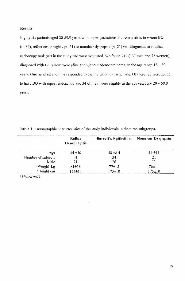
Results
Eighty six patients aged 20-59.9 years with upper gastrointestinal complaints in whom BO
(n=34), reflux oesophagitis (n=31) or nonulcer dyspepsia (n=21) was diagnosed at routine
endoscopy took part in the study and were evaluated. We found 212 (137 men and 75 women),
diagnosed with BO whom were alive and without adenocarcinoma, in the age range 18 - 80
years. One hundred and nine responded to the invitation to participate. Of these, 88 were found
to have BO with repeat endoscopy and 34 of them were eligible in the age category 20 - 59.9
years.
Table 1 Demographic characteristics of the study individuals in the three subgroups.
Reflux Oesophagitis
Baneit's Epithelium NonuIcer Dyspepsia
Age Number of subjects
Male *Weight kg
*Means :tSD.
44 ±10 31 21
81±18
48 ±8.4 34 26
77±15 I
44±11 21 11
76±1l 175±10
88
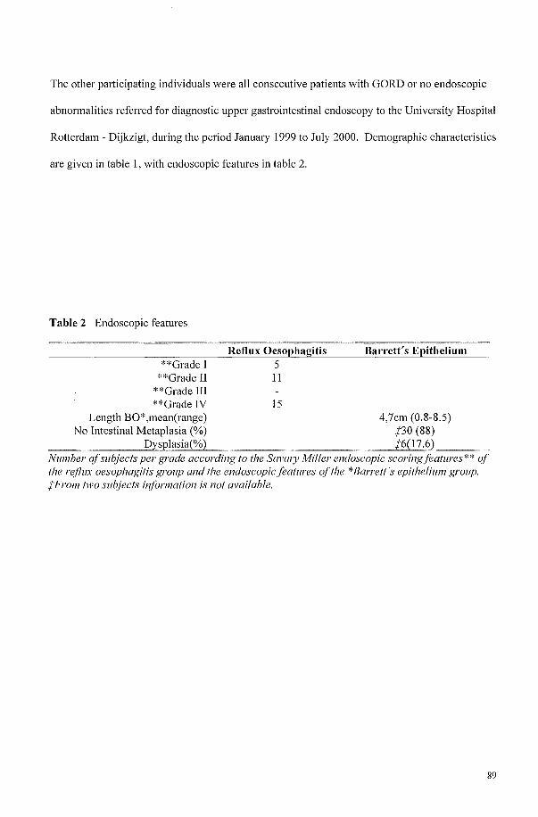
The other participating individuals were all consecutive patients with GORD or no endoscopic
abnormalities referred for diagnostic upper gastrointestinal endoscopy to the University Hospital
Rotterdam - Dijkzigt, during the period January 1999 to July 2000. Demographic characteristics
are given in table 1, with endoscopic features in table 2.
Table 2 Endoscopic features
**Grade I **Grade II
**Grade III **Grade IV
Reflux Oesophag=i_ti_s ___ B_arrett's Epithelium 5 11
IS Length BO*,mean(range) 4,7cm (0.8-8.5)
No Intestinal Metaplasia (%) /30 (88) ~~~,;;;;;D,Lysplasia(%) /6(l7.6).~_~
Number of subjects pel' grade according to the SavGlJ! Mil1er endoscopic scoringfeatures** of the reflux oesophagitis group and the endoscopic features of the *Barrett's epitheliul71 group. /From two subjects il?forl71ation is not available.
89
Twenty seven(79%) ofthe patients in the BO group were using proton pump inhibitors (mostly
omeprazole). This was less fi'equent in the reflux oesophagitis group 17(55%) and only 4(19%)
in the NUD group (p<O.OOOl).
The prevalence of risk factors for CVD are shown in table 3 and 4. Body mass index were not
significantly different between groups. The mean systolic blood pressure was significantly
higher in the BO and reflux oesophagitis group than controls (see table 3, which shows the mean
blood pressures of all patients within a group, whether or not on antihypertensive treatment). The
mean systolic blood pressure in the NUD group was higher than the reference population,
although the difference was not statistically significant. The mean diastolic blood pressure was
significantly higher in the reflux oesophagitis group but it was not different in the BO or NUD
groups. Frequencies of hypertension in study groups and controls are given in table 4.
Hypertension occurred significantly more frequently among the BO (P<O.OOOI ) (odds ratio 5.1)
and reflux oesophagitis (P<O.OOl) (odds ratio 3.8) patients than in controls, but not in the NUD
group (P=O.34) (odds ratio 1.7).
90
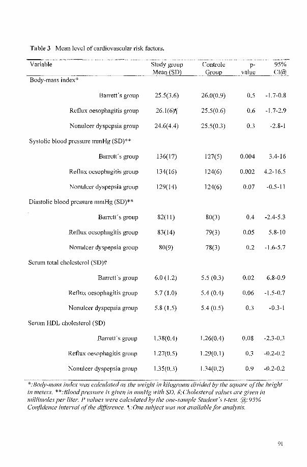
Table 3 Mean level of cardiovascular risk factors.
Variable Study group Controle p- 95% Mean (SD2 Groul2 value Cl(a?
Body-mass index*
Barrett's group 25.5(3.6) 26.0(0.9) 0.5 -1.7-0.8
Reflux oesophagitis group 26.1(6)~ 25.5(0.6) 0.6 -1.7-2.9
Nonulcer dyspepsia group 24.6(4.4) 25.5(0.3) 0.3 -2.8-1
Systolic blood pressure mmHg (SD)**
Barrett's group 136(17) 127(5) 0.004 3.4-16
Reflux oesophagitis group 134(16) 124(6) 0.002 4.2-16.5
Nonulcer dyspepsia group 129(14) 124(6) 0.07 -0.5-11
Diastolic blood pressure mmHg (SD)**
Barrett's group 82(11) 80(3) 0.4 -2.4-5.3
Reflux oesophagitis group 83(14) 79(3) 0.05 5.8-10
Nonulcer dyspepsia group 80(9) 78(3) 0.2 -\.6-5.7
Serum total cholesterol (SD)#
Barrett's group 6.0 (l.2) 5.5 (0.3) 0.02 6.8-0.9
Reflux oesophagitis group 5.7 (l.0) 5.4 (0.4) 0.06 -1.5-0.7
Nonulcer dyspepsia group 5.8 (I.5) 5.4 (0.5) 0.3 -0.3-1
Serum HDL cholesterol (SD)
Barrett's group 1.38(0.4) 1.26(0.4) 0.08 -2.3-0.3
Reflux oesophagitis group 1.27(0.5) 1.29(0.1) 0.3 -0.2-0.2
Nonulcer dyspepsia group 1.35(0.3) 1.34(0.2) 0.9 -0.2-0.2
""~ _~ M~
"""-~ "-~--=~~~~ ----~-
*:BodY-l11ass index was calculated as the weight in kilograms divided by the square of the height inll1eters. **:Blood pressure is given in 11l111Hg with SD. #:Cholestel'ol values are given in 111 illim ales per liter. P values were calculated by the one-sample Student's t-test. @:95% Confidence intel1'al qf the difference. ~:One subject was not available for analysis.
91
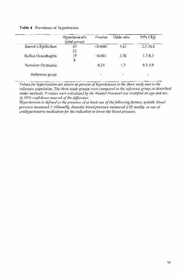
Table 4 Prevalence of hypeliension
Hypertension% (total group)
Barrett's Epithelium 47 32
Reflux Oesophagitis 19 8
Nonu1cer Dyspepsia
Reference group
P-value
<0.0001
<0.001
0.34
Odds ratio 95%CI@
5.61 2.5-10.0
3.76 1.7-8.3
1.7 0.5-5.0
Values for hypertension are shown as percent of hypertension in the three study and in the reference population. The three study groups were compared to the reference group as described under methods. P-vallles were calculated by the Mantel-Haenszel test stratified on age and sex. @ 95% confidence intel1'al of the d(f(erence. Hypertension is defined as the presence of at least one of the followingfactors: systolic blood pressure measured ~ 160mmHg, diastolic blood pressure measured ~ 95 ml11Hg, or use of antihypertensive medication for the indication to lower the blood pressure.
92
Most frequently given treatment for hypertension in the three study groups were beta blockers in
10(29%) BO patients, 2(6%) reflux oesophagitis patients and 2(9.5%) NUD patients followed by
ACE inhibitors; 4(12%), 1(3.3%) and 1(5%) respectively. In the third and fOUlih place came;
Calcium antagonists 2(6%),2(6.4%),1(5%), and hydrochloOlihiazide 2(6%), 2(6.4%) and
3(14%) respectively. Combination treatment with beta blockers and calcium antagonists was
being used by 2 BO patients, 1 reflux oesophagitis patient and 1 NUD subject. Two BO
individuals were on the combination of an ACE inhibitor and a beta blocker, and by one reflux
oesophagitis subject. Five ofthe patients with GERD or 1 with NUD diagnosed as having
hypeliension had a history of myocardial infarction. An additional 3 normotensive individuals
had a history of myocardial infarction. Five patients were known to have diabetes mellitus, no
new cases were identified by blood sugar measurements. Two of the diabetics were also
hypertensive.
Two known hypertensives had a serum creatinine slightly above the normal limit, as did one
normotensive patient. One normotensive patient had a serum creatinine of 31 0 mcmol/\. Serum
creatinine values were normal in all other cases.
Serum total cholesterol levels (see table 3) were significantly higher in the BO (P=0.02) group,
with a trend in the reflux oesophagitis (P=0.06) patients, but were not different in the NUD
group. HDL cholesterol levels and the prevalences of smoking habits were not different between
the study groups and controls. The prevalence of both current smoking and non smoking tended
to be highest in the younger age category (almost 50 percent in both categories), but decreased
with age to the lowest level of 30 percent and 20 percent respectively in the 55-59,9 years of age
category. Past smoking was lowest in the youngest age category - around 10% - and highest in
the 55-59,9 category - almost 50%. Mean blood glucose level was lower in the reflux
oesophagitis and NUD groups.
93
Discussion
This is the first study to consider the issue of the prevalence of risk factors for CYD in patients
with all grades of GORD. It was undeliaken in patients with endoscopically confirmed and
clearly defined subgroups of GORD and patients without endoscopic abnormalities, classified as
NUD. We observed a higher blood pressure in the reflux oesophagitis and BO group compared
to the reference population, which was not found in the NUD group. Whether hypertension
occurred before or after the start of gasto-intestinal complaints could not be assessed by this
study (the questions asked did not specity the chronology of complaints). We also observed a
significantly higher serum total cholesterol in the BO group, with trend in the reflux oesophagitis
patients, but not in the NUD group. The mean HDL cholesterol level was borderline significantly
higher in the BO group. However, this was not different between the two other study groups and
the reference group. Although BMI was higher in BO patients than in controls, the difference did
not reach statistical significance.
To appreciate the findings of this study, certain methodological aspects should be considered.
The diagnoses in the study groups were originally made with diagnostic endoscopy in patients
with upper gastrointestinal complaints. All patients meeting our criteria for reflux oesophagitis,
BO or NUD and diagnosed in the above-mentioned periods were asked to participate in a study
to look for the prevalence of Helicobacter pylori infection. They were not asked to participate in
a study to detect risk factors for CYD. A notice was included in the invitation that we would like
to know a few things about general health status. Therefore, it is not likely that the response of
the three study groups patients is influenced by the presence of CVD risk factors. The
individuals paJiicipating in the MORGEN study were considered a valid control group because
paJiicipants were derived from the general popUlation and living in the same area with same
period of measurements. The comparability ofthe two sets of data was achieved by using the
94
same items from the standard questionnaire ofthe reference group and the three study
populations on presence ofCVD risk factors. We therefore could not use questions about known
family-history of CVD for comparison because of differences in the phrasing of the questions
used.
The criterion for hypetiension was based on both a blood pressure measurement and treatment
for hypertension. Therefore, it could be argued that the difference in prevalence ofhypetiension
was due to selection bias, as the study groups, many being hospital outpatients, might
conceivably be a group requiring more fi'equent medical attention which would increase the
chance of detection and treatment of hypetiension. However, the invitation included also those
who had only come for diagnostic endoscopy but were not known as outpatients at our hospital.
It should therefore represent the great majority of patients today diagnosed with reflux
oesophagitis, BO, or NUD . The difference is therefore difficult to explain purely by selection
bias. In all paliicipants blood pressure was measured once with a sphygmomanometer in sitting
position after 5 minutes rest. We cannot exclude the possibility that the higher levels of blood
pressure in the reflux oesophagitis and the BO group compared to control is due to circumstances
of the measurements (white coat effect of a physician in the study groups but an research
assistant in the MORGEN project)[J6]. However, the phenomenon of white coat hypertension,
refers to a health care institute hypeliension or office hypertension phenomenon, because it
depends on multiple factors and does not correlate with the pressor response to the doctor [15].
Thus, it should imply that isolated office hypertension which might also be the earliest
manifestation of real hypertension [17], s evenly impotiant in both cases and controls._The same
remarkable outcome was not seen in the NUD group compared to controls, which makes a white
coat effect than unlikely as a reason for higher blood pressure measurements in the other groups.
A single measurement does not exactly represent a person's average, or usual blood pressure
[18], leading to overestimation of the prevalence ofhypetiension in both the cases and controls.
95
We believe that by depending only on the first measure in both the patient and the reference
group, the comparison that we made is reliable.
In the reflux oesophagitis, BO and NUD groups, weight and height were often self reported, in
contrast to the MORGEN study. Self-reporting tends to give 5-7% lower BMI's than measured
BMl's, which tend to be more biased in older and overweight groups [19]. However, self
reporting on BM! is more reliable when used on a younger population[20]. Our three study
populations are relatively young, mean age 44 years in the reflux oesophagitis and NUD groups
and 48 years in the BO group, but due to the broad age range (20 to 59.9 years), it could mean
that BMI might have been underestimated in the present study, although less likely in the
younger age group.
If the findings are true the underlying mechanisms are not known. Although high cholesterol
might contribute to an increased risk ofCVD, differences in the prevalence of hypertension in
the present study were statistically more striking. As we matched for age and gender, age does
not playa role in the comparison. Since BMI did not differ between groups, obesity as
hypeliensive factor in the reflux oesophagitis and BO groups seems unlikely. We did not include
any additional investigations in this study to determine whether the patients had secondary
hypeliension and other factors known to increase blood pressure such as insuline resistance, high
alcohol and salt intake, and perhaps sedentary lifestyle, stress, low potassium and calcium intake
were not examined. One might hypothesize that genetic factors playa role. The influence of
genes on blood pressure has been suggested by family studies and mutations in at least 10 genes
have been shown to raise or lower blood pressure [18]. Recently, on the basis of clustering of
GORD in families, it has also been suggested that there might be a GORD gene [21, 22]. The
same causative factors - either genetic or environmental factors or both - might be operative in
GORD diseases and hypertension. However, further studies need to be performed to establish
this possibility.
96
In an earlier follow-up study at our university hospital we found surprisingly high mortality rates
from CVD in a cohort of 166 patients in whom the diagnosis BO had been established between
1973 and 1986[10]. In retrospect, those patients would not have benefited from endoscopic
surveillance because of their excess mortality was in one-third due to CVD. This is in agreement
with a recently published long term observational study from MacDonald et al. on BO patients in
University Hospital in the United Kingdom [23]. This high mortality from cardiovascular
diseases rather than cancer was recently repotied as a surprising finding in a long-term follow-up
study of GORD patients treated surgically or medically[24]. Recently we found in an elderly
popUlation with BO a higher prevalence of myocardial infarction with a notably higher
prevalence of hype liens ion in the same group[25]. The present study was performed in a younger
category of the whole spectrum of GORD patients and compared with another age and sex
matched reference population. Again we noticed a noteworthy high prevalence of hype liens ion
in the GORD population, particularly the BO group. Cardiovascular and overall mortality are
known to increase continuously with increasing blood pressure [18]. If our findings are true,
they may possibly explain paJi of the observed higher mortality in the BO population in the
follow-up study fi'om our hospital performed some years ago[1 0]. Further studies are needed.
These should be prospective and include multiple measurements of blood pressure, examination
for end-organ damage such as retinopathy, and an evaluation of factors which might contribute
to the raised blood pressure such as salt intake.
In conclusion, Barrett's oesophagus and reflux oesophagitis found at diagnostic endoscopy are
associated with an increased prevalence of hype liens ion and possibly a higher total cholesterol
level than the general population. If our results can be confirmed, this provides an explanation
for the increased motiality during follow-up of Barrett's oesophagus patients, and should be
97

taken into account when designing or evaluating surveillance studies or selecting patients with
BO for surveillance.
98
References
1. Spechler SJ. Epidemiology and natural history ofgastro-oesophageal reflux disease.
Digestion 1992;51(Suppl 1):24-9.
2. McDougall NT, Johnston BT, Kee F, Collins JS, McFarland RJ, Love AH. Natural
history of reflux oesophagitis: a 10 year follow up of its effect on patient
symptomatology and quality oflife. Gut 1996;38:481-6.
3. Naef AP, Savary M, Ozzello L. Columnar-lined lower esophagus: An acquired lesion
with malignant predisposition. RepOli on 140 cases of Barrett's esophagus with 12
adenocarcinomas. J Thome Cardiovase Surg 1975;70:826-35.
4. Blot WJ, Devesa SS, I(neller RW, Fraumeni JF. Rising incidence of adenocarcinoma of
the esophagus and gastric cardia. JAMA 1991 ;265: 1287-9.
5. van Sandick JW, van Lanschot JJ, Kuiken BW, Tytgat GN, Offerhaus GJ, Obeliop H.
Impact of endoscopic biopsy surveillance of Barrett's oesophagus on pathological stage
and clinical outcome of Barrett's carcinoma. Gut 1998;43:216-22.
6. Cameron AJ, Carpenter HA. Barrett's esophagus, high-grade dysplasia, and early
adenocarcinoma: a pathological study. Am J GastroenteroI1997;92:586-91.
7. Spechler SJ, Robbins AB, Rubins HB, et. al. Adenocarcinoma and Barrett's esophagus.
An overrated risk? Gastroenterology 1984;87:927-33.
8. Cameron AJ, Ott BJ, Payne WS. The incidence of adenocarcinoma in columnar-lined
(Barrett's) esophagus. N Engl J Med 1985;313:857-9.
9. Van der Veen AH, Dees J, Blankensteijn JD, van Blankenstein M. Adenocarcinoma in
Barrett's oesophagus: an overrated risk. Gut 1989;30: I 4-8.
10. van der Burgh A, Dees J, Hop WCJ, van Blankenstein M. Oesophageal cancer is an
uncommon cause of death in patients with Barrett's oesophagus. Gut 1996;39:5-8.
11. Sharma P, Morales TG, Sampliner RE. Short segment Barrett's esophagus--the need for
standardization of the definition and of endoscopic criteria. Am J Gastroenterol
1998;93: 1 033-6.
12. Little AG, DeMeester TR, Kirchner PT, O'Sullivan GC, Skinner DB. Pathogenesis of
esophagitis in patients with gastroesophageal reflux. Surgel)' 1980;88:101-7.
13. Houterman S, Verschuren WMM, Oomen CM, Boersma-Cobbaeli CM, Kromhout D.
Trends in total and HDL cholesterol and their determinants in The Netherlands behveen
1993 and 1997. Int J Epidemiol2001 ;accepted.
99
14. The sixth repOit of the Joint National Committee on prevention, detection, evaluation,
and treatment of high blood pressure. Arch Intem Med. 1997;157:2413-2446.
15. 1999 World Health Organization-International Society of Hype It ens ion Guidelines for
the Management of Hypertension. Guidelines Subcommittee. J Hypertens. 1999; 17: 151-
183
16. Pickering TG. White coat hypertension: time for action. Circulation 1998;98: 1834-6.
17. extra Middeke M, Lemmer B. Office hypeltension: abnormal blood pressure regulation
and increased sympathetic activity compared with normotension. Blood Press Monit
1996; I :403-407.
18. OA Carretero, Oparil S. Essential hypeltension. Part I: definition and etiology.
Circulation 2000;101:329-35.
19. Roberts RJ. Can self-reported data accurately describe the prevalence of overweight?
Public Health 1995; I 09:275-84.
20. Alvarez-Torices JC, Franch-Nadal J, Alvarez-Guisasola F, Hernandez-Mejia R, Cueto
Espinal' A. Self-repOited height and weight and prevalence of obesity. Study in a Spanish
population.lnt JObes Relat Metab Disord 1993;17:663-7.
21. Trudgill NJ, Kapur KC, Riley SA. Familial clustering of reflux symptoms. Am J
Gastl'oentel'ol 1999;94: 1172-8.
22. Romero Y, Cameron AJ, Locke 3rd GR, et. al. Familial aggregation of gastroesophageal
reflux in patients with Barrett's esophagus and esophageal adenocarcinoma.
Gastroenterology 1997; 113: 1449-56.
23. Macdonald CE, Wicks AC, Playford RJ. Final results from 10 year cohOit of patients
undergoing surveillance for Barrett's oesophagus: observational study. BkU.
2000;321: 1252-5.
24. Spechler JS, Lee E, Ahnen D et al. Long-term outcome of medical and surgical therapies
for gastroesophageal reflux disease. JAM A 2001;285:2331-8
25. Gudlaugsdottir S, Romme AM, Dees J, Wilson JHP. The prevalence of coronary hemt
disease and its risk factors in patients with Barrett's esophagus. [Abstract] EliI' J
Gastl'Oentel'ol Hepatol2000; 12:A4.
100
Chapter 5 The prevalence of CagA strains of Helicobacter pylori in non ulcer dyspepsia, reflux oesophagitis and Barrett's oesophagus.
Sunna Gudlaugsdottiri,2, Jan Franckei, Felix de Rooiji, Jan Dees2
, Theo Stijnen3, JH Paul
1. Dept. Of Internal Medicine, University Hospital Rotterdam, The Netherlands 2. Dept. Of Gastroenterology and Hepatology, University Hospital Rotterdam, The Netherlands 3. Institute of Epidemiology and Biostatistics, Erasmus University Rotterdam, The Netherlands
Submitted
101
Abstract
Background. The hypothesis that colonisation with CagA-positive Hpylo/'i strains protects
against the development of complications of gastro-oesophageal reflux disease (GORD) was
tested with present study.
Methods. Patients with reflux oesophagitis and Barrett's oesophagus were studied. Two corpus
and 4 antral biopsy specimens were obtained for detection of Hpylo/'i. A serum sample was
obtained for determination of IgG and IgA antibodies to Hpylo/'i and to the CagA protein.
Results. Of 233 patients studied, 68 patients had reflux oesophagitis, 83 had Barrett's
oesophagus, 82 had no macroscopic abnormalities (the control group) and no signs of GORD.
The 151 patients with reflux disease had a trend to lower prevalence of H.pylori (35%) than did
the 82 controls (43%) (P=ns). Among 83 H.pylori-positive patients from whom serum was
available, colonisation with CagA-positive strains was detected in 35% in the control group
versus 26% in the reflux oesophagitis group (ns), and 19% in the Barrett's oesophagitis group
(P=O,02).
C011c1usiolt. Patients with reflux oesophagitis and Barrett's oesophagus tend to have a lower
prevalence of Hpylo/'i colonisation than controls. Patients with Barrett's oesophagus have a
significantly lower prevalence of colonisation by the CagA-positive type Hpylo/'i than did
controls. These data suggest that colonisation with CagA-positive Hpylo/'i strains may protect
against the development of Barrett's oesophagus.
102

Introduction
Helicobacterpylori (H pylori) cause long-term infection of the gastric and duodenal mucosa in
human's [1]. Colonisation predisposes for peptic ulcer disease, atrophic gastritis and distal
(antral) stomach cancer [2], with varied effects on gastric acid secretion. Genetic variability of H.
pylori is high [3]. Several genes have been identified that may playa role in the pathogenicity [4,
5]. Most important is the cytotoxin-associated gene A (CagA), which is associated with peptic
ulcer disease [6], and intestinal type adenocarcinoma of the stomach [7]. Patients with duodenal
ulcer often have high basal gastrin levels, high peak acid output and high 24-hour intragastric
acidity [8-10]. In contrast, patients with H. pylori-associated gastric ulcer often have
hypochlorhydria [II]. Several reports suggest that the prevalence of H. pylori and especially the
more pathogenic form - CagA might be lower in patients with GORD including Barrett's
oesophagus (BO) than in the rest of the population [17, 18]. One explanation for the negative
association between colonisation with H. pylori and GORD is the effect of H pylori on acid
production, since extensive gastritis involving the corpus may lower acid secretion by impairing
parietal function.
We conducted a cross sectional study in consecutive patients over the age 18 years coming for
routine endoscopy at our endoscopic unit, to determine the prevalence of H pylori infection and
subsequently CagA phenotype in subgroups of patients with reflux oesophagitis and proven BO
to compare with patients without endoscopic abnormalities. We tested the hypothesis that
colonisation with CagA-positive H. pylori strains protects against the development of gastro
oesophageal reflux disease (GORD) and it's complications.
103
Materials and Methods
Patients
All patients referred for upper gastrointestinal endoscopy fi'om Janumy 1, 1999 to July 31, 2000,
with dyspeptic complaints and not known to have peptic ulcer disease by previous radiology or
endoscopy were asked to pmiicipate. Our institutional medical ethics review board approved the
study and informed consent was obtained from each patient. On the basis of visual examination
of the oesophagus, stomach and duodenum, patients were subdivided into study groups. If an
ulcer or a cancer was found the patients were excluded from the study. The first study group was
formed by patients without endoscopic abnormalities or gastritis only (the same categOly). They
were classified as nonulcer dyspepsia (NUD). The second study group comprised patients with
reflux oesophagitis meeting the criteria of the Savary-Miller oesophagitis grading scheme, as
defined by Little et al. [19]. The third group was formed by patients with BO defined as an
endoscopic diagnosis of columnar epithelium lining the distal tubular oesophagus of at least 3
cm in length. We also included shorter segments ofBO (SSBO) in the BO cohOli if proved
histologically to be intestinal type colunmar epithelium [20]. BO patients were largely derived
fi'om a recall study based on records between January 1, 1992 tD December 31,1996 and reached
by letter. Patients in whom a carcinoma or high-grade dysplasia in BO was found were excluded.
Patients with SSBO at endoscopic examination, but without that histological proof were
categorised as reflux oesophagitis grade IV, according to the Savary-Miller oesophagitis grading
scheme [19]. Serum samples were obtained from each patient on the day ofthe endoscopic
procedure for serology testing for H. pylori and CagA.
104
Histologic s((mple
Hematoxylin-eosin- and modified Giemsa-stained microscope slides bearing 4-~lm histologic
section were prepared using the original paraffin blocks of biopsy specimens from antrum,
angulus and corpus of the stomach. All specimens were independently evaluated. The presence
of the H pylori colonisation was evaluated separately.
Serology
Specific serum IgG and IgA antibody levels against Hpylori were determined using an ELISA
teclmique (Pyloriset EIA-G III and Pyloriset EIA-A Ill, Orion Diagnostica, Espoo, Finland). The
assays were perfOimed according to the instructions of the manufacture. The absorbance of the
ELISA samples was quantified at 405nm in a Biorad microplate reader (3550-uv). IgG Antibody
titers were determined according to the instructions of the manufacturer using a Biorad microplote
reader with analysis software. Duplicate samples were determined and titers were calculated using
quadratic regression analysis and log-linear curve fitting from 4 antibody standards. Sensitivity and
specificity of this ELISA for the detection of H. pylori carriage is 92% and 84%, respectively and
has shown a velY good correlation with the biopsy finding [21].
Determination of H. pylori - CagA status was based on the presence of serum IgG and IgA using a
CagA H pylori pizo ELISA (Medimar, Italy, Mediphos, the Netherlands). Quantification of
antibody titers was identical as described for the H. pylori - IgG and IgA titers.
Definitions ((11(1 statistics
A patient was defined as H pylori-positive if one or more of the applied diagnostic methods were
positive. The CagA result was not used for this definition.
Statistical analysis was done with chi-square testing for contingency tables and with Student's T
test.
105
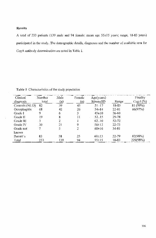
Results
A total of 233 patients (139 male and 94 female: mean age 55±15 years; range, 18-83 years)
participated in the study. The demographic details, diagnoses and the number of available sera for
CagA antibody determination are noted in Table 1.
Table 1 Characteristics ofthe study population
Clinical Number Male Female Age(years) Finality diagnosis total {n} en) Mean±SD Range CagA {%} Controls (NUD) 82 39 43 51±17 18-83 81 (99%) Oesophagitis 68 42 26 54±14 22-81 66(97%) Grade I 9 6 3 45±10 26-60 Grade II 19 8 II 52±15 29-78 Grade III 3 2 1 62±10 52-72 Grade IV 30 21 9 56±13 22-73 Grade not 7 5 2 60±16 34-81 known Barrett's 83 58 25 60±13 22-79 82(98%)
233 139 94
106
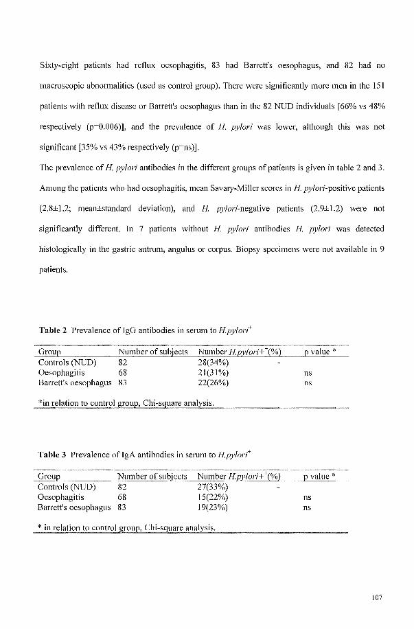
Sixty-eight patients had reflux oesophagitis, 83 had Barrett's oesophagus, and 82 had no
macroscopic abnormalities (used as control group). There were significantly more men in the 151
patients with reflux disease or Barrett's oesophagus than in the 82 NUD individuals [66% vs 48%
respectively (p=O.006)], and the prevalence of H pylori was lower, although this was not
significant [35% vs 43% respectively (p=ns)].
The prevalence of H pylori antibodies in the different groups of patients is given in table 2 and 3.
Among the patients who had oesophagitis, mean Savary-Miller scores in H pylori-positive patients
(2.8±1.2; mean±standard deviation), and H pylori-negative patients (2.9±1.2) were not
significantly different. In 7 patients without H pylori antibodies H pylori was detected
histologically in the gastric antrum, angulus or corpus. Biopsy specimens were not available in 9
patients.
Table 2 Prevalence of JgG antibodies in serum to Hpylort
Group Controls (NUD) Oesophagitis Barrett's oesophagus
Number of subjects 82 68 83
Number HJJ)li~ri+ \%) 28(34%) 2\(3\%) 22(26%)
*in relatioTl to control grollP, Chi-square analysis.
Table 3 Prevalence ofIgA antibodies in serum to Hpylort
Group Controls (NUD) Oesophagitis Barrett's oesophagus
Number of subjects 82 68 83
Number Hpy/ori+ \%) 27(33%) 15(22%) \9(23%)
* in relationto control group, Chi-square analysis.
p value *
ns ns
p value *
ns ns
107
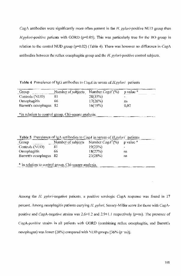
CagA antibodies were significantly more often present in the H pylori-positive NUD group than
Hpylori-positive patients with GORD (p=0.05). This was pmiicularly hue for the BO group in
relation to the control NUD group (p=0.02) (Table 4). There was however no difference in CagA
antibodies between the reflux oesophagitis group and the H pylori-positive control subjects.
Table 4 Prevalence of IgG antibodies to CagA in selUm of Hpylort patients
Group Controls (NUD) Oesophagitis Barrett's oesophagus
Number of subjects 81 66 82
28(35%) 17(26%) 16(19%)
*in relation to control group, Chi-sguare analysis.
p value *
ns 0,02
Table 5 Prevalence of IgA antibodies to CagAin serum of HEylort patients Group Number of subjects Number CagA\%) p value * Controls (NUD) 81 19(23%) Oesophagitis 66 18(27%) ns Barrett's oesophagus 82 23(28%) ns
* in_ relation to control Sf-0up, Chi~sCJuareanalJ'~s~is~. ~~~~~~~~
Among the H pylori-negative patients, a positive serologic CagA response was found in 17
percent. Among oesophagitis patients carrying H pylori, Savmy-Miller score for those with CagA-
positive and CagA-negative strains was 2.6±1.2 and 2.9±1.1 respectively (p=ns). The presence of
CagA-positive strains in all patients with GORD (combining reflux oesophagitis, and Barrett's
oesophagus) was lower (28%) compared with NUD groups [36% (p=ns)).
108
Discussion
The management of GORD and BO remains a challenging problem and this is paIily due to a lack
of knowledge of its natural history. The relationship of GORD, BO and H. pylori is also complex
[1]. There might also be a lillie between prolonged proton pump inhibition and the area of the
stomach infected by H pylori and the rate of progression to atrophic gastritis leading to
hypochlorhydria [12-14].
Strains of H. pylori have been grouped into 2 broad categories: the type I strains have the ability to
produce a vacuolating cytotoxin (vacA) and the CagA antigen, and the type II strains lack that
ability [22]. These strains differ in virulence, and the type I strains were demonstrated to be more
fi'equently associated with duodenal ulcer disease [22], with significantly more inflammation of the
gastric mucosa and with higher serum IgG levels against H. pylori [7]. In most duodenal ulcer
patients, antral gastritis predominates but corpus gastritis often develops during maintenance
therapy with acid suppressive drugs [12]. When corpus gastritis is present, hypergastrinemia does
not lead to increased acid production. H. pylori also possesses factors capable of inhibiting parietal
cells [23, 24], while host cytokines, such as TNF-alpha and IL-lf3, resulting fi'om chronic
inflammation, also inhibits parietal cell function [25]. Since the first report by Coghlan et al. in
1987 [26], a large number of studies have shown that eradication of H pylori results in a dramatic
reduction in the recurrence of ulcers during the subsequent 1-2 years. However, H pylori -
paIiicularly the CagA phenotype through gastritis and associated hypochlorhydria might protect
against GORD and its complications [18]. In recent years it has become clear that a significant
number of patients will develop reflux oesophagitis after apparently successful eradication [27].
The findings are consistent with the hypothesis that the declining infection rates of H. pylori in the
general population have led to a rise in the occurrence ofGORD and associated oesophageal
adenocarcinoma [28], with also lower prevalence of CagA phenotype in patients with complicated
GORD such as BO than in the rest of the population [18]. This hypothesis gives rise to the
109
question whether H. pylori (particularly CagA-positive strain), if detected, should always be
eradicated. Serologic testing is a sensitive method for detecting infection with CagA-positive strain
[29]. CagA antibodies are not only a useful marker for the more virulent strains that are associated
with severe gastroduodenal disease [22], but might also be helpful as an guide on decision making
- whom not to try to eradicate.
Conclusion. Patients with Barrett's oesophagus have a significantly lower prevalence of CagA
positive type of H. pylori than individuals without endoscopic abnormalities. These data suggest
that colonisation with CagA-positive H. pylori strains may protect against the development of
Barrett's oesophagus.
110
References
1. Blaser MJ. Ecology of Helicobacter pylori in the human stomach. J Clin Invest
1997;100(4):759-62.
2. Kuipers EJ. Helicobacter pylori and the risk and management of associated diseases:
gastritis, ulcer disease, atrophic gastritis and gastric cancer. Aliment Pharmacol Ther
1997;11 Suppll:7l-88.
3. Dunn BE, Cohen H, and Blaser MJ. Helicobacter pylori. Clin Microbiol Rev
1997;10(4):720-41.
4. Blaser MJ. Intrastrain differences in Helicobacter pylori: a key question in mucosal
damage? Ann Med 1995;27(5):559-63.
5. Atherton Je. The clinical relevance of strain types of Helicobacter pylori. Gut
1997;40(6):701-3.
6. van Doorn LJ, Figueiredo C, Sanna R, Plaisier A, Schneeberger P, De Boer W et al.
Clinical relevance of the cagA, vacA, and iceA status of Helicobacter pylori.
Gastroenterology 1998;115(1):58-66.
7. Kuipers EJ, Perez Perez 01, Meuwissen SGM, Blaser MJ et al. Helicobacter pylori and
atrophic gastritis: impOliance of the cagA status. J Nat! Cancer Inst 1995 ;87(23): 1777-80.
8. Calam J, Gibbons A, Healey ZV, Bliss P Arebi N. How does Helicobacterpylori cause
mucosal damage? Its effect on acid and gastrin physiology. Gastroenterology 1997;113(6
Suppl):S43-9.
9. Moss SF, Calam J. Acid secretion and sensitivity to gastrin in patients with duodenal
ulcer: effect of eradication of Helicobacterpylori. Gut 1993;34(7):888-92.
10. El-Omar E, Penman 1, Darrison CA, Ardhill JS, McColl KEL. Eradicating Helicobacter
pylori infection lowers gastrin mediated acid secretion by two thirds in patients with
duodenal ulcer. Gut 1993;34(8): 1060-5.
11. Beales IL, H pylori-associated hypochlorhydria [letter]. Gastroenterology
1998;114(3):618-21.
12. Kuipers EJ, Uyterlinde AM, Pena AS, Hazenberg HJ, Bloemena E, Lindeman J et al.
Increase of Helicobacter pylori-associated corpus gastritis during acid suppressive
therapy: implications for long-term safety. Am J Gastroenterol 1995;90(9): 1401-6.
13. Kuipers EJ, Uyterlinde AM, Pena AS, Roosendaal R, Pals G, Nelis GF, et al. Long-term
sequelae of Helicobacter pylori gastritis. Lancet 1995;345(8964): 1525-8.
111
14. Kuipers EJ, Lundell L, Klinkenberg-Knol EC, Havu N, Festen HPM, Kiedman B, et al.
Atrophic gastritis and Helicobacter pylori infection in patients with reflux esophagitis
treated with omeprazole or fundoplication. N Engl J Med 1996;334(16): 1 0 18-22.
15. Labenz J, Malfeliheiner P. Helicobacterpylori in gastro-oesophageal reflux disease:
causal agent, independent or protective factor? Gut 1997;41(3):277-80.
16. Loffeld RJLF, Ten Tije BJ, Arends JW. Prevalence and significance of Helicobacter
pylori in patients with Barrett's esophagus. Am J Gastroenterol 1992;87(11): 1598-600.
17. Werdmuller BF, Loffeld RJLF. Helicobacterpylori infection has no role in the
pathogenesis of reflux esophagitis. Dig Dis Sci 1997;42(1):103-5.
18. Vicari JJ, Peek RM, Falk GW, Goldblum JR, Easley KA, Schnell J, et al. The
seroprevalence of cagA-positive Helicobacter pylori strains in the spectrum of
gastroesophageal reflux disease. Gastroenterology 1998;115(1 ):50-7.
19. Little AG. Failed anti reflux operations. Pathophysiology and treatment. Chest Surg Clin
N Am 1994;4(4):697-704.
20. Pereira AD, Suspiro A, Chaves P, Saraiva A, Gloria L, de Almeida JC. Short segments of
Barrett's epithelium and intestinal metaplasia in normal appearing oesophagogastric
junctions: the same or two different entities? Gut 1998;42(5):659-62.
21. Granberg C, Mansikka A, Lehtonen OP, Kujari H, Gronfors R, Nurmi H, et al. Diagnosis
of Helicobacterpylori infection by using pyloriset ETA-G and ETA-A for detection of
serum immunoglobulin G (IgG) and IgA antibodies. I Clin Microbiol 1993;31(6): 1450-3
22. Xiang Z, Censini S, Bayeli PF, Telford JL, Figura N, Rappuoli R, et al. Analysis of
expression ofCagA and VacA virulence factors in 43 strains of Helicobacterpylori
reveals that clinical isolates can be divided into two major types and that CagA is not
necessary for expression of the vacuolating cytotoxin. Infect Tmmun 1995;63(1 ):94-8.
23. Huang LL, Cave DR, Gilbert IV, Wright A. Cloning and sequencing of the gene
encoding an acid-inhibitory protein in Helicobacterpylori. Gastroenterology
1996;110:A927.
24. Beil W, Birkholz C, Wagner S, Sewing KF. Interaction of Helicobacterpylori and its
fatty acids with parietal cells and gastric H+/K( + )-ATPase. Gut 1994;35(9): 1176-80.
25. Beales ILP, Cal am J. Tnterleukin-l beta and tumor necrosis factor-alpha inhibit
aminophyrine accumlation in cultured parietal cells by multiple pathways.
Gastroenterology 1996; 11 0:A62.
112
26. Coghlan JG, Humphries H, Dooley C, Keane C, Gilligan D, McKenna D, et al.
Campylobacter pylori and recurrence of duodenal ulcers--a 12-month follow-up study.
Lancet 1987;2(8568): II 09-11.
27. Labenz J, Blum AL, Bayerdorfer E, Meining A, Stolte M, Borsch G. Curing Helicobacter
pylori infection in patients with duodenal ulcer may provoke reflux esophagitis.
Gastroenterology 1997;112(5): 1442-7.
28. El-Serag HB, Sonnenberg A. Opposing time trends of peptic ulcer and reflux disease. Gut
1998;43(3):327-33.
29. Cover TL, Glupezynski Y, Lage AP, Burette A, Tummuru MK, Perez Perez GI, Blaser
MJ. Serologic detection of infection with CagA-positive Helicobacterpylori strains. J
Clin MicrobioI1995;33(6):1496-500.
113

114
Chapter 6 Prolonged use of proton pump inhibitors, CagA status and the outcome of Helicobacter pylori gastritis.
Sunna Gudlaugsdottir\ Herman van Dekken2, Theo Stijnen3
, J.H.Paul Wilson l
I. Dept. OfInternal Medicine, University Hospital Rotterdam, The Netherlands 2. Dept. Of Pathology, University Hospital Rotterdam, The Netherlands 3. Institute of Epidemiology and Biostatistics, Erasmus University Rotterdam, The Netherlands
In press in Journal ojelinical Gastroenterology 2002
115
Abstract
Goals and background. To assess if prolonged use of proton pump inhibitors in patients infected
with H. pylori has adverse effects on the gastritis.
Study. We studied 34 H. pylori positive individuals with reflux esophagitis, Barrett esophagus
or non ulcer dyspepsia. Half of them were on maintenance treatment with proton pump inhibitors
(mean; S years) and half were not. H. pylori and CagA status was tested serologically. Gastric
biopsies were classified histopathologically by the updated Sydney classification.
Results. Proton pump inhibitors in H. pylori gastritis are associated with a significantly less
antral inflammation and density of H. pylori, regardless of CagA status. There was a tendency to
more antral atrophy in patients with the CagA strain on maintenance treatment with proton pump
inhibitors (p=O.OS), but an opposite tendency in CagA negative individuals (p=O.OS). Intestinal
metaplasia was seen more frequently in CagA positive, treated individuals (p=O.02S)
Conclusions. These findings support the hypothesis that CagA status is important in progression
to atrophy and that maintenance treatment with PPls accelerate this progression, while reducing
inflammatory infiltration.
116
Introduction
It has become customary for patients diagnosed as having gastroesophageal reflux disease
(GERD) or Barrett's esophagus (BE) to be treated chronically with proton pump inhibitors
(PPJ's) (I), as maintenance ofa satisfactOlY response ofGERD depends upon effective
inhibition of gastric acid production (2). However, concerns have been expressed that long-term
acid inhibition in H pylori positive patients may increase the risk of gastric atrophy (3). This is
worrying because of the association between achlorhydria and hypochlorhydria, H pylori and
gastric carcinoma(4). Although the majority of patients with GERD are not infected with H
pylori, about one in eight is. Because we do not routinely test patients with GERD for the
presence of H pylori before stmiing with PPI treatment, we decided to assess the long-term
histological effect of chronic PPI use on the gastric mucosa in our H pylori positive population
with reflux esophagitis, BE or non-ulcer dyspepsia. Because the CagA status of H pylori appears
to be the most impOliant virulence factor we assessed the impact of the CagA status in all tested
individuals.
Materials and Methods
Study populatio11 and desig11
Adult patients who underwent upper GT endoscopy in the University Hospital Rotterdam
(Rotterdam, The Netherlands) were asked to participate in a study to look for the prevalence of
H pylori infection. Firstly, we reviewed endoscopic and histological data collected from patients
with BE found at routine endoscopy fi'om January 1992 until December 1996. The patients were
sent a letter and were offered a repeat esophagogastroduodenoscopy and serology testing for H
pylori. In addition, consecutive patients referred for upper gastrointestinal endoscopy fi'om
January 1999 to July 2000 were asked to pmiicipate. Only patients with reflux esophagitis, BE or
no endoscopic abnormalities were eligible for the study.
117
Patients who were seropositive for H pylori and willing to undergo repeat
esophagogastroduodenoscopy were divided into two groups: an index group (n=17) undergoing
long-term proton pump inhibition and a control group (n=17) not taking PPls.
The Medical Ethics committee of the University Hospital Rotterdam approved the study.
Informed consent was obtained from all participating patients, before the endoscopy was
performed. A standard questionnaire about current and past antisecretory medication use was
noted. Patients taking antacid drugs other than PPIs such as H2-receptor antagonists (a minority
of patients) were excluded. Omeprazole 20 mg and, in one case pantoprazol 30 mg were the
minimum daily doses. None of the patients in the control group had taken anti secretory
medication within the previous 6 months.
A blood sample was taken on the day of the endoscopic procedure for determination of specific
serum immunoglobulin G (IgG) antibodies against H. pylori and the CagA protein.
Definitions
Patients found at the repeat esophagogastroduodenoscopy to have reflux esophagitis, BE or no
endoscopic abnormalities and who were H pylori positive were eligible.
A patient was defined as H pylori -positive if IgG serology for H pylori was positive and CagA
positive if seropositive for the CagA protein. Only one patient was known to have had attempted
eradication before intake in the study but was found to be both serologically and histologically
positive for H. pylori. Gastroesophageal reflux disease was defined as the presence of endoscopic
signs of reflux oesophagitis, BE or both.
Endoscopical assessments
Endoscopy was performed using standardised procedures with the Olympus EVIS 100 video
endoscope (Olympus Optical Co., Hamburg, Germany), and findings were recorded in a
standardised database. Reflux oesophagitis was scored according to the Savary-Miller system (5)
as follows: grade 0, normal esophageal mucosa with no abnormalities; grade 1, erythema or
118
diffusely red mucosa, edema causing accentuated folds; grade 2, isolated round or linear erosions
extending from the gastroesophageal junction upward in relation to the folds; grade 3, confluent
erosions extending around the entire circumference or superficial ulceration; grade 4, frank ulcer
or BE.
Histological assessments
At endoscopic investigation four full-depth mucosal specimens from the antrum (two prepyloric
and two from the angulus region) and two from the midportion of the oxyntic mucosa were
obtained for histological assessment of all eligible patients and controls. The biopsy specimens
were immediately placed into separate tubes containing buffered 3.7% formaldehyde solution.
After dehydration, the specimens were embedded in paraffin wax, sectioned at 4-llm thickness
and stained with haematoxylin-eosin for pathological analysis. No separate staining for H pylori
microorganisms was done. Haematoxylin-eosin stained biopsies were graded according to the
Updated Sydney classification. Detailed description of the criteria used is given elsewhere(6).
Briefly, inflammation, gastritis activity, atrophy, intestinal metaplasia, and H. pylori colonization
of the gastric mucosa are quantified using four grades: none, mild, moderate or marked (6).
Inflammation was graded according to the increase in lymphocytes and plasma cells in the
lamina propria. Activity of gastritis describes the occurrence and density of neutrophil
granulocytes, which are known to be correlated with the degree of virulence of H. pylori strains.
Atrophy denotes the loss of glands, whether loss of a single or many acidopeptic glands. All
gastric biopsy specimens were assessed blindly by one experienced gastrointestinal pathologist
(H.vD.).
119
Labomtory assessment
Specific serum IgG antibody levels against H. pylori were determined using an enzyme-linked
immunosorbent assay technique (Pyloriset ElA-G Ill, Orion Diagnostica, Espoo, Finland). The
assays were performed according to the manufacturer instructions. The absorbance of the
enzyme-linked immunosorbent assay samples was quantified at 405nm in a Bio-Rad microplate
reader (Bio-Rad Laboratories BY, Yeenendaal, the Netherlands) (3550-uv). Immunoglobulin G
antibody titers were determined according to the manufacturer instructions using a Bio-Rad
microplate reader with analysis software. Duplicate samples were determined and titers were
calculated using quadratic regression analysis and log-linear curve fitting from four antibody
standards. Sensitivity and specificity of this enzyme-linked immunosorbent assay for the
detection of H pylori carriage is 92% and 84%, respectively and has shown a very good
correlation with the biopsy finding (7).
Determination of H pylori - CagA status was based on the presence of serum IgG antibodies
using a CagA H. pylori p120 enzyme-linked immunosorbent assay (Mediphos, Renkum, the
Netherlands) and quantified as described above.
Statistical analysis
The stratified Mann-Whitney-Wilcoxon nonparametric two-sample test was used to compare
histological features and mean between the two main independent groups (PPIs versus no PPls)
ofsampJed data and four subgroups (CagA-negative vs CagA-positive). We controlled for age
by stratifYing in three age categories.
The difference in proportions of CagA positive individuals in the two groups (PPls versus no
PPls) was assessed with the chi-square test.
A p-value < 0.05 was considered statistically significant.
Calculations were performed using SPSS 9.0 for Windows (statistical software) (SPSS Inc.,
Chicago,JL).
120
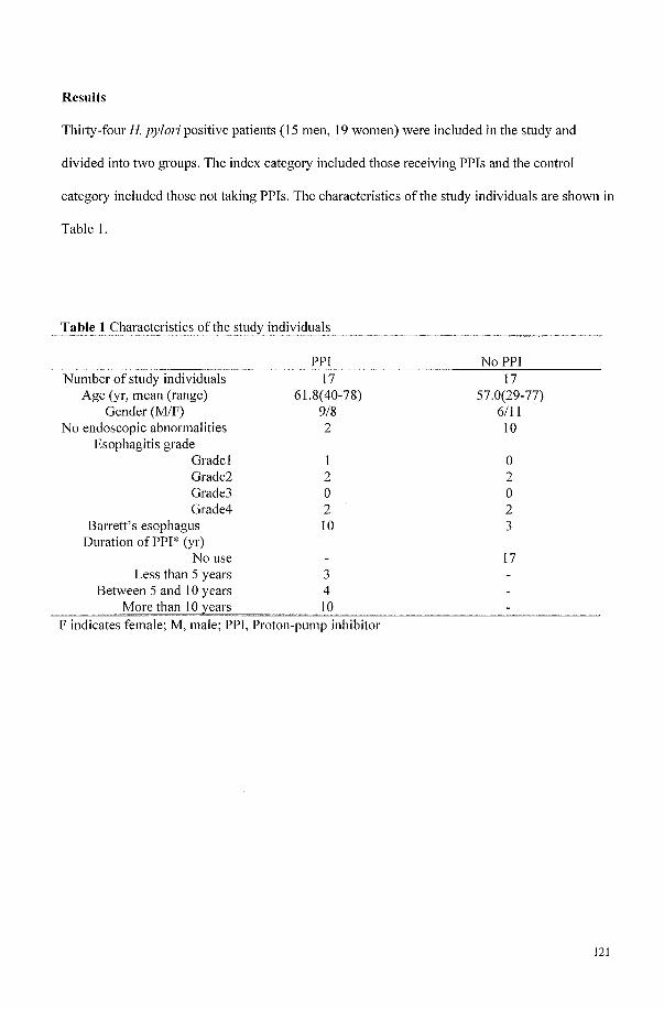
Results
ThiJiy-four H. pylori positive patients (15 men, 19 women) were included in the study and
divided into two groups. The index category included those receiving PPIs and the control
category included those not taking PPIs. The characteristics of the study individuals are shown in
Table 1.
Table 1 Characteristics of the
Number of study individuals Age (yr, mean (range)
Gender (M/F) No endoscopic abnormalities
Esophagitis grade Gradel Grade2 Grade3 Grade4
Barrett's esophagus Duration ofPPI* (yr)
No use Less than 5 years
Between 5 and 10 years
individuals
PPI 17
61.8(40-78) 9/8 2
1 2 o 2 10
3 4
F indicates female; M, male; PPI, Proton-pump inhibitor
NoPPI 17
57.0(29-77) 6111 10
o 2 o 2 3
17
121
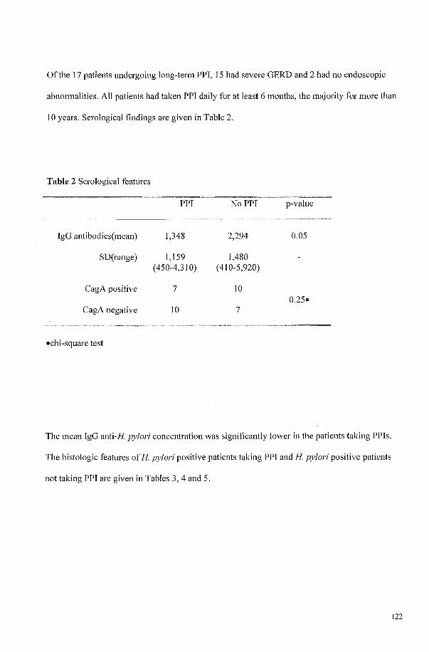
Of the 17 patients undergoing long-term PPI, 15 had severe GERD and 2 had no endoscopic
abnormalities. All patients had taken PPI daily for at least 6 months, the majority for more than
10 years. Serological findings are given in Table 2.
Table 2 Serological features
PPI NoPPI p-value
IgG antibodies(mean) 1,348 2,294 0.05
SD(range) 1,159 1,480 (450-4,310) (410-5,920)
CagA positive 7 10 0.25.
CagA negative 10 7
.chi-square test
The mean IgG anti-H pylori concentration was significantly lower in the patients taking PPls.
The histologic features of H pylori positive patients taking PPI and H pylori positive patients
not taking PPI are given in Tables 3, 4 and 5.
122
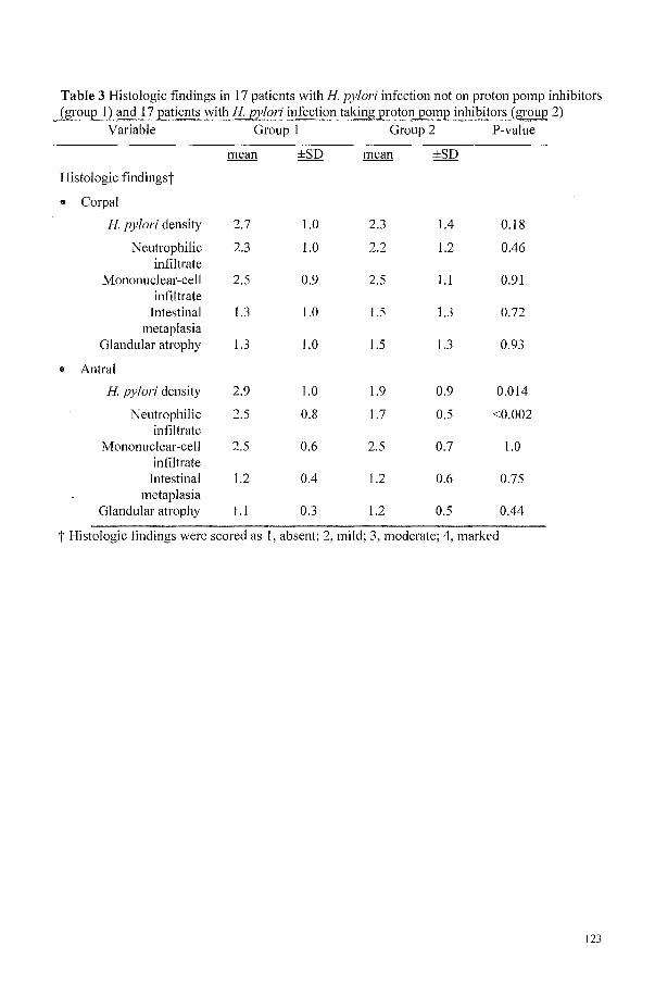
Table 3 Histologic findings in 17 patients with H. pylori infection not on proton pomp inhibitors (group 1) and} 7 patients with H. pJilori infection taking £1"OtOI1 pon!£! inhibitors {grol.!£ 2)
Variable Group I Group 2 P-value
Histologic findingst
• Corpal
H. pylori density
Neutrophilic infiltrate
Mononuclear-cell infiltrate
Intestinal metaplasia
Glandular atrophy
• Antral
H. pylori density
Neutrophilic infiltrate
Mononuclear-cell infiltrate
Intestinal metaplasia
Glandular atrophy
2.7
2.3
2.5
I.3
I.3
2.9
2.5
2.5
1.2
1.1
1.0
1.0
0.9
1.0
1.0
1.0
0.8
0.6
0.4
0.3
2.3
2.2
2.5
1.5
1.5
1.9
1.7
2.5
1.2
I.2
1.4
I.2
1.1
I.3
I.3
0.9
0.5
0.7
0.6
0.5
0.18
0.46
0.91
0.72
0.93
0.014
<0.002
1.0
0.75
0.44
t Histologic findings were scored as I, absent; 2, mild; 3, moderate; 4, marked
123
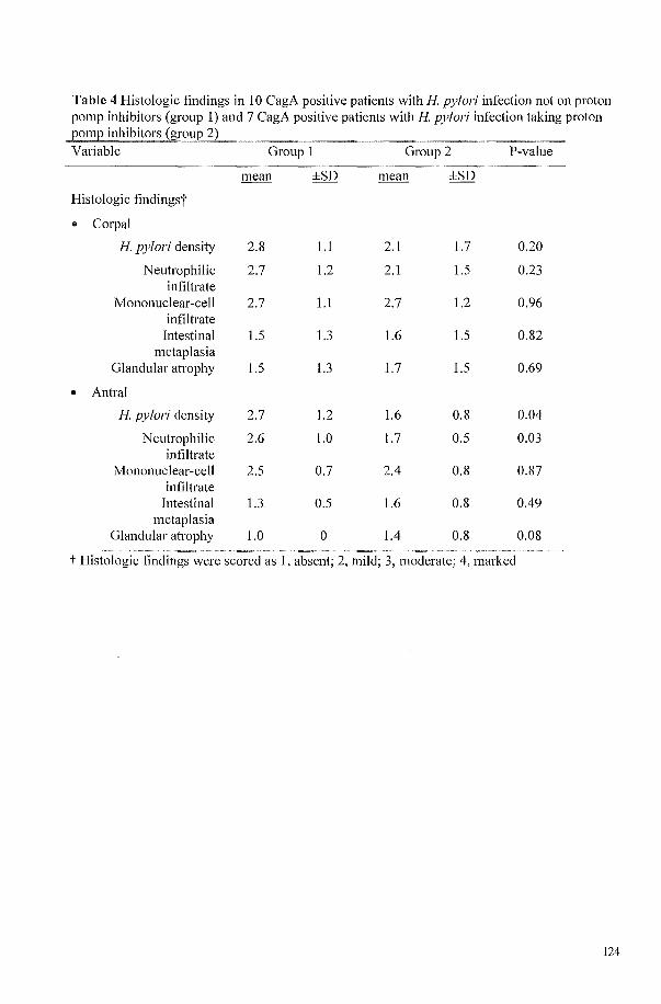
Table 4 Histologic findings in 10 CagA positive patients with H pylori infection not on proton pomp inhibitors (group 1) and 7 CagA positive patients with H pylori infection taking proton
inhibitors
Group I Group 2 P-value
mean ±SD mean ±SD
Histologic findingst
.. Corpal
H. pylori density 2.8 1.1 2.1 1.7 0.20
Neutrophilic 2.7 1.2 2.1 1.5 0.23 infiltrate
Mononuclear-cell 2.7 1.1 2.7 1.2 0.96 infiltrate
Intestinal 1.5 1.3 1.6 1.5 0.82 metaplasia
Glandular atrophy 1.5 1.3 1.7 1.5 0.69
.. Antral
H pylori density 2.7 1.2 1.6 0.8 0.04
Neutrophilic 2.6 1.0 1.7 0.5 0.03 infiltrate
Mononuclear-cell 2.5 0.7 2.4 0.8 0.87 infiltrate
Intestinal 1.3 0.5 1.6 0.8 0.49 metaplasia
Glandular atrophy 1.0 0 1.4 0.8 0.08
t Histologic findings were scored as I, absent; 2, mild; 3, moderate; 4, marked
124
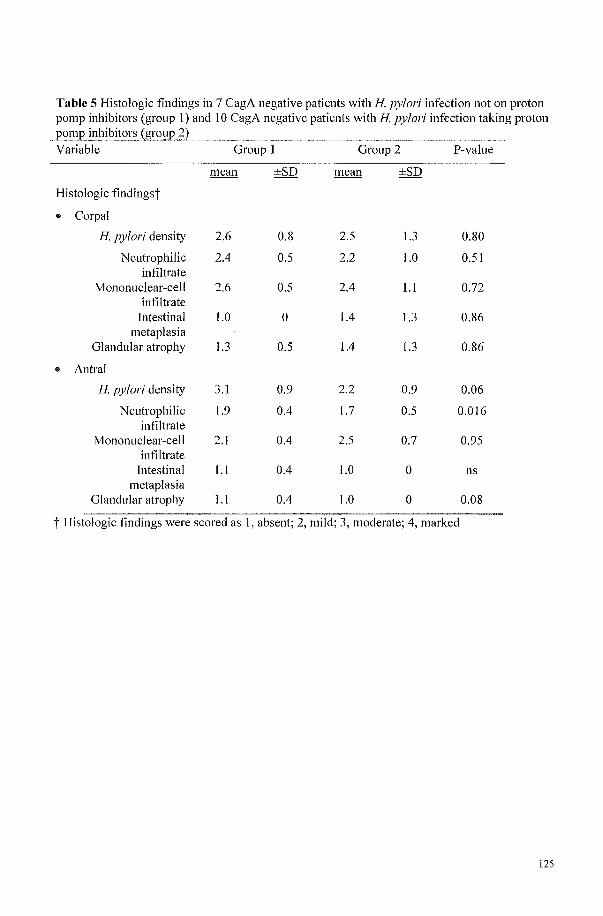
Table 5 Histologic findings in 7 CagA negative patients with H. pylori infection not on proton pomp inhibitors (group I) and 10 CagA negative patients with H. pylori infection taking proton l20mp inhibitors (gr~ul'
--~~-~~
Variable Group 1 Group 2 P-value
mean ±SD mean ±SD
Histologic findingst
.. Corpal
H. pylori density 2.6 0.8 2.5 l.3 0.80
Neutrophilic 2.4 0.5 2.2 1.0 0.51 infiltrate
Mononuclear-cell 2.6 0.5 2.4 1.1 0.72 infiltrate
Intestinal 1.0 0 1.4 l.3 0.86 metaplasia
Glandular atrophy 1.3 0.5 1.4 l.3 0.86
.. Antral
H. pylori density 3.1 0.9 2.2 0.9 0.06
Neutrophilic 1.9 0.4 1.7 0.5 0.016 infiltrate
Mononuclear-cell 2.1 0.4 2.5 0.7 0.95 infiltrate
Intestinal l.l 0.4 1.0 0 ns metaplasia
Glandular atrophy 1.1 0.4 1.0 0 0.08
i- Histologic findings were scored as 1, absent; 2, mild; 3, moderate; 4, marked
125
Patients using PPIs had significantly less H pylori, as detected histologically in the antrum, than
patients not taking PPls. Proton pump inhibitor use was also associated with significantly less
neutrophilic infiltrates in the antrum, whereas the degree of infiltration with mononuclear cells was
not different. In accordance with another cross-sectional study(8), atrophy and intestinal
metaplasia were rarely seen in the group as a whole «15% for either feature). When the histologic
features were analyzed separately for the CagA positive patients, use of PPI was associated
histologically with higher frequency of atrophy in the antrum (Table 4), approaching significance
(p=O.08). In contrast, in the CagA negative category, atrophy was less frequent in those individuals
taking PPls (Table 5) compared to those not taken PPls. The difference was also approaching
significance (p=O.08). In the PPJ treated group intestinal metaplasia was significantly more
frequently seen in CagA positive individuals (p=O.028)
Discussion
In this cross sectional study, all patients were seropositive for H pylori. The primary aim was to
examine the influence oflong-term use of PPls on histological features of the gastric mucosa.
The results suggest that maintenance treatment with PPls in H pylori gastritis reduces both the
antral bacterial density of H pylori and the neutrophilic infiltrate (inflammatory activity),
regardless of CagA status. However, individuals with CagA positive strains who take PPls seem
to run a risk of antral atrophy and intestinal metaplasia, in contrast to individuals with CagA
negative strains who are taking PPls or with CagA positive strains who are not taking PPls.
Because biopsy specimens may be falsely negative because of patchy H pylori
distribution in the stomach, we chose serology to define H pylori status. Because our index
category was using PPIs at intake, a breath test would have carried the possibility of being false
negative (9). We controlled for age in a categorical stratification, as the mean age of the index
category was higher.
126
Omeprazole is known to be bacteriostatic for H. pylori, and may also have
antiinflammatory effects (10). The majority of our index population had been using omeprazole
for a long time (mean, 8 years). It seems likely that the lower gastritis activity and density of H.
pylori detected by histology in the index group are both because of bacteriostatic and anti
inflammatory effects of omeprazole and fewer CagA positive H. pylori strains in the PPJ group
(II). Lower antibody levels in PPJ group is in agreement with less severe inflammation. The
bacteriostatic and antiinflammatory effect caused by omeprazole could have caused a selection
bias, with selection of patients with H pylori strains resistent to PPls. A prospective randomized
study would be needed to examine that possibility. However, the main aim of present study was
to gain further insight into progression of H. pylori gastritis and atrophy during PPI.
H pylori has evolved lifestyles in which inflammation is necessary for its survival(12,
13), causing acute (neutrophilic) and chronic (mononuclear) inflammatory response. It possesses
factors capable of inhibiting parietal cells (14, 15), and stimulates production of host cytokines,
such as tumor necrosis factor-alpha and interleukin-I13, which also inhibit parietal cell function
(16). Gastritis predominantly in the corpus reduces acid output, and is thought to protect against
GERD (17). However, corpus gastritis also predisposes to atrophic gastritis with presumed risk
of stomach cancer (18). The vacA s 1, CagA-positive or iceAI strains are strongly associated
with more severe gastritis, with a greater influence on the acid production (19). Mucosal
inflammation predominantly in the antrum stimulates release of gastrin from the antral mucosa,
which results in excessive secretion of acid by the parietal cell in the uninflamed corpus mucosa
(20) which may predispose to GERD (21), possible concomitant with duodenal ulcer (22). This
high acid load is associated with a slow progression to gland loss (22). A protective role against
gastric malignancy for duodenal ulcer has been described (23).
Corpus gastritis has been reported to develope in H pylori infected patients during
maintenance therapy with acid suppressive drugs (pmiicularly PPI treatment) and these
127
individuals may have an increased risk of gastric atrophy and consequently development of
gastric cancer (3). The present study failed to support this shift from antrum to corpus in
agreement with another cross-sectional study (8). An increase in corpus gastritis and atrophy
because of use ofPPls has not been confirmed in randomised, controlled studies (24). However,
it is possible that sampling error is a confounder, as atrophy can be only focal. Alternatively, the
failure of find an antrum to corpus shift could be due to the selection ofthe index group. Patients
with BE have been found to have less multifocal gastric atrophy than the control population (25).
In our study most of the patients taking PPls had BE. Barrett's esophagus and GERD are
associated with less H. pylori infection, particularly CagA positive strains. Nearly all H. pylori
isolates from patients with peptic ulcers, atrophic gastritis and gastric cancer are CagA positive
(26,27). In the current study, omeprazole therapy only appeared to increase the likelihood of
antral atrophic gastritis in patients with CagA positive strains. Of interest is that this occured
despite apparently reducing the bacterial load and acute inflammatory cells in the mucosa. The
mechanism for this phenomenon remains to be elucidated. As mentioned before, corpus atrophy
is thought to be related to H. pylori induced corpus gastritis, which impairs parietal cell function
and lowers gastric acid secretion. Thus, CagA positive strains may be both harmful (increases
risk of atrophy and distal cancer) and beneficial (protects against reflux oesophagitis and its
sequelae) (28).
Some groups have advised testing and treating for H. pylori when prescribing PPls for
prolonged period. This has lead to a dilemma, as eradication of H. pylori has been repOlied to
reduce the efficacy of PPls (29) and to increase acid reflux in some groups (30). Our results
suggest that, although maintenance treatment with acid suppression causes suppression of
inflammatOlY activity of antrum gastritis and a decreased density of H. pylori it also causes
acceleration of development of atrophic gastritis if the H. pylori is CagA positive. However, the
underlying mechanism is unknown. The mechanism whereby CagA promotes gastric mucosal
128
atrophy is still not clear. Recent studies show that the CagA protein, when delivered into gastric
mucosal cells from adherent bacteria, causes major changes in cell morphology. This is
associated with rearrangement of the cytoskeleton (31, 32). How these changes then progress to
cell loss, and how proton pump inhibition hastens the development of atrophy, is not known. We
did not measure gastric acid secretion and we did not measure gastrin levels by the patients.
Nevertheless, this issue might have clinical relevance and may show a link to earlier findings
that some H. pylori positive patients undergoing maintenance therapy with acid suppressive
drugs (particularly PPI treatment) have an increased risk of gastric atrophy (3), although not
confirmed in randomised, controlled studies (24). The relevance ofCagA needs to be studied in a
prospective fashion. Routine testing for CagA positive H. pylori and eradication prior to staliing
maintenance PPI therapy for GERD should therefore be considered; however, because of the
unceliainties this needs to be studied in a prospective fashion.
AcImowledgemellts
The authors thank Jan Francke and Felix de Rooij for the measurements of specific H.
pylori and CagA antibodies.
129
References
l. Bell NJ, Burget D, Howden CW, et al. Appropriate acid suppression for the management of gastro-oesophageal reflux disease. Digestion 1992;51 (Suppll (5»:59-67.
2. Lundell L. Acid suppression in the long-term treatment of peptic stricture and Barrett's oesophagus. Digestion 1992;51(Suppl 1(3»:49-58.
3. Kuipers EJ, Lundell L, Klinkenberg-Knol EC, et al. Atrophic gastritis and Helicobacter pylori infection in patients with reflux esophagitis treated with omeprazole or fundoplication. N Engl J Med 1996;334(16):1018-22.
4. EI-Omar EM, Oien K, EI-Nujumi A, et al. Helicobacter pylori infection and chronic gastric acid hyposecretion. Gastroenterology 1997; 113(1): 15-24.
5. Little AG, DeMeester TR, Kirchner PT, et al. Pathogenesis of esophagitis in patients with gastroesophageal reflux. Surgel)! 1980;88(1): I 01-7.
6. Dixon MF, Genta RM, Yardley JH, et al. Classification and grading of gastritis. The updated Sydney System. International Workshop on the Histopathology of Gastritis, Houston 1994. Am J Surg PathoI1996;20(1 0): 1161-81.
7. Granberg C, Mansikka A, Lehtonen OP, et al. Diagnosis of Helicobacter pylori infection by using pyloriset EIA-G and ETA-A for detection of serum immunoglobulin G (IgG) and TgA antibodies. JClin MicrobioI1993;31(6):1450-3.
8. Diebold MD, Richardson S, Duchateau A, et a1. Factors influencing corpus argyrophil cell density and hyperplasia in reflux esophagitis patients treated with antisecretory drugs and controls. Dig Dis Sci 1998;43(8):1629-35.
9. EI-Nujumi A, Hilditch TE, Williams C, et al. Current or recent proton pump inhibitor therapy markedly impairs the accuracy of the [14C]urea breath test. Eur J Gastroenterol Hepatol1998; 1 0(9):759-64.
10. Dattilo M, Figura N. Helicobacter pylori infection, chronic gastritis, and proton pump inhibitors. J Clin GastroenteroI1998;27(Suppll):SI63-9.
II. Loffeld RJ, Werdmuller BF, Kusters JG, et al. IgG antibody titer against Helicobacter pylori correlates with presence of cytotoxin associated gene A-positive H. pylori strains. FEMS Immunol Med MicrobioI2000;28(2): 139-41.
12. Blaser MJ, Parsonnet J. Parasitism by the "slow" bacterium Helicobacter pylori leads to altered gastric homeostasis and neoplasia. J Clin Invest 1994;94(1):4-8.
13. Blaser MJ. Hypotheses on the pathogenesis and natural history ofHelicobacter pyloriinduced inflammation. Gastroenterology 1992; 1 02(2):720-7.
14. Beil W, Birkholz C, Wagner S, et a1. Interaction of Helicobacter pylori and its fatty acids with parietal cells and gastric H+/K(+)-ATPase. Gut 1994;35(9):1176-80.
15. Huang LL, Cave DR, Gilbeli JV, et al. Cloning and sequencing of the gene encoding an acid inhibitory protein in Helicobacter pylori. Gastroenterology 1996; 11 0:A927.
16. Beales ILP, Calam 1.lnterleukin-l beta and tumor necrosis factor-alpha inhibit aminopyrine accumulation in cultured parietal cells by multiple pathways. Gastroenterology 1996; 11 0:A62.
17. Cal am J, Gibbons A, Healey ZV, et a1. How does Helicobacter pylori cause mucosal damage? Its effect on acid and gastrin physiology. Gastroenterology 1997; 113(6 Suppl):S43-9; discussion S50.
18. Kuipers EJ. Helicobacter pylori and the risk and management of associated diseases: gastritis, ulcer disease, atrophic gastritis and gastric cancer. Aliment Pharmacol Ther 1997;11 Suppll:71-88.
19. van Doorn LJ, Figueiredo C, Sanna R, et a1. Clinical relevance of the cagA, vacA, and iceA status of Helicobacter pylori. Gastroenterology 1998; 115(1 ):58-66.
130
20. el-Omar EM, Penman lD, Ardill JE, et al. Helicobacter pylori infection and abnormalities of acid secretion in patients with duodenal ulcer disease. Gastroenterology 1995; 1 09(3):681-91.
21. Fink SM, Barwick KW, DeLuca Y, et al. The association of histologic gastritis with gastroesophageal reflux and delayed gastric emptying. J Clin GastroenteroI1984;6( 4):301-9.
22. Boyd EI. The prevalence of esophagitis in patients with duodenal ulcer or ulcer-like dyspepsia. Am J GastroenteroI1996;91(8): 1539-43.
23. Hansson LE, Nyren 0, Hsing A W, et al. The risk of stomach cancel' in patients with gastric or duodenal ulcer disease. N Engl J Med 1996;335(4):242-9.
24. Lundell L, Miettinen P, Myrvold HE, et al. Lack of effect of acid suppression therapy on gastric atrophy. Nordic Gerd Study Group. Gastroenterology 1999; 117(2):319-26.
25. Rugge M, Russo V, Busatto G, et al. The phenotype of gastric mucosa coexisting with Barrett's oesophagus. J Clin Pathol200 1 ;54(6):456-60.
26. Parsol1l1et I, Friedman GD, Orentreich N, et al. Risk for gastric cancer in people with CagA positive or CagA negative Helicobacter pylori infection. Glit 1997;40(3):297-301.
27. Weel IF, van del' Hulst RW, Gerrits Y, et al. The interrelationship between cytotoxinassociated gene A, vacuolating cytotoxin, and Helicobacter pylori-related diseases. J llifect Dis 1996;173(5):1171-5.
28. Blaser MI. Hypothesis: the changing relationships of Helicobacter pylori and humans: implications for health and disease. J liifect Dis 1999; 179( 6): 1523-30.
29. Labenz J, Tillenburg B, Peitz U, et al. Helicobacter pylori augments the pH-increasing effect of omeprazole in patients with duodenal ulcer. Gastroenterology 1996; 11 0(3):725-32.
30. Labenz I, Blum AL, Bayerdorffer E, et al. Curing Helicobacter pylori infection in patients with duodenal ulcer may provoke reflux esophagitis. Gastroenterology 1997; 112(5): 1442-7.
31. Segall Ed, Cha J, Lo J, et al. Altered states: involvement of phosphorylated CagA in the induction of host cellular growth changes by Helicobacter pylori. Proc Nat! Acad Sci U S A 1999;96:14559-64.
32. Asahi M, Azuma T, Ito S, et al. Helicobacter pylori CagA protein can be tyrosine phosphorylated CagA in gastric epithelial cells. J Exp Med 2000; 191 :593-602
131

132

Chapter 7 General discussion and future perspectives
133
In this chapter I will briefly describe the most important findings and discuss the
methodological issues of the conducted studies. The results will be interpreted in relation to
clinical practice.
This discussion will finish with suggestions for future research on patients with Barrett's
esophagus.
7.1 Introduction
In the field of Barrett's esophagus (BE), we were paliicularly interested in answering a
number of questions concerning the surveillance for adenocarcinoma of the esophagus in
individuals with Barrett's esophagus and the treatment of Barrett's esophagus:
.. Who can benefit from endoscopic surveillance and what proportion of the population known
to have BE can theoretically benefit from endoscopic surveillance?
.. What risk factors are responsible for the increased death rate from cardiovascular disease in
the BE population?
.. Does the BE population have a lower prevalence of H. pylori infection than the general
population or other patients with dyspepsia?
.. What happens to the gastric mucosa in patients with H. pylori on maintenance treatment with
PPI's?
The concept that early diagnosis of esophageal adenocarcinoma will benefit the patient
(1-3), has lead to the development of endoscopic cancer surveillance programs as secondary
prevention for patients with known BE. However, there is no agreement about the frequency of
screening and in recent years it has been suggested that only operable patients at high risk of
developing carcinoma should be screened (4). The tendency of BE patients to die at an earlier
age than age and sex matched general population, mainly because of cardiovascular disease,
forms a strong argument against screening all patients with BE(S), and underscores the need for
134
surveillance guidelines in these patients. The indications and pitfalls of endoscopic cancer
surveillance of patients with BE are summarised in chapter l.
The first clue that H. pylori, particularly the more pathogenic CagA-expressing form,
might protect individuals against gastro-esophageal reflux disease (GERD) (including BE and
adenocarcinoma ofthe esophagus) came with the observation that the prevalence ofthis
infection was significantly lower in the BE population (6). This epidemiological observation
could suggest that eradication treatment should not be given to BE patients infected with these
particular strains,. However, prolonged use of proton pump inhibitors in patients infected with H.
pylori has been repOlied to have adverse effects on the natural history of their H. pylori
gastritis(7). Some H. pylori positive individuals with GERD require maintenance treatment with
acid inhibitors such as PPI's because of symptoms of GERD. This is a much-debated issue with
no firm conclusions, but a recent consensus repOli advised eradication of H. pylori in these
patients (8).
135
7.2 The main findings of this thesis can be summarised as follows.
e By making use of, on the one hand, published and generally accepted risk factors for
adenocarcinoma in BE, as given in table 1, as an indication for surveillance and, on the other
hand, absence of these risk factors, old age or concurrent disease as reasons for exclusion
from surveillance, we found that only 15.5% of patients diagnosed as having BE in our
hospital would be likely to benefit fi'om an endoscopic cancer surveillance program.
.. We found that, in comparison with the general population, the BE population has a higher
incidence of myocardial infarction and that the most likely risk factors for cardiovascular
diseases in this group were hypertension and obesity.
.. GERD patients without BE were also at increased risk of having hypertension.
.. We found a significant lower prevalence of infection with the CagA H pylori strain in the
BE population, which suggests that CagA positive H pylori protects against BE.
e The same strain appears to be responsible for the more fi'equent mucosal atrophy seen in the
gastric antrum when H. pylori positive patients are on maintenance treatment with PPl's.
136
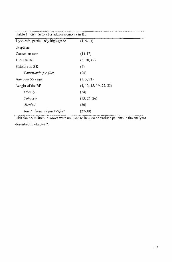
Table 1 Risk factors for adenocarcinoma in BE
Dysplasia, pmiiculariy high-grade
dysplasia
Caucasian men
Ulcer in BE
Stricture in BE
Longstanding reflux
Age over 55 years
Lenght of the BE
Obesity
Tobacco
Alcohol
Bile / duodenal juice reflux
(1,9-13)
(14-17)
(5,18,19)
(4)
(20)
(1,5,21)
(4,12,15,19,22,23)
(24)
(15,25,26)
(26)
(27-30)
Risk factors written in italics were not used to include or exclude patients in the analyses
described in chapter 2.
137
7.3 Methodological considerations and General interpretation of study results
In this thesis three types of studies are described, a retrospective cross-sectional study
combined with an analysis of the literature, a cross-sectional case-control study and a cross
sectional analytical study.
At the start of the retrospective cross-sectional study we established guidelines for
secondary prevention in patients known to have BE. Our first goal was to derive the risk factors
for adenocarcinoma in BE patients from the literature. There appear to be subtypes of BE which
do not progress or progress so slowly that even over a prolonged period they do not lead to
invasive cancers( 4). For example BE without dysplasia at the initial endoscopy generally does
not progress and therefore is not in need of surveillance( I, 9-12). A significant proportion of
patients with BE will die of other diseases before an adenocarcinoma developes in the BE and
becomes a clinical problem(5). Studies aimed at identirying risk factors for adenocarcinoma in
the oesophagus are limited, probably because they are hard to conduct and to analyse due to the
relatively small number of cases developing cancer in any single centre. The few we found are
given as references in table 1. If a factor was found to be a risk factor for adenocarcinoma
developing more frequently than 1 in 200 patient-years in BE in 2 or more series, we included it
in our calculation. Using the factors derived from the literature, we showed in this retrospective
study that most patient without dysplasia theoretically require no endoscopic surveillance and
that only a minority of patients are likely to benefit from an endoscopic surveillance
program(31 ).
In the 2 case-control studies described in this thesis, BE ascertainment was based on
consecutive patients diagnosed by endoscopy at the University Hospital Rotterdam. The control
population was derived fi'om 2 epidemiological studies in the general population (the Rotterdam
ERGO study and the RTVM MORGEN project). The studies were performed in 2 age categories
(see chapter 3 and 4). The ultimate purpose was to provide confirmation ofthe increased risk of
138
cardiovascular disease associated with reflux disease, including BE, to find clues as to the nature
of the cardiovascular disease and to recognize preventable risk factors. Cross-sectional case
control studies are patiicularly efficient in terms of both time and costs. The comparison between
diseased and non-diseased populations allows determination of features requiring further
investigation. However, due to the design, case-control studies are susceptible to bias, especially
when cases and controls are selected fi'om different populations.
Selectioll bias. Cases of BE and GERD in our study were selected fi'om hospital database
whereas controls were selected from the general population. We were patiicularly interested in
co-morbidity from coronary-heati disease in patients with BE. It can be postulated that co
morbidity is probably more frequent in hospital based cases than in the general population as a
whole. If selection bias is present it can lead to incorrect estimates of true associations, which
reduce the validity ofthe study. However, the cases used in this thesis were selected from
endoscopic database of which many patients were outpatients or patients referred from the
general practitioner making co-morbidity less likely. Also, to reduce this possible bias, we
matched for age, sex, ethnicity and the area where the controls were living. Thus, since controls
were chosen from the same source population from which cases were derived, it is not likely that
selection bias occurred due to an ill defined sampling fi'ame of controls. However, we cannot
completely exclude another possible factor in the two case control studies. Selection bias in a
case control study can occur when the response or determinants of response are different for
cases and controls. Individuals not responding to invitation, or unwilling to patiicipate in the
case control study can cause a bias. The non-response rate in the Barrett's study was 48.6%
compared to 33% in the Rotterdam ERGO study and 45% in the RTVM MORGEN project. Non
responders might be healthier subjects in the Barrett population, but the more unhealthy in the
control group or vice versa. Response in the Barrett's population was defined as the percentage
of patients who reacted to invitation and took pati in the study, out of all known patients
139
diagnosed with BE at the University hospital Rotterdam (18-80 years) in the period January 1992
until December 1996 who were known to be alive in the year 1999. We found 212 patients, of
whom 109 reacted to the invitation and took part in the study (51.4%). The reason for non
response in the Barrett's study is not known. However, as the invitation was to detect prevalence
of H. pylori and not coronary heart disease we were convinced that the invitation did not
selectively cause patients with coronary heart disease from the BE population to react and take
part in the study. In the Rotterdam ERGO study the percentage of individuals 55 years of age or
older, living in the suburb Omoord of Rotterdam and who took part in all parts of the study were
determinant of response, the same as for the RNM MORGEN project.
Information bias. The primary requirement for the validity of a case-control study is the
ability to obtain complete and accurate information on all participants. Because both cases and
controls were using somewhat different questionnaires to measure outcome in these 2 cross
sectional studies, misclassification of outcome or exposure could lead to information bias. Non
differentialmisclassification is a random misclassification, misclassification similar in cases and
controls. We selected comparable questions from the questiOlmaire. Only questions with the
same wording were used, so that we do not believe that this led to a significant bias. The only
questions used not having the same wording were about the first degree family member in
chapter 3, and these we interpreted with caution. Medication use was verified in both studies,
therefore we do not believe that this aspect could have been associated with a significant bias.
Confollndillg. An important factor that must be considered in discussing the results is
possible bias due to confounding. Confounding involves the possibility that the observed
association is due to the effect of differences between study groups other than the exposure
status. lmpOltant confounders in studies on the relation between potential determinants and
cardiovascular risk factors are age and gender. Old age and male gender are strong risk factor for
both Barrett's oesophagus and cardiovascular diseases. We controlled for the effect of age and
140
gender by stratification on risk factors for cardio-vascular diseases.
We performed 2 cross-sectional analytical studies (see chapter 5 and 6). In analytical
studies the investigator assembles groups of individuals to determine whether the risk of a
specific outcome parameter is different for individuals exposed or not exposed to a factor of
interest. The factors of interest were (I) prevalence of H. pylori infection in the GERD
population, particularly the BE population and the prevalence of CagA strain in the same groups
and (2) the effect of maintenance treatment with PPJ's in these groups. We used only our own
cases to conduct these studies. For the studies described in chapter 5 and 6 a single laboratory
technician measured by a single technique and during single session antibodies to H. pylori and
CagA in all our cases studied during the period of this thesis. This makes a selection bias
unlikely, although a selective non-response bias mentioned above can playa role in the BE
population. On the other hand in chapter 6, where we compared 2 groups of all individuals
serologically positive for H. pylori with each other with respect to exposure or not to
maintenance treatment with PPJ's we cannot be that certain. Information bias cannot be fully
excluded as we used a standard questionnaire about medication and duration of use ofa
particular drug. However, we paid patiiculair attention to the use of acid inhibitors in all
participants during interviews and when building the database. When there was any doubt on
whether or not the individal had used acid inhibitors, it was re-checked. There are, however, two
possible confounding factors in this study. First, there was an age difference between the groups.
The mean age ofthe PPI group was almost 4 years older than the control group. As age by itself,
especially in H pylori positive individuals, prediposes to atrophy this can cause confounding
bias. We tried to solve this by correcting for age-difference by stratification for age. Secondly, a
possible sampling error when biopsies are taken from the gastric mucosa can cause confounding.
Biopsies were taken in a standard fashion comparable to that recommended and used in other
studies designed to investigate effects of various exposures on the gastric mucosa. Atrophy and
141

intestinal metaplasia (1M) can be focal or multifocal and the less atrophy or 1M present the
greater the chance of sampling error.
142
7.4 Closing remarks and future perspectives
7.4.1 Endoscopic cancer surveillance for subgroups of known BE patients
At present many endoscopists advocate endoscopic surveillance for patients known to have BE,
with the intention of identifying progression in BE from metaplasia to HGD and early carcinoma
before local invasion and metastasis occur (1-3). However, our own theoretical study and a
recent observational study publishing final results of 10 year surveillance for BE patients failed
to show a benefit for the majority of these patients (4, 31). There are many possible explanation
for this failure (patients were too old, had concurrent diseases, were lost from follow-up etc.). At
present there is also still discussion about the timing oftreatment and whether an operation or
alternative intervention is indicated when histological progression is observed. There is also
uncertainty as to how treatment affects the course of the disease and the prognosis (13, 32).
Ablation therapies for intestinal metaplasia and dysplasia are at present only being performed as
clinical trials at special centers (33,34). The management of high-grade dysplasia (HGD)
remains controversial. Esophagectomy has been advocated if HGD or early adenocarcinoma is
found incidentaIIy or by surveillance (35, 36). Because ofthe risk of severe complications or
mortality, esophagectomy is not an acceptable treatment if only low-grade dysplasia is present.
This can be demonstrated by the course of 18 patients who were followed by surveillance and in
whom an indication for esophagectomy was found because of HGD or early adenocarcinoma.
Eight patients survived the operation, 3 died in the postoperative phase and 7 were in too poor a
condition to undergo an operation, refused an operation or already had metastatic disease when
the primary tumor was found during surveiIIance (1, 10,22, 37, 38). Thus, even years of
surveillance did not guarantee benefit.
FUlihermore it has recently been argued that an operation may not be indicated in HGD until
invasion is observed (32). Two studies offered their patients an alternative option for HGD in the
form of an intensive follow-up program instead of esophagectomy (32, 39). The results of these
143
studies provided meaningful information about the natural course ofHGD. Both studies used a
systematic biopsy protocol and intensive follow-up and only when invasive cancer was found
were the patients referred for operation. Leven et al published results of follow-up of 70 patients
with high-grade dysplasia or early carcinoma (39). Invasive cancer was found within 2 months
from the start of follow-up in 12 patients and in another 15 patients after a mean of 1.1 year. The
remaining 58 patients were followed for a mean period of2.5 years. In most of them (n=27) the
high-grade dysplasia persisted, but in 16 patients regression of dysplasia was observed. The other
study was very recently published. Schnell et al came to the conclusion that HGD without cancer
in BE follows a relatively benign course in majority of patients (32). They followed 79 HGD
patient without evidence of cancer when diagnosed. Cancer did not develop in 63 HGD patients
during the whole follow-up, but 12 developed cancer (16%) during a mean 7.3-years surveillance
period. Eleven of 12 patients did well after operation or ablation.
At present most patients who are diagnosed as having adenocarcinoma of the esophagus were
not previously known to have BE. This means that we are probably putting too much effOli and
money in to screening the small target group known to have BE for adenocarcinoma, without
there being a general agreement or accepted guidelines for screening ofthese patients to make
the surveillance more effective. It is probably good sense to offer surveillance only to patients
with a good health profile and high risk of developing BE carcinoma. Time, effOli and finance
could be reallocated to start looking at preventive measures for the 95% of patients who now
present with symptomatic adenocarcinoma by gaining insights into how we could detect these
patients at an earlier stage. The guidelines developed by us are given in figure 1. It is our hope
that by presenting this work-up protocol that surveillance programs for BE patients will be more
cost-effective.
144
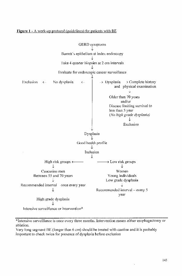
Figure 1 - A work-up protocol (guidelines) for patients with BE
GERD symptoms .J,.
Barrett's epithelium at index endoscopy .J,.
Take 4 qumier biopsies at 2 cm intervals .J,.
Evaluate for endoscopic cancer surveillance
Exclusion ~ No dysplasia
.J,.
.J,.
-+ Dysplasia -+ Complete history and physical examination
.J,.
Older than 70 years and/or
Disease limiting survival to less than 5 year (No high grade dysplasia)
.J,. Exclusion
Dysplasia .J,.
Good health profile
High risk groups +------.J,.
Caucasian men Between 55 and 70 years
.J,.
.J,. Inclusion
.J,.
------+ Low risk groups .J,.
Women Young individuals
Low grade dysplasia Recommended interval - once every year .J,.
.J,.
High grade dysplasia .J,.
Intensive surveillance or intervention*
Recommended interval - every 5 year
*Intensive surveillance is once every three months. Intervention means either esophagectomy or ablation. Very long segment BE (longer than 6 cm) should be treated with caution and it is probably impOliant to check twice for presence of dysplasia before exclusion
145
7.4.2 Population screening for subgroups in the general population
Recently, population screening limited to patients considered to have a high risk for BE
and associated adenocarcinoma has been considered (40). This might be the next step in
approaching the problem of the rising incidence of adenocarcinoma in the esophagus in the
Western countries.
Adenocarcinoma in BE has been gaining importance as a health problem in the developed
countries (14, 41). All major health problems are a reason to consider population screening.
However, although 95% of BE patients in the general population are at present undiscovered, as
yet BE does not fulfill all recommended criteria for population screening according to the
'Principles of early disease detection' as defined by World Health Organization (WHO)( 42).
The ability to treat the condition adequately when discovered is the most important criterion for
screening (42). At present BE patients are treated with acid inhibition or anti reflux surgery if
they have reflux symptoms (43-45). These therapies have little or no effect on the BE itself nor
on the development of adenocarcinoma. It would be great step forward if BE could be ablated
with minimally invasive treatment before adenocarcinoma has developed. Some ablative
therapies (photodynamic therapy, thermal techniques and endoscopic mucosal resection) have
been repOlied to be promising and are currently being evaluated for this purpose (33). Until these
therapies have been fully approved, screening of the general population for BE will not be
acceptable.
There are however fewer arguments against trying to find subgroups in the adult
population who are at highest risk of developing adenocarcinoma in the future but who at present
are not endoscopically identified as a risk group. Here are a few suggestions:
146
1) Population screening for Caucasial1 mel1 aged 55-70 years of age. The overall cancer risk
in patients with BE is lower than previously estimated and most recent larger studies with
longer follow-up have tended to report rates ofO.5%/year or lower (46). As Caucasian men
over 60 years old are the major risk group for adenocarcinoma in BE, there is evidence in
favour of starting population-based screening of Caucasian men between the ages of 55 t070
years (see table 2)
Table 2 Caucasian men at ages between 55 and 70 years are patiicularly at risk of
BE. The same age range holds true for adenocarcinoma ofthe esophagus.
• In most studies the majority of patients with BE are Caucasian men (women:men
ratio range between 1:3 and 1 :8)
• In most studies the greater majority of patients with adenocarcinoma in the
esophagus are Caucasian men( 14-17)
• The mean age at which BE was diagnosed was 63 years, although with very
broad age-range(21)
• The mean age at which adenocarcinoma in BE was diagnosed was 64 years
(range30-89) (21)
• The mean age at which adenocarcinoma was diagnosed at the University
Hospital Rotterdam in BE was 67 years (range 52-81 years)(5)
147
2) Primary prevention il1 tliejorl1l Ojil1st1'llctiol1. Another possibility could be primary
prevention in form of prophylactic instruction on a population basis about reflux symptoms
and possible consequences. Population based studies on reflux symptoms have shown that
the prevalence of reflux complaint in the general population is high. About half of the
western population suffers ii'om heartburn at least once a year (47), about 20% ofthe white
population ofthe USA experiences heaIiburn weekly (48-51). Symptoms of reflux are
considered to be a fairly accurate indicator for GERD (52), although only a small minority
consults a physician. It has been suggested that as adenocarcinoma of the esophagus does not
invariably develop from BE (53) and as a history of GERD symptoms seems to be a risk
factor for adenocarcinoma, it might be advisable to give prophylactic treatment and offer
endoscopic screening to patients with symptomatic reflux (20). However, this would lead to
an immense workload for the endoscopy units, as at present we see only small fraction of the
risk group. GERD is diagnosed in 10% of all upper gastrointestinal endoscopies performed,
BE patients represent less than 1% and patients with adenocarcinoma of the esophagus less
than 0.1 %. These last two groups are currently the targets of research. Information for the
public regarding GERD symptoms and its possible consequences might bring a patient at risk
of developing adenocarcinoma under medical attention sooner. However such a public
information program should only be undeliaken if sufficient endoscopic facilities can be
provided. In addition it should be pointed out that not all patients with adenocarcinoma of the
esophagus give a clear history ofGERD-related complaints (see for example Menke
Pluymers and Bytzer)(15, 47). An index endoscopy might be used to select the paIiicular
patient for inclusion or exclusion in endoscopic cancer surveillance in the same way as
shown for known BE above. If endoscopic cancer surveillance is done only in selective risk
groups in both the BE population and the GERD population as a whole, workload might be
reduced to feasible propOliions.
148
3) Not emdicatillg Helicobacterpylori illfectioll, particlllar~l' tlte CagA positive stmill, ill
GERD patiellts, as primary prevelltioll. The influence of H pylori infection on GERD has
in recent years received great attention. Clustering of observations demonstrates that the
greater the extent to which corpus gastritis is present in infected individuals, the greater the
protective effect against GERD is likely to be (54). This is related to how well the gastritis
lowers the acid load. Nearly all isolates from patients with peptic ulcers, atrophic gastritis
and gastric cancer are CagA positive (55). However, patients with duodenal ulcer disease do
not have low acid output(56), while patients with gastric atrophy do(57). There is evidence
that CagA phenotype of H pylori has a protective role against the development of BE (6, 58)
and esophageal adenocarcinoma. It has been shown that PPJ's are more effective in the
presence of H pylori (59). Based on these findings it can be postulated that we should not
eradicate H pylori, paIiicularly if it is CagA positive. On the other hand, concern has been
voiced that treating reflux symptoms with PPl's might worsen H. pylori gastritis (7) and
although this hypothesis is not proven (60) a current consensus advises eradication of H.
pylori infection in all positive individuals who are to be put on a maintenance treatment with
PPI's (8). This leads to a dilemma which is not yet fully solved. At present we do not have
enough evidence to not eradicate the H. pylori infection, even though the patient might
benefit by less GERD complaints and be protected on the long-run against the complications
ofGERD. At present it is probably better to follow the recommendations of the Maasticht
consensus and advise eradication of H pylori if maintenance treatment with PPJ's is to be
given to H pylori positive individuals. Our results in chapter 5 support the hypothesis that
CagA positive H pylori gastritis protects against BE development, but our data also suppOli
that concept the CagA phenotype might lead to atrophy developing more rapidly when the
patient is on maintenance treatment with PPJ's (see chapter 6).
149
7.5 Targets for future research on BE include
1. Finding biological markers that identijj' patients at higlter risk of progressing to cancer. A
better understanding of the pathophysiology of Barrett's adenocarcinoma based on
expression of novel biomarkers and use of such biomarkers in examination of biopsies or
endoscopic examination may help to identify patients at increased risk for malignant
transformation. If the genetic modifications, which lead to adenocarcinoma, are clarified in
the future this may provide an approach to the prevention of adenocarcinoma. A major
problem in current practice is the large interobserver variation in classifiying dysplasia as
low-grade, high-grade or carcinoma -in-situ. New biomarkers may prove useful in reducing
this variation.
2. Defining appropriate surveil/ance intervals and appropriate risk groups._At present there is
little agreement about the ideal frequency of endoscopic cancer surveillance in patients with
BE nor about who would benefit from such a program. Surveillance for evelyone with BE is
not cost effective and in most cases surveillance would not provide health benefits. Clinical
aspects and histological features can now be used to define patients with BE who are likely
to benefit from endoscopic cancer surveillance, as has been discussed in this thesis. The
development of biomarkers of early stages of carcinogenesis in combination with
surveillance has the potential to allow identification of patients at high risk of neoplastic
change. Large numbers of patients need to be followed prospectively to learn more about the
natural history of BE and especially the rate at which the disease progresses through the
various histological stages towards adenocarcinoma.
3. Defining the cancel' risk am/ the appropriate management of patients with short segment
Barrett's esophagus. Even though short segment BE is very common and possible precursor
of every cardia carcinoma, the exact cancer risk is not known. Short segment of BE is not
150
seen as an indication for endoscopic cancer surveillance. However, if we are to find a way to
lower the incidence of adenocarcinoma of the esophagus and cardia we need to discover the
cancer risk of patients with short segment BE and define strategies for prevention, treatment
and surveillance.
4. Understanding the natural histOlJ' of dysplasia and comparing altel'llatives for the
management of high-grade dysplasia. The question whether the presence of any degree of
dysplasia is an indication for close clinical follow-up regardless of the extent of BE needs to
be answered. Is there a form of dysplasia that does not progress and how can we recognise it?
In this regard high-grade dysplasia is very impOliant, particularly with respect to treatment
strategy. When is it safe to wait, when can we use ablative technic and when do we need to
operate?
5. Studying whether ablation can delay or prevent progression to dysplasia and
adenocarcinoma. We have to continue to develop various ablation therapies in Barrett's
patients to approved standards to be able to use one or more ofthese techniques in clinical
practice.
These questions can only be answered by large scale observational studies and well-designed,
controlled interventional trials which compare different strategies.
151
7.6 References
1. Hameeteman W, Tytgat GN, HouthoffHJ, van den Tweel JG. Barrett's esophagus: development of dysplasia and adenocarcinoma. Gastroenterology 1989;96(5 Pt 1): 1249-56.
2. Streitz JM, JI'., Andrews CW, Jr., Ellis FH, Jr. Endoscopic surveillance ofBalTett's esophagus. Does it help? J Thorac Cardiovasc Surg 1993;105(3):383-7; discussion 387-8.
3. van Sandick JW, van Lanschot JJ, Kuiken BW, Tytgat GN, Offerhaus GJ, Obertop H. Impact of endoscopic biopsy surveillance of Barrett's oesophagus on pathological stage and clinical outcome of Barrett's carcinoma. Gut 1998;43(2):216-22.
4. Macdonald CE, Wicks AC, Playford RJ. Final results from 10 year cohort of patients undergoing surveillance for Barrett's oesophagus: observational study. Bmj 2000;321(7271): 1252-5.
5. van del' Burgh A, Dees J, Hop WC, van Blankenstein M. Oesophageal cancer is an uncommon cause of death in patients with Barrett's oesophagus. Gut 1996;39(1):5-8.
6. Loffeld RJ, Werdmuller BF, Kuster JG, Perez-Perez GI, Blaser MJ, Kuipers EJ. Colonization with cagA-positive Helicobacter pylori strains inversely associated with reflux esophagitis and Barrett's esophagus. Digestion 2000;62(2-3):95-9.
7. Kuipers EJ, Lundell L, Klinkenberg-Knol EC, Havu N, Festen HP, Liedman B, et al. Atrophic gastritis and Helicobacter pylori infection in patients with reflux esophagitis treated with omeprazole or fundoplication. N Engl J Med 1996;334(16): I 0 18-22.
8. Malfetiheiner P. The Maastricht recommendations and their impact on general practice. Eur J Gastroenterol Hepatol 1999; II Suppl 2:S63-7; discussion S73.
9. Hamilton SR, Smith RR. The relationship between columnar epithelial dysplasia and invasive adenocarcinoma arising in Barrett's esophagus. Am J Clin PathoI1987;87(3):301-12.
10. Miros M, Kerlin P, Walker N. Only patients with dysplasia progress to adenocarcinoma in Barrett's oesophagus. Gut 1991;32(12):1441-6.
11. Cameron AJ, Carpenter HA. Barrett's esophagus, high-grade dysplasia, and early adenocarcinoma: a pathological study. Am J GastroenteroI1997;92(4):586-91.
12. Weston AP, Badr AS, Hassanein RS. Prospective multivariate analysis of clinical, endoscopic, and histological factors predictive of the development of Barrett's multifocal high-grade dysplasia or adenocarcinoma. Am J Gastroenterol 1999;94(12):3413-9.
13. ButtaI' NS, Wang Kl(, Sebo TJ, Riehle DM, Krishnadath KK, Lutzke LS, et al. Extent of high-grade dysplasia in barrett's esophagus correlates with risk of adenocarcinoma. Gastroenterology 2001; 120(7): 1630-9.
14. Blot WJ, Devesa SS, Kneller RW, Fraumeni IF, Jr. Rising incidence of adenocarcinoma of the esophagus and gastric cardia. Jama 1991;265(10):1287-9.
15. Menke-Pluymers MB, Hop WC, Dees J, van Blankenstein M, Tilanus HW. Risk factors for the development of an adenocarcinoma in columnar-lined (Barrett) esophagus. The Rotterdam Esophageal Tumor Study Group. Cancer 1993;72(4):1155-8.
16. Wright TA, Gray MR, Morris AI, Gilmore IT, Ellis A, Sm31i HL, et al. Cost effectiveness of detecting Barrett's cancer. Gut 1996;39(4):574-9.
17. Caygill CP, Reed PI, Johnston BJ, Hill MJ, Ali MH, Levi S. A single centre's 20 years' experience of columnar-lined (Barrett's) oesophagus diagnosis. Eur J Gastroenterol Hepatol 1999;11(12):1355-8.
18. Naef AP, Savary M, Ozzello L. Columnar-lined lower esophagus: an acquired lesion with malignant predisposition. Report on 140 cases of Barrett's esophagus with 12 adenocarcinomas. J Thorac Cardiovasc Surg 1975;70(5):826-35.
19. Van del' Veen AH, Dees J, Blankensteijn JD, Van Blankenstein M. Adenocarcinoma in Barrett's oesophagus: an overrated risk. Gut 1989;30(1):14-8.
152
20. Lagergren J, Bergstrom R Lindgren A, Nyren O. Symptomatic gastroesophageal reflux as a risk factor for esophageal adenocarcinoma. N Engl J Med 1999;340(11 ):825-31.
21. Cameron AJ, Lomboy CT. Barrett's esophagus: age, prevalence, and extent of columnar epithelium. Gastroenterology 1992; 103(4): 1241-5.
22. Tftikhar SY, James PD, Steele RJ, Hardcastle JD, Atkinson M. Length of Barrett's oesophagus: an important factor in the development of dysplasia and adenocarcinoma. Gut 1992;33(9): 1155-8.
23. Rudolph RE, Vaughan TL, Storer BE, Haggitt RC, Rabinovitch PS, Levine DS, et al. Effect of segment length on risk for neoplastic progression in patients with Barrett esophagus. Ann Intel'l1 Med 2000; 132(8):612-20.
24. Brown LM, Swanson CA, Gridley G, Swanson GM, Schoenberg JB, Greenberg RS, et al. Adenocarcinoma of the esophagus: role of obesity and diet. J Nat! Cancer Inst 1995 ;87(2): 1 04-9.
25. Spechler SJ, Robbins AH, Rubins HB, Vincent ME, Heeren T, Doos WG, et al. Adenocarcinoma and Barrett's esophagus. An overrated risk? Gastroenterology 1984;87(4):927-33.
26. Gray MR, Donnelly RJ, KingsnOlih AN. The role of smoking and alcohol in metaplasia and cancer risk in Barrett's columnar lined oesophagus. Gut 1993;34(6):727-31.
27. Attwood SE, DeMeester TR, Bremner CG, Barlow AP, Hinder RA. Alkaline gastroesophageal reflux: implications in the development of complications in Barrett's columnar-lined lower esophagus. Surgery 1989; I 06(4):764-70.
28. Marshall RE, Anggiansah A, Owen WJ. Bile in the oesophagus: clinical relevance and ambulatory detection. Br J Surg 1997;84(1):21-8.
29. Kaur BS, Ouatu-Lascar R, Omary MB, Triadafilopoulos G. Bile salts induce or blunt cell proliferation in Barrett's esophagus in an acid-dependent fashion. Am J Physiol Gastrointest Liver Physiol 2000;278( 6):G 1000-9.
30. Katz PO. Review aJiicle: the role of non-acid reflux in gastro-oesophageal reflux disease. Aliment Pharmacol Ther 2000; 14(12): 1539-51.
31. Gudlaugsdottir S, van Blankenstein M, Dees J, Wilson JH. A majority of patients with Barrett's oesophagus are unlikely to benefit from endoscopic cancer surveillance. Eur J Gastroenterol HepatoI2001;13(6):639-45.
32. Schnell TG, Sontag SJ, Chejfec G, Aranha G, Metz A, O'Connell S, et al. Long-term nonsurgical management of barrett's esophagus with high-grade dysplasia. Gastroenterology 2001;120(7): 1607-19.
33. Pech 0, Gossner L, May A, Ell C. Management of Barrett's oesophagus, dysplasia and early adenocarcinoma. Baillieres Best Pract Res Clin GastroenteroI2001;15(2):267-84.
34. Sharma P. Current status of ablative therapies in esophageal disorders. Curl' Gastroenterol Rep 2001;3(3):219-24.
35. Pera M, Trastek VF, Carpenter HA, Allen MS, Deschamps C, Pairolero Pc. Barrett's esophagus with high-grade dysplasia: an indication for esophagectomy? Ann Thorac Surg 1992;54(2): 199-204.
36. Younes Z, Duncan MD, Harmon JW. Management of Barrett's esophagus. Can J Gastroenterol 2000; 14 Suppl D:35D-43D.
37. Drewitz DJ, Sampliner RE, Garewal HS. The incidence of adenocarcinoma in Barrett's esophagus: a prospective study of 170 patients followed 4.8 years. Am J Gastroenterol 1997;92(2):212-5.
38. O'Connor JB, Falk GW, Richter JE. The incidence of adenocarcinoma and dysplasia in Barrett's esophagus: report on the Cleveland Clinic Barrett's Esophagus Registry. Am J Gastroenterol 1999;94(8):2037-42.
153
39. Levine DS, Haggitt RC, Blount PL, Rabinovitch PS, Rusch VW, Reid BJ. An endoscopic biopsy protocol can differentiate high-grade dysplasia from early adenocarcinoma in Barrett's esophagus. Gastroenterology 1993; 1 05(1 ):40-50.
40. Lieberman DA, Sampliner RE. How far to go? Screening and surveillance in Barrett's esophagus. Am J Manag Care 2001;7(1 Suppl):SI9-26.
41. Blot WJ, Devesa SS, Fraumeni JF, Jr. Continuing climb in rates of esophageal adenocarcinoma: an update. Jama 1993;270(11):1320.
42. Wilson JM, Jungner YG. [Principles and practice of mass screening for disease]. Bol Oficina Sanit Panam 1968;65(4):281-393.
43. Richter JE. Long-term management of gastroesophageal reflux disease and its complications. Am J Gastroenterol 1997;92(4 Suppl):30S-34S; discussion 34S-35S.
44. Brel11l1er CG, Bremner RM. Barrett's esophagus. Surg Clin NOlth Am 1997;77(5):1115-37. 45. Klinkenberg-Knol EC, Nelis F, Dent J, Snel P, Mitchell B, Prichard P, et al. Long-term
omeprazole treatment in resistant gastroesophageal reflux disease: efficacy, safety, and influence on gastric mucosa. Gastroenterology 2000; 118(4):661-9.
46. Shaheen NJ, Crosby MA, Bozymski EM, Sandler RS. Is there publication bias in the reporting of cancer risk in Barrett's esophagus? Gastroenterology 2000; I 19(2):333-8.
47. Bytzer P. On-demand therapy for gastro-oesophageal reflux disease. Eur J Gastroenterol Hepato12001;13 Suppll:SI9-22.
48. Nebel OT, Fornes MF, Castell DO. Symptomatic gastroesophageal reflux: incidence and precipitating factors. Am J Dig Dis 1976;2 I (I 1):953-6.
49. Thompson WG, Heaton KW. Heartburn and globus in apparently healthy people. Can Med Assoc J 1982; 126(1 ):46-8.
50. Feitelberg SP, Hogan DL, Koss MA, Isenberg JI. pH threshold for human duodenal bicarbonate secretion and diffusion of C02. Gastroenterology 1992; 1 02( 4 Pt 1): 1252-8.
51. Isolauri J, Laippala P. Prevalence of symptoms suggestive of gastro-oesophageal reflux disease in an adult population. Ann Med 1995;27(1):67-70.
52. Klauser AG, Schindlbeck NE, Muller-Lissner SA. Symptoms in gastro-oesophageal reflux disease. Lancet 1990;335(8683):205-8.
53. Cameron AJ. Epidemiology of columnar-lined esophagus and adenocarcinoma. Gastroenterol Clin North Am 1997;26(3):487-94.
54. EI-Serag HE, Sonnenberg A, Jamal MM, Inadomi JM, Crooks LA, Feddersen RM. Corpus gastritis is protective against reflux oesophagitis. Gut 1999;45(2): 18 I -5.
55. Kuipers EJ. Helicobacter pylori and the risk and management of associated diseases: gastritis, ulcer disease, atrophic gastritis and gastric cancer. Aliment Phannacol Ther 1997;11 Suppll:71-88.
56. el-Omar EM, Penman rD, Ardill JE, Chittajallu RS, Howie C, McColl KE. Helicobacter pylori infection and abnormalities of acid secretion in patients with duodenal ulcer disease. Gastroenterology 1995; 1 09(3):681-91.
57. EI-Omar EM, Oien K, EI-Nujumi A, Gillen D, Wirz A, Dahill S, et al. Helicobacter pylori infection and chronic gastric acid hyposecretion. Gastroenterology 1997;113(1):15-24.
58. Rugge M, Russo V, Busatto G, Genta RM, Di Mario F, Farinati F, et al. The phenotype of gastric mucosa coexisting with Barrett's oesophagus. J Clin PathoI2001;54(6):456-60.
59. Labenz J, Tillenburg B, Peitz U, ldstrom JP, Verdu EF, Stolte M, et al. Helicobacter pylori augments the pH-increasing effect of omeprazole in patients with duodenal ulcer. Gastroenterology 1996; I 10(3):725-32.
60. Lundell L, Miettinen P, Myrvold HE, Pedersen SA, Thor K, Andersson A, et al. Lack of effect of acid suppression therapy on gastric atrophy. Nordic Gerd Study Group. Gastroenterology 1999; I 17(2):3 19-26.
154

Chapter 8 Summary - Samenvatting
155

156
8.1 Summary
The clinical research reported in this thesis was performed from September 1997 until August
1999. The research was based both on older theories and on recently published findings and
concepts, which can be outlined as following;
1. The incidence of adenocarcinomas of the esophagus and gastric cardia has been rising
steadily since 1970. This rising incidence rate is an important stimulus for investigation
into the causes of these poorly understood types of cancer.
2. The only known risk factor for adenocarcinoma in the esophagus is Barrett's esophagus
with biopsy proven intestinal metaplasia.
3. Many centers have advocated long-term endoscopic and histologic follow-up
(surveillance) programs for patients with Barrett's esophagus, with the aim of detecting
high grade dysplasia or cancer at a curable stage.
4. So far, observational and follow-up studies have reported disappointing results for
surveillance. Most adenocarcinomas in the esophagus are diagnosed in patients not
known to have Barrett's epithelium and 50% of patients known to have Barrett's
epithelium die from an unrelated medical disorder at an earlier than expected for an age
and sex-matched normal population. The most likely reason for death has been reported
to be cardiovascular disease with pulmonary causes in second place.
5. Reflux of acid from the stomach into the esophagus is presumed to be the most important
factor in the pathogenesis of Barrett's epithelium.
6. Acid inhibitors are the most widely used treatment for gastroesophageal reflux disease
and Barrett's esophagus. Since 1985 proton pomp inhibitors have been on the market,
and have proven efficacy in controlling symptoms and healing esophagitis.
7. Since 1995 concerns have been expressed that the use of proton pump inhibitors in
individuals infected with Helicobacterpylol'i can speed up atrophy of the gastric mucosa
157

and possible increase the chance of gastric malignancy. A consensus report advised the
eradication of Helicobactel'pylol'i in infected individuals if they were to be given proton
pump inhibitors as a maintenance treatment.
Chapter 1
In chapter 1 an overview is given of publications on Barrett's oesophagus, its causes and
complications beginning with a brief historical survey and the most likely reasons for its
popularity as a field of investigation. The present day definition of Barrett's disease, its induction
and rate of development and factors which playa role in the pathogenesis of complicated and
uncomplicated reflux disease are reviewed. The association between Barrett's disease and
adenocarcinoma of the esophagus and what is known of the natural history of Barrett's disease is
described.
The interaction between Helicobacter pylori and gastroesophageal reflux disease is summarised.
The prevalence of Helicobacter pylori in this specific subgroup of the population, the impOliance
of the virulence factors of the Helicobacter pylori for effects on the gastric mucosa and the
influence of Helicobacter pylori on gastric acid production are reviewed. Concerns about atrophy
in Helicobacter pylori gastritis under maintenance treatment with proton pump inhibitors are
emphasised. Finally, arguments for and against endoscopic cancer surveillance in Barrett's
population are discussed. With regard to surveillance it is important to see the patient as a whole,
paIiicularly at the initial visit, as this is generally agreed to be the best moment to weigh the risk
of esophageal cancer against the likelihood of having other diseases which impair survival. The
observation that the Barrett's population has a mean age of death which is lower than that of an
age and sex matched normal population is mentioned, with cardiovascular diseases as the most
frequent cause of death. This chapter ends with a summary of the aims of this doctoral thesis.
158
The main aim of the thesis was:
To obtain a better insight into recognising those Barrett's patients who are particularly at
risk of developing esophageal adenocarcinoma and identification of those Barrett's
patient particularly at risk of having or getting cardiovascular disease.
Further elucidation of the interaction of Helicobacter pylori with gastroesophageal reflux
disease, especially Barrett's esophagus, was also one of the aims of the thesis. This
included studying the prevalence ofHelicobacter pylori infection and the risk of
maintenance treatment with proton pump inhibitors in specific subgroups.
Chapter 2
The propoliion of healthy Barrett's patients, who because of strong risk factors for
adenocarcinoma in the esophagus in the near future would benefit fi'om endoscopic cancer
surveillance, as a propOliion of the total population at present diagnosed as having Barrett
esophagus, was determined. The literature on factors for or against endoscopic cancer
surveillance is reviewed; the factors include factors appear to protect against adenocarcinoma in
the esophagus, factors overriding that protection and necessitating endoscopic surveillance and
other factors like illness, frailness or old age which are strong arguments against effective
endoscopic cancer surveillance. Each risk factor was evaluated with respect to the incidence-risk
of developing adenocarcinoma in the esophagus if that factor was present. If at least one cancer
occurs in 200 patients with that factor followed for one year, then patients with that factor were
classified a potentially deserving surveillance. If the risk was lower then it was regarded as being
too small or the patient was to old or to frail to benefit fi'om surveillance, then they were
classified as unlikely to benefit from a cancer surveillance program. We found that, of the
patients known to have Barrett esophagus in the University Hospital Rotterdam, only 15.5%
patients evaluated in this fashion would benefit from endoscopic cancer surveillance. If every
159
patient with Barrett's esophagus were to be included in a surveillance program, then the
workload would be too large for the health services and in most cases a burden rather than a
benefit for the patient. Guidelines are given in this chapter for the selection of patients with
Barrett's esophagus who are likely to benefit from endoscopic cancer surveillance.
Chapter 3
A COhOlt of patients with Barrett's esophagus aged 55-80 years were compared with sex and age
matching normal population with respect to the prevalence of myocardial infarction and related
risk factors (smoking, hypeltension, treatment for hypeltension, family history of cardiovascular
diseases in a first degree relative, measurement of blood pressure and serum cholesterol
spectrum).
The results show that the Barrett's population with upper gastrointestinal complaints are more
likely to have a histOlY of myocardial infarction and hypertension and are more often treated for
hypertension than the normal population. Obesity was more frequently seen in the Barrett's
population, which may in pmt explain the predisposition to hypeltension. No difference was
found in the history of smoking or present smoking habits between the groups. A new carefully
designed prospective study is needed to confirm these results. If the results are true, it implies
that these factors should be taken into account when designing prospective studies or selecting
patients for surveillance programs.
160
Chapter 4
Three groups were investigated in the age categOlY 20 - 59.9 years; Barrett's patients, patients
with endoscopically confirmed reflux esophagitis and patients with normal gastroscopic findings.
Each group was compared to the age and sex matched normal population with respect to risk
factors for coronary hemi disease (smoking, histOlY of hypertension and treatment for
hypeliension; blood pressUl'e and sel'llm cholesterol spectra were measured).
The results shows that patients diagnosed because of upper or complaints with Barrett's disease
or reflux esophagitis are more likely to have hypeliension, be treated for hypertension and to
have higher cholesterol than the normal population. This was not seen in the categOlY with a
normal gastroscopy. There was no difference in the history of smoking or present smoking habits
between the groups. A new carefully designed prospective study is again needed to confirm these
results. Ifthe results are true it implies that these factors should be taken into account when
designing prospective studies or selecting patients in this age category for sUl'veillance.
Chapter 5
Two study COhOlis (Barrett's patients and patients with endoscopically proven reflux esophagitis)
were compared to a reference population with normal endoscopical features. The total group
numbered 224 individuals. A blood sample was taken from evelyone to measure IgO antibodies
against Helicobacter pylori and the cytotoxin-associated protein A (CagA).
The results show that patients with endoscopically confirmed reflux esophagitis or Barrett's
esophagus have a lower prevalence of Helicobacter pylori compared to patients without
endoscopic abnormalities. Patients with Barrett's esophagus have a significantly lower
prevalence of colonisation with CagA Helicobacter pylori strain than the reference population.
161
The results suppOli the hypothesis that CagA positive Helicobacter pylori strains can protect
against Barrett's mucosa forming in the esophagus.
Chapter 6
Seventeen Helicobacter pylori positive patients on maintenance treatment with proton pump
inhibitors (mean; 8 years) were compared with 17 Helicobacter pylori positive patients not on
these medications. All patients underwent gastroscopy with histological sampling from the
stomach (antrum, angulus, corpus) for classification of gastritis by means of the updated Sidney
classification. The endoscopic diagnosis made in the patients were one ofthe following:
Barrett's esophagus, reflux esophagitis 01' without endoscopic abnormalities.
The results demonstrated that proton pump acid inhibitors are associated with significantly lower
gastritis activity in the gastric antrum and lower density of Helicobacter pylori regardless of the
CagA status. However, atrophy was more often detected in the gastric antrum of CagA positive
individuals on maintenance treatment with proton pump inhibitors. Intestinal metaplasia was
seen significantly more often in CagA positive individuals on maintenance treatment with proton
pump inhibitors.
The results support the hypothesis that maintenance treatment with proton pump inhibitors in
CagA positive individuals hastens the development of atrophy in the antrum. An opposite effect
was observed in CagA negative individuals. No difference was seen in the gastric corpus
between the groups.
162
Chapter 7
Conclusions.
Recommendations are made for subsequent studies. Five broad areas of investigation are
proposed:
1. Studies to find new, predictive biomarkers for progression towards a carcinoma.
Dysplasia, which is a histological finding, has proven to be too subjective and variable a
parameter for use in individuals.
2. Studies to determine the ideal interval for surveillance endoscopy, and to improve the
clinical criteria to select patients who have an increased risk of developing a carcinoma.
3. Studies to define the risk ofshOli-segment Barrett's esophagus.
4. Investigation of the natural history of dysplasia, and, in combination with these studies,
investigation of different strategies for high grade dysplasia.
5. Studies to answer the question whether ablation therapy (removal or destruction of
Barrett's mucosa) is capable of reducing the rate of progression to, or eliminating the risk
of carcinoma.
It is clear that all these proposals for further studies will require both large numbers of patients
and many years offoIlow-up.
163

164

8.2 Samenvatting
In september 1997 werd begonnen met het onderzoek naar enkele klinische aspecten van Barrett
oesophagus, beschreven in dit proefschrift. Barrett oesophagus is een aandoening van de distale
slokdarm, waarbij het normaal aanwezig plaveiselepitheel is vervangen door metaplastisch
cilinderepitheel. Ret onderzoek is gebaseerd op concepten uit zowel oudere als recente
publicaties over deze aandoening. Deze concepten kunnen als voIgt worden samengevat:
I. De incidentie van adenocarcinomen van slokdarm en cardiagebied van de maag neemt sinds
1970 duidelijk toe in de Westerse landen. Deze toename in incidentie heeft geleid tot een
stijgend aantal publicaties over klinisch epidemiologisch onderzoek en pathogenetisch
onderzoek naar de oorzaken van deze carcinomen.
2. De enig bekende risico factor voor een adenocarcinoom van de slokdarm is het Barrett
slijmvlies met intestinale metaplasie.
3. Vele centra bevelen aan levenslange endoscopisch en histologisch controle voor patienten
met Barrett slokdarm, om vroegtijdig hooggradig dysplasie of resect abe Ie kanker op te
sporen.
4. Na-controle van patienten met Barrett slokdarm he eft echter tot dusver onvoldoende steun
geleverd voor het invoeren van surveillance programma's. De meeste adenocarcinomen van
de slokdarl11worden gediagnosticeerd bij patienten die niet bekend zijn met Barrett slijmvlies
en 50% van patienten die wei bekend zijn met Barrett slijmvlies sterven op een jonger dan
verwachte leeftijd aan een oorzaak niet gerelateerd aan Barrett slijmvlies. De voornaamste
doodsoorzaak bij patienten bekend met Barrett slijmvlies is hart- en vaatziekten, gevolgd
door longziekten.
5. Reflux van maaginhoud wordt verondersteld de belangrijkste factor te zijn in het ontstaan
van Barrett slijmvlies.
165

6. Tegenwoordig zijn remmel'S van zuurproductie de meest gebruikte medicamenteuze
behandeling voor gastro-oesofageale reflux en Barrett slijmvlies. Na de introductie van
protonpomp remmel'S in 1985 is deze groep van geneesmiddelen zeer populair geworden,
voornamelijk omdat zij zeer effectiefzijn gebleken in het bestrijden van klachten en in het
genezen van de oesophagitis.
7. Na een eerste publicatie in 1995, maakt men zorgen dat langdurige behandeling met
protonpomp remmers, bij patienten met Helicobacter pylori infecties, het ontwikkelen van
atrofie en dysplasie van het maagslijmvlies kan bevorderen. Atrofie en dysplasie gaan
gepaard met een toegenomen kans op het ontstaan van een maagcarcinoom. Een consensus
rapport heeft geadviseerd Helicobacter pylori infecties te behandelen bij aile patienten die
gedurende langere tijd protonpomp remmel's zullen gaan gebruiken.
Hoofdstuk 1
In hoofdstuk 1 wordt de literatuur over de oorzaken en complicaties van Barrett slijmvlies
samengevat. Onder andere wordt ingegaan op de definities van Barrett slokdarm, de ontstaan van
Barrett slijmvlies en factoren betrokken bij de pathogenese van zowel gecompliceerde en
ongecompliceerde reflux ziekten. Het verband tussen Barrett slokdarm en adenocarcinoom wordt
beschreven.
De interacties tussen Helicobacter pylori en gastro-oesofageale reflux ziekten worden
samengevat, met aandacht voor de prevalentie van Helicobacter pylori in patienten met gastro
oesofageale reflux of Barrett slokdarm. Het belang van virulentiekenmerken van Helicobacter
pylori op secretie van maagzuur wordt benadrukt. De zorgen over een versneld pl'Ogressie naar
atrofie bij patienten met Helicobacter pylori gastritis, tijdens een onderhoudsbehandeling met
protonpomp remmel's worden besproken.
166

De argumenten voor en tegen endoscopische surveillance op kanker in patienten met Barrett
slokdarm worden toegelicht. Het Iijkt belangrijk aile aspect en van de gezondheid van de patient
te evalueren tijdens het eerste bezoek, omdat dit waarschijnlijk het beste moment is om het risico
van het ontstaan van slokdarmkanker afte wegen tegen de kans op andere ziekten die de
gezondheid of levensverwachting kunnen bei'nvloeden. De bevinding, dat personenmet Barrett
slokdarm een kOJtere levensverwachting hebben dan personen uit de bevolking met dezelfde
leeftijd en geslacht, wordt genoemd. Hmi- en vaatziekte blijkt de meest fi'equente doodsoorzaak
te zijn.
Samenvatting van de doelstelling van het proefschrift:
Doel van het onderzoek was:
• Ret identificeren van risicofactoren voor, en beschermingsfactoren tegen, ontwikkeling van
een adenocarcinoom in BE.
• Het opstellen van praktische richtlijnen voor selectie van patienten met BE die eventueel in
aanmerking zouden kunnen komen voor surveillance.
• Het opsporen van risicofactoren bij patienten met BE, die een verklaring zouden kunnen
leveren voor de oversterfte aan hart-en vaatziekten bij deze patienten.
• am de prevalentie van risicofactoren voor hart-en vaatziekten bij patienten met BE te
vergelijken met die bij patienten met andere vormen van GERD ofbovenbuikklachten.
• am de interacties tussen Helicobacter pylori en GERO te exploreren en de volgende vragen
te beantwoorden:
Is de prevalentie van Helicobacter pylori infectie bij patienten met BE of andere
vormen van GERO anders dan bij patienten met non-ulcer dyspepsie?
Is er een verschil in prevalentie van de virulente vorm (CagA positieve) van
Helicobacter pylori tussen patienten met verschillende oorzaken van
bovenbuikklachten?
167

Wat zijn de lange termijn effecten van proton-pomp remmers op het
maagslijmvlies bij patienten met een Helicobacter pylori infectie, en he eft de
CagA status van de bacterie invloed op deze effecten?
Hoofdstuk 2
Hoofdstuk 2 beschrijft een evaluatie van het percentage patienten met Barrett slokdarm, die
waarschijnlijk baat zullen hebben van endoscopische kanker surveillance. De literatuur over
factoren, die van invloed zouden kunnen zijn op een vool'stel voor surveillance, wordt eerst
samengevat. Er zijn factoren gei'dentificeerd die gepaard gaan met een verminderde kans op
adenocarcinoom van de slokdarm, factoren die gepaard gaan met een verhoogd risico (en dus
mogelijk een indicatie zouden kunnen vormen voor endoscopische surveillance) en niet aan
Barrett slokdarm gerelateerde, waaronder andere ziekten ofhoge leeftijd, die een argument zijn
tegen endoscopische kanker surveillance. De verschillende risico factoren zijn geevalueerd als
relatiefrisico voor het krijgen van adenocarcinoom van de slokdarm. Als het risico groter was
dan een carcinoom per 200 patienten gedurende 1 jaar follow-up, werd de patient geclassificeerd
als potentieel kandidaat VOOI' surveillance. Als het risico kleiner was, of de patient een
verminderde levensverwachting had wegens een andere ziekte, werd de kans op een gunstig
effect van endoscopische kanker surveillance onwaarschijnlijk geacht. Slechts 15,5% van 335
patienten met een Barrett slokdarm in het Academisch Ziekenhuis Rotterdam, op deze wijze
geevalueerd, zouden theoretisch baat kUlmen hebben van endoscopische kanker surveillance.
Eenvoudige richtlijnen worden gegeven voor het selecteren van patienten met Barrett slokdarm,
die waarschijnlijk baat van endoscopische kanker surveillance zouden hebben.
168
Hoofdsfuk 3
De prevalentie van myocardinfarct en verwante fisico factoren (roken, hypertensie, anamnese
van hart- en vaatziekte bij een eerste graad familielid) zijn gernventariseerd en bloeddruk- en
serum cholesterol metingen zijn velTicht bij een groep patienten met Barrett slokdarm met een
leeftijd tussen 55 en 80 jaar, en zijn vergeleken met personen uit de normale bevolking met
dezelfde leeftijd en geslacht. De resultaten van het onderzoek laten zien dat de oudere bevolking
met Barrett slokdarm, gediagnosticeerd tijdens endoscopisch onderzoek wegens klachten van de
bovenste tractus digestivus, hebben vaker een myocardinfarct en een hoge bloeddruk, of zijn
vaker behandeld voor hypeliensie, dan de controle bevolking.
Vetzucht werd vaker gezien in de Barrett groep. Mogelijk speeIt vetzucht een rol bij het ontstaan
van de hoge bloeddruk. Er waren geen verschillen in rookgewoontes of een anamnese van roken
tussen de groepen.
Een prospectief onderzoek dient verricht te worden om deze resultaten te bevestigen. Indien de
resultaten bevestigd worden, dan moeten deze factoren in overweging genomen worden bij het
ontwikkelen van prospectieve studies van Barrett slokdarm ofbij het selecteren van patienten
voor surveillance.
169
Hoofdstuk 4
Drie groepen zijn onderzocht in de leeftijdscategorie 20 tot 59,9 jaar: patienten met Barrett
slokdarm, patienten met een endoscopisch bevestigd reflux oesophagitis en patienten met
bovenbuik klachten en normale bevindingen bij gastroscopie. Het aanwezig zijn van risico
factoren voor corona ire hartziekten (roken, hypeliensie en behandeling voor hypeliensie,
bloeddruk en serum cholesterol metingen) bij deze drie groepen werd vergeleken met de normale
bevolking van dezelfde leeftijd en geslacht. Patienten met bovenste tractus digestivus klachten en
Barrett slokdarm of reflux oesophagitis hebben vaker een hoge bloeddruk of worden behandeld
voor een hypertensie, en hebben ook gemiddeld een hogere serum cholesterol dan de normale
bevolking. Deze veranderingen werden niet gevonden bij patienten met bovenste tractus
digestivus klachten zonder afwijkingen bij gastroscopie. Er was geen significant verschil in
huidige ofvoorafgaande rookgewoontes tussen de groepen. Een nieuwe prospectieve studie is
wenselijk om deze resultaten te bevestigen. Indien bevestigd, dient men ook rekening te houden
met deze factoren bij het opzetten van prospectieve studies met patienten met Barrett slokdarm
en bij de selectie van patienten voor surveillance.
Hoofdstuk 5
Twee groepen - patienten met Barrett slokdarm en patienten met endoscopisch bewezen reflux
oesophagitis - zijn vergeleken met een groep person en met normale endoscopische bevindingen.
In totaal zijn 224 personen onderzocht. Bloed werd afgenomen ten behoeve van de bepaling van
100 antistoffen tegen Helicobacter pylori en tegen cytotoxine geassocieerde proteYne A (CagA).
Patienten met endoscopisch bewezen reflux oesophagitis of Barrett slokdarm hadden een lager
prevalentie voor Helicobacter pylori dan patienten zonder endoscopische afwijkingen. Patienten
met Barrett slokdarm hadden ook een significant lage prevalentie van CagA-positieve
170
Helicobacter pylori dan de vergelijkingsgroep. Deze resuItaten ondersteunen de hypothese dat
infectie met CagA-positieve Helicobacter pylori bescherming biedt tegen het ontwikkelen van
Barrett slijmvlies in de slokdarm.
Hoofdstuk 6
Zeventien Helicobacter pylori positieve patienten tijdens behandeling met de protonpomp
remmel'S (gemiddelde duur van de behandeling 8 jaar) zijn vergeleken met 17 Helicobacter
pylori positieve patienten die deze middelen niet gebruikten. Aile patienten hebben een
gastroscopie ondergaan met biopsie van antrum angelus en corpus van de maag ten behoeve van
histologische cIassificatie volgens een herziene Sydney classificatie systeem. De gestelde
endoscopische diagnoses waren: Barrett slokdarm, reflux oesophagitis of geen afwijkingen.
Bij gebruik van protonpomp remmel'S neemt zowel de ontstekingsactiviteit in het antrum als de
densiteit van Helicobacter pylori af, ongeacht CagA status. Atrofie werd vaker gevonden in het
antrum van CagA positieve personen tijdens langdurig protonpomp remmer behandeling dan bij
andere personen. Intestinale metaplasie werd significant vaker gezien bij CagA positieve
individuen tijdens een onderhoudsbehandeling met protonpomp remmel's. Deze bevindingen
ondersteunen de hypothese dat onderhoudsbehandeling met protonpomp remmel'S in CagA
positieve personen het ontwikkelen van atrofie in het antrum versneIt. Een tegenovergesteld
effect werd gevonden in CagA negatieve individuen. Er zijn geen verschillen gevonden in maag
corpus slijmvlies tussen de verschillende groepen.
Hoofdstuk 7
Tn dit hoofdstuk worden de resuItaten samengevat. Methodologische problemen, deeIs inherent
aan vergelijkend klinisch onderzoek waarbij gebruik wordt gemaakt van bestaande
bevolkingsgegevens, worden besproken. In de toekomst dienen een aantal prospectieve studies
171

verricht te worden over risicofactoren voor carcinogenese bij Barrett slokdarm en refluxziekte,
en over de voor- en nadelen van surveillance. Thans zijn onvoldoende theoretische en empirische
gegevens beschikbaar om surveillance bij patienten met Barrett slokdarm als regel aan te
bevelen. In afWachting van pl'Ospectief onderzoek en ontwikkelingen op het gebied van minder
invasieve therapie, worden een aantal aanbevelingen gedaan wie wei en wie niet baat zou kUlmen
hebben van surveillance. Omdat slechts een minderheid van person en met een Barrettslokdarm
als zodanig bekend is, is het duidelijk dat surveillance een zeer gering effect zal hebben op
Conclusies
Een aantal aanbevelingen worden gedaan voor vervolgonderzoek naar:
A De klinische epidemiologie en determinanten van hart en vaatziekte en kanker bij
patienten met GERD en BE.
B Minder invasieve en meer effectieve behandelingen voor zowel slokdarmcarcinoom als
BE
c Yoor Imart- en vaatziekten bij patienten met BE te vergelijken met die bij patienten met
andere vormen van GERD ofbovenbuikklachten
172

Chapter 9 Addendum
173

j
j
j
j
j
j
j
j
j
j
j
174 j
j
j
j
j
j
j
j
j
j
j
I j
9.1 Danlnvoord
'Qui e nue nucleum esse vult, tl"angit nucem' (Diegene die de inhoud van de walnoot wi!
eten breekt de schaal). De buitenkant van de walnoot is hard en bitter maar de inhoud smaakt
heerlijk. lemand die het geluk zoekt moet eerst door moeilijkheden want zonder deze geen geluk.
Om het doel te bereiken komt een bitter begin, het vervolg is makkelijk, en het einde heerlijk.
Oeze wijsheid van de 16e-eeuwse Erasmus van Rotterdam is wei verouderd maar nog goed van
toepassing.
Om te beginnen wil ik het personeel van het Zuiderziekenhuis te Rotterdam (Medisch
Centrum Rijnmond Zuid -locatie zuider) bedanken. Aile coil ega , sen verpleegkundigen die mij
zo fantastisch hebben ontvangen toen ik, met mijn zeer beperkte taalkennis, aldaar met mijn
opleiding tot interniste begon. Mijn collega's van de arts assistenen groep in opleiding wilde ik
bedanken voor hun prettige samenwerking, vaak gezellig bonelen en vriendschap. Mijn opleider
Dr. A Berghout en andere internisten voor hun begeleiding en steun. Met name wi! ik Dr. PL
Batenburg bedanken voor zijn begeleiding met het doen van endoscopien en later zijn advies en
steun in het kader van mijn vervolgopleiding als gastroenteroloog.
Oit proefschrift is tot stand gekomen dankzij veel mensen. Ik wi! iedereen bedanken die
op enigerlei wijze een bijdrage heeft gel eveI'd aan de totstandkoming van dit proefschrift.
Alhoewel de samenwerking tussen de afdeling Interne Geneeskunde en afdeling Maag- Darm en
Leverziekten soms is tegengevallen, is de hele research-peri ode voor mij zeer leerzaam geweest
en in die zin, zeer waardevol.
Met name wil ik bedanken:
Aile onderzochte patienten en behandelende specialisten voor hun medewerking.
Prof. JHP Wilson, mijn promotor en initiatiefhemer van het uitvoeren van dit onderzoek,
voor de mogelijkheid om met wetenschappelijk onderzoek te stalien, het velirouwen en de
vrijheid die ik heb gekregen bij het verrichten daarvan. Je gafme volop de gelegenheid mijn
eigen weg te gaan, iets wat ik vooral achteraf als zeer waardevol beschouw.
Mmja Bendik, mijn trial-coordinator. Beste Mmja, bedankt voor het vele werk en
inspanning voor dit onderzoek. De momenten tijdens schrijven van de vele patientenbrieven en
organisatorische brieven voor dit onderzoek waren vaak zeel' gezellig.
Ret personeel van de MOL centrale wi! ik zeer bedanken VOOI' de prettige samenwerking.
De verpleging aldaar die me zo lief heeft opgevangen.
Dr. M van Blankenstein voor zijn enthousiaste interesse en input in het onderzoek,
begeleiding van deel dam'van en bijdrage die tot goede ideeen leidde.
175

Jan Dees voor de mogelijkheid om de vele gastroscopieen te kUlmen verrichten bij
deelnemende patienten en vaak praktische steun bij het uitvoeren van het onderzoek.
Prof. Dr. EJ Kuipers en Prof. Dr. SW Scbalm voor de stimulatie aan bet eind en voor de
gelegenheid dit promotieonderzoek afte kUl1l1en ronden.
Dr. FWM de Rooij en J Franken, VOOI' alle bepalingen die verricht zijn op Lab Interne II:
Beste Felix en Jan, hartelijk dank voor jullie inzet.
Prof. Dr. T Stijnen, be dank ik de statistische ondersteuning en voor de hulp bij
verwerking van de verschillende onderzoeken.
Dr. Jacqueline C Witteman en Frank van Rooij voor data-verwerking en belangrijk
medewerking als co-auteurs van Hoofdstuk 3
Monique Verscheuren voor het leveren van controle data en medewerking als co-auteur
van Hoofdstuk 4
Het person eel van de afdeling pathologie, met name Dr H van Dekken, dank ik voor de
waardevolle beoordeling van biopten.
Alle medewerkers van de afdeling 4 Noord, voor de prettige samenwerking en de goede
sfeer die bij dit werk onontbeerlijk zUn geweest.
Kamergenoten RA Feelders en C Lebrun: Beste Richard, samen hebben wij de spanning
rond de opleiding tot internist meegemaakt. Bedankt voor je collegialiteit en vaak praktische,
waardevolle adviezen. Beste Corinne, bedankt voor je gezelligheid. lk waardeer zeer je getoonde
intresse in het do en van wetenschappelijk onderzoek en algemeen in het vak geneeskunde.
Richard Feelders en Vigfus Sigurdsson voor hun steun als paranimf.
Medische studenten; Eis Janssen, Addy Romme en Mark A Hofstetter wi! ik bedanken
voor hun bijdrage, die in het kader van afstudeerprojekten aan de Erasmus Universitieit
Rotterdam ook aan dit onderzoek hebben meegewerkt.
De leden van de promotiecommissie, Prof. Dr. EJ Kuipers, Prof Dr. BHCh Stricker en Prof
Dr. HW Ti!anus voor de beoordeling van het manuscript.
Tot slot wil ik de voor mij belangrijkste mensen bedanken. Snorri, jouw steun en liefde
blijven van onschatbare waarde voor mij. Altijd heb je veel belangstelling getond en wist mij te
stimuleren, en mijn ambities te leren relativeren. Svanlaug en Stefan, bedankt voor julIie
blijdschap, steun, liefde en geduld tijdens gehele opleiding en voorbereiding voor dit
proefschrift.
Alle gezellige mens en die bij gelegenheid bereid zijn geweest van niet-medische
gedachten te wissel en en te filosoferen over het leven.
176
9.2 About the author
Sunna Gudlaugsdottir was born on Augustus 1 i h 1962 in Reykjavik, Iceland. She attended
secondary school at Fjolbrautarskolinn Breidholti in Reykjavik and graduated in 1981. In 1981
she started at the University of Iceland in Reykjavik for a short while at the Faculty of Nursing.
In 1982 she started her Medical Study at the University ofIceland. She graduated fi'om medical
school in 1989, after which she worked as a resident in different depmiments (General Surgery,
Obstetrics, Internal Medicine, Casualty and Emergency Dept., Clinical Oncology, Psychiatry and
Pediatrics) at the Reykjavik City Hospital and the University Hospital of Iceland fi'om Augustus
1 sl 1989 until July 1 sl 1992. During this period she performed a research project in the
Depmiment of Clinical Oncology (Dr. H. Sigurdsson, University hospital of Iceland) on flow
cytometry and breast cancer.
She worked as a senior resident at Reykjavik City Hospital, Dept ofInternal Medicine from
Oktober 1 sl 1992 until Augustus 1 sl 1993.
In Augustus 1993 she stmied her specialty training in Internal Medicine at the Zuiderziekenhuis
Rotterdam, The Netherlands (Dr. A Berghout) and subsequently at the University Hospital
Rotterdam, The Netherlands (Prof. Dr. M.A.D.H. Schalekamp and Prof. J.H.P.Wilson). She has
been registered as a specialist in internal medicine in Iceland since April 1 i h 1997 and since
September 1 sl 1999 as specialist in internal medicine in The Netherlands.
In September 1997 she started the work described in this thesis in the Depmiment of Internal
Medicine and Gastroenterology & Hepatology of the Erasmus Medical Centre in Rotterdam
(Prof. J.H.P.Wilson).
From September 1 sl1998 until September 1 st 200 1 she was in specialty training for
Gastroenterology & Hepatology in the department of Gastroenterology & Hepatology University
hospital Rotterdam, The Netherlands (Prof. Dr S.W. Schalm and Prof. Dr. E.J. Kuipers). On July
181h 200 1 she was registered as Internist/Gastroenterologist in Iceland and on September 1st 2001
she was registered as Internist/Gastroenterologist in The Netherlands.
177

Since September 2001 she has been working as a specialist in Internal Medicine and as a
Gastroenterologist at the Akranes hospital, Iceland and she will be starting at The Specialist
Medical Centre' 'Lfeknasetri5" in Reykjavik - Mj6dd in September 1 sl 2002.
178
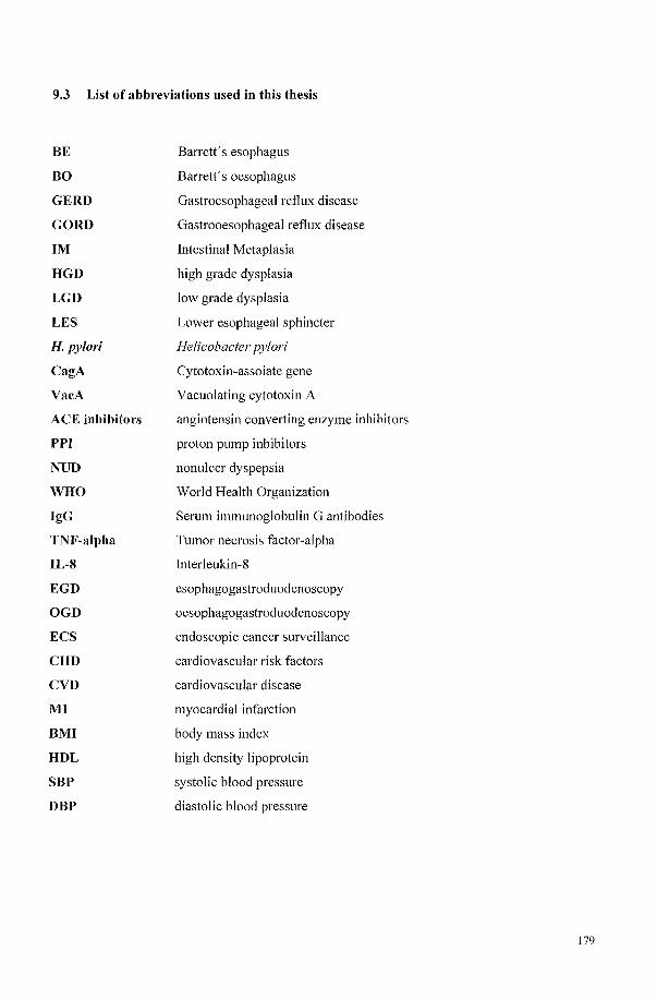
9.3 List of abbreviations used in this thesis
BE Barrett's esophagus
BO Barrett's oesophagus
GERD
GORD
1M
HGD
LGD
LES
H.pylori
CagA
VacA
ACE inhibitors
PPI
NUD
WHO
IgG
TNF-alpha
IL-8
EGD
OGD
ECS
CHD
CVD
MI
BMI
HDL
SBP
DBP
Gastroesophageal reflux disease
Gastrooesophageal reflux disease
Intestinal Metaplasia
high grade dysplasia
low grade dysplasia
Lower esophageal sphincter
Helicobacter pylori
Cytotoxin-assoiate gene
Vacuolating cytotoxin A
angiotensin converting enzyme inhibitors
proton pump inhibitors
nonulcer dyspepsia
World Health Organization
Serum immunoglobulin G antibodies
Tumor necrosis factor-alpha
Interleukin-8
esophagogastroduodenoscopy
oesophagogastroduodenoscopy
endoscopic cancer surveillance
cardiovascular risk factors
cardiovascular disease
myocardial infarction
body mass index
high density lipoprotein
systolic blood pressure
diastolic blood pressure
179


