Embed Size (px)
Citation preview
Hindawi Publishing CorporationJournal of Biomedicine and BiotechnologyVolume 2010, Article ID 752698, 13 pagesdoi:10.1155/2010/752698
Methodology Report
An Experimental Approach for the Identification ofConserved Secreted Proteins in Trypanosomatids
Rosa M. Corrales,1 Francoise Mathieu-Daude,1 Deborah Garcia,1 Simone F. Breniere,2
and Denis Sereno1
1 Departement Societes et Sante, UR016 Caracterisation et controle des populations de vecteurs,Institut de Recherche pour le Developpement, 911 Avenue Agropolis, 34394 Montpellier, France
2 Departement Societes et Sante, UR016 Caracterisation et controle des populations de vecteurs,Representation en Bolivie, Avenue Hernando Siles 5290, Esq Calle 7, Obrajes CP 9214, La Paz, Bolivia
Correspondence should be addressed to Rosa M. Corrales, [email protected]
Received 1 August 2009; Accepted 14 October 2009
Academic Editor: Jorge Morales-Montor
Copyright © 2010 Rosa M. Corrales et al. This is an open access article distributed under the Creative Commons AttributionLicense, which permits unrestricted use, distribution, and reproduction in any medium, provided the original work is properlycited.
Extracellular factors produced by Leishmania spp., Trypanosoma cruzi, and Trypanosoma brucei are important in the host-parasite relationship. Here, we describe a genome-based approach to identify putative extracellular proteins conserved amongtrypanosomatids that are likely involved in the classical secretory pathway. Potentially secreted proteins were identified bybioinformatic analysis of the T. cruzi genome. A subset of thirteen genes encoding unknown proteins with orthologs containinga signal peptide sequence in L. infantum, L. major, and T. brucei were transfected into L. infantum. Tagged proteins detected inthe extracellular medium confirmed computer predictions in about 25% of the hits. Secretion was confirmed for two L. infantumorthologs proteins using the same experimental system. Infectivity studies of transgenic Leishmania parasites suggest that one ofthe secreted proteins increases parasite replication inside macrophages. This methodology can identify conserved secreted proteinsinvolved in the classical secretory pathway, and they may represent potential virulence factors in trypanosomatids.
1. Introduction
The Trypanosomatidae comprise a large group of para-sitic protozoa, some of which cause important diseases inhumans. The organisms that have been most extensivelystudied are Trypanosoma brucei, the causative agent ofAfrican sleeping sickness, T. cruzi, responsible for Chagasdisease in South America, and Leishmania spp., which causesLeishmaniasis in Asia, South America, and Mediterraneancountries [1]. In order to complete their life cycle, theseparasites have to adapt and develop in an insect vectorand in a vertebrate host. These single-celled organisms havedeveloped several strategies to modify their surroundingenvironment, modulate host immune responses, or interferewith the host’s anti-microbial activity. Materials secretedby the parasite are involved in these processes helpingthe parasite survive in an environment more favorable forits own development [2–5]. In addition, previous studiesindicate that trypanosomatid secreted factors elicit strong
immunity and protection against infection in mice anddogs [6, 7]. Thus secreted factors could be a sourceof antigens for vaccine development, as demonstrated inthe pathogen Mycobacterium tuberculosis [8]. Nevertheless,all trypanosomatid secreted factors involved in virulenceand/or representing putative vaccine targets are not currentlyknown.
The availability of three draft trypanosomatid genomesequences provides valuable data for protein-mining usingbioinformatic tools, especially for the localization or pre-diction of function for hypothetical proteins. Given that asignificant number of trypanosomatid protein-coding genesare annotated as hypothetical, additional studies are neededto ascertain their function.
In the present study, we designed an experimentalgenome-based approach to identify potentially secretedproteins that are conserved among the three main trypanoso-matid pathogens and involved in the classical secretorypathway. We hypothesized that a phylogenetic conservation
2 Journal of Biomedicine and Biotechnology
among Leishmania, T. cruzi, and T. brucei would indicateevolutionary selection for this family of proteins and suggestan important role for such secreted proteins in the biologyof these parasites. Our results demonstrate that the bioinfor-matic analysis, combined with the functional tests, providesa useful and reliable method for the identification of novelsecreted proteins, representing potential virulence factors intrypanosomatids. The results are also discussed in relation tothe relative importance of the classical and nonclassical secre-tory pathways for the release of proteins into the extracellularenvironment.
2. Materials and Methods
2.1. In Silico Sequence Analysis. Release V 5.0 of the T.cruzi genome was extracted from the integrated T. cruzigenome resource TcruziDB (http://tcruzidb.org/tcruzidb/).Protein sequences that do not bear an initial methionineamino acid were removed manually. Proteins belonging tolarge families of surface molecules, which include trans-sialidases, mucins, gp63s, and mucin-associated surfaceproteins, were also discarded. Finally ORFs encodingproteins bearing a molecular weight (MW) above 90kDa were also eliminated. The software SignalP 3.0(http://www.cbs.dtu.dk/services/SignalP/) was used topredict the presence of a signal peptide and a cleavage sitein amino acid sequences [9]. Protein sequences having asignal peptide probability greater than 0.8 associated with acleavage site probability greater than 0.7 were analyzed forthe presence of orthologs in the related Trypanosoma bruceiand Leishmania major parasite genomes predicted by JaccardCOG clustering in Gene DB (http://www.genedb.org/).Most of these orthologs were confirmed in TriTrypDB(http://tritrypdb.org/tritrypdb/) using OrthoMCL andgenomic context analysis (gene synteny). The TMHMM2.0 (http://www.cbs.dtu.dk/services/TMHMM/) and theDAS-TMfilter (http://mendel.imp.ac.at/DAS/source.html)servers were used for the prediction of transmembranehelices in protein sequences.
2.2. Parasite Strains and Cultures. The T. cruzi TcY7 (or Ycl7) clone derived from the Y strain [10] was used through-out this study. Epimastigotes were grown in liver infusiontryptose (LIT) medium supplemented with 10% FCS at28◦C in standard conditions [11] and harvested duringthe logarithmic growth phase. Metacyclic trypomastigotes,obtained from the differentiation of late stationary phaseepimastigotes, were used to initiate infection of mousefibroblasts (L929). Trypomastigotes and amastigotes wereproduced and harvested as previously described [12]. Pelletsfor RNA purification were processed immediately in lysisbuffer. The wild-type (WT) promastigote clone from L.infantum (MHOM/MA/67/ITMAP-263) was maintained at26◦C by weekly subpassages in SDM 79 medium supple-mented with 10% heat-inactivated FCS, 100 U/mL penicillin,and 100 μg/mL streptomycin [13].
2.3. Reverse Transcription and PCR Amplifications. TotalRNA was extracted from epimastigotes, amastigotes, and
trypomastigotes with the RNeasy kit (Qiagen, Hilden, Ger-many) according to the manufacturers’ instructions, andtreated with DNase I (DNA-free kit, Ambion Inc., Austin,Texas). Reverse transcription was performed for 1 μg oftotal RNA using random hexamers and Superscript IIreverse transcriptase (Invitrogen, Carlsbad, CA) accordingto the manufacturers’ instructions. The cDNA (4 μL of 1/10dilutions) from each stage was amplified by PCR in a 20 μLreaction volume using 10 μL of 2X PCR Master Mix 1X(Promega, Madison, Wisconsin) and 0.5 μM gene-specificforward and internal reverse primers (listed in Table 1) usingthe following cycling conditions: 94◦C for 3 minutes followedby 30 cycles of 94◦C for 30 seconds, 55◦C to 58◦C (accordingto the primer pair) for 30 seconds, 72◦C for 45 seconds, anda final elongation at 72◦C for 5 minutes. Amplicons wereelectrophoresed on 2% agarose gels stained with ethidiumbromide.
2.4. Cloning and Sequencing. The encoding genes selected byin silico analysis were cloned into the pTEX expression vec-tor, carrying the Neomycin resistance gene (NEO) [14]. Fulllength ORFs were amplified from genomic DNA with specificreverse and forward primers, including different restrictionsites and a 6-Histidine-Tag in the C-terminal region (listedin Tables 1 and 3). PCR reactions were carried out in20 μL using 0.5 μM of each primer, 0.2 mM dNTP, 0.4 Uof Phusion high-fidelity polymerase (Finnzymes, Espoo,Finland), and the following cycling conditions: 98◦C for 30seconds followed by 25 cycles of 98◦C for 10 seconds, 64◦C to68◦C for 15 seconds, 72◦C for 25 to 60 seconds (according togene size), and a 72◦C elongation for 5 minutes. Digested andpurified fragments were inserted into the dephosphorylatedpTEX vector digested with the corresponding restrictionenzymes. Cloned sequences were confirmed by restrictiondigestion and sequencing. Large-scale preparations of thedifferent constructs were performed using the Plasmid Midikit (Promega).
2.5. Transfection Procedures. Promastigotes of the Leish-mania infantum clone were electroporated as describedelsewhere [15]. Briefly, promastigotes were washed twicewith HEPES-NaCl buffer saline (21 mM HEPES, 5 mMKCl, 0.7 mM NaH2PO4, 137 mM NaCl), resuspended at108 cells/mL in HEPES-NaCl electroporation buffer (pH7.2) supplemented with 6 mM glucose and cooled on icefor 10 minutes. Cells (108) were combined with 15 μg ofvector, left on ice for 10 additional minutes, and elec-troporated using an Easyject Plus (Eurogentec, Seraing,Belgium) apparatus set at 450 V and 450 μF, for one pulse.The cells were left on ice for a further 10 minutes andtransferred to 4 mL of growth medium. The antibiotic G-418 (20 μg/mL) was added 24 hours later, and parasiteswere subcultured at a dilution of 1/10 in 5 mL SDM inthe presence of 20 μg/mL G418. Drug-resistant cells wereobserved 15–20 days later. Parasites were grown in thepresence of gradually increasing concentrations of G418 andwere routinely maintained in SDM containing 150 μg/mL ofG418.
Journal of Biomedicine and Biotechnology 3
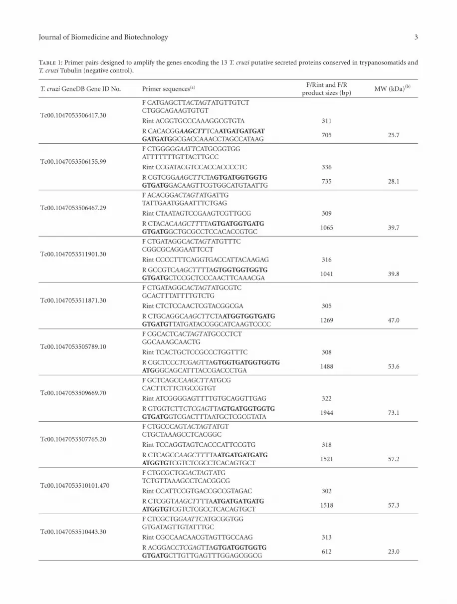
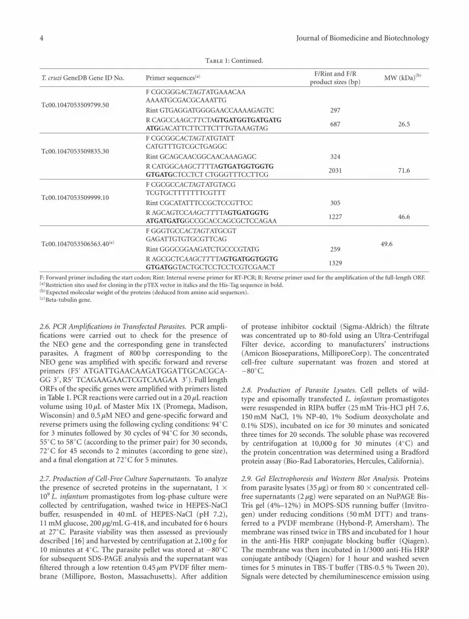
Table 1: Primer pairs designed to amplify the genes encoding the 13 T. cruzi putative secreted proteins conserved in trypanosomatids andT. cruzi Tubulin (negative control).
T. cruzi GeneDB Gene ID No. Primer sequences(a) F/Rint and F/Rproduct sizes (bp)
MW (kDa)(b)
Tc00.1047053506417.30
F CATGAGCTTACTAGTATGTTGTCTCTGGCAGAAGTGTGT
Rint ACGGTGCCCAAAGGCGTGTA 311
R CACACGGAAGCTTTCAATGATGATGATGATGATGGCGACCAAACCTAGCCATAAG
705 25.7
Tc00.1047053506155.99
F CTGGGGGAATTCATGCGGTGGATTTTTTTGTTACTTGCC
Rint CCGATACGTCCACCACCCCTC 336
R CGTCGGAAGCTTCTAGTGATGGTGGTGGTGATGGACAAGTTCGTGGCATGTAATTG
735 28.1
Tc00.1047053506467.29
F ACACGGACTAGTATGATTGTATTGAATGGAATTTCTGAG
Rint CTAATAGTCCGAAGTCGTTGCG 309
R CTACACAAGCTTTTAGTGATGGTGATGGTGATGGCTGCGCCTCCACACCGTGC
1065 39.7
Tc00.1047053511901.30
F CTGATAGGCACTAGTATGTTTCCGGCGCAGGAATTCCT
Rint CCCCTTTCAGGTGACCATTACAAGAG 316
R GCCGTCAAGCTTTTAGTGGTGGTGGTGGTGATGCTCCGCTCCCAACTTCAAACGA
1041 39.8
Tc00.1047053511871.30
F CTGATAGGCACTAGTATGCGTCGCACTTTATTTTGTCTG
Rint CTCTCCAACTCGTACGGCGA 305
R CTGCAGGCAAGCTTCTAATGGTGGTGATGGTGATGTTATGATACCGGCATCAAGTCCCC
1269 47.0
Tc00.1047053505789.10
F CGCACTCACTAGTATGCCCTCTGGCAAAGCAACTG
Rint TCACTGCTCCGCCCTGGTTTC 308
R CGCTCCCTCGAGTTAGTGGTGATGGTGGTGATGGGCAGCATTTACCGACCCTGA
1488 53.6
Tc00.1047053509669.70
F GCTCAGCCAAGCTTATGCGCACTTCTTCTGCCGTGT
Rint ATCGGGGAGTTTTGTGCAGGTTGAG 322
R GTGGTCTTCTCGAGTTAGTGATGGTGGTGGTGATGGTCGACTTTAATGCTCGCGTATA
1944 73.1
Tc00.1047053507765.20
F CTGCCCAGTACTAGTATGTCTGCTAAAGCCTCACGGC
Rint TCCAGGTAGTCACCCATTCCGTG 318
R CTCAGCCAAGCTTTTAATGATGATGATGATGGTGTCGTCTCGCCTCACAGTGCT
1521 57.2
Tc00.1047053510101.470
F CTGCGCTGGACTAGTATGTCTGTTAAAGCCTCACGGCG
Rint CCATTCCGTGACCGCCGTAGAC 302
R CTCGGTAAGCTTTTAATGATGATGATGATGGTGTCGTCTCGCCTCACAGTGCT
1518 57.3
Tc00.1047053510443.30
F CTCGCTGGAATTCATGCGGTGGGTGATAGTTGTATTTGC
Rint CGCCAACAACGTAGTTGCCAAG 313
R ACGGACCTCGAGTTAGTGATGGTGGTGGTGATGCTTGTTGAGTTTGGAGCGGCG
612 23.0
4 Journal of Biomedicine and Biotechnology
Table 1: Continued.
T. cruzi GeneDB Gene ID No. Primer sequences(a) F/Rint and F/Rproduct sizes (bp)
MW (kDa)(b)
Tc00.1047053509799.50
F CGCGGGACTAGTATGAAACAAAAAATGCGACGCAAATTG
Rint GTGAGGATGGGGAACCAAAAGAGTC 297
R CAGCCAAGCTTCTAGTGATGGTGATGATGATGGACATTCTTCTTCTTTGTAAAGTAG
687 26.5
Tc00.1047053509835.30
F CGCGGCACTAGTATGTATTCATGTTTGTCGCTGAGGC
Rint GCAGCAACGGCAACAAAGAGC 324
R CATGGCAAGCTTTTAGTGATGGTGGTGGTGATGCTCCTCT CTGGGTTTCCTTCG
2031 71.6
Tc00.1047053509999.10
F CGCGCCACTAGTATGTACGTCGTGCTTTTTTTCGTTT
Rint CGCATATTTCCGCTCCGTTCC 305
R AGCAGTCCAAGCTTTTAGTGATGGTGATGATGATGGCCGCACCAGCGCTCCAGAA
1227 46.6
Tc00.1047053506563.40(c)
F GGGTGCCACTAGTATGCGTGAGATTGTGTGCGTTCAG
49.6Rint GGGCGGAAGATCTGCCCGTATG 259
R AGCGCTCAAGCTTTTAGTGATGGTGGTGGTGATGGTACTGCTCCTCCTCGTCGAACT
1329
F: Forward primer including the start codon; Rint: Internal reverse primer for RT-PCR; R: Reverse primer used for the amplification of the full-length ORF.(a)Restriction sites used for cloning in the pTEX vector in italics and the His-Tag sequence in bold.(b)Expected molecular weight of the proteins (deduced from amino acid sequences).(c)Beta-tubulin gene.
2.6. PCR Amplifications in Transfected Parasites. PCR ampli-fications were carried out to check for the presence ofthe NEO gene and the corresponding gene in transfectedparasites. A fragment of 800 bp corresponding to theNEO gene was amplified with specific forward and reverseprimers (F5′ ATGATTGAACAAGATGGATTGCACGCA-GG 3′, R5′ TCAGAAGAACTCGTCAAGAA 3′). Full lengthORFs of the specific genes were amplified with primers listedin Table 1. PCR reactions were carried out in a 20 μL reactionvolume using 10 μL of Master Mix 1X (Promega, Madison,Wisconsin) and 0.5 μM NEO and gene-specific forward andreverse primers using the following cycling conditions: 94◦Cfor 3 minutes followed by 30 cycles of 94◦C for 30 seconds,55◦C to 58◦C (according to the primer pair) for 30 seconds,72◦C for 45 seconds to 2 minutes (according to gene size),and a final elongation at 72◦C for 5 minutes.
2.7. Production of Cell-Free Culture Supernatants. To analyzethe presence of secreted proteins in the supernatant, 1 ×10⁹ L. infantum promastigotes from log-phase culture werecollected by centrifugation, washed twice in HEPES-NaClbuffer, resuspended in 40 mL of HEPES-NaCl (pH 7.2),11 mM glucose, 200 μg/mL G-418, and incubated for 6 hoursat 27◦C. Parasite viability was then assessed as previouslydescribed [16] and harvested by centrifugation at 2,100 g for10 minutes at 4◦C. The parasite pellet was stored at −80◦Cfor subsequent SDS-PAGE analysis and the supernatant wasfiltered through a low retention 0.45 μm PVDF filter mem-brane (Millipore, Boston, Massachusetts). After addition
of protease inhibitor cocktail (Sigma-Aldrich) the filtratewas concentrated up to 80-fold using an Ultra-CentrifugalFilter device, according to manufacturers’ instructions(Amicon Bioseparations, MilliporeCorp). The concentratedcell-free culture supernatant was frozen and stored at−80◦C.
2.8. Production of Parasite Lysates. Cell pellets of wild-type and episomally transfected L. infantum promastigoteswere resuspended in RIPA buffer (25 mM Tris-HCl pH 7.6,150 mM NaCl, 1% NP-40, 1% Sodium deoxycholate and0.1% SDS), incubated on ice for 30 minutes and sonicatedthree times for 20 seconds. The soluble phase was recoveredby centrifugation at 10,000 g for 30 minutes (4◦C) andthe protein concentration was determined using a Bradfordprotein assay (Bio-Rad Laboratories, Hercules, California).
2.9. Gel Electrophoresis and Western Blot Analysis. Proteinsfrom parasite lysates (35 μg) or from 80 × concentrated cell-free supernatants (2 μg) were separated on an NuPAGE Bis-Tris gel (4%–12%) in MOPS-SDS running buffer (Invitro-gen) under reducing conditions (50 mM DTT) and trans-ferred to a PVDF membrane (Hybond-P, Amersham). Themembrane was rinsed twice in TBS and incubated for 1 hourin the anti-His HRP conjugate blocking buffer (Qiagen).The membrane was then incubated in 1/3000 anti-His HRPconjugate antibody (Qiagen) for 1 hour and washed seventimes for 5 minutes in TBS-T buffer (TBS-0.5 % Tween 20).Signals were detected by chemiluminescence emission using
Journal of Biomedicine and Biotechnology 5
the ECL Plus Western blotting detection system and ECLHyperfilms (GE Healthcare, UK).
2.10. Generation of Bioluminescent L. Infantum Promastigotesand In Vitro Infection of Human Macrophages. A homol-ogous episomal expression system was devised to furtherexamine the infection in vitro of two secreted proteinsfrom L. infantum. The vector pSP-αHYGαLUC [17] car-rying the firefly-luciferase gene was used to cotransfect L.infantum promastigotes overexpressing the secreted pro-teins LinJ19.0410 (ortholog of Tc00.1047053505789.10) orLinJ36.5780 (ortholog of. Tc00.1047053506155.99). Recom-binant parasites were selected for their growth in increasingconcentrations of Hygromycin (up to 300 μg/mL) over aperiod of several weeks. Promastigotes transfected withthe pTEX vector alone and the pSP-αHYGαLUC wereused as controls for infection experiments. The survival oftransfected parasites was evaluated within human leukemiamonocyte cells (THP-1 cells). THP-1 cells were culturedin RPMI 1640 medium supplemented with 10% FCS,2 mM glutamine, 100 IU of penicillin/mL, and 100 μg ofstreptomycin/mL. THP-1 cells in the log-phase of growthwere differentiated into macrophages by incubation for 2days in a medium containing 20 ng/mL of phorbol myristateacetate (Sigma-Aldrich). THP-1 cells treated with PMA werewashed with prewarmed medium and then infected withstationary-phase promastigotes of transfected-Leishmania ina 24-well plate at a parasite/macrophage ratio of 10 : 1 for4 hours at 37◦C with 5% CO2. Noninternalized parasiteswere removed. After different incubation periods (24 hoursto 96 hours) Luciferase activity was determined using theSteady Glo reagent (Promega, Madison,WI), according tothe manufacturers’ instructions. After 5 minutes, the platewas read using a Multilabel Counter VICTOR2model 1420(Perkin Elmer). Results are expressed as the mean of RLU(Relative Luminescence Units) activity of three independentexperiments, each performed in triplicate. Statistical signifi-cance was analyzed by the Mann-Whitney U test.
3. Results
3.1. Bioinformatic Selection for Secreted Proteins in Try-panosomatids. The preliminary analysis of the 19613 T.cruzi putative proteins from the CL-Brener genome wasperformed to discard potential uncompleted sequences. Atotal of 1796 sequences were removed manually since theydid not bear an initial methionine amino acid. The remain-ing 17817 (90.8%) protein coding sequences were kept forsubsequent analysis. House-keeping genes and sequencesbelonging to large gene families, like the trans-sialidases,mucins, Mucin-Associated Surface Proteins (MASPs), werediscarded given that the main goal was to identify novelsecreted proteins. Finally, sequences encoding proteins witha molecular weight above 90 kDa were eliminated, in orderto facilitate subsequent gene cloning. The remaining codingsequences were screened for the presence of both the signalpeptide and the peptidase cleavage site with a probabilityof 0.8 and 0.7, respectively. A total of 216 sequences wereobtained by using these criteria. Among them, 91 (42%)
were annotated as “hypothetical proteins, conserved” in thedata bank. The final criterion for selected proteins likelyto be secreted by the classical eukaryotic pathway was thepresence of the signal peptide and the signal peptidase sitein orthologs of the related parasites: Leishmania major, L.infantum, and T. brucei. Among the 91 sequences, only 45showed orthologs with the signal peptide criteria. The 13proteins bearing the highest probability for the presenceof the signal peptide were selected (Table 2) for confirma-tion of extracellular localization. Among the 13 selectedgenes, Tc00.1047053505789.10 and Tc00.1047053509835.30are homologous genes and members of a multigene family(Table 2). These genes show about 40% identity at theprotein level and possess the same predicted orthologs inthe GeneDB server. Nevertheless in the TritrypDB resourcethe prediction of orthologs is different (see Table 2). Thiscould be explained by the use of different algorithms(Jaccard cog clustering or OrthoMCL) for the predictionof orthologous groups [18]. The beta-tubulin T. cruzi gene(GeneID Tc00.1047053506563) was added to our sample asa potential negative control for protein secretion. Amongthe 13 selected genes, 7 genes were predicted to have trans-membrane domains (Table 2). These genes were includedfor the functional test because previous studies identifiedextracellular proteins with putative transmembrane domainsas constituents of the secretome of different pathogens,including L. donovani [19, 20]. Furthermore, in the protozoaToxoplasma gondii, the dense granule protein GRA5 is atransmembrane protein that bears a signal peptide and issecreted as a soluble protein into the vacuolar space, beforebeing inserted into the parasitophorous vacuole membrane.Based on these studies, we included genes with potentialtransmembrane domains to test whether the presence ofthese domains represents a useful criterion for identifyingextracellular secreted proteins.
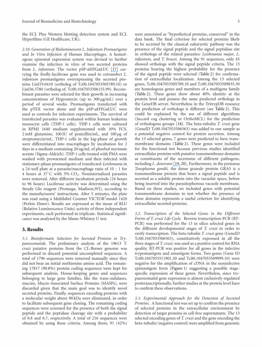
3.2. Transcription of the Selected Genes in the DifferentForms of T. cruzi Life Cycle. Reverse transcription-PCR (RT-PCR) was performed for the 13 in silico selected genes inthe different developmental stages of T. cruzi in order toverify transcription. The beta-tubulin T. cruzi gene (GeneIDTc00.1047053506563), constitutively expressed in all thethree stages of T. cruzi, was used as a positive control for RNAquality. RT-PCR was positive for all genes in the infectivetrypomastigote and amastigote forms. Two genes (Gene IDTc00.1047053511901.30 and Tc00.1047053509999.10) werenegative for the amplification of cDNA in the noninfectiveepimastigote form (Figure 1) suggesting a possible stage-specific expression of these genes. Nevertheless, since try-panosomatid gene expression is almost exclusively regulatedposttranscriptionally, further studies at the protein level haveto confirm these observations.
3.3. Experimental Approach for the Detection of SecretedProteins. A functional test was set up to confirm the presenceof selected proteins in the extracellular environment bydetection of target proteins in cell-free supernatants. The 13selected encoding genes of T. cruzi and the gene encoding thebeta-tubulin (negative control) were amplified from genomic

6 Journal of Biomedicine and Biotechnology
Table 2: T. cruzi genes selected by in silico analysis and predicted properties of the encoded hypothetical proteins.
T. cruzi GeneDBGene ID No.
Orthologs Probability TM(d) GPI(e) Conserved domains
(and E-value)(f)L. major and T. brucei Gene ID No.(a) SPP(b) CSP(c)
Tc00.1047053506417.30 LmjF22.0225 Tb927.8.2180∗ 0.937 0.917 1 Yes None
Tc00.1047053506155.99 LmjF36.5220 Tb11.01.2470 0.984 0.962 0 NoGlucosidase II betasubunit-like(E = 1.6 e-13)
Tc00.1047053506467.29 LmjF26.2000 Tb09.160.1070 0.811 0.771 0 YesMethyltransferasedomain (E = 4.3 e-4)
Tc00.1047053511901.30 LmjF24.2160 Tb927.8.6080 0.989 0.898 0 NoGlycerophosphoryldiester phosphodies-terase (E = 5.7 e-8)
Tc00.1047053511871.30 LmjF25.1010∗ Tb927.3.950∗ 0.979 0.958 0 No2OG-Fe(II) oxygenase(E = 1.7 e-13)
Tc00.1047053505789.10(g) LmjF19.0540∗ Tb927.8.6700(h)
1.000 0.768 5 NoLipocalin signature(E = 0.0)
LmjF19.0570∗ Tb11.39.0005(i)
Tc00.1047053509669.70 LmjF29.1600 Tb927.3.4190 0.999 0.980 9 NoEndomembraneprotein 70 (E = 0.0)
Tc00.1047053507765.20 LmjF11.0720 Tb11.02.4400 0.993 0.986 0 No None
Tc00.1047053510101.470 LmjF11.0720 Tb11.02.4400 0.931 0.919 0 No None
Tc00.1047053510443.30 LmjF30.3150 Tb927.6.4500 0.903 0.838 1 NoTranslocon-associatedprotein beta (TRAPB)(E = 4.1 e-4)
Tc00.1047053509799.50 LmjF36.5570 Tb10.6k15.1130 0.981 0.931 1 No None
Tc00.1047053509835.30(g) LmjF19.0540∗ Tb927.8.6700(h)
0.866 0.803 5 No
Heavy-metal-associated domain,Heavy metaltrans-port/detoxification(E = 0.0)
LmjF19.0570∗ Tb11.39.0005(i)
Tc00.1047053509999.10 LmjF29.1200∗ Tb927.3.3820 1.000 0.952 3 No None(a)L. major and T. brucei gene ID No. of putative orthologs from GeneDB.∗Nonsyntenic predicted orthologs in the TriTrypDB server.(b)SPP Signal peptide probability predicted by SignalP 3.0.(c)CSP Maximal cleavage site probability predicted by SignalP 3.0.(d)TM Number of transmembrane domains (other than peptide signal sequence) predicted by TMHMM 2.0.(e)GPI Identification of GPI-anchor signal by GPI-SOM.(f)Conserved domains (and corresponding E-value) from InterPro, PROSITE or Pfam.(g)Tc00.1047053505789.10 and Tc00.1047053509835.30 are homologous genes presenting 45% identity at the protein level. These genes are members of amultigene family including Tc00.1047053505789.20, Tc00.1047053509441.10, Tc00.1047053510063.30, Tc00.1047053510065.10, and Tc00.1047053504235.9.(h)Tb927.8.6700, Tb927.8.6710, Tb927.8.6720, and Tb927.8.6730 represent the paralogs/orthologs of Tc00.1047053505789.10, Tc00.1047053505789.20,Tc00.1047053509441.10, Tc00.1047053510063.30, and Tc00.1047053510065.10 in the TriTrypDB server.(i)Tb11.39.0005 represents the predicted ortholog of Tc00.1047053509835.30 and Tc00.1047053504235.9 in the TriTrypDB server.
DNA. A sequence encoding a 6xHis-Tag was added at theC-terminal end of each encoded gene, to allow subsequentdetection of the protein in total parasite protein extracts or inconcentrated cell-free supernatants (CCFSs). Amplified PCRproducts were cloned into the pTEX shuttle vector widelyused for expression in trypanosomatids [14]. Transforma-tion and selection of T. cruzi is not as easy to perform as forLeishmania, mainly due to longer periods required for select-ing drug-resistant parasites. Since we aimed to develop a fastand reliable approach to identify trypanosomatid conservedsecreted proteins, we used the related Leishmania parasiteas the recipient organism for the experimental validationof our selected proteins. Thus, L. infantum promastigotes
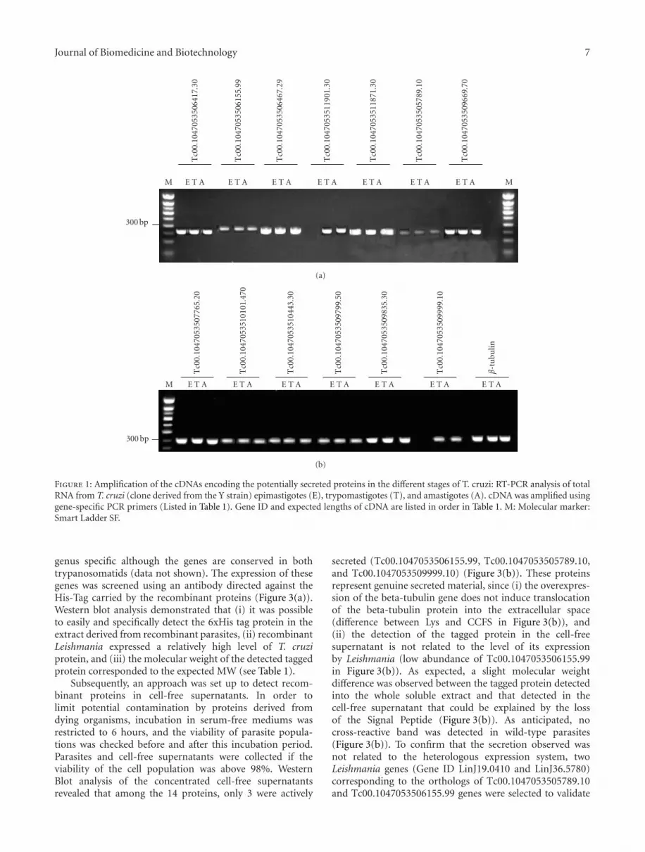
were separately transformed with pTEX carrying one of the14 selected T. cruzi genes (including the beta-tubulin gene),and the recombinant parasites were selected for resistanceto Geneticin G418. Each parasite population was checkedfor the presence of both the NEOR gene and the selectedgene whose secretion was to be analyzed. A specific 800bp fragment, indicative of the presence of the NEOR gene,was detected in the transfected promastigotes and not inwild-type parasites (Figure 2(a)). Moreover, the presence ofeach candidate gene in recombinant parasites was confirmedusing specific primers designed from T. cruzi gene sequences(Figure 2(b)). PCRs performed on wild type Leishmaniawere negative, demonstrating that the amplification was
Journal of Biomedicine and Biotechnology 7
M E T A E T A E T A E T A E T A E T A E T A M
300 bp
Tc0
0.10
4705
3506
417.
30
Tc0
0.10
4705
3506
155.
99
Tc0
0.10
4705
3506
467.
29
Tc0
0.10
4705
3511
901.
30
Tc0
0.10
4705
3511
871.
30
Tc0
0.10
4705
3505
789.
10
Tc0
0.10
4705
3509
669.
70
(a)
M E T A E T A E T A E T A E T A E T A E T A
300 bp
Tc0
0.10
4705
3507
765.
20
Tc0
0.10
4705
3510
101.
470
Tc0
0.10
4705
3510
443.
30
Tc0
0.10
4705
3509
799.
50
Tc0
0.10
4705
3509
835.
30
Tc0
0.10
4705
3509
999.
10
β-t
ubu
lin
(b)
Figure 1: Amplification of the cDNAs encoding the potentially secreted proteins in the different stages of T. cruzi: RT-PCR analysis of totalRNA from T. cruzi (clone derived from the Y strain) epimastigotes (E), trypomastigotes (T), and amastigotes (A). cDNA was amplified usinggene-specific PCR primers (Listed in Table 1). Gene ID and expected lengths of cDNA are listed in order in Table 1. M: Molecular marker:Smart Ladder SF.
genus specific although the genes are conserved in bothtrypanosomatids (data not shown). The expression of thesegenes was screened using an antibody directed against theHis-Tag carried by the recombinant proteins (Figure 3(a)).Western blot analysis demonstrated that (i) it was possibleto easily and specifically detect the 6xHis tag protein in theextract derived from recombinant parasites, (ii) recombinantLeishmania expressed a relatively high level of T. cruziprotein, and (iii) the molecular weight of the detected taggedprotein corresponded to the expected MW (see Table 1).
Subsequently, an approach was set up to detect recom-binant proteins in cell-free supernatants. In order tolimit potential contamination by proteins derived fromdying organisms, incubation in serum-free mediums wasrestricted to 6 hours, and the viability of parasite popula-tions was checked before and after this incubation period.Parasites and cell-free supernatants were collected if theviability of the cell population was above 98%. WesternBlot analysis of the concentrated cell-free supernatantsrevealed that among the 14 proteins, only 3 were actively
secreted (Tc00.1047053506155.99, Tc00.1047053505789.10,and Tc00.1047053509999.10) (Figure 3(b)). These proteinsrepresent genuine secreted material, since (i) the overexpres-sion of the beta-tubulin gene does not induce translocationof the beta-tubulin protein into the extracellular space(difference between Lys and CCFS in Figure 3(b)), and(ii) the detection of the tagged protein in the cell-freesupernatant is not related to the level of its expressionby Leishmania (low abundance of Tc00.1047053506155.99in Figure 3(b)). As expected, a slight molecular weightdifference was observed between the tagged protein detectedinto the whole soluble extract and that detected in thecell-free supernatant that could be explained by the lossof the Signal Peptide (Figure 3(b)). As anticipated, nocross-reactive band was detected in wild-type parasites(Figure 3(b)). To confirm that the secretion observed wasnot related to the heterologous expression system, twoLeishmania genes (Gene ID LinJ19.0410 and LinJ36.5780)corresponding to the orthologs of Tc00.1047053505789.10and Tc00.1047053506155.99 genes were selected to validate
8 Journal of Biomedicine and Biotechnology
WT
MTc0
0.10
4705
3506
417.
30
Tc0
0.10
4705
3506
155.
99
Tc0
0.10
4705
3506
467.
29
Tc0
0.10
4705
3511
901.
30
Tc0
0.10
4705
3511
871.
30
Tc0
0.10
4705
3505
789.
10
Tc0
0.10
4705
3509
669.
70
Tc0
0.10
4705
3507
765.
20
Tc0
0.10
4705
3510
101.
470
Tc0
0.10
4705
3510
443.
30
Tc0
0.10
4705
3509
799.
50
Tc0
0.10
4705
3509
835.
30
Tc0
0.10
4705
3509
999.
10
β-t
ubu
lin
800 bp
(a)
MTc0
0.10
4705
3506
417.
30
Tc0
0.10
4705
3506
155.
99
Tc0
0.10
4705
3506
467.
29
Tc0
0.10
4705
3511
901.
30
Tc0
0.10
4705
3511
871.
30
Tc0
0.10
4705
3505
789.
10
Tc0
0.10
4705
3509
669.
70
Tc0
0.10
4705
3507
765.
20
Tc0
0.10
4705
3510
101.
470
Tc0
0.10
4705
3510
443.
30
Tc0
0.10
4705
3509
799.
50
Tc0
0.10
4705
3509
835.
30
Tc0
0.10
4705
3509
999.
10
β-t
ubu
lin
1 Kb
2 Kb
(b)
Figure 2: PCR analyses in episomally transfected L. infantum promastigotes. (a)Amplification of NEO gene fragment in L. infantumepisomally transfected promastigotes. (b) Amplification of full length transfected genes in L. infantum promastigotes. Specific forward andReverse PCR primers and gene lengths are listed in order in Table 1. WT: Wild Type Parasites. M: Molecular marker: Smart Ladder SL.
our approach. By using the same protocol as above, Leish-mania expressing the 6xHis tagged proteins were generated(See Table 3). As expected, the presence of the taggedprotein in the extracellular medium was only detected in theepisomally transfected parasites (Figure 4). Together, theseresults indicate that this approach allows the identificationof new and genuinely extracellular proteins involved in theendoplasmic reticulum/Golgi-dependent secretory pathway.
3.4. Expression of Secreted Proteins Increases Ability ofRecombinant Leishmania Parasites to Infect and Surviveinside Macrophages In Vitro. We attempted to determinewhether the expression of Leishmania-secreted proteinscould interfere with the capacity of recombinant parasitesto replicate within human macrophages in vitro. Bothconfirmed secreted proteins (LinJ19.0410 and LinJ36.5780)from L. infantum were tested by using the luciferase reportersystem in transfected parasites overexpressing these proteins.We used bioluminescence as a quantitative indicator ofthe viability and multiplication of parasites. The numberof promastigotes cells and luciferase activity were linearlycorrelated for the different recombinant parasites beforemacrophage infection (data not shown). Results of in vitroinfection showed that overexpression of secreted proteinLinJ19.0410 (ortholog of Tc00.1047053505789.10) increasesthe capacity of Leishmania to survive in THP-1 differentiated
macrophages as early as 24 hours postinfection (Figure 5).Furthermore, a statistically significant increase in luciferaseactivity of recombinant parasites expressing LinJ19.0410was maintained throughout the experiments (P < .05).This effect was not observed in parasites overexpressingLinJ36.5780 (ortholog of Tc00.1047053506155.99) whereinfectivity levels were similar to the control parasites trans-fected with the pTEX vector alone and the pSP-αHYGαLUC(Figure 5).
4. Discussion
In trypanosomatids the secretion process is not fullyunderstood and various pathways including classical andnonclassical mechanisms may contribute to the formationof the “extracellular proteome.” Thus the individual identi-fication of secreted materials would enhance efforts towardsunderstanding mechanisms of protein secretion in thesemedically important parasites. In an attempt to provide anew approach to analyse the large number of hypotheticalconserved proteins in trypanosomatids, we developed anexperimental approach to identify hypothetical extracellularproteins likely to be involved in the classical pathway. Wecombined a web-based bioinformatics approach that usedthe Signal P 3.0 program, one of the most accurate predictorsfor the presence of a signal peptide sequence [21], with

Journal of Biomedicine and Biotechnology 9
Tc0
0.10
4705
3506
417.
30
Tc0
0.10
4705
3506
155.
99
Tc0
0.10
4705
3506
467.
29
Tc0
0.10
4705
3511
901.
30
Tc0
0.10
4705
3511
871.
30
Tc0
0.10
4705
3505
789.
10
Tc0
0.10
4705
3509
669.
70
Tc0
0.10
4705
3507
765.
20
Tc0
0.10
4705
3510
101.
470
Tc0
0.10
4705
3510
443.
30
Tc0
0.10
4705
3509
799.
50
Tc0
0.10
4705
3509
835.
30
Tc0
0.10
4705
3509
999.
10
β-t
ubu
lin
25
30
60
50
70
40
(a)
CCFSLys
1 2
Lys CCFS
3 4
Lys CCFS Lys CCFS Lys CCFS
5 6 7 8 9 10
Wild
typ
e
Tc0
0.10
4705
3509
999.
10
Tc0
0.10
4705
3506
155.
99
Tc0
0.10
4705
3505
789.
10
β-t
ubu
lin
25
30
60
50
70
40
(b)
Figure 3: Protein expression in L. infantum episomally transfected promastigotes during the exponential phase of development (a) Westernblot analysis of His-tagged proteins detected in whole cell lysate. Equal amounts of total protein (35 μg) were resolved by electrophoresisin 4–12% gradient gels (Invitrogen), blotted, and developed with anti-HisTag antibody followed by ECL (Amersham). Gene ID and thetheoretical molecular weight of detected proteins are listed in order in Table 1. (b) Identification of secreted proteins in whole cell lysate(Lys) and concentrated cell-free culture supernatant (CCFS) obtained from promastigotes incubated for 6 hours in serum-free medium.Note the absence of β Tubulin in the concentrated supernatant of Line 8. Nontransfected L. infantum promastigotes (Wild Type) were usedas a negative control. Protein molecular mass standards in kDa are shown on the left of each panel.
a functional test that takes advantage of the relative easeto genetically transform Leishmania. We assumed that aphylogenetic conservation among Leishmania, T. cruzi, andT. brucei would point to evolutionary selection for this familyof proteins and indicate an important role for these proteinsin the biology of these parasites. Using these criteria weselected a pool of 13 trypanosomatid-conserved hypotheticalproteins from the T. cruzi database for which secretion wastested.
Identification of secreted proteins has been hamperedin trypanosomatids due to the difficulty in distinguishinggenuine secretions from molecules released by lysed, dead,or dying organisms. Additionally, the in vitro growth of try-panosomatid developmental stages that occur in mammals isimpossible or laborious.
Characterization by screening cDNA libraries with seraraised against culture medium supernatants has beenperformed for the identification of extracellular proteins

10 Journal of Biomedicine and Biotechnology
Table 3: Gene ID of L. infantum orthologous genes and primers used for cloning.
Gene ID Primer sequences(a) F/R product sizes (bp) MW(kDa)(b)
LinJ19.0410(c) F CATGACCACTAGTATGGCCAAAACAGCGCTTCTC 1590 58,4
R GCAGTCCAAGCTTTTAGTGATGGTGATGATGATGAGGTGTTCTCAGGGGTGACGA
LinJ36.5780(d) F CATGCTCGACTAGTATGGGGTGCCGCAGTAGCTG
R GCAGTCCAAGCTTTTAATGATGATGGTGGTGATGATCATCCAACATCTGGCACCGC
738 28
F: Forward primer including the start codon; R: Reverse primer used for the amplification of the full-length ORF.(a)Restriction sites used for cloning in the pTEX vector in italics and the His-Tag sequence in bold.(b)Expected molecular weight of the proteins.(c)Ortholog of LmjF19.0540 and Tc00.1047053505789.10.(d)Ortholog of LmjF36.5220 and Tc00.1047053506155.99.
CCFSLys
LinJ19.0410
1 2
Lys CCFS
3 4
LinJ36.5780
Lys CCFS
5 6
Wild type
2530
40
6070
50
Figure 4: Homologous expression of secreted proteins in L. infantum episomally transfected promastigotes. L. infantum pro-mastigotes were transfected with genes LinJ19.0410 and LinJ36.5780 corresponding to secreted proteins Tc00.1047053505789.10 andTc00.1047053506155.99, respectively. Cell whole lysate (Lys) and concentrated cell-free culture supernatant (CCFS) and electrophoresisprocedure were as in Figure 3. Tagged proteins were detected only in recombinant parasites transfected with LinJ19.0410 (58 kDa) (Line 1and 2) and LinJ36.5780 (28 kDa) (Line 3 and 4). Nontransfected L. infantum promastigotes (Wild Type) were used as negative controls (Line5 and 6). Protein molecular mass standards in kDa are shown on the left.
0
2500
5000
7500
10000
Lum
ines
cen
ceR
LU
0 1 2 3 4 5
Time (days)
PTEX
PTEX-LinJ19.0410PTEX-LinJ36.5780
Figure 5: Bioluminescence activity of intracellular Leishmania expressing episomal luciferase from infected macrophages in vitro.Recombinant L. infantum promastigotes overexpressing the secreted proteins pTEX-LinJ19.0410 (�) or pTEX-LinJ36.5780 (•) werecotransfected with the pSP-YαHYGROαLUC carrying the firefly-luciferase gene. Survival of luciferase-expressing parasites was monitoredin infected human monocyte cell line THP-1 differentiated into macrophages as indicated in the Methods section. Promastigotes transfectedwith the pTEX vector alone and the pSP-αHYGαLUC (�) were used as control for infection experiments. RLUs (Relative LuminescenceUnits) were measured at various time points post infection using the Steady Glo reagent. Results are expressed as the mean of threeindependent experiments, each carried out in triplicate.

Journal of Biomedicine and Biotechnology 11
[22, 23]. However, using this approach proteins with alow abundance or that are poorly immunogenic are likelyto be missed. A more exhaustive approach relying on ahighly sensitive methodology, like the quantitative massspectrometry, was recently used to analyze the proteincontent of the whole conditioned medium from stationary-phase L. donovani promastigotes [19]. Nevertheless, proteinsproduced at low abundance and that are mainly exportedto the extracellular compartment are likely to be missed,since the method relies on the comparison of conditionedmedium versus cell-associated proteins. Indeed proteins thatare well known to be extracellular, such as chitinase andSAcP (secreted acid phosphatase), were not identified withthe SILAC-based approach [19]. The method we designedallowed us to identify three new trypanosomatid conservedproteins, likely to be secreted through the classical secretorypathway. We were confident that the proteins detected inthe cell-free supernatant were genuinely secreted since (i)we were not able to detect the beta-tubulin-tagged proteinin the cell-free supernatant after overexpression of thegene, (ii) the 6-hour incubation time avoided excessive celldeath and contamination by proteins released from deadparasites, (iii) in the extracellular environment we detectedorthologous Leishmania proteins (LinJ19.0410, LinJ36.5780)suggesting that secretion in this protein family is likelyto be evolutionary conserved, and (iv) we detected norelation between the amount of secreted protein and itsintracellular expression in the transfected parasites, demon-strating that the translocation of the his-tagged protein intothe cell supernatant is not related to the methodology weused.
A recent study involving conditioned medium derivedfrom stationary-phase Leishmania promastigotes [19]demonstrated that the extracellular proteome was mainlycomposed of proteins derived from different microvesicles.The parasite growth phase studied by these authorscontains the infectious metacyclic parasites and also ahigh percentage of dying parasites in apoptosis [24].Consequently, the analysis of extracellular material revealedthat the main contributors to the “extracellular proteome”are vesicles likely to be “apoptotic vesicles” or “blebs” [25].This work leads to the general conclusion that 98% of theproteins of the Leishmania secretome lacked a targetingsignal, indicating that nonclassical secretion pathwaysare likely to be the dominant way by which Leishmaniaexport proteins [19]. The proteomic characterization of thereleased/secreted proteins of L. braziliensis promastigotesshowed that about 5% of the identified proteins possessa putative N-Signal peptide [26], indicating that proteinexport may depend on unconventional pathways assuggested in L. donovani. However, some evidence stronglysupports the notion that the classical secretory pathwayis operational in trypanosomatids and contributes to thecomposition of the parasite’s secretome. For example, thescreening of an L. major cDNA library with antiserumraised against culture supernatant from stationary-phasepromastigotes led to the detection of 8 proteins bearinga potential signal peptide among the 33 genes identified[23]. Moreover some of the well-known Leishmania proteins
found in the extracellular environment have a signalpeptide for secretion, such as gp63 or chitinase [3, 27].In the current work we deliberately decided to experimen-tally validate the secretion of our candidate proteins duringthe exponential growth phase of Leishmania in orderto avoid contamination of the cell-free supernatant byapoptotic vesicles or exosomes. In these conditions we didnot detect the secretion of tubulin, even in an overexpressionmodel of Leishmania transfectants, while tubulin wasidentified with a significant score in the Leishmaniasecretome studied by the proteomic analysis performedduring the stationary-phase [19]. Therefore, our resultssuggest that tubulin might be associated with exosomesand/or apoptotic vesicles generated by promastigotesin the stationary-phase of growth. We suggest that thecomposition of the parasite “extracellular proteome” isvariable and depends both on the parasite stage underconsideration and on the relative contribution of the variouspathways operating in protein secretion. Further studies arerequired to highlight the importance of both classical andnonclassical secretory pathways in different developmentalstages of trypanosomatids. Since we selected and testedsecretion for several genes in the trypanosomatid genomes,our methodology provides a potential tool for genomewidescreening to identify extracellular proteins. Furthermore,our methodology may complement other strategies, suchas the SILAC approach, for the identification of proteinsmissed when using proteomic-based approaches.
Regarding proteins not detected in the extracellularenvironment, it is important to point out that proteinsbearing a signal peptide are not only targets for secretionbut also for transfer to specific organelles, for example,lysosomes or the cell surface. Thus, these proteins couldbe retained in specific organelles within the parasite orattach to the cell surface via a GPI-anchor. In this light,analysis of the 13 proteins with GPI-SOM [28] suggeststhat 2 out of 13 proteins (Tc00.1047053506417.30 andTc00.1047053506467.29) have a predicted glycosyl-inositolphosphate (GPI) anchor signature sequences. Hence, thepresence of the GPI anchored domain may explain theabsence of these proteins in the culture supernatant ofrecombinant parasites.
An unexpected finding in our results was the predictionof transmembrane helices in two of our secreted proteins(Tc00.1047053505789.10 and Tc00.1047053509999.10). Weincluded proteins with transmembrane domains to testwhether their presence in protein sequences is a suitablecriterion for the prediction of extracellular secreted proteins.Previous studies suggested that the presence of transmem-brane helices in protein sequences is not a suitable criterionfor discarding potentially secreted proteins [19, 20]. In thisregard our results provide further evidence that empiricalstudies are required to verify bioinformatic predictionssince two of the extracellular proteins bear transmemebranedomains. However, another speculative explanation forthe presence of transmembrane domains is the potentialinsertion of secreted materials into membranes after secre-tion, as demonstrated for the pathogen Toxoplasma gondii[29, 30].
12 Journal of Biomedicine and Biotechnology
The secreted protein Tc00.1047053505789.10 containsa lipocalin signature. Lipocalins are a widely distributedgroup of mostly extracellular proteins, several of whichhave been implicated in the regulation of host immuneresponses, such as, alpha-1-microglobulin and alpha-1-acidglycoprotein [31]. Despite a well-conserved tertiary structureof lipocalins, the pairwise sequence identity within thisfamily is low, often below 30%, [32]. This may explainthe absence of a predicted lipocalin signature in theLeishmania spp. or T. brucei orthologs. Remarkably, theprotein encoded by Tc00.1047053509835.30 (homolog ofTc00.1047053505789.10) lacks a lipocalin signature. Fur-thermore, this protein was not detected as extracellular inour experimental conditions. Although these genes belongto a multigene family (see Table 2) and possess about40% identity at the protein level, our findings suggesta different cellular localization and/or function for thecorresponding proteins. A clear assignment of the proteinTc00.1047053505789.10 to the lipocalin family would onlybe possible after NMR or X-ray crystallography structureanalysis. Further studies are needed to ascertain if this largeand diverse group of proteins is present in trypanosomatidsand plays a role in the transmission process. A GlucosidaseII beta subunit-like protein domain has also been detectedin the protein Tc00.1047053506155.99. Although mostlylocalized to the ER, Glucosidase II was found in endocyticstructures beneath the plasma membrane and has beenassociated with the protein-tyrosine phosphatase CD45 [33].There is also evidence that in some cell types Glucosidase IIbeta is capable of being trafficked to the cell surface [34].
Having demonstrated that our methodology is reliablefor the identification of extracellular secreted proteins intrypanosomatids, we tested the hypothesis that recombinantLeishmania parasites carrying extra copies of Leishmaniasecreted proteins may interfere with their survival or infec-tivity towards human monocyte-derived macrophages invitro. Results of these assays showed a significant survivaladvantage to Leishmania parasites overexpressing the geneLinJ19.0410 (Ortholog of Tc00.1047053505789.10) suggest-ing that this protein is involved in a process increasingsurvival and/or replication of the parasite inside its targetcell. Given that Leishmania and T. cruzi do not target thesame host cells and follow different cell invasion processes,further experiments in the T. cruzi homologous system areneeded to address whether the identified secreted proteinTc00.1047053505789.10 from T. cruzi is also involved inhost cell invasion and/or replication. Current in vitro andin vivo studies are in progress to characterize this proteinwhich represents a potential conserved virulence factor intrypanosomatids.
5. Conclusions
In conclusion, our results show that the bioinformaticsmethod combined with the functional tests, provides afast and reliable method for the identification of novelextracellular secreted proteins involved in the classical secre-tory pathway and represents potential virulence factors intrypanosomatids.
Acknowledgments
Financial support for this study was provided by theEuropean Union through a Grant to R. M. Corrales by the“Programme Alßan” European Union Programme of HighLevel Scholarships for Latin America (no. E05D057391AR)and by IRD DSF. The authors thank B. Vergnes for criticalreview of the manuscript and P. Agnew for help revising themanuscript’s English.
References
[1] K. Stuart, R. Brun, S. Croft, et al., “Kinetoplastids: related pro-tozoan pathogens, different diseases,” The Journal of ClinicalInvestigation, vol. 118, no. 4, pp. 1301–1310, 2008.
[2] N. Santarem, R. Silvestre, J. Tavares, et al., “Immune responseregulation by Leishmania secreted and nonsecreted antigens,”Journal of Biomedicine and Biotechnology, vol. 2007, Article ID85154, 6 pages, 2007.
[3] B. S. McGwire, W. A. O’Connell, K.-P. Chang, and D. M. Eng-man, “Extracellular release of the glycosylphosphatidylinositol(GPI)-linked Leishmania surface metalloprotease, gp63, isindependent of GPI phospholipolysis. Implications for para-site virulence,” The Journal of Biological Chemistry, vol. 277,no. 11, pp. 8802–8809, 2002.
[4] B. A. Burleigh and A. M. Woolsey, “Cell signalling andTrypanosoma cruzi invasion,” Cellular Microbiology, vol. 4, no.11, pp. 701–711, 2002.
[5] M. J. McConville, K. A. Mullin, S. C. Ilgoutz, and R. D.Teasdale, “Secretory pathway of trypanosomatid parasites,”Microbiology and Molecular Biology Reviews, vol. 66, no. 1, pp.122–154, 2002.
[6] J.-L. Lemesre, P. Holzmuller, R. B. Goncalves, et al., “Long-lasting protection against canine visceral leishmaniasis usingthe LiESAp-MDP vaccine in endemic areas of France: double-blind randomised efficacy field trial,” Vaccine, vol. 25, no. 21,pp. 4223–4234, 2007.
[7] W. K. Tonui, J. S. Mejia, L. Hochberg, et al., “Immunizationwith Leishmania major exogenous antigens protects suscepti-ble BALB/c mice against challenge infection with L. major,”Infection and Immunity, vol. 72, no. 10, pp. 5654–5661, 2004.
[8] M. A. Horwitz, G. Harth, B. J. Dillon, and S. Maslesa-Galic,“Enhancing the protective efficacy of Mycobacterium bovisBCG vaccination against tuberculosis by boosting with theMycobacterium tuberculosis major secretory protein,” Infectionand Immunity, vol. 73, no. 8, pp. 4676–4683, 2005.
[9] O. Emanuelsson, S. Brunak, G. von Heijne, and H. Nielsen,“Locating proteins in the cell using TargetP, SignalP andrelated tools,” Nature Protocols, vol. 2, no. 4, pp. 953–971, 2007.
[10] E. Garzon, M. C. Borges, A. Cordeiro-da-Silva, et al., “Try-panosoma cruzi carrying a targeted deletion of a Tc52 protein-encoding allele elicits attenuated Chagas’ disease in mice,”Immunology Letters, vol. 89, no. 1, pp. 67–80, 2003.
[11] E. P. Camargo, “Growth and differentiation in Trypanosomacruzi. I. Origin of metacyclic trypanosomes in liquid media,”Revista do Instituto de Medicina Tropical de Sao Paulo, vol. 12,pp. 93–100, 1964.
[12] F. Mathieu-Daude, M.-F. Bosseno, E. Garzon, et al., “Sequencediversity and differential expression of Tc52 immuno-regulatory protein in Trypanosoma cruzi: potential impli-cations in the biological variability of strains,” ParasitologyResearch, vol. 101, no. 5, pp. 1355–1363, 2007.
Journal of Biomedicine and Biotechnology 13
[13] R. Brun and M. Schonenberger, “Cultivation and in vitrocloning of procyclic culture forms of Trypanosoma brucei ina semi-defined medium. Short communication,” Acta Tropica,vol. 36, no. 3, pp. 289–292, 1979.
[14] J. M. Kelly, H. M. Ward, M. A. Miles, and G. Kendall, “A shuttlevector which facilitates the expression of transfected genes inTrypanosoma cruzi and Leishmania,” Nucleic Acids Research,vol. 20, no. 15, pp. 3963–3969, 1992.
[15] D. Sereno, G. Roy, J. Loup Lemesre, B. Papadopoulou, andM. Ouellette, “DNA transformation of Leishmania infantumaxenic amastigotes and their use in drug screening,” Antimi-crobial Agents and Chemotherapy, vol. 45, no. 4, pp. 1168–1173, 2001.
[16] B. Vergnes, D. Sereno, N. Madjidian-Sereno, J.-L. Lemesre,and A. Ouaissi, “Cytoplasmic SIR2 homologue overexpressionpromotes survival of Leishmania parasites by preventingprogrammed cell death,” Gene, vol. 296, no. 1-2, pp. 139–150,2002.
[17] A. El Fadili, C. Kundig, and M. Ouellette, “Characterization ofthe folylpolyglutamate synthetase gene and polyglutamylationof folates in the protozoan parasite Leishmania,” Molecular andBiochemical Parasitology, vol. 124, no. 1-2, pp. 63–71, 2002.
[18] A. Kuzniar, R. C. H. J. van Ham, S. Pongor, and J. A. M. Leu-nissen, “The quest for orthologs: finding the correspondinggene across genomes,” Trends in Genetics, vol. 24, no. 11, pp.539–551, 2008.
[19] J. M. Silverman, S. K. Chan, D. P. Robinson, et al., “Proteomicanalysis of the secretome of Leishmania donovani,” GenomeBiology, vol. 9, no. 2, article R35, 2008.
[20] A. Walz, C. V. Mujer, J. P. Connolly, et al., “Bacillus anthracissecretome time course under host-simulated conditions andidentification of immunogenic proteins,” Proteome Science,vol. 5, article 11, 2007.
[21] E. W. Klee and L. B. M. Ellis, “Evaluating eukaryotic secretedprotein prediction,” BMC Bioinformatics, vol. 6, article 256,2005.
[22] P. Cibrelus, E. Precigout, D. Sereno, B. Carcy, J. L. Lemesre,and A. Gorenflot, “Secreted antigens of the amastigoteand promastigote forms of Leishmania infantum inducing ahumoral response in humans and dogs,” Parasite, vol. 6, no. 2,pp. 121–129, 1999.
[23] M. Chenik, S. Lakhal, N. Ben Khalef, L. Zribi, H. Louzir,and K. Dellagi, “Approaches for the identification of potentialexcreted/secreted proteins of Leishmania major parasites,”Parasitology, vol. 132, no. 4, pp. 493–509, 2006.
[24] H. Zangger, J. C. Mottram, and N. Fasel, “Cell death in Leish-mania induced by stress and differentiation: programmed celldeath or necrosis?” Cell Death & Differentiation, vol. 9, no. 10,pp. 1126–1139, 2002.
[25] C. Thery, M. Boussac, P. Veron, et al., “Proteomic analysisof dendritic cell-derived exosomes: a secreted subcellularcompartment distinct from apoptotic vesicles,” The Journal ofImmunology, vol. 166, no. 12, pp. 7309–7318, 2001.
[26] P. Cuervo, J. B. De Jesus, L. Saboia-Vahia, L. Mendonca-Lima,G. B. Domont, and E. Cupolillo, “Proteomic characterizationof the released/secreted proteins of Leishmania (Viannia)braziliensis promastigotes,” Journal of Proteomics, vol. 73, no.1, pp. 79–92, 2009.
[27] M. B. Joshi, M. E. Rogers, A. M. Shakarian, et al., “Molecularcharacterization, expression, and in vivo analysis of LmexCht1:the chitinase of the human pathogen, Leishmania mexicana,”The Journal of Biological Chemistry, vol. 280, no. 5, pp. 3847–3861, 2005.
[28] N. Fankhauser and P. Maser, “Identification of GPI anchorattachment signals by a Kohonen self-organizing map,” Bioin-formatics, vol. 21, no. 9, pp. 1846–1852, 2005.
[29] L. Lecordier, C. Mercier, L. D. Sibley, and M.-F. Cesbron-Delauwz, “Transmembrane insertion of the Toxoplasma gondiiGRA5 protein occurs after soluble secretion into the host cell,”Molecular Biology of the Cell, vol. 10, no. 4, pp. 1277–1287,1999.
[30] V. Karsten, R. S. Hegde, A. P. Sinai, M. Yang, and K. A. Joiner,“Transmembrane domain modulates sorting of membraneproteins in Toxoplasma gondii,” The Journal of BiologicalChemistry, vol. 279, no. 25, pp. 26052–26057, 2004.
[31] J. Grzyb, D. Latowski, and K. Strzalka, “Lipocalins—a familyportrait,” Journal of Plant Physiology, vol. 163, no. 9, pp. 895–915, 2006.
[32] D. R. Flower, A. C. T. North, and C. E. Sansom, “The lipocalinprotein family: structural and sequence overview,” Biochimicaet Biophysica Acta, vol. 1482, no. 1-2, pp. 9–24, 2000.
[33] T. A. Baldwin, M. Gogela-Spehar, and H. L. Ostergaard,“Specific isoforms of the resident endoplasmic reticulumprotein glucosidase II associate with the CD45 protein-tyrosine phosphatase via a lectin-like interaction,” The Journalof Biological Chemistry, vol. 275, no. 41, pp. 32071–32076,2000.
[34] Y. M. Li, T. Mitsuhashi, D. Wojciechowicz, et al., “Molecularidentity and cellular distribution of advanced glycation end-product receptors: relationship of p60 to OST-48 and p90to 80K-H membrane proteins,” Proceedings of the NationalAcademy of Sciences of the United States of America, vol. 93, no.20, pp. 11047–11052, 1996.


