Embed Size (px)
Citation preview

J. Mol. Biol. (1996) 259, 249–263
Conserved Features in Papillomavirus andPolyomavirus Capsids
David M. Belnap 1, Norman H. Olson 1, Nancy M. Cladel 2
William W. Newcomb 3, Jay C. Brown 3, John W. Kreider 2
Neil D. Christensen 2 and Timothy S. Baker 1*
1Department of Biological Capsids of papilloma and polyoma viruses (papovavirus family) arecomposed of 72 pentameric capsomeres arranged on a skewed icosahedralSciences, Purdue University
West Lafayette, IN 47907 lattice (triangulation number of seven, T = 7). Cottontail rabbit papillo-USA mavirus (CRPV) was reported previously to be a T = 7laevo (left-handed)
structure, whereas human wart virus, simian virus 40, and murine2Departments of Pathology polyomavirus were shown to be T = 7dextro (right-handed). The CRPVand Microbiology and structure determined by cryoelectron microscopy and image reconstructionImmunology, Hershey Medical was similar to previously determined structures of bovine papillomavirusCenter, Hershey, PA 17033 type 1 (BPV-1) and human papillomavirus type 1 (HPV-1). CRPV capsidsUSA were observed in closed (compact) and open (swollen) forms. Both forms
have star-shaped capsomeres, as do BPV-1 and HPV-1, but the open3Department of MicrobiologyCRPV capsids are 02 nm larger in radius. The lattice hands of alland Cancer Centerpapillomaviruses examined in this study were found to be T = 7dextro. InUniversity of Virginia Schoolthe region of maximum contact, papillomavirus capsomeres interact in aof Medicine, Charlottesvillemanner similar to that found in polyomaviruses. Although papilloma andVA 22908, USApolyoma viruses have differences in capsid size (060 versus 050 nm),capsomere morphology (11 to 12 nm star-shaped versus 8 nmbarrel-shaped), and intercapsomere interactions (slightly different contactsbetween capsomeres), papovavirus capsids have a conserved, 72-pentamer,T = 7dextro structure. These features are conserved despite significantdifferences in amino acid sequences of the major capsid proteins. Theconserved features may be a consequence of stable contacts that occurwithin capsomeres and flexible links that form among capsomeres.
7 1996 Academic Press Limited
Keywords: cryoelectron microscopy; enantiomer; handedness;*Corresponding author Papovaviridae; three-dimensional image reconstruction
Introduction
Papovaviruses infect vertebrate animals includingmammals (e.g. cattle, elephants, humans, mice,
monkeys, and rabbits), birds (e.g. chaffinches,chickens, and parrots), and reptiles (e.g. turtles)(Howley, 1990; Murphy et al., 1995; Olson, 1987;Shah, 1990; Stoll et al., 1993). This virus family issubdivided into the genus Papillomavirus and thegenus Polyomavirus (Murphy et al., 1995). At least 70strains of human papillomavirus have beenidentified (Van Ranst et al., 1994), and these havebeen the subjects of intensive study because theycause benign and cancerous diseases (De Villiers,1989; Howley, 1990). Most humans are infected withthe polyomaviruses JC and BK virus (Eckhart,1990), but these viruses appear to cause diseaseonly when individuals have impaired immunesystems (Shah, 1990).
Papovaviruses are spherical particles with cap-sids made up of 72 morphological units (cap-someres). All papovaviruses are non-enveloped and
Present address: D. M. Belnap, Building 6, Room425, National Institutes of Health, Bethesda, MD 20892,USA.
Abbreviations used: 3D, three-dimensional; a.a.,amino acid; BPV, bovine papillomavirus; BPV-1, bovinepapillomavirus type 1; CCL, cross-common-lines; CCMV, cowpea chlorotic mottle virus; CRPV,cottontail rabbit papillomavirus; cryoEM, cryoelectronmicroscopy; d, dextro; diam., diameter; EGTA,ethyleneglycol-bis-N,N'-tetraacetic acid; HPV, humanpapillomavirus; HPV-1, human papillomavirus type 1;l, laevo; PFT, polar Fourier transform; SV40, simianvirus 40; T = 7, triangulation number of seven; VLP,virus-like particle.
0022–2836/96/220249–15 $18.00/0 7 1996 Academic Press Limited
Structures of Papovavirus Capsids250
multiply in the nucleus of the infected cell. Theycarry a double-stranded, circular DNA genome thatassociates with host-encoded histones in the virions.However, papillomaviruses (e.g. bovine, cottontailrabbit, and human papillomaviruses) and poly-omaviruses (e.g. murine polyomavirus and simianvirus 40) differ in diameter, genome size, proteincomposition and size, and capsomere morphologyand size. Papillomaviruses (060 nm diam.) have an08000 bp genome that encodes two structuralproteins: the major capsid protein, L1 (0510 aminoacid residues and 058 kDa), and the minor pro-tein, L2 (0470 a.a. and 051 kDa). Papillomaviruscapsomeres are mushroom-like protrusions, 11 to12 nm in diameter, with pentameric, ‘‘star-shaped’’heads (Baker et al., 1991). In contrast, the 050 nmdiameter polyomaviruses have an 05000 bpgenome that codes for three structural proteins:the major capsid protein, VP1 (0370 a.a. and041 kDa), and two minor proteins, VP2 (0350 a.a.and 038 kDa) and VP3 (0230 a.a. and 026 kDa).Polyomavirus capsomeres are also pentamers butare smaller (8 nm in diam.) and have a barrel-shaped morphology (Rayment et al., 1982; Bakeret al., 1988; Liddington et al., 1991; Stehle et al., 1994).
Despite these distinguishing differences, electronmicroscopy and X-ray crystallography studies haveshown that all papovavirus capsids exhibit con-served features (Table 1). The 72 capsomeres arepentamers of the major capsid protein and arearranged on a T = 7 icosahedral lattice (Figure 1).Twelve pentavalent pentamers (each surrounded byfive other capsomeres) are centered on theicosahedral 5-fold axes and 60 hexavalent pen-tamers (each surrounded by six other capsomeres)are centered on vertices that lie between the 5-foldaxes.
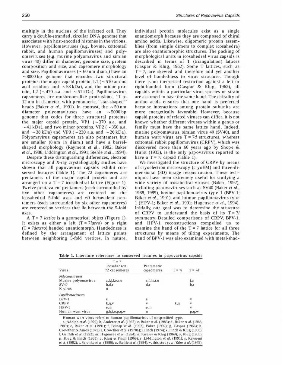
A T = 7 lattice is a geometrical object (Figure 1).It exists as either a left (T = 7laevo) or a right(T = 7dextro) handed enantiomorph. Handedness isdefined by the arrangement of lattice pointsbetween neighboring 5-fold vertices. In nature,
individual protein molecules exist as a singleenantiomorph because they are composed of chiralamino acids. Likewise, oligomeric protein assem-blies (from simple dimers to complex icosahedra)are also enantiomorphic structures. The packing ofmorphological units in icosahedral virus capsids isdescribed in terms of T (triangulation) lattices(Caspar & Klug, 1962). Some T lattices, such asT = 7, are skewed and therefore add yet anotherlevel of handedness to virus structure. Thoughthere is no theoretical restriction against a left orright-handed form (Caspar & Klug, 1962), allcapsids within a particular virus species or strainare assumed to have the same hand. The chirality ofamino acids ensures that one hand is preferredbecause interactions among protein subunits aremore energetically favorable. However, becausecapsid proteins of related viruses can differ, it is notknown whether different viruses within a genus orfamily must have the same lattice hand. Indeed,murine polyomavirus, simian virus 40 (SV40), andhuman wart virus are T = 7d structures, whereascottontail rabbit papillomavirus (CRPV), which wasdiscovered more than 60 years ago by Shope &Hurst (1933), is the only papovavirus reported tohave a T = 7l capsid (Table 1).
We investigated the structure of CRPV by meansof cryoelectron microscopy (cryoEM) and three-di-mensional (3D) image reconstruction. These tech-niques have been extremely useful for studying awide variety of icosahedral viruses (Baker, 1992),including papovaviruses such as SV40 (Baker et al.,1988, 1989), bovine papillomavirus type 1 (BPV-1;Baker et al., 1991), and human papillomavirus type1 (HPV-1; Baker et al., 1991; Hagensee et al., 1994).Initially, our goal was to determine the structureof CRPV to understand the basis of its T = 7lsymmetry. Detailed comparisons of CRPV, BPV-1,and HPV-1 reconstructions compelled us toexamine the hand of the T = 7 lattice for all threestructures by means of tilting experiments. Thehand of BPV-1 was also examined with metal-shad-
Table 1. Literature references to conserved features in papovavirus capsidsT = 7
icosahedron, PentamericVirus 72 capsomeres capsomeres T = 7l T = 7d
PolyomavirusesMurine polyomavirus a,f,j,l,o,s,u c,f,l,s,t,u j,uSV40 b,d,r d,r b,rK virus o
PapillomavirusesBPV-1 e e vCRPV k,q,v v k,q vHPV-1 e,m e,m vHuman wart virus g,h,i,o,p,q,w n p,q,w
Human wart virus refers to human papillomavirus of unspecified type.a, Adolph et al. (1979); b, Anderer et al. (1967); c, Baker et al. (1983); d, Baker et al. (1988,
1989); e, Baker et al. (1991); f, Belnap et al. (1993), Baker (1992); g, Caspar (1966); h,Crowther & Amos (1972); i, Crowther et al. (1970a); j, Finch (1974); k, Finch & Klug (1965);l, Griffith et al. (1992); m, Hagensee et al. (1994); n, Kiselev & Klug (1969); o, Klug (1965);p, Klug & Finch (1965); q, Klug & Finch (1968); r, Liddington et al. (1991); s, Raymentet al. (1982); t, Salunke et al. (1986); u, Stehle et al. (1994); v, this study; w, Yabe et al. (1979).
Structures of Papovavirus Capsids 251
owing techniques. We found that, like the otherpapillomaviruses, the CRPV capsid is composed of72 pentameric, star-shaped capsomeres arranged ona T = 7d icosahedral lattice. In addition, we dis-covered that papillomavirus capsids exist in open
and closed states similar to those observed incowpea chlorotic mottle virus, a structure withunexpected similarities to the polyomaviruses(Speir et al., 1995).
Results
CryoEM
Frozen-hydrated CRPV particles appeared ap-proximately circular in profile, whether the samplewas untilted (Figure 2a,b) or tilted (Figure 2c),indicating that the spherical shape of the virionswas well preserved. The distinct appearances ofdifferent particles showed that the particles wererandomly oriented in the vitrified sample. TheCRPV images appeared indistinguishable frompreviously recorded images of two other papillo-maviruses, BPV-1 and HPV-1 (Baker et al., 1991).New images of BPV-1, HPV-1 virus-like particle(VLP), and SV40 specimens were recorded for thisstudy (data not shown). These images were nearlyidentical to those seen previously (Baker et al., 1988,1991; Hagensee et al., 1994).
Papillomavirus morphology: open andclosed forms
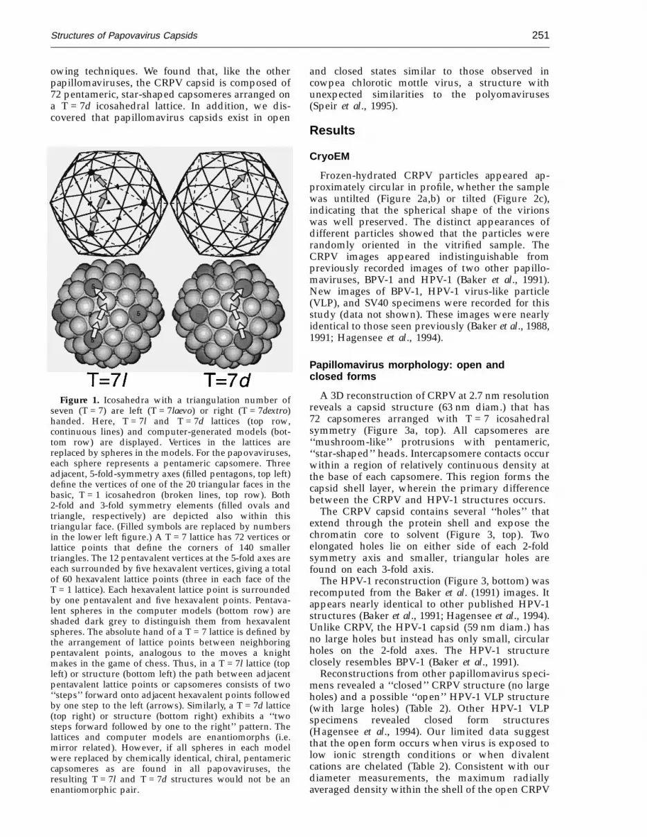
A 3D reconstruction of CRPV at 2.7 nm resolutionreveals a capsid structure (63 nm diam.) that has72 capsomeres arranged with T = 7 icosahedralsymmetry (Figure 3a, top). All capsomeres are‘‘mushroom-like’’ protrusions with pentameric,‘‘star-shaped’’ heads. Intercapsomere contacts occurwithin a region of relatively continuous density atthe base of each capsomere. This region forms thecapsid shell layer, wherein the primary differencebetween the CRPV and HPV-1 structures occurs.
The CRPV capsid contains several ‘‘holes’’ thatextend through the protein shell and expose thechromatin core to solvent (Figure 3, top). Twoelongated holes lie on either side of each 2-foldsymmetry axis and smaller, triangular holes arefound on each 3-fold axis.
The HPV-1 reconstruction (Figure 3, bottom) wasrecomputed from the Baker et al. (1991) images. Itappears nearly identical to other published HPV-1structures (Baker et al., 1991; Hagensee et al., 1994).Unlike CRPV, the HPV-1 capsid (59 nm diam.) hasno large holes but instead has only small, circularholes on the 2-fold axes. The HPV-1 structureclosely resembles BPV-1 (Baker et al., 1991).
Reconstructions from other papillomavirus speci-mens revealed a ‘‘closed’’ CRPV structure (no largeholes) and a possible ‘‘open’’ HPV-1 VLP structure(with large holes) (Table 2). Other HPV-1 VLPspecimens revealed closed form structures(Hagensee et al., 1994). Our limited data suggestthat the open form occurs when virus is exposed tolow ionic strength conditions or when divalentcations are chelated (Table 2). Consistent with ourdiameter measurements, the maximum radiallyaveraged density within the shell of the open CRPV
Figure 1. Icosahedra with a triangulation number ofseven (T = 7) are left (T = 7laevo) or right (T = 7dextro)handed. Here, T = 7l and T = 7d lattices (top row,continuous lines) and computer-generated models (bot-tom row) are displayed. Vertices in the lattices arereplaced by spheres in the models. For the papovaviruses,each sphere represents a pentameric capsomere. Threeadjacent, 5-fold-symmetry axes (filled pentagons, top left)define the vertices of one of the 20 triangular faces in thebasic, T = 1 icosahedron (broken lines, top row). Both2-fold and 3-fold symmetry elements (filled ovals andtriangle, respectively) are depicted also within thistriangular face. (Filled symbols are replaced by numbersin the lower left figure.) A T = 7 lattice has 72 vertices orlattice points that define the corners of 140 smallertriangles. The 12 pentavalent vertices at the 5-fold axes areeach surrounded by five hexavalent vertices, giving a totalof 60 hexavalent lattice points (three in each face of theT = 1 lattice). Each hexavalent lattice point is surroundedby one pentavalent and five hexavalent points. Pentava-lent spheres in the computer models (bottom row) areshaded dark grey to distinguish them from hexavalentspheres. The absolute hand of a T = 7 lattice is defined bythe arrangement of lattice points between neighboringpentavalent points, analogous to the moves a knightmakes in the game of chess. Thus, in a T = 7l lattice (topleft) or structure (bottom left) the path between adjacentpentavalent lattice points or capsomeres consists of two‘‘steps’’ forward onto adjacent hexavalent points followedby one step to the left (arrows). Similarly, a T = 7d lattice(top right) or structure (bottom right) exhibits a ‘‘twosteps forward followed by one to the right’’ pattern. Thelattices and computer models are enantiomorphs (i.e.mirror related). However, if all spheres in each modelwere replaced by chemically identical, chiral, pentamericcapsomeres as are found in all papovaviruses, theresulting T = 7l and T = 7d structures would not be anenantiomorphic pair.

Structures of Papovavirus Capsids252
capsid occurred at a radius 02 nm higher than thatin the closed HPV-1 capsid (data not shown). Thus,the open capsid is 07% larger in diameter than theclosed capsid.
Hands of BPV-1, CRPV, and HPV-1icosahedral lattices
Tilt experiments were used to determine thelattice hands of BPV-1, CRPV, and HPV-1.Enantiomeric reconstructions were rotated to simu-late specimen tilt in the microscope and then wereprojected to simulate the recorded particle images.For the three papillomaviruses we studied, projec-tions of T = 7d reconstructions correlated signifi-cantly better with images of tilted specimens thandid projections of T = 7l reconstructions (Table 3).Indeed, every particle image correlated better withthe T = 7d model (data not shown). Careful visualinspection of some cryoEM data confirmed thesecomparisons (Figure 4a to e). Difference images, inwhich projected model images were subtractedfrom the corresponding particle images, showed theT = 7d model was correct in all cases examined(Figure 4f to h). Difference images obtained withthe T = 7l model exhibited higher density variances(Figure 4g) compared to those obtained from theuntilted or the T = 7d tilted model (Figure 4f and h).
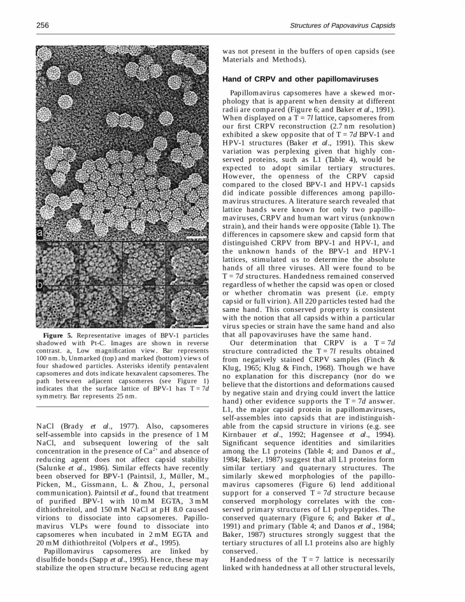
We inspected images of metal-shadowed BPV-1(Figure 5a) and found 31 particles in which adjacentpentavalent capsomeres and the path between themwere clearly resolved. All 31 showed the BPV-1hand to be T = 7d (Figure 5b).
Capsomere morphology and intercapsomerecontacts in papillomaviruses
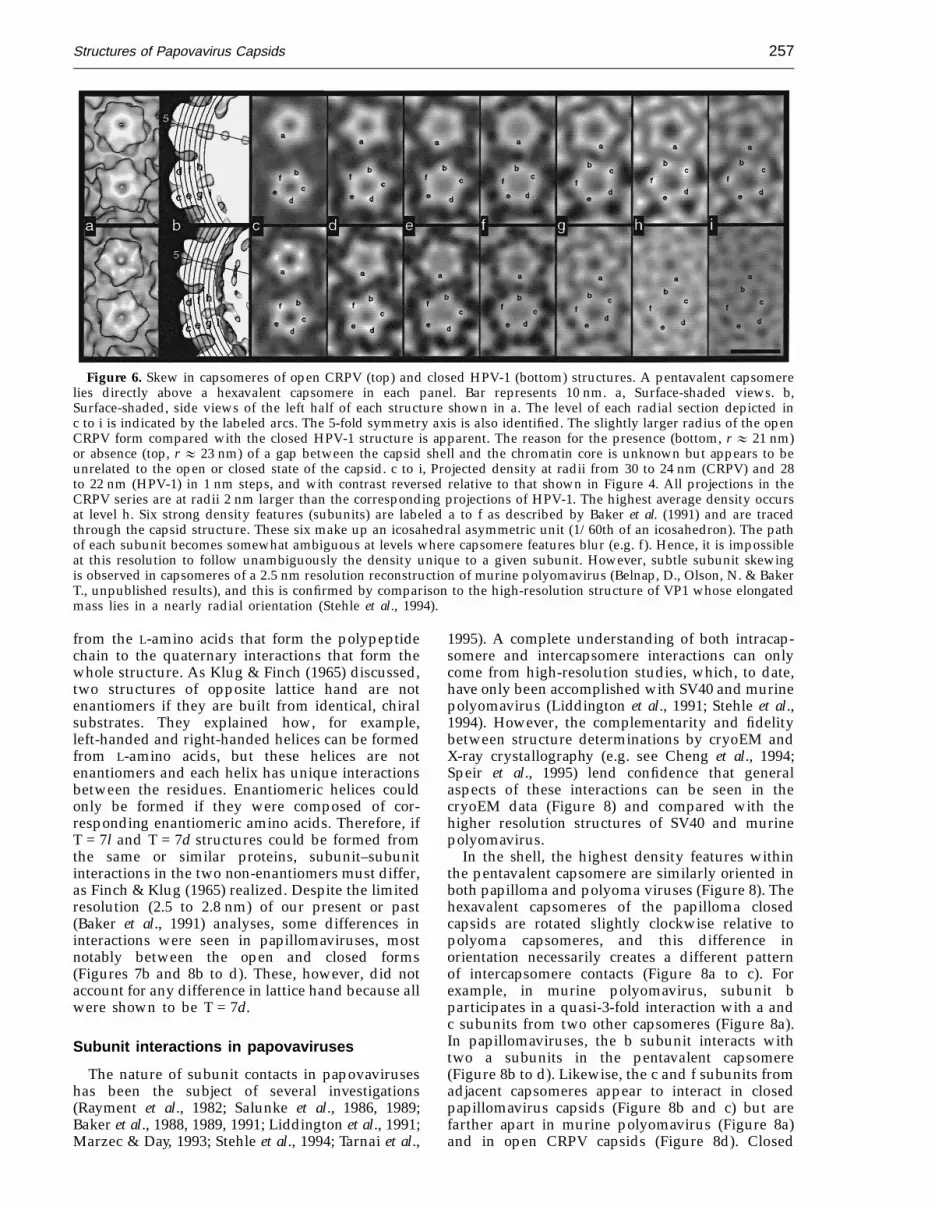
For CRPV and HPV-1 capsids, we examined themorphology of capsomeres and packing of proteinsubunits by viewing projection maps that onlydisplayed density at specific radii (Figures 6 and 7).Density at the corners of the pentameric capsomeres(Figure 6c to i, symbols a to f) usually correspondedto the maximum density at each radius that mayidentify individual protein subunits. When viewedat progressively decreasing radii, the high-densityfeatures in each capsomere follow a counterclock-wise trajectory (Figure 6). Between radii of 30 and25 nm for CRPV and between radii of 28 and 23 nmfor HPV-1, the pentavalent and hexavalent cap-someres skew by 44 to 50° (Figure 6c to h). At theinnermost radii of the open CRPV capsid (25 to24 nm), pentavalent and hexavalent capsomeresskew clockwise by 5 to 8° (Figure 6h and i). Asimilar clockwise skew was observed in the closedCRPV capsid (24 to 21 nm) but not in HPV-1 (Fig-ure 6h and i) or BPV-1 (Baker et al., 1991).
The types of intercapsomere interactions withinthe shell layer are distinct in the open and closedforms at radii of 25 nm for CRPV and 23 nm forHPV-1 (Figure 7b). The average density is highest inboth structures at these radii. Here, the hexavalentcapsomeres are about ten nm in diameter, whereas
Figure 2. Electron micrographs of vitrified CRPVsamples. CRPV particles are distributed in amonodisperse layer indicating that the frozen-hydratedsample is about as thick as the diameter of the particles.Capsomeres appear as ‘‘knob-like’’ structures, which areprominent especially along particle edges. a, CRPVdialyzed against 10 mM Tris-HCl and 1 mM EDTA(pH 8.0). These particle images were used to compute the3D reconstruction seen in Figure 3. Bar represents100 nm. b and c, Tilting experiment with CRPV dialyzedagainst 20 mM Tris-HCl, 1 M NaCl (pH 7.4). View ofparticles with specimen stage at 0° (b) and −5° (c) tilt. Thetilt axis (line in b) is approximately 23° from thehorizontal. The particle highlighted with the arrows alsoappears in Figure 4. Bar represents 100 nm.

Structures of Papovavirus Capsids 253
Figure 3. Surface-shaded representations of CRPV (top row) and HPV-1 (bottom row) reconstructions viewed alonga 2-fold symmetry axis. The CRPV capsid has an ‘‘open’’ form and the HPV-1 has a ‘‘closed’’ form. a, Outside viewof virions. b, Inside view of capsids (back half). Chromatin cores were computationally removed from the virion densitymap (radii < 46 nm for CRPV and < 42 nm for HPV-1). c, Closeup stereo views. In CRPV (top), elongated holes lieon opposite sides of the 2-fold axis. Triangular holes on the 3-fold axes can be seen at the top and bottom center, justbehind the protruding point of the capsomere. In HPV-1 (bottom), there is a single, small hole on the 2-fold axis. Barsrepresent: a,b, 50 nm; c, 10 nm.
the pentavalent capsomeres of CRPV are slightlylarger (011 nm) and those of HPV-1 are slightlysmaller (09 nm). The subunits are packed tightly inthe closed HPV-1 capsid, as Baker et al. (1991)observed. In contrast, the open CRPV capsid ex-hibits much looser packing, as indicated by thepresence of large holes on the 3-fold axes and alsonear the 2-fold axes.
Papovavirus sequence alignments
We used the program GAP (Genetics ComputerGroup, Madison, Wisconsin) to compare the L1 andL2 sequences of BPV-1, CRPV, and HPV-1 (Table 4).The L1 sequences are more highly conservedcompared to the L2 sequences. Pairwise compari-sons showed that CRPV and HPV-1 sequences havethe highest identity and similarity. Danos et al.(1984) observed a similar trend in L1 sequences.Comparison of the three L1 sequences to SV40 VP1and the three L2 sequences to SV40 VP2/VP3clearly demonstrated far less sequence similaritybetween the two papovavirus genera (Table 4). Theprogram PILEUP (Genetics Computer Group,Madison, Wisconsin) was used to align L1sequences of CRPV, deer papillomavirus, Europeanelk papillomavirus, canine oral papillomavirus,multimammate rat papillomavirus, pygmy chim-panzee papillomavirus type 1, 3 types of BPV, and47 types of HPV (data not shown). All thesepapillomaviruses showed significant sequence iden-tities and similarities. An analogous alignment of 26
L2 sequences demonstrated less agreement incomparison to the L1 alignment.
Discussion
Conserved features in papovaviruses
The CRPV capsid exhibits several features incommon with all other papovavirus capsids(Table 1). It consists solely of pentameric capsomeresas do the capsids of murine polyomavirus, SV40,BPV-1, and HPV-1 (Table 1, column 2). Seventy-twocapsomeres are arranged on a T = 7 icosahedrallattice, as was observed previously in CRPV andalso in murine polyomavirus, SV40, K virus, HPV-1,BPV-1, and human wart virus (Table 1, column 1).Our results show that the CRPV capsomeres arearranged on a T = 7dextro skew lattice, just likemurine polyomavirus, SV40, BPV-1, HPV-1, andhuman wart virus (Table 1, column 4). Therefore,the T = 7d, 72-pentamer capsid appears to beconserved in all papovaviruses.
Capsomeres in open and closed CRPV capsidshave the same star-shaped morphology. Thismorphology is conserved among the papillo-maviruses (Baker et al., 1991; Hagensee et al., 1994),but is not found in the polyomaviruses.
Open and closed papillomavirus capsids
Papillomavirus capsids exist in open and closedforms. The open capsid has several large holes and

Structures of Papovavirus Capsids254
Table 2. Data collection and processing of papovavirus imagesHand determination experiments
BPV-1 HPV-1CRPV HPV-1 capsid BPV-1 CRPV CRPV VLPs SV40
Underfocus of 1.0 1.0–1.2 1.5, 2.0 1.5, 2.0 1.3–1.4, 1.1 1.2–5, 0.8–5 4, 4 2.0, 1.5objective lens (mm)
Number of particle 76 35 16 18 31 32 14 80images
Eigenvalues (%)>100 98.0 87.9 28.2 37.3 83.1 88.0 12.9 98.710–100 2.0 12.1 71.7 62.5 16.8 12.0 86.6 1.31.0–10 0.0 0.0 0.1 0.2 0.1 0.0 0.5 0.0<1.0 0.0 0.0 0.0 0.0 0.0 0.0 0.0 0.0
Correlation coefficient 0.47 (0.04) 0.46 (0.03) 0.64 (0.04) 0.60 (0.03) 0.59 (0.04) 0.55 (0.06) 0.67 (0.05) 0.66 (0.04)
Resolution (nm) 2.7 2.5 2.9 2.9 2.8 4.4 3.6 3.5
Origin and orientation CCL PFT PFT PFT CCL PFT CCL PFTrefinement method
Form of capsid Open Closed Closed Closed Closed Open Open? —
Ionic strength (mM) 3 80 170 170 1000 0 50 —
EDTA 1 mM 0 0 0 0 0 0 —
For the six hand-determination experiments, values of objective lens underfocus for the untilted and tilted specimens are listed onthe left and right, respectively. Eigenvalue measurements include both real and imaginary components. Correlation coefficients(standard deviations in parentheses) are averages for each set of particle images and were computed over the full particle radius.Resolution of each 3D reconstruction was determined by the point of intersection between the ring correlation data and an expectedsignificance level, as described by Conway et al. (1993) and Saxton & Baumeister (1982). CCL and PFT refer to the cross-correlation/cross-common-lines (Baker et al., 1990; Fuller et al., 1996) and polar-Fourier-transform (Baker & Cheng, 1996) procedures,respectively. The first CRPV sample (column one) was purified from virus grown in athymic mice and was dialyzed against 10 mMTris-HCl, 1 mM EDTA (pH 8.0). Images of BPV-1 empty capsids and full virions (columns three and four) were recorded in the samemicrograph. The second CRPV specimen (column five) was purified from virus grown in athymic mice and was dialyzed against 20 mMTris-HCl, 1 M NaCl (pH 7.4). The last CRPV sample (column six) was purified from rabbit warts and particle images from eightmicrographs formed the data set used to compute the 3D reconstruction. Eight micrographs were needed to obtain a satisfactorynumber of particle images. The maximum resolution of this reconstruction was limited by the lowest-resolution (largest defocus)micrograph. The seven sets of particle images used to compute the seven other reconstructions came from a single micrograph each.
the entire capsid appears to have expanded by02 nm in radius when compared with the closedcapsid. Though the open and closed forms cannot
be distinguished directly in electron micrographs,the 3D reconstructions clearly reveal distinctstructures. Ionic strength and divalent-cation con-
Table 3. Hand determinations of BPV-1, CRPV, and HPV-1Average correlation coefficients
Tilted versus Tilted versusSpecimen Untilted T = 7l T = 7d n
BPV-1Capsid 0.67 (0.05) 0.33 (0.06) 0.66 (0.05) 16BPV-1 0.65 (0.03) 0.23 (0.04) 0.64 (0.03) 18CRPVClosed form 0.60 (0.04) 0.18 (0.05) 0.47 (0.04) 30CRPVOpen form 0.55 (0.06) 0.23 (0.09) 0.49 (0.07) 32HPV-1VLPs 0.68 (0.05) 0.32 (0.07) 0.62 (0.06) 13SV40 0.66 (0.04) 0.28 (0.07) 0.54 (0.05) 80BackgroundCarbon 0.00 (0.07) −0.01 (0.07) 32BackgroundIce 0.00 (0.06) 0.00 (0.06) 80
Images of the tilted specimens were compared to projections from T = 7l or T = 7dstructures. Correlation coefficients (1.0 = perfect positive correlation) are the average ofn comparisons (standard deviations in parentheses). Comparisons of model data with‘‘background’’ image data (carbon film and vitreous water) and with images of theuntilted specimen serve as control measurements. SV40, known to be T = 7d (Andereret al., 1967; Liddington et al., 1991), served as a control to establish a consistent set ofconventions for the hand determination experiments. Differences between these data andthose listed in Table 2 (i.e. n and coefficients for images of untilted particles) arise becauseonly particle images available in both the untilted and tilted views were used for thesecalculations. Also, the comparisons in this Table were made at the resolution limits ofimages of the tilted specimen (second micrograph), which usually were lower becauseof radiation damage than those of the untilted specimen (first micrograph).
Structures of Papovavirus Capsids 255
Figure 4. Representative images from the hand determinations of SV40, BPV-1 capsid, BPV-1, CRPV closed form,CRPV open form, and HPV-1 VLPs (rows 1 to 6, respectively). Darkest features represent the highest projected densities.Each panel is 70 nm square. a–c, Projection images of 3D models at orientations corresponding to images of the untilted(d) and tilted (e) particles. Correlation coefficients at the bottom left of each panel quantify the agreement between themodel projections (a to c) and particle images (d and e). a, Untilted view and correlation coefficient between a andd. b, Tilted view of T = 7l model and correlation coefficient between b and e. c, Tilted view of T = 7d model andcorrelation coefficient between c and e. d and e, Electron images of a given virus particle untilted (d) and tilted by−5° (e). The particles in row 4 appear in Figure 2b and c. f to h, Difference images computed by subtracting projectionimages (a to c) from particle images (d and e), after appropriate scaling (Baker et al., 1990). The average density in eachdifference map (from the center to the particle edge) is 0.0. Standard deviations are listed at the lower left of each panel.f, Image d − projection a. g, Image e − projection b. h, Image e − projection c.
centration appear to influence the state of the capsid(Table 2). To our knowledge, open and closed formsof polyomavirus capsids have not been reported.
The reversibility and functional significance ofthe open and closed states remains unknown.However, structural similarities between papillo-maviruses and cowpea chlorotic mottle virus(CCMV), an RNA, T = 3 icosahedral, plant virus,may be relevant. CCMV exists in swollen (open)and unswollen (closed), forms which may occurduring distinct stages of viral infection (Bancroftet al., 1967; Speir et al., 1995). At acidic pH or in thepresence of divalent metal ions, CCMV is a rigid,compact structure; but at pH 7.0 and with EDTApresent, CCMV swells by 010% in size, largeopenings appear in the capsid shell, and viral RNAis rendered susceptible to nuclease digestion(Bancroft et al., 1967; Speir et al., 1995). Lowering the
pH to 5.0 or raising the Ca2+ or Mg2+ concentrationreverses this transition (Speir et al., 1995). Thecomparison between CCMV and papillomavirusesis even more compelling because the high-resol-ution structure of the capsid protein of CCMV(Speir et al., 1995) is strikingly similar to the SV40(Liddington et al., 1991) and murine polyomavirus(Stehle et al., 1994) VP1 structures. Also, CCMV andpolyomavirus capsids are composed of capsomereslinked together by extensions of the subunitpolypeptide chains (Liddington et al., 1991; Stehleet al., 1994; Speir et al., 1995).
A few studies have shown that papovaviruscapsids are stabilized by calcium or high ionicstrength. For example, 10 mM EGTA and 3 mMdithiothreitol in 150 mM NaCl at pH 7.5 to 10.5dissociates murine polyomavirus capsids intocapsomeres, though capsids remain stable in 1 M
Structures of Papovavirus Capsids256
Figure 5. Representative images of BPV-1 particlesshadowed with Pt-C. Images are shown in reversecontrast. a, Low magnification view. Bar represents100 nm. b, Unmarked (top) and marked (bottom) views offour shadowed particles. Asterisks identify pentavalentcapsomeres and dots indicate hexavalent capsomeres. Thepath between adjacent capsomeres (see Figure 1)indicates that the surface lattice of BPV-1 has T = 7dsymmetry. Bar represents 25 nm.
was not present in the buffers of open capsids (seeMaterials and Methods).
Hand of CRPV and other papillomaviruses
Papillomavirus capsomeres have a skewed mor-phology that is apparent when density at differentradii are compared (Figure 6; and Baker et al., 1991).When displayed on a T = 7l lattice, capsomeres fromour first CRPV reconstruction (2.7 nm resolution)exhibited a skew opposite that of T = 7d BPV-1 andHPV-1 structures (Baker et al., 1991). This skewvariation was perplexing given that highly con-served proteins, such as L1 (Table 4), would beexpected to adopt similar tertiary structures.However, the openness of the CRPV capsidcompared to the closed BPV-1 and HPV-1 capsidsdid indicate possible differences among papillo-mavirus structures. A literature search revealed thatlattice hands were known for only two papillo-maviruses, CRPV and human wart virus (unknownstrain), and their hands were opposite (Table 1). Thedifferences in capsomere skew and capsid form thatdistinguished CRPV from BPV-1 and HPV-1, andthe unknown hands of the BPV-1 and HPV-1lattices, stimulated us to determine the absolutehands of all three viruses. All were found to beT = 7d structures. Handedness remained conservedregardless of whether the capsid was open or closedor whether chromatin was present (i.e. emptycapsid or full virion). All 220 particles tested had thesame hand. This conserved property is consistentwith the notion that all capsids within a particularvirus species or strain have the same hand and alsothat all papovaviruses have the same hand.
Our determination that CRPV is a T = 7dstructure contradicted the T = 7l results obtainedfrom negatively stained CRPV samples (Finch &Klug, 1965; Klug & Finch, 1968). Though we haveno explanation for this discrepancy (nor do webelieve that the distortions and deformations causedby negative stain and drying could invert the latticehand) other evidence supports the T = 7d answer.L1, the major capsid protein in papillomaviruses,self-assembles into capsids that are indistinguish-able from the capsid structure in virions (e.g. seeKirnbauer et al., 1992; Hagensee et al., 1994).Significant sequence identities and similaritiesamong the L1 proteins (Table 4; and Danos et al.,1984; Baker, 1987) suggest that all L1 proteins formsimilar tertiary and quaternary structures. Thesimilarly skewed morphologies of the papillo-mavirus capsomeres (Figure 6) lend additionalsupport for a conserved T = 7d structure becauseconserved morphology correlates with the con-served primary structures of L1 polypeptides. Theconserved quaternary (Figure 6; and Baker et al.,1991) and primary (Table 4; and Danos et al., 1984;Baker, 1987) structures strongly suggest that thetertiary structures of all L1 proteins also are highlyconserved.
Handedness of the T = 7 lattice is necessarilylinked with handedness at all other structural levels,
NaCl (Brady et al., 1977). Also, capsomeresself-assemble into capsids in the presence of 1 MNaCl, and subsequent lowering of the saltconcentration in the presence of Ca2+ and absence ofreducing agent does not affect capsid stability(Salunke et al., 1986). Similar effects have recentlybeen observed for BPV-1 (Paintsil, J., Muller, M.,Picken, M., Gissmann, L. & Zhou, J., personalcommunication). Paintsil et al., found that treatmentof purified BPV-1 with 10 mM EGTA, 3 mMdithiothreitol, and 150 mM NaCl at pH 8.0 causedvirions to dissociate into capsomeres. Papillo-mavirus VLPs were found to dissociate intocapsomeres when incubated in 2 mM EGTA and20 mM dithiothreitol (Volpers et al., 1995).
Papillomavirus capsomeres are linked bydisulfide bonds (Sapp et al., 1995). Hence, these maystabilize the open structure because reducing agent
Structures of Papovavirus Capsids 257
Figure 6. Skew in capsomeres of open CRPV (top) and closed HPV-1 (bottom) structures. A pentavalent capsomerelies directly above a hexavalent capsomere in each panel. Bar represents 10 nm. a, Surface-shaded views. b,Surface-shaded, side views of the left half of each structure shown in a. The level of each radial section depicted inc to i is indicated by the labeled arcs. The 5-fold symmetry axis is also identified. The slightly larger radius of the openCRPV form compared with the closed HPV-1 structure is apparent. The reason for the presence (bottom, r 1 21 nm)or absence (top, r 1 23 nm) of a gap between the capsid shell and the chromatin core is unknown but appears to beunrelated to the open or closed state of the capsid. c to i, Projected density at radii from 30 to 24 nm (CRPV) and 28to 22 nm (HPV-1) in 1 nm steps, and with contrast reversed relative to that shown in Figure 4. All projections in theCRPV series are at radii 2 nm larger than the corresponding projections of HPV-1. The highest average density occursat level h. Six strong density features (subunits) are labeled a to f as described by Baker et al. (1991) and are tracedthrough the capsid structure. These six make up an icosahedral asymmetric unit (1/60th of an icosahedron). The pathof each subunit becomes somewhat ambiguous at levels where capsomere features blur (e.g. f). Hence, it is impossibleat this resolution to follow unambiguously the density unique to a given subunit. However, subtle subunit skewingis observed in capsomeres of a 2.5 nm resolution reconstruction of murine polyomavirus (Belnap, D., Olson, N. & BakerT., unpublished results), and this is confirmed by comparison to the high-resolution structure of VP1 whose elongatedmass lies in a nearly radial orientation (Stehle et al., 1994).
from the L-amino acids that form the polypeptidechain to the quaternary interactions that form thewhole structure. As Klug & Finch (1965) discussed,two structures of opposite lattice hand are notenantiomers if they are built from identical, chiralsubstrates. They explained how, for example,left-handed and right-handed helices can be formedfrom L-amino acids, but these helices are notenantiomers and each helix has unique interactionsbetween the residues. Enantiomeric helices couldonly be formed if they were composed of cor-responding enantiomeric amino acids. Therefore, ifT = 7l and T = 7d structures could be formed fromthe same or similar proteins, subunit–subunitinteractions in the two non-enantiomers must differ,as Finch & Klug (1965) realized. Despite the limitedresolution (2.5 to 2.8 nm) of our present or past(Baker et al., 1991) analyses, some differences ininteractions were seen in papillomaviruses, mostnotably between the open and closed forms(Figures 7b and 8b to d). These, however, did notaccount for any difference in lattice hand because allwere shown to be T = 7d.
Subunit interactions in papovaviruses
The nature of subunit contacts in papovaviruseshas been the subject of several investigations(Rayment et al., 1982; Salunke et al., 1986, 1989;Baker et al., 1988, 1989, 1991; Liddington et al., 1991;Marzec & Day, 1993; Stehle et al., 1994; Tarnai et al.,
1995). A complete understanding of both intracap-somere and intercapsomere interactions can onlycome from high-resolution studies, which, to date,have only been accomplished with SV40 and murinepolyomavirus (Liddington et al., 1991; Stehle et al.,1994). However, the complementarity and fidelitybetween structure determinations by cryoEM andX-ray crystallography (e.g. see Cheng et al., 1994;Speir et al., 1995) lend confidence that generalaspects of these interactions can be seen in thecryoEM data (Figure 8) and compared with thehigher resolution structures of SV40 and murinepolyomavirus.
In the shell, the highest density features withinthe pentavalent capsomere are similarly oriented inboth papilloma and polyoma viruses (Figure 8). Thehexavalent capsomeres of the papilloma closedcapsids are rotated slightly clockwise relative topolyoma capsomeres, and this difference inorientation necessarily creates a different patternof intercapsomere contacts (Figure 8a to c). Forexample, in murine polyomavirus, subunit bparticipates in a quasi-3-fold interaction with a andc subunits from two other capsomeres (Figure 8a).In papillomaviruses, the b subunit interacts withtwo a subunits in the pentavalent capsomere(Figure 8b to d). Likewise, the c and f subunits fromadjacent capsomeres appear to interact in closedpapillomavirus capsids (Figure 8b and c) but arefarther apart in murine polyomavirus (Figure 8a)and in open CRPV capsids (Figure 8d). Closed

Structures of Papovavirus Capsids258
Figure 7. Views of open CRPV (left) and closed HPV-1(right) structures. The view direction coincides with theaxis of a hexavalent capsomere. A pentavalent capsomereappears directly above the central hexavalent capsomere.Bar represents 25 nm. a, Surface-shaded views. b, Densityat radii of 25 nm (CRPV) and 23 nm (HPV-1) showingdensity in center of shell region (Figure 6h) with contrastthe same as in Figure 6c to i.
Figure 8. Intercapsomere interactions in papovaviruscapsids viewed as in Figure 7. Capsid density is projectedat the radius of maximum average density in the shell(left). Interpretive diagrams of mass arrangements andintercapsomere interactions are shown to the right of eachimage. Numbers identify icosahedral symmetry axes.Labeling of subunits corresponds to that used in Figure6. Arcs depict intracapsomere contacts and thick linesrepresent intercapsomere connections not on 2- or 3-foldaxes. Holes seen in the papillomavirus capsid shells aredepicted in the diagram as shaded regions. Bar represents10 nm. a, Murine polyomavirus, r = 20 nm. b, ClosedHPV-1, r = 23 nm. Holes in the shell occur on 2-foldsymmetry axes and are bounded by e and f subunits. c,Closed BPV-1, r = 23 nm, from Baker et al. (1991). Holesare located in the same positions as in b. d, Open CRPV,r = 25 nm. Holes in the shell are bounded by b, c, d, e andf subunits. They also appear on the 3-fold axes where theyare bounded by d and e subunits.
papilloma capsids have a small hole on the 2-foldsymmetry axis whereas murine polyomaviruscapsids do not. Variations occur in the f–f, e–f, e–d,and d–d interactions (Figure 8a to c). Slightdifferences are also seen between HPV-1 and BPV-1closed capsids (e.g. interactions among a, b, and csubunits, Figure 8b,c).
Intercapsomere interactions in the shell of openpapillomavirus capsids are like those in murinepolyomavirus (Figure 8a and d). The key differencebetween the papilloma open and closed forms is the
Table 4. Sequence comparisons for the major (L1, VP1)and minor (L2, VP2/VP3) capsid proteins of papo-vaviruses obtained by use of the program GAP (GeneticsComputer Group, Madison, Wisconsin)
Sequence comparison
HPV-1 BPV-1 SV40L1 L1 VP1
CRPV L1 58 (75) 48 (67) 19 (42)HPV-1 L1 48 (69) 16 (37)BPV-1 L1 17 (39)
L2 L2 VP2/VP3
CRPV L2 42 (61) 27 (47) 18 (41)HPV-1 L2 30 (52) 19 (41)BPV-1 L2 18 (41)
The percentage of identical (left) and similar (in parentheses)residues is given for several L1–L1, L1–VP1, L2–L2, andL2–VP2/VP3 comparisons. Correlation of L1 sequences torandom sequences the same length as VP1 gave an identity of 14to 16% and a similarity of 40 to 42%.
position of the c and f subunits. They appear tointeract in the closed form but are separated in theopen form by a large, elongated hole (Figure 8b to

Structures of Papovavirus Capsids 259
d). Other subunits are in slightly different positionsbetween the two capsid forms, and might beinfluenced by the locations or interactions of the cand f subunits. Loss of the presumed c–f interactionmay allow the hexavalent capsomere to adopt themore counterclockwise orientation resulting inthe f–f contacts and the closing of the small holeon the 2-fold symmetry axis (Figures 3c and 8bto d). Also, changes in the c–f interactions couldaffect e–f contacts, because these appear to bedifferent or absent in the open capsid. Conversely,changes in the e–f interactions could cause thec-f changes. Distances between d subunits increasein the open form leaving large holes on the 3-foldaxes. The closed-to-open transition appears to havelittle if any affect on intercapsomere interactionsamong the a, b, and c subunits, despite the slightclockwise rotation of the pentavalent capsomere.The capsomere rotations and switching of inter-actions may be necessary to allow the capsid toexpand.
Speir et al. (1995) found that CCMV hexamericcapsomeres also adopt a new rotational orientationwhen the capsid changes from the native to theswollen form, and also that three closely spaced,acidic residues form putative binding sites fordivalent metal ions. They deduced that swellingof the capsid and opening of holes likely resultfrom repulsion of these negatively charged aminoacids when pH is raised and metal ions areremoved. Similar effects may occur in papillo-mavirus capsids.
Extended carboxy-terminal ‘‘arms’’ interlockcapsomeres of murine polyomavirus and SV40(Stehle et al., 1994; Liddington et al., 1991). Similarintercapsomere links may occur in the shells ofpapillomaviruses. Yabe et al. (1979) observed‘‘fibrous bridge-like structures’’ joining capsomeresof human papillomavirus. Such interactions mightact to stabilize the open structure and renderswelling reversible, as occurs in CCMV (Speir et al.,1995). Intercapsomere connections within the shell,such as c–f and e–f, which change dramaticallybetween the open and closed forms (Figure 8b to d),possibly involve ionic interactions that are dictatedby solution conditions. The larger diameter papillo-maviruses might require longer linking arms ormay have additional links compared to the poly-omaviruses. The extra 0140 amino acid residues inthe papillomavirus subunits might be responsiblefor such differences.
Preservation of conserved featuresin papovaviruses
Papovavirus capsids share many common fea-tures even though the major capsid proteins ofpolyoma (VP1) and papilloma (L1) viruses exhibitonly weak sequence similarities (Table 4; and Chenet al., 1982; Danos et al., 1982). The larger size ofL1 likely accounts for additional features, such assurface loops that give papillomavirus capsomeres a
star shape. L1 subunits also contribute to the largersize of papillomavirus capsomeres (Figure 8) andhence the larger diameter of papillomaviruscapsids.
Despite these differences, all papillomavirus andpolyomavirus capsids have 72 pentameric cap-someres arranged with T = 7d icosahedral latticesymmetry. Within the shell region, capsomerearrangement and intercapsomere contacts are onlyslightly different between the two genera (Figure 8).Tarnai et al. (1995) and Marzec & Day (1993) haveshown that capsomeres in closed papillomaviruscapsids occupy space and minimize electrostaticfree energy in the most efficient manner possible for72 pentamers distributed on a spherical surface. Ifpolyoma and papilloma viruses are evolutionarilyunrelated, as Chen et al. (1982) suggest, thenperhaps the two genera have evolved convergentlyand have merely adopted an idealized naturalstructure. If the Papillomavirus and Polyomavirusgenera diverged from a common ancestor, selectivepressures must have preserved the pentamericcapsomeres and T = 7d lattice despite the differ-ences in intercapsomere interactions, capsid size,capsomere morphology, and amino acid sequence.
Conserved and stable interactions within cap-someres might explain why papovavirus cap-someres remain pentameric while surface loopsgenerate distinct external morphologies. PurifiedVP1 protein from murine polyomavirus wasobserved in electron micrographs as either pen-tamers or assembled capsids under non-denaturingconditions (Salunke et al., 1986). Under certainnon-denaturing conditions, polyomavirus and pa-pillomavirus capsids dissociate into intact cap-someres (Brady et al., 1977; Volpers et al., 1995;Paintsil, J., Muller, M., Picken, M., Gissmann, L. &Zhou, J., personal communication). These obser-vations are consistent with the presence ofinterlocking secondary structure among the VP1subunits of polyomavirus pentamers (Liddingtonet al., 1991; Stehle et al., 1994). Perhaps apolyoma-like core exists in papillomavirus L1subunits that accounts for stable contacts withinpapillomavirus capsomeres.
Conservation of T = 7d symmetry could bedictated by flexible polypeptide extensions that linkcapsomeres. Flexible carboxy-terminal ends of SV40and murine polyomavirus subunits adopt sixdifferent conformations to accommodate six uniquebonding environments (Liddington et al., 1991;Stehle et al., 1994). Polypeptide extensions, if alsopresent in papillomaviruses, probably adopt confor-mations different from those occuring in VP1. Theseextensions would accommodate or account forseveral features that distinguish polyoma andpapilloma viruses, including the star-shaped cap-someres, different intercapsomere connections,larger subunits and capsid, and open and closedforms. However, the flexibility of these polypeptideextensions in papovaviruses might be limited, and,in the presence of stable intracapsomere inter-actions, a T = 7l structure would not be permitted.

Structures of Papovavirus Capsids260
Materials and Methods
Virus purification
Two samples of CRPV were prepared as follows: earskin fragments (2 mm × 2 mm) from wild cottontailrabbits (courtesy of the Pennsylvania Game Commission)were infected with CRPV (from a viral stock isolated fromKansas cottontail rabbits in 1968) and were implantedsubcutaneously in athymic nude mice (Kreider et al., 1979,1985). After 90 days, the resulting cysts were harvestedand the virus-rich keratin cores were removed from thecyst walls. A 15 ml portion of homogenization buffer (1 MNaCl, 0.02 M Tris-HCl, pH 7.4) was added to 5 g of keratincores. The mixture was homogenized in a Virtis homo-genizer at 30,000 rpm and 0°C for five minutes. The slurrywas spun for ten minutes at 10,000 rpm in an SA600 rotor.The supernatant was divided into fourths, and eachaliquot was layered onto a step gradient consisting of 3 mlCsCl (1.47 g/ml) overlaid with 5 ml CsCl (1.2 g/ml) inSW 41 clear tubes. Tubes were spun for two hours at25,000 rpm and 20°C. We removed samples from the topof the tube and analyzed them by ELISA using a CRPVmonoclonal antibody developed by Christensen &Kreider (1991). Samples with the highest amounts ofCRPV were combined, and the total volume wasincreased to 10 ml with CsCl (1.36 g/ml). The solutionwas spun in two clear SW 55 tubes for 20 hours at45,000 rpm and 20°C, resulting in three distinct bands pertube. Isolated bands were analyzed by ELISA. Samplesfor structural analysis were taken from the low band(1.34 g/ml) which contained full virions. They weredialyzed against 10 mM Tris-HCl and 1 mM EDTA atpH 8.0 or 20 mM Tris-HCl and 1 M NaCl at pH 7.4(Table 2).
A third sample of CRPV was purified from wartsobtained directly from wild cottontail rabbits. Virus waspurified in a manner similar to that used previously toextract BPV-1 from cattle warts (Baker et al., 1991).
The BPV-1 sample was the same as the one reported byBaker et al. (1991). SV40 was purified as described (Bakeret al., 1988). HPV-1 VLPs were expressed and purified asdescribed by Hagensee et al. (1993) with the addition ofa second CsCl gradient purification followed by dialysisagainst 10 mM potassium phosphate buffer at pH 7.2(Hagensee, M. & Galloway, D., personal communication).
Electron microscopy
Viral specimens were applied to carbon-coated(perforated or non-perforated), 400-mesh, electron micro-scope grids and were examined with cryoelectronmicroscopy techniques (Adrian et al., 1984; Olson &Baker, 1989). Before application to the grid, the SV40sample was diluted with distilled water, and the HPV-1VLP specimen was mixed with a sample of bacteriophagefX174 in distilled water. The CRPV sample from rabbitwarts was washed several times on the grid with distilledwater to remove CsCl. The other CRPV specimens and theBPV-1 sample were not diluted or washed. Images wererecorded at 80 kV and at a nominal magnification of36,000× in a Philips EM420 transmission electronmicroscope (Philips Electronics Instruments, Mahwah,NJ) equipped with a Gatan anticontaminator and a Gatansingle-tilt cryotransfer holder (Model 626, Gatan Inc.,Warrendale, PA). Each exposure was recorded with anelectron dose of 800 to 1500 e−/nm2 at the specimen.Micrographs were recorded 0.8 to 5 mm underfocus(Table 2, Figure 2). For each hand determination
experiment, two images of the same field of particles wererecorded: the first at 0° tilt and the second at −5° tilt(positive tilt corresponded to a clockwise rotation of themicroscope goniometer). The HPV-1 micrograph was thatused by Baker et al. (1991).
Metal-shadowed BPV-1 particles were prepared byapplying samples of unstained and unfixed BPV-1 tocarbon/Formvar-coated, 400-mesh, electron microscopegrids. Air-dried specimens were rotary shadowed at a13° angle with Pt-C in a Balzers BAE 080 vacuumevaporator (Hudson, NH). Micrographs were recorded at80 kV and at nominal magnifications of 33,000× or50,000×, in a JEOL 100CX transmission electron micro-scope (Peabody, MA). The specimen side of the grid facedthe electron source.
Three-dimensional image reconstruction
We computed eight different reconstructions (Table 2):one each of SV40, HPV-1, and HPV-1 VLPs; two of BPV-1;and three of CRPV. Six of these structures were used inhand determination experiments. The CRPV and HPV-1reconstructions at highest resolution (2.7 and 2.5 nm,respectively) were utilized to study fine details ofpapilloma capsid structure.
Micrographs were digitized at 25 mm intervals (0.69 nmpixels at the specimen) on a rotating-drum microdensito-meter and images were displayed on a raster graphicsdevice. Individual virus images were extracted within acircular mask (Baker et al., 1988) and floated (DeRosier &Moore, 1970). Image intensities were adjusted to removelinear background gradients (Baker et al., 1990; Booy et al.,1991) and to normalize means and variances (Carrascosa& Steven, 1978).
A preliminary 3D reconstruction of CRPV wasobtained by means of established cross-correlation(Belnap et al., 1993; Baker et al., 1990) and common-linesprocedures (Fuller, 1987; Baker et al., 1988; Fuller et al.,1996). Then, a model-based, polar-Fourier-transform(PFT) method (Cheng et al., 1994; Baker & Cheng, 1996)was used to determine initial orientation (U,F,V) andtranslation (x,y origin) parameters for all the BPV-1,CRPV, HPV-1, HPV-1 VLP, and SV40 particle images. Themodels for the PFT procedure were 3D reconstructionspreviously computed from the same image data or fromanother micrograph.
We used two iterative methods in refining the originand orientation parameters (Table 2). Parameters for fivedata sets were refined by the PFT procedure (Baker &Cheng, 1996). Origins and orientations for the three otherswere refined by cross-correlation (Baker et al., 1990) andcross-common-lines (Fuller, 1987; Baker et al., 1988; Fulleret al., 1996) routines, respectively. We computed inter-mediate and final reconstructions by using Fourier Bessel(Crowther, 1971) and 3-fold averaging (Fuller, 1987; Fulleret al., 1996) methods. Data quality and completeness wereassessed (Table 2) by cross-common-lines phase residuals(Fuller, 1987; Fuller et al., 1996), correlation coefficients(Dryden et al., 1993), eigenvalue spectra (Crowther et al.,1970a,b; Fuller et al., 1996), and Fourier ring correlationcoefficients (Conway et al., 1993; Saxton & Baumeister,1982).
Densities in the HPV-1 reconstruction were scaled tothose in the 2.7 nm resolution, CRPV reconstruction.Surface-shaded renderings were computed at the samedensity level for both structures (Figures 3, 6 and 7). Thecontour level chosen was one standard deviation abovethe average noise level. Densities corresponding to noise,unconnected to the capsid, and occupying less than 0.5%
Structures of Papovavirus Capsids 261
of the reconstruction volume, were erased from thedensity maps for some views (Conway et al., 1993, 1996).
Hand determinations
We determined the absolute hands of the T = 7 latticefor BPV-1, CRPV, and HPV-1 by means of tiltingexperiments. Essential aspects of these experiments wereadapted from the procedure in which projections fromcomputer-generated models were compared to images ofnegatively stained viruses (Klug & Finch, 1968). Wecompared projections of 3D reconstructions with cryoEMimages of unstained specimens (Table 3, Figure 4). T = 7land T = 7d reconstructions were tilted by −5° from eachuntilted U,F,V orientation to simulate the experimentperformed in the microscope. The projected image of eachtilted model was correlated against the image of eachtilted particle. SV40, whose atomic structure and T = 7dsymmetry are known (Anderer et al., 1967; Liddingtonet al., 1991), served as a standard in establishing aself-consistent set of conventions for defining the tiltdirection and angle in the EM420 microscope. As anadditional control, each reconstruction was rotated by theuntilted U,F,V orientation angles and projected. Theseprojections were correlated against the correspondingimages of each untilted particle.
We also determined the hand of BPV-1 from imagesof metal-shadowed specimens (Figure 5). Because theshadowed surfaces of the particles faced the electronsource in the microscope, micrographs were viewed withtheir emulsion sides up to give a face-on view of theshadowed surfaces.
Sequence analysis
Sequences of papillomavirus and SV40 structuralproteins were obtained from the Swiss Protein, GenPep-tide, and Protein Identification Resource data banks. Weused the programs GAP and PILEUP from the GeneticsComputer Group Sequence Analysis Software Package(Version 7.2, release October 1992, Genetics ComputerGroup, Madison, Wisconsin). GAP was used to align onesequence to another (Table 4). PILEUP was used to alignmore than two sequences.
AcknowledgementsWe thank the following people for providing speci-
mens: C. Olson and L. Cowsert for BPV-1, C. Olsonand R. Shope Jr for CRPV, D. Galloway and M. Hagenseefor HPV-1 VLP, and M. Bina for SV40. We thank H. Cheng,J. Conway, N. Dilley, T. Dinh-Phung, K. Dryden,E. Eltzroth, W. Grochulski, and B. Trus for computerprograms; R. A. Crowther for help in understandingthe relationship of handedness to projections; R. Roden,J. Johnson, P. Low, and D. Asai for helpful suggestions;and J. Paintsil and M. Muller for sharing theirunpublished data. This work was supported by U.S.Public Health Service grants GM33050 (T.S.B.), CA47622(J.W.K.), AI1375549-01 (J.C.B.), and AI27713 (PurdueUniversity, AIDS Center Laboratory for ComputationalBiology); by National Science Foundation grant MCB-941770 (J.C.B.); by a grant from the Lucille P. MarkeyCharitable Trust for Structural Biology (Purdue Univer-sity); by a Purdue University, Frederick N. Andrews
fellowship (D.M.B.); and by a National Institutes ofHealth Training Grant (D.M.B.).
ReferencesAdolph, K. W., Caspar, D. L. D., Hollingshead, C. J.,
Lattman, E. E., Philips, W. C. & Murakami, W. T.(1979). Polyoma virion and capsid crystal structures.Science, 203, 1117–1120.
Adrian, M., Dubochet, J., Lepault, J. & McDowall, A. W.(1984). Cryo-electron microscopy of viruses. Nature,308, 32–36.
Anderer, F. A., Schlumberger, H. D., Koch, M. A., Frank,H. & Eggers, H. J. (1967). Structure of simian virus40: II. symmetry and components of the virusparticle. Virology, 32, 511–523.
Baker, C. C. (1987). Sequence analysis of papillomavirusgenomes. In The Papovaviridae, vol. 2, The Papillo-maviruses (Salzman, N. P. & Howley, P. M., eds),pp. 321–385, Plenum Press, New York.
Baker, T. S. (1992). Cryo-electron microscopy andthree-dimensional image reconstruction of icosahe-dral viruses. In Electron Microscopy 92: Proceedings ofthe 10th European Congress on Electron Microscopy heldin Granada, Spain, 7-11 September 1992 (Megıas-Megıas, L., Rodrıquez-Garcıa, M. I., Rıos, A. & Arias,J. M., eds), vol. 3, pp. 275–279, Secretariado dePublicaciones de la Universidad de Granada,Granada.
Baker, T. S. & Cheng, R. H. (1996). A model-basedapproach for determining orientations of biologicalmacromolecules imaged by cryoelectron microscopy.J. Struct. Biol. 116, 120–130.
Baker, T. S., Caspar, D. L. D. & Murakami, W. T. (1983).Polyoma virus ‘hexamer’ tubes consist of pairedpentamers. Nature, 303, 446–448.
Baker, T. S., Drak, J. & Bina, M. (1988). Reconstruction ofthe three-dimensional structure of simian virus 40and visualization of the chromatin core. Proc. NatlAcad. Sci. USA, 85, 422–426.
Baker, T. S., Drak, J. & Bina, M. (1989). The capsid of smallpapova viruses contains 72 pentameric capsomeres:direct evidence from cryoelectron-microscopy ofsimian virus 40. Biophys. J. 55, 243–253.
Baker, T. S., Newcomb, W. W., Booy, F. P., Brown, J. C. &Steven, A. C. (1990). Three-dimensional structures ofmaturable and abortive capsids of equine her-pesvirus 1 from cryoelectron microscopy. J. Virol. 64,563–573.
Baker, T. S., Newcomb, W. W., Olson, N. H., Cowsert,L. M., Olson, C. & Brown, J. C. (1991). Structures ofbovine and human papillomaviruses: analysis bycryoelectron microscopy and three-dimensional im-age reconstruction. Biophys. J. 60, 1445–1456.
Bancroft, J. B., Hills, G. J. & Markham, R. (1967). A studyof the self-assembly process in a small sphericalvirus: formation of organized structures from proteinsubunits in vitro. Virology, 31, 354–379.
Belnap, D. M., Grochulski, W. D., Olson, N. H. & Baker,T. S. (1993). Use of radial density plots to calibrateimage magnification for frozen-hydrated specimens.Ultramicroscopy, 48, 347–358.
Booy, F. P., Newcomb, W. W., Trus, B. L., Brown, J. C.,Baker, T. S. & Steven, A. C. (1991). Liquid-crystalline,phage-like packing of encapsidated DNA in herpessimplex virus. Cell, 64, 1007–1015.
Brady, J. N., Winston, V. D. & Consigli, R. A. (1977).Dissociation of polyoma virus by the chelation of

Structures of Papovavirus Capsids262
calcium ions found associated with purified virions.J. Virol. 23, 717–724.
Carrascosa, J. L. & Steven, A. C. (1978). A procedure forevaluation of significant structural differences be-tween related arrays of protein molecules. Micron, 9,199–206.
Caspar, D. L. D. (1966). An analogue for negative staining.J. Mol. Biol. 15, 365–371.
Caspar, D. L. D. & Klug, A. (1962). Physical principles inthe construction of regular viruses. Cold SpringHarbor Symp. Quant. Biol. 27, 1–24.
Chen, E. Y., Howley, P. M., Levinson, A. D. & Seeburg,P. H. (1982). The primary structure and geneticorganization of the bovine papillomavirus type 1genome. Nature, 299, 529–534.
Cheng, R. H., Reddy, V. S., Olson, N. H., Fisher, A. J.,Baker, T. S. & Johnson, J. E. (1994). Functionalimplications of quasi-equivalence in a T = 3 icosahe-dral animal virus established by cryo-electronmicroscopy and X-ray crystallography. Structure, 2,271–282.
Christensen, N. D. & Kreider, J. W. (1991). Neutralizationof CRPV infectivity by monoclonal antibodies thatidentify conformational epitopes on intact virions.Virus Res. 21, 169–179.
Conway, J. F., Trus, B. L., Booy, F. P., Newcomb, W. W.,Brown, J. C. & Steven, A. C. (1993). The effects ofradiation damage on the structure of frozen hydratedHSV-1 capsids. J. Struct. Biol. 111, 222–233.
Conway, J. F., Trus, B. L., Booy, F. P., Newcomb, W. W.,Brown, J. C. & Steven, A. C. (1996). Visualization ofthree-dimensional density maps reconstructed fromcryoelectron micrographs of viral capsids. J. Struct.Biol. 116, 200–208.
Crowther, R. A. (1971). Procedures for three-dimensionalreconstruction of spherical viruses by Fouriersynthesis from electron micrographs. Phil. Trans. Roy.Soc. Lond. B, 261, 221–230.
Crowther, R. A. & Amos, L. A. (1972). Three-dimensionalimage reconstructions of some small sphericalviruses. Cold Spring Harbor Symp. Quant. Biol. 36,489–494.
Crowther, R. A., Amos, L. A., Finch, J. T., DeRosier, D. J.& Klug, A. (1970a). Three dimensional reconstruc-tions of spherical viruses by Fourier synthesis fromelectron micrographs. Nature, 226, 421–425.
Crowther, R. A., DeRosier, D. J. & Klug, A. (1970b). Thereconstruction of a three-dimensional structure fromprojections and its application to electron microscopy.Proc. Roy. Soc. Lond. A, 317, 319–340.
Danos, O., Katinka, M. & Yaniv, M. (1982). Humanpapillomavirus 1a complete DNA sequence: a noveltype of genome organization among Papovaviridae.EMBO J. 1, 231–236.
Danos, O., Giri, I., Thierry, F. & Yaniv, M. (1984).Papillomavirus genomes: sequences and conse-quences. J. Inves. Dermatol. 83(1 Supplement), 7s–11s.
DeRosier, D. J. & Moore, P. B. (1970). Reconstruction ofthree-dimensional images from electron micrographsof structures with helical symmetry. J. Mol. Biol. 52,355–369.
De Villiers, E. (1989). Heterogeneity of the humanpapillomavirus group. J. Virol. 63, 4898–4903.
Dryden, K. A., Wang, G., Yeager, M., Nibert, M. L.,Coombs, K. M., Furlong, D. B., Fields, B. N. & Baker,T. S. (1993). Early steps in reovirus infection areassociated with dramatic changes in supramolecularstructure and protein conformation: analysis ofvirions and subviral particles by cryoelectron
microscopy and image reconstruction. J. Cell Biol. 122,1023–1041.
Eckhart, W. (1990). Polyomavirinae and their replication.In Virology, 2nd edit. (Fields, B. N. & Knipe, D. M.,eds), vol. 2, pp. 1593–1607, Raven Press, New York.
Finch, J. T. (1974). The surface structure of polyoma virus.J. Gen. Virol. 24, 359–364.
Finch, J. T. & Klug, A. (1965). The structure of viruses ofthe papilloma-polyoma type: III. structure of rabbitpapilloma virus. J. Mol. Biol. 13, 1–12.
Fuller, S. D. (1987). The T = 4 envelope of Sindbis virusis organized by interactions with a complementaryT = 3 capsid. Cell, 48, 923–934.
Fuller, S. D., Butcher, S. J., Cheng, R. H. & Baker, T. S.(1996). Three-dimensional reconstruction of icosahe-dral particles – the uncommon line. J. Struct. Biol.116, 48–55.
Griffith, J. P., Griffith, D. L., Rayment, I., Murakami, W. T.& Caspar, D. L. D. (1992). Inside polyomavirus at25-A resolution. Nature, 355, 652–654.
Hagensee, M. E., Yaegashi, N. & Galloway, D. A. (1993).Self-assembly of human papillomavirus type 1capsids by expression of the L1 protein alone or bycoexpression of the L1 and L2 capsid proteins.J. Virol. 67, 315–322.
Hagensee, M. E., Olson, N. H., Baker, T. S. & Galloway,D. A. (1994). Three-dimensional structure of vacciniavirus-produced human papillomavirus type 1 cap-sids. J. Virol. 68, 4503–4505.
Howley, P. M. (1990). Papillomavirinae and theirreplication. In Virology 2nd edit. (Fields, B. N. &Knipe, D. M., eds), vol. 2, pp. 1625–1650, RavenPress, New York.
Kirnbauer, R., Booy, F., Cheng, N., Lowy, D. R. & Schiller,J. T. (1992). Papillomavirus L1 major capsid proteinself-assembles into virus-like particles that are highlyimmunogenic. Proc. Natl Acad. Sci. USA, 89,12180–12184.
Kiselev, N. A. & Klug, A. (1969). The structure of virusesof the papilloma-polyoma type: V. tubular variantsbuilt of pentamers. J. Mol. Biol. 40, 155–171.
Klug, A. (1965). Structure of viruses of the papilloma-polyoma type: II. comments on other work. J. Mol.Biol. 11, 424–431.
Klug, A. & Finch, J. T. (1965). Structure of viruses of thepapilloma-polyoma type: I. human wart virus. J. Mol.Biol. 11, 403–423.
Klug, A. & Finch, J. T. (1968). Structure of viruses of thepapilloma-polyoma type: IV. analysis of tiltingexperiments in the electron microscope. J. Mol. Biol.31, 1–12.
Kreider, J. W., Bartlett, G. L. & Sharkey, F. E. (1979).Primary neoplastic transformation in vivo of xeno-geneic skin grafts on nude mice. Cancer Res. 39,273–276.
Kreider, J. W., Howett, M. K., Wolfe, S. A., Bartlett, G. L.,Zaino, R. J., Sedlacek, T. V. & Mortel, R. (1985).Morphological transformation in vivo of humanuterine cervix with papillomavirus from condylo-mata acuminata. Nature, 317, 639–641.
Liddington, R. C., Yan, Y., Moulai, J., Sahli, R., Benjamin,T. L. & Harrison, S. C. (1991). Structure of simianvirus 40 at 3.8-A resolution. Nature, 354, 278–284.
Marzec, C. J. & Day, L. A. (1993). Pattern formation inicosahedral virus capsids: the papova viruses andNudaurelia capensis b virus. Biophys. J. 65, 2559–2577.
Murphy, F. A., Fauquet, C. M., Bishop, D. H. L., Ghabrial,S. A., Jarvis, A. W., Martelli, G. P., Mayo, M. A. &
Structures of Papovavirus Capsids 263
Summers, M. D., editors. (1995). Virus Taxonomy:Classification and Nomenclature of Viruses: Sixth Reportof the International Committee on Taxonomy of Viruses,pp. 136–142, Springer-Verlag, Vienna.
Olson, C. (1987). Animal papillomas: historical per-spectives. In The Papovaviridae (Salzman, N. P. &Howley, P. M., eds), vol. 2, pp. 39–66, Plenum, NewYork.
Olson, N. H. & Baker, T. S. (1989). Magnificationcalibration and the determination of spherical virusdiameters using cryo-microscopy. Ultramicroscopy, 30,281–297.
Rayment, I., Baker, T. S., Caspar, D. L. D. & Murakami,W. T. (1982). Polyoma virus capsid structure at 22.5 Aresolution. Nature, 295, 110–115.
Salunke, D. M., Caspar, D. L. D. & Garcea, R. L. (1986).Self-assembly of purified polyomavirus capsidprotein VP1. Cell, 46, 895–904.
Salunke, D. M., Caspar, D. L. D. & Garcea, R. L. (1989).Polymorphism in the assembly of polyomaviruscapsid protein VP1. Biophys. J. 56, 887–900.
Sapp, M., Volpers, C., Muller, M. & Streeck, R. E. (1995).Organization of the major and minor capsid proteinsin human papillomavirus type 33 virus-like particles.J. Gen. Virol. 76, 2407–2412.
Saxton, W. O. & Baumeister, W. (1982). The correlationaveraging of a regularly arranged bacterial cellenvelope protein. J. Microscopy, 127, 127–138.
Shah, K. V. (1990). Polyomaviruses. In Virology, 2nd edit.(Fields, B. N. & Knipe, D. M., eds), vol. 2,pp. 1609–1623, Raven Press, New York.
Shope, R. E. & Hurst, E. W. (1933). Infectiouspapillomatosis of rabbits: with a note on thehistopathology. J. Exp. Med. 58, 607–624.
Speir, J. A., Munshi, S., Wang, G., Baker, T. S. & Johnson,J. E. (1995). Structures of the native and swollenforms of cowpea chlorotic mottle virus determinedby X-ray crystallography and cryo-electron mi-croscopy. Structure, 3, 63–78.
Stehle, T., Yan, Y., Benjamin, T. L. & Harrison, S. C. (1994).Structure of murine polyomavirus complexed withan oligosaccharide receptor fragment. Nature, 369,160–163.
Stoll, R., Luo, D., Kouwenhoven, B., Hobom, G. & Muller,H. (1993). Molecular and biological characteristics ofavian polyomaviruses: isolates from different speciesof birds indicate that avian polyomaviruses form adistinct subgenus within the polyomavirus genus.J. Gen. Virol. 74, 229–237.
Tarnai, T., Gaspar, Z. & Szalai, L. (1995). Pentagon packingmodels for ‘‘all-pentamer’’ virus structures. Biophys.J. 69, 612–618.
Van Ranst, M. A., Tachezy, R. & Burk, R. D. (1994). Humanpapillomavirus nucleotide sequences: what’s instock? Papillomavirus Rep., 5, 65–75.
Volpers, C., Unckell, F., Schirmacher, P., Streeck, R. E. &Sapp, M. (1995). Binding and internalization ofhuman papillomavirus type 33 virus-like particles byeukaryotic cells. J. Virol. 69, 3258–3264.
Yabe, Y., Sadakane, H. & Isono, H. (1979). Connectionbetween capsomeres in human papilloma virus.Virology, 96, 547–552.
Edited by A. Klug
(Received 25 October 1995; received in revised form 11 March 1996; accepted 12 March 1996)


