Embed Size (px)
Citation preview
www.elsevier.com/locate/gene
Gene 336 (2004) 37–46
Conserved properties of HP1Hsa
Laura E. Norwooda, Stephanie K. Gradea, Diane E. Crydermana, Karrie A. Hinesa,Nicholas Furiassea, Rafael Toroa, Yuhong Lia, Archana Dhasarathyb, Michael P. Kladdeb,
Mary J.C. Hendrixc, Dawn A. Kirschmannc, Lori L. Wallratha,*
aThe Department of Biochemistry, The University of Iowa, 3136 MERF, Iowa City, IA 52242, USAbThe Department of Biochemistry and Biophysics, Texas A&M University, College Station, TX, USA
cThe Department of Anatomy and Cell Biology, The University of Iowa, Iowa City, IA, USA
Received 3 December 2003; received in revised form 3 March 2004; accepted 5 April 2004
Available online 5 June 2004
Received by
A.J. van WijnenAbstract
Heterochromatin protein 1 Hsa (HP1Hsa) is one of three human proteins that share sequence similarity with Drosophila HP1. HP1
proteins are enriched at centric heterochromatin and play a role in chromatin packaging and gene regulation. In humans, HP1Hsa is down-
regulated in highly invasive/metastatic breast cancer cells, compared to poorly invasive/non-metastatic breast cancer cells. To gain insight
into this differential regulation, we have cloned the HP1Hsa gene and characterized its genomic region. HP1Hsa is located on human
chromosome 12q13.13, 589 bp upstream of the divergently transcribed hnRNPA1 gene. Analysis of the promoter region revealed that
differential regulation of HP1Hsa between the two types of breast cancer cells is lost upon mutation of an USF/c-myc transcription factor
binding site located 172 bp upstream of the predicted HP1Hsa transcription start site. These findings provide insights into the down-regulation
of HP1Hsa in highly invasive/metastatic breast cancer cells. To examine the functional properties of HP1Hsa, experiments were performed
using Drosophila melanogaster as a genetic system. When human HP1Hsa was expressed in transgenic Drosophila, silencing of reporter
genes inserted at centric and telomeric locations was enhanced. Furthermore, expression of HP1Hsa rescued the lethality of homozygous
Su(var)2-5 mutants lacking HP1. Taken together, these results demonstrate the participation of HP1Hsa in silent chromatin formation and that
HP1Hsa is a functional homologue of Drosophila HP1.
D 2004 Elsevier B.V. All rights reserved.
Keywords: Breast cancer metastasis; Drosophila; Gene silencing; Heterochromatin
1. Introduction
Heterochromatin protein 1 (HP1) was first discovered in
Drosophila melanogaster and has since been found in a
variety of eukaryotes from Schizosaccharomyces pombe to
humans (Eissenberg and Elgin, 2000). Drosophila, mice and
humans have three HP1 family members. All HP1 proteins
0378-1119/$ - see front matter D 2004 Elsevier B.V. All rights reserved.
doi:10.1016/j.gene.2004.04.003
Abbreviations: bp, base pair(s); kb, kilobase(s); HP1, heterochromatin
protein 1; CD, chromo domain; CSD, chromo shadow domain; CAF1,
chromatin assembly factor 1; Ag, microgram; EGFP, enhanced green
fluorescent protein; PCR, polymerase chain reaction; 5VRACE, 5V rapidamplification of cDNA ends; CMV, cytolomegalovirus; USF, upstream
stimulatory factor.
* Corresponding author. Tel.: +1-319-335-7920; fax: +1-319-384-
4770.
E-mail address: [email protected] (L.L. Wallrath).
have a conserved amino domain termed the chromo domain
(CD) and a carboxy domain termed the chromo shadow
domain (CSD), separated by a less conserved hinge region
(Eissenberg and Elgin, 2000). The HP1 CD binds to
methylated lysine nine of histone H3; this interaction is
important for HP1 localization at centric regions of chro-
mosomes (Brehm et al., 2004).
The HP1 CSD homodimerizes, forming a site of
interaction for several nuclear proteins possessing a
pentapeptide motif (PxVxL), including the HP1 CSD
itself (Cowieson et al., 2000; Smothers and Henikoff,
2000). The hinge region of some HP1 family members
interacts with RNA and histone H1 (Nielsen et al., 2001;
Muchardt et al., 2002). Thus, HP1 can be thought of as a
bridging molecule that links various nuclear proteins to
the chromosome.
L.E. Norwood et al. / Gen38
The known functions of HP1 proteins are largely based
on genetic data. HP1 was identified in Drosophila mutagen-
esis screens for modifiers of heterochromatic gene silencing
(Weiler and Wakimoto, 1995). Mutations in the Drosophila
gene encoding HP1, Su(var)2-5, are homozygous lethal;
heterozygotes show suppression of silencing of genes
placed near heterochromatin, implying a role for HP1 in
chromatin packaging. In S. pombe, mutations in the HP1-
like protein Swi6 lead to chromosome segregation defects
(Ekwall et al., 1995). Information gleaned from studies of
HP1 in model organisms allows one to infer the function of
HP1 homologs in mammalian cells, where genetic assays
are not currently available.
In humans, HP1Hsa is specifically down-regulated in
highly invasive/metastatic breast cancer cells compared with
poorly invasive/non-metastatic breast cancer cells, both at
mRNA and protein levels (Kirschmann et al., 2000). Consis-
tent with these cell culture phenotypes, staining of breast
cancer tissue samples with antibodies to HP1Hsa showed that
HP1Hsa was decreased in distant metastases compared to
primary tumor tissues (Kirschmann et al., 2000). In this study,
we identify sequenceswithin theHP1Hsa promoter region that
are responsible for differential expression in metastatic vs.
nonmetastatic breast cancer cell lines. In addition, we show
here that HP1Hsa is a functional homolog of Drosophila HP1.
2. Materials and methods
2.1. Isolation of genomic clones
A lambda genomic library made from the whole placenta
of a 27-year-old healthy Caucasian female (Clontech) was
screened to recover HP1Hsa genomic clones. 1.5� 106
independent clones were screened using standard techni-
ques. HP1Hsa cDNA was random prime labeled with 32P-
dATP (Amersham Multiprime labeling kit) and used for
hybridization. Southern analysis was used to identify frag-
ments within the clones containing HP1Hsa. These frag-
ments were isolated and sequenced to determine intron/exon
boundaries of the HP1Hsa gene.
2.2. 5VRACE
RNA was isolated from MCF-7 cells using TRIzol
reagent (Life Technologies). RNA was amplified according
to the 5VRACE System (Life Technologies) using a primer
specific to a region 34–59 bp downstream of the stop
codon. cDNA was amplified at 50jC instead of the typical
42jC to minimize secondary structure. The cDNAwas PCR
amplified using the abridged anchor primer (Life Technol-
ogies) specific to the C-tailed cDNA and a primer specific to
the fifth exon. An additional extension cycle at 72jC for 3
min was added at the end of the PCR cycles. The PCR
products were cloned (TA cloning system, Invitrogen) and
sequenced.
2.3. Cells and culture conditions
MCF-7 cells were kindly supplied by Dr. F. Miller
(Karmanos Cancer Institute, Detroit, MI). MDA-MB-231
cells were obtained from the American Type Culture Col-
lection (ATCC, Manassas, VA). Cell lines were maintained
as previously described (Kirschmann et al., 1999).
2.4. Bisulfite genomic sequencing
Genomic DNA was isolated and analyzed by bisulfite
genomic sequencing as previously described (Kladde et al.,
1996). PCR products, amplified from bisulfite-deaminated
DNA using JumpStart Taq DNA polymerase (Sigma), were
purified and subjected to primer extension as described
(Kladde et al., 1996), except that the final concentrations
of dNTPs (A, C, T) and ddGTP were 50 and 150 AM,
respectively. Exclusion of dGTP from the PCR product
primer extension reactions generated high termination effi-
ciencies (>96%) (Kladde et al., 1996) at template cytidines
(nucleotides that were 5meC residues in vivo).
2.5. Plasmid constructs
Fragments of the HP1Hsa promoter region (positions
� 600, � 466, � 418, � 286, � 166, and � 110 bp
relative to transcription start at + 1) fused to exon one
( + 143) were cloned into the pGL3-Basic vector (Promega).
Forward primers used for generating the deletion constructs
were as follows: � 600 bp primer 5V-GCAGAAAGGAGC-GAGCTCACGAACGTATC-3V, � 466 bp primer 5V-CCTGCTATTGAGCTCTGGTGCCACATTGC-3V,� 418
bp primer 5V-GGTCGTTCTACGAGCTCTCCACC-3V,� 286 bp primer 5V-CTTCCACGAGCTCATATTACAGT-CAAG-3V, � 166 bp primer 5V-GTAAAATGGCGA-
GCTCTGCGCA-3V, � 110 bp primer 5V-CGTGAAATG-GAGCTCAGGAGTAGG-3V. The reverse primer used for
all of the HP1Hsa promoter deletion constructs recognizes
HP1Hsa exon one and the pGL3-Basic vector: 5V-AGATCTCGAGCCCGGGATTGAGAGTGATCA-3V. The
mutant transcription factor binding sites were generated
using the QuikChange site-directed mutagenesis kit (Stra-
tagene) and the following primers: yEF1 (myEF1):5V-CGCATTAAGAAGTTCCCCCCCCCTCTGAGAA-
CACG-3V and 5V-CGTGTTCTCAGAGGGGGGGG-
GAACTTCTTAATGCG-3V, c -myb (mmyb) : 5V-CGTTTTGGCGGGCCCCCCCCCTTGCGCAGAAGG-3Vand 5V-CCTTCTGCGCAAGGGGGGGGGCCCGCC-
AAAACG-3V, USF’c-myc site at position � 109 (mUSFp):
5V-CCTCTGAGACCCCCCCCAATGGCGGGCAGGAG-TAG-3V and 5V-CTACTCCTGCCCGCCATTGGGG-
GGGGTCTCAGAGG-3V, USF/c-myc site at position
� 172 (mUSFd) : 5V-CTCTTGTTGACCGGGGG-
GGGAGTAAAATGGCG-3V and 5V-CGCCATTT-
TACTCCCCCCCCGGTCAACAAGAG-3V. The double
mutants of mmyb and mUSFp (mmybUSFp) and mUSFp
e 336 (2004) 37–46
L.E. Norwood et al. / Gen
and mUSFd (mUSFpUSFd) were also made using the
above primers.
2.6. Transient transfection assays
MCF-7 and MDA-MB-231 cells were grown to 80%
confluency. A total of 1 Ag of DNA, including 0.5 Ag of
promoter construct and 0.5 Ag of CMV-lacZ (kind gift of
Dr. Andrew Russo), was transfected into the cell lines
using Effectene Transfection Reagent (Qiagen). The cells
were grown for 48 h, collected with Cell Culture Lysis
Reagent (Promega), and assayed for luciferase and h-galatosidase expression. Luciferase expression was mea-
sured using Luciferase Assay Substrate (Promega) to
monitor expression from HP1Hsa promoter constructs. h-Galactosidase expression was measured using Galacto-
Light Plus System (Applied Biosystems) to normalize
for transfection efficiency. Light units were measured on
a 96-well plate luminometer (Dynex). Normalized lucifer-
ase light unit measurements were set relative to light unit
measurements obtained for a promoter construct contain-
ing 4 kb of HP1Hsa upstream sequences, including exon
one. This 4-kb construct gives uniform low levels of
expression in both cell lines. These data were analyzed
using the Microsoft Excel two samples unequal variance
Student’s t-test.
2.7. P-element construct and Drosophila germ line
transformation
HP1Hsa was fused in frame with EGFP and inserted
into the P-element vector pCaSpeRhs-act (http://thummel.
genetics.utah.edu/) containing an hsp70 promoter to drive
expression of the fusion gene and a mini-white+ gene for
selection of transformants. To generate an untagged
HP1Hsa construct, HP1Hsa cDNA was inserted into the
P-element vector pCaSpeR-hs-act. Both resulting P-ele-
ment constructs were independently injected into y, w67c23
Drosophila embryos, along with P-turbo helper plasmid
encoding transposase, according to standard germ-line
transformation procedures. Daily heat-shock treatments
lead to an estimated three-fold higher expression of
HP1Hsa-EGFP than the endogenous HP1 protein as judged
by western analysis (data not shown).
2.8. Drosophila genetics
All Drosophila stocks were raised on standard corn
meal sucrose media (Shaffer et al., 1994) at 25jC. Females
with the genotype P[w+, hsp70-HP1Hsa]; Su(var)2-504/
CyO, GFP were crossed to males of the genotype
Su(var)2-502/CyO, GFP. Crosses were heat shocked at
37jC for 45 min daily. Rescue of lethality was indicated
by the presence of straight winged adults, representing
the genotype P[w+, hsp70-HP1Hsa]; Su(var)2-504/
Su(var)2-502.
2.9. Immunostaining of polytene chromosomes
Third instar larvae were heat shocked at 37jC for 1 h and
allowed to recover at room temperature for 2 h. Salivary
glands were dissected, fixed, squashed and stained with a
monoclonal antibody to HP1 (C1A9) and a polyclonal
antibody to GFP (Molecular Probes) according to published
procedures (Platero et al., 1995).
2.10. Northern analysis
RNA for Northern analysis was isolated from third instar
larvae after heat shock at 37jC for 1 h according to
published procedures (Wallrath et al., 1990). Levels of
mRNA produced by the heterochromatic transgenes were
measured by hybridization with barley cDNA sequences
fused to the hsp26 transgene and labeled with 32P-dATP
(Amersham) using random prime labeling (Amersham). An
rp49 cDNA was used as a control for RNA loading.
e 336 (2004) 37–46 39
3. Results
3.1. Structure of the HP1Hsa genomic region
HP1Hsa is down-regulated in highly invasive/metastatic
breast cancer cells in comparison to poorly invasive/non-
metatstatic breast cancer cells (Kirschmann et al., 2000). To
better understand the mechanism of HP1Hsa down-regula-
tion, we have determined the structure of the HP1Hsa
genomic region, including the promoter region (Fig. 1).
Clone F2-10 contains exons two and three surrounded by
repetitive sequences typically found in introns. This clone
spans a region approximately 9 kb upstream of exon two to
300 bp downstream of exon three. Exon two contains the
methionine translation start codon. Clone F2-11 contains
exons three, four, and five. A second screen, using sequen-
ces corresponding to exon one and 150 bp upstream as a
probe, identified four clones containing the HP1Hsa promot-
er region. Clone 3-4 contains 11 kb of the HP1Hsa promoter
region in addition to exon one that is 5V untranslated
sequence. Taken together, HP1Hsa is encoded by five exons
spanning 38 kb.
5V RACE was performed using a primer to HP1Hsa
(positions + 799 to + 824) to identify the potential
transcription start site. Three products, having their 5Vends within 22 bp of each other, were identified. We
designated + 1 as the 5V end of the longest 5V RACE
product, extending exon one of HP1Hsa an additional 54
bp upstream as compared to the NCBI CBX5 cDNA
sequence NM_012117 (Fig. 1).
A bioinformatics analysis was performed on HP1Hsa
promoter region sequences. Using MatInspector V2.2
http://transfac.gbf.de/TRANSFAC/) at stringent conditions
(core sim 1.0, matrix sim 0.95), sequences from � 601 to
+ 143 were analyzed for known transcription factor binding
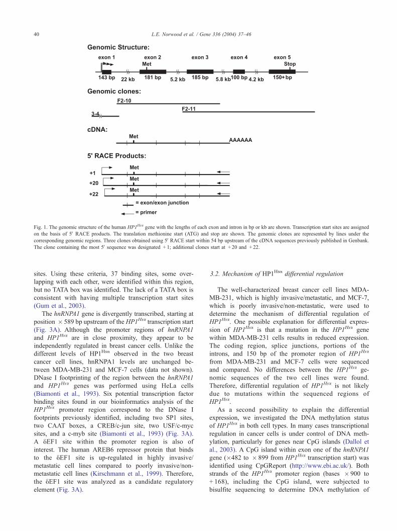
Fig. 1. The genomic structure of the human HP1Hsa gene with the lengths of each exon and intron in bp or kb are shown. Transcription start sites are assigned
on the basis of 5VRACE products. The translation methionine start (ATG) and stop are shown. The genomic clones are represented by lines under the
corresponding genomic regions. Three clones obtained using 5VRACE start within 54 bp upstream of the cDNA sequences previously published in Genbank.
The clone containing the most 5Vsequence was designated + 1; additional clones start at + 20 and + 22.
L.E. Norwood et al. / Gene 336 (2004) 37–4640
sites. Using these criteria, 37 binding sites, some over-
lapping with each other, were identified within this region,
but no TATA box was identified. The lack of a TATA box is
consistent with having multiple transcription start sites
(Gum et al., 2003).
The hnRNPA1 gene is divergently transcribed, starting at
position � 589 bp upstream of the HP1Hsa transcription start
(Fig. 3A). Although the promoter regions of hnRNPA1
and HP1Hsa are in close proximity, they appear to be
independently regulated in breast cancer cells. Unlike the
different levels of HP1Hsa observed in the two breast
cancer cell lines, hnRNPA1 levels are unchanged be-
tween MDA-MB-231 and MCF-7 cells (data not shown).
DNase I footprinting of the region between the hnRNPA1
and HP1Hsa genes was performed using HeLa cells
(Biamonti et al., 1993). Six potential transcription factor
binding sites found in our bioinformatics analysis of the
HP1Hsa promoter region correspond to the DNase I
footprints previously identified, including two SP1 sites,
two CAAT boxes, a CREB/c-jun site, two USF/c-myc
sites, and a c-myb site (Biamonti et al., 1993) (Fig. 3A).
A yEF1 site within the promoter region is also of
interest. The human AREB6 repressor protein that binds
to the yEF1 site is up-regulated in highly invasive/
metastatic cell lines compared to poorly invasive/non-
metastatic cell lines (Kirschmann et al., 1999). Therefore,
the yEF1 site was analyzed as a candidate regulatory
element (Fig. 3A).
3.2. Mechanism of HP1Hsa differential regulation
The well-characterized breast cancer cell lines MDA-
MB-231, which is highly invasive/metastatic, and MCF-7,
which is poorly invasive/non-metastatic, were used to
determine the mechanism of differential regulation of
HP1Hsa. One possible explanation for differential expres-
sion of HP1Hsa is that a mutation in the HP1Hsa gene
within MDA-MB-231 cells results in reduced expression.
The coding region, splice junctions, portions of the
introns, and 150 bp of the promoter region of HP1Hsa
from MDA-MB-231 and MCF-7 cells were sequenced
and compared. No differences between the HP1Hsa ge-
nomic sequences of the two cell lines were found.
Therefore, differential regulation of HP1Hsa is not likely
due to mutations within the sequenced regions of
HP1Hsa.
As a second possibility to explain the differential
expression, we investigated the DNA methylation status
of HP1Hsa in both cell types. In many cases transcriptional
regulation in cancer cells is under control of DNA meth-
ylation, particularly for genes near CpG islands (Dallol et
al., 2003). A CpG island within exon one of the hnRNPA1
gene (�482 to � 899 from HP1Hsa transcription start) was
identified using CpGReport (http://www.ebi.ac.uk/). Both
strands of the HP1Hsa promoter region (bases � 900 to
+ 168), including the CpG island, were subjected to
bisulfite sequencing to determine DNA methylation of
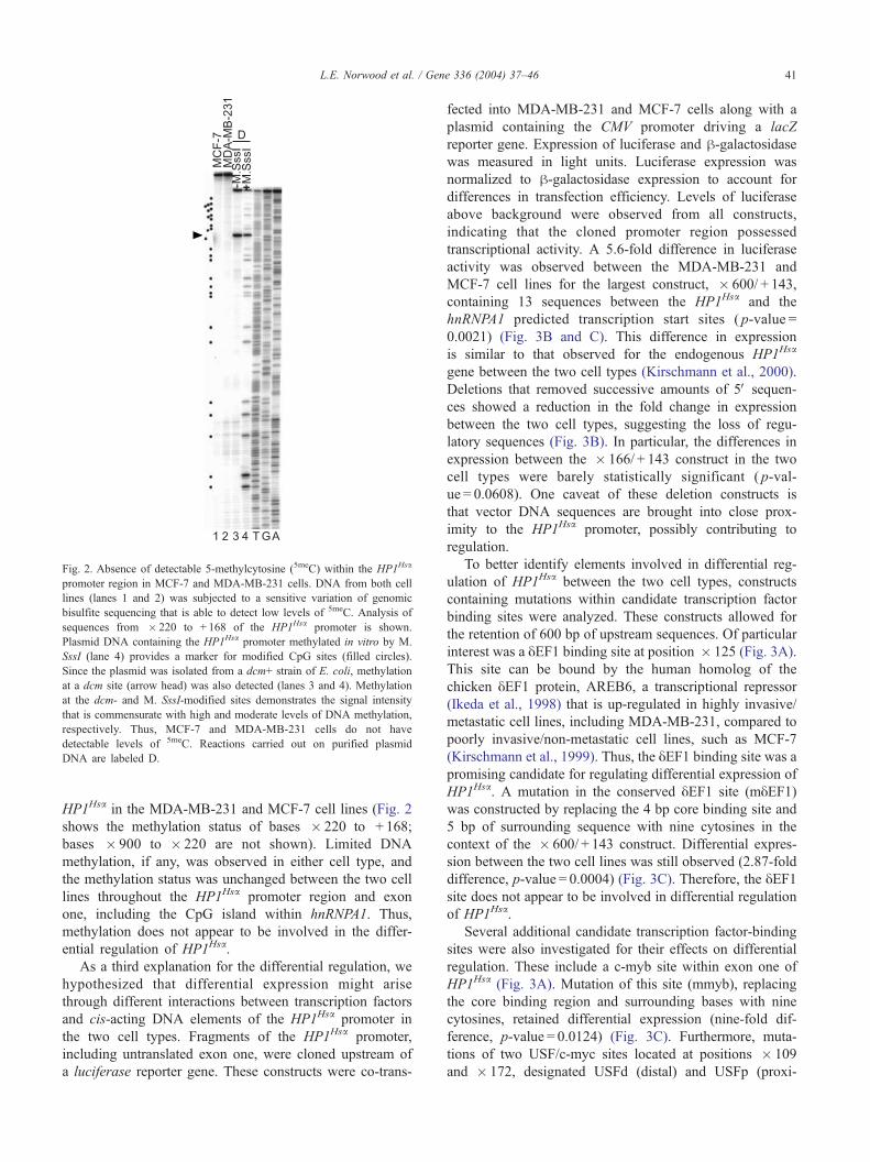
Fig. 2. Absence of detectable 5-methylcytosine (5meC) within the HP1Hsa
promoter region in MCF-7 and MDA-MB-231 cells. DNA from both cell
lines (lanes 1 and 2) was subjected to a sensitive variation of genomic
bisulfite sequencing that is able to detect low levels of 5meC. Analysis of
sequences from � 220 to + 168 of the HP1Hsa promoter is shown.
Plasmid DNA containing the HP1Hsa promoter methylated in vitro by M.
SssI (lane 4) provides a marker for modified CpG sites (filled circles).
Since the plasmid was isolated from a dcm+ strain of E. coli, methylation
at a dcm site (arrow head) was also detected (lanes 3 and 4). Methylation
at the dcm- and M. SssI-modified sites demonstrates the signal intensity
that is commensurate with high and moderate levels of DNA methylation,
respectively. Thus, MCF-7 and MDA-MB-231 cells do not have
detectable levels of 5meC. Reactions carried out on purified plasmid
DNA are labeled D.
L.E. Norwood et al. / Gene 336 (2004) 37–46 41
HP1Hsa in the MDA-MB-231 and MCF-7 cell lines (Fig. 2
shows the methylation status of bases � 220 to + 168;
bases � 900 to � 220 are not shown). Limited DNA
methylation, if any, was observed in either cell type, and
the methylation status was unchanged between the two cell
lines throughout the HP1Hsa promoter region and exon
one, including the CpG island within hnRNPA1. Thus,
methylation does not appear to be involved in the differ-
ential regulation of HP1Hsa.
As a third explanation for the differential regulation, we
hypothesized that differential expression might arise
through different interactions between transcription factors
and cis-acting DNA elements of the HP1Hsa promoter in
the two cell types. Fragments of the HP1Hsa promoter,
including untranslated exon one, were cloned upstream of
a luciferase reporter gene. These constructs were co-trans-
fected into MDA-MB-231 and MCF-7 cells along with a
plasmid containing the CMV promoter driving a lacZ
reporter gene. Expression of luciferase and h-galactosidasewas measured in light units. Luciferase expression was
normalized to h-galactosidase expression to account for
differences in transfection efficiency. Levels of luciferase
above background were observed from all constructs,
indicating that the cloned promoter region possessed
transcriptional activity. A 5.6-fold difference in luciferase
activity was observed between the MDA-MB-231 and
MCF-7 cell lines for the largest construct, � 600/ + 143,
containing 13 sequences between the HP1Hsa and the
hnRNPA1 predicted transcription start sites ( p-value =
0.0021) (Fig. 3B and C). This difference in expression
is similar to that observed for the endogenous HP1Hsa
gene between the two cell types (Kirschmann et al., 2000).
Deletions that removed successive amounts of 5V sequen-ces showed a reduction in the fold change in expression
between the two cell types, suggesting the loss of regu-
latory sequences (Fig. 3B). In particular, the differences in
expression between the � 166/ + 143 construct in the two
cell types were barely statistically significant ( p-val-
ue = 0.0608). One caveat of these deletion constructs is
that vector DNA sequences are brought into close prox-
imity to the HP1Hsa promoter, possibly contributing to
regulation.
To better identify elements involved in differential reg-
ulation of HP1Hsa between the two cell types, constructs
containing mutations within candidate transcription factor
binding sites were analyzed. These constructs allowed for
the retention of 600 bp of upstream sequences. Of particular
interest was a yEF1 binding site at position � 125 (Fig. 3A).
This site can be bound by the human homolog of the
chicken yEF1 protein, AREB6, a transcriptional repressor
(Ikeda et al., 1998) that is up-regulated in highly invasive/
metastatic cell lines, including MDA-MB-231, compared to
poorly invasive/non-metastatic cell lines, such as MCF-7
(Kirschmann et al., 1999). Thus, the yEF1 binding site was apromising candidate for regulating differential expression of
HP1Hsa. A mutation in the conserved yEF1 site (myEF1)was constructed by replacing the 4 bp core binding site and
5 bp of surrounding sequence with nine cytosines in the
context of the � 600/ + 143 construct. Differential expres-
sion between the two cell lines was still observed (2.87-fold
difference, p-value = 0.0004) (Fig. 3C). Therefore, the yEF1site does not appear to be involved in differential regulation
of HP1Hsa.
Several additional candidate transcription factor-binding
sites were also investigated for their effects on differential
regulation. These include a c-myb site within exon one of
HP1Hsa (Fig. 3A). Mutation of this site (mmyb), replacing
the core binding region and surrounding bases with nine
cytosines, retained differential expression (nine-fold dif-
ference, p-value = 0.0124) (Fig. 3C). Furthermore, muta-
tions of two USF/c-myc sites located at positions � 109
and � 172, designated USFd (distal) and USFp (proxi-
0 10 20 30
0 10 20 30
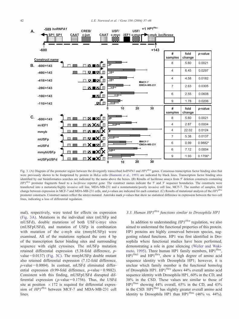
Fig. 3. (A) Diagram of the promoter region between the divergently transcribed hnRNPA1 and HP1Hsa genes. Consensus transcription factor binding sites that
were previously shown to be footprinted by protein in HeLa cells (Biamonti et al., 1993) are indicated by black lines. Transcription factor binding sites
identified by our bioinformatics searches are indicated by the name above the boxes. (B) Results of luciferase assays from 5Vdeletion constructs containing
HP1Hsa promoter fragments fused to a luciferase reporter gene. The construct names indicate the 5V and 3V sequence boundaries. The constructs were
transfected into a metastatic/highly invasive cell line, MDA-MB-231 and a nonmetastatic/poorly invasive cell line, MCF-7. The number of samples, fold
change between expression in MCF-7 and MDA-MB-231 cells, and p-values are indicated for each construct. (C) Results of mutational analysis of the HP1Hsa
promoter constructs. Construct names reflect the site(s) mutated. Asterisks mark p-values that show no statistical difference in expression between the two cell
lines, indicating a loss of differential regulation.
L.E. Norwood et al. / Gene 336 (2004) 37–4642
mal), respectively, were tested for effects on expression
(Fig. 3A). Mutations in the individual sites (mUSFp and
mUSFd), double mutations of both USF/c-myc sites
(mUSFpUSFd), and mutation of USFp in combination
with mutation of the c-myb site (mmybUSFp) were
examined. All of the mutations replaced the core 4 bp
of the transcription factor binding sites and surrounding
sequence with eight cytosines. The mUSFp mutation
retained differential expression (5.38-fold difference, p-
value = 0.0137) (Fig. 3C). The mmybUSFp double mutant
also retained differential expression (7.12-fold difference,
p-value = 0.0004). In contrast, mUSFd eliminated differ-
ential expression (0.99-fold difference, p-value = 0.9882).
Consistent with this finding, mUSFpUSFd disrupted dif-
ferential expression ( p-value = 0.1756). Thus, the USFd
site at position � 172 is required for differential expres-
sion of HP1Hsa between MCF-7 and MDA-MB-231 cell
lines.
3.3. Human HP1Hsa functions similar to Drosophila HP1
In addition to understanding HP1Hsa regulation, we also
aimed to understand the functional properties of this protein.
HP1 proteins are highly conserved between species, sug-
gesting related functions. HP1 was first identified in Dro-
sophila where functional studies have been performed,
demonstrating a role in gene silencing (Weiler and Waki-
moto, 1995). Three human HP1 family members, HP1Hsa,
HP1Hsh and HP1Hsg, show a high degree of amino acid
sequence identity with Drosophila HP1; however, it is
unclear which family member is the functional homolog
of Drosophila HP1. HP1Hsg shows 44% overall amino acid
sequence identity with Drosophila HP1, 60% in the CD, and
38% in the CSD. These values are similar to those of
HP1Hsa showing 44% overall, 65% in the CD, and 43%
in the CSD. HP1Hsh has slightly greater overall amino acid
identity to Drosophila HP1 than HP1Hsa (46% vs. 44%).
L.E. Norwood et al. / Gene 336 (2004) 37–46 43
The HP1Hsh CD shows slightly more identity with the
Drosophila HP1 CD than the HP1Hsg and HP1Hsa CDs
(68% vs. 60% and 65%, respectively). In contrast, the
HP1Hsh CSD shows less identity to the Drosophila HP1
CSD than the HP1Hsa CSD (39% vs. 43%). In sum,
comparisons of the amino acid sequences of human and
Drosophila HP1 identified only minor differences in the
percent identity without immediately suggesting a function-
al homologue.
Another protein feature that might suggest similar func-
tion between Drosophila HP1 and a human HP1 is the
chromosome localization pattern. Drosophila HP1 shows
enrichment at heterochromatic regions and localizes to
approximately 200 euchromatic sites on larval polytene
chromosomes (Fanti et al., 2003). HP1Hsa and HP1Hsh
predominantly localize to centric heterochromatin, showing
partial overlap with anti-centromere antibodies (Minc et al.,
1999). In contrast, HP1Hsg localizes to centric heterochro-
matin and euchromatic regions (Minc et al., 2000). Based on
this localization data, HP1Hsg appears to have a more similar
pattern to that of Drosophila HP1.
To investigate the functional properties of the HP1Hsa
protein and determine whether it is a functional homologue
of Drosphila HP1, we generated transgenic Drosophila that
expressed an HP1Hsa-EGFP fusion gene under the control
of an hsp70 heat shock promoter. Homozygous HP1Hsa-
EGFP larvae were heat shocked 1 h at 37jC and allowed
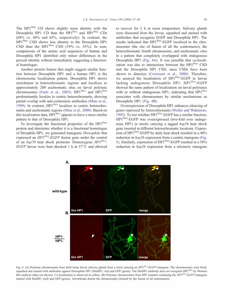
Fig. 4. (A) Polytene chromosomes from third instar larvae salivary glands from a
squashed and stained with antibodies against Drosophila HP1 (DmHP1; red) and
blot analysis (data not shown). Co-localization is observed in yellow. (B) Polyten
stained with DmHP1 (red) and GFP (green). Arrowheads denote the chromocent
to recover for 2 h at room temperature. Salivary glands
were dissected from the larvae, squashed and stained with
antibodies that recognize EGFP and Drosophila HP1. The
results indicated that HP1Hsa-EGFP localized to the chro-
mocenter (the site of fusion of all the centromeres), the
heterochromatic fourth chromosome, and euchromatic sites
in a pattern that completely overlapped with endogenous
Drosophila HP1 (Fig. 4A). It was possible that co-locali-
zation was due to interactions between the HP1Hsa CSD
and the Drosophila HP1 CSD, since CSDs have been
shown to dimerize (Cowieson et al., 2000). Therefore,
we assayed the localization of HP1Hsa-EGFP in larvae
lacking endogenous Drosophila HP1. HP1Hsa-EGFP
showed the same pattern of localization on larval polytenes
with or without endogenous HP1, indicating that HP1Hsa
associates with chromosomes by similar mechanisms as
Drosophila HP1 (Fig. 4B).
Overexpression of Drosophila HP1 enhances silencing of
genes repressed by heterochromatin (Weiler and Wakimoto,
1995). To test whether HP1Hsa-EGFP has a similar function,
HP1Hsa-EGFP was overexpressed (two-fold over endoge-
nous HP1) in stocks carrying a tagged hsp26 heat shock
gene inserted at different heterochromatic locations. Expres-
sion of HP1Hsa-EGFP by daily heat shock resulted in a 40%
reduction in hsp26 expression from a centric transgene (Fig.
5). Similarly, expression of HP1Hsa-EGFP resulted in a 50%
reduction in hsp26 expression from a telomeric transgene
stock carrying an HP1Hsa-EGFP transgene. The chromosomes were fixed,
GFP (green). The DmHP1 antibody does not recognize HP1Hsa by Western
e chromosomes from HP1 mutants containing the HP1Hsa-EGFP transgene
er (formed by the fusion of all centromeres).
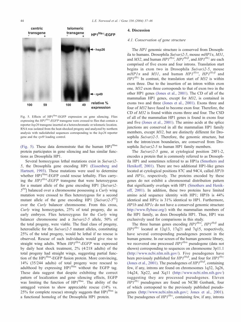
Fig. 5. Effects of HP1Hsa-EGFP expression on gene silencing. Flies
expressing the HP1Hsa-EGFP transgene were crossed to flies that contain a
reporter hsp26 transgene inserted at a heterochromatic or telomeric location.
RNAwas isolated from the heat-shocked progeny and analyzed by northern
analysis with radiolabeled sequences corresponding to the hsp26 reporter
gene and the rp49 loading control.
L.E. Norwood et al. / Gene 336 (2004) 37–4644
(Fig. 5). These data demonstrate that the human HP1Hsa
protein participates in gene silencing and has similar func-
tions as Drosophila HP1.
Several homozygous lethal mutations exist in Su(var)2-
5, the Drosophila gene encoding HP1 (Eissenberg and
Hartnett, 1993). These mutations were used to determine
whether HP1Hsa-EGFP could rescue lethality. Flies carry-
ing the HP1Hsa-EGFP transgene that were heterozygous
for a mutant allele of the gene encoding HP1 [Su(var)2-
504] balanced over a chromosome possessing a Curly wing
mutation were crossed to flies heterozygous for a second
mutant allele of the gene encoding HP1 [Su(var)2-502]
over the Curly balancer chromosome. From this cross,
Curly wing homozygotes, 25% of total progeny, die as
early embryos. Flies heterozygous for the Curly wing
balancer chromosome and a Su(var)2-5 allele, 50% of
the total progeny, were viable. The final class of progeny,
heteroallelic for the Su(var)2-5 mutant alleles, constituting
25% of the total progeny, would be lethal if no rescue is
observed. Rescue of such individuals would give rise to
straight wing adults. When HP1Hsa-EGFP was expressed
by daily heat shock treatment, 2% (4/218 adults) of the
total progeny had straight wings, suggesting partial func-
tion of the HP1Hsa-EGFP fusion protein. More convincing,
14% (35/244 adults) of total progeny were rescued to
adulthood by expressing HP1Hsa without the EGFP tag.
These data suggest that despite exhibiting the correct
pattern of localization and gene silencing effects, EGFP
was limiting the function of HP1Hsa. The ability of the
untagged version to show appreciable rescue (14% vs.
25% for complete rescue) strongly suggests that HP1Hsa is
a functional homolog of the Drosophila HP1 protein.
4. Discussion
4.1. Conservation of gene structure
The HP1 genomic structure is conserved from Drosoph-
ila to humans. Drosophila Su(var)2-5, mouse mHP1a, M31,
and M32, and human HP1Hsa, HP1Hsb, and HP1Hsc are each
comprised of five exons and four introns. Translation start
begins in exon two in Drosophila Su(var)2-5, mouse
mHP1a and M31, and human HP1Hsa, HP1Hsb and
HP1Hsc In contrast, the translation start of M32 is within
exon three. Due to the insertion of an intron within exon
one, M32 exon three corresponds to that of exon two in the
other HP1 genes (Jones et al., 2001). The CD of all of the
mammalian HP1 genes, except for M32, is contained in
exons two and three (Jones et al., 2001). Exons three and
four of M32 have fused to become exon four. Therefore, the
CD of M32 is found within exons three and four. The CSD
of all of the mammalian HP1 genes is found in exons four
and five (Jones et al., 2001). The amino acids at the splice
junctions are conserved in all the mammalian HP1 family
members, except M32, but are distinctly different for Dro-
sophila Su(var)2-5. Therefore, the genomic structure, but
not the intron/exon boundaries, are conserved from Dro-
sophila Su(var)2-5 to human HP1 family members.
The Su(var)2-5 gene, at cytological position 28F1-2,
encodes a protein that is commonly referred to as Drosoph-
ila HP1 and sometimes referred to as HP1a (Smothers and
Henikoff, 2001). There are two additional HP1-like genes
located at cytological positions 87C and 94C4, called HP1b
and HP1c, respectively. The proteins encoded by these
genes do not exhibit a chromosomal distribution pattern
that significantly overlaps with HP1 (Smothers and Henik-
off, 2001). In addition, these two proteins have limited
amino acid sequence identity with HP1; HP1b is 44%
identical and HP1c is 31% identical to HP1. Furthermore,
HP1b and HP1c do not have a conserved genomic structure
http://www.flybase.org/) with the mammalian members of
the HP1 family, as does Drosophila HP1. Thus, HP1 was
exclusively used for comparisons in this study.
The three human genes encoding HP1Hsa, HP1Hsb and
HP1Hsc located at 12q13, 17q21 and 7q15, respectively,
have several corresponding pseudogenes present in the
human genome. In our screen of the human genomic library,
we recovered one processed HP1Hsa pseudogene (data not
shown) corresponding to sequences on chromosome 3p11.1
(http://www.ncbi.nlm.nih.gov/). Five pseudogenes have
been previously published for HP1Hsb, and four for HP1Hsc
(Jones et al., 2001). The pseudogenes of HP1Hsb, containing
few, if any, introns are found on chromosomes 1q32, 3q26,
14q24, Xp22, and Xq11 (http://www.ncbi.nlm.nih.gov/)
suggesting they are processed pseudogenes. Eleven
HP1Hsc pseudogenes are found on NCBI Genbank, four
of which correspond to the previously published pseudo-
genes (http://www.ncbi.nlm.nih.gov/; Jones et al., 2001).
The pseudogenes of HP1Hsc, containing few, if any, introns
L.E. Norwood et al. / Gene 336 (2004) 37–46 45
are found on chromosomes 2q24, 3p22, 5q22, 6q22.2,
11p11, 11p14, 11q14, 12p13, 12q23, 16p13, and 18p11
(http://www.ncbi.nlm.nih.gov/). Thus, each of the functional
human HP1 family members is encoded by separate un-
linked genes that have multiple pseudogenes scattered
throughout the genome.
4.2. HP1Hsa regulation in breast cancer metastasis
HP1Hsa is down-regulated in highly invasive/metastatic
breast cancer cells compared to poorly invasive/non-meta-
static cells (Kirschmann et al., 2000). This regulation likely
occurs, at least in part, at the transcriptional level and does
not involve differential DNA methylation. An analysis of
the DNA sequences in the HP1Hsa promoter region identi-
fied potential binding sites for transcriptional regulators that
might be involved in differential regulation. Only three of
the binding sites shown in Fig. 3, the two SP1 sites
immediately upstream of hnRNPA1, the CAAT box (posi-
tion � 244) and the proximal USF/c-myc site (position
� 109), are conserved between mouse and humans; none
of the elements can be identified upstream of the gene
encoding Drosophila HP1.
Mutation of a yEF1 binding site (at position � 125),
which associates with the AREB6 protein found to be up-
regulated in highly invasive/metastatic breast cancer cells
(Kirschmann et al., 1999), does not appear to be involved in
the differential expression of HP1Hsa. Mutation of a c-myb
binding site at position + 97 and a USF/c-myc site at
position � 109 does not appear to play a role in differential
regulation either. In contrast, mutation of a USF/c-myc site
at position � 172 abolishes differential regulation. This
USF/c-myc was protected from DNase I digestion, suggest-
ing occupancy by protein(s) in HeLa cells (Biamonti et al.,
1993). USF/c-myc sites, commonly called E-boxes, are
frequently bound by a variety of proteins, including USF
and Myc family members. USF proteins are involved in
both gene silencing and activation, sometimes at the same
site under different circumstances (Goueli and Janknecht,
2003). Myc proteins are also involved in both gene activa-
tion and repression, depending on their dimerization partner
(Queva et al., 1998). Therefore, the function of the distal
USF/c-myc site in the differential regulation of HP1Hsa is
difficult to predict and warrants further investigation.
4.3. Function of HP1Hsa
Our data strongly suggest that HP1Hsa is a functional
homolog of the Drosophila HP1 protein. The results
showing that HP1Hsa can localize to the same sites on
polytene chromosomes as Drosophila HP1 are consistent
with previously published results (Ma et al., 2001). We
extend these findings by demonstrating that HP1Hsa
exhibits the Drosophila HP1 pattern of localization even
in the absence of Drosophila HP1. These results suggest a
conserved mechanism for localization. Previously pub-
lished results show that HP1Hsa can enhance silencing
induced by a transgene array in Drosophila (Ma et al.,
2001). These arrays have similar, yet distinctly different,
properties than heterochromatin (Prasad-Sinha et al.,
2000). Our results clearly demonstrate that HP1Hsa can
participate in heterochromatin formation and silence eu-
chromatic genes placed within heterochromatin. Thus,
HP1Hsa has gene silencing functions similar to Drosophila
HP1.
Species specificity of protein function can be addressed
by determining whether a protein from one organism can
provide complete function of the homologous protein in
another organism. HP1Hsa can rescue the lethality of a
Drosophila HP1 homozygous mutant; therefore, HP1Hsa is
a functional homolog of Drosophila HP1. In contrast to
our findings, the mouse M31 protein was unable to rescue
mutant phenotypes associated with S. pombe Swi6 mutants
(Wang et al., 2000). Rescue was obtained, however, when
the Swi6 CSD was substituted for the M31 CSD (Wang et
al., 2000). The overall amino acid sequence identity
between S. pombe Swi6 and mouse M31 is 37%. This is
much less than the 44% overall amino acid sequence
identity between Drosophila HP1 and human HP1Hsa. In
particular, the amino acid sequence identity between the S.
pombe Swi6 CSD and the mouse M31 CSD is 39%,
whereas the amino acid sequence identity between Dro-
sophila HP1 CSD and the human HP1Hsa CSD is 43%.
Therefore, the CSD of Drosophila HP1 and human HP1Hsa
is more conserved than the CSD of S. pombe Swi6 and
mouse M31. The amino acid sequence differences between
mouse and S. pombe might explain the species-specificity
observed.
4.4. Model for HP1Hsa function in breast cancer metastasis
Given the conserved function of HP1Hsa in gene regula-
tion, one possible role for HP1Hsa in breast cancer metas-
tasis is gene silencing. Accordingly, the HP1Hsa gene would
be expressed in normal and primary breast cancer tumor
cells where it produces protein that functions to silence
genes required for invasion and metastasis. In highly inva-
sive/metastatic breast cancer cells, HP1Hsa expression is
reduced and less HP1Hsa protein is available to carry out
gene silencing functions. Clearly in Drosophila and mice
HP1 affects gene expression in a dosage-dependent manner
(Weiler and Wakimoto, 1995; Festenstein et al., 1999).
According to this model, loss of silencing would occur at
genes encoding proteins that are required for invasion and
metastasis. Therefore, the identification of HP1Hsa regulated
genes is a goal for future investigation.
Acknowledgements
Funding for the breast cancer research was supported by
the DOD Breast Cancer Research Program (DAMD17-02-1-
L.E. Norwood et al. / Gene 336 (2004) 37–4646
0424). The Drosophila research was supported by NIH
(GM61513). We thank members of the Wallrath lab for
comments regarding the manuscript.
References
Biamonti, G., Bassi, M.T., Cartegni, L., Mechta, F., Buvoli, M., Cobianchi,
F., Riva, S., 1993. Human hnRNP protein A1 gene expression. Struc-
tural and functional characterization of the promoter. J. Mol. Biol. 230,
77–89.
Brehm, A., Tufteland, K.R., Aasland, R., Becker, P.B., 2004. The many
colours of chromodomains. Bioessays 26, 133–140.
Cowieson, N.P., Partridge, J.F., Allshire, R.C., McLaughlin, P.J., 2000.
Dimerisation of a chromo shadow domain and distinctions from the
chromodomain as revealed by structural analysis. Curr. Biol. 10,
517–525.
Dallol, A., Krex, D., Hesson, L., Eng, C., Maher, E.R., Latif, F., 2003.
Frequent epigenetic inactivation of the SLIT2 gene in gliomas. Onco-
gene 22, 4611–4616.
Eissenberg, J.C., Elgin, S.C., 2000. The HP1 protein family: getting a grip
on chromatin. Curr. Opin. Genet. Dev. 10, 204–210.
Eissenberg, J.C., Hartnett, T., 1993. A heat shock-activated cDNA res-
cues the recessive lethality of mutations in the heterochromatin-asso-
ciated protein HP1 of Drosophila melanogaster. Mol. Gen. Genet.
240, 333–338.
Ekwall, K., Javerzat, J.P., Lorentz, A., Schmidt, H., Cranston, G., Allshire,
R., 1995. The chromodomain protein Swi6: a key component at fission
yeast centromeres. Science 269, 1429–1431.
Fanti, L., Berloco, M., Piacentini, L., Pimpinelli, S., 2003. Chromosomal
distribution of heterochromatin protein 1 (HP1) in Drosophila: a cyto-
logical map of euchromatic HP1 binding sites. Genetica 117, 135–147.
Festenstein, R., Sharghi-Namini, S., Fox, M., Roderick, K., Tolaini, M.,
Norton, T., Saveliev, A., Kioussis, D., Singh, P., 1999. Heterochromatin
protein 1 modifies mammalian PEV in a dose- and chromosomal-con-
text-dependent manner. Nat. Genet. 23, 457–461.
Goueli, B.S., Janknecht, R., 2003. Regulation of telomerase reverse tran-
scriptase gene activity by upstream stimulatory factor. Oncogene 22,
6098–6103.
Gum Jr., J.R., Hicks, J.W., Crawley, S.C., Dahl, C.M., Yang, S.C., Rob-
erton, A.M., Kim, Y.S., 2003. The MUC3A human intestinal mucin:
Initiation of transcription from a TATA-less promoter and comparison to
the MUC3B amino terminus. J. Biol. Chem.
Ikeda, K., Halle, J.P., Stelzer, G., Meisterernst, M., Kawakami, K., 1998.
involvement of negative cofactor NC2 in active repression by zinc
finger-homeodomain transcription factor AREB6. Mol. Cell Biol. 18,
10–18.
Jones, D.O., Mattei, M.G., Horsley, D., Cowell, I.G., Singh, P.B., 2001.
The gene and pseudogenes of Cbx3/mHP1 gamma. DNA Seq. 12,
147–160.
Kirschmann, D.A., Seftor, E.A., Nieva, D.R., Mariano, E.A., Hendrix,
M.J., 1999. Differentially expressed genes associated with the meta-
static phenotype in breast cancer. Breast Cancer Res. Treat. 55,
127–136.
Kirschmann, D.A., Lininger, R.A., Gardner, L.M., Seftor, E.A., Odero,
V.A., Ainsztein, A.M., Earnshaw, W.C., Wallrath, L.L., Hendrix,
M.J., 2000. Down-regulation of HP1Hsalpha expression is associat-
ed with the metastatic phenotype in breast cancer. Cancer Res. 60,
3359–3363.
Kladde, M.P., Xu, M., Simpson, R.T., 1996. Direct study of DNA-protein
interactions in repressed and active chromatin in living cells. EMBO J.
15, 6290–6300.
Ma, J., Hwang, K.K., Worman, H.J., Courvalin, J.C., Eissenberg, J.C.,
2001. Expression and functional analysis of three isoforms of human
heterochromatinassociated protein HP1 in Drosophila. Chromosoma
109, 536–544.
Minc, E., Allory, Y., Worman, H.J., Courvalin, J.C., Buendia, B., 1999.
Localization and phosphorylation of HP1 proteins during the cell cycle
in mammalian cells. Chromosoma 108, 220–234.
Minc, E., Courvalin, J.C., Buendia, B., 2000. HP1gamma associates with
euchromatin and heterochromatin in mammalian nuclei and chromo-
somes. Cytogenet. Cell Genet. 90, 279–284.
Muchardt, C., Guilleme,M., Seeler, J.S., Trouche, D., Dejean, A., Yaniv, M.,
2002. Coordinated methyl and RNA binding is required for heterochro-
matin localization of mammalian HP1alpha. EMBO Rep. 3, 975–981.
Nielsen, A.L., Oulad-Abdelghani, M., Ortiz, J.A., Remboutsika, E., Cham-
bon, P., Losson, R., 2001. Heterochromatin formation in mammalian
cells: interaction between histones and HP1 proteins. Mol. Cell 7,
729–739.
Platero, J.S., Hartnett, T., Eissenberg, J.C., 1995. Functional analysis of the
chromo domain of HP1. EMBO J. 14, 3977–3986.
Prasad-Sinha, J., Furuyama, T., Dorer, D., Harte, P.J., 2000. The Polycomb
Group proteins ESC and E(Z) are required for repeat-induced gene
silencing. 41st Annual Drosophila Research Conference. The Genetics
Society of America, Pittsburgh, PA, p. 231B.
Queva, C., Hurlin, P.J., Foley, K.P., Eisenman, R.N., 1998. Sequential
expression of the MAD family of transcriptional repressors during dif-
ferentiation and development. Oncogene 16, 967–977.
Shaffer, C.D., Wuller, J.M., Elgin, S.C., 1994. Raising large quantities
of Drosophila for biochemical experiments. Methods Cell Biol. 44,
99–108.
Smothers, J.F., Henikoff, S., 2000. The HP1 chromo shadow domain binds
a consensus peptide pentamer. Curr. Biol. 10, 27–30.
Smothers, J.F., Henikoff, S., 2001. The hinge and chromo shadow domain
impart distinct targeting of HP1-like proteins. Mol. Cell Biol. 21,
2555–2569.
Wallrath, L.L., Burnett, J.B., Friedman, T.B., 1990. Molecular character-
ization of the Drosophila melanogaster urate oxidase gene, an ecdy-
sone-repressible gene expressed only in the malpighian tubules. Mol.
Cell Biol. 10, 5114–5127.
Wang, G., Ma, A., Chow, C.M., Horsley, D., Brown, N.R., Cowell, I.G.,
Singh, P.B., 2000. Conservation of heterochromatin protein 1 function.
Mol. Cell Biol. 20, 6970–6983.
Weiler, K.S., Wakimoto, B.T., 1995. Heterochromatin and gene expression
in Drosophila. Annu. Rev. Genet. 29, 577–605.


