Embed Size (px)
Citation preview
Journal of Medical Microbiology (2004), 53, 679–690 DOI 10.1099/jmm.0.45539-0
45539 & 2004 SGM Printed in Great Britain 679
Correspondence
Kalai Mathee
Received 11 November 2003
Accepted 5 March 2004
Alginate production affects Pseudomonasaeruginosa biofilm development and architecture,but is not essential for biofilm formation
Andres Plata Stapper,1 Giri Narasimhan,2 Dennis E. Ohman,3
Johnny Barakat,1 Morten Hentzer,4 Søren Molin,4 Arsalan Kharazmi,5
Niels Høiby5 and Kalai Mathee1
1,2Department of Biological Sciences1 and School of Computer Science2, Florida InternationalUniversity, Miami, FL 33199, USA
3Department of Microbiology and Immunology, Virginia Commonwealth University, Richmond, VA23298, USA
4Section of Molecular Microbiology, The Technical University of Denmark, DK-2800 Lyngby,Denmark
5Department of Clinical Microbiology, University Hospital of Copenhagen, DK-2100 Copenhagen,Denmark
Extracellular polymers can facilitate the non-specific attachment of bacteria to surfaces and hold
together developing biofilms. This study was undertaken to qualitatively and quantitatively compare
the architecture of biofilms produced by Pseudomonas aeruginosa strain PAO1 and its alginate-
overproducing (mucA22) and alginate-defective (algD) variants in order to discern the role of
alginate in biofilm formation. These strains, PAO1, Algþ PAOmucA22 and Alg� PAOalgD, tagged
with green fluorescent protein, were grown in a continuous flow cell system to characterize the
developmental cycles of their biofilm formation using confocal laser scanning microscopy. Biofilm
Image Processing (BIP) and Community Statistics (COMSTAT) software programs were used to
provide quantitative measurements of the two-dimensional biofilm images. All three strains formed
distinguishable biofilm architectures, indicating that the production of alginate is not critical for
biofilm formation. Observation over a period of 5 days indicated a three-stage development pattern
consisting of initiation, establishment and maturation. Furthermore, this study showed that
phenotypically distinguishable biofilms can be quantitatively differentiated.
INTRODUCTION
The most commonmode of bacterial growth in nature is theformation on surfaces of organized biofilm communities,held together by a matrix composed of exopolysaccharides(EPS) (Costerton et al., 1987). The EPS matrix has beenimplicated in maintaining the individual cells and commu-nities of a biofilm in close proximity and in providing aunique microenvironmental niche (Costerton et al., 1994).Much of our understanding of biofilms comes from studiesof the opportunistic pathogen Pseudomonas aeruginosa,which causes chronic lung infection and is the major causeof morbidity and mortality in patients with cystic fibrosis(CF) (Høiby, 1975). P. aeruginosa strains isolated from thelungs of patients, especially with advanced stages of disease,
are distinctive because about 85% have a mucoid colonymorphology (Fick et al., 1992). In contrast, only 1% ofstrains isolated from other sites of infection are mucoid(Doggett et al., 1966). These observations suggest thatmucoid P. aeruginosa cells have a distinct survival advantagein the CF lung environment. This mucoid phenotype isindicative of the overproduction of the EPS alginate, anO-acetylated linear polymer of D-mannuronate and L-guluronate residues (Evans & Linker, 1973). The expressionof this polymer confers increased resistance to the hostimmune response and results in chronic pulmonary infec-tion and poor prognosis for the patient (Baltimore &Mitchell, 1980; Govan & Harris, 1986). Infection withalginate-producing P. aeruginosa in CF patients has beenassociated with an overactive immune response and a poorclinical condition, suggesting that alginate production is avirulence factor (Høiby, 1974; Baltimore et al., 1989;Pedersen et al., 1992). Also, mucoid organisms have been
Abbreviations: CF, cystic fibrosis; CLSM, confocal laser scanningmicroscopy; EPS, exopolysaccharide.

observed in a biofilmmode of growth in the lungs (Lam et al.,1980). Animal studies support the view that alginate produc-tion impedes host immune clearance, contributes to tissuedamage and favours survival in the lung (Boucher et al., 1997;Yu et al., 1998; Song et al., 2003).
In recent years, it has become apparent that predicting actualbacterial behaviour in their natural environment, on thebasis of experiments done in liquid suspension growthmedia(i.e. planktonic form), may not always be reliable. As a result,many laboratories have begun to investigate how cells cancoordinate their activities and build the complex structuresthat are found in mature biofilms. The advent of non-destructive techniques, especially the use of live-monitorsystems with confocal laser scanning microscopy (CLSM),has greatly increased our understanding and appreciation ofthe complex architecture of biofilms (Lawrence et al., 1991).Such analyses of biofilms have shown that there is often athree-dimensional distribution of organisms with specificsubstructures, leading to a model for biofilm structure calledthe ‘water channel model’ (Costerton et al., 1987). In thismodel, the biofilm is not just multiple layers of evenlydistributed cells, but is composed of many substructuresprotruding from the substratum to the top of the biofilm.These substructures have void sectors representing channelsthroughwhich substrate andwaste products canmove. Thesesubstructures, designated mounds, mushrooms and voidchannels, penetrate from the substratum, and are heldtogether by the EPS matrix (Costerton et al., 1994).
The biofilm architecture of an organism on a surface can bedescribed in terms of the direction of growth, which can behorizontal, vertical or combined. Horizontal biofilm growthoccurs when the bacteria colonize an available substratum.However, it can also occur when nutrients are scarce andbacteria are forced to extend the biofilm in order to find anew carbon or nutrient source (Stoodley et al., 1999). In thismode, the biofilm grows parallel to the substratum andspreads horizontally. This increases the surface area coveredby the biofilm, with little increase in thickness, leading toconfluent growth covering the available surface area. Whenthe bacteria have already colonized the available substratum,the biofilm extends vertically, increasing the thickness of thebiofilm. Vertical growth can also be a result of physicalbarriers when a biofilm has covered the available surface.Differences and combinations of horizontal and verticalbiofilm growth determine the different structures, such asmushrooms, towers and channels as described in the waterchannel model (Costerton et al., 1987). Differences andcombinationsmay occur in different ratios at different times,and at different points in the biofilm, and they aremost likelycontrolled by a combination of gene expression, as well asfactors generated by environmental conditions.
P. aeruginosa biofilm cells typically exhibit very slow growthrates relative to planktonic cells grown in liquid cultures(Brown et al., 1988). The difference in physiology of thebacteria in these two modes of growth contributes to thedifferences in their cellular metabolism (Brown et al., 1988).
Differences have been observed in the expressions of proteinprofiles (Brown & Williams, 1985; Sauer & Camper, 2001),�-lactamase (Gilbert & Brown, 1998), fimbriae (O’Toole &Kolter, 1998), pili (O’Toole & Kolter, 1998), superoxidedismutases (Hassett et al., 1999) and catalases (Hassett et al.,1999). The biofilm mode of growth contributes to increasedresistance to hydrogen peroxide (Hassett et al., 1999;Cochran et al., 2000), antibiotics (Hoyle & Costerton,1991; Ashby et al., 1994; Gander, 1996; Schierholz et al.,1999) and phagocytosis (Jensen et al., 1990).
The formation of an EPS matrix may play an important rolein establishing a sustainable biofilm. It was postulated thatthe expression of alginate genes may be critical to biofilmformation by P. aeruginosa because expression of algC is highin the mucoid strain PA8830 under biofilm conditions(Davies et al., 1993; Davies & Geesey, 1995). However, theproduct of the algC gene is involved in lipopolysaccharide(LPS) synthesis and rhamnolipid and alginate production(Goldberg et al., 1993; Olvera et al., 1999). As a result,activation of the algC gene could reflect activation of any ofthe three pathways. Expression of an algD–lacZ fusion wasexamined in a mucoid derivative of PAO1 called PAO579(Hoyle et al., 1993). The algD gene promoter drives the 18 kbalginate biosynthetic operon, and thus activation of thispromoter indicates expression of alginate. The algD–lacZactivity in adherent cells within a modified Robbins device,compared with cells in the effluent, showed a transientelevation of promoter activity, suggesting enhanced EPSproduction following adherence. Nivens et al. (2001) ex-plored the role of alginate and its O-acetylation in theformation of biofilm using a CF clinical isolate FRD1 andits mutant derivatives. They showed that O-acetylation ofalginate was critical for successful biofilmmaturation. How-ever, the production of alginate per se may not be critical forstrain FRD1 biofilm formation.
Based on the available data, we argued that alginate may notplay a role in biofilmmatrix formation (Mathee et al., 2002).In this study, we examined the role of alginate production inthe formation and architecture of biofilms by comparing theprototype strain of P. aeruginosa, PAO1 (Holloway &Morgan, 1986), to its alginate-overproducing (Algþ) deri-vative, Algþ PAOmucA22 (PDO300; Mathee et al., 1999a, b)and to the alginate-defective (Alg�) strain PAOalgD (Garrettet al., 1999).
METHODS
Bacterial strains and plasmids. Bacterial strains and plasmids usedin this study are shown in Table 1. The prototypic non-mucoidP. aeruginosa strain PAO1 (Holloway &Morgan, 1986) and its isogenicderivatives were used. These derivatives were the mucoid strain Algþ
PAOmucA22 (PDO300) and Alg� PAOalgD. The Algþ PAOmucA22strain carries the mucA22 allele, resulting in the constitutive productionof alginate (Mathee et al., 1999a, b). TheAlg� PAOalgD is a non-mucoidstrain carrying a deletion in the biosynthetic gene algD (Garrett et al.,1999). The algD gene encodes GDP-mannose dehydrogenase that wasproposed to commit metabolic sugar intermediates to alginate produc-tion (Deretic et al., 1987; Roychoudhury et al., 1989).
A. Plata Stapper and others
680 Journal of Medical Microbiology 53

Media and growth condition. Escherichia coli and P. aeruginosa wereroutinely cultured in LB broth (10 g tryptone, 5 g yeast extract and5 gNaCl l�1). Low-salt LB broth contained 2.5 gNaCl l�1. LA/PIA, usedin triparental matings, was a 1 : 1mixture of Pseudomonas isolation agar(Difco) and LB agar. All incubations were carried out at 37 8C. Thestrains of P. aeruginosa were grown as biofilms in plexiglass flowchambers (Fig. 1) with modified EPRI media (Davies et al., 1998).
Antibiotics, when used, were at the following concentrations per mlunless indicated otherwise: ampicillin at 50 �g, kanamycin at 30 �g,tetracycline at 20 �g for E. coli; carbenicillin at 300 �g, tetracycline at60 �g and tellurite at 10 �g for P. aeruginosa.
DNAmanipulations. General DNA manipulations were performed asdescribed previously (Maniatis et al., 1982). Plasmidswere isolated fromE. coli using columns and procedures as described by the manufacturer(Qiagen). Triparental matings were used as described (Goldberg &Ohman, 1984) tomobilize plasmids from E. coli HB101 to P. aeruginosausing the conjugation helper plasmid pRK600 (Kessler et al., 1992).Transconjugants following triparental matings were selected on LA/PIAwith appropriate antibiotics.
Electroporation. Electroporation of P. aeruginosa was performedusing a procedure described previously (Mathee et al., 1997). Briefly,0.2 ml of a P. aeruginosa PAO1 overnight culture was used to inoculate20 ml fresh LB broth andwas incubated at 37 8Cwith shaking.When theculture reached an OD600 of 0.6–0.8, the cells were pelleted bycentrifugation (10 000 r.p.m., 10 min at 4 8C) and washed with 20 mlSMH buffer (300 mM sucrose, 1 mMMgCl2, 1 mM HEPES, pH 7.0).This procedure was then followed by two more washes with 10 ml coldSMH buffer and the pellet was resuspended in 1 ml cold SMH. PlasmidDNA (1–2 �g) was added to a 100 �l aliquot of these competent cells,which were then mixed and incubated on ice for 1 min. The bacteria–DNAmixture was transferred to a chilled electroporation cuvette (Bio-Rad Gene Pulser/E. coli Pulser cuvette) with a 0.2 cm electrode gap.Electroporationwas performed at 800�, 25 �F and 8 kV (cmgap)�1 ona Gene Pulser electroporator (Bio-Rad). Immediately after electropora-tion, 900 �l cold SOC medium (2.0% tryptone, 0.5% yeast extract,10 mMMgCl2, 10 mMNaCl, 2.5 mMKCl, 10 mMMgSO4, 20 mMglucose) was added to the cells and incubated on ice for 30 min,followed by another 30 min incubation at 37 8C. Samples (200 �l) of theelectroporated cells were spread onto LB agar plates containing theappropriate antibiotics followed by incubation for �36 h at 37 8C.
Construction of green fluorescent protein (GFP) reporter strains
for biofilm architecture. Architectural analysis of biofilms requires areporter system for the visualization of cells and structures within abiofilm without the use of disruptive techniques. GFP from the jellyfish
Table 1. Strains and plasmids used in this study
Strains and plasmids Relevant genotype and characteristics Reference
E. coli
CC118 ºpir ˜(ara-leu) araD ˜lacX74 galE galK phoA20 thi-1 rps-1 rpoB argE(Amp) recA thi pro
hsdRMþ RP4-2-Tc : :Mu-Km : : Tn7 ºpir
Herrero et al. (1990)
MT102 F� thi araD139 ara-leu˜7679 ˜(lacIOPZY) galU gal9K r� mþ SmR T. Hansen, Novo Nordisk A/S
P. aeruginosa
PAO1 Prototypic non-mucoid wild-type strain Holloway & Morgan (1986)
PDO300 PAO1mucA22 Mathee et al. (1999b)
PAOalgD PAO1algD : : xylE aacC1, GmR Garrett et al. (1999)
KMD226 PAO1-mini-Tn5-PA1=04=03 –gfpmut3*–T0–T1, TelR This study
KMD227 PAOalgD-mini-Tn5-PA1=04=03 –gfpmut3*–T0–T1, TelR This study
KMD230 PDO300-mini-Tn5-PA1=04=03 –gfpmut3*–T0–T1, TelR This study
Plasmids
pJBA25 pUC18NotI, RBSII–gfpmut3*–T0–T1, ApR J. B. Andersen, unpublished
pUT-Tel mini-Tn5 delivery vector, ApR TelR de Lorenzo et al. (1990)
pRK600 CmR; ori ColE1 RK2-Mobþ RK2-Traþ Kessler et al. (1992)
pMH305 pUCP22 Not-derived GFP-cloning vector, ApR GmR This study
Fig. 1. Bubble traps (a) and flow cells (b). These two units weremanufactured using computerized robotic tools to ensure uniformityand quality of the products (University of Tennessee at Memphis). Thebubble trap (a) is used to trap any air bubbles introduced upstream inthe flow cell set up. The three chambers in the flow cells (b) were usedto culture three strains in the same flow cell, minimizing experimentalvariability.
Alginate and P. aeruginosa biofilms
http://jmm.sgmjournals.org 681

Aequorea victoria was used to track gene expression in biofilms becauseit allows an online, non-destructive study of biofilms. GFP absorbs bluelight at 396 and 475 nm (minor peak) and emits green light at 509 and540 nm (Prasher et al., 1992). The gfp gene has beenmutated in order toimprove the fluorescence intensity of the reporter protein. The mutantof the gfp gene, gfpmut3*, emits light at 511 nm when excited with awavelength of 488 nm. This mutant protein has an enhanced fluores-cence of up to eight times that of the wild-type protein and was used forconstructing the reporter strains used in the study (Cormack et al.,1996). The three P. aeruginosa strains mentioned above were taggedusing the PA1=04=03 –gfp–T0–T1 cassette (Andersen et al., 1998). Theexpression system consists of a multiple cloning site, a syntheticribosomal binding sequence including the start codon, as well astranscriptional and translational terminators at the end of the gfp gene.Two NotI sites flank the cassette containing the PA1=04=03 –gfp–T0–T1cassette for easy excision. This 2 kb NotI fragment was subcloned into aTn5 transposable element-based suicide vector, pJMT6, to createpMH94 (de Lorenzo et al., 1990; Hentzer et al., 2001). The advantageof using this system is that only one single stable promoter fusion isinserted into the chromosome. This is achieved by not including thetransposase gene, which prevents the transposon from replicatingoutside its suicide vector (pUT-mini-Tn5). This cassette was transposedinto the P. aeruginosa strains PAO1, Algþ PAOmucA22 and Alg�
PAOalgD by triparental mating with E. coli CC118ºpir (Hentzer et al.,2001). Since we were concerned about the effect of the Tn insertion inthe chromosome, we also tagged all the strains with pJBA129, apME6030 derivative carrying the PA1=04=03 –gfp–T0–T1 cassette. Thislow copy vector, pME6030, is extremely stable in the absence of selection(Heeb et al., 2000). Our results were similar for both the chromosomallyand plasmid-tagged strains.
Flow cell experiments. Biofilms were grown at room temperature inthree-channel flow cell chamberswith individual channel dimensions of1 3 4 3 40 mm. The original flow cells and bubble-traps of the biofilmexperimental system introduced by Caldwell (Wolfaardt et al., 1994)were modified extensively and machining was computerized to stan-dardize every unit (Fig. 1; K. Mathee and B. Gallick, unpublished). Useof these flow cells has now been described in several publications(Heydorn et al., 2000, 2002; Hentzer et al., 2001, 2002) The flow cellsystem was assembled and prepared as described previously(Christensen et al., 1999). The substratum consisted of a microscopecoverslip. Cultures for inoculation of flow cells were prepared as follows.A single colony of each strain was used to inoculate test tubes containinglow-salt LB broth. The test tubes were then incubated overnight in ashaker at 37 8C. Cultures were diluted to an OD600 of 0.1 in 0.9% NaCland used for inoculation. The flow chambers were inoculated withstrains in duplicate by injecting 350 �l of the prepared inoculum with a0.5 ml syringe (28G1/2; 0.36 3 13 mm). Following inoculation, theflow cells were left at room temperature for 1 h withoutmediumflow toallow cell attachment to the substratum. Themediumflowwas resumedat a constant rate of 3 ml h�1 using a Marlow 205S peristaltic pumpconsistent with a flow velocity of 0.2 mm s�1 within the flow cells.
Qualitative analysis of biofilms. For the architectural analysis, eachflow chamber was examined at different time-points [days 1 (24 h afterinoculation), 3 and 5 (after inoculation)] in order to monitor thedevelopment of the biofilm. Images were taken at random locations ofeach flow cell biofilmusing a confocal laser scanningmicroscope (TVS4;Leika Lasertechnik). Seven random positions were chosen for sagittalsections (xz position) to minimize experimental variability, avoidingimages close to the inlets or outlets. The number of images within eachstack depended on the thickness of the biofilm. Images were takenwith a603/0.75 oil-immersion objective. Image scanning was performedusing the 488 nm laser line from an Ar/Kr laser. The Unix-basedimaging software IMARIS was used to generate simulated fluorescence
projections and sections through the biofilm. IMARIS runs on a SiliconGraphics Indigo 2 workstation.
Quantitative analysis of biofilms using Community Statistics
(COMSTAT) software. Images obtained by CLSM were processed usingCOMSTAT (Heydorn et al., 2000). This image analysis program includesseveral features for quantifying three-dimensional biofilm image stacks.In this study, we analysed three quantities to define the biofilmarchitecture. The quantities, selected on the basis of their biologicaland physical significance, were: (i) biomass (measurement of overallvolume of the biofilm, not including pores and voids inside the biofilm,thus providing an estimate of the biomass in the biofilm), (ii) meanthickness [measurement ofmean thickness of the biofilm (including thepores and voids inside the biofilm), thus providing a measure of thespatial size occupied by the biofilm], and (iii) substratum coverage(measurement of surface area covered by the biofilm). Since some of thequantities displayed an exponential increase, all of the quantities werelog-transformed before further statistical analysis.
Quantitative analysis of biofilms using the Biofilm Image
Processing (BIP) program. Images obtained by CLSM were alsoprocessed using BIP. The BIP programprovides a number ofmeasures forquantifying two-dimensional biofilm images (Ji, 2000; Narasimhan,2004). BIP was written in Microsoft Visual C++ for the Windowsoperating system. The quantities computed using BIP selected on thebasis of their biological and physical significance were: (i) texturalentropy (measurement of disorganization and heterogeneity of thebiofilm), (ii) mean diffusion distance (measurement of mean distanceof an image pixel to its nearest void pixel, thus measuring the meandistance of a bacterial cell to the substrate or nutrient), and (iii) arealporosity (measurement of biofilm porosity, thusmeasuring the amountof bacteria exposed to the nutrient). All these quantities have precisemathematical specifications, and have been described in detail (Yanget al., 2000).
Statistical analysis. The results computed using COMSTAT and BIP
were analysed using the statistical software package SPSS (version 10.0for Windows).
RESULTS AND DISCUSSION
Alginate production is not critical for biofilm
formation
We examined the role of alginate production on biofilmgrowth and architecture by comparing the growth of theprototypic non-mucoid, alginate-inducible strain PAO1,which has the capacity to produce alginate (Holloway &Morgan, 1986), to its isogenic alginate-overproducing Algþ
PAOmucA22 (Mathee et al., 1999a, b) and alginate-defectiveAlg� PAOalgD (Garrett et al., 1999) derivatives. All strainscarried a chromosomal gene for GFP (tagged with GFP)allowing biofilm development to be easily monitored usingCLSM (Fig. 2). For this, we used an experimental set-up thatpermitted not only the usual qualitative analysis, but alsoquantitative description of biofilm formation. The flow cellsused in our study were constructed using computer-driveninstrumentation, with each flow cell having three identicalflow chambers that allowed a direct comparison of a set ofstrains in a single unit (Fig. 1). For each biofilm, the CLSMimages were taken at seven random spots.
A. Plata Stapper and others
682 Journal of Medical Microbiology 53

Qualitative analysis reveals that alginate
production is not critical for biofilm formation but
contributes to distinct biofilm architecture
All three strains formed biofilms. The PAO1 biofilm on day 1was mostly composed of single cells and small clusters of nomore than three or four bacterial cells, with no apparentorganization (Fig. 2a). There was some movement of cells,suggesting a transient attachment. By day 3, PAO1 spreadhorizontally as a confluent layer of cells covering the entireavailable substratum, with a uniform increase in thicknesswithout noticeable architectural structures (Fig. 2b). By day5, this biofilm was mainly composed of a relatively uniformand dense architecture, except for the occasional presence ofmounds and channels (Fig. 2c). The latter may be due to thevertical growth of the biofilm once a confluent layer hascovered the available substratum.
Compared with the prototypic non-mucoid PAO1, the Algþ
PAOmucA22 cells behaved differently. On day 1, these cellsshowed a tendency to stay together and form compactaggregations of cells (Fig. 2d). This biofilm appeared to grow
both vertically and horizontally from the moment of initialattachment. On day 3, the Algþ PAOmucA22 biofilm formeddistinct microcolonies, growing as cell clusters with limitedhorizontal spreading and with some structures such asmushrooms and mounds. A confluent layer could be ob-served at certain sections (Fig. 2e). In contrast to day 1, therewas an increase in the number of microcolonies as well as inthickness. On day 5, the Algþ PAOmucA22 cells almostcompletely covered the substratum with mounds and mush-rooms of varying thicknesses and showed a significantincrease in thickness as compared to day 3 (Fig. 2f).
The Alg� PAOalgD strain behaved differently from the othertwo strains discussed above. These cells formed filamentousclusters (Fig. 2g). As a result, the thickness of the Alg�
PAOalgD biofilm varied from being composed of a single cell(2 �m) to cell clusters several cells thick (8 �m).Onday 3, theAlg� PAOalgD biofilm showed extensive heterogeneousstructures, microcolony mounds and mushrooms of thick-nesses varying from 10 to 40 �m (Fig. 2h). The Alg�
PAOalgD biofilm on day 5 had a very heterogeneous
Fig. 2. Confocal photomicrographs of P. aeruginosa biofilms. Biofilms shown in (a, b and c) are from wild-type PAO1 (alginate-inducible); (d, e and f) are from Algþ PAOmucA22 (alginate-constitutive); and (g, h and i) are from Alg� PAOalgD (alginate-noninducible). Panels (a, d and g) are day 1, (b, e and h) are day 3 and (c, f and i) are day 5 biofilms. The strains were tagged with GFPand grown in modified EPRI medium in a continuous flow cell as described in Methods. Images are shown with z projections (for day 5)on the side of the image.
Alginate and P. aeruginosa biofilms
http://jmm.sgmjournals.org 683
architecture, covering most of the available surface withvarying thicknesses and architecture from confluent todistinct structures (Fig. 2i).
This qualitative analysis showed that, when inoculated in aflow cell with a constant supply of fresh medium, the threeP. aeruginosa strains, PAO1, Algþ PAOmucA22 and Alg�
PAOalgD, successfully attached to the surface and establishedbiofilms. If alginate production were critical for biofilmformation, then Alg� PAOalgD should not have formed anybiofilm. However, Alg� PAOalgD did form a biofilm thatshared characteristics with both PAO1 and Algþ PAOmucA22. It started growing from single cells and cell clusters,and continued to develop into a heterogeneous biofilm. TheAlg� PAOalgD biofilm formed structures that protrudedfrom the base layer of cells; these structures were less definedthan those of Algþ PAOmucA22.
The role of alginate and its O-acetylation in the formation ofbiofilm using a mucoid CF clinical isolate FRD1, whichharboured a mucA22 mutation, and its mutant derivativeshas been examined (Nivens et al., 2001). Of the three non-alginate producing mutants used, two contained mutationsin algT/U that are essential for alginate production. The thirdderivative had a transposon inserted in the alginate biosyn-thetic algD operon. A fourth derivative was an algJ mutantthat affected acetylation of alginate but not its production.One of the algT alleles used had a single base change, resultingin amissensemutation that made four to five times less algT/U transcripts, whereas the other, an algT : : Tn insertionalmutant, made no algT/U transcripts in planktonically growncells (DeVries & Ohman, 1994). All of these strains formedbiofilms with varying thickness and architecture, suggestingthat, in themucoid CF strain FRD1,O-acetylation of alginatewas more critical for successful biofilm maturation thanalginate itself (Nivens et al., 2001). This was themost detailedstudy performed so far comparing the biofilm of mucoidFRD1 to non-mucoid strains, although the non-mucoidstrains had double mutations.
This study, using three isogenic strains, overtly demonstratedthat alginate production was not critical for biofilm forma-tion. All three strains could deposit on a flow-cell surfacewithin an hour and maintain themselves against a constantflow of medium. Qualitative analysis showed that the threeP. aeruginosa strains, PAO1, Algþ PAOmucA22 and Alg�
PAOalgD, matured over time into visibly distinguishablebiofilms. Our analysis is further supported by a recent workin which the authors, using the same strains as describedhere, demonstrate that alginate was not required for biofilmformation (Wozniak et al., 2003). These authors also con-cluded that PAO1 and Alg� PAOalgD biofilms were similarin structure, although their quantitative analysis of their day2 biofilm suggests otherwise (Wozniak et al., 2003). In fact,our analysis shows that biofilm differentiation is moreprominent after day 3 (Fig. 2).
Intuitively, one would expect some sort of progression ofbiofilm characteristics along the lines of Alg� PAOalgD .
PAO1 . Algþ PAOmucA22. The Alg� PAOalgD biofilm
appeared to have a pattern of development that lay betweenthat of PAO1 andAlgþ PAOmucA22. Indeed,wedonot knowif any extracellular polysaccharide or other compound isinvolved. It is possible that bacterial LPSmay be an importantcomponent of the Alg� PAOalgD matrix. Alternatively, thesecreted mannose–rhamnose polysaccharide may play a roleintheAlg�PAOalgDbiofilm(Kocharovaetal.,1989).Arecentchemical analysisof thebiofilmmatrix showed thepresenceofan unknown amino sugar that may form the predominantmatrix component (Wozniak et al., 2003). It is possible thatAlg� PAOalgD may overexpress the non-alginate polysac-charide and/or LPS to compensate for the lack of alginateproduction,giving rise to the intermediate architecture. If thisis true, it is likely that infection with Alg� PAOalgD is moresevere compared with PAO1. In fact, we showed that animalsinfected with Alg� PAOalgD had a significantly higherbacterial load and a more severe pathology compared withanimals infected with PAO1 (Song et al., 2003).
Although alginate production may not be required forbiofilm formation in vitro on a glass surface, it undeniablyaffects the biofilm architecture. The Algþ PAOmucA22biofilm phenotype is presumably due to alginate productionand/or the absence of twitchingmotility (data not shown). Ina parallel study, Algþ PAOmucA22 was shown to exhibit ahighly structured architecture compared to PAO1 (Hentzeret al., 2001). In fact, it was shown that alginate-dependentmicrocolony formation does contribute to antibiotic resis-tance against antibiotics and biocides, both in vivo and invitro (Evans et al., 1991; Hoyle & Costerton, 1991; Hentzer etal., 2001). In vitro analyses show that alginate aids in bacterialadherence to human cells (Doig et al., 1987; Marcus & Baker,1985; Ramphal & Pier, 1985), protects the bacteria from hostdefences such as lymphocytes, phagocytes, the ciliary actionof the respiratory tract, antibodies and complement(Pedersen et al., 1990) and aids in biofilm growth (Lam etal., 1980).
Quantitative analysis to assess the biofilm
heterogeneity
It is clear from qualitative observations that the three strainsmake biofilms, and that these biofilms differ in structure.Weattempted to make a more quantitative assessment of ourmany microscope images, taking advantage of the variety ofanalyses in the BIP and COMSTAT software packages (Heydornet al., 2000; Ji, 2000). Quantitative analysis of images requiresreproducible results with a standardized experimental sys-tem, as described earlier. For each biofilm, the CLSM imageswere taken at seven random spots across the length of theflow cell (approx. 6 mmapart). All quantities computedweretabulated and analysed using SPSS, a standard statisticalsoftware package. For each bacterial strain and for each ofthe three times considered (days 1, 3 and 5), each computedquantity was shown as a range of values (the bar indicates a99% confidence interval) of mean (Figs 3 and 4).
BothCOMSTATandBIP compute anumberofmeasures suchasroughness coefficient, mean diffusion distance, textural en-
A. Plata Stapper and others
684 Journal of Medical Microbiology 53
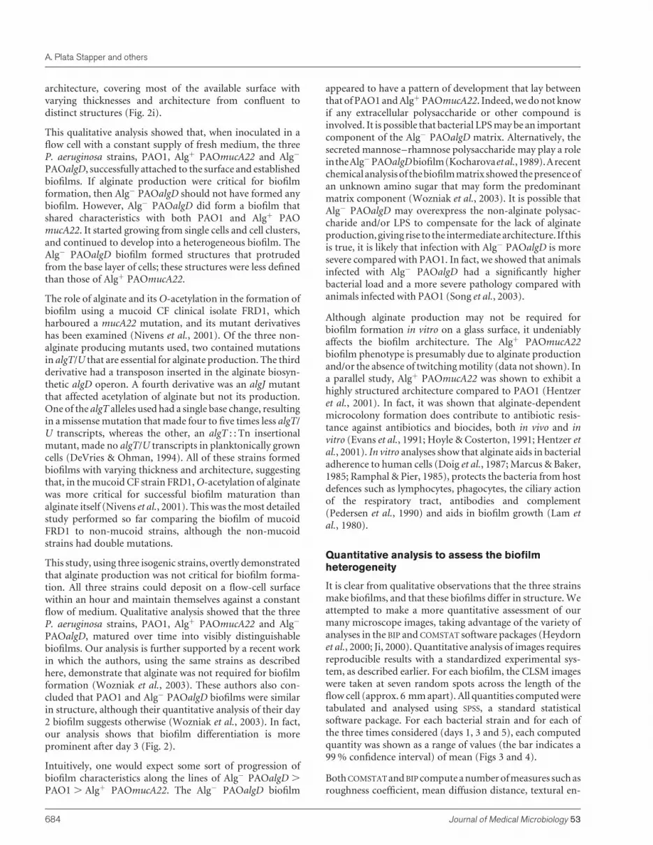
tropyandarealporositythatcanbeusedasindicatorsofbiofilmheterogeneity (Heydorn et al., 2000; Ji, 2000). The texturalentropy(Fig.3a) isameasureof theextenttowhichthebiofilmorganization varies. Thus, it should increase with increasedheterogeneityandthevaluesclosetozeroshouldcorrespondto
biofilms that are least heterogeneous. Areal porosity (Fig. 3b)measures the ratio of the void area to the total image, and thusshoulddecrease as thebiofilmextends itself on its surface. Thediffusion distance (Fig. 3c) measures the distance over whichthesubstratehastodiffusefromthevoidspacetoreachbacteriawithin the clusters. Thus, the value should decrease withincreased biofilm heterogeneity. The results of applying thesemeasures to our data are shown in Fig. 3.
The textural entropy values (Fig. 3a) on days 1 and 3 weresignificantly higher for Algþ PAOmucA22 and Alg�
PAOalgD than PAO1. They then levelled off by day 5,although they appeared to increase with time for PAO1.Similarly, the areal porosity (Fig. 3b) decreased and thenlevelled off for Algþ PAOmucA22 and Alg� PAOalgD,whereas it appeared to decrease exponentially for PAO1 evenon day 5. These data (Fig. 3b) appeared to behave in amannerthat was exactly opposite to that of the textural entropyanalysis (Fig. 3a). Diffusion distance (Fig. 3c) analysisshowed an interesting pattern, Algþ PAOmucA22 startedhigh and reduced over time, reaching the same value as thatof the day 1 PAO1 biofilm (levelling off). PAO1 and Alg�
PAOalgD showed similar trends and values, and theseincreased with time. The confidence interval for diffusiondistance increased for strains Alg� PAOalgD and PAO1, anddecreased for Algþ PAOmucA22.
Qualitative analysis clearly showed the distinct architectureconferred by alginate production in the Algþ PAOmucA22strain. Quantitative analyses of day 1 biofilms showed thatthe textural entropy, mean diffusion distance and arealporosity were similar in all three strains, but slightly elevatedin Algþ PAOmucA22 (Fig. 3). The increase in size of theconfidence intervals is surprising because one would expectthis value to decrease in size with increasing maturity of thebiofilm. However, it may represent the increasing variability(�complexity) in structure as the biofilm matures. This issupported by Fig. 3(a), where the textural entropy increaseswith time. The textural entropy analysis that measures thebiofilm heterogeneity was perhaps the most meaningful inexplaining the qualitative data. The least heterogeneity wasobserved in the PAO1 biofilm, which was thicker and denserand was depicted by the rapid and continuous loss of arealporosity (Fig. 3b). Areal porosity shows uniformly smallconfidence intervals (high confidence) throughout the timeperiod except, quite strangely, for PAO1 on day 5. In aparallel study, Algþ PAOmucA22 was shown to exhibit ahighly structured architecture compared to PAO1 (Hentzeret al., 2001). This is reflected in the fact that Algþ PAOmucA22 is the only strain whose biofilm showed a decrease inmean diffusion distance over time. The other strains ex-hibited the predicted behaviour of an increase of meandiffusion distance over time. It is intuitive to observe adecrease in areal porosity along with an increase in meandiffusion distance as a function of time, as was observed withthe Algþ PAOmucA22 biofilm.
Fig. 3. Quantitative analysis of the biofilm images for heterogeneity.CLSM images of Alg-inducible PAO1 (d), Alg� PAOalgD (m) andAlgþ PAOmucA22 (j) were analysed using the BIP image analysisprogram as described in Methods. The strains were grown in acontinuous flow cell in modified EPRI medium. Seven independentstacks were analysed per strain per day in replicates. The panels showlog-transformed textural entropy (a), areal porosity (b) and meandiffusion distance (c).
Alginate and P. aeruginosa biofilms
http://jmm.sgmjournals.org 685
Qualitative and quantitative analyses reveal
distinct developmental cycle of biofilm
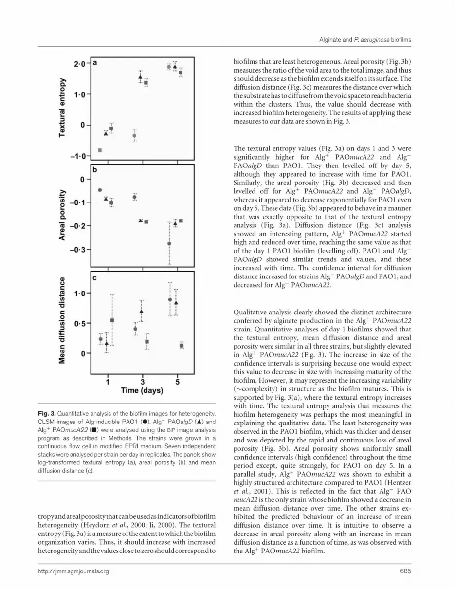
We observed a three-stage developmental pattern for bio-films over a period of 5 days: initiation, establishment andmaturation. Qualitative analysis was validated by quantita-tive measurements such as biomass, substratum coverageand mean thickness using the COMSTAT software. Biomassmeasures the overall volume of the cells (Fig. 4a). For strainPAO1, biomass increased sharply over the entire time period.It showed an exponential increase over this time period asseen by the linearity of the growth of the log-transformedvalues. The quantities for strains Algþ PAOmucA22 andAlg�
PAOalgD also increased, but levelled off by day 5. The meanthickness of the biofilm (Fig. 4b), which provided a measureof the spatial size of the biofilm, was positively correlated(r ¼ 0.993) to the biomass for the three strains. Thesubstratum coverage (Fig. 4c) measures the amount of areaoccupied by the biofilm, and this quantity also showed acorrelation to the biomass. Thus, all three strains followed asimilar pattern as was observed in the biomass analysis. Boththe Algþ PAOmucA22 and Alg� PAOalgD biofilms reached aplateau by day 5, while the PAO1 biofilm appeared tocontinue to increase. Overall, these data suggest that theprincipal structures of the Algþ PAOmucA22 and Alg�
PAOalgD biofilms were built during the first 3 days ofdevelopment, with a slight detachment of structures fromdays 3 to 5 of development.
In fact, the growth pattern of Algþ PAOmucA22 and Alg�
PAOalgD strains is reminiscent of the planktonic bacterialgrowth cycle, showing lag, exponential and stationary phases.We hypothesized that Algþ PAOmucA22 and Alg� PAOalgDwould form mature biofilms by day 5, whereas wild-typePAO1 would continue to develop. In a recent study, Saueret al. (2002) examined 12-day PAO1 biofilms and reportedfive distinct stages of biofilm development, of which the twoinitial stages are reversible attachment and irreversibleattachment. The latter stage is accompanied by loss oftwitching motility and a change in the pattern of proteinexpression (Sauer et al., 2002). Interestingly, both theattachment and detachment stages were observed in ourday 1 PAO1 and Alg� PAOalgD biofilms, but not in the Algþ
PAOmucA22. Similarly, other studies have shown thatmotility is not a prerequisite for successful biofilm develop-ment (Heydorn et al., 2002; Sauer et al., 2002). In our study,depending on the strains, the biofilmwas established by day 3and matured by day 5. In fact, the PAO1 biofilms studied bySauer et al. (2002) appeared tomature by day 6, implying thatit levelled off later than the other strains.
In addition to mounds and mushrooms, doughnut-shapedstructureswere also observed in several places across the Algþ
PAOmucA22 and Alg� PAOalgD biofilms on day 3 (Fig. 5),but not in PAO1 biofilms. These doughnut-shaped struc-tures had a cylindrical hollow centre and a ring of bacterialcells. Live microscopy observations showed that the hollowcentre inside the doughnut was filled with motile bacteriawhere cells darted about vigorously and disappeared into the
void space, generating the ‘doughnut’ structures. In contrastto day 3, on day 5, the hollow centres of the doughnuts wereoccupied and no movement was detected. These hollowcentres are due to the detachment of cells or cell aggregatesfrom the established biofilms (Stoodley et al., 2001). A
Fig. 4. Quantitative analysis of the biofilm images showing distinctdevelopmental cycle. CLSM images of Alg-inducible PAO1 (d), Alg�
PAOalgD (m) and Algþ PAOmucA22 (j) were analysed using theCOMSTAT image analysis program as described in Methods. The strainswere grown in a continuous flow cell in modified EPRI medium. Sevenindependent stacks were analysed per strain per day in replicates. Thepanels show log-transformed biomass (a), mean thickness (b) andsubstratum coverage (c).
A. Plata Stapper and others
686 Journal of Medical Microbiology 53

similar phenotype was observed in a 9-day-old PAO1 biofilmand was dubbed the dispersion stage by Sauer et al. (2002).Since this phenomenon appeared to be a post-maturationprocess, it supports our hypothesis that Algþ PAOmucA22and Alg� PAOalgD mature earlier than PAO1. It was clearthat the biofilmdevelopmental curve that includes initiation,establishment, maturation and dispersion was similar to lag,exponential, stationary and death phases of planktonicgrowth curves. Although these strains have similar growthpatterns in planktonic cells (data not shown), they showdistinct patterns in their biofilm mode of growth.
In summary, the quantitative measures of these three strainsundoubtedly support the qualitative descriptions givenabove. Among the computed measures used in this study,biomass, textural entropy, mean diffusion distance and arealporosity quantify information from within the biofilm,whereas the substratum coverage quantifies surface informa-tion. Both COMSTAT and BIP were able to evaluate successfullyarchitecture andmorphology features to differentiate the celldistribution within the biofilms of the three bacterial strains.The increased use of quantitative measures among biofilm
researchers will improve our understanding and interpreta-tion of biofilm growth, environmental and genetic influ-ences.
We have evaluated the role of alginate in biofilm develop-ment. Indisputably, the biofilm mode of growth contributesto P. aeruginosa persistence in lungs of CF patients. Thedetachment of cell aggregates from the mature biofilm(which results in doughnut structures) that can be trans-ported elsewhere and will subsequently establish new focalpoints of infection is indeed a health concern (Stoodley et al.,2001). It is likely that the physiology of the aggregates is suchthat they are better at resisting antibiotics and host immuneresponses. Although the in vitro analysis argues against thealginate requirement, it does not rule out its expression in thelungs of CFpatients. In fact, all the available data to date agreethat the appearance of mucoid microcolonies in CF patients’lungs provides the bacteria with a distinct survival advantage,suggesting that alginate is themajor virulence factor involvedin chronic infection in CF patients. Using the same threeisogenic strains, we demonstrated that alginate protects thecells against the host immune response and impedes host
Fig. 5. Doughnut structures. Algþ PAOmucA22 and Alg� PAOalgD gave rise to circular doughnut-shaped structures (denoted byarrow heads). Such structures were not observed in the wild-type PAO1 biofilm.
Alginate and P. aeruginosa biofilms
http://jmm.sgmjournals.org 687

immune clearance in a mouse model of acute lung infection(Song et al., 2003). Early infection by non-mucoid variantssupported by a non-alginate matrix may ensure that thebiofilm is compact and would thus better resist environ-mental stresses. The intermediate biofilm architecture seen inAlg� PAOalgD correlates with its intermediate infectiousphenotype in the acute animal infection model, and issuggestive that the alternate biofilm matrix may contributeto the bacterial pathogenesis, especially during the initialinfection before the genotypic conversion to constitutivelyproducing mucoid strains (Song et al., 2003).
ACKNOWLEDGEMENTS
This work was partially supported by NIH-MBRS SCORE (S06GM08205), a Danish Medical Research Council Grant (K.M., S.M.andN.H.), Veterans AdministrationMedical Research Funds (D. E. O.)and a Public Health Service grant AI-19146 (D. E. O.). We thankQichang Li and Zhou Ji for their contribution to the design andimplementation of BIP, and Arne Heydorn and Janus Haagensen for theuse of COMSTAT. We are grateful to E. B. Newman of ConcordiaUniversity, R. J. C. (Bob) McLean and Christa L. Bates of SouthwestTexas State University and Shalaka Indulkar and John Makemson ofFlorida International University for critical reading of the manuscriptand helpful suggestions. We also thank Brooke Larson-Crandall for hereditorial assistance.
REFERENCES
Andersen, J. B., Sternberg, C., Poulsen, L. K., Bjorn, S. P., Givskov,M. &Molin, S. (1998).New unstable variants of green fluorescent protein forstudies of transient gene expression in bacteria. Appl Environ Microbiol64, 2240–2246.
Ashby, M. J., Neale, J. E., Knott, S. J. & Critchley, I. A. (1994). Effect ofantibiotics on non-growing planktonic cells and biofilms of Escherichiacoli. J Antimicrob Chemother 33, 443–452.
Baltimore, R. S. & Mitchell, M. (1980). Immunologic investigations ofmucoid strains ofPseudomonas aeruginosa: comparison of susceptibilityto opsonic antibody in mucoid and nonmucoid strains. J Infect Dis 141,238–247.
Baltimore, R. S., Christie, C. D. & Smith, G. J. (1989). Immunohisto-pathologic localization of Pseudomonas aeruginosa in lungs frompatients with cystic fibrosis. Implications for the pathogenesis ofprogressive lung deterioration. Am Rev Respir Dis 140, 1650–1661.
Boucher, J. C., Yu, H., Mudd, M. H. & Deretic, V. (1997). MucoidPseudomonas aeruginosa in cystic fibrosis: characterization of mucmutations in clinical isolates and analysis of clearance in a mousemodel of respiratory infection. Infect Immun 65, 3838–3846.
Brown,M. R.W. &Williams, P. (1985). The influence of environment onenvelope properties affecting survival of bacteria in infections.Annu RevMicrobiol 39, 527–556.
Brown, M. R., Allison, D. G. & Gilbert, P. (1988). Resistance of bacterialbiofilms to antibiotics: a growth-rate related effect? J AntimicrobChemother 22, 777–780.
Christensen, B. B., Sternberg, C., Andersen, J. B., Palmer, R. J., Jr,Nielsen, A. T., Givskov, M. &Molin, S. (1999).Molecular tools for studyof biofilm physiology. Methods Enzymol 310, 20–42.
Cochran, W. L., Suh, S. J., McFeters, G. A. & Stewart, P. S. (2000). Roleof RpoS and AlgT in Pseudomonas aeruginosa biofilm resistance tohydrogen peroxide andmonochloramine. J Appl Microbiol 88, 546–553.
Cormack, B. P., Valdivia, R. H. & Falkow, S. (1996). FACS-optimizedmutants of the green fluorescent protein (GFP). Gene 173, 33–38.
Costerton, J. W., Cheng, K. J., Geesey, G. G., Ladd, T. I., Nickel, J. C.,Dasgupta, M. & Marrie, T. J. (1987). Bacterial biofilms in nature anddisease. Annu Rev Microbiol 41, 435–464.
Costerton, J. W., Lewandowski, Z., DeBeer, D., Caldwell, D., Korber, D.& James, G. (1994). Biofilms, the customized microniche. J Bacteriol176, 2137–2142.
Davies, D. G. & Geesey, G. G. (1995). Regulation of the alginatebiosynthesis gene algC in Pseudomonas aeruginosa during biofilmdevelopment in continuous culture. Appl Environ Microbiol 61,860–867.
Davies, D. G., Chakrabarty, A. M. & Geesey, G. G. (1993). Exopoly-saccharide production in biofilms: substratum activation of alginategene expression by Pseudomonas aeruginosa. Appl Environ Microbiol 59,1181–1186.
Davies, D. G., Parsek, M. R., Pearson, J. P., Iglewski, B. H., Costerton,J. W. & Greenberg, E. P. (1998). The involvement of cell-to-cell signalsin the development of a bacterial biofilm. Science 280, 295–298.
de Lorenzo, V., Herrero, M., Jakubzik, U. & Timmis, K. (1990). Mini-Tn5 transposon derivatives for insertion mutagenesis, promoter prob-ing, and chromosomal insertion of cloned DNA in gram-negativeeubacteria. J Bacteriol 172, 6568–6572.
Deretic, V., Gill, J. F. & Chakrabarty, A. M. (1987). Pseudomonasaeruginosa infection in cystic fibrosis: nucleotide sequence and tran-scriptional regulation of the algD gene.Nucleic Acids Res 11, 4567–4581.
DeVries, C. A. & Ohman, D. E. (1994).Mucoid-to-nonmucoid conver-sion in alginate-producing Pseudomonas aeruginosa often results fromspontaneous mutations in algT, encoding a putative alternative sigmafactor, and shows evidence for autoregulation. J Bacteriol 176,6677–6687.
Doggett, R. G., Harrison, G. M., Stillwell, R. N. &Wallis, E. S. (1966). Anatypical Pseudomonas aeruginosa associated with cystic fibrosis of thepancreas. J Pediatr 68, 215–221.
Doig, P., Smith, N. R., Todd, T. & Irvin, R. T. (1987). Characterization ofthe binding of Pseudomonas aeruginosa alginate to human epithelialcells. Infect Immun 55, 1517–1522.
Evans, L. R. & Linker, A. (1973). Production and characterization of theslime polysaccharide of Pseudomonas aeruginosa. J Bacteriol 116,915–924.
Evans, D. J., Allison, D. G., Brown, M. R. & Gilbert, P. (1991).Susceptibility of Pseudomonas aeruginosa and Escherichia coli biofilmstowards ciprofloxacin: effect of specific growth rate. J AntimicrobChemother 27, 177–184.
Fick, R. B., Jr, Sonoda, F. & Hornick, D. B. (1992). Emergence andpersistence of Pseudomonas aeruginosa in the cystic fibrosis airway.Semin Respir Infect 7, 168–178.
Gander,S. (1996).Bacterial biofilms: resistance to antimicrobial agents.J Antimicrob Chemother 37, 1047–1050.
Garrett, E. S., Perlegas, D. & Wozniak, D. J. (1999).Negative control offlagellum synthesis in Pseudomonas aeruginosa is modulated by thealternative sigma factor AlgT (AlgU). J Bacteriol 181, 7401–7404.
Gilbert, P. & Brown, M. R. (1998). Biofilms and �-lactam activity.J Antimicrob Chemother 41, 571–572.
Goldberg, J. B. & Ohman, D. E. (1984). Cloning and expression inPseudomonas aeruginosa of a gene involved in the production ofalginate. J Bacteriol 158, 1115–1121.
Goldberg, J. B., Hatano, K. & Pier, G. B. (1993). Synthesis oflipopolysaccharide O side chains by Pseudomonas aeruginosaPAO1 requires the enzyme phosphomannomutase. J Bacteriol 175,1605–1611.
A. Plata Stapper and others
688 Journal of Medical Microbiology 53
Govan, J. R. & Harris, G. S. (1986). Pseudomonas aeruginosa and cysticfibrosis: unusual bacterial adaptation and pathogenesis. Microbiol Sci 3,302–308.
Hassett, D. J., Elkins, J. G., Ma, J. F. & McDermott, T. R. (1999).Pseudomonas aeruginosa biofilm sensitivity to biocides: use of hydrogenperoxide as model antimicrobial agent for examining resistancemechanisms. Methods Enzymol 310, 599–608.
Heeb,S., Itoh, Y., Nishijyo, T., Schnider, U., Keel, C.,Wade, J., Walsh,U.,O’Gara, F. & Haas, D. (2000). Small, stable shuttle vectors based on theminimal pVS1 replicon for use in gram-negative, plant-associatedbacteria. Mol Plant Microbe Interact 13, 232–237.
Hentzer,M., Teitzel, G.M., Balzer, G. J., Heydorn, A., Molin, S., Givskov,M.&Parsek,M.R. (2001).Alginate overproduction affectsPseudomonasaeruginosa biofilm structure and function. J Bacteriol 183, 5395–5401.
Hentzer, M., Riedel, K., Rasmussen, T. B. & 9 other authors (2002).Inhibition of quorum sensing in Pseudomonas aeruginosa biofilmbacteria by a halogenated furanone compound. Microbiology 148,87–102.
Herrero, M., de Lorenzo, V. & Timmis, K. N. (1990). Transposon vectorscontaining non-antibiotic resistance selection markers for cloning andstable chromosomal insertion of foreign genes in gram-negativebacteria. J Bacteriol 172, 6557–6567.
Heydorn, A., Nielsen, A. T., Hentzer, M., Sternberg, C., Givskov, M.,Ersboll, B. K. &Molin, S. (2000).Quantification of biofilm structures bythe novel computer program COMSTAT. Microbiology 146, 2395–2407.
Heydorn, A., Ersboll, B., Kato, J., Hentzer, M., Parsek, M. R., Tolker-Nielsen, T., Givskov, M. & Molin, S. (2002). Statistical analysis ofPseudomonas aeruginosa biofilm development: impact of mutationsin genes involved in twitching motility, cell-to-cell signaling, andstationary-phase sigma factor expression. Appl Environ Microbiol 68,2008–2017.
Høiby, N. (1974). Pseudomonas aeruginosa infection in cystic fibrosis.Relationship between mucoid strains of Pseudomonas aeruginosa andthe humoral immune response. Acta Pathol Microbiol Scand B 82,551–558.
Høiby, N. (1975). Prevalence of mucoid strains of Pseudomonasaeruginosa in bacteriological specimens from patients with cysticfibrosis and patients with other diseases. Acta Pathol Microbiol ScandSuppl 83, 549–552.
Holloway, B. W. & Morgan, A. F. (1986). Genome organization inPseudomonas. Annu Rev Microbiol 40, 79–105.
Hoyle,B.D.&Costerton, J.W. (1991).Bacterial resistance to antibiotics:the role of biofilms. Prog Drug Res 37, 91–105.
Hoyle, B. D., Williams, L. J. & Costerton, J. W. (1993). Production ofmucoid exopolysaccharide during development of Pseudomonas aeru-ginosa biofilms. Infect Immun 61, 777–780.
Jensen, E. T., Kharazmi, A., Lam, K., Costerton, J.W. &Høiby, N. (1990).Human polymorphonuclear leukocyte response to Pseudomonas aeru-ginosa grown in biofilms. Infect Immun 58, 2383–2385.
Ji, Z. (2000). Quantitative analysis of biofilm images using fractaldimensions. MSc thesis, University of Memphis, TN, USA.
Kessler, B., de Lorenzo, V. & Timmis, K. N. (1992). A general system tointegrate lacZ fusions into the chromosomes of gram-negative eubac-teria: regulation of the Pm promoter of the TOL plasmid studied with allcontrolling elements in monocopy. Mol Gen Genet 233, 293–301.
Kocharova, N. A., Hatano, K., Shaskov, A. S., Knirel, Y. A., Kochetkov,N. K. & Pier, G. B. (1989). The structure and serologic distribution of anextracellular neutral polysaccharide from Pseudomonas aeruginosaimmunotype 3. J Biol Chem 264, 15569–15573.
Lam, J., Chan, R., Lam, K. & Costerton, J. W. (1980). Production ofmucoidmicrocolonies byPseudomonas aeruginosawithin infected lungsin cystic fibrosis. Infect Immun 28, 546–556.
Lawrence, J. R., Korber, D. R., Hoyle, B. D., Costerton, J. W. & Caldwell,D. E. (1991). Optical sectioning of microbial biofilms. J Bacteriol 173,6558–6567.
Maniatis, T., Fritsch, E. F. & Sambrook, J. (1982). Molecular Cloning: aLaboratory Manual. Cold Spring Harbor, NY: Cold Spring HarborLaboratory.
Marcus, H. & Baker, N. R. (1985).Quantitation of adherence of mucoidand nonmucoid Pseudomonas aeruginosa to hamster tracheal epithe-lium. Infect Immun 47, 723–729.
Mathee, K., McPherson, C. J. & Ohman, D. E. (1997). Posttranslationalcontrol of the algT (algU)-encoded �22 for expression of the alginateregulon in Pseudomonas aeruginosa and localization of its antagonistproteins MucA and MucB (AlgN). J Bacteriol 179, 3711–3720.
Mathee, K., Ciofu, O., Givskov, M., Ohman, D. E., Molin, S., Høiby, N. &Kharazmi, A. (1999a). Induction of Pseudomonas aeruginosa alginateproduction in vivomediated by inflammatory response in lungs of cysticfibrosis patients. Clin Microbiol 5, S8–S9.
Mathee,K., Ciofu,O., Sternberg,C.&9other authors (1999b).Mucoidconversion of Pseudomonas aeruginosa by hydrogen peroxide: amechanism for virulence activation in the cystic fibrosis lung. Micro-biology 145, 1349–1357.
Mathee, K., Kharazmi, A. &Høiby, N. (2002).Role of exopolysaccharidein biofilm matrix formation, the alginate paradigm. In MolecularEcology of Biofilms, chapter 2. Edited by R. J. C. McLean & A.W. Decho.Wymondham, UK: Horizon Scientific Press.
Narasimhan, G. (2004). Biofilm Image Processing Program at http://www.cs.fiu.edu/�giri/BIP/
Nivens, D. E., Ohman, D. E., Williams, J. & Franklin, M. J. (2001). Role ofalginate and its O-acetylation in formation of Pseudomonas aeruginosamicrocolonies and biofilms. J Bacteriol 183, 1047–1057.
Olvera, C., Goldberg, J. B., Sanchez, R. & Soberon-Chavez, G. (1999).The Pseudomonas aeruginosa algC gene product participates in rham-nolipid biosynthesis. FEMS Microbiol Lett 179, 85–90.
O’Toole, G. A. & Kolter, R. (1998). Flagellar and twitching motility arenecessary for Pseudomonas aeruginosa biofilm development. MolMicrobiol 30, 295–304.
Pedersen, S. S., Kharazmi, A., Espersen, F. & Høiby, N. (1990).Pseudomonas aeruginosa alginate in cystic fibrosis sputum and theinflammatory response. Infect Immun 58, 3363–3368.
Pedersen, S. S., Høiby, N., Espersen, F. & Koch, C. (1992). Role ofalginate in infection with mucoid Pseudomonas aeruginosa in cysticfibrosis. Thorax 47, 6–13.
Prasher, D. C., Eckenrode, V. K., Ward, W. W., Prendergast, F. G. &Cormier, M. J. (1992). Primary structure of the Aequorea victoria green-fluorescent protein. Gene 111, 229–233.
Ramphal, R. & Pier, G. B. (1985). Role of Pseudomonas aeruginosamucoid exopolysaccharide in adherence to tracheal cells. Infect Immun47, 1–4.
Roychoudhury, S., May, T. B., Gill, J. F., Singh, S. K., Feingold, D. S. &Chakrabarty, A. M. (1989). Purification and characterization ofguanosine diphospho-D-mannose dehydrogenase. A key enzyme inthe biosynthesis of alginate byPseudomonas aeruginosa. J Biol Chem 264,9380–9385.
Sauer, K. & Camper, A. K. (2001). Characterization of phenotypicchanges in Pseudomonas putida in response to surface-associatedgrowth. J Bacteriol 183, 6579–6589.
Sauer, K., Camper, A. K., Ehrlich, G. D., Costerton, J. W. & Davies, D. G.(2002). Pseudomonas aeruginosa displays multiple phenotypes duringdevelopment as a biofilm. J Bacteriol 184, 1140–1154.
Alginate and P. aeruginosa biofilms
http://jmm.sgmjournals.org 689
Schierholz, J. M., Beuth, J., Konig, D., Nurnberger, A. & Pulverer, G.(1999). Antimicrobial substances and effects on sessile bacteria.Zentralbl Bakteriol 289, 165–177
Song, Z.,Wu,H., Ciofu,O., Kong,K.-F.,Høiby,N., Rygaard, J., Kharazmi,A. &Mathee, K. (2003). Pseudomonas aeruginosa alginate is refractory toTh1 immune response and impedes host immune clearance in a mousemodel of acute lung infection. J Med Microbiol 52, 731–740.
Stoodley, P., Dodds, I., Boyle, J. D. & Lappin-Scott, H. M. (1999).Influence of hydrodynamics and nutrients on biofilm structure. J ApplMicrobiol 85, S19–S28.
Stoodley, P., Wilson, S., Hall-Stoodley, L., Boyle, J. D., Lappin-Scott,H. M. & Costerton, J. W. (2001). Growth and detachment of cellclusters frommaturemixed-species biofilms.Appl Environ Microbiol 67,5608–5613.
Wolfaardt, G. M., Lawrence, J. R., Robarts, R. D., Caldwell, S. J. &Caldwell, D. E. (1994). Multicellular organization in a degradativebiofilm community. Appl Environ Microbiol 60, 434–446.
Wozniak, D. J., Wyckoff, T. J., Starkey,M., Keyser, R., Azadi, P., O’Toole,G. A. & Parsek, M. R. (2003). Alginate is not a significant component ofthe extracellular polysaccharidematrix of PA14 andPAO1Pseudomonasaeruginosa biofilms. Proc Natl Acad Sci U S A 100, 7907–7912.
Yang, X., Beyenal, H., Harkin, G. & Lewandowski Z. (2000). Quantify-ing biofilm structure using image analysis. J Microbiol Methods 39,109–119.
Yu, H., Hanes, M., Chrisp, C. E., Boucher, J. C. & Deretic, V. (1998).Microbial pathogenesis in cystic fibrosis: pulmonary clearance ofmucoid Pseudomonas aeruginosa and inflammation in a mouse modelof repeated respiratory challenge. Infect Immun 66, 280–288.
A. Plata Stapper and others
690 Journal of Medical Microbiology 53


