Embed Size (px)
Citation preview
Advances in Clinical and Experimental M
edicine2021, Vol. 30, N
o. 12 (Decem
ber)
Advances in Clinical and Experimental Medicine
Impact Factor (IF) – 1.727Ministry of Science and Higher Education – 70 ptsIndex Copernicus (ICV) – 166.39 pts
2021, Vol. 30, No. 12 (December)
MONTHLY ISSN 1899-5276 (PRINT) ISSN 2451-2680 (ONLINE) www.advances.umw.edu.pl

Advances in Clinical and Experimental M
edicine2021, Vol. 30, N
o. 12 (Decem
ber)
Advances in Clinical and Experimental
Medicine

ISSN 1899-5276 (PRINT) ISSN 2451-2680 (ONLINE) www.advances.umw.edu.pl
Advances in Clinical and Experimental Medicine
MONTHLY 2021 Vol. 30, No. 12 (December)
Editor-in-ChiefProf. Donata Kurpas
Deputy EditorProf. Wojciech Kosmala
Managing EditorMarek Misiak
Advances in Clinical and Experimental Medicine (Adv Clin Exp Med) publishes high quality original articles, research-in-progress, research letters and systematic reviews and meta-analyses of recognized scientists that deal with all clinical and experimental medicine.
Section EditorsBasic SciencesDr. Anna LebedevaDr. Mateusz OlbromskiDr. Maciej SobczyńskiBiochemistryProf. Małgorzata Krzystek-Korpacka Clinical Anatomy, Legal Medicine,
Innovative TechnologiesProf. Rafael Boscolo-BertoDentistryProf. Marzena Dominiak Prof. Tomasz Gedrange Prof. Jamil Shibli
Scientific CommitteeProf. Sabine Bährer-Kohler Prof. Antonio Cano Prof. Breno Diniz Prof. Erwan Donal Prof. Chris Fox Prof. Naomi Hachiya Prof. Carol Holland Prof. Markku Kurkinen Prof. Christos Lionis
Statistical EditorsWojciech Bombała, MScKatarzyna Giniewicz, MSc Eng.Anna Kopszak, MScDr. Krzysztof Kujawa
Manuscript editingMarek Misiak, Jolanta Krzyżak
Prof. Raimundo Mateos Prof. Zbigniew W. Ras Prof. Jerzy W. Rozenblit Prof. Silvina Santana Prof. James Sharman Prof. Jamil ShibliProf. Michal Toborek Prof. László Vécsei Prof. Cristiana Vitale
DermatologyProf. Jacek Szepietowski Emergency Medicine, Innovative TechnologiesProf. Jacek Smereka Gynecology and ObstetricsProf. Olimpia Sipak-SzmigielHistology and EmbryologyProf. Marzenna Podhorska-Okołów Internal Medicine
AngiologyDr. Angelika ChachajCardiologyProf. Wojciech Kosmala Dr. Daniel Morris
Editorial Officeul. Marcinkowskiego 2–6 50-368 Wrocław, PolandTel.: +48 71 784 11 36 E-mail: [email protected]
PublisherWroclaw Medical UniversityWybrzeże L. Pasteura 1 50-367 Wrocław, Poland
© Copyright by Wroclaw Medical University, Wrocław 2021
Online edition is the original version of the journal

Editorial Policy Advances in Clinical and Experimental Medicine (Adv Clin Exp Med) is an independent multidisciplinary forum for exchange of scientific and clinical information, publishing original research and news encompassing all aspects of medicine, including molecular biology, biochemistry, genetics, biotechnology and other areas. During the review pro-cess, the Editorial Board conforms to the “Uniform Requirements for Manuscripts Submitted to Biomedical Journals: Writing and Editing for Biomedical Publication” approved by the International Committee of Medical Journal Editors (www.ICMJE.org/). The journal publishes (in English only) original papers and reviews. Short works considered origi-nal, novel and significant are given priority. Experimental studies must include a statement that the experimental protocol and informed consent procedure were in compliance with the Helsinki Convention and were approved by an ethics committee.
For all subscription-related queries please contact our Editorial Office: [email protected]
For more information visit the journal’s website: www.advances.umw.edu.pl
Pursuant to the ordinance No. 134/XV R/2017 of the Rector of Wroclaw Medical University (as of December 28, 2017) from January 1, 2018 authors are required to pay a fee amounting to 700 euros for each manuscript accepted for publication in the journal Advances in Clinical and Experimental Medicine.
Indexed in: MEDLINE, Science Citation Index Expanded, Journal Citation Reports/Science Edition, Scopus, EMBASE/Excerpta Medica, Ulrich’sTM International Periodicals Directory, Index Copernicus
Typographic design: Piotr Gil, Monika KolędaDTP: Wydawnictwo UMWCover: Monika KolędaPrinting and binding: Soft Vision Mariusz Rajski
EndocrinologyProf. Marek BolanowskiGastroenterologyProf. Piotr EderAssoc. Prof. Katarzyna Neubauer HematologyProf. Andrzej DeptałaProf. Dariusz Wołowiec Nephrology and TransplantologyAssoc. Prof. Dorota Kamińska Assoc. Prof. Krzysztof Letachowicz PulmonologyProf. Elżbieta Radzikowska
MicrobiologyProf. Marzenna Bartoszewicz Assoc. Prof. Adam Junka Molecular BiologyDr. Monika BieleckaProf. Jolanta Saczko Dr. Marta Sochocka
NeurologyAssoc. Prof. Magdalena KoszewiczAssoc. Prof. Anna Pokryszko-Dragan Dr. Masaru TanakaOncologyDr. Marcin Jędryka Prof. Lucyna Kępka
Gynecological OncologyDr. Marcin Jędryka
OphthalmologyProf. Marta Misiuk-Hojło OrthopedicsProf. Paweł Reichert OtolaryngologyAssoc. Prof. Tomasz Zatoński Pediatrics
Pediatrics, Metabolic Pediatrics, Clinical Genetics, Neonatology, Rare DisordersProf. Robert Śmigiel Pediatric NephrologyProf. Katarzyna Kiliś-Pstrusińska Pediatric Oncology and HematologyAssoc. Prof. Marek Ussowicz
Pharmaceutical SciencesAssoc. Prof. Maria KepinskaProf. Adam Matkowski Pharmacoeconomics, RheumatologyDr. Sylwia Szafraniec-Buryło PsychiatryProf. Istvan BoksayProf. Jerzy Leszek Public HealthProf. Monika Sawhney Prof. Izabella Uchmanowicz Qualitative Studies, Quality of CareProf. Ludmiła Marcinowicz RehabilitationProf. Jakub Taradaj SurgeryAssoc. Prof. Mariusz Chabowski Prof. Renata Taboła Telemedicine, Geriatrics, MultimorbidityAssoc. Prof. Maria Magdalena
Bujnowska-Fedak

MONTHLY 2021, Vol. 30, No. 12 (December)
ISSN 1899-5276 (PRINT) ISSN 2451-2680 (ONLINE)
www.advances.umw.edu.pl
Advances in Clinical and Experimental Medicine
Contents
© Copyright by Wroclaw Medical University, Wrocław 2021
Editorials1221 James E. Sharman, Wojciech Kosmala
High-quality medical research requires that equipment has been validated for accuracy
Original papers1225 Ádám Annus, Ferenc Tömösi, Ferenc Rárosi, Evelin Fehér, Tamás Janáky, Gábor Kecskeméti, József Toldi, Péter Klivényi, László Sztriha, László Vécsei
Kynurenic acid and kynurenine aminotransferase are potential biomarkers of early neurological improvement after thrombolytic therapy: A pilot study
1233 Mehmet Murat Bala, Keziban Aslı BalaSevere cases of osteogenesis imperfecta type VIII due to a homozygous mutation in P3H1 (LEPRE1) and review of the literature
1239 Anna Rams, Joanna Kosałka-Węgiel, Piotr Kuszmiersz, Aleksandra Matyja-Bednarczyk, Stanisław Polański, Lech Zaręba, Stanisława Bazan-SochaCharacteristics of idiopathic inflammatory myopathies with novel myositis-specific autoantibodies
1249 Patryk Kuliński, Łukasz Tomczyk, Piotr MorasiewiczEffect of the COVID-19 pandemic on foot surgeries
1255 Fatih Ada, Ferit Kasimzade, Ali Sefa Mendil, Hasan GocmezEffects of nano-sized titanium dioxide powder and ultraviolet light on superficial veins in a rabbit model
1263 En Zhou, Yinghua Zou, Chengyu Mao, Dongjiu Li, Changqian Wang, Zongqi ZhangMicroRNA-221 inhibits the transition of endothelial progenitor cells to mesenchymal cells via the PTEN/FoxO3a signaling pathway
1271 Chunwen Jia, Feng Gao, Yanan Zhao, Siyang Ji, Shidao CaiIdentification and functional analysis of changes to the ox-LDL-induced microRNA-124-3p/DLX5 axis in vascular smooth muscle cells
1283 Fadime Mutlu Içduygu, Hale Samli, Asuman Özgöz, Buse Vatansever, Kuyas Hekimler Oztürk, Egemen AkgünPossibility of paclitaxel to induce the stemness-related characteristics of prostate cancer cells
Reviews1293 Katarzyna Rakoczy, Wojciech Szlasa, Jolanta Saczko, Julita Kulbacka
Therapeutic role of vanillin receptors in cancer
1303 Kacper Turek, Michał Jarocki, Julita Kulbacka, Jolanta SaczkoDualistic role of autophagy in cancer progression
Research letters1315 Adam Nowiński, Katarzyna Stachyra, Maria Szybińska, Michał Bednarek, Robert Pływaczewski, Paweł Śliwiński
The influence of comorbidities on mortality in bronchiectasis: A prospective, observational study
1323 Jakub Szymon Mercik, Jadwiga Radziejewska, Katarzyna Pach, Dorota Zyśko, Jacek GajekST-segment depression in atrioventricular nodal reentrant tachycardia: Preliminary results
1329 Annual Contents
1341 Index of Authors


Reviewers in 2021:Dawud Abawi, Abdelrahman I. Abushouk, Marcin Adamczak, Anil Ahsan, Sinan Akıncı, Alessio Alogna, Durdu Altuner, Artur Anisiewicz, Mabel Aoun, Muhammad Sohaib Asghar, Jan Azarov, Karolis Azukaitis, Teresa Bachanek, Dagmara Baczyńska, Christopher Baines, Waldemar Balcerzak, Agata Bałdys-Waligórska, Julia Bar, Simge Bardak, Srikanth Barkeer, Tomasz Baron, Rafał Bartoszewski, Grzegorz Bartoszewski, Sandip Basu, Simone Battaglia, Ewelina Bąk, Ilona Bednarek, Wojciech Bednarz, Evgeny Belyavskiy, Joanna Bereta, Marta Berghausen-Mazur, Sebastjan Bevc, Sonu Bhaskar, Maciej Biały, Mariusz Bidziński, Monika Bielecka, Tomasz Bielecki, Beata Bieniaś, Marta Bižić, Anna Bizoń, Krzysztof Błaszyk, Vladimir Bobek, Sylwia Bobis-Wozowicz, Joanna Bogusławska, Andrzej Bohatyrewicz, Romuald Bohatyrewicz, Agnieszka Bojarska-Junak, Dariusz Boroń, Rafael Boscolo-Berto, Sergey Brodsky, Mariusz Bromke, Anna Brzecka, Natalia Buda, Grzegorz Bukato, Paweł Burchardt, Marco Capuzzo, Alessandra Cassoni, Nidia Castro, Ersin Çelik, Magdalena Celińska-Lowenhoff, Yubo Chai, Andrzej Chamienia, Andrzej Chciałowski, Chong Chen, Min Chen, Magdalena Chmielewska, Liyan Chu, Jerzy Chudek, Halina Cichoż-Lach, Joanna Cieślińska-Swider, Lidia Ciszak, Erica Costantini, Alessandro Crestani, Sifu Cui, Monika Czerwińska, Mirosław Czuczwar, Małgorzata Daczewska, Zofia Danilczuk, Jacek Daroszewski, Alicja Dąbrowska-Kugacka, Alessandro De Cassai, Beatriz De Lucas, Carolina D’Elia, Urszula Demkow, Andrzej Deptała, Katarzyna Derwich, Engin Deveci, Rosanna di Paola, Dimitrios Dimitroulis, Łukasz Dobrek, Piotr Domagała, Erwan Donal, Adrian Doroszko, Ines Drenjančević, Anna Dubaniewicz, Magdalena Dubińska-Magiera, Murat Dursun, Fazilet Duygu, Jadwiga Dwilewicz-Trojaczek, Dariusz Dziedzic, Piotr Dzięgiel, Gabriella Emri, Angela Faga, Kenneth Faulkner, Izabela Fecka, Julia Fedotova, Anna Filipowicz-Sosnowska, Martin Floer, Marcin Frączek, Mónica Furlano, Tomasz Gabryelewicz, Jacek Gajek, Agata Gajos, Andrea Galassi, Piotr Gałecki, Maria Ganeva, Eugenia Gatiatulina, Dariusz Gawryluk, Zbigniew Gąsior, Adam Gesing, Ashraf Ghulam, Maciej Głyda, Justyna Gołębiewska, Andrzej Gołębiewski, Maciej Gonciarz, Monika Gos, Jolanta Gozdowska, Waldemar Goździk, Stuart Grossman, Grzegorz Grześk, Andrzej Grzybowski, Jakob Gubenšek, Cecilia Guiot, Katarzyna Gustaw-Rothenberg, Maciej Guziński, Katarzyna Haczkiewicz-Leśniak, Piotr Harbut, Marek Hartleb, Zehra Hatipoğlu, Olga Haus, Kai He, Weiwei He, Lynne Hinterbuchner, András Hrabák, Jian Hu, Rong Hu, Qiang Huang, Hongxia Hui, Quan Huynh, Irena Ilić, Stanislav Isayenkov, Arda Işık, Zbigniew Jabłonowski, Anna Janas-Naze, Dominika Janiszewska, Ewa Jankowska, Alina Jankowska-Konsur, Barbara Jarząb, Monika Jasek, Marek Jasiński, Aleksandra Jezela-Stanek, Marcin Jędryka, Zhang Jingfa, James John, Krystian Josiak, Piotr Kałmucki, Marta Kałużna-Oleksy, Grzegorz Kamiński, Karol Kamiński, Paweł Karpiński, Martyna Kasper-Jędrzejewska, Beata Kasztelan-Szczerbińska, Theodora Katopodi, Karolina Kędzierska, Ehsan Khalili, Jarosław Kierkuś, Yoji Kishi, Mariusz Klencki, Maria Kłopocka, Aureliusz Kolonko, Parastou Kordestani-Moghadam, Wojciech Korlacki, Magdalena Koszewicz, Euphrosyni Koutsouraki, Jerzy Kowalczyk, Marcin Kozakiewicz, Katarzyna Koziak, Magdalena Krajewska, Iwona Krela-Kaźmierczak, Soňa Křížková, Tadeusz Krzemiński, Paweł Kubasiewicz-Ross, Eliza Kubicka, Adriana Kubis-Kubiak, Paweł Kuca, Ernest Kuchar, Piotr Kuczera, Tamara Kujawska, Julita Kulbacka, Wiktor Kuliczkowski, Sonam Kumari, Paweł Kunicki, Markku Kurkinen, Ilona Kurnatowska, Jacek Kusa, Damian Kusz, Michał Kuszewski, Halyna Kuznietsova, Katarzyna Kwiatkowska, Paweł Lampe, Małgorzata Latocha, Anna Lebedeva, Aleksandra Lesiak, Damian Lewandowski, Łukasz Lewandowski, Ewa Lewicka, Fang Li, Fuyang Li, Piotr Lipiec, Mariusz Lipski, Xuejian Liu, Kamila Ludwikowska, Izabela Łaczmańska, Łukasz Łaczmański, Tadeusz Łapiński, Joanna Maj, Jennifer Majer, Przemysław Majewski, Preeti Malik, Grzegorz Małek, Małgorzata Małodobra-Mazur, Hina Manzoor, Wojciech Marlicz, Andrzej Marszałek, Helena Martynowicz, Marcin Masalski, Maria Maślińska, Ivan Matia, Rafał Matkowski, Małgorzata Matusiewicz, Jacek Matys, Grzegorz Mazur, Richard Merrill, Frederic Meyer, Krystyna Michalak, Michał Michalak, Marta Migocka-Patrzałek, Przemysław Mikołajczak, Wioletta Mikulakova, Joanna Miłkowska-Dymanowska, Małgorzata Mizerska-Wasiak, Agnieszka Młynarska, Gustavo Molina, Piotr Morasiewicz, Daniel Morris, Janusz Moryś, Bożena Mroczek, Tomoyuki Mukai, Agata Mulak, Sameh Naguib, Karol Nartowski, Anna Nasierowska-Guttmejer, Joseph Nassif, Kazuaki Negishi, Jadwiga Nessler, Piotr Niewiński, Przemysław Niewiński, Marita Nittner-Marszalska, Beata Nowak, Krzysztof Nowak, Michał Nowak, Marta Nowakowska-Kotas, Antonio Núñez, Mateusz Olbromski, Paweł Olczyk, Robert Olszewski, Hanna Osadnik, Abdulkadir Özgür, Olga Pakhomova, Dorota Paluszyńska, Lijian Pan, Anatol Panasiuk, Małgorzata Pańczyk-Tomaszewska, Mark Parker, Tomasz Pawiński, Edyta Pawlak, Guido Pelletti, Joanna Peredzyńska, Cristian Persu, Mariya Petrosyan, Adriano Piattelli, Grzegorz Piecha, Aleksandra Piechota-Polańczyk, Jadwiga Pietkiewicz, Anna Pietrzak, Jakub Piotrkowski, Elżbieta Płuciennik, Adam Płużański, Indrani Poddar, Monika Podhorecka, Ilona Pokora, Renata Polaniak, Katarzyna Połtyn-Zaradna, Michał Pomorski, Raluca Pop, Daniela Popescu, Tanja Popp, Tomasz Porażko, Małgorzata Poręba, Rafał Poręba, Francesco Prattichizzo,
We would like to express our gratitude to all reviewers who devoted their time and expertise to evaluate manuscripts in Advances in Clinical and Experimental Medicine. We sincerely appreciate all your hard work and dedication. It is due to your contribution that we can achieve the standard of excellence.
Editors
Acknowledgements

Monika Przewłocka-Kosmala, Tadeusz Przybyłowski, Anna Raciborska, Elżbieta Radzikowska, Reza Rahbarghazi, Dariusz Rakus, Wafaa Rashed, Sanjay Rastogi, Mariusz Z. Ratajczak, Laura Reed, Bożena Regulska-Ilow, Paweł Reichert, Rafi Romano, Michał Romanski, Umberto Romeo, Christine Rondanino, Joanna Rossowska, Piotr Rozentryt, Monika Rucińska, Lidia Rutkowska-Sak, Przemysław Rutkowski, Justyna Rybka, Józef Ryżko, Makoto Saito, Ioanna Sakellari, Javier Santabarbara, Elżbieta Sarnowska, Naila Sattar, Ewa Sawicka, Ana Scocate, Agata Sebastian, Silvia Secco, James Sharman, Aboubacar Sidibé, Paweł Siekierski, Halina Sienkiewicz-Jarosz, Jerzy Sieńko, Piotr Sieroszewski, Santhi Silambanan, Scott Sills, Krzysztof Simon, Maciej Siński, Rosalba Siracusa, Joanna Siuda, Magdalena Skarżyńska, Andrzej Skorek, Kajetan Słomka, Krzysztof Słowiński, Jacek Smereka, Maciej Sobczyński, Magdalena Sobieska, Grzegorz Sobota, Joanna Socha, Marta Sochocka, Francisco Solano, Roman Sosnowski, Eleonora Spekker, Zbigniew Sroka, Agata Stanek, Ivan Starchenko, Anna Starzyńska, Ewelina Stelcer, Elena Stocco, Rostyslav Stoika, Tomasz Stompór, Jan Styczyński, Pan Su, Lina Suarez, Halis Suleyman, Andrei Surguchov, Edyta Sutkowska, Krzysztof Szczałuba, Tomasz Szczapa, Aleksandra Szczepankiewicz, Jolanta Szelachowska, Grzegorz Szewczyk, Milena Ściskalska, Agnieszka Świdnicka-Siergiejko, Aisha Tahir, Özgür Tan, Hui Tang, Onur Telli, Luca Testarelli, Thavaree Thilavech, Liza Thomas, Dariusz Timler, Michał Tkaczyszyn, Andrzej Torbé, Jonel Trebicka, Krzysztof Tupikowski, Maciej Ugorski, Tomasz Urbaniak, Wiktor Urbański, Marek Ussowicz, Juha Väyrynen, András Végh, Karolina Walewicz, Marta Waliszewska-Prosół, Changqian Wang, Guodong Wang, Lidong Wang, Wenan Wang, Yichao Wang, Peter Ward, Dorota Waśko-Czopnik, Adam Whitley, Wojciech Widuchowski, Bartosz Wielgomas, Karina Wierzbowska-Drabik, Anna Witkowska, Tomasz Witkowski, Kamil Wojnicki, Beata Wojtczak, Dariusz Wołowiec, Olga Wysocka, Yong Xia, Liuling Xiao, Huan-Yu Xiong, Zhihao Xu, Kazumi Yagasaki, Ulyana Yanyshyn, Yongjie Yin, Xiaojun Yu, Agnieszka Zabłocka, Katarzyna Zabłocka-Słowińska, Maria Paz Zafra, Edyta Zagórowicz, Jan Zaucha, Renata Zaucha, Zygmunt Zdrojewicz, Bin-Fei Zhang, Yijian Zhang, Shixi Zhao, Sławomir Zmonarski, Agnieszka Zubkiewicz-Kucharska, Anna Zubrzycka-Sienkiewicz, Istvan Zupko, Małgorzata Zwolińska-Wcisło, Robert Zymliński, Dorota Zyśko, Maciej Żarow
Cite asSharman JE, Kosmala W. High-quality medical research requires that equipment has been validated for accuracy. Adv Clin Exp Med. 2021;30(12):1221–1223. doi:10.17219/acem/144440
DOI10.17219/acem/144440
Copyright© 2021 by Wroclaw Medical University This is an article distributed under the terms of theCreative Commons Attribution 3.0 Unported (CC BY 3.0)(https://creativecommons.org/licenses/by/3.0/)
Address for correspondenceJames E. SharmanE-mail: [email protected]
Funding sourcesNone declared
Conflict of interestNone declared
Received on November 10, 2021Accepted on December 2, 2021
Published online on December 13, 2021
AbstractIt is not always appreciated that medical equipment may be cleared by regulatory authorities to sell within a country, without ever having been tested for accuracy performance according to scientific validation standards. Instead, manufacturers can undertake in-house accuracy testing, using variable methods and without any requirement for test results to be made publicly available. This lack of full transparency together with potential for industry bias can place doubt over the quality of validation results provided to regulatory authorities. Currently, this situation affects the field of hypertension research, where most blood pressure devices have not been independently validated for accuracy according to international scientific standards, nor as expected in clinical practice guidelines. More attention should be paid to such practices in order to improve the quality of research and to optimize further translation of scientific findings to clinical practice. The clinical implications of inaccurate measurements in research can be far-reaching, ultimately impacting on a patient’s health. Well-planned validating studies should be more widely considered for new devices that are candidates to be used in research protocols. The awareness of the lack or uncertain validation of equipment used for verifying research hypotheses should prompt all investigators to revisit the idea of conducting the study or, at least, to acknowledge this issue as a relevant study limitation. One of the ways in which authors submitting research findings for publication can add to the quality of the reporting of their work is to ensure reference to the accuracy validation of their research equipment.
Key words: medical devices, equipment and supplies, standards, performance
Editorials
High-quality medical research requires that equipment has been validated for accuracy
James E. Sharman1,D, Wojciech Kosmala2,E
1 Menzies Institute for Medical Research, University of Tasmania, Hobart, Australia2 Department of Cardiovascular Imaging, Institute of Heart Diseases, Wroclaw Medical University, Poland
A – research concept and design; B – collection and/or assembly of data; C – data analysis and interpretation; D – writing the article; E – critical revision of the article; F – final approval of the article
Advances in Clinical and Experimental Medicine, ISSN 1899–5276 (print), ISSN 2451–2680 (online) Adv Clin Exp Med. 2021;30(12):1221–1223
J. Sharman, W. Kosmala. Research equipment validation1222
The international community and funders of research expect the study to be conducted ethically and with high quality, such that it is rigorous, transparent, reproducible, and avoids waste.1 These expectations are not only essen-tial for the translation of research knowledge to the ben-efit of the wider community, but also help to promote trust in research findings and deliver the highest value for the money that is invested in research.2 The failure of many research discoveries to be reproduced by independent in-vestigators is a symptom of rigorous research methods not always being applied, and indicates that more training to improve the understanding of high-quality research methods, particularly for research apprentices, is needed.1,3
Resources to aid the rigor and accountability of re-search practice, from the design to conduct and reporting of research outcomes, have been developed for research-ers (e.g., Enhancing the QUAlity and Transparency Of health Research (EQUATOR) network, Cochrane Col-laboration).4,5 Working towards similar intent to improve the quality of research publications, the International Committee of Medical Journal Editors (ICMJE) publishes “Recommendations for the Conduct, Reporting, Editing and Publication of Scholarly Work in Medical Journals”.6 These recommendations align with best practice stan-dards to assist all those involved in publishing medical research to present unbiased findings clearly and accu-rately. The journal “Advances in Clinical and Experimen-tal Medicine” is an ICMJE member journal that requires authors of all submitted papers to adhere to the ICMJE recommendations.
The ICMJE Recommendations rightly state that, in order to allow others to reproduce the results of research, there should be sufficient detail provided on the methods, equip-ment and procedures of the research.6 However, a factor that is not considered within the ICMJE recommendations, but can have a marked influence on the results of research, is providing confirmation that the research equipment is accurate, preferably through testing it in accordance with established scientific validation standards. Without such confirmation, there is little to assure that the equipment can measure what it purports to measure, nor whether its use may critically undermine the quality of research outputs. This concept of confirming the quality of all data to increase the confidence in research results is familiar to researchers across disciplines.7–9
It is not always appreciated that medical equipment may be cleared by regulatory authorities to sell within a coun-try, without ever having been tested for accuracy perfor-mance according to scientific validation standards. Instead, manufacturers can undertake in-house accuracy testing, using variable methods and without any requirement for test results to be made publicly available. This lack of full transparency together with potential for industry bias can place doubt over the quality of validation results provided to regulatory authorities. Currently, this situation affects the field of hypertension research,10,11 where most blood
pressure devices have not been independently validated for accuracy according to international scientific standards, nor as expected in clinical practice guidelines.12–14 Global estimates indicate that less than 20% of blood pressure devices from among thousands of separate models avail-able for purchase have been independently validated.15,16
As may be anticipated, the untested, non-validated devices are more likely to be inaccurate when compared with established reference standards,17–19 which in addition to the impact on research quality has obvious implica-tions for appropriate clinical diagnosis and management. Compared with validated devices, those whose accuracy has not been tested also have a greater dispersion of mea-surement values (higher variance),20 which will influence estimation of sample size for research projects. Higher variance in the measurement of a primary outcome vari-able means that more research study participants will be needed to answer the research question and demonstrate statistical between-group differences.21 This places extra burden on research resources and also has ethical implica-tions in higher-risk study protocols by needlessly exposing more research participants to otherwise avoidable risk.
Regulatory loopholes that enable widespread availability of blood pressure devices with uncertain quality assur-ance10 may also apply to other medical equipment used in research, such as blood glucose monitors and pulse ox-imeters,22–24 although the breadth of the problem more generally across equipment being used in research is not known. This being the case, the onus of responsibility to determine the worthiness of medical equipment to be used in research falls to the shoulders of individual re-searchers in the planning and conduct of their research projects. What can researchers do in relation to this?
Researchers can check that all research equipment has been assessed, and passed validation testing, or the per-formance can be verified (including where participants of the study use personal monitoring equipment to collect research data).17 Ideally, the values derived from the equip-ment have been compared using an established, standard-ized protocol with a recognized reference standard, for example, blood glucose measured with a point-of-care de-vice compared with an automated procedure from an ac-credited laboratory with known accuracy and precision. Researchers can also seek information from relevant pro-fessional societies,25 web-based resources,5 other sources of published guidance,26 or ask the equipment distributor to provide published material as proof of accuracy. Where available, they should cite the published validation mate-rial within the methods of research manuscripts when referencing the equipment used. It is important to note that validation testing is only relevant to the device under scrutiny and the results cannot be extrapolated to other models or devices made by different manufacturers.
In the hypertension field, international efforts are being undertaken to redress the problem of non-validated equip-ment used in research, clinical practice and population
Adv Clin Exp Med. 2021;30(12):1221–1223 1223
level interventions.27–30 This requires ongoing, multisec-toral lobbying for regulatory change, as well as widespread education and advocacy for exclusive use of validated equipment. Whether such activities are needed in other research areas is something for the wider research com-munity to be alert for.
Indeed, the scale of the problem being discussed is in-creasing, and more attention should be paid to it in order to improve the quality of research and to optimize fur-ther translation of scientific findings to clinical practice. The clinical implications of inaccurate measurements in research can be far-reaching, ultimately impacting on a patient’s health. Well-planned validating studies should be more widely considered for new devices that are candidates to be used in research protocols. The awareness of the lack or uncertain validation of equipment used for verifying research hypotheses should prompt all investiga-tors to revisit the idea of conducting the study or, at least, to acknowledge this issue as a relevant study limitation. One of the ways that authors submitting research find-ings for publication can add to the quality of the reporting of their work is to ensure reference to the accuracy valida-tion of their research equipment.
ORCID iDsJames E. Sharman https://orcid.org/0000-0003-2792-0811Wojciech Kosmala https://orcid.org/0000-0003-3807-8201
References1. Chalmers I, Glasziou P. Avoidable waste in the production and report-
ing of research evidence. Lancet. 2009;374(9683):86–89. doi:10.1016/S0140-6736(09)60329-9
2. National Health and Medical Research Council. NHMRC’s Research Quality Strategy. https://www.nhmrc.gov.au/file/16823/download? token=ha80bBhZ. Accessed November 25, 2021.
3. Begley CG, Ioannidis JPA. Reproducibility in science: Improving the standard for basic and preclinical research. Circ Res. 2015;116(1): 116–126. doi:10.1161/CIRCRESAHA.114.303819
4. Green S, Mcdonald S. Cochrane Collaboration: More than system-atic reviews? Intern Med J. 2005;35(1):3–4. doi:10.1111/j.1445-5994. 2004.00747.x
5. Simera I, Moher D, Hirst A, Hoey J, Schulz KF, Altman DG. Transpar-ent and accurate reporting increases reliability, utility, and impact of your research: Reporting guidelines and the EQUATOR Network. BMC Med. 2010;8:24. doi:10.1186/1741-7015-8-24
6. International Committee of Medical Journal Editors. Recommenda-tions for the Conduct, Reporting, Editing and Publication of Scholarly Work in Medical Journals – updated December 2018. http://www.ICMJE.org. Accessed August 17, 2019.
7. Ehrenstein V, Petersen I, Smeeth L, et al. Helping everyone do bet-ter: A call for validation studies of routinely recorded health data. Clin Epidemiol. 2016;8:49–51. doi:10.2147/clep.S104448
8. Lash TL, Olshan AF. EPIDEMIOLOGY announces the “Validation Study” submission category. Epidemiology. 2016;27(5):613–614. doi:10.1097/ede.0000000000000532
9. Coecke S, Bernasconi C, Bowe G, et al. Practical aspects of design-ing and conducting validation studies involving multi-study trials. Adv Exp Med Biol. 2016;856:133–163. doi:10.1007/978-3-319-33826-2_5
10. Alpert BS. Can ‘FDA-cleared’ blood pressure devices be trusted? A call to action. Blood Press Monit. 2017;22(4):179–181. doi:10.1097/mbp.000 0000000000279
11. Sharman JE, Padwal R, Campbell NRC. Global marketing and sale of accurate cuff blood pressure measurement devices. Circulation. 2020;142(4):321–323. doi:10.1161/circulationaha.120.046205
12. Campbell NR, Gelfer M, Stergiou GS, et al. A call to regulate manu-facture and marketing of blood pressure devices and cuffs: A posi-tion statement from the World Hypertension League, International Society of Hypertension and supporting hypertension organizations. J Clin Hypertens (Greenwich). 2016;18(5):378–380. doi:10.1111/jch.12782
13. Muntner P, Einhorn PT, Cushman WC, et al. Blood pressure assessment in adults in clinical practice and clinic-based research: JACC Scientif-ic Expert Panel. J Am Coll Cardiol. 2019;73(3):317–335. doi:10.1016/j.jacc.2018.10.069
14. Whelton PK, Carey RM, Aronow WS, et al. 2017 ACC/AHA/AAPA/ABC/ACPM/AGS/APhA/ASH/ASPC/NMA/PCNA guideline for the preven-tion, detection, evaluation, and management of high blood pressure in adults: A report of the American College Of Cardiology/American Heart Association task force on clinical practice guidelines. J Am Coll Cardiol. 2018;71(19):e127–e248. doi:10.1016/j.jacc.2017.11.006
15. Medaval. Blood pressure monitors. https://medaval.ie/blood-pressure- monitors. Accessed October 26, 2021.
16. Picone DS, Deshpande RA, Schultz MG, et al. Nonvalidated home blood pressure devices dominate the online marketplace in Australia: Major implications for cardiovascular risk management. Hypertension. 2020;75(6):1593–1599. doi:10.1161/HYPERTENSIONAHA.120.14719
17. Jung MH, Kim GH, Kim JH, et al. Reliability of home blood pressure monitoring: In the context of validation and accuracy. Blood Press Monit. 2015;20(4):215–220. doi:10.1097/mbp.0000000000000121
18. Akpolat T, Dilek M, Aydogdu T, et al. Home sphygmomanometers: Validation versus accuracy. Blood Press Monit. 2009;14(1):26–31. doi:10. 1097/MBP.0b013e3283262f31
19. Dilek M, Adibelli Z, Aydogdu T, Koksal AR, Cakar B, Akpolat T. Self-measurement of blood pressure at home: Is it reliable? Blood Press. 2008;17(1):34–41. doi:10.1080/08037050701758018
20. Ringrose JS, Polley G, McLean D, Thompson A, Morales F, Padwal R. An assessment of the accuracy of home blood pressure monitors when used in device owners. Am J Hypertens. 2017;30(7):683–689. doi:10.1093/ajh/hpx041
21. Jones B, Jarvis P, Lewis JA, Ebbutt AF. Trials to assess equivalence: The importance of rigorous methods. BMJ. 1996;313(7048):36–39. doi:10.1136/bmj.313.7048.36
22. Medaval. Pulse oximeters. https://medaval.ie/mv-pulse-oximeters. Accessed November 2, 2021.
23. Medaval. Blood glucose meters. https://medaval.ie/mv-blood-glu-cose-meters. Accessed November 2, 2021.
24. Ward JR, Clarkson PJ. An analysis of medical device-related errors: Prevalence and possible solutions. J Med Eng Technol. 2004;28(1):2–21. doi:10.1080/0309190031000123747
25. Stergiou GS, Alpert B, Mieke S, et al. A universal standard for the valida-tion of blood pressure measuring devices: Association for the Advance-ment of Medical Instrumentation/European Society of Hyperten-sion/International Organization for Standardization (AAMI/ESH/ISO) collaboration statement. Hypertension. 2018;71(3):368–374. doi:10. 1161/hypertensionaha.117.10237
26. Picone DS, Padwal R, Campbell NRC, et al. How to check whether a blood pressure monitor has been properly validated for accuracy. J Clin Hypertens (Greenwich). 2020;22(12):2167–2174. doi:10.1111/jch. 14065
27. Olsen MH, Angell SY, Asma S, et al. A call to action and a lifecourse strategy to address the global burden of raised blood pressure on current and future generations: The Lancet Commission on Hypertension. Lancet. 2016;388(10060):2665–2712. doi:10.1016/s0140-6736(16)31134-5
28. Sharman JE, O’Brien E, Alpert B, et al. Declaración de posición del Grupo de la Comisión Lancet de Hipertensión con respecto a la mejora mundial de las normas de exactitud para los dispositivos de medición de la presión arterial. Pan American Journal of Public Health. 2020;44(e21). https://iris.paho.org/bitstream/handle/10665.2/51862/v44 e212020.pdf?sequence=1&isAllowed=y. Accessed November 25, 2021.
29. Sharman JE, O’Brien E, Alpert B, et al. Lancet Commission on Hyper-tension group position statement on the global improvement of accu-racy standards for devices that measure blood pressure. J Hypertens. 2020;38(1):21–29. doi:10.1097/hjh.0000000000002246
30. Lombardi C, Sharman JE, Padwal R, et al. Weak and fragmented reg-ulatory frameworks on the accuracy of blood pressure-measuring devices pose a major impediment for the implementation of HEARTS in the Americas. J Clin Hypertens (Greenwich). 2020;22(12):2184–2191. doi:10.1111/jch.14058

DOI10.17219/acem/141646
Copyright© 2021 by Wroclaw Medical University This is an article distributed under the terms of theCreative Commons Attribution 3.0 Unported (CC BY 3.0)(https://creativecommons.org/licenses/by/3.0/)
Address for correspondenceLászló VécseiE-mail: [email protected]
Funding sourcesGrants No. GINOP 2.3.2-15-2016-00048; EFOP-3.6.1-16-2016-00008; TUDFO/47138-1/2019-ITM; 20391-3/2018/FEKUSTRAT.
Conflict of interestNone declared
AcknowledgementsWe are grateful for the dedicated work of the nurses of the Stroke Unit at Department of Neurology, Albert Szent-Györgyi Clinical Centre, University of Szeged. We also appreciate the help of our co-workers in our Biobank facility, who handled and stored the blood samples.
Received on March 16, 2021Reviewed on June 25, 2021Accepted on August 27, 2021
Published online on October 12, 2021
AbstractBackground. Biomarkers for predicting treatment response to thrombolysis in acute ischemic stroke are currently lacking. Both, animal models and clinical studies have provided evidence that the kynurenine (KYN) pathway is activated in ischemic stroke.
Objectives. In our pilot study, we aimed to investigate whether KYN pathway enzymes and metabolites could serve as potential biomarkers for treatment response in the hyperacute phase of ischemic stroke.
Materials and methods. We included 48 acute ischemic stroke patients who received thrombolysis. Blood samples were taken both before and 12 h after treatment. Concentrations of 11 KYN metabolites were determined using ultra-high-performance liquid chromatography-mass spectrometry. To assess the treatment response, we used early neurological improvement (ENI), calculated as the difference between the admission and discharge National Institutes of Health Stroke Scale (NIHSS) scores. We performed receiver operating characteristic (ROC) analysis for KYN pathway metabolites and enzymes that showed a correlation with ENI.
Results. In the samples taken before thrombolysis, significantly lower concentrations of kynurenic acid (KYNA) and kynurenine aminotransferase (KAT) activity were found in patients who had ENI (p = 0.01 and p = 0.002, respectively). According to the ROC analysis, the optimal cut-off value to predict ENI for KYNA was 37.80 nM (sensitivity (SN) 69.2%, specificity (SP) 68.4%) and 0.0127 for KAT activity (SN 92.3%, SP 73.7%).
Conclusions. Our research is the first clinical pilot study to analyze changes in the KYN pathway in ischemic stroke patients who received thrombolytic treatment. Based on our results, baseline KYNA concentration and KAT activity could serve as potential biomarkers to predict early treatment response to thrombolysis.
Key words: kynurenine, biomarker, ischemic stroke, acute stroke, thrombolysis
Original papers
Kynurenic acid and kynurenine aminotransferase are potential biomarkers of early neurological improvement after thrombolytic therapy: A pilot study
Ádám Annus1,B–D, Ferenc Tömösi2,B,D, Ferenc Rárosi3,C, Evelin Fehér4,B, Tamás Janáky2,B,E, Gábor Kecskeméti2,B, József Toldi4,E, Péter Klivényi1,A, László Sztriha5,E,F, László Vécsei1,6,7,A,E,F
1 Department of Neurology, Albert Szent-Györgyi Health Centre, University of Szeged, Hungary2 Department of Medical Chemistry, University of Szeged, Hungary3 Department of Medical Physics and Informatics, University of Szeged, Hungary4 Department of Physiology, Anatomy and Neuroscience, University of Szeged, Hungary5 Department of Neurology, King’s College Hospital, London, UK6 MTA-SZTE Neuroscience Research Group, Szeged, Hungary7 Interdisciplinary Excellence Centre, University of Szeged, Hungary
A – research concept and design; B – collection and/or assembly of data; C – data analysis and interpretation; D – writing the article; E – critical revision of the article; F – final approval of the article
Advances in Clinical and Experimental Medicine, ISSN 1899–5276 (print), ISSN 2451–2680 (online) Adv Clin Exp Med. 2021;30(12):1225–1232
Cite asAnnus Á, Tömösi F, Rárosi F, et al. Kynurenic acid and kynurenine aminotransferase are potential biomarkers of early neurological improvement after thrombolytic therapy: A pilot study. Adv Clin Exp Med. 2021;30(12):1225–1232. doi:10.17219/acem/141646
Á. Annus et al. Kynurenines are biomarkers of good response1226
Introduction
Biomarkers are objective indicators of physiological or pathological processes and have valuable applications in predicting and monitoring clinical response to thera-peutic interventions.1 At present, biomarkers aiding in pre-diction of response to intravenous thrombolysis treatment and prognosis in acute ischemic stroke are lacking in rou-tine clinical practice.2 However, a few blood biomarkers have shown promise: copeptin, a fragment of vasopressin produced in the hypothalamus, was shown to increase the prognostic accuracy of the National Institutes of Health Stroke Scale (NIHSS) in predicting functional outcome and mortality.3 Matrix metalloproteinase-9 (MMP-9) levels correlated with hemorrhagic transformation after thrombolytic therapy, and S100B was elevated in patients with malignant middle cerebral artery syndrome.4,5 Higher activated/inactivated thrombin-activatable fibrinolysis in-hibitor levels correlated with higher NIHSS scores 2 days after treatment, and with poor outcome on the modified Rankin Scale (mRS) score at day 90.6 Faille et al. reported that low admission levels of soluble thrombomodulin and soluble endothelial protein C receptor in patients with arte-rial occlusion were associated with higher recanalization rates after thrombolytic therapy.7 In another study, low endogenous thrombin potential before thrombolysis was found to be an independent predictor of both short- and long-term mortality following treatment.8
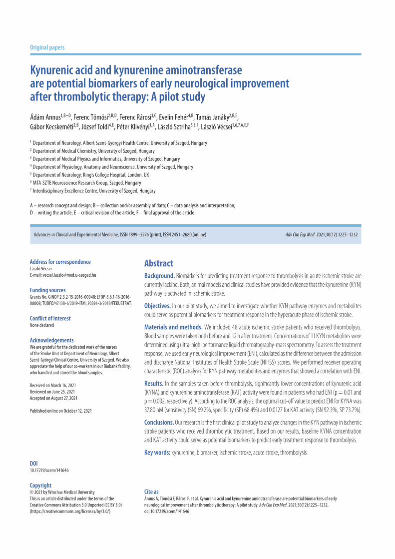
The kynurenine (KYN) pathway is the main route of tryptophan (TRP) metabolism. Animal models and clinical studies have unequivocally proven that the KYN pathway is activated in acute ischemic stroke.9–15 The first step of the pathway is the metabolism of TRP to KYN by indoleamine-2,3-dioxygenase (IDO). Inflammatory cytokines (e.g., interleukin 1β (IL-1β), tumor necrosis fac-tor α (TNF-α) and interferon γ (INF-γ)) were shown to in-crease the expression of IDO.16,17 Therefore, the activation of the KYN pathway following ischemic brain injury is likely part of a secondary inflammatory reaction.18,19 The most well-studied metabolites of the pathway are kynurenic acid (KYNA), 3-hydroxykynurenine (3-HK) and quinolinic acid (QUIN). Kynurenic acid is metabolized by kynurenine ami-notransferase (KAT) from KYN. It is a known endogenous, competitive inhibitor of the N-methyl-D-aspartate receptor (NMDAR) and is therefore thought to have neuroprotective properties.20 In contrast, 3-HK and QUIN are neurotoxic compounds that produce free radicals and cause oxidative stress. Further metabolites and enzymes of the KYN path-way that were analyzed in our study are highlighted in Fig. 1.
The KYN pathway is linked to a number of traditional cerebrovascular risk factors that could influence serum levels of KYN metabolites.21 It has been demonstrated that IDO expression regulates blood pressure in mouse mod-els of systemic inflammation.22 Administration of KYNA into the rostral ventrolateral medulla of spontaneously hypertensive rats, decreased mean arterial blood pressure
by approx. 40 mm Hg.23 Median blood KYN levels of pa-tients with stable angina pectoris were higher in hyper-tensive patients compared to normotensive individuals.24
Elevated xanthurenic acid (XA) levels have been found in diabetic patients.25 Xanthurenic acid forms a complex with insulin that does not activate insulin receptors.26 There-fore, elevated XA levels contribute to insulin resistance.27
Aging, the most relevant non-modifiable risk factor for cerebrovascular diseases, showed a significant association in a multivariate linear regression analysis with serum con-centrations of KYN, TRP, and IDO activity.28 In the Horda-land Health Study, an inverse association was found between heavy smoking and anthranilic acid (AA), TRP, KYN, KYNA, XA, and 3-hydroxyanthranilic acid (3-HANA).29
Objectives
Our aim in this single-center pilot study was to investi-gate whether metabolites of the KYN pathway and activ-ity of relevant enzymes measured before and 12 h after thrombolytic therapy in ischemic stroke could serve as po-tential biomarkers for predicting treatment response and prognosis.
Patients and methods
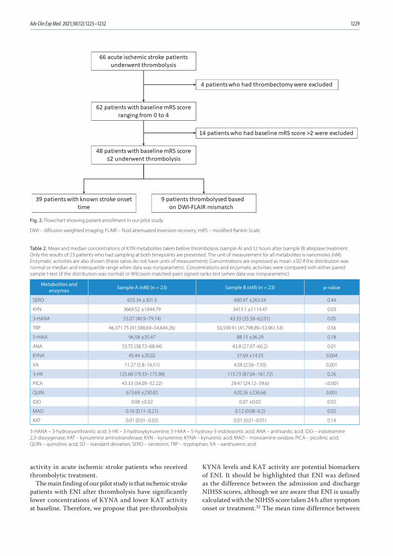
Patients and outcomes
Our inclusion criteria were patients with a diagnosis of acute ischemic stroke who underwent intravenous thrombolysis with alteplase between January and December 2018. We excluded patients who received thrombectomy and those who had a baseline mRS score >2. The pilot study
Fig. 1. Kynurenine pathway and other tryptophan metabolites measured in our pilot study. Dotted lines indicate more than 1 enzymatic step. Dotted boxes highlight the most relevant enzymes of the pathway
3-HANA – 3-hydroxyanthranilic acid; 3-HK – 3-hydroxy-kynurenine; 5-HIAA – 5-hydroxy-3-indoleacetic acid; ANA – anthranilic acid; IDO – indoleamine 2,3-dioxygenase; KAT – kynurenine aminotransferase; KYN – kynurenine; KYNA – kynurenic acid; MAO – monoamine oxidase; PICA – picolinic acid; QUIN – quinolinic acid; SERO – serotonin; TRP – tryptophan; XA – xanthurenic acid.
Adv Clin Exp Med. 2021;30(12):1225–1232 1227
was conducted in accordance with the revised Declaration of Helsinki, and the protocol was approved by the Ethics Committee of the University of Szeged, Albert Szent-Györ-gyi Clinical Centre (Project GINOP 2.3.2-15-2016-00048). All patients or their relatives gave informed consent for inclusion before the participation in the study.
C-reactive protein (CRP) and white cell count were mea-sured from blood samples taken upon arrival in the emer-gency department. The time when the alteplase bolus was administered is referred to as “needle time”. All patients underwent repeat imaging approx. 24 h after treatment. The NIHSS and mRS scores were established by a certified expert. The NIHSS scores were calculated on admission before thrombolysis, and at discharge from the stroke unit.
Efficacy endpoints were early neurological improvement (ENI) and good functional outcome at 30 and 90 days after the stroke. We defined ENI as a ≥4 point decrease in the NIHSS score from admission to discharge. The cri-terion for good functional outcome was an mRS score ≤2.
Sampling
Peripheral venous blood samples were taken just before thrombolysis and 12 h after the initiation of treatment (sam-ples A and B, respectively). Blood samples were centrifuged at 3000/min for 13 min, and sera were then stored at −80°C until further analysis. Due to restricted opening times of our biobank facility, samples were only processed on weekdays.
Measurement of kynurenines by ultra-high-performance liquid chromatography (UHPLC) coupled to tandem mass spectrometry (MS/MS)
Reagents and chemicals
All reagents and chemicals were of analytical or liquid chromatography-mass spectrometry (LC-MS) grade. Tryp-tophan and its metabolites and d4-picolinic acid (PICA) were purchased from Sigma-Aldrich (St. Louis, USA). The d3-3-HK was obtained from Buchem BV (Apeldoorn, the Netherlands). The other deuterated internal standards (ISs; d4-serotonin (SERO), d4-KYN, d3-3-3-HANA, d5-TRP, d5-5-hydroxy-3-indoleacetic acid (5-HIAA), d5-KYNA, d4-XA and d3-QUIN) were purchased from To-ronto Research Chemicals (Toronto, Canada). Acetone, methanol (MeOH) and water were obtained from VWR Chemicals (Monroeville, USA). Formic acid (FA) was pur-chased from Thermo Fisher Scientific (Portsmouth, USA).
Preparation of standard, IS and quality control solutions
Stock solutions, calibration standards and quality con-trol (QC) samples were prepared as described previously.30
Calibration standards consisted of 100 μL of “blank” serum, 10 μL of standard solution mix (156.25–5000 nM SERO, 312.5–10,000 nM KYN, 7.8–250 nM 3-HANA, 6.25–200 µM TRP, 7.8–250 nM 5-HIAA, 6.25–200 nM ANA, 4.7–150 nM KYNA, 6.25–200 nM 3-HK, 1.5–50 nM XA, 3.125–100 nM PICA, and 62.5–2000 nM QUIN in 0.1% (v/v) aqueous FA), were treated with 370 μL of ice-cold acetone: MeOH (1:1, (v/v)) containing 10 μL of the SIL-IS mix (1500 nM d4-SERO, 1000 nM d4-KYN, 65 nM d3-3-HANA, 5250 nM d5-TRP, 200 nM d5-5-HIAA, 50 nM d5-KYNA, 90 nM d3-3-HK, 25 nM d4-XA, 80 nM d4-PICA, and 300 nM d3-QUIN) to precipitate proteins. After centrifugation, 400 μL of su-pernatant were transferred to a new tube, spun for 15 s and split into 2 equal parts. After concentration under a vacuum (Savant SC 110 A Speed Vac Plus; Savant, Holbrook, USA), half of the sample was treated with 70 μL of derivatizing re-agent (n-butanol-acetyl chloride, 9:1, (v/v)) and was incubated for 1 h at 60°C. The mixture was dried under nitrogen be-fore reconstitution. Both parts of the sample were dissolved in 100–100 μL of the starting eluent, vortexed, centrifuged, and combined.
Preparation of human serum samples for analysis
The human serum samples were prepared as described previously.30 Briefly, to 100 μL of each serum sample, 10 μL 0.1% (v/v) of aqueous FA and 370 μL of ice-cold ac-etone–MeOH (1:1, (v/v)) containing 10 μL of the SIL-IS mix (the same as used in the preparation of the calibration standards) were added, and 400 μL of supernatant was treated as above.
Instrumentation and UHPLC-MS/MS analysis
The UHPLC separation of TRP and its metabolites was performed on a pentafluorophenyl (PFP) column (100 Å, 100 mm × 2.1 mm, particle size 2.6 μm; Phenomenex, Torrance, USA) connected to an ACQUITY I-Class UPLC™ liquid chromatography system (Waters, Man-chester, UK) using 0.1% (v/v) aqueous FA as solvent A and MeOH containing 0.1% (v/v) FA as solvent B. All mass spectrometric measurements were carried out on an on-line connected Q Exactive™ Plus Hybrid Quadrupole-Orbitrap Mass Spectrometer (Thermo Fisher Scientific, San Jose, USA), operating in the positive electrospray ion-ization mode. For quantitative mass spectrometric analysis through MS/MS, the parallel reaction monitoring (PRM) data acquisition mode was chosen. The optimization of parameters and the validation of the UHPLC-MS/MS analysis for human serum were carried out previously.30
Statistical analyses
Our outcome measures were categorical variables (ENI, good outcome at 30 and 90 days). Based on the Shapiro–Wilk test, some KYN metabolites and enzymatic activities
Á. Annus et al. Kynurenines are biomarkers of good response1228
did not show normal distribution (namely, 3-HANA, TRP, ANA, 3-HK, PICA, XA, monoamine oxidase (MAO), and KAT). Therefore, based on the distribution, continuous variables were either expressed as mean ± standard de-viation (SD) or median and interquartile range (IQR). Kynurenine metabolite concentrations and enzymatic ac-tivities measured at the 2 timepoints were compared with either paired sample t-test (if the distribution was normal), or Wilcoxon matched-pairs signed-ranks test (when data was nonparametric) for cases where both samples were taken. To compare means of concentrations and enzymatic activities between groups with and without ENI or good functional outcome, we used the independent sample t-test or Mann–Whitney U test (depending on the distribution of the data). Boxplots were drawn to allow for better visu-alization of statistically significant findings. Furthermore, if statistical significance was met, we performed receiver operating characteristic (ROC) analysis. We calculated area under the curve (AUC), as well as sensitivity (SN) and specificity (SP) for different cut-off values. Due to the small sample size of our pilot study, logistic regression was not performed. A p-value of <0.05 was regarded statistically significant. Confidence intervals (CI) of 95% were presented where appropriate. Analyses were carried out with IBM SPSS v. 24 (IBM Corp., Armonk, USA) statistical software.
Results
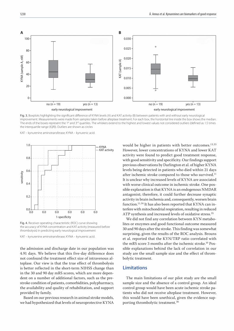
Our pilot study included 48 patients. Thirty-nine were known to be within the 4.5 h thrombolysis time window. In the remaining 9 patients with unknown stroke onset time, intravenous alteplase was administered on the ba-sis of a diffusion-weighted imaging (DWI)-fluid-attenu-ated inversion recovery (FLAIR) mismatch demonstrated on an acute brain magnetic resonance imaging (MRI), as per the WAKE-UP trial.31 The flowchart of patient selection is shown in Fig. 2. The clinical characteristics of our study population are highlighted in Table 1. Seventeen patients had large vessel occlusion (LVO), but mechanical thrombec-tomy was not performed due to limited availability of this service in our center at the time. We collected 32 blood samples before thrombolysis and 36 samples 12 h after treatment. Twenty-three patients had samples taken at both timepoints. The UHPLC-MS/MS method provided simul-taneous quantification of TRP and its 10 most important metabolites (SERO, KYN, 3-HANA, 5-HIAA, ANA, KYNA, 3-HK, XA, PICA, and QUIN).30 Concentrations of the mea-sured KYN metabolites of the 23 patients who had sampling at both timepoints are shown in Table 2. Significant changes in paired serum levels were observed for KYN, ANA, KYNA, XA, PICA, and QUIN. Enzymatic activity of IDO, MAO and KAT were calculated by the following ratios: KYN/TRP, 5-HIAA/SERO and KYNA/KYN, respectively. Enzymatic activities are also demonstrated in Table 2. The activity of IDO and MAO decreased significantly after 12 h.
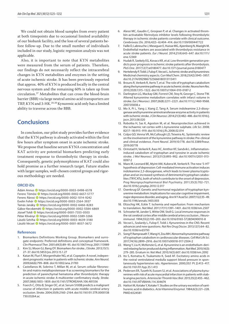
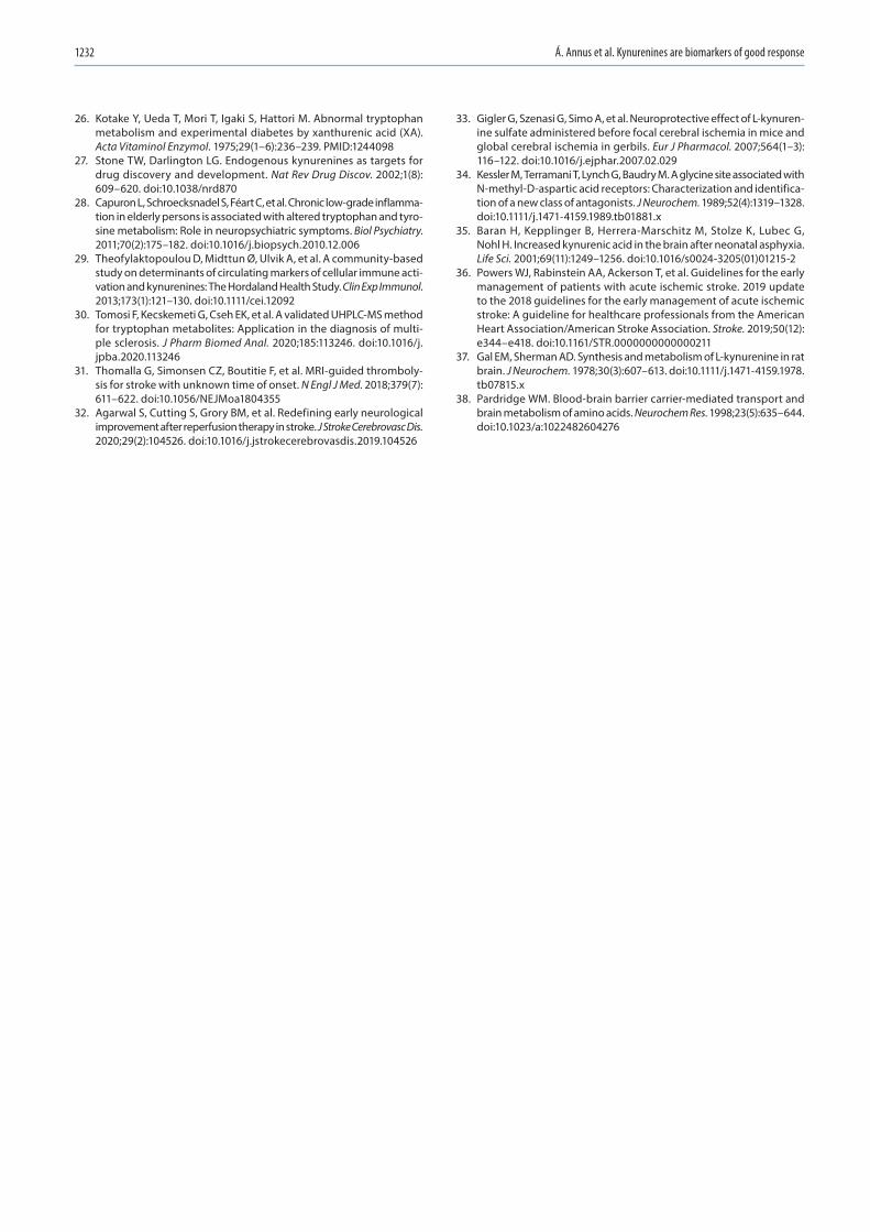
Patients with ENI had significantly lower concentra-tions of KYNA and lower KAT activity in sample A (in-dependent sample t-test, p = 0.01, df = 30, t = −2.722; and Mann–Whitney U test, p = 0.002, z = −3.050, respectively, Fig. 3). There was no statistically significant difference in sample B. Regarding the presence or absence of good outcome at 30 and 90 days, concentrations and enzymatic activities did not statistically significantly differ in sam-ples A or B. Receiver operating characteristic analysis for ENI was performed using KYNA levels and KAT activity measured before treatment (Fig. 4). The AUC for KYNA concentrations was 0.74, 95% CI = 0.57–0.91, p = 0.02. The optimal cut-off value to predict ENI was 37.8 nM (SN 69.2%, SP 68.4%). Similarly, the AUC for KAT activity was 0.82, 95% CI = 0.67–0.98, p = 0.002. The optimal cut-off activity was 0.0127 (SN 92.3%, SP 73.7%).
Discussion
To our knowledge, this is the first study to analyze the changes in KYN metabolite serum levels and enzymatic
Table 1. Clinical data of our study population (n = 48). Some data were not available for all patients. These are highlighted after each criteria accordingly
Patient characteristics Value
Mean age ±SD [years] 67.33 ±12.04
Female 24 (50%)
Male 24 (50%)
Hypertension 41 (85.42%)
Diabetes mellitus 13 (27.08%)
Hyperlipidemia 39 (81.25%)
Smoking 14 (29.17%)
Atrial fibrillation 7 (14.58%)
Coronary artery disease 10 (20.83%)
Mean baseline NIHSS score ±SD 8.81 ±4.29
Mean baseline mRS score ±SD 0.79 ±0.77
Large vessel occlusion 17 (35.42%)
Mean SOtN time ±SD (min, n = 39) 136.59 ±53.9
Mean DtN time ±SD (min, n = 47) 57.45 ±35.72
Length of stay in stroke unit [days] 4.91 ±2.05
Intracerebral hemorrhage after treatment 4 (8.33%)
Mean C-reactive protein ±SD [mg/L], n = 47) 10.44 ±18.78
Mean white cell count ±SD [g/L] 8.07 ±2.31
Mean discharge NIHSS score ±SD 6.71 ±7.89
Early neurological improvement 19 (39.58%)
Mean mRS score at 30 days ±SD (n = 45) 2.47 ±1.84
Mean mRS score at 90 days ±SD (n = 40) 2.38 ±1.9
Good functional outcome at day 30 (n = 45) 27 (60%)
Good functional outcome at day 90 (n = 40) 24 (60%)
DtN – door to needle; mRS – modified Rankin Scale; NIHSS – National Institutes of Health Stroke Scale; SD – standard deviation; SOtN – stroke onset to needle.
Adv Clin Exp Med. 2021;30(12):1225–1232 1229
activity in acute ischemic stroke patients who received thrombolytic treatment.
The main finding of our pilot study is that ischemic stroke patients with ENI after thrombolysis have significantly lower concentrations of KYNA and lower KAT activity at baseline. Therefore, we propose that pre-thrombolysis
KYNA levels and KAT activity are potential biomarkers of ENI. It should be highlighted that ENI was defined as the difference between the admission and discharge NIHSS scores, although we are aware that ENI is usually calculated with the NIHSS score taken 24 h after symptom onset or treatment.32 The mean time difference between
Table 2. Mean and median concentrations of KYN metabolites taken before thrombolysis (sample A) and 12 hours after (sample B) alteplase treatment. Only the results of 23 patients who had sampling at both timepoints are presented. The unit of measurement for all metabolites is nanomoles (nM). Enzymatic activities are also shown (these ratios do not have units of measurement). Concentrations are expressed as mean ±SD if the distribution was normal or median and interquartile range when data was nonparametric. Concentrations and enzymatic activities were compared with either paired sample t-test (if the distribution was normal) or Wilcoxon matched-pairs signed-ranks test (when data was nonparametric)
Metabolites and enzymes Sample A (nM) (n = 23) Sample B (nM) (n = 23) p-value
SERO 655.34 ±301.3 680.97 ±263.54 0.44
KYN 3669.52 ±1044.79 3413.1 ±1114.47 0.03
3-HANA 53.07 (40.9–79.74) 43.33 (35.58–62.01) 0.05
TRP 46,371.75 (41,588.69–54,644.26) 50,599.91 (41,798.89–53,961.54) 0.56
5-HIAA 96.58 ±35.47 88.15 ±36.29 0.18
ANA 53.75 (38.73–68.44) 43.8 (27.07–60.2) 0.01
KYNA 45.44 ±20.02 37.69 ±14.55 0.004
XA 11.27 (5.8–16.51) 4.58 (2.56–7.93) 0.001
3-HK 125.68 (79.33–175.98) 115.73 (87.04–161.72) 0.26
PICA 43.33 (34.09–52.22) 29.47 (24.12–39.6) <0.001
QUIN 673.69 ±230.82 620.26 ±236.66 0.001
IDO 0.08 ±0.02 0.07 ±0.02 0.02
MAO 0.16 (0.11–0.21) 0.12 (0.08–0.2) 0.02
KAT 0.01 (0.01–0.02) 0.01 (0.01–0.01) 0.14
3-HANA – 3-hydroxyanthranilic acid; 3-HK – 3-hydroxykynurenine; 5-HIAA – 5-hydroxy-3-indoleacetic acid; ANA – anthranilic acid; IDO – indoleamine 2,3-dioxygenase; KAT – kynurenine aminotransferase; KYN – kynurenine; KYNA – kynurenic acid; MAO – monoamine oxidase; PICA – picolinic acid; QUIN – quinolinic acid; SD – standard deviation; SERO – serotonin; TRP – tryptophan; XA – xanthurenic acid.
Fig. 2. Flowchart showing patient enrollment in our pilot study
DWI – diffusion weighted imaging; FLAIR – fluid attenuated inversion recovery; mRS – modified Rankin Scale.
Á. Annus et al. Kynurenines are biomarkers of good response1230
the admission and discharge date in our population was 4.91 days. We believe that this five-day difference does not confound the treatment effect size of intravenous al-teplase. Our view is that the true effect of thrombolysis is better reflected in the short-term NIHSS change than in the 30 and 90 day mRS scores, which are more depen-dent on a number of additional factors, such as the pre-stroke condition of patients, comorbidities, polypharmacy, the availability and quality of rehabilitation, and support provided by family.
Based on our previous research in animal stroke models, we had hypothesized that levels of neuroprotective KYNA
would be higher in patients with better outcomes.13,33 However, lower concentrations of KYNA and lower KAT activity were found to predict good treatment response, with good sensitivity and specificity. Our findings support previous observations by Darlington et al. of higher KYNA levels being detected in patients who died within 21 days after ischemic stroke compared to those who survived.11 It is unclear why increased levels of KYNA are associated with worse clinical outcome in ischemic stroke. One pos-sible explanation is that KYNA is an endogenous NMDAR antagonist; therefore, it could further decrease synaptic activity in brain ischemia and, consequently, worsen brain function.11,34 It has also been reported that KYNA can in-terfere with mitochondrial respiration, resulting in reduced ATP synthesis and increased levels of oxidative stress.35
We did not find any correlation between KYN metabo-lites or enzymes and good functional outcome measured 30 and 90 days after the stroke. This finding was somewhat surprising, given the results of the ROC analysis. Brouns et al. reported that the KYN/TRP ratio correlated with the mRS score 3 months after the ischemic stroke.10 Pos-sible explanations behind the lack of correlation in our study are the small sample size and the effect of throm-bolytic treatment.
Limitations
The main limitations of our pilot study are the small sample size and the absence of a control group. An ideal control group would have been acute ischemic stroke pa-tients who did not receive alteplase treatment. However, this would have been unethical, given the evidence sup-porting thrombolytic treatment.36
Fig. 3. Boxplots highlighting the significant difference of KYNA levels (A) and KAT activity (B) between patients with and without early neurological improvement. Measurements were made from samples taken before alteplase treatment. For each box, the horizontal line inside the box shows the median. The ends of the boxes represent the 1st and 3rd quartiles. The whiskers extend to the highest and lowest values not considered outliers (defined as 1.5 times the interquartile range (IQR)). Outliers are shown as circles
KAT – kynurenine aminotransferase; KYNA – kynurenic acid.
Fig. 4. Receiver operating characteristic (ROC) curve showing the accuracy of KYNA concentration and KAT activity (measured before thrombolysis) in predicting early neurological improvement
KAT – kynurenine amino transferase; KYNA – kynurenic acid.
100
80
60
40
20
0
no (n = 19) yes (n = 13)
early neurological improvement
KYN
A (s
ampl
e A,
nM
)
A0.030
0.025
0.020
0.015
0.010
0.005
0.000
no (n = 19) yes (n = 13)
early neurological improvement
KAT
(sam
ple
A)
B
0.0
1.0
0.8
0.6
0.4
0.2
0.00.0 0.0 0.0
1-specificity
sens
itivi
ty
0.0 0.0
KYNAKAT activity
Adv Clin Exp Med. 2021;30(12):1225–1232 1231
We could not obtain blood samples from every patient at both timepoints due to occasional limited availability of our biobank facility, and the loss of several patients be-fore follow-up. Due to the small number of individuals included in our study, logistic regression analysis was not applicable.
Also, it is important to note that KYN metabolites were measured from the serum of patients. Therefore, our findings do not necessarily reflect the intracerebral changes in KYN metabolites and enzymes in the setting of acute ischemic stroke. It has been previously reported that approx. 40% of KYN is produced locally in the central nervous system and the remaining 60% is taken up from circulation.37 Metabolites that can cross the blood-brain barrier (BBB) via large neutral amino acid transporters are TRP, KYN and 3-HK.20,38 Kynurenic acid only has a limited ability to traverse across the BBB.
Conclusions
In conclusion, our pilot study provides further evidence that the KYN pathway is already activated within the first few hours after symptom onset in acute ischemic stroke. We propose that baseline serum KYNA concentration and KAT activity are potential biomarkers predicting early treatment response to thrombolytic therapy in stroke. Consequently, genetic polymorphisms of KAT could also hold promise as a further research target. Future studies with larger samples, well-chosen control groups and rigor-ous methodology are needed.
ORCID iDsÁdám Annus https://orcid.org/0000-0003-0498-6578Ferenc Tömösi https://orcid.org/0000-0002-6657-5777Ferenc Rárosi https://orcid.org/0000-0002-1014-9242Evelin Fehér https://orcid.org/0000-0003-2564-3937Tamás Janáky https://orcid.org/0000-0002-6466-8283Gábor Kecskeméti https://orcid.org/0000-0002-5584-6869József Toldi https://orcid.org/0000-0001-7355-0503Péter Klivényi https://orcid.org/0000-0002-5389-3266László Sztriha https://orcid.org/0000-0003-4639-3180László Vécsei https://orcid.org/0000-0001-8037-3672
References1. Biomarkers Definitions Working Group. Biomarkers and surro-
gate endpoints: Preferred definitions and conceptual framework. Clin Pharmacol Ther. 2001;69(3):89–95. doi:10.1067/mcp.2001.113989
2. Kim SJ, Moon GJ, Bang OY. Biomarkers for stroke. J Stroke. 2013;15(1): 27–37. doi:10.5853/jos.2013.15.1.27
3. Katan M, Fluri F, Morgenthaler NG, et al. Copeptin: A novel, indepen-dent prognostic marker in patients with ischemic stroke. Ann Neurol. 2009;66(6):799–808. doi:10.1002/ana.21783
4. Castellanos M, Sobrino T, Millan M, et al. Serum cellular fibronec-tin and matrix metalloproteinase-9 as screening biomarkers for the prediction of parenchymal hematoma after thrombolytic therapy in acute ischemic stroke: A multicenter confirmatory study. Stroke. 2007;38(6):1855–1859. doi:10.1161/STROKEAHA.106.481556
5. Foerch C, Otto B, Singer OC, et al. Serum S100B predicts a malignant course of infarction in patients with acute middle cerebral artery occlusion. Stroke. 2004;35(9):2160–2164. doi:10.1161/01.STR.0000138 730.03264.ac
6. Alessi MC, Gaudin C, Grosjean P, et al. Changes in activated throm-bin-activatable fibrinolysis inhibitor levels following thrombolytic therapy in ischemic stroke patients correlate with clinical outcome. Cerebrovasc Dis. 2016;42(5–6):404–414. doi:10.1159/000447722
7. Faille D, Labreuche J, Meseguer E, Huisse MG, Ajzenberg N, Mazighi M. Endothelial markers are associated with thrombolysis resistance in acute stroke patients. Eur J Neurol. 2014;21(4):643–647. doi:10.1111/ene.12369
8. Hudak R, Szekely EG, Kovacs KR, et al. Low thrombin generation pre-dicts poor prognosis in ischemic stroke patients after thrombolysis. PloS One. 2017;12(7):e0180477. doi:10.1371/journal.pone.0180477
9. Hertelendy P, Toldi J, Fulop F, Vecsei L. Ischemic stroke and kynurenines: Medicinal chemistry aspects. Curr Med Chem. 2018;25(42):5945–5957. doi:10.2174/0929867325666180313113411
10. Brouns R, Verkerk R, Aerts T, et al. The role of tryptophan catabolism along the kynurenine pathway in acute ischemic stroke. Neurochem Res. 2010;35(9):1315–1322. doi:10.1007/s11064-010-0187-2
11. Darlington LG, Mackay GM, Forrest CM, Stoy N, George C, Stone TW. Altered kynurenine metabolism correlates with infarct volume in stroke. Eur J Neurosci. 2007;26(8):2211–2221. doi:10.1111/j.1460-9568. 2007.05838.x
12. Mo X, Pi L, Yang J, Xiang Z, Tang A. Serum indoleamine 2,3-dioxy-genase and kynurenine aminotransferase enzyme activity in patients with ischemic stroke. J Clin Neurosci. 2014;21(3):482–486. doi:10.1016/j.jocn.2013.08.020
13. Robotka H, Sas K, Agoston M, et al. Neuroprotection achieved in the ischaemic rat cortex with L-kynurenine sulphate. Life Sci. 2008; 82(17–18):915–919. doi:10.1016/j.lfs.2008.02.014
14. Colpo GD, Venna VR, McCullough LD, Teixeira AL. Systematic review on the involvement of the kynurenine pathway in stroke: Pre-clinical and clinical evidence. Front Neurol. 2019;10:778. doi:10.3389/fneur. 2019.00778
15. Ormstad H, Verkerk R, Aass HC, Amthor KF, Sandvik L. Inflammation-induced catabolism of tryptophan and tyrosine in acute ischemic stroke. J Mol Neurosci. 2013;51(3):893–902. doi:10.1007/s12031-013-0097-2
16. Maes M, Leonard BE, Myint AM, Kubera M, Verkerk R. The new ‘5-HT’ hypothesis of depression: Cell-mediated immune activation induces indoleamine 2,3-dioxygenase, which leads to lower plasma trypto-phan and an increased synthesis of detrimental tryptophan catabo-lites (TRYCATs), both of which contribute to the onset of depression. Prog Neuropsychopharmacol Biol Psychiatry. 2011;35(3):702–721. doi:10.1016/j.pnpbp.2010.12.017
17. Oxenkrug GF. Genetic and hormonal regulation of tryptophan kyn-urenine metabolism: Implications for vascular cognitive impairment, major depressive disorder, and aging. Ann N Y Acad Sci. 2007;1122:35–49. doi:10.1196/annals.1403.003
18. Eltzschig HK, Eckle T. Ischemia and reperfusion: From mechanism to translation. Nat Med. 2011;17(11):1391–1401. doi:10.1038/nm.2507
19. Schroeter M, Jander S, Witte OW, Stoll G. Local immune responses in the rat cerebral cortex after middle cerebral artery occlusion. J Neuroimmunol. 1994;55(2):195–203. doi:10.1016/0165-5728(94)90010-8
20. Vecsei L, Szalardy L, Fulop F, Toldi J. Kynurenines in the CNS: Recent advances and new questions. Nat Rev Drug Discov. 2013;12(1):64–82. doi:10.1038/nrd3793
21. Song P, Ramprasath T, Wang H, Zou MH. Abnormal kynurenine path way of tryptophan catabolism in cardiovascular diseases. Cell Mol Life Sci. 2017;74(16):2899–2916. doi:10.1007/s00018-017-2504-2
22. Wang Y, Liu H, McKenzie G, et al. Kynurenine is an endothelium-deri-ved relaxing factor produced during inflammation. Nat Med. 2010;16(3): 279–285. Erratum in: Nat Med. 2010;16(5):607. doi:10.1038/nm.2092
23. Ito S, Komatsu K, Tsukamoto K, Sved AF. Excitatory amino acids in the rostral ventrolateral medulla support blood pressure in spon-taneously hypertensive rats. Hypertension. 2000;35(1 Pt 2):413–417. doi:10.1161/01.hyp.35.1.413
24. Pedersen ER, Tuseth N, Eussen SJ, et al. Associations of plasma kynu-renines with risk of acute myocardial infarction in patients with stab-le angina pectoris. Arterioscler Thromb Vasc Biol. 2015;35(2):455–462. doi:10.1161/ATVBAHA.114.304674
25. Hattori M, Kotake Y, Kotake Y. Studies on the urinary excretion of xant-hurenic acid in diabetics. Acta Vitaminol Enzymol. 1984;6(3):221–228. PMID:6524581
Á. Annus et al. Kynurenines are biomarkers of good response1232
26. Kotake Y, Ueda T, Mori T, Igaki S, Hattori M. Abnormal tryptophan metabolism and experimental diabetes by xanthurenic acid (XA). Acta Vitaminol Enzymol. 1975;29(1–6):236–239. PMID:1244098
27. Stone TW, Darlington LG. Endogenous kynurenines as targets for drug discovery and development. Nat Rev Drug Discov. 2002;1(8): 609–620. doi:10.1038/nrd870
28. Capuron L, Schroecksnadel S, Féart C, et al. Chronic low-grade inflamma-tion in elderly persons is associated with altered tryptophan and tyro-sine metabolism: Role in neuropsychiatric symptoms. Biol Psychiatry. 2011;70(2):175–182. doi:10.1016/j.biopsych.2010.12.006
29. Theofylaktopoulou D, Midttun Ø, Ulvik A, et al. A community-based study on determinants of circulating markers of cellular immune acti-vation and kynurenines: The Hordaland Health Study. Clin Exp Immunol. 2013;173(1):121–130. doi:10.1111/cei.12092
30. Tomosi F, Kecskemeti G, Cseh EK, et al. A validated UHPLC-MS method for tryptophan metabolites: Application in the diagnosis of multi-ple sclerosis. J Pharm Biomed Anal. 2020;185:113246. doi:10.1016/j.jpba.2020.113246
31. Thomalla G, Simonsen CZ, Boutitie F, et al. MRI-guided thromboly-sis for stroke with unknown time of onset. N Engl J Med. 2018;379(7): 611–622. doi:10.1056/NEJMoa1804355
32. Agarwal S, Cutting S, Grory BM, et al. Redefining early neurological improvement after reperfusion therapy in stroke. J Stroke Cerebrovasc Dis. 2020;29(2):104526. doi:10.1016/j.jstrokecerebrovasdis.2019.104526
33. Gigler G, Szenasi G, Simo A, et al. Neuroprotective effect of L-kynuren-ine sulfate administered before focal cerebral ischemia in mice and global cerebral ischemia in gerbils. Eur J Pharmacol. 2007;564(1–3): 116–122. doi:10.1016/j.ejphar.2007.02.029
34. Kessler M, Terramani T, Lynch G, Baudry M. A glycine site associated with N-methyl-D-aspartic acid receptors: Characterization and identifica-tion of a new class of antagonists. J Neurochem. 1989;52(4):1319–1328. doi:10.1111/j.1471-4159.1989.tb01881.x
35. Baran H, Kepplinger B, Herrera-Marschitz M, Stolze K, Lubec G, Nohl H. Increased kynurenic acid in the brain after neonatal asphyxia. Life Sci. 2001;69(11):1249–1256. doi:10.1016/s0024-3205(01)01215-2
36. Powers WJ, Rabinstein AA, Ackerson T, et al. Guidelines for the early management of patients with acute ischemic stroke. 2019 update to the 2018 guidelines for the early management of acute ischemic stroke: A guideline for healthcare professionals from the American Heart Association/American Stroke Association. Stroke. 2019;50(12): e344–e418. doi:10.1161/STR.0000000000000211
37. Gal EM, Sherman AD. Synthesis and metabolism of L-kynurenine in rat brain. J Neurochem. 1978;30(3):607–613. doi:10.1111/j.1471-4159.1978.tb07815.x
38. Pardridge WM. Blood-brain barrier carrier-mediated transport and brain metabolism of amino acids. Neurochem Res. 1998;23(5):635–644. doi:10.1023/a:1022482604276
Cite asBala MM, Bala KA. Severe cases of osteogenesis imperfecta type VIII due to a homozygous mutation in P3H1 (LEPRE1) and review of the literature. Adv Clin Exp Med. 2021;30(12):1233–1238. doi:10.17219/acem/141367
DOI10.17219/acem/141367
Copyright© 2021 by Wroclaw Medical University This is an article distributed under the terms of theCreative Commons Attribution 3.0 Unported (CC BY 3.0)(https://creativecommons.org/licenses/by/3.0/)
Address for correspondenceMehmet Murat BalaE-mail: [email protected]
Funding sourcesNone declared
Conflict of interestNone declared
AcknowledgementsWe would like to thank the patients and their parents for participating in this study. We would like to also thank Prof. Dr. Hüseyin Yüce for his contribution to the genetic analysis in the study.
Received on June 16, 2021Reviewed on August 8, 2021Accepted on August 18, 2021
Published online on October 12, 2021
AbstractBackground. Osteogenesis imperfecta (OI) is a genetic disorder that causes skeletal fragility, multiple fractures and several extraskeletal disorders. Most cases of OI are caused by mutations in COL1A1/A2. Osteo-genesis imperfecta type VIII typically causes a severe and fatal phenotype that presents at birth with severe osteopenia, congenital fractures and other clinical manifestations.
Objectives. We describe the cases of an 11-year-old female and a 9-year-old male with homozygous truncating mutations in P3H1. Both cases were born with intrauterine fractures and suffered multiple fractures shortly after birth, requiring multiple operations to correct both fractures and severe scoliosis. The patients have been treated with pamidronate since the age of 2.
Materials and methods. Whole exome sequencing (WES) was performed by Gene by Gene using Twist Bioscience technology. Initially, ~36.5 Mb of consensus coding sequences (targeting >98% of RefSeq and Gencode v. 28 regions obtained from the human genome) was replicated from fragmented genomic DNA using the Twist Human Core Exome Plus kit. The subsequent library was sequenced on the Illumina Novaseq Next Generation Sequencing platform to achieve at least ×20 reading depth for >98% of the targeted bases. Variant annotations and filtering was performed using Ingenuity Variant Analysis software.
Results. We identified a homozygous mutation in the 3rd exon of P3H1 (c.628C>T/p.Arg210 Ter). Our cases broaden the phenotypic spectrum of OI type VIII as, to the best of our knowledge, these are the first postnatal cases with P3H1 (c.628C>T/p.Arg210 Ter) mutations published in the literature.
Conclusions. We present the first recorded postnatal cases from unrelated families of OI type VIII, broadening our understanding of the severe, but nonfatal spectrum of clinical phenotype of this recessive form of OI.
Key words: osteogenesis imperfecta, severe, homozygous mutation, P3H1, LEPRE1
Original papers
Severe cases of osteogenesis imperfecta type VIII due to a homozygous mutation in P3H1 (LEPRE1) and review of the literature
Mehmet Murat Bala1,A–F, Keziban Aslı Bala2,B
1 Department of Orthopaedics and Traumatology, Health Sciences University, Trabzon Kanuni Training and Research Hospital, Turkey2 Department of Pediatric Endocrinology, Faculty Of Medicine, Health Sciences University, Trabzon Kanuni Training and Research Hospital, Turkey
A – research concept and design; B – collection and/or assembly of data; C – data analysis and interpretation; D – writing the article; E – critical revision of the article; F – final approval of the article
Advances in Clinical and Experimental Medicine, ISSN 1899–5276 (print), ISSN 2451–2680 (online) Adv Clin Exp Med. 2021;30(12):1233–1238
M.M. Bala, K.A. Bala. Homozygous mutation in P3H1 (LEPRE1)1234
Background
Osteogenesis imperfecta (OI) is a clinically and geneti-cally heterogeneous skeletal dysplasia that occurs in approx. 1 in 10,000–20,000 births.1 It is characterized by multiple fractures caused by skeletal fragility and extra-skeletal find-ings, such as blue sclera, dentinogenesis imperfecta, hearing loss, joint hypermobility, and hyperlaxity.1 Most OI cases (type I–IV) are associated with heterozygous mutations in COL1A1 (MIM 120150) or COL1A2 (MIM 120160), which encode the type I procollagen alpha chain to proalpha1 and proalpha2.1 Osteogenesis imperfecta type V is caused by heterozygous mutations in IFITM5 (MIM 614757).2
Osteogenesis imperfecta types VI–XV are inherited in a recessive manner.3 Homozygous truncating muta-tions in P3H1 (LEPRE 1) (NM_022356) are responsible for OI type VIII and were first reported in 2007.4 The P3H1 encodes prolyl 3-hydroxylase 1, which forms a molecular complex with cyclophilin B, encoded by the cartilage-associated protein (CRTAP) and peptidyl prolyl isomer-ase B (PPIB). The P3H1 is involved in the post-translational modification of collagen in the endoplasmic reticulum and prolyl 3-hydroxylation of specific proline residues (espe-cially a1 (I) Pro986).5,6 So far, 48 different mutant P3H1 alleles have been reported in patients with OI.7
Objectives
We describe 2 cases – of an 11-year-old female and a 9-year-old male – with a clinical presentation of severe OI, and identify a homozygous mutation in the 3rd exon of P3H1 (c.628C>T/p.Arg210 Ter). Our cases broaden the phenotypic spectrum of OI type VIII as, to the best of our knowledge, these are the first postnatal cases with P3H1 (c.628C>T/p.Arg210 Ter) mutations published in the literature.
Case reports
Case 1
The female patient was 11 years and 7 months of age at the time of the study. She was born from the first preg-nancy of a 21-year-old mother. The mother and 27-year-old father of the child were first cousins, Turkish in origin, and had a height of 150 cm and 180 cm, respectively. Neither of the parents had a history of chronic illness or fracture. The patient was an only child, and the mother had no his-tory of stillbirth or miscarriage. Moreover, there was no known familial history of OI or any other bone dysplasia.
The mother’s pregnancy was followed up every 4–8 weeks starting from the 8th gestational week, and no mater-nal medical problems affecting pregnancy were found. She took prenatal vitamins regularly. The ultrasound
performed during the 12th gestational week revealed the fe-tus had curved and short legs. The amniotic fluid level was normal. The patient was born in the 39th gestational week via normal vaginal delivery without intervention. Her birth weight was 3020 g and height was 47 cm.
Her right upper arm was swollen, and leg movements were reduced on the 1st postnatal day. The patient was evaluated by orthopedic surgeons, and direct radiographic examination was performed. Fractures were detected in the right humeral diaphysis, right clavicle, distal aspect of both the femurs, right proximal tibia, right distal fibula, and left distal tibia. Spinal body fracture was not detected. Chronic changes were detected in the symmetrical, curved and weakened femurs and ribs, suggesting intrauterine fractures.
Pamidronate treatment at a dose of 9.0 mg/kg/year was initiated when the patient was 2 years of age. She underwent a total of 6 operations, 5 of which were for the fractures and 1 for scoliosis, and she has been followed up for the last 3 years, over which she did not sustain any new fractures. The latest bone mineral density (BMD) of the patient was −2.2 SDS (standard deviation score). During follow-up, her calcium, phosphorus and alkaline phosphatase levels were normal, and she was given vitamin D supplements to ensure that her 25-hydroxy vitamin D (25(OH)D) levels were within the normal range.
On physical examination, her height was −7 SDS and weight was −4.7 SDS, according to her age and sex. The pa-tient had normal results for echocardiographic, urinary, hearing and dental examinations, and did not have blue sclera (Table 1). Both femurs had anterolateral curvatures. The patient was able to walk with the help of a walker. Radiographic examinations of the patient demonstrated severe osteopenia, thoracolumbar scoliosis, diaphyseal ex-pansion in the distal humerus and proximal radioulnar metaphyses, trabeculae in the bones, and popcorn-like concentric concentrations (Fig. 1, 1a–f).
No mutation was found in COL1A1, COL1A2, CRTAP, and SERPINH1 in the initial genetic analyses of the pa-tient. Following the initial investigations, whole exome sequencing (WES) was performed using peripheral blood in the Microgen Genetic Diseases Diagnosis Center, Ankara, Turkey, and a homozygous nonsense mutation (c.628C>T/p.Arg210 Ter) was detected in the 3rd exon of P3H1. Mutations in CRTAP, PPIB, FKBP10, SERPINF1, PLOD2, SERPINH1, SP7, or ALPL were excluded using WES. The patient’s mother and father were found to be heterozygous for the same mutation, based on the targeted mutation analyses.
Case 2
The male patient was 9 years and 3 months of age at the time of evaluation. He was born from the 2nd preg-nancy of a 30-year-old mother, whose 1st child was a 10-year-old, healthy female at the time of the investigation.
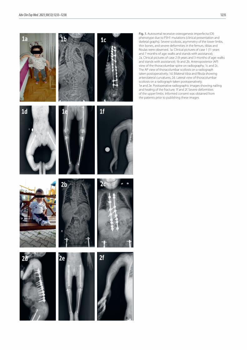
Adv Clin Exp Med. 2021;30(12):1233–1238 1235
Fig. 1. Autosomal recessive osteogenesis imperfecta (OI) phenotype due to P3H1 mutations (clinical presentation and skeletal graphs). Severe scoliosis, asymmetry of the lower limbs, thin bones, and severe deformities in the femurs, tibias and fibulas were observed. 1a. Clinical pictures of case 1 (11 years and 7 months of age: walks and stands with assistance); 2a. Clinical pictures of case 2 (9 years and 3 months of age: walks and stands with assistance); 1b and 2b. Anteroposterior (AP) view of the thoracolumbar spine on radiography; 1c and 2c. The AP view of thoracolumbar scoliosis on a radiograph taken postoperatively; 1d. Bilateral tibia and fibula showing anterolateral curvatures; 2d. Lateral view of thoracolumbar scoliosis on a radiograph taken postoperatively; 1e and 2e. Postoperative radiographic images showing nailing and healing of the fracture; 1f and 2f. Severe deformities of the upper limbs. Informed consent was obtained from the patients prior to publishing these images
M.M. Bala, K.A. Bala. Homozygous mutation in P3H1 (LEPRE1)1236
The mother and 35-year-old father were of Turkish origin, not related, and had a height of 157 cm and 175 cm, re-spectively. Neither of them had a history of chronic illness or fracture. Moreover, the mother had no history of still-birth or miscarriage, nor any known familial history of OI or other bone dysplasia.
The pregnancy was followed up every 4–8 weeks starting from the 6th gestational week, and no maternal medical problems that may affect the pregnancy were found. The mother took prenatal vitamins regularly. The fetus was found to have short legs during an ultra-sound in the 12th gestational week. The amniotic fluid level was normal. The child was delivered via cesarean section in the 38th gestational week. His birth weight was 2850 g and height was 48 cm.
The patient had swollen arms and legs on the 1st post-natal day. Orthopedic surgeons evaluated the patient and direct radiographic examination was conducted. Fractures were detected in both, the distal femurs and humeri. Spinal body fractures were not detected.
Pamidronate treatment at a dose of 9.0 mg/kg/year was initiated when the patient was 2 years old. He un-derwent a total of 11 operations, 10 of which were for fractures and 1 was for scoliosis, and the patient has been followed up for the last 2.5 years, over which time he did not sustain any new fractures. The latest BMD of the patient was −3.7 SDS. During follow-up, the pa-tient’s calcium, phosphorus and alkaline phosphatase levels were normal, and vitamin D supplements were administered to ensure that his 25(OH)D levels were within the normal range.
On physical examination, the patient’s height was −6.6 SDS and weight was –9 SDS, according to his age and sex. His echocardiographic and dental examinations were normal. However, conductive deafness in the right ear and bilateral nephrolithiasis were detected on the audio-logical and urinary examination, respectively. He did not have blue sclera, but had frequent pulmonary infections due to restrictive lung disease (Table 1). The patient was unable to walk because of his almost immobile legs. Severe osteopenia, thinning of long bones, marked appearance of epiphysis, and curvatures caused by multiple fractures were observed on radiographic examinations. The ribs were extremely thin and thoracolumbar scoliosis was also observed (Fig. 1,2a–f).
The COL1A1, COL1A2, CRTAP, and SERPINH1 genes were excluded in the initial patient’s 1st genetic analysis. Subsequent WES analyses of the patient demonstrated a homozygous pathogenic variant (c.628C>T/p.Arg210 Ter) in the 3rd exon of the P3H1 gene (performed at the Mi-crogen Genetic Diseases Diagnosis Center). Mutations in other genes, including CRTAP, PPIB, FKBP10, SER-PINF1, PLOD2, SERPINH1, SP7, and ALPL, were excluded using WES. Sanger sequencing of parental DNA samples revealed that both parents were heterozygous for c.628C>T.
Method of genetic analyses
Whole exome sequencing was performed by Gene by Gene using Twist Bioscience technology (Microgen Genetic Dis-eases Diagnosis Center). Initially, ~36.5 Mb of consensus coding sequences (targeting >98% of RefSeq and Gencode
Table 1. Clinical features of the presented cases
Parameter Case 1 Case 2
P3H1 (LEPRE1) mutation (c.628C>T/p.Arg210 Ter) (c.628C>T/p.Arg210 Ter)
Growth databirthcurrent age [years]weight (SDS)length (SDS)
term, 3.2 kg11
–4.7–7
term, 2.8 kg9
–9–6.6
Facial featuresfacial shapescleral huedentiogenesis imperfecta
triangularwhite
no
triangularwhite
no
Hearing loss no yes
Phenotypesevere OI,
type B progressivesevere OI,
type B progressive
Pulmonary functions normal moderate restrictive
Echocardiography normal normal
Spinal abnormalitiesscoliosis,
multiple T-L vertebralcompression
scoliosis,multiple T-L vertebral
compression
Rhizomelia yes yes
Bone mineral density score (pre- and post-biphosphonate therapy) (SDS)
–8.1 (pre-therapy) –9.3 (pre-therapy)
–2.2 (post-therapy) –3.7 (post-therapy)
OI – osteogenesis imperfecta; SDS – standard deviation score; T-L – thoracolumbar.
Adv Clin Exp Med. 2021;30(12):1233–1238 1237
v. 28 regions obtained from the human genome) were repli-cated from fragmented genomic DNA using the Twist Hu-man Core Exome Plus kit (Twist Bioscience, South San Fran-cisco, USA). The subsequent library was then sequenced on the Illumina Novaseq NGS (Illumina, San Diego, USA) platform to achieve at least a ×20 reading depth for >98% of the targeted bases. Variant annotations and filtering was performed using the Ingenuity Variant Analysis software (Qiagen, Hilden, Germany).
Discussion
The P3H1 (c.628C>T/p.Arg210 Ter) was first reported by Willaert et al. in a consanguineous Turkish family.8 The mother had 2 pregnancies in which the fetuses were found to have genetic mutations, and elective termina-tion was performed at the 20th and 18th gestational week, respectively. To the best of our knowledge, our cases are the first recorded postnatal cases of OI type VIII carry-ing homozygous P3H1 (c.628C>T/p.Arg210 Ter) mutation.
The P3H1 mutations typically have a clinical presentation of severe or fatal OI, and are characterized by rhizomelic short limb, white sclera, severe demineralization of bones, excessive growth retardation, intrauterine fractures, and bulbous expansion of metaphyses.4 Similar to typical P3H1 mutations in literature, our cases also had considerably short stature, severe demineralization, white sclera, and fractures detected at birth. According to the Van Dijk and Sillence cri-teria, the 2 patients were classified into OI type B group with progressive deformation and severe OI group.9 Currently, Willaert et al. is the only publication to present cases with the same mutation.8 Their study reported a case of siblings who were electively aborted, and hence there was no data on postnatal physical examination findings. In addition, similar to their findings, intrauterine skeletal dysplasia-associated findings were detected in both of our cases.
Literature on pamidronate treatment in OI type VIII is limited. In a case published by Santana et al., pami-dronate treatment was initiated at 7 weeks of age. Al-though the measurements were made using different equipment in different centers, a good response was obtained with >300% improvement.10 Takagi et al. also reported an OI type VIII case in which pamidronate treatment was started at 2 months of age. The patient developed very few fractures and presented a good re-sponse.5 In both of our cases, pamidronate treatment was started at ~2 years of age at a dose of 9.0 mg/kg/year. These patients, who were followed up in different clinics until they presented to our center, had a history of many fractures because of starting the treatment later than the previous cases. In their study, Willaert et al. reported a significant improvement in BMD Z-score (from −11.0 to −6.8) in an OI type VIII case of a six-year-old patient treated with pamidronate.8 When our patients presented to our center, their BMD Z-scores were −2.2 SDS and
−3.7 SDS for case 1 and case 2, respectively. This indi-cated that they responded well to the treatment. They were followed up and did not sustain any new fractures over the last 3 years.
The c.628C>T mutation in P3H1 was identified as patho-genic.8 A significant decrease in P3H1 mRNA was observed on evaluation of fibroblast samples from fetuses with this mutation, using quantitative polymerase chain reaction (qPCR), as expected with the degradation of mutant tran-scripts by the nonsense-mediated decay pathway. They found that mRNA expression in the patients’ parents, who were heterozygous carriers of these nonsense P3H1 muta-tions, was similar to that of the control group. The western blot analysis of fibroblast cell lysates probed with anti-P3H1 antibody showed that P3H1 was completely absent in patients and slightly decreased in their parents.
Interestingly, in the review from 2010, Marini et al. reported 17 mutant P3H1 alleles occuring through-out the gene.6 Most of these mutations showed a loss of P3H1 expression in real-time reverse transcription PCR (RT-PCR) with RNA isolated from the patients’ fibroblasts.4 Mrosk et al., in their study on genotype–phenotype corre-lation, found 5 novel pathogenic variants in P3H1 in 5 cases in an Indian OI cohort. Moreover, they observed a variant that was predicted to affect a donor splice site, resulting in a splicing alteration in another patient.11 Scollo et al. reported a retinal tear in a 9-year-old OI type VIII case with a c.1914 + 1G>A (NM_001243246.1) homozygous mutation in P3H1.12 Furthermore, de Souza et al. reported retinal detachment in a 28-year-old OI type VIII case with a similar mutation (c.1914 + 1G>C (NM_001243246.1)).13 However, fundoscopic examinations of our cases were found to be normal.
Fratzl-Zelman et al. performed the first study in the lit-erature on the effect of null mutations in P3H1 on the pa-tient’s bone tissue. They evaluated bone histology and his-tomorphometry, bone mineralization density distribution, and procollagen 3-hydroxylation measurements in bone and skin tissue in non-lethal OI type VIII.14 Although the bones sampled from these OI type VIII children re-sembled those of OI type VII, its distinctive features were bone matrix hypermineralization, extremely thin trabecu-lae, focal osteoid deposition, and an increase in the propor-tion of bone with low mineral density.
Li et al. found that P3H1 mutations had a prevalence of 4.05% in an autosomal recessive OI Chinese cohort, and they reported 3 mutations, including 2 new muta-tions (c.652G>T; c.1948G>C, c.652G>T; c.2164C>T), and 1 previously reported mutation (c.1466T>C; c.1915-1G>A). The most common clinical manifestations were reported to be gait problems, scoliosis and frequent fractures (frac-tures ≥2/year) in this autosomal recessive OI cohort, which was similar to our cases.15 Madhuri et al. found 2 new homo-zygous duplications in P3H1 in an Indian OI cohort of 52 pa-tients (c.2131dup (p.Leu711Profs*19) (SCV000987189) and c.1980dup (p.Val661Serfs*33) (SCV000987190)).16
M.M. Bala, K.A. Bala. Homozygous mutation in P3H1 (LEPRE1)1238
Progressive deformities, blue sclera, recurrent long bone fractures, rhizomelia, scoliosis/kyphosis, vertebral com-pression fractures, and wormian bones have been reported in these children, which are similar to the findings in our cases. The researchers have also observed a previously re-ported variant (c.1346-1G>C) in 5 of their patients, and considered it a recurrent P3H1 variant in India.
Pepin et al. examined 200 samples from an African popu-lation and a neonatal death record of a hospital in Tobago, and they found the prevalence of carriers of the c.1080 + 1G>T mutation in P3H1 was approx. 1 in 200.17 During se-quence analysis, they found a surprisingly high LEPRE1 al-lelic diversity in the DNA samples from Tobago. These find-ings suggested that the milder end of the clinical spectrum may be due to the as yet unidentified missense mutations in P3H1. Caudevilla Lafuente et al. reported 1 (fatal) homozy-gous case in which both parents were heterozygous carriers of the most common variant (c.1080 + 1G>T/IVS5 + 1G>T) in P3H1 in West African populations.18 They also reported a case having a previously reported pathogenic heterozy-gous variant (c.1080 + 1G>T (i5) with c.35T>G (p.Leu12Arg) exon 1, P3H1-VUS and c.969C>T (p.Gly323Gly) exon 7, SERPINF1 gene-VUS). However, this case exhibited a very severe, although nonfatal clinical course. Finally, Tonelli et al. found that the defective chaperone role of the 3-hy-droxylation complex was the primary cause of the skeletal phenotype.19 However, Cabral et al. observed that the func-tions of the modification complex as a collagen chaperone are distinct from its role as prolyl 3-hydroxylase.20
Limitations
Our study has several limitations. First, we only have 2 cases, although our findings may be useful in clinical practice. Second, we could not study in vivo or in vitro models, and hence, there is a need for further studies in this field.
Conclusions
In conclusion, we reported cases of OI type VIII from 2 unrelated families that are the first postnatal cases in which this mutation was detected, and they presented a more severe, but nonfatal spectrum of the clinical phe-notype of this recessive form of OI.
ORCID iDsMehmet Murat Bala https://orcid.org/0000-0002-7213-5647Keziban Aslı Bala https://orcid.org/0000-0001-8755-7714
References1. Forlino A, Marini JC. Osteogenesis imperfecta. Lancet. 2016;387(10028):
1657–1671. doi:10.1016/S0140-6736(15)00728-X
2. Semler O, Garbes L, Keupp K, et al. A mutation in the 5’-UTR of IFITM5 cre-ates an in-frame start codon and causes autosomal-dominant osteo-genesis imperfecta type V with hyperplastic callus. Am J Hum Genet. 2012;91(2):349–357. doi:10.1016/j.ajhg.2012.06.011
3. Byers PH, Pyott SM. Recessively inherited forms of osteogenesis imperfecta. Annu Rev Genet. 2012;46:475–497. doi:10.1146/annurev-genet-110711-155608
4. Cabral WA, Chang W, Barnes AM, et al. Prolyl 3-hydroxylase 1 defi-ciency causes a recessive metabolic bone disorder resembling lethal/severe osteogenesis imperfecta. Nat Genet. 2007;39(3):359–365. doi:10.1038/ng1968
5. Takagi M, Ishii T, Barnes AM, et al. A novel mutation in LEPRE1 that eliminates only the KDEL ER-retrieval sequence causes non-lethal osteogenesis imperfecta. PLoS One. 2012;7(5):e36809. doi:10.1371/journal.pone.0036809
6. Marini JC, Cabral WA, Barnes AM. Null mutations in LEPRE1 and CRTAP cause severe recessive osteogenesis imperfecta. Cell Tissue Res. 2010;339(1):59–70. doi:10.1007/s00441-009-0872-0
7. Cabral WA, Barnes AM, Adeyemo A, et al. A founder mutation in LEPRE1 carried by 1.5% of West Africans and 0.4% of African Amer-icans causes lethal recessive osteogenesis imperfecta. Genet Med. 2012;14(5):543–551. doi:10.1038/gim.2011.44
8. Willaert A, Malfait F, Symoens S, et al. Recessive osteogenesis imper-fecta caused by LEPRE1 mutations: Clinical documentation and iden-tification of the splice form responsible for prolyl 3-hydroxylation. J Med Genet. 2009;46(4):233–241. doi:10.1136/jmg.2008.062729
9. Van Dijk FS, Sillence DO. Osteogenesis imperfecta: Clinical diagno-sis, nomenclature and severity assessment. Am J Med Genet A. 2014; 164A(6):1470–1481. doi:10.1002/ajmg.a.36545
10. Santana A, Franzone JM, McGreal CM, Kruse RW, Bober MB. A moder-ate form of osteogenesis imperfecta caused by compound heterozy-gous LEPRE1 mutations. Bone Reports. 2018;9:132–135. doi:10.1016/j.bonr.2018.09.002
11. Mrosk J, Bhavani GS, Shah H, et al. Diagnostic strategies and geno-type–phenotype correlation in a large Indian cohort of osteogenesis imperfecta. Bone. 2018;110:368–377. doi:10.1016/j.bone.2018.02.029
12. Scollo P, Snead MP, Richards AJ, Pollitt R, DeVile C. Bilateral giant ret-inal tears in osteogenesis imperfecta. BMC Med Genet. 2018;19(1):8. doi:10.1186/s12881-018-0521-0
13. de Souza LT, Nunes RR, de Azevedo Magalhães O, Maria Félix T. A new case of osteogenesis imperfecta type VIII and retinal detachment. Am J Med Genet Part A. 2021;185(1):238–241. doi:10.1002/ajmg.a.61934
14. Fratzl-Zelman N, Barnes AM, Weis MA, et al. Non-lethal type VIII osteogenesis imperfecta has elevated bone matrix mineralization. J Clin Endocrinol Metab. 2016;101(9):3516–3525. doi:10.1210/jc.2016-1334
15. Li S, Cao Y, Wang H, et al. Genotypic and phenotypic analysis in Chi-nese cohort with autosomal recessive osteogenesis imperfecta. Front Genet. 2020;11:984. doi:10.3389/fgene.2020.00984
16. Madhuri V, Selina A, Loganathan L, et al. Osteogenesis imperfecta: Novel genetic variants and clinical observations from a clinical exome study of 54 Indian patients. Ann Hum Genet. 2021;85(1):37–46. doi:10.1111/ahg.12403
17. Pepin MG, Schwarze U, Singh V, Romana M, Jones-Lecointe A, Byers PH. Allelic background of LEPRE1 mutations that cause reces-sive forms of osteogenesis imperfecta in different populations. Mol Genet Genomic Med. 2013;1(4):194–205. doi:10.1002/mgg3.21
18. Caudevilla Lafuente P, Izquierdo-Álvarez S, Labarta Aizpún JI. Osteo-genesis imperfecta caused by COL1A1, CRTAP and LEPRE1 mutations: Report of 2 cases. Med Clin (Barc). 2019;153(8):336–337. doi:10.1016/j.medcli.2018.08.017
19. Tonelli F, Cotti S, Leoni L, et al. Crtap and p3h1 knock out zebrafish support defective collagen chaperoning as the cause of their osteo-genesis imperfecta phenotype. Matrix Biol. 2020;90:40–60. doi:10. 1016/j.matbio.2020.03.004
20. Cabral WA, Fratzl-Zelman N, Weis M, et al. Substitution of murine type I collagen A1 3-hydroxylation site alters matrix structure but does not recapitulate osteogenesis imperfecta bone dysplasia. Matrix Biol. 2020;90:20–39. doi:10.1016/j.matbio.2020.02.003
Cite asRams A, Kosałka-Węgiel J, Kuszmiersz P, et al. Characteristics of idiopathic inflammatory myopathies with novel myositis-specific autoantibodies. Adv Clin Exp Med. 2021;30(12):1239–1248. doi:10.17219/acem/141181
DOI10.17219/acem/141181
Copyright© 2021 by Wroclaw Medical University This is an article distributed under the terms of theCreative Commons Attribution 3.0 Unported (CC BY 3.0)(https://creativecommons.org/licenses/by/3.0/)
Address for correspondenceStanisława Bazan-SochaE-mail: [email protected]
Funding sourcesResearch grant of Jagiellonian University Medical College No. N41/DBS/000687.
Conflict of interestNone declared
Received on May 28, 2021Reviewed on July 6, 2021Accepted on August 10, 2021
Published online on October 5, 2021
AbstractBackground. In recent years, many novel myositis-specific autoantibodies (MSAs) have been identified. However, their links with the pathogenesis and clinical manifestations of inflammatory myopathies remain uncertain.
Objectives. To characterize the population of adult dermatomyositis (DM) and polymyositis (PM) patients treated at our center for autoimmune diseases using clinical and laboratory measures.
Materials and methods. According to the Bohan and Peter criteria, we retrospectively analyzed patients who fulfilled diagnostic criteria for DM or PM. Myositis-specific autoantibodies and myositis-associated autoantibodies (MAAs) were identified using immunoblot assays.
Results. Fifty-one PM (71% women) and 36 DM (67% women) Caucasian patients with a median age of 58 (range: 21–88) years who met the definite or probable diagnostic criteria for myositis were included in the study. Myositis-specific autoantibodies were identified in 63 (72%) patients, whereas MAAs were observed in 43 (49%) of them. Interstitial lung disease (ILD) was characteristic of PM patients (67%, χ2 with Yates’s correction (χc
2) = 13.8078, df = 1, p = 0.0002), being associated with anti-Jo-1 or anti-PL-12 antibodies (fraction comparison test (FCT) 6.4878, p < 0.0001, 6.8354, p = 0.0003, respectively). Interestingly, among patients with anti-MDA5 antibodies (n = 8, 9.2%), all but one had an amyopathic form, with more frequent ILD, skin changes and arthralgias than observed in other patients (FCT 4.7029, p = 0.0228 and p = 7.7986, p = 0.0357, p = 4.7029 and p = 0.0228, respectively). Anti-signal recognition particle (SRP) was strongly associated with the Raynaud’s phenomenon (FCT 4.1144, p = 0.0289) and the highest muscle injury mark-ers (Mann–Whitney U test, z = 2.5293, p = 0.0114). Malignancy was recorded in 14 (16%) patients and was equally common in those with PM and DM. The anti-TIF-1γ was the most frequently related to cancer χ2 = 14.7691, df = 1, p < 0.0001). The anti-Mi-2α, similarly prevalent in DM and PM, was typically accom-panied by skin changes (FCT 7.7986, p = 0.0357) but not ILD (FCT 8.7339, p = 0.0026).
Conclusions. Identification of MSAs might help to predict the clinical course of the autoimmune myopathy and malignancy risk. However, these antibodies were absent in about 30% of patients with typical PM or DM manifestations, which encourages further research in this area.
Keywords: myositis-specific antibodies, polymyositis, dermatomyositis, idiopathic inflammatory myopathies
Original papers
Characteristics of idiopathic inflammatory myopathies with novel myositis-specific autoantibodies
Anna Rams1,A–D, Joanna Kosałka-Węgiel2,B, Piotr Kuszmiersz2,B, Aleksandra Matyja-Bednarczyk3,B, Stanisław Polański3,B, Lech Zaręba3,C, Stanisława Bazan-Socha3,A,D–F
1 Department of Pulmonology and Allergology, University Hospital, Kraków, Poland2 Department of Rheumatology and Immunology, University Hospital, Kraków, Poland3 Faculty of Medicine, Jagiellonian University, Medical College, Kraków, Poland
A – research concept and design; B – collection and/or assembly of data; C – data analysis and interpretation; D – writing the article; E – critical revision of the article; F – final approval of the article
Advances in Clinical and Experimental Medicine, ISSN 1899–5276 (print), ISSN 2451–2680 (online) Adv Clin Exp Med. 2021;30(12):1239–1248
A. Rams et al. Novel myositis-specific autoantibodies1240
Background
Dermatomyositis (DM) and polymyositis (PM) belong to the heterogeneous group of rare inflammatory my-opathies. The prevalence of these disorders ranges from 5 to 22 per 100,000. Interestingly, in Europe, morbidity significantly increases from the north to the south, likely due to the environmental or genetic factors.1
Multiple epidemiological studies have reported an associa-tion between inflammatory myopathies and cancer, strongly linked with DM.2–4 It has been demonstrated that even 1/3 of patients with DM may present malignancy in the 3 years after DM diagnosis. An exact explanation for this relation-ship remains unknown, although it may be related to altered cellular and humoral immunity.5,6 Interestingly, the type of associated neoplasm varies between races. In the Asian population, the most frequent is nasopharyngeal and lung cancer, while in Europe and North America, it is ovarian cancer.7
Few reports have described the clinical manifestations of inflammatory myopathies over the world. However, the clinical presentation seems to be similar and inde-pendent of race. Both diseases are characterized by proxi-mal skeletal muscle weakness and evidence of immune-mediated muscle injury. On the other hand, skin changes are more common in DM and may not be accompanied by laboratory confirmed or clinically diagnosed muscle injury. In turn, interstitial lung disease (ILD), dysphagia and polyarthritis occur with the same frequency in both disorders, together with constitutional symptoms and the Raynaud’s phenomenon.8
The exact pathogenesis of inflammatory myopathies remains unknown. However, autoantibodies in peripheral blood and T cell muscle infiltrations suggest an autoim-mune background with unidentified or heterogenic anti-gens. It has been postulated that capillary, myofiber and keratinocyte injury in DM might be related to the interfer-ons9–11 and antigen-antibody complexes.12,13 On the other hand, common PM findings include the endomysial T cells surrounding and invading myofibers,14,15 and muscle infil-tration of macrophages,15,16 myeloid dendritic cells17 and plasma cells.18
The PM and DM are diagnosed based on clinical pre-sentations and laboratory findings, including the presence of myositis-specific autoantibodies (MSAs) and myositis-associated autoantibodies (MAAs). Myositis-specific au-toantibodies are considered relatively specific for DM/PM, whereas MAAs may also be found in other autoimmune diseases.
The MSA group consists of antibodies directed against aminoacyl-transfer RNA synthetases, such as anti-Jo-1, anti-PL-7, anti-PL-12, anti-OJ, and anti-EJ. The other iden-tified antigens for MSAs include a signal recognition par-ticle (anti-SRP antibody), nuclear helicase Mi-2 (anti-Mi-2 antibody), 155-kD nuclear protein transcriptional interme-diary factor 1 gamma (anti-TIF-1γ antibody), RNA helicase
encoded by the melanoma differentiation-associated gene 5 (anti-MDA5 antibody), nuclear matrix protein 2 (anti-NXP-2 antibody), and small ubiquitin-like modifier acti-vating enzyme (anti-SAE1 antibody). To date, a few publi-cations have suggested that the type of detected MSA may indicate PM and DM specificity, and thus may help to pre-dict clinical prognosis, including cancer risk.19–21 However, there is a deficiency of large-scale studies characterizing patients with inflammatory myopathies in the Caucasian population.
Objectives
This study aimed to analyze the population of adult DM and PM patients with particular MSAs and MAAs treated at our large center for autoimmune diseases in southern Poland to determine whether the presence of specific an-tibodies is associated with certain clinical and laboratory features.
Materials and methods
Study design
A retrospective analysis of clinical and laboratory data was carried out.
Setting
This study was conducted in the Department of Allergy and Clinical Immunology, University Hospital, Kraków, Poland, from October 1, 2014 to September 30, 2019.
Participants
All included patients fulfilled the diagnostic criteria for DM or PM. The diagnosis of myositis was established ac-cording to the Bohan and Peter criteria, which include: 1) symmetrical, progressing muscle weakness of the limb-girdle muscles; 2) muscle biopsy evidence of myositis; 3) increased serum levels of muscle-associated enzymes (creatine kinase (CK), aldolase, lactate dehydrogenase (LDH), transaminases); 4) electromyographic features of primary muscle damage; and 5) skin rashes, typical for DM. If the patient fulfilled the first 4 criteria, “definite PM” was diagnosed. If they met 3 of the first 4 criteria then “probable PM” was diagnosed, while in the case of 2, “possible PM” was established. On the other hand, “def-inite DM” was diagnosed if the patient had a rash and 3 of the elective criteria listed above, “probable DM” was diagnosed in those with a rash and 2 elective criteria, and “possible DM” was established when rash and any myositis criterion were observed. Only those who met “definite” or “probable” myositis criteria were included in the study.

Adv Clin Exp Med. 2021;30(12):1239–1248 1241
Variables
The complete medical history of patients, results of labo-ratory tests, spirometry and echocardiographic investiga-tions, and high resolution computed tomography (HRCT) of the chest scans were recorded. Potential confounders such as obesity, smoking, hypertension, hypercholester-olemia, diabetes mellitus, heart failure, coronary artery disease, kidney disease, liver failure and other autoimmune diseases were taken into account in the statistical analysis.
Data sources/measurement
Antinuclear antibodies (ANAs) were detected with an in-direct immunofluorescence assay using Hep-2 cell lines. The MSAs and MAAs were identified with immunoblot-ting (Euroline, Lübeck, Germany). The MSAs analyzed included anti-Jo-1, anti-PL-7, anti-PL-12, anti-EJ, anti-OJ, anti-SRP, anti-Mi-2α, anti-Mi-2β, anti-TIF-1γ, anti-MDA5, anti-NXP-2, and anti-SAE1. The MAAs were also identi-fied, including anti-PM-Scl 75, anti-PM-Scl 100, anti-Ku, anti-SSB/La, anti-SSA/Ro, and anti-Ro-52 kDa.
Interstitial lung disease was diagnosed by a radiologist based on interstitial lung infiltration (ground glass and reticular opacities, or honeycombing) demonstrated on HRCT scans. Constitutional symptoms included fever, weight loss and fa-tigue. The high probability of pulmonary hypertension (PH) was assessed based on echocardiography when pulmonary artery systolic pressure was above 45 mm Hg. This approach has a 95% specificity compared to right heart catheterization, which is considered the gold standard for PH diagnosis.
Quantitative variables
Laboratory features of muscle injury were defined as cre-atine kinase, myoglobin high-sensitive (hs) I troponin levels above the upper limit of the normal range (>180 U/L, ≥110 μg/L and ≥47.3 μg/L, respectively). An ANA titer higher than 1:160 was considered positive.
Bias
Two researchers checked the accuracy and relevance of the database.
Study size
In order to obtain the appropriate number of patients with rare diseases such as DM and PM treated at 1 center, the enrollment of patients into the study lasted for 5 years.
Statistical methods
Statistical analyses were performed using STATISTICA Tibco v. 13.3 software (StatSoft, Tulsa, USA). The Shapiro–Wilk test was used to evaluate the data distribution. According
to the data distribution, continuous variables are shown as the mean and standard deviation (SD) or median and in-terquartile range (IQR), as appropriate. Categorical variables are given as numbers and percentages. In comparison to other MSA types, patient subsets were compared using the χ2 test with the Yates’s correction. The Mann–Whitney U test and the fraction comparison test (FCT; i.e., standard z-test compar-ing proportions or its equivalent) were also applied depending on the number of elements. The considered proportions were obtained from conditional probability distributions. A value of p < 0.05 was considered statistically significant.
Results
A total of 87 patients were identified, with 51 (58.6%) meet-ing the PM criteria and 36 (41.4%) meeting the DM criteria. All patients were of the white Caucasian race. The demo-graphic, clinical and laboratory characteristics of these pa-tients are provided in Table 1. Women (n = 60) constituted a group more than twice as large as men (n = 27). The me-dian age of the analyzed individuals was 58 (range: 21–88) years, while the median disease duration since the onset was 3.5 (range: 0.7–8) years. Statistical analysis did not confirm the influence of potential confounders such as obesity, smok-ing, hypertension, hypercholesterolemia, diabetes, heart fail-ure, coronary artery disease, kidney disease, liver failure and other autoimmune diseases on the observed relationships.
As expected, ILD was significantly more common in pa-tients with PM (n = 34, 66.7% of PM patients) than DM (n = 9, 25% of DM patients, χc
2 = 13.8078, df = 1, p = 0.0002). Cancer was diagnosed in 14 (16%) subjects with 7 cases in each sub-group. The majority of patients were treated with corticoste-roids (n = 83, 95.4%). Thirty (34.4%) individuals also received methotrexate, 29 (33.3%) azathioprine, while mycophenolate mofetil was used by 13 (14.9%) subjects. Patients with a more severe disease received cyclophosphamide and/or rituximab (n = 32 (36.8%) and n = 8 (9.2%) for PM and DM, respectively).
Immunological characteristics of PM and DM patients
Detectable ANAs were reported in 73 (83.9%) patients, MSAs in 63 (72.4%) and MAAs in 43 (49.4%) of them. The most frequent was anti-Ro-52 (n = 32, 36.8%) followed by anti-Jo-1 (n = 17, 19.5%), and both of these antibodies often coexisted (χc
2 = 5.0633, df = 1, p = 0.0244). Figure 1 depicts the MSA frequency in the cohort.
Associations of MSAs with clinical and laboratory presentations
Table 2 presents the leading associations between MSAs and clinical symptoms, as well as imaging and laboratory investigations. These associations are also briefly described in the following paragraphs.

A. Rams et al. Novel myositis-specific autoantibodies1242
Anti-Jo-1 antibody
Anti-Jo-1 antibodies were detected in 17 (19.5%) pa-tients, 76.5% of whom were female. As expected, these
antibodies were detected more frequently in PM than in DM cases (15 compared to 2 cases, χc
2 = 6.8971, df = 1, p = 0.0086). All but 1 had radiological signs of ILD (FCT 6.4878, p < 0.0001). These patients were also characterized
Table 1. Clinical characteristics of subjects studied
Variable Polymyositis (PM)n = 51
Dermatomyositis (DM)n = 36
PM compared to DMp-value
Age, mean (range) [years] 59 (30–88) 59 (21–79)Mann–Whitney U test, z = 0.4047
p = 0.9624
Females, n (%) 36 (71) 24 (67)χc
2 = 0.0238, df = 1p = 0.8775
Constitutional symptoms, n (%) 20 (39) 12 (33)χc
2 = 0.0565, df = 1p = 0.8122
Elevated muscle injury markers, n (%) 36 (71) 24 (67)χc
2 = 0.0328, df = 1p = 0.8564
Shoulder/pelvic girdle weakness, n (%) 36 (71) 30 (83)χc
2 = 1.2407, df = 1p = 0.2653
Interstitial lung disease, n (%) 34 (67) 9 (25)χc
2 = 13.8078, df = 1p = 0.0002
Pulmonary artery systolic pressure >45 mm Hg, n (%)
6 (12) 2 (6)χc
2 = 0.0620, df = 1p = 0.8033
Pulmonary artery systolic pressure 31–45 mm Hg, n (%)
22 (43) 8 (22)χc
2 = 0.5313, df = 1p = 0.4661
Cutaneous involvement, n (%) 14 (27) 34 (94)χc
2 = 38.0997, df = 1p = 0.0000
Mechanic’s hands, n (%) 10 (20) 5 (14)χc
2 = 0.1223, df = 1p = 0.7265
Gottron’s sign, n (%) 1 (2) 9 (25)χc
2 = 9.2023, df = 1p = 0.0024
Heliotrope rash, n (%) 4 (8) 19 (53)χc
2 = 20.5417, df = 1p < 0.0001
Shawl sign, n (%) 1 (2) 11 (31)χc
2 = 12.6573, df = 1p = 0.0004
Thigh rash, n (%) 0 (0) 4 (11)χc
2 = 3.8075, df = 1p = 0.0510
Raynaud’s phenomenon, n (%) 14 (27) 4 (11)χc
2 = 2.3243, df = 1p = 0.1274
Heart involvement, n (%) 2 (4) 2 (6)χc
2 = 0.0244, df = 1p = 0.8758
Dysphagia, n (%) 3 (6) 8 (22)χc
2 = 3.8043, df = 1p = 0.0511
Malignancy, n (%) 7 (14) 7 (19)χc
2 = 1.2350, df = 1p = 0.539
Treatment used
Glucocorticoids, n (%) 47 (92) 36 (100)χc
2 = 1.4416, df = 1p = 0.2299
Cyclophosphamide, n (%) 22 (43) 10 (28)χc
2 = 1.5315, df = 1p = 0.2159
Azathioprine, n (%) 18 (35) 11 (31)χc
2 = 0.0533, df = 1p = 0.8174
Methotrexate, n (%) 14 (27) 16 (44)χc
2 = 1.9977, df = 1p = 0.1575
Mycophenolate mofetil, n (%) 8 (16) 5 (14)χc
2 = 0.0054, df = 1p = 0.941
Rituximab, n (%) 7 (14) 1 (3)χc
2 = 1.8599, df = 1p = 0.1726
df – degrees of freedom; χc2 – χ2 with Yates’s correction.

Adv Clin Exp Med. 2021;30(12):1239–1248 1243
by severe constitutional symptoms, including fever and weight loss reported in 9 (53%) anti-Jo-1-positive patients, and arthralgia or arthritis documented in 12 (71%) of them. Six (35%) individuals in this group had mechanic’s hands as a unique skin manifestation of the disease. Laboratory features of muscle injury were recorded in 11 (65%) of anti-Jo-1-positive patients, while proximal muscle weakness was observed in 15 (88%) of them.
Anti-PL-12 antibody
Six patients (6.9% of all individuals) had circulating anti-PL-12 antibodies. Five of these patients were diag-nosed with PM and 1 with DM. All had ILD (FCT 6.8354, p = 0.0003) and 5 (83% of anti-PL-12-positive patients) had severe constitutional symptoms, including recurrent fever (FCT 7.8243, p = 0.0105). Three (50%) patients in this group complained of arthralgia and arthritis, and the same number had laboratory features of muscle injury.
Anti-PL-7 antibody
Anti-PL-7 antibodies were detected in 4 cases (4.6% of all patients), of which 50% were female. Three of these pa-tients were diagnosed with PM, and 1 had systemic lupus erythematosus (SLE)/myositis overlap syndrome. All but 1 had ILD and 1 had pericarditis. Only 1 patient presented laboratory features of muscle injury.
Anti-OJ and anti-EJ antibodies
Anti-OJ antibodies were reported in 2 female patients (2.3% of all patients). In 1 subject, these antibodies co-existed with anti-Jo-1 and were associated with a very severe PM manifestation. The 2nd anti-OJ patient had an amyopathic form of PM. Both patients had ILD and a heliotrope rash. An amyopathic form of PM with ILD and arthritis also characterized the only individual with anti-EJ antibodies.
Anti-TIF-1γ antibody
Twelve patients (13.8%, 9 DM, 2 PM and 1 SLE/myositis overlap syndrome) were positive for anti-TIF-1γ antibodies. The majority of these patients reported proximal muscle weakness and all but 2 had elevated markers of muscle injury. In this subgroup, skin changes were also common, predominantly shawl sign and/or a heliotrope rash (n = 9, 75%, FCT 6.1568, p = 0.007). On the other hand, ILD and joint involvement were rare (i.e., in 1 (8%) and 4 (33%) of anti-TIF-1γ-positive patients, respectively). Interestingly, more than half of these subjects were diagnosed with ma-lignancy (χc
2 = 19.3782, df = 1, p < 0.0001).
Anti-MDA5 antibody
Five DM and 3 PM patients (9.2%) had anti-MDA5 anti-bodies. Interestingly, all of them complained of severe proxi-mal muscle weakness, although all but 1 had an amyopathic form of the disease (χc
2 = 10.7787, df = 1, p = 0.001). The only individual who exhibited laboratory features of muscle injury was characterized by a coexistence of anti-TIF-1γ and various MAAs, such as anti-Ku and anti-PM-Scl 100. Interstitial lung disease was detected in 3/4 of these pa-tients, similar to joint involvement (FCT 6.5504, p = 0.0228). Half of these patients complained of severe constitutional symptoms, whereas skin involvement, such as Gottron’s and shawl signs, and heliotrope rash was demonstrated in 5 of anti-MDA5-positive patients (62.5%, FCT 7.798, p = 0.0357). Three (37.5%) patients in this group died dur-ing the follow-up due to severe ILD, lymphoma and cancer.
Anti-Mi-2α and anti-Mi-2β antibodies
Eight patients (18.4% of all individuals) had anti-Mi-2α and anti-Mi-2β antibodies each. These patients were alike in PM and DM. All of them reported proximal muscle weakness and all but 2 (88%) had laboratory signs of mus-cle injury. Interstitial lung disease was reported only in 1 (12.5%) of anti-Mi-2α and in 2 (25%) of anti-Mi-2β-positive patients (FCT 6.1456, p = 0.0026, 3.3763, p = 0.0392, re-spectively; Table 2). Skin lesions occurred in 5 (62.5%) anti-Mi-2α and 4 (50%) of anti-Mi-2β-positive subjects. One woman with anti-Mi-2α antibodies accompanied by Ro-52 had heart involvement in the form of heart failure with preserved ejection fraction.
Anti-NXP-2 antibody
Anti-NXP-2 antibodies were observed in 6 patients (6.9%; 4 DM and 2 PM). All complained of proximal muscle weakness and half of them had general muscle weakness. In 1 case, heart involvement was recorded, in 2 dysphagia (33%) and 3 (50%) of anti-NXP-2-positive subjects had typi-cal DM skin changes.
Fig. 1. Prevalence of specific autoantibodies in idiopathic inflammatory myopathies
0%
5%
10%
15%
20%
25%
anti-SAE
anti-EJ
anti-Jo-1
anti-PL-1
2
anti-PL-7
anti-OJ
anti-SRP
anti-Mi-2
α
anti-TIF-1γ
anti-MDA5
anti-Mi-2
β
anti-NXP-2
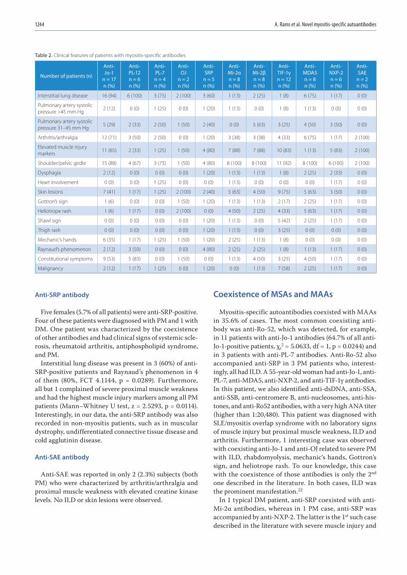
A. Rams et al. Novel myositis-specific autoantibodies1244
Anti-SRP antibody
Five females (5.7% of all patients) were anti-SRP-positive. Four of these patients were diagnosed with PM and 1 with DM. One patient was characterized by the coexistence of other antibodies and had clinical signs of systemic scle-rosis, rheumatoid arthritis, antiphospholipid syndrome, and PM.
Interstitial lung disease was present in 3 (60%) of anti-SRP-positive patients and Raynaud’s phenomenon in 4 of them (80%, FCT 4.1144, p = 0.0289). Furthermore, all but 1 complained of severe proximal muscle weakness and had the highest muscle injury markers among all PM patients (Mann–Whitney U test, z = 2.5293, p = 0.0114). Interestingly, in our data, the anti-SRP antibody was also recorded in non-myositis patients, such as in muscular dystrophy, undifferentiated connective tissue disease and cold agglutinin disease.
Anti-SAE antibody
Anti-SAE was reported in only 2 (2.3%) subjects (both PM) who were characterized by arthritis/arthralgia and proximal muscle weakness with elevated creatine kinase levels. No ILD or skin lesions were observed.
Coexistence of MSAs and MAAs
Myositis-specific autoantibodies coexisted with MAAs in 35.6% of cases. The most common coexisting anti-body was anti-Ro-52, which was detected, for example, in 11 patients with anti-Jo-1 antibodies (64.7% of all anti-Jo-1-positive patients, χc
2 = 5.0633, df = 1, p = 0.0244) and in 3 patients with anti-PL-7 antibodies. Anti-Ro-52 also accompanied anti-SRP in 3 PM patients who, interest-ingly, all had ILD. A 55-year-old woman had anti-Jo-1, anti-PL-7, anti-MDA5, anti-NXP-2, and anti-TIF-1γ antibodies. In this patient, we also identified anti-dsDNA, anti-SSA, anti-SSB, anti-centromere B, anti-nucleosomes, anti-his-tones, and anti-Ro52 antibodies, with a very high ANA titer (higher than 1:20,480). This patient was diagnosed with SLE/myositis overlap syndrome with no laboratory signs of muscle injury but proximal muscle weakness, ILD and arthritis. Furthermore, 1 interesting case was observed with coexisting anti-Jo-1 and anti-OJ related to severe PM with ILD, rhabdomyolysis, mechanic’s hands, Gottron’s sign, and heliotrope rash. To our knowledge, this case with the coexistence of those antibodies is only the 2nd one described in the literature. In both cases, ILD was the prominent manifestation.22
In 1 typical DM patient, anti-SRP coexisted with anti-Mi-2α antibodies, whereas in 1 PM case, anti-SRP was accompanied by anti-NXP-2. The latter is the 1st such case described in the literature with severe muscle injury and
Table 2. Clinical features of patients with myositis-specific antibodies
Number of patients (n)
Anti- Jo-1
n = 17n (%)
Anti- PL-12n = 6n (%)
Anti- PL-7n = 4n (%)
Anti- OJ
n = 2n (%)
Anti- SRP
n = 5n (%)
Anti- Mi-2α n = 8n (%)
Anti- Mi-2βn = 8n (%)
Anti- TIF-1γ n = 12n (%)
Anti- MDA5n = 8n (%)
Anti- NXP-2 n = 6n (%)
Anti- SAE
n = 2n (%)
Interstitial lung disease 16 (94) 6 (100) 3 (75) 2 (100) 3 (60) 1 (13) 2 (25) 1 (8) 6 (75) 1 (17) 0 (0)
Pulmonary artery systolic pressure >45 mm Hg
2 (12) 0 (0) 1 (25) 0 (0) 1 (20) 1 (13) 0 (0) 1 (8) 1 (13) 0 (0) 0 (0)
Pulmonary artery systolic pressure 31–45 mm Hg
5 (29) 2 (33) 2 (50) 1 (50) 2 (40) 0 (0) 5 (63) 3 (25) 4 (50) 3 (50) 0 (0)
Arthritis/arthralgia 12 (71) 3 (50) 2 (50) 0 (0) 1 (20) 3 (38) 3 (38) 4 (33) 6 (75) 1 (17) 2 (100)
Elevated muscle injury markers
11 (65) 2 (33) 1 (25) 1 (50) 4 (80) 7 (88) 7 (88) 10 (83) 1 (13) 5 (83) 2 (100)
Shoulder/pelvic girdle 15 (88) 4 (67) 3 (75) 1 (50) 4 (80) 8 (100) 8 (100) 11 (92) 8 (100) 6 (100) 2 (100)
Dysphagia 2 (12) 0 (0) 0 (0) 0 (0) 1 (20) 1 (13) 1 (13) 1 (8) 2 (25) 2 (33) 0 (0)
Heart involvement 0 (0) 0 (0) 1 (25) 0 (0) 0 (0) 1 (13) 0 (0) 0 (0) 0 (0) 1 (17) 0 (0)
Skin lesions 7 (41) 1 (17) 1 (25) 2 (100) 2 (40) 5 (63) 4 (50) 9 (75) 5 (63) 3 (50) 0 (0)
Gottron’s sign 1 (6) 0 (0) 0 (0) 1 (50) 1 (20) 1 (13) 1 (13) 2 (17) 2 (25) 1 (17) 0 (0)
Heliotrope rash 1 (6) 1 (17) 0 (0) 2 (100) 0 (0) 4 (50) 2 (25) 4 (33) 5 (63) 1 (17) 0 (0)
Shawl sign 0 (0) 0 (0) 0 (0) 0 (0) 1 (20) 1 (13) 0 (0) 5 (42) 2 (25) 1 (17) 0 (0)
Thigh rash 0 (0) 0 (0) 0 (0) 0 (0) 1 (20) 1 (13) 0 (0) 3 (25) 0 (0) 0 (0) 0 (0)
Mechanic’s hands 6 (35) 1 (17) 1 (25) 1 (50) 1 (20) 2 (25) 1 (13) 1 (8) 0 (0) 0 (0) 0 (0)
Raynaud’s phenomenon 2 (12) 3 (50) 0 (0) 0 (0) 4 (80) 2 (25) 2 (25) 1 (8) 1 (13) 1 (17) 0 (0)
Constitutional symptoms 9 (53) 5 (83) 0 (0) 1 (50) 0 (0) 1 (13) 4 (50) 3 (25) 4 (50) 1 (17) 0 (0)
Malignancy 2 (12) 1 (17) 1 (25) 0 (0) 1 (20) 0 (0) 1 (13) 7 (58) 2 (25) 1 (17) 0 (0)
Adv Clin Exp Med. 2021;30(12):1239–1248 1245
dysphagia. Interestingly, this female patient was also di-agnosed with papillary thyroid cancer and endometrial adenocarcinoma 13 and 5 years before autoimmune disease onset, respectively.
Among the 12 anti-TIF-1γ-positive patients, 3 also had anti-MDA5 antibodies. One of these patients fulfilled the criteria for DM and the other one for PM. Interest-ingly, both had a neoplastic disease in anamnesis. The 3rd patient had SLE/myositis overlap syndrome without malignancy.
Myositis-specific autoantibodies and myositis-associated autoantibodies-negative patients
Nine patients (5 DM and 4 PM, 10.3% of all patients) had no detectable MSAs or MAAs. Neoplasm and ILD were found in 1 (11%) of these patients, whereas skin changes and arthralgia were observed in 7 (77.8%) of these cases (FCT 5.4177, p = 0.0091, both).
Myositis-specific autoantibodies and their associations with malignancy
As expected, neoplastic diseases were frequent in this cohort. Fourteen (16% of all patients) cancer or lymphoma cases were documented, equally common in DM and PM. In half of the cases, the neoplasm was diagnosed before my-ositis. Malignancy was strongly associated with anti-TIF-1γ antibodies (χc
2 = 19.3782, df = 1, p < 0.0001). Neoplasms occurred in 7 (58.3%) of anti-TIF-1γ-positive patients, 6 (85.7%) of them were diagnosed with DM and 1 with PM.
Two of these patients had lung cancer and the others presented with urethral, endometrial and renal cell car-cinoma, diffuse large B-cell lymphoma, or carcinoma of unknown primary. Two of the anti-TIF-1γ-positive ma-lignancy patients had coexisting anti-MDA5 antibodies. Bladder cancer and cervical cancer in patients with anti-Jo-1 antibodies were also observed, and ovarian cancer and prostate cancer in those with anti-PL-7 and anti-PL-12 coexisting with anti-Mi-2β antibodies were documented. No tumors were recorded in anti-Mi-2α-positive patients.
Discussion
In this study, we characterized 87 adult Caucasian pa-tients with autoimmune inflammatory myopathies using clinical and laboratory measures. The majority of these patients presented with MSAs or MAAs, indicating that both of these antibody groups are valuable biomarkers for determining disease course and malignancy risk. However, the presence or absence of these antibodies cannot be used to differentiate DM from PM. Furthermore, a specific MSA may accompany other MSAs or MAAs and be associated with different clinical presentations, such as those seen
in SLE or systemic scleroderma in the current data. Some of these antibodies may also be seen in other immune or nonimmune disorders. For example, an anti-SRP anti-body has been documented in association with muscular dystrophy or cold agglutinin disease at our center.
The role of MSAs in the pathogenesis of inflamma-tory myopathies is not entirely understood. These anti-bodies likely sustain inflammatory processes, although their pathological links with muscle injury remain un-known.23 The anti-Jo-1 antibody is the most common MSA, as confirmed by the current data.8 However, this antibody was detected in only 19.5% of patients, thus in-dicating the immunologic heterogeneity of autoimmune myopathies. Typically, this type of antibody is linked with the presence of ILD, the Raynaud’s phenomenon, arthritis, and mechanic’s hands,24,25 findings confirmed by the present study. Furthermore, similar to our data, anti-Jo-1 often accompanies anti-Ro-52 antibodies. Ac-cording to the literature, such a coincidence may be a risk factor for a more severe disease course and cancer.26,27 Interestingly, 1 of our 2 anti-Jo-1-positive cancer patients had coexisting anti-Ro-52, and in both cases anti-PM-Scl-75 was also detected.
Anti-Jo-1, anti-PL-12, anti-PL-7, anti-EJ, and anti-OJ an-tibodies are associated with the common anti-synthetase syndrome. However, several recent studies have suggested a clinical heterogeneity, particularly regarding ILD or mus-cle injury signs.28 Some authors documented that antibod-ies other than anti-Jo-1 may be related to a poorer clini-cal prognosis and more aggressive ILD.29,30 On the other hand, Hamaguchi et al. reported that muscle injury was closely associated with anti-Jo-1, anti-EJ and anti-PL-7 anti-bodies.31 At the same time, ILD might have been observed in all of these patients, while skin changes, such as helio-trope rash and Gottron’s sign, were less common but also frequent.31 The anti-OJ has also been shown to be associ-ated with ILD, often being the sole manifestation of id-iopathic inflammatory myopathy. However, if recorded, myositis seems to be more severe than with other anti-syn-thetase antibodies.25,32 The anti-PL-12 antibodies were also documented in linkage with ILD and, to a lesser extent, with muscle injury and arthritis.28,33–35 In turn, pericar-dial effusion may be a characteristic of anti-PL-7-positive patients.25 Our data are in line with the presented reports, indicating that all anti-synthetase antibodies have a strong relationship with ILD and PM, but not with the typical DM skin changes. Also, 1 patient with heart involvement in the current cohort had an anti-PL-7 antibody.
The another interesting antibody that relates to PM and DM is anti-SRP. Targoff et al. reported a classical PM manifestation with a low prevalence of ILD, arthritis and Raynaud’s phenomenon in these subjects.36 However, some of these cases were severe and/or rapid in onset.28,36 The current results do not entirely confirm these find-ings. Although most of anti-SRP-positive patients in this study complained of proximal muscle weakness and had
A. Rams et al. Novel myositis-specific autoantibodies1246
very high creatine kinase, more than half of them had ILD or Raynaud’s phenomenon. Recent studies have reported a histologically unique necrotizing myopathy, which might explain the particularly high levels of muscle injury bio-markers in anti-SRP-positive patients.37–39
In contrast to anti-SRP antibodies, anti-MDA5 is likely related to the amyopathic PM/DM form but with rapidly progressive ILD, acute respiratory failure and poor clinical prognosis.40,41 Indeed, most of our anti-MDA5 patients had aggressive ILD leading to rapid and severe lung fibrosis, and even to a related death in 1 case.
In the literature, anti-Mi-2 and anti-NXP-2 antibodies have mainly been associated with DM.42,43 Anti-Mi-2 was shown to be associated with Gottron’s sign or pap-ules, and heliotrope rash, lung-sparing and an excel-lent clinical response to corticosteroids.43 Surprisingly, in the current study, the PM and DM distribution among anti-Mi-2-positive patients was almost equal. However, ILD was rare.
Anti-NXP-2 autoantibodies are associated with a young-onset DM with subcutaneous edema, skin cal-cinosis and severe muscle involvement with dyspha-gia.42,44,45 Moreover, these antibodies may be linked with malignancy.46,47 Our data partially mirror these re-ports, including the typical skin changes reported in half of the current patients.
One of the rarest antibody types in the current cohort was anti-SAE1, identified in only 2 individuals. Previous reports demonstrated that these patients may have se-vere skin changes, mild muscle involvement and ILD.48,49 Surprisingly, our anti-SAE-1 subjects had no skin lesions or ILD, but proximal muscle weakness, laboratory signs of muscle injury and arthritis. This observation points to the heterogeneity of autoimmune myopathies and the need for further research on this subject.
The last issue that merits comment is the relation of PM, DM, and MSAs to malignancy. Surprisingly, in the current data, a strong association was demonstrated only with anti-TIF-1γ, which is in line with the data in the literature.28,50,51 In this subgroup of patients, 58% had documented malig-nancy. Moreover, patients with anti-TIF-1γ antibodies had a lower prevalence of fever, Raynaud’s phenomenon, arthri-tis, ILD, and mechanic’s hands. At the same time, these patients have more frequent DM-typical skin changes, par-ticularly shawl sign rash.28,52 The current study confirms these observations. The presence of anti-TIF-1γ antibodies was highly associated with malignancy, while typical skin rash was reported in 3/4 of these patients. Only 1 patient had ILD, while joint involvement was demonstrated in 1/3 of them.
Among patients with no detectable MSAs or MAAs, ILD was rarely observed, in contrast to skin changes and ar-thralgia. One patient in this group was diagnosed with can-cer. There are limited data on the clinical course of these patients in the available literature.
Limitations
The current study has several limitations. First, it was retrospective in nature. In addition, the number of sub-jects studied was relatively small, especially with regard to some MSA types, such as anti-OJ, anti-EJ and anti-SAE anti bodies. Therefore, future multicenter studies charac-terizing patients with DM and PM are needed.
Conclusions
The MSA type cannot differentiate DM from PM, although some antibodies may be more prevalent in PM (e.g., anti-Jo1) and others in DM (e.g., anti-TIF-1γ). Interstitial lung disease was common in patients with anti-synthetase antibodies, particularly anti-Jo-1 and anti-PL-12, but also in those with anti-MDA5 antibodies. The majority of ILD patients ful-filled PM criteria. Patients with anti-SRP were characterized by the presence of the Raynaud’s phenomenon and the high-est serum concentration of muscle injury markers. In turn, an amyopathic form characterized anti-MDA5-positive indi-viduals. Malignancy was highly associated with anti-TIF-1γ antibodies, but also with anti-Jo-1, anti-PL-7, anti-PL-12, anti-SRP, anti-MDA-5, anti-NXP-2, and anti-Mi-2β antibodies.
ORCID iDsAnna Rams https://orcid.org/0000-0001-7590-7318Joanna Kosałka-Węgiel https://orcid.org/0000-0003-1013-2253Piotr Kuszmiersz https://orcid.org/0000-0002-2031-6657Aleksandra Matyja-Bednarczyk https://orcid.org/0000-0003-4038-5016
Stanisław Polański https://orcid.org/0000-0002-4638-3823Lech Zaręba https://orcid.org/0000-0002-2221-614XStanisława Bazan-Socha https://orcid.org/0000-0001-9634-0963
References1. Cheeti A, Brent LH, Panginikkod S. Autoimmune myopathies. In:
StatPearls [Internet]. Treasure Island: StatPearls Publishing; 2021. PMID:30422455
2. Ungprasert P, Bethina NK, Jones CH. Malignancy and idiopathic inflam-matory myopathies. N Am J Med Sci. 2013;5(10):569–572. doi:10.4103/ 1947-2714.120788
3. Fayyaz B, Rehman HJ, Uqdah H. Cancer-associated myositis: An elu-sive entity. J Community Hosp Intern Med Perspect. 2019;9(1):45–49. doi:10.1080/20009666.2019.1571880
4. Tiniakou E, Mammen AL. Idiopathic Inflammatory myopathies and malignancy: A comprehensive review. Clin Rev Allergy Immunol. 2017; 52(1):20–33. doi:10.1007/s12016-015-8511-x
5. Casciola-Rosen L, Nagaraju K, Plotz P, et al. Enhanced autoantigen expression in regenerating muscle cells in idiopathic inflammatory myopathy. J Exp Med. 2005;201(4):591–601. doi:10.1084/jem.20041367
6. Jakubaszek M, Kwiatkowska B, Maślińska M. Polymyositis and derma-tomyositis as a risk of developing cancer. Reumatologia. 2015;53(2): 101–105. doi:10.5114/reum.2015.51510
7. Ungprasert P, Leeaphorn N, Hosiriluck N, Chaiwatcharayut W, Amman-nagari N, Raddatz DA. Clinical features of inflammatory myopathies and their association with malignancy: A systematic review in Asian popu-lation. ISRN Rheumatol. 2013;2013:509354. doi:10.1155/2013/509354
8. Miller M, Vleugels R, Amato AA. Clinical manifestations of derma-tomyositis and polymyositis in adults. https://www.uptodate.com/contents/clinical-manifestations-of-dermatomyositis-and-polymyo sitis-in-adults. Accessed March 17, 2020.
Adv Clin Exp Med. 2021;30(12):1239–1248 1247
9. Greenberg SA. Type 1 interferons and myositis. Arthritis Res Ther. 2010;12(Suppl 1):S4. doi:10.1186/ar2885
10. Greenberg SA. Proposed immunologic models of the inflammato-ry myopathies and potential therapeutic implications. Neurology. 2007;69(21):2008–2019. doi:10.1212/01.WNL.0000291619.17160.b8
11. Greenberg SA. Dermatomyositis and type 1 interferons. Curr Rheumatol Rep. 2010;12(3):198–203. doi:10.1007/s11926-010-0101-6
12. Dalakas MC. Inflammatory disorders of muscle: Progress in polymyo-sitis, dermatomyositis and inclusion body myositis. Curr Opin Neurol. 2004;17(5):561–567. doi:10.1097/00019052-200410000-00006
13. Dalakas MC, Hohlfeld R. Polymyositis and dermatomyositis. Lancet. 2003;362(9388):971–982. doi:10.1016/S0140-6736(03)14368-1
14. Arahata K, Engel AG. Monoclonal antibody analysis of mononuclear cells in myopathies. IV: Cell-mediated cytotoxicity and muscle fiber necrosis. Ann Neurol. 1988;23(2):168–173. doi:10.1002/ana.410230210
15. Engel AG, Arahata K. Monoclonal antibody analysis of mononuclear cells in myopathies. II: Phenotypes of autoinvasive cells in polymy-ositis and inclusion body myositis. Ann Neurol. 1984;16(2):209–215. doi:10.1002/ana.410160207
16. Arahata K, Engel AG. Monoclonal antibody analysis of mononuclear cells in myopathies. I: Quantitation of subsets according to diagnosis and sites of accumulation and demonstration and counts of muscle fibers invaded by T cells. Ann Neurol. 1984;16(2):193–208. doi:10.1002/ana.410160206
17. Greenberg SA, Pinkus GS, Amato AA, Pinkus JL. Myeloid dendrit-ic cells in inclusion-body myositis and polymyositis. Muscle Nerve. 2007;35(1):17–23. doi:10.1002/mus.20649
18. Greenberg SA, Bradshaw EM, Pinkus JL, et al. Plasma cells in muscle in inclusion body myositis and polymyositis. Neurology. 2005;65(11): 1782–1787. doi:10.1212/01.wnl.0000187124.92826.20
19. Hirakata M. Autoantibodies and their clinical significance in idio-pathic inflammatory myopathies: Polymyositis/dermatomyositis and related conditions. Japanese J Clin Immunol. 2007;30(6):444–454. doi:10.2177/jsci.30.444
20. Yang H, Peng Q, Yin L, et al. Identification of multiple cancer-asso-ciated myositis-specific autoantibodies in idiopathic inflammatory myopathies: A large longitudinal cohort study. Arthritis Res Ther. 2017; 19(1):259. doi:10.1186/s13075-017-1469-8
21. Ceribelli A, Isailovic N, De Santis M, et al. Myositis-specific autoantibod-ies and their association with malignancy in Italian patients with poly-myositis and dermatomyositis. Clin Rheumatol. 2017;36(2):469–475. doi:10.1007/s10067-016-3453-0
22. Gelpí C, Kanterewicz E, Gratacos J, Targoff IN, Rodriguez-Sanchez JL. Coexistence of two antisynthetases in a patient with the antisyn-thetase syndrome. Arthritis Rheum. 1996;39(4):692–697. doi:10.1002/art.1780390424
23. Miller FW, Waite KA, Biswas T, Plotz PH. The role of an autoantigen, histidyl-tRNA synthetase, in the induction and maintenance of auto-immunity. Proc Natl Acad Sci U S A. 1990;87(24):9933–9937. doi:10. 1073/pnas.87.24.9933
24. Love LA, Leff RL, Fraser DD, et al. A new approach to the classifica-tion of idiopathic inflammatory myopathy: Myositis-specific auto-antibodies define useful homogeneous patient groups. Medicine (Baltimore). 1991;70(6):360–374. doi:10.1097/00005792-199111000-00002
25. Labirua-Iturburu A, Selva-O’Callaghan A, Vincze M, et al. Anti-PL-7 (anti-threonyl-tRNA synthetase) antisynthetase syndrome: Clinical manifestations in a series of patients from a European multicenter study (EUMYONET) and review of the literature. Medicine (Baltimore). 2012;91(4):206–211. doi:10.1097/MD.0b013e318260977c
26. La Corte R, Lo Mo Naco A, Locaputo A, Dolzani F, Trotta F. In patients with antisynthetase syndrome the occurrence of anti-Ro/SSA anti-bodies causes a more severe interstitial lung disease. Autoimmunity. 2006;39(3):249–253. doi:10.1080/08916930600623791
27. Marie I, Hatron PY, Dominique S, et al. Short-term and long-term outcome of anti-Jo1-positive patients with anti-Ro52 antibody. Semin Arthritis Rheum. 2012;41(6):890–899. doi:10.1016/j.semarthrit. 2011.09.008
28. Satoh M, Tanaka S, Ceribelli A, Calise SJ, Chan EKL. A comprehensive overview on myositis-specific antibodies: New and old biomarkers in idiopathic inflammatory myopathy. Clin Rev Allergy Immunol. 2017; 52(1):1–19. doi:10.1007/s12016-015-8510-y
29. Pinal-Fernandez I, Casal-Dominguez M, Huapaya JA, et al. A longi-tudinal cohort study of the anti-synthetase syndrome: Increased severity of interstitial lung disease in black patients and patients with anti-PL7 and anti-PL12 autoantibodies. Rheumatology (Oxford). 2017;56(6):999–1007. doi:10.1093/rheumatology/kex021
30. Mejía M, Herrera-Bringas D, Pérez-Román DI, et al. Interstitial lung disease and myositis-specific and associated autoantibodies: Clini-cal manifestations, survival and the performance of the new ATS/ERS criteria for interstitial pneumonia with autoimmune features (IPAF). Respir Med. 2017;123:79–86. doi:10.1016/j.rmed.2016.12.014
31. Hamaguchi Y, Fujimoto M, Matsushita T, et al. Common and distinct clinical features in adult patients with anti-aminoacyl-tRNA synthe-tase antibodies: Heterogeneity within the syndrome. PLoS One. 2013; 8(4):e60442. doi:10.1371/journal.pone.0060442
32. Vulsteke JB, Satoh M, Malyavantham K, Bossuyt X, De Langhe E, Mahler M. Anti-OJ autoantibodies: Rare or underdetected? Autoimmun Rev. 2019;18(7):658–664. doi:10.1016/j.autrev.2019.05.002
33. Targoff IN, Arnett FC. Clinical manifestations in patients with anti-body to PL-12 antigen (alanyl-tRNA synthetase). Am J Med. 1990;88(3): 241–251. doi:10.1016/0002-9343(90)90149-8
34. Hirakata M, Suwa A, Nagai S, et al. Anti-KS: Identification of auto-antibodies to asparaginyl-transfer RNA synthetase associated with interstitial lung disease. J Immunol. 1999;162(4):2315–2320. PMID: 9973509
35. Friedman AW, Targoff IN, Arnett FC. Interstitial lung disease with auto-antibodies against aminoacyl-tRNA synthetases in the absence of clin-ically apparent myositis. Semin Arthritis Rheum. 1996;26(1):459–467. doi:10.1016/S0049-0172(96)80026-6
36. Targoff IN, Johnson AE, Miller FW. Antibody to signal recognition particle in polymyositis. Arthritis Rheum. 1990;33(9):1361–1370. doi:10. 1002/art.1780330908
37. Hengstman GJD, Ter Laak HJ, Vree Egberts WTM, et al. Anti-signal rec-ognition particle autoantibodies: Marker of a necrotising myopathy. Ann Rheum Dis. 2006;65(12):1635–1638. doi:10.1136/ard.2006.052191
38. Aggarwal R, Oddis CV, Goudeau D, et al. Anti-signal recognition particle autoantibody ELISA validation and clinical associations. Rheumatology (Oxford). 2015;54(7):1194–1199. doi:10.1093/rheuma tology/keu436
39. Suzuki S, Nishikawa A, Kuwana M, et al. Inflammatory myopathy with anti-signal recognition particle antibodies: Case series of 100 patients. Orphanet J Rare Dis. 2015;10:61. doi:10.1186/s13023-015-0277-y
40. Ichiyasu H, Sakamoto Y, Yoshida C, et al. Rapidly progressive inter-stitial lung disease due to anti-MDA-5 antibody-positive clinical-ly amyopathic dermatomyositis complicated with cervical cancer: Successful treatment with direct hemoperfusion using polymyxin B-immobilized fiber column therapy. Respir Med Case Rep. 2017;20: 51–54. doi:10.1016/j.rmcr.2016.11.015
41. Tansley SL, Betteridge ZE, McHugh NJ. The diagnostic utility of auto-antibodies in adult and juvenile myositis. Curr Opin Rheumatol. 2013; 25(6):772–777. doi:10.1097/01.bor.0000434664.37880.ac
42. Ceribelli A, Fredi M, Taraborelli M, et al. Anti-MJ/NXP-2 autoantibody specificity in a cohort of adult Italian patients with polymyositis/der-matomyositis. Arthritis Res Ther. 2012;14(2):R97. doi:10.1186/ar3822
43. Ghirardello A, Borella E, Beggio M, Franceschini F, Fredi M, Doria A. Myo-sitis autoantibodies and clinical phenotypes. Auto Immun Highlights. 2014;5(3):69–75. doi:10.1007/s13317-014-0060-4
44. Valenzuela A, Chung L, Casciola-Rosen L, Fiorentino D. Identifica-tion of clinical features and autoantibodies associated with calcino-sis in dermatomyositis. JAMA Dermatol. 2014;150(7):724–729. doi:10. 1001/jamadermatol.2013.10416
45. Albayda J, Pinal-Fernandez I, Huang W, et al. Antinuclear matrix pro-tein 2 autoantibodies and edema, muscle disease, and malignancy risk in dermatomyositis patients. Arthritis Care Res (Hoboken). 2017; 69(11):1771–1776. doi:10.1002/acr.23188
46. Ichimura Y, Matsushita T, Hamaguchi Y, et al. Anti-NXP2 autoantibod-ies in adult patients with idiopathic inflammatory myopathies: Possi-ble association with malignancy. Ann Rheum Dis. 2012;71(5):710–713. doi:10.1136/annrheumdis-2011-200697
47. Fiorentino DF, Chung LS, Christopher-Stine L, et al. Most patients with cancer-associated dermatomyositis have antibodies to nucle-ar matrix protein NXP-2 or transcription intermediary factor 1γ. Arthritis Rheum. 2013;65(11):2954–2962. doi:10.1002/art.38093

A. Rams et al. Novel myositis-specific autoantibodies1248
48. Fujimoto M, Matsushita T, Hamaguchi Y, et al. Autoantibodies to small ubiquitin-like modifier activating enzymes in Japanese patients with der-matomyositis: Comparison with a UK Caucasian cohort. Ann Rheum Dis. 2013;72(1):151–153. doi:10.1136/annrheumdis-2012-201736
49. Betteridge ZE, Gunawardena H, Chinoy H, et al. Clinical and human leuco-cyte antigen class II haplotype associations of autoantibodies to small ubiquitin-like modifier enzyme, a dermatomyositis-specific autoan-tigen target, in UK Caucasian adult-onset myositis. Ann Rheum Dis. 2009;68(10):1621–1625. doi:10.1136/ard.2008.097162
50. Targoff IN, Mamyrova G, Trieu EP, et al. A novel autoantibody to a 155-kd protein is associated with dermatomyositis. Arthritis Rheum. 2006; 54(11):3682–3689. doi:10.1002/art.22164
51. Kang EH, Nakashima R, Mimori T, et al. Myositis autoantibodies in Korean patients with inflammatory myositis: Anti-140-kDa poly-peptide antibody is primarily associated with rapidly progressive interstitial lung disease independent of clinically amyopathic derma-tomyositis. BMC Musculoskelet Disord. 2010;11:223. doi:10.1186/1471-2474-11-223
52. Targoff IN, Trieu E, Levy-Neto M, Prasertsuntarasai T, Miller FW. Auto-antibodies to transcriptional intermediary factor 1-gamma (TIF1-g) in dermatomyositis. Arthritis Rheum. 2007;54:S518.
Cite asKuliński P, Tomczyk Ł, Morasiewicz P. Effect of the COVID-19 pandemic on foot surgeries. Adv Clin Exp Med. 2021;30(12):1249–1253. doi:10.17219/acem/140610
DOI10.17219/acem/140610
Copyright© 2021 by Wroclaw Medical University This is an article distributed under the terms of theCreative Commons Attribution 3.0 Unported (CC BY 3.0)(https://creativecommons.org/licenses/by/3.0/)
Address for correspondencePiotr MorasiewiczE-mail: [email protected]
Funding sourcesNone declared
Conflict of interestNone declared
Received on April 8, 2021Reviewed on May 28, 2021Accepted on July 28, 2021
Published online on October 5, 2021
AbstractBackground. Musculoskeletal dysfunction due to deformities and injuries of the foot is a common ortho-pedic problem.
Objectives. To analyze the effect of the COVID-19 pandemic on both elective and emergency foot surgeries.
Materials and methods. This study analyzed the effect of the COVID-19 pandemic on elective and emergency foot surgeries. The comparison included data collected in 2019 (March 15–October 15, when the epidemic did not influence the Polish healthcare system or patient demographics) and in a corresponding period in 2020. This study was conducted in the trauma and orthopedic surgery wards and the emergency departments of 2 regional Polish hospitals.
Results. The analysis of the data from the orthopedic wards showed a reduction in the total number of admissions in 2020 by 55%. The number of elective and emergency interventions was differently related to the pandemic, with elective interventions declining by 72% and emergency interventions increasing by 27% in 2020 compared to 2019. The total number of elective foot surgeries in children decreased by 59% in 2020. The mean duration of hospital stay was approx. 2.5 days shorter in adults and 1.7 days shorter in children during the 2nd evaluation period. The emergency department data showed a decline of 32% in the number of patients presenting with injuries during the pandemic. Nonetheless, the pandemic did not affect the mean age of patients and the female-to-male ratio.
Conclusions. The global COVID-19 pandemic affected the epidemiology of foot injuries as well as the preva-lence of foot surgeries in children and adults.
Key words: COVID-19, pandemic, epidemiology, foot surgery, lockdown
Original papers
Effect of the COVID-19 pandemic on foot surgeries
Patryk Kuliński1,A–F, Łukasz Tomczyk2,B–D, Piotr Morasiewicz3,A,C–F
1 Department of Trauma and Orthopaedic Surgery, T. Marciniak Lower Silesia Specialist Hospital – Emergency Medicine Center, Wrocław, Poland2 Department of Food Safety and Quality Management, Poznan University of Life Sciences, Poland3 Department of Orthopaedic and Trauma Surgery, University Hospital in Opole, Institute of Medical Sciences, University of Opole, Poland
A – research concept and design; B – collection and/or assembly of data; C – data analysis and interpretation; D – writing the article; E – critical revision of the article; F – final approval of the article
Advances in Clinical and Experimental Medicine, ISSN 1899–5276 (print), ISSN 2451–2680 (online) Adv Clin Exp Med. 2021;30(12):1249–1253
P. Kuliński, Ł. Tomczyk, P. Morasiewicz. COVID-19 and foot surgeries1250
Background
Musculoskeletal dysfunction due to deformities and inju-ries of the foot is a common orthopedic problem.1,2 Accord-ing to the Illinois Podiatric Medical Association, approx. 6% of the USA population are affected by foot injuries, fore-foot deformities or flat feet (pes planus). The global spread of severe acute respiratory syndrome coronavirus type 2 (SARS-CoV-2) infections has had an impact on many as-pects of everyday life, including changes in people’s habits, lifestyles and fears regarding their health and the health of their loved ones.2–19
The COVID-19 pandemic unquestionably affected the functioning of national healthcare systems.2–19 This included changes in the character of hospitals, includ-ing conversions of surgical wards into infectious wards, limiting the number of elective admissions and reducing the number of medical staff. The latter was due to tempo-rary quarantines placed on those who had a contact with patients suspected of having COVID-19 infections and the furloughing of elderly or chronic disease-burdened staff.3–5,10
The available literature has a limited number of reports on the effects the COVID-19 pandemic has had on foot surgeries in adults and children.2,4,5 The available reports assess the number of surgeries for foot injuries during the pandemic to a limited extent2 or estimate elective foot surgery restrictions based on surveys conducted among orthopedic surgeons.4,5
Objectives
The purpose of this study was to analyze the effect of the COVID-19 pandemic on foot surgery in both elec-tive and emergency settings.
Materials and methods
Study design and patients
The study was conducted in the trauma and orthope-dic surgery departments and the emergency departments of 2 Polish regional hospitals. Adult and pediatric patients were included in this study. The compared periods were March 15–October 15, 2019, when the (at that time non-existent) COVID-19 epidemic did not influence the Polish healthcare system or patient demographics, and the cor-responding period in 2020 (March 15–October 15, 2020). Informed consent was obtained for experimentation with human subjects. The study was approved by the local eth-ics committee (approval No. 4/PNDR/2021).
Evaluation of epidemiological parameters
This study analyzed the number of elective foot sur-geries most commonly performed in the surgical wards, i.e., correction of forefoot deformities, including bunions (hallux valgus) and hammertoes. A large group of patients presented with multiple forefoot deformities, which were corrected simultaneously during a single procedure. Other elective procedures included arthrodesis, planovalgus foot surgery and removal of implants. The total number of elective foot surgeries in children was assessed. The pro-cedures performed on children were divided into those most frequently performed in our departments (correction of the planovalgus foot, correction of clubfoot).
Injury interventions were divided into 2 groups. The 1st group comprised interventions for foot fractures, defined as fractures involving any bone in the foot from the talus to the phalanges. The 2nd group comprised interventions for injuries such as wounds, dislocations and sprains within the foot (excluding those of the ankle joint).
The groups were analyzed in terms of the number of sur-geries, distribution of the sexes, age distribution, and mean duration of hospital stay. The data from 2019 were com-pared with the data collected in 2020.
Statistical analyses
Statistical analyses were performed using Statistica v. 13.1 software (StatSoft Inc., Tulsa, USA). The Shapiro–Wilk test was used to assess distribution normality. The variables did not follow a normal distribution; there-fore, Pearson’s χ2 test and Mann–Whitney test was used. The level of significance was set at α = 0.05.
Results
All results have been compiled in Table 1 and Table 2. The analysis of the orthopedic ward data showed a re-duction in the total number of adult admissions by 55% in 2020.
The number of elective and emergency interventions was differently affected by the pandemic, with elective interventions declining by 72% and emergency inter-ventions increasing by 27% in 2020 compared to 2019. These changes were statistically significant (p = 0.01770; p = 0.01922). In adults, arthrodesis and planovalgus foot surgeries decreased by 50% in 2020, implant removal sur-geries decreased by 63% and hallux valgus and hammertoe surgeries decreased by 82%.
The total number of elective foot surgeries performed in children decreased by 59% in 2020 compared to 2019. This difference was statistically significant (p = 0.0487). In chil-dren, the number of planovalgus foot surgeries decreased by 61% and clubfoot surgeries decreased by 56% in 2020.
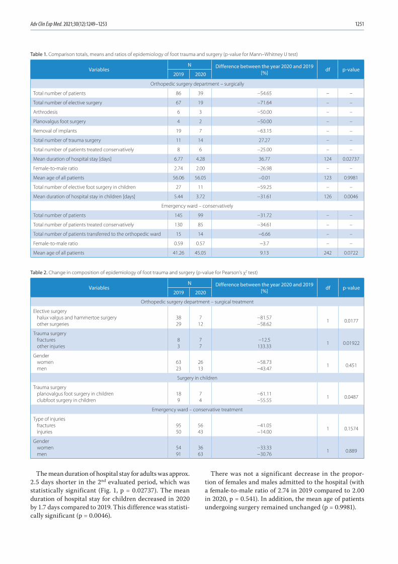
Adv Clin Exp Med. 2021;30(12):1249–1253 1251
The mean duration of hospital stay for adults was approx. 2.5 days shorter in the 2nd evaluated period, which was statistically significant (Fig. 1, p = 0.02737). The mean duration of hospital stay for children decreased in 2020 by 1.7 days compared to 2019. This difference was statisti-cally significant (p = 0.0046).
There was not a significant decrease in the propor-tion of females and males admitted to the hospital (with a female-to-male ratio of 2.74 in 2019 compared to 2.00 in 2020, p = 0.541). In addition, the mean age of patients undergoing surgery remained unchanged (p = 0.9981).
Table 1. Comparison totals, means and ratios of epidemiology of foot trauma and surgery (p-value for Mann–Whitney U test)
VariablesN Difference between the year 2020 and 2019
[%] df p-value2019 2020
Orthopedic surgery department – surgically
Total number of patients 86 39 −54.65 – –
Total number of elective surgery 67 19 −71.64 – –
Arthrodesis 6 3 −50.00 – –
Planovalgus foot surgery 4 2 −50.00 – –
Removal of implants 19 7 −63.15 – –
Total number of trauma surgery 11 14 27.27 – –
Total number of patients treated conservatively 8 6 −25.00 – –
Mean duration of hospital stay [days] 6.77 4.28 36.77 124 0.02737
Female-to-male ratio 2.74 2.00 −26.98 – –
Mean age of all patients 56.06 56.05 −0.01 123 0.9981
Total number of elective foot surgery in children 27 11 −59.25 – –
Mean duration of hospital stay in children [days] 5.44 3.72 −31.61 126 0.0046
Emergency ward – conservatively
Total number of patients 145 99 −31.72 – –
Total number of patients treated conservatively 130 85 −34.61 – –
Total number of patients transferred to the orthopedic ward 15 14 −6.66 – –
Female-to-male ratio 0.59 0.57 −3.7 – –
Mean age of all patients 41.26 45.05 9.13 242 0.0722
Table 2. Change in composition of epidemiology of foot trauma and surgery (p-value for Pearson’s χ2 test)
VariablesN Difference between the year 2020 and 2019
[%] df p-value2019 2020
Orthopedic surgery department – surgical treatment
Elective surgeryhalux valgus and hammertoe surgeryother surgeries
3829
712
−81.57−58.62
1 0.0177
Trauma surgeryfracturesother injuries
83
77
−12.5133.33
1 0.01922
Gender womenmen
6323
2613
−58.73−43.47
1 0.451
Surgery in children
Trauma surgeryplanovalgus foot surgery in childrenclubfoot surgery in children
189
74
−61.11−55.55
1 0.0487
Emergency ward – conservative treatment
Type of injuries fracturesinjuries
9550
5643
−41.05−14.00
1 0.1574
Gender womenmen
5491
3663
−33.33−30.76
1 0.889

P. Kuliński, Ł. Tomczyk, P. Morasiewicz. COVID-19 and foot surgeries1252
The greatest rise in emergency procedures (133%) was observed in interventions performed due to other foot in-juries (mostly soft-tissue injuries) (p = 0.01922). The emer-gency department data showed 32% fewer patients present-ing with injuries during the evaluated pandemic period. These changes were statistically significant (p = 0.0153). Moreover, the number of patients with fractures showed a greater decline (41%) in 2020 compared with pre-pan-demic figures than the number of patients with soft-tissue injuries (14% decline), although these differences did not reach statistical significance (p = 0.1574).
The mean patient age in the emergency ward increased from 41 years in 2019 to 45 years in 2020, but the differ-ence was not statistically significant (p = 0.0722). The pro-portion of patients seen in the emergency departments who required surgical treatment or more diagnostic tests on the orthopedic ward did not change between the evalu-ated periods (with only a single case fewer in the 2nd period).
Discussion
Key results and generalizability
The COVID-19 pandemic considerably altered the or-ganization of healthcare services across the world.2–19 Elective admissions were halted or limited, medical per-sonnel numbers decreased and patients’ fears associated with hospitalization increased.3–5,10 However, the impact of the COVID-19 pandemic on elective and emergency treatment of foot injuries has not been fully evaluated.
Our study confirms the impact of the pandemic in the form of lower numbers of elective procedures in children and adults, and shorter hospital stays to mini-mize the amount of contact between patients and medical staff, who are the potential vectors in coronavirus trans-mission. Similarly, other authors have reported decreases in the number of elective procedures during the pandemic by 23–100%.3–5,10 An estimated 26.6–75.2% of centers
cancelled or rescheduled (postponed) foot surgeries dur-ing the COVID-19 pandemic.4,5 We observed a decrease in all types of elective surgeries performed in both, adults and children. The reduction in the number of elective ad-missions of children during the COVID-19 pandemic, ob-served in our study, may have negative effects in the form of increasing the severity of deformities in patients awaiting surgery. The postponing of any musculoskeletal surgery in a growing child is particularly dangerous, and can have irreversible consequences and a significant impact on their health.
No statistically significant reduction was seen in the ratio of female-to-male patients admitted to orthopedic surgery departments in 2020, largely due to the fact that females are generally more prone to develop forefoot deformities.20,21
During the pandemic, the introduction of travel restric-tions and remote work led to fewer patients presenting with pain, which undoubtedly resulted in a reduction of patients admitted for elective forefoot procedures. In many cases, patients decided to postpone their treatment due to less severe pain, and – particularly among the elderly – out of fear of the coronavirus.
In contrast to the pronounced change in sex distribu-tion among orthopedic ward patients, the change in sex distribution among emergency room patients was negli-gible. Males presented nearly 2 times more frequently than females in both evaluated periods (the female-to-male ratio was 0.59 in 2019 and 0.57 in 2020), which indicates a higher incidence of foot injuries in males.
The increased number of foot injury patients transferred to the orthopedic ward from the emergency room to un-dergo surgical treatment was due to several factors. These include the increasingly stringent restrictions imposed by the Polish government during the period of national lockdown, which directly affected the etiology of in-juries, with lower numbers of high-energy injuries and only slightly lower numbers of soft-tissue injuries. These changes were a result of travel restrictions and the con-sequent decline in the number of traffic accidents.13,15,18 Conversely, the amount of work around the house in-creased due to no restrictions being imposed on hardware and DIY stores. This, combined with increased alcohol consumption, resulted in a higher incidence of injuries at home.13,15,18 Moreover, heavy industry did not experience any direct lockdown measures, which was associated with undiminished injuries in this field.
Limitations
The limitation in the assessment of the impact of the COVID-19 pandemic on foot surgery is that it was only conducted at 2 centers and on a limited group of patients. This allowed the authors to quickly assess and present the important research results. However, in the future, we plan to conduct a multicenter study on a larger group of patients.
Fig. 1. Mean duration of hospital stay in days
days
year
median25–75%min–max
2019 2020
12
11
10
9
8
7
6
5
4
3
2
Adv Clin Exp Med. 2021;30(12):1249–1253 1253
In light of the uncertain future and lack of reliable es-timates as to the duration of this pandemic, orthopedic wards and surgery scheduling must develop protocols to increase their availability and ensure the safety of pa-tients and staff.
Conclusions
Our analysis of the collected data showed the effects of the global COVID-19 pandemic on the types of pa-tients, availability of elective foot surgeries in children and adults, and epidemiology of foot injuries. This study highlights the need to make elective surgeries more avail-able, while simultaneously improving the safety for both hospital staff and patients by minimizing the risk of pos-sible SARS CoV-2 transmission. The COVID-19 pandemic saw a reduction in the number of all types of elective foot surgeries in children and adults, an increase in the number of trauma-related foot surgeries, and shortening of hospital stays. Nonetheless, the pandemic had no effect on mean patient age and female-to-male ratio.
ORCID iDsPatryk Kuliński https://orcid.org/0000-0002-1048-7460Łukasz Tomczyk https://orcid.org/0000-0002-4644-0111Piotr Morasiewicz https://orcid.org/0000-0002-7587-666X
References1. Cichero MJ. Elective foot and ankle surgery: Activity and perioperative
complications in Queensland public hospitals, Australia. Foot (Edinb). 2009;19(3):139–144. doi:10.1016/j.foot.2009.02.003
2. Vosoughi AR, Borazjani R. COVID-19 effect on foot & ankle surgery in Shiraz, south Iran. J Foot Ankle Surg. 2020;59(5):887. doi:10.1053/j.jfas.2020.06.013
3. Thaler M, Khosravi I, Hirschmann MT, et al. Disruption of joint arthro-plasty services in Europe during the COVID-19 pandemic: An online survey within the European Hip Society (EHS) and the European Knee Associates (EKA). Knee Surg Sports Traumatol Arthrosc. 2020;28(6): 1712–1719. doi:10.1007/s00167-020-06033-1
4. Liebensteiner MC, Khosravi I, Hirschmann MT, Heuberer PR, Thaler M; Board of the AGA – Society of Arthroscopy and Joint-Surgery. Mas-sive cutback in orthopaedic healthcare services due to the COVID-19 pandemic. Knee Surg Sports Traumatol Arthrosc. 2020;28(6):1705–1711. doi:10.1007/s00167-020-06032-2
5. Liebensteiner MC, Khosravi I, Hirschmann MT, Saffarini M, Thaler M; The Board of the AGA – Society of Arthroscopy and Joint-Surgery. It is not ‘business as usual’ for orthopaedic surgeons in May 2020: The Austrian-German-Swiss experience. J Exp Orthop. 2020;7:61. doi:10.1186/s40634-020-00272-4
6. Kort NP, Gomez Barrena E, Bédard M, et al. Resuming elective hip and knee arthroplasty after the first phase of the SARS-CoV-2 pandemic: The European Hip Society and European Knee Associates recommen-dations. Knee Surg Sports Traumatol Arthrosc. 2020;28(9):2730–2746. doi:10.1007/s00167-020-06233-9
7. Kort NP, Gomez Barrena E, Bédard M. Recommendations for resuming elective hip and knee arthroplasty in the setting of the SARS-CoV-2 pandemic: The European Hip Society and European Knee Associates Survey of Members. Knee Surg Sports Traumatol Arthrosc. 2020;28(9): 2723–2729. doi:10.1007/s00167-020-06212-0
8. Kort NP, Zagra L, Gomez Barrena E. Resuming hip and knee arthroplasty after COVID-19: Ethical implications for wellbeing, safety and the econ-omy. Hip Int. 2020;30(5):492–499. doi:10.1177/1120700020941232
9. Athey AG, Cao L, Okazaki K. Survey of AAHKS International Members on the impact of COVID-19 on hip and knee arthroplasty practices. J Arthroplasty. 2020;35(7S):S89–S94. doi:10.1016/j.arth.2020.04.053
10. D’Apolito R, Faraldi M, Ottaiano I, Zagra L. Disruption of arthroplasty practice in an orthopedic center in northern Italy during the coro-navirus disease 2019 pandemic. J Arthroplasty. 2020;35(7S):S6–S9. doi:10.1016/j.arth.2020.04.057
11. Verde F, D’Ambrosi R, Biazzo A, Masia F, Perazzo P. Guidelines for resum-ing elective hip and knee surgical activity following the COVID-19 pandemic: An Italian perspective [Epub ahead of print]. HSS J. 2020; 13:1–6. doi:10.1007/s11420-020-09809-w
12. Donell ST, Thaler M, Budhiparama NC, et al. Preparation for the next COVID-19 wave: The European Hip Society and European Knee Asso-ciates recommendations. Knee Surg Sports Traumatol Arthrosc. 2020; 28(9):2747–2755. doi:10.1007/s00167-020-06213-z
13. Upadhyaya GK, Iyengar K, Jain VK, Vaishya R. Challenges and strate-gies in management of osteoporosis and fragility fracture care dur-ing COVID-19 pandemic. J Orthop. 2020;21:287–290. doi:10.1016/j.jor.2020.06.001
14. Hashmi P, Fahad S, Khan HN, Zahid M, Sadruddin A, Noordina S. Covid-19 pandemic: Economic burden on patients with musculo-skeletal injuries in a tertiary care hospital of LMIC. Retrospective cross sectional study. Ann Med Surg (Lond). 2020;60:5–8. doi:10.1016/j.amsu.2020.09.049
15. Yu P, Wu C, Zhuang C, et al. The patterns and management of frac-ture patients under COVID-19 outbreak in China. Ann Transl Med. 2020;8(15):932. doi:10.21037/atm-20-4174
16. Maryada VR, Mulpur P, Guravareddy AV, Pedamallu SK, Bhasker BV. Impact of COVID-19 pandemic on orthopaedic trauma volumes: A multi-centre perspective from the state of Telangana. Indian J Orthop. 2020;54(Suppl 2):1–6. doi:10.1007/s43465-020-00226-z
17. Mitkovic MM, Bumbasirevic M, Milenkovic S, Gajdobranski D, Bum-basirevic V, Mitkovic MB. Influence of coronavirus disease 2019 pan-demic state of emergency in orthopaedic fracture surgical treatment. Int Orthop. 2021;45(4):815–820. doi:10.1007/s00264-020-04750-3
18. Murphy T, Akehurst H, Mutimer J. Impact of the 2020 COVID-19 pan-demic on the workload of the orthopaedic service in a busy UK district general hospital. Injury. 2020;51(10):2142–2147. doi:10.1016/j.injury. 2020.07.001
19. Lv H, Zhang Q, Yin Y, et al. Epidemiologic characteristics of traumatic fractures during the outbreak of coronavirus disease 2019 (COVID-19) in China: A retrospective & comparative multi-center study. Injury. 2020;51(8):1698–1704. doi:10.1016/j.injury.2020.06.022
20. Roddy E, Zhang W, Doherty M. Prevalence and associations of hallux valgus in a primary care population. Arthritis Rheum. 2008;59:857–862. doi:10.1002/art.23709
21. Nix S, Smith M, Vicenzino B. Prevalence of hallux valgus in the general population: A systematic review and meta-analysis. J Foot Ankle Res. 2010;3:21. doi:10.1186/1757-1146-3-21


Cite asAda F, Kasimzade F, Mendil AS, Gocmez H. Effects of nano-sized titanium dioxide powder and ultraviolet light on superficial veins in a rabbit model. Adv Clin Exp Med. 2021;30(12):1255–1262. doi:10.17219/acem/141501
DOI10.17219/acem/141501
Copyright© 2021 by Wroclaw Medical University This is an article distributed under the terms of theCreative Commons Attribution 3.0 Unported (CC BY 3.0)(https://creativecommons.org/licenses/by/3.0/)
Address for correspondenceFatih AdaE-mail: [email protected]
Funding sourcesNone declared
Conflict of interestNone declared
Received on April 22, 2021Reviewed on July 1, 2021Accepted on August 21, 2021
Published online on October 12, 2021
AbstractBackground. Titanium dioxide (TiO2) is widely used in many fields such as food, cosmetics, and paper industries. Studies on the photocatalytic properties of TiO2 on living tissue are limited.
Objectives. To examine the histopathological effects of TiO2 solution on the marginal veins of rabbit ears under ultraviolet (UV) light.
Materials and methods. In this study, 4 groups of rabbits (8 rabbits per group) were used: the 1st group was the control group, the 2nd group received 20% of nano-TiO2 only, the 3rd group received UV light only, and the 4th group received nano-TiO2 and UV light, simultaneously. The study lasted for 14 days and samples were taken from the marginal ear vein on the 15th day.
Results. The ear tissues of rabbits in the control and TiO2 groups showed a normal histological appearance. In the UV group, the results showed severe chronic inflammation due to mononuclear cells around the hair follicles and perivascular areas. However, these findings decreased in the UV/nano-TiO2 group.
Conclusions. The method applied in this study can be used in the treatment of telangiectasia in the future. However, this study investigating the effects of nano-TiO2 on vascular structures under UV light had a pre-dominantly histological and observational nature. Further studies involving genetic, cytogenetic, biochemical, histochemical, and immunohistochemical analyses need to be performed to test the theories we proposed.
Key words: animal model, titanium dioxide, vein, nano-TiO2, ultraviolet light
Original papers
Effects of nano-sized titanium dioxide powder and ultraviolet light on superficial veins in a rabbit model
Fatih Ada1,A–D, Ferit Kasimzade2,C–E, Ali Sefa Mendil3,B,C, Hasan Gocmez4,E,F
1 Department of Cardiovascular Surgery, School of Medicine, Sivas Cumhuriyet University,2 Department of Cardiovascular Surgery, Ministry of Health Ankara City Hospital, Turkey3 Faculty of Veterinary Medicine, Erciyes University, Kayseri, Turkey4 Faculty of Engineering, Kütahya Dumlupınar University, Turkey
A – research concept and design; B – collection and/or assembly of data; C – data analysis and interpretation; D – writing the article; E – critical revision of the article; F – final approval of the article
Advances in Clinical and Experimental Medicine, ISSN 1899–5276 (print), ISSN 2451–2680 (online) Adv Clin Exp Med. 2021;30(12):1255–1262
F. Ada et al. TiO2 powder effects on veins using UV light1256
Background
Titanium dioxide (TiO2), also known as titania, is widely used in many fields such as paints, cosmetics, food prod-ucts, and pharmaceuticals. With the discovery of the pho-tocatalytic activity of TiO2, the use of this material has expanded.1 Titanium dioxide can be found in many crys-talline structures in nature, but it has 2 basic structures: the rutile and anatase polymorphic phases. Titania also has many favorable properties such as semiconductivity, non-toxicity, white color, low cost, chemical stability, and photocatalytic activity.
The nano-TiO2 electron band gap is higher than 3 eV and has a high absorption in the ultraviolet (UV) region. However, due to its strong optical and biological properties, it can be used in UV light protection applications. Many studies have been conducted to examine the photocatalytic activity of TiO2.
Photocatalysts can be defined as semiconductors that be-come active when interacting with light, forming strongly oxidized or reductive active surfaces. Sunlight promotes the purification of water systems in nature, such as rivers, streams, lakes, and pools. The sunrays initiate the break-down of large organic molecules into smaller, simpler molecules. This breakdown reaction eventually results in the formation of carbon dioxide, water and other molec-ular products. The results of laboratory studies in the early 1980s indicated that semiconductors accelerated this natu-ral purification process induced by sunlight.2
High-efficiency photocatalysts under UV light also have high efficiency under visible light when various methods are applied. The S-doped TiO2 has high photocatalytic activity under visible light.3 For example, S-doped TiO2 has high ef-ficiency at a 500 nm wavelength, while Ru-doped TiO2 has high photocatalytic activity at a 440 nm wavelength.3
Studies on the photocatalytic properties of TiO2 on liv-ing tissue are limited. To address this research gap, this study investigated the histopathological effects of TiO2 on the superficial veins of rabbit ears under UV light by uti-lizing its photocatalytic properties.
Objectives
Photocatalysts, which are commonly used for clean-ing the environment, water and air, are now being used
in different fields. Watanabe et al. evaluated the photo-catalytic activity of nano-sized TiO2 in an artificial skin model.4 They sequentially measured the CO2 levels on arti-ficial skin with the addition of nano-sized TiO2, and found an increase in ambient CO2 level with photocatalytic activ-ity. Shen et al. encapsulated TiO2 with zeolite to increase its photocatalytic activity in sunscreens, and found that the harmful effects of UV light were minimized.5
The present study aimed to investigate the effects of nano-sized TiO2 on superficial veins under UV light with a 368 nm wavelength in a rabbit model.
Materials and methods
This study was approved by the Local Ethics Committee of Sivas Cumhuriyet University (Sivas, Turkey; approval No. 65202830-050.04.04-62). All procedures were carried out at Sivas Cumhuriyet University Laboratory of Experi-mental Animals in accordance with the local rules of care and use of experimental animals.
The study was conducted as a controlled randomized animal experiment. This study employed a total of 8 male and female New Zealand white rabbits from 6 to 8 months of age. The rabbits weighed between 3.2 kg and 3.5 kg for males and between 2.75 kg and 3 kg for females. All ani-mals were fed a standard laboratory diet with free access to water. The rabbits were housed with 1 animal in each cage. Rabbits that were able to perform their normal activi-ties in the cages were kept in rooms with a temperature of 22 ±2°C, a humidity level between 50% and 70%, and a 12 h day/12 h night cycle. All animals were kept un-der observation for 1 day to determine whether they were healthy before the experiments. Healthy animals without any problems were included in the study.
The rabbits were divided into the following 4 groups (8 rabbits per group; Table 1):
– group 1 – control group: no treatment was performed;– group 2 – 20% nano-TiO2 group: 0.2 mL of nano-sized
20% TiO2 solution was topically applied to the visible mar-ginal vein of the right ears of the rabbits every day. No other procedure was performed;
– group 3 – UV group: rabbits were exposed to ultra-violet A (UVA) light at a wavelength range of 368 nm for 12 h a day, at a distance of approx. 150 cm. No other pro-cedure was performed;
Table 1. Experimental groups and their characteristics
Groups Procedure Method of procedure Procedure modality and duration Procedure features
Group 1 control group – – –
Group 2 20% nano-TiO2 topical daily 0.2 mL solution
Group 3 UV 12 h 12 h a day, at a distance of 150 cm at a wavelength of 368 nm UV
Group 4 20% nano-TiO2 + UV topical + UV 12 h a day, at a distance of 150 cm0.2 mL of solution also
at a wavelength of 368 nm UV
UV – ultraviolet light; TiO2 – titanium dioxide.
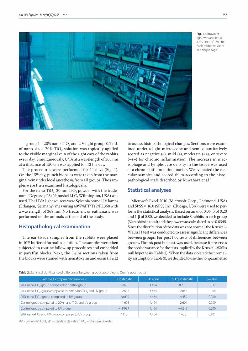
Adv Clin Exp Med. 2021;30(12):1255–1262 1257
– group 4 – 20% nano-TiO2 and UV light group: 0.2 mL of nano-sized 20% TiO2 solution was topically applied to the visible marginal vein of the right ears of the rabbits every day. Simultaneously, UVA at a wavelength of 368 nm at a distance of 150 cm was applied for 12 h a day.
The procedures were performed for 14 days (Fig. 1). On the 15th day, punch biopsies were taken from the mar-ginal vein under local anesthesia from all groups. The sam-ples were then examined histologically.
For the nano-TiO2, 20 nm TiO2 powder with the trade-name Degussa p25 (Nanoshel LLC, Wilmington, USA) was used. The UVA light sources were Sylvania brand UV lamps (Erlangen, Germany), measuring 40W/4FT/T12/BL368 with a wavelength of 368 nm. No treatment or euthanasia was performed on the animals at the end of the study.
Histopathological examination
The ear tissue samples from the rabbits were placed in 10% buffered formalin solution. The samples were then subjected to routine follow-up procedures and embedded in paraffin blocks. Next, the 5-µm sections taken from the blocks were stained with hematoxylin and eosin (H&E)
to assess histopathological changes. Sections were exam-ined under a light microscope and semi-quantitatively scored as negative (−), mild (+), moderate (++), or severe (+++) for chronic inflammation. The increase in mac-rophage and lymphocyte density in the tissue was used as a chronic inflammation marker. We evaluated the vas-cular samples and scored them according to the histo-pathological scale described by Kuwahara et al.6
Statistical analyses
Microsoft Excel 2010 (Microsoft Corp., Redmond, USA) and SPSS v. 16.0 (SPSS Inc., Chicago, USA) were used to per-form the statistical analysis. Based on an α of 0.05, β of 0.20 and 1-β of 0.80, we decided to include 8 rabbits in each group (32 rabbits in total) and the power was calculated to be 0.8345. Since the distribution of the data was not normal, the Kruskal–Wallis H test was conducted to assess significant differences between groups. For post hoc tests of differences between groups, Dunn’s post hoc test was used, because it preserves the pooled variance for the tests implied by the Kruskal–Wallis null hypothesis (Table 2). When the data violated the normal-ity assumption (Table 3), we decided to use the nonparametric
Fig. 1. Ultraviolet light was applied at a distance of 150 cm. Each rabbit was kept in a single cage
Table 2. Statistical significance of differences between groups according to Dunn’s post hoc test
Sample 1 compared to sample 2 Test statistic SD error SD test statistic p-value
20% nano-TiO2 group compared to control group 1.063 4.464 0.238 0.812
20% nano-TiO2-group compared to 20% nano-TiO2 and UV group −12.687 4.464 −2.842 0.004
20% nano-TiO2 -group compared to UV group −20.000 4.464 −4.480 0.000
Control group compared to 20% nano-TiO2 and UV group −11.625 4.464 −2.604 0.009
Control group compared to UV group −18.937 4.464 −4.242 0.000
20% nano-TiO2 and UV group compared to UV group 7.313 4.464 1.638 0.101
UV – ultraviolet light; SD – standard deviation; TiO2 – titanium dioxide.
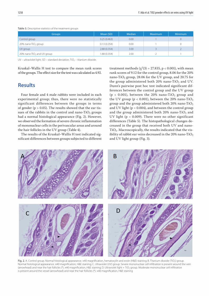
F. Ada et al. TiO2 powder effects on veins using UV light1258
Kruskal–Wallis H test to compare the mean rank scores of the groups. The effect size for the test was calculated as 4.92.
Results
Four female and 4 male rabbits were included in each experimental group; thus, there were no statistically significant differences between the groups in terms of gender (p > 0.05). The results showed that the ear tis-sues of the rabbits in the control and nano-TiO2 groups had a normal histological appearance (Fig. 2). However, we observed the formation of severe chronic inflammation of mononuclear cells in the perivascular areas and around the hair follicles in the UV group (Table 4).
The results of the Kruskal–Wallis H test indicated sig-nificant differences between groups subjected to different
treatment methods (χ2(3) = 27.835, p < 0.001), with mean rank scores of 9.12 for the control group, 8.06 for the 20% nano-TiO2 group, 28.06 for the UV group, and 20.75 for the group administered both 20% nano-TiO2 and UV. Dunn’s pairwise post hoc test indicated significant dif-ferences between the control group and the UV group (p < 0.001), between the 20% nano-TiO2 group and the UV group (p < 0.001), between the 20% nano-TiO2 group and the group administered both 20% nano-TiO2 and UV light (p = 0.004), and between the control group and the group administered both 20% nano-TiO2 and UV light (p = 0.009). There were no other significant differences (Table 5). The histopathological changes de-creased in the group that received both UV and nano-TiO2. Macroscopically, the results indicated that the vis-ibility of rabbit ear veins decreased in the 20% nano-TiO2 and UV light group (Fig. 3).
Fig. 2. A. Control group. Normal histological appearance. ×40 magnification, hematoxylin and eosin (H&E) staining; B. Titanium dioxide (TiO2) group. Normal histological appearance. ×40 magnification, H&E staining; C. Ultraviolet (UV) group. Severe mononuclear cell infiltration is present around the vein (arrowhead) and near the hair follicles (*). ×40 magnification, H&E staining; D. Ultraviolet light + TiO2 group. Moderate mononuclear cell infiltration is present around the vessel (arrowhead) and near the hair follicles (*). ×40 magnification, H&E staining
Table 3. Descriptive statistics of the treatment groups
Groups Mean (SD) Median Maximum Minimum
Control group 0.25 (0.463) 0.00 1 0
20% nano-TiO2 group 0.13 (0.354) 0.00 1 0
UV group 2.88 (0.354) 3.00 3 2
20% nano-TiO2 and UV group 1.88 (0.354) 2.00 2 1
UV – ultraviolet light; SD – standard deviation; TiO2 – titanium dioxide.
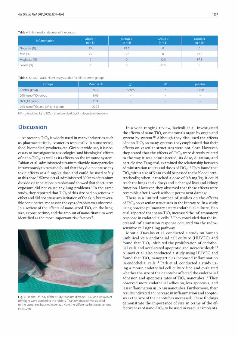
Adv Clin Exp Med. 2021;30(12):1255–1262 1259
Discussion
At present, TiO2 is widely used in many industries such as pharmaceuticals, cosmetics (especially in sunscreens), food, biomedical products, etc. Given its wide use, it is nec-essary to investigate the toxicological and histological effects of nano-TiO2, as well as its effects on the immune system. Fabian et al. administered titanium dioxide nanoparticles intravenously to rats and found that they did not cause any toxic effects at a 5 mg/kg dose and could be used safely at this dose.7 Warheit et al. administered 300 nm of titanium dioxide via inhalation in rabbits and showed that short-term exposure did not cause any lung problems.8 In the same study, they reported that TiO2 of this size had no genotoxic effect and did not cause any irritation of the skin, but revers-ible conjunctival redness in the eyes of rabbits was observed. In a review of the effects of nano-sized TiO2 on the lung, size, exposure time, and the amount of nano-titanium were identified as the most important risk factors.9
In a wide-ranging review, Iavicoli et al. investigated the effects of nano-TiO2 on mammals organ by organ and system by system.10 Although they discussed the effects of nano-TiO2 on many systems, they emphasized that their effects on vascular structures were not clear. However, they stated that the effects of TiO2 were directly related to the way it was administered, its dose, duration, and particle size. Tang et al. examined the relationship between administration routes and doses of TiO2.11 They found that TiO2 with a size of 5 nm could be passed to the blood intra-tracheally; when it reached a dose of 0.8 mg/kg, it could reach the lungs and kidneys and it changed liver and kidney function. However, they observed that these effects were reversible after 1 week without permanent damage.
There is a limited number of studies on the effects of TiO2 on vascular structures in the literature. In a study using porcine pulmonary artery endothelial culture, Han et al. reported that nano-TiO2 increased the inflammatory response in endothelial cells.12 They concluded that the in-creased inflammation response occurred via the redox-sensitive cell signaling pathway.
Montiel-Dávalos et al. conducted a study on human umbilical vein endothelial cell culture (HUVEC) and found that TiO2 inhibited the proliferation of endothe-lial cells and accelerated apoptotic and necrotic death.13 Alinovi et al. also conducted a study using HUVEC and found that TiO2 nanoparticles increased inflammation in endothelial cells.14 Park et al. conducted a study us-ing a mouse endothelial cell culture line and evaluated whether the size of the nanotube affected the endothelial adhesion and apoptosis rates of TiO2 nanotubes.15 They observed more endothelial adhesion, less apoptosis, and less inflammation in 15 nm nanotubes. Furthermore, their results indicated an increase in inflammation and apopto-sis as the size of the nanotubes increased. These findings demonstrate the importance of size in terms of the ef-fectiveness of nano-TiO2 to be used in vascular implants.
Fig. 3. On the 14th day of the study, titanium dioxide (TiO2) and ultraviolet (UV) light were applied to the rabbits. Titanium dioxide was applied to the upper ear, but not lower ear. Note the difference between venous structures
Table 4. Inflammation degrees of the groups
Inflammation Group 1(n = 8)
Group 2(n = 8)
Group 3(n = 8)
Group 4(n = 8)
Negative [%] 75 87.5 0 0
Mild [%] 25 12.5 0 12.5
Moderate [%] 0 0 12.5 87.5
Severe [%] 0 0 87.5 0
Table 5. Kruskal–Wallis H test analysis table for all treatment groups
Groups Mean rank χ2 df p-value
Control group 9.12 27.835 3 0.000
20% nano-TiO2 group 8.06 – – –
UV light group 28.06 – – –
20% nano-TiO2 and UV light group 20.75 – – –
UV – ultraviolet light; TiO2 – titanium dioxide; df – degrees of freedom.
F. Ada et al. TiO2 powder effects on veins using UV light1260
Ge et al. conducted a study using HUVEC and their results indicated that thin film nano-TiO2 provided laminin im-mobilization and an increase in endothelial adhesion.16
Ultraviolet light has been used in the treatment of skin diseases since the beginning of the 20th century. Although the mechanism of UV light action is not clear, hypotheses that it increases cell proliferation, epidermal thickness and blood flow in cutaneous capillaries have been proposed.17 In a study that applied UV light to patients with venous ulcers, Dodd et al. concluded that UV light had no benefit in the treatment of venous ulcers.18 However, they observed that UV light increased skin oxygen permeability and in-hibited normal vasoconstrictor response.
When light treatment is required in medicine, the pre-ferred UV light wavelength is in the 300–320 nm range. In dermatology, psoralen ultraviolet A (PUVA) treatment for psoriasis, eczema, vitiligo, and cutaneous lymphoma is widely used.19 Although UV light has positive results when applied to appropriate patients for a certain period of time, dose and treatment, excessive exposure can lead to many adverse conditions such as skin cancer, T cell damage, im-mune depression, sunburn, and premature skin aging.20
Telangiectasias are enlarged venous vessels with a diam-eter of 0.5–1 mm near the skin surface. Telangiectasias are enlarged postcapillary venules located in the papillary and superficial reticular dermis with histologically complete meaning.21
Engel et al. conducted a field study with a large popula-tion in the USA and found that the rate of telangiectasia in the legs was 29–41% in women and 6–15% in men.22 Fa-cial telangiectasia affects tens of millions of people around the world.23 Sclerotherapy is the standard approach for the treatment of telangiectasias. Sclerosis of the telangi-ectatic vein is the main target in sclerotherapy. In order to achieve sclerosis, various chemical sclerosing agents are administered directly into the enlarged telangiectatic vein, resulting in the formation of a thrombosis and endothelial damage within the vein.24
Apart from sclerotherapy, many different laser treatments, such as argon ion lasers, diode lasers and intense pulsed light sources, are applied.25,26 Laser treatment was first per-formed in the treatment of telangiectasias in 1960 using light at a 694 nm wavelength, followed by different systems and wavelengths.27 The underlying logic of laser treatment in telangiectasia is to cause the loss of veins close to the skin surface by creating thermal damage to the vascular struc-tures through the heat generated by UV light. Hemoglobin in the vessel absorbs light best at 418 nm, 542 nm and 577 nm wavelengths.28 However, the wavelengths used in the treat-ment of vascular lesions are usually 488 nm and 600 nm.28
Photocatalysts can be described as semiconductors that create a strongly oxidized environment on the surface through the effect of UV light. Therefore, photocatalysts can cause degradation in organic tissues close to the region where they are applied. Among the known semiconductor photocatalysts, TiO2 is widely used due to its high activity,
high stability under lighting, nontoxic nature, and low price. Titanium dioxide performs its photocatalytic effect best under 340 nm wavelength light, but a wavelength range of 320–380 nm is also good for photocatalyst activity.29
In general, studies have demonstrated that organic cell degradation increases as the wavelength decreases. This means that at a wavelength of 280 nm or less, TiO2 has its maximum photocatalytic effect, leading to the degrada-tion of DNA and RNA.30,31 Therefore, the light wavelength selected in our study was in the range of 320–380 nm. The same ultraviolet light wavelength and application meth-ods used in previous animal experiments, as well as the dose, concentration, and routes of TiO2 administration were ap-plied in the current study.32,33 What distinguishes this study from the other studies is that we investigated the effects of TiO2 and UV light on vascular structures simultaneously.
Photodynamic therapy, especially in dermatology and plastic surgery, is used effectively and widely for the treat-ment of many dermatological diseases in the form of a com-bination of light and various creams applied to the skin. The main principle of creams with methyl aminolevulinate as the active ingredient is to be absorbed by the skin and subcutaneous tissue, and to provide local apoptosis with the effect of light. In fact, Galvão found that telangiectasias on the face were not visible after photodynamic therapy in a patient with facial actinic keratosis.34
Photodynamic therapy consists of 3 main elements: a photosensitive material, a light source and oxygen. When these 3 factors are combined effectively, they can be ap-plied for the successful treatment of many skin lesions. In photodynamic therapy, the therapeutic effect is achieved by a slight activation of a light-sensitive substance, and reactive oxygen intermediates are formed in the pres-ence of oxygen. These intermediates irreversibly oxidize the essential cellular components that cause apoptosis and necrosis, thereby providing the treatment of lesions on the skin.35 Essentially, our study has shown similar results to photodynamic therapy. In this sense, the study demonstrated that substances with photocatalytic activa-tion may be an appropriate choice for this treatment.
Limitations
The method applied in the present study could be used in the treatment of telangiectasia in the future. However, our study has a predominantly histological and observa-tional nature. Further studies involving genetic, cytoge-netic, biochemical, histochemical, and immunohistochemi-cal analyses are needed to test the theories described above.
Conclusion
The results of this study revealed that nano-TiO2 protected the skin and the main vascular structures close to the skin from the harmful effects of UV light.
Adv Clin Exp Med. 2021;30(12):1255–1262 1261
Furthermore, this study determined that telangiectatic structures close to the skin were not observed in the UV and nano-TiO2 group.
Several theories were put forward regarding the absence of telangiectatic structures in the rabbit ear on macro-scopic observation. The 1st is that nano-TiO2 and UV light induced apoptosis in telangiectatic structures, just as in photodynamic therapy. The 2nd is that thermal dam-age was caused by heating during the photocatalytic acti-vation of nano-TiO2. The 3rd theory is that collagen tissue deposited under the epidermis compressed the telangi-ectatic veins or precipitated them towards the dermis. The 4th is damage of hemoglobin in the telangiectatic vein. The 5th is selective photothermolysis that led to panendo-thelial obliteration. Any combination of these theories, or some other mechanism of action, may have led to these results.
ORCID iDsFatih Ada https://orcid.org/0000-0002-6953-5906Ferit Kasimzade https://orcid.org/0000-0003-3646-3181Ali Sefa Mendil https://orcid.org/0000-0003-2722-3290Hasan Gocmez https://orcid.org/0000-0003-3748-0311
References1. Şam ED, Ürgen M, Tepehan FZ. TiO2 fotokatalistleri. İTÜ Dergisi/D
Mühendislik. 2007;6(5–6):81–92. http://itudergi.itu.edu.tr/index.php/itudergisi_d/article/viewFile/395/337. Accessed November 1, 2020.
2. Ochiai T, Fujishima A. Design and optimization of photocatalytic water purification reactors. In: Pichat P, ed. Photocatalysis Water Purification: From Fundamentals to Recent Applications. Weinheim, Ger-many: Wiley-VCH; 2013:361. doi:10.1002/9783527645404.ch14
3. Ohno T, Akiyoshi M, Umebayashi T, et al. Preparation of S-doped TiO2 photocatalysts and their photocatalytic activities under visib-le light. Appl Catal A Gen. 2004;265(1):115–121. doi:10.1016/j.apca-ta.2004.01.007
4. Watanabe E, Fukaya M, Nishizawa K, Miki T, Taoda H. Test method for skin damage of titania photocatalyst nanoparticles in vitro. Mater Sci Forum. 2008;569:9–12. doi:10.4028/www.scientific.net/MSF.569.9
5. Shen B, Scaiano JC, English AM. Zeolite encapsulation decreases TiO2‐photosensitized ROS generation in cultured human skin fibroblasts. Photochem Photobiol. 2006;82(1):5–12. doi:10.1562/2005-05-29-RA-551
6. Kuwahara T, Asanami S, Kubo S. Experimental infusion phlebitis: Tole-rance osmolality of peripheral venous endothelial cells. Nutrition. 1998;14(6):496–501. doi:10.1016/s0899-9007(98)00037-9
7. Fabian E, Landsiedel R, Ma-Hock L, Wiench K, Wohlleben W, van Ravenzwaay B. Tissue distribution and toxicity of intravenously admi-nistered titanium dioxide nanoparticles in rats. Arch Toxicol. 2008; 82(3):151–157. doi:10.1007/s00204-007-0253-y
8. Warheit DB, Hoke RA, Finlay C, Donner EM, Reed KL, Sayes CM. Deve-lopment of a base set of toxicity tests using ultrafine TiO2 particles as a component of nanoparticle risk management. Toxicol Lett. 2007; 171(3):99–110. doi:10.1016/j.toxlet.2007.04.008
9. Warheit DB. How to measure hazards/risks following exposures to nanoscale or pigment-grade titanium dioxide particles. Toxicol Lett. 2013;220(2):193–204. doi:10.1016/j.toxlet.2013.04.002
10. Iavicoli I, Leso V, Fontana L, Bergamaschi A. Toxicological effects of titanium dioxide nanoparticles: A review of in vitro mammalian studies. Eur Rev Med Pharmacol Sci. 2011;15(5):481–508. doi:10.1155/ 2012/964381
11. Tang M, Zhang T, Xue Y, et al. Dose dependent in vivo metabolic cha-racteristics of titanium dioxide nanoparticles. J Nanosci Nanotechnol. 2010;10(12):8575–8583. doi:10.1166/jnn.2010.2482
12. Han SG, Newsome B, Hennig B. Titanium dioxide nanoparticles incre-ase inflammatory responses in vascular endothelial cells. Toxicology. 2013;306:1–8. doi:10.1016/j.tox.2013.01.014
13. Montiel-Dávalos A, Ventura-Gallegos JL, Alfaro-Moreno E, et al. TiO2 nanoparticles induce dysfunction and activation of human endothe-lial cells. Chem Res Toxicol. 2012;25(4):920–930. doi:10.1021/tx200551u
14. Alinovi R, Goldoni M, Pinelli S, et al. Oxidative and pro-inflamma-tory effects of cobalt and titanium oxide nanoparticles on aortic and venous endothelial cells. Toxicol In Vitro. 2015;29(3):426–437. doi:10.1016/j.tiv.2014.12.007
15. Park J, Bauer S, Schmuki P, von der Mark K. Narrow window in nanos-cale dependent activation of endothelial cell growth and differen-tiation on TiO2 nanotube surfaces. Nano Lett. 2009;9(9):3157–3164. doi:10.1021/nl9013502
16. Ge SN, Chen JY, Leng YX, Huang N. Laminin immobilized on titani-um oxide films for enhanced human umbilical vein endothelial cell adhesion and growth. Key Eng Mater. 2007;342:305–308. doi:10.4028/www.scientific.net/KEM.342-343.305
17. Thai TP, Houghton PE, Keast DH, Woodbury MG. Ultraviolet C in the tre-atment of chronic wounds with MRSA: A case study. Ostomy Wound Manage. 2002;48(11):52–60. PMID:12426452
18. Dodd HJ, Sarkany I, Gaylarde PM. The short‐term benefit and long‐term failure of ultraviolet light in the treatment of venous leg ulcers. Br J Dermatol. 1989;120(6):809–818. doi:10.1111/j.1365-2133.1989.tb01379.x
19. Hönigsmann H, Szeimies RM, Knobler R, et al. Photochemotherapy and photodynamic therapy. In: Goldsmith LA, Katz SI, Gilchrest BA, Paller AS, Leffell DJ, Wolff K. Dermatology in General Medicine. New York, USA: McGraw-Hill; 1999:2477–2493.
20. Elmets CA, Bergstresser PR, Tigelaar RE, Wood PJ, Streilein JW. Analy-sis of the mechanism of unresponsiveness produced by haptens painted on skin exposed to low dose ultraviolet radiation. J Exp Med. 1983;158:781–794. doi:10.1084/jem.158.3.781
21. Walker JG, Stirling J, Beroukas D, et al. Histopathological and ult-rastructural features of dermal telangiectasias in systemic sclerosis. Pathology. 2005;37(3):220–225. doi:10.1080/00313020500033262
22. Engel A, Johnson ML, Hynes SG. Health effects of sunlight exposure in the United States: Results from the first National Health and Nutri-tion Examination Survey 1971–1974. Arch Dermatol. 1988;124:72–79. doi:10.1001/archderm.1988.01670010036018
23. Cassuto DA, Ancona, DM, Emanuelli G. Treatment of facial telangiecta-sias with a diode-pumped Nd: YAG laser at 532 nm. J Cutan Laser Ther. 2000;2(3):141–146. doi:10.1080/14628830050516399
24. Rabe E, Breu FX, Cavezzi A, et al. European guidelines for sclerothe-rapy in chronic venous disorders. Phlebology. 2014;29(6):338–354. doi:10.1177/0268355513483280
25. Landthaler M, Hohenleutner U. Laser therapy of vascular lesions. Photodermatol Photoimmunol Photomed. 2006;22(6):324–332. doi:10. 1111/j.1600-0781.2006.00254
26. Goldman MP, Martin DE, Fitzpatrick RE, Ruiz-Esparza J. Pulsed dye laser treatment of telangiectases with and without subtherapeutic sclerotherapy: Clinical and histologic examination in the rabbit ear vein model. J Am Acad Dermatol. 1990;23(1):23–30. doi:10.1016/0190-9622(90)70180-p
27. Apfelberg DB, Maser MR, Lash H, White DN, Flores JT. Use of the argon and carbon dioxide lasers for the treatment of superficial venous varicosities of the lower extremity. Lasers Surg Med. 1984;4:221–232. doi:10.1002/lsm.1900040302
28. Spendel S, Prandl EC, Schintler MV, et al. Treatment of spider leg veins with the KTP (532 nm) laser: A prospective study. Lasers Surg Med. 2002;31(3):194–201. doi:10.1002/lsm.10088
29. Thiruvenkatachari R, Vigneswaran S, Moon IS. A review on UV/TiO2 pho-tocatalytic oxidation process. Korean J Chem Eng. 2008;25(1):64–72. doi:10.1007/s11814-008-0011-8
30. Matthews RW, McEvoy SR. Photocatalytic degradation of phenol in the presence of near-UV illuminated titanium dioxide. J Photochem Photobiol. 1992;64(2):231–246. doi:10.1016/1010-6030(92)85110-G
31. Li PG, Yue PL. Photocatalytic oxidation of chlorophenols in single component and multicomponent systems. Ind Eng Chem Res. 1999; 38(9):3238–3245. doi:10.1021/ie9807598
32. Emerson JA, Whittington JK, Allender MC, Mitchell MA. Effects of ult-raviolet radiation produced from artificial lights on serum 25-hydro-xyvitamin D concentration in captive domestic rabbits (Oryctola-guscuniculi). Am J Vet Res. 2014;75(4):380–384. doi:10.2460/ajvr.75. 4.380

F. Ada et al. TiO2 powder effects on veins using UV light1262
33. Lansdown ABG, Taylor A. Zinc and titanium oxides: Promising UV-absor-bers but what influence do they have on the intact skin? Int J Cosmet Sci. 1997;19(4):167–172. doi:10.1111/j.1467-2494.1997.tb00180.x
34. Galvão LEG. Terapia fotodinâmica com luz do dia: benefício clíni-co e estético com sessões repetidas para ceratoses actínicas faciais. Surg Cosmet Dermatol. 2016;8(4):40–42. doi:10.5935/scd1984-8773. 201683103
35. Wan MT, Lin JY. Current evidence and applications of photodynamic therapy in dermatology. Clin Cosmet Investig Dermatol. 2014;7:145–163. doi:10.2147/CCID.S35334
Cite asZhou E, Zou Y, Mao C, Li D, Wang C, Zhang Z. MicroRNA-221 inhibits the transition of endothelial progenitor cells to mesenchymal cells via the PTEN/FoxO3a signaling pathway. Adv Clin Exp Med. 2021;30(12):1263–1270. doi:10.17219/acem/141446
DOI10.17219/acem/141446
Copyright© 2021 by Wroclaw Medical University This is an article distributed under the terms of theCreative Commons Attribution 3.0 Unported (CC BY 3.0)(https://creativecommons.org/licenses/by/3.0/)
Address for correspondenceZongqi ZhangE-mail: [email protected]
Funding sourcesNational Natural Science Foundation of China (grant No. 81200206 to ZQ Zhang); Scientific Research and Innovation Project of Shanghai Education Commission (grant No. 12YZ045 to ZQ Zhang); Shanghai Committee of Science and Technology, China (grant No. 18411950500); National Natural Science Foundation of China (grant No. 81870264).
Conflict of interestNone declared
*En Zhou, Yinghua Zou, Chengyu Mao and Dongjiu Li contributed equally to this study.
Received on March 9, 2021Reviewed on May 16, 2021Accepted on August 19, 2021
Published online on October 5, 2021
AbstractBackground. Coronary heart disease is one of the most common cardiovascular diseases worldwide and is often associated with vascular endothelial injury. Endothelial–mesenchymal transition (EndMT) is an im-portant process in vascular endothelial injury.
Objectives. This study investigated the function of miR-221 in the EndMT process of endothelial progenitor cells (EPCs).
Materials and methods. Transforming growth factor beta (TGF-β1) was used to induce EndMT in EPCs, and SM22α expression was detected using immunocytochemistry. Western blot was used to detect alpha smooth muscle actin (αSMA) expression, and miR-221 function was evaluated using inhibitors or mimics of the miR-221 sequences that were transfected into EPCs. Reverse transcription polymerase chain reaction (RT-PCR) was used to detect the expression of miR-221 and western blot was used to detect the expres-sion of αSMA, myocardin, phosphatase and tensin homolog (PTEN), p-FoxO3a, and FoxO3a in EPCs. Finally, the expression of the miR-221 target genes was determined using RT-PCR.
Results. The expression of SM22α and αSMA increased in EPCs treated with TGF-β1, while the expression of miR-221 was decreased in EPCs on the 5th day, when compared with the control. The expression of SM22α increased after inhibiting miR-221 in EPCs treated with TGF-β1 and this was reversed by the overexpression of miR-221. The expression of αSMA and myocardin was significantly increased after inhibiting miR-221 in EPCs treated with TGF-β1 and decreased in EPCs overexpressing miR-221. Conversely, PTEN was increased in TGF-β1-treated EPCs and decreased following the overexpression of miR-221. The decrease in phosphory-lated-FoxO3a expression in EPCs was accompanied by an increase in αSMA expression, which was reversed in the presence of miR-221 mimics. This effect was nearly abolished following the addition of PTEN cDNA.
Conclusions. The overexpression of miR-221 inhibits EndMT in EPCs, possibly by interacting with PTEN to regulate FoxO3a signaling, to facilitate the repair of the endothelium by EPCs.
Key words: EPC, miR-221, EndMT, PTEN/FoxO3a
Original papers
MicroRNA-221 inhibits the transition of endothelial progenitor cells to mesenchymal cells via the PTEN/FoxO3a signaling pathway
*En Zhou1,C–E, *Yinghua Zou2,C–E, *Chengyu Mao1,C–E, *Dongjiu Li1,C–E, Changqian Wang1,A,F, Zongqi Zhang1,A,E,F
1 Department of Cardiology, Shanghai Ninth People’s Hospital, School of Medicine, Shanghai Jiao Tong University, China2 Department of Anesthesiology, Shuguang Hospital, Shanghai University of Traditional Chinese Medicine, China
A – research concept and design; B – collection and/or assembly of data; C – data analysis and interpretation; D – writing the article; E – critical revision of the article; F – final approval of the article
Advances in Clinical and Experimental Medicine, ISSN 1899–5276 (print), ISSN 2451–2680 (online) Adv Clin Exp Med. 2021;30(12):1263–1270
E. Zhou et al. miR-221 inhibits EndMT of EPC via PTEN/FoxO3a1264
Background
Coronary heart disease is one of the most common car-diovascular diseases worldwide and is caused by vascular endothelial injury. It has been demonstrated that once the vascular endothelium is damaged, the endothelial progenitor cells (EPCs) migrate from the bone marrow (BM) to the ischemic tissue and differentiate into mature vascular endothelial cells in an attempt to repair the in-jured site.1 However, accumulating evidence suggests that EPCs contribute to intimal hyperplasia during this process. For example, BM-derived EPCs have been shown to aug-ment intimal hyperplasia by migrating into the intima of balloon-injured carotid arteries,2 and our previous study showed that BM-derived EPCs can transdifferentiate into smooth muscle lineage cells that are positive for alpha smooth muscle actin (αSMA), producing an endothelial-to-mesenchymal transition (EndMT).3 These results sug-gest that the EndMT capacity of EPCs may contribute to intimal hyperplasia. This is further supported by evi-dence that EndMT increases the thickness of the intimal layer in pulmonary arteries.4 However, the mechanisms underlying the EndMT process in EPCs have not yet been fully elucidated.
MicroRNAs (miRs) are short (only 21–23 nucleotides), highly conserved, non-coding RNA sequences that bind to the 3’-untranslated region of a variety of mRNA tar-gets and function to regulate gene expression at the post-transcriptional level by inhibiting the translation or pro-moting mRNA degradation.5,6 Previous microarray-based expression profiles have shown that several miRs are ex-pressed in EPCs, including miR-126, miR-21, miR-27a, and miR-221.7,8 Furthermore, some studies have shown that both miR-21 and miR-127 are involved in mature endo-thelial cell EndMT that can be induced by TGF-β1 and TGF-β2.8,9 In addition, miR-221, an endothelial-specific miR, has been shown to exert anti-angiogenic, anti-in-flammatory and pro-apoptotic effects by targeting both ZEB2 and Ets-1.10,11 Finally, miR-221 maintains the an-giogenic capacity of EPCs by inhibiting p27 expression.12 However, few studies have described the role of miR-221 in the EndMT process of EPCs.
Previous investigations have confirmed that phosphatase and tensin homolog (PTEN) is a direct target of miR-221.13 In vascular endothelial cells, downregulation of miR-221 decreased cell viability, migration and invasion, while all of these effects were reversed in response to the overex-pression of PTEN.13 Furthermore, recent studies have shown that a decrease in PTEN activates the phosphory-lation of FoxO3a.14,15 In our previous study, we showed that phosphorylated FoxO3a plays an inhibitory role in EndMT by inactivating the Smad3/4 pathway in the nucleus of EPCs.16 Given this, we hypothesized that the activa-tion of the miR-221/PTEN/FoxO3a pathway may inhibit EndMT in EPCs.
Objectives
This study was designed to determine the function of miR-221 in the EndMT process of EPCs and to explore the potential signaling pathways underlying these functions.
Materials and methods
Ethical approval
All animal experiments were approved by the Animal Research Ethics Committee of the No. 9 People’s Hospital affiliated with the Shanghai Jiao Tong University School of Medicine, Shanghai, China. All animals were sacrificed using bilateral thoracotomy during anesthesia.
Cell isolation and culture
Bone marrow-derived EPCs were isolated from male Sprague Dawley rats, as described previously.2 Ficoll-Isopaque Plus (Histopaque-1077; Sigma-Aldrich, St. Louis, USA) density gradient centrifugation was used to isolate mononuclear cells from rat bone marrow. The cells were cultured in endothelial cell growth medium (EGM2; Lonza, Basel, Switzerland) consisting of endothelial cell basal me-dium-2 (EBM-2), supplemented with 20% fetal bovine serum (FBS). Cells were then cultured on a fibronectin-coated dish at 37°C in a 5% CO2 incubator. The medium was removed after 4–7 days, and cells were gently washed with phos-phate-buffered solution (PBS). The attached cells were then used for further experiments. The CD31 fluorescence stain-ing was then performed as previously described7 to identify vascular endothelial cells.
Inducing EndMT in EPCs
Endothelial progenitor cell EndMT was induced as pre-viously described.17 Briefly, the cells were seeded on culture plates in Ham’s F-12 media containing 5% FBS, and then treated with TGF-β1 (5 ng/mL) for 7 days.
Immunocytochemistry
The EPCs were cultured on cell slides and then rinsed with PBS, before being fixed with 4% paraformaldehyde phos-phate buffer for 15 min at room temperature. Slides were then blocked for 2 h in PBS containing 5% normal goat se-rum and 2% bovine serum albumin (BSA), and incubated with monoclonal antibodies (diluted 1:500) against SM22α (Cat. No. SAB1400272; Sigma-Aldrich) overnight at 4°C. Cells were washed 5 times in PBS and incubated with AlexaFluor® 594-conjugated goat anti-mouse/rabbit antibody diluted 1:500 (Santa Cruz Biotechnology, Dallas, USA) for 1 h at room temperature. The mean fluorescence intensity of SM22α was
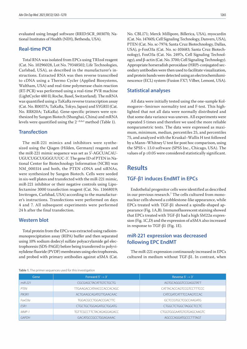
Adv Clin Exp Med. 2021;30(12):1263–1270 1265
evaluated using ImageJ software (RRID:SCR_003070; Na-tional Institutes of Health (NIH), Bethesda, USA).
Real-time PCR
Total RNA was isolated from EPCs using TRIzol reagent (Cat. No. 10296028, Lot No. 79348502; Life Technologies, Carlsbad, USA), as described in the manufacturer’s in-structions. Extracted RNA was then reverse transcribed to cDNA using a Thermo Cycler (Applied Biosystems, Waltham, USA) and real-time polymerase chain reaction (RT-PCR) was performed using a real-time PVR machine (LightCycler 480 II; Roche, Basel, Switzerland). The mRNA was quantified using a TaKaRa reverse transcription assay (Cat. No. R0037A; TaKaRa, Tokyo, Japan) and SYGRII (Cat. No. RR820A; TaKaRa). Gene-specific primers were syn-thesized by Sangon Biotech (Shanghai, China) and mRNA levels were quantified using the 2–ΔΔct method (Table 1).
Transfection
The miR-221 mimics and inhibitors were synthe-sized using the Qiagen (Hilden, Germany) reagents and the miR-221 mimic sequence was set as 5’-AGCUACAU-UGUCUGCUGGGUUUC-3’. The gene ID of PTEN in Na-tional Center for Biotechnology Information (NCBI) was NM_000314 and both, the PTEN cDNA and siRNAs, were synthesized by Sangon Biotech. Cells were seeded in six-well plates and transfected with the miR-221 mimic, miR-221 inhibitor or their negative controls using Lipo-fectamine 3000 transfection reagent (Cat. No. 11668019; Invitrogen, Carlsbad, USA) according to the manufactur-er’s instructions. Transfections were performed on days 4 and 7. All subsequent experiments were performed 24 h after the final transfection.
Western blot
Total protein from the EPCs was extracted using radioim-munoprecipitation assay (RIPA) buffer and then separated using 10% sodium dodecyl sulfate polyacrylamide gel elec-trophoresis (SDS-PAGE) before being transferred to polyvi-nylidene fluoride (PVDF) membranes using electrophoresis, and probed with primary antibodies against αSMA (Cat.
No. CBL171; Merck Millipore, Billerica, USA), myocardin (Cat. No. 14760S; Cell Signaling Technology, Danvers, USA), PTEN (Cat. No. sc-7974; Santa Cruz Biotechnology, Dallas, USA), p-FoxO3a (Cat. No. sc-101683; Santa Cruz Biotech-nology), FoxO3a (Cat. No. 2497s, Cell Signaling Technol-ogy), and β-actin (Cat. No. 3700; Cell Signaling Technology). Appropriate horseradish peroxidase (HRP)-conjugated sec-ondary antibodies were then used to facilitate visualization, and protein bands were detected using an electrochemilumi-nescence (ECL) system (Fusion FX7; Vilber, Lemont, USA).
Statistical analyses
All data were initially tested using the one-sample Kol-mogorov–Smirnov normality test and F-test. This high-lighted that not all data were normally distributed and that some data variance was uneven. All experiments were repeated 5 times and therefore we used the more reliable nonparametric tests. The data were expressed as maxi-mum, minimum, median, percentiles 25, and percentiles 75, and analyzed with the Kruskal–Wallis H test followed by a Mann–Whitney U test for post hoc comparison, using the SPSS v. 13.0 software (SPSS Inc., Chicago, USA). The values of p ≤0.05 were considered statistically significant.
Results
TGF-β1 induces EndMT in EPCs
Endothelial progenitor cells were identified as described in our previous research.7 The cells cultured from mono-nuclear cells showed a cobblestone-like appearance, while EPCs treated with TGF-β1 showed a spindle-shaped ap-pearance (Fig. 1A,B). Immunofluorescent staining showed that EPCs treated with TGF-β1 had a high SM22α expres-sion (Fig. 1C,D) and the expression of αSMA also increased in response to TGF-β1 (Fig. 1E).
miR-221 expression was decreased following EPC EndMT
The miR-221 expression continuously increased in EPCs cultured in medium without TGF-β1. In contrast, when
Table 1. The primer sequences used for this investigation
Gene Forward 5’ → 3’ Reverse 5’ → 3’
miR-221 CGCGAGCTACATTGTCTGCTG AGTGCAGGGTCCGAGGTATT
PTEN TTGAAGACCATAACCCACCACAGC CATTACACCAGTCCGTCCTTTCCC
PIK3R1 ACTGAAGCAGATGTTGAACAAC CATCGATCATTTCCAAGTCCAC
FoxO3a TGGACGCCTGGACCGACTTC GCTCCGTGCTCGCCAAGATG
ESR1 CTGCTGCTGGAGATGCTGGATG CTGGCTCTGGCTAGGCTCCTC
MMP-1 TGTTCGCCTTCTACAGAGGAGACC CTGGTGGGAATGTGTGAGCAAGTC
GAPDH GACATGCCGCCTGGAGAAAC AGCCCAGGATGCCCTTTAGT
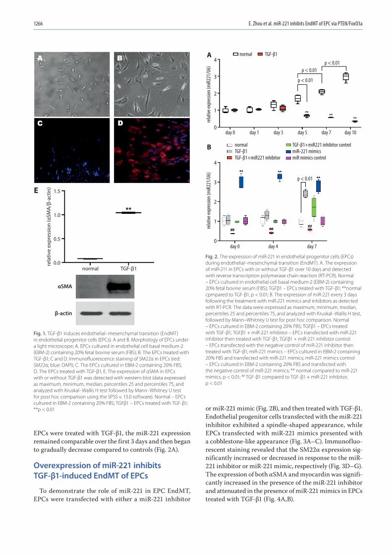
E. Zhou et al. miR-221 inhibits EndMT of EPC via PTEN/FoxO3a1266
EPCs were treated with TGF-β1, the miR-221 expression remained comparable over the first 3 days and then began to gradually decrease compared to controls (Fig. 2A).
Overexpression of miR-221 inhibits TGF-β1-induced EndMT of EPCs
To demonstrate the role of miR-221 in EPC EndMT, EPCs were transfected with either a miR-221 inhibitor
or miR-221 mimic (Fig. 2B), and then treated with TGF-β1. Endothelial progenitor cells transfected with the miR-221 inhibitor exhibited a spindle-shaped appearance, while EPCs transfected with miR-221 mimics presented with a cobblestone-like appearance (Fig. 3A–C). Immunofluo-rescent staining revealed that the SM22α expression sig-nificantly increased or decreased in response to the miR-221 inhibitor or miR-221 mimic, respectively (Fig. 3D–G). The expression of both αSMA and myocardin was signifi-cantly increased in the presence of the miR-221 inhibitor and attenuated in the presence of miR-221 mimics in EPCs treated with TGF-β1 (Fig. 4A,B).
Fig. 1. TGF-β1 induces endothelial–mesenchymal transition (EndMT) in endothelial progenitor cells (EPCs). A and B. Morphology of EPCs under a light microscope; A. EPCs cultured in endothelial cell basal medium-2 (EBM-2) containing 20% fetal bovine serum (FBS); B. The EPCs treated with TGF-β1; C and D. Immunofluorescence staining of SM22α in EPCs (red: SM22α; blue: DAPI); C. The EPCs cultured in EBM-2 containing 20% FBS; D. The EPCs treated with TGF-β1; E. The expression of αSMA in EPCs with or without TGF-β1 was detected with western blot (data expressed as maximum, minimum, median, percentiles 25 and percentiles 75, and analyzed with Kruskal–Wallis H test followed by Mann–Whitney U test for post hoc comparison using the SPSS v. 13.0 software). Normal – EPCs cultured in EBM-2 containing 20% FBS; TGFβ1 – EPCs treated with TGF-β1; **p < 0.01
Fig. 2. The expression of miR-221 in endothelial progenitor cells (EPCs) during endothelial–mesenchymal transition (EndMT). A. The expression of miR-211 in EPCs with or without TGF-β1 over 10 days and detected with reverse transcription polymerase chain reaction (RT-PCR). Normal – EPCs cultured in endothelial cell basal medium-2 (EBM-2) containing 20% fetal bovine serum (FBS); TGFβ1 – EPCs treated with TGF-β1; **normal cpmpared to TGF-β1, p < 0.01; B. The expression of miR-221 every 3 days following the treatment with miR-221 mimics and inhibitors as detected with RT-PCR. The data were expressed as maximum, minimum, median, percentiles 25 and percentiles 75, and analyzed with Kruskal–Wallis H test, followed by Mann–Whitney U test for post hoc comparison. Normal – EPCs cultured in EBM-2 containing 20% FBS; TGFβ1 – EPCs treated with TGF-β1; TGFβ1 + miR-221 inhibitor – EPCs transfected with miR-221 inhibitor then treated with TGF-β1; TGFβ1 + miR-221 inhibitor control – EPCs transfected with the negative control of miR-221 inhibitor then treated with TGF-β1; miR-221 mimics – EPCs cultured in EBM-2 containing 20% FBS and transfected with miR-221 mimics; miR-221 mimics control – EPCs cultured in EBM-2 containing 20% FBS and transfected with the negative control of miR-221 mimics; ** normal compared to miR-221 mimics, p < 0.01; ## TGF-β1 compared to TGF-β1 + miR-221 inhibitor, p < 0.01
normal
rela
tive
expr
essi
on (α
SMA
/β-a
ctin
)
αSMA
β-actin
1.5
1.0
0.5
0.0TGF-β1
E
normal
normalTGF-β1TGF-β1+miR221 inhibitor
TGF-β1+miR221 inhibitor controlmiR-221 mimicsmiR mimics control
4
3
2
1
0
TGF-β1
p < 0.01p < 0.01
relat
ive ex
pres
sion (
miR
221/
U6)
4
3
2
1
0
relat
ive ex
pres
sion (
miR
221/
U6)
p < 0.01
p < 0.01
day 0 day 1 day 3 day 5 day 7
day 0 day 7day 4
day 10
A
B
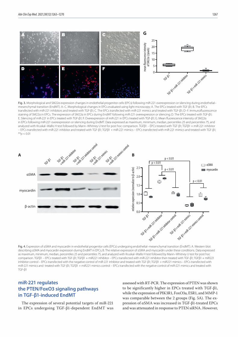
Adv Clin Exp Med. 2021;30(12):1263–1270 1267
miR-221 regulates the PTEN/FoxO3 signaling pathways in TGF-β1-induced EndMT
The expression of several potential targets of miR-221 in EPCs undergoing TGF-β1-dependent EndMT was
assessed with RT-PCR. The expression of PTEN was shown to be significantly higher in EPCs treated with TGF-β1, while the expression of PIK3R1, FoxO3a, ESR1, and MMP-1 was comparable between the 2 groups (Fig. 5A). The ex-pression of αSMA was increased in TGF-β1-treated EPCs and was attenuated in response to PTEN siRNA. However,
Fig. 4. Expression of αSMA and myocardin in endothelial progenitor cells (EPCs) undergoing endothelial–mesenchymal transition (EndMT). A. Western blot describing αSMA and myocardin expression during EndMT in EPCs; B. The relative expression of αSMA and myocardin under these conditions. Data expressed as maximum, minimum, median, percentiles 25 and percentiles 75, and analyzed with Kruskal–Wallis H test followed by Mann–Whitney U test for post hoc comparison. TGFβ1 – EPCs treated with TGF-β1; TGFβ1 + miR221 inhibitor – EPCs transfected with miR-221 inhibitor then treated with TGF-β1; TGFβ1 + miR221 inhibitor control – EPCs transfected with the negative control of miR-221 inhibitor and treated with TGF-β1; TGFβ1 + miR221 mimics – EPCs transfected with miR-221 mimics and treated with TGF-β1; TGFβ1 + miR221 mimics control – EPCs transfected with the negative control of miR-221 mimics and treated with TGF-β1
Fig. 3. Morphological and SM22α expression changes in endothelial progenitor cells (EPCs) following miR-221 overexpression or silencing during endothelial–mesenchymal transition (EndMT). A–C. Morphological changes in EPCs evaluated using light microscopy; A. The EPCs treated with TGF-β1; B. The EPCs transfected with miR-221 inhibitors and treated with TGF-β1; C. The EPCs transfected with miR-221 mimics and treated with TGF-β1; D–F. Immunofluorescence staining of SM22α in EPCs. The expression of SM22α in EPCs during EndMT following miR-221 overexpression or silencing; D. The EPCs treated with TGF-β1; E. Silencing of miR-221 in EPCs treated with TGF-β1; F. Overexpression of miR-221 in EPCs treated with TGF-β1; G. Mean fluorescence intensity of SM22α in EPCs following miR-221 overexpression or silencing during EndMT. Data expressed as maximum, minimum, median, percentiles 25 and percentiles 75, and analyzed with Kruskal–Wallis H test followed by Mann–Whitney U test for post hoc comparison. TGFβ1 – EPCs treated with TGF-β1; TGFβ1 + miR-221 inhibitor – EPCs transfected with miR-221 inhibitor and treated with TGF-β1; TGFβ1 + miR-221 mimics – EPCs transfected with miR-221 mimics and treated with TGF-β1; **p < 0.01
mea
n fluo
resce
nce i
nten
sity
of SM
22α (
/pixe
ls)
TGF-β1
TGF-β1+miR-221 inhibitor
TGF-β1+miR-221 mimics
25
20
15
10
5
0
G
p < 0.01
relat
ive ex
pres
sion (
norm
alize
to β-
actin
) 2.5
2.0
1.5
1.0
0.5
0.0
p < 0.01
p < 0.01
p < 0.01
αSMAmyocardin
αSMA
myocardin
β-actin
TGF-β1+miR-221 inhibitor
TGF-β1+miR-221 inhibitor contro
l
TGF-β1+miR-221 mimics
TGF-β1+miR-221 mimics contro
lTGF-β1
TGF-β1
+miR-221 inhibitor
TGF-β1
+miR-221 inhibitor contro
l
TGF-β1
+miR-221 mimics
TGF-β1
+miR-221 mimics contro
l
TGF-β1A B
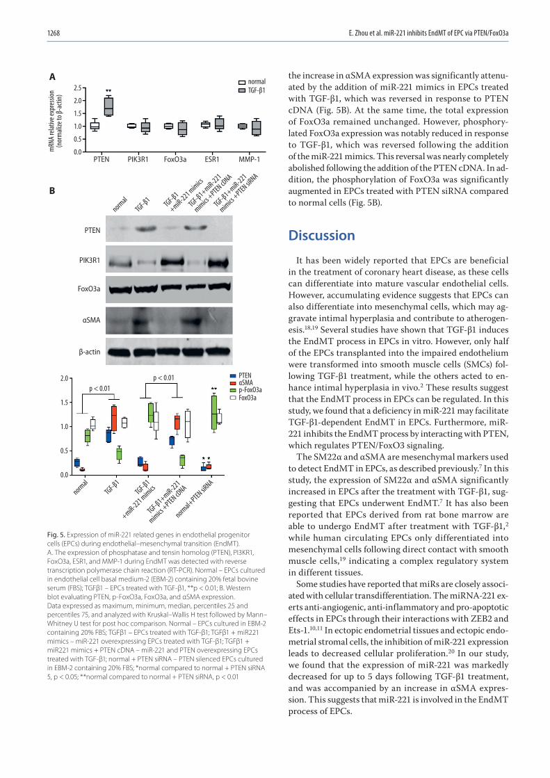
E. Zhou et al. miR-221 inhibits EndMT of EPC via PTEN/FoxO3a1268
the increase in αSMA expression was significantly attenu-ated by the addition of miR-221 mimics in EPCs treated with TGF-β1, which was reversed in response to PTEN cDNA (Fig. 5B). At the same time, the total expression of FoxO3a remained unchanged. However, phosphory-lated FoxO3a expression was notably reduced in response to TGF-β1, which was reversed following the addition of the miR-221 mimics. This reversal was nearly completely abolished following the addition of the PTEN cDNA. In ad-dition, the phosphorylation of FoxO3a was significantly augmented in EPCs treated with PTEN siRNA compared to normal cells (Fig. 5B).
Discussion
It has been widely reported that EPCs are beneficial in the treatment of coronary heart disease, as these cells can differentiate into mature vascular endothelial cells. However, accumulating evidence suggests that EPCs can also differentiate into mesenchymal cells, which may ag-gravate intimal hyperplasia and contribute to atherogen-esis.18,19 Several studies have shown that TGF-β1 induces the EndMT process in EPCs in vitro. However, only half of the EPCs transplanted into the impaired endothelium were transformed into smooth muscle cells (SMCs) fol-lowing TGF-β1 treatment, while the others acted to en-hance intimal hyperplasia in vivo.2 These results suggest that the EndMT process in EPCs can be regulated. In this study, we found that a deficiency in miR-221 may facilitate TGF-β1-dependent EndMT in EPCs. Furthermore, miR-221 inhibits the EndMT process by interacting with PTEN, which regulates PTEN/FoxO3 signaling.
The SM22α and αSMA are mesenchymal markers used to detect EndMT in EPCs, as described previously.7 In this study, the expression of SM22α and αSMA significantly increased in EPCs after the treatment with TGF-β1, sug-gesting that EPCs underwent EndMT.7 It has also been reported that EPCs derived from rat bone marrow are able to undergo EndMT after treatment with TGF-β1,2 while human circulating EPCs only differentiated into mesenchymal cells following direct contact with smooth muscle cells,19 indicating a complex regulatory system in different tissues.
Some studies have reported that miRs are closely associ-ated with cellular transdifferentiation. The miRNA-221 ex-erts anti-angiogenic, anti-inflammatory and pro-apoptotic effects in EPCs through their interactions with ZEB2 and Ets-1.10,11 In ectopic endometrial tissues and ectopic endo-metrial stromal cells, the inhibition of miR-221 expression leads to decreased cellular proliferation.20 In our study, we found that the expression of miR-221 was markedly decreased for up to 5 days following TGF-β1 treatment, and was accompanied by an increase in αSMA expres-sion. This suggests that miR-221 is involved in the EndMT process of EPCs.
Fig. 5. Expression of miR-221 related genes in endothelial progenitor cells (EPCs) during endothelial–mesenchymal transition (EndMT). A. The expression of phosphatase and tensin homolog (PTEN), PI3KR1, FoxO3a, ESR1, and MMP-1 during EndMT was detected with reverse transcription polymerase chain reaction (RT-PCR). Normal – EPCs cultured in endothelial cell basal medium-2 (EBM-2) containing 20% fetal bovine serum (FBS); TGFβ1 – EPCs treated with TGF-β1, **p < 0.01; B. Western blot evaluating PTEN, p-FoxO3a, FoxO3a, and αSMA expression. Data expressed as maximum, minimum, median, percentiles 25 and percentiles 75, and analyzed with Kruskal–Wallis H test followed by Mann–Whitney U test for post hoc comparison. Normal – EPCs cultured in EBM-2 containing 20% FBS; TGFβ1 – EPCs treated with TGF-β1; TGFβ1 + miR221 mimics – miR-221 overexpressing EPCs treated with TGF-β1; TGFβ1 + miR221 mimics + PTEN cDNA – miR-221 and PTEN overexpressing EPCs treated with TGF-β1; normal + PTEN siRNA – PTEN silenced EPCs cultured in EBM-2 containing 20% FBS; *normal compared to normal + PTEN siRNA 5, p < 0.05; **normal compared to normal + PTEN siRNA, p < 0.01
PTEN PIK3R1 FoxO3a
PTEN
PIK3R1
FoxO3a
αSMA
PTENp < 0.01p < 0.01
FoxO3ap-FoxO3aαSMA
β-actin
ESR1 MMP-1
TGF-β1
+miR-221 mimics
TGF-β1+miR-221
mimics +PTEN cD
NA
TGF-β1+miR-221
mimics +PTEN siR
NA
TGF-β1norm
al
TGF-β1
+miR-221 mimics
TGF-β1+miR-221
mimics +PTEN cD
NA
normal+PTEN siR
NATGF-β1
normal
normalTGF-β12.5
2.0
1.5
1.0
0.5
0.0
2.0
1.5
1.0
0.5
0.0
mRN
A rela
tive e
xpre
ssion
(nor
mali
ze to
β-ac
tin)
A
B
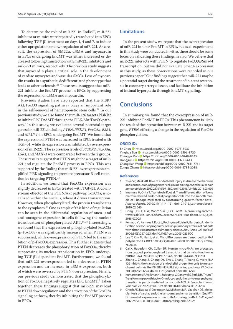
Adv Clin Exp Med. 2021;30(12):1263–1270 1269
To determine the role of miR-221 in EndMT, miR-221 inhibitor or mimics were repeatedly transfected into EPCs following TGF-β1 treatment on days 1, 4 and 7, to induce either upregulation or downregulation of miR-221. As a re-sult, the expression of SM22α, αSMA and myocardin in EPCs undergoing EndMT was either increased or de-creased following transfection with miR-221 inhibitors and miR-221 mimics, respectively. The previous study suggests that myocardin plays a critical role in the development of cardiac myocytes and vascular SMCs. Loss of myocar-din results in a synthetic, dedifferentiated phenotype that leads to atherosclerosis.21 These results suggest that miR-221 inhibits the EndMT process in EPCs by suppressing the expression of αSMA and myocardin.
Previous studies have also reported that the PI3K/Akt/FoxO3 signaling pathway plays an important role in the self-renewal of hematopoietic stem cells.22 In our previous study, we also found that miR-126 targets PI3KR2 to inhibit EPC EndMT through the PI3K/Akt/FoxO3 path-way.7 In this study, we evaluated several potential target genes for miR-221, including PTEN, PI3KR1, FoxO3a, ESR1, and MMP-1, in EPCs undergoing EndMT. We found that the expression of PTEN was increased in EPCs treated with TGF-β1, while its expression was inhibited by overexpres-sion of miR-221. The expression levels of PI3KR2, FoxO3a, ESR1, and MMP-1 were comparable between the 2 groups. These results suggest that PTEN might be a target of miR-221 and regulate the EndMT process in EPCs. This was supported by the finding that miR-221 overexpression am-plified PI3K signaling to promote precursor B-cell reten-tion by targeting PTEN.23
In addition, we found that FoxO3a expression was slightly decreased in EPCs treated with TGF-β1. A down-stream effector of the PTEN/Akt pathway, FoxO3a, is lo-calized within the nucleus, where it drives transcription. However, when phosphorylated, the protein translocates to the cytoplasm.24 One example of this kind of regulation can be seen in the differential regulation of onco- and anti-oncogene expression in cells following the nuclear translocation of phosphorylated AKT.14,25 Interestingly, we found that the expression of phosphorylated FoxO3a (p-FoxO3a) was significantly increased when PTEN was suppressed, while overexpression of PTEN led to the inhi-bition of p-FoxO3a expression. This further suggests that PTEN decreases the phosphorylation of FoxO3a, thereby suppressing its nuclear translocation in EPCs undergo-ing TGF-β1-dependent EndMT. Furthermore, we found that miR-221 overexpression led to a decrease in PTEN expression and an increase in αSMA expression, both of which were reversed by PTEN overexpression. Finally, our previous study demonstrated that the phosphoryla-tion of FoxO3a negatively regulates EPC EndMT.7 Taken together, these findings suggest that miR-221 may lead to PTEN downregulation and the activation of the FoxO3a signaling pathway, thereby inhibiting the EndMT process in EPCs.
Limitations
In the present study, we report that the overexpression of miR-221 inhibits EndMT in EPCs, but as all experiments in this study were conducted in vitro, there should be some focus on validating these findings in vivo. We believe that miR-221 interacts with PTEN to regulate FoxO3a/Smad4 transcription, but we did not evaluate Smad4 expression in this study, as these observations were recorded in our previous paper.7 Our findings suggest that miR-221 may be a potential target during the treatment of in-stent resteno-sis in coronary artery disease, and facilitate the inhibition of intimal hyperplasia through EndMT signaling.
Conclusions
In summary, we found that the overexpression of miR-221 inhibited EndMT in EPCs. This phenomenon is likely the result of the interaction between miR-221 and its target gene, PTEN, effecting a change in the regulation of FoxO3a phosphorylation.
ORCID iDsEn Zhou https://orcid.org/0000-0002-6073-8037Yinghua Zou https://orcid.org/0000-0002-6596-8720Chengyu Mao https://orcid.org/0000-0001-9740-8835Dongjiu Li https://orcid.org/0000-0003-4372-6613Changqian Wang https://orcid.org/0000-0002-7611-7761Zongqi Zhang https://orcid.org/0000-0001-6785-2038
References1. Toya SP, Malik AB. Role of endothelial injury in disease mechanisms
and contribution of progenitor cells in mediating endothelial repair. Immunobiology. 2012;217(5):569–580. doi:10.1016/j.imbio.2011.03.006
2. Imamura H, Ohta T, Tsunetoshi K, et al. Transdifferentiation of bone marrow-derived endothelial progenitor cells into the smooth mus-cle cell lineage mediated by tansforming growth factor-beta1. Atherosclerosis. 2010;211(1):114–121. doi:10.1016/j.atherosclerosis. 2010.02.040
3. Hong L, Du X, Li W, Mao Y, Sun L, Li X. EndMT: A promising and con-troversial field. Eur J Cell Biol. 2018;97(7):493–500. doi:10.1016/j.ejcb. 2018.07.005
4. Peinado VI, Ramírez J, Roca J, Rodriguez-Roisin R, Barberà JA. Identi-fication of vascular progenitor cells in pulmonary arteries of patients with chronic obstructive pulmonary disease. Am J Respir Cell Mol Biol. 2006;34(3):257–263. doi:10.1165/rcmb.2005-0255OC
5. Lee Y, Kim M, Han J, et al. MicroRNA genes are transcribed by RNA polymerase II. EMBO J. 2004;23(20):4051–4060. doi:10.1038/sj.emboj. 7600385
6. Cai X, Hagedorn CH, Cullen BR. Human microRNAs are processed from capped, polyadenylated transcripts that can also function as mRNAs. RNA. 2004;10(12):1957–1966. doi:10.1261/rna.7135204
7. Zhang J, Zhang Z, Zhang DY, Zhu J, Zhang T, Wang C. microRNA 126 inhibits the transition of endothelial progenitor cells to mesen-chymal cells via the PIK3R2-PI3K/Akt signalling pathway. PloS One. 2013;8(12):e83294. doi:10.1371/journal.pone.0083294
8. Kumarswamy R, Volkmann I, Jazbutyte V, Dangwal S, Park DH, Thum T. Transforming growth factor-β-induced endothelial-to-mesenchymal transition is partly mediated by microRNA-21. Arterioscler Thromb Vasc Biol. 2012;32(2):361–369. doi:10.1161/atvbaha.111.234286
9. Ghosh AK, Nagpal V, Covington JW, Michaels MA, Vaughan DE. Molec-ular basis of cardiac endothelial-to-mesenchymal transition (EndMT): Differential expression of microRNAs during EndMT. Cell Signal. 2012;24(5):1031–1036. doi:10.1016/j.cellsig.2011.12.024

E. Zhou et al. miR-221 inhibits EndMT of EPC via PTEN/FoxO3a1270
10. Chen Y, Banda M, Speyer CL, Smith JS, Rabson AB, Gorski DH. Regu-lation of the expression and activity of the antiangiogenic homeo-box gene GAX/MEOX2 by ZEB2 and microRNA-221. Mol Cell Biol. 2010; 30(15):3902–3913. doi:10.1128/mcb.01237-09
11. Zhu N, Zhang D, Chen S, et al. Endothelial enriched microRNAs regu-late angiotensin II-induced endothelial inflammation and migration. Atherosclerosis. 2011;215(2):286–293. doi:10.1016/j.atherosclerosis. 2010.12.024
12. Ni HZ, Liu Z, Sun LL, et al. Metformin inhibits angiogenesis of endo-thelial progenitor cells via miR-221-mediated p27 expression and autophagy. Future Med Chem. 2019;11(17):2263–2272. doi:10.4155/fmc-2019-0017
13. Peng H, Yang H, Xiang X, Li S. ΜicroRNA-221 participates in cerebral ischemic stroke by modulating endothelial cell function by regulat-ing the PTEN/PI3K/AKT pathway. Exp Ther Med. 2020;19(1):443–450. doi:10.3892/etm.2019.8263
14. Zhang Q, Li X, Li Y, et al. Expression of the PTEN/FOXO3a/PLZF signal-ling pathway in pancreatic cancer and its significance in tumourigen-esis and progression. Invest New Drugs. 2020;38(2):321–328. doi:10. 1007/s10637-019-00791-7
15. Mziaut H, Henniger G, Ganss K, et al. MiR-132 controls pancreatic beta cell proliferation and survival through Pten/Akt/Foxo3 signal-ing. Mol Metab. 2020;31:150–162. doi:10.1016/j.molmet.2019.11.012
16. Zhang Z, Zhang T, Zhou Y, et al. Activated phosphatidylinositol 3-kinase/Akt inhibits the transition of endothelial progenitor cells to mesenchymal cells by regulating the forkhead box subgroup O-3a signaling. Cell Physiol Biochem. 2015;35(4):1643–1653. doi:10.1159/ 000373978
17. Moonen JR, Krenning G, Brinker MG, Koerts JA, van Luyn MJ, Harmsen MC. Endothelial progenitor cells give rise to pro-angiogenic smooth mus-cle-like progeny. Cardiovasc Res. 2010;86(3):506–515. doi:10.1093/cvr/cvq012
18. Rotmans JI, Heyligers JM, Verhagen HJ, et al. In vivo cell seeding with anti-CD34 antibodies successfully accelerates endothelializa-tion but stimulates intimal hyperplasia in porcine arteriovenous expanded polytetrafluoroethylene grafts. Circulation. 2005;112(1): 12–18. doi:10.1161/CIRCULATIONAHA.104.504407
19. Diez M, Musri MM, Ferrer E, Barbera JA, Peinado VI. Endothelial pro-genitor cells undergo an endothelial-to-mesenchymal transition-like process mediated by TGFbetaRI. Cardiovasc Res. 2010;88(3):502–511. doi:10.1093/cvr/cvq236
20. Du XH, Shi G, Lü DH, et al. The expression of microRNA-221 in endo-metriosis and its impact on endometrial stromal cells [in Chinese]. Sichuan Da Xue Xue Bao Yi Xue Ban. 2018;49(4):546–550. PMID:30378307
21. Xia XD, Zhou Z, Yu XH, Zheng XL, Tang CK. Myocardin: A novel player in atherosclerosis. Atherosclerosis. 2017;257:266–278. doi:10.1016/j.atherosclerosis.2016.12.002
22. Miyamoto K, Araki KY, Naka K, et al. Foxo3a is essential for mainte-nance of the hematopoietic stem cell pool. Cell Stem Cell. 2007;1(1): 101–112. doi:10.1016/j.stem.2007.02.001
23. Petkau G, Kawano Y, Wolf I, Knoll M, Melchers F. MiR221 promotes pre-cursor B-cell retention in the bone marrow by amplifying the PI3K-signaling pathway in mice. Eur J Immunol. 2018;48(6):975–989. doi:10. 1002/eji.201747354
24. Wang K, Li PF. Foxo3a regulates apoptosis by negatively targeting miR-21. J Biol Chem. 2010;285(22):16958–16966. doi:10.1074/jbc.M109. 093005
25. Jang H, Lee OH, Lee Y, et al. Melatonin prevents cisplatin-induced primordial follicle loss via suppression of PTEN/AKT/FOXO3a path-way activation in the mouse ovary. J Pineal Res. 2016;60(3):336–347. doi:10.1111/jpi.12316
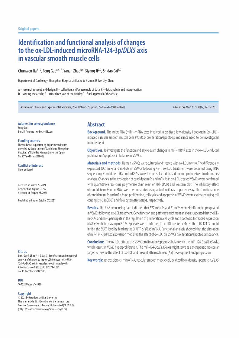
Cite asJia C, Gao F, Zhao Y, Ji S, Cai S. Identification and functional analysis of changes to the ox-LDL-induced microRNA- 124-3p/DLX5 axis in vascular smooth muscle cells. Adv Clin Exp Med. 2021;30(12):1271–1281. doi:10.17219/acem/141580
DOI10.17219/acem/141580
Copyright© 2021 by Wroclaw Medical University This is an article distributed under the terms of theCreative Commons Attribution 3.0 Unported (CC BY 3.0)(https://creativecommons.org/licenses/by/3.0/)
Address for correspondenceFeng GaoE-mail: [email protected]
Funding sourcesThe study was supported by departmental funds provided by Department of Cardiology, Zhongshan Hospital, affiliated to Xiamen University (grant No. ZSYY-XN-res-201806).
Conflict of interestNone declared
Received on March 25, 2021Reviewed on August 17, 2021Accepted on August 25, 2021
Published online on October 27, 2021
AbstractBackground. The microRNA (miR)–mRNA axes involved in oxidized low-density lipoprotein (ox-LDL)-induced vascular smooth muscle cells (VSMCs) proliferation/apoptosis imbalance need to be investigated in more detail.
Objectives. To investigate the function and any relevant changes to miR–mRNA axes in the ox-LDL-induced proliferation/apoptosis imbalance in VSMCs.
Materials and methods. Human VSMCs were cultured and treated with ox-LDL in vitro. The differentially expressed (DE) miRs and mRNAs in VSMCs following 48-h ox-LDL treatment were detected using RNA sequencing. Candidate miRs and mRNAs were further selected, based on comprehensive bioinformatics analysis. Changes in the expression of candidate miRs and mRNAs in ox-LDL-treated VSMCs were confirmed with quantitative real-time polymerase chain reaction (RT-qPCR) and western blot. The inhibitory effect of candidate miRs on mRNAs were demonstrated using a dual luciferase reporter assay. The functional role of candidate miRs and mRNAs on proliferation, cell cycle and apoptosis of VSMCs were estimated using cell couting kit-8 (CCK-8) and flow cytometry assays, respectively.
Results. The RNA sequencing data indicated that 577 mRNAs and 81 miRs were significantly upregulated in VSMCs following ox-LDL treatment. Gene function and pathway enrichment analysis suggested that the DE-mRNAs and miRs participate in the regulation of proliferation, cell cycle and apoptosis. Increased expression of DLX5 with decreasing miR-124-3p levels were confirmed in ox-LDL-treated VSMCs. The miR-124-3p could inhibit the DLX5 level by binding the 3’ UTR of DLX5 mRNA. Functional analysis showed that the alteration of miR-124-3p/DLX5 expression mediated the effect of ox-LDL on VSMCs proliferation/apoptosis imbalance.
Conclusions. The ox-LDL affects the VSMC proliferation/apoptosis balance via the miR-124-3p/DLX5 axis, which results in VSMC hyperproliferation. The miR-124-3p/DLX5 axis might serve as a therapeutic molecular target to reverse the effect of ox-LDL and prevent atherosclerosis (AS) development and progression.
Key words: atherosclerosis, microRNA, vascular smooth muscle cell, oxidized low-density lipoprotein, DLX5
Original papers
Identification and functional analysis of changes to the ox-LDL-induced microRNA-124-3p/DLX5 axis in vascular smooth muscle cells
Chunwen JiaA–D, Feng GaoA,C–F, Yanan ZhaoB,C, Siyang JiC,D, Shidao CaiB,D
Department of Cardiology, Zhongshan Hospital affiliated to Xiamen University, China
A – research concept and design; B – collection and/or assembly of data; C – data analysis and interpretation; D – writing the article; E – critical revision of the article; F – final approval of the article
Advances in Clinical and Experimental Medicine, ISSN 1899–5276 (print), ISSN 2451–2680 (online) Adv Clin Exp Med. 2021;30(12):1271–1281

C. Jia et al. miR-124-3p/DLX5 axis in VSMCs1272
Background
Atherosclerosis (AS) is a vascular disease characterized by stiff, thickened artery walls with reduced elasticity, to-gether with the formation of atherosclerotic plaques within the intimal layer.1 Its development and progression involves vascular endothelial cell damage and dysfunction, inflam-matory cell recruitment, lipid deposition, and thrombo-sis, in addition to abnormal vascular smooth muscle cell (VSMC) proliferation.2 Excessive proliferation of VSMCs causes an inflammatory response and thickening of vessel walls, both of which facilitate lipoprotein deposition and fibrous cap formation.3
Elevated low-density lipoprotein (LDL) is a major risk factor for AS. Low-density lipoprotein comprises a core (mainly triglycerides and cholesterol lipids) and a sur-face rich in polyunsaturated fatty acids.4 Therefore, LDL is easily oxidized and modified by stimulatory factors such as cigarette smoke, hyperglycemia and diabetes.5 Oxidized LDL (ox-LDL) is even more potent in inducing AS. It is able to damage vascular endothelial cells, causing dysfunction and increased permeability, which initiates AS.6 Further-more, ox-LDL intake upregulates the transition of mac-rophages to foam cells within plaques. Previous studies have reported that ox-LDL promotes excessive prolifera-tion of VSMCs by promoting endothelin-1 secretion and inhibiting prostacyclin synthesis.7 Some of the ox-LDL-induced cytokines involved in the regulation of VSMC pro-liferation include platelet-derived growth factor (PDGF), basic fibroblast growth factor (bFGF) and transforming growth factor beta (TGF-β).8 Reversing ox-LDL-induced VSMC hyperproliferation may represent a new strategy in AS treatment. However, the mechanisms underlying ox-LDL-induced regulation of VSMC proliferation need to be explored prior to the clinical application of this strategy.9
Recent studies investigating the regulatory mechanisms of microRNAs (miRs) have shown that miRs play an impor-tant regulatory role in ox-LDL-induced VSMC prolifera-tion, and several potential miR therapeutic targets have been identified there. Sun et al. reported that miR-490-3p is associated with increased ox-LDL-induced VSMC prolif-eration through the regulation of its target gene, PAPP-A. The Ox-LDL inhibits miR-490-3p expression, and the up-regulated miR-490-3p reduces ox-LDL-induced abnor-mal VSMC proliferation by inhibiting PAPP-A.10 Further-more, Wang et al. identified another ox-LDL-induced miR, miR-195. They found that ox-LDL inhibits miR-195 levels in VSMCs and that miR-195 inhibits VSMC proliferation and migration.11
Objectives
In this study, we aimed to analyze RNA sequencing data from ox-LDL-treated VSMCs to find novel miR–messenger RNA (mRNA) axes involved in ox-LDL-induced VSMC
proliferation. We selected differentially expressed (DE) miRs and mRNAs in ox-LDL-treated cells and conducted functional and bioinformatics analyses on these genes. We selected the miR-124-3p/DLX5 axis as one potentially related to VSMC proliferation, which was confirmed fol-lowing ox-LDL treatment. Considering that ox-LDL pro-motes VSMC proliferation by breaking the cell prolifera-tion/apoptosis balance, we further explored the regulatory function of the miR-124-3p/DLX5 axis in VSMC prolifera-tion and apoptosis.
Materials and methods
Cell culture and ox-LDL treatment
Human aorta smooth muscle cells (HA-VSMC, prod-uct code: CRL-1999) were obtained from the American Type Culture Collection (ATCC, Manassas, USA) and cultured using F-12K media (Thermo Fisher Scien-tific, Waltham, USA) in a humidified incubator at 5% CO2 and 37°C. To make the complete growth medium, the following components were provided by Procell Life Science&Technology (Wuhan, China) and added to the base medium: 0.05 mg/mL of ascorbic acid; 0.01 mg/mL of insulin; 0.01 mg/mL of transferrin; 10 ng/mL of sodium selenite; 0.03 mg/mL of Endothelial Cell Growth Supplement (ECGS); fetal bovine serum (FBS; Thermo Fisher Scientific) to a final concentration of 10%, HEPES to a final concentration of 10 mM, and TES to a fi-nal concentration of 10 mM. Antibiotics were not added into the medium. The HA-VSMCs in their 5th passage were treated with 100 μg/mL of ox-LDL (Thermo Fisher Scien-tific) for 24 h before further experiments.
No approval was required from bioethical committee due to the fact that no human or animals were enrolled in the present study.
RNA sequencing and data analysis
After 48 h of exposure to ox-LDL, cells were collected, and samples sent for RNA sequencing (BGI Technology Service, Shenzheng, China). The DE-mRNAs and miRs were analyzed using theDESeq2 package in R software v. 3.4.1 (R Foundation for Statistical Computing, Vienna, Austria). The DE-genes are defined as genes with a fold-change of expression >2.0 and p < 0.05.
Bioinformatic analysis
To explore the potential biological function of DE mRNA and miRs, further functional and signaling path-way analyses were performed. Gene Ontology (GO) gene enrichment analysis and Kyoto Encyclopedia of Genes and Genomes (KEGG) signaling pathway enrichment analyses were performed on a set of DE-mRNAs using
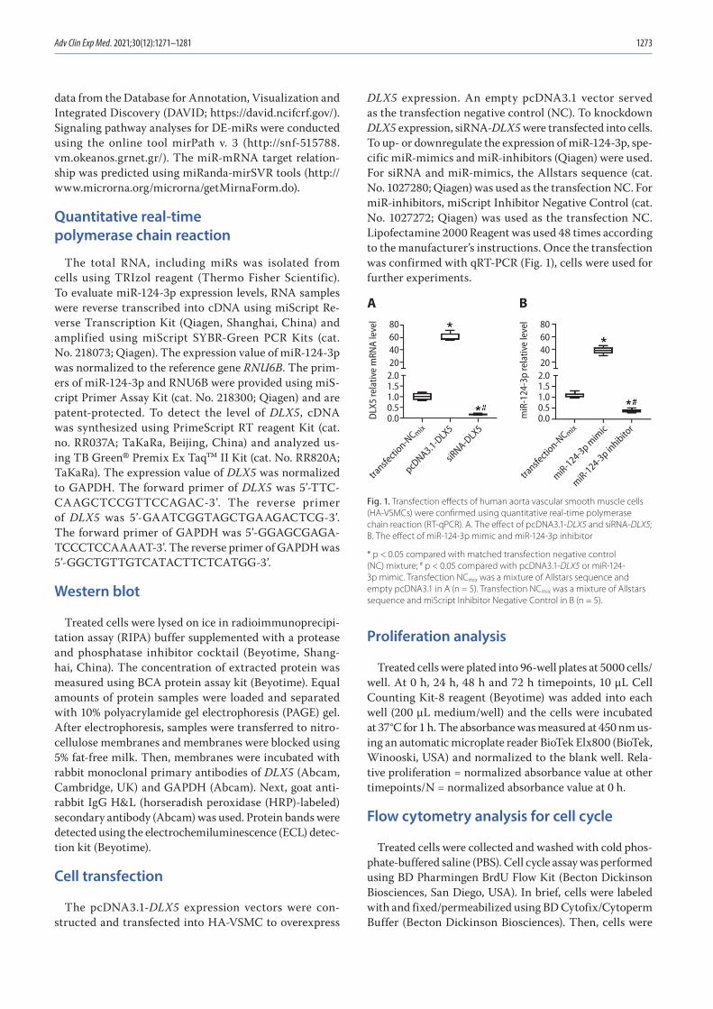
Adv Clin Exp Med. 2021;30(12):1271–1281 1273
data from the Database for Annotation, Visualization and Integrated Discovery (DAVID; https://david.ncifcrf.gov/). Signaling pathway analyses for DE-miRs were conducted using the online tool mirPath v. 3 (http://snf-515788.vm.okeanos.grnet.gr/). The miR-mRNA target relation-ship was predicted using miRanda-mirSVR tools (http://www.microrna.org/microrna/getMirnaForm.do).
Quantitative real-time polymerase chain reaction
The total RNA, including miRs was isolated from cells using TRIzol reagent (Thermo Fisher Scientific). To evaluate miR-124-3p expression levels, RNA samples were reverse transcribed into cDNA using miScript Re-verse Transcription Kit (Qiagen, Shanghai, China) and amplified using miScript SYBR-Green PCR Kits (cat. No. 218073; Qiagen). The expression value of miR-124-3p was normalized to the reference gene RNU6B. The prim-ers of miR-124-3p and RNU6B were provided using miS-cript Primer Assay Kit (cat. No. 218300; Qiagen) and are patent-protected. To detect the level of DLX5, cDNA was synthesized using PrimeScript RT reagent Kit (cat. no. RR037A; TaKaRa, Beijing, China) and analyzed us-ing TB Green® Premix Ex Taq™ II Kit (cat. No. RR820A; TaKaRa). The expression value of DLX5 was normalized to GAPDH. The forward primer of DLX5 was 5’-TTC-CAAGCTCCGTTCCAGAC-3’. The reverse primer of DLX5 was 5’-GAATCGGTAGCTGAAGACTCG-3’. The forward primer of GAPDH was 5’-GGAGCGAGA-TCCCTCCAAAAT-3’. The reverse primer of GAPDH was 5’-GGCTGTTGTCATACTTCTCATGG-3’.
Western blot
Treated cells were lysed on ice in radioimmunoprecipi-tation assay (RIPA) buffer supplemented with a protease and phosphatase inhibitor cocktail (Beyotime, Shang-hai, China). The concentration of extracted protein was measured using BCA protein assay kit (Beyotime). Equal amounts of protein samples were loaded and separated with 10% polyacrylamide gel electrophoresis (PAGE) gel. After electrophoresis, samples were transferred to nitro-cellulose membranes and membranes were blocked using 5% fat-free milk. Then, membranes were incubated with rabbit monoclonal primary antibodies of DLX5 (Abcam, Cambridge, UK) and GAPDH (Abcam). Next, goat anti-rabbit IgG H&L (horseradish peroxidase (HRP)-labeled) secondary antibody (Abcam) was used. Protein bands were detected using the electrochemiluminescence (ECL) detec-tion kit (Beyotime).
Cell transfection
The pcDNA3.1-DLX5 expression vectors were con-structed and transfected into HA-VSMC to overexpress
DLX5 expression. An empty pcDNA3.1 vector served as the transfection negative control (NC). To knockdown DLX5 expression, siRNA-DLX5 were transfected into cells. To up- or downregulate the expression of miR-124-3p, spe-cific miR-mimics and miR-inhibitors (Qiagen) were used. For siRNA and miR-mimics, the Allstars sequence (cat. No. 1027280; Qiagen) was used as the transfection NC. For miR-inhibitors, miScript Inhibitor Negative Control (cat. No. 1027272; Qiagen) was used as the transfection NC. Lipofectamine 2000 Reagent was used 48 times according to the manufacturer’s instructions. Once the transfection was confirmed with qRT-PCR (Fig. 1), cells were used for further experiments.
Proliferation analysis
Treated cells were plated into 96-well plates at 5000 cells/well. At 0 h, 24 h, 48 h and 72 h timepoints, 10 μL Cell Counting Kit-8 reagent (Beyotime) was added into each well (200 μL medium/well) and the cells were incubated at 37°C for 1 h. The absorbance was measured at 450 nm us-ing an automatic microplate reader BioTek Elx800 (BioTek, Winooski, USA) and normalized to the blank well. Rela-tive proliferation = normalized absorbance value at other timepoints/N = normalized absorbance value at 0 h.
Flow cytometry analysis for cell cycle
Treated cells were collected and washed with cold phos-phate-buffered saline (PBS). Cell cycle assay was performed using BD Pharmingen BrdU Flow Kit (Becton Dickinson Biosciences, San Diego, USA). In brief, cells were labeled with and fixed/permeabilized using BD Cytofix/Cytoperm Buffer (Becton Dickinson Biosciences). Then, cells were
Fig. 1. Transfection effects of human aorta vascular smooth muscle cells (HA-VSMCs) were confirmed using quantitative real-time polymerase chain reaction (RT-qPCR). A. The effect of pcDNA3.1-DLX5 and siRNA-DLX5; B. The effect of miR-124-3p mimic and miR-124-3p inhibitor
* p < 0.05 compared with matched transfection negative control (NC) mixture; # p < 0.05 compared with pcDNA3.1-DLX5 or miR-124-3p mimic. Transfection NCmix was a mixture of Allstars sequence and empty pcDNA3.1 in A (n = 5). Transfection NCmix was a mixture of Allstars sequence and miScript Inhibitor Negative Control in B (n = 5).
miR-124-3p mim
ic
miR-124-3p inhibito
r
transfe
ction-N
C mix
transfe
ction-N
C mix
pcDNA3.1-D
LX5
siRNA-D
LX5
80604020
2.01.51.00.50.0D
LX5
rela
tive
mRN
A le
vel
A80604020
2.01.51.00.50.0m
iR-1
24-3
p re
lativ
e le
vel
B
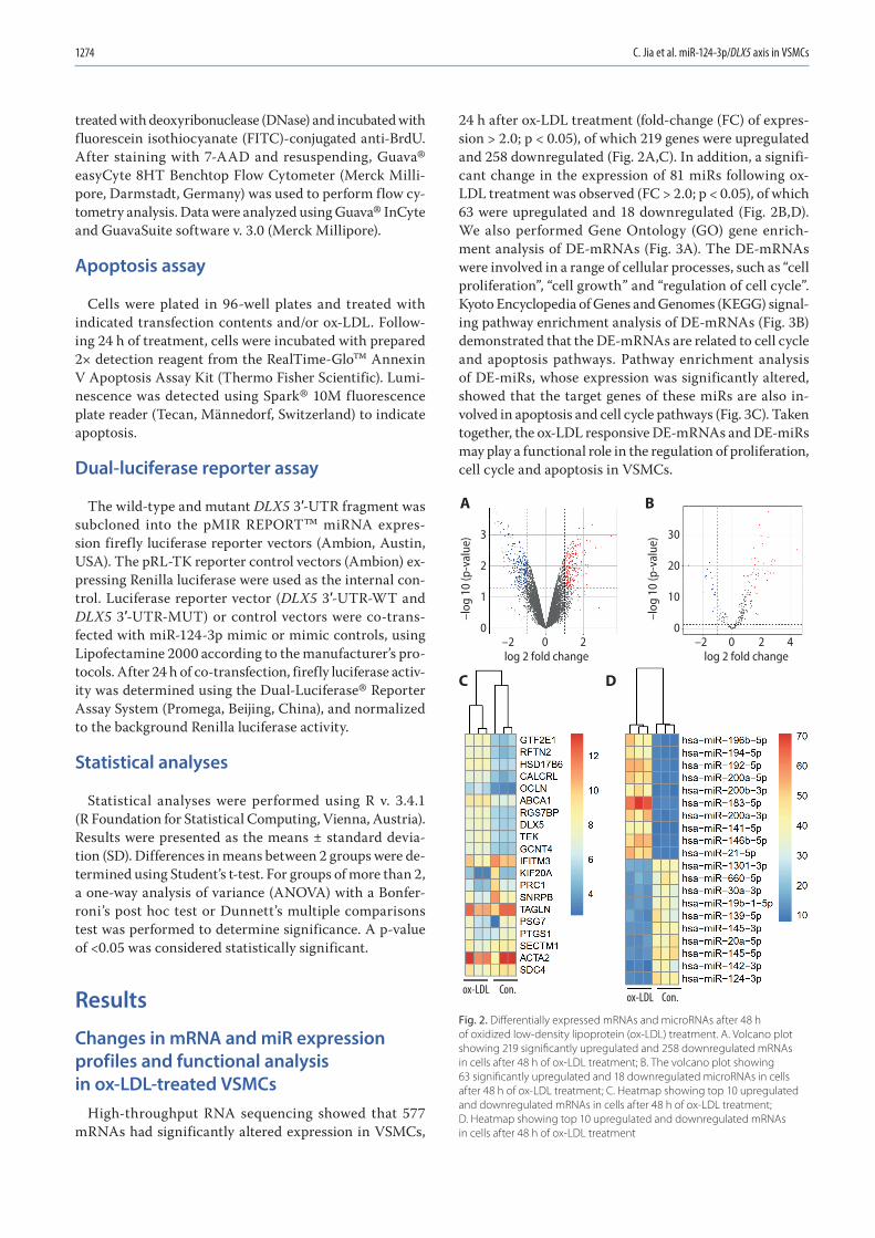
C. Jia et al. miR-124-3p/DLX5 axis in VSMCs1274
treated with deoxyribonuclease (DNase) and incubated with fluorescein isothiocyanate (FITC)-conjugated anti-BrdU. After staining with 7-AAD and resuspending, Guava® easyCyte 8HT Benchtop Flow Cytometer (Merck Milli-pore, Darmstadt, Germany) was used to perform flow cy-tometry analysis. Data were analyzed using Guava® InCyte and GuavaSuite software v. 3.0 (Merck Millipore).
Apoptosis assay
Cells were plated in 96-well plates and treated with indicated transfection contents and/or ox-LDL. Follow-ing 24 h of treatment, cells were incubated with prepared 2× detection reagent from the RealTime-Glo™ Annexin V Apoptosis Assay Kit (Thermo Fisher Scientific). Lumi-nescence was detected using Spark® 10M fluorescence plate reader (Tecan, Männedorf, Switzerland) to indicate apoptosis.
Dual-luciferase reporter assay
The wild-type and mutant DLX5 3′-UTR fragment was subcloned into the pMIR REPORT™ miRNA expres-sion firefly luciferase reporter vectors (Ambion, Austin, USA). The pRL-TK reporter control vectors (Ambion) ex-pressing Renilla luciferase were used as the internal con-trol. Luciferase reporter vector (DLX5 3′-UTR-WT and DLX5 3′-UTR-MUT) or control vectors were co-trans-fected with miR-124-3p mimic or mimic controls, using Lipofectamine 2000 according to the manufacturer’s pro-tocols. After 24 h of co-transfection, firefly luciferase activ-ity was determined using the Dual-Luciferase® Reporter Assay System (Promega, Beijing, China), and normalized to the background Renilla luciferase activity.
Statistical analyses
Statistical analyses were performed using R v. 3.4.1 (R Foundation for Statistical Computing, Vienna, Austria). Results were presented as the means ± standard devia-tion (SD). Differences in means between 2 groups were de-termined using Student’s t-test. For groups of more than 2, a one-way analysis of variance (ANOVA) with a Bonfer-roni’s post hoc test or Dunnett’s multiple comparisons test was performed to determine significance. A p-value of <0.05 was considered statistically significant.
Results
Changes in mRNA and miR expression profiles and functional analysis in ox-LDL-treated VSMCs
High-throughput RNA sequencing showed that 577 mRNAs had significantly altered expression in VSMCs,
24 h after ox-LDL treatment (fold-change (FC) of expres-sion > 2.0; p < 0.05), of which 219 genes were upregulated and 258 downregulated (Fig. 2A,C). In addition, a signifi-cant change in the expression of 81 miRs following ox-LDL treatment was observed (FC > 2.0; p < 0.05), of which 63 were upregulated and 18 downregulated (Fig. 2B,D). We also performed Gene Ontology (GO) gene enrich-ment analysis of DE-mRNAs (Fig. 3A). The DE-mRNAs were involved in a range of cellular processes, such as “cell proliferation”, “cell growth” and “regulation of cell cycle”. Kyoto Encyclopedia of Genes and Genomes (KEGG) signal-ing pathway enrichment analysis of DE-mRNAs (Fig. 3B) demonstrated that the DE-mRNAs are related to cell cycle and apoptosis pathways. Pathway enrichment analysis of DE-miRs, whose expression was significantly altered, showed that the target genes of these miRs are also in-volved in apoptosis and cell cycle pathways (Fig. 3C). Taken together, the ox-LDL responsive DE-mRNAs and DE-miRs may play a functional role in the regulation of proliferation, cell cycle and apoptosis in VSMCs.
Fig. 2. Differentially expressed mRNAs and microRNAs after 48 h of oxidized low-density lipoprotein (ox-LDL) treatment. A. Volcano plot showing 219 significantly upregulated and 258 downregulated mRNAs in cells after 48 h of ox-LDL treatment; B. The volcano plot showing 63 significantly upregulated and 18 downregulated microRNAs in cells after 48 h of ox-LDL treatment; C. Heatmap showing top 10 upregulated and downregulated mRNAs in cells after 48 h of ox-LDL treatment; D. Heatmap showing top 10 upregulated and downregulated mRNAs in cells after 48 h of ox-LDL treatment
log 2 fold change
3
2
1
0
30
20
10
0
log 2 fold change
ox-LDL Con.ox-LDL Con.
0 2–2 0 2 4–2
–log
10
(p-v
alue
)
–log
10
(p-v
alue
)
A B
C D
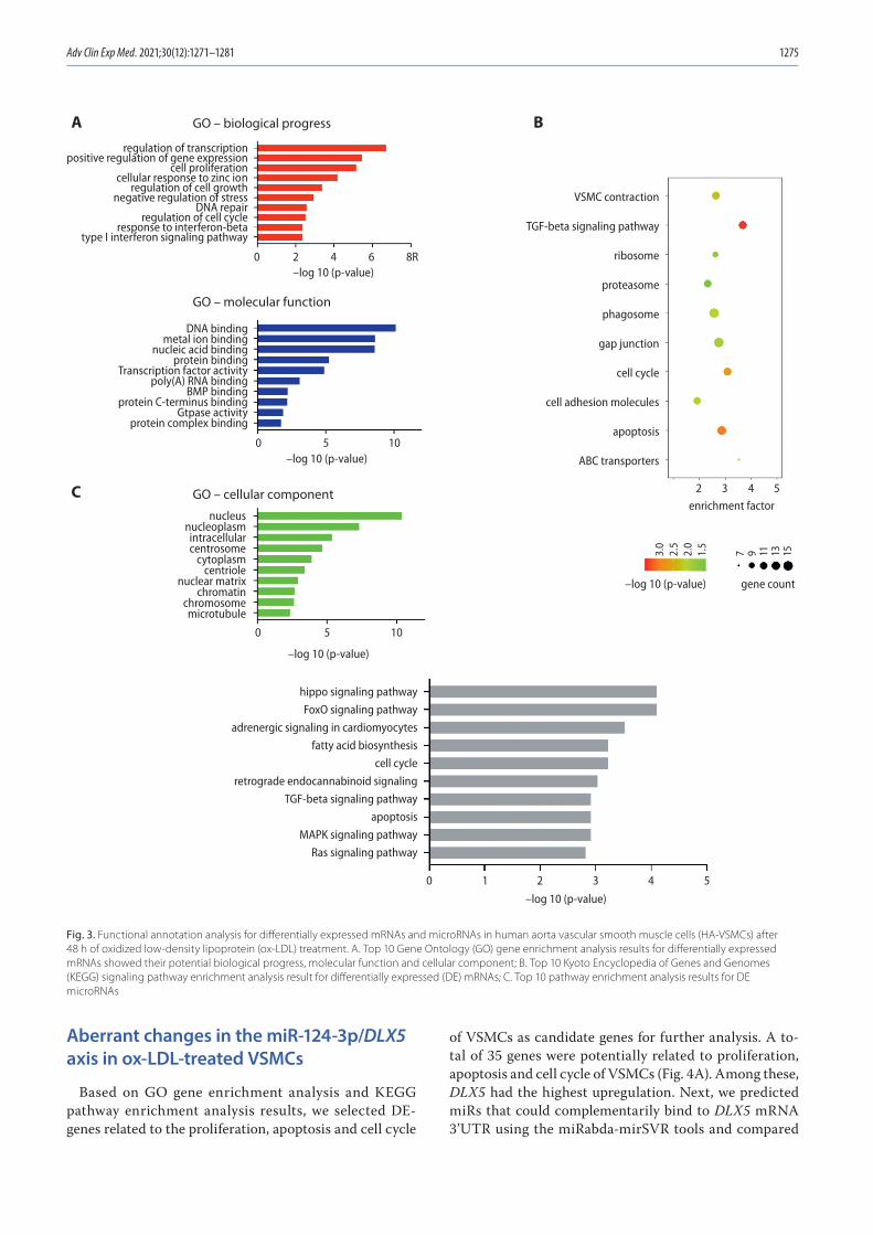
Adv Clin Exp Med. 2021;30(12):1271–1281 1275
Aberrant changes in the miR-124-3p/DLX5 axis in ox-LDL-treated VSMCs
Based on GO gene enrichment analysis and KEGG pathway enrichment analysis results, we selected DE-genes related to the proliferation, apoptosis and cell cycle
of VSMCs as candidate genes for further analysis. A to-tal of 35 genes were potentially related to proliferation, apoptosis and cell cycle of VSMCs (Fig. 4A). Among these, DLX5 had the highest upregulation. Next, we predicted miRs that could complementarily bind to DLX5 mRNA 3’UTR using the miRabda-mirSVR tools and compared
Fig. 3. Functional annotation analysis for differentially expressed mRNAs and microRNAs in human aorta vascular smooth muscle cells (HA-VSMCs) after 48 h of oxidized low-density lipoprotein (ox-LDL) treatment. A. Top 10 Gene Ontology (GO) gene enrichment analysis results for differentially expressed mRNAs showed their potential biological progress, molecular function and cellular component; B. Top 10 Kyoto Encyclopedia of Genes and Genomes (KEGG) signaling pathway enrichment analysis result for differentially expressed (DE) mRNAs; C. Top 10 pathway enrichment analysis results for DE microRNAs
VSMC contraction
TGF-beta signaling pathway
ribosome
proteasome
phagosome
gap junction
cell cycle
cell adhesion molecules
apoptosis
ABC transporters
GO – biological progress
GO – molecular function
GO – cellular component
regulation of transcriptionpositive regulation of gene expression
cell proliferationcellular response to zinc ion
regulation of cell growthnegative regulation of stress
DNA repairregulation of cell cycle
response to interferon-betatype I interferon signaling pathway
DNA bindingmetal ion binding
nucleic acid bindingprotein binding
Transcription factor activitypoly(A) RNA binding
BMP bindingprotein C-terminus binding
Gtpase activityprotein complex binding
nucleusnucleoplasmintracellularcentrosome
cytoplasmcentriole
nuclear matrixchromatin
chromosomemicrotubule
–log 10 (p-value)
–log 10 (p-value)
–log 10 (p-value)
–log 10 (p-value)
–log 10 (p-value) gene count
enrichment factor
hippo signaling pathway
FoxO signaling pathway
adrenergic signaling in cardiomyocytes
fatty acid biosynthesis
cell cycle
retrograde endocannabinoid signaling
TGF-beta signaling pathway
apoptosis
MAPK signaling pathway
Ras signaling pathway
0
0
3.0
2.5
2.0
1.5
7 9 11 13 15
2 3 4 5
5 10
0
0 2 4 6 8R
5 10
1 2 3 4 5
A B
C
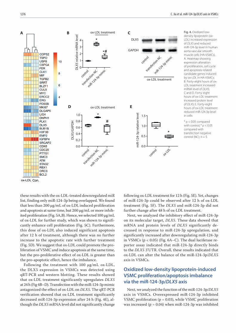
C. Jia et al. miR-124-3p/DLX5 axis in VSMCs1276
these results with the ox-LDL-treated downregulated miR list, finding only miR-124-3p being overlapped. We found that less than 200 μg/mL of ox-LDL induced proliferation and apoptosis at same time, but 200 μg/mL or more inhib-ited proliferation (Fig. 5A,B). Hence, we selected 100 μg/mL of ox-LDL for further study, which was shown to signifi-cantly enhance cell proliferation (Fig. 5C). Furthermore, this dose of ox-LDL also induced significant apoptosis after 12 h of treatment, although there was no further increase to the apoptotic rate with further treatment (Fig. 5D). We suggest that ox-LDL could promote the pro-liferation of VSMC and induce apoptosis at the same time, but the pro-proliferative effect of ox-LDL is greater than the pro-apoptotic effect, hence the imbalance.
Following the treatment with 100 μg/mL ox-LDL, the DLX5 expression in VSMCs was detected using qRT-PCR and western blotting. These results showed that ox-LDL treatment significantly upregulates DLX5 at 24 h (Fig 4B–D). Transfection with the miR-124-3p mimic antagonized the effect of ox-LDL on DLX5. The qRT-PCR verification showed that ox-LDL treatment significantly decreased miR-124-3p expression after 24 h (Fig. 4E), al-though the DLX5 mRNA level did not significantly change
following ox-LDL treatment for 12 h (Fig. 5E). Yet, changes of miR-124-3p could be observed after 12 h of ox-LDL treatment (Fig. 5F). The DLX5 and miR-124-3p did not further change after 48 h of ox-LDL treatment.
Next, we analyzed the inhibitory effect of miR-124-3p on its molecular target, DLX5. These data showed that mRNA and protein levels of DLX5 significantly de-creased in response to miR-124-3p upregulation, and significantly increased after downregulating miR-124-3p in VSMCs (p < 0.05) (Fig. 6A–C). The dual luciferase re-porter assay indicated that miR-124-3p directly binds to the DLX5 3’UTR. Overall, these results indicated that ox-LDL can alter the balance of the miR-124-3p/DLX5 axis in VSMCs.
Oxidized low-density lipoprotein-induced VSMC proliferation/apoptosis imbalance via the miR-124-3p/DLX5 axis
Next, we analyzed the function of the miR-124-3p/DLX5 axis in VSMCs. Overexpressed miR-124-3p inhibited VSMC proliferation (p = 0.03), while VSMC proliferation was increased (p = 0.04) when miR-124-3p was inhibited
Fig. 4. Oxidized low-density lipoprotein (ox-LDL) increased expression of DLX5 and reduced miR-124-3p level in human aorta vascular smooth muscle cells (HA-VSMCs). A. Heatmap showing expression alteration of proliferation, cell cycle and apoptosis-related candidate genes induced by ox-LDL in HA-VSMCs; B. Forty-eight hours of ox-LDL treatment increased mRNA level of DLX5; C and D. Forty-eight hours of ox-LDL treatment increased protein level of DLX5; E. Forty-eight hours of ox-LDL treatment reduced miR-124-3p level in cells
* p < 0.05 compared with control; # p < 0.05 compared with transfection negative control (NC); n = 5.
miR-124-3p mim
ic
transfe
ction-N
C
contro
l
miR-124-3p mim
ic
transfe
ction-N
C
contro
l
miR-124-3p mim
ic
transfe
ction-N
C
contro
l
contro
l
ox-LDL
DLX5
GAPDH
4
3
2
1
0
1.0
0.8
0.6
0.4
0.2
0.0
1.5
1.0
0.5
0.0
ox-LDL treatment
ox-LDL treatment
ox-LDL treatmentD
LX5
rela
tive
mRN
A le
vel
DLX
5/G
APD
H
miR
-124
-3p
rela
tive
leve
l
A B C
D E
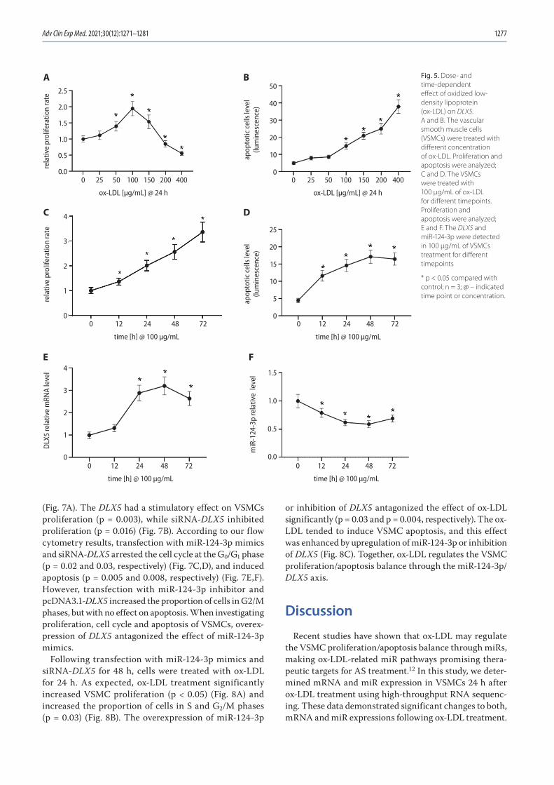
Adv Clin Exp Med. 2021;30(12):1271–1281 1277
(Fig. 7A). The DLX5 had a stimulatory effect on VSMCs proliferation (p = 0.003), while siRNA-DLX5 inhibited proliferation (p = 0.016) (Fig. 7B). According to our flow cytometry results, transfection with miR-124-3p mimics and siRNA-DLX5 arrested the cell cycle at the G0/G1 phase (p = 0.02 and 0.03, respectively) (Fig. 7C,D), and induced apoptosis (p = 0.005 and 0.008, respectively) (Fig. 7E,F). However, transfection with miR-124-3p inhibitor and pcDNA3.1-DLX5 increased the proportion of cells in G2/M phases, but with no effect on apoptosis. When investigating proliferation, cell cycle and apoptosis of VSMCs, overex-pression of DLX5 antagonized the effect of miR-124-3p mimics.
Following transfection with miR-124-3p mimics and siRNA-DLX5 for 48 h, cells were treated with ox-LDL for 24 h. As expected, ox-LDL treatment significantly increased VSMC proliferation (p < 0.05) (Fig. 8A) and increased the proportion of cells in S and G2/M phases (p = 0.03) (Fig. 8B). The overexpression of miR-124-3p
or inhibition of DLX5 antagonized the effect of ox-LDL significantly (p = 0.03 and p = 0.004, respectively). The ox-LDL tended to induce VSMC apoptosis, and this effect was enhanced by upregulation of miR-124-3p or inhibition of DLX5 (Fig. 8C). Together, ox-LDL regulates the VSMC proliferation/apoptosis balance through the miR-124-3p/DLX5 axis.
Discussion
Recent studies have shown that ox-LDL may regulate the VSMC proliferation/apoptosis balance through miRs, making ox-LDL-related miR pathways promising thera-peutic targets for AS treatment.12 In this study, we deter-mined mRNA and miR expression in VSMCs 24 h after ox-LDL treatment using high-throughput RNA sequenc-ing. These data demonstrated significant changes to both, mRNA and miR expressions following ox-LDL treatment.
Fig. 5. Dose- and time-dependent effect of oxidized low-density lipoprotein (ox-LDL) on DLX5. A and B. The vascular smooth muscle cells (VSMCs) were treated with different concentration of ox-LDL. Proliferation and apoptosis were analyzed; C and D. The VSMCs were treated with 100 μg/mL of ox-LDL for different timepoints. Proliferation and apoptosis were analyzed; E and F. The DLX5 and miR-124-3p were detected in 100 μg/mL of VSMCs treatment for different timepoints
* p < 0.05 compared with control; n = 3; @ – indicated time point or concentration.
0 25 50 100
ox-LDL [µg/mL] @ 24 h
150 200 4000
2.5
2.0
1.5
1.0
0.5
0.0
1.5
1.0
0.5
0.0
4
3
2
1
0
25
20
15
10
5
0
4
3
2
1
0
50
40
30
20
10
025 50 100
ox-LDL [µg/mL] @ 24 h
0 12 24 48 72
time [h] @ 100 µg/mL
0 12 24 48 72
time [h] @ 100 µg/mL
0 12 24 48 72
time [h] @ 100 µg/mL
0 12 24 48 72
time [h] @ 100 µg/mL
150 200 400
rela
tive
prol
ifera
tion
rate
rela
tive
prol
ifera
tion
rate
DLX
5 re
lativ
e m
RNA
leve
l
miR
-124
-3p
rela
tive
leve
lap
opto
tic c
ells
leve
l(lu
min
esce
nce)
apop
totic
cel
ls le
vel
(lum
ines
cenc
e)
A B
C D
E F

C. Jia et al. miR-124-3p/DLX5 axis in VSMCs1278
Then, to further reveal the biological functions of these mRNAs and miRs, we conducted functional enrichment analysis of these DE-mRNAs using the GO database, find-ing that some of the DE-mRNAs are related to the prolif-eration and cell cycle of VSMCs. In addition, KEGG signal pathway enrichment analysis showed that some of the DE-mRNAs are the components of signaling pathways in-volved in VSMC functions. The DIANA-mirPath (http://snf-515788.vm.okeanos.grnet.gr/) is an online tool that reveals the function of miRs of interest through pathway enrichment analysis of target genes for those miRs. This analysis highlighted that cell cycle, apoptosis and MAPK and RAS signaling pathways are involved after ox-LDL treatment. This evidence indicated that miRs might also participate in ox-LDL-induced VSMC proliferation/apop-tosis imbalance.
Because it was not possible for us to verify all these sequencing data, it was necessary to screen out mRNAs of interest and their corresponding miRs for further analysis. With a focus on the regulatory effect of ox-LDL on the VSMC proliferation/apoptosis balance, together with the results of GO gene enrichment analysis, only DE-genes related to the proliferation and cell cycle regulation of VSMCs were selected as candidates for verification. However, we found many such candidates, and we selected the gene with the highest expression of FC, DLX5, for further analysis. Next, miRs that can use DLX5 as the target gene were predicted and compared with previously obtained DE-miRs. These candidate miRs should be expressed at a low
level in ox-LDL-treated VSMCs and bound to the 3’UTR of DLX5. Finally, we only selected miR-124-3p in this study and assumed that ox-LDL can regulate the VSMC prolif-eration/apoptosis balance through the miR-124-3p/DLX5 axis, which was verified in vitro. Quantitative real-time polymerase chain reaction (RT-qPCR) confirmed that miR-124-3p expression decreased in ox-LDL-treated VSMCs. In addition, the expression of mRNA and protein levels of DLX5 increased in VSMCs after ox-LDL treatment. Furthermore, we confirmed the targeted inhibitory effect of miR-124-3p on DLX5. Together, these data indicated the presence of the miR-124-3p/DLX5 axis that can be regulated by ox-LDL in VSMCs.
Some studies investigating cancer have reported that miR-124-3p inhibits cell proliferation. Xu et al. showed that miR-124-3p inhibits the proliferation of nasopharyngeal carcinoma cells,13 while Wu et al. reported the inhibitory effect of miR-124-3p on glioma proliferation.14 The in-hibitory effect of miR-124-3p on VSMC proliferation and migration has also been reported previously.15 However, Cheng et al. showed that miR-124-3p promotes VSMC proliferation, so the role of miR-124-3p in VSMCs remains controversial.16 The current study showed that the upregu-lation of miR-124-3p inhibits VSMC proliferation, arrests the cell cycle and increases apoptosis, while its inhibition accelerates VSMC proliferation. Therefore, we believe that miR-124-3p can inhibit VSMC growth. Moreover, we discovered that miR-124-3p overexpression counteracts the stimulatory effect of ox-LDL on VSMC proliferation.
Fig. 6. The miR-124-3p negatively regulated DLX5 expression. A. Up- or downregulation of miR-124-3p changed the mRNA level of DLX5; B and C. Up- or downregulation of miR-124-3p changed the protein level of DLX5; D. The miR-124-3p binding to the DLX5 3’UTR was indicated using luciferase reporter assay (left). The wild-type and mutant DLX5 3’UTR sequence and the predicted binding site to miR-124-3p is shown (right)
* p < 0.05 compared with transfection negative control (NC); # p < 0.05 compared with miR-124-3p mimic.
miR-124-3p mim
ic
miR-124-3p inhibito
r
transfe
ction-N
C
miR-124-3p mim
ic
miR-124-3p inhibito
r
transfe
ction-N
C
miR-124-3p mim
ic
miR-124-3p inhibito
r
transfe
ction-N
C
miR-124-3p mimictransfection-NC
DLX5
GAPDH
DLX53’UTR WT
DLX53’UTR MUT
DLX
5/G
APD
H
1.0
0.8
0.6
0.4
0.2
0.0
2.5
2.0
1.5
1.0
0.5
0.0DLX
5 re
lativ
e m
RNA
leve
lre
lativ
e lu
cife
rase
act
ivity 1.5
1.0
0.5
0.0
A
D
B C
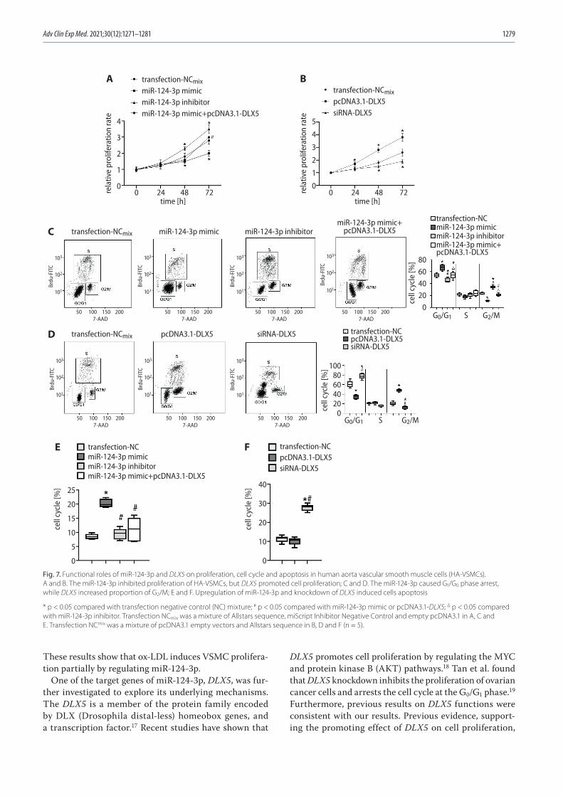
Adv Clin Exp Med. 2021;30(12):1271–1281 1279
These results show that ox-LDL induces VSMC prolifera-tion partially by regulating miR-124-3p.
One of the target genes of miR-124-3p, DLX5, was fur-ther investigated to explore its underlying mechanisms. The DLX5 is a member of the protein family encoded by DLX (Drosophila distal-less) homeobox genes, and a transcription factor.17 Recent studies have shown that
DLX5 promotes cell proliferation by regulating the MYC and protein kinase B (AKT) pathways.18 Tan et al. found that DLX5 knockdown inhibits the proliferation of ovarian cancer cells and arrests the cell cycle at the G0/G1 phase.19 Furthermore, previous results on DLX5 functions were consistent with our results. Previous evidence, support-ing the promoting effect of DLX5 on cell proliferation,
Fig. 7. Functional roles of miR-124-3p and DLX5 on proliferation, cell cycle and apoptosis in human aorta vascular smooth muscle cells (HA-VSMCs). A and B. The miR-124-3p inhibited proliferation of HA-VSMCs, but DLX5 promoted cell proliferation; C and D. The miR-124-3p caused G1/G0 phase arrest, while DLX5 increased proportion of G2/M; E and F. Upregulation of miR-124-3p and knockdown of DLX5 induced cells apoptosis
* p < 0.05 compared with transfection negative control (NC) mixture; # p < 0.05 compared with miR-124-3p mimic or pcDNA3.1-DLX5; Δ p < 0.05 compared with miR-124-3p inhibitor. Transfection NCmix was a mixture of Allstars sequence, miScript Inhibitor Negative Control and empty pcDNA3.1 in A, C and E. Transfection NCmix was a mixture of pcDNA3.1 empty vectors and Allstars sequence in B, D and F (n = 5).
transfection-NCmix
miR-124-3p mimicmiR-124-3p inhibitormiR-124-3p mimic+pcDNA3.1-DLX5
miR-124-3p inhibitor
transfection-NCmiR-124-3p mimicmiR-124-3p inhibitormiR-124-3p mimic+
transfection-NCmiR-124-3p mimicmiR-124-3p inhibitormiR-124-3p mimic+pcDNA3.1-DLX5
miR-124-3p mimic+pcDNA3.1-DLX5miR-124-3p mimic
time [h] time [h]
transfection-NCmix
pcDNA3.1-DLX5siRNA-DLX5
transfection-NCmix
Brdu
-FIT
C
7-AAD50
103
102
101
100 150 200
Brdu
-FIT
C
7-AAD50
103
102
101
100 150 200
Brdu
-FIT
C
7-AAD50
103
102
101
100 150 200
Brdu
-FIT
C
7-AAD50
103
102
101
100 150 200
Brdu
-FIT
C
7-AAD50
103
102
101
100 150 200
Brdu
-FIT
C
7-AAD50
103
102
101
100 150 200
Brdu
-FIT
C7-AAD
50
103
102
101
100 150 200
transfection-NCmix siRNA-DLX5
G0/G1 S
80604020
0
cell
cycl
e [%
]
G2/M
G0/G1 S
10080604020
0cell
cycl
e [%
]
40
30
20
10
0
cell
cycl
e [%
]25
20
15
10
5
0
cell
cycl
e [%
]
G2/M
transfection-NCpcDNA3.1-DLX5siRNA-DLX5
transfection-NCpcDNA3.1-DLX5siRNA-DLX5
pcDNA3.1-DLX5
4
3
2
1
0rela
tive
prol
ifera
tion
rate 5
4
3
2
1
0
rela
tive
prol
ifera
tion
rate
0 24 48 72 0 24 48 72
pcDNA3.1-DLX5
A
C
D
E F
B
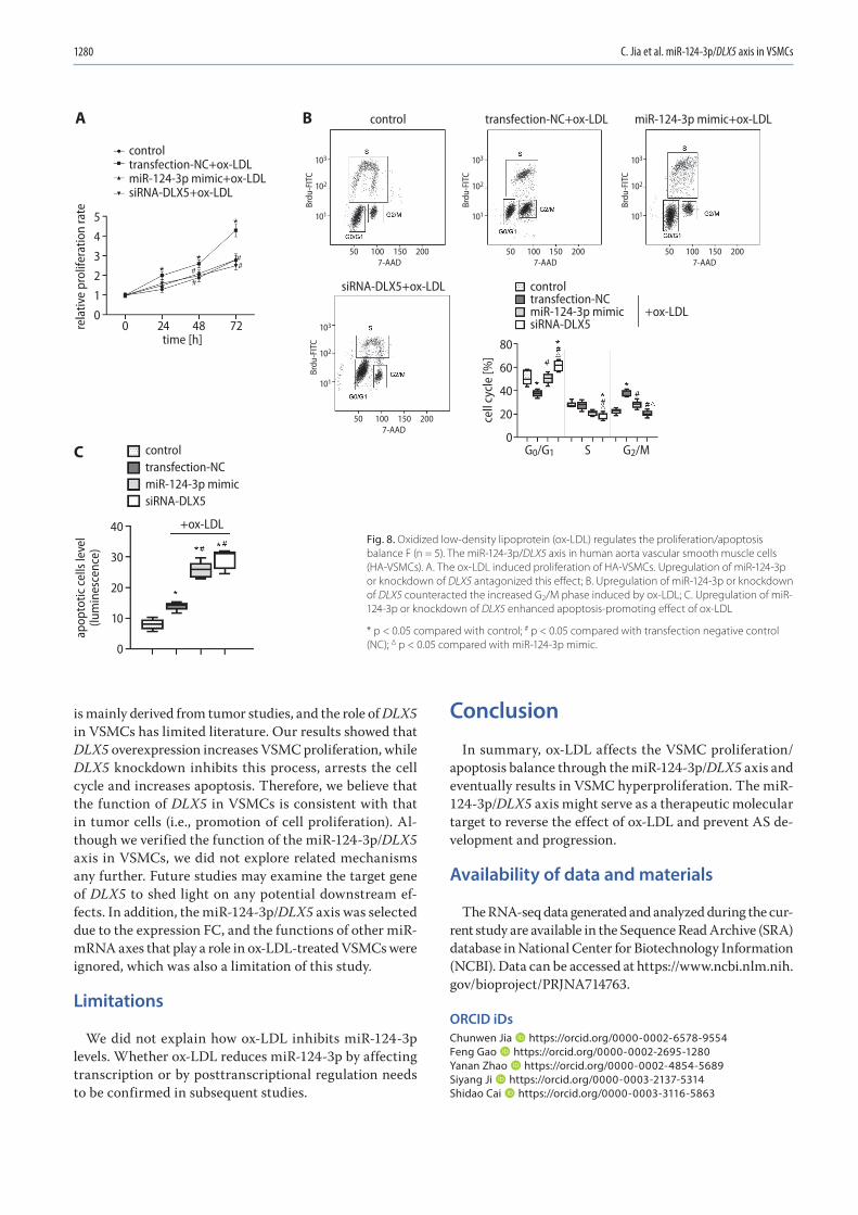
C. Jia et al. miR-124-3p/DLX5 axis in VSMCs1280
is mainly derived from tumor studies, and the role of DLX5 in VSMCs has limited literature. Our results showed that DLX5 overexpression increases VSMC proliferation, while DLX5 knockdown inhibits this process, arrests the cell cycle and increases apoptosis. Therefore, we believe that the function of DLX5 in VSMCs is consistent with that in tumor cells (i.e., promotion of cell proliferation). Al-though we verified the function of the miR-124-3p/DLX5 axis in VSMCs, we did not explore related mechanisms any further. Future studies may examine the target gene of DLX5 to shed light on any potential downstream ef-fects. In addition, the miR-124-3p/DLX5 axis was selected due to the expression FC, and the functions of other miR-mRNA axes that play a role in ox-LDL-treated VSMCs were ignored, which was also a limitation of this study.
Limitations
We did not explain how ox-LDL inhibits miR-124-3p levels. Whether ox-LDL reduces miR-124-3p by affecting transcription or by posttranscriptional regulation needs to be confirmed in subsequent studies.
Conclusion
In summary, ox-LDL affects the VSMC proliferation/apoptosis balance through the miR-124-3p/DLX5 axis and eventually results in VSMC hyperproliferation. The miR-124-3p/DLX5 axis might serve as a therapeutic molecular target to reverse the effect of ox-LDL and prevent AS de-velopment and progression.
Availability of data and materials
The RNA-seq data generated and analyzed during the cur-rent study are available in the Sequence Read Archive (SRA) database in National Center for Biotechnology Information (NCBI). Data can be accessed at https://www.ncbi.nlm.nih.gov/bioproject/PRJNA714763.
ORCID iDsChunwen Jia https://orcid.org/0000-0002-6578-9554Feng Gao https://orcid.org/0000-0002-2695-1280Yanan Zhao https://orcid.org/0000-0002-4854-5689Siyang Ji https://orcid.org/0000-0003-2137-5314Shidao Cai https://orcid.org/0000-0003-3116-5863
miR-124-3p mimic+ox-LDLtransfection-NC+ox-LDLcontrol
Brdu
-FIT
C
7-AAD50
103
102
101
100 150 200
Brdu
-FIT
C
7-AAD50
103
102
101
100 150 200
Brdu
-FIT
C
7-AAD50
103
102
101
100 150 200
siRNA-DLX5+ox-LDL
Brdu
-FIT
C
7-AAD50
103
102
101
100 150 200
A
C
B
controltransfection-NCmiR-124-3p mimicsiRNA-DLX5
controltransfection-NCmiR-124-3p mimicsiRNA-DLX5
controltransfection-NC+ox-LDLmiR-124-3p mimic+ox-LDLsiRNA-DLX5+ox-LDL
+ox-LDL
+ox-LDL
G0/G1 S
80
60
40
20
0
cell
cycl
e [%
]
G2/M
time [h]
5
4
3
2
1
0
40
30
20
10
0
rela
tive
prol
ifera
tion
rate
0 24 48 72
apop
totic
cel
ls le
vel
(lum
ines
cenc
e)
Fig. 8. Oxidized low-density lipoprotein (ox-LDL) regulates the proliferation/apoptosis balance F (n = 5). The miR-124-3p/DLX5 axis in human aorta vascular smooth muscle cells (HA-VSMCs). A. The ox-LDL induced proliferation of HA-VSMCs. Upregulation of miR-124-3p or knockdown of DLX5 antagonized this effect; B. Upregulation of miR-124-3p or knockdown of DLX5 counteracted the increased G2/M phase induced by ox-LDL; C. Upregulation of miR-124-3p or knockdown of DLX5 enhanced apoptosis-promoting effect of ox-LDL
* p < 0.05 compared with control; # p < 0.05 compared with transfection negative control (NC); Δ p < 0.05 compared with miR-124-3p mimic.
Adv Clin Exp Med. 2021;30(12):1271–1281 1281
References1. Kobiyama K, Ley K. Atherosclerosis. Circ Res. 2018;123(10):1118–1120.
doi:10.1161/CIRCRESAHA.118.3138162. Gimbrone MA, García-Cardeña G. Endothelial cell dysfunction and
the pathobiology of atherosclerosis. Circ Res. 2016;118(4):620–636. doi:10.1161/CIRCRESAHA.115.306301
3. Dzau VJ, Braun-Dullaeus RC, Sedding DG. Vascular proliferation and atherosclerosis: New perspectives and therapeutic strategies. Nat Med. 2002;8(11):1249–1256. doi:10.1038/nm1102-1249
4. Kattoor AJ, Kanuri SH, Mehta JL. Role of Ox-LDL and LOX-1 in athero-genesis. Curr Med Chem. 2019;26(9):1693–1700. doi:10.2174/092986 7325666180508100950
5. Gao S, Zhao D, Wang M, et al. Association between circulating oxi-dized LDL and atherosclerotic cardiovascular disease: A meta-anal-ysis of observational studies. Can J Cardiol. 2017;33(12):1624–1632. doi:10.1016/j.cjca.2017.07.015
6. Mitra S, Goyal T, Mehta JL. Oxidized LDL, LOX-1 and atherosclerosis. Cardiovasc Drugs Ther. 2011;25(5):419–429. doi:10.1007/s10557-011-6341-5
7. Pirillo A, Norata GD, Catapano AL. LOX-1, OxLDL, and atherosclerosis. Mediators Inflamm. 2013;2013:152786. doi:10.1155/2013/152786
8. Rhoads JP, Major AS. How oxidized low-density lipoprotein acti-vates inflammatory responses. Crit Rev Immunol. 2018;38(4):333–342. doi:10.1615/CritRevImmunol.2018026483
9. Balzan S, Lubrano V. LOX-1 receptor: A potential link in atherosclero-sis and cancer. Life Sci. 2018;198:79–86. doi:10.1016/j.lfs.2018.02.024
10. Sun Y, Chen D, Cao L, et al. MiR-490-3p modulates the proliferation of vascular smooth muscle cells induced by ox-LDL through targeting PAPP-A. Cardiovasc Res. 2013;100(2):272–279. doi:10.1093/cvr/cvt172
11. Wang YS, Wang HYJ, Liao YC, et al. MicroRNA-195 regulates vascular smooth muscle cell phenotype and prevents neointimal formation. Cardiovasc Res. 2012;95(4):517–526. doi:10.1093/cvr/cvs223
12. Nilsson J, Nordin Fredrikson G, Schiopu A, Shah PK, Jansson B, Carlsson R. Oxidized LDL antibodies in treatment and risk assessment of athero-sclerosis and associated cardiovascular disease. Curr Pharm Des. 2007; 13(10):1021–1030. doi:10.2174/138161207780487557
13. Xu S, Zhao N, Hui L, Song M, Miao ZW, Jiang XJ. MicroRNA-124-3p inhibits the growth and metastasis of nasopharyngeal carcinoma cells by targeting STAT3. Oncol Rep. 2016;35(3):1385–1394. doi:10. 3892/or.2015.4524
14. Wu Q, Xu L, Wang C, Fan W, Yan H, Li Q. MicroRNA-124-3p repress-es cell growth and cell motility by targeting EphA2 in glioma. Biochem Biophys Res Commun. 2018;503(4):2436–2442. doi:10.1016/j.bbrc.2018.06.173
15. Afzal TA, Luong LA, Chen D, et al. NCK Associated protein 1 modu-lated by miRNA-214 determines vascular smooth muscle cell migra-tion, proliferation, and neointima hyperplasia. J Am Heart Assoc. 2016; 5(12):e004629. doi:10.1161/JAHA.116.004629
16. Cheng C, Xu BL, Sheng JL, He F, Yang T, Shen SC. LncRNA MALAT1 regulates proliferation and apoptosis of vascular smooth muscle cells by targeting miRNA-124-3p/PPARα axis. Eur Rev Med Pharmacol Sci. 2019;23(20):9025–9032. doi:10.26355/eurrev_201910_19304
17. Eisenstat DD, Liu JK, Mione M, et al. DLX-1, DLX-2, and DLX-5 expression define distinct stages of basal forebrain differentiation. J Comp Neurol. 1999;414(2):217–237. doi:10.1002/(sici)1096-9861(1999 1115)414:2<217::aid-cne6>3.0.co;2-i
18. Tan Y, Sementino E, Xu J, et al. The homeoprotein Dlx5 drives murine T-cell lymphomagenesis by directly transactivating Notch and upregulating Akt signaling. Oncotarget. 2017;8(9):14941–14956. doi:10.18632/oncotarget.14784
19. Tan Y, Cheung M, Pei J, Menges CW, Godwin AK, Testa JR. Upregula-tion of DLX5 promotes ovarian cancer cell proliferation by enhancing IRS-2-AKT signaling. Cancer Res. 2010;70(22):9197–9206. doi:10.1158/ 0008-5472.CAN-10-1568

Cite asIçduygu FM, Samli H, Özgöz A, Vatansever B, Oztürk KH, Akgün E. Possibility of paclitaxel to induce the stemness-related characteristics of prostate cancer cells. Adv Clin Exp Med. 2021;30(12):1283–1291. doi:10.17219/acem/140590
DOI10.17219/acem/140590
Copyright© 2021 by Wroclaw Medical University This is an article distributed under the terms of theCreative Commons Attribution 3.0 Unported (CC BY 3.0)(https://creativecommons.org/licenses/by/3.0/)
Address for correspondenceFadime Mutlu IçduyguE-mail: [email protected]
Funding sourcesScientific Research Projects Committee of Giresun University, Turkey (Project No. SAĞ-BAP-A-230218-80 and Project No. SAĞ-BAP-A-140316-93).
Conflict of interestNone declared
Received on June 11, 2021Reviewed on June 29, 2021Accepted on July 28, 2021
Published online on October 5, 2021
AbstractBackground. Drug resistance poses a crucial problem in the treatment of prostate cancer. Recent studies have shown that chemotherapy agents may cause cancer cells to acquire stem cell-like properties, resulting in drug resistance and, eventually, treatment failure.
Objectives. To evaluate whether long-term paclitaxel exposure causes an increase in the stem cell-like properties of prostate cancer cells.
Materials and methods. Paclitaxel-resistant PC-3 cells were generated from parental PC-3 cells by treating them with increasing concentrations of paclitaxel. The expression levels of the stem cell markers NANOG, C-MYC, CD44, and ABCG2 were evaluated using quantitative real-time polymerase chain reaction (RT-qPCR). A sphere formation assay was performed to test the potential of the cells to behave as stem cells, and a wound healing assay was carried out to evaluate migration ability of the cells.
Results. The expression levels of C-MYC and NANOG were significantly higher in paclitaxel-resistant PC-3 cells compared to the parental PC-3 cells. However, there was no significant increase in the expression of CD44 or ABCG2. In addition, the sphere-forming capacity and migration ability of resistant PC-3 cells were increased.
Conclusions. The results of the current study indicate that paclitaxel exposure may increase the stem cell-like properties of PC-3 prostate cancer cells.
Key words: prostate cancer, cancer stem cell, paclitaxel, taxane resistance
Original papers
Possibility of paclitaxel to induce the stemness-related characteristics of prostate cancer cells
Fadime Mutlu Içduygu1,A–D, Hale Samli2,A,C,F, Asuman Özgöz3,C–E, Buse Vatansever2,B,C, Kuyas Hekimler Oztürk4,C,E, Egemen Akgün5,B,C
1 Department of Medical Genetics, Faculty of Medicine, Giresun University, Turkey2 Department of Genetics, Faculty of Veterinary Medicine, Bursa Uludağ University, Turkey3 Department of Medical Genetics, Faculty of Medicine, Kastamonu University, Turkey4 Department of Medical Genetics, Faculty of Medicine, Süleyman Demirel University, Isparta, Turkey5 Department of Medical Biology, Faculty of Medicine, Giresun University, Turkey
A – research concept and design; B – collection and/or assembly of data; C – data analysis and interpretation; D – writing the article; E – critical revision of the article; F – final approval of the article
Advances in Clinical and Experimental Medicine, ISSN 1899–5276 (print), ISSN 2451–2680 (online) Adv Clin Exp Med. 2021;30(12):1283–1291
F. Mutlu Içduygu et al. Paclitaxel, stemness of prostate cancer cells1284
Background
Prostate cancer is the 2nd most common cancer and one of the leading causes of cancer death among men world-wide.1 Generally, the first treatment choice for advanced prostate cancer patients is androgen deprivation therapy (ADT). Although patients tend to respond to ADT at first, treatment resistance develops in the majority of individu-als, and the disease is termed castration-resistant prostate cancer (CRPC) at this stage. Taxanes (paclitaxel, abrax-ane, docetaxel) – a class of microtubule stabilizing agents – causes cell cycle arrest at the G2/M phase and apoptosis.2 Taxane-based treatment provides a good initial response and an increased survival in CRPC patients. However, al-most all patients develop resistance over time, and the clin-ical use of these drugs is severely limited. Therefore, eluci-dating the mechanisms of taxane resistance is critical for the development of improved treatment strategies, favora-ble prognosis and prolonged survival in CRPC patients.2,3 Despite current data on possible resistance mechanisms (such as increased activation and expression of efflux transporters, reactivation of the androgen receptor path-way, impairment in the apoptotic pathway, cytokine and chemokine induction, changes in the structure and func-tion of microtubules, upregulation of stress survival pro-teins) and new drugs targeting these mechanisms, CRPC is known to cause death in many patients.2,4–6 Therefore, other mechanisms causing taxane resistance need to be investigated.
Recent data indicate that tumors are composed of het-erogeneous cell populations and most of these cells have a limited self-renewal capacity. On the other hand, a small group of cells within the heterogeneous tumor tissue has been reported to have a high self-renewal capacity, the abil-ity to transform into different cancer cells, and tumor in-itiation properties. These are known as cancer stem cells (CSCs).5,7 The CSCs form cellular spheroids in serum-free and non-adherent conditions, and have differential gene expression profiles, as well as enhanced epithelial-to-mes-enchymal transformation (EMT) properties. High levels of expression for genes that encode cell surface receptors (such as CD44 or CD133), transcription factors associated with pluripotency and other stemness characteristics (i.e., NANOG, OCT4, SOX2, C-MYC), and proteins associated with drug resistance (i.e., ABCG2 and ABCB1) are all the ex-amples of differential gene expression profiles in CSCs.7,8 An embryonic transcription factor – NANOG – is highly expressed in CSCs, and shows a lower expression in differ-entiated tissues. It plays a key role in CSCs gaining properties such as self-renewal, pluripotency, stemness, metastasis, in-vasiveness, angiogenesis and drug resistance, with the help of proteins such as WNT, OCT4, SOX2, and Hedgehog.9
A transcription factor C-MYC plays an important role in processes such as the cell cycle, cell growth, proliferation, differentiation and apoptosis, as well as tumorigenesis, because it regulates the expression of many genes. Recent
studies have revealed that C-MYC effects the self-renewal capacity of cells and is one of the most important core markers for stem cells.10,11 The CD44 is a cell surface recep-tor. Hyaluronic acid, osteopontin and many other ligands bind to this receptor to mediate processes such as metasta-sis, invasion and migration. It is one of the most commonly used cell surface receptors in identifying prostate cancer stem cells.12 The ABCG2 is one of the ABC family efflux proteins and is responsible for removing drugs and harm-ful substances from cells. This receptor is responsible for the development of the resistance to many different drugs, and is defined as a stem cell marker for side populations in many types of tumors, including prostate cancer.13
Recent studies also suggest that CSCs may be responsible for chemotherapy and radiotherapy resistance in several cancer types, including prostate cancer.3,14,15 An emerg-ing hypothesis about CSCs and chemotherapy resistance argues that cancer non-stem cells may acquire stemness properties when they are treated with chemotherapy, and that this process may result in chemotherapy resistance.7,16
Some studies have reported an increase in the number of chemotherapy- or radiotherapy-resistant cancer cell groups that exhibit stem-like characteristics, as well as features of EMT.3,17,18 In studies examining the relationship between prostate CSCs and taxane resistance, researchers have iso-lated CSCs from prostate cancer cell lines and have reported that those cells were resistant to taxanes. However, these studies failed to reveal whether taxanes affected the stem cell-like characteristics of prostate cancer cells. To the best of our knowledge, the present study is the first attempt to investigate whether a long-term exposure to paclitaxel affects the stem cell characteristics of prostate cancer cells.
Objectives
The aim of this study is to evaluate whether there is an increase in the stem cell-like properties of PC-3 pros-tate cancer cells following treatment with paclitaxel for a prolonged period. Therefore, the expression of the CSC markers CD44, C-MYC, NANOG, and ABCG2, as well as sphere formation and wound healing capabilities, were compared between parental and paclitaxel-resistant PC-3 cell lines (PC-3-R).
Materials and methods
Cell culture and the establishment of paclitaxel-resistant cell lines
PC-3 androgen-independent cells (CRL-1435™; Amer-ican Type Culture Collection (ATCC), Manassas, USA) were cultured in RPMI 1640 Media (Biological Industries, Beit HaEmek, Israel), containing 10% heat-inactivated fetal bovine serum (FBS; Biological Industries), 1% L-glutamine
Adv Clin Exp Med. 2021;30(12):1283–1291 1285
(Biological Industries), penicillin (100 U/mL), and strepto-mycin (100 μg/mL). Cells were incubated at 37°C in a hu-midified atmosphere containing 5% CO2.
PC-3-R cells were generated from parental PC-3 cells by treating them with increasing concentrations of paclitaxel (Sigma–Aldrich, St. Louis, USA). Parental PC-3 cells were incubated for 24 h, and then treated with 5 nM of paclitaxel for 48 h. Then, the culture media was replaced with fresh media without paclitaxel, and the cells were incubated until they reached 80–90% confluency. The same protocol was repeated with gradually increasing concentrations of pacl-itaxel (5 nM each time) until 100 nM paclitaxel-resistant and viable PC-3 cells (PC-3-R) could be obtained. Cytotoxicity tests were conducted, and the viability of PC-3 and PC-3-R cells was confirmed under an inverted microscope.
Evaluating the viability and the number of the cells
A trypan blue stain was used to determine the number of viable cells. The cell resistance level and cytotoxic effects of paclitaxel in PC-3 and PC-3-R cells were assessed using a MTT assay. Resistant and parental cells were inoculated in 96-well plates at a density of 5 × 10³ cells per well (for each dose, triplicate wells were used) and incubated for 24 h. Afterwards, the cells were treated with increasing concentrations of paclitaxel (1.6–600 nM) and incubated for 72 h. After incubation, a MTT test was conducted and the absorbances were measured using a microplate reader at a wavelength of 570 nM.
Quantitative real-time polymerase chain reaction (RT-qPCR)
The total RNA extraction from cells was conducted using a High Pure RNA Isolation Kit (Roche Diagnostics, Basel, Switzerland). A transcriptor First Strand cDNA Synthesis Kit (Roche Diagnostics) was used to obtain cDNA samples from 1 μg of total RNA. The expression levels of genes were quantified using a Lightcycler 480 (Roche Diagnostics). Suitable probes and gene specific primers were designed by the Universal ProbeLibrary (UPL) Assay Design Center. Each sample was quantified in triplicate, and the mean value was used for further calculations. The B-actin was used to normalize the target gene expressions. Relative changes in the amount of mRNA were calculated based on ∆∆CT. The following primers were used for each target gene:Β-ACTIN forward 5′- CCCAGCACAATGAAGATCAA -3′ reverse 5′- CGATCCACACGGAGTACTTG -3′NANOG forward 5′- ACAGGTGAAGACCTGGTTCC -3′ reverse 5′- TTGCTATTCTTCGGCCAGTT -3′CD44 forward 5′- TCCAACACCTCCCAGTATGACA -3′ reverse 5′- GGCAGGTCTGTGACTGATGTACA -3′
C-MYC forward 5′- TCCACCTCCAGCTTGTACCT-3′ reverse 5′- TGAGAGGGTAGGGGAAGACC-3′ABCG2 forward 5′- TGGCTTAGACTCAAGCACAGC-3′ reverse 5′- TCGTCCCTGCTTAGACATCC-3′
Sphere formation assay
PC-3 and PC-3-R cells were inoculated onto 96-well ultra-low attachment plates at a density of 750 cells per well in serum-free DMEM/F12 (Thermo Fisher Scien-tific, Waltham, USA), supplemented with 20 ng/mL EGF (Sigma–Aldrich), B27 (1:50; Invitrogen, Waltham, USA), 0.4% bovine serum albumin (BSA; Sigma–Aldrich), and 4 mg/mL insulin (Sigma–Aldrich). Fresh stem cell media was added every 3–4 days. The cultures were incubated at 37°C in a humidified incubator containing 5% CO2. The sphere growth was monitored for 14 days and the num-ber of spheres was counted on days 7 and 14. Six wells were inoculated for both PC-3 and PC-3-R cells. The assay was conducted in triplicate.
Wound healing assay
PC-3 and PC-3-R cells were inoculated in six-well plates at a density of 5 × 10³ cells per well and incubated in or-der to reach a confluency of 90% in a complete medium. The complete medium was removed, and cells were starved in a serum-free medium for 24 h. Following the formation of cell monolayers, an artificial wound on the monolayer was scratched with a sterile 200 µL pipette tip. The cells were then washed with phosphate-buffered saline (PBS) and cultured in complete media. The assay was conducted in triplicate. Cells migrating into the wound were pho-tographed at 0 h, 8 h, and 24 h using a camera attached to an inverted microscope.
Statistical analysis
Statistical analyses were conducted using SPSS v. 15.0 (SPSS Inc., Chicago, USA). The data are expressed as the mean ± standard error (SE) and were analyzed us-ing the Mann–Whitney U test. The statistical significance of mRNA expression levels was analyzed using REST 2009 v. 2.0.13 (Qiagen, Hilden, Germany). The values of p <0.05 were deemed statistically significant.
Results
Development of paclitaxel resistance
The PC-3-R cells were obtained by treating the paren-tal cells with increasing concentrations of paclitaxel over a period of several months. The proliferation performance

F. Mutlu Içduygu et al. Paclitaxel, stemness of prostate cancer cells1286
of PC-3-R cells at the 100 nM paclitaxel level was higher than the PC-3 cells (Fig. 1A). A MTT test was conducted to assess the cytotoxic effect of paclitaxel in the PC-3 and PC-3-R cells. The IC50 dose of PC-3-R cells was higher (>600 nM) than the PC-3 cells (2.65 nM; Fig. 1B).
Expression of stem cell markers in the PC-3 and PC-3-R cells
The expressions of several stem cell markers, in-cluding CD44, C-MYC, NANOG, and ABCG2 in PC-3 and PC-3-R cells were analyzed. The expression levels of NANOG and C-MYC genes significantly increased with fold changes of 15.5 (p = 0.0130, 95% confidence in-terval (95% CI) = 12.930–18.259) and 3.28 (p = 0.0400, 95% CI = 2.802–3.909) in PC-3-R cells, respectively, when compared to PC-3 cells. There was no significant differ-ence in the expression levels of ABCG2 (fold change 1.69, p = 0.0650, 95% CI = 1.647–1.727) and CD44 (fold change 1.12, p = 0.0630, 95% CI = 1.051–1.226), between resistant and parental cell lines (Fig. 2; Table 1).
Sphere-forming capacity of PC-3 and PC-3-R cells
The sphere formation assay was applied to PC-3 and PC-3-R cells in order to find out whether there was an increase in the stem-like cell population with high sphere forming
ability in the PC-3-R cell line (compared to parental PC-3 cells). The results showed that the number of spheres was higher in the PC-3-R cells than in the PC-3 cells, and the difference between the groups was statistically sig-nificant both on day 7 (p = 0.0130, z = −2.4810), and day 14 (p = 0.0450, z = −2.0010; Fig. 3; Table 1).
Wound healing assay
The ability of cell migration to serve as an indica-tor of the stemness-like phenotype was assessed using a wound healing assay. The PC-3-R cells healed wounds better than the PC-3 cells. The rate of wound closure at 8 h for PC-3R cells was 65%, whereas it was only 35.4% for PC-3 cells. At 24 h, the wound had closed at the rate of 100% in PC-3R cells and at 54.5% in the PC-3 cells. The differences in wound closure rates between both groups at 8 h (p = 0.0500, z = −1.964) and 24 h (p = 0.0370, z = −2.0870) were statistically significant (Fig. 4; Table 1).
Discussion
Recent studies have suggested that chemotherapeutics may cause cancer cells to gain stem cell properties, re-sulting in drug resistance.19,20 An increased expression in several genes has been reported in CSCs. These genes
Fig. 1. A. Images of PC-3 and PC-3-R cells at 100 nM paclitaxel concentration (at ×40 total magnification); B. Cytotoxic effect of paclitaxel against PC-3 and PC-3-R cells in MTT assay. The MTT test was conducted in triplicate. Data are shown as mean ± standard error (SE)
MTT assay (72 h)
dose [nM]
120
100
80
60
40
20
0NK 1.56 3.13 6.25 12.5 25 50 100 200 300 400 500 600
viab
ility
[%]
PC-3PC-3-R
A
B
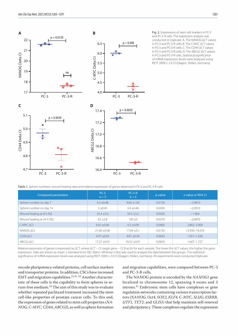
Adv Clin Exp Med. 2021;30(12):1283–1291 1287
encode pluripotency-related proteins, cell surface markers and transporter proteins. In addition, CSCs have increased EMT and migration capabilities.15,21–23 Another character-istic of these cells is the capability to form spheres in se-rum-free medium.24 The aim of this study was to evaluate whether repeated paclitaxel treatment increased the stem cell-like properties of prostate cancer cells. To this end, the expression of genes related to stem cell properties (NA-NOG, C-MYC, CD44, ABCG2), as well as sphere formation
and migration capabilities, were compared between PC-3 and PC-3-R cells.
The NANOG protein is encoded by the NANOG1 gene localized to chromosome 12, spanning 4 exons and 3 introns.25 Embryonic stem cells have complexes or gene regulation networks containing various transcription fac-tors (NANOG, Oct4, SOX2, KLF4, C-MYC, SLUG, ESRRB, UTF1, TET2, and GLIS1) that help maintain self-renewal and pluripotency. These complexes regulate the expression
Fig. 2. Expressions of stem cell markers in PC-3 and PC-3-R cells. The expression analysis was conducted in triplicate. A. The NANOG ∆CT values in PC-3 and PC-3-R cells; B. The C-MYC ∆CT values in PC-3 and PC-3-R cells; C. The CD44 ∆CT values in PC-3 and PC-3-R cells; D. The ABCG2 ∆CT values in PC-3 and PC-3-R cells. Statistical significance of mRNA expression levels were analyzed using REST 2009 v. 2.0.13 (Qiagen, Hilden, Germany)
Table 1. Sphere numbers, wound healing rates and relative expression of genes observed in PC-3 and PC-3-R cells
Compared parameters PC-3n = 3
PC-3-Rn = 3 p-value z-value or 95% CI
Sphere number on day 7 4.3 ±0.48 9.60 ±1.83 0.0130 −2.4810
Sphere number on day 14 2 ±0.45 3.4 ±0.46 0,0450 −2.0010
Wound healing at 8 h [%] 35.4 ±5.6 54.5 ±3.2 0.0500 −1.964
Wound healing at 24 h [%] 65 ±2.8 100 ±0 0.0370 −2.0870
C-MYC ∆Ct 6.02 ±0.06 4.3 ±0.09 0.0400 2.802–3.909
NANOG ∆Ct 21.60 ±0.06 17.64 ±0.1 0.0130 12.930–18.259
CD44 ∆Ct 4.97 ±0.03 4.81 ±0.04 0.0630 1.051–1.226
ABCG2 ∆Ct 17.27 ±0.01 16.52 ±0.01 0.0650 1.647–1.727
Relative expression of genes is expressed as ΔCT, where ΔCT − Ct target gene − Ct B-actin for each sample. The lower the ΔCT values, the higher the gene expression. Data are shown as mean ± standard error (SE). Mann–Whitney U test was used to analyze the data between the groups. The statistical significance of mRNA expression levels was analyzed using REST 2009 v. 2.0.13 (Qiagen, Hilden, Germany). All experiments were conducted triplicate.
PC-3
p = 0.0130p = 0.040
p = 0.0650
p = 0.0630
NA
NO
G D
elta
Ct
22
21
20
19
18
17PC-3-R
PC-3
CD44
Del
ta C
t
5.1
5.0
4.9
4.8
4.7PC-3-R PC-3
ABC
G2
Del
ta C
t
17.4
17.2
17.0
16.8
16.6
16.4PC-3-R
PC-3C-
MYC
Del
ta C
t
6.5
6.0
5.5
5.0
4.5
4.0PC-3-R
A B
C D

F. Mutlu Içduygu et al. Paclitaxel, stemness of prostate cancer cells1288
Fig. 3. A. The number of spheres in PC-3 and PC-3-R cells on day 7; B. The number of spheres in PC-3 and PC-3-R cells on day 14; C. Representative micrographs of spheres formed by PC-3 and PC-3-R cells (at ×100 total magnification). Sphere formation assay was conducted in triplicate. Mann–Whitney U test was used to analyze the data between the groups
Fig. 4. A. Wound healing rates of PC-3 and PC-3-R cells at 8 h; B. Wound healing rates of PC-3 and PC-3-R cells at 24 h; C. Representative images of wound healing experiments (at ×40 total magnification). Mann–Whitney U test was used to analyze the data between the groups. The wound healing test was conducted in triplicate
PC-3
p = 0.0130 p = 0.045020
18
16
14
12
10
8
6
4
2
0PC-3-R PC-3
nunb
er o
f sph
eres
at d
ay 1
4
num
ber o
f sph
eres
at d
ay 7
8
6
4
2
0PC-3-R
A B
C
PC-3
p = 0.0500
p = 0.0370
80
70
60
50
40
30
20
10
0PC-3-R
PC-3 PC-3-R
PC-3
0 h
8 h
24 h
% w
ound
hea
ling
at 8
h%
wou
nd h
ealin
g at
24
h
110
100
90
80
70
60
50PC-3-R
A C
B
Adv Clin Exp Med. 2021;30(12):1283–1291 1289
of proteins in signaling pathways, such as Wnt, BMP4 and TGF-β, to maintain pluripotency and self-renewal. On the other hand, these complexes interact with vari-ous repressors, such as the NuRD complex, REST and co-REST, to inhibit differentiation. NANOG plays a key role in these complexes. It activates 2 other crucial proteins, SOX2 and OCT4, and regulates the expression of many downstream proteins.26 Data from various studies suggest that NANOG has oncogenic properties, such as increasing the migration and invasion capabilities of cancer cells, and that this is achieved through its roles in the formation and maintenance of CSCs.27
C-MYC is an oncogene localized to chromosome 8. It plays important roles in many processes, including the regulation of the cell cycle, proliferation, metabolic reprogramming, and cellular survival in cancer cells. Re-cent studies have revealed that C-MYC induces dediffer-entiation through the downregulation of lineage-specific transcription factors. C-MYC downregulates GATA3 and ESR1 (the master regulator genes of cell differentiation) by binding to their cis-regulatory elements. Hence, onco-genic and epigenetic reprogramming induced by C-MYC protein leads cells to acquire CSC-like properties.28
The present study demonstrated that the expression of NANOG and C-MYC increased significantly in resist-ant PC-3 cell lines to which paclitaxel had been applied (Fig. 2). Moreover, compared to parental cells, an increase in sphere formation abilities (Fig. 3) and wound healing properties (Fig. 4) was detected in PC-3-R cells. Many stud-ies have investigated the effects of various chemotherapeu-tics on stem cell characteristics in different types of can-cers. For example, Martins-Neves et al. applied increasing concentrations of doxorubicin, cisplatin and methotrexate to osteosarcoma cell lines and evaluated the expression of pluripotency related markers (KLF4, NANOG, SOX2, OCT4). They reported that KLF4, NANOG and SOX2 ex-pressions increased in the cell line to which doxorubicin was applied, that NANOG, KLF4 and OCT4 expressions increased in the cell line to which cisplatin was applied, and that the expressions of KLF4 and OCT4 had increased in the cell line to which methotrexate was applied.19 When the findings of the current study are evaluated in the light of those obtained by Martins-Neves et al., the entire body of data suggests that different chemotherapeutics stimu-late the expressions of different genes related to stem cell characteristics, thus leading to an increase in the stem-like features in various ways. The majority of the studies suggesting that chemotherapeutics can increase the stem cell-like properties of cancer cells show that NANOG ex-pression increases.8,19,29,30 The other research has reported that NANOG plays a key role in the regulation of stem cell properties, and that it activates many genes in downstream signaling pathways.26 Considering all these findings, the increase in NANOG expression, C-MYC expression, sphere formation, and migration capability in paclitax-el-resistant prostate cancer cells (compared to parental
cells) observed in the current study suggests that paclitaxel may induce the stem cell-related features of PC-3 prostate cancer cells.
A study by Yoshiyama et al. reported that – in addition to the capability of forming spheres – NANOG, C-MYC and SOX2 expressions increased significantly in zoledro-nate-resistant non-small cell lung cancer and osteosarcoma cell lines, compared to the parental cell lines.8 Similarly, Wiechert et al. found that CSC-related properties such as increased NANOG, SOX2 and OCT4 expressions, as well as in vivo tumor-forming abilities were induced in ovarian cancer cells that were resistant to cisplatin.29 Liu et al. also reported that neoadjuvant chemotherapy induced the stem-like characteristics of breast cancer cells in both mice and humans. It has been shown that neoadjuvant chemotherapy caused an increase in monocyte chemoat-tractant proteins (MCPs), which then resulted in elevated expressions of NANOG and SOX9, together with an in-creased sphere-forming capability.30 Cajigas-Du Ross et al. performed transcriptomic profiling using RNA sequencing in the docetaxel-resistant and parental PC-3 and Du145 prostate cancer cells. Their results indicated that 75% of the top 25 genes that are upregulated in the docetax-el-resistant cells are associated with stem cell character-istics (e.g., NES, TSPAN8, DPPP, DNAJC12, and MYC).5 Similarly to the current study, that study was performed on prostate cancer cells (PC-3 and Du145). Moreover, gene expression levels were compared between cells resistant to docetaxel, which is also a taxane (like paclitaxel), and parental cells. In this study, MYC was among the top 25 genes to exhibit increased expression, which supports the data of the current study. In light of the aforemen-tioned research, it was observed that some genes related to the stem cell characteristics (such as NANOG, SOX2, OCT4, C-MYC, and ABCG2) were upregulated in cells re-sistant to chemotherapeutics. Furthermore, these genes block apoptosis and cause drug resistance. They are also associated with stem cell features such as pluripotency and self-renewal, suggesting a close relationship between the increase in CSC characteristics and drug resistance. Our findings that NANOG and C-MYC expressions are upregulated in PC-3-R cells support this idea. Addition-ally, the increased sphere-forming capacity and migration capability of the PC-3-R cells indicate an increase in stem cell properties.
The ABCG2 is located on chromosome 4 and consists of 16 exons. ABCG2 is a member of the family of ATP bind-ing cassette (ABC) transporters, which pump endogenous and exogenous harmful compounds (such as drugs) out of the cell. Many drugs commonly used in cancer chemo-therapy are among the substrates of ABCG2. This gene was first cloned from doxorubicin-resistant MCF-7 breast can-cer cells; hence, it is also known as breast cancer resistance protein (BCRP). Increased expression of ABCG2 is thought to cause both active extracellular removal of chemotherapy agents and resistance to chemotherapy. This gene is also

F. Mutlu Içduygu et al. Paclitaxel, stemness of prostate cancer cells1290
highly expressed in stem cell subpopulations. ABCG2 is thought to inhibit cell death in stem cell populations, and to maintain stem cell homeostasis under extreme stress. It is also believed to be associated with both the stem cell phenotype and drug resistance.31
The CD44 is a single polypeptide chain cell surface re-ceptor that is encoded by the CD44 gene. The CD44 gene is located on chromosome 11 and has 19 exons. Alternative splicing of exons 6 to 15 of the CD44 gene leads to the for-mation of multiple CD44 isoforms, while their removal produces the standard CD44 transcript. This protein acts as a receptor for hyaluronan and many other extracellular matrix components. It also acts as a cofactor for various growth factors and cytokines. Therefore, CD44 is thought to be a signaling platform that processes and transfers mul-tiple signals to membrane-associated cytoskeleton proteins or the nucleus, thereby regulating the expression of many genes involved in differentiation, cell migration, prolifera-tion, adhesion, EMT, and survival. For example, many cell surface receptors such as EGFR, TGF-BRI, c-MET, ErbB2, and oncogenic signaling pathways become activated when hyaluronan binds to CD44. Increasing evidence suggests that CD44 is a critically important regulator of CSC prop-erties (e.g., self-renewal, tumor initiation, chemoresistance, and metastasis).32–34
There was no significant increase in ABCG2 and CD44 expressions in paclitaxel-resistant PC-3 cells in the current study (Fig. 2). The lack of change in ABCG2 expression is possibly related to the fact that paclitaxel may not be an ABCG2 substrate.35 Martins-Neves et al. evaluated the expressions of the multidrug resistance transport proteins ABCB1 and ABCG2 in osteosarcoma cell lines to which increasing concentrations of doxorubicin, cis-platin and methotrexate were applied. The expressions of these genes increased in doxorubicin-applied cells, whereas the same effect was not observed in cisplatin-ap-plied and methotrexate-applied cells.19 Similar to the cur-rent study, no increase was observed in ABCB1 and ABCG2 expressions in cell lines treated with cisplatin and meth-otrexate in the study of Martins-Neves et al. This may be because paclitaxel, cisplatin and methotrexate are not the substrates of ABCG2 or ABCB1, or that they are ex-creted in different ways.
Several studies have reported that CD44-negative cells can transform into CD44-positive cells, or that CD44-neg-ative cells may display stem cell-like characteristics.36,37 One such a study reported that CD44-negative PC-3 cells spontaneously converted into CD44-high cells ex-pressing a different CD44 isoform in stem cell medium – which was more invasive and had higher clonogenic and self-renewal potential than CD44-high cells expressing the standard isoform.38 In the present study, there may be an increase in the expression of CD44 isoforms different from the standard one observed in PC-3-R cells.
The present study evaluated the effects of paclitaxel, a chemotherapeutic from the taxane group, on the stem
cell properties of prostate cancer cells, and filled a gap in the literature on this subject. The current study indicated that paclitaxel can cause a heightened sphere-forming and migration capability in prostate cancer cells, in addition to an increased expression of NANOG and C-MYC. In light of these findings, it was thought that targeting CSCs together with taxanes and the use of therapeutic agents regulating the expression of stem cell phenotype-related genes (such as NANOG and C-MYC) will minimize treatment failures due to taxane resistance. Pouyafar et al. showed that natural compounds such as resveratrol can increase the mesen-chymal endothelial transformation rate and transdifferen-tiation of CSCs. On the other hand, they can also reduce the resistance of cells to chemotherapeutics by modulating GALNT11 synthesis and autophagy signals.39 To overcome taxane resistance, natural compounds or different treatment agents that target CSCs could be exploited.
Limitations
The present study has several limitations. First, the ex-pressions of other genes related to stem cell characteristics were not evaluated. Second, the changes in gene expression have not yet been confirmed with western blot analysis. Third, other methods that analyze stem cell populations (e.g., using a flow cytometer to measure aldehyde dehy-drogenase activity) were not used.
Conclusions
Repeatedly applying paclitaxel to prostate cancer cell lines is thought to increase the stem cell-like properties of the cells since this is one of the mechanisms causing drug resistance. The combined use of therapies target-ing CSCs with classical chemotherapy may help patients overcome drug resistance and thus, may improve their treatment. Future studies evaluating the expressions of dif-ferent genes related to stem cell characteristics and using additional protein expression analyses and flow cytome-try techniques may prove beneficial in supporting these conclusions.
ORCID iDsFadime Mutlu Içduygu https://orcid.org/0000-0002-4913-9420Hale Samli https://orcid.org/0000-0003-4728-0735Asuman Özgöz https://orcid.org/0000-0003-4018-5807Buse Vatansever https://orcid.org/0000-0002-2873-3885Kuyas Hekimler Oztürk https://orcid.org/0000-0002-7075-8875Egemen Akgün https://orcid.org/0000-0003-1979-419X
References1. World Cancer Research Fund. Prostate cancer statistics. https://www.
wcrf.org/dietandcancer/cancer-trends/prostate-cancer-statistics. Accessed June 12, 2020.
2. Bumbaca B, Li W. Taxane resistance in castration-resistant prostate cancer: Mechanisms and therapeutic strategies. Acta Pharm Sin B. 2018;8(4):518–529. doi:10.1016/j.apsb.2018.04.007
Adv Clin Exp Med. 2021;30(12):1283–1291 1291
3. Kroon J, Kooijman S, Cho NJ, Storm G, Van der Pluijm G. Improving taxane-based chemotherapy in castration-resistant prostate cancer. Trends Pharmacol Sci. 2016;37(6):451–462. doi:10.1016/j.tips.2016.03.003
4. Galletti G, Leach BI, Lam L, Tagawa ST. Mechanisms of resistance to systemic therapy in metastatic castration-resistant prostate cancer. Cancer Treat Rev. 2017;57:16–27. doi:10.1016/j.ctrv.2017.04.008
5. Cajigas-Du Ross CK, Martinez SR, Woods-Burnham L, et al. RNA sequencing reveals upregulation of a transcriptomic program asso-ciated with stemness in metastatic prostate cancer cells selected for taxane resistance. Oncotarget. 2018;9(54):30363–30384. doi:10.18632/oncotarget.25744
6. Dong L, Zieren RC, Xue W, De Reijke TM, Pienta KJ. Metastatic pros-tate cancer remains incurable: Why? Asian J Urol. 2019;6(1):26–41. doi:10.1016/j.ajur.2018.11.005
7. Zhang S, Zhang H, Ghia EM, et al. Inhibition of chemotherapy-resis-tant breast cancer stem cells by a ROR1 specific antibody. Proc Natl Acad Sci U S A. 2019;116(4):1370–1377. doi:10.1073/pnas.1816262116
8. Yoshiyama A, Morii T, Ohtsuka K, et al. Development of stemness in cancer cell lines resistant to the anticancer effects of zoledronic acid. Anticancer Res. 2016;36(2):625–631. PMID:26851017
9. Najafzadeh B, Asadzadeh Z, Motafakker Azad R, et al. The oncogen-ic potential of NANOG: An important cancer induction mediator. J Cell Physiol. 2021;236(4):2443–2458. doi:10.1002/jcp.30063
10. Zhang HL, Wang P, Lu MZ, Zhang SD, Zheng L. c-Myc maintains the self-renewal and chemoresistance properties of colon cancer stem cells. Oncol Lett. 2019;17(5):4487–4493. doi:10.3892/ol.2019.10081
11. Elbadawy M, Usui T, Yamawaki H, Sasaki K. Emerging roles of c-Myc in cancer stem cell-related signaling and resistance to cancer che-motherapy: A potential therapeutic target against colorectal cancer. Int J Mol Sci. 2019;20(9):2340. doi:10.3390/ijms20092340
12. Senbanjo LT, AlJohani H, Majumdar S, Chellaiah MA. Characterization of CD44 intracellular domain interaction with RUNX2 in PC3 human prostate cancer cells. Cell Commun Signal. 2019;17(1):80. doi:10.1186/s12964-019-0395-6
13. Harris KS, Kerr BA. Prostate cancer stem cell markers drive progres-sion, therapeutic resistance, and bone metastasis. Stem Cells Int. 2017; 2017:8629234. doi:10.1155/2017/8629234
14. Kerr CL, Hussain A. Regulators of prostate cancer stem cells. Curr Opin Oncol. 2014;26(3):328–333. doi:10.1097/CCO.0000000000000080
15. Phi LTH, Sari IN, Yang YG, et al. Cancer stem cells (CSCs) in drug resistance and their therapeutic implications in cancer treatment. Stem Cells Int. 2018;2018:5416923. doi:10.1155/2018/5416923
16. Batlle E, Clevers H. Cancer stem cells revisited. Nat Med. 2017;23(10): 1124–1134. doi:10.1038/nm.4409
17. Marín-Aguilera M, Codony-Servat J, Reig Ò, et al. Epithelial-to-mes-enchymal transition mediates docetaxel resistance and high risk of relapse in prostate cancer. Mol Cancer Ther. 2014;13(5):1270–1284. doi:10.1158/1535-7163.MCT-13-0775
18. Takebe N, Harris PJ, Warren RQ, Ivy SP. Targeting cancer stem cells by inhibiting Wnt, Notch, and Hedgehog pathways. Nat Rev Clin Oncol. 2011;8(2):97–106. doi:10.1038/nrclinonc.2010.196
19. Martins-Neves SR, Paiva-Oliveira DI, Wijers-Koster PM, et al. Che-motherapy induces stemness in osteosarcoma cells through acti-vation of Wnt/β-catenin signaling. Cancer Lett. 2016;370(2):286–295. doi:10.1016/j.canlet.2015.11.013
20. Yuan B, Liu Y, Yu X, et al. FOXM1 contributes to taxane resistance by regulating UHRF1-controlled cancer cell stemness. Cell Death Dis. 2018;9(5):562. doi:10.1038/s41419-018-0631-9
21. Najafi M, Farhood B, Mortezaee K. Cancer stem cells (CSCs) in can-cer progression and therapy. J Cell Physiol. 2019;234(6):8381–8395. doi:10.1002/jcp.27740
22. Cao S, Wang Z, Gao X, et al. FOXC1 induces cancer stem cell‐like prop-erties through upregulation of betacatenin in NSCLC. J Exp Clin Cancer Res. 2018;37(1):220. doi:10.1186/s13046-018-0894-0
23. Hadjimichael C, Chanoumidou K, Papadopoulou N, Arampatzi P, Papamatheakis J, Kretsovali A. Common stemness regulators of embry-onic and cancer stem cells. World J Stem Cells. 2015;7(9):1150–1184. doi:10.4252/wjsc.v7.i9.1150
24. Ma XL, Sun YF, Wang BL, et al. Sphere-forming culture enriches liver cancer stem cells and reveals Stearoyl-CoA desaturase 1 as a poten-tialtherapeutic target. BMC Cancer. 2019;19(1):760. doi:10.1186/s12885-019-5963-z
25. Zhang W, Sui Y, Ni J, Yang T. Insights into the Nanog gene: A propeller for stemness in primitive stem cells. Int J Biol Sci. 2016;12(11):1372–1381. doi:10.7150/ijbs.16349
26. De Kumar B, Parker HJ, Parrish ME, et al. Dynamic regulation of Nanog and stem cell-signaling pathways by Hoxa1 during early neuro-ectodermal differentiation of ES cells. Proc Natl Acad Sci U S A. 2017; 114(23):5838–5845. doi:10.1073/pnas.1610612114
27. Zhang J, Chen M, Zhu Y, et al. SPOP promotes Nanog destruction to suppress stem cell traits and prostate cancer progression. Dev Cell. 2019;48(3):329–344.e5. doi:10.1016/j.devcel.2018.11.035
28. Yoshida GJ. Emerging roles of Myc in stem cell biology and novel tumor therapies. J Exp Clin Cancer Res. 2018;37(1):173. doi:10.1186/s13046-018-0835-y
29. Wiechert A, Saygin C, Thiagarajan PS, et al. Cisplatin induces stemness in ovarian cancer. Oncotarget. 2016;7(21):30511–30522. doi:10.18632/oncotarget.8852
30. Liu L, Yang L, Yan W, et al. Chemotherapy induces breast cancer stem-ness in association with dysregulated monocytosis. Clin Cancer Res. 2018;24(10):2370–2382. doi:10.1158/1078-0432.CCR-17-2545
31. Ding XW, Wu JH, Jiang CP. ABCG2: A potential marker of stem cells and novel target in stem cell and cancer therapy. Life Sci. 2010;86(17–18): 631–637. doi:10.1016/j.lfs.2010.02.012
32. Skandalis SS, Karalis TT, Chatzopoulos A, Karamanos NK. Hyaluro-nan-CD44 axis orchestrates cancer stem cell functions. Cell Signal. 2019;63:109377. doi:10.1016/j.cellsig.2019.109377
33. Xu H, Niu M, Yuan X, Wu K, Liu A. CD44 as a tumor biomarker and ther-apeutic target. Exp Hematol Oncol. 2020;9(1):36. doi:10.1186/s40164-020-00192-0
34. Yan Y, Zuo X, Wei D. Concise review: Emerging role of CD44 in can-cer stem cells. A promising biomarker and therapeutic target. Stem Cells Transl Med. 2015;4(9):1033–1043. doi:10.5966/sctm.2015-0048
35. Mao Q, Unadkat JD. Role of the breast cancer resistance protein (BCRP/ABCG2) in drug transport: An update. AAPS J. 2015;17(1): 65–82. doi:10.1208/s12248-014-9668-6
36. Oh SY, Kang HJ, Kim YS, Kim H, Lim YC. CD44-negative cells in head and neck squamous carcinoma also have stemcell like traits. Eur J Cancer. 2013;49(1):272–280. doi:10.1016/j.ejca.2012.06.004
37. Qian H, Le Blanc K, Sigvardsson M. Primary mesenchymal stem and progenitor cells from bone marrow lack expression of CD44 protein. J Biol Chem. 2012;287(31):25795–25807. doi:10.1074/jbc.M112.339622
38. Di Stefano C, Grazioli P, Fontanella RA, et al. Stem-like and highly invasive prostate cancer cells expressing CD44v8-10 marker origi-nate from CD44-negative cells. Oncotarget. 2018;9(56):30905–30918. doi:10.18632/oncotarget.25773
39. Pouyafar A, Rezabakhsh A, Rahbarghazi R, et al. Treatment of cancer stem cells from human colon adenocarcinoma cell line HT-29 with resveratrol and sulindac induced mesenchymal–endothelial tran-sition rate. Cell Tissue Res. 2019;376(3):377–388. doi:10.1007/s00441-019-02998-9

Cite asRakoczy K, Szlasa W, Saczko J, Kulbacka J. Therapeutic role of vanillin receptors in cancer. Adv Clin Exp Med. 2021;30(12):1293–1301.doi:10.17219/acem/139398
DOI10.17219/acem/139398
Copyright© 2021 by Wroclaw Medical University This is an article distributed under the terms of theCreative Commons Attribution 3.0 Unported (CC BY 3.0)(https://creativecommons.org/licenses/by/3.0/)
Address for correspondenceJulita KulbackaE-mail: [email protected]
Funding sourcesThis research was financially supported by Statutory Subsidy Funds of the Department of Molecular and Cellular Biology No. SUB.D260.21.095.
Conflict of interestNone declared
Received on April 21, 2021Reviewed on May 18, 2021Accepted on June 25, 2021
Published online on October 5, 2021
AbstractNatural products play significant roles in the development of novel drugs. One of such compounds is vanillin – a natural substance commonly used in food. Anticancer potential of the substance is continually encouraging researchers to conduct further investigations. A rising number of publications describe the role of 4-hydroxy-3-methoxybenzaldehyde (vanillin) in the process of inhibiting tumor growth. Four vanilloid receptors play significant roles in the response of cancer cells to the natural compound. Each of these proteins can be indi-vidually affected by vanillin; thus, the substance either leads to inhibition of the cell proliferation or increases the Ca2+ level. The TRPV1, a non-selective cation channel permeable to calcium, acts on cancer development and progression. Thus, vanilloid receptors have the potential to become the target for therapeutical research. Moreover, selective inhibitors of the receptor have proved their efficacy in vitro. CK2α is an antiapoptotic, cancer-sustaining protein and, therefore, the inhibitor of apoptosis. Thus, drugs that exhibit allosteric and ATP-competitive inhibition of the protein might be crucial for cancer therapy. CAMK4 is a protein kinase expression associated with a wide array of cancers. Also, MARK4 is another kinase responsible for the stability of microtubules, overexpressed in many cancer types. Studies concerning this protein revealed that microtubule impairment might be a cancer therapy direction.This review aims to demonstrate the crucial role of described vanilloid receptors in inhibiting the proliferation of cancer cells and to prove the usefulness of using vanillin and its derivatives in the process of drug design.
Key words: vanillin, cancer, TRPV1 protein, CK2α protein, CAMK4 protein
Reviews
Therapeutic role of vanillin receptors in cancer
Katarzyna Rakoczy1,A,B,D, Wojciech Szlasa1,C–E, Jolanta Saczko2,E,F, Julita Kulbacka2,A,C,E,F
1 Students’ Research Group, Faculty of Medicine, Wroclaw Medical University, Poland2 Department of Molecular and Cellular Biology, Faculty of Pharmacy, Wroclaw Medical University, Poland
A – research concept and design; B – collection and/or assembly of data; C – data analysis and interpretation; D – writing the article; E – critical revision of the article; F – final approval of the article
Advances in Clinical and Experimental Medicine, ISSN 1899–5276 (print), ISSN 2451–2680 (online) Adv Clin Exp Med. 2021;30(12):1293–1301

K. Rakoczy et al. Vanillin receptors in cancer1294
Introduction
Vanilla extract consists of various natural substances. One of its most prominent components is vanillin (C8H8O3), a hydrophilic and phenolic aldehyde. The chemical struc-ture of vanillin is comprised of aldehyde, hydroxyl and ether groups located around an aromatic ring. This com-pound is mostly known for its aromatic flavor and specific odor.1 Vanillin can be of both synthetic and natural origin. When extracted from the seedpods of Vanilla planifolia, the compound remains conjugated with β-D-glucose and initially lacks its taste properties. However, in the course of production, free vanillin is released from the glucoside by hydrolytic enzymes.2 Vanillin may be synthesized from small molecule natural compounds. Clove oil is the source of eugenol, which, through the oxidation of a vinyl group (attached to the aromatic ring), forms vanillin. In a simi-lar way, vanillin may be synthesized from coniferyl alco-hol (from spruce tree lignin) and from ferrulic acid (from rice). Interestingly, the vanillin precursor guaiacol may be found in petroleum.2 Due to its relatively simple structure, vanillin-like fragments are likely to be found in higher mass molecules. Following metabolism, radiation, heat and decomposition, vanillin may be released. For instance, irra-diation of curcumin solution leads to the generation of free vanillin, feruloylmethane and acetone. In addition, vanillin remains one of the products of metabolism of curcumin.3
Vanillin has a significant antitumor potential,4 and its
activity may be more valuable than previously considered.5 The antimutagenic effects of 4-hydroxy-3-methoxybenz-aldehyde seem to be due to its influence on cell redox homeostasis and DNA repair pathways. However, not only does vanillin exhibit antimutagenic properties, but it is also considered to be an antimutagen.6–8 Moreover, it has been shown to have antioxidant,9,10 antimicrobial,11 analgesic,12,13 and anti-erythrocyte-sickling14 properties. Taking the quintessential properties of the discussed phe-nolic aldehyde into consideration, the process by which the described organic compound affects certain cells is worth further discussion and warrants closer analysis.
Vanillin receptors overview and their role in carcinogenesis
Vanillin may exert its antitumor potential by targeting membrane and intracellular receptors. The current litera-ture suggests that this compound acts on 4 main proteins (transmembrane channel TRPV1) and 3 cytoplasmic pep-tides – MARK4, CAMK4 and CK2 (Fig. 1).
TRPV1
The TRPV1 receptor belongs to the transient recep-tor potential gene superfamily that includes 28 separate genes, grouped into 6 subfamilies: TRPC, TRPV, TRPP,
Fig. 1. Four vanilloid receptors play significant roles in the response of cancer cells to the natural compound. Each of the proteins is affected by vanillin in a different way; thus, the substance either leads to an inhibition of cell proliferation or to an increase in Ca2+ levels

Adv Clin Exp Med. 2021;30(12):1293–1301 1295
TRPM, TRPA, and TRPML.15–17 Each of these genes en-code a non-selective cation channel responsible for a va-riety of functions in the organism.15,16 These channels function as a molecular gateway, which transforms stimuli of both chemical and physical origins into action poten-tials. Moreover, these channels are the major transducers for a multitude of biological functions, including vision, taste, olfaction, mechanonsensation, osmosensation, and nociception.16,18,19 The products encoded by transient re-ceptor potential (TRP) superfamily genes are crucially important sensory receptors, and among the members of the family, there is a group of thermoreceptors,20 which detect hot (TRPV1 and TRPV2) and cold (TRPA1) temperatures – from adverse stimuli to harmless ones (TRPV3, TRPV4 and TRPM8).15,20 However, these channels are activated not only by temperature stimuli, but also by chemical substances like menthol (TRPM8), camphor (TRPV3), mustard oil (TRPA1), wasabi (TRPA1), and cap-saicin (TRPV1).20–25
The TRPV1 is a non-selective cation channel that is per-meable to calcium and is gated by noxious heat, vanilloids, extracellular protons, and endocannabinoids.18,21 This channel is a tetrameric membrane protein that contains 4 indistinguishable subunits gathered around an aqueous pore in the central part of the channel.26 Each subunit of this multimeric protein consists of 6 transmembrane segments (S1, S2, S3, S4, S5, and S6) with an amphipathic region between the 5th and 6th segment that forms the chan-nel conductive pore (Fig. 2). Glutamic acid docks into the ampiphatic region in a pH-sensitive manner and gates the whole ionic current through the channel.21 In the cy-toplasmic N-terminus of the protein, there are 3 ankyrin domains. These domains present consensus sequences for protein kinases and conciliate the protein-protein model of cytosolic protein interactions. The C-terminus domain includes phosphoinositide calmodulin binding (CAM) do-mains and phosphorylation sites.15 In addition, this re-ceptor exhibits the TRP-like motif, which plays the role of an associative domain for the whole receptor to interact with other membrane-bound proteins.27
With regard to the involvement of TRPV1 in cancer, the receptor is now the most important target for the treat-ment of chronic pain in bone cancer.28 For this reason,
the search for TRPV1 specific antagonists is becoming a promising direction in drug discovery and development.29 As a regulator of inflammation and calcium signaling path-ways, the TPRV1 channel can affect the development and progression of cancer.30 The receptor is also upregulated in various neoplasms, including breast and urothelial cancers,31 and this relationship may find an application in the therapies targeting tumors.32 The anticancer poten-tial of TRPV1 activation arises from an increased perme-ability of the cell membrane for chemotherapy agents.33,34 The effects of receptor activation depend on the prelimi-nary sensitization of TRPV1. The activation of TRPV1 in neurons leads to the release of pro-inflammatory sub-stance P and calcitonin-related peptide (CGRP).35,36 How-ever, the summarized systemic effect of TRPV1 knockout turned out to be anti-inflammatory.37 Most of the studies aiming to evaluate the proliferation of cancer cells with the use of selective TRPV1 antagonists have shown that the inhibition of the channel is not related to a higher prob-ability of cancer.38 In addition, a study by Hwang et al. showed that TRPV1 is not involved in carcinogenesis in-duced by activation of the Akt pathway.39
The other example are vanilloids, which induce TRPV1-unrelated apoptosis in oral cancer.40 Activation of calcium signaling has been widely evaluated for its potential appli-cation in the activation of the intrinsic apoptotic pathway in cancer.41 The assumption is that activation of calcium inflow to the cytoplasm and the mitochondria would result in programmed cell death. However, in the case of TRPV1, the effect varies between analyzed cell lines and, curi-ously, is related to the level of TRPV1 expression.31 Namely, breast cancer cell lines (MDA-MB231, MCF7) that express the TRPV1 channel are not sensitive to the administra-tion of its selective agonist – vanilloids. However, after the transfection with the cDNA of the channel, the sensi-tivity increases significantly.
The biological effects of TRPV1 activation may also arise from its interactions with the other transmem-brane proteins, especially those from TRP family.42 In the case of a therapeutic approach, selective inhibitors of TRPV1 have proven their efficacy in vitro. The applica-tion of a TRPV1 activator simultaneously with standard chemotherapy agents (cisplatin, 5-Fluoruracil, Pirarubi-cin), leads to a synergistic effect.43–45 The standalone effect of TRPV1 activation in thyroid adenocarcinoma cells has been proven effective as well.46
CK2α
Although casein kinase II (CK2) is not an oncogene, its activity is significant in various types of cancers, espe-cially hematological malignancies.47–49 The antiapoptotic properties of this kinase are associated with an ability to sustain cancer growth. The CK2α subunit is the anti-apoptotic protein by which vanillin might induce apop-tosis in cancer cells through the inhibition of NF-κB
Fig. 2. The TRPV1 receptor consists of 6 transmembrane segments (S1, S2, S3, S4, S5, and S6) with an amphipathic region between the 5th and 6th segment that forms the channel conductive pore
K. Rakoczy et al. Vanillin receptors in cancer1296
phosphorylation and activation.50 An interaction between the receptor and 4-hydroxy-3-methoxybenzaldehyde inhibits the kinase function of CK2. A guaiacol func-tional group interacts with the positively charged region of the CK2α ATP blinding pocket. The potency of the in-hibitory activity of vanillin is approximately equivalent to the effects induced by other substances, like feruloyl methane or ferulic aldehyde. The use of vanillin and its derivatives in the design of specific CK2 inhibitors may prove to be very successful.51 Protein kinase 2 is associ-ated with the increased growth and proliferation of cancer cells.52 This kinase acts as a potent inhibitor of apoptosis, allowing the cells to proliferate. Moreover, increased ex-pression is related to the level of dysplasia among cancer cells.53 The CK2 mechanism of action involves activation of the NF-κB pathway by the phosphorylation (and further degradation) of its inhibitor IκB.54 This kinase is also in-volved in the induction of drug resistance.55 In addition, CK2 inhibitors cause the suppression of angiogenesis56 and the cancer-specific, PTEN-related energetic shift.57 Various drugs have been tested for allosteric and ATP-competitive inhibition of CK2 in cancer therapy,58 with the latter including the small molecule inhibitors TBB, DMAT, IQA, CX-4945, and CX-5011.59 The ATP ana-logues contain the 2-aminothiazole-derived compounds.59 Moreover, some CK2 inhibitors have proven efficacious in preclinical studies, like CX4945 for high-risk pediatric leukemias.47 Aside from the novel chemotherapy agents, curcumin and its degradation products (ferulic acid, van-illin, feruloylmethane and coniferyl aldehyde) turned out to be potent CK2 inhibitors.51
CAMK4
Human calcium/calmodulin-dependent protein kinase IV (CAMK4), a member of the Ser/Thr kinase family, is associated with different types of cancer. Vanillin is considered a potential anticancer agent, and there-fore its compatibility with the receptor binding pocket of this kinase has been investigated. It has been found that this molecule binds strongly to the active site cavity of CAMK4.60 As further research has shown, the anti-cancer potential of vanillin is related to its interaction with the described receptor, successfully inhibiting the proliferation of HepG2 (human hepatocyte carci-noma) and SH-SY5Y neuroblastoma cells.61 Moreover, vanillin treatment does not only affect tumor prolifera-tion directly, but it also reduces ROS production and mitochondrial membrane depolarization, which even-tually leads to the apoptosis in human hepatocyte car-cinoma and neuroblastoma cells.62 Therefore, targeting CAMK4 as a novel therapeutic prospect might result in the common usage of vanillin (a natural chemical mol-ecule) and its derivatives, which could be crucial when it comes to minimizing possible side effects.62 CAMK4 is also involved in the calmodulin-dependent protein
kinase kinase 2 (CAMKK2) signal transduction path-way. Due to its activation of various transcription fac-tors, neuronal communication and immune response, CAMK4 was considered a molecular target for anticancer therapy.63 Nuclear localization of CAMK4 is associated with the malignant potential of ovarian cancer.64 It has also been detected in lung and hepatocellular carcino-mas.64 Indeed, hepatic cancer is essentially regulated by the CAMKK2/CAMK4 pathway.61 The CAMK4 has been examined as a potential target in the case of vari-ous malignancies, including hepatocellular carcinoma (HCC), breast cancer, neuroblastoma, prostate cancer, and acute myelogenous leukemia (AML).65 Vanillin has proven to be efficacious against cancer through binding to CAMK4 in HepG2 and SH-SY5Y cancer cells.62 Also, ellagic acid and quercetin were shown to inhibit the ac-tivity of this kinase.63 In addition, the natural inhibitor of CAMK4 – miR-129-5p – was found to inhibit MAPK and therefore the proliferation, migration and invasion of hepatic cells.66
MARK4
Microtubule affinity-regulating kinase 4 (MARK4) is a Ser/Thr kinase that belongs to the AMPK-like fam-ily.67,68 This kinase regulates the stability of microtu-bules, and the cell cycle, signaling, differentiation and polarization69,70; and its highest expression is observed in kidney, brain and testes.71,72 Any fluctuations in MARK4 expression can disrupt important cellular pathways, such as mTOR and NF-κB, which may result in countless health disorders.73,74 For instance, MARK4 has been reported to promote the proliferation of breast cancer cells through the hippo signaling pathway.74 Vanillin is considered to be a potential inhibitor of MARK4 and future antican-cer research might target MARK4 overexpression.75–77 Therefore, exploring the interaction between MARK4 and vanillin may provide an effective tool to fight cancer.78 When it comes to the chemical structure and the process of binding, vanillin connects to this protein kinase only by a single hydrogen bond between A135 and the hydroxyl group of vanillin. However, alongside that connection, there is also a π–π bond with Y134.78 The MARK4 is over-expressed in different types of cancers, and this protein is responsible for the control of cell division in its early stages. This kinase regulates the microtubular system dur-ing cell division, and thus could be assigned as a target for novel drugs and naturally derived substances. Several studies have investigated the ability of natural compounds, like vanillin, rutin or rosmarinic acid, to act as inhibitors of MAPK4.78,79 Also, several novel small molecule drugs have been shown to be potent MAPK4 inhibitors, exhibit-ing the potential of microtubule impairment for anticancer activity in MCF-7 and HepG2 cells.80 Dietary polypheno-lics, ferulic acid, hesperidin, and gallic acid have also been shown to inhibit MARK4.78

Adv Clin Exp Med. 2021;30(12):1293–1301 1297
Studies examining the use of vanillin to target cancers
The 4-hydroxy-3-methoxybenzaldehyde is considered to be generally non-cytotoxic. However, there is strong evidence confirming that this compound increases the cytotoxicity of cisplatin81 and mitomycin C,82 which are DNA-damaging agents. This fact is evidently related to the capability of vanillin to impair DNA double-strand break repair through the inhibition of DNA-PKcs.81 Even though this organic compound is incapable of directly suppressing the progression of a tumor, it might enhance the efficiency of chemotherapy, as suggested by Marton et al.,83 who showed that vanillin inhibits angiogenesis in a chorioallantois membrane assay of a chick.84 More-over, vanillin suppresses the activation of NF-ĸB, which is induced by various inflammatory stimuli such as tumor necrosis factor α (TNF-α),50 trinitrobenzene sulfonic acid85 and 12-otetradecanoylphorbol-13-acetate.86 To conclude, even though the intrinsic cytotoxicity of vanillin is low, this organic compound can be used to sensitize cancer cells towards standard chemothrapeutic drugs, which results in the activation of NF-kB,83 and therefore may increase the effectiveness of these treatments.
In vitro studies concerning vanillin receptors
The administration of 4-hydroxy-3-methoxybenzalde-hyde in non-cytotoxic concentrations has shown anticancer potential in in vitro studies on mouse mammary adeno-carcinoma 4T1 cells.87 Both invasion and migration were successfully inhibited by the described organic compound. Furthermore, experiments on human HepG2 cells have confirmed the usefulness of 4-hydroxy-3-methoxybenz-aldehyde. Vanillin contributed to suppression of the en-zymatic activity of matrix metalloproteinase 9 (MMP-9), induced by 12-O-tetradecanoylphorbol-13-acetate and de-creased its mRNA level.86 The anticancer potential of van-illin was also shown in research on lung A549 carcinoma cells. The migration of these cells induced by hepatocyte growth factor (HGF) was successfully inhibited by vanil-lin.87 As demonstrated by these in vitro results, the inhibi-tory effects of vanillin on the activity of cancer-related pro-teins make it a very promising potential anticancer agent.
Vanillin is also considered a bio-antimutagen, as it pre-vents mutagenesis by reducing mutation progress after DNA damage.88 Studies by Rodrigues de Andrade et al. have shown that the application of 4-hydroxy-3-methoxybenz-aldehyde decreases mitomycin C-induced and spontaneous ring X-loss.89 Even though mitomycin C-induced mutations were not effectively inhibited by vanillin, the proportion of recombination in somatic cells of Drosophila melano-gaster that were treated with the alkylating agent increased significantly.90 When applied in combination with ethyl methanesulfonate (EMS), N-methyl-N-nitrosourea (MNU)
or bleomycin, 4-hydroxy-3-methoxybenzaldehyde pre-vented the cellular genotoxicity induced by these chemi-cal substances.91,92 Furthermore, measurements of DNA repair have shown the impact of vanillin on the repair of lethal damage induced by N-ethyl-N-nitrosourea (ENU) and EMS.93 Finally, studies on the anti-mutagenic effects of 4-hydroxy-3-methoxybenzaldehyde have shown its in-hibitory potential and stimulating effects on detoxification enzymes.92,93
In contrast to the research cited above, the following stud-ies were conducted on mammalian ovary fibroblast CHO K-1 cells. Apart from overall antimutagenic effects of 4-hy-droxy-3-methoxybenzaldehyde, the influence of the cur-rent phase of the cell cycle on these effects was examined. The results showed that vanillin caused significant growth in the incidence of sister chromatid exchange (SCE) induced in cells treated with N-methyl-N′-nitro-N-nitrosoguanidine (MNNG) and methyl methanesulphonate (MMS), EMS and ENU. However, the effects obtained in cells treated with MMC were dependent on the cell cycle and occurred in the S phase.94 Also, vanillin treatment applied in the G2 phase in these cells decreased the frequency of breakage types of chromosome aberrations caused using X-ray radia-tion and ultraviolet (UV) light, whereas in phase G1, it sup-pressed both breakage and exchange types of aberrations induced with X-ray radiation.95 Even though the experiment was conducted successfully for several mutagens, in cells treated with N-ethyl-N′-nitro-N-nitrosoguanidine (ENNG) and mitomycin (MMC), the usage of vanillin did not pro-duce significant changes. Cricetulus griseus lung fibroblast V79 cells have also been used to examine the suppressive properties of 4-hydroxy-3-methoxybenzaldehyde. Vanillin did not only minimize the incidence of 6-thioguanine-re-sistant mutations generated using ENU, X-ray radiation and UV light,96 but it also suppressed chromosomal aberrations evoked by hydrogen peroxide.97,98
Human colon cancer HCT116 cells have also been used to study the antimutagenic capability of vanillin. The 4-hy-droxy-3-methoxybenzaldehyde applied at antimutagenic concentrations caused DNA damage in mismatch-profi-cient (HCT116 + CHR3), as well as mismatch-deficient (HCT116) cells. In the end, the HCT116 cells treated with vanillin exhibited a change in the expression of 64 genes, mainly related to the DNA damage, oxidative damage, cell growth, and apoptosis.95 In addition, studies examin-ing the effects of vanillin on DNA damage caused using UV light in human keratinocyte stem cells suggested that the ATM/p53 pathway relates to vanillin-induced protec-tion of the cell.84
In vivo studies concerning vanillin receptors
The first in vivo studies testing the anticancerogenic potential of vanillin were conducted in rats. These animal studies involved supplementing the diet with vanillin for
K. Rakoczy et al. Vanillin receptors in cancer1298
7 days and administering a hepatocarcinogen on the 6th day. Partial hepatectomy, as well as the application of pheno-barbital and D-galactosamine, demonstrated antioxidant and inhibitory effects of vanillin on hepatocarcinogenesis initiation.99 Another study concerning medium-term mul-tiorgan rat carcinogenesis used male F344 rats that were given vanillin in their diet, either from 1 day before and through the exposure to carcinogen, or afterwards. This study showed that 4-hydroxy-3-methoxybenzaldehyde suppressed the carcinogenesis of small intestine cancer and lung cancer, even though only for a week, whereas its effect on colon cancer cells in the initiation phase was strikingly different, and cancer cells progression was observed.100 Other studies have examined the effects of vanillin in rats with aberrant crypt foci (ACF) induced with azoxymethane.101 In these studies, the animals were given vanillin at either a low or a high dose, and several parameters, such as ACF density and distribution, as well as gene expression, were monitored. Although orally de-livered 4-hydroxy-3-methoxybenzaldehyde did not yield any significant results, vanillin administered through in-traperitoneal injection (at the higher concentration) was cancerogenic. However, the expression levels of many pa-rameters were affected by the substance. For instance, the levels of the protooncogenes XRCC2, PMS2, p21, and cyclin B were increased.101 Both the cancerogenic and anticancerogenic effects of vanillin suggest that further studies are required in the field of vanillin-related changes in DNA.
Clinical trials involving vanillin
To date, several trials examining the use of vanillin in clinical practice have been conducted. Most of these studies have applied this natural compound for the treat-ment of hypoxic events102 or apneas103 in premature infants. In addition, the calming effects of vanilla odor have been tested for the potential induction of analgesia in preterm newborns.104 The repellent properties of vanillin have also been tested against flies, and thus its potential applica-tion for interrupting the transmission of trachoma has been examined.105 However, no trials have aimed to use vanillin as an anticancer therapy. Although vanillin has never been clinically applied in cancer patients, an extract from vanilla beans has been examined as a supportive/adjuvant therapy for cancer. Vanilla in nutraceuticals (especially flaxseed) has been tested for the prevention of doxorubicin- and trastuzumab-mediated cardiotoxic-ity.106 In the future, a registered clinical trial will evalu-ate its effects on the chemotherapy-induced nausea and vomiting (trial No. NCT04478630). The only study that concerned the standalone anticancer activity of vanilla was conducted on PSA-recurrent prostate cancer.107 This phase II clinical study revealed a decline in PSA slope after the administration of isoflavones (including vanilla).108 Although several studies aimed to evaluate the effects
of whole food interventions (including vanilla) on muco-sitis in patients treated for thoracic109 and head and neck cancers,110 the studies were terminated early.
Conclusions
Substances of natural origin are effectively used in thera-pies, as these compounds tend to exhibit significantly less side effects. Even though our knowledge is still very lim-ited, the anticancer potential of a widely accessible organic compound, vanillin, and the multitude of studies examin-ing the role of vanilloid receptors in carcinogenesis, make the prospect of using vanillin and its derivatives in clinical trials very promising. The studies presented in this review reveal the antitumor activity of vanillin and its therapeutic potential in cancer treatment and prevention. The data re-viewed above summarize the results of the most important research related to the role of 4-hydroxy-3-methoxybenzal-dehyde and its derivatives as effective inhibitors of the patho-physiology of cancer. As it turns out, the described incon-spicuous organic compound may be one of the substances that indirectly contributes to the inhibition of the tumor growth and it may become an effective treatment to combat the fatal consequences of carcinogenesis.
ORCID iDsKatarzyna Rakoczy https://orcid.org/0000-0002-0978-8266Wojciech Szlasa https://orcid.org/0000-0002-5477-5771Jolanta Saczko https://orcid.org/0000-0001-5273-5293Julita Kulbacka https://orcid.org/0000-0001-8272-5440
References1. Gulsia O. Vanillin: One drug, many cures. Resonance. 2020;25(7):
981–986. doi:10.1007/s12045-020-1013-z2. Walton NJ, Mayer MJ, Narbad A. Vanillin. Phytochemistry. 2003;63(5):
505–515. doi:10.1016/S0031-9422(03)00149-33. Sinha AK, Sharma UK, Sharma N. A comprehensive review on vanil-
la flavor: Extraction, isolation and quantification of vanillin and oth-ers constituents. Int J Food Sci Nutr. 2008;59(4):299–326. doi:10.1080/ 09687630701539350
4. Santos Pedroso L, Marino Fávero G, Erzinger Alves de Camargo L, Mara Mainardes R, Maissar Khalil N. Effect of the o-methyl catechols apoc-ynin, curcumin and vanillin on the cytotoxicity activity of tamoxifen. J Enzyme Inhib Med Chem. 2013;28(4):734–740. doi:10.3109/14756366. 2012.680064
5. Tai A, Sawano T, Yazama F, Ito H. Evaluation of antioxidant activity of vanillin by using multiple antioxidant assays. Biochim Biophys Acta. 2011;1810(2):170–177. doi:10.1016/j.bbagen.2010.11.004
6. King AA, Shaughnessy DT, Mure K, et al. Antimutagenicity of cinna-maldehyde and vanillin in human cells: Global gene expression and possible role of DNA damage and repair. Mutat Res. 2007;616(1–2): 60–69. doi:10.1016/j.mrfmmm.2006.11.022
7. Shaughnessy DT, Setzer RW, DeMarini DM. The antimutagenic effect of vanillin and cinnamaldehyde on spontaneous mutation in Salmo-nella TA104 is due to a reduction in mutations at GC but not AT sites. Mutat Res. 2001;480–481:55–69. doi:10.1016/S0027-5107(01)00169-5
8. Shaughnessy DT, Schaaper RM, Umbach DM, DeMarini DM. Inhibi-tion of spontaneous mutagenesis by vanillin and cinnamaldehyde in Escherichia coli: Dependence on recombinational repair. Mutat Res. 2006;602(1–2):54–64. doi:10.1016/j.mrfmmm.2006.08.006
9. Kumar SS, Priyadarsini KI, Sainis KB. Inhibition of peroxynitrite–medi-ated reactions by vanillin. J Agric Food Chem. 2004;52(1):139–145. doi:10.1021/jf030319d
Adv Clin Exp Med. 2021;30(12):1293–1301 1299
10. Chou TH, Ding HY, Hung WJ, Liang CH. Antioxidative characteris-tics and inhibition of α-melanocyte-stimulating hormone-stimu-lated melanogenesis of vanillin and vanillic acid from Origanum vulgare. Exp Dermatol. 2010;19(8):742–750. doi:10.1111/j.1600-0625. 2010.01091.x
11. Fitzgerald DJ, Stratford M, Gasson MJ, Ueckert J, Bos A, Narbad A. Mode of antimicrobial of vanillin against Escherichia coli, Lactobacillus plantarum and Listeria innocua. J Appl Microbiol. 2004;97(1): 104–113. doi:10.1111/j.1365-2672.2004.02275.x
12. Park SH, Sim YB, Choi SM, et al. Antinociceptive profiles and mech-anisms of orally administered vanillin in the mice. Arch Pharm Res. 2009;32(11):1643–1649. doi:10.1007/s12272-009-2119-8
13. Beaudry F, Ross A, Lema PP, Vachon P. Pharmacokinetics of vanillin and its effects on mechanical hypersensitivity in a rat model of neu-ropathic pain. Phyther Res. 2010;24(4):525–530. doi:10.1002/ptr.2975
14. Abraham DJ, Mehanna AS, Wireko FC, Whitney J, Thomas RP, Orringer EP. Vanillin, a potential agent for the treatment of sickle cell anemia. Blood. 1991;77(6):1334–1341. doi:10.1182/blood.v77.6.1334.blood journal7761334
15. Clapham DE. TRP channels as cellular sensors. Nature. 2003;426(6966): 517–524. doi:10.1038/nature02196
16. Montell C, Birnbaumer L, Flockerzi V. The TRP channels, a remark-ably functional family. Cell. 2002;108(5):595–598. doi:10.1016/S0092-8674(02)00670-0
17. Moran MM, Xu H, Clapham DE. TRP ion channels in the nervous sys-tem. Curr Opin Neurobiol. 2004;14(3):362–369. doi:10.1016/j.conb. 2004.05.003
18. Julius D, Basbaum AI. Molecular mechanisms of nociception. Nature. 2001;413(6852):203–210. doi:10.1038/35093019
19. Minke B, Cook B. TRP channel proteins and signal transduction. Physiol Rev. 2002;82(2):429–472. doi:10.1152/physrev.00001.2002
20. Montell C. Thermosensation: Hot findings make TRPNs very cool. Curr Biol. 2003;13(12):R476–R478. doi:10.1016/S0960-9822(03)00406-8
21. Caterina MJ, Julius D. The vanilloid receptor: A molecular gateway to the pain pathway. Annu Rev Neurosci. 2001;24:487–517. doi:10.1146/annurev.neuro.24.1.487
22. Jordt SE, Bautista DM, Chuang H, et al. Mustard oils and cannabi-noids excite sensory nerve fibres through the TRP channel ANKTM1. Nature. 2004;427(6971):260–265. doi:10.1038/nature02282
23. McKemy DD, Neuhausser WM, Julius D. Identification of a cold recep-tor reveals a general role for TRP channels in thermosensation. Nature. 2002;416(6876):52–58. doi:10.1038/nature719
24. Moqrich A, Hwang SW, Earley TJ, et al. Impaired thermosensation in mice lacking TRPV3, a heat and camphor sensor in the skin. Science. 2005;307(5714):1468–1472. doi:10.1126/science.1108609
25. Story GM, Peier AM, Reeve AJ, et al. ANKTM1, a TRP-like channel expressed in nociceptive neurons, is activated by cold temperatures. Cell. 2003;112(6):819–829. doi:10.1016/S0092-8674(03)00158-2
26. Smith GD, Gunthorpe MJ, Kelsell RE, et al. TRPV3 is a temperature-sen-sitive vanilloid receptor-like protein. Nature. 2002;418(6894):186–190. doi:10.1038/nature00894
27. García-Sanz N, Fernández-Carvajal A, Morenilla-Palao C, et al. Iden-tification of a tetramerization domain in the C terminus of the vanil-loid receptor. J Neurosci. 2004;24(23):5307–5314. doi:10.1523/JNEURO SCI.0202-04.2004
28. Ghilardi JR, Röhrich H, Lindsay TH, et al. Selective blockade of the cap-saicin receptor TRPV1 attenuates bone cancer pain. J Neurosci. 2005; 25(12):3126–3131. doi:10.1523/JNEUROSCI.3815-04.2005
29. Messeguer A, Planells-Cases R, Ferrer-Montiel A. Physiology and pharmacology of the vanilloid receptor. Curr Neuropharmacol. 2005; 4(1):1–15. doi:10.2174/157015906775202995
30. Bujak JK, Kosmala D, Szopa IM, Majchrzak K, Bednarczyk P. Inflamma-tion, cancer and immunity: Implication of TRPV1 channel. Front Oncol. 2019;9:1087. doi:10.3389/fonc.2019.01087
31. Pecze L, Jósvay K, Blum W, et al. Activation of endogenous TRPV1 fails to induce overstimulation-based cytotoxicity in breast and prostate cancer cells but not in pain-sensing neurons. Biochim Biophys Acta. 2016;1863(8):2054–2064. doi:10.1016/j.bbamcr.2016.05.007
32. Ortega-Guerrero A, Espinosa-Duran JM, Velasco-Medina J. TRPV1 channel as a target for cancer therapy using CNT-based drug deliv-ery systems. Eur Biophys J. 2016;45(5):423–433. doi:10.1007/s00249-016-1111-8
33. Hofmann NA, Barth S, Waldeck-Weiermair M, et al. TRPV1 mediates cellular uptake of anandamide and thus promotes endothelial cell proliferation and network-formation. Biol Open. 2014;3(12):1164–1172. doi:10.1242/bio.20149571
34. Nabissi M, Morelli MB, Santoni M, Santoni G. Triggering of the TRPV2 channel by cannabidiol sensitizes glioblastoma cells to cytotoxic che-motherapeutic agents. Carcinogenesis. 2013;34(1):48–57. doi:10.1093/carcin/bgs328
35. Liddle RA. The role of transient receptor potential vanilloid 1 (TRPV1) channels in pancreatitis. Biochim Biophys Acta. 2007;1772(8):869–878. doi:10.1016/j.bbadis.2007.02.012
36. Meng J, Ovsepian SV, Wang J, et al. Activation of TRPV1 mediates calcitonin gene-related peptide release, which excites trigeminal sensory neurons and is attenuated by a retargeted botulinum toxin with anti-nociceptive potential. J Neurosci. 2009;29(15):4981–4992. doi:10.1523/JNEUROSCI.5490-08.2009
37. Feng J, Yang P, Mack MR, et al. Sensory TRP channels contribute dif-ferentially to skin inflammation and persistent itch. Nat Commun. 2017;8(1):980. doi:10.1038/s41467-017-01056-8
38. Park M, Naidoo AA, Burns A, et al. Do TRPV1 antagonists increase the risk for skin tumorigenesis? A collaborative in vitro and in vivo assessment. Cell Biol Toxicol. 2018;34(2):143–162. doi:10.1007/s10565- 017-9407-8
39. Hwang MK, Bode AM, Byun S, et al. Cocarcinogenic effect of capsa-icin involves activation of EGFR signaling but not TRPV1. Cancer Res. 2010;70(17):6859–6869. doi:10.1158/0008-5472.CAN-09-4393
40. Gonzales CB, Kirma NB, De La Chapa JJ, et al. Vanilloids induce oral cancer apoptosis independent of TRPV1. Oral Oncol. 2014;50(5): 437–447. doi:10.1016/j.oraloncology.2013.12.023
41. Kiełbik A, Szlasa W, Michel O, et al. In vitro study of calcium micro-second electroporation of prostate adenocarcinoma cells. Molecules. 2020;25(22):5406. doi:10.3390/molecules25225406
42. Staruschenko A, Jeske NA, Akopian AN. Contribution of TRPV1–TRPA1 interaction to the single channel properties of the TRPA1 channel. J Biol Chem. 2010;285(20):15167–15177. doi:10.1074/jbc.M110.106153
43. Nur G, Nazıroğlu M, Deveci HA. Synergic prooxidant, apoptotic and TRPV1 channel activator effects of alpha-lipoic acid and cisplatin in MCF-7 breast cancer cells. J Recept Signal Transduct Res. 2017;37(6): 569–577. doi:10.1080/10799893.2017.1369121
44. Deveci HA, Nazıroğlu M, Nur G. 5-Fluorouracil-induced mitochondrial oxidative cytotoxicity and apoptosis are increased in MCF-7 human breast cancer cells by TRPV1 channel activation but not Hypericum perforatum treatment. Mol Cell Biochem. 2018;439(1–2):189–198. doi:10.1007/s11010-017-3147-1
45. Zheng L, Chen J, Ma Z, et al. Capsaicin enhances anti-proliferation efficacy of pirarubicin via activating TRPV1 and inhibiting PCNA nuclear translocation in 5637 cells. Mol Med Rep. 2016;13(1):881–887. doi:10.3892/mmr.2015.4623
46. Xu S, Zhang L, Cheng X, Yu H, Bao J, Lu R. Capsaicin inhibits the metas-tasis of human papillary thyroid carcinoma BCPAP cells through the modulation of the TRPV1 channel. Food Funct. 2018;9(1):344–354. doi:10.1039/c7fo01295k
47. Gowda C, Soliman M, Kapadia M, Ding Y, Payne K, Dovat S. Casein kinase II (CK2), glycogen synthase kinase-3 (GSK-3) and Ikaros mediat-ed regulation of leukemia. Adv Biol Regul. 2017;65:16–25. doi:10.1016/j.jbior.2017.06.001
48. Piazza F, Manni S, Ruzzene M, Pinna LA, Gurrieri C, Semenzato G. Pro-tein kinase CK2 in hematologic malignancies: Reliance on a pivotal cell survival regulator by oncogenic signaling pathways. Leukemia. 2012;26(6):1174–1179. doi:10.1038/leu.2011.385
49. Buontempo F, McCubrey JA, Orsini E, et al. Therapeutic targeting of CK2 in acute and chronic leukemias. Leukemia. 2018;32(1):1–10. doi:10.1038/leu.2017.301
50. Lirdprapamongkol K, Sakurai H, Suzuki S, et al. Vanillin enhances TRAIL-induced apoptosis in cancer cells through inhibition of NF-κB activation. In Vivo. 2010;24(4):501–506. PMID:20668316
51. Cozza G, Zonta F, Dalle Vedove A, et al. Biochemical and cellular mechanism of protein kinase CK2 inhibition by deceptive curcumin. FEBS J. 2020;287(9):1850–1864. doi:10.1111/febs.15111
52. Trembley JH, Wang G, Unger G, Slaton J, Ahmed K. CK2: A key player in cancer biology. Cell Mol Life Sci. 2009;66(11–12):1858–1867. doi:10. 1007/s00018-009-9154-y
K. Rakoczy et al. Vanillin receptors in cancer1300
53. Faust RA, Niehans G, Gapany M, et al. Subcellular immunolocalization of protein kinase CK2 in normal and carcinoma cells. Int J Biochem Cell Biol. 1999;31(9):941–949. doi:10.1016/S1357-2725(99)00050-3
54. Kato T, Delhase M, Hoffmann A, Karin M. CK2 is a C-terminal IκB kinase responsible for NF-κB activation during the UV response. Mol Cell. 2003;12(4):829–839. doi:10.1016/S1097-2765(03)00358-7
55. Borgo C, Ruzzene M. Role of protein kinase CK2 in antitumor drug resistance. J Exp Clin Cancer Res. 2019;38(1):1–15. doi:10.1186/s13046-019-1292-y
56. Ljubimov AV, Caballero S, Aoki AM, Pinna LA, Grant MB, Castellon R. Involvement of protein kinase CK2 in angiogenesis and retinal neo-vascularization. Investig Ophthalmol Vis Sci. 2004;45(12):4583–4591. doi:10.1167/iovs.04-0686
57. Silva-Pavez E, Tapia J. Protein kinase CK2 in cancer energetics. Front Oncol. 2020;10:893. doi:10.3389/fonc.2020.00893
58. Lian H, Su M, Zhu Y, Zhou Y, Soomro SH, Fu H. Protein kinase CK2, a potential therapeutic target in carcinoma management. Asian Pacific J Cancer Prev. 2019;20(1):23–32. doi:10.31557/APJCP.2019.20.1.23
59. D’Amore C, Borgo C, Sarno S, Salvi M. Role of CK2 inhibitor CX-4945 in anti-cancer combination therapy: Potential clinical relevance. Cell Oncol. 2020;43(6):1003–1016. doi:10.1007/s13402-020-00566-w
60. Beg A, Khan FI, Lobb KA, Islam A, Ahmad F, Hassan MI. High through-put screening, docking, and molecular dynamics studies to identify potential inhibitors of human calcium/calmodulin-dependent pro-tein kinase IV. J Biomol Struct Dyn. 2019;37(8):2179–2192. doi:10.1080 /07391102.2018.1479310
61. Lin F, Marcelo KL, Rajapakshe K, et al. The CaMKK2/CaMKIV relay is an essential regulator of hepatic cancer. Hepatology. 2015;62(2): 505–520. doi:10.1002/hep.27832
62. Naz H, Tarique M, Khan P, et al. Evidence of vanillin binding to CAM-KIV explains the anti-cancer mechanism in human hepatic carcino-ma and neuroblastoma cells. Mol Cell Biochem. 2018;438(1–2):35–45. doi:10.1007/s11010-017-3111-0
63. Gupta P, Khan S, Fakhar Z, et al. Identification of potential inhibi-tors of calcium/calmodulin-dependent protein kinase IV from bio-active phytoconstituents. Oxid Med Cell Longev. 2020;8:1–14. doi:10. 1155/2020/2094635
64. Takai N, Miyazaki T, Nishida M, Nasu K, Miyakawa I. Ca2+/calmodulin-dependent protein kinase IV expression in epithelial ovarian cancer. Cancer Lett. 2002;183(2):185–193. doi:10.1016/S0304-3835(02)00107-6
65. Naz H, Tarique M, Suhail M, et al. Calcium-/calmodulin-dependent protein kinase IV (CAMKIV): A multifunctional enzyme and its role in various cancers. An update. Curr Mol Biol Rep. 2020;6(3):139–147. doi:10.1007/s40610-020-00138-9
66. Li Z, Lu J, Zeng G, et al. MiR-129-5p inhibits liver cancer growth by tar-geting calcium calmodulin-dependent protein kinase IV (CAMK4). Cell Death Dis. 2019;10(11):1–14. doi:10.1038/s41419-019-1923-4
67. Heidary Arash E, Shiban A, Song S, Attisano L. MARK4 inhibits Hippo signaling to promote proliferation and migration of breast cancer cells. EMBO Rep. 2017;18(3):420–436. doi:10.15252/embr.201642455
68. Sun W, Lee S, Huang X, et al. Attenuation of synaptic toxicity and MARK4/PAR1-mediated Tau phosphorylation by methylene blue for Alzheimer’s disease treatment. Sci Rep. 2016;6:34784. doi:10.1038/srep34784
69. Rovina D, Fontana L, Monti L, et al. Microtubule-associated pro-tein/microtubule affinity-regulating kinase 4 (MARK4) plays a role in cell cycle progression and cytoskeletal dynamics. Eur J Cell Biol. 2014;93(8–9):355–365. doi:10.1016/j.ejcb.2014.07.004
70. Marx A, Nugoor C, Panneerselvam S, Mandelkow E. Structure and function of polarity‐inducing kinase family MARK/Par‐1 within the branch of AMPK/Snf1‐related kinases. FASEB J. 2010;24(6):1637–1648. doi:10.1096/fj.09-148064
71. Trinczek B, Brajenovic M, Ebneth A, Drewes G. MARK4 is a novel micro-tubule-associated proteins/microtubule affinity-regulating kinase that binds to the cellular microtubule network and to centrosomes. J Biol Chem. 2004;279(7):5915–5923. doi:10.1074/jbc.M304528200
72. Tang EI, Xiao X, Mruk DD, et al. Microtubule affinity-regulating kinase 4 (MARK4) is a component of the ectoplasmic specialization in the rat testis. Spermatogenesis. 2012;2(2):117–126. doi:10.4161/spmg.20724
73. Liu Z, Gan L, Chen Y, et al. Mark4 promotes oxidative stress and inflam-mation via binding to PPARγ and activating NF-κB pathway in mice adipocytes. Sci Rep. 2016;6:21382. doi:10.1038/srep21382
74. Li L, Guan KL. Microtubule-associated protein/microtubule affinity-regulating kinase 4 (MARK4) is a negative regulator of the mammali-an target of rapamycin complex 1 (mTORC1). J Biol Chem. 2013;288(1): 703–708. doi:10.1074/jbc.C112.396903
75. Naz F, Sami N, Naqvi AT, Islam A, Ahmad F, Imtaiyaz Hassan M. Evalua-tion of human microtubule affinity-regulating kinase 4 inhibitors: Flu-orescence binding studies, enzyme, and cell assays. J Biomol Struct Dyn. 2017;35(14):3194–3203. doi:10.1080/07391102.2016.1249958
76. Naz F, Sami N, Islam A, Ahmad F, Hassan MI. Ubiquitin-associated domain of MARK4 provides stability at physiological pH. Int J Biol Macromol. 2016;93(Pt A):1147–1154. doi:10.1016/j.ijbiomac.2016.09.087
77. Jenardhanan P, Mannu J, Mathur PP. The structural analysis of MARK4 and the exploration of specific inhibitors for the MARK family: A compu-tational approach to obstruct the role of MARK4 in prostate cancer pro-gression. Mol Biosyst. 2014;10(7):1845–1868. doi:10.1039/c3mb70591a
78. Khan P, Rahman S, Queen A, et al. Elucidation of dietary polypheno-lics as potential inhibitor of microtubule affinity regulating kinase 4: In silico and in vitro studies. Sci Rep. 2017;7(1):9470. doi:10.1038/s41598-017-09941-4
79. Anwar S, Shamsi A, Shahbaaz M, et al. Rosmarinic acid exhibits anti-cancer effects via MARK4 inhibition. Sci Rep. 2020;10(1):10300. doi:10. 1038/s41598-020-65648-z
80. Voura M, Khan P, Thysiadis S, et al. Probing the inhibition of microtu-bule affinity regulating kinase 4 by N-substituted acridones. Sci Rep. 2019;9(1):1676. doi:10.1038/s41598-018-38217-8
81. Durant S, Karran P. Vanillins: A novel family of DNA-PK inhibitors. Nucleic Acids Res. 2003;31(19):5501–5512. doi:10.1093/nar/gkg753
82. Gustafson DL, Franz HR, Ueno AM, Smith CJ, Doolittle DJ, Waldren CA. Vanillin (3-methoxy-4-hydroxybenzaldehyde) inhibits mutation induced by hydrogen peroxide, N-methyl-N-nitrosoguanidine and mitomycin C but not 137Cs-γ-radiation at the CD59 locus in human-hamster hybrid A(L) cells. Mutagenesis. 2000;15(3):207–213. doi:10. 1093/mutage/15.3.207
83. Marton A, Kúsz E, Kolozsi C, et al. Vanillin analogues o-vanillin and 2,4,6-trihydroxybenzaldehyde inhibit NFκB activation and sup-press growth of A375 human melanoma. Anticancer Res. 2016;36(11): 5743–5750. doi:10.21873/anticanres.11157
84. Lirdprapamongkol K, Kramb JP, Suthiphongchai T, et al. Vanillin sup-presses metastatic potential of human cancer cells through PI3K inhibition and decreases angiogenesis in vivo. J Agric Food Chem. 2009;57(8):3055–3063. doi:10.1021/jf803366f
85. Wu SL, Chen JC, Li CC, Lo HY, Ho TY, Hsiang CY. Vanillin improves and prevents trinitrobenzene sulfonic acid-induced colitis in mice. J Pharmacol Exp Ther. 2009;330(2):370–376. doi:10.1124/jpet.109.152835
86. Liang JA, Wu SL, Lo HY, Hsiang CY, Ho TY. Vanillin inhibits matrix metalloproteinase-9 expression through down-regulation of nucle-ar factor-κB signaling pathway in human hepatocellular carcinoma cells. Mol Pharmacol. 2009;75(1):151–157. doi:10.1124/mol.108.049502
87. Lirdprapamongkol K, Sakurai H, Kawasaki N, et al. Vanillin suppresses in vitro invasion and in vivo metastasis of mouse breast cancer cells. Eur J Pharm Sci. 2005;25(1):57–65. doi:10.1016/j.ejps.2005.01.015
88. Kada T, Shimoi K. Desmutagens and bio‐antimutagens: Their modes of action. BioEssays. 1987;7(3):113–116. doi:10.1002/bies.950070305
89. Rodrigues de Andrade HH, Santos JH, Gimmler-Luz MC, Correa MJF, Lehmann M, Reguly ML. Suppressing effect of vanillin on chromo-some aberrations that occur spontaneously or are induced by mito-mycin C in the germ cell line of Drosophila melanogaster. Mutat Res. 1992;279(4):281–287. doi:10.1016/0165-1218(92)90245-U
90. Santos JH, Graf U, Reguly ML, Rodrigues de Andrade HH. The syn-ergistic effects of vanillin on recombination predominate over its antimutagenic action in relation to MMC-induced lesions in somatic cells of Drosophila melanogaster. Mutat Res. 1999;444(2):355–365. doi:10.1016/S1383-5718(99)00101-1
91. Sinigaglia M, Reguly ML, Rodrigues de Andrade HH. Effect of vanillin on toxicant-induced mutation and mitotic recombination in prolifer-ating somatic cells of Drosophila melanogaster. Environ Mol Mutagen. 2004;44(5):394–400. doi:10.1002/em.20067
92. Sinigaglia M, Lehmann M, Baumgardt P, et al. Vanillin as a modulator agent in SMART test: Inhibition in the steps that precede N-methyl-N-nitrosourea-, N-ethyl-N-nitrosourea-, ethylmethanesulphonate- and bleomycin-genotoxicity. Mutat Res. 2006;607(2):225–230. doi:10.1016 /j.mrgentox.2006.04.012
Adv Clin Exp Med. 2021;30(12):1293–1301 1301
93. Furlanetto MP, Sinigaglia M, do Amaral VS, Dihl RR, Rodrigues de Andrade HH. Effect of vanillin on toxicant-induced lethality in the Drosophila melanogaster DNA repair test. Environ Mol Mutagen. 2007;48(1):67–70. doi:10.1002/em.20275
94. Sasaki YF, Imanishi H, Ohta T, Shirasu Y. Effects of vanillin on sister-chromatid exchanges and chromosome aberrations induced by mitomycin C in cultured Chinese hamster ovary cells. Mutat Res. 1987; 191(3–4):193–200. doi:10.1016/0165-7992(87)90153-9
95. Sasaki YF, Imanishi H, Watanabe M, Ohta T, Shirasu Y. Suppressing effect of antimutagenic flavorings on chromosome aberrations induced by UV-light or X-rays in cultured Chinese hamster cells. Mutat Res. 1990;229(1):1–10. doi:10.1016/0027-5107(90)90002-L
96. Imanishi H, Sasaki YF, Matsumoto K, et al. Suppression of 6-TG-resis-tant mutations in V79 cells and recessive spot formations in mice by vanillin. Mutat Res. 1990;243(2):151–158. doi:10.1016/0165-7992(90) 90038-L
97. Tamai K, Tezuka H, Kuroda Y. Different modifications by vanillin in cytotoxicity and genetic changes induced by EMS and H2O2 in cul-tured Chinese hamster cells. Mutat Res. 1992;268(2):231–237. doi:10. 1016/0027-5107(92)90229-U
98. Keshava C, Keshava N, Ong T, Nath J. Protective effect of vanillin on radiation-induced micronuclei and chromosomal aberrations in V79 cells. Mutat Res. 1998;397(2):149–159. doi:10.1016/S0027-5107 (97)00203-0
99. Tsuda H, Uehara N, Iwahori Y, et al. Chemopreventive effects of β‐car-otene, α‐tocopherol and five naturally occurring antioxidants on ini-tiation of hepatocarcinogenesis by 2‐amino‐3‐methylimidazo[4,5‐f] qumoline in the rat. Jpn J Cancer Res. 1994;85(12):1214–1219. doi:10. 1111/j.1349-7006.1994.tb02932.x
100. Akagi K, Hirose M, Hoshiya T, Mizoguchi Y, Ito N, Shirai T. Modulating effects of ellagic acid, vanillin and quercetin in a rat medium term multi-organ carcinogenesis model. Cancer Lett. 1995;94(1):113–121. doi:10.1016/0304-3835(95)03833-I
101. Ho KL, Chong PP, Yazan LS, Ismail M. Vanillin differentially affects azoxy-methane-injected rat colon carcinogenesis and gene expression. J Med Food. 2012;15(12):1096–1102. doi:10.1089/jmf.2012.2245
102. Praud JP. Effects of vanilla on hypoxic intermittent events in pre-mature infants. ClinicalTrials.gov. Accessed March 19, 2021. https://clinicaltrials.gov/ct2/show/NCT02630147
103. Hospices Civils de Lyon. Odors to insufflate life. ClinicalTrials.gov. Accessed March 19, 2021. https://clinicaltrials.gov/ct2/show/results/NCT02851979
104. Assistance Publique Hopitaux De Marseille. The calming effect of vanilla odor on preterm infant without mother’s breast milk feed-ing. ClinicalTrials.gov. Accessed March 19, 2021. https://clinicaltrials.gov/ct2/show/NCT03626974
105. London School of Hygiene and Tropical Medicine. Testing insect repellents against Musca sorbens, the vector of trachoma. Clinical-Trials.gov. Accessed March 19, 2021. https://clinicaltrials.gov/ct2/show/NCT03813069
106. Davinder S. Jassal SBH. Can flaxseed prevent broken hearts in women with breast cancer study? ClinicalTrials.gov. Accessed March 19, 2021. https://clinicaltrials.gov/ct2/show/NCT04632407
107. University of Florida. Isoflavone in prostate-specific antigen recur-rent prostate cancer. ClinicalTrials.gov. Accessed March 19, 2021. https://clinicaltrials.gov/ct2/show/NCT00596895
108. Pendleton JM, Tan WW, Anai S, et al. Phase II trial of isoflavone in prostate-specific antigen recurrent prostate cancer after previous local therapy. BMC Cancer. 2008;8:132. doi:10.1186/1471-2407-8-132
109. Providence Health & Services. The effects of whole food interven-tion on mucositis in patients treated for thoracic cancer. Clinical Trials.gov. Accessed March 19, 2021. https://clinicaltrials.gov/ct2/show/NCT02575391
110. Providence Health & Services. The effects of whole food intervention on mucositis in patients treated for head and neck cancer. Clinical Trials.gov. Accessed March 19, 2021. https://clinicaltrials.gov/ct2/show/NCT02575313

Cite asTurek K, Jarocki M, Kulbacka J, Saczko J. Dualistic role of autophagy in cancer progression. Adv Clin Exp Med. 2021;30(12):1303–1314. doi:10.17219/acem/141191
DOI10.17219/acem/141191
Copyright© 2021 by Wroclaw Medical University This is an article distributed under the terms of theCreative Commons Attribution 3.0 Unported (CC BY 3.0)(https://creativecommons.org/licenses/by/3.0/)
Address for correspondenceJulita KulbackaE-mail: [email protected]
Funding sourcesThis research was financially supported by the Statutory Subsidy Funds of the Department of Molecular and Cellular Biology, Faculty of Pharmacy, Wroclaw Medical University, Poland (grant No. SUB.D.260.21.095).
Conflict of interestNone declared
Received on June 21, 2021Reviewed on July 16, 2021Accepted on August 11, 2021
Published online on October 5, 2021
AbstractThis review aims to characterize the dualistic role of autophagy in both the suppression and propagation of carcinogenesis. The process of autophagy is responsible for maintaining the delicate balance between the survival and death of a cell, and in the past years it has been studied profoundly. It has been proven that the role of autophagy in maintaining genomic and structural integrity can lead to the suppression of carcinogenesis in its early stages. However, once carcinogenesis has occurred, the process of autophagy may contribute to the survival of tumor cells and, consequently, lead to tumor progression. Additionally, autophagy can modulate the response of the tumor cells to therapy, leading to radiotherapy and chemo-therapy resistance or reduced susceptibility to anticancer drugs that propagate autophagy-related cell death. Although the role and course of autophagy are not yet fully known, the essence of it seems to be within our grasp. We have observed the identification of an increasing number of autophagy-related genes (ATG). Therefore, more research concerning its molecular course and potential applications in cancer treatment and prevention needs to be conducted.
Key words: oncogenes, cancer progression, autophagy, chaperons
Reviews
Dualistic role of autophagy in cancer progression
Kacper Turek1,A,B,D, Michał Jarocki1,A,C,D, Julita Kulbacka1,E,F, Jolanta Saczko2,E,F
1 Faculty of Medicine, Wroclaw Medical University, Poland2 Department of Molecular and Cellular Biology, Faculty of Pharmacy, Wroclaw Medical University, Poland
A – research concept and design; B – collection and/or assembly of data; C – data analysis and interpretation; D – writing the article; E – critical revision of the article; F – final approval of the article
Advances in Clinical and Experimental Medicine, ISSN 1899–5276 (print), ISSN 2451–2680 (online) Adv Clin Exp Med. 2021;30(12):1303–1314
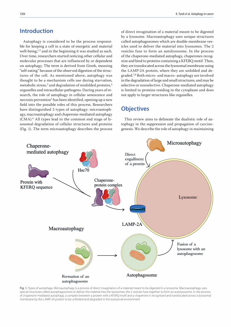
K. Turek et al. Autophagy in cancer1304
Introduction
Autophagy is considered to be the process responsi-ble for keeping a cell in a state of energetic and material well-being,1,2 and in the beginning it was studied as such. Over time, researchers started noticing other cellular and molecular processes that are influenced by or dependent on autophagy. The term is derived from Greek, meaning “self-eating” because of the observed digestion of the struc-tures of the cell. As mentioned above, autophagy was thought to be a mechanism cells use during starvation, metabolic stress,3 and degradation of misfolded proteins,4 organelles and intracellular pathogens. During years of re-search, the role of autophagy in cellular senescence and necrosis prevention5 has been identified, opening up a new field into the possible roles of this process. Researchers have distinguished 3 types of autophagy: microautoph-agy, macroautophagy and chaperone-mediated autophagy (CMA).6 All types lead to the common end stage of ly-sosomal degradation of cellular structures and proteins (Fig. 1). The term microautophagy describes the process
of direct invagination of a material meant to be digested by a lysosome. Macroautophagy uses unique structures called autophagosomes which are double-membrane ves-icles used to deliver the material into lysosomes. The 2 vesicles fuse to form an autolysosome. In the process of the chaperone-mediated autophagy, chaperones recog-nize and bind to proteins containing a KFERQ motif. Then, they are translocated across the lysosomal membrane using the LAMP-2A protein, where they are unfolded and de-graded.7,8 Both micro- and macro- autophagy are involved in the degradation of large and small structures, and may be selective or nonselective. Chaperone-mediated autophagy is limited to proteins residing in the cytoplasm and does not apply to larger structures like organelles.
Objectives
This review aims to delineate the dualistic role of au-tophagy in the suppression and propagation of carcino-genesis. We describe the role of autophagy in maintaining
Fig. 1. Types of autophagy. Microautophagy is a process of direct invagination of a material meant to be digested in a lysosome. Macroautophagy uses special structures called autophagosomes to deliver the material into the lysosomes; the 2 vesicles fuse together to form an autolysosome. In the process of chaperone-mediated autophagy, a complex between a protein with a KFERQ motif and a chaperone is recognized and translocated across a lysosomal membrane by the LAMP-2A protein to be unfolded and degraded in the lysosomal environment

Adv Clin Exp Med. 2021;30(12):1303–1314 1305
the balance between cell survival and death. Based on the available literature, we summarize the role of au-tophagy in carcinogenesis and the possible modulation of the tumor cell response to therapy.
Methodology
This review was prepared after a literature search was conducted using PubMed, Mendeley, Google, Scopus, Embase, and AccessMedicine. The literature search was limited to articles published between 2001 and 2021, using the following search terms: autophagy regulation, mac-roautophagy, chaperone-mediated autophagy, autopha-gosome formation inhibition, autophagy inhibition, and autophagy and treatment.
The complexity of autophagy function
Autophagy, or more specifically the deregulation of it, is considered an essential factor in the development of many types of diseases.6 The different types of au-tophagy and its selective and nonselective mechanisms of action result in an astounding diversity of diseases it may play a role in. The list of diseases that deregulated autophagy may be involved in, as a meaningful contribu-tive component, is constantly getting longer.9 Autoph-agy occurs on a basal and constitutional level. The role of the basal level has been recently highlighted as a home keeping mechanism for the cell. Different tissues demand various levels of autophagy. Therefore, tissues that do not divide after differentiation, such as neurons and myo-cytes, are acutely sensitive to the inhibition of autoph-agy. However, some tissues are less prone to the absence of autophagy. Neurodegenerative disorders are among the most commonly mentioned pathologies proven to be linked to autophagy.10–12 However, the main focus of this paper is to provide a highly researched descrip-tion of the relationship between autophagy and cancer. Nowadays, we can state that autophagy plays a notewor-thy role in both cellular protection against cancer and the growth of a tumor itself.3 Autophagy supports tumor growth by promoting the survival of neoplastic cells that have to cope with stressors as a result of their rapid pro-liferation, such as hypoxia and nutrient starvation. Its protective role is fulfilled by the inhibition of chronic inflammation and necrosis. The more critical roles of au-tophagy are removing reactive oxygen species (ROS) that emerge from dysfunctional mitochondria, causing in-tense damage to the cell, preventing cellular oncogene-induced senescence, and maintaining genomic stability.13 Thus, a conclusion may be drawn that autophagy plays a role in the early stages of tumor progression and even initiation.
The induction and regulation of autophagy
Autophagy consists of 5 main stages: initiation, elon-gation, maturation, fusion, and degradation. The com-plex process of initiation has many different triggers. Extensive investigations have been conducted on yeast and have established that nitrogen, carbon and nucleic acids starvation are all activators of autophagy, although some are stronger than others.14 In yeast, the starvation of compounds essential to cell survival is a strong stimu-lus to initiate autophagy, as is the same for mammalian cells. The starvation of amino acids and energy, the ab-sence of growth factors, and the accumulation of mis-folded proteins, ROS, AMP-activated protein kinase (AMPK) and calcium play a significant role in autophagy induction.15 Most of the triggers suppress the common pathway mTORC1, referred to as a nutrient-sensing ki-nase, but some have been proven to activate other sig-naling routes independent of mTORC1.16 The presence of a trigger begins the complex process of autophagy (Fig. 2). The process is controlled by over 30 autophagy-related (Atg) proteins.17 The Atg proteins are essential to control the correct course of autophagy. Numerous Atg genes have been proven to play an important role in tumor suppression. Consequently, decreased levels of many Atg proteins are found in tumor cells.18 Under “normal” conditions, mTORC1 suppresses autophagy via mTOR-mediated phosphorylation of Atg13 when nutrient levels are sufficient.19 However, in the presence of a trig-ger, the mTORC1 pathway is suppressed and the AMPKs (considered the most vital activator of autophagy) path-way allows autophagy to proceed.
The AMPK is the principal energy sensor for a cell and is activated when AMP:ATP and ADP:ATP ratios increase, thereby directly linking the process of autophagy with energy starvation.20 The AMPK has been proven to have a dualistic role in the induction of autophagy. By sup-pressing mTORC1, AMPK indirectly induces autophagy, but can also induce phosphorylation ULK1/2 to directly activate the process.21 The ULK1 is one of 5 orthologs of Atg13 found in yeast. So far, ULK1 and ULK2 have been proven to be involved in the induction of autophagy, and despite slight differences in function and constitution between them, they are often referred to interchange-ably. The ULK1/2, Atg13, FIP200, and Atg101 proteins form the so-called ULK1/2 complex.22 Although the exact course of interaction between these proteins has not been fully elucidated, it has been suggested that ULK1/2 re-quires both Atg13 and FIP200 for stability and correct function, as both enhance its kinase activity.23 The role of the ULK1/2 complex seems to be the phosphorylation of 2 proteins: AMBRA1 and ZIPK.24,25 The effect of these interactions causes the downstream events required for phagophore formation. As shown above, the relationship between mTORC1 and AMPK activity plays a vital role
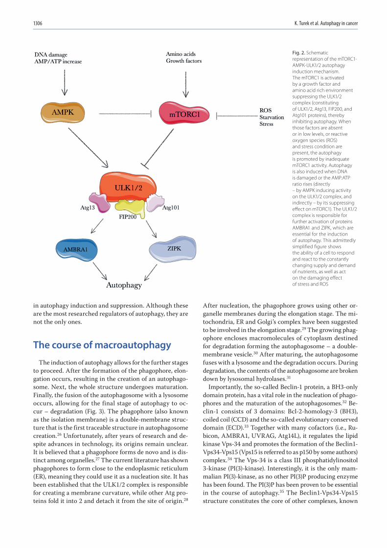
K. Turek et al. Autophagy in cancer1306
in autophagy induction and suppression. Although these are the most researched regulators of autophagy, they are not the only ones.
The course of macroautophagy
The induction of autophagy allows for the further stages to proceed. After the formation of the phagophore, elon-gation occurs, resulting in the creation of an autophago-some. Next, the whole structure undergoes maturation. Finally, the fusion of the autophagosome with a lysosome occurs, allowing for the final stage of autophagy to oc-cur – degradation (Fig. 3). The phagophore (also known as the isolation membrane) is a double-membrane struc-ture that is the first traceable structure in autophagosome creation.26 Unfortunately, after years of research and de-spite advances in technology, its origins remain unclear. It is believed that a phagophore forms de novo and is dis-tinct among organelles.27 The current literature has shown phagophores to form close to the endoplasmic reticulum (ER), meaning they could use it as a nucleation site. It has been established that the ULK1/2 complex is responsible for creating a membrane curvature, while other Atg pro-teins fold it into 2 and detach it from the site of origin.28
After nucleation, the phagophore grows using other or-ganelle membranes during the elongation stage. The mi-tochondria, ER and Golgi’s complex have been suggested to be involved in the elongation stage.29 The growing phag-ophore encloses macromolecules of cytoplasm destined for degradation forming the autophagosome – a double-membrane vesicle.30 After maturing, the autophagosome fuses with a lysosome and the degradation occurs. During degradation, the contents of the autophagosome are broken down by lysosomal hydrolases.31
Importantly, the so-called Beclin-1 protein, a BH3-only domain protein, has a vital role in the nucleation of phago-phores and the maturation of the autophagosomes.32 Be-clin-1 consists of 3 domains: Bcl-2-homology-3 (BH3), coiled coil (CCD) and the so-called evolutionary conserved domain (ECD).33 Together with many cofactors (i.e., Ru-bicon, AMBRA1, UVRAG, Atg14L), it regulates the lipid kinase Vps-34 and promotes the formation of the Beclin1-Vps34-Vps15 (Vps15 is referred to as p150 by some authors) complex.34 The Vps-34 is a class III phosphatidylinositol 3-kinase (PI(3)-kinase). Interestingly, it is the only mam-malian PI(3)-kinase, as no other PI(3)P producing enzyme has been found. The PI(3)P has been proven to be essential in the course of autophagy.35 The Beclin1-Vps34-Vps15 structure constitutes the core of other complexes, known
Fig. 2. Schematic representation of the mTORC1-AMPK-ULK1/2 autophagy induction mechanism. The mTORC1 is activated by a growth factor and amino acid rich environment suppressing the ULK1/2 complex (constituting of ULK1/2, Atg13, FIP200, and Atg101 proteins), thereby inhibiting autophagy. When those factors are absent or in low levels, or reactive oxygen species (ROS) and stress condition are present, the autophagy is promoted by inadequate mTORC1 activity. Autophagy is also induced when DNA is damaged or the AMP:ATP ratio rises (directly – by AMPK inducing activity on the ULK1/2 complex, and indirectly – by its suppressing effect on mTORC1). The ULK1/2 complex is responsible for further activation of proteins AMBRA1 and ZIPK, which are essential for the induction of autophagy. This admittedly simplified figure shows the ability of a cell to respond and react to the constantly changing supply and demand of nutrients, as well as act on the damaging effect of stress and ROS
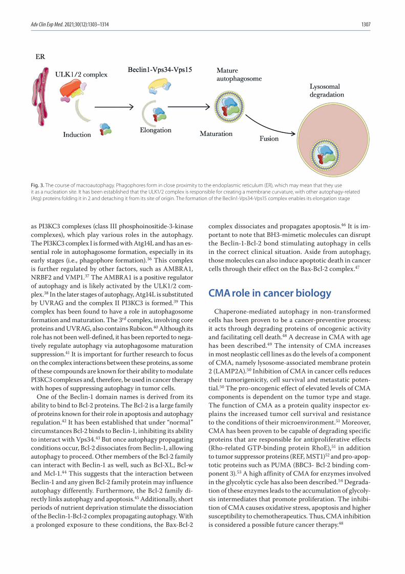
Adv Clin Exp Med. 2021;30(12):1303–1314 1307
as PI3KC3 complexes (class III phosphoinositide-3-kinase complexes), which play various roles in the autophagy. The PI3KC3 complex I is formed with Atg14L and has an es-sential role in autophagosome formation, especially in its early stages (i.e., phagophore formation).36 This complex is further regulated by other factors, such as AMBRA1, NRBF2 and VMP1.37 The AMBRA1 is a positive regulator of autophagy and is likely activated by the ULK1/2 com-plex.38 In the later stages of autophagy, Atg14L is substituted by UVRAG and the complex II PI3KC3 is formed.39 This complex has been found to have a role in autophagosome formation and maturation. The 3rd complex, involving core proteins and UVRAG, also contains Rubicon.40 Although its role has not been well-defined, it has been reported to nega-tively regulate autophagy via autophagosome maturation suppression.41 It is important for further research to focus on the complex interactions between these proteins, as some of these compounds are known for their ability to modulate PI3KC3 complexes and, therefore, be used in cancer therapy with hopes of suppressing autophagy in tumor cells.
One of the Beclin-1 domain names is derived from its ability to bind to Bcl-2 proteins. The Bcl-2 is a large family of proteins known for their role in apoptosis and autophagy regulation.42 It has been established that under “normal” circumstances Bcl-2 binds to Beclin-1, inhibiting its ability to interact with Vps34.43 But once autophagy propagating conditions occur, Bcl-2 dissociates from Beclin-1, allowing autophagy to proceed. Other members of the Bcl-2 family can interact with Beclin-1 as well, such as Bcl-XL, Bcl-w and Mcl-1.44 This suggests that the interaction between Beclin-1 and any given Bcl-2 family protein may influence autophagy differently. Furthermore, the Bcl-2 family di-rectly links autophagy and apoptosis.45 Additionally, short periods of nutrient deprivation stimulate the dissociation of the Beclin-1-Bcl-2 complex propagating autophagy. With a prolonged exposure to these conditions, the Bax-Bcl-2
complex dissociates and propagates apoptosis.46 It is im-portant to note that BH3-mimetic molecules can disrupt the Beclin-1-Bcl-2 bond stimulating autophagy in cells in the correct clinical situation. Aside from autophagy, those molecules can also induce apoptotic death in cancer cells through their effect on the Bax-Bcl-2 complex.47
CMA role in cancer biology
Chaperone-mediated autophagy in non-transformed cells has been proven to be a cancer-preventive process; it acts through degrading proteins of oncogenic activity and facilitating cell death.48 A decrease in CMA with age has been described.49 The intensity of CMA increases in most neoplastic cell lines as do the levels of a component of CMA, namely lysosome-associated membrane protein 2 (LAMP2A).50 Inhibition of CMA in cancer cells reduces their tumorigenicity, cell survival and metastatic poten-tial.50 The pro-oncogenic effect of elevated levels of CMA components is dependent on the tumor type and stage. The function of CMA as a protein quality inspector ex-plains the increased tumor cell survival and resistance to the conditions of their microenvironment.51 Moreover, CMA has been proven to be capable of degrading specific proteins that are responsible for antiproliferative effects (Rho-related GTP-binding protein RhoE),51 in addition to tumor suppressor proteins (REF, MST1)52 and pro-apop-totic proteins such as PUMA (BBC3- Bcl-2 binding com-ponent 3).53 A high affinity of CMA for enzymes involved in the glycolytic cycle has also been described.54 Degrada-tion of these enzymes leads to the accumulation of glycoly-sis intermediates that promote proliferation. The inhibi-tion of CMA causes oxidative stress, apoptosis and higher susceptibility to chemotherapeutics. Thus, CMA inhibition is considered a possible future cancer therapy.48
Fig. 3. The course of macroautophagy. Phagophores form in close proximity to the endoplasmic reticulum (ER), which may mean that they use it as a nucleation site. It has been established that the ULK1/2 complex is responsible for creating a membrane curvature, with other autophagy-related (Atg) proteins folding it in 2 and detaching it from its site of origin. The formation of the Beclin1-Vps34-Vps15 complex enables its elongation stage

K. Turek et al. Autophagy in cancer1308
Relation between autophagy and cancer
The role of autophagy in cancer is a double-edged sword. Under physiological conditions and a functional autophagy apparatus, a cell exposed to unfavorable factors (mentioned above) can recover. For instance, a cell is exposed to ROS and the damaged proteins emerge. Autophagy activates to adequately degrade the damaged proteins and organ-elles, averting potential carcinogenesis. On the other hand, when autophagy is impaired and the cellular re-sponse is inadequate, damaged proteins and organelles accumulate, and further oxidative stress results in DNA damage and genome instability. Many cells undergo apop-tosis or necrosis when the damage is fatal, but others may mutate unfavorably. The effect of these mutations may re-sult in chronic inflammation, compensatory proliferation to replace lost cells, and tumor initiation. In this instance, before carcinogenesis occurs, autophagy, as a protective mechanism, mitigates the damage done through vari-ous factors and prevents a tumor-initiating environment from occurring. The opposite happens once carcinogenesis has begun. Defective autophagy, in this instance, can lead to a better response of cancer cells to treatment, hypoxia and, in some cases, cancer cells death and tumor atrophy. This is because cancer cells are considered to be more autophagy-dependent than noncancerous cells. It is es-pecially applicable to cancer cells within the central part
of tumors, as this environment is notorious for nutrient deficiencies and oxygen deprivation. Additionally, these cells have higher bioenergetic and biosynthetic needs due to their high proliferation rates. It has been proven that autophagy in the central cells is significantly more intense, compared to those in the tumor periphery. In this case, autophagy might be seen as a mechanism that allows for the survival and even proliferation in highly unfavorable conditions, leading to further growth, invasion and propa-gation of metastases (Fig. 4).
By promoting the survival of cancer cells, autophagy also influences future phases of tumor development. The initial steps of metastasis require signals promoting migration and invasion. Many types of cells, such as in-flammatory cells, can provide such signals.55 Various inflammatory cells infiltrate the tumor reacting to ne-crosis, commonly seen in solid tumors, and produce dif-ferent effects on the tumor. Natural killer (NK) cells and cytotoxic T lymphocytes generally have anti-metastatic influence, while macrophages in the tumor environment usually correlate with a poor clinical prognosis.56 By pro-moting the survival of tumor cells under metabolic stress and hypoxic conditions, autophagy reduces tumor cells necrosis and tumor infiltration through pro-metastatic mediators, such as macrophages. Therefore, in the early stages of metastasis, autophagy may act as a suppressor of metastasis, necrosis, and consequently tumor infiltra-tion by inflammatory cells. However, in the later stages
Fig. 4. The growth of an aggressive tumor in conditions of supporting autophagy and a defect in the process. Autophagy is crucial for the cell in the hypoxic regions of a tumor, promoting neoplastic cell survival. Defects of autophagy result in lower resistance to harsh conditions within the tumor microenvironment and accumulation of reactive oxygen species (ROS) and defective mitochondria, hence preventing proliferation and consequently, cell death
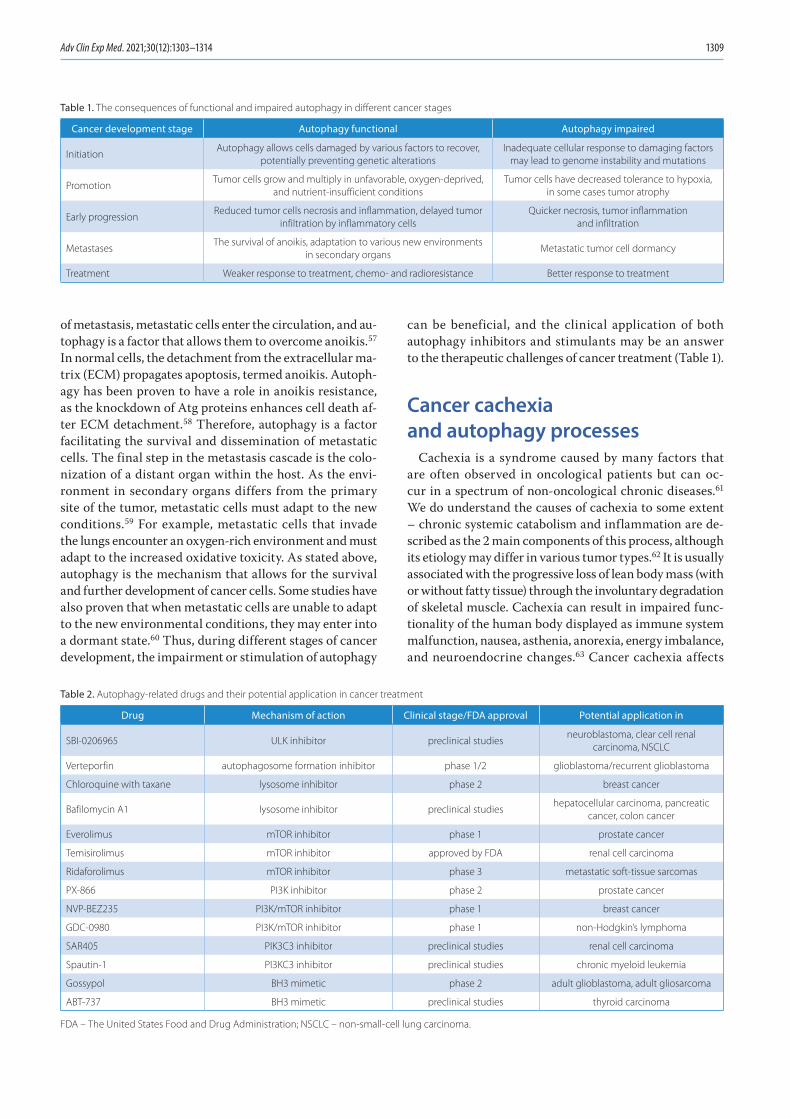
Adv Clin Exp Med. 2021;30(12):1303–1314 1309
of metastasis, metastatic cells enter the circulation, and au-tophagy is a factor that allows them to overcome anoikis.57 In normal cells, the detachment from the extracellular ma-trix (ECM) propagates apoptosis, termed anoikis. Autoph-agy has been proven to have a role in anoikis resistance, as the knockdown of Atg proteins enhances cell death af-ter ECM detachment.58 Therefore, autophagy is a factor facilitating the survival and dissemination of metastatic cells. The final step in the metastasis cascade is the colo-nization of a distant organ within the host. As the envi-ronment in secondary organs differs from the primary site of the tumor, metastatic cells must adapt to the new conditions.59 For example, metastatic cells that invade the lungs encounter an oxygen-rich environment and must adapt to the increased oxidative toxicity. As stated above, autophagy is the mechanism that allows for the survival and further development of cancer cells. Some studies have also proven that when metastatic cells are unable to adapt to the new environmental conditions, they may enter into a dormant state.60 Thus, during different stages of cancer development, the impairment or stimulation of autophagy
can be beneficial, and the clinical application of both autophagy inhibitors and stimulants may be an answer to the therapeutic challenges of cancer treatment (Table 1).
Cancer cachexia and autophagy processes
Cachexia is a syndrome caused by many factors that are often observed in oncological patients but can oc-cur in a spectrum of non-oncological chronic diseases.61 We do understand the causes of cachexia to some extent – chronic systemic catabolism and inflammation are de-scribed as the 2 main components of this process, although its etiology may differ in various tumor types.62 It is usually associated with the progressive loss of lean body mass (with or without fatty tissue) through the involuntary degradation of skeletal muscle. Cachexia can result in impaired func-tionality of the human body displayed as immune system malfunction, nausea, asthenia, anorexia, energy imbalance, and neuroendocrine changes.63 Cancer cachexia affects
Table 1. The consequences of functional and impaired autophagy in different cancer stages
Cancer development stage Autophagy functional Autophagy impaired
InitiationAutophagy allows cells damaged by various factors to recover,
potentially preventing genetic alterationsInadequate cellular response to damaging factors
may lead to genome instability and mutations
PromotionTumor cells grow and multiply in unfavorable, oxygen-deprived,
and nutrient-insufficient conditionsTumor cells have decreased tolerance to hypoxia,
in some cases tumor atrophy
Early progressionReduced tumor cells necrosis and inflammation, delayed tumor
infiltration by inflammatory cellsQuicker necrosis, tumor inflammation
and infiltration
MetastasesThe survival of anoikis, adaptation to various new environments
in secondary organsMetastatic tumor cell dormancy
Treatment Weaker response to treatment, chemo- and radioresistance Better response to treatment
Table 2. Autophagy-related drugs and their potential application in cancer treatment
Drug Mechanism of action Clinical stage/FDA approval Potential application in
SBI-0206965 ULK inhibitor preclinical studiesneuroblastoma, clear cell renal
carcinoma, NSCLC
Verteporfin autophagosome formation inhibitor phase 1/2 glioblastoma/recurrent glioblastoma
Chloroquine with taxane lysosome inhibitor phase 2 breast cancer
Bafilomycin A1 lysosome inhibitor preclinical studieshepatocellular carcinoma, pancreatic
cancer, colon cancer
Everolimus mTOR inhibitor phase 1 prostate cancer
Temisirolimus mTOR inhibitor approved by FDA renal cell carcinoma
Ridaforolimus mTOR inhibitor phase 3 metastatic soft-tissue sarcomas
PX-866 PI3K inhibitor phase 2 prostate cancer
NVP-BEZ235 PI3K/mTOR inhibitor phase 1 breast cancer
GDC-0980 PI3K/mTOR inhibitor phase 1 non-Hodgkin’s lymphoma
SAR405 PIK3C3 inhibitor preclinical studies renal cell carcinoma
Spautin-1 PI3KC3 inhibitor preclinical studies chronic myeloid leukemia
Gossypol BH3 mimetic phase 2 adult glioblastoma, adult gliosarcoma
ABT-737 BH3 mimetic preclinical studies thyroid carcinoma
FDA – The United States Food and Drug Administration; NSCLC – non-small-cell lung carcinoma.
K. Turek et al. Autophagy in cancer1310
50% of all cancer patients in the early stages of the disease and 80% as the disease advances,64 making it a concerning challenge for medicine. Cachexia can affect the entire or-ganism by reducing the quality of life, tolerance, response to treatment, and consequently, survival. Inflammatory fac-tors described in cancer cachexia are eicosanoids,65 tumor necrosis factor, interferon, interleukin-6 (IL-6), and IL-1β.66 Chronic inflammatory processes can lead to increased fatty acid content within the blood.67 Pettersen et al. found that IL-6 secreted by tumor cells stimulates autophagy in myo-tubes when complexed with soluble IL-6 receptors.68 Poor prognosis has also been described in lung cancer patients with elevated IL-6 levels.68 Systemic chronic inflammation, characteristic in cancer cachexia, is a result of immune cell infiltration of the tumor microenvironment and constant stimulation of the immune system by cancer. Systemic inflammation increases the demand for nutrients and en-ergy through catabolic upregulation. Selective autophagy is a process induced by various forms of stress – ROS and adenosine diphosphate (ADP) accumulation, hypoxia and inflammation. It ensures a continuous supply of energy and nutrients for a tumor as a functional adaptive cellular response. Penna et al. demonstrated that muscle atrophy occurring in cancer cachexia is associated with an aug-mented autophagy rate by studying markers of the process – Beclin-1 (autophagy induction marker), LC3B conver-sion (autophagosome levels) and p62/SQSTM1 (substrate sequestration).69 Another study, looking at the association between weight loss and autophagy-accelerated bioactiv-ity, showed no link in a heterogeneous group of oncologi-cal patients. However, a different study focusing on lung cancer patients indicated that autophagy-induced activity and weight loss was a characteristic in men only and was responsible for a worse prognosis.70
Oncogenes and tumor suppressors involved in autophagy
Years of studies on oncogenesis and autophagy identified shared genes in both processes.71 Today we know that both oncogenes and tumor suppressor genes play a part in au-tophagy. An oncogene, AKT1, has been identified as an in-hibitor of autophagy via mTOR activation.72 Mutations in the Ras gene are common in several types of aggressive cancers and also play a crucial role in autophagy.73 The Ras gene is a known upstream inhibitor of mTOR. On the other hand, PI3K inhibits autophagy via AKT1 activation and is responsible for the gain-of-function mutations seen in tu-mor cells.71,72 The Bcl-2 gene is known for its anti-apop-totic characteristics and inhibition of Beclin-1,73 a crucial protein in the initial stages of macroautophagy. Beclin-1 is also considered a tumor suppressor gene that is com-monly deleted in breast, ovarian and prostate cancers.73 The NF1 is a suppressor often mutated in von Reckling-hausen’s disease and juvenile myelomonocytic leukemia,
and can relieve Ras-mediated inhibition of autophagy.73 The TSC1 and TSC2 genes are mutated in tuberous sclero-sis and indirectly inhibit the PI3K-AKT1-mTOR pathway.74 Another tumor suppressor gene, RAB7A, is known for its rearrangement in leukemias, deletion in solid tumors and involvement in autophagosomal maturation.75 The DAPK1, yet another suppressor gene silenced in various tumors, relieves the inhibition of autophagy mediated by Bcl-2/Bcl-XL.76 The PTEN gene is downregulated in ovarian cancer and stops autophagy inhibition mediated by PI3K-AKT1.72 These examples highlight the fact that autophagy and carcinogenesis have a lot in common.
Treatment opportunities
The research on autophagy has brought to light new can-cer treatment possibilities.77 Thanks to the dualistic role of autophagy in cancer initiation and progression, there are many areas we can target to capitalize on the effect of autophagy in order to suit clinical treatment. Consider-ing that autophagy has an important role in tumor sup-pression, we can try to modulate and enhance it in patients with an increased risk of developing cancers (Table 2). On the other hand, once carcinogenesis has occurred, the inhibition of autophagy in cancer cells can be very beneficial. The 3rd potential application of our knowledge regarding autophagy is in modulating it to increase the sus-ceptibility of a tumor to radio- and chemotherapy.78,79
ULK inhibitors
The ULK proteins have a vital and comparatively well-researched role in autophagy induction and have been one of the main focuses of numerous studies. Many compounds involved in the inhibition of ULK kinase activity have been identified: compound 6, MRT68921, MRT67307, SBI-0206965, ULK100, and ULK101.77 The most researched compound is SBI-0206965. It has been proven to reduce the growth and induce apoptosis in some neuroblastoma cell lines, and sensitize one line to treatment. Further-more, it suppresses non-small cell lung cancer (NSCLC) cell growth, enhances its susceptibility to cisplatin, and even induces apoptosis in clear cell renal carcinoma (i.e., by blocking autophagy).80,81 Some studies have also in-dicated its utility in the potential treatment of myeloid leukemia FLT3-ITD.82
Autophagosome formation inhibition
Since autophagosome formation is an essential stage in macroautophagy, there have been studies to find a com-pound that suppresses it. Verteporfin disrupts autophago-some formation and other pathways in autophagy.83 Re-search has shown that verteporfin sensitizes osteosarcoma cells to cytotoxic drugs.84
Adv Clin Exp Med. 2021;30(12):1303–1314 1311
Lysosomes inhibitors
Lysosome function in autophagy prompted studies on chloroquine (CQ) and its analog hydroxychloroquine. Both substances are known for their use in malaria treatment and rheumatoid diseases, and their ap-plication in cancer therapy seems to be the next step. It is known that they can inhibit autophagy by directly interacting with lysosomes. As weak bases, they diffuse into the lysosome and undergo protonation to cause overall alkalization of the lysosome lumen. This creates an unfa-vorable environment for lysosomal enzymes and impairs their function. A protonated CQ cannot then diffuse out of the lysosome as it loses its ability to transverse cell mem-branes.85 Chloroquine also inhibits the fusion of the lyso-some with the autophagosome.86 Importantly, those effects did not go unnoticed and CQ has already entered clinical trials in combination with taxane and taxane-like drugs to evaluate the response of breast cancer cells to this drug combination.87 The challenge of long-term use of CQ and its derivatives is their well-known toxicity, which highly depends on the dose and duration of treatment. The most common side effects are retinopathy and cardiotoxicity.88,89
Another drug with similar effects on lysosomes is bafilo-mycin A1 (BafA1). It also alkalizes the lumen of lysosomes through a different mechanism than CQ. The BafA1 tar-gets the vacuolar-type H+-ATPase, a transmembrane proton pump responsible for acidifying the lysosome.90 Similarly to CQ and its derivatives, it also inhibits the au-tophagosome-lysosome fusion and impairs TORC1 signal-ing. Thus, many researchers speculate about BafA1 future use in cancer treatments.91
mTOR inhibitors
The name of mTORC1 derived from the fact that it is a target of rapamycin.92 Rapamycin is a macrolide compound known for its role in transplant surgery (ad-ministered to prevent transplant rejection) and coronary stent coating. Additionally, studies have shown, that it might be considered an anti-aging drug (e.g., it ex-tended the lifespan of mice in some studies).93 These ef-fects are mainly due to its inhibition of mTOR proteins and consequently, autophagy activation. Many research-ers have suggested that rapamycin could be used to slow down most (if not all) age-related diseases in humans, including some cancers.94 Because mTOR activity is com-monly upregulated in cancer, its inhibitors have signifi-cant promise in cancer therapy.95 Therefore, rapamycin and its derivatives (known as rapalogs) such as temsi-rolimus, everolimus and ridaforolimus are undergoing clinical trials to evaluate their potential use in cancer treatment. Temsirolimus has already been approved by the The United States Food and Drug Administration (FDA) in 2007 for use in advanced renal cell carcinoma (RCC) treatment.
PI3K inhibitors
The critical role of PI3K in autophagy inspired the re-search into compounds able to block its function.96 Sev-eral molecules have been found, such as 3-methyladenine (3-MA), LY294002, wortmannin, and its analog PX-866 (Oncothyreon). Clinical trials have been conducted using PX-866 on patients with advanced prostate cancer, although only modest single-agent activity has been reported. How-ever, more research concerning the activity of PX-866 com-bined with other drugs needs to be conducted.97 Addition-ally, the compounds able to inhibit both PI3K and mTOR, such as NVP-BEZ235 and GDC-0980, have entered clinical trials evaluating their potential use in cancer therapy.
PI3KC3 complex inhibitors
Since PI3KC3 complexes consist of various proteins, there are a few ways to block their activity. The SAR405 is a selective Vps34 inhibitor that affects late autophago-some-lysosome interaction and prevents autophagy.98 Fur-thermore, the available research has shown that SAR405 combined with everolimus (mTOR inhibitor) to produce strong antiproliferative effects on renal tumor cells.99 The potential drawback of SAR405 administration is that PI3KC3 is also involved in endosomal trafficking and therefore, can negatively affect cellular secretion.
Spautin-1 is a compound that impairs PI3KC3 by a differ-ent approach. It inhibits autophagy by enhancing the deg-radation of Beclin-1 and thus, prevents the formation of the PI3KC3 complex.100 Shao et al. reported promising results with its potential application in overcoming resis-tance to imatinib mesylate-induced autophagy in chronic myeloid leukemia.101 Not only does it show proapoptotic properties, but it also potentiates the efficacy of imatinib mesylate in chronic myeloid leukemia cells.
BH3 mimetics
The theory behind administering BH3 mimetics is to stim-ulate autophagy in cells by liberating Beclin-1 from Bcl-2.102 Stimulating this interaction can be beneficial also because of the possibility of inducing apoptotic cell death by negating the anti-apoptotic effects of the Bcl-2-Bax complex. Gossypol is a polyphenolic aldehyde that works as a pan-Bcl-2 inhibi-tor.103 Clinical trials have been conducted in patients with glioblastoma and despite some adverse events (including car-diac and gastrointestinal disorders), the results are promis-ing.104 Another BH3-mimetic drug, ABT-737 has been tested on various thyroid carcinoma cell lines and has been proven to cause apoptotic cell death and a synergistic effect with chemotherapeutic drugs.105 Furthermore, there is an ana-log of ABT-737 that can be administered orally, ABT-236. Although the clinical application for our constantly evolving knowledge about autophagy is within our grasp, there is still a magnitude of research that needs to be conducted.
K. Turek et al. Autophagy in cancer1312
Conclusions
From many years of studies, we have learned a lot about autophagy and its role in the pathogenesis of certain dis-eases. As far as our understanding of the molecular pathways of autophagy is concerned, their relation to cancer seems to be more profound and sophisticated than initially antici-pated. We know that it is a housekeeping mechanism of a cell that decreases chances of neoplastic transformation, but after malignant transformation, it supports the prolifera-tion and prosperity of cancer cells. Confirming autophagy as a mechanism that promotes tumor growth, provides us with the knowledge to develop new treatment options for on-cologic patients and brings hope to the idea of curing cancer.
ORCID iDsKacper Turek https://orcid.org/0000-0001-6551-8079Michał Jarocki https://orcid.org/0000-0001-8394-6018Julita Kulbacka https://orcid.org/0000-0001-8272-5440Jolanta Saczko https://orcid.org/0000-0001-5273-5293
References1. Levine B, Klionsky DJ. Development by self-digestion: Molecular
mechanisms and biological functions of autophagy. Dev Cell. 2004; 6(4):463–477. doi:10.1016/s1534-5807(04)00099-1
2. Mizushima N. Autophagy: Process and function. Genes Dev. 2007; 21(22):2861–2873. doi:10.1101/gad.1599207
3. Mathew R, Karantza-Wadsworth V, White E. Role of autophagy in cancer. Nat Rev Cancer. 2007;7(12):961–967. doi:10.1038/nrc2254
4. Williams A, Jahreiss L, Sarkar S, et al. Aggregate-prone proteins are cleared from the cytosol by autophagy: Therapeutic implications. Curr Top Dev Biol. 2006;76:89–101. doi:10.1016/S0070-2153(06)76003-3
5. Degenhardt K, Mathew R, Beaudoin B, et al. Autophagy promotes tumor cell survival and restricts necrosis, inflammation, and tumor-igenesis. Cancer Cell. 2006;10(1):51–64. doi:10.1016/j.ccr.2006.06.001
6. Mizushima N, Levine B, Cuervo AM, Klionsky DJ. Autophagy fights disease through cellular self-digestion. Nature. 2008;451(7182): 1069–1075. doi:10.1038/nature06639
7. Massey AC, Zhang C, Cuervo AM. Chaperone-mediated autophagy in aging and disease. Curr Top Dev Biol. 2006;73:205–235. doi:10.1016/S0070-2153(05)73007-6
8. Saftig P, Beertsen W, Eskelinen EL. LAMP-2: A control step for phago-some and autophagosome maturation. Autophagy. 2008;4(4):510–512. doi:10.4161/auto.5724
9. Levine B, Kroemer G. Autophagy in the pathogenesis of disease. Cell. 2008;132(1):27–42. doi:10.1016/j.cell.2007.12.018
10. Komatsu M, Waguri S, Chiba T, et al. Loss of autophagy in the central nervous system causes neurodegeneration in mice. Nature. 2006; 441(7095):880–884. doi:10.1038/nature04723
11. Martinez-Vicente M, Cuervo AM. Autophagy and neurodegenera-tion: When the cleaning crew goes on strike. Lancet Neurol. 2007;6(4): 352–361. doi:10.1016/S1474-4422(07)70076-5
12. Shibata M, Lu T, Furuya T, et al. Regulation of intracellular accumu-lation of mutant Huntingtin by Beclin 1. J Biol Chem. 2006;281(20): 14474–14485. doi:10.1074/jbc.M600364200
13. Nelson DA, Tan TT, Rabson AB, Anderson D, Degenhardt K, White E. Hypoxia and defective apoptosis drive genomic instability and tumor-igenesis. Genes Dev. 2004;18(17):2095–2107. doi:10.1101/gad.1204904
14. Reggiori F, Klionsky DJ. Autophagic processes in yeast: Mechanism, machinery and regulation. Genetics. 2013;194(2):341–361. doi:10.1534/genetics.112.149013
15. Wang L, Ye X, Zhao T. The physiological roles of autophagy in the mammalian life cycle. Biol Rev Camb Philos Soc. 2019;94(2):503–516. doi:10.1111/brv.12464
16. Sabatini DM. Twenty-five years of mTOR: Uncovering the link from nutrients to growth. Proc Natl Acad Sci U S A. 2017;114(45):11818–11825. doi:10.1073/pnas.1716173114
17. Nishimura T, Tooze SA. Emerging roles of ATG proteins and mem-brane lipids in autophagosome formation. Cell Discov. 2020;6(1):32. doi:10.1038/s41421-020-0161-3
18. Li X, He S, Ma B. Autophagy and autophagy-related proteins in cancer. Mol Cancer. 2020;19(1):12. doi:10.1186/s12943-020-1138-4
19. Puente C, Hendrickson RC, Jiang X. Nutrient-regulated phosphory-lation of ATG13 inhibits starvation-induced autophagy. J Biol Chem. 2016;291(11):6026–6035. doi:10.1074/jbc.M115.689646
20. Hardie DG. AMP-activated/SNF1 protein kinases: Conserved guard-ians of cellular energy. Nat Rev Mol Cell Biol. 2007;8(10):774–785. doi:10.1038/nrm2249
21. Hardie DG. AMPK and autophagy get connected. EMBO J. 2011;30(4): 634–635. doi:10.1038/emboj.2011.12
22. Mizushima N. The role of the Atg1/ULK1 complex in autophagy reg-ulation. Curr Opin Cell Biol. 2010;22(2):132–139. doi:10.1016/j.ceb. 2009.12.004
23. Ganley IG, Lam DH, Wang J, Ding X, Chen S, Jiang X. ULK1.ATG13.FIP200 complex mediates mTOR signaling and is essential for auto-phagy. J Biol Chem. 2009;284(18):12297–12305. doi:10.1074/jbc.M900 573200
24. Di Bartolomeo S, Corazzari M, Nazio F, et al. The dynamic interaction of AMBRA1 with the dynein motor complex regulates mammalian autophagy. J Cell Biol. 2010;191(1):155–168. doi:10.1083/jcb.201002100
25. Alers S, Löffler AS, Wesselborg S, Stork B. Role of AMPK-mTOR-Ulk1/2 in the regulation of autophagy: Cross talk, shortcuts, and feedbacks. Mol Cell Biol. 2012;32(1):2–11. doi:10.1128/MCB.06159-11
26. Juhasz G, Neufeld TP. Autophagy: A forty-year search for a miss-ing membrane source. PLoS Biol. 2006;4(2):e36. doi:10.1371/journal.pbio.0040036
27. Schütter M, Graef M. Localized de novo phospholipid synthesis drives autophagosome biogenesis. Autophagy. 2020;16(4):770–771. doi:10.1080/15548627.2020.1725379
28. Hurley JH, Young LN. Mechanisms of autophagy initiation. Annu Rev Biochem. 2017;86:225–244. doi:10.1146/annurev-biochem-061516- 044820
29. Perrotta I. The origin of the autophagosomal membrane in human atherosclerotic plaque: A preliminary ultrastructural study. Ultrastruct Pathol. 2017;41(5):327–334. doi:10.1080/01913123.2017.1349853
30. Graef M. Recent advances in the understanding of autophagosome bio-genesis. F1000Res. 2020;9:F1000. doi:10.12688/f1000research.22111.1
31. Stefaniak S, Wojtyla Ł, Pietrowska-Borek M, Borek S. Completing autophagy: Formation and degradation of the autophagic body and metabolite salvage in plants. Int J Mol Sci. 2020;21(6):2205. doi:10. 3390/ijms21062205
32. Oberstein A, Jeffrey PD, Shi Y. Crystal structure of the Bcl-XL-Beclin 1 peptide complex: Beclin 1 is a novel BH3-only protein. J Biol Chem. 2007;282(17):13123–13132. doi:10.1074/jbc.M700492200
33. Kang R, Zeh HJ, Lotze MT, Tang D. The Beclin 1 network regulates autophagy and apoptosis. Cell Death Differ. 2011;18(4):571–580. doi:10. 1038/cdd.2010.191
34. Kihara A, Kabeya Y, Ohsumi Y, Yoshimori T. Beclin-phosphatidylinosi-tol 3-kinase complex functions at the trans-Golgi network. EMBO Rep. 2001;2(4):330–335. doi:10.1093/embo-reports/kve061
35. Nascimbeni AC, Codogno P, Morel E. Phosphatidylinositol-3-phos-phate in the regulation of autophagy membrane dynamics. FEBS J. 2017;284(9):1267–1278. doi:10.1111/febs.13987
36. Matsunaga K, Morita E, Saitoh T, et al. Autophagy requires endoplasmic reticulum targeting of the PI3-kinase complex via Atg14L. J Cell Biol. 2010;190(4):511–521. doi:10.1083/jcb.200911141
37. Zhong Y, Morris DH, Jin L, et al. Nrbf2 protein suppresses autophagy by modulating Atg14L protein-containing Beclin 1-Vps34 complex architecture and reducing intracellular phosphatidylinositol-3 phos-phate levels. J Biol Chem. 2014;289(38):26021–26037. doi:10.1074/jbc.M114.561134
38. Cianfanelli V, Nazio F, Cecconi F. Connecting autophagy: AMBRA1 and its network of regulation. Mol Cell Oncol. 2015;2(1):e970059. doi:10.4161/23723548.2014.970059
39. Liang C, Lee J, Inn K, et al. Beclin1-binding UVRAG targets the class C Vps complex to coordinate autophagosome maturation and endo-cytic trafficking. Nat Cell Biol. 2008;10(7):776–787. doi:10.1038/ncb1740
40. Sun Q, Zhang J, Fan W, et al. The RUN domain of rubicon is important for hVps34 binding, lipid kinase inhibition, and autophagy suppres-sion. J Biol Chem. 2011;286(1):185–191. doi:10.1074/jbc.M110.126425
Adv Clin Exp Med. 2021;30(12):1303–1314 1313
41. Tabata K, Matsunaga K, Sakane A, Sasaki T, Noda T, Yoshimori T. Rubi-con and PLEKHM1 negatively regulate the endocytic/autophagic pathway via a novel Rab7-binding domain. Mol Biol Cell. 2010;21(23): 4162–4172. doi:10.1091/mbc.E10-06-0495
42. Hatok J, Racay P. Bcl-2 family proteins: Master regulators of cell sur-vival. Biomol Concepts. 2016;7(4):259–270. doi:10.1515/bmc-2016-0015
43. Pattingre S, Tassa A, Qu X, et al. Bcl-2 antiapoptotic proteins inhib-it Beclin 1-dependent autophagy. Cell. 2005;122(6):927–939. doi:10. 1016/j.cell.2005.07.002
44. Erlich S, Mizrachy L, Segev O, et al. Differential interactions between Beclin 1 and Bcl-2 family members. Autophagy. 2007;3(6):561–568. doi:10.4161/auto.4713
45. Marquez RT, Xu L. Bcl-2:Beclin 1 complex: Multiple, mechanisms regu-lating autophagy/apoptosis toggle switch. Am J Cancer Res. 2012;2(2): 214–221. PMID:22485198
46. Wei Y, Sinha S, Levine B. Dual role of JNK1-mediated phosphory-lation of Bcl-2 in autophagy and apoptosis regulation. Autophagy. 2008;4(7):949–951. doi:10.4161/auto.6788
47. Campbell KJ, Tait SWG. Targeting BCL-2 regulated apoptosis in cancer. Open Biol. 2018;8(5):180002. doi:10.1098/rsob.180002
48. Kaushik S, Cuervo AM. The coming of age of chaperone-mediated autophagy. Nat Rev Mol Cell Biol. 2018;19(6):365–381. doi:10.1038/s41580-018-0001-6
49. Kiffin R, Kaushik S, Zeng M, et al. Altered dynamics of the lysosom-al receptor for chaperone-mediated autophagy with age. J Cell Sci. 2007;120(Pt 5):782–791. doi:10.1242/jcs.001073
50. Kon M, Kiffin R, Koga H, et al. Chaperone-mediated autophagy is required for tumor growth. Sci Transl Med. 2011;3(109):109ra117. doi:10.1126/scitranslmed.3003182
51. Zhou J, Yang J, Fan X, et al. Chaperone-mediated autophagy regu-lates proliferation by targeting RND3 in gastric cancer. Autophagy. 2016;12(3):515–528. doi:10.1080/15548627.2015.1136770
52. Li L, Fang R, Liu B, et al. Deacetylation of tumor-suppressor MST1 in Hippo pathway induces its degradation through HBXIP-elevated HDAC6 in promotion of breast cancer growth. Oncogene. 2016;35(31): 4048–4057. doi:10.1038/onc.2015.476
53. Xie W, Zhang L, Jiao H, et al. Chaperone-mediated autophagy pre-vents apoptosis by degrading BBC3/PUMA. Autophagy. 2015;11(9): 1623–1635. doi:10.1080/15548627.2015.1075688
54. Lv L, Li D, Zhao D, et al. Acetylation targets the M2 isoform of pyruvate kinase for degradation through chaperone-mediated autophagy and promotes tumor growth. Mol Cell. 2011;42(6):719–730. doi:10.1016/j.molcel.2011.04.025
55. Zhao H, Wu L, Yan G, et al. Inflammation and tumor progression: Signal-ing pathways and targeted intervention. Signal Transduct Target Ther. 2021;6(1):263. doi:10.1038/s41392-021-00658-5
56. Coussens LM, Werb Z. Inflammation and cancer. Nature. 2002; 420(6917):860–867. doi:10.1038/nature01322
57. Dower CM, Wills CA, Frisch SM, Wang HG. Mechanisms and context underlying the role of autophagy in cancer metastasis. Autophagy. 2018;14(7):1110–1128. doi:10.1080/15548627.2018.1450020
58. Douma S, Van Laar T, Zevenhoven J, Meuwissen R, Van Garderen E, Peeper DS. Suppression of anoikis and induction of metastasis by the neurotrophic receptor TrkB. Nature. 2004;430(7003):1034–1039. doi:10.1038/nature02765
59. Schild T, Low V, Blenis J, Gomes AP. Unique metabolic adaptations dictate distal organ-specific metastatic colonization. Cancer Cell. 2018;33(3):347–354. doi:10.1016/j.ccell.2018.02.001
60. Chavez-Dominguez R, Perez-Medina M, Lopez-Gonzalez JS, Galicia-Velasco M, Aguilar-Cazares D. The double-edge sword of autopha-gy in cancer: From tumor suppression to pro-tumor activity. Front Oncol. 2020;10:578418. doi:10.3389/fonc.2020.578418
61. Fearon K, Strasser F, Anker SD, et al. Definition and classification of cancer cachexia: An international consensus. Lancet Oncol. 2011; 12(5):489–495. doi:10.1016/S1470-2045(10)70218-7
62. Baracos VE, Martin L, Korc M, Guttridge DC, Fearon KCH. Cancer-associat-ed cachexia. Nat Rev Dis Primers. 2018;4(1):17105. doi:10.1038/nrdp.2017.105
63. Seelaender M, Laviano A, Busquets S, Püschel GP, Margaria T, Batista ML. Inflammation in cachexia. Mediators Inflamm. 2015;2015:536954. doi:10.1155/2015/536954
64. Gonçalves R de C, Freire PP, Coletti D, Seelaender M. Tumor micro-environment autophagic processes and cachexia: The missing link? Front Oncol. 2021;10:617109. doi:10.3389/fonc.2020.617109
65. Vaughan VC, Martin P, Lewandowski PA. Cancer cachexia: Impact, mechanisms and emerging treatments. J Cachexia Sarcopenia Muscle. 2013;4(2):95–109. doi:10.1007/s13539-012-0087-1
66. Tisdale MJ. Mechanisms of cancer cachexia. Physiol Rev. 2009;89(2): 381–410. doi:10.1152/physrev.00016.2008
67. Flueck JL. Body composition in swiss elite wheelchair athletes. Front Nutr. 2020;7:1. doi:10.3389/fnut.2020.00001
68. Pettersen K, Andersen S, Degen S, et al. Cancer cachexia associates with a systemic autophagy-inducing activity mimicked by cancer cell-derived IL-6 trans-signaling. Sci Rep. 2017;7(1):2046. doi:10.1038/s41598-017-02088-2
69. Penna F, Costamagna D, Pin F, et al. Autophagic degradation contrib-utes to muscle wasting in cancer cachexia. Am J Pathol. 2013;182(4): 1367–1378. doi:10.1016/j.ajpath.2012.12.023
70. Cosper PF, Leinwand LA. Cancer causes cardiac atrophy and autoph-agy in a sexually dimorphic manner. Cancer Res. 2011;71(5):1710–1720. doi:10.1158/0008-5472.CAN-10-3145
71. Ávalos Y, Canales J, Bravo-Sagua R, Criollo A, Lavandero S, Quest AFG. Tumor suppression and promotion by autophagy. Biomed Res Int. 2014;2014:603980. doi:10.1155/2014/603980
72. Cully M, You H, Levine AJ, Mak TW. Beyond PTEN mutations: The PI3K pathway as an integrator of multiple inputs during tumorigenesis. Nat Rev Cancer. 2006;6(3):184–192. doi:10.1038/nrc1819
73. Levine B, Sinha SC, Kroemer G. Bcl-2 family members: Dual regula-tors of apoptosis and autophagy. Autophagy. 2008;4(5):600–606. doi:10.4161/auto.6260
74. Schwartz RA, Fernández G, Kotulska K, Jóźwiak S. Tuberous sclero-sis complex: Advances in diagnosis, genetics, and management. J Am Acad Dermatol. 2007;57(2):189–202. doi:10.1016/j.jaad.2007.05.004
75. Edinger AL, Cinalli RM, Thompson CB. Rab7 prevents growth factor-independent survival by inhibiting cell-autonomous nutrient trans-porter expression. Dev Cell. 2003;5(4):571–582. doi:10.1016/S1534-5807(03)00291-0
76. Harrison B, Kraus M, Burch L, et al. DAPK-1 binding to a linear pep-tide motif in MAP1B stimulates autophagy and membrane blebbing. J Biol Chem. 2008;283(15):9999–10014. doi:10.1074/jbc.M706040200
77. Pérez-Hernández M, Arias A, Martínez-García D, Pérez-Tomás R, Que-sada R, Soto-Cerrato V. Targeting autophagy for cancer treatment and tumor chemosensitization. Cancers (Basel). 2019;11(10):1599. doi:10.3390/cancers11101599
78. Tam SY, Wu VWC, Law HKW. Influence of autophagy on the efficacy of radiotherapy. Radiat Oncol. 2017;12(1):57. doi:10.1186/s13014-017-0795-y
79. Sui X, Chen R, Wang Z, et al. Autophagy and chemotherapy resistance: A promising therapeutic target for cancer treatment. Cell Death Dis. 2013;4(10):e838. doi:10.1038/cddis.2013.350
80. Tang F, Hu P, Yang Z, et al. SBI0206965, a novel inhibitor of Ulk1, sup-presses non-small cell lung cancer cell growth by modulating both autophagy and apoptosis pathways. Oncol Rep. 2017;37(6):3449–3458. doi:10.3892/or.2017.5635
81. Lu J, Zhu L, Zheng LP, et al. Overexpression of ULK1 represents a poten-tial diagnostic marker for clear cell renal carcinoma and the antitumor effects of SBI-0206965. EBioMedicine. 2018;34:85–93. doi:10.1016/j. ebiom.2018.07.034
82. Hwang DY, Eom JI, Jang JE, et al. ULK1 inhibition as a targeted ther-apeutic strategy for FLT3-ITD-mutated acute myeloid leukemia. J Exp Clin Cancer Res. 2020;39(1):85. doi:10.1186/s13046-020-01580-4
83. Donohue E, Tovey A, Vogl AW, et al. Inhibition of autophagosome formation by the benzoporphyrin derivative verteporfin. J Biol Chem. 2011;286(9):7290–7300. doi:10.1074/jbc.M110.139915
84. Saini H, Sharma H, Mukherjee S, Chowdhury S, Chowdhury R. Verte-porfin disrupts multiple steps of autophagy and regulates p53 to sen-sitize osteosarcoma cells. Cancer Cell Int. 2021;21(1):52. doi:10.1186/s12935-020-01720-y
85. Browning DJ. Pharmacology of chloroquine and hydroxychloroquine. In: Browning DJ, ed. Hydroxychloroquine and Chloroquine Retinopathy. New York, USA: Springer; 2014:35–63.
86. Mauthe M, Orhon I, Rocchi C, et al. Chloroquine inhibits autophagic flux by decreasing autophagosome-lysosome fusion. Autophagy. 2018;14(8):1435–1455. doi:10.1080/15548627.2018.1474314
87. Sparano JA. Taxanes for breast cancer: An evidence-based review of randomized phase II and phase III trials. Clin Breast Cancer. 2000; 1(1):32. doi:10.3816/cbc.2000.n.002
K. Turek et al. Autophagy in cancer1314
88. Oluleye TS, Babalola Y, Ijaduola M. Chloroquine retinopathy: Pattern of presentation in Ibadan, Sub-Sahara Africa. Eye (Lond). 2016;30(1): 64–67. doi:10.1038/eye.2015.185
89. Chatre C, Roubille F, Vernhet H, Jorgensen C, Pers YM. Cardiac com-plications attributed to chloroquine and hydroxychloroquine: A sys-tematic review of the literature. Drug Saf. 2018;41(10):919–931. doi:10. 1007/s40264-018-0689-4
90. Fedele AO, Proud CG. Chloroquine and bafilomycin A mimic lyso-somal storage disorders and impair mTORC1 signalling. Biosci Rep. 2020;40(4):BSR20200905. doi:10.1042/BSR20200905
91. Mauvezin C, Nagy P, Juhász G, Neufeld TP. Autophagosome-lysosome fusion is independent of V-ATPase-mediated acidification. Nat Commun. 2015;6(1):7007. doi:10.1038/ncomms8007
92. Lamming DW. Inhibition of the mechanistic target of rapamycin (mTOR): Rapamycin and beyond. Cold Spring Harb Perspect Med. 2016;6(5):a025924. doi:10.1101/cshperspect.a025924
93. Lamming DW, Ye L, Katajisto P, et al. Rapamycin-induced insulin resis-tance is mediated by mTORC2 loss and uncoupled from longevity. Science. 2012;335(6076):1638–1643. doi:10.1126/science.1215135
94. Blagosklonny MV. Rapamycin for longevity: Opinion article. Aging (Albany NY). 2019;11(19):8048–8067. doi:10.18632/aging.102355
95. Ballou LM, Lin RZ. Rapamycin and mTOR kinase inhibitors. J Chem Biol. 2008;1(1–4):27–36. doi:10.1007/s12154-008-0003-5
96. McNamara CR, Degterev A. Small-molecule inhibitors of the PI3K signaling network. Future Med Chem. 2011;3(5):549–565. doi:10.4155/fmc.11.12
97. Hotte SJ, Chi KN, Joshua AM, et al. A phase II study of PX-866 in patients with recurrent or metastatic castration-resistant prostate cancer: Canadian cancer trials group study IND205. Clin Genitourin Cancer. 2019;17(3):201–208.e1. doi:10.1016/j.clgc.2019.03.005
98. Ronan B, Flamand O, Vescovi L, et al. A highly potent and selective Vps34 inhibitor alters vesicle trafficking and autophagy. Nat Chem Biol. 2014;10(12):1013–1019. doi:10.1038/nchembio.1681
99. Pasquier B. SAR405, a PIK3C3/VPS34 inhibitor that prevents autoph-agy and synergizes with MTOR inhibition in tumor cells. Autophagy. 2015;11(4):725–726. doi:10.1080/15548627.2015.1033601
100. Liu J, Xia H, Kim M, et al. Beclin1 controls the levels of p53 by regu-lating the deubiquitination activity of USP10 and USP13. Cell. 2011; 147(1):223–234. doi:10.1016/j.cell.2011.08.037
101. Shao S, Li S, Qin Y, et al. Spautin-1, a novel autophagy inhibitor, enhances imatinib-induced apoptosis in chronic myeloid leukemia. Int J Oncol. 2014;44(5):1661–1668. doi:10.3892/ijo.2014.2313
102. Maiuri MC, Criollo A, Tasdemir E, et al. BH3-only proteins and BH3 mimetics induce autophagy by competitively disrupting the interac-tion between Beclin 1 and Bcl-2/Bcl-XL. Autophagy. 2007;3(4):374–376. doi:10.4161/auto.4237
103. Vogler M, Weber K, Dinsdale D, et al. Different forms of cell death induced by putative BCL2 inhibitors. Cell Death Differ. 2009;16(7): 1030–1039. doi:10.1038/cdd.2009.48
104. Voss V, Senft C, Lang V, et al. The pan-Bcl-2 inhibitor (-)-gossypol triggers autophagic cell death in malignant glioma. Mol Cancer Res. 2010;8(7):1002–1016. doi:10.1158/1541-7786.MCR-09-0562
105. Broecker-Preuss M, Becher-Boveleth N, Müller S, Mann K. The BH3 mimetic drug ABT-737 induces apoptosis and acts synergistically with chemotherapeutic drugs in thyroid carcinoma cells. Cancer Cell Int. 2016;16(1):27. doi:10.1186/s12935-016-0303-8

Cite asNowiński A, Stachyra K, Szybińska M, Bednarek M, Pływaczewski R, Śliwiński P. The influence of comorbidities on mortality in bronchiectasis: A prospective, observational study. Adv Clin Exp Med. 2021;30(12):1315–1321. doi:10.17219/acem/144200
DOI10.17219/acem/144200
Copyright© 2021 by Wroclaw Medical University This is an article distributed under the terms of theCreative Commons Attribution 3.0 Unported (CC BY 3.0)(https://creativecommons.org/licenses/by/3.0/)
Address for correspondenceAdam NowińskiE-mail: [email protected]
Funding sourcesNone declared
Conflict of interestNone declared
Received on August 25, 2021Reviewed on October 18, 2021Accepted on November 23, 2021
Published online on December 17, 2021
AbstractBackground. Bronchiectasis is a progressive chronic disease associated with an increased risk of mortality.
Objectives. To identify the prevalence of comorbidities in patients with bronchiectasis and the impact of these comorbidities on mortality.
Materials and methods. A cohort of 93 patients with computed tomography (CT)-confirmed bronchiec-tasis admitted consecutively to a tertiary teaching hospital was observed over a period of 5 years. All patients were carefully observed for comorbidities and mortality.
Results. A total of 43 men (46.2%) and 50 women (53.8%) with a median age of 66.0 years (interquartile range (IQR) 59.7–74.0 years), and a median of 3 comorbidities at baseline (IQR 1–5) were observed. The mor-tality rate during the observation period was 16%. The median number of comorbidities was significantly higher in the group of non-survivors (5 (IQR 3–5.75)) compared with survivors (3 (IQR 1–4); p = 0.0100). The burden of comorbidities was associated with an increased hazard of death: having 4 or more comorbidities was associated with an increased risk of death compared to patients with 2 or 3 coexisting illnesses (hazard ratio (HR) = 1.35 (95% confidence interval (95% CI) [0.41, 4.41]); p = 0.0494). The Bronchiectasis Aetiology Comorbidity Index (BACI) was a significant predictor of death in patients with severe bronchiectasis.
Conclusions. We found a significant number of comorbidities in patients with bronchiectasis. In these patients, the comorbidity burden has an impact on mortality. The BACI is a useful tool for the clinical assess-ment of patients with severe bronchiectasis.
Key words: survival, bronchiectasis, comorbidities, mortality
Research letters
The influence of comorbidities on mortality in bronchiectasis: A prospective, observational study
Adam NowińskiA–F, Katarzyna StachyraA–F, Maria SzybińskaA–F, Michał BednarekA–F, Robert PływaczewskiA–F, Paweł ŚliwińskiA–F
2nd Department of Respiratory Medicine, Institute of Tuberculosis and Lung Diseases, Warszawa, Poland
A – research concept and design; B – collection and/or assembly of data; C – data analysis and interpretation; D – writing the article; E – critical revision of the article; F – final approval of the article
Advances in Clinical and Experimental Medicine, ISSN 1899–5276 (print), ISSN 2451–2680 (online) Adv Clin Exp Med. 2021;30(12):1315–1321
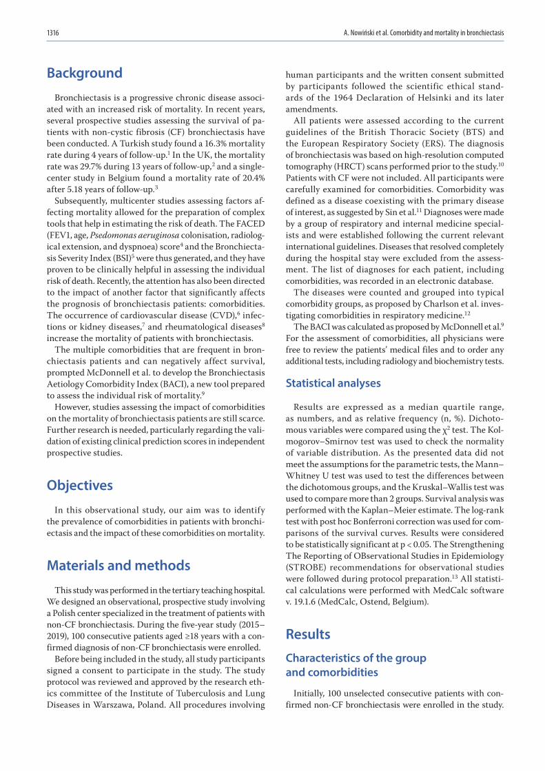
A. Nowiński et al. Comorbidity and mortality in bronchiectasis1316
Background
Bronchiectasis is a progressive chronic disease associ-ated with an increased risk of mortality. In recent years, several prospective studies assessing the survival of pa-tients with non-cystic fibrosis (CF) bronchiectasis have been conducted. A Turkish study found a 16.3% mortality rate during 4 years of follow-up.1 In the UK, the mortality rate was 29.7% during 13 years of follow-up,2 and a single-center study in Belgium found a mortality rate of 20.4% after 5.18 years of follow-up.3
Subsequently, multicenter studies assessing factors af-fecting mortality allowed for the preparation of complex tools that help in estimating the risk of death. The FACED (FEV1, age, Psedomonas aeruginosa colonisation, radiolog-ical extension, and dyspnoea) score4 and the Bronchiecta-sis Severity Index (BSI)5 were thus generated, and they have proven to be clinically helpful in assessing the individual risk of death. Recently, the attention has also been directed to the impact of another factor that significantly affects the prognosis of bronchiectasis patients: comorbidities. The occurrence of cardiovascular disease (CVD),6 infec-tions or kidney diseases,7 and rheumatological diseases8 increase the mortality of patients with bronchiectasis.
The multiple comorbidities that are frequent in bron-chiectasis patients and can negatively affect survival, prompted McDonnell et al. to develop the Bronchiectasis Aetiology Comorbidity Index (BACI), a new tool prepared to assess the individual risk of mortality.9
However, studies assessing the impact of comorbidities on the mortality of bronchiectasis patients are still scarce. Further research is needed, particularly regarding the vali-dation of existing clinical prediction scores in independent prospective studies.
Objectives
In this observational study, our aim was to identify the prevalence of comorbidities in patients with bronchi-ectasis and the impact of these comorbidities on mortality.
Materials and methods
This study was performed in the tertiary teaching hospital. We designed an observational, prospective study involving a Polish center specialized in the treatment of patients with non-CF bronchiectasis. During the five-year study (2015–2019), 100 consecutive patients aged ≥18 years with a con-firmed diagnosis of non-CF bronchiectasis were enrolled.
Before being included in the study, all study participants signed a consent to participate in the study. The study protocol was reviewed and approved by the research eth-ics committee of the Institute of Tuberculosis and Lung Diseases in Warszawa, Poland. All procedures involving
human participants and the written consent submitted by participants followed the scientific ethical stand-ards of the 1964 Declaration of Helsinki and its later amendments.
All patients were assessed according to the current guidelines of the British Thoracic Society (BTS) and the European Respiratory Society (ERS). The diagnosis of bronchiectasis was based on high-resolution computed tomography (HRCT) scans performed prior to the study.10 Patients with CF were not included. All participants were carefully examined for comorbidities. Comorbidity was defined as a disease coexisting with the primary disease of interest, as suggested by Sin et al.11 Diagnoses were made by a group of respiratory and internal medicine special-ists and were established following the current relevant international guidelines. Diseases that resolved completely during the hospital stay were excluded from the assess-ment. The list of diagnoses for each patient, including comorbidities, was recorded in an electronic database.
The diseases were counted and grouped into typical comorbidity groups, as proposed by Charlson et al. inves-tigating comorbidities in respiratory medicine.12
The BACI was calculated as proposed by McDonnell et al.9 For the assessment of comorbidities, all physicians were free to review the patients’ medical files and to order any additional tests, including radiology and biochemistry tests.
Statistical analyses
Results are expressed as a median quartile range, as numbers, and as relative frequency (n, %). Dichoto-mous variables were compared using the χ2 test. The Kol-mogorov–Smirnov test was used to check the normality of variable distribution. As the presented data did not meet the assumptions for the parametric tests, the Mann–Whitney U test was used to test the differences between the dichotomous groups, and the Kruskal–Wallis test was used to compare more than 2 groups. Survival analysis was performed with the Kaplan–Meier estimate. The log-rank test with post hoc Bonferroni correction was used for com-parisons of the survival curves. Results were considered to be statistically significant at p < 0.05. The Strengthening The Reporting of OBservational Studies in Epidemiology (STROBE) recommendations for observational studies were followed during protocol preparation.13 All statisti-cal calculations were performed with MedCalc software v. 19.1.6 (MedCalc, Ostend, Belgium).
Results
Characteristics of the group and comorbidities
Initially, 100 unselected consecutive patients with con-firmed non-CF bronchiectasis were enrolled in the study.

Adv Clin Exp Med. 2021;30(12):1315–1321 1317
From this group, 7 patients were excluded due to a lack of proper written consent, being lost to follow-up, or due to the withdrawal of consent during the observation period. Mortality was evaluated in the remaining patients at the end of the five-year follow-up period. The final assessment in-cluded a group of 93 patients: 43 men (46.2%) and 50 women (53.8%) with a median age of 66.0 (59.7–74.0) years.
The anthropometric data and comorbidities of the pa-tients are shown in Table 1 and Table 2, respectively.
The group consisted predominantly of middle-aged Caucasians with a median forced expiratory volume in 1 s (FEV1%) predicted of 79.8% (53.7–98.6%). The 3 BSI groups were evenly represented: 28 (30.1%) patients displayed mild bronchiectasis, 28 (30.1%) had moderate symptoms and 37 patients (39.8%) had severe symptoms. Bronchiectasis was considered idiopathic in most of the patients (60.2%), but it was classified as post-tuberculosis bronchiectasis in 9.7% of patients. Other, less common causes of bronchi-ectasis were identified in the remaining patients.
Half of the patients required hospital treatment in the previous year. Twelve patients (12.9%) were chroni-cally colonized with Pseudomonas aeruginosa. The median number of comorbidities at baseline in the whole group was 3 (interquartile range (IQR) 1–5).
Mortality
The total observation period in our group was 50 months, but because patients were enrolled consecutively in the study, the median follow-up period for the whole group was 31.0 (IQR 18.5–42.0) months. During the obser-vation period, 15 patients died (16.1%). The median survival period in non-survivors was 14.0 (IQR 5.2–23.0) months, with a range of 2.0–46.0 months. The median follow-up
Table 1. Characteristics of the study group. The data are reported as median with interquartile range (IQR) or number with prevalence
Demographics Cohort (n = 93)
Age [years] 66.0 (59.7–74.0)
Female sex, n (%) 50 (53.8)
BMI [kg/m2] 25.9 (21.9–29.6)
Ex-smokers, n (%) 47 (50.5)
Current smokers, n (%) 15 (16.1)
Never smokers, n (%) 31 (33.3)
Etiology
ABPA, n (%) 1 (1.1)
Alpha1 antitrypsin deficiency, n (%) 1 (1.1)
Asthma, n (%) 2 (2.2)
COPD, n (%) 6 (6.5)
Idiopathic, n (%) 56 (60.2)
IgG subclass deficiency, n (%) 1 (1.1)
Mounier–Kuhn syndrome, n (%) 1 (1.1)
Nontuberculous mycobacteria, n (%) 2 (2.2)
Postinfective, n (%) 11 (11.8)
Post-tuberculous, n (%) 9 (9.7)
Primary ciliary dyskinesia, n (%) 1 (1.1)
Yellow nail syndrome, n (%) 2 (2.2)
BSI score risk class
Mild, n (%) 28 (30.1)
Moderate, n (%) 28 (30.1)
Severe, n (%) 37 (39.8)
Clinical status
Sputum volume [mL/day] 10 (10–20)
MRC dyspnea score [points] 2 (1–2)
Exacerbations not requiring secondary care in the previous year, n
1 (0–2)
At least 1 hospitalization in the previous year, n (%)
46 (49.5)
Functional status
FEV1, % predicted 79.8 (53.7–98.6)
Microbiology
P. aeruginosa infection, n (%) 12 (12.9)
Comorbidity burden
Number of comorbidities at baseline, n 3 (1–5)
Number of comorbidities range, n 0–8
BMI – body mass index; COPD – chronic obstructive pulmonary disease; TB – tuberculosis; ABPA – allergic bronchopulmonary aspergillosis; BSI – bronchiectasis severity index; MRC – Medical Research Council; FEV1 – forced expiratory volume in 1 s; P. aeruginosa – Pseudomonas aeruginosa; IgG – immunoglobulin G.
Table 2. Most prevalent comorbidities in the study group
Comorbidity Number of patients Prevalence (%)
Cardiovascular diseases 61 65.5
COPD 48 51.6
Hypertension 32 34.4
Asthma 25 26.9
Obesity 22 23.6
Coronary heart disease 18 19.4
Diabetes 16 17.2
Cancer/neoplasm 12 12.9
Congestive heart failure 12 12.9
Chronic respiratory failure 11 11.8
Osteoporosis 9 9.7
Tuberculosis in anamnesis 9 9.7
Chronic atrial fibrillation 8 8.6
Pulmonary hypertension 7 7.5
Anxiety/depression 7 7.5
Anemia 5 5.4
Chronic renal disease 4 4.3
Liver cirrhosis 3 3.2
Other arrhythmia 2 2.2
Aortic aneurysm 1 1.1
Sarcoidosis 1 1.1
COPD – chronic obstructive pulmonary disease.
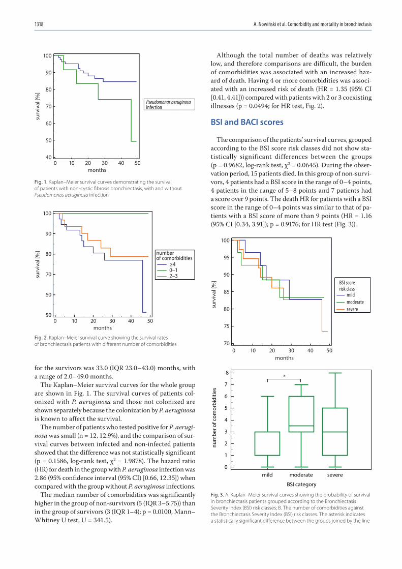
A. Nowiński et al. Comorbidity and mortality in bronchiectasis1318
for the survivors was 33.0 (IQR 23.0–43.0) months, with a range of 2.0–49.0 months.
The Kaplan–Meier survival curves for the whole group are shown in Fig. 1. The survival curves of patients col-onized with P. aeruginosa and those not colonized are shown separately because the colonization by P. aeruginosa is known to affect the survival.
The number of patients who tested positive for P. aerugi-nosa was small (n = 12, 12.9%), and the comparison of sur-vival curves between infected and non-infected patients showed that the difference was not statistically significant (p = 0.1586, log-rank test, χ2 = 1.9878). The hazard ratio (HR) for death in the group with P. aeruginosa infection was 2.86 (95% confidence interval (95% CI) [0.66, 12.35]) when compared with the group without P. aeruginosa infections.
The median number of comorbidities was significantly higher in the group of non-survivors (5 (IQR 3–5.75)) than in the group of survivors (3 (IQR 1–4); p = 0.0100, Mann–Whitney U test, U = 341.5).
Although the total number of deaths was relatively low, and therefore comparisons are difficult, the burden of comorbidities was associated with an increased haz-ard of death. Having 4 or more comorbidities was associ-ated with an increased risk of death (HR = 1.35 (95% CI [0.41, 4.41])) compared with patients with 2 or 3 coexisting illnesses (p = 0.0494; for HR test, Fig. 2).
BSI and BACI scores
The comparison of the patients’ survival curves, grouped according to the BSI score risk classes did not show sta-tistically significant differences between the groups (p = 0.9682, log-rank test, χ2 = 0.0645). During the obser-vation period, 15 patients died. In this group of non-survi-vors, 4 patients had a BSI score in the range of 0–4 points, 4 patients in the range of 5–8 points and 7 patients had a score over 9 points. The death HR for patients with a BSI score in the range of 0–4 points was similar to that of pa-tients with a BSI score of more than 9 points (HR = 1.16 (95% CI [0.34, 3.91]); p = 0.9176; for HR test (Fig. 3)).
Fig. 3. A. Kaplan–Meier survival curves showing the probability of survival in bronchiectasis patients grouped according to the Bronchiectasis Severity Index (BSI) risk classes; B. The number of comorbidities against the Bronchiectasis Severity Index (BSI) risk classes. The asterisk indicates a statistically significant difference between the groups joined by the line
Fig. 1. Kaplan–Meier survival curves demonstrating the survival of patients with non-cystic fibrosis bronchiectasis, with and without Pseudomonas aeruginosa infection
Fig. 2. Kaplan–Meier survival curve showing the survival rates of bronchiectasis patients with different number of comorbidities
months
Pseudomonas aeruginosainfection
surv
ival
[%]
0
100
90
80
70
60
50
4010 20 30 40 50
months
number of comorbidities
≥40–12–3su
rviv
al [%
]
0
100
90
80
70
60
5010 20 30 40 50
0
1
2
3
4
5
6
7
8
BSI category
num
ber o
f com
orbi
ditie
s
mild moderate severe
months
*
BSI score risk class
mildmoderateseveresu
rviv
al [%
]
0
100
95
90
85
80
75
7010 20 30 40 50
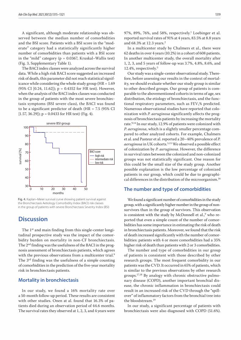
Adv Clin Exp Med. 2021;30(12):1315–1321 1319
A significant, although moderate relationship was ob-served between the median number of comorbidities and the BSI score. Patients with a BSI score in the “mod-erate” category had a statistically significantly higher number of comorbidities than patients with a BSI score in the “mild” category (p = 0.0367, Kruskal–Wallis test) (Fig. 3, Supplementary Table 1).
The BACI index classes were analyzed across the survival data. While a high risk BACI score suggested an increased risk of death, this parameter did not reach statistical signif-icance while considering the whole study group (HR = 1.69 (95% CI [0.24, 11.62]); p = 0.4352 for HR test). However, when the analysis of the BACI index classes was conducted in the group of patients with the most severe bronchiec-tasis symptoms (BSI severe class), the BACI was found to be a significant predictor of death (HR = 7.5 (95% CI [1.57, 36.29]); p = 0.0433 for HR test) (Fig. 4).
Discussion
The 1st and main finding from this single-center longi-tudinal prospective study was the impact of the comor-bidity burden on mortality in non-CF bronchiectasis. The 2nd finding was the usefulness of the BACI in the prog-nosis assessment of bronchiectasis patients, which agrees with the previous observations from a multicenter trial.9 The 3rd finding was the usefulness of a simple counting of comorbidities in the prediction of the five-year mortality risk in bronchiectasis patients.
Mortality in bronchiectasis
In our study, we found a 16% mortality rate over a 50-month follow-up period. These results are consistent with other studies. Onen at al. found that 16.3% of pa-tients died during an observation period of 44.6 months. The survival rates they observed at 1, 2, 3, and 4 years were
97%, 89%, 76%, and 58%, respectively.1 Loebinger et al. reported survival rates of 91% at 4 years, 83.5% at 8.8 years and 68.3% at 12.3 years.2
In a multicenter study by Chalmers et al., there were 62 deaths in over 4 years (10.2%) in a cohort of 608 patients. In another multicenter study, the overall mortality after 1, 2, 3, and 5 years of follow-up was 3.7%, 4.8%, 8.6%, and 12.4%, respectively.5
Our study was a single-center observational study. There-fore, before assessing our results in the context of mortal-ity, we should evaluate whether our study group is similar to other described groups. Our group of patients is com-parable to the abovementioned cohorts in terms of age, sex distribution, the etiology of bronchiectasis, and the func-tional respiratory parameters, such as FEV1% predicted. Numerous observational studies have reported that colo-nization with P. aeruginosa significantly affects the prog-nosis of bronchiectasis patients by increasing the mortality rate.9,14 In our study, 12.9% of patients were colonized with P. aeruginosa, which is a slightly smaller percentage com-pared to other analyzed cohorts. For example, Chalmers et al. and Pasteur et al. reported a 20–40% prevalence of P. aeruginosa in UK cohorts.5,15 We observed a possible effect of colonization by P. aeruginosa. However, the difference in survival rates between the colonized and non-colonized groups was not statistically significant. One reason for this could be the small size of the study group. Another possible explanation is the low percentage of colonized patients in our group, which could be due to geographi-cal differences in the distribution of the microorganism.16
The number and type of comorbidities
We found a significant number of comorbidities in the study group, with a significantly higher number in the group of non-survivors than in the group of survivors. This observation is consistent with the study by McDonnell et al.,9 who re-ported that even a simple count of the number of comor-bidities has some importance in estimating the risk of death in bronchiectasis patients. Moreover, we found that the risk of death increased significantly with the number of comor-bidities: patients with 4 or more comorbidities had a 35% higher risk of death than patients with 2 or 3 comorbidities.
The number and type of comorbidities in our group of patients is consistent with those described by other research groups. The most frequent comorbidity in our patients was the CVD. It occurred in 65% of patients, which is similar to the previous observations by other research groups.17,18 By analogy with chronic obstructive pulmo-nary disease (COPD), another important bronchial dis-ease, the chronic inflammation in bronchiectasis could result in an increased risk of the CVD through the “spill-over” of inflammatory factors from the bronchial tree into the bloodstream.19
In our study, a significant percentage of patients with bronchiectasis were also diagnosed with COPD (51.6%).
Fig. 4. Kaplan–Meier survival curve showing patient survival against the Bronchiectasis Aetiology Comorbidity Index (BACI) risk classes in the group of patients with severe Bronchiectasis Severity Index (BSI)
months
severe BSI group
BACI scorehigh riskintermediate risklow risksu
rviv
al [%
]
0
100
90
80
70
60
50
40
3010 20 30 40 50
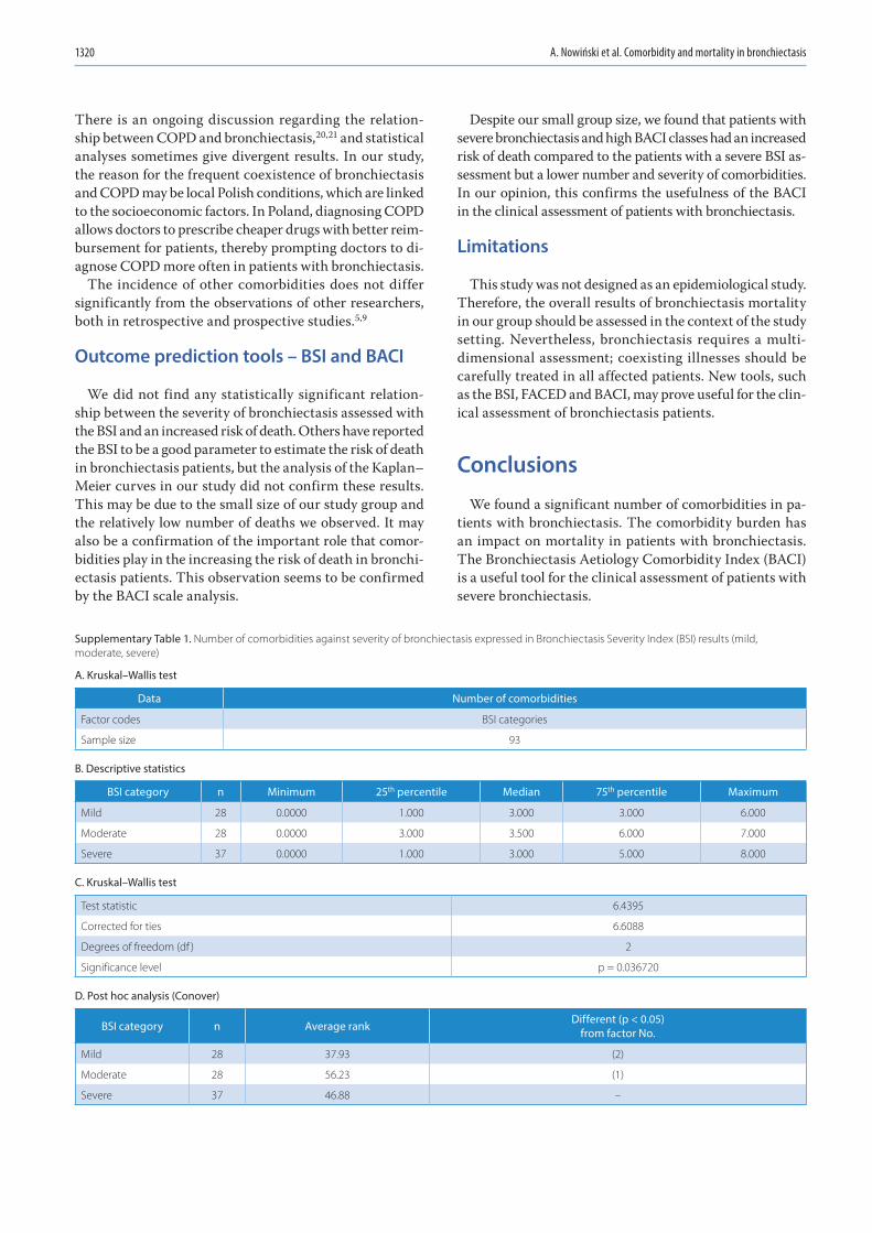
A. Nowiński et al. Comorbidity and mortality in bronchiectasis1320
There is an ongoing discussion regarding the relation-ship between COPD and bronchiectasis,20,21 and statistical analyses sometimes give divergent results. In our study, the reason for the frequent coexistence of bronchiectasis and COPD may be local Polish conditions, which are linked to the socioeconomic factors. In Poland, diagnosing COPD allows doctors to prescribe cheaper drugs with better reim-bursement for patients, thereby prompting doctors to di-agnose COPD more often in patients with bronchiectasis.
The incidence of other comorbidities does not differ significantly from the observations of other researchers, both in retrospective and prospective studies.5,9
Outcome prediction tools – BSI and BACI
We did not find any statistically significant relation-ship between the severity of bronchiectasis assessed with the BSI and an increased risk of death. Others have reported the BSI to be a good parameter to estimate the risk of death in bronchiectasis patients, but the analysis of the Kaplan–Meier curves in our study did not confirm these results. This may be due to the small size of our study group and the relatively low number of deaths we observed. It may also be a confirmation of the important role that comor-bidities play in the increasing the risk of death in bronchi-ectasis patients. This observation seems to be confirmed by the BACI scale analysis.
Despite our small group size, we found that patients with severe bronchiectasis and high BACI classes had an increased risk of death compared to the patients with a severe BSI as-sessment but a lower number and severity of comorbidities. In our opinion, this confirms the usefulness of the BACI in the clinical assessment of patients with bronchiectasis.
Limitations
This study was not designed as an epidemiological study. Therefore, the overall results of bronchiectasis mortality in our group should be assessed in the context of the study setting. Nevertheless, bronchiectasis requires a multi-dimensional assessment; coexisting illnesses should be carefully treated in all affected patients. New tools, such as the BSI, FACED and BACI, may prove useful for the clin-ical assessment of bronchiectasis patients.
Conclusions
We found a significant number of comorbidities in pa-tients with bronchiectasis. The comorbidity burden has an impact on mortality in patients with bronchiectasis. The Bronchiectasis Aetiology Comorbidity Index (BACI) is a useful tool for the clinical assessment of patients with severe bronchiectasis.
Supplementary Table 1. Number of comorbidities against severity of bronchiectasis expressed in Bronchiectasis Severity Index (BSI) results (mild, moderate, severe)
A. Kruskal–Wallis test
Data Number of comorbidities
Factor codes BSI categories
Sample size 93
B. Descriptive statistics
BSI category n Minimum 25th percentile Median 75th percentile Maximum
Mild 28 0.0000 1.000 3.000 3.000 6.000
Moderate 28 0.0000 3.000 3.500 6.000 7.000
Severe 37 0.0000 1.000 3.000 5.000 8.000
C. Kruskal–Wallis test
Test statistic 6.4395
Corrected for ties 6.6088
Degrees of freedom (df ) 2
Significance level p = 0.036720
D. Post hoc analysis (Conover)
BSI category n Average rank Different (p < 0.05) from factor No.
Mild 28 37.93 (2)
Moderate 28 56.23 (1)
Severe 37 46.88 –
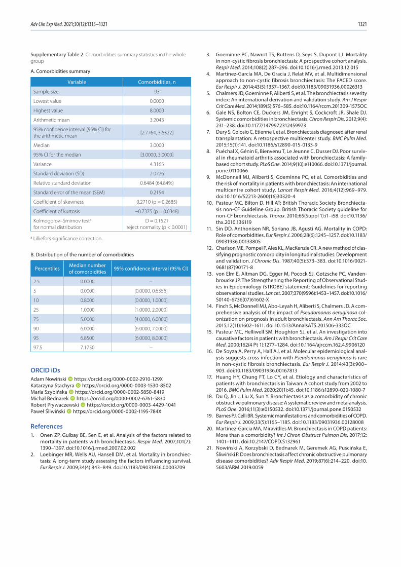
Adv Clin Exp Med. 2021;30(12):1315–1321 1321
ORCID iDsAdam Nowiński https://orcid.org/0000-0002-2910-129XKatarzyna Stachyra https://orcid.org/0000-0003-1530-8502Maria Szybińska https://orcid.org/0000-0002-5850-8419Michał Bednarek https://orcid.org/0000-0002-6761-5830Robert Pływaczewski https://orcid.org/0000-0003-4429-1041Paweł Śliwiński https://orcid.org/0000-0002-1195-784X
References1. Onen ZP, Gulbay BE, Sen E, et al. Analysis of the factors related to
mortality in patients with bronchiectasis. Respir Med. 2007;101(7): 1390–1397. doi:10.1016/j.rmed.2007.02.002
2. Loebinger MR, Wells AU, Hansell DM, et al. Mortality in bronchiec-tasis: A long-term study assessing the factors influencing survival. Eur Respir J. 2009;34(4):843–849. doi:10.1183/09031936.00003709
3. Goeminne PC, Nawrot TS, Ruttens D, Seys S, Dupont LJ. Mortality in non-cystic fibrosis bronchiectasis: A prospective cohort analysis. Respir Med. 2014;108(2):287–296. doi:10.1016/j.rmed.2013.12.015
4. Martínez-García MA, De Gracia J, Relat MV, et al. Multidimensional approach to non-cystic fibrosis bronchiectasis: The FACED score. Eur Respir J. 2014;43(5):1357–1367. doi:10.1183/09031936.00026313
5. Chalmers JD, Goeminne P, Aliberti S, et al. The bronchiectasis severity index: An international derivation and validation study. Am J Respir Crit Care Med. 2014;189(5):576–585. doi:10.1164/rccm.201309-1575OC
6. Gale NS, Bolton CE, Duckers JM, Enright S, Cockcroft JR, Shale DJ. Systemic comorbidities in bronchiectasis. Chron Respir Dis. 2012;9(4): 231–238. doi:10.1177/1479972312459973
7. Dury S, Colosio C, Etienne I, et al. Bronchiectasis diagnosed after renal transplantation: A retrospective multicenter study. BMC Pulm Med. 2015;15(1):141. doi:10.1186/s12890-015-0133-9
8. Puéchal X, Génin E, Bienvenu T, Le Jeunne C, Dusser DJ. Poor surviv-al in rheumatoid arthritis associated with bronchiectasis: A family-based cohort study. PLoS One. 2014;9(10):e110066. doi:10.1371/journal. pone.0110066
9. McDonnell MJ, Aliberti S, Goeminne PC, et al. Comorbidities and the risk of mortality in patients with bronchiectasis: An international multicentre cohort study. Lancet Respir Med. 2016;4(12):969–979. doi:10.1016/S2213-2600(16)30320-4
10. Pasteur MC, Bilton D, Hill AT; British Thoracic Society Bronchiecta-sis non-CF Guideline Group. British Thoracic Society guideline for non-CF bronchiectasis. Thorax. 2010;65(Suppl 1):i1–i58. doi:10.1136/thx.2010.136119
11. Sin DD, Anthonisen NR, Soriano JB, Agusti AG. Mortality in COPD: Role of comorbidities. Eur Respir J. 2006;28(6):1245–1257. doi:10.1183/ 09031936.00133805
12. Charlson ME, Pompei P, Ales KL, MacKenzie CR. A new method of clas-sifying prognostic comorbidity in longitudinal studies: Development and validation. J Chronic Dis. 1987;40(5):373–383. doi:10.1016/0021-9681(87)90171-8
13. von Elm E, Altman DG, Egger M, Pocock SJ, Gøtzsche PC, Vanden-broucke JP. The Strengthening the Reporting of Observational Stud-ies in Epidemiology (STROBE) statement: Guidelines for reporting observational studies. Lancet. 2007;370(9596):1453–1457. doi:10.1016/S0140-6736(07)61602-X
14. Finch S, McDonnell MJ, Abo-Leyah H, Aliberti S, Chalmers JD. A com-prehensive analysis of the impact of Pseudomonas aeruginosa col-onization on prognosis in adult bronchiectasis. Ann Am Thorac Soc. 2015;12(11):1602–1611. doi:10.1513/AnnalsATS.201506-333OC
15. Pasteur MC, Helliwell SM, Houghton SJ, et al. An investigation into causative factors in patients with bronchiectasis. Am J Respir Crit Care Med. 2000;162(4 Pt 1):1277–1284. doi:10.1164/ajrccm.162.4.9906120
16. De Soyza A, Perry A, Hall AJ, et al. Molecular epidemiological anal-ysis suggests cross-infection with Pseudomonas aeruginosa is rare in non-cystic fibrosis bronchiectasis. Eur Respir J. 2014;43(3):900–903. doi:10.1183/09031936.00167813
17. Huang HY, Chung FT, Lo CY, et al. Etiology and characteristics of patients with bronchiectasis in Taiwan: A cohort study from 2002 to 2016. BMC Pulm Med. 2020;20(1):45. doi:10.1186/s12890-020-1080-7
18. Du Q, Jin J, Liu X, Sun Y. Bronchiectasis as a comorbidity of chronic obstructive pulmonary disease: A systematic review and meta-analysis. PLoS One. 2016;11(3):e0150532. doi:10.1371/journal.pone.0150532
19. Barnes PJ, Celli BR. Systemic manifestations and comorbidities of COPD. Eur Respir J. 2009;33(5):1165–1185. doi:10.1183/09031936.00128008
20. Martinez-Garcia MA, Miravitlles M. Bronchiectasis in COPD patients: More than a comorbidity? Int J Chron Obstruct Pulmon Dis. 2017;12: 1401–1411. doi:10.2147/COPD.S132961
21. Nowiński A, Korzybski D, Bednarek M, Geremek AG, Puścińska E, Śliwiński P. Does bronchiectasis affect chronic obstructive pulmonary disease comorbidities? Adv Respir Med. 2019;87(6):214–220. doi:10. 5603/ARM.2019.0059
Supplementary Table 2. Comorbidities summary statistics in the whole group
A. Comorbidities summary
Variable Comorbidities, n
Sample size 93
Lowest value 0.0000
Highest value 8.0000
Arithmetic mean 3.2043
95% confidence interval (95% CI) for the arithmetic mean
[2.7764, 3.6322]
Median 3.0000
95% CI for the median [3.0000, 3.0000]
Variance 4.3165
Standard deviation (SD) 2.0776
Relative standard deviation 0.6484 (64.84%)
Standard error of the mean (SEM) 0.2154
Coefficient of skewness 0.2710 (p = 0.2685)
Coefficient of kurtosis −0.7375 (p = 0.0348)
Kolmogorov–Smirnov testa for normal distribution
D = 0.1521 reject normality (p < 0.0001)
a Lilliefors significance correction.
B. Distribution of the number of comorbidities
Percentiles Median number of comorbidities 95% confidence interval (95% CI)
2.5 0.0000 −
5 0.0000 [0.0000, 0.6356]
10 0.8000 [0.0000, 1.0000]
25 1.0000 [1.0000, 2.0000]
75 5.0000 [4.0000, 6.0000]
90 6.0000 [6.0000, 7.0000]
95 6.8500 [6.0000, 8.0000]
97.5 7.1750 −


Cite asMercik JS, Radziejewska J, Pach K, Zyśko D, Gajek J. ST-segment depression in atrioventricular nodal reentrant tachycardia: Preliminary results. Adv Clin Exp Med. 2021;30(12):1323–1328. doi:10.17219/acem/144161
DOI10.17219/acem/144161
Copyright© 2021 by Wroclaw Medical University This is an article distributed under the terms of theCreative Commons Attribution 3.0 Unported (CC BY 3.0)(https://creativecommons.org/licenses/by/3.0/)
Address for correspondenceJakub MercikE-mail: [email protected]
Funding sourcesThis research was financially supported by the Ministry of Health subvention (No. STM.A280.20.107 from the IT Simple system of Wroclaw Medical University).
Conflict of interestNone declared
Received on November 9, 2020Reviewed on November 26, 2020Accepted on November 22, 2021
Published online on December 13, 2021
AbstractBackground. The ST-segment is part of the electrocardiogram and physiologically, it forms an isoelectric line. The ST-segment depression is often observed in young, healthy people with paroxysmal tachycardia with narrow QRS complexes. In this group of patients, the ‘mysterious tachycardia-induced ST-segment depression’, ‘subendocardial myocardial ischemia’ and other not fully understood terms are used to explain this phenomenon.
Objectives. To assess the presence and possible mechanisms of ST-segment depression during atrioventricu-lar nodal reentrant tachycardia (AVNRT) in patients undergoing radiofrequency (RF) ablation of the underlying arrhythmia.
Materials and methods. The study included 50 patients (35 women and 15 men) aged about 49 years with clinically relevant paroxysmal narrow QRS complex tachycardia. During electrophysiological study (EPS), all patients had measured QRS components – QR, RS and RJ during the tachycardia and during the sinus rhythm. All of the measurements were done in lead V5.
Results. There was a statistically significant difference in cycle length during sinus rhythm and tachycardia (707.0 ±137.8 ms compared to 327.5 ±29.1 ms, p = 0.000), the RJ component (0.819 ±0.381 mV compared to 0.878 ±0.376 mV, p = 0.003) and the difference RJ-QR (0.081 ±0.083 mV compared to 0.163 ±0.108 mV, p = 0.000). The differences in RS and QR components during sinus rhythm and tachycardia did not reach the statistical significance. The difference RJ-QR during tachycardia correlated negatively with tachycardia cycle length (R = −0.39, p = 0.0049). The tachycardia cycle length correlated positively with the age of the studied patients (R = 0.28, p = 0.043).
Conclusions. In patients with AVNRT, there is a ST-segment depression during the episodes of tachycardia and the degree of this change is related to tachycardia cycle length. The most probable explanation of the ST-segment depression is the overlap of the QRS complex on the preceded T wave. Some intrinsic properties of individual electrocardiogram (ECG) also influence this phenomenon. The ischemic origin of the presented ST-segment change can be excluded.
Key words: tachycardia, AVNRT, ST-segment depression
Research letters
ST-segment depression in atrioventricular nodal reentrant tachycardia: Preliminary results
Jakub Szymon Mercik1,A–F, Jadwiga Radziejewska2,A–F, Katarzyna Pach3,A–F, Dorota Zyśko1,A–F, Jacek Gajek4,A–F
1 Department of Emergency Medicine, Wroclaw Medical University, Poland2 Klodzko County Hospital, Poland3 Students’ Scientific Association, Department of Emergency Medical Service, Wroclaw Medical University, Poland4 Department of Emergency Medical Service, Wroclaw Medical University, Poland
A – research concept and design; B – collection and/or assembly of data; C – data analysis and interpretation; D – writing the article; E – critical revision of the article; F – final approval of the article
Advances in Clinical and Experimental Medicine, ISSN 1899–5276 (print), ISSN 2451–2680 (online) Adv Clin Exp Med. 2021;30(12):1323–1328
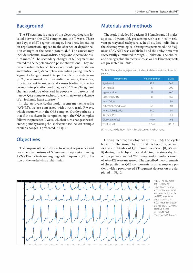
J. Mercik et al. ST-segment depression in AVNRT1324
Background
The ST-segment is a part of the electrocardiogram lo-cated between the QRS complex and the T wave. There are 2 types of ST-segment changes. First ones, depending on repolarization, appear in the absence of depolariza-tion changes of the action potential.1,2 The causes may include ischemia, myocarditis, drugs and electrolyte dis-turbances.3,4 The secondary changes of ST-segment are related to the depolarization phase aberrations. They are present in bundle branch blocks, ventricular pre-excitation and ventricular QRS complexes including pacing. The ST-segment changes constitute part of electrocardiogram (ECG) assessment for myocardial ischemia; therefore, it is important to understand causes leading to the in-correct interpretation and diagnosis.5,6 The ST-segment changes could be observed in people with paroxysmal narrow QRS complex tachycardia, with no overt evidence of an ischemic heart disease.7–9
In the atrioventricular nodal reentrant tachycardia (AVNRT), we are concerned with a retrograde P wave, which occurs within the QRS complex. Our hypothesis is that if the tachycardia is rapid enough, the QRS complex follows the preceded T wave, which in turn changes the ref-erence point by raising the isoelectric baseline. An example of such changes is presented in Fig. 1.
Objectives
The purpose of the study was to assess the presence and possible mechanisms of ST-segment depression during AVNRT in patients undergoing radiofrequency (RF) abla-tion of the underlying arrhythmia.
Materials and methods
The study included 50 patients (35 females and 15 males) approx. 49 years old, presenting with a clinically rele-vant paroxysmal tachycardia. In all studied individuals, the electrophysiological testing was performed, the diag-nosis of AVNRT was established and the arrhythmia was successfully eliminated through RF ablation. The clinical and demographic characteristics, as well as laboratory tests are presented in Table 1.
During electrophysiological study (EPS), the cycle length of the sinus rhythm and tachycardia, as well as the amplitudes of QRS components – QR, RS and RJ during the tachycardia and during the sinus rhythm with a paper speed of 200 mm/s and an enhancement of ×64–128 were measured. The described measurements of the particular QRS components in an exemplary pa-tient with a pronounced ST-segment depression are de-picted in Fig. 2.
Table 1. Clinical, demographic and biochemical characteristics of studied patients
Parameters Mean/number SD/%
Age [years] 49.1 14.2
Sex (female) 35 70.0
Hypertension 22 44.0
Diabetes mellitus 8 16.0
Heart failure 1 2.0
Ischemic heart disease 2 4.0
Hemoglobin [g/dL] 14.0 1.4
K+ [mmol/L] 4.4 0.4
Glucose [mg/dL] 101.9 16.5
TSH [mIU/L] 1.644 0.918
SD – standard deviation; TSH – thyroid-stimulating hormone.
Fig. 1. The example of ST-segment depressions during atrioventricular nodal reentrant tachycardia (AVNRT) in selected electrocardiogram (ECG) leads in 40-year-old male (CL – 279 ms, delta ST in lead V5 – 0.641 mV). Paper speed 50 mm/s
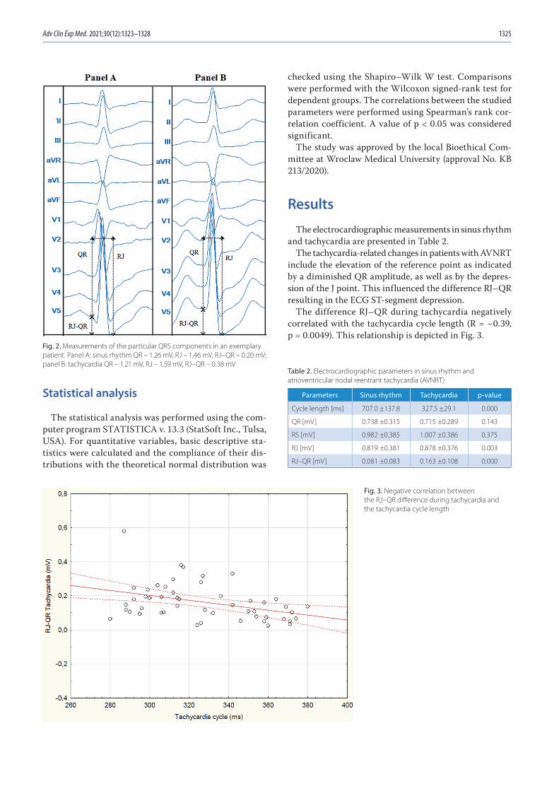
Adv Clin Exp Med. 2021;30(12):1323–1328 1325
Statistical analysis
The statistical analysis was performed using the com-puter program STATISTICA v. 13.3 (StatSoft Inc., Tulsa, USA). For quantitative variables, basic descriptive sta-tistics were calculated and the compliance of their dis-tributions with the theoretical normal distribution was
checked using the Shapiro–Wilk W test. Comparisons were performed with the Wilcoxon signed-rank test for dependent groups. The correlations between the studied parameters were performed using Spearman’s rank cor-relation coefficient. A value of p < 0.05 was considered significant.
The study was approved by the local Bioethical Com-mittee at Wroclaw Medical University (approval No. KB 213/2020).
Results
The electrocardiographic measurements in sinus rhythm and tachycardia are presented in Table 2.
The tachycardia-related changes in patients with AVNRT include the elevation of the reference point as indicated by a diminished QR amplitude, as well as by the depres-sion of the J point. This influenced the difference RJ–QR resulting in the ECG ST-segment depression.
The difference RJ–QR during tachycardia negatively correlated with the tachycardia cycle length (R = −0.39, p = 0.0049). This relationship is depicted in Fig. 3.
Table 2. Electrocardiographic parameters in sinus rhythm and atrioventricular nodal reentrant tachycardia (AVNRT)
Parameters Sinus rhythm Tachycardia p-value
Cycle length [ms] 707.0 ±137.8 327.5 ±29.1 0.000
QR [mV] 0.738 ±0.315 0.715 ±0.289 0.143
RS [mV] 0.982 ±0.385 1.007 ±0.386 0.375
RJ [mV] 0.819 ±0.381 0.878 ±0.376 0.003
RJ–QR [mV] 0.081 ±0.083 0.163 ±0.108 0.000
Fig. 3. Negative correlation between the RJ–QR difference during tachycardia and the tachycardia cycle length
Fig. 2. Measurements of the particular QRS components in an exemplary patient. Panel A: sinus rhythm QR – 1.26 mV, RJ – 1.46 mV, RJ–QR – 0.20 mV; panel B: tachycardia QR – 1.21 mV, RJ – 1.59 mV, RJ–QR – 0.38 mV
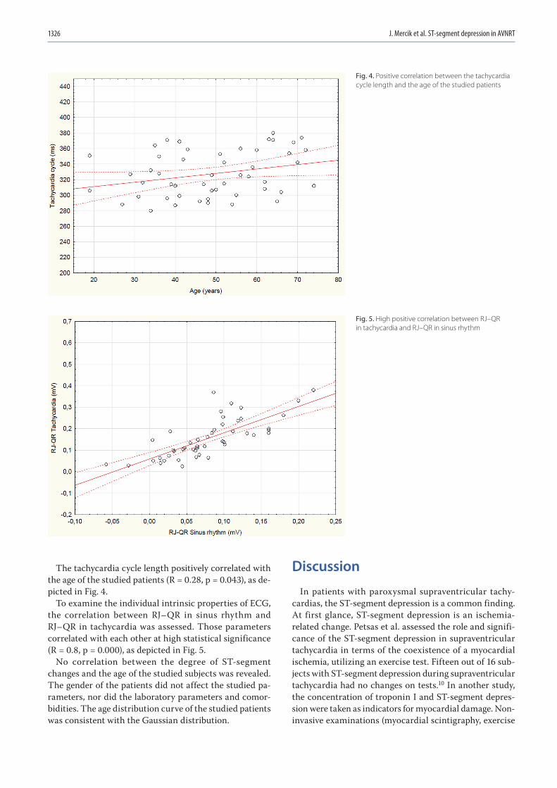
J. Mercik et al. ST-segment depression in AVNRT1326
The tachycardia cycle length positively correlated with the age of the studied patients (R = 0.28, p = 0.043), as de-picted in Fig. 4.
To examine the individual intrinsic properties of ECG, the correlation between RJ–QR in sinus rhythm and RJ–QR in tachycardia was assessed. Those parameters correlated with each other at high statistical significance (R = 0.8, p = 0.000), as depicted in Fig. 5.
No correlation between the degree of ST-segment changes and the age of the studied subjects was revealed. The gender of the patients did not affect the studied pa-rameters, nor did the laboratory parameters and comor-bidities. The age distribution curve of the studied patients was consistent with the Gaussian distribution.
Discussion
In patients with paroxysmal supraventricular tachy-cardias, the ST-segment depression is a common finding. At first glance, ST-segment depression is an ischemia-related change. Petsas et al. assessed the role and signifi-cance of the ST-segment depression in supraventricular tachycardia in terms of the coexistence of a myocardial ischemia, utilizing an exercise test. Fifteen out of 16 sub-jects with ST-segment depression during supraventricular tachycardia had no changes on tests.10 In another study, the concentration of troponin I and ST-segment depres-sion were taken as indicators for myocardial damage. Non-invasive examinations (myocardial scintigraphy, exercise
Fig. 5. High positive correlation between RJ–QR in tachycardia and RJ–QR in sinus rhythm
Fig. 4. Positive correlation between the tachycardia cycle length and the age of the studied patients

Adv Clin Exp Med. 2021;30(12):1323–1328 1327
echocardiography and exercise test) and coronary angiog-raphy were used for confirmation. The authors concluded that elevated troponin I levels and the ST-segment depres-sion are not significant markers of myocardial damage in patients with paroxysmal tachycardia.11
In many studies, the duration of an arrhythmia episode is not associated with an increase in troponins. However, in the case of longer-lasting tachycardia, the instabil-ity of the circulation may occur, resulting in troponin release.12–14
During the exercise-related sinus tachycardia, there is a gradual QT-interval shortening, which is associated with adrenergic activation. It is not a linear process, but according to the latest research there is a type of hysteresis in which, when accelerating, the heart shortens the QT-interval depending on the heart rate, first slowly, then increasingly faster. After the exertion, the QT-interval lengthens more slowly at first, then it gets faster until it returns to the primary duration. This phenomenon oc-curs because of the slow responsiveness of QT-interval to changes in the heart rate.15,16 The patient’s gender also constitutes an important aspect that may affect the QT length. It has been proven that women of a given age have a significantly longer corrected QT-interval than men. Additionally, the correlation between the change in the QT-interval and a patient’s age was demonstrated. The older the patient is, the shorter the QT-interval gets.17,18
The activation of the sympathetic system leads to the ac-celeration of the heart rate, decrease of the PR interval, the ST-segment depression and, in some cases, to the inver-sion of the T waves, while the parasympathetic activation slows down the heart rate and elevates the ST-segment.19 It was also observed in some healthy people. Their base-line ECG was showing slight changes in the ST-segment (<1 mm), often referred to as nonsignificant. This is in line with our results, as the initial J-point depression correlated with depression during tachycardia.
The ST-segment changes could be caused by projection of the retrograde P wave onto the ST-segment, more com-mon in patients with atrioventricular reentrant tachycar-dia, due to longer ventriculoatrial interval (usually exceed-ing 100 ms). In AVNRT, the interval is usually below 70 ms, affecting only the QRS complex.20
The most probable explanation of the findings
In our study group, the myocardial ischemia was un-likely to occur, due to the short-lasting arrhythmia par-oxysms, relatively young age of the subjects and lack of patients’ complaints. In AVNRT, the overlapping of ret-rograde P waves could not be explained. The most prob-able reason of the ST change is the overlapping of R wave on the preceded T wave with shortening of QT-interval with tachycardia, and something that could be referred to as intrinsic characteristics of ECG. This combination
might be explained by our results: a decrease of the ampli-tude of the QR component concomitantly with an increase of the RJ component, and a strong correlation between initial RJ–QR and tachycardia RJ–QR differences. Those conclusions are further supported by the correlation be-tween the ST-segment depression and the tachycardia cycle length, as well as by the correlation between this last pa-rameter and the age. As the tachycardia gets slower with age while the ST-depression is not related to age, the ST-segment depression in those settings is purely electrocar-diographic and not an ischemic one.
Limitations
The study is observational, hence the causality cannot be directly derived from the results. The study group was relatively small. The mechanisms of ST-segment depres-sion could also be different across the age groups. All these factors could have influenced the obtained results. Never-theless, it does not make our conclusions less important.
Conclusions
1. In patients with AVNRT, the ST-segment is lowered during the episodes of tachycardia, and the degree of this change is related to the tachycardia cycle length.
2. The most probable explanation of the ST-segment de-pression is the overlapping of the QRS complex on the pre-ceded T wave.
3. Some intrinsic properties of an individual ECG have been observed to be influential in this aspect.
ORCID iDsJakub Szymon Mercik https://orcid.org/0000-0002-5627-7071Jadwiga Radziejewska https://orcid.org/0000-0001-9153-9754Katarzyna Pach https://orcid.org/0000-0002-3643-9467Dorota Zyśko https://orcid.org/0000-0001-9190-0052Jacek Gajek https://orcid.org/0000-0002-0038-1750
References1. Shapiro E. The first textbook of electrocardiography. Thomas Lewis:
Clinical Electrocardiography. J Am Coll Cardiol. 1983;1(4):1160–1161. doi:10.1016/s0735-1097(83)80120-x
2. Rautaharju PM, Surawicz B, Gettes LS, et al. AHA/ACCF/HRS recom-mendations for the standardization and interpretation of the elec-trocardiogram. J Am Coll Cardiol. 2009;53(11):982–991. doi:10.1016/j.jacc.2008.12.014
3. Ghaffar A. Clinical electrocardiography. Pak J Health. 1953;3(1):6–12. PMID:13063999.
4. Kenny BJ, Brown KN. ECG T Wave. Treasure Island, USA: StatPearls Publishing; 2020.
5. Coppola G, Carità P, Corrado E, et al. ST segment elevations: Always a marker of acute myocardial infarction? Indian Heart J. 2013;65(4): 412–423. doi:10.1016/j.ihj.2013.06.013
6. Kozłowski D. Method in the chaos: A step-by-step approach to ECG interpretation. Eur J Transl Clin Med. 2018;1(1):76–92. doi:10.31373/ejtcm/92255
7. Arya A, Kottkamp H, Piorkowski C, et al. Differentiating atrioven-tricular nodal reentrant tachycardia from tachycardia via concealed accessory pathway. Am J Cardiol. 2005;95(7):875–878. doi:10.1016/j.amjcard.2004.12.020
J. Mercik et al. ST-segment depression in AVNRT1328
8. Dorenkamp M, Zabel M, Sticherling C. Role of coronary angiogra-phy before radiofrequency ablation in patients presenting with par-oxysmal supraventricular tachycardia. J Cardiovasc Pharmacol Ther. 2007;12(2):137–144. doi:10.1177/1074248407300775
9. Jastrzębski M. ST-segment depression and elevation during supra-ventricular tachycardias [in Polish]. Kardiol Pol. 2012;70(3):291–293. PMID:22430416.
10. Petsas AA, Anastassiades LC, Antonopoulos AG. Exercise testing for assessment of the significance of ST segment depression observed dur-ing episodes of paroxysmal supraventricular tachycardia. Eur Heart J. 1990;11(11):974–979. doi:10.1093/oxfordjournals.eurheartj.a059637
11. Bukkapatnam RN, Robinson M, Turnipseed S, Tancredi D, Amsterdam E, Srivatsa UN. Relationship of myocardial ischemia and injury to cor-onary artery disease in patients with supraventricular tachycardia. Am J Cardiol. 2010;106(3):374–377. doi:10.1016/j.amjcard.2010.03.035
12. Ben Yedder N, Roux JF, Paredes FA. Troponin elevation in supraven-tricular tachycardia: Primary dependence on heart rate. Can J Cardiol. 2017;27(1):105–109. doi:10.1016/j.cjca.2010.12.004
13. Miranda RC, Machado MDN, Takakura IT, et al. Elevated troponin lev-els after prolonged supraventricular tachycardia in patient with nor-mal coronary angiography. Cardiology. 2006;106(1):10–13. doi:10.1159/ 000092449
14. Zellweger MJ, Schaer BA, Cron TA, Pfisterer ME, Osswald S. Elevated troponin levels in the absence of coronary artery disease after supra-ventricular tachycardia. Swiss Med Wkly. 2003;133(31–32):439–441. PMID:14562187.
15. Gravel H, Jacquemet V, Dahdah N, Curnier D. Clinical applications of QT/RR hysteresis assessment: A systematic review. Ann Noninvasive Electrocardiol. 2018;23(1):e12514. doi:10.1111/anec.12514
16. Kannankeril PJ, Harris PA, Norris KJ, Warsy I, Smith PD, Roden DM. Rate-independent QT shortening during exercise in healthy sub-jects: Terminal repolarization does not shorten with exercise. J Cardiovasc Electrophysiol. 2008;19(12):1284–1288. doi:10.1111/j.1540-8167.2008.01266.x
17. Królik M, Milnerowicz H. The effect of using estrogens in the light of sci-entific research. Adv Clin Exp Med. 2012;21(4):535–543. PMID:23240460.
18. Pearl W. Effects of gender, age, and heart rate on QT intervals in chil-dren. Pediatr Cardiol. 1996;17(3):135–136. doi:10.1007/BF02505201
19. Friedman HS. Determinants of the total cosine of the spatial angle between the QRS complex and the T wave (TCRT): Implications for distinguishing primary from secondary T-wave abnormalities. J Electrocardiol. 2007;40(1):12–17. doi:10.1016/j.jelectrocard.2006.05.008
20. Rivera S, De La Paz Ricapito M, Conde D, Verdu MB, Roux JF, Paredes FA. The retrograde P-wave theory: Explaining ST segment depression in supraventricular tachycardia by retrograde AV node conduction. Pacing Clin Electrophysiol. 2014;37(9):1100–1105. doi:10.1111/pace.12394
No. 1 (January)5 Preface
Original papers7 Janina Golob Deeb, Lenart Skrjanc, Domen Kanduti, Caroline Carrico, Andrea Marquez Saturno, Kinga Grzech-Leśniak
Evaluation of Er:YAG and Er,Cr:YSGG laser irradiation for the debonding of prefabricated zirconia crowns
17 Hanna Danielewicz, Anna Dębińska, Anna Drabik-Chamerska, Danuta Kalita, Andrzej BoznańskiIL4RA gene expression in relation to I50V, Q551R and C-3223T polymorphisms
23 Jadwiga Radziejewska, Martek Frączkowski, Agnieszka Sławuta, Bernard PanaszekCan the in-hospital mortality rate in patients with ST-elevation myocardial infarctions be lowered any further?
29 Edyta Dziadkowiak, Maciej Guziński, Justyna Chojdak-Łukasiewicz, Małgorzata Wieczorek, Bogusław ParadowskiPredictive factors in post-stroke epilepsy: Retrospective analysis
35 Chung-Ze Wu, Chang-Hsun Hsieh, Chieh-Hua Lu, Dee Pei, Jin-Shuen Chen, Yen-Lin ChenFirst-phase insulin secretion is positively correlated with alanine aminotransferase in young adults
41 Shinsuke Suzuki, Satoshi Toyoma, Yohei Kawasaki, Hiroshi Nanjo, Takechiyo YamadaCD147 promotes invasion and MMP-9 expression through MEK signaling and predicts poor prognosis in hypopharyngeal squamous cell carcinoma
49 Milena Kozera, Joanna Konopińska, Marek RękasMid-term evaluation of the safety and efficacy of the iStent trabecular micro-bypass system combined with phacoemulsification
55 Chunfeng Sun, Chen Shen, Yaping Zhang, Chunhong HuLncRNA ANRIL negatively regulated chitooligosaccharide-induced radiosensitivity in colon cancer cells by sponging miR-181a-5p
67 Małgorzata Lelonek, Sylwia Wiśniowska-Śmiałek, Paweł Rubiś, Izabela Nowakowska, Agnieszka PawlakSacubitril/valsartan for heart failure with reduced ejection fraction: A first real-life observational study in Poland
77 Paweł Hackemer, Bartosz Małkiewicz, Fryderyk Menzel, Aleksandra Drabik, Krzysztof Tupikowski, Romuald ZdrojowyDeterminants of survival in patients with bladder cancer undergoing radical cystectomy: The impact of serum creatinine level
83 Iwona Chlebicka, Beata Jastrząb, Aleksandra Stefaniak, Jacek SzepietowskiBasal cell carcinoma secondary to trauma: A 3-year experience of the single center
87 Monika Augustynowicz, Krzysztof Kałwak, Danuta Zwolińska, Kinga MusiałThe incidence of acute kidney injury in children undergoing allogenic hematopoietic stem cell transplantation: A pilot study
93 Guoguo Yi, Ruiwen Yi, Xinglu Chen, Ling Peng, Guoqiang Huang, Min Fu, Xiao-he Lu, Hongwei LiThe role of soluble programmed death protein-1 (sPD-1) and soluble programmed death ligand-1 (sPD-L1) in rat corneal transplantation rejection
Reviews101 Burak Erdinc, Sonu Sahni, Vladimir Gotlieb
Hematological manifestations and complications of COVID-19
109 Jacek Czubak, Karolina Stolarczyk, Anna Orzeł, Marcin Frączek, Tomasz ZatońskiComparison of the clinical differences between COVID-19, SARS, influenza, and the common cold: A systematic literature review
Annual Contents
1330 Annual Contents
No. 2 (February)Original papers119 Josep Arnabat-Dominguez, Alessandro Del Vecchio, Carmen Todea, Kinga Grzech-Leśniak, Paolo Vescovi,
Umberto Romeo, Samir NammourLaser dentistry in daily practice during the COVID-19 pandemic: Benefits, risks and recommendations for safe treatments
127 Agnieszka Zubkiewicz-Kucharska, Monika Seifert, Michał Stępkowski, Anna NoczyńskaDiagnosis of type 1 diabetes during the SARS-CoV-2 pandemic: Does lockdown affect the incidence and clinical status of patients?
135 Jacek Białecki, Przemysław Pyda, Ryszard Antkowiak, Paweł DomosławskiUnsuspected femoral hernias diagnosed during endoscopic inguinal hernia repair
139 Yuxian Li, Ke Hu, Minghua Liang, Qing Yan, Minjiang Huang, Ling Jin, Yuefu Chen, Xirong Yang, Xiaobo LiStilbene glycoside upregulates SIRT3/AMPK to promotes neuronal mitochondrial autophagy and inhibit apoptosis in ischemic stroke
147 Weiming Sun, Tingting Chi, Xiaowei Chen, Zeyang LiHO-1 participate in the protection of RES in rat heart suffered from hypothermic preservation
153 Shipeng Huang, Congyang Zhou, Zheng Yuan, Hui Xiao, Xiaoping WuThe clinical value of high-density lipoprotein in the evaluation of new coronavirus pneumonia
157 Yuntao Liu, Dan Zhu, Guofeng Dong, Yuqin Zeng, Pan Jiang, Yaoling XiaoLiver paraoxonase 3 expression and the effect of liraglutide treatment in a rat model of diabetes
165 Paweł Piwowarczyk, Marta Szczukocka, Paweł Kutnik, Michał Borys,Anna Mikłaszewska, Sławomir Kiciak, Mirosław CzuczwarRisk factors and outcomes for acute respiratory failure in coronavirus disease 2019: An observational cohort study
173 Kejun Dai, Ling Chen, Jun Liu, Yuqiong Ding, Cheng Gu, Xujing LuMiR-147a mediated by sodium new houttuyfonate could enhance radiosensitivity of non-small cell lung cancer cells via suppressing STAT3
183 Haijing Sui, Chenggong Yan, Juan Yang, Xiaohui ZhaoClinical significance of evaluation of collateral circulation in short-term prognosis of wake-up stroke patients
189 Magdalena Putra-Szczepaniak, Adam Reich, Alina Jankowska-Konsur, Anna Czarnecka, Marta Bagłaj-Oleszczuk, Hryncewicz-Gwóźdź AnitaPack-year cigarette smoking affects the course of palmoplantar pustulosis
197 Grzegorz Raba, Marian Kacerovsky, Piotr LaudańskiEotaxin-2 as a potential marker of preterm premature rupture of membranes: A prospective, cohort, multicenter study
203 Wojciech Połom, Wojciech Cytawa, Anna Połom, Edyta Szurowska, Piotr Lass, Marcin MatuszewskiRadionuclide-guided sentinel lymph node mapping in urachal cancer
211 Anna Czarnecka, Agnieszka Odziomek, Magdalena Murzyn, Joanna Dubis, Marta Bagłaj-Oleszczuk, Anita Hryncewicz-GwóźdźWharton’s jelly-derived mesenchymal stem cells in the treatment of four patients with alopecia areata
Reviews219 Anna Szymczak, Mariusz Kusztal, Magdalena Krajewska
Overhydration: A cause or an effect of kidney damage and how to treat it
1331Adv Clin Exp Med. 2021;30
No. 3 (March)Original papers233 Bartosz Bogusz, Małgorzata Smolec-Zamora, Andrzej Zając, Adam Mol, Wojciech Górecki
Laparoscopic histological mapping for the determination of the length of aganglionic segment in children with Hirschsprung disease
239 Pavel Suchánek, Věra Lánská, Petr Stávek, Jaroslav Alois HubáčekShort-term trajectories of exercise-induced plasma lipid changes in overweight females, with a focus on HDL-cholesterol
245 Paweł Rubiś, Katarzyna Holcman, Ewa Dziewięcka, Sylwia Wiśniowska-Śmiałek, Aleksandra Karabinowska, Maria Szymonowicz, Lusine Khachatryan, Ewa Wypasek, Ann Garlitski, Andrzej Gackowski, Piotr PodolecRelationships between circulating galectin-3, extracellular matrix fibrosis and outcomes in dilated cardiomyopathy
255 Feng Chen, Zhi-Qing Chen, He Wang, Ji-jin ZhuPuerarin pretreatment inhibits myocardial apoptosis and improves cardiac function in rats after acute myocardial infarction through the PI3K/Akt signaling pathway
263 Suwen Zhang, Simeng PanmiR-124-3p targeting of TGF-β1 inhibits the proliferation of hypertrophic scar fibroblasts
273 Bartłomiej Szynglarewicz, Bartosz Dołęga-Kozierowski, Rafał Szulc, Piotr Kasprzak, Rafał MatkowskiIdentification of a localization wire tip in an occult breast lesion using a handheld magnetometer
279 Michał Lewandowski, Ilona KowalikRR interval analysis based on a newly developed PC program as a predictor of interventions in implantable cardioverter-defibrillator patients
289 Lanni Chen, Haihua Huang, Huijiao Zhang, Gaohui Zhu, Min ZhuThree cases of 3β-hydroxysteroid dehydrogenase deficiency: Clinical analysis
301 Konrad Kisielowski, Bogna Drozdzowska, Rafał Koszowski, Magdalena Rynkiewicz, Mariusz Szuta, Mansur Rahnama, Karolina Babiuch, Michał Tyrakowski, Anna Bednarczyk, Tomasz KaczmarzykImmunoexpression of RANK, RANKL and OPG in sporadic odontogenic keratocysts and their potential association with recurrence
309 Maciej Antkiewicz, Wiktor Kuliczkowski, Marcin Protasiewicz, Magdalena Kobielarz, Piotr Barć, Maciej Malinowski, Katarzyna Frączkowska, Katarzyna Kulikowska, Marcin Merenda, Krzysztof Jacyna, Tomasz Dawiskiba, Dariusz JanczakIntra-aneurysm sac pressure measurement using a thin pressure wire during endovascular aneurysm repair
315 Suat Terzi, Abdulkadir Özgür, Metin Çeliker, Tolga Mercantepe, Adnan Yilmaz, Levent Tümkaya, Şeyma Kaya, Emine Demir, Engin DursunThe protective effect of astaxanthin on cisplatin-induced ototoxicity
323 Maciej Siewiński, Ewa Kilar, Lidia Hirnle, Teresa Skiba, Jakub Gburek, Krzysztof Gołąb, Marek Murawski, Anna JanochaThe effects of chicken egg white cystatin and proteinase inhibitor on cysteine peptidase-like activity in the sera of patients with breast cancer
Reviews331 Janusz Springer, Tomasz Szmuda, Dariusz Kozłowski
Does the choice of drug in pharmacologic cardioversion correlate with the guidelines? Systematic review
349 Justyna Chojdak-Łukasiewicz, Edyta Dziadkowiak, Anna Zimny, Bogusław ParadowskiCerebral small vessel disease: A review

1332 Annual Contents
No. 4 (April)Original papers361 Yifeng Zhou, Yameng Peng, Hao Yuan, Zhenyi Long, Sixian Wu, Jiping Yang
Serum hepatitis B virus ribonucleic acid and its influencing factors in chronic hepatitis B
369 Maria Teresa Płazińska, Agata Czarnywojtek, Nadia Sawicka-Gutaj, Kosma Woliński, Iwona Krela-Kaźmierczak, Małgorzata Zgorzalewicz-Stachowiak, Izabela Miechowicz, Paweł Gut, Ewa Florek, Karolina Skonieczna-Żydecka, Marek Ruchała, Leszek KrólickiIs low radioiodine uptake a contraindication to radioiodine therapy in patients with benign thyroid disease?
379 Maciej Kentel, Michał Barnaś, Jarosław Witkowski , Paweł ReichertTreatment results and safety assessment of the LARS system for the reconstruction of the anterior cruciate ligament
387 Li-Shuang Duan, Yang Liu, Zhen-Zhou Li, Huan Wang, Xiao-Fang Zhou, Xiao-Xiao Wang, Zi-Wei Zhang, Yi-Qun Kang, Yong-jun Su, Jian-Rong GuoThe effect of different storage times on the oxygen-carrying capacity of the exosomes of red blood cells
395 Jun Qi, Yangyang Wu, Haijian Zhang, Yifei LiuLncRNA NORAD regulates scar hypertrophy via miRNA-26a mediating the regulation of TGFβR1/2
405 Dandan Wang, Minglei Wang, Pingping Sun, Qiaoyan GaoEplerenone inhibits oxidized low-density lipoprotein-induced proliferation and migration of vascular smooth muscle cells by downregulating GPER expression
413 Lu Wang, Na Li, Fei Wang, Lianqun CuiP2Y12 inhibition in macrophages reduces ventricular arrhythmias in rats after myocardial ischemia-reperfusion
421 Rui Li, Huimin Yuan, Tao Zhao, Yimin Yan, Zhaochen Liu, Jiayu Cai, Chunli Qiu, Chuanjing LimiR-874 ameliorates retinopathy in diabetic rats by NF-κB signaling pathway
431 Wei Zheng, Haiyan Wu, Ying Li, Helin Li, Zhaojun Liu, Yongzhi Nie, Lingling Shi, Hongyu WangPhenalen-1-one-mediated photodynamic therapy inhibits keloid graft progression by reducing vessel formation and promoting fibroblast apoptosis
441 Krzysztof Gawriołek, Tomasz Klatkiewicz, Agnieszka Przystańska, Zofia Maciejewska-Szaniec, Tomasz Gedrange, Agata Czajka-JakubowskaStandardization of the ultrasound examination of the masseter muscle with size-independent calculation of records
Multicenter study449 Mariusz Kusztal, Mariusz Kłopotowski, Stanisława Bazan-Socha, Beata Błażejewska-Hyżorek,
Krzysztof Pawlaczyk, Andrzej Oko, Magdalena Krajewska, Michał NowickiIs home-based therapy in Fabry disease the answer to compelling patients’ needs during the COVID-19 pandemic? Survey results from the Polish FD Collaborative Group
Reviews455 Zhao Chen, Nurlan Turxun, Fangyan Ning
Meta-analysis of the diagnostic value of procalcitonin in adult burn sepsis
465 Agnieszka Pawłowska-Kamieniak, Paulina Krawiec, Elżbieta Pac-KożuchowskaInterleukin 6: Biological significance and role in inflammatory bowel diseases
471 Monika Kwiatkowska, Aneta KrogulskaThe significance of the gut microbiome in children with functional constipation
1333Adv Clin Exp Med. 2021;30
No. 5 (May)Original papers485 Tomasz Wójcik, Paweł Szymkiewicz, Krzysztof Ściborski, Marceli Łukaszewski, Grzegorz Onisk, Andrzej Mysiak,
Andrzej Gamian, Jerzy Wiśniewski, Tadeusz Dobosz, Arleta Lebioda, Anna Jonkisz, Marcin ProtasiewiczOriginal and generic clopidogrel: A comparison of antiplatelet effects and active metabolite concentrations in patients without polymorphisms in the ABCB1 gene and the allele variants CYPC19*2 and *3
491 Michał Barnaś, Maciej Kentel, Piotr Morasiewicz, Jarosław Witkowski, Paweł ReichertClinical assessment and comparison of ACL reconstruction using synthetic graft (Neoligaments versus FiberTape)
499 Iwona Urbanowicz, Dariusz Wołowiec, Barbara Wysoczańska, Piotr Łacina, Anna Jonkisz, Wiesława Nahaczewska, Andrzej Tukiendorf, Iwona Bil-Lula, Katarzyna Bogunia-Kubik, Edyta PawlakNF-κB1 -94del/del ATTG polymorphic variant maintains CLL at an early, mildest stage
507 Jose Alfredo Sierra-Ramirez, Emmanuel Seseña-Mendez, Marycarmen Godinez-Victoria, Marta Elena Hernandez-CaballeroAn insight into the promoter methylation of PHF20L1 and the gene association with metastasis in breast cancer
517 Gulhan Kocaman, Eyup Altinoz, Mehmet Erman Erdemli, Mehmet Gul, Zeynep Erdemli, Emrah Zayman, Harika Gozde Gozukara Bag, Tugba AydınCrocin attenuates oxidative and inflammatory stress-related periodontitis in cardiac tissues in rats
525 Yuan Liu, Le Wang, Youguo Yang, Jianbin XiongSilencing Hoxa2 reverses dexamethasone-induced dysfunction of MC3T3-E1 osteoblasts and osteoporosis in rats
535 Chaoting Ma, Dandan Zhang, Qiuyan Ma, Yu Liu, Yingxin YangArbutin inhibits inflammation and apoptosis by enhancing autophagy via SIRT1
545 Juan Qin, Guolin Song, Yan Wang, Qin Liu, Hong Lin, Jinyun ChenUltrasound irradiation inhibits proliferation of cervical cancer cells by initiating endoplasmic reticulum stress-mediated apoptosis and triggering phosphorylation of JNK
555 Yunyun Yang, Qingyi ZhaoExenatide regulates inflammation and the production of reactive oxygen species via inhibition of S1PR2 synthesis
Reviews563 Renata Talar-Wojnarowska, Krzysztof Jamroziak
Intestinal amyloidosis: Clinical manifestations and diagnostic challenge
No. 6 (June)Original papers575 Cyprian Olchowy, Anna Olchowy, Jakub Hadzik, Paweł Dąbrowski, Dorota Mierzwa
Dentists can provide reliable shear wave elastography measurements of the stiffness of masseter muscles: A possible scenario for a faster diagnostic process
581 Mingming Yu, Yu Bian, Lin Wang, Fang ChenLow-intensity pulsed ultrasound enhances angiogenesis in rabbit capsule tissue that acts as a novel vascular bed in vivo
591 Wen Liu, Jianhuan Che, Yan Gu, Ling Song, Yingying Jiao, Shui YuSilencing of lncRNA SNHG12 inhibits proliferation and migration of vascular smooth muscle cells via targeting miR-766-5p/EIF5A axis
599 Yan-Long Tang, Xiao-Bo Wang, Yue Zhou, Ya-Ping Wang, Ji-Chao DingGinsenoside Rg1 induces senescence of leukemic stem cells by upregulating p16INK4a and downregulating hTERT expression
1334 Annual Contents
607 Qingxian Tu, Qianfeng Jiang, Min Xu, Yang Jiao, Huishan He, Shajin He, Weijin ZhengEGCG decreases myocardial infarction in both I/R and MIRI rats through reducing intracellular Ca2+ and increasing TnT levels in cardiomyocytes
617 Yong Cheng, Zhen-Zhou Li, Huan Wang, Jin-Huo Wang, Xiao-Fang Zhou, Jia-Ming Xu, Xun Zhou, Jian-Rong GuoThe effect of erythrocyte transfusion on macrophage pyroptosis and inflammation in a sepsis model
623 Xiaole Wu, Xiaoyu Wang, Yiyu Yin, Lei Zhu, Fengchao Zhang, Jianping YangInvestigation of the role of miR-221 in diabetic peripheral neuropathy and related molecular mechanisms
633 Sebastian Dominiak, Ewa Karuga-Kuźniewska, Paweł Popecki, Paweł Kubasiewicz-RossPRF versus xenograft in sinus augmentation in case of HA-coating implant placement: A 36-month retrospective study
Reviews641 Agnieszka Stembalska, Lech Dudarewicz, Robert Śmigiel
Lethal and life-limiting skeletal dysplasias: Selected prenatal issues
No. 7 (July)Editorials653 Markku Kurkinen
Alzheimer’s trials: A cul-de-sac with no end in sight
Original papers655 Martin Floer, Mareike Clausen, Tobias Meister, Richard Vollenberg, Dominik Bettenworth, Phil-Robin Tepasse
Soluble syndecan-1 as marker of intestinal inflammation: A preliminary study and evaluation of a new panel of biomarkers for non-invasive prediction of active ulcerative colitis
661 Li Tong, Jinglin Cheng, Heping Zuo, Jingrong LiMicroRNA-197 promotes proliferation and inhibits apoptosis of gallbladder cancer cells by targeting insulin-like growth factor-binding protein 3
673 Yi Qiao, Jie ChenInvestigating the inflammatory cascade effect of basophil activation in children with allergic rhinitis or asthma, via the IgE-FcεRI-NF-κB signaling pathway
681 Beata Wyrębek, Renata Górska, Katarzyna Gawron, Małgorzata Nędzi-Góra, Bartłomiej Górski, Paweł PlakwiczPeriodontal condition of mandibular incisors treated with modified Kazanjian vestibuloplasty compared to untreated sites: A prospective study
691 Yongbo Wang, Yue Wang, Chunna Ren, Haicun Wang, Yang Zhang, Yunxia XiuUpregulation of centromere protein K is crucial for lung adenocarcinoma cell viability and invasion
701 Zijun Zhao, Li Ma, Yishuai Li, Qi Zhang, Ying Wang, Yanlei Tai, Qiujun WangMiR-124 protects against cognitive dysfunction induced by sevoflurane anesthesia in vivo and in vitro through targeting calpain small subunit 1 via NF-κB signaling pathway
711 Weiting Chen, Zhongen Shen, Shuiqi Cai, Long Chen, Dabin WangFGF21 promotes wound healing of rat brain microvascular endothelial cells through facilitating TNF-α-mediated VEGFA and ERK1/2 signaling pathway
721 Xiaojie Li, Dapeng Liao, Gang Sun, HanWen ChuNotch pathway activation promotes the differentiation of beagle dog periodontal ligament stem cells to Schwann cells
727 Hongwei Lei, Jingbin Shi, Yun Teng, Chenghui Song, Lijuan Zou, Fuxiu Ye, Haichen ZhangBaicalein modulates the radiosensitivity of cervical cancer cells in vitro via miR-183 and the JAK2/STAT3 signaling pathway
1335Adv Clin Exp Med. 2021;30
Reviews737 Szymon Urban, Mikołaj Błaziak, Jan Biegus, Robert Zymliński
Ultrafiltration in acute heart failure: Current knowledge and fields for further research
747 Agata Czarnywojtek, Alicja Ochmańska, Małgorzata Zgorzalewicz-Stachowiak, Nadia Sawicka-Gutaj, Beata Matyjaszek-Matuszek, Magdalena Woźniak, Marek RuchałaInfluence of SARS-CoV-2 infection on thyroid gland function: The current knowledge
Research-in-progress757 Maciej Dejnek, Helena Moreira, Sylwia Płaczkowska, Piotr Morasiewicz, Ewa Barg, Jarosław Witkowski, Paweł Reichert
Analysis and comparison of autologous platelet-rich plasma preparation systems used in the treatment of enthesopathies: A preliminary study
765 Julia Rudno-Rudzińska, Wojciech Kielan, Maciej Guziński, Julita KulbackaEffects of calcium electroporation, electrochemotherapy, and irreversible electroporation on quality of life and progression-free survival in patients with pancreatic cancer: IREC clinical study
No. 8 (August)Editorials775 Masaru Tanaka, László Vécsei
Monitoring the kynurenine system: Concentrations, ratios or what else?
Original papers779 Boxin Zhao, Zhiyong Zhang, Lin Gui, Yingyu Xiang, Xueyuan Sun, Lijuan Huang
MiR-let-7i inhibits CD4 T cell apoptosis in patients with acute coronary syndrome
789 Cuifang Nie, Guangju Meng, Yongyun Wu, Li Liu, Li Chen, Shengqiang Yang, Yan HuExpression of miR-9a-5p in cirrhosis patients with recurrent portal hypertension after treatment
797 Marta Rorat, Tomasz Jurek, Krzysztof Simon, Maciej GuzińskiThe chest radiographic scoring system in initial diagnosis of COVID-19: Is a radiologist needed?
805 Joanna Kufel-Grabowska, Mikołaj Bartoszkiewicz, Rodryg Ramlau, Maria LitwiniukCancer patients and internal medicine patients attitude towards COVID-19 vaccination in Poland
813 Krzysztof Kamil Kotulski, Joanna Bartczak-Kotulska, Julia Rudno-Rudzińska, Wojciech Kielan, Ewelina Frejlich, Wojciech HapThe sense of coherence and sense of satisfaction with life in patients hospitalized in Polish and Irish surgical departments
823 Ye Gu, Yihua Wu, Liang ChenGP6 promotes the development of cerebral ischemic stroke induced by atherosclerosis via the FYN-PKA-pPTK2/FAK1 signaling pathway
831 Jie Chen, Daiyue Yuan, Qingya Hao, Dongmei Zhu, Zhong ChenLncRNA PCGEM1 mediates oxaliplatin resistance in hepatocellular carcinoma via miR-129-5p/ETV1 axis in vitro
839 Tongtong Zhang, Suyang Yu, Shipeng ZhaoLncRNA FEZF1-AS1 promotes colorectal cancer progression through regulating the miR-363-3p/PRRX1 pathway
849 Tianjian Lu, Weiping Lu, Chunyi Jia, Shanguang Lou, Yan ZhangKnockdown of miR-15b partially reverses the cisplatin resistance of NSCLC through the GSK-3β/MCL-1 pathway
859 Jinzi Zhou, Fenghua Chen, Aimin Yan, Xiaobo XiaOverexpression of HTRA1 increases the proliferation and migration of retinal pigment epithelium cells

1336 Annual Contents
Reviews865 Maciej Rachwalik, Magdalena Hurkacz, Beata Sienkiewicz-Oleszkiewicz, Marek Jasiński
Role of resistin in cardiovascular diseases: Implications for prevention and treatment
No. 9 (September)Editorials879 Ana Monzó-Miralles, Víctor Martín-González, Sara Smith-Ballester, Victoria Iglesias-Miguel, Antonio Cano
The RANKL/RANK system in female reproductive organ tumors: A preclinical and clinical overview
Original papers885 Seong Ji Choi, Jae Min Lee, Kang Won Lee, Hyuk Soon Choi, Eun Sun Kim, Bora Keum,
Jai Hoon Yoon, Yoon Tae Jeen, Hoon Jai Chun, Hong Sik Lee, Ho Soon ChoiEffect of histological examination on the diagnosis of pancreatic mass using endoscopic ultrasound fine-needle aspiration
893 Xijuan Wang, Mingwu Li, Ruimao Zheng, Ting Cui, Jiayin Qin, Zhijie Su, Meng Shang, Yongzhen BaoHigh irisin and low BDNF levels in aqueous humor of high myopia
905 Agata Tarkowska, Wanda Furmaga-JabłońskaIs N-terminal pro-brain type natriuretic peptide a useful marker in newborns with heart defects?
913 Maria Henryka Listewnik, Hanna Piwowarska-Bilska, Krystyna Jasiakiewicz, Bożena BirkenfeldInfluence of high tissue-absorbed dose on anti-thyroid antibodies in radioiodine therapy of Graves’ disease patients
923 Martyna Kluszczyńska, Agnieszka MłynarskaInfluence of frailty syndrome on patient prognosis after coronary artery bypass grafting
933 Wei Wang, Caizhi Xiao, Hong Chen, Fangfei Li, Dongqin XiaRadiation induces submandibular gland damage by affecting Cdkn1a expression and regulating expression of miR-486a-3p in a xerostomia mouse model
941 Mustafa Tosun, Hasan Olmez, Edhem Unver, Yusuf Kemal Arslan, Ferda Keskin Cimen, Adalet Ozcicek, Mehmet Aktas, Halis SuleymanOxidative and pro-inflammatory lung injury induced by desflurane inhalation in rats and the protective effect of rutin
949 Halil Kara, Ceyhun Çağlar, Mehmet Asiltürk, Siyami Karahan, Mahmut UğurluComparison of a manual walking platform and the CatWalk gait analysis system in a rat osteoarthritis model
957 Qianxi Deng, Linju Wu, Yiming Li, Long ZouMYBL2 in synergy with CDC20 promotes the proliferation and inhibits apoptosis of gastric cancer cells
967 Piotr Celichowski, Karol Jopek, Marta Szyszka, Paulina Milecka, Marianna Tyczewska, Svetlana Sakhanova, Witold Szaflarski, Ludwik Kazimierz Malendowicz, Marcin RucińskiExtracellular Nampt (eNampt/visfatin/PBEF) directly and indirectly stimulates ACTH and CCL2 protein secretion from isolated rat corticotropes
Research letter981 Magdalena Łyko, Mateusz Kaczmarek, Polina Nekrasova, Anita Hryncewicz-Gwóźdź, Joanna Maj, Alina Jankowska-Konsur
What factors affect the length of hospitalization in patients with erysipelas? A 10-year retrospective study of patients hospitalized in Lower Silesia, Poland
1337Adv Clin Exp Med. 2021;30
No. 10 (October)Editorials991 Erwan Donal, Vasileios Panis
Interaction between mitral valve apparatus and left ventricle. Functional mitral regurgitation: A brief state-of-the-art overview
Original papers999 Yuan Yuan, Jun Liu, Yongxin Zhou, Xufang Du, Qian Chen, Jiongying Zhou, Miao Hou
The relationship between monocyte-to-lymphocyte ratio and the risk of gastrointestinal system involvement in children with IgA vasculitis: A preliminary report
1007 Sebastian Podlewski, Natalia Gołębiowska, Maciej RadekEvaluation of changes in cervical sagittal balance and clinical parameters in patients undergoing two-level anterior cervical discectomy and fusion
1013 Zahide Betül Gündüz, Filiz Aktas, Husamettin Vatansev, Merve Solmaz, Ender ErdoğanEffects of amantadine and topiramate on neuronal damage in rats with experimental cerebral ischemia-reperfusion
1025 Nezahat Kurt, Özge Nur Türkeri, Bahadir Suleyman, Nuri BakanThe effect of taxifolin on high-dose-cisplatin-induced oxidative liver injury in rats
1031 Tao Wang, Huihe LuGanoderic acid A inhibits ox-LDL-induced THP-1-derived macrophage inflammation and lipid deposition via Notch1/PPARγ/CD36 signaling
1043 Guohua Cheng, Yarong Li, Zhaoyu Liu, Xiang SonglncRNA PSMA3-AS1 promotes the progression of non-small cell lung cancer through targeting miR-17-5p/PD-L1
1051 Rafał Olszewski, Paweł Ptaszyński, Iwona Cygankiewicz, Krzysztof KaczmarekImpedance fluctuation and steam pop occurrence during radiofrequency current ablation: An experimental in vitro model
1057 Anna Markowska, Anna Gryboś, Andrzej Marszałek, Wiesława Bednarek, Violetta Filas, Marian Gryboś, Janina Markowska, Radosław Mądry, Barbara Więckowska, Danuta Nowalińska, Monika SzarszewskaExpression of selected molecular factors in two types of endometrial cancer
Reviews1065 Jialing Liu, Ye Chen, Shihao Li, Zhihe Zhao, Zhihong Wu
Machine learning in orthodontics: Challenges and perspectives
1075 Mikołaj Błaziak, Szymon Urban, Maksym Jura, Wiktor KuliczkowskiFractional flow reserve-guided treatment in coronary artery disease: Clinical practice
Research letters1085 Vasyl Suvorov, Viktor Filipchuk, Vadym Mazevich, Leonid Suvorov
Simulation of pelvic osteotomies applied for DDH treatment in pediatric patients using piglet models
1091 Erkan Dalbaşı, Ömer Lütfi AkgülThe effectiveness of methotrexate and low-dose steroid therapy in the treatment of idiopathic granulomatous mastitis
1099 Joanna Adamiec-Mroczek27-gauge sutureless vitrectomy under topical anesthesia: A pilot study

1338 Annual Contents
No. 11 (November)Editorials1111 Carol Holland, Ian Garner, Jane Simpson, Fiona Eccles, Esperanza Navarro Pardo, Calum Marr, Sandra Varey
Impacts of COVID-19 lockdowns on frailty and wellbeing in older people and those living with long-term conditions
Original papers1115 Martin Floer, Mario Ziegler, Bodo Lenkewitz, Agneta Auer, Tobias Meister
Out-of-hospital sepsis recognition by paramedics improves the course of disease and mortality: A single center retrospective study
1127 Salime Mucuk, Tülay BülbülEffects of position on non-stress test results and maternal satisfaction
1133 Murat Şahin, Ayten Oguz, Dilek Tuzun, Gülsüm Akkus, Gul Inci Törün, Abdulkadir Yasir Bahar, Hatice Şahin, Kamile GülEffectiveness of TI-RADS and ATA classifications for predicting malignancy of thyroid nodules
1141 Agata Sebastian, Marta Madej, Maciej Sebastian, Ewa Morgiel, Piotr Wawryka, Piotr WilandDifferences in clinical phenotypes of primary Sjögren’s syndrome depending on early or late onset
1147 Anna Teresa Goździk, Ewelina Jasic-Szpak, Jakub Michałowicz, Monika Przewłocka-Kosmala, James Edward Sharman, Wojciech KosmalaAssociation of arterial hemodynamics with left ventricular systolic function in hypertensive patients: A longitudinal study
1157 Yuan Cheng, Mengzuo Wu, Min Liu, Birong Zhou, Xianhe Lin, Bangning WangCholecystokinin-mediated pharmacological preconditioning effects on ischemic rat hearts: Possible signaling pathways
1167 Pinar Gokcen, Oguzhan Ozturk, Gupse Adali, Ilkay Tosun, Halef Okan Dogan, Haki Kara, Yucel Yalman, Hamdi Levent Doganay, Kamil OzdilA novel therapeutic approach to NASH: Both polyethylene glycol 3350 and lactulose reduce hepatic inflammation in C57BL/6J mice
1175 Hüseyin Kocaturk, Fevzi Bedir, Ömer Turangezli, Remzi Arslan, Taha Abdulkadir Çoban, Durdu Altuner, Halis SuleymanEffect of adenosine triphosphate, benidipine and their combinations on bevacizumab-induced kidney damage in rats
1185 Halil Ibrahim Tas, Eyup Burak SancakProtective effect of metformin on lithium-induced nephrogenic diabetes insipidus: An experimental study in rats
1195 Haijun Ran, Han Liu, Ping WuEchinatin mitigates H
2O
2-induced oxidative damage and apoptosis in lens epithelial cells
via the Nrf2/HO-1 pathway
1205 Nurdina Charong, Nateelak Kooltheat, Thunyaluk PlyduangHigh-sensitivity detection of clinically significant red blood cell antibodies by the column agglutination technique

1339Adv Clin Exp Med. 2021;30
No. 12 (December)Editorials1221 James E. Sharman, Wojciech Kosmala
High-quality medical research requires that equipment has been validated for accuracy
Original papers1225 Ádám Annus, Ferenc Tömösi, Ferenc Rárosi, Evelin Fehér, Tamás Janáky, Gábor Kecskeméti, József Toldi, Péter Klivényi, László Sztriha, László Vécsei
Kynurenic acid and kynurenine aminotransferase are potential biomarkers of early neurological improvement after thrombolytic therapy: A pilot study
1233 Mehmet Murat Bala, Keziban Aslı BalaSevere cases of osteogenesis imperfecta type VIII due to a homozygous mutation in P3H1 (LEPRE1) and review of the literature
1239 Anna Rams, Joanna Kosałka-Węgiel, Piotr Kuszmiersz, Aleksandra Matyja-Bednarczyk, Stanisław Polański, Lech Zaręba, Stanisława Bazan-SochaCharacteristics of idiopathic inflammatory myopathies with novel myositis-specific autoantibodies
1249 Patryk Kuliński, Łukasz Tomczyk, Piotr MorasiewiczEffect of the COVID-19 pandemic on foot surgeries
1255 Fatih Ada, Ferit Kasimzade, Ali Sefa Mendil, Hasan GocmezEffects of nano-sized titanium dioxide powder and ultraviolet light on superficial veins in a rabbit model
1263 En Zhou, Yinghua Zou, Chengyu Mao, Dongjiu Li, Changqian Wang, Zongqi ZhangMicroRNA-221 inhibits the transition of endothelial progenitor cells to mesenchymal cells via the PTEN/FoxO3a signaling pathway
1271 Chunwen Jia, Feng Gao, Yanan Zhao, Siyang Ji, Shidao CaiIdentification and functional analysis of changes to the ox-LDL-induced microRNA-124-3p/DLX5 axis in vascular smooth muscle cells
1283 Fadime Mutlu Içduygu, Hale Samli, Asuman Özgöz, Buse Vatansever, Kuyas Hekimler Oztürk, Egemen AkgünPossibility of paclitaxel to induce the stemness-related characteristics of prostate cancer cells
Reviews1293 Katarzyna Rakoczy, Wojciech Szlasa, Jolanta Saczko, Julita Kulbacka
Therapeutic role of vanillin receptors in cancer
1303 Kacper Turek, Michał Jarocki, Julita Kulbacka, Jolanta SaczkoDualistic role of autophagy in cancer progression
Research letters1315 Adam Nowiński, Katarzyna Stachyra, Maria Szybińska, Michał Bednarek, Robert Pływaczewski, Paweł Śliwiński
The influence of comorbidities on mortality in bronchiectasis: A prospective, observational study
1323 Jakub Szymon Mercik, Jadwiga Radziejewska, Katarzyna Pach, Dorota Zyśko, Jacek GajekST-segment depression in atrioventricular nodal reentrant tachycardia: Preliminary results

Ada Fatih 1255Adalı Gupse 1167Adamiec-Mroczek Joanna 1099Akgül Ömer Lütfi 1091Akgün Egemen 1283Akkus Gülsüm 1133Aktas Filiz 1013Aktas Mehmet 941Altınöz Eyüp 517Altuner Durdu 1175Annus Ádám 1225Antkiewicz Maciej 309Antkowiak Ryszard 135Arnabat-Dominguez Josep 119Arslan Remzi 1175Arslan Yusuf Kemal 941Asiltürk Mehmet 949Auer Agneta 1115Augustynowicz Monika 87Aydın Tugba 517
Babiuch Karolina 301Bag Harika Gozde Gozukara 517Bagłaj-Oleszczuk Marta 189, 211Bahar Abdulkadir Yasir 1133Bakan Nuri 1025Bala Keziban Aslı 1233Bala Mehmet Murat 1233Bao Yongzhen 893Barć Piotr 309Barg Ewa 757Barnaś Michał 379, 491Bartczak-Kotulska Joanna 813Bartoszkiewicz Mikołaj 805Bazan-Socha Stanisława 449, 1239Bedir Fevzi 1175Bednarczyk Anna 301Bednarek Michał 1315Bednarek Wiesława 1057Bettenworth Dominik 655Białecki Jacek 135Bian Yu 581Biegus Jan 737Bil-Lula Iwona 499Birkenfeld Bożena 913
Błaziak Mikołaj 737, 1075Błażejewska-Hyżorek Beata 449Bogunia-Kubik Katarzyna 499Bogusz Bartosz 233Borys Michał 165Boznański Andrzej 17Bülbül Tülay 1127
Çağlar Ceyhun 949Cai Jiayu 421Cai Shidao 1271Cai Shuiqi 711Cano Antonio 879Carrico Caroline 7Celichowski Piotr 967Çeliker Metin 315Charong Nurdina 1205Che Jianhuan 591Chen Fang 581Chen Feng 255Chen Fenghua 859Chen Hong 933Chen Jie 673, 831Chen Jin-Shuen 35Chen Jinyun 545Chen Lanni 289Chen Li 789Chen Liang 823Chen Ling 173Chen Long 711Chen Qian 999Chen Weiting 711Chen Xiaowei 147Chen Xinglu 93Chen Ye 1065Chen Yen-Lin 35Chen Yuefu 139Chen Zhao 455Chen Zhi-Qing 255Chen Zhong 831Cheng Guohua 1043Cheng Jinglin 661Cheng Yong 617Cheng Yuan 1157Chi Tingting 147
Chlebicka Iwona 83Choi Ho Soon 885Choi Hyuk Soon 885Choi Seong Ji 885Chojdak-Łukasiewicz Justyna 29, 349Chu HanWen 721Chun Hoon Jai 885Çimen Ferda Keskin 941Clausen Mareike 655Çoban Taha Abdulkadir 1175Cui Lianqun 413Cui Ting 893Cygankiewicz Iwona 1051Cytawa Wojciech 203Czajka-Jakubowska Agata 441Czarnecka Anna 189, 211Czarnywojtek Agata 369, 747Czubak Jacek 109Czuczwar Mirosław 165
Dai Kejun 173Dalbaşı Erkan 1091Danielewicz Hanna 17Dawiskiba Tomasz 309Dąbrowski Paweł 575Dejnek Maciej 757Demir Emine 315Deng Qianxi 957Dębińska Anna 17Ding Ji-Chao 599Ding Yuqiong 173Dobosz Tadeusz 485Dogan Halef Okan 1167Doganay Hamdi Levent 1167Dołęga-Kozierowski Bartosz 273Dominiak Sebastian 633Domosławski Paweł 135Donal Erwan 991Dong Guofeng 157Drabik Aleksandra 77Drabik-Chamerska Anna 17Drozdzowska Bogna 301Du Xufang 999Duan Li-Shuang 387Dubis Joanna 211
Index of Authors
Index of Authors1342
Dudarewicz Lech 641Dursun Engin 315Dziadkowiak Edyta 29, 349Dziewięcka Ewa 245
Eccles Fiona 1111Erdemli Mehmet Erman 517Erdemli Zeynep 517Erdinç Burak 101Erdoğan Ender 1013
Fehér Evelin 1225Filas Violetta 1057Filipchuk Viktor 1085Floer Martin 655, 1115Florek Ewa 369Frączek Marcin 109Frączkowska Katarzyna 309Frączkowski Martek 23Frejlich Ewelina 813Fu Min 93Furmaga-Jabłońska Wanda 905
Gackowski Andrzej 245Gajek Jacek 1323Gamian Andrzej 485Gao Feng 1271Gao Qiaoyan 405Garlitski Ann 245Garner Ian 1111Gawriołek Krzysztof 441Gawron Katarzyna 681Gburek Jakub 323Gedrange Tomasz 441Göçmez Hasan 1255Godinez-Victoria Marycarmen 507Gökçen Pinar 1167Golob Deeb Janina 7Gołąb Krzysztof 323Gołębiowska Natalia 1007Gotlieb Vladimir 101Goździk Anna Teresa 1147Górecki Wojciech 233Górska Renata 681Górski Bartłomiej 681Gryboś Anna 1057Gryboś Marian 1057Grzech-Leśniak Kinga 7, 119
Gu Cheng 173Gu Yan 591Gu Ye 823Gui Lin 779Gül Kamile 1133Gül Mehmet 517Gündüz Zahide Betül 1013Guo Jian-Rong 387, 617Gut Paweł 369Guziński Maciej 29, 765, 797
Hackemer Paweł 77Hadzik Jakub 575Hao Qingya 831Hap Wojciech 813He Huishan 607He Shajin 607Hernandez-Caballero Marta Elena 507Hirnle Lidia 323Holcman Katarzyna 245Holland Carol 1111Hou Miao 999Hryncewicz-Gwóźdź Anita 189, 211, 981Hsieh Chang-Hsun 35Hu Chunhong 55Hu Ke 139Hu Yan 789Huang Guoqiang 93Huang Haihua 289Huang Lijuan 779Huang Minjiang 139Huang Shipeng 153Hubáček Jaroslav Alois 239Hurkacz Magdalena 865
Iglesias-Miguel Victoria 879
Jacyna Krzysztof 309Jamroziak Krzysztof 563Janáky Tamás 1225Janczak Dariusz 309Jankowska-Konsur Alina 189, 981Janocha Anna 323Jarocki Michał 1303Jasiakiewicz Krystyna 913Jasic-Szpak Ewelina 1147Jasiński Marek 865Jastrząb Beata 83
Jeen Yoon Tae 885Ji Siyang 1271Jia Chunwen 1271Jia Chunyi 849Jiang Pan 157Jiang Qianfeng 607Jiao Yang 607Jiao Yingying 591Jin Ling 139Jonkisz Anna 485, 499Jopek Karol 967Jura Maksym 1075Jurek Tomasz 797
Kacerovský Marian 197Kaczmarek Krzysztof 1051Kaczmarek Mateusz 981Kaczmarzyk Tomasz 301Kalita Danuta 17Kałwak Krzysztof 87Kanduti Domen 7Kang Yi-Qun 387Kara Haki 1167Kara Halil 949Karabinowska Aleksandra 245Karahan Siyami 949Karuga-Kuźniewska Ewa 633Kasimzade Ferit 1255Kasprzak Piotr 273Kawasaki Yohei 41Kaya Şeyma 315Kecskeméti Gábor 1225Kentel Maciej 379, 491Keum Bora 885Khachatryan Lusine 245Kiciak Sławomir 165Kielan Wojciech 765, 813Kilar Ewa 323Kim Eun Sun 885Kisielowski Konrad 301Klatkiewicz Tomasz 441Klivényi Péter 1225Kluszczyńska Martyna 923Kłopotowski Mariusz 449Kobielarz Magdalena 309Kocaman Gülhan 517Kocatürk Hüseyin 1175Konopińska Joanna 49
Adv Clin Exp Med. 2021;30 1343
Kooltheat Nateelak 1205Kosałka-Węgiel Joanna 1239Kosmala Wojciech 1147, 1221Koszowski Rafał 301Kotulski Krzysztof Kamil 813Kowalik Ilona 279Kozera Milena 49Kozłowski Dariusz 331Krajewska Magdalena 219, 449Krawiec Paulina 465Krela-Kaźmierczak Iwona 369Krogulska Aneta 471Królicki Leszek 369Kubasiewicz-Ross Paweł 633Kufel-Grabowska Joanna 805Kulbacka Julita 765, 1293, 1303Kuliczkowski Wiktor 309, 1075Kulikowska Katarzyna 309Kuliński Patryk 1249Kurkinen Markku 653Kurt Nezahat 1025Kuszmiersz Piotr 1239Kusztal Mariusz 219, 449Kutnik Paweł 165Kwiatkowska Monika 471
Lánská Věra 239Lass Piotr 203Laudański Piotr 197Lebioda Arleta 485Lee Hong Sik 885Lee Jae Min 885Lee Kang Won 885Lei Hongwei 727Lelonek Małgorzata 67Lenkewitz Bodo 1115Lewandowski Michał 279Li Chuanjing 421Li Dongjiu 1263Li Fangfei 933Li Helin 431Li Hongwei 93Li Jingrong 661Li Mingwu 893Li Na 413Li Rui 421Li Shihao 1065Li Xiaobo 139
Li Xiaojie 721Li Yarong 1043Li Yiming 957Li Ying 431Li Yishuai 701Li Yuxian 139Li Zeyang 147Li Zhen-Zhou 387, 617Liang Minghua 139Liao Dapeng 721Lin Hong 545Lin Xianhe 1157Listewnik Maria Henryka 913Litwiniuk Maria 805Liu Han 1195Liu Jialing 1065Liu Jun 173, 999Liu Li 789Liu Min 1157Liu Qin 545Liu Wen 591Liu Yang 387Liu Yifei 395Liu Yu 535Liu Yuan 525Liu Yuntao 157Liu Zhaochen 421Liu Zhaojun 431Liu Zhaoyu 1043Long Zhenyi 361Lou Shanguang 849Lu Chieh-Hua 35Lu Huihe 1031Lu Tianjian 849Lu Weiping 849Lu Xiao-he 93Lu Xujing 173
Łacina Piotr 499Łukaszewski Marceli 485Łyko Magdalena 981
Ma Chaoting 535Ma Li 701Ma Qiuyan 535Maciejewska-Szaniec Zofia 441Madej Marta 1141Maj Joanna 981
Malendowicz Ludwik Kazimierz 967Malinowski Maciej 309Małkiewicz Bartosz 77Mao Chengyu 1263Markowska Anna 1057Markowska Janina 1057Marr Calum 1111Marszałek Andrzej 1057Martín-González Víctor 879Matkowski Rafał 273Matuszewski Marcin 203Matyja-Bednarczyk Aleksandra 1239Matyjaszek-Matuszek Beata 747Mazevich Vadym 1085Mądry Radosław 1057Meister Tobias 655, 1115Mendil Ali Sefa 1255Meng Guangju 789Menzel Fryderyk 77Mercantepe Tolga 315Mercik Jakub Szymon 1323Merenda Marcin 309Michałowicz Jakub 1147Miechowicz Izabela 369Mierzwa Dorota 575Mikłaszewska Anna 165Milecka Paulina 967Młynarska Agnieszka 923Mol Adam 233Monzó-Miralles Ana 879Morasiewicz Piotr 491, 757, 1249Moreira Helena 757Morgiel Ewa 1141Mucuk Salime 1127Mutlu Içduygu Fadime 1283Murawski Marek 323Murzyn Magdalena 211Musiał Kinga 87Mysiak Andrzej 485
Nahaczewska Wiesława 499Nammour Samir 119Nanjo Hiroshi 41Nekrasova Polina 981Nędzi-Góra Małgorzata 681Nie Cuifang 789Nie Yongzhi 431Ning Fangyan 455
Index of Authors1344
Noczyńska Anna 127Nowakowska Izabela 67Nowalińska Danuta 1057Nowicki Michał 449Nowiński Adam 1315
Ochmańska Alicja 747Odziomek Agnieszka 211Oğuz Ayten 1133Oko Andrzej 449Olchowy Anna 575Olchowy Cyprian 575Ölmez Hasan 941Olszewski Rafał 1051Onisk Grzegorz 485Orzeł Anna 109Özçiçek Adalet 941Özdil Kamil 1167Özgöz Asuman 1283Özgür Abdulkadir 315Öztürk Kuyas Hekimler 1283Öztürk Oguzhan 1167
Pach Katarzyna 1323Pac-Kożuchowska Elżbieta 465Pan Simeng 263Panaszek Bernard 23Panis Vasileios 991Paradowski Bogusław 29, 349Pardo Esperanza Navarro 1111Pawlaczyk Krzysztof 449Pawlak Agnieszka 67Pawlak Edyta 499Pawłowska-Kamieniak Agnieszka 465Pei Dee 35Peng Ling 93Peng Yameng 361Piwowarczyk Paweł 165Piwowarska-Bilska Hanna 913Plakwicz Paweł 681Plyduang Thunyaluk 1205Płaczkowska Sylwia 757Płazińska Maria Teresa 369Pływaczewski Robert 1315Podlewski Sebastian 1007Podolec Piotr 245Polański Stanisław 1239Połom Anna 203
Połom Wojciech 203Popecki Paweł 633Protasiewicz Marcin 309, 485Przewłocka-Kosmala Monika 1147Przystańska Agnieszka 441Ptaszyński Paweł 1051Putra-Szczepaniak Magdalena 189Pyda Przemysław 135
Qi Jun 395Qiao Yi 673Qin Jiayin 893Qin Juan 545Qiu Chunli 421
Raba Grzegorz 197Rachwalik Maciej 865Radek Maciej 1007Radziejewska Jadwiga 23, 1323Rahnama Mansur 301Rakoczy Katarzyna 1293Ramlau Rodryg 805Rams Anna 1239Ran Haijun 1195Rárosi Ferenc 1225Reich Adam 189Reichert Paweł 379, 491, 757Ren Chunna 691Rękas Marek 49Romeo Umberto 119Rorat Marta 797Rubiś Paweł 67, 245Ruchała Marek 369, 747Ruciński Marcin 967Rudno-Rudzińska Julia 765, 813Rynkiewicz Magdalena 301
Saczko Jolanta 1293, 1303Şahin Hatice 1133Şahin Murat 1133Sahni Sonu 101Sakhanova Svetlana 967Samli Hale 1283Sancak Eyup Burak 1185Saturno Andrea Marquez 7Sawicka-Gutaj Nadia 369, 747Sebastian Agata 1141Sebastian Maciej 1141
Seifert Monika 127Seseña-Mendez Emmanuel 507Shang Meng 893Sharman James E. 1147, 1221Shen Chen 55Shen Zhongen 711Shi Jingbin 727Shi Lingling 431Sienkiewicz-Oleszkiewicz Beata 865Sierra-Ramirez Jose Alfredo 507Siewiński Maciej 323Simon Krzysztof 797Simpson Jane 1111Skiba Teresa 323Skonieczna-Żydecka Karolina 369Skrjanc Lenart 7Sławuta Agnieszka 23Smith-Ballester Sara 879Smolec-Zamora Małgorzata 233Solmaz Merve 1013Song Chenghui 727Song Guolin 545Song Ling 591Song Xiang 1043Springer Janusz 331Stachyra Katarzyna 1315Stávek Petr 239Stefaniak Aleksandra 83Stembalska Agnieszka 641Stępkowski Michał 127Stolarczyk Karolina 109Su Yong-jun 387Su Zhijie 893Suchánek Pavel 239Sui Haijing 183Suleyman Bahadir 1025Suleyman Halis 941, 1175Sun Chunfeng 55Sun Gang 721Sun Pingping 405Sun Weiming 147Sun Xueyuan 779Suvorov Leonid 1085Suvorov Vasyl 1085Suzuki Shinsuke 41Szaflarski Witold 967Szarszewska Monika 1057Szczukocka Marta 165
Adv Clin Exp Med. 2021;30 1345
Szepietowski Jacek 83Szlasa Wojciech 1293Szmuda Tomasz 331Sztriha László 1225Szulc Rafał 273Szurowska Edyta 203Szuta Mariusz 301Szybińska Maria 1315Szymczak Anna 219Szymkiewicz Paweł 485Szymonowicz Maria 245Szynglarewicz Bartłomiej 273Szyszka Marta 967
Ściborski Krzysztof 485Śliwiński Paweł 1315Śmigiel Robert 641
Tai Yanlei 701Talar-Wojnarowska Renata 563Tanaka Masaru 775Tang Yan-Long 599Tarkowska Agata 905Taş Halil Ibrahim 1185Teng Yun 727Tepasse Phil-Robin 655Terzi Suat 315Todea Carmen 119Toldi József 1225Tomczyk Łukasz 1249Tömösi Ferenc 1225Tong Li 661Törün Gul Inci 1133Tosun Ilkay 1167Tosun Mustafa 941Toyoma Satoshi 41Tu Qingxian 607Tukiendorf Andrzej 499Tümkaya Levent 315Tupikowski Krzysztof 77Turangezli Ömer 1175Turek Kacper 1303Türkeri Özge Nur 1025Turxun Nurlan 455Tuzun Dilek 1133Tyczewska Marianna 967Tyrakowski Michał 301
Uğurlu Mahmut 949Unver Edhem 941Urban Szymon 737, 1075Urbanowicz Iwona 499
Varey Sandra 1111Vatansev Husamettin 1013Vatansever Buse 1283Vecchio Alessandro Del 119Vécsei László 775, 1225Vescovi Paolo 119Vollenberg Richard 655
Wang Bangning 1157Wang Changqian 1263Wang Dabin 711Wang Dandan 405Wang Fei 413Wang Haicun 691Wang He 255Wang Hongyu 431Wang Huan 387, 617Wang Jin-Huo 617Wang Le 525Wang Lin 581Wang Lu 413Wang Minglei 405Wang Qiujun 701Wang Tao 1031Wang Wei 933Wang Xiao-Bo 599Wang Xiao-Xiao 387Wang Xiaoyu 623Wang Xijuan 893Wang Yan 545Wang Ya-Ping 599Wang Ying 701Wang Yongbo 691Wang Yue 691Wawryka Piotr 1141Wieczorek Małgorzata 29Więckowska Barbara 1057Wiland Piotr 1141Wiśniewski Jerzy 485Wiśniowska-Śmiałek Sylwia 67, 245Witkowski Jarosław 379, 491, 757Woliński Kosma 369Wołowiec Dariusz 499
Woźniak Magdalena 747Wójcik Tomasz 485Wu Chung-Ze 35Wu Haiyan 431Wu Linju 957Wu Mengzuo 1157Wu Ping 1195Wu Sixian 361Wu Xiaole 623Wu Xiaoping 153Wu Yangyang 395Wu Yihua 823Wu Yongyun 789Wu Zhihong 1065Wypasek Ewa 245Wyrębek Beata 681Wysoczańska Barbara 499
Xia Dongqin 933Xia Xiaobo 859Xiang Yingyu 779Xiao Caizhi 933Xiao Hui 153Xiao Yaoling 157Xiong Jianbin 525Xiu Yunxia 691Xu Jia-Ming 617Xu Min 607
Yalman Yücel 1167Yamada Takechiyo 41Yan Aimin 859Yan Chenggong 183Yan Qing 139Yan Yimin 421Yang Jianping 623Yang Jiping 361Yang Juan 183Yang Shengqiang 789Yang Xirong 139Yang Yingxin 535Yang Youguo 525Yang Yunyun 555Ye Fuxiu 727Yi Guoguo 93Yi Ruiwen 93Yilmaz Adnan 315Yin Yiyu 623

Index of Authors1346
Yoon Jai Hoon 885Yu Mingming 581Yu Shui 591Yu Suyang 839Yuan Daiyue 831Yuan Hao 361Yuan Huimin 421Yuan Yuan 999Yuan Zheng 153
Zając Andrzej 233Zaręba Lech 1239Zatoński Tomasz 109Zayman Emrah 517Zdrojowy Romuald 77Zeng Yuqin 157Zgorzalewicz-Stachowiak Małgorzata
369, 747Zhang Dandan 535Zhang Fengchao 623Zhang Haichen 727Zhang Haijian 395Zhang Huijiao 289
Zhang Qi 701Zhang Suwen 263Zhang Tongtong 839Zhang Yan 849Zhang Yang 691Zhang Yaping 55Zhang Zhiyong 779Zhang Zi-Wei 387Zhang Zongqi 1263Zhao Boxin 779Zhao Qingyi 555Zhao Shipeng 839Zhao Tao 421Zhao Xiaohui 183Zhao Yanan 1271Zhao Zhihe 1065Zhao Zijun 701Zheng Ruimao 893Zheng Wei 431Zheng Weijin 607Zhou Birong 1157Zhou Congyang 153Zhou En 1263
Zhou Jinzi 859Zhou Jiongying 999Zhou Xiao-Fang 387, 617Zhou Xun 617Zhou Yifeng 361Zhou Yongxin 999Zhou Yue 599Zhu Dan 157Zhu Dongmei 831Zhu Gaohui 289Zhu Ji-jin 255Zhu Lei 623Zhu Min 289Ziegler Mario 1115Zimny Anna 349Zou Lijuan 727Zou Long 957Zou Yinghua 1263Zubkiewicz-Kucharska Agnieszka 127Zuo Heping 661Zwolińska Danuta 87Zymliński Robert 737Zyśko Dorota 1323


