Embed Size (px)
Citation preview

www.esciencecentral.org/ebooks
Advances in Protein ChemistryEdited by
Ghulam Md AshrafIshfaq Ahmed Sheikh
Advances in Protein ChemistryAdvances in Protein Chemistry
Ghulam Md AshrafIshfaq Ahmed Sheikh
Advances in Protein ChemistryAdvances in Protein ChemistryAdvances in Protein ChemistryAdvances in Protein ChemistryAdvances in Protein ChemistryAdvances in Protein ChemistryAdvances in Protein ChemistryAdvances in Protein ChemistryAdvances in Protein ChemistryAdvances in Protein ChemistryEdited by
Ghulam Md AshrafIshfaq Ahmed Sheikh
Ghulam Md AshrafIshfaq Ahmed Sheikh
Advances in Protein ChemistryAdvances in Protein ChemistryAdvances in Protein ChemistryAdvances in Protein ChemistryAdvances in Protein ChemistryAdvances in Protein Chemistry
Ghulam Md AshrafIshfaq Ahmed Sheikh
Advances in Protein ChemistryAdvances in Protein ChemistryEdited by
Ghulam Md AshrafIshfaq Ahmed Sheikh
www.esciencecentral.org/ebookswww.esciencecentral.org/ebookswww.esciencecentral.org/ebookswww.esciencecentral.org/ebookswww.esciencecentral.org/ebookswww.esciencecentral.org/ebookswww.esciencecentral.org/ebookswww.esciencecentral.org/ebookswww.esciencecentral.org/ebookswww.esciencecentral.org/ebookswww.esciencecentral.org/ebookswww.esciencecentral.org/ebookswww.esciencecentral.org/ebookswww.esciencecentral.org/ebookswww.esciencecentral.org/ebookswww.esciencecentral.org/ebookswww.esciencecentral.org/ebookswww.esciencecentral.org/ebookswww.esciencecentral.org/ebookswww.esciencecentral.org/ebookswww.esciencecentral.org/ebookswww.esciencecentral.org/ebookswww.esciencecentral.org/ebookswww.esciencecentral.org/ebookswww.esciencecentral.org/ebooks
Advances in Protein ChemistryAdvances in Protein ChemistryAdvances in Protein ChemistryAdvances in Protein ChemistryAdvances in Protein ChemistryAdvances in Protein ChemistryAdvances in Protein ChemistryAdvances in Protein ChemistryAdvances in Protein ChemistryAdvances in Protein ChemistryAdvances in Protein ChemistryAdvances in Protein ChemistryAdvances in Protein ChemistryAdvances in Protein ChemistryAdvances in Protein ChemistryAdvances in Protein Chemistry
Advances in Protein Chemistry
Edited by: Ghulam Md Ashraf and Ishfaq Ahmed Sheikh
Published by OMICS Group eBooks
731 Gull Ave, Foster City. CA 94404, USA
Copyright © 2013 OMICS GroupAll book chapters are Open Access distributed under the Creative Commons Attribution 3.0 license, which allows users to download, copy and build upon published articles even for commercial purposes, as long as the author and publisher are properly credited, which ensures maximum dissemination and a wider impact of our publications. However, users who aim to disseminate and distribute copies of this book as a whole must not seek monetary compensation for such service (excluded OMICS Group representatives and agreed collaborations). After this work has been published by OMICS Group, authors have the right to republish it, in whole or part, in any publication of which they are the author, and to make other personal use of the work. Any republication, referencing or personal use of the work must explicitly identify the original source.
Notice:
Statements and opinions expressed in the book are these of the individual contributors and not necessarily those of the editors or publisher. No responsibility is accepted for the accuracy of information contained in the published chapters. The publisher assumes no responsibility for any damage or injury to persons or property arising out of the use of any materials, instructions, methods or ideas contained in the book.
Cover OMICS Group Design team
First published September, 2013
A free online edition of this book is available at www.esciencecentral.org/ebooks
Additional hard copies can be obtained from orders @ www.esciencecentral.org/ebooks
Chapter: Advances in Protein Thermodynamics

Editors
Dr. Ghulam Md Ashraf
(Email: [email protected])
Dr. Ishfaq Ahmed Sheikh
(Email: [email protected])
King Fahd Medical Research Center
King Abdulaziz University, P.O. Box 80216
Jeddah, Saudi Arabia

OM
ICS
Gro
up e
Boo
ks
004
Advances in Protein Thermodynamics
Arif Malik1, Mahmood Rasool2*, Abdul Manan1, Aamer Qazi3 and Mahmood Husain Qazi4
1Institute of Molecular Biology and Biotechnology (IMBB), The University of Lahore, Pakistan2Center of Excellence in Genomic Medicine Research (CEGMR), King Abdulaziz University, Jeddah, Saudi Arabia3Ontario Institute for Cancer Research, Toronto, Canada4Center for Research in Molecular Medicine (CRiMM), The University of Lahore-Pakistan
*Corresponding author: Dr. Mahmood Rasool, PhD Assistant Professor, Center of Excellence in Genomic Medicine Research, Post Box No. 80216, King Abdulaziz University, Jeddah 21589, Saudi Arabia, Tel: +966-582-254267; E-mail: [email protected]
IntroductionProteins are synthesized in the cytoplasm or in vitro as amorphous polypeptide chains that, usually, assemble into their functionally
active three-dimensional (3D) shapes, a process; known as protein folding. In physico-chemical perspective, it is achievable in characterizing the folding mechanism of a given protein at molecular as well as atomic level and to restructure its free-energy landscape. Biological systems abide by the natural laws of chemistry and physics. The gradual progress in the field of biological sciences involve the information on various mechanisms/action that have affected the living systems at the molecular level during the investigations of the biological mechanisms in turn the structure of several molecules like proteins and nucleic acids have been determined. The physical sciences, including thermodynamics, can make a crucial contribution to the biological sciences for understanding of various biological mechanisms.
Thermodynamics is distinct by the energy level distribution; the dilemma lies in the production of a pragmatic/practical protein-like model that would suitably take hold of most of thermodynamic properties of proteins like the denaturation temperature, Entropy, enthalpy, heat capacity. Thermodynamics plays an important part in driving the dynamics of protein folding. It is well recognized that the greater part of proteins can achieve their native states and they can also refold to their native states after been denatured. To account for these astonishing properties the familiarity of the energy level distribution is not satisfactory, in other words the distribution of energy in forming the new bonds between the residues of protein need to be explored at molecular as well as atomic levels. It turns out that the dynamics of protein folding is much more complicated to understand regarding protein thermodynamics. Simple systems quickly develop towards equilibrium, because an isolated system is distinguished by utmost Entropy (Disorder). On the contrary, the living system does not reach at the state of stability or equilibrium easily. The biological development shows the increasing trend of growth of multi-cellular organism from the unit cell. The thermodynamic studies of such a complicated system in various organisms are really a big challenge to biologists.
Equilibrium thermodynamics helps for the study of biological processes in vitro environment. Equilibrium characteristics of biological macromolecules are one of the basic requirements and essential for the researchers to know about them. Moreover, the combination of statistical and molecular models with equilibrium thermodynamics provides microscopic understanding of various mechanisms operating the living systems. In receipt of above mentioned mechanisms there are some processes regarding binding between biological macromolecules in cells or between a macromolecule and a ligand outside the cell. Various complex events take place in the biological system; they comprise active diffusion, translocation of proteins, conformational changes, membrane fusion, virus assembly, vesicle budding and DNA unwinding. Moreover, posttranslational modification (PTM), a crucial process, is everywhere in the cell and control the function of proteins frequently by modulating their biophysical properties. These modifications are methylation, glycosylation, acetylation, ubiquitination, S-nitrosylation, lipidation and phosphorylation which can regulate the structural features of proteins thermodynamic and kinetic.
The current chapter has main two parts one based on practical/experimental approach and the other on computational approach in which tools of bioinformatics and various databases have been mentioned which are used to study various aspects of proteins including their thermodynamic aspects. Thermodynamics deals with the association between heat and work. Proteins, polymer of amino acids, exhibit several crucial physiological behaviors and have unique structural characters like stability. Environmental conditions are also crucial factors in the folding of proteins, since a change in conditions can annihilate the native (Folded) structure. The folding and unfolding mechanisms are described in terms of thermodynamics.
S r.no Various Applications of Thermodynamics in Proteins1 Stability of proteins2 Improvement of bioprocess3 Biomass and metabolite production4 Cell transport5 Equilibrium studies in downstream process6 Properties of biomolecules
Study of thermodynamics depends upon the basic physical laws of thermodynamics. The first law (law of energy conservation) deals

OM
ICS
Gro
up e
Boo
ks
005
with the total energy of an isolated system are constant despite internal changes. Moreover, second law which is related to Entropy of the system, states the mechanical work results when a body interacts with another body at lower temperature. Therefore, any autonomous process shows an increase in Entropy. Another important law that deals with thermal equilibrium (zeroth law of thermodynamics) states that if two objects are in thermal equilibrium with a third object then the first two objects would be in thermal equilibrium with respect to each other. The Gibbs free energy (ΔG) equation is helpful in understanding of thermodynamic activities of a system. In general, free energy is a thermodynamic quantity equivalent to the capacity of a physical system to do work. Thermodynamics of protein stability reveals a general tendency for proteins that denature at higher temperatures to have greater free energies of maximal stability. There is a constant equilibrium between proteins in denatured and native states. The conditions in the system determine the amount of one of the species. There are usually one or few states of folded proteins and numerous states of unfolded proteins.
ΔGF < ΔGD
All the systems desire for the lowest energy possible. There is a barrier between denatured and folded protein, a transition state, which needs to be overcome to shift from one form to the other. Mathematical relation of thermodynamic parameters in the form of Gibbs free energy equation is below:
ΔG = ΔH – TΔS
Where, ΔG-free energy is measure of protein stability. The more negative is the free energy, the more stable structure of protein is observed, ΔH– enthalpy is bond formation and breaking in protein while ΔS-Entropy is degrees of freedom in protein. Free Energy (ΔG) is a thermodynamic quantity equivalent to the capacity of a physical system to do work. Entropy (ΔS), quantity representing the amount of energy in a system that is no longer available for doing mechanical work, favors unfolding of protein, and you have to overcome its barrier by lowering the ΔG. On the other hand, Enthalpy (ΔH), a thermodynamic quantity equal to the internal energy of a system plus the product of its volume and pressure, favors folding of protein, because the number of inter-atomic interactions increases upon protein folding. Enthalpy (ΔH) and Entropy (ΔS) are about the same size usually, so they cancel each other showing proteins are not particularly stable, which is very imperative for their biological function. The resulting ΔG is therefore rather small, although ΔH and ΔS themselves are large.
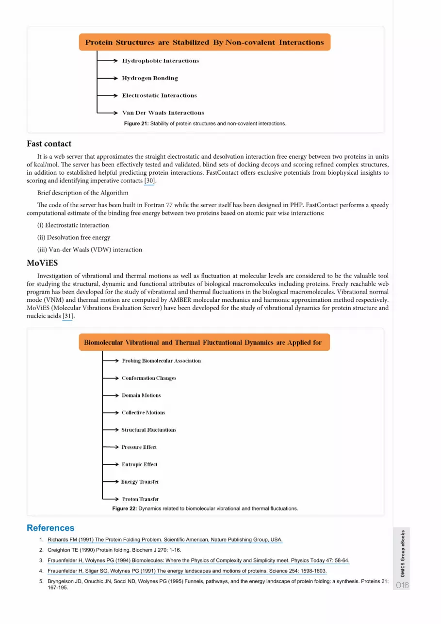
Forces that Govern Protein Stability
Figure 1: Forces in Atoms of Amino Acids that governs Protein Stability.
Hydrophobic effect: This is the most significant force in protein stability. Hydrophobic residues tend to associate and turn to the inside of the core of the protein molecule. Hydrophilic residues will face the outside (in contact with water). For example, water molecules surround the oil and lose some degrees of freedom. When hydrophobic residues (oil) are buried inside the protein, the disorder of water increases, and ΔG decreases- the reaction is more spontaneous.
Van der Waals (VDW) interactions: these are relatively weak attractive forces between neutral atoms and molecules arising from polarization induced in each particle by the presence of other particles. Inside the protein, the residues are tightly packed due to Van der Waals interactions. The types of Van der Waals interactions govern the stability of proteins e.g. London dispersion forces and dipole-dipole forces.
Van der Waals potential: If the atoms are too far, the interaction between them is not observable. If they are too close- they collide with each other. It should be noted that there is some optimum distance in which attractive and repulsive forces are in equilibrium.
Hydrogen bonds: Hydrogen bonds are interaction between a hydrogen atom attached to an electronegative atom and a lone pair of electrons (Also on an electronegative atom).
Practical/Experimental ApproachFluorescence: The amino acid Tryptophan is hydrophobic in nature, if it is buried (inside the folded or native protein) the fluorescence
is small. In principle, when it is exposed to the surface (for instance in the unfolded state) fluorescence is increased, as a result.
Calorimetery: Heat the protein and measure denaturation over different temperature ranges. When protein unfolds, it produces heat. Hence, calorimeter measures the released heat gives the indication for denaturation.
CD Spectroscopy: There are characteristic absorption spectra for different protein structures. Proteins of approximately 300 residues other than small globular structures have been most studied and the results cannot be applicable to other larger proteins like fibrous proteins and/or those present in plasma membrane. However, the basic rules of protein folding determined during the study of similar globular proteins, could be applicable on the more complex protein structure and mechanism of folding could be revealed properly.
Other various techniques for analyzing protein dynamics:
1. Fluorescence recovery after photobleaching (FRAP)
2. Single particle tracking (SPT)
OM
ICS
Gro
up e
Boo
ks
006
3. Fluorescence correlation spectroscopy (FCS)
4. Nuclear Magnetic Resonance (NMR)
5. X-Ray and Neutron Scattering
6. Fluorescence Technique
7. Single Molecule Technique
8. Hydrogen Exchange Mass Spectrometry
9. Fourier Transform Infrared (FTIR) Spectroscopy
10. Circular Dichroism (CD)
11. Raman Spectroscopy
12. Dual Polarization Interferometry (DPI)
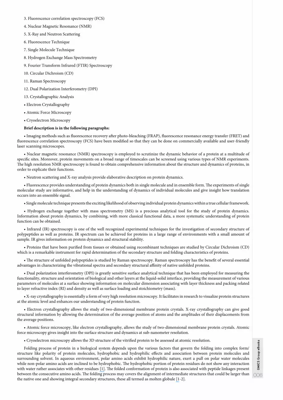
13. Crystallographic Analysis
• Electron Crystallography
• Atomic Force Microscopy
• Cryoelectron Microscopy
Brief description is in the following paragraphs:
• Imaging methods such as fluorescence recovery after photo-bleaching (FRAP), fluorescence resonance energy transfer (FRET) and fluorescence correlation spectroscopy (FCS) have been modified so that they can be done on commercially available and user-friendly laser scanning microscopes.
• Nuclear magnetic resonance (NMR) spectroscopy is employed to scrutinize the dynamic behavior of a protein at a multitude of specific sites. Moreover, protein movements on a broad range of timescales can be screened using various types of NMR experiments. The high resolution NMR spectroscopy is found to obtain comprehensive information about the structure and dynamics of proteins, in order to explicate their functions.
• Neutron scattering and X-ray analysis provide elaborative description on protein dynamics.
• Fluorescence provides understanding of protein dynamics both in single molecule and in ensemble form. The experiments of single molecular study are informative, and help in the understanding of dynamics of individual molecules and give insight how translation occurs into an ensemble signal.
• Single molecule technique presents the exciting likelihood of observing individual protein dynamics within a true cellular framework.
• Hydrogen exchange together with mass spectrometry (MS) is a precious analytical tool for the study of protein dynamics. Information about protein dynamics, by combining, with more classical functional data, a more systematic understanding of protein function can be obtained.
• Infrared (IR) spectroscopy is one of the well recognized experimental techniques for the investigation of secondary structure of polypeptides as well as proteins. IR spectrum can be achieved for proteins in a large range of environments with a small amount of sample. IR gives information on protein dynamics and structural stability.
• Proteins that have been purified from tissues or obtained using recombinant techniques are studied by Circular Dichroism (CD) which is a remarkable instrument for rapid determination of the secondary structure and folding characteristics of proteins.
• The structure of unfolded polypeptides is studied by Raman spectroscopy. Raman spectroscopy has the benefit of several essential advantages in characterizing the vibrational spectra and secondary structural affinity of native unfolded proteins.
• Dual polarization interferometry (DPI) is greatly sensitive surface analytical technique that has been employed for measuring the functionality, structure and orientation of biological and other layers at the liquid-solid interface, providing the measurement of various parameters of molecules at a surface showing information on molecular dimension associating with layer thickness and packing related to layer refractive index (RI) and density as well as surface loading and stoichiometry (mass).
• X-ray crystallography is essentially a form of very high resolution microscopy. It facilitates in research to visualize protein structures at the atomic level and enhances our understanding of protein function.
• Electron crystallography allows the study of two-dimensional membrane protein crystals. X-ray crystallography can give good structural information by allowing the determination of the average position of atoms and the amplitudes of their displacements from the average positions.
• Atomic force microscopy, like electron crystallography, allows the study of two-dimensional membrane protein crystals. Atomic force microscopy gives insight into the surface structure and dynamics at sub-nanometer resolution.
• Cryoelectron microscopy allows the 3D structure of the vitrified protein to be assessed at atomic resolution.
Folding process of protein in a biological system depends upon the various factors that govern the folding into complex form/structure like polarity of protein molecules, hydrophobic and hydrophilic effects and association between protein molecules and surrounding solvent. In aqueous environment, polar amino acids exhibit hydrophilic nature, exert a pull on polar water molecules while non-polar amino acids are inclined to be hydrophobic. The hydrophobic portion of protein residues do not show any interaction with water rather associates with other residues [1]. The folded conformation of protein is also associated with peptide linkages present between the consecutive amino acids. The folding process may covers the alignment of intermediate structures that could be larger than the native one and showing integral secondary structures, these all termed as molten globule [1-2].

OM
ICS
Gro
up e
Boo
ks
007
The finishing stage of folding depends on the precise and specific sequence of amino acids, while previously folding stages are supposed to be mostly insensitive to information of sequence [3].
Thermodynamics of Protein Conformational ChangesThe folded conformation seems to be at narrow free energy (ΔG) state but any change in the folded conformation depends upon
the significant increment in free energy. The change in heat capacity when protein unfolding occurs is the temperature at which the stability is high in its value. As far as free energy is concerned, the highest stability occurs when S=0, on the other hand calculation by the equilibrium constant appears when enthalpy change is zero. The maximum stabilities could be occurred at different temperature ranges, but both are considered in different cases. The stabilities of native form decrease at both lower and higher temperatures.
The other factors such as binding interactions among the amino acids seem to be playing an elaborative role in the stability of the protein conformation but these are not responsible for the significant stability and similar situation happens in the unfolded state, while the bonding between native protein and solvent is expected to be strong enough as compared to the bonding between unfolded coil and the surrounding environment. Moreover, the hydrophobic effect is considered to be the major stabilizing player.
Energy Landscape Theory and Thermodynamics of Protein StabilityProtein folding is one of the complex processes. Frauenfelder et al. and Bryngelson et al. describe the complexity of protein folding by
restoring to a statistical approach to the energetic of protein conformation in the form of energy landscape [4-5]. Bryngelson et al. refers it mathematical procedure that assist in understanding of microscopic behavior of molecular system. It gives quantitative explanations of folded protein state, ensembles of conformational sub-states, ensembles of folding intermediates and denatured or unfolded states and considered as the realistic model of protein [6-7]. Bryngelson et al. describes an energy landscape, in mathematical form, of a system with “n” degrees of freedom is an energy function [5]:
F(x) = F(x1, x2, x3,..., xn)
Here, x1, x2, x3,…, xn are variables for the microscopic state of the system [8] and F(x) is defined as the free energy. As far as protein is concerned, these variables (x1, x2, x3,…, xn) are all the dihedral angles of the chain and showing a single conformation of protein. The stability of the protein can be determined by examining the set of values x1, x2, x3,…, xn that gives minimum value of free energy function, F(x). Moreover, the thermodynamics of protein stability is modeled reasonably well by the Energy Landscape Theory. Stability of medium depends upon the ground and excited states of atoms and nuclear particles which are the simplest as well as basic models while the understanding of complicated system like protein molecules contains far more complex idea and insight into ground and excited states. The ground state of native structure is well illustrated by using energy landscape model where energy represents a function of the topological alignment of atoms. Energy values generated by mountains and ridges are representation of spatial surface with the large number of different co-ordinates.
Figure 2: Uses of Energy Landscape Models.
Protein Folding and UnfoldingFree and attached ribosomes are involved in the production of proteins within the biological system in the form of linear polypeptide
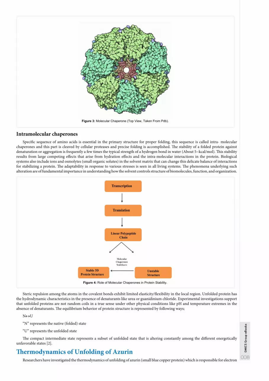
chain. The primary or linear structure is converted into stable three dimensional structures with the help of folding process. The whole process of folding is controlled thermodynamically or kinetically by other proteins and/or enzymes, like molecular chaperones. The process of folding represents that the information required for the precise folding is available in the polypeptide chain. On the other hand, the three dimensional structure of proteins are damaged or affected by various reasons including miscoding due to errors in protein synthesis and/or misfolding due to errors in protein folding.
Molecular ChaperonesThe chaperones have significant impact on the process of protein folding. As far as their function is concerned, it belongs to the class
of proteins that assist in precise folding of proteins. The molecular chaperones are divided into following classes;
Class I chaperones exhibit the affinity to bind with the hydrophobic regions, subsequently preventing aggregation and unfolding polymer is transported to various organelles.
Class II chaperones present inside the organelles assist in misfolding with the help of multiple bonds. It has been observed that when cells are under stress proteins folding become improper.
OM
ICS
Gro
up e
Boo
ks
008
Figure 3: Molecular Chaperone (Top View, Taken From Pdb).
Intramolecular chaperonesSpecific sequence of amino acids is essential in the primary structure for proper folding, this sequence is called intra- molecular
chaperones and this part is cleaved by cellular proteases and precise folding is accomplished. The stability of a folded protein against denaturation or aggregation is frequently a few times the typical strength of a hydrogen bond in water (About 5~kcal/mol). This stability results from large competing effects that arise from hydration effects and the intra-molecular interactions in the protein. Biological systems also include ions and osmolytes (small organic solutes) in the solvent matrix that can change this delicate balance of interactions for stabilizing a protein. The adaptability in response to various stresses is seen in all living systems. The phenomena underlying such alteration are of fundamental importance in understanding how the solvent controls structure of biomolecules, function, and organization.
Figure 4: Role of Molecular Chaperones in Protein Stability.
Steric repulsion among the atoms in the covalent bonds exhibit limited elasticity/flexibility in the local region. Unfolded protein has the hydrodynamic characteristics in the presence of denaturants like urea or guanidinium chloride. Experimental investigations support that unfolded proteins are not random coils in a true sense under other physical conditions like pH and temperature extremes in the absence of denaturants. The equilibrium behavior of protein structure is represented by following ways;
N↔U
“N” represents the native (folded) state
“U” represents the unfolded state
The compact intermediate state represents a subset of unfolded state that is altering constantly among the different energetically unfavorable states [2].
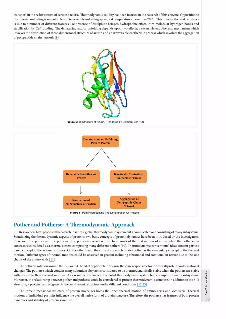
Thermodynamics of Unfolding of AzurinResearchers have investigated the thermodynamics of unfolding of azurin (small blue copper protein) which is responsible for electron
09
OM
ICS
Gro
up e
Boo
ks
transport in the redox system of certain bacteria. Thermodynamic solidity has been focused in the research of this enzyme. Opposition to the thermal unfolding is remarkable and irreversible unfolding appears at temperatures more than 70ºC. This unusual thermal resistance is due to a number of different features like presence of disulphide bridges, hydrophobic effect, intra-molecular hydrogen bonds and stabilization by Cu2+ binding. The denaturing and/or unfolding depends upon two effects; a reversible endothermic mechanism which involves the destruction of three-dimensional structure of azurin and an irreversible exothermic process which involves the aggregation of polypeptide chain network [9].
Figure 5: 3d Structure of Azurin. (Rendered by Chimera, ver. 1.8)
Figure 6: Path Representing The Denaturation Of Proteins.
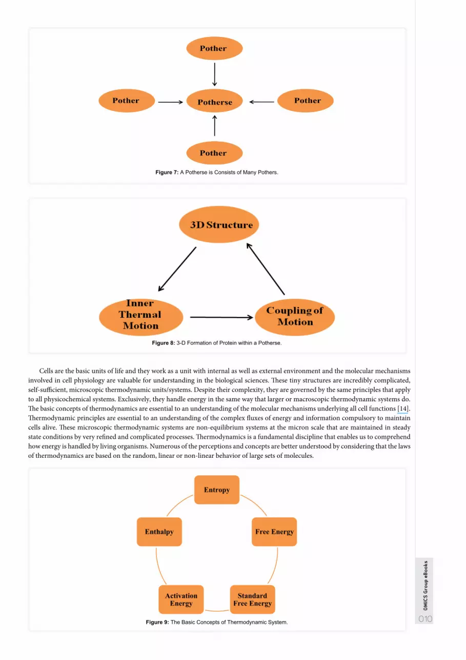
Pother and Potherse: A Thermodynamic ApproachResearchers have proposed that a protein is not a global thermodynamic system but a complicated one consisting of many subsystems.
Scrutinizing the thermodynamic aspects of proteins, two basic concepts of protein dynamics have been introduced by the investigators, there were the pother and the potherse. The pother is considered the basic units of thermal motion of atoms while the potherse, in contrast, is considered as a thermal system comprising many different pothers [10]. Thermodynamic conventional ideas contain particle based concept in the axiomatic theory. On the other hand, the current approach carries pother as the elementary concept of the thermal motion. Different types of thermal motions could be observed in protein including vibrational and rotational in nature due to the side chains of the amino acids [11].
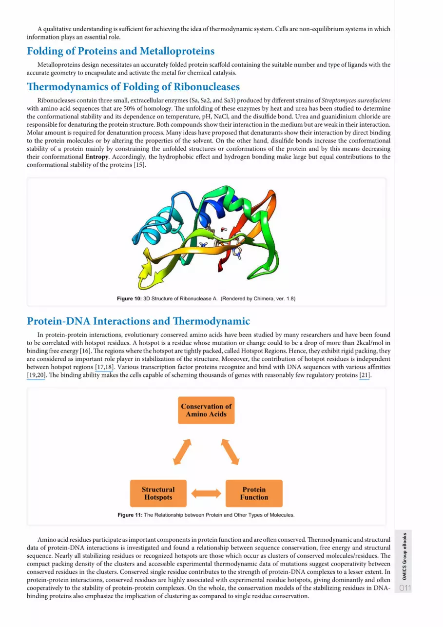
The pother is rotation around the C-N or C-C bond of peptide plain because these are responsible for the overall protein conformational changes. The potherse which contain many subunits/subsystems considered to be thermodynamically stable when the pothers are stable with respect to their thermal motions. As a result, a protein is not a global thermodynamic system but a complex of many subsystems. Moreover, the relationship between pother and potherse could be considered as protein thermodynamic structure. In addition to the 3-D structure, a protein can recognize its thermodynamic structure under different conditions [12,13].
The three-dimensional structure of protein molecules holds the inner thermal motion of amino acids and vice versa. Thermal motions of individual particles influence the overall native form of protein structure. Therefore, the potherse has features of both protein dynamics and stability of protein structure.
010
OM
ICS
Gro
up e
Boo
ks
Figure 7: A Potherse is Consists of Many Pothers.
Figure 8: 3-D Formation of Protein within a Potherse.
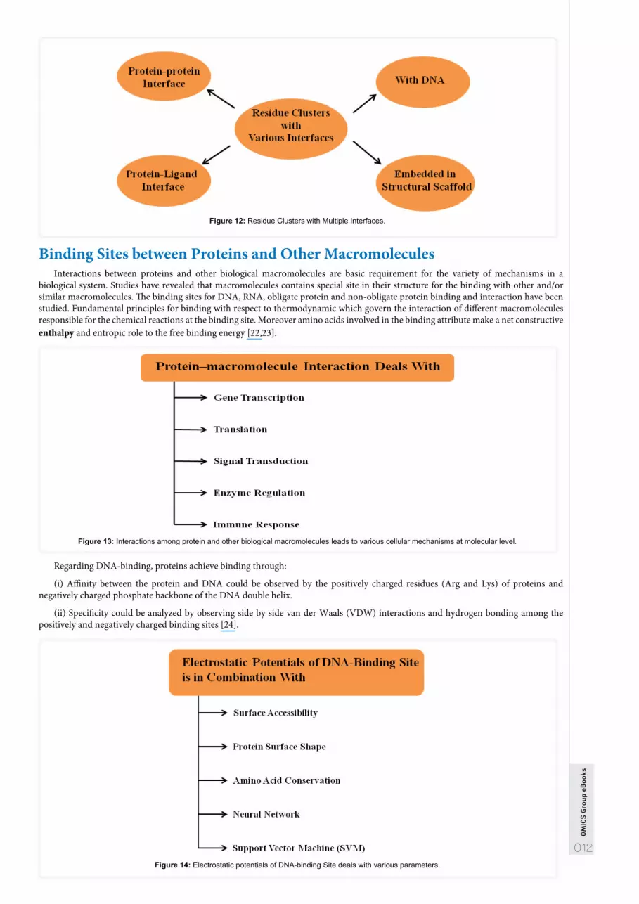
Cells are the basic units of life and they work as a unit with internal as well as external environment and the molecular mechanisms involved in cell physiology are valuable for understanding in the biological sciences. These tiny structures are incredibly complicated, self-sufficient, microscopic thermodynamic units/systems. Despite their complexity, they are governed by the same principles that apply to all physicochemical systems. Exclusively, they handle energy in the same way that larger or macroscopic thermodynamic systems do. The basic concepts of thermodynamics are essential to an understanding of the molecular mechanisms underlying all cell functions [14]. Thermodynamic principles are essential to an understanding of the complex fluxes of energy and information compulsory to maintain cells alive. These microscopic thermodynamic systems are non-equilibrium systems at the micron scale that are maintained in steady state conditions by very refined and complicated processes. Thermodynamics is a fundamental discipline that enables us to comprehend how energy is handled by living organisms. Numerous of the perceptions and concepts are better understood by considering that the laws of thermodynamics are based on the random, linear or non-linear behavior of large sets of molecules.
Entropy
Free Energy
Standard Free Energy
Activation Energy
Enthalpy
Figure 9: The Basic Concepts of Thermodynamic System.
011
OM
ICS
Gro
up e
Boo
ks
A qualitative understanding is sufficient for achieving the idea of thermodynamic system. Cells are non-equilibrium systems in which information plays an essential role.
Folding of Proteins and MetalloproteinsMetalloproteins design necessitates an accurately folded protein scaffold containing the suitable number and type of ligands with the
accurate geometry to encapsulate and activate the metal for chemical catalysis.
Thermodynamics of Folding of RibonucleasesRibonucleases contain three small, extracellular enzymes (Sa, Sa2, and Sa3) produced by different strains of Streptomyces aureofaciens
with amino acid sequences that are 50% of homology. The unfolding of these enzymes by heat and urea has been studied to determine the conformational stability and its dependence on temperature, pH, NaCl, and the disulfide bond. Urea and guanidinium chloride are responsible for denaturing the protein structure. Both compounds show their interaction in the medium but are weak in their interaction. Molar amount is required for denaturation process. Many ideas have proposed that denaturants show their interaction by direct binding to the protein molecules or by altering the properties of the solvent. On the other hand, disulfide bonds increase the conformational stability of a protein mainly by constraining the unfolded structures or conformations of the protein and by this means decreasing their conformational Entropy. Accordingly, the hydrophobic effect and hydrogen bonding make large but equal contributions to the conformational stability of the proteins [15].
Figure 10: 3D Structure of Ribonuclease A. (Rendered by Chimera, ver. 1.8)
Protein-DNA Interactions and ThermodynamicIn protein-protein interactions, evolutionary conserved amino acids have been studied by many researchers and have been found
to be correlated with hotspot residues. A hotspot is a residue whose mutation or change could to be a drop of more than 2kcal/mol in binding free energy [16]. The regions where the hotspot are tightly packed, called Hotspot Regions. Hence, they exhibit rigid packing, they are considered as important role player in stabilization of the structure. Moreover, the contribution of hotspot residues is independent between hotspot regions [17,18]. Various transcription factor proteins recognize and bind with DNA sequences with various affinities [19,20]. The binding ability makes the cells capable of scheming thousands of genes with reasonably few regulatory proteins [21].
Conservation of Amino Acids
Protein Function
Structural Hotspots
Figure 11: The Relationship between Protein and Other Types of Molecules.
Amino acid residues participate as important components in protein function and are often conserved. Thermodynamic and structural data of protein-DNA interactions is investigated and found a relationship between sequence conservation, free energy and structural sequence. Nearly all stabilizing residues or recognized hotspots are those which occur as clusters of conserved molecules/residues. The compact packing density of the clusters and accessible experimental thermodynamic data of mutations suggest cooperativity between conserved residues in the clusters. Conserved single residue contributes to the strength of protein-DNA complexes to a lesser extent. In protein-protein interactions, conserved residues are highly associated with experimental residue hotspots, giving dominantly and often cooperatively to the stability of protein-protein complexes. On the whole, the conservation models of the stabilizing residues in DNA-binding proteins also emphasize the implication of clustering as compared to single residue conservation.
012
OM
ICS
Gro
up e
Boo
ks
Figure 12: Residue Clusters with Multiple Interfaces.
Binding Sites between Proteins and Other MacromoleculesInteractions between proteins and other biological macromolecules are basic requirement for the variety of mechanisms in a
biological system. Studies have revealed that macromolecules contains special site in their structure for the binding with other and/or similar macromolecules. The binding sites for DNA, RNA, obligate protein and non-obligate protein binding and interaction have been studied. Fundamental principles for binding with respect to thermodynamic which govern the interaction of different macromolecules responsible for the chemical reactions at the binding site. Moreover amino acids involved in the binding attribute make a net constructive enthalpy and entropic role to the free binding energy [22,23].
Figure 13: Interactions among protein and other biological macromolecules leads to various cellular mechanisms at molecular level.
Regarding DNA-binding, proteins achieve binding through:
(i) Affinity between the protein and DNA could be observed by the positively charged residues (Arg and Lys) of proteins and negatively charged phosphate backbone of the DNA double helix.
(ii) Specificity could be analyzed by observing side by side van der Waals (VDW) interactions and hydrogen bonding among the positively and negatively charged binding sites [24].
Figure 14: Electrostatic potentials of DNA-binding Site deals with various parameters.

013
OM
ICS
Gro
up e
Boo
ks
The Hydrophobic effectThe hydrophobic effect is an effective force that acts to minimize the amount of surface area that non-polar molecules expose to
their aqueous solvent. The effect originates from the robustly favorable hydrogen-bonding interactions between water molecules that are disrupted by the insertion of a non-polar solute into the solution. The exact dependencies of the free energy of solvation on uncovered surface area, however, depend on the size of the solute. Small solutes can be house without hydrogen-bond breakage through the ordering of the bond network in the surrounding water molecules. On the other hand, for larger solutes the breakage of hydrogen bonds at the surface of the solute is inescapable [25]. Proteins are polymers consisting of chains of amino acids. Each protein folds into an exacting conformation called the native structure, which determines its function, under the physiological condition. The native structure is composed of characteristic local structures called α-helix and β-sheet, hydrogen bonds help for their stabilization. It has been proven experimentally that the native structure is a thermodynamically stable state, called Anfinsen’s dogma.
Computational Approach-Computer Simulation of Proteins for Thermody-namics and Structure Prediction
Computer simulations have developed into an increasingly important tool to study proteins. They are used for regularly complement experimental investigations and often are the only tool to explore processes in the cell. Here is a summary of thermodynamic databases and simulation techniques that are used to study the multiple aspects of proteins regarding thermodynamics.
Sr.# TOOLS PARTICULARS1 ProTherm Thermodynamic database for proteins2 ProTherm, 2.0 Thermodynamic Database for Proteins and Mutants3 ProTherm, 3.0 Thermodynamic Database for Proteins and Mutants8 Protein Folding Database (PFD) A Database for The Investigation of Protein Folding Kinetics and Stability9 Protein Folding Database (PFD 2.0) An Online Environment for The International Foldeomics Consortium
13 SRide A Server for Identifying Stabilizing Residues In Proteins16 FastCo ntact A Free Energy Scoring Tool for Protein–Protein Complex Structures
17 MoViES Molecular Vibrations Evaluation Server For Analysis of Fluctuational Dynamics of Proteins and Nucleic Acids
ProTherm
ProTherm has many tools for the users and provide many functions for the determination of various aspects of proteins and their residues and facilitates the users with several options like empirical energy function, distance and contact potential, average assignment analysis, amino acid properties and many other tools to find out the relationship between protein and their stability with respect to the environment in biological system [26].
ProTherm 2.0 is the second version of the database, which not only contains the thermodynamic data of the protein but also the information related to the mutants. Similar its first version, it provides experimental conditions, literature and structural as well as functional information. Protein Mutant Database (PMD) has been developed to facilitate with natural and artificial mutants of proteins.
ProTherm 3.0 contains more features than the previous two versions of the thermodynamic database. As compared to the previous version, it covers more than 10,000 entries for a number of thermodynamic parameters. The protein recognition code from the SWISS-PROT and Protein Data Bank (PDB) has been added into new version and provides cross-links with other databases.
Figure 15: Protherm (2.0), Several Aspects of Thermodynamic Data.
014
OM
ICS
Gro
up e
Boo
ks
Figure 16: Various Features at the Protherm Site.
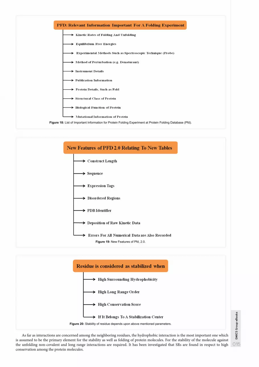
Figure 17: Various features of Protherm Version 3.0. Figure 18: List of Important Information for Protein Folding Experiment at Protein Folding Database (Pfd).
Protein Folding Database (Pfd)It is really an informative, easily accessible and innovative resource and database that facilitates protein folding data of more than
50 different proteins belongs to various families. Data of structural, kinetic as well as thermodynamic in nature is available on protein folding database that could be accessed through web platform. With the various useful features it links with the other protein databases as well [27].
Protein Folding Database 2.0 provides freely accessible and centralized data repository. It has been built as a comparative repository providing kinetic and thermodynamic features for the folding of protein in a useful and user friendly way [27].
Site Directed Mutator (Sdm)SDM is a server for predicting effects of mutations on protein stability and malfunction. Site Directed Mutator is a statistical potential
energy function that exercises environment-specific amino-acid replacement frequencies within homologous protein families to compute a stability score, which is corresponding to the free energy difference between the wild-type and mutant protein [28].
SRideSRide server provides data that could be employed for the calculation of van der Waals atomic radius among the amino acids. The
residues present in the polypeptide and play a crucial role for the stability of protein molecules are called stabilizing residues (SRs). These residues are selected by using various methods like interaction of a given residue with its neighboring residues and observing the evolutionary conservation in the protein structures [29].
015
OM
ICS
Gro
up e
Boo
ks
Figure 18: List of Important Information for Protein Folding Experiment at Protein Folding Database (Pfd).
Figure 19: New Features of Pfd, 2.0.
Figure 20: Stability of residue depends upon above mentioned parameters.
As far as interactions are concerned among the neighboring residues, the hydrophobic interaction is the most important one which is assumed to be the primary element for the stability as well as folding of protein molecules. For the stability of the molecule against the unfolding non-covalent and long range interactions are required. It has been investigated that SRs are found in respect to high conservation among the protein molecules.
016
OM
ICS
Gro
up e
Boo
ks
Figure 21: Stability of protein structures and non-covalent interactions.
Fast contactIt is a web server that approximates the straight electrostatic and desolvation interaction free energy between two proteins in units
of kcal/mol. The server has been effectively tested and validated, blind sets of docking decoys and scoring refined complex structures, in addition to established helpful predicting protein interactions. FastContact offers exclusive potentials from biophysical insights to scoring and identifying imperative contacts [30].
Brief description of the Algorithm
The code of the server has been built in Fortran 77 while the server itself has been designed in PHP. FastContact performs a speedy computational estimate of the binding free energy between two proteins based on atomic pair wise interactions:
(i) Electrostatic interaction
(ii) Desolvation free energy
(iii) Van-der Waals (VDW) interaction
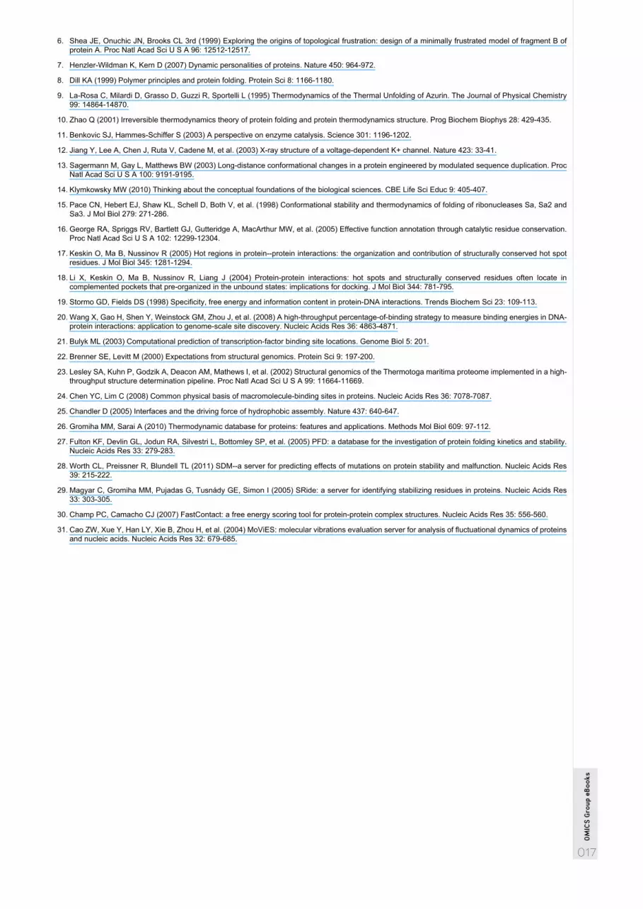
MoViESInvestigation of vibrational and thermal motions as well as fluctuation at molecular levels are considered to be the valuable tool
for studying the structural, dynamic and functional attributes of biological macromolecules including proteins. Freely reachable web program has been developed for the study of vibrational and thermal fluctuations in the biological macromolecules. Vibrational normal mode (VNM) and thermal motion are computed by AMBER molecular mechanics and harmonic approximation method respectively. MoViES (Molecular Vibrations Evaluation Server) have been developed for the study of vibrational dynamics for protein structure and nucleic acids [31].
Figure 22: Dynamics related to biomolecular vibrational and thermal fluctuations.
References1. Richards FM (1991) The Protein Folding Problem. Scientific American, Nature Publishing Group, USA.
2. Creighton TE (1990) Protein folding. Biochem J 270: 1-16.
3. Frauenfelder H, Wolynes PG (1994) Biomolecules: Where the Physics of Complexity and Simplicity meet. Physics Today 47: 58-64.
4. Frauenfelder H, Sligar SG, Wolynes PG (1991) The energy landscapes and motions of proteins. Science 254: 1598-1603.
5. Bryngelson JD, Onuchic JN, Socci ND, Wolynes PG (1995) Funnels, pathways, and the energy landscape of protein folding: a synthesis. Proteins 21: 167-195.
017
OM
ICS
Gro
up e
Boo
ks
6. Shea JE, Onuchic JN, Brooks CL 3rd (1999) Exploring the origins of topological frustration: design of a minimally frustrated model of fragment B of protein A. Proc Natl Acad Sci U S A 96: 12512-12517.
7. Henzler-Wildman K, Kern D (2007) Dynamic personalities of proteins. Nature 450: 964-972.
8. Dill KA (1999) Polymer principles and protein folding. Protein Sci 8: 1166-1180.
9. La-Rosa C, Milardi D, Grasso D, Guzzi R, Sportelli L (1995) Thermodynamics of the Thermal Unfolding of Azurin. The Journal of Physical Chemistry 99: 14864-14870.
10. Zhao Q (2001) Irreversible thermodynamics theory of protein folding and protein thermodynamics structure. Prog Biochem Biophys 28: 429-435.
11. Benkovic SJ, Hammes-Schiffer S (2003) A perspective on enzyme catalysis. Science 301: 1196-1202.
12. Jiang Y, Lee A, Chen J, Ruta V, Cadene M, et al. (2003) X-ray structure of a voltage-dependent K+ channel. Nature 423: 33-41.
13. Sagermann M, Gay L, Matthews BW (2003) Long-distance conformational changes in a protein engineered by modulated sequence duplication. Proc Natl Acad Sci U S A 100: 9191-9195.
14. Klymkowsky MW (2010) Thinking about the conceptual foundations of the biological sciences. CBE Life Sci Educ 9: 405-407.
15. Pace CN, Hebert EJ, Shaw KL, Schell D, Both V, et al. (1998) Conformational stability and thermodynamics of folding of ribonucleases Sa, Sa2 and Sa3. J Mol Biol 279: 271-286.
16. George RA, Spriggs RV, Bartlett GJ, Gutteridge A, MacArthur MW, et al. (2005) Effective function annotation through catalytic residue conservation. Proc Natl Acad Sci U S A 102: 12299-12304.
17. Keskin O, Ma B, Nussinov R (2005) Hot regions in protein--protein interactions: the organization and contribution of structurally conserved hot spot residues. J Mol Biol 345: 1281-1294.
18. Li X, Keskin O, Ma B, Nussinov R, Liang J (2004) Protein-protein interactions: hot spots and structurally conserved residues often locate in complemented pockets that pre-organized in the unbound states: implications for docking. J Mol Biol 344: 781-795.
19. Stormo GD, Fields DS (1998) Specificity, free energy and information content in protein-DNA interactions. Trends Biochem Sci 23: 109-113.
20. Wang X, Gao H, Shen Y, Weinstock GM, Zhou J, et al. (2008) A high-throughput percentage-of-binding strategy to measure binding energies in DNA-protein interactions: application to genome-scale site discovery. Nucleic Acids Res 36: 4863-4871.
21. Bulyk ML (2003) Computational prediction of transcription-factor binding site locations. Genome Biol 5: 201.
22. Brenner SE, Levitt M (2000) Expectations from structural genomics. Protein Sci 9: 197-200.
23. Lesley SA, Kuhn P, Godzik A, Deacon AM, Mathews I, et al. (2002) Structural genomics of the Thermotoga maritima proteome implemented in a high-throughput structure determination pipeline. Proc Natl Acad Sci U S A 99: 11664-11669.
24. Chen YC, Lim C (2008) Common physical basis of macromolecule-binding sites in proteins. Nucleic Acids Res 36: 7078-7087.
25. Chandler D (2005) Interfaces and the driving force of hydrophobic assembly. Nature 437: 640-647.
26. Gromiha MM, Sarai A (2010) Thermodynamic database for proteins: features and applications. Methods Mol Biol 609: 97-112.
27. Fulton KF, Devlin GL, Jodun RA, Silvestri L, Bottomley SP, et al. (2005) PFD: a database for the investigation of protein folding kinetics and stability. Nucleic Acids Res 33: 279-283.
28. Worth CL, Preissner R, Blundell TL (2011) SDM--a server for predicting effects of mutations on protein stability and malfunction. Nucleic Acids Res 39: 215-222.
29. Magyar C, Gromiha MM, Pujadas G, Tusnády GE, Simon I (2005) SRide: a server for identifying stabilizing residues in proteins. Nucleic Acids Res 33: 303-305.
30. Champ PC, Camacho CJ (2007) FastContact: a free energy scoring tool for protein-protein complex structures. Nucleic Acids Res 35: 556-560.
31. Cao ZW, Xue Y, Han LY, Xie B, Zhou H, et al. (2004) MoViES: molecular vibrations evaluation server for analysis of fluctuational dynamics of proteins and nucleic acids. Nucleic Acids Res 32: 679-685.


