Embed Size (px)
Citation preview
ARTICLE
6q22.1 microdeletion and susceptibility to pediatricepilepsy
Przemyslaw Szafranski1, Gretchen K Von Allmen2, Brett H Graham1, Angus A Wilfong3, Sung-Hae L Kang1,Jose A Ferreira4, Sheila J Upton5, John B Moeschler5, Weimin Bi1, Jill A Rosenfeld6, Lisa G Shaffer7,Sau Wai Cheung1, Paweł Stankiewicz1 and Seema R Lalani*,1
Genomic copy-number variations (CNVs) constitute an important cause of epilepsies and other human neurological disorders.
Recent advancement of technologies integrating genome-wide CNV mapping and sequencing is rapidly expanding the molecular
field of pediatric neurodevelopmental disorders. In a previous study, a novel epilepsy locus was identified on 6q16.3q22.31 by
linkage analysis in a large pedigree. Subsequent array comparative genomic hybridization (array CGH) analysis of four unrelated
cases narrowed this region to B5Mb on 6q22.1q22.31. We sought to further narrow the critical region on chromosome 6q22.
Array CGH analysis was used in genome-wide screen for CNVs of a large cohort of patients with neurological abnormalities.
Long-range PCR and DNA sequencing were applied to precisely map chromosomal deletion breakpoints. Finally, real-time
qPCR was used to estimate relative expression in the brain of the candidate genes. We identified six unrelated patients with
overlapping microdeletions within 6q22.1q22.31 region, three of whom manifested seizures. Deletions were found to be de
novo in 5/6 cases, including all subjects presenting with seizures. We sequenced the deletion breakpoints in four patients
and narrowed the critical region to a B250-kb segment at 6q22.1 that includes NUS1, several expressed sequence tags
(ESTs) that are highly expressed in the brain, and putative regulatory sequences of SLC35F1. Our findings indicate that
dosage alteration in particular, of NUS1, EST AI858607, or SLC35F1 are important contributors to the neurodevelopmental
phenotype associated with 6q22 deletion, including epilepsy and tremors.
European Journal of Human Genetics (2015) 23, 173–179; doi:10.1038/ejhg.2014.75; published online 14 May 2014
INTRODUCTION
Despite a wealth of information on the developmental pathways thatoperate during neurogenesis, only a limited number of epileptogenicgenes were identified before the advent of high-throughput techno-logies for studies of structural genomic variations. It has now becomeclear that copy-number variations (CNVs) frequently cause epilepsyand contribute to other neurodevelopmental disorders, includingintellectual disability, neuropsychiatric disorders, autism spectrumdisorder (ASD), and behavior abnormalities. Undoubtedly, the impactof recurrent genomic CNVs involving 14q12, 7q11.23, 15q13.3,16p11.2, and 16p13.11 loci on pediatric epilepsy and other neurode-velopmental problems is substantial.1–5 Using linkage analysis in 15subjects from a large family with a history of genetic epilepsy withfebrile seizures plus (GEFSþ ), Poduri et al6 delineated an 18.1Mbhaplotype on 6q16.3q22.31 that defined a novel GEFSþ 8 locus(OMIM 613828). However, no mutations were found in any of the 16protein-coding genes expressed in the brain within this interval in theaffected subjects. In a recent study, Rosenfeld et al7 reported fourindividuals with seizures and overlapping microdeletions on6q22.1q22.31, and narrowed the critical region to a B5-Mbgenomic segment. Here, applying array comparative genomichybridization (array CGH), we report overlapping microdeletions insix additional patients, three of them presenting with seizures, and
narrow the locus to a B250 kb region on 6q22.1 including NUS1,brain-specific ESTs of unknown function, and putative cis-regulatorysequences of the SLC35F1 promoter. Our findings delineate a criticaldisease region within 6q22 and implicate novel genes in pediatricepilepsy and tremors within this region.
MATERIALS AND METHODS
Patient ascertainmentIndividuals with 6q21q22.31 deletions reported here were identified after
referral for chromosomal microarray analysis (CMA) to clinical laboratories,
including Baylor College of Medicine (BCM) Medical Genetics Laboratories
(MGL) (patients 1–4) and Signature Genomic Laboratories (SGL) (patients 5
and 6). Between June 2008 and June 2013, 13 208 children with neurological
phenotypes were studied by array CGH at MGL. Of these, 8601 children were
referred for developmental delay (65%), 2887 were studied for ASD (22%),
and 1720 subjects were evaluated for seizures (13%). Informed consents via a
protocol approved by the Institutional Review Board for Human Subject
Research at BCM were obtained for patients with deletions within the 6q22
region. DNA samples were re-evaluated by DNA sequencing.
SubjectsPatient 1 was evaluated at 3 years of age with severe speech/language delay,
ASDs, stereotypical repetitive behaviors, and staring episodes suggestive of
atypical absence seizures. He walked independently at 15 months of age.
1Department of Molecular and Human Genetics, Baylor College of Medicine, Houston, TX, USA; 2Division of Child and Adolescent Neurology, Department of Pediatrics,University of Texas Health Science Center at Houston, TX, USA; 3Department of Pediatric Neurology, Texas Children’s Hospital, Baylor College of Medicine, Houston, TX, USA;4Department of Pediatrics, University of South Florida, Tampa, FL, USA; 5Section of Medical Genetics, Department of Pediatrics, Dartmouth Hitchcock Medical Center, Lebanon,NH, USA; 6Signature Genomic Laboratories, PerkinElmer Inc., Spokane, WA, USA; 7Paw Print Genetics, Genetic Veterinary Sciences Inc., Spokane, WA, USA*Correspondence: Dr SR Lalani, Department of Molecular and Human Genetics, Baylor College of Medicine, One Baylor Plaza, MARB, Rm 8006, Houston, TX 77030, USA.Tel: þ 1 713 798 8921; Fax: þ1 832 825 4294; E-mail: [email protected]
Received 9 October 2013; revised 4 March 2014; accepted 7 March 2014; published online 14 May 2014
European Journal of Human Genetics (2015) 23, 173–179& 2015 Macmillan Publishers Limited All rights reserved 1018-4813/15
www.nature.com/ejhg
His EEG background was diffusely slow, mostly in the theta range, with brief
bursts of generalized high-amplitude spike and slow wave discharges of
1.5–2.5hz. Plasma amino acids, urine organic acids, and cerebrospinal fluid
(CSF) neurotransmitters were normal. The MRI of the brain was unremarkable.
His hearing test was normal. No focal deficits were noted on his neurological exam.
Patient 2 was born at term after an uneventful pregnancy. His prenatal and
neonatal course was unremarkable. Mild hypotonia was noted by 6 months of
age and physical therapy was initiated. He walked independently at 21 months
of age. At 4 years of age, he had severe articulation problems, speaking mostly
in two-word phrases. He continued to have difficulties with balance and
coordination when evaluated. MRI of the brain was unremarkable with no
structural abnormalities. Karyotype study, DNA for Fragile X, plasma amino
acids, urine organic acids, and creatine and guanidinoacetate studies
were normal. His physical examination at 4 years of age showed facial
dysmorphisms including broad, flat nasal bridge, bilateral epicanthal folds, and
a mildly high arched palate. His neurological exam revealed mild hypotonia
and clumsy gait.
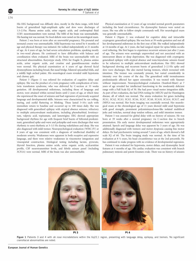
Patient 3 (Figure 1) was referred for evaluation of cognitive delay and
epilepsy. She was the product of a twin pregnancy with complication of twin-
twin transfusion syndrome, who was delivered by C-section at 33 weeks
gestation. All developmental milestones, including those of language and
motor, were attained within normal limits until 2 years of age, at which time
she experienced the onset of seizures and had regression of previously acquired
language and developmental skills. Seizures were characterized by eye rolling,
staring, and eyelid fluttering or blinking. These lasted 3–10 s each with
immediate return to baseline and occurred up to 100 times daily. She was
diagnosed with generalized epilepsy with atypical absence seizures, refractory
to multiple anticonvulsant medications, including phenobarbital, levetirace-
tam, valproic acid, topiramate, and lamotrigine. EEG showed appropriate
background rhythms for age with frequent brief bursts of bifrontal-predomi-
nant, generalized spike-and-wave and polyspike-and-wave discharges that were
rhythmic to semi-rhythmic at 3–3.5 Hz during wakefulness and sleep. She was
also diagnosed with mild tremor. Neuropsychological evaluation (WISC-IV) at
8 years of age was consistent with a diagnosis of intellectual disability of
moderate severity. Weaknesses were apparent in both receptive and expressive
language, and there were relative weaknesses in visuomotor integration and
visuospatial construction. Etiological testing including lactate, pyruvate,
thyroid function, plasma amino acids, urine organic acids, acylcarnitine
profile, CSF neurotransmitter levels, and febrile seizure panel (including
SCN1A) were normal. MRI of the brain was also unremarkable.
Physical examination at 12 years of age revealed normal growth parameters
including the head circumference. No dysmorphic features were noted on
exam. She was able to follow simple commands well. Her neurological exam
was generally unremarkable.
Patient 4 (Figure 1) was evaluated for cognitive delay and intractable
cryptogenic generalized epilepsy. She was born at 39 weeks gestation and met all
her developmental milestones in the first year of life. She walked independently
at 14 months of age. At 2 years, she had surgical repair for spina bifida occulta
cord tethering. She first began to experience recurrent seizures just after 2 years
of age. The seizures were seemingly unprovoked and not associated with an
intercurrent illness or sleep deprivation. She was diagnosed with cryptogenic
generalized epilepsy with atypical absence and tonic/myoclonic seizures found
to be refractory to multiple anticonvulsant medications. Her EEG showed
background slowing and recurrent bursts of generalized 2–2.5Hz spike and
slow wave discharges. She also started having tremors, which worsened with
intention. The tremor was constantly present, but varied considerably in
intensity over the course of the day. The generalized mild tremulousness
predominately affected her upper extremities. It was treated with Sinemet
without improvement. Neuropsychological evaluation (Stanford-Binet) at 7
years of age revealed her intellectual functioning in the moderately deficient
range with a Full Scale IQ of 50. She had poor visual motor integration skills.
As part of her evaluation, she had DNA testing for MECP2 and for Huntington
disease, all of which was normal. The ataxia evaluation for genes including
SCA1, SCA2, SCA3, SCA5, SCA6, SCA7, SCA8, SCA10, SCA14, SCA17, and
DRPLA was normal. Her brain imaging was essentially normal. Her neurolo-
gical exam at the chronological age of 11 years showed mild axial hypotonia
with good strength, prominent polyminimyoclonus-like isolated multifocal
jerks and twitches, normal deep tendon reflexes, and mild intention tremor.
Patient 5 was assessed for global delay with no history of seizures. He was
born at 39 weeks after a normal pregnancy via C-section due to breech
presentation. His early motor developmental milestones were appropriately
attained. Speech and language delay was apparent by 3 years of age. He was
additionally diagnosed with tremors and motor dyspraxia causing fine motor
delays. He had psychometric testing around 7 years of age, which showed a full
scale IQ of 60. The brain imaging study was normal. At the time of his
evaluation at 8 ½ years, his head circumference was at the 2nd percentile. He
has continued to make progress with no evidence of developmental regression.
Patient 6 was evaluated for hypotonia, motor delays, and dysmorphic facial
features at 4 months of age. His cardiac evaluation was consistent with branch
pulmonary stenosis and patent foramen ovale. There was no history of seizures
Figure 1 Patients 3 and 4 with de novo microdeletions within the 6q22.1 region, presenting with language delay, epilepsy, and tremors. No significant
craniofacial abnormalities are noted.
Narrowing the critical region of 6q22 deletionP Szafranski et al
174
European Journal of Human Genetics
by report. His physical examination was remarkable for an adducted left thumb
and borderline microcephaly. His neurological exam was otherwise normal.
DNA and RNA isolationGenomic DNA was extracted from peripheral blood using the Puregene DNA
isolation kit (Gentra System, Minneapolis, MN, USA). Total RNA from the
normal human brain was purchased from Ambion (Austin, TX, USA).
Reference RNA was extracted from normal fetal lung fibroblasts, IMR-90
(ATCC CCL-186), using MasterPur Complete DNA and RNA Purification Kit
(Epicentre, Madison, WI, USA). The RNA sample was treated with DNase
using DNA-free Kit (Ambion).
Array CGH analysisThe four unrelated subjects referred for clinical CMA at the MGL at BCM were
screened using custom-designed exon-targeted CGH oligonucleotide micro-
arrays (patient 1 V7.1 OLIGO, 105K; patient 2 V8.1 OLIGO, 180K and
patients 3 and 4 V8.2 OLIGO, 180K) designed by MGL at BCM (http://
www.bcm.edu/geneticlabs) and manufactured by Agilent Technologies (Santa
Clara, CA, USA), as previously described.8,9 Patients 1, 3, and 4 were also
screened using custom-designed high-resolution 3� 720K microarrays
covering 12Mb in 6q21-6q22.31, designed and produced by Roche-
NimbleGen (Madison, WI, USA). Labeling and hybridization of DNA
samples were done according to the manufacturer’s instructions (NimbleGen
Array User’s Guide v9.1 for array CGH). Patients 5 and 6 were screened using
whole-genome oligonucleotide-based CGH microarrays custom-designed by
SGL: 105K SignatureChipOS v1.1 (manufactured by Agilent) and 135K
SignatureChipOS v2.0 (manufactured by Roche–NimbleGen), respectively, as
previously described.10,11
FISH analysisConfirmatory and parental fluorescence in situ hybridization (FISH) analyses
were performed using standard procedures. Deletions were verified by
using the chromosome 6q-specific BAC clones RP11-61K10, RP11-282C5,
RP11-141L18, or RP11-435B17.
Long-range PCR and DNA sequencingPrimers were designed using the Primer 3 software (http://frodo.wi.mit.edu/
primer3). Amplification of junction fragments was performed using Takara LA
Taq Polymerase (TaKaRa Bio USA, Madison, WI, USA) applying 30 cycles of
94 1C for 30 s and 68 1C for 5min. PCR products were treated with ExoSAP-IT
(USB, Cleveland, OH, USA) to remove unconsumed dNTPs and primers, and
directly sequenced by the Sanger method using primers flanking deletion
breakpoints (see Supplementary Table S1).
Real-time qPCR analysis of 6q22.1 transcriptsTotal RNA from the normal human brain and lung fibroblasts was reverse-
transcribed using High Capacity cDNA Reverse Transcription Kit (Applied
Biosystems, Branchburg, NJ, USA). cDNA from piRNA, piR-56963, was
synthesized using SuperScript First Strand Synthesis System for RT-PCR
(Invitrogen, Carlsbad, CA, USA) and stem-loop primer 50-GTTGGCTCTGGTGCAGGGTCCGAGGTATTCGCACCAGAGCCAACACCCAG-30. Primers to
quantify piR-56963 were designed using miRNA Primer Design Tool. Primers
to quantify transcripts of NUS1, SLC35F1, GAPDH (internal control),
ESTs: CB111179, BI825833, AI858607, BQ429202, and a lncRNA,
TCONS_00011550, were designed using PrimerQuest (http://www.idtdna.
com/Primerquest/Home/Index) (see Supplementary Table S2). qPCR was
repeated four times using Power SYBR Green PCR Master Mix (Applied
Biosystems). qPCR conditions included 40 cycles of 95 1C for 15 s and 60 1C
for 1min. For relative quantification, the comparative CT method was used.
BioinformaticsGenomic sequences based on oligonucleotide coordinates obtained from the
array CGH experiments were downloaded from the UCSC Genome Browser
(NCBI build 37, May 2009, hg19, http://www.genome.ucsc.edu) and assembled
using BLAT (UCSC genome browser), BLAST2 (NCBI, http://blast.ncbi.nlm.
nih.gov), and the Sequencher v4.8 software (Gene Codes, Ann Arbor, MI,
USA). Interspersed repeat sequences were identified using RepeatMasker
(http://www.repeatmasker.org). GC content was analyzed using Cpgplot
(http://www.ebi.ac.uk/Tools/seqstat/emboss_cpgplot).
RESULTS
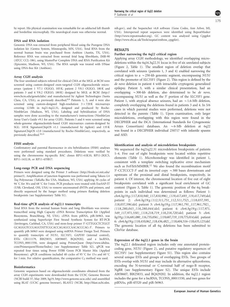
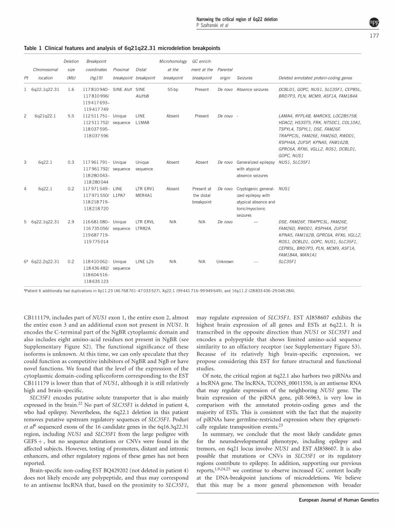
Further narrowing the 6q22 critical regionApplying array CGH methodology, we identified overlapping micro-deletions within the 6q16.3q22.31 locus in five of six unrelated subjects(Figure 2, Table 1). The smallest region of deletion overlap thatcorrelated with seizures (patients 1, 3 and 4) enabled narrowing thecritical region to a B250-kb genomic segment, encompassing NUS1and the promoter of SLC35F1 (Figure 2). This region is defined by thede novo deletion in patient 4 with intractable cryptogenic generalizedepilepsy. Patient 3, with a similar clinical presentation, had anoverlapping B300-kb deletion, also determined to be de novo,encompassing NUS1 as well as the 50 portion of the SLC35F1 gene.Patient 1, with atypical absence seizures, had an B1.6-Mb deletion,completely overlapping the deletions found in patients 3 and 4. In 5/6cases in which parental studies were performed, deletions were notdetected in the parents (Table 1). Upon examination, only largermicrodeletions, overlapping with this region were found in theDECIPHER and the ISCA (International Standards for CytogenomicArrays Consortium) databases. An B6-Mb deletion at 6q22was found in a DECIPHER individual 250717 with infantile spasmsand ASD.
Identification and analysis of microdeletion breakpointsWe sequenced the 6q21q22.31 microdeletion breakpoints of patients1–4. Five out of eight breakpoints were located within repetitiveelements (Table 1). Microhomology was identified in patient 1,consistent with a template switching replicative error mechanismsuch as FoSTeS/MMBIR.12 We also found the recombination motif50-CCTCCCT-30 and its inverted copy B300 bases downstream andupstream of the proximal and distal breakpoints, respectively, inpatient 4. Of interest, the chromosomal location of five out of eightbreakpoints correlated with a significant local enrichment of GCcontent (Figure 3, Table 1). The genomic position of the 6q break-points in each individual was determined as follows: Patient 1:chr6.hg19:g.(117,810,940_117,810,996)_(119,417,693_119,417,749)del;patient 2: chr6.hg19:g.(112,511,751_112,511,752)_(118,037,595_118,037,596)del; patient 3: chr6.hg19:g.(117,961,791_117,961,792)_(118,280,043_118,280,044)del; patient 4: chr6.hg19:g.(117,971,549_117,971,550)_(118,218,719_118,218,720)del; patient 5: chr6.hg19:g.(116,681,080_116,735,056)_(119,687,719_119,775,014)del; patient6: chr6.hg19:g.(118,410,062_118,436,482)_(118,604,516–118,635,123)del.The genomic location of all 6q deletions has been submitted toClinVar database.
Expression of the 6q22.1 genes in the brainThe 6q22.1 delineated region includes only one annotated protein-coding gene, NUS1 (Figure 2), and putative regulatory sequences ofSLC35F1 (see Supplementary Figure S1). This region also containsseveral unique ESTs and groups of overlapping ESTs. Two groups ofESTs overlap with NUS1 and may include its alternative spliceoforms,encoding the N-terminal or C-terminal half of nogo-B receptor,NgBR (see Supplementary Figure S2). The unique ESTs includeAI858607, BI825833, and BQ429202. In addition, the 6q22.1 regionincludes the gene for a lncRNA, TCONS_00011550, and two genes forpiRNAs, piR-45520 and piR-56963.
Narrowing the critical region of 6q22 deletionP Szafranski et al
175
European Journal of Human Genetics
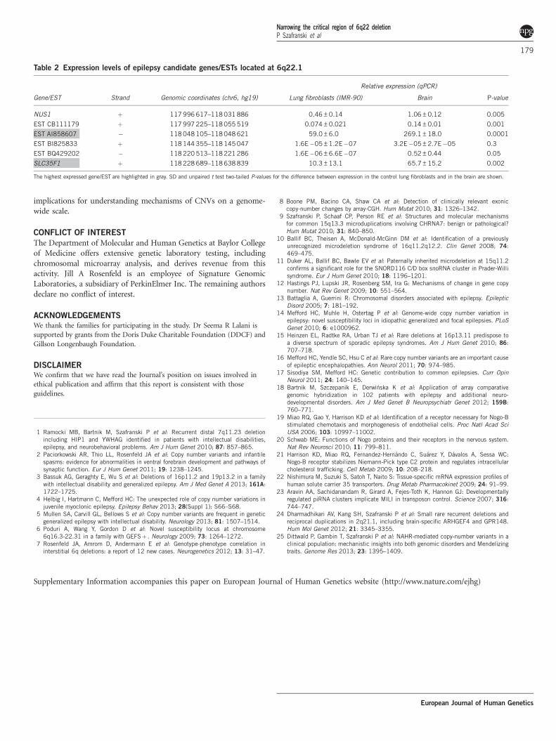
Besides NUS1 and SLC35F1, the expression of the ESTs andthe non-protein-coding genes located at 6q22.1 is unknown.To determine whether any of them could be considered as anadditional candidate gene for epilepsy, we measured by qPCR theabundance of their transcripts in total RNA from the normal humanbrain and lung fibroblasts as a reference (Table 2). We confirmedenrichment of the expression of NUS1 and SLC35F1 in the brain andfound that ESTs CB111179, AI858607, and BQ429202 also exhibitedsignificantly higher expression in the brain compared with controllung fibroblasts, with EST AI858607 having the highest expression.Expression of the lncRNA and piR-56963 genes was very low both inthe brain and the lung fibroblasts (data not shown).
DISCUSSION
In this report, we identified six unrelated patients with non-recurrentdeletions within 6q21q22.31 and established that the loss of thisregion results in a neurodevelopmental disorder, characterized bylanguage delay, cognitive deficits, epilepsy, and tremors with variableexpressivity. Language delay was ascertained in all patients after 2years of age. Seizure disorder and tremors were observed in three outof six individuals. The incomplete penetrance for the seizurephenotype in this study is consistent with the published data, whichsuggest that epilepsy is observed in about half of cases when 6q22 isdeleted.6 At least two out of six patients with non-overlappingdeletions were reported to have mild dysmorphic features. Thevariability in the phenotype could be indicative of effects due tocontiguous gene deletion, rather than loss of a single dosage sensitivegene. Alternatively, the variability may result from the deletion ofdifferent loci, as seen in patient 6 with hypotonia, motor delays anddysmorphic facial features. In addition to the 200 kb loss on
6q22.2q22.31 involving SLC35F1, patient 6 had additional CNVsincluding 265-kb duplication in Xp11.23, 508-kb duplication inXq22.1, and 213-kb gain of 16p11.2. Parental samples wereunavailable for further evaluation in the case of patient 6 and it isunclear whether the small CNVon chromosome 6 is truly pathogenicin this individual. The deletions arose de novo in the remaining fiveout of six cases.Despite increasing interest in the genetic bases of pediatric epilepsy,
only a few genome-wide studies of CNVs have been performed inindividuals with seizure disorder.2,13–18 In this study, we narrowed thecritical region to the B250-kb segment at 6q22.1, harboring only oneprotein-coding annotated gene, NUS1, several ESTs, two piRNAgenes, and a lncRNA gene. Quantitative analyses of the transcriptsof these genes and ESTs by qPCR showed that NUS1, ESTs AI858607,CB111179, BQ429202, and adjacent to the deletion overlappingregion, SLC35F1 gene are relatively highly expressed in the brainwhen compared with their expression in cultured lung fibroblastsused as a control.NUS1 encodes a precursor of the membrane receptor (NgBR)
for the N terminus of nogo-B.19 Nogo proteins A, B, and C areimportant regulators of neuronal and endothelial cell motility andgrowth.20 NgBR also interacts with and stabilizes NPC2 protein.Of interest, mutations in NPC2 result in Niemann-Pick Type C,21
an autosomal recessive disease characterized by inability tometabolize cholesterol by the cell, and also associated withseizures and ataxia. NUS1 overlaps with two groups of ESTs,some of which likely correspond to its alternative spliceoforms.One of these groups, represented by EST HY124277, encodes theN-terminal half of NgBR, including its transmembrane helix (seeSupplementary Figure S2). The other group, represented by EST
Figure 2 Schematic representation of the analyzed genomic region at 6q22.1q22.31. Genomic coordinates correspond to the hg19 build of the human
genome. Red bars represent microdeletions. Dotted vertical lines mark the region of the microdeletion overlap in patients 1, 3, and 4 presenting with
seizures. Protein-coding genes, ESTs, and non-protein-coding genes are shown. Black and green color depict opposite directions of transcription. Array CGH
log ratio plots of microdeletions in patients 3 and 4 are shown. ASD, autism spectrum disorder; DD, developmental delay; DF, dysmorphic features; FS,
febrile seizures; GTCS, generalized tonic-clonic seizures; ID, intellectual disability.
Narrowing the critical region of 6q22 deletionP Szafranski et al
176
European Journal of Human Genetics
CB111179, includes part of NUS1 exon 1, the entire exon 2, almostthe entire exon 3 and an additional exon not present in NUS1. Itencodes the C-terminal part of the NgBR cytoplasmic domain andalso includes eight amino-acid residues not present in NgBR (seeSupplementary Figure S2). The functional significance of theseisoforms is unknown. At this time, we can only speculate that theycould function as competitive inhibitors of NgBR and NgB or havenovel functions. We found that the level of the expression of thecytoplasmic domain-coding spliceoform corresponding to the ESTCB111179 is lower than that of NUS1, although it is still relativelyhigh and brain-specific.SLC35F1 encodes putative solute transporter that is also mainly
expressed in the brain.22 No part of SLC35F1 is deleted in patient 4,who had epilepsy. Nevertheless, the 6q22.1 deletion in this patientremoves putative upstream regulatory sequences of SLC35F1. Poduriet al6 sequenced exons of the 16 candidate genes in the 6q16.3q22.31region, including NUS1 and SLC35F1 from the large pedigree withGEFSþ , but no sequence alterations or CNVs were found in theaffected subjects. However, testing of promoters, distant and intronicenhancers, and other regulatory regions of these genes has not beenreported.Brain-specific non-coding EST BQ429202 (not deleted in patient 4)
does not likely encode any polypeptide, and thus may correspondto an antisense lncRNA that, based on the proximity to SLC35F1,
may regulate expression of SLC35F1. EST AI858607 exhibits thehighest brain expression of all genes and ESTs at 6q22.1. It istranscribed in the opposite direction than NUS1 or SLC35F1 andencodes a polypeptide that shows limited amino-acid sequencesimilarity to an olfactory receptor (see Supplementary Figure S3).Because of its relatively high brain-specific expression, wepropose considering this EST for future structural and functionalstudies.Of note, the critical region at 6q22.1 also harbors two piRNAs and
a lncRNA gene. The lncRNA, TCONS_00011550, is an antisense RNAthat may regulate expression of the neighboring NUS1 gene. Thebrain expression of the piRNA gene, piR-56963, is very low incomparison with the annotated protein-coding genes and themajority of ESTs. This is consistent with the fact that the majorityof piRNAs have germline-restricted expression where they epigeneti-cally regulate transposition events.23
In summary, we conclude that the most likely candidate genesfor the neurodevelopmental phenotype, including epilepsy andtremors, on 6q21 locus involve NUS1 and EST AI858607. It is alsopossible that mutations or CNVs in SLC35F1 or its regulatoryregions contribute to epilepsy. In addition, supporting our previousreports,1,9,24,25 we continue to observe increased GC content locallyat the DNA-breakpoint junctions of microdeletions. We believethat this may be a more general phenomenon with broader
Table 1 Clinical features and analysis of 6q21q22.31 microdeletion breakpoints
Pt
Chromosomal
location
Deletion
size
(Mb)
Breakpoint
coordinates
(hg19)
Proximal
breakpoint
Distal
breakpoint
Microhomology
at the
breakpoint
GC enrich-
ment at the
breakpoint
Parental
origin Seizures Deleted annotated protein-coding genes
1 6q22.1q22.31 1.6 117810 940–
117 810 996/
119417 693–
119 417 749
SINE AluY SINE
AluYb8
55bp Present De novo Absence seizures DCBLD1, GOPC, NUS1, SLC35F1, CEP85L,
BRD7P3, PLN, MCM9, ASF1A, FAM184A
2 6q21q22.1 5.5 112511 751–
112 511 752/
118037 595–
118 037 596
Unique
sequence
LINE
L1MA8
Absent Present De novo - LAMA4, RFPL4B, MARCKS, LOC285758,
HDAC2, HS3ST5, FRK, NT5DC1, COL10A1,
TSPYL4, TSPYL1, DSE, FAM26F,
TRAPPC3L, FAM26E, FAM26D, RWDD1,
RSPH4A, ZUFSP, KPNA5, FAM162B,
GPRC6A, RFX6, VGLL2, ROS1, DCBLD1,
GOPC, NUS1
3 6q22.1 0.3 117961 791–
117 961 792/
118280 043–
118 280 044
Unique
sequence
Unique
sequence
Absent Absent De novo Generalized epilepsy
with atypical
absence seizures
NUS1, SLC35F1
4 6q22.1 0.2 117971 549–
117 971 550/
118218 719–
118 218 720
LINE
L1PA7
LTR ERV1
MER4A1
Absent Present at
the distal
breakpoint
De novo Cryptogenic general-
ized epilepsy with
atypical absence and
tonic/myoclonic
seizures
NUS1
5 6q22.1q22.31 2.9 116681 080–
116 735 056/
119687 719–
119 775 014
Unique
sequence
LTR ERVL
LTR82A
N/A N/A De novo — DSE, FAM26F, TRAPPC3L, FAM26E,
FAM26D, RWDD1, RSPH4A, ZUFSP,
KPNA5, FAM162B, GPRC6A, RFX6, VGLL2,
ROS1, DCBLD1, GOPC, NUS1, SLC35F1,
CEP85L, BRD7P3, PLN, MCM9, ASF1A,
FAM184A, MAN1A1
6a 6q22.2q22.31 0.2 118410 062–
118 436 482/
118604 516–
118 635 123
Unique
sequence
LINE L2b N/A N/A Unknown — SLC35F1
aPatient 6 additionally had duplications in Xp11.23 (46 768761–47033527), Xq22.1 (99441 716–99 949 649), and 16p11.2 (28833 436–29046 284).
Narrowing the critical region of 6q22 deletionP Szafranski et al
177
European Journal of Human Genetics
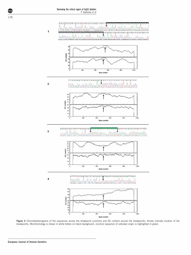
Figure 3 Chromatopherograms of the sequences across the breakpoint junctions and GC content around the breakpoints. Arrows indicate location of the
breakpoints. Microhomology is shown in white letters on black background. Junction sequence of unknown origin is highlighted in green.
Narrowing the critical region of 6q22 deletionP Szafranski et al
178
European Journal of Human Genetics
implications for understanding mechanisms of CNVs on a genome-wide scale.
CONFLICT OF INTEREST
The Department of Molecular and Human Genetics at Baylor Collegeof Medicine offers extensive genetic laboratory testing, includingchromosomal microarray analysis, and derives revenue from thisactivity. Jill A Rosenfeld is an employee of Signature GenomicLaboratories, a subsidiary of PerkinElmer Inc. The remaining authorsdeclare no conflict of interest.
ACKNOWLEDGEMENTSWe thank the families for participating in the study. Dr Seema R Lalani is
supported by grants from the Doris Duke Charitable Foundation (DDCF) and
Gillson Longenbaugh Foundation.
DISCLAIMERWe confirm that we have read the Journal’s position on issues involved in
ethical publication and affirm that this report is consistent with those
guidelines.
1 Ramocki MB, Bartnik M, Szafranski P et al: Recurrent distal 7q11.23 deletionincluding HIP1 and YWHAG identified in patients with intellectual disabilities,epilepsy, and neurobehavioral problems. Am J Hum Genet 2010; 87: 857–865.
2 Paciorkowski AR, Thio LL, Rosenfeld JA et al: Copy number variants and infantilespasms: evidence for abnormalities in ventral forebrain development and pathways ofsynaptic function. Eur J Hum Genet 2011; 19: 1238–1245.
3 Bassuk AG, Geraghty E, Wu S et al: Deletions of 16p11.2 and 19p13.2 in a familywith intellectual disability and generalized epilepsy. Am J Med Genet A 2013; 161A:1722–1725.
4 Helbig I, Hartmann C, Mefford HC: The unexpected role of copy number variations injuvenile myoclonic epilepsy. Epilepsy Behav 2013; 28(Suppl 1): S66–S68.
5 Mullen SA, Carvill GL, Bellows S et al: Copy number variants are frequent in geneticgeneralized epilepsy with intellectual disability. Neurology 2013; 81: 1507–1514.
6 Poduri A, Wang Y, Gordon D et al: Novel susceptibility locus at chromosome6q16.3-22.31 in a family with GEFSþ . Neurology 2009; 73: 1264–1272.
7 Rosenfeld JA, Amrom D, Andermann E et al: Genotype-phenotype correlation ininterstitial 6q deletions: a report of 12 new cases. Neurogenetics 2012; 13: 31–47.
8 Boone PM, Bacino CA, Shaw CA et al: Detection of clinically relevant exoniccopy-number changes by array-CGH. Hum Mutat 2010; 31: 1326–1342.
9 Szafranski P, Schaaf CP, Person RE et al: Structures and molecular mechanismsfor common 15q13.3 microduplications involving CHRNA7: benign or pathological?Hum Mutat 2010; 31: 840–850.
10 Ballif BC, Theisen A, McDonald-McGinn DM et al: Identification of a previouslyunrecognized microdeletion syndrome of 16q11.2q12.2. Clin Genet 2008; 74:469–475.
11 Duker AL, Ballif BC, Bawle EV et al: Paternally inherited microdeletion at 15q11.2confirms a significant role for the SNORD116 C/D box snoRNA cluster in Prader-Willisyndrome. Eur J Hum Genet 2010; 18: 1196–1201.
12 Hastings PJ, Lupski JR, Rosenberg SM, Ira G: Mechanisms of change in gene copynumber. Nat Rev Genet 2009; 10: 551–564.
13 Battaglia A, Guerrini R: Chromosomal disorders associated with epilepsy. EpilepticDisord 2005; 7: 181–192.
14 Mefford HC, Muhle H, Ostertag P et al: Genome-wide copy number variation inepilepsy: novel susceptibility loci in idiopathic generalized and focal epilepsies. PLoSGenet 2010; 6: e1000962.
15 Heinzen EL, Radtke RA, Urban TJ et al: Rare deletions at 16p13.11 predispose toa diverse spectrum of sporadic epilepsy syndromes. Am J Hum Genet 2010; 86:707–718.
16 Mefford HC, Yendle SC, Hsu C et al: Rare copy number variants are an important causeof epileptic encephalopathies. Ann Neurol 2011; 70: 974–985.
17 Sisodiya SM, Mefford HC: Genetic contribution to common epilepsies. Curr OpinNeurol 2011; 24: 140–145.
18 Bartnik M, Szczepanik E, Derwinska K et al: Application of array comparativegenomic hybridization in 102 patients with epilepsy and additional neuro-developmental disorders. Am J Med Genet B Neuropsychiatr Genet 2012; 159B:760–771.
19 Miao RQ, Gao Y, Harrison KD et al: Identification of a receptor necessary for Nogo-Bstimulated chemotaxis and morphogenesis of endothelial cells. Proc Natl Acad SciUSA 2006; 103: 10997–11002.
20 Schwab ME: Functions of Nogo proteins and their receptors in the nervous system.Nat Rev Neurosci 2010; 11: 799–811.
21 Harrison KD, Miao RQ, Fernandez-Hernando C, Suarez Y, Davalos A, Sessa WC:Nogo-B receptor stabilizes Niemann-Pick type C2 protein and regulates intracellularcholesterol trafficking. Cell Metab 2009; 10: 208–218.
22 Nishimura M, Suzuki S, Satoh T, Naito S: Tissue-specific mRNA expression profiles ofhuman solute carrier 35 transporters. Drug Metab Pharmacokinet 2009; 24: 91–99.
23 Aravin AA, Sachidanandam R, Girard A, Fejes-Toth K, Hannon GJ: Developmentallyregulated piRNA clusters implicate MILI in transposon control. Science 2007; 316:744–747.
24 Dharmadhikari AV, Kang SH, Szafranski P et al: Small rare recurrent deletions andreciprocal duplications in 2q21.1, including brain-specific ARHGEF4 and GPR148.Hum Mol Genet 2012; 21: 3345–3355.
25 Dittwald P, Gambin T, Szafranski P et al: NAHR-mediated copy-number variants in aclinical population: mechanistic insights into both genomic disorders and Mendelizingtraits. Genome Res 2013; 23: 1395–1409.
Supplementary Information accompanies this paper on European Journal of Human Genetics website (http://www.nature.com/ejhg)
Table 2 Expression levels of epilepsy candidate genes/ESTs located at 6q22.1
Relative expression (qPCR)
Gene/EST Strand Genomic coordinates (chr6, hg19) Lung fibroblasts (IMR-90) Brain P-value
NUS1 þ 117 996 617–118 031 886 0.46±0.14 1.06±0.12 0.005
EST CB111179 þ 117 997 225–118 055 519 0.074±0.021 0.14±0.01 0.001
EST AI858607 � 118 048 105–118 048 621 59.0±6.0 269.1±18.0 0.0001
EST BI825833 þ 118 144 355–118 145 047 1.6E�05±1.2E�07 3.2E�05±2.7E�05 0.3
EST BQ429202 � 118 220 513–118 221 286 1.6E�06±6.6E�07 0.52±0.44 0.05
SLC35F1 þ 118 228 689–118 638 839 10.3±13.1 65.7±15.2 0.002
The highest expressed gene/EST are highlighted in gray. SD and unpaired t test two-tailed P-values for the difference between expression in the control lung fibroblasts and in the brain are shown.
Narrowing the critical region of 6q22 deletionP Szafranski et al
179
European Journal of Human Genetics


