Embed Size (px)
Citation preview

221-D100 Controlled Substances Procedures Manual Qualtrax ID: 2480Issued by Chemistry Program Manager Qualtrax Revision 4Issue Date: 30-November-2017 Page 1 of 148
Department of Forensic Science
CONTROLLED SUBSTANCES PROCEDURES MANUAL
UNCONTROLLED COPY
COPYRIGHT © 2017
VIRGINIA DEPARTMENT
OF FORENSIC SCIENCE
Table of Contents
221-D100 Controlled Substances Procedures Manual Qualtrax ID: 2480Issued by Chemistry Program Manager Qualtrax Revision 4Issue Date: 30-November-2017 Page 2 of 148
TABLE OF CONTENTS 1 Introduction
1.1 Introductions 1.2 Examination Documentation
2 Analytical Schemes
2.1 Introduction 2.2 General Unknowns/Powders/Illicit Tablets 2.3 Tablets and Capsules 2.4 Marijuana 2.5 Pharmaceutical Identifiers 2.6 Color Tests 2.7 Chromatography 2.8 Infrared Spectroscopy/Mass Spectrometry 2.9 Further Testing 2.10 Liquids 2.11 Plant Material
3 Drug Item Reduction Program
3.1 Introduction 3.2 Procedures
4 Weighing Practices
4.1 Procedures 4.2 Weighing Practices 4.3 Weighing Practices for Hypergeometric Sampling Plan
5 Sampling
5.1 Introduction 5.2 General Sampling 5.3 Administrative Sampling Plan 5.4 Hypergeometric Sampling Plan 5.5 Multiple Specimens 5.6 Bulk Materials 5.7 Residue Samples 5.8 Sampling for Quantitative Analysis 5.9 References
6 Marijuana, Hashish Oil, and Cannabinoid Products
6.1 Introduction 6.2 Macroscopic Identification 6.3 Microscopic Identification 6.4 Thin Layer Chromatography (TLC) 6.5 Duquenois-Levine 6.6 Gas Chromatography/Mass Spectrometry (GC/MS) 6.7 Hashish Oil 6.8 Extraction of THC from Food Products (candy, brownies, etc.) 6.9 Cannabinoid Quantitation by High Performance Liquid Chromatography – Diode Array Detection
(HPLC-DAD) 6.10 References
UNCONTROLLED COPY
COPYRIGHT © 2017
VIRGINIA DEPARTMENT
OF FORENSIC SCIENCE
Table of Contents
221-D100 Controlled Substances Procedures Manual Qualtrax ID: 2480Issued by Chemistry Program Manager Qualtrax Revision 4Issue Date: 30-November-2017 Page 3 of 148
7 Pharmaceutical Identifiers
7.1 Introduction 7.2 Procedure
8 Color Tests
8.1 Introduction 8.2 Procedures 8.3 Color Tests and Reagents 8.4 References
9 Thin Layer Chromatography
9.1 Introduction 9.2 Materials 9.3 Methods 9.4 TLC Baths 9.5 Visualization Reagents 9.6 Preparative Thin Layer Chromatography 9.7 Comparative Semi-Quantitative Chromatography 9.8 References
10 Gas Chromatography
10.1 Introduction 10.2 Materials 10.3 Methods 10.4 Quantitation
11 High Performance Liquid Chromatography
11.1 Introduction 11.2 Materials 11.3 Methods 11.4 Quantitation
12 Analysis of Pharmaceutical Injectable Dosage Forms
12.1 Introduction 12.2 Procedure 12.3 Reporting
13 Infrared Spectroscopy
13.1 Introduction 13.2 Sample Preparation 13.3 Solid State FTIR via Gas Chromatography 13.4 Acceptance Criteria
14 Gas Chromatography/Mass Spec
14.1 Introduction 14.2 Procedure 14.3 Data Interpretation and Acceptance Criteria
UNCONTROLLED COPY
COPYRIGHT © 2017
VIRGINIA DEPARTMENT
OF FORENSIC SCIENCE
Table of Contents
221-D100 Controlled Substances Procedures Manual Qualtrax ID: 2480Issued by Chemistry Program Manager Qualtrax Revision 4Issue Date: 30-November-2017 Page 4 of 148
15 DART-TOF Mass Spectrometry
15.1 Introduction 15.2 General Drug Screening Method 15.3 Negative Screening Method including GHB 15.4 Pharmaceutical Confirmation via DART-TOF 15.5 References
16 General Analytical Methodology
16.1 Introduction 16.2 Techniques 16.3 Reference Collections 16.4 Structural Similarity Evaluation
17 Stimulant Methodology
17.1 Brief Pharmacology 17.2 Drug Group Examples 17.3 Types of Samples 17.4 Scheduling 17.5 Extraction 17.6 Color Test Results 17.7 TLC 17.8 GC 17.9 GC/MS 17.10 FTIR 17.11 Amphetamine/Methamphetamine Quantitation 17.12 Differentiation of the Stereoisomers of Methamphetamine using GC Derivatization (Determination of
“ICE”)
18 Cocaine and Local Anesthetic Methodology
18.1 Brief Pharmacology 18.2 Drug Group Examples 18.3 Scheduling 18.4 Extraction 18.5 Color Test Results 18.6 TLC 18.7 FTIR 18.8 Cocaine Quantitation
19 Barbiturate Methodology
19.1 Brief Pharmacology 19.2 Drug Group Examples 19.3 Types of Samples 19.4 Scheduling 19.5 Extraction 19.6 Color Test Results 19.7 TLC 19.8 GC 19.9 GC/MS 19.10 FTIR
UNCONTROLLED COPY
COPYRIGHT © 2017
VIRGINIA DEPARTMENT
OF FORENSIC SCIENCE

Table of Contents
221-D100 Controlled Substances Procedures Manual Qualtrax ID: 2480Issued by Chemistry Program Manager Qualtrax Revision 4Issue Date: 30-November-2017 Page 5 of 148
20 Narcotic Methodology
20.1 Brief Pharmacology 20.2 Drug Group Examples 20.3 Scheduling 20.4 Extraction 20.5 Color Test Results 20.6 TLC 20.7 Dextromethorphan Enantiomer Determination 20.8 Heroin Quantitation
21 Phencyclidine (PCP) and Analog Methodology
21.1 Brief Pharmacology 21.2 Scheduling 21.3 Extraction 21.4 Color Test Results 21.5 TLC 21.6 GC 21.7 FTIR 21.8 PCP Quantitation
22 Lysergic Acid Diethylamide (LSD) Methodology
22.1 Scheduling 22.2 Analog 22.3 Extraction 22.4 Color Test Results 22.5 TLC 22.6 GC 22.7 FTIR
23 Mescaline Methodology
23.1 Scheduling 23.2 Extraction 23.3 Color Test Results 23.4 TLC
24 Psilocybin and Psilocyn Methodology
24.1 Scheduling 24.2 Extractions 24.3 Color Test Results 24.4 TLC
24.5 GC 24.6 FTIR
25 Cathinone Methodology
25.1 Scheduling 25.2 Sample Handling 25.3 Extraction 25.4 TLC 25.5 GC/MS 25.6 References
UNCONTROLLED COPY
COPYRIGHT © 2017
VIRGINIA DEPARTMENT
OF FORENSIC SCIENCE
Table of Contents
221-D100 Controlled Substances Procedures Manual Qualtrax ID: 2480Issued by Chemistry Program Manager Qualtrax Revision 4Issue Date: 30-November-2017 Page 6 of 148
26 MDA/MDMA Methodology
26.1 Scheduling 26.2 Color Test Results 26.3 TLC 26.4 GC 26.5 FTIR 26.6 MDMA Quantitation
27 Anabolic Steroid Methodology
27.1 Brief Pharmacology 27.2 Drug Group Examples 27.3 Scheduling 27.4 Extraction 27.5 Color Test Results 27.6 Pharmaceutical Identifiers 27.7 TLC 27.8 GC 27.9 FTIR 27.10 GC/MS 27.11 References
28 GHB Methodology
28.1 Brief Pharmacology 28.2 Drug Group Examples 28.3 Scheduling 28.4 Chemical Properties 28.5 pH 28.6 Color Test Results 28.7 TLC 28.8 GC 28.9 GC/MS 28.10 FTIR 28.11 References
29 Salvinorin A Methodology
29.1 Scheduling 29.2 Extractions 29.3 TLC 29.4 GC and GC/MS 29.5 Reporting 29.6 References
30 Cannabimimetic Agent Methodology
30.1 Scheduling 30.2 Extractions 30.3 TLC 30.4 GC and GC/MS 30.5 References
31 Clandestine Laboratories
31.1 Introduction
UNCONTROLLED COPY
COPYRIGHT © 2017
VIRGINIA DEPARTMENT
OF FORENSIC SCIENCE
Table of Contents
221-D100 Controlled Substances Procedures Manual Qualtrax ID: 2480Issued by Chemistry Program Manager Qualtrax Revision 4Issue Date: 30-November-2017 Page 7 of 148
31.2 Analytical Approach 31.3 Procedure 31.4 References
32 Estimation of the Uncertainty of Measurement (UoM)
32.1 Scope 32.2 Uncertainty Elements 32.3 Weights 32.4 Case File Records and Reporting for Weights 32.5 Measurement Traceability 32.6 Quantitations 32.7 References
33 Reporting Guidelines 34 Drug Reversals
34.1 Introduction 34.2 Procedure
35 Quality Assurance
35.1 Introduction 35.2 Reagents 35.3 Standards 35.4 Balances 35.5 Thin Layer Chromatography 35.6 Gas Chromatographs 35.7 Liquid Chromatographs 35.8 Mass Spectrometers 35.9 FTIR 35.10 DART-TOF 35.11 Refrigerators/Freezers 35.12 DiscovIR GC-FTIR 35.13 Glassware 35.14 Mechanical Pipettes
36 Bibliography 37 Scheduling
37.1 Schedule I 37.2 Schedule II 37.3 Schedule III 37.4 Schedule IV 37.5 Schedule V 37.6 Schedule VI
38 Weight Thresholds 39 Hypergeometric Table 40 Commonly Used Abbreviations
UNCONTROLLED COPY
COPYRIGHT © 2017
VIRGINIA DEPARTMENT
OF FORENSIC SCIENCE
1 Introduction
221-D100 Controlled Substances Procedures Manual Qualtrax ID: 2480Issued by Chemistry Program Manager Qualtrax Revision 4Issue Date: 30-November-2017 Page 8 of 148
1 INTRODUCTION 1.1 Introduction
The information in this Procedures Manual was collected from numerous sources and is presented here for easy reference for drug chemists. In all cases, it is acceptable to use the most current edition of listed literature references. This manual presents a basic outline of the types of drugs analyzed, a source book on reagent and standard preparation and a description of the analytical techniques used with a review of instrumentation. This manual is not all-inclusive, and will reference other sources where appropriate. It is always the chemist's responsibility to choose the best analytical scheme for each individual case. It is expected that supervisors will be consulted for extraordinary procedures.
1.2 Examination Documentation
1.2.1 Specific worksheets for use in analytical notes are provided as controlled forms and should be used as designed. Examiners are reminded to take appropriate notes which will allow for another examiner/supervisor to repeat the analysis under conditions as close as possible to the original, evaluate the data, interpret the results and come to the same conclusion.
1.2.2 The General Drug Worksheet is a generic worksheet for controlled substances casework. The comments
section should be used for explanations of tests or lists of weights, etc.
1.2.3 The Blank Worksheet is available for cases requiring more notes than will easily fit on the general worksheet. Cases involving tampering, for example, would not be expected to fit on a general form.
1.2.4 The Pharmaceutical Identifiers (PI) Worksheet is designed for tablet and capsule analysis. Tablet/capsule
analysis may still be documented on the general worksheet; however, the PI worksheet is useful when cases contain multiple items of tablets and/or capsules.
1.2.5 The Controlled Substances Weighing Event Uncertainty of Measurement Calculation Worksheet is used
to calculate total measurement uncertainty when weighing multiple items on a balance. The worksheet shall be included in all case files for which a weighing was performed and UoM is required including single weighing events.
1.2.6 Date(s) of examination shall be noted as “Date started” and “Date completed”. The completion date
reflects the date when all data has been incorporated into a recorded conclusion. 1.3 The Department’s laboratory facilities provide sufficient environmental conditions to conduct all tests listed in the
Procedures Manual with no further consideration required. 1.4 New procedures must be validated before use. Published procedures must be verified to work in each Regional
Laboratory before use. Prior to beginning a validation process, consult the Chemistry Program Manager and the SWGDRUG guidelines for an appropriate validation plan.
UNCONTROLLED COPY
COPYRIGHT © 2017
VIRGINIA DEPARTMENT
OF FORENSIC SCIENCE
2 Analytical Schemes
221-D100 Controlled Substances Procedures Manual Qualtrax ID: 2480Issued by Chemistry Program Manager Qualtrax Revision 4Issue Date: 30-November-2017 Page 9 of 148
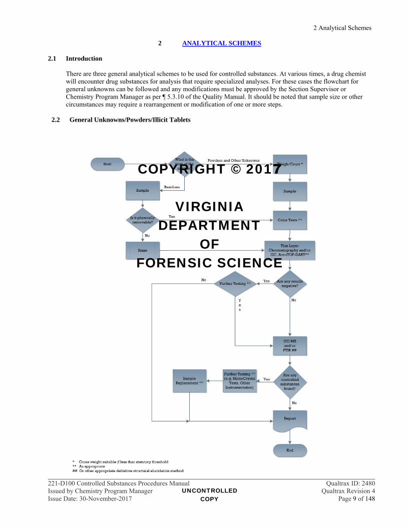
2 ANALYTICAL SCHEMES 2.1 Introduction
There are three general analytical schemes to be used for controlled substances. At various times, a drug chemist will encounter drug substances for analysis that require specialized analyses. For these cases the flowchart for general unknowns can be followed and any modifications must be approved by the Section Supervisor or Chemistry Program Manager as per ¶ 5.3.10 of the Quality Manual. It should be noted that sample size or other circumstances may require a rearrangement or modification of one or more steps.
2.2 General Unknowns/Powders/Illicit Tablets
UNCONTROLLED COPY
COPYRIGHT © 2017
VIRGINIA DEPARTMENT
OF FORENSIC SCIENCE

2 Analytical Schemes
221-D100 Controlled Substances Procedures Manual Qualtrax ID: 2480Issued by Chemistry Program Manager Qualtrax Revision 4Issue Date: 30-November-2017 Page 10 of 148
2.3 Tablets and Capsules
UNCONTROLLED COPY
COPYRIGHT © 2017
VIRGINIA DEPARTMENT
OF FORENSIC SCIENCE
2 Analytical Schemes
221-D100 Controlled Substances Procedures Manual Qualtrax ID: 2480Issued by Chemistry Program Manager Qualtrax Revision 4Issue Date: 30-November-2017 Page 11 of 148
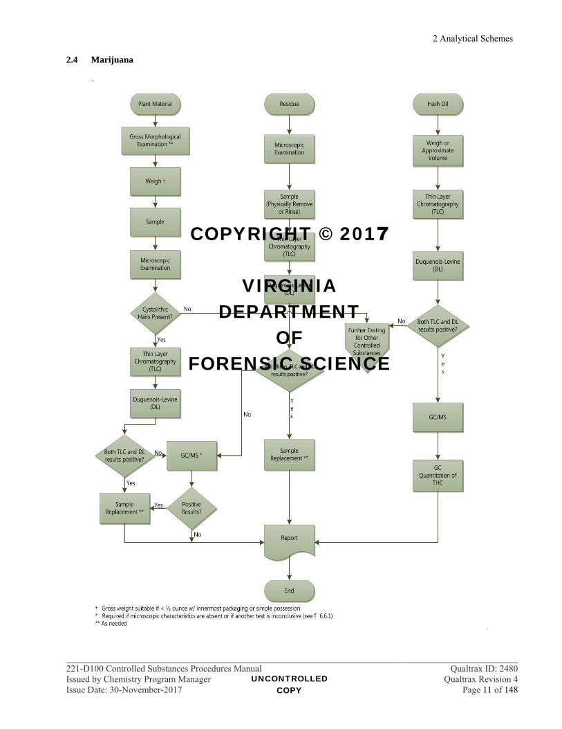
2.4 Marijuana
UNCONTROLLED COPY
COPYRIGHT © 2017
VIRGINIA DEPARTMENT
OF FORENSIC SCIENCE

2 Analytical Schemes
221-D100 Controlled Substances Procedures Manual Qualtrax ID: 2480Issued by Chemistry Program Manager Qualtrax Revision 4Issue Date: 30-November-2017 Page 12 of 148
2.5 Pharmaceutical Identifiers
2.5.1 Check the Physician’s Desk Reference (PDR), Poison Control, DEA Logo Index, Identadrug, Drug ID Bible/Amera-Chem, Inc CD, Pillbox, ePocrates, webpoisoncontrol.org, NIH-DailyMed, or manufacturer’s resources for information relating to inscriptions on tablets and capsules. Two unrelated references are recommended for unfamiliar tablets. Additional references may be used following approval by Chemistry Program Manager.
2.5.2 Additional analyses may be required as described in the general analytical scheme in ¶ 7.2.
2.6 Color Tests
2.6.1 If the size of the sample is sufficient, perform the appropriate color tests required to provide an indication of any compounds present.
2.6.2 Check the available literature (e.g., Clarke) for the interpretation of results of these and/or ask other
chemists, as necessary. 2.7 Chromatography
2.7.1 Dissolve the sample directly into a suitable solvent (e.g., methanol). If appropriate, extract the sample from an acidic or basic medium (or both if the contents of the sample are still unknown at this time).
2.7.2 If sample is an unknown, run appropriate screening systems via TLC and/or GC.
2.7.2.1 For TLC, if there are no spots visible under UV light or with visualization sprays, the
examiner may halt the analysis and report “No controlled substances found”. The examiner may continue with the analytical scheme for any number of reasons to include information provided on the RFLE or other clues from the sample.
2.7.2.2 For GC, if there are no peaks present, the examiner may halt the analysis and report “No
controlled substances found”. The examiner may continue with the analytical scheme for any number of reasons to include information provided on the RFLE or other clues from the sample.
2.7.3 If sample identity was indicated previously, choose the appropriate two system TLC and/or two system
GC systems, as needed, with a standard. The choice of TLC or GC should be based on which is most appropriate under the circumstances. Note: TLC is the preferred method due to the possibility of thermally labile compounds.
2.7.4 The chromatography requirement is waived for pharmaceutical preparations that are confirmed with a
structural elucidation technique following visual examination under ¶ 7 Pharmaceutical Identifiers. 2.7.5 In addition to two system TLC, sample retention times obtained from GC/MS systems are often
compared to standard retention times. 2.8 Infrared Spectroscopy/Mass Spectrometry
2.8.1 If the identity of the sample is still unclear at this point, the IR or GC/MS will provide further information.
2.8.2 The DART-TOF screening method provides accurate mass information, which may assist in identifying components present in the sample. This screening method can be useful at any point in the analytical scheme.
UNCONTROLLED COPY
COPYRIGHT © 2017
VIRGINIA DEPARTMENT
OF FORENSIC SCIENCE
2 Analytical Schemes
221-D100 Controlled Substances Procedures Manual Qualtrax ID: 2480Issued by Chemistry Program Manager Qualtrax Revision 4Issue Date: 30-November-2017 Page 13 of 148
2.8.3 A definitive structural identification technique such as GC/MS or IR is required to be used on all substances where the identities will be reported. Confirmation of pharmaceutical identifiers may be accomplished using DART-TOF as described in ¶ 15.4.
2.9 Further Testing
2.9.1 If the sample is still an unknown or other confirmation is needed, the chemist should use any instrumental techniques available (or combinations thereof) to arrive at a sound analytical conclusion about the identities of sample. This may involve using Department Instrument Support as approved by the Chemistry Program Manager.
2.9.2 Microcrystal tests are used for isomer determination only. They are to be used only in combination with a
structural elucidation technique. 2.10 Liquids
2.10.1 Liquid samples may be submitted for analysis as part of a clandestine lab prosecution or for general drug analysis. These types of samples may require additional steps in the general analytical scheme in ¶ 2.2. Occasionally, it will be necessary to consult with a supervisor, the investigating officer, or both to determine the best analytical course.
2.10.2 Information provided on the RFLE or by the investigating officer should be used to determine the
purpose of the examination. In some cases, evidence may need to be transferred to Trace Evidence for analysis or, depending on the overall case, may not require analysis.
2.10.3 Information such as package labeling, visible precipitates, number of layers present, viscosity and color
of liquid should be considered when deciding on an analytical scheme. 2.10.4 The approximate volume of the liquid and pH of aqueous liquids will be determined as sample size
allows. The density and solubility of the liquid may be determined. 2.10.5 Additional considerations regarding liquids submitted in association with a clandestine laboratory or the
intent to manufacture methamphetamine, methcathinone or amphetamine as per § 18.2-248 J are addressed in the Clandestine Laboratory section.
2.10.6 It may not be sufficient to simply screen a neat liquid with TLC or FTIR. Liquids submitted for general
drug analysis may require extraction prior to TLC or GCMS to concentrate the analyte during the screening process or to remove the analyte from the matrix. Screening for suspected amyl nitrite will require GC/MS headspace analysis. Examples of samples which come in liquid forms are:
Solutions containing GHB, GBL or 1,4-butanediol Cough syrups Suspected amyl nitrite or other inhalants Eye drops or other liquids in dropper bottles Injectables such as tubexes
2.11 Plant Material
2.11.1 Initially, all plant material shall be screened via stereomicroscopy for cystolithic hairs.
2.11.1.1 In the presence of cystolithic hairs or marijuana seeds, the analytical scheme for marijuana shall be completed.
2.11.1.2 Contextual clues, such as package labels or banana leaf wrappers, shall be recorded in the
case notes.
UNCONTROLLED COPY
COPYRIGHT © 2017
VIRGINIA DEPARTMENT
OF FORENSIC SCIENCE

2 Analytical Schemes
221-D100 Controlled Substances Procedures Manual Qualtrax ID: 2480Issued by Chemistry Program Manager Qualtrax Revision 4Issue Date: 30-November-2017 Page 14 of 148
2.11.1.3 In the absence of cystolithic hairs and in conjunction with contextual clues from the evidence, samples shall be screened for controlled substances including, but not limited to, Salvinorin A, cathinone/cathine and cannabimimetic agents.
2.11.1.3.1 Apparent plant material residues, if analyzed, should be handled in the same
manner.
2.11.2 Fungal material shall be screened as per the Psilocybin and Psilocyn Methodology (see ¶ 24).
UNCONTROLLED COPY
COPYRIGHT © 2017
VIRGINIA DEPARTMENT
OF FORENSIC SCIENCE
3 Drug Item Reduction Program
221-D100 Controlled Substances Procedures Manual Qualtrax ID: 2480Issued by Chemistry Program Manager Qualtrax Revision 4Issue Date: 30-November-2017 Page 15 of 148
3 DRUG ITEM REDUCTION PROGRAM
3.1 Introduction 3.1.1 The Drug Item Reduction Program (DIRP) allows for the analysis of key items within a case to maximize
the resources of the laboratory. 3.1.2 In every case, the most significant items in terms of quantity and schedule are analyzed. This “rule of
thumb” cannot address every drug case. Consideration must be given to the information contained on the Request for Laboratory Examination (RFLE). This includes things such as the specific charges or types of offense, items unique to a single suspect, the statement of fact and examinations requested and the descriptions of evidence submitted as well as the chemist’s visual inspection of the items. Currency lacking visible residue should not be analyzed.
3.1.3 If, during the pretrial process, it becomes apparent that items not analyzed will require analysis for
successful prosecution, then upon re-submission, that item will receive top priority at the laboratory.
3.2 Procedures 3.2.1 Steps should be taken to discourage “no suspect, information only” requests. 3.2.2 Syringes should only be analyzed if they are the only item in the case. 3.2.3 Quantitative analyses will not routinely be performed and only will be done at the request of a
Commonwealth’s Attorney. A suitable justification should be obtained. 3.2.4 In general, residues in drug paraphernalia, cigarettes or cigarette butts will not be analyzed when
measurable quantities of the associated drugs are also included among the items submitted.
3.2.4.1 Example 1: Submitted evidence includes a plastic bag corner containing solid material and a glass tube smoking device with residue. The solid material would be analyzed and the smoking device would not.
3.2.4.2 Example 2: Submitted evidence includes five tablets containing oxycodone and a plastic straw
section with residue. The tablets would be analyzed and the straw section would not.
3.2.4.3 Example 3: Submitted evidence includes five tablets containing alprazolam and a plastic straw section with residue. The tablets would be analyzed and the straw section would not unless information on the RFLE indicates that the straw section was used for a different drug.
3.2.4.4 Example 4: Submitted evidence includes a plastic bag of plant material and a glass tube
smoking device with residue. Both the plant material and the smoking device would be analyzed.
3.2.5 When multiple residue specimens are submitted within an item (without an item with a measurable
quantity), similar residues (e.g., two spoons with residue) may be combined after appropriate screening tests to result in only one GC/MS sample.
3.2.6 Pharmaceutical preparations should be visually examined using pharmaceutical identifiers and
appropriate reference compendia.
3.2.6.1 No further analysis is required for misdemeanor offense intact, marked pharmaceutical preparations (e.g., tablets or untampered capsules) indicated as non-controlled or Schedule VI preparations. These may be reported as “Not Analyzed” or using the “Visual examination determined…” language delineated in the Reporting Guidelines section of this manual. See ¶ 7.2.1.4 for tamperable capsules.
UNCONTROLLED COPY
COPYRIGHT © 2017
VIRGINIA DEPARTMENT
OF FORENSIC SCIENCE

3 Drug Item Reduction Program
221-D100 Controlled Substances Procedures Manual Qualtrax ID: 2480Issued by Chemistry Program Manager Qualtrax Revision 4Issue Date: 30-November-2017 Page 16 of 148
3.2.6.2 If identical intact, marked pharmaceutical preparations (e.g., tablets or untampered capsules) are present in multiple items, analysis is required for only one item. Those preparations not analyzed may be reported as “Not Analyzed” or using the “Visual examination determined…” language delineated in the Reporting Guidelines section of this manual. See ¶ 7.2.2.2 for tamperable capsules.
3.2.6.3 Partial pharmaceutical preparations may be not analyzed when intact pharmaceutical
preparations or measurable quantities of drugs are present.
3.2.6.4 In simple possession cases, if intact, marked pharmaceutical preparations (e.g., tablets or untampered capsules) indicated as containing the same controlled substance are present in multiple items, analysis is required for only one item. Those preparations not analyzed will be reported using the “Visual examination determined…” language delineated in the Reporting Guidelines section of this manual. See ¶ 7.2.2.2 for tamperable capsules.
3.2.7 If items are not analyzed per this procedure, case notes shall indicate this by a notation of “Not
Analyzed” or “DIRP”.
UNCONTROLLED COPY
COPYRIGHT © 2017
VIRGINIA DEPARTMENT
OF FORENSIC SCIENCE
4 Weighing Practices
221-D100 Controlled Substances Procedures Manual Qualtrax ID: 2480Issued by Chemistry Program Manager Qualtrax Revision 4Issue Date: 30-November-2017 Page 17 of 148
4 WEIGHING PRACTICES 4.1 Procedures
4.1.1 Weights of evidence will be measured and recorded prior to sampling. The gross weight (GW) of the evidence, including innermost packaging, should be measured whenever possible. The net weight (NW) of the material may be measured at the discretion of the examiner.
4.1.2 If the gross weight of the specimen(s), including innermost packaging, is equal to or greater than an established weight threshold as defined in the Code of Virginia, the net weight of a sufficient number of specimens shall be reported and designated as such in the case notes. ¶ 38
4.1.3 Dosage units (e.g., cigarettes, cigarette butts, blotter papers, tablets or capsules) will not routinely be
weighed.
4.1.4 Analytical, top-loading or high-capacity electronic balances are acceptable for routine casework. The balance used will be recorded in the case notes.
4.1.4.1 Minimum balance loads:
4.1.4.1.1 5-Place Balance = 0.00450 gram 4.1.4.1.2 4-Place Balance = 0.0300 gram 4.1.4.1.3 3-Place Balance = 0.150 gram 4.1.4.1.4 2-Place Balance = 0.90 gram 4.1.4.1.5 High Capacity (g) Balance (CD-33) = 27.0 grams 4.1.4.1.6 High Capacity (g) Balance (AND) = 1 gram 4.1.4.1.7 High Capacity (g) Balance (Kg) = 1.1 Kg
4.1.4.2 Plastic weigh boats shall not be used for determining net weights on 4-place or 5-place
balances.
4.1.5 Weights will be recorded in the analytical notes as they are displayed on the balance.
4.1.5.1 Calculations involving weights will be done using the weights as they are recorded.
4.1.6 If the estimated uncertainty is equal or larger than the weight, a more accurate balance shall be used or the substance shall be reported as a residue, whichever is appropriate.
4.1.7 When available balances allow for it, net weights of multiple specimens shall be measured and recorded individually. Measuring and recording the net weight of multiple specimens at the same time shall be avoided whenever it is possible to do so.
4.1.8 When multiple balances are used to record net weights of specimens within one item, the sum of the
weights recorded with each balance shall be reported separately (see ¶ 33.4.2.2 for an example).
4.2 Weighing Practices 4.2.1 Weights of evidence will be measured prior to sampling. The gross weight of the evidence, including
innermost packaging, should be measured whenever possible. The net weight of the material may be measured at the discretion of the examiner.
UNCONTROLLED COPY
COPYRIGHT © 2017
VIRGINIA DEPARTMENT
OF FORENSIC SCIENCE
4 Weighing Practices
221-D100 Controlled Substances Procedures Manual Qualtrax ID: 2480Issued by Chemistry Program Manager Qualtrax Revision 4Issue Date: 30-November-2017 Page 18 of 148
4.2.1.1 In cases where the container weight is clearly much greater than the sample weight, obtain the net weight (without packaging) of the material and report appropriately.
4.2.1.2 If the gross weight, including innermost packaging, is equal to or greater than an established
weight threshold, the net weight of a sufficient number of specimens shall be obtained and designated as such in the case notes.
4.2.1.2.1 The gross weight of the remaining specimens including innermost packaging
will be obtained and designated as such in the case notes, if applicable.
4.2.1.2.2 Both the net weight of the analyzed specimens and the gross weight of the remaining specimens will be reported on the Certificate of Analysis.
4.2.2 Cases with Weight Thresholds
4.2.2.1 In instances where statutory requirements or state sentencing guidelines designate weight thresholds, sufficient specimens will be weighed and analyzed to exceed the threshold. A list of these instances can be found in ¶ 38.
4.2.2.1.1 The net weight of the specimens required to exceed the threshold will be
obtained and designated as such in the case notes. 4.2.2.1.2 The gross weight of the remaining specimens including innermost packaging
will be obtained and designated as such in the case notes. 4.2.2.1.3 Both the net weight of the analyzed specimens and the gross weight of the
remaining specimens will be reported on the Certificate of Analysis.
4.3 Weighing Practices for Hypergeometric Sampling Plan
4.3.1 Resubmissions
The hypergeometric sampling model will be used most often for cases being resubmitted at the request of a Commonwealth’s Attorney. 4.3.1.1 The net weight or gross weight, as applicable, of the additional samples will be obtained and
reported on the Supplemental Certificate of Analysis.
4.3.2 Initial Submissions
4.3.2.1 Weights of evidence will be measured prior to sampling. The gross weight of the evidence, including innermost packaging, should be measured whenever possible. The net weight of the material may be measured at the discretion of the examiner. 4.3.2.1.1 If the gross weight, including innermost packaging, is equal to or greater than an
established weight threshold, the net weight of a sufficient number of specimens shall be obtained and designated as such in the case notes.
4.3.2.1.1.1 The gross weight of the remaining specimens including innermost
packaging will be obtained and designated as such in the case notes.
UNCONTROLLED COPY
COPYRIGHT © 2017
VIRGINIA DEPARTMENT
OF FORENSIC SCIENCE
5 Sampling
221-D100 Controlled Substances Procedures Manual Qualtrax ID: 2480Issued by Chemistry Program Manager Qualtrax Revision 4Issue Date: 30-November-2017 Page 19 of 148
5 SAMPLING
5.1 Introduction Sampling evidence is the most important initial step in forensic drug analysis. One must be sure that what is sampled is truly representative of the total population. The analyst must take into consideration the homogeneity (or lack thereof) among drug packaging (bags, packets, capsules, etc.) and its contents. Careful visual inspections and personal experience are essential in determining the proper sampling procedure. For items containing multiple specimens, statistically-based sampling models (e.g., hypergeometric distribution) will allow the analyst to analyze a portion of the specimens and subsequently make statistical inferences about the population. Alternatively, a fixed number of specimens within a population may be analyzed with the purpose in mind of meeting the requirements of a particular criminal charge (e.g., simple possession, distribution). In these instances, an inference to the entire population will not be drawn and the number of specimens that were analyzed will be indicated on the Certificate of Analysis.
5.2 General Sampling 5.2.1 Every effort should be made to avoid handling evidence repeatedly. The material should be sampled and
immediately sealed. If necessary, the evidence may be closed and maintained in short term storage until the analysis is complete. Evidence generally will not remain in short term storage for longer than 30 days.
5.2.2 In order to minimize detailed labeling on small items such as very small metal foil packets, plastic bags
or plastic bag corners, they may be secured in a bandolier of tape, which is then labeled. If needed, items may be placed in an additional plastic bag which can be sealed, fully labeled and properly documented in the case notes.
5.2.3 For chemical analyses, a representative sample shall be removed from the specimen. When sample size
allows, testing should be applied on separate samplings of the material. Taking a small amount of material for use in a color test prior to taking a separate sampling for additional tests is an appropriate method. For suspected marijuana, performing the microscopic examination on a larger population prior to taking a representative sample for thin layer chromatography and the Duquenois-Levine test will suffice. For pharmaceutical tablets and capsules, the use of pharmaceutical identifiers as a screening test prior to taking a representative sample for confirmatory testing will suffice.
5.3 Administrative Sampling Plan
The administrative sampling plan will be used in cases to answer a specific legal question. If more specimens than listed below need to be analyzed for successful prosecution, additional analysis utilizing the hypergeometric sampling plan will be conducted upon written request from the Commonwealth’s Attorney. Items requiring quantitative analysis should be analyzed using the hypergeometric sampling plan. 5.3.1 Simple possession
5.3.1.1 One specimen will be randomly selected and fully analyzed.
5.3.1.2 All remaining specimens will be left intact in case further analysis is required. 5.3.2 Possession with intent to distribute or distribution
5.3.2.1 Items containing up to 5 specimens
Each specimen will be analyzed separately and fully.
UNCONTROLLED COPY
COPYRIGHT © 2017
VIRGINIA DEPARTMENT
OF FORENSIC SCIENCE

5 Sampling
221-D100 Controlled Substances Procedures Manual Qualtrax ID: 2480Issued by Chemistry Program Manager Qualtrax Revision 4Issue Date: 30-November-2017 Page 20 of 148
5.3.2.2 Items containing greater than 5 specimens
5.3.2.2.1 Five randomly selected specimens will be analyzed separately and fully. 5.3.2.2.2 The remaining specimens will be left intact in case further analysis is required.
5.3.3 Cases with weight thresholds
5.3.3.1 In instances where statutory or state sentencing guidelines have weight thresholds, enough specimens will be weighed and analyzed, separately and fully, to exceed the threshold. A list of these instances can be found in ¶ 38.
5.3.3.2 The remaining specimens will be left intact in case further analysis is required.
5.3.4 Pharmaceutical preparations
5.3.4.1 Due to the unique physical identifiers present in pharmaceutical preparations, a consistent sample population can easily be determined. The thoroughness represented by the sampling scheme used for street drugs is not required for pharmaceutical preparations which are clearly visually consistent with each other.
5.3.4.2 For drug substances involving misdemeanor prosecutions in Schedules V and VI, sampling is not normally required. For drug substances involving Schedule IV and above, at least one representative sample must be analyzed fully.
5.3.4.2.1 For tamperable dosage units, screen a sample chosen using the hypergeometric
scheme described below utilizing TLC, DART-TOF, and/or color tests prior to fully analyzing one unit.
5.3.4.2.1.1 If tampering is suspected, analyze dosage units utilizing the
hypergeometric scheme.
5.3.4.2.2 If the evidence is resubmitted for further analysis, resample and analyze using either the administrative sampling plan (¶ 5.3.2) or the hypergeometric sampling scheme (¶ 5.4) depending on the legal requirements.
5.3.5 Exceptions to this Plan may occur only at the discretion of the Section Supervisors in consultation with
the Chemistry Program Manager.
5.4 Hypergeometric Sampling Plan Hypergeometric sampling is a statistically-based model involving a defined confidence level with an associated probability of finding failures in a population. (¶¶ 5.9.1, 5.9.2, and 5.9.5) The hypergeometric model is used for specimens with no significant markings or labels (e.g., the contents of plastic bags and bag corners, vials, and glassine packets). This model should also be used when the item requires a quantitative analysis. 5.4.1 Hypergeometric sampling may be used when additional analysis is requested for successful prosecution. 5.4.2 The appropriate number of specimens within the population will be randomly selected to give a 95%
level of confidence that at least 90% of the population contains the analyte in question. Refer to ¶ 39 of this manual.
5.4.3 Record the number of specimens indicated by the table in ¶ 39 along with an indication of the statistical
relevance of the number in the case notes. 5.4.4 Each specimen sampled will be analyzed separately and fully.
UNCONTROLLED COPY
COPYRIGHT © 2017
VIRGINIA DEPARTMENT
OF FORENSIC SCIENCE

5 Sampling
221-D100 Controlled Substances Procedures Manual Qualtrax ID: 2480Issued by Chemistry Program Manager Qualtrax Revision 4Issue Date: 30-November-2017 Page 21 of 148
5.5 Multiple Specimens 5.5.1 If all specimens are not analyzed, the number of those that are fully analyzed will be recorded in the case
notes.
5.5.2 Net weights, when applicable, and autosampler vial numbers will be associated with specific specimens by the use of sub-numbering in the case notes.
5.5.3 Within any sampling scheme, Administrative or Hypergeometric, if the first set of observations
determines that more than one population is present, further samples from each population must be taken. 5.5.4 If presumptive testing indicates that no controlled substances are present in the samples chosen, a
screening test must be done using the hypergeometric sampling scheme.
5.5.4.1 For items consisting of specimens, which are obviously non-controlled such as gum, candy or vitamins, a single representative sample may be screened.
5.5.5 When multiple balances are required to record weights within one item, the sum of the samples taken and
analyzed should meet the requirement of the selected sampling plan.
5.6 Bulk Materials Bulk materials (e.g., bricks of compressed powder, bales of plant material) should be broken or cored to obtain a representative sample. Depending on the size of the material, samples from several locations may be required to obtain a representative sample. The examiner will record the locations from which the samples were obtained in the case notes.
5.7 Residue Samples Residues are samples which are either too small to be weighed accurately or that which remains after the bulk has been removed. Residues can be sampled by mechanical means (e.g., shaking or scooping) or chemical means (e.g., rinsing with solvent). Case notes must reflect the method by which the sample was removed. 5.7.1 When possible, a sample should be removed while leaving a portion of the residue intact. 5.7.2 When it is not possible to redeposit and return the residue as received, the extract used in analysis will be
returned to the evidence as per the Quality Manual (¶ 14.10.5).
5.7.2.1 Procedure: Evaporate the solvent from the extract in the autosampler vial used in analysis. Seal the autosampler vial (ASV) into a ziplock bag. Label the ziplock bag with the FS Lab #, Item #, initials and a statement similar to “vial and bag added at lab.” Record the date in the case notes that the ASV was placed in the evidence.
5.8 Sampling for Quantitative Analysis
5.8.1 Quantitative analyses require homogenized representative samples. Generally, a relatively large sample is homogenized with a mortar and pestle prior to taking the small samples required by the quantitative method to make the solutions. The remainder of the homogenized portion should be returned with the evidence in a suitably labeled plastic bag provided by the laboratory. By their nature, suspected hashish oil samples should require no further homogenization.
5.8.2 Single specimen items
5.8.2.1 Homogenize the entire specimen, take the six samples required for the quantitation method
and return the bulk of the material to the evidence.
UNCONTROLLED COPY
COPYRIGHT © 2017
VIRGINIA DEPARTMENT
OF FORENSIC SCIENCE
5 Sampling
221-D100 Controlled Substances Procedures Manual Qualtrax ID: 2480Issued by Chemistry Program Manager Qualtrax Revision 4Issue Date: 30-November-2017 Page 22 of 148
5.8.2.2 For large specimens such as kilos of cocaine, six core samples should be taken from multiple locations and homogenized. The locations of samples taken shall be described in the case notes.
5.8.3 Multiple specimen items
5.8.3.1 Items with multiple specimens should be analyzed qualitatively using the hypergeometric
sampling plan.
5.8.3.2 A composite will be formed consisting of the specimens analyzed in the hypergeometric sampling plan. Homogenize the composite and take the six samples required for the quantitation method. The remainder of the composite should be returned to the evidence in a ziplock bag provided by the laboratory, clearly marked as a composite.
5.9 References
5.9.1 Shark, Robert E. “Sampling Your Drugs: A User's Guide”, Commonwealth of Virginia, Bureau of
Forensic Science, Technical Brief, c. 1986. 5.9.2 Frank, Richard S. et.al. "Representative Sampling of Drug Seizures in Multiple Containers." Journal of
Forensic Sciences, JFSCA, Vol. 36, No. 2, March 1991, pp. 350-357. 5.9.3 Colon, Maria et.al. “Representative Sampling of ‘Street’ Drug Exhibits” Journal of Forensic Sciences,
JFSCA, Vol. 38, No.3, May 1993, pp. 641-648.
5.9.4 SWGDRUG Recommendations, 5.1 ed. “PART III A - Methods of Analysis/Sampling Seized Drugs for Qualitative Analysis”, January, 2011.
5.9.5 European Network of Forensic Science Institutes Drugs Working Group, Guidelines on Representative
Drug Sampling, 2003.
UNCONTROLLED COPY
COPYRIGHT © 2017
VIRGINIA DEPARTMENT
OF FORENSIC SCIENCE
6 Marijuana, Hashish Oil, and Cannabinoid Products
221-D100 Controlled Substances Procedures Manual Qualtrax ID: 2480Issued by Chemistry Program Manager Qualtrax Revision 4Issue Date: 30-November-2017 Page 23 of 148
6 MARIJUANA, HASHISH OIL, AND CANNABINOID PRODUCTS 6.1 Introduction
6.1.1 Marijuana is neither a "controlled substance" nor is it "scheduled" under Virginia Law. However, it is defined and covered under separate sections of the Drug Control Act of Virginia and has associated penalties.
6.1.2 Cannabis (marijuana) contains tetrahydrocannabinol (THC) in both the male and female plants. THC is
found in all parts of the plant in varying concentrations.
6.1.3 Hashish oil is an oily extract containing one or more cannabinoids with little, if any, plant material present and containing 12% or more THC. It is controlled in Schedule I.
6.1.4 Cannabinoid products may contain Cannabidiol (CBD), Tetrahydrocannabinol (THC),
Tetrahydrocannabinol acid (THC-A), or a combination thereof. Cannabinoid products shall be tested pursuant to the Code of Virginia (§ 54.1-3408.3).
6.2 Macroscopic Identification
6.2.1 Gross morphological characteristics that may be observed include the palmate arrangement of the leaflets, the pinnate appearance of the leaflets, the serrated edges of the leaflet, the buds (with or without seeds) and, if present, fluted stems and stalks.
6.2.2 Due to the compressed or mutilated nature of many samples, many of these characteristics may not be
discernable.
6.2.3 Positive macroscopic examination results may be recorded in the analytical notes by the use of an abbreviation of positive for characteristics of Marijuana (e.g., pos. characteristics MJ), a plus (+), or a plus circled (). A result is considered positive when sufficient characteristics are observed and are specified in the case notes. Negative observations may be recorded in a similar fashion.
6.3 Microscopic Identification
6.3.1 View the sample at varying magnifications (approximately 10 – 40x) using a stereomicroscope.
6.3.2 Cystolithic hairs are unicellular, “bear claw” shaped hairs with a cystolith of CaCO3 at the base. They are
found in greatest abundance on the upper side of the leaf with longer covering hairs on the underside.
6.3.3 Seeds are coconut shaped, veined (with lacy markings) and have a ridge around the circumference.
6.3.4 The observation of the presence of appropriate cystolithic hairs or characteristic seeds is sufficient for a positive test. The observation of additional characteristics is considered supportive. Positive microscopic examination results will be recorded in the analytical notes by the use of an abbreviation of positive for characteristics of Marijuana (e.g., pos. characteristics MJ), a plus (+), or a plus circled (). A result is considered positive when sufficient characteristics are observed. Negative observations will be recorded in a similar fashion.
6.4 Thin Layer Chromatography (TLC)
6.4.1 TLC plates should be silica gel or equivalent, sufficient to resolve the three major cannabinoids listed in
¶ 6.4.6.4 (examples: Analtech Silica Gel GHLF 250um 10 X 20, Analtech Silica Gel 250um GF 10x20, EMD TLC Silica Gel 250um 60 F254 5 X 10, EMD TLC Silica Gel 250um 60 F254 10 X 20).
6.4.2 Extract sample into a suitable solvent (e.g., hexane, petroleum ether or methanol). The solvent used must
be recorded in the case notes.
UNCONTROLLED COPY
COPYRIGHT © 2017
VIRGINIA DEPARTMENT
OF FORENSIC SCIENCE
6 Marijuana, Hashish Oil, and Cannabinoid Products
221-D100 Controlled Substances Procedures Manual Qualtrax ID: 2480Issued by Chemistry Program Manager Qualtrax Revision 4Issue Date: 30-November-2017 Page 24 of 148
6.4.3 Spot sample(s), standard(s) and solvent blank on the plate. The maximum number of spots when using a 10 x 20 cm plate is 32. (See ¶ 9.3 for further information)
6.4.4 The results of the blank must be recorded in the case notes. This may be done by using a check mark ()
or “ok” or “-” to record that the results of the blank were acceptable (e.g., Blk ). 6.4.5 Mobile Phase: 4-8% diethylamine in toluene
6.4.6 Visualization Sprays
6.4.6.1 Fast Blue B Salt (Tetrazotized o-dianisidine zinc chloride salt)
6.4.6.2 Fast Blue BB Salt (4-benzoylamino-2,5-diethoxy-benzenediazonium chloride hemi [zinc
chloride] salt)
6.4.6.3 Reagent preparation is listed in the Thin Layer Chromatography section of this manual. (see ¶ 9.5.6)
6.4.6.4 Results:
The three major cannabinoids migrate and develop in the following order:
o Top spot - Cannabidiol – orange o Middle spot - Tetrahydrocannabinol (∆9-THC) - red o Lower spot - Cannabinol – purple
A red spot at the origin may be present in unburned marijuana due to cannabinolic acid(s).
6.4.7 Samples will be screened for the presence of other commonly encountered drugs such as cocaine or PCP
by either overspraying with acidified iodoplatinate or by running a separate plate in an appropriate drug bath (e.g., TLC1 or TLC2.) (see ¶ 9.4)
6.4.8 Specific solvent systems and developing sprays utilized in casework will be denoted in the analytical
case notes. Positive TLC results may be recorded in the analytical notes by the use of a plus (+), a plus circled () or an abbreviation (e.g., pos) along with the standard used in the comparison. A result is considered positive when the distance traveled and the reaction with the visualization methods compare favorably with a standard. It is not necessary for each of the three major cannabinoids to be present for the results to be considered positive. Negative reactions may be recorded in a similar fashion.
6.4.9 After the plate is sprayed with FBB or FBBB, it shall be scanned and a color hardcopy printed. Each lane
shall be labeled (FS Lab# and Item #, standard identifier, blank, etc.) either in the image or on the original color copy. Templates may be used to assist with labeling.
6.4.9.1 When multiple samples/cases are run on the same plate, the color printout shall be stored in
one case file. 6.4.9.2 Black and white copies of the original shall be placed in other case files with a reference to
where the original is stored. 6.5 Duquenois-Levine
6.5.1 Extract sample into a suitable solvent (e.g., hexane, petroleum ether or methanol). If a large amount of
solvent is used, most of it must be evaporated.
6.5.2 Add approximately equal amounts of Duquenois reagent and concentrated HCl to extract. A positive reaction to the Duquenois portion is a blue/purple color.
UNCONTROLLED COPY
COPYRIGHT © 2017
VIRGINIA DEPARTMENT
OF FORENSIC SCIENCE
6 Marijuana, Hashish Oil, and Cannabinoid Products
221-D100 Controlled Substances Procedures Manual Qualtrax ID: 2480Issued by Chemistry Program Manager Qualtrax Revision 4Issue Date: 30-November-2017 Page 25 of 148
6.5.3 Add sufficient CHCl3 to form two discernible layers and mix. For a positive reaction to the Levine portion of the test, the bottom layer turns pink/purple in the presence of THC or other cannabinoids.
6.5.4 Run a solvent blank as a negative control with each batch of samples. The results of the negative control
must be documented in the case notes. This may be done by using a check mark () or “ok” or “-“ to record that the results of the blank were acceptable (e.g., Blk ).
6.5.4.1 If a color develops in the blank, it should be repeated to determine the source of the
contamination.
6.5.4.1.1 If the results of the second blank are acceptable, all samples should be re-run. 6.5.4.1.2 If the results of the second blank are unacceptable or if the blank and samples
are not available to be re-tested, the analyst should take steps to resolve the issue (e.g., replacing the solvent in the bottle, checking the reagents) prior to re-sampling and any further analysis.
6.5.5 Reagent preparation is listed in the Color Test section of this manual. (see ¶ 8.3.6)
6.5.6 The Rapid Duquenois-Levine Procedure
6.5.6.1 Place a small amount of plant material in a culture tube, add Duquenois reagent and
concentrated HCl in approximately equal proportions. Observe a blue/purple color. Add CHCl3 and observe extraction of pink/purple color into the CHCl3 layer.
6.5.6.2 A blank (negative control) will be run in a separate culture tube. The results of the negative
control must be documented in the case notes.
6.5.6.2.1 If a color develops in the blank, it should be repeated to determine the source of the contamination.
6.5.6.2.2 If the results of the second blank are acceptable, all samples should be re-run. 6.5.6.2.3 If the results of the second blank are unacceptable or if the blank and samples
are not available to be re-tested, the analyst should take steps to resolve the issue (e.g., replacing the solvent in the bottle, checking the reagents) prior to re-sampling and any further analysis.
6.5.6.3 If the Rapid Duquenois-Levine is utilized, it should be recorded in the case notes.
6.5.7 Results must be recorded in the case notes. This may be accomplished either with a single plus (+), a plus circled () or an abbreviation (e.g., pos) indicating a positive result in both steps or the colors of each step may be noted. Negative reactions may be recorded in a similar fashion.
6.6 Gas Chromatography/Mass Spectrometry (GC/MS)
6.6.1 GC/MS shall be performed if the results from any of the prior three tests are inconclusive.
6.6.2 Retention time data is not required. 6.7 Hashish Oil
6.7.1 Analytical Scheme
6.7.1.1 Weigh or approximate the volume of the material. The weight of the material should be
recorded whenever possible.
UNCONTROLLED COPY
COPYRIGHT © 2017
VIRGINIA DEPARTMENT
OF FORENSIC SCIENCE

6 Marijuana, Hashish Oil, and Cannabinoid Products
221-D100 Controlled Substances Procedures Manual Qualtrax ID: 2480Issued by Chemistry Program Manager Qualtrax Revision 4Issue Date: 30-November-2017 Page 26 of 148
6.7.1.2 Remove a representative sample for testing.
6.7.1.3 Dilute with appropriate solvent and perform TLC as listed for marijuana. The presence of additional cannabinoids will confirm that the THC is most likely from the marijuana plant.
6.7.1.4 Perform the Duquenois-Levine test.
6.7.1.5 Either two-system TLC or two-system GC against a THC standard must be used in the analytical scheme.
6.7.1.6 Dilute with appropriate solvent and run on GC/MS to confirm the presence of THC.
6.7.1.7 Quantitate THC using method below.
6.7.2 THC Quantitation
See GC ¶ 10 for general quantitation procedure.
6.7.2.1 Materials
n-Hexane: High purity Ethyl Alcohol: (95%) USP Grade Delta-9-Tetrahydrocannabinol: (10 mg/mL in EtOH) n-Triacontane: 98% pure or greater. 4N NaOH Class A volumetric flasks Calibrated mechanical pipettes Calibrated volumetric flasks Analytical balance
6.7.2.2 Internal Standard Solution
6.7.2.2.1 Prepare a sufficient volume to dilute the THC standard solution and all samples.
6.7.2.2.2 Prepare a 1 mg/mL solution of n-triacontane in n-hexane in the appropriate
volumetric flask.
6.7.2.2.3 Refrigerated solutions should be allowed to return to ambient temperature prior to use.
6.7.2.2.4 Internal standard blank shall be washed with 4N NaOH prior to placing in
autosampler vial.
6.7.2.3 THC Standard Solution
6.7.2.3.1 Using a calibrated mechanical pipette, pipette 0.25 mL of the THC standard into a culture tube. Evaporate to dryness. Using a calibrated mechanical pipette, pipette 2.5 mL of internal standard solution into the culture tube and mix thoroughly. This results in a 1 mg/mL solution of THC in internal standard solution, which will serve as a check standard. Other volumes may be pipetted which result in a 1 mg/mL solution. Add 1 mL 4N NaOH solution to the culture tube, shake vigorously for 30 seconds and let settle. Pipette the hexane layer into an autosampler vial for analysis. If a THC standard concentration other than 10mg/mL is used, adjust pipetted volumes to achieve the desired final concentration.
UNCONTROLLED COPY
COPYRIGHT © 2017
VIRGINIA DEPARTMENT
OF FORENSIC SCIENCE
6 Marijuana, Hashish Oil, and Cannabinoid Products
221-D100 Controlled Substances Procedures Manual Qualtrax ID: 2480Issued by Chemistry Program Manager Qualtrax Revision 4Issue Date: 30-November-2017 Page 27 of 148
6.7.2.3.2 Prepare a second 2.5 mg/mL standard solution, which will serve as the calibration standard for the one point calibration. Using a calibrated mechanical pipette, transfer 0.5 mL of the THC standard into a test tube. Evaporate to dryness. Using a calibrated mechanical pipette, pipette 2 mL of internal standard solution into the culture tube and mix thoroughly. This results in a 2.5 mg/mL solution of THC in internal standard solution. Other volumes may be pipetted which result in a 2.5 mg/mL solution. Add 1 mL 4N NaOH solution to the culture tube, shake vigorously for 30 seconds and let settle. Pipette the hexane layer into an autosampler vial for analysis. If a THC standard concentration other than 10mg/mL is used, adjust pipetted volumes to achieve the desired final concentration.
6.7.2.4 Sample Preparation
Prepare six separate sample solutions. To determine approximate sample concentrations, the samples may be first compared to a 12% (w/v) tetrahydrocannabinol stock solution using semi-quantitative TLC. Subsequently for each, weigh at least 15 mg of sample into a test tube and add an appropriate amount of internal standard solution via calibrated mechanical pipette. THC acid converts to THC upon heating in the injection port, causing high quantitative results if present. To avoid this, Add 1 mL 4N NaOH solution to the culture tube, shake vigorously for 30 seconds and let settle. Pipette the hexane into an autosampler vial for analysis.
6.7.2.5 GC parameters
Column 15 m HP-1 (0.25 mm i.d., 0.25 µm film thickness) Oven temperature: approximately 240-260 C
6.7.2.6 Linear Range
6.7.2.6.1 The validated linear range of the THC method is 0.5 – 5 mg/mL.
6.7.2.6.2 Once the percentage purity has been calculated for the sample, verify that the calculated concentration of the sample was within the linear range of the method. If it was outside the linear range, remake the sample solutions using a more appropriate amount of material.
6.7.2.7 THC elutes before n-triacontane.
6.7.2.8 THC solutions and internal standard solutions should be closed and stored in the refrigerator
when not in use.
6.7.2.9 Calculations, Acceptance Criteria and Reporting
See ¶¶ 10.4.4.7 – 10.4.4.10
6.7.3 Reporting 6.7.3.1 If the mean THC concentration of the six samples, prior to rounding, is 12.0% by weight or
greater, report concentration and measurement uncertainty rounded to one (1) decimal place: Hashish oil will be reported as currently defined by the Code of Virginia. Current example: Hashish oil (Schedule I), found to contain 34.6 ± 9.2% tetrahydrocannabinol by weight.
UNCONTROLLED COPY
COPYRIGHT © 2017
VIRGINIA DEPARTMENT
OF FORENSIC SCIENCE
6 Marijuana, Hashish Oil, and Cannabinoid Products
221-D100 Controlled Substances Procedures Manual Qualtrax ID: 2480Issued by Chemistry Program Manager Qualtrax Revision 4Issue Date: 30-November-2017 Page 28 of 148
6.7.3.2 If the mean THC concentration of the six samples, prior to rounding, is less than 12.0% by weight:
Marijuana, 13.30 ± 0.57 grams (0.469 ± 0.020 ounce) of material found to contain less than 12% tetrahydrocannabinol by weight.
6.8 Food Products (candy, brownies, etc.)
6.8.1 If plant material is visible, remove sample of plant material and analyze appropriately. If an extraction is necessary, see ¶ 6.8.2.
6.8.2 Extraction of THC from Food Products (6.9.3)
A procedure blank shall be run with the extraction and documented in the case notes.
Add hexane to suitable quantity of sample. Vortex and centrifuge, as necessary. Transfer hexane to a new test tube. Extract with 0.5N KOH (methanolic solution). The bottom layer retains THC if present. Discard top hexane layer. Wash with at least 3 aliquots of hexane. Acidify using 1N HCl to pH 1-2. Extract with hexane (top layer, retains THC). Dry hexane extract with sodium sulfate. Remove and retain hexane. Concentrate hexane through evaporation. Resultant concentrated extract will yield THC.
6.9 Cannabinoid Quantitation by High Performance Liquid Chromatography – Diode Array Detection
(HPLC-DAD) 6.9.1 Materials
Chloroform, HPLC grade or higher
Methanol, HPLC grade or higher
Acetonitrile, HPLC grade or higher
Type I water
Formic acid, HPLC >99.0%
Cannabidiol (CBD): (1.0 mg/mL)
Delta-9-Tetrahydrocannabinol (THC): (1.0 mg/mL)
Delta-9-Tetrahydrocannabinol Acid (THC-A): (1.0 mg/mL)
4-Androsten-3,17-dione (Androstenedione)
Calibrated mechanical pipettes
5-place analytical balance
Millex-FH13 Millipore syringe filter, or equivalent
6.9.2 Instrument Parameters HPLC-DAD Parameters
o Column: Agilent Zorbax Eclipse XDB-C18, 3.0 x 150 mm, 3.5 µm particle size, or equivalent o Column Thermostat: 28 °C o Mobile Phase A: 0.1% formic acid in water
UNCONTROLLED COPY
COPYRIGHT © 2017
VIRGINIA DEPARTMENT
OF FORENSIC SCIENCE
6 Marijuana, Hashish Oil, and Cannabinoid Products
221-D100 Controlled Substances Procedures Manual Qualtrax ID: 2480Issued by Chemistry Program Manager Qualtrax Revision 4Issue Date: 30-November-2017 Page 29 of 148
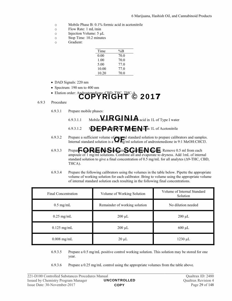
o Mobile Phase B: 0.1% formic acid in acetonitrile o Flow Rate: 1 mL/min o Injection Volume: 5 µL o Stop Time: 10.2 minutes o Gradient:
Time %B 0.00 70.0 1.00 70.0 5.00 77.0 10.00 77.0 10.20 70.0
DAD Signals: 220 nm
Spectrum: 190 nm to 400 nm
Elution order: Androstenedione, CBD, THC, THC-A
6.9.3 Procedure 6.9.3.1 Prepare mobile phases:
6.9.3.1.1 Mobile Phase A: 1 mL of formic acid in 1L of Type I water
6.9.3.1.2 Mobile Phase B: 1 mL of formic acid in 1L of Acetonitrile
6.9.3.2 Prepare a sufficient volume of internal standard solution to prepare calibrators and samples.
Internal standard solution is a 1.0 mg/ml solution of androstenedione in 9:1 MeOH:CHCl3.
6.9.3.3 Prepare a working solution containing all analytes of interest. Remove 0.5 ml from each ampoule of 1 mg/ml solutions. Combine all and evaporate to dryness. Add 1mL of internal standard solution to give a final concentration of 0.5 mg/mL for all analytes (Δ9-THC, CBD, THCA).
6.9.3.4 Prepare the following calibrators using the volumes in the table below. Pipette the appropriate volume of working solution for each calibrator. Bring to volume using the appropriate volume of internal standard solution each resulting in the following final concentrations.
6.9.3.5 Prepare a 0.5 mg/mL positive control working solution. This solution may be stored for one
year.
6.9.3.6 Prepare a 0.25 mg/mL control using the appropriate volumes from the table above.
Final Concentration Volume of Working Solution Volume of Internal Standard
Solution
0.5 mg/mL Remainder of working solution No dilution needed
0.25 mg/mL 200 µL 200 µL
0.125 mg/mL 200 µL 600 µL
0.008 mg/mL 20 µL 1230 µL
UNCONTROLLED COPY
COPYRIGHT © 2017
VIRGINIA DEPARTMENT
OF FORENSIC SCIENCE
6 Marijuana, Hashish Oil, and Cannabinoid Products
221-D100 Controlled Substances Procedures Manual Qualtrax ID: 2480Issued by Chemistry Program Manager Qualtrax Revision 4Issue Date: 30-November-2017 Page 30 of 148
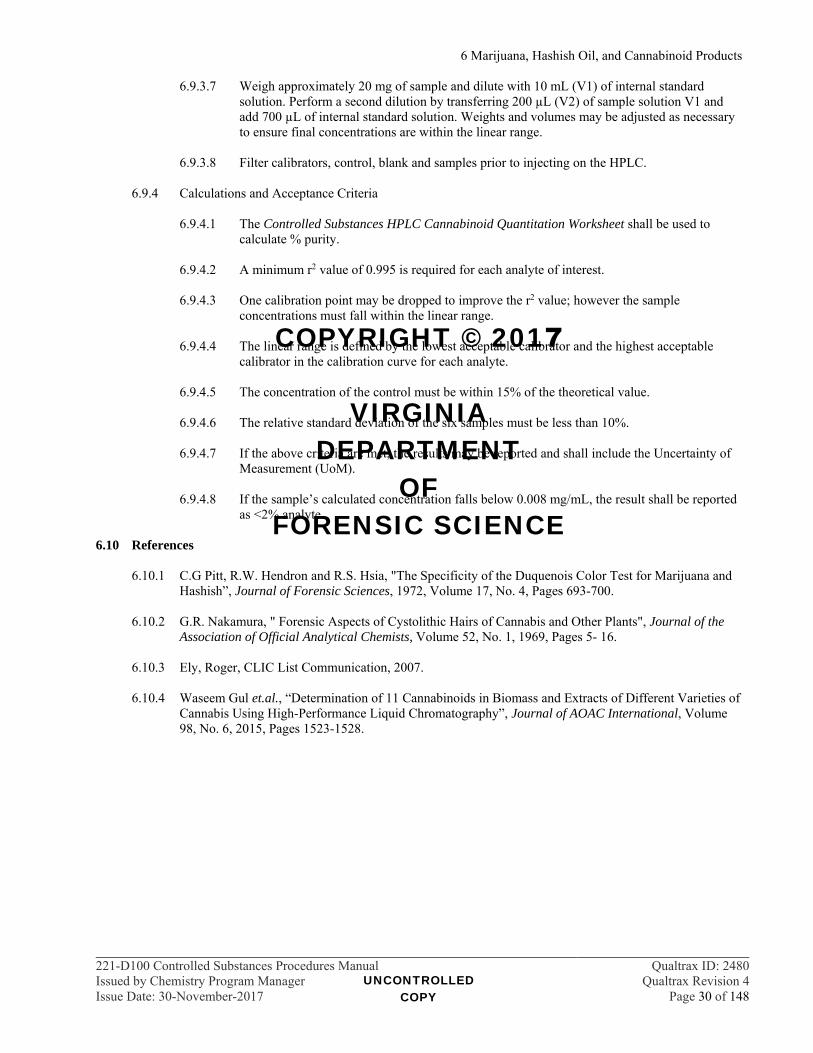
6.9.3.7 Weigh approximately 20 mg of sample and dilute with 10 mL (V1) of internal standard solution. Perform a second dilution by transferring 200 µL (V2) of sample solution V1 and add 700 µL of internal standard solution. Weights and volumes may be adjusted as necessary to ensure final concentrations are within the linear range.
6.9.3.8 Filter calibrators, control, blank and samples prior to injecting on the HPLC.
6.9.4 Calculations and Acceptance Criteria
6.9.4.1 The Controlled Substances HPLC Cannabinoid Quantitation Worksheet shall be used to
calculate % purity.
6.9.4.2 A minimum r2 value of 0.995 is required for each analyte of interest.
6.9.4.3 One calibration point may be dropped to improve the r2 value; however the sample concentrations must fall within the linear range.
6.9.4.4 The linear range is defined by the lowest acceptable calibrator and the highest acceptable
calibrator in the calibration curve for each analyte.
6.9.4.5 The concentration of the control must be within 15% of the theoretical value.
6.9.4.6 The relative standard deviation of the six samples must be less than 10%.
6.9.4.7 If the above criteria are met, the results may be reported and shall include the Uncertainty of Measurement (UoM).
6.9.4.8 If the sample’s calculated concentration falls below 0.008 mg/mL, the result shall be reported
as <2% analyte.
6.10 References
6.10.1 C.G Pitt, R.W. Hendron and R.S. Hsia, "The Specificity of the Duquenois Color Test for Marijuana and Hashish”, Journal of Forensic Sciences, 1972, Volume 17, No. 4, Pages 693-700.
6.10.2 G.R. Nakamura, " Forensic Aspects of Cystolithic Hairs of Cannabis and Other Plants", Journal of the
Association of Official Analytical Chemists, Volume 52, No. 1, 1969, Pages 5- 16.
6.10.3 Ely, Roger, CLIC List Communication, 2007.
6.10.4 Waseem Gul et.al., “Determination of 11 Cannabinoids in Biomass and Extracts of Different Varieties of Cannabis Using High-Performance Liquid Chromatography”, Journal of AOAC International, Volume 98, No. 6, 2015, Pages 1523-1528.
UNCONTROLLED COPY
COPYRIGHT © 2017
VIRGINIA DEPARTMENT
OF FORENSIC SCIENCE

7 Pharmaceutical Identifiers
221-D100 Controlled Substances Procedures Manual Qualtrax ID: 2480Issued by Chemistry Program Manager Qualtrax Revision 4Issue Date: 30-November-2017 Page 31 of 148
7 PHARMACEUTICAL IDENTIFIERS 7.1 Introduction
Pharmaceutical preparations possess unique identifying information both in the general appearance of the preparation and the inscriptions or markings.
7.2 Procedure
7.2.1 It is normally acceptable to visually examine intact, marked tablets or untampered, marked capsules in those cases involving misdemeanor prosecutions in Schedules V and VI. Results should be reported as given in the Reporting Guidelines section of this manual. (See ¶ 33.6)
7.2.1.1 Tablet descriptions in case notes should clearly reflect the physical characteristics used in the
visual examination. 7.2.1.2 Check the PDR, Poison Control, DEA Logo Index, Identadrug, Drug ID Bible / Amera-Chem,
Inc. CD, Pillbox, ePocrates, webpoisoncontrol.org, NIH-DailyMed, or manufacturer’s resources for information relating to inscriptions on tablets and capsules. Two unrelated references are recommended for unfamiliar tablets. Additional references may be used following approval by Chemistry Program Manager.
7.2.1.2.1 The physical characteristics, such as shape and color, should be considered in
addition to the comparison of the markings. Tablets with partial markings may be reported as “visually examined” if they are mixed with intact tablets which are identical in other aspects. If the partial markings are not clear enough to compare to the intact tablets, a screening test should be performed.
7.2.1.2.2 Reference information including page number shall be recorded in the case
notes and/or a hardcopy of relevant entries from an electronic database shall be included in the case file. The results of the visual examination shall be recorded in the case notes.
7.2.1.3 It should be recorded in the case notes if any tampering is evident from the dosage unit
appearance.
7.2.1.4 Tamperable capsules should be screened for tampering using appropriate color tests, TLC, or DART-TOF using the hypergeometric sampling scheme.
7.2.1.5 If tampering is not detected, it may be acceptable to report as visually examined.
7.2.1.6 If tampering is suspected, then a complete analytical scheme including a structural elucidation
technique is required for identification.
7.2.2 At least one dosage unit must be fully tested in those cases involving Schedule IV and above.
7.2.2.1 Visually examine the tablets, capsules, etc., to determine that their size, color and markings are consistent. Check the PDR, Poison Control, DEA Logo Index, Identadrug, Drug ID Bible / Amera-Chem, Inc. CD, Pillbox, ePocrates, webpoisoncontrol.org, NIH-DailyMed, or manufacturer’s resources for information relating to inscriptions on tablets and capsules. Only one reference is necessary. Reference information including page number shall be recorded in the case notes and/or a hardcopy of relevant entries from an electronic database shall be included in the case file. The results of the visual examination shall be recorded in the case notes. Additional references may be used following approval by Chemistry Program Manager.
7.2.2.2 Tamperable capsules shall be screened for tampering using appropriate color tests, TLC, or
DART-TOF using the hypergeometric sampling scheme.
UNCONTROLLED COPY
COPYRIGHT © 2017
VIRGINIA DEPARTMENT
OF FORENSIC SCIENCE
7 Pharmaceutical Identifiers
221-D100 Controlled Substances Procedures Manual Qualtrax ID: 2480Issued by Chemistry Program Manager Qualtrax Revision 4Issue Date: 30-November-2017 Page 32 of 148
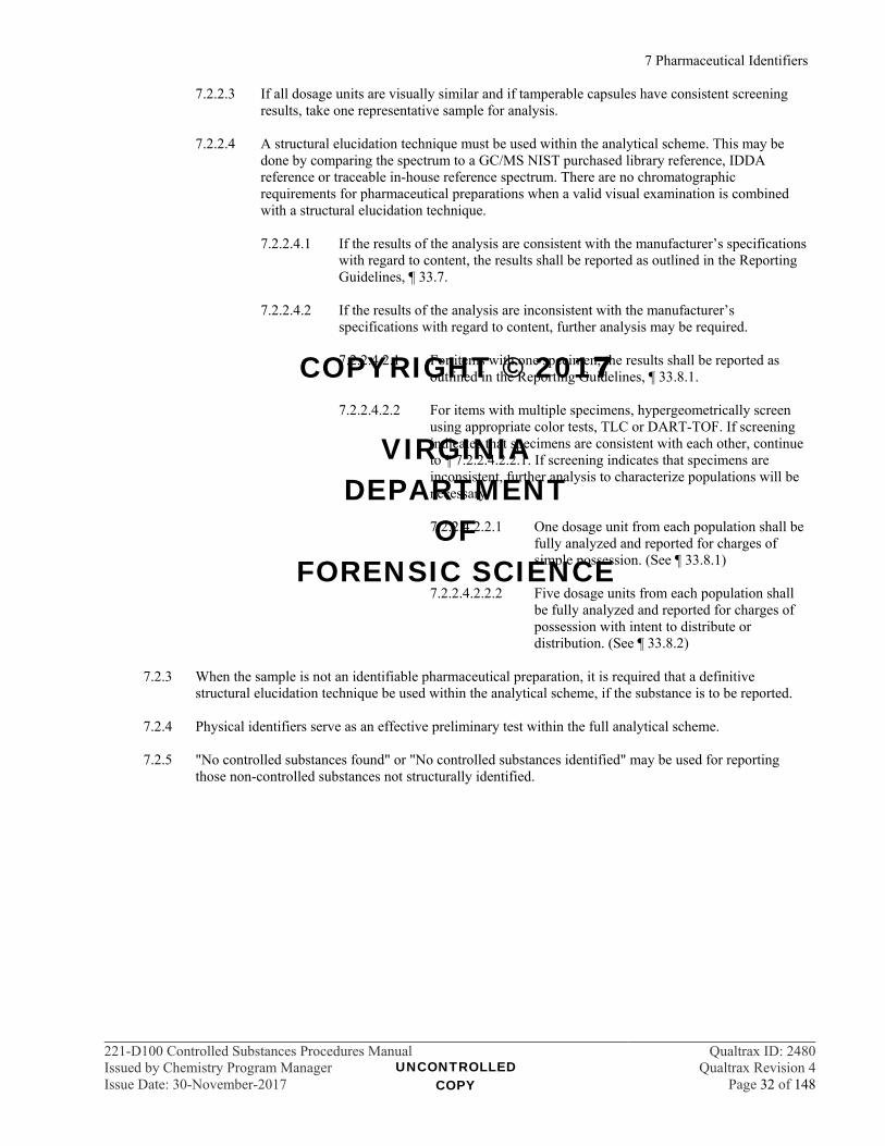
7.2.2.3 If all dosage units are visually similar and if tamperable capsules have consistent screening results, take one representative sample for analysis.
7.2.2.4 A structural elucidation technique must be used within the analytical scheme. This may be
done by comparing the spectrum to a GC/MS NIST purchased library reference, IDDA reference or traceable in-house reference spectrum. There are no chromatographic requirements for pharmaceutical preparations when a valid visual examination is combined with a structural elucidation technique.
7.2.2.4.1 If the results of the analysis are consistent with the manufacturer’s specifications
with regard to content, the results shall be reported as outlined in the Reporting Guidelines, ¶ 33.7.
7.2.2.4.2 If the results of the analysis are inconsistent with the manufacturer’s
specifications with regard to content, further analysis may be required.
7.2.2.4.2.1 For items with one specimen, the results shall be reported as outlined in the Reporting Guidelines, ¶ 33.8.1.
7.2.2.4.2.2 For items with multiple specimens, hypergeometrically screen
using appropriate color tests, TLC or DART-TOF. If screening indicates that specimens are consistent with each other, continue to ¶ 7.2.2.4.2.2.1. If screening indicates that specimens are inconsistent, further analysis to characterize populations will be necessary.
7.2.2.4.2.2.1 One dosage unit from each population shall be
fully analyzed and reported for charges of simple possession. (See ¶ 33.8.1)
7.2.2.4.2.2.2 Five dosage units from each population shall
be fully analyzed and reported for charges of possession with intent to distribute or distribution. (See ¶ 33.8.2)
7.2.3 When the sample is not an identifiable pharmaceutical preparation, it is required that a definitive
structural elucidation technique be used within the analytical scheme, if the substance is to be reported.
7.2.4 Physical identifiers serve as an effective preliminary test within the full analytical scheme.
7.2.5 "No controlled substances found" or "No controlled substances identified" may be used for reporting those non-controlled substances not structurally identified.
UNCONTROLLED COPY
COPYRIGHT © 2017
VIRGINIA DEPARTMENT
OF FORENSIC SCIENCE

8 Color Tests
221-D100 Controlled Substances Procedures Manual Qualtrax ID: 2480Issued by Chemistry Program Manager Qualtrax Revision 4Issue Date: 30-November-2017 Page 33 of 148
8 COLOR TESTS 8.1 Introduction
8.1.1 Color tests are used as a screening test at the beginning of an analysis. Most are performed on clean porcelain or disposable plastic spot plates; however some may be performed in disposable culture tubes (e.g., Scott's, Tannic Acid).
8.1.2 Thin Layer Chromatography visualization sprays may act as color tests when sprayed on a TLC plate or
filter paper where a drop of sample solution has been placed.
8.2 Procedures 8.2.1 The test reagent should be added to the plate or tube first, and then the questioned sample. This practice
determines if the plate or tube was clean before the analysis.
8.2.1.1 If a reaction occurs prior to the addition of the sample, the plate or tube shall be discarded or cleaned before testing the sample.
8.2.2 Several of the listed reagents have more than one recipe listed. Any of the listed, referenced recipes may
be utilized in casework and should be reflected in the reagent logbook.
8.2.3 The Department Reagent Worksheet shall be used to record reagent preparation.
8.2.3.1 It is acceptable to make final volumes different than those listed below as long as the amount of each component is recorded.
8.2.4 Reagents should be made in quantities to minimize waste. The shelf life of color test reagents is two
years, unless otherwise listed.
8.2.5 Reagents, indicators and solutions listed in the USP-NF may be used for their published purposes.
8.2.5.1 Positive controls and blanks shall be performed when using reagents or tests listed in the USP-NF. The results shall be recorded in the case notes.
8.2.5.2 Case notes shall include the procedure with the appropriate reference and the results of the
test. 8.2.6 The color and/or reaction observed must be noted for drugs. Negative reactions should likewise be
documented in the case notes. (For Duquenois-Levine results see ¶ 6.5.7) 8.2.7 Most color test reagents are comprised of strong acids and chemicals requiring careful handling.
Appropriate safety precautions should be observed. Refer to MSDSs for storage and handling. 8.3 Color Tests and Reagents
The following lists the commonly used color test reagents and some examples of reactions with various drugs. The references for each test are in parenthesis.
8.3.1 Bates Test (¶ 8.4.5) tests for cocaine base.
8.3.1.1 Procedure: The Bates test is used as the second part of the Cobalt thiocyanate test (¶ 8.3.4). If
the Cobalt thiocyanate test is negative, add Marquis reagent to spot well.
8.3.1.2 Results: The formation of a very blue precipitate indicates cocaine base, other compounds give weaker blue or no reaction.
UNCONTROLLED COPY
COPYRIGHT © 2017
VIRGINIA DEPARTMENT
OF FORENSIC SCIENCE

8 Color Tests
221-D100 Controlled Substances Procedures Manual Qualtrax ID: 2480Issued by Chemistry Program Manager Qualtrax Revision 4Issue Date: 30-November-2017 Page 34 of 148
8.3.2 Benedict's Solution (¶ 8.4.2) tests for reducing sugars and some antibiotics.
8.3.2.1 Recipe: 1.73 g of copper sulfate in 10 mL of water. With the aid of heat, dissolve 17.3 g trisodium citrate and 10 g of anhydrous sodium carbonate in 80 mL of H2O. Pour the two solutions together and let cool. Dilute to 100 mL with water.
8.3.2.2 Procedure: Add 0.5 mL of the reagent to sample and heat.
8.3.2.3 Results:
Ascorbic acid, strong reducing agents, glucose, tetracycline – red Streptomycin - orange/brown
8.3.3 Chen's Test (¶ 8.4.2) tests for phenethylamines.
8.3.3.1 Recipe: 1 g copper sulfate and 1 mL glacial acetic acid in 100 mL H2O.
8.3.3.2 Procedure: Make an approximate 1% aqueous solution of the sample, add equal volumes of
Chen's reagent and 2N NaOH. 8.3.3.3 Results: ephedrine, PPA and pseudoephedrine – purple
8.3.4 Cobalt Thiocyanate reacts with tertiary and quaternary amines to form a blue precipitate and is used for general screening. May be used in conjunction with the Bates test (¶ 8.3.1) or the Stannous Chloride test (¶ 8.3.21).
8.3.4.1 Recipes:
2 g cobalt thiocyanate in 100 mL H2O or methanol (¶ 8.4.1) 2 g cobalt thiocyanate in 100 mL H2O and 100 mL of glycerine. (¶ 8.4.3) 1.4 g CoCl2. 6H2O and 0.9 g NH4SCN in 100 mL H2O. (¶ 8.4.7)
8.3.4.2 Procedure: Place reagent in well and add sample.
8.3.4.3 Results:
Cocaine HCl – blue precipitate forms, cocaine base may be initially negative or faintly
blue, but blue intensifies upon the addition of dilute HCl. PCP - blue Amitriptyline / doxepin - blue barbiturates with unsaturated side chain (i.e., butalbital) - faint blue
8.3.5 Dille - Koppanyi Test (¶ 8.4.9) reacts with barbiturates.
8.3.5.1 Recipe:
DK1: 0.1 g cobaltous acetate tetrahydrate in 100 mL methanol plus 0.2 mL glacial acetic
acid DK2: 5 mL isopropyl amine in 95 mL methanol
8.3.5.2 Procedure: This is a two part test. Place 2 drops of DK1 reagent in a well. Add sample. Add 1
drop of DK2 reagent. When doing multiple samples, they should be separated to avoid cross-contamination due to reagent spreading.
UNCONTROLLED COPY
COPYRIGHT © 2017
VIRGINIA DEPARTMENT
OF FORENSIC SCIENCE

8 Color Tests
221-D100 Controlled Substances Procedures Manual Qualtrax ID: 2480Issued by Chemistry Program Manager Qualtrax Revision 4Issue Date: 30-November-2017 Page 35 of 148
8.3.5.3 Results:
barbiturates - blue purple theophylline, glutethimide and hydantoins - purple ampicillin - brown
8.3.6 Duquenois - Levine Test (¶¶ 8.4.3 and 8.4.4) reacts with marijuana and hash oil.
8.3.6.1 Recipe: 4 g vanillin and 2.5 mL fresh acetaldehyde per 200 mL 95% ethanol
8.3.6.2 Procedure: See Marijuana section (¶ 6.5).
8.3.6.3 Results: marijuana/hash oil – blue/purple, pink/purple extracts into CHCl3
8.3.7 Ehrlich's Reagent (¶ 8.4.7) reacts with indole moiety and some amines.
8.3.7.1 Recipe: 5 g p-dimethylaminobenzaldehyde to 50 mL of 95% ethanol and 50 mL of conc. HCl
8.3.7.2 Procedure: Place reagent in well and add sample. 8.3.7.3 Results:
LSD, psilocyn - purple (beware of leaching of dyes in blotter paper or tablets) benzocaine, procaine - yellow
8.3.8 Fehlings Solution (¶ 8.4.8) reacts with reducing compounds such as sugars.
8.3.8.1 Recipe:
Fehlings1 - 3.46 g copper sulfate per 50 mL H2O Fehlings2 - 86.5 g sodium potassium tartrate and 35 g of NaOH per 250 mL of H2O
8.3.8.2 Procedure: Dissolve sample in water and mix. Add five drops of Fehlings1 and five drops of
Fehlings2 and mix. Heat on steambath for approximately five minutes or until warm.
8.3.8.3 Results: reducing sugars – yellow to red.
8.3.9 Ferric Chloride (FeCl3) tests for phenols and GHB.
8.3.9.1 Recipes:
9% aqueous solution (¶ 8.4.14) 5% aqueous solution (¶ 8.4.2)
8.3.9.2 Procedure: Place sample into a solution of water or methanol and add a drop of reagent.
8.3.9.3 Results:
salicylamide - dark purple acetaminophen - blue hydrolyzed aspirin – purple (to hydrolyze a sample, place in H2O, add a little acid and
heat) GHB – red/brown
UNCONTROLLED COPY
COPYRIGHT © 2017
VIRGINIA DEPARTMENT
OF FORENSIC SCIENCE
8 Color Tests
221-D100 Controlled Substances Procedures Manual Qualtrax ID: 2480Issued by Chemistry Program Manager Qualtrax Revision 4Issue Date: 30-November-2017 Page 36 of 148
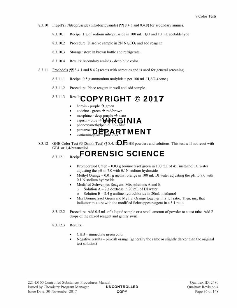
8.3.10 Fiegel's / Nitroprusside (nitroferricyanide) (¶¶ 8.4.3 and 8.4.8) for secondary amines.
8.3.10.1 Recipe: 1 g of sodium nitroprusside in 100 mL H2O and 10 mL acetaldehyde
8.3.10.2 Procedure: Dissolve sample in 2N Na2CO3 and add reagent.
8.3.10.3 Storage: store in brown bottle and refrigerate.
8.3.10.4 Results: secondary amines - deep blue color.
8.3.11 Froehde’s (¶¶ 8.4.1 and 8.4.2) reacts with narcotics and is used for general screening.
8.3.11.1 Recipe: 0.5 g ammonium molybdate per 100 mL H2SO4 (conc.)
8.3.11.2 Procedure: Place reagent in well and add sample.
8.3.11.3 Results:
heroin - purple green codeine - green red/brown morphine - deep purple slate aspirin - blue purple phenoxymethylpenicillin - blue pentazocine – blue acetaminophen – pale blue
8.3.12 GHB Color Test #3 (Smith Test) (¶ 8.4.13) for GHB powders and solutions. This test will not react with
GBL or 1,4-butanediol.
8.3.12.1 Recipe:
Bromocresol Green – 0.03 g bromocresol green in 100 mL of 4:1 methanol:DI water adjusting the pH to 7.0 with 0.1N sodium hydroxide
Methyl Orange – 0.01 g methyl orange in 100 mL DI water adjusting the pH to 7.0 with 0.1 N sodium hydroxide
Modified Schweppes Reagent: Mix solutions A and B o Solution A – 2 g dextrose in 20 mL of DI water o Solution B – 2.4 g aniline hydrochloride in 20mL methanol
Mix Bromocresol Green and Methyl Orange together in a 1:1 ratio. Then, mix that indicator mixture with the modified Schweppes reagent in a 3:1 ratio.
8.3.12.2 Procedure: Add 0.5 mL of a liquid sample or a small amount of powder to a test tube. Add 2
drops of the mixed reagent and gently swirl.
8.3.12.3 Results:
GHB – immediate green color Negative results – pinkish orange (generally the same or slightly darker than the original
test solution)
UNCONTROLLED COPY
COPYRIGHT © 2017
VIRGINIA DEPARTMENT
OF FORENSIC SCIENCE

8 Color Tests
221-D100 Controlled Substances Procedures Manual Qualtrax ID: 2480Issued by Chemistry Program Manager Qualtrax Revision 4Issue Date: 30-November-2017 Page 37 of 148
8.3.13 Marquis (¶¶ 8.4.1 and 8.4.2) reacts with opiates and phenethylamines and is used for general screening.
8.3.13.1 Recipe:
2 mL 37% formaldehyde in 75 mL H2SO4 (conc.)
WARNING! 37% formaldehyde is a “particularly hazardous substance”, for which OSHA has established very low permissible airborne concentrations and prohibited skin or eye contact. Therefore, it must be handled in an
exhaust hood using appropriate PPE (laboratory coat, appropriate forearm length gloves, and face shield).
8.3.13.2 Procedure: Place reagent in well and add sample.
8.3.13.3 Storage: Keep tightly capped.
8.3.13.4 Results:
opiates - purple amphetamine/methamphetamine - orange/brown aspirin – pink deep red on standing phenoxymethylpenicillin - red MDA/MDMA - black
8.3.14 Mayer's Reagent (¶ 8.4.7) reacts with alkaloids
8.3.14.1 Recipe: Dissolve 1 g of mercuric chloride in 100 mL H2O. Add solid potassium iodide until the reddish precipitate first formed dissolves. Reagent should be clear and pale yellow in color.
8.3.14.2 Procedure: Add 0.1 N HCl to a test tube. Add sample to acid and mix. Add Mayer's reagent to
the acid solution. 8.3.14.3 Results: alkaloids – a white to yellow precipitate is formed
8.3.15 Mecke’s (¶¶ 8.4.1 and 8.4.2) reacts with narcotics and is used for general screening.
8.3.15.1 Recipe: 1 g selenious acid per 100 mL H2SO4 (conc.)
8.3.15.2 Procedure: Place reagent in well and add sample.
8.3.15.3 Results:
heroin - green/blue codeine - bright-green/blue green PCP - light yellow quinine - light yellow
8.3.16 Methylene Blue (¶ 8.4.10) reacts with vitamin C.
8.3.16.1 Recipe: 12.5 mg of methylene blue dissolved in 25 mL of 95% ethanol.
8.3.16.2 Procedure: Add reagent to well and add sample. It may be helpful to run a blank to compare
the results.
8.3.16.3 Results: Vitamin C - slowly decolorizes solution from dark blue to light blue.
UNCONTROLLED COPY
COPYRIGHT © 2017
VIRGINIA DEPARTMENT
OF FORENSIC SCIENCE
8 Color Tests
221-D100 Controlled Substances Procedures Manual Qualtrax ID: 2480Issued by Chemistry Program Manager Qualtrax Revision 4Issue Date: 30-November-2017 Page 38 of 148
8.3.17 Nitric Acid (HNO3) (¶¶ 8.4.1 and 8.4.2) reacts with opiates and mescaline.
8.3.17.1 Recipe: concentrated nitric acid
8.3.17.2 Procedure: Place reagent in well and add sample. 2
8.3.17.3 Results:
heroin - yellow green morphine - red codeine - orange mescaline - red acetaminophen – fumes, orange brown
8.3.18 Parri (¶ 8.4.11) reacts with barbiturates.
8.3.18.1 Recipe: cobaltous acetate (solid), barium oxide (solid), and methanol
8.3.18.2 Procedure: Mix cobaltous acetate, BaO and powdered sample in equal parts in a spot plate
well, add methanol.
8.3.18.3 Results: barbiturates – blue
8.3.19 Scotts - Modified Cobalt Thiocyanate (¶ 8.4.3) reacts with cocaine.
8.3.19.1 Recipe: 2 g cobalt thiocyanate in 100 mL H2O and 100 mL of glycerine.
8.3.19.2 Procedure: Add reagent to well or tube and add sample. Dissolve the blue precipitate from the Co(SCN)2 by the addition of HCl. Add CHCl3
8.3.19.3 Results: cocaine - blue color in the lower (CHCl3) layer.
8.3.20 Silver Nitrate (¶ 8.4.15) indicates the presence of chloride ions.
8.3.20.1 Recipe: 5.0% w/v solution of silver nitrate in DI water. 8.3.20.2 Caution: Poison; will cause staining. 8.3.20.3 Storage: Store in the refrigerator in a dark environment. 8.3.20.4 Procedure: Dissolve sample in water. Add silver nitrate solution. A white, curdy precipitate
will form in the presence of chloride ions which will be insoluble in nitric acid. The precipitate will be soluble in 6N ammonium hydroxide.
8.3.21 Stannous Chloride modification for Co(SCN)2 - HCl acidified (¶ 8.4.9) differentiates between “caines”.
8.3.21.1 Recipe: 5 g SnCl2 and 10 mL conc. HCl diluted to 100 mL with H2O
8.3.21.2 Procedure: The Stannous Chloride test is used as the second part of the Cobalt thiocyanate test
(¶ 8.3.4). After performing the cobalt thiocyanate test, add a drop of stannous chloride reagent.
8.3.21.3 Results:
Cocaine salts - blue remains
UNCONTROLLED COPY
COPYRIGHT © 2017
VIRGINIA DEPARTMENT
OF FORENSIC SCIENCE
8 Color Tests
221-D100 Controlled Substances Procedures Manual Qualtrax ID: 2480Issued by Chemistry Program Manager Qualtrax Revision 4Issue Date: 30-November-2017 Page 39 of 148
Cocaine base - blue color forms (initially negative) Other compounds which turned blue initially - blue fades
8.3.22 Sulfuric Acid (H2SO4) (¶¶ 8.4.1 and 8.4.2)
8.3.22.1 Recipe: concentrated sulfuric acid
8.3.22.2 Procedure: Add reagent to well and add sample.
8.3.22.3 Results:
tetracycline - purple turning to yellow upon addition of water 2,3-MDMA, 2,3-MDA – rose 3,4-MDMA, 3,4-MDA – gray-brown
8.3.23 Tannic Acid (¶ 8.4.3) reacts with xanthines.
8.3.23.1 Recipe: 1% aqueous solution of tannic acid
8.3.23.2 Procedure: Add reagent to test tube then add powdered sample.
8.3.23.3 Results: caffeine and theophylline - positive test will produce a precipitate which develops
from "streamers" immediately visible in the solution.
8.3.24 TBPEE Solution (¶ 8.4.8) differentiates between amines.
8.3.24.1 Recipe:
0.01g Tetrabromophenolphthalein ethyl ester (TBPEE) in 100 mL CCl4 10.6 g sodium carbonate in 100 mL H2O (2N solution)
8.3.24.2 Caution: Carbon tetrachloride is carcinogenic. Use appropriate safety precautions.
8.3.24.3 Procedure: Dissolve suspected amine in 2N Na2CO3 solution and add TBPEE solution. Note
color change in the bottom TBPEE layer.
8.3.24.4 Results:
primary amines - violet secondary amine - blue tertiary amine – red
8.3.25 Van Urk's (¶ 8.4.12) reacts with the indole moiety and some amines.
8.3.25.1 Recipe: 125 mg p-dimethylaminobenzaldehyde, 65 mL of concentrated H2SO4, and 2 drops of
ferric chloride (USP T. S.) diluted to 100 mL with distilled water.
8.3.25.2 Procedure: Add reagent to well then add sample.
8.3.25.3 Results: LSD – blue/purple
8.3.26 Weber Test (¶ 8.4.6) reacts with psilocyn.
8.3.26.1 Recipe: 0.01 g of Fast Blue B or Fast Blue BB in 10 mL H2O 8.3.26.2 Shelf life: One month from preparation.
UNCONTROLLED COPY
COPYRIGHT © 2017
VIRGINIA DEPARTMENT
OF FORENSIC SCIENCE
8 Color Tests
221-D100 Controlled Substances Procedures Manual Qualtrax ID: 2480Issued by Chemistry Program Manager Qualtrax Revision 4Issue Date: 30-November-2017 Page 40 of 148
8.3.26.3 Procedure: Add 2 to 3 drops of reagent to a sample of mushrooms. Observe slow color change. Add 1 to 2 drops of conc. HCl, observe color change.
8.3.26.4 Results: Psilocyn – Initially the solution turns red. The solution will turn from red to blue
when the acid is added. 8.4 References
8.4.1 Johns, S. H. et. al., "Spot Tests: A Color Chart Reference for Forensic Chemists," Journal of Forensic Sciences, Volume 24, No.3, July 1979, pp. 631-649.
8.4.2 Moffat, A. C., Clarke's Isolation and Identification of Drugs, The Pharmaceutical Press, London, 1986.
Clarke, E. G. C., Isolation and Identification of Drugs, The Pharmaceutical Press, London, 1972, Vol. 1, 2.
8.4.3 Basic Training Program for Forensic Drug Chemists, 2nd Edition, U. S. Dept. of Justice.
8.4.4 Pitt, C. G. et. al., "The Specificity of the Duquenois Color Test for Marijuana and Hashish", Journal of
Forensic Science, Vol. 17, No. 4, Oct. 1972, pp. 693-700.
8.4.5 Oklahoma City Police Dept. Laboratory, Jane Bates et. al.
8.4.6 Garrett, Steve A. et. al., "The Weber Tests," N. E. A. F. S. Newsletter, Vol. X, No. 2 June 1985.
8.4.7 Gunn, John W. Jr., Analysis of Drugs., United States Dept. of Justice.
8.4.8 Feigel, Fritz., Spot Tests in Organic Analysis, 7th Edition, Elsevier Publishing Co., New York, 1966.
8.4.9 Butler, W. P., Methods of Analysis, Internal Revenue Service, Reprinted by BNDD, June 1967.
8.4.10 United States Pharmacopeia XIII
8.4.11 Connors, K. A., A Textbook of Pharmaceutical Analysis, 2nd Edition, John Wiley & Sons, New York, 1975
8.4.12 Beyer, E. and Dechert, D. D., "Identification of LSD and LSD Tartrate by Thin Layer Chromatography,
"Interbureau By-Lines No. 1, BNDD, July 1967
8.4.13 Smith, Pam and Bozenko, Joseph, Jr. “New Presumptive Tests for GHB” Microgram, Vol. XXXV, No. 1, pp. 10-15 and DEA Communication, Pam Smith, DEA Special Testing and Research Laboratory, 2003
8.4.14 United States Pharmacopeia XXII
8.4.15 United States Pharmocopeia National Formulary, United States Pharmacopeial Convention, Inc.
Rockville, MD, 1995, p. 1722.
UNCONTROLLED COPY
COPYRIGHT © 2017
VIRGINIA DEPARTMENT
OF FORENSIC SCIENCE

9 Thin Layer Chromatography
221-D100 Controlled Substances Procedures Manual Qualtrax ID: 2480Issued by Chemistry Program Manager Qualtrax Revision 4Issue Date: 30-November-2017 Page 41 of 148
9 THIN LAYER CHROMATOGRAPHY 9.1 Introduction
9.1.1 Thin layer chromatography (TLC) is a useful method for screening, separation and preliminary identification. Both the approximate concentration of the sample and the number of components contained in the sample can be ascertained by TLC. TLC can provide valuable information before proceeding on to instrumental tests. Clues as to the chemical structure of an analyte can be obtained by noting the distance traveled in different solvent systems and noting the reactions to a variety of chemical sprays.
9.1.2 The specificity of TLC is greatly increased by using multiple solvent systems of different polarities or
pH. Because many drug compounds (or other organic compounds) have similar Rf values in any one solvent system, at least two solvent systems should be used, except for marijuana and hashish oil.
9.2 Materials
9.2.1 Solvent tank - Any covered glass container with a level bottom can be used. Rectangular tanks are most common. The developing solvent should be at a depth of approximately 0.5 cm to maintain constant contact with the stationary phase throughout the analysis. Filter paper or some other suitable absorbent paper should line the back inside wall of the tank at a height greater than the plate being used when it is required to maintain an atmosphere saturated with solvent vapor. This can provide for better migration and more consistent results. Care should be taken to maintain this atmosphere. The absorbent paper is not required for marijuana analysis.
9.2.2 Thin layer plates - silica gel (250µm) coated glass plates with a fluorescent indicator, or equivalent (Most
drug comp2ounds quench fluorescence when visualized under short wave UV light)
9.2.3 Capillary tubes or Micropipettes
9.2.4 Long wave/short wave UV light source
9.2.5 Solvent baths
9.2.6 Visualization reagents 9.3 Methods
9.3.1 The sample to be tested is dissolved in CHCl3, MeOH or other suitable solvent. The solvent used must be recorded in the case notes.
9.3.2 The solution is drawn up into a capillary tube and 1-10 µl (depending on concentration) is spotted on a
dry plate approximately 0.5 - 1 cm from the bottom, making sure that the spot is above the solvent level in the developing tank. The spot size should be kept to a minimum as its diameter will increase while the compound migrates up the plate during development. Heavy concentrations should be avoided as this causes streaking and tailing.
9.3.3 A standard is spotted beside the sample(s) for comparison. Care should be taken that the standard and
sample(s) are approximately the same concentration. Unequal concentrations may result in unequal rates of advance. This can easily be checked by visualizing the plate under UV light before development.
9.3.4 A blank of the solvent used to dissolve the sample is also spotted on the plate. The results of the blank
must be recorded in the case notes. This may be done by using a check mark () or “ok” or “-“ to record that the results of the blank were acceptable (e.g., Blk ).
9.3.4.1 If spots are visualized in the blank region, the blank should be run again under the same
conditions.
UNCONTROLLED COPY
COPYRIGHT © 2017
VIRGINIA DEPARTMENT
OF FORENSIC SCIENCE
9 Thin Layer Chromatography
221-D100 Controlled Substances Procedures Manual Qualtrax ID: 2480Issued by Chemistry Program Manager Qualtrax Revision 4Issue Date: 30-November-2017 Page 42 of 148
9.3.4.1.1 If the results of the second blank are acceptable, the entire plate should be re-spotted and re-run.
9.3.4.1.2 If the results of the second blank are unacceptable, the analyst should take steps
to resolve the issue (e.g., replacing the solvents in the bottle, checking the baths) prior to re-sampling and any further analysis.
9.3.5 The plate is placed in the tank and allowed to develop until the solvent reaches the top. The plate is then
removed, dried, inspected under UV light, and/or sprayed with the appropriate visualizing reagent. Do not allow the plate to stand in the solvent after development is complete as this will cause a gradual diffusion of the compound.
9.3.6 Specific solvent systems and developing sprays utilized in casework will be denoted in the analytical
case notes. Positive TLC results may be recorded in the analytical notes by the use of a plus (+), a plus circled () or an abbreviation (e.g., pos) along with the standard used in the comparison. A result is considered positive when the distance traveled and the reaction with the visualization methods compare favorably with a standard. Negative reactions may be recorded in a similar fashion; standards used for which negative results are observed need not be documented.
9.4 TLC Baths
TLC1: CHCl3/CH3OH (9:1)(v/v) – general drug screening (¶ 9.8.1) TLC2: Ammonia saturated CHCl3/CH3OH - (18:1) (v/v) – general drug screening (¶ 9.8.2) TLC3: T-1 Methanol/NH4OH (100:1.5) (v/v) – general drug screening (¶ 9.8.1) TLC4: 8% Diethylamine in Toluene (v/v) – marijuana / general drug screening (¶ 9.8.3) TLC5: 4% Diethylamine in Toluene (v/v) – marijuana / general drug screening (¶ 9.8.3) TLC6: Chloroform/Ethyl Acetate (80:20) (v/v) – steroids (¶ 9.8.5) TLC7: Isopropyl Ether – barbiturates (¶ 9.8.8) TLC8: Acetone /CHCl3 (2:1)(v/v) – LSD/LAMPA (¶ 9.8.2) TLC9: Cyclohexane/Toluene/Diethylamine (75:15:10) (v/v) – MDMA/methamphetamine (¶ 9.8.1) TLC10: Ethyl Acetate – GHB, GBL, 1,4-butanediol TLC11: Deionized water - vitamins TLC12: Acetone – amphetamines and other basic drugs (¶ 9.8.4) TLC13: Ethyl acetate/acetone/ammonium hydroxide (25:5:1) (v/v) – ephedrine/pseudoephedrine TLC14: Ethyl acetate/hexane (1:1) (v/v) – salvinorin A (¶ 9.8.11)
9.5 Visualization Reagents
9.5.1 All visualization sprays must be used in a fume hood. 9.5.2 Ceric Sulfate (¶ 9.8.10)
9.5.2.1 Used as an overspray to intensify the reaction with iodoplatinate, especially for caffeine
9.5.2.2 Recipe: 5 g Ce(SO4)2 in 500 mL H2O and 14 mL H2SO4.
9.5.3 Diphenylcarbazone (¶ 9.8.6)
9.5.3.1 Used as an overspray with mercuric sulfate for barbiturates. Can also be freshly mixed 50/50
with the mercuric sulfate reagent (¶ 9.5.12).
9.5.3.2 Recipe: 19 mg diphenylcarbazone in 200 mL (50% acetone/water).
UNCONTROLLED COPY
COPYRIGHT © 2017
VIRGINIA DEPARTMENT
OF FORENSIC SCIENCE
9 Thin Layer Chromatography
221-D100 Controlled Substances Procedures Manual Qualtrax ID: 2480Issued by Chemistry Program Manager Qualtrax Revision 4Issue Date: 30-November-2017 Page 43 of 148
9.5.4 Dragendorff (¶ 9.8.1)
9.5.4.1 General spray which visualizes alkaloids and other nitrogen containing compounds, including methamphetamine and diazepam.
9.5.4.2 Recipe: 1.3 g of bismuth subnitrate in 60 mL water with 15 mL glacial acetic acid. Add this to
12 g potassium iodide in 30 mL H2O. Dilute with 100 mL of H2O and 25 mL glacial acetic acid.
9.5.5 Ehrlich's or p-Dimethylaminobenzaldehyde (p-DMAB) (¶ 9.8.7)
9.5.5.1 Visualizes LSD and psilocybin, reacts with indole nucleus of alkaloids. Plate may be heated
after spraying to increase intensity of color.
9.5.5.2 Recipe: 2 g of p-DMAB in 50 mL 95% ethanol and 50 mL 37% HCl.
9.5.6 Fast Blue B (¶ 9.8.1) or Fast Blue BB (¶ 9.8.9)
9.5.6.1 Visualizes the three major cannabinoids in marijuana. They migrate and develop in the following order:
Top spot - Cannabidiol – orange Middle spot - Tetrahydrocannabinol (Δ9-THC) - red Lower spot - Cannabinol – purple
9.5.6.2 Visualizes psilocyn - red which then turns blue when acidified with HCl.
9.5.6.3 Recipe: Approximately 0.05% solution of Fast Blue B salt OR Fast Blue BB salt in water
9.5.6.4 Shelf life: One month from preparation.
9.5.7 Fluorescamine (FluramR)
9.5.7.1 Visualizes amino acids, primary amines and amino sugars.
9.5.7.2 Recipe: 20 mg FluramR in 100 mL acetone.
9.5.7.3 Procedure: Spray plate with reagent, then check under long wave UV light (amphetamine
fluoresces green-yellow). Heating the plate may intensify the visualization.
9.5.8 Furfuraldehyde and HCl (¶ 9.8.1)
9.5.8.1 Visualizes meprobamate and other carbamates.
9.5.8.2 Recipe: 10% solution of furfuraldehyde in ethanol. Overspray with concentrated HCl.
9.5.8.3 Procedure: Spray plate and heat, if necessary. Spots are black on a white background.
9.5.9 6N HCl
Used to acidify plates
9.5.10 Iodine Vapors (¶ 9.8.1)
9.5.10.1 Visualizes general unknowns and compounds which are not UV active. Suitable for GHB, 1,4-butanediol and GBL analysis. This is a good method of visualization if further testing is to be done on the sample on the plate, as it is reversible.
UNCONTROLLED COPY
COPYRIGHT © 2017
VIRGINIA DEPARTMENT
OF FORENSIC SCIENCE
9 Thin Layer Chromatography
221-D100 Controlled Substances Procedures Manual Qualtrax ID: 2480Issued by Chemistry Program Manager Qualtrax Revision 4Issue Date: 30-November-2017 Page 44 of 148
9.5.10.2 Procedure: Place iodine crystals in an enclosed chamber. Let TLC plate develop in the chamber. Many organic compounds will produce a brown spot.
9.5.10.3 Results:
GHB – white spot on yellow background GBL, 1,4-butanediol – brown spot on yellow background
9.5.11 Iodoplatinate (¶ 9.8.1)
9.5.11.1 Visualizes nitrogen-containing compounds, may be acidified with HCl to intensify some
reactions.
9.5.11.2 Recipes:
5 mL of 10% platinic chloride aqueous solution and 10 g of potassium iodide in 500 mL of H2O.
1 g of platinic chloride and 10 g of potassium iodide in 500 mL H2O.
9.5.11.3 If acidified iodoplatinate is preferred, either overspray the TLC plate with 6 N HCl or prepare stock solution with approximately 5% HCl.
9.5.11.4 Results may be intensified with an overspray of ceric sulfate reagent.
9.5.12 Mercuric Sulfate (¶ 9.8.8)
9.5.12.1 Visualizes barbiturates, which appear as white spots on off-white background. The plate may
need to be sprayed heavily.
9.5.12.2 Recipe: Suspend 5 g HgO in 100 mL of H2O. Add 20 mL concentrated H2SO4. Cool, dilute with 250 mL H2O.
9.5.12.3 Mercuric Sulfate can also be freshly mixed 50/50 with the diphenylcarbazone reagent (¶
9.5.3).
9.5.13 Ninhydrin (¶ 9.8.1)
9.5.13.1 Visualizes amino acid, primary and some secondary amines and amine sugars. (¶ 9.8.1)
9.5.13.2 Recipe: Add 0.5 gram of ninhydrin to 10 mL concentrated HCl. Dilute to 100 mL with acetone.
9.5.13.3 Procedure: Spray with ninhydrin solution and heat the plate (e.g., hotplate, approximately
100ºC oven) for 2 minutes. After spraying, the plate may be irradiated under long wave UV light for 2 minutes prior to heating.
9.5.13.4 Results: yields pink-violet or orange-brown spots.
9.5.13.5 Alternatively, the commercially available Chem Print Ninhydrin may be used.
9.5.14 Potassium Permanganate (¶ 9.8.1)
9.5.14.1 Visualizes unsaturated hydrocarbons. KMnO4 is an alternative to Mercuric Sulfate for
barbiturates which contain a double bond. KMnO4 may be used as an underspray or overspray with Iodoplatinate.
9.5.14.2 Recipe: Dissolve 1 g KMnO4 in 100 mL H2O.
UNCONTROLLED COPY
COPYRIGHT © 2017
VIRGINIA DEPARTMENT
OF FORENSIC SCIENCE
9 Thin Layer Chromatography
221-D100 Controlled Substances Procedures Manual Qualtrax ID: 2480Issued by Chemistry Program Manager Qualtrax Revision 4Issue Date: 30-November-2017 Page 45 of 148
9.5.14.3 Results: yields a yellow spot on a purple background.
9.5.15 Sulfuric Acid/Ethanol Reagent for Steroids (¶ 9.8.5)
9.5.15.1 Recipe: Add gradually 10 mL of conc. sulfuric acid to 90 mL of ethanol. 9.5.15.2 Procedure: Spray plate and heat gently on a hot plate to develop. 9.5.15.3 Results:
Testosterone - green Testosterone esters – purple Oxymethalone – red Nandrolone decanoate – purple
9.5.16 Vanillin Reagent for Salvinorin A (¶ 9.8.11)
9.5.16.1 Recipe: 1 g of vanillin with 50 mL anhydrous ethanol and 0.3 mL concentrated H2SO4
9.5.16.2 Procedure: After developing plate, dry thoroughly. For plate developed in the basic TLC2 bath, spray plate with 6N HCl prior to Vanillin. Spray plate generously with reagent and heat with heat gun for approximately 2 minutes or place in oven at 110 ºC for several minutes.
9.5.16.3 Results: Salvinorin A - Pink/Purple spot. Marijuana also gives a pink/purple spot, but at a
different Rf when using TLC14.
9.5.16.4 Store in refrigerator when not in use. 9.6 Preparative Thin Layer Chromatography
9.6.1 Introduction
9.6.1.1 Frequently, samples contain other organic compounds which interfere with the drug analysis
(e.g., heroin and quinine). Preparative TLC can be used to clean up a sample for other methods of testing such as IR or MS.
9.6.1.2 If cleaning up cocaine for a base determination, be careful to use a neutral bath to develop the
plate so that the original salt form will not be altered.
9.6.2 Materials
9.6.2.1 Thin layer plates
9.6.2.1.1 A section of 250µm thin layer plate can be used if only a small quantity of pure compound is needed.
9.6.2.1.2 For larger quantities, use a 1000µm plate.
9.6.3 Procedure
9.6.3.1 The sample is dissolved in an appropriate solvent and streaked along the bottom of the plate
using a capillary tube, long tipped Pasteur pipette or a commercial streaking device (if avail-able).
9.6.3.2 A standard may be spotted separately at either the beginning or end of the plate in order to
identify the desired compound after development.
UNCONTROLLED COPY
COPYRIGHT © 2017
VIRGINIA DEPARTMENT
OF FORENSIC SCIENCE
9 Thin Layer Chromatography
221-D100 Controlled Substances Procedures Manual Qualtrax ID: 2480Issued by Chemistry Program Manager Qualtrax Revision 4Issue Date: 30-November-2017 Page 46 of 148
9.6.3.3 Develop the plate as in regular TLC. 9.6.3.4 After drying the plate, the desired area is located and marked under UV light. (For compounds
not UV visible, iodine vapors can be used.)
9.6.3.5 Scrape off the desired area, wash thoroughly with solvent in a small beaker and filter to remove the silica gel. Smaller quantities can be filtered using a disposable Pasteur pipette with a pre-washed glass wool plug.
9.6.3.5.1 After development, most compounds adhere strongly to the deactivated silica
gel and therefore must be washed with a fairly polar solvent. Methanol is recommended. For some compounds, an extraction from an aqueous acidic or basic solution may be necessary.
9.6.3.6 If using two-dimensional TLC, first develop the plate as usual, and then develop the plate in a
polar solvent system at a 90 degree angle in order to concentrate the sample into a tighter spot. The standard would need to be removed by breaking off the portion of the plate containing the standard prior to this step. The compound is then removed from the silica as described above in Section 9.6.3.5.
9.7 Comparative Semi-Quantitative Thin Layer Chromatography
9.7.1 Thin layer chromatography can be used to determine relative concentration between a sample and a
standard. This is useful when it is necessary to determine whether a pharmaceutical preparation has been diluted or substituted. In cases where an exact assay is needed, a suitable quantitation should be performed.
9.7.2 Procedure:
9.7.2.1 Obtain or prepare a standard at the concentration expected for the sample.
9.7.2.2 Apply equal amounts of the standard solution and the sample solution to the TLC plate.
9.7.2.3 Develop and visualize the plate as described above for regular TLC.
9.7.2.4 Visually compare the size and color of the spots to determine if the substance has been
substituted or diluted.
9.7.2.5 Approximate concentrations can be estimated by bracketing the observed sample concentration within appropriate standard dilutions. Visually compare the sample response to that of the closest standard dilution. This approximation should be recorded in the case notes but not indicated on the report.
9.7.3 Reporting: Any controlled substance present will be initially identified in the usual manner. The
concentration or substitution of the sample will normally be addressed in the report with one of the two following statements for suspected tampering cases.
9.7.3.1 Meets label specifications (with regard to concentration and/or contents).
9.7.3.2 Does not meet label specifications (with regard to concentration and/or contents).
9.7.3.3 Any pharmaceutical substitutions or adulterants, properly identified, should be reported as
usual. Their concentrations will not normally be required. (e.g., Diazepam substituted into a device labeled to contain morphine would be identified and reported with no concentration value required).
UNCONTROLLED COPY
COPYRIGHT © 2017
VIRGINIA DEPARTMENT
OF FORENSIC SCIENCE
9 Thin Layer Chromatography
221-D100 Controlled Substances Procedures Manual Qualtrax ID: 2480Issued by Chemistry Program Manager Qualtrax Revision 4Issue Date: 30-November-2017 Page 47 of 148
9.8 References
9.8.1 Moffat, A. C., Clarke's Isolation and Identification of Drugs, The Pharmaceutical Press, London, 1986. Clarke, E. G. C., Isolation and Identification of Drugs, The Pharmaceutical Press, London, 1972, Vol. 1, 2.
9.8.2 Gunn, John W. Jr., Analysis of Drugs., United States Dept. of Justice.
9.8.3 Hughes, R. B. and Warner, V. J., Jr. "A Study of False Positives in the Chemical Identification of
Marihuana", Journal of Forensic Sciences, Volume 23 (2), pp. 304-309. (NOTE: Substitute toluene for benzene, a known carcinogen.)
9.8.4 Rasmussen, K. E. and Knutsen, P. “Techniques for the detection and identification of amphetamines and
amphetamine-like substances” Bulletin on Narcotics, Vol. XXXVII, No. 1, 1985, pp. 95-112.
9.8.5 Koverman, Gary, “Analysis of Anabolic Steroids” Microgram, Vol. XXII, No. 5, May 1989.
9.8.6 Liu, Ray and Gadzala, Daniel Handbook of Drug Analysis, American Chemical Society, Washington, DC, 1997, p. 67.
9.8.7 Sperling, Albert, “Thin-Layer and Gas Chromatographic Identification of LSD”, Journal of
Chromromatographic Science, Vol. 12, May 1974, pp. 265 – 266.
9.8.8 Sunshine, Irving, Manual of Analytical Toxicology, CRC Press, Cleveland, Ohio, 1971.
9.8.9 Hughes, R. B. and Kessler, R. R. “Increased Safety and Specificity in the Thin-Layer Chromatographic Identification of Marijuana” Journal of Forensic Science, Vo. 24, No. 4, 1979, pp. 842 – 846.
9.8.10 Macek, Karel, Pharmaceutical Applications of Thin Layer and Paper Chromatography, Elsevier
Publishing Co. New York, 1972, p. 654. 9.8.11 Siebert D. J. “Localization of Salvinorin A and Related Compounds in Glandular Trichomes of the
Psychoactive Sage Salvia Divinorum” Annals of Botany 2004; 93: 763-771.
UNCONTROLLED COPY
COPYRIGHT © 2017
VIRGINIA DEPARTMENT
OF FORENSIC SCIENCE
10 Gas Chromatography
221-D100 Controlled Substances Procedures Manual Qualtrax ID: 2480Issued by Chemistry Program Manager Qualtrax Revision 4Issue Date: 30-November-2017 Page 48 of 148
10 GAS CHROMATOGRAPHY
10.1 Introduction
10.1.1 Gas Chromatography (GC) is a useful method for screening, separation and preliminary identification. GC provides both qualitative and quantitative information about the components of samples. Specificity is dependent on a variety of factors including stationary phase and type of detector. GC can be used to determine such things as isomers along with sufficient structurally definitive information from other techniques.
10.1.2 GC retention times of the analyte are compared to that of a known standard. The specificity of GC is
increased by using two columns with stationary phases of different polarities.
10.1.3 Specific column designations, conditions and detectors utilized in casework will be denoted in the analytical case file. Positive GC results may be recorded in the analytical notes by the use of a plus (+), a plus circled () or an abbreviation (e.g., pos) along with the standard used in the comparison.
10.2 Materials
10.2.1 Capillary Columns:
10.2.1.1 All routine methods employ gas chromatography using flexible fused silica capillary columns of 0.20 to 0.320 mm i.d.
10.2.1.2 The stationary phase is chosen to effect needed resolution. Methylsilicone (e.g., HP-1) and
5% phenylmethyl silicone (e.g., HP-5 and HP-5MS) are utilized in routine casework. The film thickness should be approximately 0.25 microns. The normal general purpose column has a 0.25 µm film thickness and 0.25 mm internal diameter. (35% phenyl)-methylpolysiloxane (e.g., HP-35 and HP-35MS) columns may be used when increased polarity is beneficial for separating compounds with similar structures, such as cannabimimetic agents.
10.2.1.3 All routine methods utilize columns containing a bonded, cross-linked stationary phase.
10.2.1.4 If a stationary phase is required that is outside of these recommended parameters for a
specialized analysis or if more resolving power is required, an alternative column may be used or an additional, different diameter and/or phase column can be temporarily attached at the end of the existing column by using an appropriate connector.
10.2.1.4.1 These changes should be made under the guidance of the instrument operator
and must be approved by the Chemistry Program Manager. 10.2.1.4.2 In the case of thick films or non-bonded stationary phases, the extra column
bleed generated may require more frequent maintenance of the detector.
10.2.2 Additional Instrument Parameters
10.2.2.1 The carrier gas is normally a high purity helium at a flow rate of 0.5 to 3 mL/min.
10.2.2.2 Nitrogen makeup gas is recommended in order to support gas flow at the FID to provide optimal detector sensitivity.
10.2.2.3 Split/splitless liners designed specifically for use with the particular instrument should be
used.
10.2.2.3.1 Injection port liners may be reused after appropriate cleaning and deactivation.
UNCONTROLLED COPY
COPYRIGHT © 2017
VIRGINIA DEPARTMENT
OF FORENSIC SCIENCE

10 Gas Chromatography
221-D100 Controlled Substances Procedures Manual Qualtrax ID: 2480Issued by Chemistry Program Manager Qualtrax Revision 4Issue Date: 30-November-2017 Page 49 of 148
10.2.2.4 For “open tubular" liners, a small amount of silanized glass wool shall be inserted in the center of the liner.
10.2.2.4.1 If the liner is to be packed with GC column packing material, the packing
material should be sandwiched between layers of silanized glass wool or equivalent. Packed liners may require "on column" silyl treatment when first installed.
10.2.2.4.2 Liner packing material: The solid support should be either Chromasorb W-HP or
Gas Chrom Q. Mesh size should be 80/100 or 100/120 mesh. Stationary phases such as OV-1, OV-17, OV-7, and SE 30 series or their equivalent at 2-5% loading may be utilized.
10.2.2.5 The use of a "two hole" capillary ferrule allows two capillary columns of slightly different
polarities to be connected into the same injection port. The sample is analyzed on two columns with a single injection of typically less than 5 µL.
10.2.2.6 Detectors most appropriate for normal drug analysis include both flame ionization detectors
and mass spectrometers. Other specific detectors such as NPD and ECD may be used in circumstances requiring them in consultation with the Chemistry Program Manager. Retention time comparison may be accomplished with any detector. Quantitative analyses should use the flame ionization detector.
10.3 Methods
10.3.1 Analysis conditions are generally set to allow for sample elution time to be greater than 3 - 5 times that of the solvent front. This allows the sample to interact sufficiently with the stationary phase.
10.3.2 The maximum allowable temperature program ramp rate for reproducible retention times is 30 degrees
centigrade per minute for Agilent GCs.
10.3.3 In most instances injection is made in the split mode at a split ratio of 5 - 100:1. Splitless injections may be used when required to increase the amount of analyte delivered to the column and the detector.
10.3.4 Normal injection volume and sample size should be sufficient to provide 8 - 160 nanograms of analyte
"on column" for the normal setup. This correlates to a 1 µL injection of an approximate range of solution concentrations of 0.5 – 10 mg/mL, based on a typical 60:1 split ratio.
10.3.5 Samples should be dissolved in n-hexane, CH2Cl2, CHCl3, ammonia saturated CHCl3 or MeOH for GC
analysis. Depending on the nature of the samples, some samples must be cleaned up by extraction, but most may be directly dissolved in the solvent.
10.3.6 Sample concentrations should be approximately the same concentration as the GC standard and should
be within the linear dynamic range of the chromatographic system and detector.
10.3.7 For comparison purposes, a standard must be run using the same method conditions as the samples. Standards used in the comparison must be run on the same day as the sample. “Same day” is defined as an approximate 24 hour period.
10.3.8 At a minimum, a blank consisting of the solvent(s) used to dissolve the samples, must be run on both the
GC and GC/MS systems, when any of the following conditions are met:
Before each analyst’s series of sample runs whether manual or autosampler methods are utilized.
No more than 10 samples can be run before another blank or standard/blank combination is required. A sample’s position relative to the blank shall be documented in the case file. This may be
UNCONTROLLED COPY
COPYRIGHT © 2017
VIRGINIA DEPARTMENT
OF FORENSIC SCIENCE
10 Gas Chromatography
221-D100 Controlled Substances Procedures Manual Qualtrax ID: 2480Issued by Chemistry Program Manager Qualtrax Revision 4Issue Date: 30-November-2017 Page 50 of 148
accomplished by several methods, including consecutive data file numbering when using “windows macros”.
Whenever there is a change in the chromatographic conditions of the instrument. Changes include
other methods being loaded (except those that merely change the injection volume or split ratio) or run between blank and sample.
It is strongly suggested that a solvent blank be injected and properly documented immediately prior
to a sample known to be extremely weak.
Additional blanks may be run at the examiner’s discretion. The injection order when running samples with standards should be either “standard, blank,
sample(s)” or “blank, sample(s), standard.” 10.3.8.1 The solvent blank must be of at least as large an injection volume of the same solvent as the
sample to be injected. The upper limit injection volume is normally 4 µL.
10.3.8.2 The solvent blank must be run at the same or lower split ratio as the sample. The solvent blank shall be run directly before samples which are run at a reduced split ratio (e.g., 20:1 when typical methods use 60:1).
10.3.8.3 Any significant peaks in blank chromatograms must be properly investigated and documented
in the referenced case file.
10.3.8.3.1 If a controlled substance or related compound is present in any concentration, the blanks and associated samples should be re-run.
10.3.8.3.2 If an interfering substance is present, the blanks and associated samples should
be re-run. 10.3.8.3.3 Blanks and associated samples should be replaced and re-sampled, respectively,
prior to further analysis if the same extraneous peaks are still present.
10.3.9 In all instances, the GC standards file may be referred to for chromatographic conditions. Broad screening methods can be surmised from these files.
10.3.10 Sequencing via autosampler should be utilized whenever practical.
10.3.11 Sequences and/or samples (including standards and blanks) shall be recorded in the instrument logbook.
10.3.11.1 Data files should not be overwritten. 10.3.11.2 Sequence files should not be overwritten unless additional data files have been added during
the sequence run.
10.3.11.3 Sequences and sequence log files shall be archived along with data files as per ¶ 35.6.4.2.
10.3.12 Integrated retention times for analytes must agree with the standard within 2 seconds (± 2 sec.) or 0.033 minutes for this to be considered a positive result.
10.3.13 Derivatization
10.3.13.1 Some compounds, such as amphetamines or barbiturates, do not chromatograph well.
Derivatives may need to be made to help effect good chromatographic peak shape.
UNCONTROLLED COPY
COPYRIGHT © 2017
VIRGINIA DEPARTMENT
OF FORENSIC SCIENCE

10 Gas Chromatography
221-D100 Controlled Substances Procedures Manual Qualtrax ID: 2480Issued by Chemistry Program Manager Qualtrax Revision 4Issue Date: 30-November-2017 Page 51 of 148
10.3.13.2 Procedures:
10.3.13.2.1 Acetyl derivatives – appropriate for primary and secondary amines
The acetyl derivative of phenethylamines is made by drawing up 1 µL of sample followed by 1 µL of acetic anhydride, separated by an air bubble. Acetyl derivatives generally have a longer retention time than the underivatized compound and may require a higher temperature than the underivatized compound.
These derivatives can also be formed prior to injection by heating the
sample and derivatizing reagent (~ 70 C) in a closed vial.
10.3.13.2.2 Alkyl derivatives – appropriate for barbiturates
The methyl derivative of barbiturates is made by the same procedure as listed above, only using trimethylanilinium hydroxide (TMAH) instead of acetic anhydride. Methyl derivatives often have a shorter retention time and may require a lower temperature than the underivatized compound.
10.3.13.2.3 Silyl derivatives
10.3.13.2.3.1 Silyl derivatives are often very helpful in the analysis of
compounds that exhibit chromatographic difficulties due to polar functional groups such as alcohols, amines, acids, and phenols (e.g., GHB, morphine). Silyl derivatives exhibit an M-15 and M-57 peak and sometimes do not exhibit a molecular ion peak in electron impact (EI) mass spectrometry.
10.3.13.2.3.2 There are several good silylation reagents available from Regis
Chemical Co., Pierce Chemical Co. and others which are designed for various applications. Catalogues from these companies are quite useful in determining the most useful derivatizing agent and their application procedure. BSTFA and BSTFA with 1% TMCS work well as silylating reagents for drug compounds.
10.3.13.2.3.3 A suitable aprotic solvent (e.g., pyridine, chloroform, toluene)
may be used to dissolve the analyte and the manufacturers’ directions must be followed carefully to achieve the desired result.
10.3.14 Split/Splitless Liner Cleaning and Preparation Methods
10.3.14.1 Liner Cleaning with aluminum oxide
Remove any packing material. Dip cotton swab in aluminum oxide slurry and twist swab into liners to remove residue. Sonicate liners in water with laboratory glassware soap, rinse several times with water,
acetone and then methanol. Dry thoroughly. Immerse liners in silylating reagent (10% dichlorodimethylsilane in toluene) and let stand
at least 2 hours, preferably overnight. Rinse three times with toluene and dry thoroughly. Wear nylon gloves when handling clean liners and glass wool.
UNCONTROLLED COPY
COPYRIGHT © 2017
VIRGINIA DEPARTMENT
OF FORENSIC SCIENCE
10 Gas Chromatography
221-D100 Controlled Substances Procedures Manual Qualtrax ID: 2480Issued by Chemistry Program Manager Qualtrax Revision 4Issue Date: 30-November-2017 Page 52 of 148
Insert packing, such as silanized glass wool, prior to installation in the instrument. After installation, put injection port and oven temperatures to 290 ºC. Allow to sit for at
least five minutes at this temperature before running QA mix.
10.3.14.2 Liner Cleaning with HCl
Remove any packing or glass wool. Soak liners in 1N HCl for at least eight hours (overnight is fine). Rinse with distilled water and methanol. After thoroughly drying, soak liners in silylating reagent (10% dichlorodimethylsilane in
toluene) and let stand at least 2 hours, preferably overnight. Rinse three times with toluene and dry thoroughly. Wear nylon gloves when handling clean liners and glass wool. Insert packing, such as silanized glass wool, prior to installation in the instrument. After installation, put injection port and oven temperatures to 290 ºC. Allow to sit for at
least five minutes at this temperature before running QA mix. 10.4 Quantitation
10.4.1 Gas Chromatography utilizing a flame ionization detector is an excellent method for quantitative
analysis. The preferred method is the internal standard method. 10.4.2 The Department does not require routine quantitation of drugs. When it is specifically requested and
required to assay a sample, this is the general procedure for a suitable GC quantitation method. Specific examples are found in the sections specific to a particular compound.
10.4.3 Validation
10.4.3.1 Select an appropriate internal standard which will not coelute with components of the sample.
Ideally, this internal standard should elute prior to the analyte of interest. In instances where this is not practical, an internal standard with a similar elution time, though later, may be chosen.
10.4.3.2 Run standards at 4 or 5 different concentrations over the range of interest. The target mid-
range is 1.0 mg/mL. All instrument conditions must remain constant over the range. Inject each standard at least three times. A methylsilicone (e.g., HP-1) column with a 0.25 µm film thickness, 0.25 mm internal diameter and approximate 15 m length will be utilized for quantitative methods.
10.4.3.3 The low standard will define the method’s lower limit of quantitation. The high standard will
define the upper limit of quantitation.
10.4.3.4 Average the response ratios for each standard and calculate the best fit of average response ratio (y-axis) vs. concentration (x-axis) using linear regression using the Linearity Worksheet – 4 points or the Linearity Worksheet – 5 points. The plot of the fit must appear linear. The regression coefficient (R2) must be greater than or equal to 0.99.
10.4.3.5 Calculations
10.4.3.5.1 Back calculate the apparent concentration of each injection using the linear
regression equation from the plot. 10.4.3.5.2 Calculate the mean and % standard deviation of the apparent concentrations for
each standard.
UNCONTROLLED COPY
COPYRIGHT © 2017
VIRGINIA DEPARTMENT
OF FORENSIC SCIENCE
10 Gas Chromatography
221-D100 Controlled Substances Procedures Manual Qualtrax ID: 2480Issued by Chemistry Program Manager Qualtrax Revision 4Issue Date: 30-November-2017 Page 53 of 148
10.4.3.5.3 Calculate the % difference of each mean from the known concentration of the corresponding standard; each value (the accuracy) must be less than or equal to 10%.
% Difference (Accuracy) = [Calculated] – [Theoretical] * 100
[Theoretical]
10.4.3.5.4 Each % standard deviation (the precision) must be less than or equal to 3%.
10.4.3.6 The data and calculations for each validation will be kept in the lab of origin and a copy will be sent to the Chemistry Program Manager for approval.
10.4.4 General Quantitation Procedure
10.4.4.1 Weights will be measured using an analytical balance with a readability of ± 0.00001 gram.
Quantities used in the preparation of primary standards and samples for quantitative purposes shall be at or above 10 milligrams (0.01000 gram). Weights shall be adjusted for purity when solid standards are used in the preparation of the calibration and check standards.
10.4.4.2 Certified reference materials shall be used, where available, for all quantitations and be
acquired from vendors accredited to ISO/IEC 17025:2005 and ISO Guide 34:2009 and include the supplier’s Certificate of Analysis. Standard solutions shall be prepared using calibrated volumetric flasks and/or transferred with calibrated mechanical pipettes as specified in the individual method. Serial numbers of calibrated flasks and/or mechanical pipettes shall be recorded in examination documentation or documented in the standard preparation record with a lot number reference. Internal standard and sample solutions shall be prepared in Class A volumetric flasks, calibrated volumetric flasks, and/or transferred with calibrated mechanical pipettes. Graduated pipettes are not acceptable for quantitative purposes.
10.4.4.3 Make up an internal standard solution of known concentration between 1-2 mg/mL, which
will be used in making all standard and sample solutions. Refrigerated solutions should be allowed to return to ambient temperature prior to use.
10.4.4.4 Make up two standard solutions (1 mg/mL and 2.5 mg/mL) in the internal standard solution as
defined in the method. Do not use serial dilutions.
10.4.4.4.1 The 1 mg/mL standard solution will serve as a check standard. 10.4.4.4.2 The 2.5 mg/mL standard solution will serve as the calibration standard for the
one point calibration.
10.4.4.5 All quantitations shall be performed by preparing six separate sample solutions as per the method, unless a historical uncertainty exists for the method. Refer to Quantitative Sampling (¶ 5.8) for further information.
10.4.4.6 Run the two standards, a blank of the internal standard solution, and the six samples using the
appropriate GC method. The standards will be injected three times and the ratios will be averaged to calculate the concentration. Samples will be injected once each and one injection of the blank is sufficient. The injection volume should be 1-2 µl.
10.4.4.7 Control charts are used to establish the historical standard deviation for quantitative
procedures. Results from quantitative quality control materials are recorded in control charts to readily detect trends such as deterioration of reagents, calibrators and controls.
10.4.4.7.1 All staff are responsible for entering the 1 mg/mL check standard values into the
statewide control charts.
UNCONTROLLED COPY
COPYRIGHT © 2017
VIRGINIA DEPARTMENT
OF FORENSIC SCIENCE
10 Gas Chromatography
221-D100 Controlled Substances Procedures Manual Qualtrax ID: 2480Issued by Chemistry Program Manager Qualtrax Revision 4Issue Date: 30-November-2017 Page 54 of 148
10.4.4.7.2 The DTS Research Analyst or designee should review the statewide control charts each month to ensure consistency between laboratories and communicate results to the Chemistry Program Manager as necessary.
10.4.4.8 Using the equation listed below, calculate the % purity of both the check standard and the
samples. The Drug Quantitation Worksheet shall be used for amphetamine/methamphetamine, cocaine, heroin, PCP and MDMA quantitation calculation purposes. The THC Quantitation Worksheet shall be used for THC quantitation calculation purposes.
10.4.4.8.1 With the exception of THC quantitations, the concentration of the check
standard must be within 5% of the theoretical value. For THC quantitations the check standard must be within 7% of the theoretical value.
10.4.4.8.2 The precision of the check standard must be within 3%. 10.4.4.8.3 With the exception of THC quantitations, the relative standard deviation of the
six samples must be 10% or less. For THC quantitations the relative standard deviation of the six samples must be 15% or less.
10.4.4.8.4 If the above criteria are met, the results may be reported and shall include the
Uncertainty of Measurement (UoM). 10.4.4.8.5 If any of the above criteria are not met, take appropriate steps (e.g., perform
appropriate corrective instrument maintenance, remake both of the standard solutions and repeat, reevaluate the linearity of the instrument) to resolve the problem.
10.4.4.9 Standard Salt Form Calculation
10.4.4.9.1 Unless the salt form of the drug is to be reported, the concentration of the
analyte in its base form will be calculated and reported. If the calibration standard is not in free base form, it will need to be corrected.
10.4.4.9.2 Example: Heroin hydrochloride monohydrate is used to quantitate a sample
containing heroin. The sample will be reported as “Heroin” without a specified salt form. The calibration standard is made up using 22.4 mg of heroin hydrochloride in 10.0 mL of internal standard solution. The corrected concentration will be:
[Heroin base] = (22.4 mg ÷ 10.0 mL) * (369.4 g/mol ÷ 423.9 g/mol) [Heroin base] = 1.95 mg/mL
10.4.4.10 Purity Calculation
% Drug = [STD] x R2 x V x 100
R1 x W
[STD] = concentration of calibration standard in mg/mL R2 = peak area (height) of sample
peak area (height) of internal standard R1 = peak area (height) of standard
peak area (height) of internal standard
UNCONTROLLED COPY
COPYRIGHT © 2017
VIRGINIA DEPARTMENT
OF FORENSIC SCIENCE

10 Gas Chromatography
221-D100 Controlled Substances Procedures Manual Qualtrax ID: 2480Issued by Chemistry Program Manager Qualtrax Revision 4Issue Date: 30-November-2017 Page 55 of 148
V = volume of internal standard solution used in mL W = sample weight in mg
10.4.4.11 Reporting
10.4.4.11.1 Take the average of the six replicates and round to one (1) decimal place unless
specified otherwise in the procedure (see ¶ 32.5). 10.4.4.11.2 If the purity is calculated as the salt, the salt form of the drug should be
confirmed by FTIR/ATR and reported. 10.4.4.11.3 The “show form” option will be utilized in LIMS. 10.4.4.11.4 Examples:
24.55 ± 0.07 grams of solid material, found to contain Heroin (Schedule I),
26.7 ± 8.3% pure.
12.25 ± 0.07 grams of powder, found to contain Cocaine Hydrochloride (Schedule II), 45.2 ± 9.8% pure.
The contents of five were analyzed separately and each was found to
contain Methamphetamine Hydrochloride (Schedule II); total net weight of the five: 12.93 ± 0.07 grams of powder. A composite sample of the five was subsequently analyzed and found to be 45.8 ± 9.4% pure.
Utilizing a hypergeometric sampling plan, the contents of eleven were
analyzed separately and each was found to contain Methamphetamine Hydrochloride (Schedule II); total net weight of the eleven: 15.77 ± 0.54 grams of powder. Based on these results, there is a 95% level of confidence that at least 90% of the packages contain Methamphetamine Hydrochloride. A composite sample of the eleven was subsequently analyzed and found to be 45.8 ± 9.5% pure. The gross weight of the reminder was 2.02 gram(s) including innermost packaging.
UNCONTROLLED COPY
COPYRIGHT © 2017
VIRGINIA DEPARTMENT
OF FORENSIC SCIENCE
11 High Performance Liquid Chromatography (HPLC)
221-D100 Controlled Substances Procedures Manual Qualtrax ID: 2480Issued by Chemistry Program Manager Qualtrax Revision 4Issue Date: 30-November-2017 Page 56 of 148
11 HIGH PERFORMANCE LIQUID CHROMATOGRAPHY (HPLC)
11.1 Introduction
11.1.1 High Performance Liquid Chromatography (HPLC) is a useful method for separation and quantification. Specificity is dependent on a variety of factors including stationary phase and type of detector.
11.1.2 HPLC retention times of the analyte are compared to that of a known standard.
11.1.3 Specific column designations, conditions and detectors utilized in casework will be denoted in the analytical case file. Positive HPLC results may be recorded in the analytical notes by the use of a plus (+), a plus circled () or an abbreviation (e.g., pos) along with the standard used in the comparison.
11.2 Materials
11.2.1 Columns:
11.2.1.1 There are a wide variety of column choices available for use. The stationary phase is chosen to affect needed resolution.
11.2.1.2 Columns and chromatographic conditions will be dependent on the currently used instrument
and technology available. Literature resources are useful for specific separations. If a specialized analysis is required, consult the primary operator for the HPLC.
11.2.2 Additional Instrument Parameters
11.2.2.1 Mobile phases will be selected based on the target analytes.
11.2.2.2 Detectors most appropriate for normal drug analyses include both diode array detectors
(DAD) and mass spectrometers. Retention time comparison may be accomplished with any detector. Quantitative analyses shall use the diode array detector.
11.3 Methods
11.3.1 Analysis conditions should allow the sample to interact sufficiently with the stationary phase.
11.3.2 Normal injection volumes range from 1 to 5 microliters but may be adjusted depending on the sample concentration.
11.3.3 Samples should be prepared in an appropriate solvent based on the chosen method. Depending on the
nature of the samples an extraction may be required; however, most may be directly dissolved in the solvent. Samples may require filtration using syringe filters, or similar, before injection onto the HPLC system.
11.3.4 Sample concentrations should be within the linear dynamic range of the chromatographic system and
detector.
11.3.5 For comparison purposes, a standard must be run using the same method conditions as the samples. Standards used in the comparison must be run on the same day as the sample. “Same day” is defined as an approximate 24 hour period.
11.3.6 At a minimum, a blank consisting of the solvent(s) used to dissolve the samples, must be run on the
HPLC system, when any of the following conditions are met:
Before each analyst’s series of sample runs.
UNCONTROLLED COPY
COPYRIGHT © 2017
VIRGINIA DEPARTMENT
OF FORENSIC SCIENCE
11 High Performance Liquid Chromatography (HPLC)
221-D100 Controlled Substances Procedures Manual Qualtrax ID: 2480Issued by Chemistry Program Manager Qualtrax Revision 4Issue Date: 30-November-2017 Page 57 of 148
No more than 10 samples can be run before another blank or standard/blank combination is required. A sample’s position relative to the blank shall be documented in the case file. This may be accomplished by consecutive data file numbering.
Whenever there is a change in the chromatographic conditions of the instrument. Changes include
other methods being loaded or run between blank and sample.
It is strongly suggested that a solvent blank be injected and properly documented immediately prior to a sample known to be extremely weak.
Additional blanks may be run at the examiner’s discretion. The injection order when running samples with standards should be either “standard, blank,
sample(s)” or “blank, sample(s), standard.” 11.3.6.1 The blank solvent shall be the same solvent utilized for samples. In addition, the injection
volume shall be equal to, or greater than, the volume injected for the samples.
11.3.6.2 Any significant peaks in the blank chromatograms must be properly investigated and documented in the referenced case file.
11.3.6.2.1 If a controlled substance or related compound is present in any concentration,
the blanks and associated samples shall be re-run. 11.3.6.2.2 If an interfering substance is present, the blanks and associated samples shall be
re-run. 11.3.6.2.3 Blanks and associated samples shall be replaced and re-sampled, respectively,
prior to further analyses if the same extraneous peaks are still present.
11.3.7 Sequences and/or samples (including standards and blanks) shall be recorded in the instrument logbook.
11.3.7.1 Data files should not be overwritten. 11.3.7.2 Sequence files should not be overwritten unless additional data files have been added during
the sequence run.
11.3.7.3 Sequences and sequence log files shall be archived along with data files as per ¶ 35.7.3.1.
11.3.8 Retention times for analytes must agree with the standard within 2 seconds (± 2 sec.) or 0.033 minutes for this to be considered a positive result.
11.4 Quantitation
11.4.1 High Performance Liquid Chromatography utilizing a diode array detector is commonly used to perform
quantitative analyses. 11.4.2 The Department does not require routine quantitation of drugs. When quantitation of an analyte is
necessary, the following is the general procedure for quantitations performed using HPLC. Specific procedures for certain analytes can be found in other sections of this manual.
11.4.3 General Quantitation Procedure
11.4.3.1 Weights will be measured using an analytical balance with a readability of ± 0.00001 gram.
Quantities used in the preparation of primary standards and samples for quantitative purposes shall be at or above 10 milligrams (0.01000 gram). Weights shall be adjusted for purity when solid standards are used in the preparation of the calibration and check standards.
UNCONTROLLED COPY
COPYRIGHT © 2017
VIRGINIA DEPARTMENT
OF FORENSIC SCIENCE
11 High Performance Liquid Chromatography (HPLC)
221-D100 Controlled Substances Procedures Manual Qualtrax ID: 2480Issued by Chemistry Program Manager Qualtrax Revision 4Issue Date: 30-November-2017 Page 58 of 148
11.4.3.2 Certified reference materials shall be used, where available, for all quantitations and be acquired from vendors accredited to ISO/IEC 17025:2005 and ISO Guide 34:2009 and include the supplier’s Certificate of Analysis. Standard solutions shall be prepared in calibrated volumetric flasks and/or transferred with calibrated mechanical pipettes. Serial numbers of calibrated flasks and/or mechanical pipettes shall be recorded in examination documentation or documented in the standard preparation record with a lot number reference. Internal standard and sample solutions shall be prepared using Class A volumetric flasks and/or transferred with calibrated mechanical pipettes. Graduated pipettes are not acceptable for quantitative purposes.
11.4.3.3 Prepare an internal standard solution of known concentration, which will be used in preparing
all standard and sample solutions. Refrigerated solutions should be allowed to return to ambient temperature prior to use.
11.4.3.4 Prepare an appropriate number of calibrators to create a calibration curve as defined in the
method.
11.4.3.5 Prepare a positive control as defined in the method.
11.4.3.6 All quantitations shall be performed by preparing six separate sample solutions as per the method, unless a historical uncertainty exists for the method. Refer to Quantitative Sampling (¶ 5.8) for further information.
11.4.3.7 Run the calibrators, control, a blank of the internal standard solution, and the six samples
using the appropriate HPLC method. The standards, control and samples shall be injected once each in addition to a sufficient number of blanks.
11.4.3.8 Control charts are used to establish the historical standard deviation for quantitative
procedures. Results from quantitative quality control materials are recorded in control charts to readily detect trends such as deterioration of reagents, calibrators and controls.
11.4.3.8.1 All staff are responsible for entering control values into the statewide control
charts as necessary. 11.4.3.8.2 The DTS Research Analyst or designee shall review the statewide control charts
monthly to ensure consistency between laboratories and communicate results to the Chemistry Program Manager as necessary.
11.4.3.9 The Controlled Substances HPLC Cannabinoid Quantitation Worksheet shall be used for
cannabinoid quantitation calculation purposes.
11.4.3.9.1 A minimum r2 value of 0.995 is required for each analyte of interest.
11.4.3.9.2 One calibration point may be dropped to improve the r2 value; however the sample concentrations must fall within the linear range.
11.4.3.9.3 The linear range is defined as the lowest acceptable calibrator and the highest
acceptable calibrator in the calibration curve for each analyte.
11.4.3.9.4 The concentration of the control must be within 15% of the theoretical value for the cannabinoid quantitation.
11.4.3.9.5 The relative standard deviation of the six samples must be less than 10%.
11.4.3.9.6 If the above criteria are met, the results may be reported and shall include the
Uncertainty of Measurement (UoM).
UNCONTROLLED COPY
COPYRIGHT © 2017
VIRGINIA DEPARTMENT
OF FORENSIC SCIENCE
11 High Performance Liquid Chromatography (HPLC)
221-D100 Controlled Substances Procedures Manual Qualtrax ID: 2480Issued by Chemistry Program Manager Qualtrax Revision 4Issue Date: 30-November-2017 Page 59 of 148
11.4.3.9.7 If the sample’s calculated concentration falls below 0.008 mg/mL, the result shall be reported as <2% analyte.
11.4.3.10 Reporting
11.4.3.10.1 Take the average of the six replicates and round to one (1) decimal place unless
specified otherwise in the procedure (see ¶ 33.6). 11.4.3.10.2 Examples:
Cannabidiol oil, found to contain 20.4 ± 3.4% cannabidiol by weight and
2.6 ± 1.1% tetrahydrocannabinol by weight.
Hashish oil (Schedule I), found to contain 34.6 ± 9.2% tetrahydrocannabinol by weight.
Marijuana, 4.52 ± 0.05 grams (0.159 ± 0.002 ounce) of material. Quantitative analysis of the material determined that it contained 6.3 ± 1.5% cannabidiol by weight and 8.5 ± 1.7% tetrahydrocannabinol by weight.
UNCONTROLLED COPY
COPYRIGHT © 2017
VIRGINIA DEPARTMENT
OF FORENSIC SCIENCE
12 Analysis of Pharmaceutical Injectable Dosage Forms
221-D100 Controlled Substances Procedures Manual Qualtrax ID: 2480Issued by Chemistry Program Manager Qualtrax Revision 4Issue Date: 30-November-2017 Page 60 of 148
12 ANALYSIS OF PHARMACEUTICAL INJECTABLE DOSAGE FORMS 12.1 Introduction
12.1.1 The analysis of cases involving pharmaceutical injectable dosage forms may require more than just the identification of the contents.
12.1.2 Tampering can involve the dilution, contamination, or removal of the contents of a Tubex®, Carpuject®,
injection vial, pharmacy intravenous (IV) preparation, IV stock solution, or IV supplies and apparatus. 12.2 Procedure
12.2.1 Carefully visually inspect the item. Note stopper condition (for small punctures), plunger location and
condition, fill volume, appearance, color and consistency of the contents.
12.2.2 If possible, a standard of the same brand and lot number should be requested from the submitting agency or a licensed pharmacy for comparison of the visual characteristics, chemical contents and concentration.
12.2.3 Any controlled substance present will be identified in the usual manner.
12.2.4 Analyses for concentration are normally run with semi-quantitative TLC, comparative UV quantitation,
or standard GC quantitation methods. 12.3 Reporting
The concentration of the sample will normally be addressed in the report with one of the two following statements for suspected tampering cases. Additional clarifying wording may be used at the discretion of the Section Supervisor, such as “Does not meet label specifications with regard to concentration” or “Does not meet label specifications with regard to concentration and contents.”
12.3.1 Meets label specifications.
12.3.2 Does not meet label specifications.
12.3.3 Any pharmaceutical substitutions or adulterants, properly identified, should be reported as usual. Their
concentrations will not normally be required. (e.g., Diazepam substituted into a device labeled to contain morphine would be identified and reported with no concentration value required).
UNCONTROLLED COPY
COPYRIGHT © 2017
VIRGINIA DEPARTMENT
OF FORENSIC SCIENCE

13 Infrared Spectroscopy
221-D100 Controlled Substances Procedures Manual Qualtrax ID: 2480Issued by Chemistry Program Manager Qualtrax Revision 4Issue Date: 30-November-2017 Page 61 of 148
13 INFRARED SPECTROSCOPY 13.1 Introduction
13.1.1 Infrared spectroscopy (IR) is a specific method of identification in most instances and is therefore a desirable analytical tool for the forensic drug chemist. IR may be used to obtain semiquantitative data on known mixtures to express relative percentages, but is not normally used for quantitation.
13.1.2 This method of spectral analysis is based on the molecular vibrational energies of an organic compound.
Infrared light containing wavelengths from 4000 cm-1 to 400 cm-1 is generated and passed through the sample. When the frequency of light matches a frequency of vibration within the molecule, absorption occurs. The absorptions are translated electronically and recorded on a data system. The resulting spectrum will have characteristic bands corresponding to each different vibration among atoms in the molecule.
13.1.3 The IR spectrum of an unknown compound can be compared to the IR spectrum of a known or suitable
reference spectrum for confirmation.
13.1.4 The Fourier Transform Infrared Spectrophotometer (FTIR) collects the composite spectrum in the time domain and mathematically transforms it to the frequency domain.
13.1.5 Non-chemical separations (spectral subtraction) may be performed to determine components of a
mixture. The components would need to be separated and structural confirmation of the pure compounds done by this or other structural identification techniques, if needed.
13.1.6 Spectra may be collected using an Attenuated Total Reflectance (ATR) accessory and compared to
standards also collected utilizing the ATR. These standards may be stored in a user generated library. For unknown compounds, an ATR correction may be utilized in order to search a library of transmission spectra. The uncorrected unknown spectrum would then be compared to that of a known uncorrected standard spectrum.
13.1.7 Spectra may be collected using the DiscovIR Gas Chromatograph-Solid State Infrared
Spectrophotometer and compared to standards also collected on the DiscovIR. These standards may be stored in a user generated library.
13.1.8 If unique sample preparation or data reduction techniques are required, consult the Primary Operator for
the FTIR.
13.2 Sample Preparation
13.2.1 Samples should be relatively pure and can be cleaned up by extraction, preparative TLC, recrystallization, or precipitation and filtration, depending upon the quantity and type of contaminants present.
13.2.2 Pure liquid organics can be run neat between two salt (NaCl) plates or using the ATR accessory.
13.2.3 Pure solids can be dissolved in a suitable organic solvent and run in solution cells, mixed with KBr and
pressed into a pellet, mixed with a saturated long chain hydrocarbon oil (mulled) or run using the ATR accessory.
13.2.4 Solution Technique
13.2.4.1 A small amount of the sample is dissolved in a non-polar solvent such as CCl4 or CS2. Polar
solvents such as MeOH or EtOH should be avoided. Other slightly polar solvents, such as CHCl3, can also be used but will have some interfering absorption bands due to C-H.
UNCONTROLLED COPY
COPYRIGHT © 2017
VIRGINIA DEPARTMENT
OF FORENSIC SCIENCE
13 Infrared Spectroscopy
221-D100 Controlled Substances Procedures Manual Qualtrax ID: 2480Issued by Chemistry Program Manager Qualtrax Revision 4Issue Date: 30-November-2017 Page 62 of 148
13.2.4.2 Oils or dissolved solids may be deposited or "cast" on a salt plate (e.g., standard NaCl window) and placed in the sample beam. (Care must be taken to drive off all residual solvent).
13.2.4.3 The solvent absorption bands may be subtracted from the spectrum. Either a pair of salt plates
with the solution solvent or a solution cell (of the same pathlength) containing only solvent can be scanned into the background spectrum.
13.2.5 Gas Techniques
Standard 10 cm gas cells or other similar hardware (e.g., multiple internal reflectance units) can be used.
13.2.6 Mull Technique
The sample is finely ground and suspended in mineral oil (Nujol). A thin film of the suspension is placed between two salt plates.
13.2.7 Pellet Technique
13.2.7.1 Infrared grade KBr should be kept dry by storing it in a suitable location such as a dessicator. 13.2.7.2 Infrared grade KBr and the sample each must be finely ground. The KBr and sample are
mixed by grinding with a mortar and pestle in an approximate ratio of 100 parts KBr to 1 part sample.
13.2.7.3 The mixture is placed in a pellet press to prepare the pellet. A hand press with a 7 mm die or
the Hydraulic 13 mm die set may be used. The 7 mm hand press KBr pellet is the preferred preparation technique.
13.2.8 ATR Accessory
13.2.8.1 Clean the diamond crystal and anvil surface before and after analysis with acetone or methanol soaked wipes. Methanol takes a slightly longer time to evaporate.
13.2.8.2 A background is collected prior to each sample.
13.2.8.2.1 An acceptable background shall be noted on the data. 13.2.8.2.2 If unexpected peaks are present in the background, the ATR crystal and anvil
shall be cleaned and the background repeated. 13.2.8.2.3 If the results of the second background are unacceptable, the analyst should take
steps to resolve the issue prior to any analysis.
13.2.8.3 For ATR, the background run prior to consecutive samples serves as the blank for that case or sequences of cases. This background/blank shall be printed and stored in the corresponding case file(s).
13.2.8.4 For solid samples, cover the center of the crystal with sample. Press the anvil against the
sample by turning the anvil screw clockwise until it spins without further tightening. 13.2.8.5 For liquid samples, place a drop or two of liquid directly onto the ATR crystal. Use enough
sample to cover the crystal completely. If the sample is volatile, place the cover over the sampling area to prevent evaporation during analysis.
13.2.8.6 If the sample requires an extraction, the sample in an organic solvent may be dropped on a
crystal and allowed to evaporate to form a film. An o-ring may be used to contain the liquid as it is placed on the crystal.
UNCONTROLLED COPY
COPYRIGHT © 2017
VIRGINIA DEPARTMENT
OF FORENSIC SCIENCE
13 Infrared Spectroscopy
221-D100 Controlled Substances Procedures Manual Qualtrax ID: 2480Issued by Chemistry Program Manager Qualtrax Revision 4Issue Date: 30-November-2017 Page 63 of 148
13.3 Solid State FTIR via Gas Chromatography
13.3.1 The DiscovIR instrument deposits the eluent from a gas chromatograph onto a liquid nitrogen cooled disk allowing for collection of solid phase transmission spectra. The MCT-A detector scans from 4000 – 650 cm-1 and requires cooling with liquid nitrogen prior to analysis.
13.3.2 The spectra produced must be compared to known spectra taken under similar conditions. Spectra
collected with the DiscovIR may differ from traditional FTIR libraries due to the water content or salt form of the material.
13.3.3 DiscovIR analysis is especially useful for positional isomer differentiation which may be required with
cannabimimetic agents and research chemical analysis. The solid phase spectra collected with the DiscovIR have a higher resolution (4 cm-1) than the gas phase spectra collected with the Gas Phase FTIR (8 cm-1).
13.3.4 Procedure:
13.3.4.1 Liquid nitrogen is added to both the large Dewar and the MCT-A detector daily
approximately 30 minutes prior to use. The large Dewar holds up to 10 L of liquid nitrogen where each liter allows for approximately one hour of instrument operation. The MCT-A detector holds approximately ½ L of liquid nitrogen which allows for approximately 18 hours of instrument operation.
13.3.4.2 Method parameters for the instrumentation are as follows:
Column HP-35 15m x 0.25 mm i.d. x 0.25µm film thickness (or equivalent) Flow 1.5 – 8.0 mL/min. Resolution 4.0 Transfer line 325 ◦C Oven temp. 325 ◦C Restrictor temp. 325 ◦C Disk temp. -40 ◦C Dewer cap temp. 20 ◦C
13.3.4.3 Methods selected from those listed below can be documented in the case notes by name. If
alternate method parameters are utilized, the parameters must be recorded in the case notes.
Method Injection Volume
(µL)
Temperature Range (ºC)
Ramp Rate (ºC/min.)
Split Ratio
Flow Rate
(mL/min.)
Inlet Temperature
(ºC)
Disk Speed
(mm/min.) LOW1 1 90 – 190 30 10:1 1.5 250 3 LOW4 4 90 – 190 30 10:1 1.5 250 3 MID1 1 170 – 270 30 10:1 1.5 250 3 MID4 4 170 - 270 30 10:1 1.5 250 3
HIGH1 1 225 – 315 30 10:1 1.8 270 3 HIGH4 4 225 – 315 30 10:1 1.8 270 3
SCREEN1 1 105 – 315 40 10:1 1.8 270 3 SCREEN4 4 105 - 315 40 10:1 1.8 270 3
13.3.5 Samples should be dissolved in n-hexane, CH2Cl2, CHCl3, ammonia saturated CHCl3 or MeOH for GC
analysis. Depending on the nature of the samples, some samples must be cleaned up by extraction, but most may be directly dissolved in the solvent.
13.3.6 An amount of sample (1 – 4 μL) is injected utilizing an autosampler.
UNCONTROLLED COPY
COPYRIGHT © 2017
VIRGINIA DEPARTMENT
OF FORENSIC SCIENCE
13 Infrared Spectroscopy
221-D100 Controlled Substances Procedures Manual Qualtrax ID: 2480Issued by Chemistry Program Manager Qualtrax Revision 4Issue Date: 30-November-2017 Page 64 of 148
13.3.7 Sample concentrations should be approximately the same concentration as the standard. Concentrations of approximately 2 mg/mL are recommended.
13.3.8 Basic extractions are recommended for suspected clandestine laboratory samples or other
phenethylamine type compounds.
13.3.9 For GC comparison purposes, a standard must be run using the same method conditions as the samples. Standards used in the comparison must be run on the same day as the sample. “Same day” is defined as an approximate 24 hour period.
13.3.10 Solvent blanks shall be run prior to each sample in the same location where the sample is to be deposited
on the disk.
13.3.11 After the data is processed, the sample spectrum will be compared to a known solid phase spectrum. For unknown compounds, a search of a library of solid state transmission spectra may be conducted, however for an identification, the spectrum produced must be compared to a known spectrum taken under similar conditions.
13.3.12 Integrated retention times for analytes must agree with the standard within 2 seconds (± 2 sec.) or 0.033
minutes for this to be considered a positive GC result. 13.4 Acceptance Criteria
13.4.1 When using FTIR as the primary structural elucidation technique, the sample spectrum should compare favorably with a spectrum of a known standard in both its overall appearance and in the presence and location of the major peaks. Due caution should be exercised when using the similarity index generated by the library search algorithm.
13.4.2 When using FTIR to differentiate cocaine base from cocaine hydrochloride or another salt form where
GC/MS has been previously performed, the areas of the spectrum which are different between cocaine base and cocaine hydrochloride should be clear. Other areas may have interfering peaks present that do not mask the “salt form” identity.
13.4.3 Data which supports the analyst’s conclusion shall be printed and included in the case file.
13.4.3.1 At a minimum, the data shall include:
FS Lab number and Item number Date Instrument name Sampling information (e.g., DiscovIR, ATR, KBr pellet, pentane extract) Method parameters (e.g., # scans, resolution, sample gain, mirror velocity, aperture) (for
DiscovIR see ¶¶ 13.4.4.2 – 13.4.4.3)
UNCONTROLLED COPY
COPYRIGHT © 2017
VIRGINIA DEPARTMENT
OF FORENSIC SCIENCE

14 Gas Chromatography/Mass Spectrometry
221-D100 Controlled Substances Procedures Manual Qualtrax ID: 2480Issued by Chemistry Program Manager Qualtrax Revision 4Issue Date: 30-November-2017 Page 65 of 148
14 GAS CHROMATOGRAPHY/MASS SPECTROMETRY 14.1 Introduction
14.1.1 Gas Chromatography/Mass Spectrometry (GC/MS) is a specific method of identification for most drug substances. MS cannot differentiate between optical isomers. A sample is passed through a gas chromatographic column, effecting a separation of the components of the sample. The individual compounds then move into the mass spectrometer source where they are bombarded by electrons, producing charged ions. The ions of interest are positively charged fragments of the original compound. The ions are then separated, through a mass filtering process, according to their mass-to-charge ratios (m/z) and then collected by a detector. In the detector, the ions are converted to a proportional electrical current. The data system records the magnitude of these electrical signals as a function of m/z and converts this information into a mass spectrum. The mass spectrum is a record of the different ions (m/z) and the relative numbers of each ion (abundance). These spectra are characteristic for individual compounds, giving specificity for most types of drug substances.
14.1.2 Depending on the structure of the molecule, the amount and type of fragmentation will vary. Due to this,
some drugs do not exhibit a molecular ion using electron impact mass spectrometry. Examples include barbiturates, lorazepam and methylphenidate.
14.1.3 Confirmation of an unknown spectrum is done by direct comparison with a known or suitable reference
spectrum or through use of interpretation methods. Positive mass spectral results may be recorded in the analytical notes by listing the drug identified. It is not required to record the analyst’s disagreement with library search results on the data.
14.2 Procedure
14.2.1 Samples will be dissolved in a suitable solvent, preferably methanol.
14.2.2 The general concentration should be determined by TLC or GC before being run on the GC/MS. The usual amount of sample delivered to the ion source for good qualitative results should be 8 - 160 ng. This correlates to an approximate range of solution concentrations of 0.5 – 10 mg/mL, based on a typical 60:1 split ratio with a 1 µL injection volume. In any case, sufficient abundance of the total ion chromatogram peaks needs to be achieved in order to produce acceptable spectra, without overloading the chromatographic system.
14.2.3 For analysis of volatile organics, such as amyl nitrite, the headspace may be injected. An air blank must
be run prior to headspace analysis.
14.2.4 Chromatographic conditions may be determined by the chemist from the GC/MS standards file.
14.2.5 The mass spectrum will be obtained in full scan mode using an appropriate scan range for the compounds to be analyzed.
14.2.6 At a minimum, a blank consisting of the solvent(s) used to dissolve the samples must be run on the
GC/MS systems, when any of the following conditions are met:
Before each analyst’s series of sample runs whether manual or autosampler methods are utilized. No more than 10 samples can be run before another blank or standard/blank combination is required.
A sample’s position relative to the blank shall be documented. This may be accomplished by several methods, including consecutive data file numbering when using “windows macros”.
Whenever there is a change in the chromatographic conditions of the instrument. Changes include
other methods being loaded (except those that merely change the injection volume or split ratio) or run between blank and sample.
UNCONTROLLED COPY
COPYRIGHT © 2017
VIRGINIA DEPARTMENT
OF FORENSIC SCIENCE

14 Gas Chromatography/Mass Spectrometry
221-D100 Controlled Substances Procedures Manual Qualtrax ID: 2480Issued by Chemistry Program Manager Qualtrax Revision 4Issue Date: 30-November-2017 Page 66 of 148
It is strongly suggested that a solvent blank be injected immediately prior to a sample known to be extremely weak.
Additional blanks may be run at the examiner’s discretion. The injection order when running samples with standards should be either “standard, blank,
sample(s)” or “blank, sample(s), standard.” 14.2.6.1 The solvent blank must be of at least as large an injection volume of the same solvent as the
sample to be injected. The upper limit injection volume is normally 4 µL.
14.2.6.2 The solvent blank must be run at the same or lower split ratio as the sample. The solvent blank shall be run directly before samples which are run at a reduced split ratio (e.g., 20:1 when typical methods use 60:1).
14.2.6.3 Any significant peaks in blank chromatograms must be properly investigated and documented
in the referenced case file.
14.2.6.3.1 If a controlled substance or related compound is present in any concentration, the blanks and associated samples should be re-run.
14.2.6.3.2 If an interfering substance is present, the blanks and associated samples should
be re-run. 14.2.6.3.3 Blanks and associated samples should be replaced and re-sampled, respectively,
prior to further analysis if the same extraneous peaks are still present.
14.2.7 Sequencing via autosampler should be utilized whenever practical.
14.2.8 Sequences and/or samples (including standards and blanks) shall be recorded in the instrument logbook.
14.2.8.1 Data files should not be overwritten. 14.2.8.2 Sequence files should not be overwritten unless additional data files have been added during
the sequence run.
14.2.8.3 Sequences and sequence log files shall be archived along with data files as per ¶ 35.8.5.
14.2.9 Data which supports the analyst’s conclusion shall be printed and included in the case file.
14.2.9.1 At a minimum, the data shall include:
Data file name Date and time Instrument name Method name Sample name Barcode number (may be handwritten) and vial number, if applicable Integrated total ion chromatogram A background subtracted mass spectrum and normalized tabulation for peaks
14.2.10 It is permissible to use GC/MS integrated retention times for GC retention time data. Standards used in
the comparison must be run within a 24 hour period of the sample.
14.2.11 Compare spectra to standard spectra run under the same conditions, in-house or reputable "library" spectra or published standard spectra to verify sample identification. Due caution should be exercised
UNCONTROLLED COPY
COPYRIGHT © 2017
VIRGINIA DEPARTMENT
OF FORENSIC SCIENCE

14 Gas Chromatography/Mass Spectrometry
221-D100 Controlled Substances Procedures Manual Qualtrax ID: 2480Issued by Chemistry Program Manager Qualtrax Revision 4Issue Date: 30-November-2017 Page 67 of 148
when using the PBM similarity index generated by the library search algorithm. Spectra may also be identified through the use of interpretation methods in conjunction with data generated from additional testing with the approval of the Section Supervisor.
14.3 Data Interpretation and Acceptance Criteria
14.3.1 Integrated retention times for analytes are expected to agree with the standard within 2 seconds (± 2 sec.) or 0.033 minutes for this to be considered a positive GC result.
14.3.2 Autosampler vials used for standards, blanks, and case samples shall have a barcode attached. The
barcode number shall be printed on the data. In addition, barcode numbers for case samples shall be documented in the case notes. If the barcode is not printed on the data during data analysis, it must be handwritten and initialed after checking the vial’s tray location.
14.3.3 In order for a mass spectrum to be considered definitive, all major peaks must have associated 13C
isotope peaks present.
14.3.4 For compounds such as cocaine, heroin and LSD, a molecular ion peak with associated 13C isotope peak must be present in order for the result to be considered definitive.
14.3.5 For compounds, such as methamphetamine, amphetamine and related compounds, the [M-H]+ ion and its
associated 13C isotope peak/molecular ion shall be present for the result to be considered definitive (e.g., methamphetamine must have a 148 and 149 m/z ion). In the absence of the [M-H]+ ion and/or its associated 13C isotope peak/molecular ion, the DART-TOF may be used to supplement mass spectral data for identification purposes. A molecular ion peak with associated 13C isotope peak shall be present in order to confirm ephedrine/pseudoephedrine. In the absence of the molecular ion peak and/or its associated 13C isotope peak the DART-TOF may be used to supplement the GC/MS data to satisfy this requirement.
14.3.6 For compounds that do not typically exhibit a molecular ion, examples include methylphenidate and
fentanyl, the mass spectrum is sufficient for identification when used in combination with DART-TOF or TLC, retention time data and other testing.
14.3.7 Compounds such as barbiturates and some benzodiazepines should be derivatized to improve
chromatographic performance or confirm the predicted molecular ion. Techniques of derivatization include silylation, alkylation and acetylation (see ¶ 10.3.13). DART-TOF may be used to confirm the predicted molecular ion. Alternate ionization methods for mass spectrometry can also be used via Instrument Support with the approval of the Section Supervisor and Chemistry Program Manager.
14.3.8 For compounds identified and reported, anomalous mass peaks occurring above the molecular ion must
be explained with data documentation in the case file. This may be accomplished using the ion reconstruct function of the ChemStation software. Easily recognizable column/septum bleed peaks, e.g., 207, 221, 267, 281, 327, 341, 355, 385, 415 and 429 m/z, occurring above the molecular ion may be labeled as such on the spectrum without further data documentation.
14.3.9 The strength of the sample/sensitivity of the instrument can be enhanced in the following ways:
Up to 4 µL of solution may be injected. The sample can be concentrated and placed into an autosampler vial insert. The split can be lowered to 10:1.for split methods Splitless methods may be employed for samples containing small amounts of drugs including, for
example, residues, LSD and fentanyl.
14.3.9.1 If the spectrum still does not meet the criteria, it should be reported as “Insufficient for Identification”.
UNCONTROLLED COPY
COPYRIGHT © 2017
VIRGINIA DEPARTMENT
OF FORENSIC SCIENCE

14 Gas Chromatography/Mass Spectrometry
221-D100 Controlled Substances Procedures Manual Qualtrax ID: 2480Issued by Chemistry Program Manager Qualtrax Revision 4Issue Date: 30-November-2017 Page 68 of 148
14.3.10 Chromatographic and mass spectrometer conditions will be dependent on the currently used instrument and technology available. If there is any question as to either, consult with the primary operator of the instrument being utilized.
UNCONTROLLED COPY
COPYRIGHT © 2017
VIRGINIA DEPARTMENT
OF FORENSIC SCIENCE
15 DART-TOF Mass Spectrometry
221-D100 Controlled Substances Procedures Manual Qualtrax ID: 2480Issued by Chemistry Program Manager Qualtrax Revision 4Issue Date: 30-November-2017 Page 69 of 148
15 DART-TOF MASS SPECTROMETRY 15.1 Introduction
15.1.1 Direct Analysis in Real Time (DART) is an atmospheric pressure ionization technique that can analyze solids, liquids and gases by placing the test material into a heated gas flowing through the sampling area. Ionization occurs from the surface of the sampling medium. Coupling of this ion source with an accurate mass time-of-flight mass spectrometer (TOF) gives quick and simple analyses with little to no sample preparation.
15.1.2 While ionization can be done in both positive and negative mode, the large majority of drugs give usable
spectra in positive ion mode. Ionization in positive ion mode is accomplished by charging a heated helium gas stream, forming metastable helium ions which react with ambient water vapor, producing hydronium ions which subsequently react with the sample molecules to induce ionization. The mechanism of positive and negative ion production with the DART is discussed by Cody, et. al. (See Reference 15.5.1)
15.1.3 In general, DART ionization produces spectra with a characteristic peak at the protonated or
deprotonated molecule. These ions are measured at their exact mass in the TOF mass spectrometer. Elemental composition calculations, based on empirical formulas, can be performed on these ions to determine whether they fall within a specified range, usually measured in milliDalton units (mDa), of a known compound. While accurate-mass spectra have an inherent specificity, full identification is difficult if the possibility of an isomer exists. Collision induced dissociation (CID) (either up-front or via a collision cell) can be utilized to produce spectra with extensive fragmentation, thus increasing the specificity of the technique. If a JEOL AccuTOF mass spectrometer is used, this can be accomplished by utilizing the function switching mode of the operating software. Function switching allows for simultaneous collection of spectra at several different orifice1 voltages (See References 15.5.3 and 15.5.5). Higher orifice1 voltages generally result in more characteristic ions being produced. The combination of accurate mass measurement of the protonated molecule and characteristic CID fragmentation allows for the production of spectra that can be used as part of an identification scheme for drugs of abuse.
15.2 General Drug Screening Method
15.2.1 Instrumentation, Instrument Parameters and Materials
15.2.1.1 While the original DART ion source was marketed to be sold only with a JEOL AccuTOF mass spectrometer, this is no longer the case. IonSense, the manufacturer of the DART source, makes interfaces for all ambient ionization-capable mass spectrometers currently on the market. Instrument parameters will be determined by the specific model of mass spectrometer upon which the DART source is mounted. (Instructions for operation of the DART SVP-AccuTOF can be found in Reference 15.5.3.)
15.2.1.2 Internal mass calibration is accomplished using a dilute solution of polyethylene glycol (PEG)
600 (Chem. Service, West Chester, PA) in methanol. Drift compensation calibration is accomplished with a solution of equal parts methamphetamine and cocaine lab solution standards with a small portion of a tablet (or other source) that contains nefazodone.
15.2.1.3 Cleaned glass melting point tubes
15.2.1.3.1 It is necessary to clean the melting point tubes prior to use as a sampling device.
The procedure below removes the majority of the dioctyladipate contaminant from the tubes. It does not, however, remove ALL of this contaminant from every tube. A more exhaustive cleaning method may need to be employed if this peak interferes with analyte peaks of interest.
UNCONTROLLED COPY
COPYRIGHT © 2017
VIRGINIA DEPARTMENT
OF FORENSIC SCIENCE

15 DART-TOF Mass Spectrometry
221-D100 Controlled Substances Procedures Manual Qualtrax ID: 2480Issued by Chemistry Program Manager Qualtrax Revision 4Issue Date: 30-November-2017 Page 70 of 148
15.2.1.3.2 Capillary Tube Cleaning Procedure:
Remove tubes from plastic container and place into beaker, closed end down.
Squirt acetone onto tubes while moving them around in the beaker to attempt to rinse as well as possible.
Remove tubes and discard acetone. Shake tubes slightly to remove excess acetone. Place tubes back in beaker, closed end down. Squirt, vigorously, with methanol, moving tubes to attempt to squirt down
all tubes. Remove tubes, discard methanol. Repeat methanol wash. Place tubes, closed end UP, in another beaker and place in the vacuum
oven. Turn on vacuum oven and dry tubes until methanol is evaporated. Transfer tubes to another dry beaker, closed end down. Remove several tubes at a time and hold them in the HOT effluent of a heat
gun for several seconds. Repeat previous step until all tubes have been “heat treated”. Place tubes into a clean, screw-top vial until ready for use.
15.2.2 Procedure
15.2.2.1 Although samples may be run in any chemical state, it is recommended that powders, plant
materials, tablets and capsules be dissolved in a suitable solvent (e.g., methanol, methylene chloride, ammonia saturated chloroform).
15.2.2.2 In general, samples are run by dipping the closed end of the glass melting point tube into the
sample solution and then immediately inserting the tube into the DART gas stream for several seconds. Replicate samplings (2-3) are recommended within the data file to more fully represent spectra of the analytes of interest. Let the melting point tube cool briefly between samplings in order to achieve better consistency when sampling volatile solvents. Other sampling methods (e.g., solids, plant materials, gases, dried liquids) may be run after consultation with primary instrument operator.
15.2.2.3 Instructions for operation of the DART SVP-AccuTOF can be found in Reference 15.5.3.
15.2.2.4 PEG 600 calibrant solution shall be run within each data file. Replicate samplings (2-3) are
recommended for proper internal mass calibration.
15.2.2.5 A mixture of cocaine, methamphetamine and nefazodone shall be run within each data file. The cocaine protonated molecule (304.1549 Da) will be used for drift compensation (where applicable) and the methamphetamine and nefazodone are positive controls. The protonated molecules of methamphetamine and nefazodone shall be within ± 5 mDa of their calculated values of 150.1283 Da and 470.2323 Da, respectively. The spectrum demonstrating that the cocaine, methamphetamine and nefazodone protonated molecules are within the above acceptance criteria shall be included in the case file.
15.2.3 Data Interpretation
15.2.3.1 Comparison of spectra to in-house library search results should be included in the case file.
15.2.3.2 To report the identity of a drug indicated by this screening method, confirmation utilizing the
normal analytical scheme is required.
UNCONTROLLED COPY
COPYRIGHT © 2017
VIRGINIA DEPARTMENT
OF FORENSIC SCIENCE
15 DART-TOF Mass Spectrometry
221-D100 Controlled Substances Procedures Manual Qualtrax ID: 2480Issued by Chemistry Program Manager Qualtrax Revision 4Issue Date: 30-November-2017 Page 71 of 148
15.3 Negative Ion Screening Method including GHB
15.3.1 The Negative Ion Screening Method must be run in consultation with the DART operator or designated backup.
15.3.2 Instrumentation, Instrument Parameters and Materials
15.3.2.1 If using a DART SVP-AccuTOF system, refer to Reference 15.5.4 for operating parameters.
15.3.2.2 Internal mass calibration solution: polyethylene glycol (PEG) 600 (Chem. Service, West
Chester, PA) in methanol.
15.3.2.3 QC check solution: malic acid (calculated mass: 133.0137 Da) in methanol.
15.3.2.4 GHB (calculated mass: 103.0395 Da) solution in methanol.
15.3.3 Procedure
15.3.3.1 Liquid samples may be run whole or diluted (with DI water) if too viscous. 15.3.3.2 Data files should contain PEG600 calibration, malic acid QC check, GHB standard and
samples.
15.3.3.3 If the sample is a drink, refer to the Bennett article to determine administrative cutoffs for various drink matrices and the reasoning behind these.
15.3.3.4 For samples that turn out to be negative, and if sample size permits, spike a milliliter of
sample with one milligram of GHB standard and run on the DART-TOF to demonstrate that if GHB were present in the sample, it would have produced a spectrum under the conditions used for that matrix.
15.4 Pharmaceutical Confirmation via DART-TOF
For licit pharmaceutical preparations, the combination of accurate mass measurement of the protonated molecule and characteristic CID fragmentation allows for the production of spectra that can be used as part of an identification scheme.
15.4.1 Refer to section 7.2.2.4 for information on pharmaceutical identifiers.
15.4.2 Procedure
15.4.2.1 Settings for the DART and TOF can be found in Reference 15.5.3.
15.4.2.2 Based on indicated concentrations obtained from the visual examination, a portion of the
pharmaceutical preparation shall be placed in a suitable solvent and diluted to approximately 0.5 to 2.0 mg/mL.
15.4.2.3 The sample is run on the DART-TOF system in the same manner as described for general
screening including use of PEG600 and cocaine/methamphetamine/nefazodone drift compensation (where applicable) / QC checks. PEG600 and drift compensation criteria can be found in 15.2.2.5.
15.4.2.4 A solvent blank shall be screened using the DART-TOF system. A blank screen using the
Spectrum Monitor (JEOL system) either prior to or after sample analysis is sufficient for this purpose. The results of the blank must be recorded in the case notes. This may be done by using a check mark (√) or “ok” or “-“ to record that the results of the blank were acceptable
UNCONTROLLED COPY
COPYRIGHT © 2017
VIRGINIA DEPARTMENT
OF FORENSIC SCIENCE

15 DART-TOF Mass Spectrometry
221-D100 Controlled Substances Procedures Manual Qualtrax ID: 2480Issued by Chemistry Program Manager Qualtrax Revision 4Issue Date: 30-November-2017 Page 72 of 148
(e.g., Blk √). If the results of the blank are unacceptable, the analyst should take steps to resolve the issue.
15.4.2.5 For data collected on a JEOL system, case file data shall include the 30V total ion
chronogram, drift compensation/QC data and at a minimum, the 20V or 30V spectrum for comparison of the protonated molecule and data generated at either 60V or 90V, as appropriate to the compound(s), for confirmatory fragment ions. Sample spectra shall have all calibrations applied for any data included in a case file.
15.4.2.5.1 Protonated molecule data shall be within ± 5 mDa of calculated mass for the
compound(s) being confirmed. 15.4.2.5.2 Appropriate CID spectra shall compare favorably with library spectra generated
on a DART-TOF system.
15.4.2.6 In the case of possible counterfeit pharmaceuticals, where the DART-TOF data does not agree with the visual examination, other analytical techniques shall be employed for further identification of any controlled substances present.
15.5 References
15.5.1 Cody RB, Laramee JA, Nilles JM, Durst HD. “Direct Analysis in Real Time (DART) mass
spectrometry” JEOL News 2005; 40(1): 8-12.
15.5.2 JEOL, Inc. Application note: LC/MS: Identification of unknowns by combining exact mass measurement with the NIST 02 Mass Spectral Database Similarity Search, www.JEOL.com/ms/accutof.html, January 2003.
15.5.3 221-D102 Controlled Substances Instructions for the AccuTOF-DART SVP.
15.5.4 Bennett MJ and Steiner RR, “Detection of Gamma-Hydroxybutyric Acid in Various Drink Matrices via
AccuTOF-DART”, J Forensic Sci, 2009, 54(2), 370-5.
15.5.5 Steiner RR, Larson RL. “Validation of the Direct Analysis in Real Time source for use in forensic drug screening.” J Forensic Sci 2009; 54(3):617-22.
UNCONTROLLED COPY
COPYRIGHT © 2017
VIRGINIA DEPARTMENT
OF FORENSIC SCIENCE

16 General Analytical Methodology
221-D100 Controlled Substances Procedures Manual Qualtrax ID: 2480Issued by Chemistry Program Manager Qualtrax Revision 4Issue Date: 30-November-2017 Page 73 of 148
16 GENERAL ANALYTICAL METHODOLOGY 16.1 Introduction
The following sections include the general methodology for drug groups and drug compounds. At various times, a drug chemist will encounter drug substances for analysis which do not fall under the following categories or that require specialized analysis. For these cases a general guideline for analysis can be followed based on the general unknown/powder flow chart. It should be noted that sample size or other circumstances may require a rearrangement or modification of one or more steps. These modifications must be documented in the case file.
16.2 Techniques
16.2.1 Positive color test reactions are denoted in each drug section.
16.2.2 Recommended extraction solvents and procedures are listed in each drug section. For compounds not individually listed, extraction and solubility information are found in references such as Clarke’s Isolation and Identification of Drugs and the Merck Index. Extraction solvents used in case work will be recorded in the case notes. A procedure blank shall be run for multi-step extractions and documented in the case notes.
16.2.3 Unless otherwise noted in the following sections, the chemist should consult the MS file for GC/MS
method conditions and the GC file for GC method conditions.
16.2.4 All efforts should be made to utilize the automated data acquisition and reduction functions on the mass spectrometers and other instruments.
16.2.5 In the presence of controlled substances, minor or inconsequential GC peaks or TLC spots may, at the
examiner’s discretion, be ignored (e.g., cis- and/or trans-cinnamoylcocaine in the presence of cocaine). 16.3 Reference Collections
16.3.1 Reference collections of data or materials used for identification, comparison or interpretation shall be
fully documented, uniquely identified and properly controlled.
16.3.2 Purchased data libraries (reference collections) are fully documented and uniquely identified. No changes may be made to purchased reference collections. Examples of such libraries include:
GC/MS NIST GC/MS Wiley FTIR Aldrich FTIR Georgia State ATR FTIR Thermo/Nicolet White Powders FTIR Thermo/Nicolet Chemical Warfare Agents FTIR Georgia State Crime Lab FTIR Georgia Forensic Sample Library FTIR DEA Full 4cm-1 resolution KBr collection FTIR DEA Full 4 cm-1 resolution KBr Liquids
16.3.3 Data libraries (reference collections) obtained from reputable forensic sources are fully documented and uniquely identified. No changes may be made to these reference collections. The addition or removal of forensic libraries must be approved by the Chemistry Program Manager. Current forensic libraries approved for use:
GC/MS AAFS GC/MS ENFSI GC/MS TIAFT
UNCONTROLLED COPY
COPYRIGHT © 2017
VIRGINIA DEPARTMENT
OF FORENSIC SCIENCE
16 General Analytical Methodology
221-D100 Controlled Substances Procedures Manual Qualtrax ID: 2480Issued by Chemistry Program Manager Qualtrax Revision 4Issue Date: 30-November-2017 Page 74 of 148
Synthetic Cannabinoid MS Library (syncann) SWGDRUG Mass Spectral Library FTIR Durascope ATR (NCIS) FTIR Mills (Georgia) SWGDRUG FTIR Library Cayman Chemicals Spectral Library (GC/MS)
16.3.4 In-house data libraries include:
GC/MS VAL FTIR DFS ATR FTIR NOVA DRUGS ATR FTIR VCU General Chemicals DART-TOF Drug Standard Library (_ori20, _ori30, _ori60 and _ori90) DART-TOF Prep Library (_ori20, _ori30, _ori60 and _ori90) GC/IR Library
16.3.5 At a minimum the following information shall be included with each new entry into in-house data
libraries:
Compound name Pharmaceutical preparation or drug standard identifier Date Initials of person entering data
16.3.6 For in-house libraries, each entry is automatically identified by a unique tracking number generated by
the instrument software.
16.3.7 In-house libraries shall be generated or modified either by an instrument operator or by a designee of the Section Supervisor.
16.3.8 A reference collection of pharmaceutical preparations and drug standards is maintained for use as
reference standards. Receipt and use of these standards is recorded and maintained as required by § 54.1-3404. These standards are uniquely identified through the use of laboratory lot/tracking numbers in addition to the manufacturer’s lot number. Access to drug standards and records is limited to section members.
16.4 Structural Similarity Evaluation
Structural similarity with regard to possible controlled substance analogs (as defined in §51.4-3401) shall be evaluated by completing the Structural Similarity Evaluation Worksheet. The Structural Similarity Evaluation Worksheet shall be included in any case file for which the structural similarity was evaluated. Confirmation and reporting of evaluated compounds determined to be substantially similar to a schedule I or II compound require a letter from a Commonwealth’s Attorney (refer to Sections 33.18 and 33.19 for reporting guidelines). Compounds determined not to be substantially similar to a schedule I or II compound shall be reported as “No controlled substances found” or “No controlled substances identified”.
16.4.1 A compound is reported as “substantially similar” to a listed controlled substance if the following
requirements are met.
16.4.1.1 The chemical structure has no more than 2 minor substituents replaced, added, removed, or extended within the chemical structure.
16.4.1.1.1 Minor substituents are defined as: a carbon, a halogen, an oxygen, a sulfur, a
hydroxyl group, a methoxy group, an acetyl group, a cyano group, a nitro group, and alkyl group with no more than two carbon units added or deleted as a side
UNCONTROLLED COPY
COPYRIGHT © 2017
VIRGINIA DEPARTMENT
OF FORENSIC SCIENCE

16 General Analytical Methodology
221-D100 Controlled Substances Procedures Manual Qualtrax ID: 2480Issued by Chemistry Program Manager Qualtrax Revision 4Issue Date: 30-November-2017 Page 75 of 148
chain to/from a molecule or added or deleted from a side chain of a molecule; including a haloalkyl and thioalkyl side chains with 5 carbons or less.
16.4.2 Structural changes involving substituents beyond those described in ¶ 16.4.1.1.1 may be allowed
following unanimous approval by the Technical Resource Team (TRT).
UNCONTROLLED COPY
COPYRIGHT © 2017
VIRGINIA DEPARTMENT
OF FORENSIC SCIENCE

17 Stimulant Methodology
221-D100 Controlled Substances Procedures Manual Qualtrax ID: 2480Issued by Chemistry Program Manager Qualtrax Revision 4Issue Date: 30-November-2017 Page 76 of 148
17 STIMULANT METHODOLOGY 17.1 Brief Pharmacology
Central nervous system stimulants and appetite suppressants which are commonly referred to as "uppers".
17.2 Drug Group Examples
Amphetamine, methamphetamine, phentermine, phendimetrazine, phenmetrazine, methcathinone and methylphenidate.
17.3 Types of Samples
17.3.1 Many stimulants are found in pharmaceutical preparations.
17.3.2 Methamphetamine, in particular, is often clandestinely manufactured.
17.4 Scheduling
Schedule I - methcathinone Schedule II - amphetamine, methamphetamine, phenmetrazine and methylphenidate Schedule III - phendimetrazine Schedule IV - phentermine Non-scheduled, but listed in Code of Virginia §18.2-248(K) as “methamphetamine precursor drugs” –
ephedrine, pseudoephedrine and phenylpropanolamine
17.5 Extraction
17.5.1 May be extracted from basic aqueous solutions with organic solvents. This is routinely necessary to obtain good chromatographic results with the phenethylamine-type compounds. A procedure blank shall be run for multi-step extractions and documented in the case notes.
17.5.2 May be dry extracted with methanol or other organic solvents.
17.6 Color Tests Results
17.6.1 Marquis Results
Most phenethylamines - Orange Brown Phentermine, phenmetrazine and phendimetrazine - do not give an orange color Add water to the well after noting color results and place under longwave UV. Methamphetamine
fluoresces blue while MDMA will not.
17.6.2 Nitroprusside (Fiegel's Test) Results
Secondary amines - dark blue
17.6.3 TBPEE Results
Primary amines - purple Secondary amines - blue Tertiary amines - red
17.7 TLC
17.7.1 Extraction of the sample may be necessary to get good TLC results.
UNCONTROLLED COPY
COPYRIGHT © 2017
VIRGINIA DEPARTMENT
OF FORENSIC SCIENCE
17 Stimulant Methodology
221-D100 Controlled Substances Procedures Manual Qualtrax ID: 2480Issued by Chemistry Program Manager Qualtrax Revision 4Issue Date: 30-November-2017 Page 77 of 148
17.7.2 Baths
TLC1, TLC2, TLC3, TLC4 and TLC5 are recommended. TLC9 separates methamphetamine from MDMA. TLC13 separates ephedrine from pseudoephedrine.
17.7.3 Detection sprays
17.7.3.1 Fluorescamine (Fluram) for primary amines. 17.7.3.2 Iodoplatinate for secondary and tertiary amines. Iodoplatinate results may be enhanced by
overspraying with ceric sulfate. 17.7.3.3 Dragendorff 17.7.3.4 Ninhydrin is recommended for ephedrine and pseudoephedrine.
17.8 GC
17.8.1 Extraction of the sample may be necessary to obtain good chromatography.
17.8.2 Acetyl Derivative, to improve chromatographic performance, if necessary: The acetyl derivative of
phenethylamines is made by drawing up 1 µL of sample followed by 1 µL of acetic anhydride, separated by an air bubble. The acetyl derivative should have a longer retention time than the underivatized compound and may require a higher chromatographic temperature than the underivatized compound.
17.9 GC/MS
17.9.1 The concentration of the sample must be strong enough to detect the [M-H]+ ion and its associated 13C
isotope peak/molecular in order for the result to be considered definitive (e.g., methamphetamine must have a 148 and 149 m/z ion). In the absence of the [M-H]+ ion and/or its associated 13C isotope peak/molecular ion, the DART-TOF may be used to supplement mass spectral data for identification purposes.
17.9.2 Ephedrine and pseudoephedrine cannot be differentiated by their mass spectra. In the absence of the 166
ion, an acetyl derivative or DART-TOF is required for identification.
17.10 FTIR
17.10.1 Extraction from excipients may be necessary to obtain a good spectrum or chromatographic performance.
17.10.2 GC-FTIR is a useful technique to differentiate between phenethylamine-type compounds.
17.11 Amphetamine/Methamphetamine Quantitation
See GC ¶ 10 for general quantitation procedure. 17.11.1 Materials
Methylene Chloride or Chloroform Amphetamine Sulfate Methamphetamine HCl Tridecane 4N NaOH solution Class A volumetric flasks
UNCONTROLLED COPY
COPYRIGHT © 2017
VIRGINIA DEPARTMENT
OF FORENSIC SCIENCE
17 Stimulant Methodology
221-D100 Controlled Substances Procedures Manual Qualtrax ID: 2480Issued by Chemistry Program Manager Qualtrax Revision 4Issue Date: 30-November-2017 Page 78 of 148
Calibrated mechanical pipettes Analytical balance
17.11.2 Internal Standard Solution
17.11.2.1 Prepare a sufficient volume to dilute the standard solutions and all samples. 17.11.2.2 Prepare a 1 mg/mL solution of tridecane in methylene chloride or chloroform in the
appropriate volumetric flask. 17.11.2.3 Refrigerated solutions should be allowed to return to ambient temperature prior to use.
17.11.3 Standard Solutions
17.11.3.1 Prepare a 2.5 mg/mL standard solution which will serve as the calibration standard for the one
point calibration.
Weigh approximately 25 mg of the desired standard Dilute to approximately 5 mL with D.I. or R.O. water Add five drops of 4N sodium hydroxide Use a calibrated mechanical pipette to deliver 10 mL of the internal standard solution.
Use a vessel large enough to allow vigorous vortexing. Vortex, centrifuge (if necessary), then remove organic layer for further analysis The volume of internal standard solution may be modified from the volume indicated in
the method herein, however the concentration of the standard solution must remain the same as directed
17.11.3.2 Prepare a 1 mg/mL standard solution which will serve as a check standard. Weigh at least 10
mg of standard and prepare as outlined above. The volume of internal standard solution may be modified from the volume indicated in
the method herein, however the concentration of the standard solution must remain the same as directed
17.11.3.3 Mathematical Conversion:
Unless amphetamine sulfate or methamphetamine hydrochloride is to be reported with its salt form, the concentration in its base form will be calculated.
To convert amphetamine sulfate to free base, multiply the weight amount of amphetamine sulfate by 0.7338 (270.42 F.B.2/368.48 SO4). Amphetamine sulfate is dibasic. This will give the free base weight of amphetamine in the standard solution.
To convert methamphetamine HCl to free base, multiply the weight amount of methamphetamine HCl by 0.8037 (149.24 F.B./185.70 HCl). This will give the free base weight of methamphetamine in the standard solution.
17.11.4 Sample Solution
Prepare six separate sample solutions for analysis. Weigh approximately 20 mg of the sample Dilute to approximately 5 mL with D.I. or R.O. water Add five drops of 4N sodium hydroxide Use a calibrated mechanical pipette to deliver 10 mL of the internal standard solution
UNCONTROLLED COPY
COPYRIGHT © 2017
VIRGINIA DEPARTMENT
OF FORENSIC SCIENCE
17 Stimulant Methodology
221-D100 Controlled Substances Procedures Manual Qualtrax ID: 2480Issued by Chemistry Program Manager Qualtrax Revision 4Issue Date: 30-November-2017 Page 79 of 148
Vortex, centrifuge (if necessary), then remove organic layer for further analysis The volume of internal standard solution may be modified from the volume indicated in the method
herein, however the sample weight shall be adjusted proportionally
17.11.5 GC parameters
Column: 15 m HP-1 capillary (0.25 mm i.d, 0.25 µm film thickness) Oven temperature: approximately 70 - 210ºC FID temperature : 280ºC
17.11.6 Linear Range
17.11.6.1 The validated linear range of both the amphetamine and methamphetamine method is 0.5 – 5
mg/mL.
17.11.6.2 Once the percentage purity has been calculated for the sample, verify that the calculated concentration of the sample was within the linear range of the method. If it was outside the linear range, remake the sample solutions using a more appropriate amount of material.
17.11.7 Amphetamine elutes prior to Methamphetamine which elutes prior to tridecane.
17.11.8 Calculations, Acceptance Criteria and Reporting
See ¶¶ 10.4.4.7 – 10.4.4.10
17.12 Differentiation of the Stereoisomers of Methamphetamine using GC Derivatization (Determination of "ICE")
17.12.1 "Ice" is a crystalline form of nearly pure d -methamphetamine.
17.12.2 Isomer determination is not required for normal analysis, but may be requested by an agency to provide
information as to the manufacturing process.
17.12.3 Procedure
17.12.3.1 Samples of methamphetamine should be dissolved in CHCl3 or CH2Cl2 for GC analysis. Extraction is not usually necessary.
17.12.3.2 Standards, consisting of d- or l- (optically pure) methamphetamine and the d, l -racemate
should be prepared in CHCl3 or CH2Cl2 at concentrations of approximately 1-2 mg/mL. It is not necessary to use both optically pure isomer standards.
17.12.3.3 n-Trifluoroacetyl-l-prolylchloride (l -TPC) may be obtained from Regis Chemical Co.
(Chicago, Il.) or Sigma/Aldrich. l -TPC is supplied as 0.1M in CHCl3 with 1-2% of the d isomer (d -TPC).
17.12.3.4 GC parameters:
Injection port: 270º C Detector: 280º C; Oven: 215º C isothermal Split flow: approx 100:1 (standard split liner) Columns: HP-1 (Methyl silicone) 0.25 mm x 15 m x 0.25 µm (i.d. x length x film thickness) HP-5 (5% Phenyl methyl silicone) 0.25 mm x 15 m x 0.25 µm Carrier gas: helium
UNCONTROLLED COPY
COPYRIGHT © 2017
VIRGINIA DEPARTMENT
OF FORENSIC SCIENCE
17 Stimulant Methodology
221-D100 Controlled Substances Procedures Manual Qualtrax ID: 2480Issued by Chemistry Program Manager Qualtrax Revision 4Issue Date: 30-November-2017 Page 80 of 148
17.12.3.5 Both the optically pure and the racemate standards need to be injected. The racemate will check the resolution of the chromatographic system and the optically pure standard will determine the peak of interest. Baseline resolution should occur with the racemate/ l -TPC derivatives.
17.12.3.6 Load a 10 µL syringe with 1.0 µL l -TPC, 0.5 µL air and 1.0 µL methamphetamine solution
(sample or std). Inject directly into the GC.
17.12.3.7 On both columns, the l - methamphetamine / l -TPC derivative elutes first.
17.12.3.8 Several additional peaks may be seen in the chromatogram. One such peak, occurring at a retention time approximately one minute prior to the l - methamphetamine / l -TPC peaks, is due to excess l -TPC. As methamphetamine concentration increases, this peak will decrease in height. Other peaks, very close to the solvent front, appear to be due to decomposition of the l -TPC reagent.
17.12.4 Methamphetamine isomers are not to be routinely reported on the certificate of analysis.
17.12.5 References
17.12.5.1 Fitzgerald, R.L., et. al., “Resolution of Methamphetamine Stereoisomers in Urine Drug
Testing: Urinary Excretion of R(-)-Methamphetamine Following use of Nasal Inhalers”, J. Anal. Tox., Vol 12, Sept/Oct 1988, pp. 255-259.
17.12.5.2 Fitzgerald, R.L., et. al., “Determination of 3,4-Methylenedioxyamphetamine and 3,4-
Methylenedioxymethamphetamine Enantiomers in Whole Blood”, J. Chromatogr., 490 (1989), pp. 59-69.
UNCONTROLLED COPY
COPYRIGHT © 2017
VIRGINIA DEPARTMENT
OF FORENSIC SCIENCE

18 Cocaine and Local Anesthetic Methodology
221-D100 Controlled Substances Procedures Manual Qualtrax ID: 2480Issued by Chemistry Program Manager Qualtrax Revision 4Issue Date: 30-November-2017 Page 81 of 148
18 COCAINE AND LOCAL ANESTHETIC METHODOLOGY 18.1 Brief Pharmacology
Depresses sensation of pain, may cause CNS stimulation producing excitement and erratic behavior.
18.2 Drug Group Examples
Cocaine, procaine, benzocaine, tetracaine, lidocaine, as well as the isomers of cocaine such as pseudococaine, allococaine, pseudoallococaine.
18.3 Scheduling
Schedule II – cocaine Schedule VI or non-controlled, depending on their packaging – procaine, lidocaine, benzocaine and tetracaine
18.4 Extraction
18.4.1 May be extracted from basic aqueous solutions with organic solvents. A procedure blank shall be run for multi-step extractions and documented in the case notes.
18.4.2 May be dry extracted with methanol or other organic solvents.
18.5 Color Test Results
18.5.1 Co(SCN)2 Results
Cocaine HCl, lidocaine, procaine, tetracaine, benzocaine – blue precipitate PCP, heroin and other compounds, including flour – weak blue
18.5.2 Acid Modification to Co(SCN)2 Results
This test may also aid in distinguishing cocaine base.
Cocaine base + Co(SCN)2 – no reaction Upon the addition of the HCl, a blue precipitate readily forms and remains.
18.5.3 SnCl2 Modification to Co(SCN)2 Results
18.5.3.1 This test can help to distinguish between some “caines”.
Cocaine salt + Co(SCN)2 – blue precipitate forms If SnCl2 is added to the spot well, the blue color remains in the presence of cocaine salt,
but the blue color will fade with some other “caines”.
18.5.3.2 This test may also aid in distinguishing cocaine base.
Cocaine base + Co(SCN)2 – no reaction Upon the addition of the SnCl2 reagent which contains HCl, a blue precipitate readily
forms and remains.
18.5.4 Scott’s Modification of Ruybal’s test for Cocaine Results
18.5.4.1 May get false positive with lidocaine and diethylpropion.
UNCONTROLLED COPY
COPYRIGHT © 2017
VIRGINIA DEPARTMENT
OF FORENSIC SCIENCE
18 Cocaine and Local Anesthetic Methodology
221-D100 Controlled Substances Procedures Manual Qualtrax ID: 2480Issued by Chemistry Program Manager Qualtrax Revision 4Issue Date: 30-November-2017 Page 82 of 148
18.5.4.2 Sample is placed in Co(SCN)2 solution to give blue precipitate. Concentrated HC1 is added (1 drop) to make the precipitate disappear and give a pink solution. CHCl3 is added, and the mixture is shaken. The CHCl3 layer turns blue in the presence of cocaine.
18.5.5 Bate’s Modification to Co(SCN)2 Results
This test may aid in distinguishing cocaine base from its salts.
Cocaine base + Co(SCN)2 – No Reaction Upon the addition of Marquis reagent, a blue precipitate readily forms and remains.
18.6 TLC
18.6.1 Baths: TLC1, TLC2, TLC3, TLC4 and TLC5 are recommended.
PCP migrates similar to cocaine if the baths are not fresh or if samples are very concentrated. Cocaine and tetracaine separate if baths are fresh. Lidocaine migrates close to cocaine in TLC2 (18:1) but not in TLC1 (9:1).
18.6.2 Detection sprays:
18.6.2.1 Iodoplatinate, results may be enhanced by overspraying with ceric sulfate.
18.6.2.2 Ehrlich’s: Procaine and benzocaine are yellow if the plate is oversprayed.
18.7 FTIR
18.7.1 FTIR is the most easily performed and definitive method for distinguishing cocaine base from its salts.
18.7.2 Base determinations will be routinely performed in the following types of cases:
The weight of cocaine is over 250 grams The officer has requested cocaine base analysis for possible federal prosecution Task Force or Interdiction cases, when required Cases from certain jurisdictions involving firearms
18.7.3 Sample preparation
KBr pellet ATR
18.7.4 Dry extraction with high purity n-pentane or n-hexane will distinguish cocaine base from its salts.
18.7.5 Further extractions based on solubility differences between the cocaine and excipients may be required.
18.7.6 Reporting
18.7.6.1 Materials containing cocaine base (including mixtures of cocaine base and cocaine
hydrochloride) will be reported as “Cocaine base.”
18.7.6.2 Materials containing cocaine hydrochloride (unless mixed with cocaine base) will be reported as “Cocaine Hydrochloride.”
18.8 Cocaine Quantitation
18.8.1 See GC ¶ 10 for general quantitation procedure.
UNCONTROLLED COPY
COPYRIGHT © 2017
VIRGINIA DEPARTMENT
OF FORENSIC SCIENCE

18 Cocaine and Local Anesthetic Methodology
221-D100 Controlled Substances Procedures Manual Qualtrax ID: 2480Issued by Chemistry Program Manager Qualtrax Revision 4Issue Date: 30-November-2017 Page 83 of 148
18.8.2 Materials
Methylene Chloride or Chloroform Dicyclohexylphthalate (DCHP) Cocaine HCl Class A volumetric flasks Analytical balance Calibrated volumetric flasks
18.8.3 Internal Standard Solution
18.8.3.1 Prepare a sufficient volume to dilute the cocaine standard solutions and all samples.
18.8.3.2 Prepare a 1.5 - 2 mg/mL solution of DCHP in methylene chloride or chloroform in the
appropriate volumetric flask.
18.8.3.3 Refrigerated solutions should be allowed to return to ambient temperature prior to use.
18.8.4 Cocaine Standard Solutions
18.8.4.1 Weigh approximately 10 mg of cocaine HCl and quantitatively transfer to a calibrated 10 mL volumetric flask. Fill to mark with internal standard solution. This results in a solution of approximately 1 mg/mL cocaine HCl in internal standard solution which will serve as a check standard.
18.8.4.2 Prepare a second 2.5 mg/mL standard solution as outlined above which will serve as the
calibration standard for the one point calibration.
18.8.5 Standard Salt Form Conversion
Unless cocaine hydrochloride is to be reported with its salt form, the concentration in its base form will be calculated. To convert the concentration of cocaine HCl to free base, multiply the concentration (mg/mL) of cocaine HCl by 0.8929 (303.4 F.B./339.8 HCl). This will give the concentration of free base in the standard solution.
18.8.6 Sample Preparation
Prepare six separate sample solutions. For each, weigh approximately 20 mg of sample into a 10 mL volumetric flask. Dilute with internal standard to the mark.
18.8.7 GC parameters
Column: 15 m HP-1 (0.25 mm i.d, 0.25 µm film thickness) Oven temperature: approximately 220 – 245 ºC FID temperature : 280ºC
18.8.8 Linear Range
18.8.8.1 The validated linear range of the cocaine method is 0.5 – 5 mg/mL.
18.8.8.2 Once the percentage purity has been calculated for the sample, verify that the calculated
concentration of the sample was within the linear range of the method. If it was outside the linear range, remake the sample solutions using a more appropriate amount of material.
18.8.9 Cocaine elutes prior to DCHP.
UNCONTROLLED COPY
COPYRIGHT © 2017
VIRGINIA DEPARTMENT
OF FORENSIC SCIENCE
18 Cocaine and Local Anesthetic Methodology
221-D100 Controlled Substances Procedures Manual Qualtrax ID: 2480Issued by Chemistry Program Manager Qualtrax Revision 4Issue Date: 30-November-2017 Page 84 of 148
18.8.10 Calculations, Acceptance Criteria and Reporting
See ¶¶ 10.4.4.7 - 10.4.4.10
UNCONTROLLED COPY
COPYRIGHT © 2017
VIRGINIA DEPARTMENT
OF FORENSIC SCIENCE

19 Barbiturate Methodology
221-D100 Controlled Substances Procedures Manual Qualtrax ID: 2480Issued by Chemistry Program Manager Qualtrax Revision 4Issue Date: 30-November-2017 Page 85 of 148
19 BARBITURATE METHODOLOGY 19.1 Brief Pharmacology
Central nervous system depressants commonly known as "Downers"
19.2 Drug Group Examples
Butalbital, pentobarbital, secobarbital, allobarbital, amobarbital, butabarbital, barbital, and phenobarbital
19.3 Types of Samples
Most barbiturates are found in pharmaceutical preparations.
19.4 Scheduling Schedule II Amobarbital, secobarbital, and pentobarbital Schedule III Most barbiturates Schedule IV Phenobarbital Schedule VI or non-controlled Some preparations of phenobarbital, butalbital, and other such barbiturates are specifically exempted from control. Appropriate caution must be exercised when determining their control status. Any questions should be
answered by consulting appropriate compendia references such as the PDR, Poison Control, DEA Logo Index and the appropriate state or federal codes, as well as, informing the Section Supervisor. If any question remains, DO NOT include the schedule in your report.
Code of Virginia - §54.1-3445 – 54.1-3455 – Searchable using the online Legislative Information System (http://leg1.state.va.us/000/src.htm)
Federal Controlled Substances Schedules - www.deadiversion.usdoj.gov/schedules/index.html#list
19.5 Extraction
Barbiturates may be extracted from either acidic or weak basic aqueous solutions with organic solvents. A procedure blank shall be run for multi-step extractions and documented in the case notes.
19.6 Color Tests Results
19.6.1 Dille-Koppanyi - This is a two part test. Place 2 drops of DK1 reagent in a well. Add sample. Add 1 drop of DK2 reagent. When doing multiple samples, they should be separated to avoid cross-contamination due to reagent spreading. Barbiturates give a purple color. False positives from: glutethimide, theophylline and hydantoins.
19.6.2 Co(SCN)2 - faint blue on barbiturates with an unsaturated side chain (i.e., butalbital).
19.6.3 Parri - blue
19.7 TLC
19.7.1 Baths: The isopropyl ether bath (TLC7) will separate most of the barbiturates from one another.
19.7.2 Detection sprays
19.7.2.1 KMnO4 reacts with barbiturates with an unsaturated side chain to yield a yellow spot on a
purple background.
19.7.2.2 HgSO4 - spray very heavily to give light spots on an off-white background.
19.7.2.3 Diphenylcarbazone - overspray for HgSO4 gives pink spots for barbiturates.
UNCONTROLLED COPY
COPYRIGHT © 2017
VIRGINIA DEPARTMENT
OF FORENSIC SCIENCE
19 Barbiturate Methodology
221-D100 Controlled Substances Procedures Manual Qualtrax ID: 2480Issued by Chemistry Program Manager Qualtrax Revision 4Issue Date: 30-November-2017 Page 86 of 148
19.8 GC
19.8.1 Extraction or derivatization of the sample may be necessary to get good chromatographic peak shape.
19.8.2 Alkyl Derivative: trimethylanilinium hydroxide (TMAH)
19.8.2.1 See GC section 10 for procedure.
19.8.2.2 Formation of the methyl derivative will generally decrease the retention time significantly. 19.9 GC/MS
Barbiturates most often do not exhibit a molecular ion peak and require derivatization.
19.10 FTIR
Extraction may be necessary to obtain a useful FTIR spectrum.
UNCONTROLLED COPY
COPYRIGHT © 2017
VIRGINIA DEPARTMENT
OF FORENSIC SCIENCE
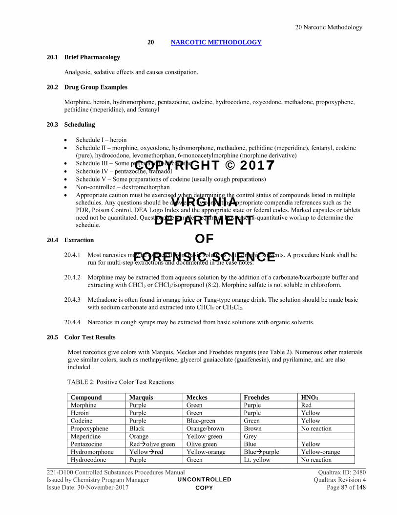
20 Narcotic Methodology
221-D100 Controlled Substances Procedures Manual Qualtrax ID: 2480Issued by Chemistry Program Manager Qualtrax Revision 4Issue Date: 30-November-2017 Page 87 of 148
20 NARCOTIC METHODOLOGY 20.1 Brief Pharmacology
Analgesic, sedative effects and causes constipation.
20.2 Drug Group Examples
Morphine, heroin, hydromorphone, pentazocine, codeine, hydrocodone, oxycodone, methadone, propoxyphene, pethidine (meperidine), and fentanyl
20.3 Scheduling
Schedule I – heroin Schedule II – morphine, oxycodone, hydromorphone, methadone, pethidine (meperidine), fentanyl, codeine
(pure), hydrocodone, levomethorphan, 6-monoacetylmorphine (morphine derivative) Schedule III – Some preparations of codeine Schedule IV – pentazocine, tramadol Schedule V – Some preparations of codeine (usually cough preparations) Non-controlled – dextromethorphan Appropriate caution must be exercised when determining the control status of compounds listed in multiple
schedules. Any questions should be answered by consulting appropriate compendia references such as the PDR, Poison Control, DEA Logo Index and the appropriate state or federal codes. Marked capsules or tablets need not be quantitated. Questionable samples require at least a semi-quantitative workup to determine the schedule.
20.4 Extraction
20.4.1 Most narcotics may be extracted from basic solutions with organic solvents. A procedure blank shall be
run for multi-step extractions and documented in the case notes.
20.4.2 Morphine may be extracted from aqueous solution by the addition of a carbonate/bicarbonate buffer and extracting with CHCl3 or CHCl3/isopropanol (8:2). Morphine sulfate is not soluble in chloroform.
20.4.3 Methadone is often found in orange juice or Tang-type orange drink. The solution should be made basic
with sodium carbonate and extracted into CHCl3 or CH2Cl2.
20.4.4 Narcotics in cough syrups may be extracted from basic solutions with organic solvents.
20.5 Color Test Results
Most narcotics give colors with Marquis, Meckes and Froehdes reagents (see Table 2). Numerous other materials give similar colors, such as methapyrilene, glycerol guaiacolate (guaifenesin), and pyrilamine, and are also included.
TABLE 2: Positive Color Test Reactions
Compound Marquis Meckes Froehdes HNO3 Morphine Purple Green Purple Red Heroin Purple Green Purple Yellow Codeine Purple Blue-green Green Yellow Propoxyphene Black Orange/brown Brown No reaction Meperidine Orange Yellow-green Grey Pentazocine Redolive green Olive green Blue Yellow Hydromorphone Yellowred Yellow-orange Bluepurple Yellow-orange Hydrocodone Purple Green Lt. yellow No reaction
UNCONTROLLED COPY
COPYRIGHT © 2017
VIRGINIA DEPARTMENT
OF FORENSIC SCIENCE

20 Narcotic Methodology
221-D100 Controlled Substances Procedures Manual Qualtrax ID: 2480Issued by Chemistry Program Manager Qualtrax Revision 4Issue Date: 30-November-2017 Page 88 of 148
Compound Marquis Meckes Froehdes HNO3 Oxycodone Yellowpurple Yellowolive Yellow No reaction
Methadone Slow pink Yellow-greengreen
Methapyrilene Purple Purple Purple No reaction
Guaifenesin Reddish purple Green/purple Green with purple streaks
Yellow
Pyrilamine Purple Purple Purple 20.6 TLC
20.6.1 Baths: TLC1, TLC2, TLC3, TLC4 and TLC5 are recommended.
20.6.2 Detection sprays:
20.6.2.1 Iodoplatinate
20.6.2.2 Ceric Sulfate - Fentanyl may require the use of ceric sulfate as an overspray due to the minute
amounts of this material found in most pharmaceutical preparations. 20.7 Dextromethorphan Enantiomer Determination
20.7.1 Because of the scheduling differences between levomethorphan (Schedule II) and dextromethorphan
(non-controlled), a microcrystal test must be performed for differentiation unless found in a recognizable pharmaceutical preparation.
20.7.2 Materials:
Glass slides and coverslips Polarizing microscope 10% Platinic Chloride solution in water (w/v) 1% acetic acid solution in water Dextromethorphan reference standard
20.7.3 Procedure
20.7.3.1 As a negative control/blank, place one drop of 1% acetic acid in water on a glass slide. Then
add a portion of a drop of 10% platinum chloride in water and cover. No crystals should form within 2 minutes.
20.7.3.1.1 If crystals form within 2 minutes, discard slide and cover slip, if used, and
repeat. 20.7.3.1.2 If the results of the second blank are unacceptable, the analyst should take steps
to resolve the issue (e.g., remaking reagents) prior to any analysis.
20.7.3.2 Place one drop of 1% acetic acid in water to a small portion (less than 1mg of pure dextromethorphan is needed) of the dextromethorphan standard on a glass slide. Then add a portion of a drop of 10% platinum chloride in water and cover. Feathery dendrites will form within 2 minutes.
20.7.3.3 Place one drop of 1% acetic acid in water to a small portion of the sample on a glass slide.
Then add a portion of a drop of 10% platinum chloride in water and cover. Feathery dendrites will form when the sample is a pure enantiomer. A racemic mixture will not form these microcrystals.
UNCONTROLLED COPY
COPYRIGHT © 2017
VIRGINIA DEPARTMENT
OF FORENSIC SCIENCE
20 Narcotic Methodology
221-D100 Controlled Substances Procedures Manual Qualtrax ID: 2480Issued by Chemistry Program Manager Qualtrax Revision 4Issue Date: 30-November-2017 Page 89 of 148
20.7.3.4 To verify the identity as the dextro isomer, mix a small amount of sample with approximately the same amount of dextromethorphan reference standard on a glass slide. Add the acetic acid and platinum chloride solutions as stated above. Observe any microcrystalline formation. If the sample is dextromethorphan, the feathery dendrites will form; if the sample is levomethorphan or racemethorphan, no dendrite-shaped crystals will form.
20.7.3.5 The microcrystal formations should be contemporaneously verified or photographed.
Verification must be documented in the case notes with the initials of the verifier and the date.
20.7.3.6 The sample may need to be purified to allow for crystal formation. Mixtures of dextromethorphan and MDMA will need to be separated before the crystal test, as pure MDMA reacts with platinum chloride to form similar, but not the same, microcrystals.
20.7.3.7 Reference: Fulton, Charles C. Modern Microcrystal Tests for Drugs, New York: Wiley-
Interscience, 1969, pp. 58-59. 20.8 Heroin Quantitation
20.8.1 See GC ¶ 10 for general quantitation procedure.
20.8.2 Reagents:
Methylene Chloride or Chloroform Heroin HCl or Heroin HCl monohydrate Standard Dicyclohexylphthalate (DCHP) Class A volumetric flasks Analytical balance Calibrated volumetric flasks
20.8.3 Internal Standard Solution:
20.8.3.1 Prepare a sufficient volume to dilute the heroin standard solution and all samples.
20.8.3.2 Prepare a 1.5 - 2 mg/mL solution of DCHP in methylene chloride or chloroform in an
appropriate volumetric flask.
20.8.3.3 Refrigerated solutions should be allowed to return to ambient temperature prior to use.
20.8.4 Heroin Standard Solutions:
20.8.4.1 Weigh approximately 10 mg of Heroin HCl and quantitatively transfer to a calibrated 10 mL volumetric flask. Fill to mark with internal standard solution. This results in a solution of approximately 1 mg/mL Heroin HCl in internal standard solution which will serve as a check standard.
20.8.4.2 Prepare a second 2.5 mg/mL standard solution as outlined above which will serve as the
calibration standard for the one point calibration.
20.8.5 Standard Salt Form Conversion
Unless heroin hydrochloride is to be reported with its salt form, the concentration in its base form will be calculated. To convert the concentration of heroin hydrochloride monohydrate to free base, multiply the concentration (mg/mL) of heroin HCl by 0.8714 (369.4 F.B./423.9 salt). This will give the concentration of free base in the standard solution.
UNCONTROLLED COPY
COPYRIGHT © 2017
VIRGINIA DEPARTMENT
OF FORENSIC SCIENCE
20 Narcotic Methodology
221-D100 Controlled Substances Procedures Manual Qualtrax ID: 2480Issued by Chemistry Program Manager Qualtrax Revision 4Issue Date: 30-November-2017 Page 90 of 148
20.8.6 Sample Preparation
Prepare six separate sample solutions. For each, weigh 10-40 mg of sample into a 10 mL volumetric flask. Dilute with internal standard to the mark.
20.8.7 GC parameters:
Column: 15 m HP-1 capillary (0.25 mm i.d., 0.25 µm film thickness) Oven temperature: approximately 260-280 C FID temperature: 280º C
20.8.8 Linear Range
20.8.8.1 The validated linear range of the heroin method is 0.5 – 5 mg/mL.
20.8.8.2 Once the percentage purity has been calculated for the sample, verify that the calculated concentration of the sample was within the linear range of the method. If it was outside the linear range, remake the sample solutions using a more appropriate amount of material.
20.8.9 DCHP elutes prior to Heroin. Peaks between DCHP and Heroin are often due to monoacetylmorphine or
acetylcodeine.
20.8.10 Calculations, Acceptance Criteria and Reporting
See ¶¶ 10.4.4.7 - 10.4.4.10
UNCONTROLLED COPY
COPYRIGHT © 2017
VIRGINIA DEPARTMENT
OF FORENSIC SCIENCE
21 Phencyclidine (PCP) and Analog Methodology
221-D100 Controlled Substances Procedures Manual Qualtrax ID: 2480Issued by Chemistry Program Manager Qualtrax Revision 4Issue Date: 30-November-2017 Page 91 of 148
21 PHENCYCLIDINE (PCP) AND ANALOG METHODOLOGY 21.1 Brief Pharmacology
PCP is classified as a dissociative anesthetic. PCP is used as an animal tranquilizer. It is sometimes called angel dust, crystal, or hog, and known as boat or loveboat when placed on marijuana.
21.2 Scheduling
Schedule I TCP thienylcyclohexylpiperidine (thiophene analog of PCP) Schedule I PHP 1-(1-phenylcyclohexyl) pyrrolidine (pyrrolidine analog of PCP) Schedule I PPP 1-(-phenylcyclopentyl) piperidine Schedule I PCE N-(1-phenylcyclohexyl) ethylamine (N-ethyl analog of PCP) Schedule II PCP phencyclidine; 1-(1-phenylcyclohexyl) piperidine Schedule II PCC 1-piperidinocyclohexane carbonitrile (precursor)
21.3 Extraction
21.3.1 May be extracted from basic or acidic aqueous solution with organic solvents. 21.3.2 May be dry extracted with methanol or other organic solvents. 21.3.3 Plant material samples may be extracted with a suitable solvent (e.g., hexane, methanol) and the extract
de -colorized by passing it through a pre-washed Pasteur pipette in which activated charcoal has been placed over a plug of glass wool. The resulting solution may be concentrated and used for further testing. A procedure blank shall be run and documented in the case notes.
21.4 Color Test Results
Co(SCN)2 Results – blue
21.5 TLC
21.5.1 Baths: TLC1, TLC2, TLC3, TLC4 and TLC5 are recommended.
21.5.1.1 TLC1 is recommended for separating the PCP analogs.
21.5.2 Detection methods:
21.5.2.1 Does not show well under UV light due to weak quenching.
21.5.2.2 Detection spray - iodoplatinate 21.6 GC
Analogs can be separated by GC at appropriate temperatures. See GC file for conditions. 21.7 FTIR
Basic extract often results in an oil which may be run as a smear between salt plates.
21.8 PCP Quantitation
21.8.1 See GC ¶ 10 for general quantitation procedure.
UNCONTROLLED COPY
COPYRIGHT © 2017
VIRGINIA DEPARTMENT
OF FORENSIC SCIENCE
21 Phencyclidine (PCP) and Analog Methodology
221-D100 Controlled Substances Procedures Manual Qualtrax ID: 2480Issued by Chemistry Program Manager Qualtrax Revision 4Issue Date: 30-November-2017 Page 92 of 148
21.8.2 Materials
Chloroform Docosane PCP or PCP HCl (quantitative standard) Class A volumetric flasks Analytical balance Calibrated volumetric flasks
21.8.3 Internal standard solution:
21.8.3.1 Prepare a sufficient volume to dilute the PCP standards and all samples.
21.8.3.2 Prepare a 1 mg/mL solution of docosane in chloroform in an appropriate volumetric flask.
21.8.3.3 Refrigerated solutions should be allowed to return to ambient temperature prior to use. 21.8.4 PCP standard solutions
21.8.4.1 Weigh approximately 10 mg of PCP and quantitatively transfer to a calibrated 10 mL volumetric flask. Fill to mark with internal standard solution. This results in a solution of approximately 1 mg/mL PCP in internal standard solution which will serve as a check standard.
21.8.4.2 Prepare a second 2.5 mg/mL standard solution as outlined above which will serve as the calibration standard for the one point calibration.
21.8.5 Standard Salt Form Conversion
Unless phencyclidine hydrochloride is to be reported with its salt form, the concentration in its base form will be calculated. To convert the concentration of phencyclidine hydrochloride to free base, multiply the concentration (mg/mL) of PCP HCl by 0.8696 (243.4 F.B./279.9 salt). This will give the concentration of free base in the standard solution.
21.8.6 Sample preparation
Prepare six separate sample solutions. For each, weigh approximately 20 mg of sample into a 10 mL volumetric flask. Fill to the mark with internal standard solution.
21.8.7 GC Parameters
Column: 15 m HP-1 capillary or equivalent (0.25 mm i.d., 0.25 µm film thickness) Oven temperature: approximately 210 ºC isothermal FID temperature: 280 ºC
21.8.8 Linear Range
21.8.8.1 The validated linear range of the PCP method is 0.5 – 5.0 mg/mL.
21.8.8.2 Once the percentage purity has been calculated for the sample, verify that the calculated concentration of the sample was within the linear range of the method. If it was outside the linear range, remake the sample solutions using a more appropriate amount of material.
21.8.9 PCP elutes prior to docosane.
UNCONTROLLED COPY
COPYRIGHT © 2017
VIRGINIA DEPARTMENT
OF FORENSIC SCIENCE
21 Phencyclidine (PCP) and Analog Methodology
221-D100 Controlled Substances Procedures Manual Qualtrax ID: 2480Issued by Chemistry Program Manager Qualtrax Revision 4Issue Date: 30-November-2017 Page 93 of 148
21.8.10 Calculations, Acceptance Criteria and Reporting See ¶¶ 10.4.4.7 - 10.4.4.10
UNCONTROLLED COPY
COPYRIGHT © 2017
VIRGINIA DEPARTMENT
OF FORENSIC SCIENCE

22 Lysergic Acid Diethylamide (LSD) Methodology
221-D100 Controlled Substances Procedures Manual Qualtrax ID: 2480Issued by Chemistry Program Manager Qualtrax Revision 4Issue Date: 30-November-2017 Page 94 of 148
22 LYSERGIC ACID DIETHYLAMIDE (LSD) METHODOLOGY 22.1 Scheduling
Schedule I – LSD
22.2 Analog
LAMPA (Lysergic acid methyl propyl amide) is a positional isomer of LSD and is included in Schedule I.
22.3 Extraction
22.3.1 LSD may be dry extracted with Methanol from blotter paper and other matrices. 22.3.2 LSD can be extracted from basic aqueous solution with organic solvents. 22.3.3 It may be necessary to dissolve the samples in a MeOH/CHCl3 mixture to extract LSD out of samples
like plastic “window panes.” 22.3.4 If samples are in a matrix which is impervious to organic solvents, LSD may be extracted by creating the
tartrate salt, followed by base extraction. A procedure blank shall be run and documented in the case notes.
22.3.4.1 Procedure (Analysis of Drugs - DEA publication):
Soak sample in 1% aqueous tartaric acid solution. Extract acid portion with CHCl3. (Discard CHCl3) Make basic with sodium bicarbonate powder. Extract into CHCl3 for further analysis.
22.4 Color Test Results
p-DMAB (Erhlich’s or Van Urk’s) – purple or blue
22.5 TLC
22.5.1 Baths: The TLC8 system is useful to distinguish LSD from LAMPA. 22.5.2 Detection Methods
22.5.2.1 LSD fluoresces blue under long wave UV light.
22.5.2.2 Detection spray - p-DMAB (Ehrlich’s or Van Urk’s), it may be necessary to heat the plate to
get good visualization (Blue spot on a white background). 22.6 GC
22.6.1 Very small samples may require reduced split ratio or splitless injection techniques. 22.6.2 LSD and LAMPA separate well on GC.
22.7 FTIR
Not usually possible because of the small quantity of LSD present in samples.
UNCONTROLLED COPY
COPYRIGHT © 2017
VIRGINIA DEPARTMENT
OF FORENSIC SCIENCE
23 Mescaline Methodology
221-D100 Controlled Substances Procedures Manual Qualtrax ID: 2480Issued by Chemistry Program Manager Qualtrax Revision 4Issue Date: 30-November-2017 Page 95 of 148
23 MESCALINE METHODOLOGY 23.1 Scheduling
23.1.1 Schedule I – Mescaline (3, 4, 5-Trimethoxyphenylethylamine), which is found in Peyote buttons. 23.1.2 The chemical, rather than the botanical, is controlled.
23.2 Extraction
Dry button and soak in methanol. Filter off plant material prior to analysis.
23.3 Color Test Results
23.3.1 Marquis- orange
23.3.2 Meckes – green dark brown
23.3.3 Froehdes- green blue
23.3.4 HNO3 - bright red
23.4 TLC
23.4.1 Baths: TLC1, TLC2, TLC3, TLC4 and TLC5 are recommended.
23.4.2 Detection spray – iodoplatinate
UNCONTROLLED COPY
COPYRIGHT © 2017
VIRGINIA DEPARTMENT
OF FORENSIC SCIENCE
24 Psilocybin and Psilocyn Methodology
221-D100 Controlled Substances Procedures Manual Qualtrax ID: 2480Issued by Chemistry Program Manager Qualtrax Revision 4Issue Date: 30-November-2017 Page 96 of 148
24 PSILOCYBIN AND PSILOCYN METHODOLOGY 24.1 Scheduling
24.1.1 Schedule I – psilocybin and psilocyn, which are found in mushrooms. 24.1.2 The chemicals, rather than the botanical, are controlled.
24.2 Extractions
24.2.1 Dry sample (in drying oven or microwave). Grind and soak in methanol for a period of 1 – 24 hours. Filter off mushroom material prior to analysis. This extraction method will allow for the analysis of either psilocyn or psilocybin.
24.2.2 Acetic Acid Extraction Technique (recommended for mushrooms in chocolate or other matrices). This
extraction will allow for the analysis of psilocyn. A procedure blank shall be run and documented in the case notes.
Dry up to approximately 3 grams material, as sample allows. (Easier to grind when dry.) Sample may be ground to increase extraction efficiency. Let soak in 6% acetic acid for 30 minutes - 1 hour. Filter off insoluble material. Extract acid portion with three aliquots of CHCl3* (Discard CHCl3.) Basify acid portion with concentrated NH4OH to pH 8 - 10. Extract basic solution with three aliquots of CHCl3* Combine aliquots of CHCl3. Evaporate CHCl3 with air (low heat). Resultant residue will yield psilocyn.
* Do not mix vigorously as an emulsion will probably form.
24.3 Color Test Results
24.3.1 Ehrlich’s- purple (positive for psilocyn and psilocybin). 24.3.2 Weber Test- psilocyn - Fast Blue B or Fast Blue BB gives red color; addition of conc. HCl gives blue
color. 24.4 TLC
24.4.1 Baths: TLC1, TLC2, TLC3, TLC4 and TLC5 are recommended. 24.4.2 Detection Sprays:
24.4.2.1 p-DMAB and HCl - reddish violet area for psilocybin, blue for psilocyn.
24.4.2.2 Weber Test (Fast Blue B or Fast Blue BB) can also be used as a TLC spray to detect psilocyn.
24.5 GC
24.5.1 GC and GC/MS will give only psilocyn due to the dephosphorylation of the psilocybin caused by the GC injection port temperatures.
24.5.2 After methanol extraction and drying, silylation with BSTFA prior to GC or GC/MS will allow
differentiation of psilocybin from psilocyn. If psilocybin is to be confirmed, derivatization is required.
UNCONTROLLED COPY
COPYRIGHT © 2017
VIRGINIA DEPARTMENT
OF FORENSIC SCIENCE
24 Psilocybin and Psilocyn Methodology
221-D100 Controlled Substances Procedures Manual Qualtrax ID: 2480Issued by Chemistry Program Manager Qualtrax Revision 4Issue Date: 30-November-2017 Page 97 of 148
24.6 FTIR
KBr pellet/ATR on extract from acetic acid extraction yields psilocyn.
UNCONTROLLED COPY
COPYRIGHT © 2017
VIRGINIA DEPARTMENT
OF FORENSIC SCIENCE

25 Cathinone Methodology
221-D100 Controlled Substances Procedures Manual Qualtrax ID: 2480Issued by Chemistry Program Manager Qualtrax Revision 4Issue Date: 30-November-2017 Page 98 of 148
25 CATHINONE METHODOLOGY 25.1 Scheduling
25.1.1 Schedule I – cathinone, which is found in Catha Edulis (Khat) 25.1.2 Schedule IV – cathine (also known as: (+)-norpseudoephedrine). 25.1.3 The chemical components, rather than the botanical, are controlled.
25.2 Sample Handling
25.2.1 Suspected freshly harvested Khat should be refrigerated upon receipt into the laboratory and subsequently analyzed as soon as possible.
25.2.2 Dried or freeze-dried samples may be refrigerated, but refrigeration is not required. 25.2.3 Cathinone can enzymatically convert to Cathine.
25.3 Extraction
A procedure blank shall be run with either extraction and documented in the case notes.
25.3.1 Suggested Khat extraction technique for freshly harvested plant material Cut up the leaves and stems to obtain the sample. The sample size should be based upon the amount
submitted, and the need to retain material for reanalysis if requested. Homogenize or sonicate the material for 5 minutes in enough 0.1N HCl to cover the sample Filter off insoluble material Basify the solution with 1N NaOH to pH 11-12 Extract basic solution with aliquots of CHCl3. Evaporate combined CHCl3 aliquots with air Reconstitute sample with an appropriate solvent, if necessary Analyze residue as soon as possible or refrigerate to avoid degradation. Resulting sample should contain Cathinone and Cathine.
25.3.2 Suggested Khat extraction technique for dried plant material
The sample size should be based upon the amount submitted, and the need to retain material for
reanalysis if requested. Dried material need not be cut up. Sonicate or soak dried material in enough 0.1N HCl to cover the sample, for 5 minutes Filter off insoluble material Basify the solution with 1N NaOH to pH 11-12 Extract basic solution with aliquots of CHCl3. Evaporate combined CHCl3 aliquots with air Reconstitute sample with an appropriate solvent, if necessary Analyze residue as soon as possible or refrigerate to avoid degradation. Resulting sample should contain Cathinone and Cathine.
25.4 TLC
25.4.1 Baths: TLC1, TLC2, TLC3, TLC4 and TLC5 are recommended. 25.4.2 Cathinone and Cathine reference standards shall be run on the plate for comparison.
UNCONTROLLED COPY
COPYRIGHT © 2017
VIRGINIA DEPARTMENT
OF FORENSIC SCIENCE
25 Cathinone Methodology
221-D100 Controlled Substances Procedures Manual Qualtrax ID: 2480Issued by Chemistry Program Manager Qualtrax Revision 4Issue Date: 30-November-2017 Page 99 of 148
25.4.3 Detection Spray: Ninhydrin/heat gives a red-brown color and can be used for visualization. Alternatively, Fluram may be used for visualization.
25.5 GC/MS
Consult standard verification files for conditions.
25.5.1 It is recommended that the Cathine standard be run along with the Cathinone standard to illustrate
spectral differences. 25.5.2 Derivatization may also be useful in increasing retention time difference between Cathinone and Cathine,
as well as providing additional spectral information. Acetylation and Silylation are recommended. 25.5.3 Chiral derivatization using l-TPC is required for the identification of Cathine.
25.6 References
25.6.1 Morselli et al., “Gas-Chromatography/Mass Spectrometry Determination of the Active Principles of (Catha Edulis) African Vegetable,” Microgram, Vol. XXV, No. 11, November 1992, pp. 290-294.
25.6.2 Lee, M. M., “The Identification of Cathinone in Khat (Catha edulis): A Time Study,” Journal of Forensic
Sciences, JFSCA, Vol. 40, No. 1, January 1995, pp. 116-121. 25.6.3 Chappell, John S., Lee, Marsha M., “Cathinone preservation in khat evidence via drying,” Forensic
Science International, 195 (2010), pp. 108-120. 25.6.4 LeBelle et al., “Gas chromatographic-mass spectrometric identification of chiral derivatives of the
alkaloids of Khat,” Forensic Science International, 61 (1993), pp. 53-64. 25.6.5 Lehmann et al., “Rapid TLC Identification Test for Khat (Catha Edulis),” Forensic Science
International, 45 (1990), pp. 47-51. 25.6.6 Ripani et al., “GC/MS identification of Catha edulis stimulant-active principles,” Forensic Science
International, 78 (1996), pp. 39-46.
UNCONTROLLED COPY
COPYRIGHT © 2017
VIRGINIA DEPARTMENT
OF FORENSIC SCIENCE

26 MDA/MDMA Methodology
221-D100 Controlled Substances Procedures Manual Qualtrax ID: 2480Issued by Chemistry Program Manager Qualtrax Revision 4Issue Date: 30-November-2017 Page 100 of 148
26 MDA/MDMA METHODOLOGY 26.1 Scheduling
Schedule I - 3,4-methylenedioxyamphetamine (MDA) Schedule I - 3,4-methylenedioxymethamphetamine (MDMA, Ecstasy) Schedule I - 3,4-methylenedioxy-N-ethylamphetamine (MDEA, Eve) Schedule I - 4-bromo-2,5-dimethoxyphenethylamine (2C-B, Nexus)
26.2 Color Tests Results
26.2.1 The sulfuric acid series of color tests generally give intense colors that undergo vivid transitions with
MDA and MDMA. These may all appear black with very concentrated samples. 26.2.2 Marquis
MDA/MDMA - dark violet black Nexus – light green green
26.2.3 Meckes
MDA/MDMA - green dark blue/violet black Nexus - yellow
26.2.4 Froehdes
MDA/MDMA - brown dark blue/violet black Nexus – yellow
26.2.5 TBPEE
MDA – purple MDMA – blue MDEA - blue Nexus – purple
26.3 TLC
26.3.1 Baths
TLC1, TLC2, TLC3, TLC4 and TLC5 are recommended. TLC9 separates methamphetamine from MDMA.
26.3.2 Detection sprays
Iodoplatinate, results may be enhanced by overspraying with Ceric Sulfate. Dragendorff Fluram visualizes MDA, Nexus and other primary amines.
26.4 GC
26.4.1 Extraction of the sample may be necessary to get good chromatography.
26.4.2 Acetyl Derivative: The acetyl derivative of MDMA-type compounds is made by drawing up 1 µL of
sample followed by 1 µL of acetic anhydride, separated by an air bubble. The acetyl derivative should
UNCONTROLLED COPY
COPYRIGHT © 2017
VIRGINIA DEPARTMENT
OF FORENSIC SCIENCE
26 MDA/MDMA Methodology
221-D100 Controlled Substances Procedures Manual Qualtrax ID: 2480Issued by Chemistry Program Manager Qualtrax Revision 4Issue Date: 30-November-2017 Page 101 of 148
have a longer retention time than the underivatized compound and may require a higher temperature than the underivatized compound.
26.5 FTIR
26.5.1 Extraction from excipients may be necessary to obtain a good spectrum. 26.5.2 GC-FTIR is a useful tool to differentiate MDMA-type compounds.
26.6 MDMA Quantitation
26.6.1 See GC ¶ 10 for general quantitation procedure. 26.6.2 Materials:
Methylene Chloride or Chloroform Octadecane 3,4-MDMA HCl 4N NaOH Calibrated mechanical pipettes Class A volumetric flasks Analytical balance
26.6.3 Internal Standard Solution:
26.6.3.1 Prepare a sufficient volume to dilute the standard solutions and all samples.
26.6.3.2 Prepare a 1 mg/mL solution of octadecane in methylene chloride or chloroform in the appropriate volumetric flask.
26.6.3.3 Refrigerated solutions should be allowed to return to ambient temperature prior to use.
26.6.4 MDMA Standard Solutions:
26.6.4.1 Prepare a 2.5 mg/mL standard solution which will serve as the calibration standard for the one point calibration.
Weigh approximately 25 mg of the standard Dilute to approximately 5 mL with D.I. or R.O. water Add five drops of 4N sodium hydroxide Use a calibrated mechanical pipette to deliver 10 mL of the internal standard solution.
Use a vessel large enough to allow vigorous vortexing. Vortex, centrifuge (if necessary), then remove organic layer for further analysis The volume of internal standard solution may be modified from the volume indicated in
the method herein, however the concentration of the standard solution must remain the same as directed
26.6.4.2 Prepare a 1 mg/mL standard solution which will serve as a check standard. Weigh at least 10
mg of standard and prepare as outlined above. The volume of internal standard solution may be modified from the volume indicated in
the method herein, however the concentration of the standard solution must remain the same as directed
UNCONTROLLED COPY
COPYRIGHT © 2017
VIRGINIA DEPARTMENT
OF FORENSIC SCIENCE
26 MDA/MDMA Methodology
221-D100 Controlled Substances Procedures Manual Qualtrax ID: 2480Issued by Chemistry Program Manager Qualtrax Revision 4Issue Date: 30-November-2017 Page 102 of 148
26.6.5 Standard Salt Form Conversion
Unless MDMA HCl is to be reported with its salt form, the concentration in its base form will be calculated. To convert the concentration of MDMA HCl to free base, multiply the concentration (mg/mL) of MDMA HCl by 0.8413 (193.25 F.B./229.71 HCl). This will give the concentration of free base in the standard solution.
26.6.6 Sample Preparation:
Prepare six separate sample solutions for analysis. Weigh approximately 20 – 40 mg of the sample Dilute to approximately 5 mL with D.I. or R.O. water Add five drops of 4N sodium hydroxide Use a calibrated mechanical pipette to deliver 10 mL of the internal standard solution Vortex, centrifuge (if necessary), then remove organic layer for further analysis The volume of internal standard solution may be modified from the volume indicated in the method
herein, however the sample weight shall be adjusted proportionally
26.6.7 GC parameters:
Column: 15 m HP-1 capillary (0.25 mm i.d, 0.25 µm film thickness) Oven temperature: approximately 170 - 240ºC FID temperature : 280ºC
26.6.8 Linear Range
26.6.8.1 The validated linear range of the MDMA method is 0.5 – 5 mg/mL.
26.6.8.2 Once the percentage purity has been calculated for the sample, verify that the calculated concentration of the sample was within the linear range of the method. If it was outside the linear range, remake the sample solutions using a more appropriate amount of material.
26.6.9 Octadecane comes out after MDMA. 26.6.10 Calculations, Acceptance Criteria and Reporting
See ¶¶ 10.4.4.7 - 10.4.4.10
UNCONTROLLED COPY
COPYRIGHT © 2017
VIRGINIA DEPARTMENT
OF FORENSIC SCIENCE
27 Anabolic Steroid Methodology
221-D100 Controlled Substances Procedures Manual Qualtrax ID: 2480Issued by Chemistry Program Manager Qualtrax Revision 4Issue Date: 30-November-2017 Page 103 of 148
27 ANABOLIC STEROID METHODOLOGY 27.1 Brief Pharmacology
Promotes muscle growth (anabolic effect)
27.2 Drug Group Examples
Testosterone (associated esters), stanozolol, boldenone
27.3 Scheduling
Anabolic steroids are listed in Schedule III.
27.4 Extraction
27.4.1 May be dry extracted into methanol or other organic solvents. 27.4.2 Injectables are often found in oils which may be extracted with methanol for further analysis.
27.5 Color Test Results
There are no good screening tests for steroids.
27.6 Pharmaceutical Identifiers
Many substituted or negative preparations are encountered which make pharmaceutical identifiers less useful than with other types of preparations. They should not be ignored, but may need to be discounted.
27.7 TLC
27.7.1 Baths: TLC6 separates many anabolic steroids. TLC1, TLC2, TLC3, TLC4 and TLC5 are also
recommended. 27.7.2 Detection methods:
UV Sulfuric Acid/Ethanol Reagent for steroids Iodoplatinate will visualize Stanozolol KMnO4 will visualize steroids with unsaturated bonds
27.8 GC
Some of these materials will require elevated temperatures and have long retention times. Special derivatizing techniques may assist chromatographic performance.
27.9 FTIR
May need additional extraction to eliminate oils.
27.10 GC/MS
Molecular weights may exceed 500 and the usual mass spectral mass range would then need to be extended.
27.11 References
27.11.1 Chiong et al. "The Analysis and Identification of Steroids", Journal of Forensic Sciences, March 1992.
UNCONTROLLED COPY
COPYRIGHT © 2017
VIRGINIA DEPARTMENT
OF FORENSIC SCIENCE
27 Anabolic Steroid Methodology
221-D100 Controlled Substances Procedures Manual Qualtrax ID: 2480Issued by Chemistry Program Manager Qualtrax Revision 4Issue Date: 30-November-2017 Page 104 of 148
27.11.2 Walters et al. "Analysis of Illegally Distributed Anabolic Steroid Products", JAOAC, Vol. 73, No. 6, 1990.
UNCONTROLLED COPY
COPYRIGHT © 2017
VIRGINIA DEPARTMENT
OF FORENSIC SCIENCE
28 GHB Methodology
221-D100 Controlled Substances Procedures Manual Qualtrax ID: 2480Issued by Chemistry Program Manager Qualtrax Revision 4Issue Date: 30-November-2017 Page 105 of 148
28 GHB METHODOLOGY 28.1 Brief Pharmacology
Central nervous system depressant
28.2 Drug Group Examples
28.2.1 Gamma hydroxybutyric acid (GHB), gamma-butyrolactone (GBL) and 1,4-butanediol (BD) 28.2.2 Other names for gamma hydroxybutyric acid include gamma hydroxybutyrate; 4-hydroxybutyrate; 4-
hydroxybutanoic acid; sodium oxybate; and sodium oxybutyrate. 28.3 Scheduling
Schedule I : GHB (not found in an approved drug product) Schedule III : Any drug product containing gamma hydroxybutyric acid, including its salts, isomers, and salts
of isomers, for which an application is approved under section 505 of the Federal Food, Drug, and Cosmetic Act
Enhanced penalty: Possession and distribution of gamma-butyrolactone and 1,4-butanediol when intended for human consumption is a Class 3 felony.
28.4 Chemical Properties
28.4.1 GHB: Pure GHB is a white powder. It is encountered dissolved into various liquids. 28.4.2 GBL: Pure GBL is a clear liquid. It is encountered dissolved into various liquids. 28.4.3 BD: Pure BD is a viscous clear liquid.
28.5 pH
If the sample is in a liquid form, take the pH of the solution prior to beginning analysis. GHB is generally found in basic solutions while GBL is generally found in acidic solutions. However, equilibrium occurs between the two in solution.
28.6 Color Test Results
28.6.1 Ferric Chloride – GHB will turn red-brown (Results can vary depending on sample pH and liquid matrix.
Therefore, further screening is necessary.) 28.6.2 GHB Color Test #3 (Smith Test) – GHB – immediate green
28.7 TLC
28.7.1 Bath: TLC3 and TLC10 (Ethyl Acetate) are recommended. 28.7.2 Detection: Iodine Vapors Results (in TLC10)
GHB off-white spot at origin GBL brown spot near solvent front BD off-white to brown bearding spot midway up plate
28.8 GC
28.8.1 GHB will form gamma-butyrolactone (GBL) in the heated injection port. The silyl derivative, prepared
prior to injection, is required to differentiate GHB from GBL.
UNCONTROLLED COPY
COPYRIGHT © 2017
VIRGINIA DEPARTMENT
OF FORENSIC SCIENCE

28 GHB Methodology
221-D100 Controlled Substances Procedures Manual Qualtrax ID: 2480Issued by Chemistry Program Manager Qualtrax Revision 4Issue Date: 30-November-2017 Page 106 of 148
28.8.1.1 If the solution contains a mixture of GBL and GHB, perform multiple chloroform rinses of the solution to remove the GBL prior to derivatization. GHB is not soluble in chloroform.
28.8.1.1.1 This chloroform extract containing GBL can be used for GC retention time
comparison with a GBL standard.
28.8.1.2 Monitor the chloroform extracts by GC/MS to see when the majority of the GBL has been removed. After performing the extracts, dry down your sample under an air stream and/or in a vacuum oven without heat. Then, derivatize your sample with BSTFA with 1% TMCS.
28.8.2 BD does not chromatograph well and may breakdown (lose water) in the heated injection port.
28.9 GC/MS
28.9.1 GHB and BD - Derivatize dry sample with BSTFA with 1% TMCS. See MS file for conditions. 28.9.2 GBL: A chloroform extract of a liquid containing GBL can be used to confirm GBL by GC/MS.
28.10 FTIR
28.10.1 GHB: Direct KBr pellet or ATR on powder sample for GHB. 28.10.2 BD: Light liquid smear on prepared KBr pellet or ATR. 28.10.3 GBL: Light liquid smear on prepared KBr pellet or ATR. If results are unfavorable, perform a
chloroform extract of liquid. Use this extract to prepare either a liquid smear on a prepared KBr pellet or dry it on KBr prior to making a pellet. Alternatively, the ATR may be used.
28.10.4 GHB/GBL Mixtures:
For liquid mixture samples of GHB and GBL, it may be necessary to separate the GHB from the matrix and/or accompanying GBL using preparatory TLC. Streak the origin of a TLC plate with the liquid sample, run in TLC10, vacuum/scrape off silica gel at origin, and elute GHB from silica gel with multiple rinses of the same methanol extract. Dry down the methanol extract and prepare a KBr pellet of the resultant powder.
28.11 References
28.11.1 Ciolino, L. A. et al. “The Chemical Interconversion of GHB and GBL” Forensic Issues and Implications” Journal of Forensic Sciences, 2001, Vol. 46, No. 6, pp. 1315-1323.
28.11.2 Bommarito, C. “Analytical Profile of Gamma-Hydroxybutyric Acid (GHB)” Journal of the Clandestine
Laboratory Investigating Chemists Association, Vol. 3, No. 3, 1993. 28.11.3 Chappell, J. S. “The Non-equilibrium Aqueous Solution Chemistry of Gamma-Hydroxybutyric Acid”
Journal of the Clandestine Laboratory Investigating Chemists Association, Vol. 12, No. 4, 2002.
UNCONTROLLED COPY
COPYRIGHT © 2017
VIRGINIA DEPARTMENT
OF FORENSIC SCIENCE
29 Salvinorin A Methodology
221-D100 Controlled Substances Procedures Manual Qualtrax ID: 2480Issued by Chemistry Program Manager Qualtrax Revision 4Issue Date: 30-November-2017 Page 107 of 148
29 SALVINORIN A METHODOLOGY 29.1 Scheduling
29.1.1 Schedule I – Salvinorin A, which is found in Salvia divinorum
29.1.2 The chemical, rather than the botanical, is controlled. 29.2 Extractions
29.2.1 Dry sample (in drying oven or microwave) if not already in a dried form.
29.2.2 Soak approximately 50-100 mg of plant material in CH2Cl2 for at least 30 minutes. CH2Cl2 is the recommended solvent as it is the most efficient extraction solvent of Salvinorin A and the least efficient extraction solvent of other plant component interferences. Chloroform and methanol may also be used. Hexane does not effectively extract Salvinorin A and basic extractions may hydrolyze the ester groups on other salvinorins present and should be avoided. A procedure blank shall be run for multi-step extractions and documented in the case notes.
29.2.3 For smaller samples or suspected weak samples, sonicating the plant material in the solvent may help
increase the efficiency of the extraction.
29.2.4 Filter off plant material prior to analysis.
29.2.5 Concentrate the extraction solvent into an autosampler vial for analysis.
29.2.6 Residues
29.2.6.1 Rinse suspected residues with CH2Cl2 and concentrate into an autosampler vial insert.
29.2.6.2 A reduced split of 20:1 on GC and GC/MS may be necessary to concentrate the sample sufficiently for identification. Vials should be returned with the evidence as outlined in ¶ 5.7.2.
29.3 TLC
29.3.1 Baths: TLC14 (see ¶ 9.4) separates Salvinorin A from Salvinorin B/D and C (see reference 29.6.4). Use TLC1 or TLC2 for the additional bath.
29.3.2 Detection Sprays:
29.3.2.1 Vanillin spray (see ¶ 9.5.16)
For plate developed in the basic TLC2 bath, spray plate with 6N HCl prior to Vanillin. Generously spray Vanillin on developed and dried plate Heat with heat gun or in oven to 110 ºC for several minutes Pinkish purple spots will develop for salvinorins
29.4 GC and GC/MS
29.4.1 GC and GC/MS
Split 50:1 or less HP-5 or HP-1 or equivalent Approximate temperature range - 240-300 degrees C at 30/min MS scan range - 500-14 Da.
UNCONTROLLED COPY
COPYRIGHT © 2017
VIRGINIA DEPARTMENT
OF FORENSIC SCIENCE

29 Salvinorin A Methodology
221-D100 Controlled Substances Procedures Manual Qualtrax ID: 2480Issued by Chemistry Program Manager Qualtrax Revision 4Issue Date: 30-November-2017 Page 108 of 148
29.4.2 Salvinorin A is the most abundant salvinorin in Salvia divinorum. The other salvinorins are much less concentrated and will elute on either side of Salvinorin A.
29.5 Reporting
See ¶ 33.
29.6 References
29.6.1 Giroud C, Felber F, Augsburger M, Horisberger B, Rivier L, Mangin P. “Salvia divinorum: an hallucinogenic mint which might become a new recreational drug in Switzerland”. Forensic Science International 112(2000) 143-150.
29.6.2 Jermain JD. “Analyzing Salvia divinorum and its active ingredient Salvinorin A utilizing thin layer
chromatography and gas chromatography/mass spectrometry”. Permission from author March 2008, CLIC list.
29.6.3 Medana C, Massolino C, Pazzi M, Baiocchi C. “Determination of salvinorins and divinatorins in Salvia
divinorum by liquid chromatography/multistage mass spectrometry”. Rapid Commun. Mass Spectrum 2006; 20:131-136.
29.6.4 Siebert DJ. “Localization of Salvinorin A and Related Compounds in Glandular Trichomes of the
Psychoactive Sage, Salvia divinorum”. Annuals of Botany 2004;93:763-771.
29.6.5 Wolowich WR, Perkins AM, and Cienki JD. “Analysis of the Psychoactive Terpenoid Salvinorin A content in five Salvia divinorum herbal products”. Pharmacotherapy 2006;26(9): 1268-1272.
UNCONTROLLED COPY
COPYRIGHT © 2017
VIRGINIA DEPARTMENT
OF FORENSIC SCIENCE
30 Cannabimimetic Agent Methodology
221-D100 Controlled Substances Procedures Manual Qualtrax ID: 2480Issued by Chemistry Program Manager Qualtrax Revision 4Issue Date: 30-November-2017 Page 109 of 148
30 CANNABIMIMETIC AGENT METHODOLOGY 30.1 Scheduling
30.1.1 Code of Virginia, §54.1-3446(6) defines synthetic cannabinoids as “cannabimimetic agents” that includes compounds that are either listed specifically or fall within one of the defined structural classes.
30.1.1.1 Structural classes
2-(3-hydroxycyclohexyl)phenol with substitution at the 5-position of the phenolic ring by alkyl or alkenyl, whether or not substituted on the cyclohexyl ring to any extent
3-(1-naphthoyl)indole or 1H-indol-3-yl-(1-napthyl)methane with substitution at the
nitrogen atom of the indole ring, whether or not further substituted on the indole ring to any extent, whether or not substituted on the naphthoyl or naphthyl ring to any extent
3-(1-naphthoyl)pyrrole with substitution at the nitrogen atom of the pyrrole ring, whether
or not further substituted in the pyrrole ring to any extent, whether or not substituted on the naphthoyl ring to any extent
1-(1-naphthylmethyl)indene with substitution of the 3-position of the indene ring,
whether or not further substituted in the indene ring to any extent, whether or not substituted on the naphthyl ring to any extent
3-phenylacetylindole or 3-benzoylindole with substitution at the nitrogen atom of the
indole ring, whether or not further substituted in the indole ring to any extent, whether or not substituted on the phenyl ring to any extent
3-cyclopropoylindole with substitution at the nitrogen atom of the indole ring, whether or
not further substituted on the indole ring to any extent, whether or not substituted on the cyclopropyl ring to any extent
3-adamantoylindole with substitution at the nitrogen atom of the indole ring, whether or
not further substituted on the indole ring to any extent, whether or not substituted on the adamantyl ring to any extent
N-(adamantyl)-indole-3-carboxamide with substitution at the nitrogen atom of the indole
ring, whether or not further substituted on the indole ring to any extent, whether or not substituted on the adamantyl ring to any extent
N-(adamantyl)-indazole-3-carboxamide with substitution at a nitrogen atom of the
indazole ring, whether or not further substituted on the indazole ring to any extent, whether or not substituted on the adamantyl ring to any extent.
30.1.2 The compounds themselves, rather than the herbal blends in which they are commonly found, are
controlled.
30.1.3 Under §54.1-3456, analogs of the listed cannabimimetic agents are also subject to the same criminal penalties.
30.2 Extractions
Extract sample into a suitable solvent (e.g., methanol, CHCl3 or hexane).
UNCONTROLLED COPY
COPYRIGHT © 2017
VIRGINIA DEPARTMENT
OF FORENSIC SCIENCE
30 Cannabimimetic Agent Methodology
221-D100 Controlled Substances Procedures Manual Qualtrax ID: 2480Issued by Chemistry Program Manager Qualtrax Revision 4Issue Date: 30-November-2017 Page 110 of 148
30.3 TLC
30.3.1 Baths: TLC1, TLC2, or TLC5 are recommended.
30.3.2 Detection Sprays:
KMnO4 (Ceric Sulfate or 6 N HCl may be used as an overspray) Fast Blue B or BB overspray with 6 N HCl
30.3.3 Due to the limitations of TLC in distinguishing isomers, two system GC must be run to determine if
more than one isomer is present in the sample. 30.4 GC and GC/MS
30.4.1 Two system GC must be utilized.
30.4.2 GC and GC/MS
Split 60:1 Columns: HP-5, HP-35 ((35% phenyl)-methylpolysiloxane) and HP-1 or equivalent Approximate temperature range 225-300 degrees C at 30/min, although broader temperature ranges
may be indicated MS scan range, 500-14 Da or 600-14 Da
30.5 References
30.5.1 Rainer Lindigkeit, Anja Boehme, Ina Eiserloh, Maike Luebbecke, Marion Wiggermann, Ludger Ernst, Till Beuerle, “Spice: A Never Ending Story?”, Forensic Science International, 191 (2009), pp. 58-63.
30.5.2 Journal of Mass Spectrometry (2009), JMS Letter via www.interscience.wiley.com/journal/jms.
30.5.3 National Forensic Laboratory Information System (NFLIS), Drug Enforcement Administration (DEA), Year 2008 Annual Report, DEA Update “Spice – Request for Information”, pg. 5.
30.5.4 “Identification of Synthetic Cannabinoids in Herbal Incense Blends by GC/MS” Application
Compendium, Agilent Technologies, 5990-7967EN, 2011.
30.5.5 Nahoko Uchiyama, Maiko Kawamura, Ruri Kikura-Hanajiri, Yukihiro Goda, “Identification and quantitation of two cannabimimetic phenylacetylindoles JWH-251 and JWH-250, and four cannabimimetic naphthoylindoles JWH-081, JWH-015, JWH-200, and JWH-073 as designer drugs in illegal products”, Forensic Toxicology, DOI 10.1007/s11419-010-0100-3 (Nov 2010).
30.5.6 Malinda Combs and Jeremiah A. Morris, “Analytical Profile of Two Synthetic Cannabinoids – JWH–018
and JWH–073”, Journal of the Clandestine Laboratory Investigating Chemists Association, Vol. 20(2) (April 2010), pp. 2-7.
30.5.7 N. Uchiyama, et al., “Chemical analysis of synthetic cannabinoids as designer drugs in herbal products”,
Forensic Sci. Int. (2010), doi:10.1016/j.forsciint.2010.01.004
30.5.8 Journal of Mass Spectrometry Letter “‘Spice’ and other herbal blends: harmless incense or cannabinoid designer drugs?” (www.interscience.com) DOI 10.1002/jms.1558, (2009).
30.5.9 Code of Virginia, §54.1-3446(6).
UNCONTROLLED COPY
COPYRIGHT © 2017
VIRGINIA DEPARTMENT
OF FORENSIC SCIENCE
31 Clandestine Laboratories
221-D100 Controlled Substances Procedures Manual Qualtrax ID: 2480Issued by Chemistry Program Manager Qualtrax Revision 4Issue Date: 30-November-2017 Page 111 of 148
31 CLANDESTINE LABORATORIES 31.1 Introduction
31.1.1 Analysis of samples collected from clandestine labs may involve the use of both routine and non-routine analytical techniques. Analysis and subsequent identification of inorganic compounds, including acids and bases, may require the transfer of certain items to Trace Evidence. For examiners qualified in both the Controlled Substances and Trace Evidence General Chemical disciplines, the results from both should be reported on one Certificate of Analysis.
31.1.2 Due to the nature of clandestine laboratories, it is not uncommon for a relatively large number of items to
be submitted. In order to determine the best analytical approach, it may be necessary to confer with a supervisor, the investigating officer, and the prosecuting attorney to assess the probative value of each piece of submitted evidence. Refer to the DFS “Evidence Handling and Laboratory Capabilities Guide” under the “Controlled Substances – Clandestine Laboratories” section for guidelines on evidence collection, evidence packaging, and evidence submission.
31.1.3 Occasionally, clandestine labs may be processed by a federal agency or in conjunction with a federal
agency. Evidence collected by federal agencies for clandestine lab cases that will be prosecuted in a Federal venue should be submitted to a federal laboratory for analysis.
31.1.4 The evidence submitted for a clandestine laboratory investigation can pose significant health hazards that
are not commonly encountered with routine controlled substance examinations. These hazards may include but are not limited to: corrosives, caustic materials, explosives, toxic gases, and flammable solvents. Due caution should be exercised when opening and examining evidence of this nature by utilizing appropriate personal protective equipment and sampling in appropriately ventilated areas (e.g., fume hood). Every effort should be made to prevent exposure of other employees to potentially hazardous materials. Special storage precautions may be necessary.
31.2 Analytical Approach
31.2.1 Ideally, the submitted items of evidence should collectively contain the necessary components to fully demonstrate either the intent to manufacture or the successful manufacture of a controlled substance. In addition to the controlled substance which is suspected to be the target product, precursors and essential chemicals should be identified when present.
31.2.1.1 When the suspected target product is methamphetamine, methcathinone, or amphetamine, it is
important to attempt to identify at least two of the substances listed in §18.2-248(J). The identification of many of the chemicals on this list will require analysis by the Trace Evidence section.
31.2.2 The evidence that has been determined to have the greatest probative value should be sampled and
analyzed first. The Drug Item Reduction Policy should be followed once sufficient analysis has been performed to support the charge on the RFLE.
31.2.3 If possible, the investigating officer should be encouraged to provide a copy of any notes or procedures
found at the clandestine laboratory scene to aide in the identification of synthetic routes. 31.3 Procedure
31.3.1 Solid Materials and Powders
31.3.1.1 Solid materials and powders should be sampled and analyzed following the schematic illustrated in ¶ 2.2 of the Procedures Manual. Samples submitted in the course of clandestine laboratory investigations may require additional analysis.
UNCONTROLLED COPY
COPYRIGHT © 2017
VIRGINIA DEPARTMENT
OF FORENSIC SCIENCE
31 Clandestine Laboratories
221-D100 Controlled Substances Procedures Manual Qualtrax ID: 2480Issued by Chemistry Program Manager Qualtrax Revision 4Issue Date: 30-November-2017 Page 112 of 148
31.3.1.2 If the sample is soluble in water, cation and anion analysis may be performed for screening purposes by using the assays for specific ions found in the U.S. Pharmacopeia.
31.3.1.3 If a solid sample or powder is soluble in water, record the pH of an aqueous solution made
from a portion of the sample.
31.3.1.4 Solid materials and powders that are most likely an inorganic essential chemical (e.g., iodine, red phosphorus) should be transferred to the Trace Evidence section for identification.
31.3.2 Liquid Samples
31.3.2.1 Liquids should be sampled and analyzed following the procedure in ¶ 2.11 of the Procedures
Manual. Samples submitted in the course of clandestine laboratory investigations may require additional analysis.
31.3.2.2 Presumptive identification of suspected ionized species in aqueous solution may be achieved
by using the assays for specific cations and anions found in the U.S. Pharmacopeia.
31.3.2.3 The miscibility of liquid samples with both water and a water insoluble solvent (e.g., CHCl3, Hexane) should be determined.
31.3.2.4 For liquids with multiple layers, care should be taken to note the number of layers, the
location of each layer relative to the others, the color and clarity of each layer. When liquid samples with multiple layers are encountered each layer shall be sampled and analyzed.
31.3.2.5 When a precipitate is discovered in a liquid sample, the precipitate should be sampled and
analyzed in addition to or in conjunction with the liquid.
31.3.2.6 Liquid samples that are most likely an essential chemical (e.g., a brown liquid in a labeled “Tincture of Iodine” bottle) should be treated as a Trace Evidence General Chemical examination or transferred to the Trace Evidence section for identification.
31.3.3 A definitive instrumental analysis technique must be employed to identify a drug, precursor or essential
chemical. The relevant section of the Procedures Manual for the suspected target drug may help to clarify potential analytical issues that arise during the course of the clandestine laboratory analysis. Significant deviations from routine analytical procedures must be documented in accordance with QM ¶ 5.3.10. Non-routine analytical procedures shall be clearly documented in the examination documentation along with proper approval from the Section Supervisor and the Chemistry Program Manager, if appropriate.
31.4 References
31.4.1 “Clandestine Lab Basic Guide” presented 12th Annual Training Seminar, Clandestine Laboratories Investigating Chemists, New Orleans, LA Sept. 4-7, 2002
31.4.2 U.S. Pharmacopeia National Formulary
UNCONTROLLED COPY
COPYRIGHT © 2017
VIRGINIA DEPARTMENT
OF FORENSIC SCIENCE
32 Estimation of the Uncertainty of Measurement (UoM)
221-D100 Controlled Substances Procedures Manual Qualtrax ID: 2480Issued by Chemistry Program Manager Qualtrax Revision 4Issue Date: 30-November-2017 Page 113 of 148
32 ESTIMATION OF THE UNCERTAINTY OF MEASUREMENT (UoM) 32.1 Scope
32.1.1 An estimation of the Uncertainty of Measurement of weight measurements shall be calculated for sample
weights that are reported on the Certificate of Analysis with the exception of gross weights.
32.1.2 An estimation of UoM shall be determined for quantitative results that are reported on the Certificate of Analysis. Current analyses which fall into this category involve methamphetamine, amphetamine, phencyclidine (PCP), cocaine, heroin, MDMA and tetrahydrocannabinol in hash oil.
32.1.3 The expanded uncertainty shall be reported to a 95.45% level of confidence. In addition, the expanded
uncertainty shall be reported to the same number of decimal places as the readability of the balance used.
32.1.4 Surrogate weights shall be weighed and recorded weekly on all balances as an ongoing component of measurement assurance. Occasional lapses in weekly measurements for personal balances (e.g., due to absences from the laboratory) are anticipated and do not require compensatory measurements. These measurements shall be evaluated annually and included in the uncertainty of measurement program.
32.2 Uncertainty Elements
32.2.1 Uncertainty Budget
32.2.1.1 Estimations of the uncertainty of measurement shall be conducted and documented using an uncertainty budget.
32.2.1.2 The uncertainty budget for a given procedure shall include both Type A standard uncertainty
and Type B standard uncertainties.
32.2.1.3 Calculations used to estimate the uncertainty and the final combined uncertainty shall be rounded using conventional rounding rules (see Quality Manual ¶ 5.4.6.3).
32.2.1.4 In order to combine the uncertainty, the units of uncertainty values should be measured in the
same units.
32.2.1.5 Uncertainty budgets shall be re-evaluated on an annual basis.
32.2.2 Type A Standard Uncertainty
32.2.2.1 Type A standard uncertainty results from measurement values being scattered in a random fashion due to laws of chance, thus has a normal or Gaussian shaped distribution.
32.2.2.2 Type A standard uncertainty is best determined by historical data of a large number of
repeated measurements.
32.2.3 Type B Standard Uncertainties
32.2.3.1 Type B standard uncertainty results from the inherent biases in measuring systems and quantitative analytical methods. These uncertainties may be reduced by optimizing the method or measuring system, but can never be completely eliminated.
32.2.3.2 Type B standard uncertainties resulting from measurement bias typically have an equal
chance of falling within a range and therefore follow a rectangular or random distribution.
32.2.3.2.1 With rectangular distribution, the range (± a) of the outer limits is used to estimate the standard deviation (σ) using the equation σ = a/√3.
UNCONTROLLED COPY
COPYRIGHT © 2017
VIRGINIA DEPARTMENT
OF FORENSIC SCIENCE
32 Estimation of the Uncertainty of Measurement (UoM)
221-D100 Controlled Substances Procedures Manual Qualtrax ID: 2480Issued by Chemistry Program Manager Qualtrax Revision 4Issue Date: 30-November-2017 Page 114 of 148
32.2.3.2.2 For example, a 10 mL volumetric flask has a tolerance of ± 0.2 mL. The calculated uncertainty associated with this measurement is 0.2/√3 or 0.115. To maintain only 2 significant figures, the uncertainty for this measurement used in the uncertainty budget is 0.12 (after rounding).
32.2.4 Determination of Combined Uncertainty
Uncertainties are combined using the Root Sum Squares technique
Ucombined = √(U1
2 + U22 + U3
2 + U42 ....)
32.2.5 Determination of expanded uncertainty and level of confidence
32.2.5.1 In order to determine the expanded uncertainty, the combined uncertainty is multiplied by the
coverage factor (k) using the equation Uexpanded = Ucombined x k
32.2.5.2 For routine measurements with a large amount of historical data (n ≥ 100), the coverage factor for 95.45% level of confidence is k=2
32.2.5.3 For analysis with reduced level of confidence due to lack of historical data, a corrected
coverage factor (kcorr) is used based on the Student’s t table.
32.2.5.3.1 For example, for an analysis with no historical control data, a control is analyzed 6 times (degrees of freedom or df = n-1, or 5 in this example).
32.2.5.3.2 Using the Student’s t table, kcorr value of 2.65 would be used to calculate the
expanded uncertainty at 2σ or 95.45% level of confidence. 32.3 Weights
32.3.1 An uncertainty budget shall be completed which will include both random (Type A) uncertainty and systematic (Type B) uncertainty for each balance.
32.3.1.1 Random uncertainty will be the standard deviation (σ) of the weight with the largest standard
deviation for each balance type from the weekly QA data.
32.3.1.2 Systematic uncertainty will include consideration of digital balance resolution, corner loading (shift test) and uncertainty of the calibration check weight. Systematic uncertainties given without a level of confidence should be treated as rectangular distributions, and as such will be divided by the √3 prior to combining. (see 36.32)
32.3.2 The combined uncertainty for the balance will be calculated using the Root Sum Squares.
32.3.3 Calculate the expanded uncertainty with a level of confidence of 95.45% by using a value of k=2. This
final uncertainty value will be used for the calculating the uncertainty of weights in casework.
32.3.4 Items with Single Specimens
The calculated expanded uncertainty is the uncertainty for that measurement.
32.3.5 Items with Multiple Specimens When weights are added to calculate a total net weight, the uncertainties associated with each individual value must be taken into account in the total uncertainty. The estimated expanded uncertainty is multiplied by the number of weighing events to determine the total expanded uncertainty. The “Controlled Substances Weighing Event Uncertainty of Measurement Calculation” form shall be used to calculate the total expanded uncertainty.
UNCONTROLLED COPY
COPYRIGHT © 2017
VIRGINIA DEPARTMENT
OF FORENSIC SCIENCE

32 Estimation of the Uncertainty of Measurement (UoM)
221-D100 Controlled Substances Procedures Manual Qualtrax ID: 2480Issued by Chemistry Program Manager Qualtrax Revision 4Issue Date: 30-November-2017 Page 115 of 148
32.4 Case File Records and Reporting for Weights
32.4.1 Weights shall be recorded and reported to reflect full balance readability.
32.4.2 Expanded uncertainty values shall be derived from the appropriate budget worksheet specific to the class of balance used.
32.4.3 The “Controlled Substances Weighing Event Uncertainty of Measurement Calculation” form shall be
used to calculate the reported total expanded uncertainty when two or more specimen weights are added together and will be stored in the case file. The worksheet shall be included in all case files for which a weighing was performed and UoM is required including single weighing events.
32.4.3.1 Calculations used to estimate measurement uncertainty shall be rounded using conventional
rounding rules (see Quality Manual ¶ 5.4.6.3).
32.4.3.2 Whenever possible, the measurement result, rounded expanded uncertainty, and associated ounce conversion, if applicable, shall be reported to the same level of significance.
32.4.3.2.1 Example:
The contents of five were analyzed separately and each was found to contain Marijuana; total net weight of the five: 12.312 ± 0.055 grams (0.434 ± 0.002 ounce) of plant material. The gross weight of the remainder was 10.314 gram(s) including innermost packaging.
32.5 Measurement Traceability
Traceability is an essential element of the Department’s measurement assurance program and is required for all measurements where uncertainty of measurement is reported. The estimation of measurement uncertainty for common controlled substances analyses is accomplished in the following manner.
32.5.1 Measurand: The weight of evidentiary samples submitted for controlled substances analysis.
32.5.2 Traceability of measurement:
32.5.2.1 Balance Calibration: Traceability is established by the annual, external calibration of balances
by an ISO/IEC 17025:2005 accredited calibration provider. Calibration certificates are maintained in the laboratory.
32.5.2.2 Steel Weights: Balances are checked weekly using traceable check weights. Check weights
are calibrated every three years by an external, ISO/IEC 17025:2005 accredited calibration provider. Calibration certificates are maintained in the laboratory.
32.5.2.3 The “Auto Cal” function of balances is approved due to the measurement traceability afforded
by the intermediate checks with calibrated weights.
32.5.3 Equipment used: 32.5.3.1 Calibrated balances (5-Place Analytical, 4-Place Analytical, 3-Place Top Loading, 2-Place
Top Loading, and High Capacity).
32.5.3.2 Calibrated, traceable steel check weights.
32.5.3.3 Bags of sand for measurement assurance.
32.5.3.4 Logbook for calibration checks.
UNCONTROLLED COPY
COPYRIGHT © 2017
VIRGINIA DEPARTMENT
OF FORENSIC SCIENCE

32 Estimation of the Uncertainty of Measurement (UoM)
221-D100 Controlled Substances Procedures Manual Qualtrax ID: 2480Issued by Chemistry Program Manager Qualtrax Revision 4Issue Date: 30-November-2017 Page 116 of 148
32.5.3.5 Spreadsheet control charts for measurement assurance.
32.6 Quantitations 32.6.1 Uncertainty shall be calculated using the Drug Quantitation Worksheet, HPLC Cannabinoid
Quantitation Worksheet or THC Quantitation Worksheet.
32.6.2 Calculate the expanded uncertainty with a level of confidence of 95.45% by using a value appropriate to the number of measurements.
32.6.3 Calculations to arrive at the reported quantitative result shall be recorded in the case notes.
32.7 References
32.7.1 ASCLD/LAB Policy on Measurement Uncertainty. (AL-PD-3060 Ver. 1.1)
32.7.2 ASCLD/LAB Guidance on the Estimation of Measurement Uncertainty – ANNEX B, Drug Chemistry
Discipline. (AL-PD-3063)
UNCONTROLLED COPY
COPYRIGHT © 2017
VIRGINIA DEPARTMENT
OF FORENSIC SCIENCE

33 Reporting Guidelines
221-D100 Controlled Substances Procedures Manual Qualtrax ID: 2480Issued by Chemistry Program Manager Qualtrax Revision 4Issue Date: 30-November-2017 Page 117 of 148
33 REPORTING GUIDELINES 33.1 Controlled substances are to be reported simply with wording consistent with the Code of Virginia.
33.1.1 The identity and schedule will be reported whenever clearly known. 33.1.2 The “show form” option will be utilized for most types of evidence including plant material, powders and
solid material. 33.1.3 Items consisting of one tablet or capsule which were chemically identified will be reported as outlined in
¶ 33.4.1. 33.1.4 Results shall be specific to the item tested. Additional language should be added to the results to clarify
which specimen was tested when more than one specimen is present in the item (e.g., the item consists of two pipes so the results clarify that only one was tested.)
33.1.5 The condition of the outer packaging shall be on the Certificate of Analysis (CoA). 33.1.6 Non-pharmaceutical compounds shall be reported using their common names as generated by LIMS. If a
compound is being reported that is not currently controlled, report the name as approved by the Chemistry Program Manager. Approved names for substantially similar compounds can be found on the shared drive.
33.2 When residue samples contain controlled substances or marijuana, the results section must reflect the term
“residue.”
33.2.1 Examples:
Marijuana, residue Heroin (Schedule I), residue
33.3 Weights will be routinely reported for controlled substances and marijuana.
33.3.1 Total weights that have been approximated will be reported as “approximate total weight”. Measurement
uncertainty will not be reported for approximate total weights. Approximated volumes should not be reported.
33.3.2 Net weights will not include any packaging and shall be reported to include the expanded uncertainty at a
95.45% level of confidence. In addition, the expanded uncertainty shall be reported to the same number of decimal places as the readability of the balance used and a statement of the level of confidence included. The level of confidence statement shall be placed on Certificates of Analysis when reporting net weights.
33.3.2.1 Example 1: The contents of three were analyzed separately and each was found to contain
Cocaine (Schedule II); total net weight of the three: 0.476 ± 0.033 gram of powder. Measurement uncertainty of weight measurements is reported at a 95.45% level of confidence.
33.3.2.2 Example 2: 25.93 ± 0.07 grams of solid material, found to contain Cocaine (Schedule II), 35.6
± 8.2% pure. Measurement uncertainty of weight and purity measurements is reported at a 95.45% level of confidence.
33.3.3 Gross weights will include innermost packaging and be clearly delineated when they are reported.
Measurement uncertainty will not be reported for gross weights.
UNCONTROLLED COPY
COPYRIGHT © 2017
VIRGINIA DEPARTMENT
OF FORENSIC SCIENCE

33 Reporting Guidelines
221-D100 Controlled Substances Procedures Manual Qualtrax ID: 2480Issued by Chemistry Program Manager Qualtrax Revision 4Issue Date: 30-November-2017 Page 118 of 148
33.3.4 Weigh and report weight of food products. Edible preparations found to contain Marijuana shall be reported as “found to contain Marijuana”.
33.4 The following are examples of report wording for typical drug items using the Administrative Sampling Plan.
33.4.1 Simple possession
33.4.1.1 Items with one specimen
Marijuana, 5.14 grams (0.18 ounce) of plant material including innermost packaging. 0.501 gram of solid material including innermost packaging, found to contain Heroin
(Schedule I). 0.254 ± 0.011 gram of solid material, found to contain Heroin (Schedule I). Tablet, found to contain Oxycodone (Schedule II) and Acetaminophen. The tablet was found to contain Oxycodone (Schedule II) and Acetaminophen.
33.4.1.2 Items with more than one specimen
Total weight: 5.14 grams (0.18 ounce) of plant material including innermost packaging.
The contents of one were analyzed and found to contain Marijuana. Total weight: 0.954 gram of solid material including innermost packaging. The contents
of one were analyzed and found to contain Cocaine (Schedule II). 33.4.2 Possession with intent to distribute or distribution
33.4.2.1 Five specimens or less
The contents of three were analyzed separately and each was found to contain Cocaine (Schedule II); total net weight of the three: 0.476 ± 0.033 gram of powder.
The contents of two were analyzed separately and each was found to contain Marijuana; total net weight of the two: 16.31 ± 0.14 grams (0.58 ± 0.01 ounce) of plant material.
Total weight: 6.31 grams (0.22 ounce) of plant material including innermost packaging. The contents of four were analyzed separately and each was found to contain Marijuana.
Total weight: 5.123 grams of powder including innermost packaging. The contents of four were analyzed separately and each found to contain Cocaine (Schedule II)
33.4.2.2 More than five specimens
The contents of five were analyzed separately and each was found to contain Cocaine
(Schedule II); total net weight of the five: 0.4732 ± 0.0135 gram of powder. The gross weight of the remainder was 2.3314 gram(s) including innermost packaging.
Total weight: 6.31 grams (0.22 ounce) of plant material including innermost packaging. The contents of five were analyzed separately and each was found to contain Marijuana.
The contents of fourteen were analyzed separately and each was found to contain Marijuana; total net weight of eleven: 13.30 ± 0.57 grams (0.469 ± 0.020 ounce) of plant material; total net weight of three: 2.8339 ± 0.0095 grams (0.0999 ± 0.0003 ounce) of plant material.
Total weight: 5.1234 grams of powder including innermost packaging. The contents of five were analyzed separately and each found to contain Cocaine (Schedule II).
33.4.2.3 More than five specimens, meeting a weight threshold
The contents of eight were analyzed separately and each was found to contain Cocaine
(Schedule II); total net weight of the eight: 30.51 ± 0.56 grams of solid material. The gross weight of the remainder was 12.32 gram(s) including innermost packaging.
UNCONTROLLED COPY
COPYRIGHT © 2017
VIRGINIA DEPARTMENT
OF FORENSIC SCIENCE

33 Reporting Guidelines
221-D100 Controlled Substances Procedures Manual Qualtrax ID: 2480Issued by Chemistry Program Manager Qualtrax Revision 4Issue Date: 30-November-2017 Page 119 of 148
The contents of six were analyzed separately and each was found to contain Marijuana; total net weight of the six: 16.34 ± 0.42 grams (0.58 ± 0.01 ounce) of plant material. The gross weight of the remainder was 12.37 gram(s) including innermost packaging.
The contents of six were analyzed separately and each was found to contain Marijuana; total net weight of the six: 16.31 ± 0.35 grams (0.58 ± 0.01 ounce) of plant material. The gross weight of the remainder was 12.34 gram(s) including innermost packaging.
33.5 The following are examples of report wording for typical drug items using the Hypergeometric Sampling Plan.
Utilizing a hypergeometric sampling plan, the contents of twenty-nine bags were analyzed separately and
each was found to contain Cocaine (Schedule II); total net weight of the twenty-nine: 2.427 ± 0.319 grams of powder. Based on these results, there is a 95% level of confidence that at least 90% of the packages contain Cocaine. The gross weight of the remainder was 12.332 gram(s) including innermost packaging. (initial submissions)
Utilizing a hypergeometric sampling plan, the contents of fourteen additional bags were analyzed separately and each was found to contain Cocaine (Schedule II); total net weight of the fourteen: 1.312 ± 0.154 grams of powder. Based on these results, there is a 95% level of confidence that at least 90% of the packages contain Cocaine. (resubmissions)
Total weight: 15.1234 grams of powder including innermost packaging. Utilizing a hypergeometric sampling plan, the contents of twenty-nine bags were analyzed separately and each was found to contain Cocaine (Schedule II). Based on these results, there is a 95% level of confidence that at least 90% of the packages contain Cocaine. (initial submissions – gross weight)
Utilizing a hypergeometric sampling plan, the contents of twenty-two additional bags were analyzed separately and each was found to contain Cocaine (Schedule II). Based on these results, there is a 95% level of confidence that at least 90% of the packages contain Cocaine. (resubmissions – gross weight)
33.6 For substances in Schedule V – VI involving misdemeanor prosecutions or non-controlled substances, tablets and
capsules visually examined using only pharmaceutical identifiers will be reported following these two examples.
33.6.1 “Visual examination of the physical characteristics, including shape, color and manufacturer’s markings, was consistent with Zoloft, a Schedule VI pharmaceutical preparation containing Sertraline. There was no apparent tampering of the dosage units and no further tests are being conducted.”
33.6.2 “Visual examination of the physical characteristics, including shape, color and manufacturer’s markings,
was consistent with a Schedule VI pharmaceutical preparation containing Sertraline. There was no apparent tampering of the dosage units and no further tests are being conducted.”
33.7 For substances and preparations in Schedules IV and above, tablets and capsules which were representatively
sampled will be reported following these two examples.
33.7.1 “One dosage unit was analyzed and found to contain Diazepam, Schedule IV. Visual examination of the physical characteristics, including shape, color and manufacturer’s markings of both the analyzed and remaining dosage units, was consistent with Valium, a pharmaceutical preparation containing Diazepam. There was no apparent tampering of the dosage units.”
33.7.2 “One dosage unit was analyzed and found to contain Diazepam, Schedule IV. Visual examination of the
physical characteristics, including shape, color and manufacturer’s markings of both the analyzed and remaining dosage units, was consistent with a pharmaceutical preparation containing Diazepam. There was no apparent tampering of the dosage units.”
33.8 For tablets and capsules with substances and preparations in Schedules IV and above, where the analytical results
are inconsistent with the manufacturer’s specifications with regard to content, report following these two examples.
33.8.1 “One dosage unit was analyzed and found to contain Acetaminophen (non-controlled). Visual
examination of the physical characteristics, including shape, color and manufacturer’s markings, was
UNCONTROLLED COPY
COPYRIGHT © 2017
VIRGINIA DEPARTMENT
OF FORENSIC SCIENCE
33 Reporting Guidelines
221-D100 Controlled Substances Procedures Manual Qualtrax ID: 2480Issued by Chemistry Program Manager Qualtrax Revision 4Issue Date: 30-November-2017 Page 120 of 148
consistent with a Schedule II pharmaceutical preparation containing Hydrocodone and Acetaminophen; therefore, the contents were inconsistent with dosage unit labeling.”
33.8.2 “Five dosage units were analyzed separately and each was found to contain Diazepam (Schedule IV).
Visual examination of the physical characteristics, including shape, color and manufacturer’s markings, was consistent with a Schedule IV pharmaceutical preparation containing Alprazolam; therefore, the contents were inconsistent with dosage unit labeling.”
33.9 “No controlled substances found” or “No controlled substances identified” will be used to describe items where
no compounds were found or identified that are controlled substances.
33.10 “Not analyzed” will be used for items not examined chemically or visually. 33.11 “Insufficient for identification” will be used to describe items with too little sample for a complete identification. 33.12 In cases where items have been analyzed for possible tampering or substitution, the results will have an additional
statement of either “Meets label specifications” or “Does not meet label specifications”. Additional clarifying wording should be used such as “Does not meet label specifications with regard to concentration” at the discretion of the Section Supervisor.
33.12.1 Reporting Example: The contents of eight were analyzed separately and each was found to contain
Methylone (Schedule I); total net weight of the eight: 1.2345 ± 0.0218 grams of powder. Visual examination of the analyzed capsule determined that the physical characteristics were consistent with a non-prescription pharmaceutical preparation containing Diphenhydramine. Therefore the contents do not meet label specifications.
33.13 Quantitation results and measurement uncertainty shall be rounded to one (1) decimal place and reported using a
95.45% level of confidence following these examples unless specified otherwise in the method:
33.13.1 25.93 ± 0.07 grams of solid material, found to contain Cocaine (Schedule II), 35.6 ± 8.7% pure.
33.13.2 12.523 ± 0.011 grams of powder, found to contain Cocaine Hydrochloride (Schedule II), 45.8 ± 9.4% pure.
33.13.3 Hashish oil (Schedule I), found to contain 34.6 ± 9.2% tetrahydrocannabinol by weight. 33.13.4 Marijuana, 13.30 ± 0.57 grams (0.469 ± 0.020 ounce) of material found to contain less than 12%
tetrahydrocannabinol by weight.
33.13.5 The contents of five were analyzed separately and each was found to contain Methamphetamine Hydrochloride (Schedule II); total net weight of the five: 12.93 ± 0.07 grams of powder. A composite sample of the five was subsequently analyzed and found to be 45.8 ± 9.4% pure.
33.13.6 The contents of five were analyzed separately and each was found to contain Tetrahydrocannabinol. A
composite sample of the five was subsequently analyzed and found to contain 28.7 ± 10.6% Tetrahydrocannabinol by weight, Hashish oil (Schedule I).
33.13.7 The contents of five were analyzed separately and each was found to contain Marijuana, total net weight
of the five: 15.54 ± 0.25 grams (0.548 ± 0.009 ounce) of material. Quantitative analysis of a composite sample of the five found the material to contain less than 12% Tetrahydrocannabinol by weight.
33.14 Resubmissions for cocaine salt/base analysis will be reported as per the following examples:
33.14.1 “The contents of the previously examined five plastic bag corners were analyzed separately and each was found to contain Cocaine base (Schedule II).”
UNCONTROLLED COPY
COPYRIGHT © 2017
VIRGINIA DEPARTMENT
OF FORENSIC SCIENCE
33 Reporting Guidelines
221-D100 Controlled Substances Procedures Manual Qualtrax ID: 2480Issued by Chemistry Program Manager Qualtrax Revision 4Issue Date: 30-November-2017 Page 121 of 148
33.14.2 “The contents of the previously examined three ziplock bags were analyzed separately and each was found to contain Cocaine Hydrochloride (Schedule II).”
33.14.3 Solid material, found to contain Cocaine base (Schedule II). 33.14.4 The solid material was found to contain Cocaine base (Schedule II).
33.15 Substances listed as precursors in Code of Virginia §18.2-248(J) or are defined in §18.2-248(K) shall be reported
as such as per the following examples:
33.15.1 Tablet, found to contain Pseudoephedrine (a listed substance in §18.2-248(J)). 33.15.2 Liquid, found to contain Ephedrine/Pseudoephedrine (a listed substance in §18.2-248(J)).
33.16 Cannabimimetic agents that are specifically listed by both their chemical and common name in the Code of
Virginia, or pursuant to Board of Pharmacy Regulation, shall be reported using their common names as generated by LIMS. Cannabimimetic agents will be reported as per the following examples: 33.16.1 When a scheduled cannabimimetic agent is present: “0.254 ± 0.011 gram of powder, found to contain
JWH-018 (a Schedule I cannabimimetic agent listed in § 54.1-3446(6)(b)).” 33.16.2 When a compound within a defined structural class is present (but not specifically listed): “0.254 ± 0.011
gram of plant material, found to contain JWH-015 (Schedule I). This compound is a cannabimimetic agent as defined in § 54.1-3446(6)(a) and is within the structural class 3-(1-naphthoyl)indole.”
33.16.3 When a Non-Listed/Non-Structural Class cannabimimetic agent may be present, but is not identified:
“No controlled substances identified.”
33.16.4 When a mixture of compounds is present (where one is within a defined structural class but not specifically listed along with a listed compound): “0.254 ± 0.011 gram of plant material, found to contain a mixture of cannabimimetic agents: JWH-018 (a Schedule I cannabimimetic agent listed in § 54.1-3446(6)(b)) and JWH-015, a Schedule I cannabimimetic agent as defined in § 54.1-3446(6)(a) and is within the structural class 3-(1-naphthoyl)indole.”
33.17 Isomers of substances listed in Schedule I will be reported as per the following examples:
33.17.1 Option 1 (the isomers can be analytically distinguished): “3-Fluoromethamphetamine (Schedule I), an isomer of 4-fluoromethamphetamine.”
33.17.2 Option 2 (the isomers have not been analytically distinguished): “4-Fluoromethamphetamine or one of its
isomers as defined in §54.1-3446(3) (Schedule I).”
33.17.3 Option 3 (isomer of a BoP scheduled compound): “Isobutyryl fentanyl (Schedule I), an isomer of Butyryl fentanyl. Butyryl fentanyl was placed into Schedule I of the Drug Control Act pursuant to Board of Pharmacy Regulation 18VAC110-20-322.”
33.18 Compounds which are substantially similar to compounds listed in Schedules I and II shall be reported as per the
following examples:
33.18.1 If the compound meets requirements as defined in ¶ 16.4.1 or 16.4.2, report as follows:
0.254 ± 0.011 gram of solid material, found to contain (Compound). The chemical structure of (compound) is substantially similar to the chemical structure of (list controlled substance and schedule). A chemical structure is considered substantially similar if there are no more than two minor substituents that have been replaced, added, removed, or extended within the chemical structure.
UNCONTROLLED COPY
COPYRIGHT © 2017
VIRGINIA DEPARTMENT
OF FORENSIC SCIENCE

33 Reporting Guidelines
221-D100 Controlled Substances Procedures Manual Qualtrax ID: 2480Issued by Chemistry Program Manager Qualtrax Revision 4Issue Date: 30-November-2017 Page 122 of 148
33.18.2 If the compound does not meet requirements as defined in ¶ 16.4.1 or 16.4.2, report the compound as: “No controlled substances found” or “No controlled substances identified”.
33.19 Compounds which have been evaluated for substantial similarity to a Schedule I or II controlled substance, in the
absence of a request from a Commonwealth’s Attorney, will be reported as per the following examples:
33.19.1 If the compound in question is mixed with a controlled substance, only the controlled substance will be reported.
33.19.2 If the compound is the only compound present in an item report as follows: “No controlled substances
identified. A substance that may be substantially similar to the chemical structure of a controlled substance in Schedule I or II was indicated. If identification of the compound is required as per § 54.1-3401 (controlled substance analog), the evidence should be resubmitted for further analysis along with a letter of request from the Office of the Commonwealth’s Attorney”.
33.20 Compounds scheduled pursuant to Board of Pharmacy Regulation shall be reported using their common names as
generated by LIMS per the following examples:
33.20.1 Cannabimimetic agents:
0.254 ± 0.011 gram of solid material, found to contain NM-2201. This compound was placed into Schedule I of the Drug Control Act as a cannabimimetic agent pursuant to Board of Pharmacy Regulation 18VAC110-20-322.
33.20.2 All other non-cannabimimetic agents:
0.254 ± 0.011 gram of solid material, found to contain 4-bromomethcathinone (4-BMC). This compound was placed into Schedule I of the Drug Control Act pursuant to Board of Pharmacy Regulation 18VAC110-20-322.
33.21 Different reporting options consistent with applicable laws may be used at the discretion of the Section
Supervisor.
UNCONTROLLED COPY
COPYRIGHT © 2017
VIRGINIA DEPARTMENT
OF FORENSIC SCIENCE
34 Drug Reversals
221-D100 Controlled Substances Procedures Manual Qualtrax ID: 2480Issued by Chemistry Program Manager Qualtrax Revision 4Issue Date: 30-November-2017 Page 123 of 148
34 DRUG REVERSALS 34.1 Introduction
The Department will assist law enforcement agencies with preparation of materials to be used in drug reversals, buy/bust scenarios, internal security setup operations, and “show and tell” drugs. In all instances, the requesting agency must assume full responsibility for distribution of these materials.
34.2 Procedure
34.2.1 The agency must make a written request on the Agency’s letterhead detailing the scope of the operation, materials needed, and projected timeframes. This request must be approved by the Section Supervisor or Chemistry Program Manager and Laboratory Director.
34.2.2 Any controlled substances must be provided by the requesting agency or a cooperative neighboring
jurisdiction.
34.2.2.1 If the material is from a case previously analyzed by the Department, this must be made clear by the agency.
34.2.3 Cutting materials must be supplied by the requesting agency and will be subject to approval by the
Section Supervisor.
34.2.3.1 The Department may provide substances added to the material for “marking” purposes. Examples of such substances are benzocaine, lidocaine and diphenhydramine.
34.2.3.2 There may be times when uncut materials are required. However, whenever possible, the
controlled substances should be diluted to approximately 1% drug by weight to allow for future analysis while enhancing the safety of the operation. This will be evaluated on a case by case basis.
34.2.4 All packaging material must be supplied by the requesting agency.
34.2.4.1 Materials will be packaged at the laboratory in accordance with the agency’s request. 34.2.4.2 Packages must allow for future testing of the material when it is re-submitted.
34.2.5 An authentic sample of the final preparation will be kept by the analyst for future comparison. All
analytical data such as weights, composition, notes, chromatograms and spectra are to remain with the authentic sample.
34.2.5.1 Samples and supporting analytical data shall be maintained for at least six years from the date
of preparation.
34.2.6 The requesting agency must submit all cases resulting from a particular operation to the attention of the analyst who prepared the material. These should be clearly marked on the RFLE.
34.2.7 The Department assumes no responsibility or liability for the security of these materials once the law
enforcement agency takes possession of them.
34.2.8 The Department assumes no responsibility or liability for any use of these materials by the requesting agency or any other person.
UNCONTROLLED COPY
COPYRIGHT © 2017
VIRGINIA DEPARTMENT
OF FORENSIC SCIENCE

35 Quality Assurance
221-D100 Controlled Substances Procedures Manual Qualtrax ID: 2480Issued by Chemistry Program Manager Qualtrax Revision 4Issue Date: 30-November-2017 Page 124 of 148
35 QUALITY ASSURANCE 35.1 Introduction
35.1.1 The purpose of this section is to provide a uniform Quality Assurance Program for the Controlled Substances Section of the Virginia Department of Forensic Science. It is to establish a baseline or reference point of reliability and system performance.
35.1.2 It is expected that the analyst will report any unacceptable or anomalous behavior of any of our analytical
systems immediately to either their Section Supervisor or the appropriate Instrument Specialist (Primary Operator). It is further expected that appropriate steps which ensure resolution of the issue will follow ASAP and be properly documented.
35.2 Reagents
35.2.1 Chemicals and solvents used in qualitative reagents should be of at least ACS reagent grade. 35.2.2 Solvents used to dissolve samples or standards should be a high quality, low residue solvent (e.g., HPLC
grade, OMNISOLV, OPTIMA). 35.2.3 Water used in reagent preparation should be either deionized (DI) or reverse osmosis (RO). 35.2.4 Stock solutions of general color test reagents and TLC sprays will be made up as needed. The
Department Reagent Worksheet shall be used to record reagent preparation. After they are made, they will be checked with the corresponding primary or secondary standard listed below in Table 3 and results, date and initials will be recorded.
35.2.5 Color test reagents and TLC sprays will be verified every three months during the shelf life of the reagent
(2 years unless otherwise noted). The Reagent QA Check Worksheet shall be used for this purpose. 35.2.6 Individual chemists may have unique reagents other than the ones listed in Table 3 and it will be their
responsibility to check them with an appropriate standard and document accordingly. For single use reagents, this documentation may be in the case file.
TABLE 3: Common Reagents and Appropriate Check Compounds
REAGENT CHECK COMPOUND Duquenois Marijuana Marquis Heroin Froehdes Heroin Meckes Heroin
Cobalt Thiocyanate Cocaine Ehrlichs LSD
Iodoplatinate Cocaine Iodoplatinate/Ceric Sulfate Caffeine
KMnO4 Acetaminophen Fast Blue B and Fast Blue BB Hash Oil or Marijuana
35.3 Standards
35.3.1 Primary and quantitative reference standards should normally be at least of United States Pharmacopeia –
National Formulary (USP-NF) quality. This applies to both powders purchased from a manufacturer/supplier and pharmaceutical preparations.
35.3.1.1 Receipt, storage and use of controlled drug standards shall be recorded and records
maintained as required by § 54.1-3404.
UNCONTROLLED COPY
COPYRIGHT © 2017
VIRGINIA DEPARTMENT
OF FORENSIC SCIENCE

35 Quality Assurance
221-D100 Controlled Substances Procedures Manual Qualtrax ID: 2480Issued by Chemistry Program Manager Qualtrax Revision 4Issue Date: 30-November-2017 Page 125 of 148
35.3.1.2 Standards used as reference materials in casework are considered critical supplies and shall be purchased from manufacturers approved by the Chemistry Program Manager.
35.3.1.2.1 The following manufacturers/vendors/suppliers are pre-approved for the
purchase of new reference standards:
USP Alltech Associates (Grace – Discovery Sciences) Cerilliant Sigma-Aldrich and its subsidiaries Steraloids, Inc. Cayman Chemicals Lipomed LGC Crescent Chemical VWR
35.3.1.2.2 Pharmaceutical preparations may be purchased from any licensed pharmacy or
the patented drug manufacturer for use as reference materials. 35.3.1.3 Primary reference standards are those purchased from a reputable manufacturer. 35.3.1.4 Secondary standards are those which are obtained or synthesized within the laboratory. These
may be from previously analyzed case samples. 35.3.1.5 Quantitative standards have a known purity, known accuracy and are purchased from a
reputable manufacturer including those listed in ¶ 35.3.1.2.1. Certified reference materials shall be used, where available, for all quantitations and be acquired from vendors accredited to ISO/IEC 17025:2005 and ISO Guide 34:2009 and include the supplier’s Certificate of Analysis.
35.3.2 For all standards obtained for drug lab use, a qualified examiner will be responsible for obtaining a mass
spectrum, IR or other suitable definitive instrumental data (data provided by the manufacturer is not sufficient, although it should be stored with lab generated data). The hard copy data will be filed. The hard copy should include the following data:
Lot# or log book code Standard name Concentration, as appropriate Amount injected (for MS only) Analyst’s initials and date
35.3.3 After the examiner gathers the data and insures that it agrees with known published spectra or that the
data is consistent with the unique compound represented from both a chemical and data interpretation perspective, label the standard bottle with "MS", "IR", etc., the date and initials. Include the source of the standard and record all pertinent information in the Standards File.
35.3.4 If an examiner needs a standard from a new lot that has not been documented in this fashion, the
examiner must perform the above procedure prior to using it for drug case work. 35.3.5 When positive results are achieved in casework, the corresponding standard(s) must be properly
documented in the case file. Standards used for TLC should be documented in the case notes, whereas standards used for instrumental methods may be documented either in the case notes or on the data. If the same standard is used for both types of tests, it only needs to be documented once in the case file.
UNCONTROLLED COPY
COPYRIGHT © 2017
VIRGINIA DEPARTMENT
OF FORENSIC SCIENCE

35 Quality Assurance
221-D100 Controlled Substances Procedures Manual Qualtrax ID: 2480Issued by Chemistry Program Manager Qualtrax Revision 4Issue Date: 30-November-2017 Page 126 of 148
35.4 Balances
35.4.1 All analytical and toploading balances will be checked weekly for accuracy using Class S-1 weights or better. Record the weights in the log book with the date and initials.
35.4.2 All high capacity balances will be checked monthly for accuracy using Class S-1 or Class F weights or
better. Record the weights in the log book with the date and initials. 35.4.3 Balances shall be calibrated by an outside vendor annually that is accredited to ISO/IEC 17025:2005 and
whose scope of accreditation covers the calibration performed. New balances shall be calibrated prior to being placed into service. Calibration certificates shall be evaluated by the Section Supervisor, Group Supervisor, or designee prior to placing the balance into service.
35.4.4 Weights used to check balance accuracy shall be re-certified every three years by an ISO/IEC
17025:2005 accredited vendor whose scope of accreditation covers the certification performed. 35.4.5 The balances listed below in Table 4 represent examples of a balance class or type along with the
corresponding check weights. If a balance does not fit into these categories, use three weights within its range as approved by the Chemistry Program Manager.
35.4.5.1 Place weigh paper or boat on balance, tare and add weight. 35.4.5.2 If a result from the performance check is outside of the acceptable range, first ensure that the
balance is level and clean prior to rechecking. 35.4.5.3 If applicable, use the internal calibration function of the balance prior to rechecking. 35.4.5.4 If a result is outside of the acceptable range after performing the actions found in 35.4.5.2 and
35.4.5.3, the balance shall be immediately taken out of service until maintenance and/or calibration are performed by an approved vendor.
TABLE 4: Balances and Appropriate Check Weights
BALANCE TYPE BALANCE EXAMPLES CHECK WEIGHTS
Analytical (dual range) Mettler XS 105
0.01000* (± 0.00005) gram 20.00000* (± 0.00005) grams
50.0000 (± 0.0005) grams 100.0000 (± 0.0005) grams
Analytical Mettler AE 160 Sartorius Basic
0.0500 (± 0.0002) gram 1.0000 (± 0.0003) gram
20.0000 (± 0.0005) grams
Toploading (± 0.01) gram
Mettler PE 2000 Mettler PE 1600 Mettler PB302 Ohaus Port-O-Gram Sartorius 2100
5.00 (± 0.02) gram, 10.00 (± 0.03) grams, 100.00 (± 0.03) grams
Toploading (± 0.001) gram Ohaus Explorer Mettler PB303
0.500 (± 0.002) gram 1.000 (± 0.002) gram
100.000 (± 0.005) grams
High Capacity (g)
A.N.D. Electronic (1000, 10,000, 30,000 g) Ohaus CQ10R33 (100, 1000, 10,000 g)
100 (± 1) grams 1000 (± 1) grams
10,000 (± 5) grams 30,000 (± 10) grams
High Capacity (kg) Ohaus DS5-M (1 or 2, 10, 20kg) Ohaus DS10-L (1 or 2, 10, 30 kg) Ohaus ES50L (1 or 2, 10, 30 kg)
1.00 or 2.00 (± 0.02) kilograms 10.00 (± 0.02) kilograms 20.00 (± 0.02) kilograms 30.00 (±0.02) kilograms
UNCONTROLLED COPY
COPYRIGHT © 2017
VIRGINIA DEPARTMENT
OF FORENSIC SCIENCE

35 Quality Assurance
221-D100 Controlled Substances Procedures Manual Qualtrax ID: 2480Issued by Chemistry Program Manager Qualtrax Revision 4Issue Date: 30-November-2017 Page 127 of 148
*Use the Conventional Mass Calibration Value as reported on the most recent calibration certificate, rounded to the fifth decimal place.
35.4.6 Accuracy and precision must be established after a balance has been put into service after purchase or repair. The Balance Accuracy and Precision Worksheet should be used for this purpose.
35.4.6.1 The check weights listed in Table 4 are weighed and recorded five times. 35.4.6.2 The mean and % relative standard deviation (%RSD) are calculated for each weight.
%RSD = 100 * (standard deviation / mean)
35.4.6.3 Acceptance Criteria:
35.4.6.3.1 The accuracy of each weight should meet the criteria in Table 4. 35.4.6.3.2 %RSD must be less than or equal to 5 %. 35.4.6.3.3 The balance will be immediately taken out of service if these criteria are not
met. 35.5 Thin Layer Chromatography
35.5.1 TLC bath solutions will be made up as needed. After they are made, they will be checked with the
compound(s) listed below in Table 5 and results, date and initials will be recorded in the logbook. Limited use baths not listed in Table 5 will be checked by running appropriate standards along with the sample(s).
35.5.2 The baths should be refreshed daily. 35.5.3 Day-to-day performance is checked by running the standard along with the sample(s).
TABLE 5
REAGENT CHECK COMPOUNDS TLC1 (9:1) Cocaine/Heroin TLC2 (18:1) Cocaine/Heroin TLC3 (T-1) Cocaine/Heroin
TLC4 / TLC5 Marijuana
35.6 Gas Chromatographs
35.6.1 Record any maintenance performed in the logbook, date and initial. 35.6.2 The functionality of new macros shall be verified by the instrument operator prior to use in casework and
documented in the corresponding instrument logbook. 35.6.3 Weekly
35.6.3.1 Change all septa (unless a Merlin Microseal is installed). 35.6.3.2 The column performance is checked by injecting a standard with the sample(s).
UNCONTROLLED COPY
COPYRIGHT © 2017
VIRGINIA DEPARTMENT
OF FORENSIC SCIENCE
35 Quality Assurance
221-D100 Controlled Substances Procedures Manual Qualtrax ID: 2480Issued by Chemistry Program Manager Qualtrax Revision 4Issue Date: 30-November-2017 Page 128 of 148
35.6.4 Monthly
35.6.4.1 Run a mixture of DFTPP, Methamphetamine, Cocaine, Tetracaine and Heroin standards. The concentration of these standards should be 2 mg/mL or less. Method conditions should mimic those used in a general screen run by examiners. 35.6.4.1.1 Record in log book, date and initial. The chromatogram should demonstrate
good chromatographic performance. Acceptable results must be documented in either the logbook or on the data.
35.6.4.1.2 Any performance discrepancies or degradation must be reported immediately to
a supervisor. 35.6.4.1.3 Store hardcopy of data for approximately one year.
35.6.4.2 Archive data files, sequence files and sequence log files to suitable long-term storage media.
Retain for at least six years.
35.6.5 Every 3 Months
35.6.5.1 Change injection port liners as needed. 35.6.5.2 Clean FID detectors. Low usage FID detectors will be cleaned only as needed.
35.6.6 Semi-annually
Archive important non-data files to suitable long-term storage media, including macros and methods. Retain for at least six years.
35.6.7 Yearly
35.6.7.1 Remove columns, clean injection ports and FID detectors. Reinstall or replace columns as
needed. 35.6.7.2 Replace the split vent traps for Agilent 6890 and higher series gas chromatographs.
35.6.8 Placement of Instrument into Service
35.6.8.1 After significant maintenance has been performed, run the standard mixture as outlined in ¶ 35.6.4.1.
35.6.8.2 New instrument installation
35.6.8.2.1 Obtain documentation from the instrument service representative which
demonstrates that the instrument performs to manufacturer’s specifications. 35.6.8.2.2 Load/modify appropriate macros and test functionality. 35.6.8.2.3 After methods are created, run a standard on a representative sample of the
methods (e.g., low temp., mid temp., high temp., screen methods) to demonstrate efficacy.
35.6.8.2.4 Run the standard mixture as outlined in ¶ 35.6.4.1 ten times to demonstrate
chromatographic reproducibility. 35.6.8.2.5 Archive methods and data analysis macros to suitable long-term storage media.
UNCONTROLLED COPY
COPYRIGHT © 2017
VIRGINIA DEPARTMENT
OF FORENSIC SCIENCE

35 Quality Assurance
221-D100 Controlled Substances Procedures Manual Qualtrax ID: 2480Issued by Chemistry Program Manager Qualtrax Revision 4Issue Date: 30-November-2017 Page 129 of 148
35.6.8.2.6 A summary of the verification shall be sent to the Chemistry Program Manager for approval prior to placing a new instrument into service.
35.6.8.2.7 Retain instrument verification documentation.
35.7 Liquid Chromatographs
35.7.1 Record any maintenance performed in the logbook, date and initial.
35.7.2 Day of Use
35.7.2.1 Check mobile phase volumes and dates of preparation. Mobile phase shelf-life is one month
unless otherwise stated. Prepare fresh mobile phase if necessary.
35.7.2.2 The column performance is checked by injecting a standard(s) with the sample(s).
35.7.2.3 A record of all samples will be kept in a logbook which includes the date, file/sequence name, and the initials of the user.
35.7.3 Monthly
35.7.3.1 Archive data files and sequence files to suitable long-term storage media. Retain for at least
six years.
35.7.4 Semi-Annually 35.7.4.1 Archive important non-data files to suitable long-term storage media, including methods and
libraries. Retain for at least six years.
35.7.5 Yearly 35.7.5.1 Schedule preventative maintenance as required.
35.7.6 As Needed
35.7.6.1 Instrument components shall be maintained to manufacturer’s specification, using
repair/replacement guidelines set forth by the manufacturer, or to maintain optimum operating conditions.
35.7.6.2 Columns and other components should be replaced as necessary.
35.7.7 Placement of Instrument into Service
35.7.7.1 After significant maintenance has been performed, run an appropriate standard to check
instrument performance.
35.7.7.2 New instrument installation
35.7.7.2.1 Obtain documentation from the instrument service representative which demonstrates that the instrument performs to manufacturer’s specifications.
35.7.7.2.2 After methods are created, run a representative standard sample to demonstrate
efficacy.
35.7.7.2.3 Run a standard mixture ten times to demonstrate chromatographic reproducibility.
UNCONTROLLED COPY
COPYRIGHT © 2017
VIRGINIA DEPARTMENT
OF FORENSIC SCIENCE
35 Quality Assurance
221-D100 Controlled Substances Procedures Manual Qualtrax ID: 2480Issued by Chemistry Program Manager Qualtrax Revision 4Issue Date: 30-November-2017 Page 130 of 148
35.7.7.2.4 Archive methods to suitable long-term storage media.
35.7.7.2.5 A summary of the verification shall be sent to the Chemistry Program Manager for approval prior to placing a new instrument into service.
35.7.7.2.6 Retain instrument verification documentation.
35.8 Mass Spectrometers
35.8.1 Record any maintenance performed in the logbook, date and initial. 35.8.2 The functionality of new macros shall be verified by the instrument operator prior to use in casework and
documented in the corresponding instrument logbook. 35.8.3 Daily
Autotune - Use “Reset to Defaults”, if available, and “Autotune” for GC/MS systems for routine
“seized” drug analysis. Quicktune or its equivalent may be used as determined by the primary operator. For applicable instruments, “Etune” may be substituted for “Autotune”. Specialized tunes may be used on a case by case basis as determined by the primary operator and the examiner in concert with the Chemistry Program Manager.
Table 6: Autotune/Etune Acceptance Criteria
TUNE
PARAMETER SPECIFIC PARAMETER ACCEPTANCE RANGE
Peak widths 0.50 Da ± 0.05 Da (spread between values ≤ 0.05)
Mass assignment 69.00, 219.00, 502.00 Da ± 0.10 Da
Isotope Ratios Ratio of mass 70 to 69 0.5 – 1.6%
Isotope Ratios Ratio of mass 220 to 219 3.2 – 5.4%
Isotope Ratios Ratio of mass 503 to 502 7.9– 12.3%
An acceptable tune will be noted in the logbook with the date and initials of the approving examiner. Run Background and note in logbook with the date and initials. Retain a copy of the daily Autotune/Etune report and background for 6 years. Change septum or perform maintenance on “Merlin Microseal”, if needed. If GC/MS is used for GC retention time data, and this data is not stored in an appropriate case file,
run standards as needed and retain hardcopy for lab files. Lab standards must be retrievable and maintained for the same time period as the case files. They may be kept in the laboratory until the case files are sent to archives. At that time the lab standard files must be archived along with the case files.
35.8.4 Weekly
Run a mixture of Methamphetamine, DFTPP, Cocaine, Tetracaine and Heroin reference standards
and store representative peaks and spectra in file. Concentration should be 2 mg/mL or less. Method conditions should mimic those used in a general screen run by examiners. The chromatogram should demonstrate good chromatographic performance. Acceptable results must be documented in the logbook. Retain a copy for at least one year.
Check Helium and other gas supplies, replace as needed. Replace injection port liner as needed.
UNCONTROLLED COPY
COPYRIGHT © 2017
VIRGINIA DEPARTMENT
OF FORENSIC SCIENCE

35 Quality Assurance
221-D100 Controlled Substances Procedures Manual Qualtrax ID: 2480Issued by Chemistry Program Manager Qualtrax Revision 4Issue Date: 30-November-2017 Page 131 of 148
35.8.5 Semi-monthly
Archive data files, sequence files and sequence log files to suitable long-term storage media. Retain for at least six years.
35.8.6 Monthly
Check PFTBA level and do full source clean, if needed. Check mechanical pumps oil level. Clean injection port if needed.
35.8.7 Semi-annually
Archive important non-data files to suitable long-term storage media. Include macros, methods and
user libraries for Agilent systems. Retain for at least six years.
35.8.8 Yearly
Replace GC column as needed. Schedule preventive maintenance as required.
35.8.9 Placement of Instrument into Service
35.8.9.1 After significant maintenance has been performed
35.8.9.1.1 Tune and run background, as necessary, as outlined in section 35.8.3. 35.8.9.1.2 Run the standard mixture as outlined in section 35.6.4.
35.8.9.2 New instrument installation
35.8.9.2.1 Obtain documentation from the instrument service representative which
demonstrates that the instrument performs to manufacturer’s specifications. 35.8.9.2.2 Run blanks with the threshold set to various values, beginning with zero, to
determine the proper mass detect threshold setting for the instrument. 35.8.9.2.3 Load/modify appropriate macros and test functionality. 35.8.9.2.4 After methods are created, run a standard on a representative sample of the
methods (e.g., low temp., mid temp., high temp., screen methods) to demonstrate efficacy.
35.8.9.2.5 Run either the QA mixture or a cocaine standard ten times to demonstrate
chromatographic reproducibility. 35.8.9.2.6 Load applicable user libraries. 35.8.9.2.7 Archive methods and data analysis macros to suitable long-term storage media. 35.8.9.2.8 A summary of the verification shall be sent to the Chemistry Program Manager
for approval prior to placing a new instrument into service. 35.8.9.2.9 Retain instrument verification documentation.
UNCONTROLLED COPY
COPYRIGHT © 2017
VIRGINIA DEPARTMENT
OF FORENSIC SCIENCE
35 Quality Assurance
221-D100 Controlled Substances Procedures Manual Qualtrax ID: 2480Issued by Chemistry Program Manager Qualtrax Revision 4Issue Date: 30-November-2017 Page 132 of 148
35.9 FTIR
35.9.1 The functionality of new macros shall be verified by the instrument operator prior to use in casework and documented in the corresponding instrument logbook.
35.9.2 Day of Use 35.9.2.1 The throughput of the bench shall be documented. Both the maximum and minimum values of
the interferogram, the location of the centerburst, and the gain setting will be recorded in the log book, dated and initialed.
35.9.2.2 Record all maintenance performed in a logbook, date and initial. 35.9.2.3 The location of the centerburst must be stable at 1024 ± 16 (Nicolet 6700) or 2048 ± 16
(iS50), if not, align the bench and perform maintenance if needed. 35.9.2.4 If the sum (absolute value) of the maximum and minimum is less than 8.0 at a gain of 1.0,
then align the bench and perform maintenance if needed. 35.9.2.5 Perform the VAL-Q or VAL-Pro performance check. The validation software checks a
number of different performance characteristics including checking the peak-to-peak noise and collecting polystyrene spectra using an internal validation wheel, which is replaced as needed. If the instrument does not pass any of the acceptance criteria, perform maintenance as needed. Record successful results in the logbook, date and initial. A hardcopy of the report shall be generated and stored in the laboratory for six years.
35.9.2.6 An uncorrected standard of procaine hydrochloride, searched against a library containing
uncorrected spectra with a match of 95% or greater, or NIST traceable polystyrene that passes manufacturer’s specifications shall be run on the ATR accessory. Record in logbook, date and initial. A hardcopy of the standard and library match will be generated and stored in the laboratory for six years.
35.9.2.7 A record of all samples will be kept in a logbook which includes the date, FS Lab# and/or file
name, and the initials of the user.
35.9.3 Weekly 35.9.3.1 The bench will be aligned weekly using the "Align Bench..." function and the throughput
values will be documented following the same criteria as listed above.
35.9.4 Monthly
Archive data files to suitable long-term storage media. Retain for at least six years.
35.9.5 Placement of Instrument into Service 35.9.5.1 After significant maintenance has been performed, run the daily and weekly QA as outlined
above.
35.9.5.2 New instrument installation
35.9.5.2.1 Obtain documentation from the instrument service representative which demonstrates that the instrument performs to manufacturer’s specifications.
35.9.5.2.2 After experiments are created, run a cocaine base and a cocaine hydrochloride
standard on each to demonstrate efficacy. An additional standard of procaine hydrochloride must be run on methods utilizing the ATR attachment.
UNCONTROLLED COPY
COPYRIGHT © 2017
VIRGINIA DEPARTMENT
OF FORENSIC SCIENCE
35 Quality Assurance
221-D100 Controlled Substances Procedures Manual Qualtrax ID: 2480Issued by Chemistry Program Manager Qualtrax Revision 4Issue Date: 30-November-2017 Page 133 of 148
35.9.5.2.3 Run a cocaine standard ten times to demonstrate reproducibility. 35.9.5.2.4 Archive experiments and macros to suitable long-term storage media. 35.9.5.2.5 A summary of the verification shall be sent to the Chemistry Program Manager
for approval prior to placing a new instrument into service. 35.9.5.2.6 Retain instrument verification documentation.
35.10 DART-TOF
35.10.1 Record any maintenance performed in the logbook, date and initial.
35.10.2 The functionality of new macros shall be verified by the instrument operator prior to use in casework and
documented in the corresponding instrument logbook.
35.10.3 Daily
35.10.3.1 Start DART and Mass Center software, and turn on DART gases and power supply. 35.10.3.2 Check size of current Mass Center project. Open new project, if needed. 35.10.3.3 Turn on DART gas temperature control, load AccuTOF tune file, put AccuTOF in “operate”
mode and allow five minutes to equilibrate. 35.10.3.4 Using the appropriate positive mode switching method, acquire PEG600, drift
compensation/positive control mix (cocaine, methamphetamine and nefazodone) and methyl stearate spectra.
35.10.3.5 Perform calibration and check methyl stearate (30V function).
35.10.3.5.1 Generate and save a PEG600 internal mass calibration file. 35.10.3.5.2 Generate and save an internal mass drift compensation file (where applicable)
on the protonated molecule of cocaine (304.1549 Da). 35.10.3.5.3 Acceptance criteria for methamphetamine and nefazodone positive controls: the
[M+H]+ peaks shall be within ± 5 mmu of the calculated protonated molecules at 150.1283 Da and 470.2323 Da, respectively.
35.10.3.5.4 Using internal mass calibration and internal mass drift compensation (where
applicable) produce an averaged, background subtracted, centroided spectrum of methyl stearate.
35.10.3.5.5 Acceptance Criterion: The [M+H]+ peak is within ± 3 mmu of the calculated
protonated molecule at 299.2950 Da.
35.10.3.6 Store copy of daily calibration for at least 6 years.
35.10.4 Weekly 35.10.4.1 Check rough pump oil level and siphon oil back into pump from mist filter. 35.10.4.2 Close all open software and defragment hard drive. 35.10.4.3 Check helium and other gas supplies and replace as needed.
UNCONTROLLED COPY
COPYRIGHT © 2017
VIRGINIA DEPARTMENT
OF FORENSIC SCIENCE
35 Quality Assurance
221-D100 Controlled Substances Procedures Manual Qualtrax ID: 2480Issued by Chemistry Program Manager Qualtrax Revision 4Issue Date: 30-November-2017 Page 134 of 148
35.10.5 Monthly 35.10.5.1 Clean ion guide components of TOF, as needed, to maintain performance. 35.10.5.2 If ion guide is cleaned, after pumping down system and conditioning the MCP detector, attach
electrospray ionization source and infuse 100 ppb reserpine in methanol to check resolution and intensity of reserpine [M+H]+.
35.10.5.2.1 Adjust TOF settings as needed, and save tune file. Print screen shot of
“Spectrum Monitor” showing resolution and intensity achieved. 35.10.5.2.2 Reset the PEG+H global calibration file. 35.10.5.2.3 Resave method tune files with appropriate Orifice1 voltages.
35.10.5.3 Archive data files and spectral libraries to suitable long-term storage media and retain for at
least 6 years.
35.10.6 Annually 35.10.6.1 Schedule preventative maintenance for DART-TOF system, as required. 35.10.6.2 Perform maintenance on nitrogen generator, as required.
35.10.7 Placement of Instrument into Service
35.10.7.1 After significant maintenance has been performed, run the daily and weekly QA as outlined
above. 35.10.7.2 New instrument installation
35.10.7.2.1 Obtain documentation from the instrument service representative which
demonstrates that the instrument performs to manufacturer’s specifications. 35.10.7.2.2 Run QC check mix (methamphetamine, cocaine, nefazodone) 10 times within
one data file (using PEG600 and cocaine drift lock calibrations) using the function switching method (20, 30, 60 and 90 volts). Using the 30V data, determine that all protonated molecules are within ± 5 mDa of their calculated mass.
35.10.7.2.3 Run QC check mix 5 times in individual data files with function switching.
Using the 30V data, determine that all protonated molecules are within ± 5 mDa of their calculated mass.
35.10.7.2.4 LLOD studies: Using cocaine, heroin, methamphetamine and alprazolam
standards, prepare dilutions in methanol of 0.5, 0.1, 0.05, 0.03 and 0.01 mg/mL. Using function switching, run each dilution 5 times (five data files that contain all four drugs at each dilution level). Determine the dilution level where protonated molecules in the 30V data begin to stray beyond ± 5 mDa.
35.10.7.2.5 Load all applicable libraries for Mass Mountaineer. Using the data from #4, ensure operation of Mass Mountaineer by searching lists with 30V data and matching spectra with 90V data. Ensure that all searches correctly identify the peaks of interest.
35.10.7.2.6 Selectivity check: Run, using function switching,
methamphetamine/phentermine, hydromorphone/morphine and
UNCONTROLLED COPY
COPYRIGHT © 2017
VIRGINIA DEPARTMENT
OF FORENSIC SCIENCE
35 Quality Assurance
221-D100 Controlled Substances Procedures Manual Qualtrax ID: 2480Issued by Chemistry Program Manager Qualtrax Revision 4Issue Date: 30-November-2017 Page 135 of 148
cocaine/scopolamine standards. Check to ensure differences can be seen between the pairs of spectra.
35.10.7.2.7 A summary of the verification shall be sent to the Chemistry Program Manager
for approval prior to placing a new instrument into service. 35.10.7.2.8 Retain instrument verification documentation.
35.11 Refrigerators/Freezers
35.11.1 The temperature of refrigerators and freezers which store reagents, standards or evidentiary material
should be checked and recorded on a weekly basis. The CS Refrigerator Temperature Log and the CS Freezer Temperature Log should be used for this purpose.
35.11.2 For refrigerators, the temperature shall be between 2 – 8 º C. 35.11.3 For freezers, the temperature shall be below -5 º C. 35.11.4 If temperatures fall outside the range, the thermostat should be adjusted. If necessary, the contents of the
refrigerator or freezer should be moved to another refrigerator or freezer.
35.11.4.1 Critical reagents and standards should be re-verified if the temperature in the refrigerator exceeds 15 º C or the freezer exceeds 0 º C prior to use in case work.
35.11.4.2 Retain temperature logs for at least 6 years.
35.12 DiscovIR GC-FTIR
35.12.1 Record any maintenance performed in the logbook, date and initial. 35.12.2 Day of Use.
35.12.2.1 Allow all temperatures to reach set points (see ¶ 13.4.4). Fill liquid nitrogen Dewars
approximately 30 minutes prior to QC. 35.12.2.2 Run “Align” function and record the Maximum Interferometer Voltage. Range: 4 – 7.5 volts. 35.12.2.3 Run “Noise” function and record the average of five scans. Range: 0.5 – 2.0 mAbs. 35.12.2.4 Run “Polystyrene” function. The 1601 cm-1 peak must be 1601 ± 2 cm-1 and have a height of
at least 400. If the peak height is less than 400, adjust the polystyrene film and repeat. 35.12.2.5 Run a blank and a mixture of AB-CHMINACA, Methamphetamine, Cocaine, and
Pseudoephedrine standards on an appropriate method. The concentration of these standards should be approximately 2 mg/mL.
35.12.3 Monthly 35.12.3.1 Run a blank and a mixture of AB-CHMINACA, Methamphetamine, Cocaine and
Pseudoephedrine standards on an appropriate method. The concentration of these standards should be approximately 2 mg/mL. 35.12.3.1.1 Record in log book, date and initial. The chromatogram should demonstrate
good chromatographic performance. Acceptable results must be documented in either the logbook or on the data.
35.12.3.1.2 Any performance discrepancies or degradation must be reported immediately to
a supervisor.
UNCONTROLLED COPY
COPYRIGHT © 2017
VIRGINIA DEPARTMENT
OF FORENSIC SCIENCE

35 Quality Assurance
221-D100 Controlled Substances Procedures Manual Qualtrax ID: 2480Issued by Chemistry Program Manager Qualtrax Revision 4Issue Date: 30-November-2017 Page 136 of 148
35.12.3.1.3 Store hardcopy of data for approximately one year.
35.12.3.2 Change septum or Merlin Microseal, as needed. 35.12.3.3 Change injection port liner as needed. 35.12.3.4 Clean disk as needed. 35.12.3.5 Archive data files, sequence files and sequence log files to suitable long-term storage media.
Retain for at least six years.
35.12.4 Placement of Instrument into Service 35.12.4.1 After significant maintenance has been performed
35.12.4.1.1 Perform daily QA as outlined in ¶ 35.12.2. 35.12.4.1.2 Run the blank and standard mixture as outlined in ¶ 35.12.3.1.
35.12.4.2 New instrument installation
35.12.4.2.1 Obtain documentation from the instrument service representative which
demonstrates that the instrument performs to manufacturer’s specifications. 35.12.4.2.2 Create methods listed in ¶ 13.4.4.3. After methods are created, run a standard on
each of the methods to demonstrate efficacy. Low: Amphetamine Mid: MDPV High: a cannabimimetic agent Screen: QA mixture
35.12.4.2.3 Run the QA mixture ten times to demonstrate chromatographic reproducibility. 35.12.4.2.4 Load applicable user libraries. 35.12.4.2.5 Archive methods to suitable long-term storage media.
35.12.4.2.6 A summary of the verification shall be sent to the Chemistry Program Manager
for approval prior to placing a new instrument into service. 35.12.4.2.7 Retain instrument verification documentation.
35.13 Glassware
Glassware used to prepare calibrators and check standards shall be calibrated by an outside vendor that is accredited to ISO/IEC 17025:2005 and whose scope of accreditation covers the calibration performed. New volumetric flasks, requiring measurement traceability, shall be calibrated prior to being placed into service. Calibration certificates shall be evaluated by the Section Supervisor, Group Supervisor, or designee prior to placing the flask into service. Volumetric glassware shall be visually inspected prior to each use.
35.14 Mechanical Pipettes
Pipettes shall be calibrated annually by an outside vendor that is accredited to ISO/IEC 17025:2005 and whose scope of accreditation covers the calibration performed. New pipettes shall be calibrated prior to being placed into service. Calibration certificates shall be evaluated by the Section Supervisor, Group Supervisor, or designee prior to placing the pipette into service.
UNCONTROLLED COPY
COPYRIGHT © 2017
VIRGINIA DEPARTMENT
OF FORENSIC SCIENCE

36 Bibliography
221-D100 Controlled Substances Procedures Manual Qualtrax ID: 2480Issued by Chemistry Program Manager Qualtrax Revision 4Issue Date: 30-November-2017 Page 137 of 148
36 BIBLIOGRAPHY 36.1 United States Pharmacopoeia 23/National Formulary 18, 1995 and its accompanying Supplements (First through
Ninth), 1995-1998 36.2 O’Neil (ed.), The Merck Index, Fourteenth Edition 2006 36.3 Physician's Desk Reference, 65 Edition PDR Network 2011 36.4 The Logo Index for Tablets and Capsules, Sixth Edition, U.S. Department of Justice, Drug Enforcement
Administration, Office of Forensic Sciences, 2002 36.5 Identadrug Reference, Therapeutic Research Center, 2007 Edition 36.6 Horowitz (ed.), Official Methods of Analysis of the Association of Official Analytical Chemists, Thirteenth
Edition, 1980 36.7 Schirmer, Modern Methods of Pharmaceutical Analysis, Volumes 1-3, CRC Press Inc., 1982. 36.8 Clark, E.G.C. (ed.), Isolation and Identification of Drugs, Vol.1, The Pharmaceutical Press, 1969. 36.9 Clark, E.G.C. (ed.), Isolation and identification of Drugs, Vol. 2, The Pharmaceutical Press, 1975. 36.10 Florey (ed.), Analytical Profiles of Drug Substances, Volumes 1 – 20, Academic Press Inc., 1972-1991. 36.11 Brittain (ed.), Analytical Profiles of Drug Substances, Volumes 21-29, Academic Press Inc., 1992-2002. 36.12 Moffat, et al. (eds.), Clarke’s Analysis of Drugs and Poisons, Volumes 1-2, Third Edition, Pharmaceutical Press,
2004. 36.13 Fulton, Modern Microcrystal Tests For Drugs, John Wiley & Sons Inc., 1969. 36.14 Mills, et al. (eds.), Instrumental Data for Drug Analysis, Volumes 1-6, Third Edition, CRC Press, 2006 36.15 Schultes and Hofmann, The Botany and Chemistry of Hallucinogens, Charles C. Thomas (Publisher) 1973. 36.16 Bailey and Rothblatt, Handling Narcotic and Drug Cases, The Lawyers Co-operative Publishing Company, 1972. 36.17 Feigl, Spot Tests in Organic Analysis, Seventh Edition, Elsevier Publishing Company, 1966. 36.18 Analysis of Drugs - DEA Publication 36.19 Microgram Bulletin – Vol. I, No. 1 Nov 1967 to Present – DEA Periodical 36.20 Microgram Journal – Vol. Numbers 1-2 Jan-Jun 2003 to Present – DEA Periodical 36.21 McLafferty and Turecek, Interpretation of Mass Spectra, Fourth Edition, University Science Books, 1993. 36.22 I. Sunshine, Handbook of Mass Spectra of Drugs, CRC Press Inc., 1981. 36.23 Willard, Merritt, Dean, & Settle, Instrumental Methods of Analysis, Sixth Edition, Litton Educational Publishing,
Inc., 1981. 36.24 W. H. McFadden, Techniques of Combined Gas Chromatography/Mass Spectrometry: Applications in Organic
Analysis, John Wiley & Sons, Inc., 1973. 36.25 Beynon, Saunders and Williams, The Mass Spectra of Organic Molecules, Elsevier Publishing Company, 1968.
UNCONTROLLED COPY
COPYRIGHT © 2017
VIRGINIA DEPARTMENT
OF FORENSIC SCIENCE
36 Bibliography
221-D100 Controlled Substances Procedures Manual Qualtrax ID: 2480Issued by Chemistry Program Manager Qualtrax Revision 4Issue Date: 30-November-2017 Page 138 of 148
36.26 J. T. Watson, Introduction to Mass Spectrometry; Biomedical, Environmental and Forensic Applications, Third Edition, Lippincott-Raven, 1997.
36.27 CND ANALYTICAL REFERENCES
Amphetamines and related phenethylamines Substituted 3,4 Methylenedioxyamphetamines (1988) (Volume II – 1991) Cocaine, Local Anesthetics, and common diluents (1990) Precursors and Chemicals (1990) Methylaminorex and analogs (1990) Narcotics Anabolic Steroids (1989) (Volume II, 1991) Hallucinogens (1991) Barbiturates and Other Depressants
36.28 Drozd, J., Chemical Derivatization in Gas Chromatography, Elsevier Scientific Publishing Company, 1981. 36.29 Saferstein, Richard (ed.), Forensic Science Handbook, Volume II, Prentice Hall, 1988. 36.30 AccuTOF-DART Training Guide, JEOL, Version 20061113
36.31 Rösner, Peter, et al. Mass Spectra of Designer Drugs. Volumes 1 and 2, Germany: Wiley-VCH, 2007. 36.32 Quantifying Uncertainty in Analytical Measurements, Eurachem, 2000, 2nd edition. 36.33 Reese, A., Vickers A. and George C. “1445 GC Column Bleed: A MASS PerSPECtive” Agilent Technologies,
Inc., 2001. 36.34 Drug Identification Bible, 2010 Edition, Amera-Chem, Inc. 2010. 36.35 Skoog et al. Principles of Instrumental Analysis, Fifth Edition, Thompson Learning Inc., 1998. 36.36 Journal of Clandestine Laboratory Investigating Chemists 36.37 Rood, Dean, A Practical Guide to the Care, Maintenance, and Troubleshooting of Capillary Gas
Chromatographic Systems. Third Edition, Wiley-VCH, 1999. 36.38 Smith (ed.) Handbook of Forensic Drug Analysis. Elsevier Academic Press, 2005 36.39 Smith, Brian, Fundamentals of Fourier Transform Infrared Spectroscopy. CRC Press LLC, 1996. See Training Manual for additional references
UNCONTROLLED COPY
COPYRIGHT © 2017
VIRGINIA DEPARTMENT
OF FORENSIC SCIENCE
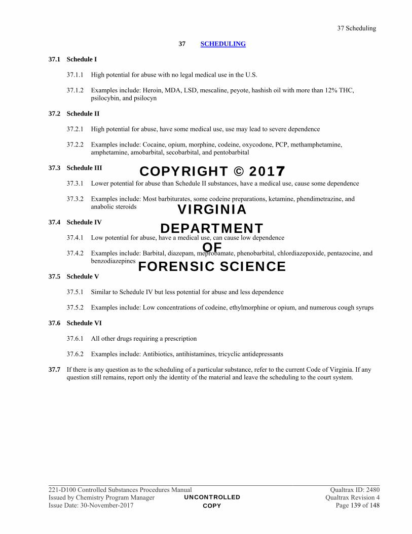
37 Scheduling
221-D100 Controlled Substances Procedures Manual Qualtrax ID: 2480Issued by Chemistry Program Manager Qualtrax Revision 4Issue Date: 30-November-2017 Page 139 of 148
37 SCHEDULING 37.1 Schedule I
37.1.1 High potential for abuse with no legal medical use in the U.S.
37.1.2 Examples include: Heroin, MDA, LSD, mescaline, peyote, hashish oil with more than 12% THC, psilocybin, and psilocyn
37.2 Schedule II
37.2.1 High potential for abuse, have some medical use, use may lead to severe dependence
37.2.2 Examples include: Cocaine, opium, morphine, codeine, oxycodone, PCP, methamphetamine, amphetamine, amobarbital, secobarbital, and pentobarbital
37.3 Schedule III
37.3.1 Lower potential for abuse than Schedule II substances, have a medical use, cause some dependence
37.3.2 Examples include: Most barbiturates, some codeine preparations, ketamine, phendimetrazine, and anabolic steroids
37.4 Schedule IV
37.4.1 Low potential for abuse, have a medical use, can cause low dependence
37.4.2 Examples include: Barbital, diazepam, meprobamate, phenobarbital, chlordiazepoxide, pentazocine, and benzodiazepines
37.5 Schedule V
37.5.1 Similar to Schedule IV but less potential for abuse and less dependence
37.5.2 Examples include: Low concentrations of codeine, ethylmorphine or opium, and numerous cough syrups 37.6 Schedule VI
37.6.1 All other drugs requiring a prescription
37.6.2 Examples include: Antibiotics, antihistamines, tricyclic antidepressants 37.7 If there is any question as to the scheduling of a particular substance, refer to the current Code of Virginia. If any
question still remains, report only the identity of the material and leave the scheduling to the court system.
UNCONTROLLED COPY
COPYRIGHT © 2017
VIRGINIA DEPARTMENT
OF FORENSIC SCIENCE
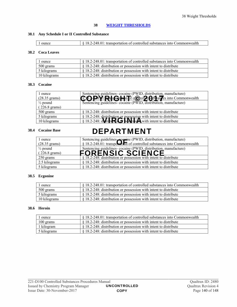
38 Weight Thresholds
221-D100 Controlled Substances Procedures Manual Qualtrax ID: 2480Issued by Chemistry Program Manager Qualtrax Revision 4Issue Date: 30-November-2017 Page 140 of 148
38 WEIGHT THRESHOLDS 38.1 Any Schedule I or II Controlled Substance
1 ounce § 18.2-248.01: transportation of controlled substances into Commonwealth 38.2 Coca Leaves
1 ounce § 18.2-248.01: transportation of controlled substances into Commonwealth 500 grams § 18.2-248: distribution or possession with intent to distribute 5 kilograms § 18.2-248: distribution or possession with intent to distribute 10 kilograms § 18.2-248: distribution or possession with intent to distribute
38.3 Cocaine
1 ounce (28.35 grams)
Sentencing guidelines- cocaine (PWID, distribution, manufacture) § 18.2-248.01: transportation of controlled substances into Commonwealth
½ pound ( 226.8 grams)
Sentencing guidelines- cocaine (PWID, distribution, manufacture)
500 grams § 18.2-248: distribution or possession with intent to distribute 5 kilograms § 18.2-248: distribution or possession with intent to distribute 10 kilograms § 18.2-248: distribution or possession with intent to distribute
38.4 Cocaine Base
1 ounce (28.35 grams)
Sentencing guidelines- cocaine (PWID, distribution, manufacture) § 18.2-248.01: transportation of controlled substances into Commonwealth
½ pound ( 226.8 grams)
Sentencing guidelines- cocaine (PWID, distribution, manufacture)
250 grams § 18.2-248: distribution or possession with intent to distribute 2.5 kilograms § 18.2-248: distribution or possession with intent to distribute 5 kilograms § 18.2-248: distribution or possession with intent to distribute
38.5 Ecgonine
1 ounce § 18.2-248.01: transportation of controlled substances into Commonwealth 500 grams § 18.2-248: distribution or possession with intent to distribute 5 kilograms § 18.2-248: distribution or possession with intent to distribute 10 kilograms § 18.2-248: distribution or possession with intent to distribute
38.6 Heroin
1 ounce § 18.2-248.01: transportation of controlled substances into Commonwealth 100 grams § 18.2-248: distribution or possession with intent to distribute 1 kilogram § 18.2-248: distribution or possession with intent to distribute 5 kilograms § 18.2-248: distribution or possession with intent to distribute
UNCONTROLLED COPY
COPYRIGHT © 2017
VIRGINIA DEPARTMENT
OF FORENSIC SCIENCE
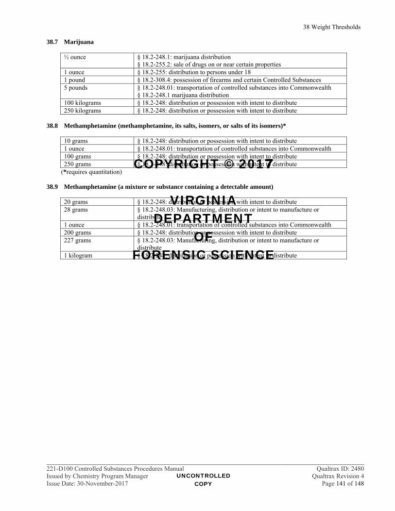
38 Weight Thresholds
221-D100 Controlled Substances Procedures Manual Qualtrax ID: 2480Issued by Chemistry Program Manager Qualtrax Revision 4Issue Date: 30-November-2017 Page 141 of 148
38.7 Marijuana
½ ounce § 18.2-248.1: marijuana distribution § 18.2-255.2: sale of drugs on or near certain properties
1 ounce § 18.2-255: distribution to persons under 18 1 pound § 18.2-308.4: possession of firearms and certain Controlled Substances 5 pounds § 18.2-248.01: transportation of controlled substances into Commonwealth
§ 18.2-248.1 marijuana distribution 100 kilograms § 18.2-248: distribution or possession with intent to distribute 250 kilograms § 18.2-248: distribution or possession with intent to distribute
38.8 Methamphetamine (methamphetamine, its salts, isomers, or salts of its isomers)*
10 grams § 18.2-248: distribution or possession with intent to distribute 1 ounce § 18.2-248.01: transportation of controlled substances into Commonwealth 100 grams § 18.2-248: distribution or possession with intent to distribute 250 grams § 18.2-248: distribution or possession with intent to distribute
(*requires quantitation)
38.9 Methamphetamine (a mixture or substance containing a detectable amount)
20 grams § 18.2-248: distribution or possession with intent to distribute 28 grams § 18.2-248.03: Manufacturing, distribution or intent to manufacture or
distribute 1 ounce § 18.2-248.01: transportation of controlled substances into Commonwealth 200 grams § 18.2-248: distribution or possession with intent to distribute 227 grams § 18.2-248.03: Manufacturing, distribution or intent to manufacture or
distribute 1 kilogram § 18.2-248: distribution or possession with intent to distribute
UNCONTROLLED COPY
COPYRIGHT © 2017
VIRGINIA DEPARTMENT
OF FORENSIC SCIENCE
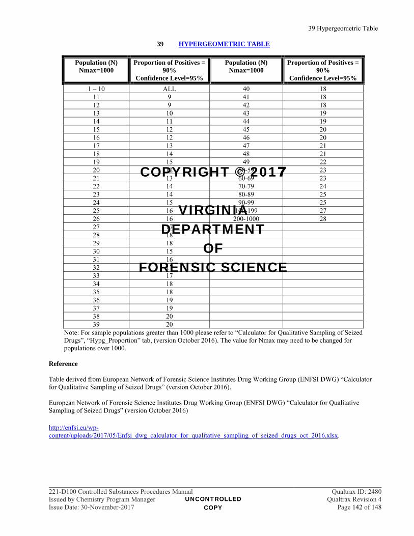
39 Hypergeometric Table
221-D100 Controlled Substances Procedures Manual Qualtrax ID: 2480Issued by Chemistry Program Manager Qualtrax Revision 4Issue Date: 30-November-2017 Page 142 of 148
39 HYPERGEOMETRIC TABLE
Population (N) Nmax=1000
Proportion of Positives = 90%
Confidence Level=95%
Population (N) Nmax=1000
Proportion of Positives = 90%
Confidence Level=95%
1 – 10 ALL 40 18 11 9 41 18 12 9 42 18 13 10 43 19 14 11 44 19 15 12 45 20 16 12 46 20 17 13 47 21 18 14 48 21 19 15 49 22 20 12 50-59 23 21 13 60-69 23 22 14 70-79 24 23 14 80-89 25 24 15 90-99 25 25 16 100-199 27 26 16 200-1000 28 27 17 28 18 29 18 30 15 31 16 32 17 33 17 34 18 35 18 36 19 37 19 38 20 39 20
Note: For sample populations greater than 1000 please refer to “Calculator for Qualitative Sampling of Seized Drugs”, “Hypg_Proportion” tab, (version October 2016). The value for Nmax may need to be changed for populations over 1000.
Reference Table derived from European Network of Forensic Science Institutes Drug Working Group (ENFSI DWG) “Calculator for Qualitative Sampling of Seized Drugs” (version October 2016). European Network of Forensic Science Institutes Drug Working Group (ENFSI DWG) “Calculator for Qualitative Sampling of Seized Drugs” (version October 2016) http://enfsi.eu/wp-content/uploads/2017/05/Enfsi_dwg_calculator_for_qualitative_sampling_of_seized_drugs_oct_2016.xlsx.
UNCONTROLLED COPY
COPYRIGHT © 2017
VIRGINIA DEPARTMENT
OF FORENSIC SCIENCE
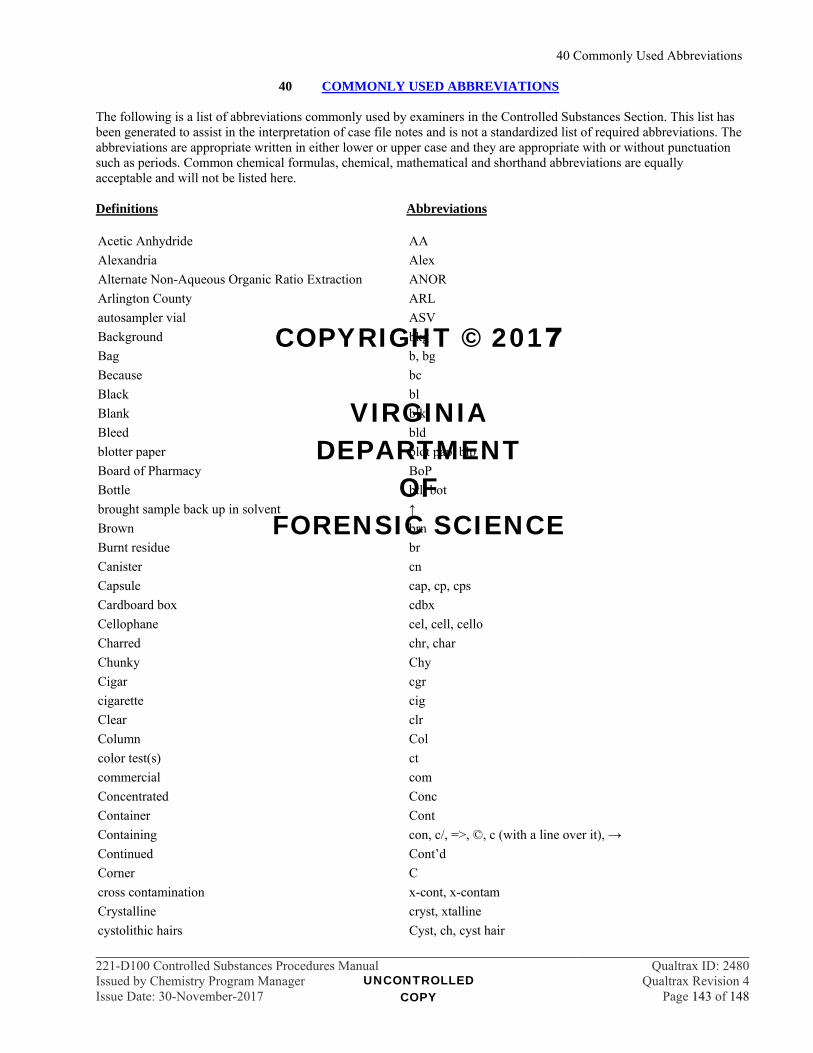
40 Commonly Used Abbreviations
221-D100 Controlled Substances Procedures Manual Qualtrax ID: 2480Issued by Chemistry Program Manager Qualtrax Revision 4Issue Date: 30-November-2017 Page 143 of 148
40 COMMONLY USED ABBREVIATIONS The following is a list of abbreviations commonly used by examiners in the Controlled Substances Section. This list has been generated to assist in the interpretation of case file notes and is not a standardized list of required abbreviations. The abbreviations are appropriate written in either lower or upper case and they are appropriate with or without punctuation such as periods. Common chemical formulas, chemical, mathematical and shorthand abbreviations are equally acceptable and will not be listed here. Definitions Abbreviations Acetic Anhydride AA
Alexandria Alex
Alternate Non-Aqueous Organic Ratio Extraction ANOR
Arlington County ARL
autosampler vial ASV
Background bkg
Bag b, bg
Because bc
Black bl
Blank blk
Bleed bld
blotter paper blot pap, blp
Board of Pharmacy BoP
Bottle btl, bot
brought sample back up in solvent ↑
Brown brn
Burnt residue br
Canister cn
Capsule cap, cp, cps
Cardboard box cdbx
Cellophane cel, cell, cello
Charred chr, char
Chunky Chy
Cigar cgr
cigarette cig
Clear clr
Column Col
color test(s) ct
commercial com
Concentrated Conc
Container Cont
Containing con, c/, =>, ©, c (with a line over it), →
Continued Cont’d
Corner C
cross contamination x-cont, x-contam
Crystalline cryst, xtalline
cystolithic hairs Cyst, ch, cyst hair
UNCONTROLLED COPY
COPYRIGHT © 2017
VIRGINIA DEPARTMENT
OF FORENSIC SCIENCE
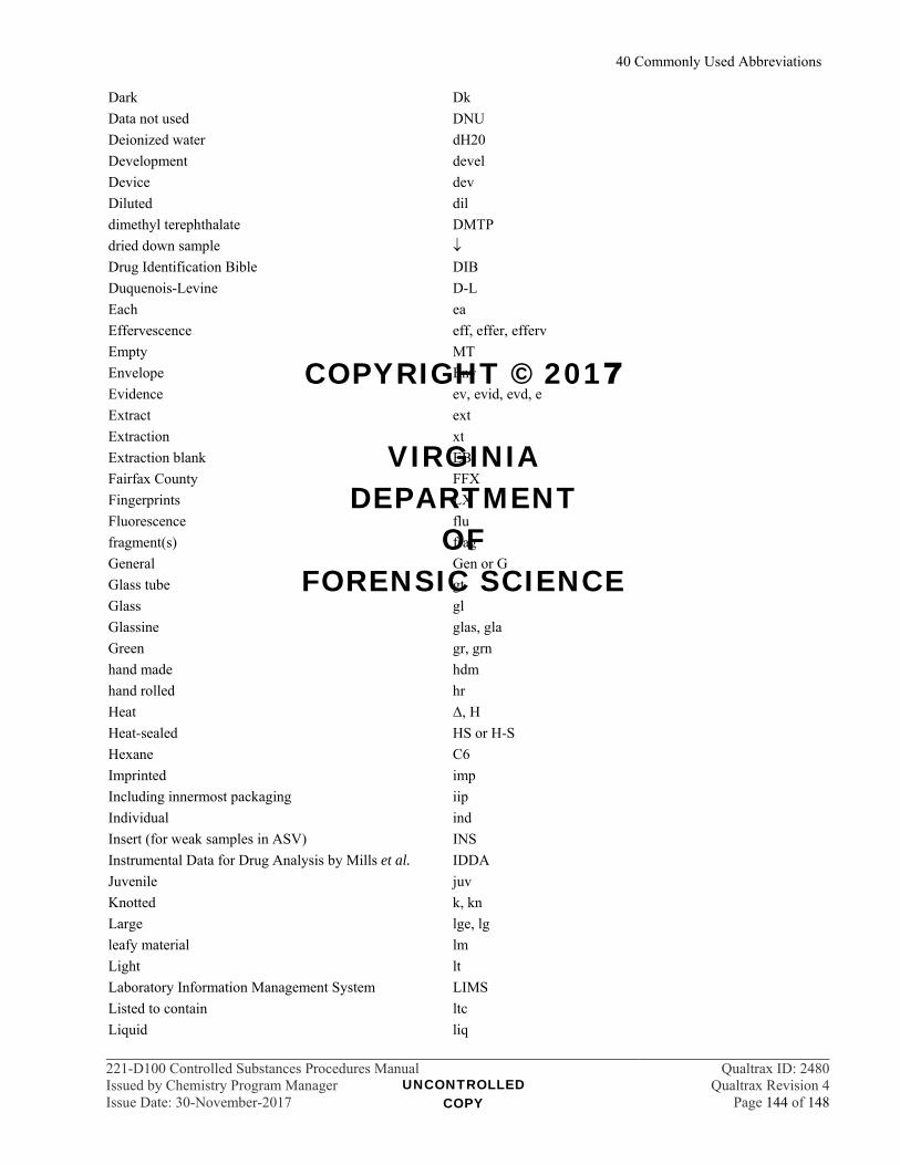
40 Commonly Used Abbreviations
221-D100 Controlled Substances Procedures Manual Qualtrax ID: 2480Issued by Chemistry Program Manager Qualtrax Revision 4Issue Date: 30-November-2017 Page 144 of 148
Dark Dk
Data not used DNU
Deionized water dH20
Development devel
Device dev
Diluted dil
dimethyl terephthalate DMTP
dried down sample
Drug Identification Bible DIB
Duquenois-Levine D-L
Each ea
Effervescence eff, effer, efferv
Empty MT
Envelope Env
Evidence ev, evid, evd, e
Extract ext
Extraction xt
Extraction blank EB
Fairfax County FFX
Fingerprints LX
Fluorescence flu
fragment(s) frag
General Gen or G
Glass tube gt
Glass gl
Glassine glas, gla
Green gr, grn
hand made hdm
hand rolled hr
Heat Δ, H
Heat-sealed HS or H-S
Hexane C6
Imprinted imp
Including innermost packaging iip
Individual ind
Insert (for weak samples in ASV) INS
Instrumental Data for Drug Analysis by Mills et al. IDDA
Juvenile juv
Knotted k, kn
Large lge, lg
leafy material lm
Light lt
Laboratory Information Management System LIMS
Listed to contain ltc
Liquid liq
UNCONTROLLED COPY
COPYRIGHT © 2017
VIRGINIA DEPARTMENT
OF FORENSIC SCIENCE

40 Commonly Used Abbreviations
221-D100 Controlled Substances Procedures Manual Qualtrax ID: 2480Issued by Chemistry Program Manager Qualtrax Revision 4Issue Date: 30-November-2017 Page 145 of 148
Manila man
Material mat, mat'l
Metal met
microscopic examination micro
multi-colored mc, multi
Mushrooms mush
negative Result for a test -, circled -, neg
nitroprusside color test np
no change nc
no color reaction ncr
no reaction nr
no significant reaction nsr
not analyzed na, not anal., nap, nt, DIRP
not examined ne
not opened n/o, no
off-white off-wh, ow One plastic container which contained one glass
i hi h i dclp
Pack pk
Package pkg
Packet pkt
Paper pap
Pentane pent
physical identification code for brand name product PIB, PIBr
physical identification code for generic product PIG
physically identified with references PID
Physicians Desk Reference PDR
placed in zip(s) in lab PIZIL
plant material (only ) pm
Plastic p, plas, pls, plst
Plastic Safety Tube PST
Powder pwd, pW, pwdr
Precipitate ppt
Prescription Rx
Prince William County Pr W
Probable prob
Purple prpl, pur, pl, ppl
quantity insufficient qns
Reaction rxn
Rectangular rec
Remaining rem
reopened to return rtr
repackage(d) repkged
Residue res
Returned ret
Rock-like rl
UNCONTROLLED COPY
COPYRIGHT © 2017
VIRGINIA DEPARTMENT
OF FORENSIC SCIENCE
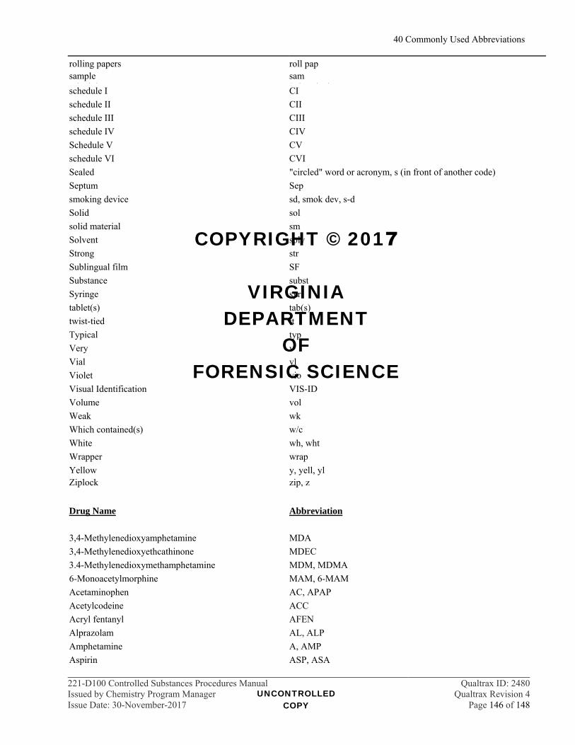
40 Commonly Used Abbreviations
221-D100 Controlled Substances Procedures Manual Qualtrax ID: 2480Issued by Chemistry Program Manager Qualtrax Revision 4Issue Date: 30-November-2017 Page 146 of 148
rolling papers roll pap sample
h d lsam S h h dschedule I CI
schedule II CII
schedule III CIII
schedule IV CIV
Schedule V CV
schedule VI CVI
Sealed "circled" word or acronym, s (in front of another code)
Septum Sep
smoking device sd, smok dev, s-d
Solid sol
solid material sm
Solvent solv
Strong str
Sublingual film SF
Substance subst
Syringe syr
tablet(s) tab(s)
twist-tied tt
Typical typ
Very v
Vial vl
Violet vio
Visual Identification VIS-ID
Volume vol
Weak wk
Which contained(s) w/c
White wh, wht
Wrapper wrap
Yellow y, yell, yl Ziplock zip, z
Drug Name Abbreviation
3,4-Methylenedioxyamphetamine MDA
3,4-Methylenedioxyethcathinone MDEC
3.4-Methylenedioxymethamphetamine MDM, MDMA
6-Monoacetylmorphine MAM, 6-MAM
Acetaminophen AC, APAP
Acetylcodeine ACC
Acryl fentanyl AFEN
Alprazolam AL, ALP
Amphetamine A, AMP
Aspirin ASP, ASA
UNCONTROLLED COPY
COPYRIGHT © 2017
VIRGINIA DEPARTMENT
OF FORENSIC SCIENCE

40 Commonly Used Abbreviations
221-D100 Controlled Substances Procedures Manual Qualtrax ID: 2480Issued by Chemistry Program Manager Qualtrax Revision 4Issue Date: 30-November-2017 Page 147 of 148
Benzocaine BNZ, BENZO
Bufotenine BUF
Buprenorphine BUP
Benzylpiperazine BZP
Caffeine CAF
Cannabidiol CBD
Cannabimimetic agent CMA, CA
Carisoprodol CAR
Clonazepam CZM
Cocaine CO, COC
Cocaine base CB, CFB
Cocaine Hydrochloride COH
Codeine COD
Cyclobenzaprine CBP
1,4-cyclohexadienyl-2-methaminopropane CMP
Dextromethorphan DXM
Diazepam DZM
Dibutylone DBU
Diltiazem DLT
Dimethyltryptamine DMT
Diphenhydramine DPH
Ephedrine EP
Etizolam ETZ
Fentanyl FENT, FEN
Fluoroisobutyryl fentanyl FIBF
Furanyl fentanyl FUF
Gabapentin GAB
Gamma-butyrolactone GBL
Gamma-hydroxybutyric acid GHB
Guaifenesin GUI
Heroin HER
Hydrocodone HC, HYD
Hydromorphone HM
Ibuprofen IB
Insufficient for analysis IFA
Insufficient for identification IFI
Ketamine KET
Levamisole LEV
Lidocaine LIDO
Lisdexamfetamine LIS
Lorazepam LO
Lysergic acid diethylamide LSD
Marijuana MJ
Methadone MN, MDN
Methamphetamine M, METH
UNCONTROLLED COPY
COPYRIGHT © 2017
VIRGINIA DEPARTMENT
OF FORENSIC SCIENCE
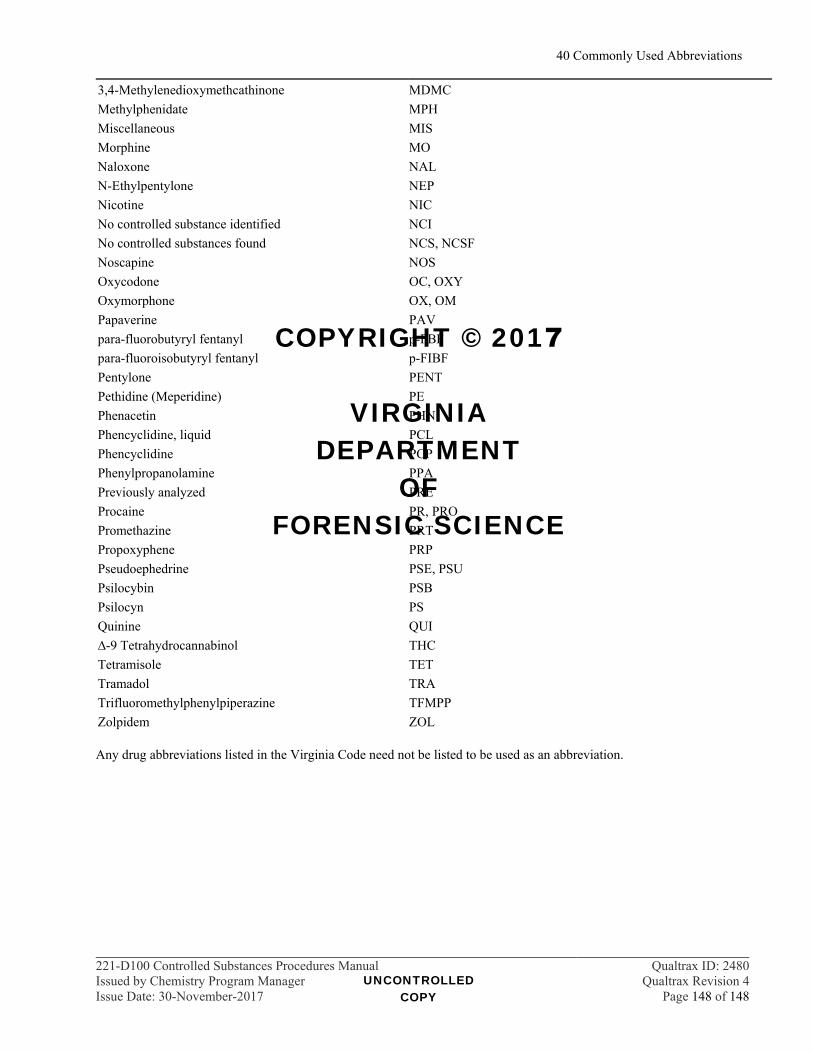
40 Commonly Used Abbreviations
221-D100 Controlled Substances Procedures Manual Qualtrax ID: 2480Issued by Chemistry Program Manager Qualtrax Revision 4Issue Date: 30-November-2017 Page 148 of 148
3,4-Methylenedioxymethcathinone MDMC
Methylphenidate MPH
Miscellaneous MIS
Morphine MO
Naloxone NAL
N-Ethylpentylone NEP
Nicotine NIC
No controlled substance identified NCI
No controlled substances found NCS, NCSF
Noscapine NOS
Oxycodone OC, OXY
Oxymorphone OX, OM
Papaverine PAV
para-fluorobutyryl fentanyl p-FBF
para-fluoroisobutyryl fentanyl p-FIBF
Pentylone PENT
Pethidine (Meperidine) PE
Phenacetin PHN
Phencyclidine, liquid PCL
Phencyclidine PCP
Phenylpropanolamine PPA
Previously analyzed PRE
Procaine PR, PRO
Promethazine PRT
Propoxyphene PRP
Pseudoephedrine PSE, PSU
Psilocybin PSB
Psilocyn PS
Quinine QUI
∆-9 Tetrahydrocannabinol THC
Tetramisole TET
Tramadol TRA
Trifluoromethylphenylpiperazine TFMPP
Zolpidem ZOL
Any drug abbreviations listed in the Virginia Code need not be listed to be used as an abbreviation.
UNCONTROLLED COPY
COPYRIGHT © 2017
VIRGINIA DEPARTMENT
OF FORENSIC SCIENCE


![[2020] JMSC Civ 221 - DunnCox](https://img.dokumen.tips/doc/110x75/631555a1c72bc2f2dd04ab32/2020-jmsc-civ-221-dunncox.jpg)