Embed Size (px)
Citation preview
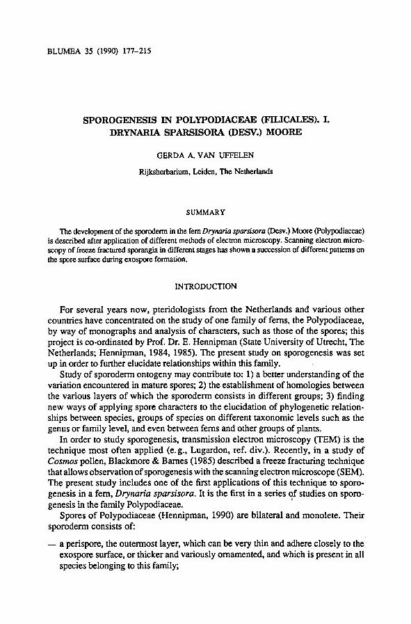
BLUMEA 35 (1990) 177-215
Sporogenesis in Polypodiaceae (Filicales). I.
Drynaria sparsisora (Desv.) Moore
Gerda+A. van Uffelen
Rijksherbarium, Leiden, The Netherlands
Summary
The developmentof the sporoderm in the fem Drynaria sparsisora (Desv.) Moore (Polypodiaceae)
is described after application of different methods of electron microscopy. Scanning electron micro-
scopy of freeze fractured sporangia in different stages has shown a succession of different patterns on
the spore surface during exospore formation.
Introduction
In order to study sporogenesis, transmission electron microscopy (TEM) is the
technique most often applied (e.g., Lugardon, ref. div.). Recently, in a study of
Cosmos pollen, Blackmore & Barnes (1985) described a freeze fracturing technique
thatallows observation ofsporogenesis with the scanning electron microscope (SEM).
The present study includes one of the first applications of this technique to sporo-
genesis in a fem, Drynaria sparsisora. It is the first in a series of studies on sporo-
genesis in the family Polypodiaceae.
Spores of Polypodiaceae (Hennipman, 1990) are bilateral and monolete. Their
sporoderm consists of:
— a perispore, the outermost layer, which can be very thin and adhere closely to the
exospore surface, or thicker and variously ornamented, and which is present in all
species belonging to this family;
For several years now, pteridologists from the Netherlands and various other
countries have concentrated on the study of one family of fems, the Polypodiaceae,
by way of monographs and analysis of characters, such as those of the spores; this
project is co-ordinatedby Prof. Dr. E. Hennipman (State University of Utrecht, The
Netherlands; Hennipman, 1984, 1985). The present study on sporogenesis was set
up in order to further elucidaterelationships within this family.
Study of sporoderm ontogeny may contribute to: 1) a better understanding of the
variationencountered in mature spores; 2) the establishmentof homologies between
the various layers of which the sporoderm consists in different groups; 3) finding
new ways of applying spore characters to the elucidation of phylogenetic relation-
ships between species, groups of species on different taxonomic levels such as the
genus or family level, and even between fems and other groups of plants.
BLUMEA VOL. 35, No. 1, 1990178
— a relatively thick exospore, the middlelayer, which may be smooth or ornamented
in different ways;
— an endospore, the innermost layer, which is not present until germination sets in.
Stages in sporogenesis
In an earlier paper (Van Uffelen, 1986) I distinguished six main stages during
sporogenesis, mainly based on the study ofTEM micrographs:
1. presence of spore mothercells (smc's) (see Plates I & II)
2. meiosis (see Plate IE)
3. formationof the spore plasmalemma (see Plate III)
4. formationofthe innerexospore (ie) (see Plates IV & V)
5. formation of the outer exospore (oe) (see Plates V-IX)
6. formation of the perispore (see Plate X)
Now that SEM micrographs of sporogenesis in Drynaria sparsisora have become
available as well, in this species stage 5, the formationof the outer exospore, can be
divided into different substages by the differentsurface patterns found during the de-
position of this layer.
Previous studies
Many others have already studied sporogenesis ofland plants, during the last two
decades mainly with the TEM:
— bryophytes: Brown & Lemmon (1988,review), Denizot (1974, Hepaticae), Neid-
hart (1979, review);
— ferns andfern allies: Bell (1981,1985, Marsileamegaspores), Brunkener (1973),
Buchen & Sievers (1978a & b, Selaginella megaspores), Gabarajeva (1984, early
stages in Psilotum), Hennipman (1970,1977, Bolbitis), Herd, Cutter, & Wata-
nabe (1985, Azolla microspores), Huang & Chiang (1983, Isoëtes), Lehmann,
Neidhart, & Schlenkermann (1984, Equisetum), Lugardon (1966, 1969, Blech-
num; 1971, Ophioglossum, Osmunda, Blechnum; 1972, 1974, 1979,Psilotum),
Mitui (1974, Polystichum perispore), Pettitt (1971, 1979), Schraudolf (1984,
Anemia), Sheffield& Bell (1979, Pteridium, 1987), Singh & Devi (1989, Pteris),
Surova (1981a & b, Anemia), Tilquin (1980, Hymenophyllaceae), Uehara &
Kurita (1989, Equisetum), Van Uffelen (1985, 1986), Verma & Khullar (1976);
— spermatophytes: Bhandari, 1984 (review).
On sporogenesis in general work has been published by Blackmore & Crane
(1985, 1988), Buchen & Sievers (1981), Heslop-Harrison (1964), and Locquin(1981). Some special aspects of sporogenesis have been treated by Bienfait& Water-
keyn (1976, sporocytic layers), Pacini et al. (1985, tapetum), and Pettitt & Jermy
(1974, surface coats on spores).
G.A. van Uffelen: Sporogenesis in Drynaria sparsisora 179
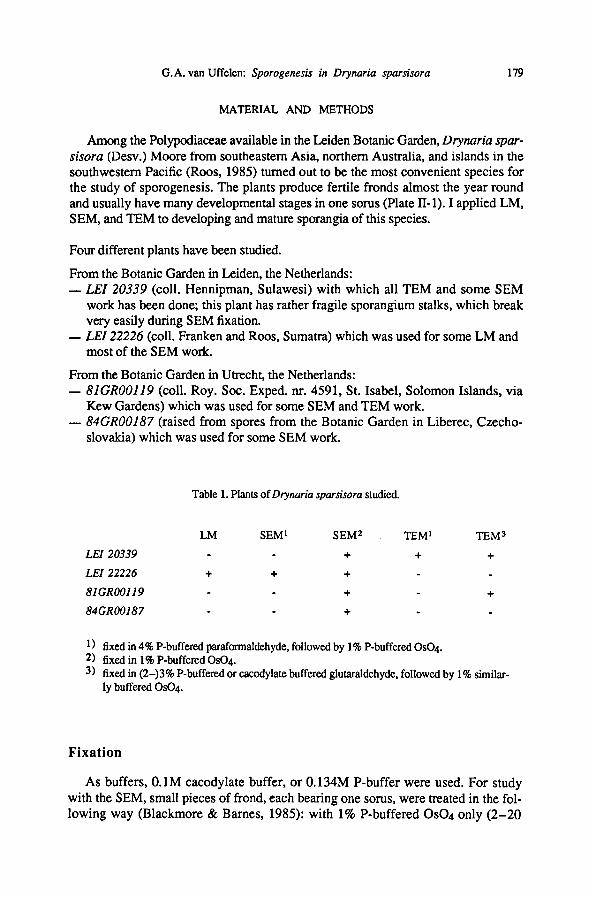
MATERIAL AND METHODS
Among the Polypodiaceae available in the Leiden Botanic Garden, Drynaria spar-
sisora (Desv.) Moore from southeastern Asia, northern Australia, and islands in the
southwestern Pacific (Roos, 1985) turnedout to be the most convenient species for
the study of sporogenesis. The plants produce fertile fronds almost the year round
and usually have many developmental stages in one sorus (Plate II-1). I applied LM,
SEM, and TEM to developing and mature sporangia of this species.
Four differentplants have been studied.
From the Botanic Garden in Leiden, the Netherlands:
—LEI 20339 (coll. Hennipman, Sulawesi) with which all TEM and some SEM
work has been done; this plant has rather fragile sporangium stalks, which break
very easily during SEM fixation.
— LEI 22226 (coll. Franken and Roos, Sumatra) which was used for some LM and
most of the SEM work.
From the Botanic Garden in Utrecht, the Netherlands:
—81GR00119 (coll. Roy. Soc. Exped. nr. 4591, St. Isabel, Solomon Islands, via
Kew Gardens) which was used for some SEM and TEM work.
— 84GR00187 (raised from spores from the Botanic Garden in Liberec, Czecho-
slovakia) which was used for some SEM work.
Fixation
As buffers, 0.1M cacodylate buffer, or 0.134M P-buffer were used. For studywith the SEM, small pieces of frond, each bearing one sorus, were treated in the fol-
lowing way (Blackmore & Barnes, 1985): with 1% P-buffered OSO4 only (2-20
1 ) fixed in 4% P-buffered paraformaldehyde, followedby 1% P-buffered OSO4.2) fixed in 1% P-buffered OSO4.
fixed in (2-)3% P-buffered or cacodylate buffered glutaraldehyde, followed by 1% similar-
ly buffered OSO4.
Table 1. Plants of Drynaria sparsisora studied.
LM SEM 1 SEM2 TEM 1 TEM 3
LEI 20339 - - + + +
LEI 22226 + + + - -
81GR001I9- - + - +
84GR00187.
. + __
BLUMEA VOL. 35, No. 1, 1990180
hrs), or 4% P-buffered paraformaldehyde (Pease, 1964) followed by 1% P-buffered
OsCU; freeze fracturing in 50% DMSO; long (14 days) postfixation in 0.1% P-buf-
fered OSO4, followed by 1% P-buffered OSO4 (1 hr), 2% tannic acid (16 hrs), 1%
P-buffered OSO4 (1 hr); dehydration in an alcohol series and critical point drying in
DMM.
After this treatment, the broken sori were mounted onto aluminum stubs (10 mm
0) with Scotch double-sided tape and coated with gold. For the study of mature
spores, untreated spores from dried fronds were mounted with glue in aceton and
coatedwith gold in the usual way (Van Uffelen & Hennipman, 1985). Micrographs
were madeon Kodak Panatomic-X FXP film (60 x 60 mm negatives) with the JEOL
JSM-35 scanning electron microscope at the Rijksherbarium, Leiden.
Some micrographs were made on the Hitachi S800 scanning microscope in the
Natural History Museum (London).
For study with the TEM, sori, preferably without any part of the frond attached,
were treated in the following way (Pease, 1964; Lugardon, 1971): with 4% P-buf-
fered paraformaldehyde (2-4 months), or with 3% P-bufferedor cacodylate-buffered
glutaraldehyde (c. 5 or 24 hrs), or with 0.75% K111NO4 in water (30 minutes); with
1% buffered OSO4 (60-80 minutes or 24 hrs); dehydration in an alcohol series and
embedding in Epon resin.
Sections were made with glass knives or a diamondknife on various microtomes.
Sections were mounted on one holeor mesh grids and colouredwith a 1-5% (almost
saturated to saturated) solution of uranyl acetate in water and lead citrate (Reynolds,
1963). The sections were observed with a Hitachi HU 1 IB microscope (and photo-
graphed on Gevaert 'Scientia' 23-D-50 plates) or with a Philips EM-300 (and pho-
tographed on Kodak FRP 426 film).
Measurements
Measurements have been madeof sporangium size, spore size, sporoderm thick-
ness and some other features as seen on SEM and TEM micrographs. I am fully
aware of the relative unreliability of these measurements by reason of the obliquity
of sections (TEM) or spore position (SEM), and fluctuations in magnification of t
he apparatus used; moreover, the number of measurements is always rather small
(see also Mogensen, 1981). Despite these difficultiesin using SEM and TEM micro-
graphs in order to make measurements of spores, LM could not be applied as it is not
possible to assess accurately the stage of a sporangium using a light microscope.
Terminology
The following list contains only terms used in this publication and is not a com-
plete list of terms as used in describing the morphology and ontogeny of fern spores.
Such a list in English is still lacking for the spores of ferns and fern allies and is be-
ing prepared by the present author; a list of terms applicable to pollen, and partly to
spores, is being prepared by S. Blackmore, A. Le Thomas, S. Nilsson, and W. Punt,
and a list pertaining to fossil spores and pollen has recently been published
(Traverse, 1988).
181G.A. van Uffelen: Sporogenesis in Drynaria sparsisora
The terminology used in this publication is mainly based on the ultrastructural
work of Lugardon (1971, 1975, 1978a, 1978b, 1981), who studied sporogenesis in
several fems with the transmission electron microscope. Very few terms are purely
topographic or morphologic; most of the terms describing spore morphology have
some ontogenetic connotations.
Fern terminology
Ferns and fern allies can be homosporous (Psilotales, Lycopodiales, Equisetales,
Ophioglossales, Marattiales, and Filicales) or heterosporous (Selaginellales, Isoeta-
les, Marsileales, and Salviniales). Their life cycle consists of a diploid sporophyte
producing haploid spores, and a haploid gametophyte producing gametes.
Fern—plant belonging to the order Ophioglossales, Marattiales,Filicales, Marsileales,or Salvini-
ales. It is a megaphyllous spore-bearing vascular plant.
Fern ally — plant belonging to the order Psilotales, Lycopodiales, Selaginellales, Isoetalcs, or Equi-
setales. It is a spore-bearing vascular plant with small leaves.
Frond— a megaphyllous fern leaf.
Gametophyte (adj. gametophytic) — The sexual generation ofa plant that produces gametes, or an
individual of this generation; e.g., the haploid generationof an embryophytic plant, produced by
germinationof the spores (see Traverse, 1988).
Leptosporangiate —of a fern with sporangia initiating from a single cell; the sporangium wall is
only one cell layer thick (contr. eusporangiate, as in Ophioglossales and Marattiales).
Pteridophyte —a fern or fern ally (see Mabberley, 1987).
Sorus (pi. sori) — a group of sporangia (Plate II-1)
Sporangium (pi. sporangia) — an organ of the sporophyte in which meiosis takes place and haploid
spores develop with the aid of a diploid tissue, the tapetum(Plates II-2, HI-1).
Sporophyte (adj. sporophytic) —the diploidphase of the pteridophyte life cycle. In sporogenesis the
term sporophytic applies to the diploid cells (spore mother cell, tapetum, sporangium wall) pos-
sibly involved in some stage of sporogenesis.
Spores and sporogenesis
Bilateral—
of monolete spores, having two planes of symmetry perpendicular to each other, both
passing throughthe polar axis (see Lugardon, 1971) (Plate XI-1).
Callose (adj. callosic) — a carbohydrate component of cell walls in certain plants; e.g., the amor-
phous cell wall substance that envelops the pollen mother cell during pollen grain development(see Traverse, 1988).
Channels—
ofthe outer exospore: perforations that traverse the outer exospore and that connect the
inner side of this layer with the exospore environment (see Lugardon, 1971) (Plate IX-2).
Colliculate— ofa surface pattern: with rounded broad elevations, closely spaced, covering the sur-
face (Van Uffelen & Hennipman, 1985) (Plate IX-5).
Cytokinesis —the formation ofindividual cells separated by a cell wall afternuclear division.
Distal—
the part of a spore (or pollen grain) away from the centre of the tetrad, i.e. the part not
bearing the laesura (see Traverse, 1988).
BLUMEA VOL. 35, No. 1, 1990182
Distal pole —the centre ofthe distal surface of a spore orpollen grain (seeTraverse, 1988).
Elevation — part of a surface that is higher than other parts of that same surface (Plate XI-2).
Endospore —the innermost spore wall layer, which is in contact with the spore cytoplasm and is
continuous with the cell walls of the gametophyteafter mitosis during spore germination (see
Lugardon, 1971).
Exine —main layer of pollen grains lying outside the intine (Lugardon, 1971).
Exospore — main layer of the spore wall; it is continuous and acetolysis-resistant, and consists of
several interconnected sublayers; it bears a differentiated aperture (the laesura) on the proximal
side of the spore (see Lugardon, 1971) (Plate IX-5).
Haptotypic —of a feature of a spore or pollen grain that is a product of contact with other members
ofthe tetrad in which it was formed; e.g., the laesura of spores (see Traverse, 1988).
Heterosporous — of a plantproducing both microspores and megaspores (see Traverse, 1988).
Homosporous — of a plant producing only one kind of spore (see Traverse, 1988).
Inner exospore (ie) —innermost layer of the exospore. In the Filicales it is a compact and continu-
ous layer; it forms the proximal fold ofthe laesura (Lugardon, 1971) (Plate V-l).
Intine —innermost layer of pollen grains, consisting of cellulosic compounds; it forms the outer
layer of the pollen tube during germination(Lugardon, 1971).
Laesura —the trace on the proximal face of an embryophytic spore that marks the original contact
with other members of the tetrad; it is a specialized area of the exo-spore, which is adapted to let
out the gametophyte during germination; the term aperture is a much more generalone, which
also applies to certain pollen features (Plates IX-6,7, X-l).
Laesural fold — (= Lugardon, 1978a: proximal fold) narrow projecting fold of the inner exospore
layer, a first stage in the developmentof the laesura (Plate V-l, 7).
Layer — 1) well-defined part of the sporoderm, such as the endospore, exospore, perispore; 2) as a
more general term, applied to any part of the spore wall (this is in conflict with APLF-recom-
mendations, 1975).
Meiocyte —cell destined to go into meiosis, e.g., spore mother cell, pollen mother cell.
Meiosis—
cell division with reduction of the chromosome number.
Microspore — one ofthe kinds of spores of a heterosporous embryophytic plant that germinates to
produce a microgametophyte (Traverse, 1988).
Monolete— of a bilateral spore which has an oblong laesura; also applied to this type of laesura.
Ornamentation — any modification of the spore surface. Some authors prefer to use the term sculp-
ture, but this term is linked to the controversy about the correct use of the terms structure and
sculpture as applied to pollen exine morphology (Praglowski, 1975).
Outer exospore (oe) — outermost exospore layer which covers the underlying exospore layer(s) en-
tirely and constitutes the surface relief in ornamented exospores; it is perforated by channels or
small fissures connecting the base of this layerwith the exospore environment (Lugardon, 1971)
(Plate IX-5).
Periplasmodial tapetum — tapetum of which the cell walls are broken down during intrusion be-
tween the smc's, after which the cytoplasts fuse, usually in late meiotic prophase (Pacini et al.,
1985) (Plates II-2, II1-3).
183G.A. van Uffelen: Sporogenesis in Drynaria sparsisora
Perispore — spore wall layer depositedover the exospore surface, usually acetolysis-resistant; vari-
able between species: thin or very thick, tightly or not connected to the exospore (Lugardon,
1971) (Plate XI-1).
Pits—
round holes on the surface of a layer; if they are present, the surface is called foveolate (see
Traverse, 1988) (Plate VIM, 8).
Plasmalemma— (= plasma membrane, Gunning & Steer, 1986) the bounding membrane of the
protoplast, normally in close contact with the inner face of the cell wall (Plate III-8).
Plasmodesma (pi. plasmodesmata) — narrow cytoplasmic channel, bounded by the plasma mem-
brane, and interconnecting adjacent protoplasts throughthe intervening wall (Gunning & Steer,
1986) (Plate 1-2).
Polar axis — imaginary line connecting the distal pole with the proximal pole.
Proximal— the part ofa spore or pollen grain nearest or toward the centre of the original tetrad,
i.e. the part bearing the laesura (see Traverse, 1988) (Plate X-7).
Proximal pole —the centre of the proximal surface of a spore or pollen grain (see Traverse, 1988).
Rosette stage — the stage during sporogenesis in which the spore mother cells lie close to each
other, only separated by onelayer of sporocyte coat and forming a rosette on cross section (Plate
1-1).
Rounded stage —the stage duringsporogenesisin which the spore mother cells have become globular in
form and are lying further from each other, each surrounded by its own layer of sporocyte coat
(Plates 1-5, II-2).
Sculpture — see ornamentation.
Smc— see spore mother cell.
Spherical bodies— Small globular structures consisting of the same material as the outer exospore
and finally covered with a layer of perispore; found on the exospore surface, sometimes contained
in the perispore and in the mature sporangium ofmany pteridophytes; thought to be homologous to
Ubisch bodies in pollen (see Lugardon, 1981;Traverse, 1988) (Plates X-6, XI-8).
Spine — a projection from the spore surface with a more or less sharp apex and a tapering trunk
with a broad base (Harris, 1955) (Plate XI-7).
Spore — (Lugardon, 1971: only applied to ripe or almost ripe spores) haploid reproductive body
produced by a sporophyte, germinatinginto a gametophyte, and surrounded by a resistant sporo-
derm (see Traverse, 1988).
Spore coat — the coat which covers the surface of the spore (the postmeiotic cell) (Pettitt & Jermy,
1974) (Plate III-7).
SporeIsporocyte coat — see surface coat.
Spore height — height measured along the polar axis.
Spore length —the maximum length in lateral view.
Spore mother cell (smc) —the mother cell in the sporangium ofa spore-bearingplant, which, by reduc-
tion division, produces a tetrad of haploid spores (Traverse, 1988) (Plate 1-4).
Spore wall— see sporoderm.
Spore width—
the maximum width in polar view.
Sporocyte (adj. sporocytic) — see spore mother cell.
Sporocyte coat —the coat which covers the surface of the sporocyte (the premeiotic cell) (Pettitt &
Jermy, 1974) (Plate 1-6).
BLUMEA VOL. 35, No. 1, 1990184
Sporoderm — the entire wall of a spore or pollen grain (Traverse, 1988); the sum ofall layers sur-
roundinga spore or pollen grain (see Lugardon, 1971).
Sporogenesis —the formation of spores from the differentiationof the spore mother cell to the final
maturation of the spores. Spore wall formationis only part ofthe process of sporogenesis.
Sporopollenin —the very resistant organic substance of which the exine/exospore of spores and
pollen is composed (see Traverse, 1988).
Surface coat — purely morphographic term, covers both the sporocyte and the spore coat (Pettitt &
Jermy, 1974) (Plate III-7). I prefer the term spore/sporocyte coat as this is more specific than
surface coat.
Tapetum (adj. tapetal) —tissue of nutritive cells in the sporangium of embryophytic plants, largely
used up during developmentofthe spores (Traverse, 1988).
Tetrad — product of onespore mother cell after meiosis; each tetrad consists of four spores and the
enveloping coat (see Lugardon, 1971); a usually symmetric grouping of four embryophytic
spores or pollen grains that result from meiotic division of one spore mother cell (Traverse,
1988) (Plate III-6, 7).
Tetrahedal— of a tetrad in which each grain rests atop three others (Traverse, 1988); it produces tri-
lete spores (Lugardon, 1971).
Tetraspore — one of the four cells of a tetrad after cytokinesis. Towards the end of sporogenesis,
each tetraspore becomes a spore (see Lugardon, 1971). I start using the term spore as soon as the
laesural fold has been formed (Plates IV-2, V-l).
Unit — clearly distinguishable part of the spore surface separated from other units by grooves (Plate
V-7).
Verrucate— bearing verrucae, i.e. broad projections with a trunk thatis not constricted (see Harris,
1955) (Plate VIII-4).
While line — the centre of a tripartite lamella,on both sides of which sporopollenin has been de-
posited, occurring in the exine/exospore of many embryophytes (see Brown & Lemmon, 1988;
Rowley, 1988) (Plate V-3).
RESULTS
In young sporangia, within the sporangial wall consisting of a single cell layer,
differentiationin tapetal cells and spore mother cells takes place. After division of
the single layer of tapetal cells, before smc prophase, the smc's are surrounded by a
double layer of tapetum (Plate 1-1).
1. The spore mother cell
Before meiosis usually 16 spore mother cells are present in each sporangium. At
first, in what is called the rosette stage (Lugardon, pers. comm.), the smc's lie close
to each other, separated only by a common layer of dark granulate sporocyte coat
(Plate 1-1, 2). Smc's are connected with each other by way of thin cytoplasmicstrands (c. 0.06 pm wide), bounded by a plasmalemma, penetrating the sporocyte
coat (Plate 1-2).
Later on in the rosette stage, while the smc's are still in close contact with each
other, the fluffy sporocyte coat splits into two layers (Plate 1-3), so that each smc is
surrounded by its own sporocyte coat.
G.A. van Uffelen: Sporogenesis in Drynaria sparsisora 185
As the sporocyte coat doubles, the smc's change shape and become more round-
ed, so that they gradually detach from each other (Plate 1-4). When the smc's all
have their own sporocyte coat (Plate II-5), they have entered the smc-rounded stage
(Plate II-2, 3, 6); in this stage, the sporocyte coat may show a continuous growth,
resulting in the formation of loops (Plate 1-5). However, this may be due to a fixa-
tion artifact. Another such artifact may be found in smc's of very irregular outline
during meiosis (Plate 1-6). In many smc's, cytoplasmic protrusions in the nucleus
can be observed (Plate 1-5; Sheffield& Bell, 1979).
While the smc's change shape from angular to rounded, tapetal cell walls start to
break down and the tapetum moves in between the smc's (Plates 1-4, II-2). In this
stage, smc's are about 15 pm in diameter.
On TEM the tapetal nuclei are the most conspicuous tapetal organelles (Plate 1-4);
on SEM, nuclei and vacuoles are most conspicuous (Plate II-3, 4).
2. Meiosis
In Drynaria sparsisora I found the onset of cytokinesis to take place after com-
pletion of both nuclear divisions and not after each of the nuclear divisions.
The position of the spores in their tetrads is pairwise (Plate III-6) and not tetra-
hedal, which determines their form as bilateral and monolete. The pairwise-90° con-
figuration, where one pair of the tetrad lies in an angle of 90° with the other, has been
found on TEM (Plate III-7); angles less than 90° have also been found between pairs
(Plate III-5); some of these may be due to oblique cuts (see Huynh, 1973 and Verma
& Khullar, 1976). The tapetum has further degenerated; the nuclei are lying close
against the young tetrads (Plate III-2, 3).
3. Formation of the plasmalemma
In Drynaria sparsisora a cell plate is formedin association with aggregated mito-
chondria and vesicles (Plate III-3, 4, 5); on Plate EI-4 the nuclear membrane is again
(at least in part) present. As in smc's, invaginations between nucleus and cytoplasm
are present in young tetraspores (cf. Bell, 1981) in order to increase the surface of
contact between nucleus and cytoplasm (Plate III-6).
After cytokinesis, each tetrad is still enclosed in the sporocyte coat. Each tetrasporeis surrounded by a plasmalemma, and tetraspores are separated from each other bythe spore coat (Plate III-7, 8); they may be lying quite far apart (Plates III-7, IV-3).After conclusion of meiosis the tetrads start to increase in size. On TEM, the space
between the tetraspores in the tetrad is occupied by loose, flaky material (Plate HI-7,
8). On SEM, this material has been removed during fixation, and one tetrad consists
of four tetraspores lying apart in an otherwise empty space, surrounded by tapetalnuclei (Plate IV-3, 5). On Plate IV-3 and IV-5 the young tetraspores are lying close
to what is probably the spore/sporocyte coat, which is lying quite far away from
the tapetum. Details (Plate IV-4, 6) show that this coat looks more massive than on
TEM, and is densely set with granules; sometimes there are foldlike structures (Plate
IV-4) to be seen. Plate IV-7 shows the rather smooth inner side of a sporocyte coat
that has stayed in place after the spores have fallen out during fracturing, and is still
BLUMEA VOL. 35, No. 1, 1990186
lying close to the tapetum. On SEM, tetrads and young tetraspores are rather variable
in appearance, depending on the plane of fraction and subsequent rigorous postfixa-
tion (Plate IV-3, 5, 8). As all tetrads on Plate IV have been found in the same spo-
rangium, they are approximately in the same stageof development.
4. Formation of the inner exospore layer (ie)
The inner exospore layer is deposited on the outside of the plasmalemma as a
thin, opaque, sporopolleninous layer. During the formationof this layer a thin white
line (Brown & Lemmon, 1988) may be visible (Plate V-3), showing that the material
of which the inner exospore is formed is deposited by the tetraspore cytoplasm as
well as from the outsideof the tetraspore. Deposition of innerexospore material from
the spore cytoplasm may continue during the first stages of outer exospore formation
(Plate V-4). Later on in spore wall formation the white line disappears completely.
The Golgi apparatus appears to play a role in the secretion of sporopollenin as seen
on Plate V-4. The thickness of the complete inner exospore layer varies from 0.025
pm on the distal side to 0.040 pm on the proximal side.
While the inner exospore layer is being deposited, the laesural fold (Lugardon,
1969) is formed on the proximal side of the young tetraspore (Plate V-l, 2). Eventu-
ally this fold develops into the monolete laesura. During formationof the inner exo-
spore layer and of the first part of the outer exospore, the inner surfaces of the fold
move towards each other, the cytoplasm moving centripetally out of the fold, and
the surfaces finally come to lie close to each other (Plate VI-1). Plate IV-2 depicts a
laesural fold during its formation as seen with SEM. Here the fold is only c. 1 pm
high, and thus still in formation, as the completed laesural fold usually measures up
to 4 pm in its highest part.
5. Formation of the outer exospore layer (oe)
In Drynaria sparsisora almost all of the outer exospore layer is deposited in smal-
ler or larger grains, and the exospore is not smooth until the very end of its for-
mation. In the course of the formationof the outer exospore layer, several distinct
surface types can be distinguished in this species.
5a. Formation and presence of a surface consist of neatly defined units
On the outside of the smooth thin innerexospore layer, the innermostpart of the
outer exospore layer is deposited in the form of irregular sporopolleninous lumps.
Deposition begins near the laesural fold, so that in this stage the proximal side of the
wall is always slightly further developed than the distal side (Lugardon, 1971). The
first lumps to be deposited are very fine, usually less than 0.25 pm in diameter(Plate
V-4-6). Gradually the surface becomes covered with neatly defined, roundish or
angular units of c. 0.5 pm in diameter (Plate VI-4). On Plate V-7 and its detail, Plate
V-8, different sizes of lumps are visible, especially on the laesural fold. Plate VI-1
shows a cross section of a spore early in this stage; both sides of the laesural fold are
already lying close to each other; the irregular form of this spore is probably a
fixation artifact. Plate VI-5 shows a section of collapsed spores in a slightly later
187G.A. van Uffelen: Sporogenesis in Drynaria sparsisora
stage; it shows that the lower part of the laesural fold is already covered with exo-
spore material; under the base of the laesural fold a thick deposition can be seen, the
'amas sous-aperturaT (Lugardon, 1971). The inner surface of the spore wall (Plate
VI-3) is rather smooth with small pits and clearly shows the closed and thickened
base of the laesural fold.
On TEM, tapetal nuclei are still recognizable (Plate VI-1); on SEM, the tapetum is
more of an amorphous mass (Plate VI-2).
5b. Grains cover the surface units
The sharply definedunits gradually get covered by a layer of grains, which are
also deposited inbetween the units, but in such a way that the original units remain
visible (Plate VII-1-3). Hereby the relief of the spore surface becomes less pro-
nounced. Plate VII-2 shows grains to be c. 0.1-0.3 pm in diameter. On Plate VII-4,
which shows the spore surface magnified 45,000 times, the grains can be seen to be
even smaller, c. 0.05 pm in diameter; many small (0.25 pm diameter) roundholes -
channels connecting the surface with the innerparts of the exospore - are left open.
Plate VII-7 shows a TEM section of approximately the same stage; the exospore is
0.4-0.8 pm thick, and the laesural fold is rather high, 4.2 pm; channels connecting
the innerexospore layer with depressions on the exospore surface are visible as dark
lines. Spherical bodies (Plate VII-3) may be present in the tapetum and consist of
sporopollenin secreted by the tapetum; their surface is much more smooth than the
exospore surface in this stage; they are c. 1-5 pm in diameter.
5c. Surface becomes almost smooth, pitted
Gradually, the granular surface as seen on Plate VII-1-4 becomes covered with a
layer of fine grains, obscuring the units that were present just after the onset of the
outer exospore deposition (Plate VII-5). Numerous small pits, indicating the pres-
ence of channels, occur very frequently (Plate VII-6); they are larger and more num-
erous around the laesura (Plate VII-8). The openings of these channels form an
important part of the surface pattern at this stage. The spore surface gets more and
more smooth; deposition of material does not start around the laesura any more, but
on the distal side of the spores, as seen on Plate VIII-1. The tapetum, although rather
desorganized, still seems to play a role in the deposition of exospore material (Plate
VIII-3). During this stage most of the laesural fold becomes covered with exospore
material(Plate VIII-2).
5d. Pits disappear, surface becomes almost smooth, sometimes slightly verrucate
During the last stage of outer exospore formation, the exospore surface becomes
covered with a very fine-grained material which forms low rounded or angular ver-
rucae of c. 1.5-2.5 pm in diameter(Plate VIII-4-7). Tiny holes, especially around
the laesura, show the position of channels (Plate VIII-5). These channels are well
visible on TEM cross sections, as on Plate IX-1, 2; on Plate IX-2, former surfaces
of the exospore during its formation can be traced by the presence of dark granules
halfway the present outer exospore layer which is 1.1 pm thick. Plate IX-3 and 4
show similar sections with SEM: channels are not visible; even the innerexospore
layer in the part of the laesural fold already covered with exospore material is not
BLUMEA VOL. 35, No. 1, 1990188
visible; on these sections, the exospore is little more than 1 pm thick. Plate IX-4
shows that the inner surface of the exospore is rather smooth, more so than on Plate
VI-3, which suggests thateither deposition of additionalmaterial or consolidationof
the existing material takes place on the inner surface of the exospore during exospore
formation.
The tapetummay lie close to the spore surface and leave tiny fragments of mate-
rial (Plate VIH-5, 6). It sometimes contains spherical bodies (Plate IX-4).
Later a smooth or almost smooth, slightly colliculate outer surface is formed
(Plate IX-5, 8).
The laesural fold becomes completely covered with exospore material only at the
end of this last stage of exospore formation (Plate IX-6, 7). Its height depends on
the site of section: on Plate IX-6, 7, it is 2.4 and 4.6 pm high, respectively.
5e. The mature exospore
The mature exospore is about 1.5 pm thick, being only slightly thicker on the
proximal than on the distal side (Plate IX-5). Its surface is smooth or very slightly
verrucate, the verrucae are colliculate, 2-4pm in diameter(Plate X-l). The laesura
extends over the greater part of the length of the proximal side of the spore (Plate
X-l).
6. Perispore formation
After the exospore formation is completed, the perispore is formed. Perispore for-
mation, usually described as the quick precipitation of tapetal residues, is a very elu-
sive process, probably mainly because it takes such an extremely short time.
At the base of the developing perispore, lamellae are often to be found(Plate X-
3-5); they are not always forming a single layer, but are sometimes present in slight-
ly overlapping pieces of 0.2 pm long (Plate X-4); similar strips of lamellae are also
observed in the tapetum at the end of exospore maturation(Plate X-2). Angular blobs
of dark material, in size varying from 0.05 to 0.2 pm, are present in the tapetum
(Plate X-2) and in the vicinity of the spores (Plate X-3) and are eventually deposited
on the spore surface over the lamellae(Plate X-3-5). They may end up covering most
of the spore surface.
In Drynaria sparsisora the perispore consists of a rather thick slightly verrucate
layer on which spines are formed later on during perispore formation. It is not clear
whether the spines are formed out of these blobs, as is suggested by the presence of
the small spine on Plate X-5, or are assembled from other building blocks formed
by the tapetum. Plate X-7 and 8 show SEM pictures of the spore surface of which
the scale of the surface structures seems to correspond to the TEM pictures of Plate
X-3-6.
The formation of the basal layer is very elusive. The resemblance between the or-
namentationof this layer (Plate XI-2) and the pattern found after condensation of
glycerin jelly on a glass slide (Plate XI-5) suggests the possibility of a condensation
process being involved in perispore formation(Van Uffelen, in press). TEM-picturesof the mature perispore (Plate XI-6) are not very instructive with respect to the struc-
ture of the basal layer and the spines.
G.A. van Uffelen: Sporogenesis in Drynaria sparsisora 189
Spines are deposited perpendicular to the perispore surface. This is best to be
seen on the laesura (Plate XI-7) or on detailed micrographs taken exactly from above
(Plate XI-2).
7. The mature perispore
The mature perispore (Plate XI-1, 3) consists of a 0.07-0.4 pm thick basal layer
which is slightly and irregularly verrucate; elevations usually colliculate (Plate XI-6,
7), but sometimes not touching (Plate XI-2), flat and roundor longish, irregular in
outline; basal layer set with many tapering short spines; spines 1-1.5 or 2(-5, rarely
up to 10) pm long, 0.2-1.5, usually around 0.6 pm in diameter, usually straight and
pointed, sometimes slightly bent, or ending in more than one point (Plate XI-2, 4),
one or more globules (Plate XI-4, 7) or abruptly thinnercones (Plate XI-2), or in a
very long point (Plate XI-3); the longer the spines, the more irregular their distribu-
tion over the spore surface, the shorter they are, the closer and more evenly spaced
(Plate XI-1 vs. Plate XI-3). Spines are solid, as shown in TEM pictures (Plate XI-6)and on Plate XI-4. The position of the spines is not independent of the basal peri-
spore pattern: spines tend to be deposited where the basal pattern is highest, not in
between the elevations (Plate XI-2, 4, 7).
Mature spores of LEI 20339 (Plate XI-1) and 84GR00187 are most alike, their
perispores very densely set with short spines; spores of LEI 22226 are more or less
intermediate between LEI 20339 and 84GR00187on the one hand and 81GR00119
(Plate XI-3) on the other, with a less neat perispore not very densely set with longer
spines; spores of 81GR00119 have much less but longer spines on theirperisporesand are rather different from thoseof the other three specimens.
Mature spores ofDrynaria sparsisora (Plate XI) are about 50 pm long and c. 0.7
times as high and broad. The laesura is c. 0.4 times as long as the spore.
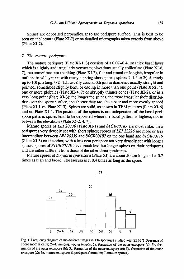
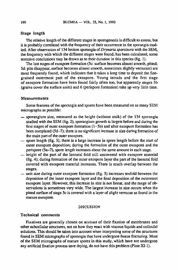
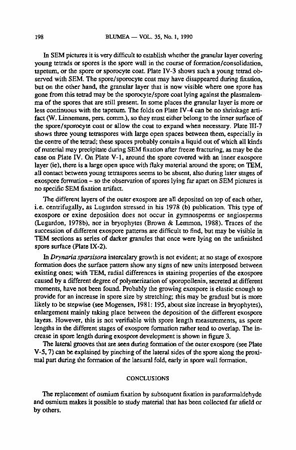
Fig. 1. Frequency diagram of the different stages in 134 sporangia studied with SEM (1. Presence of
spore mother cells; 2-4. meiosis, young tetrads; 5a. formation of the outer exospore (a); 5b. for-
mation of the outer exospore (b); 5c. formation of the outer exospore (c); 5d. formation of the outer
exospore (d); 5e. mature exospore; 6. perispore formation;7. mature spores).
BLUMEA VOL. 35, No. 1, 1990190
Stage length
The relative length of the different stages in sporogenesis is difficult to assess, but
it is probably correlated with the frequency of their occurrence in the sporangia stud-
ied. After observation of 134broken sporangia of Drynaria sparsisora with the SEM,
the frequency withwhich the different stages were found, has been calculated; some
tentative conclusions may be drawn as to their duration in this species (fig. 1).
The last stages ofexospore formation (5c: surface becomes almost smooth, pitted;
5d: pits disappear, surface becomes almost smooth, sometimes slightly verrucate) are
most frequently found, which indicates that it takes a long time to deposit the fine-
grained outermost part of the exospore. Young tetrads and the first stage
of exospore formation have been found fairly often too, but apparently stages 5b
(grains cover the surface units) and 6 (perispore formation) take up very little time.
Measurements
Some features of the sporangia and spores have been measuredon as many SEM
micrographs as possible:
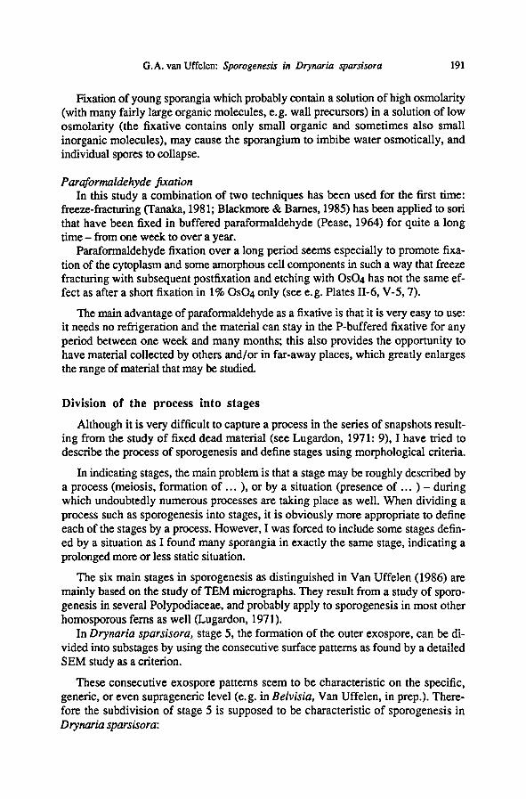
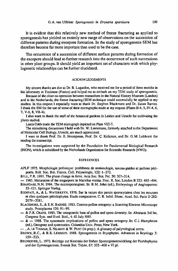
— sporangium size, measured as the height (without stalk) of the 134 sporangia
studied with the SEM (fig. 2); sporangium growth is largest before and during the
first stages of outer exospore formation (l-5b) and after exospore formation has
been completed (5d-7); there is no significant increase in size during formationof
the mainpart of the outer exospore.
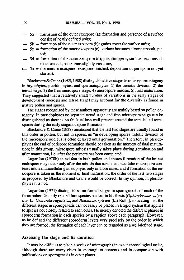
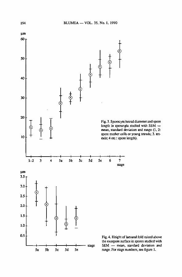
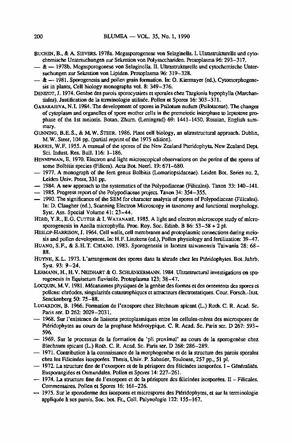
— spore length (fig. 3); there is a large increase in spore length before the start of
outer exospore deposition; during the formationof the outer exospore and the
perispore (5a-7), spore length increases about the same amount in each stage.
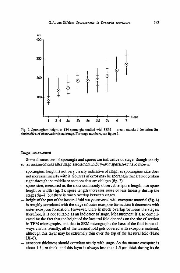
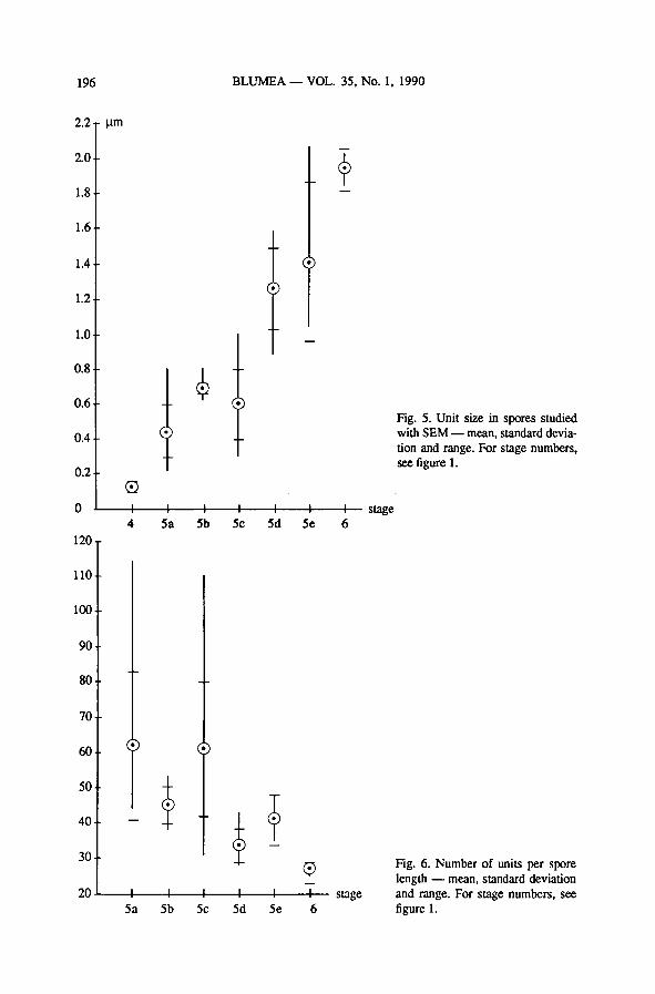
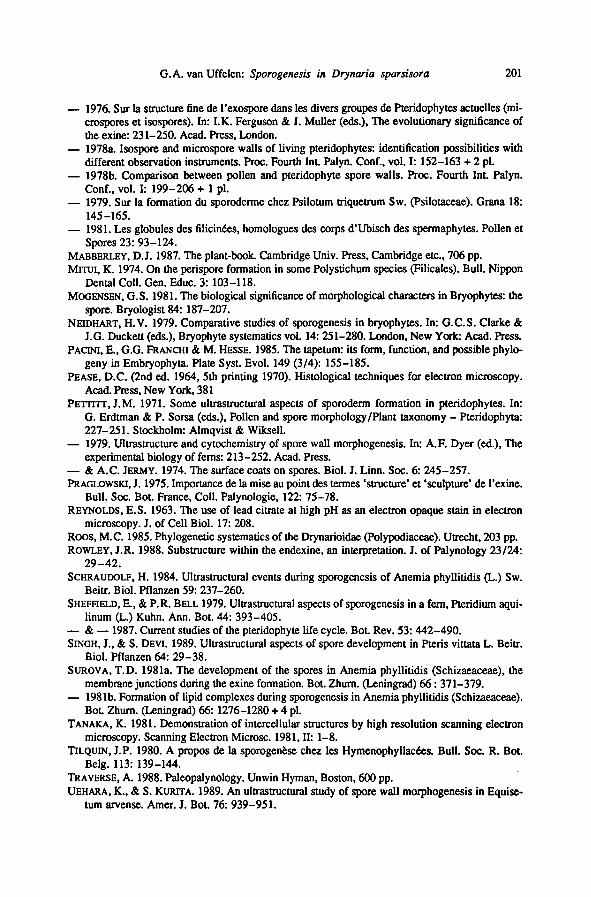
— height of the part of the laesural fold still uncovered with exospore material
(fig. 4); during formationof the outer exospore layer the part of the laesural fold
covered with exospore material increases. There is much overlap between the
stages.
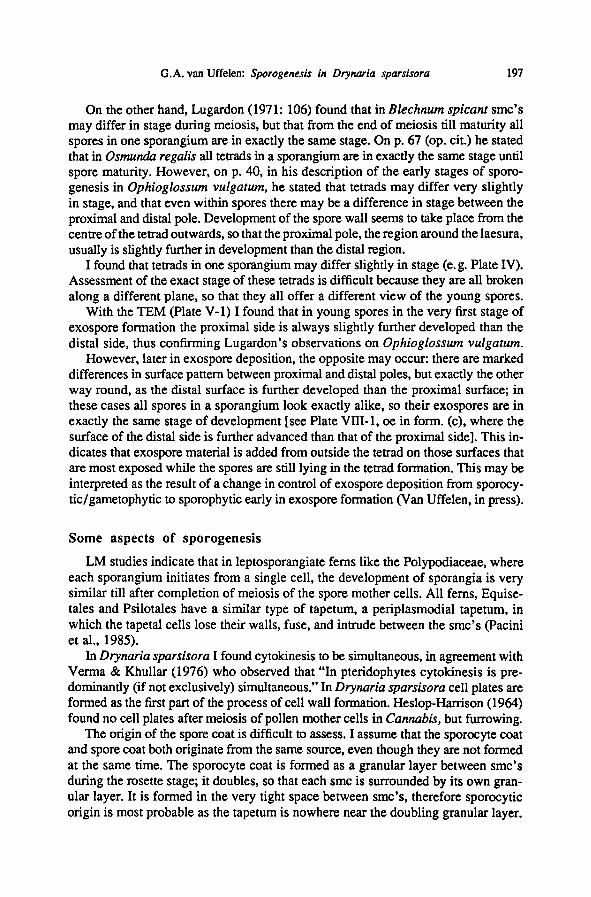
—unit size during outer exospore formation (fig. 5) increases tenfold between the
deposition of the innerexospore layer and the final deposition of the outermost
exospore layer. However, this increase in size is not linear, and the range of ob-
servations is sometimes very wide. The largest increase in size occurs when the
pitted surface of stage 5c is covered with a layer of slight verrucae as found in the
mature exospore.
DISCUSSION
Technical comments
Fixatives are generally chosen on account of their fixation of membranes and
other subcellularstructures, not on how they react with viscous liquids and colloidal
solutions. This should be taken into account when interpreting some of the structures
foundin SEM micrographs of sporangia that have undergone freeze fracturing. Most
of the SEM micrographs of mature spores in this study, which have not undergone
any artificial fixation process save drying, do not have this problem (Plate XI-1).
191G.A. van Uffelen: Sporogenesis in Drynaria sparsisora
Fixation of young sporangia which probably contain a solution of high osmolality
(with many fairly large organic molecules, e.g. wall precursors) in a solution of low
osmolality (the fixative contains only small organic and sometimes also small
inorganic molecules), may cause the sporangium to imbibe water osmotically, and
individual spores to collapse.
Paraformaldehyde fixation
In this study a combinationof two techniques has been used for the first time:
freeze-fracturing (Tanaka, 1981; Blackmore& Barnes, 1985) has been applied to sori
that have been fixed in buffered paraformaldehyde (Pease, 1964) for quite a long
time - from one week to over a year.
Paraformaldehyde fixation over a long period seems especially to promote fixa-
tion of the cytoplasm and some amorphous cell components in such a way that freeze
fracturing with subsequent postfixation and etching with OSO4 has not the same ef-
fect as after a short fixation in 1% OSO4 only (see e.g. Plates II-6, V-5,7).
The mainadvantage of paraformaldehyde as a fixative is that it is very easy to use:
it needs no refrigeration and the material can stay in the P-buffered fixative for any
period between one week and many months; this also provides the opportunity to
have materialcollected by others and/or in far-away places, which greatly enlarges
the range ofmaterial that may be studied.
Division of the process into stages
Although it is very difficult to capture a process in the series of snapshots result-
ing from the study of fixed dead material (see Lugardon, 1971: 9), I have tried to
describe the process of sporogenesis and definestages using morphological criteria.
In indicating stages, the mainproblem is that a stage may be roughly described by
a process (meiosis, formation of...
), or by a situation (presence of ... ) - duringwhich undoubtedly numerous processes are taking place as well. When dividing a
process such as sporogenesis into stages, it is obviously more appropriate to define
each of the stages by a process. However, I was forced to include some stages defin-
ed by a situation as I found many sporangia in exactly the same stage, indicating a
prolonged more or less static situation.
The six main stages in sporogenesis as distinguished in Van Uffelen (1986) are
mainly based on the study of TEM micrographs. They result froma study of sporo-
genesis in several Polypodiaceae, and probably apply to sporogenesis in most other
homosporous ferns as well (Lugardon, 1971).
In Drynaria sparsisora, stage 5, the formation of the outer exospore, can be di-
vided into substages by using the consecutive surface patterns as found by a detailed
SEM study as a criterion.
These consecutive exospore patterns seem to be characteristic on the specific,
generic, or even suprageneric level (e.g. in Belvisia, Van Uffelen, in prep.). There-
fore the subdivision of stage 5 is supposed to be characteristic of sporogenesis in
Drynaria sparsisora:
BLUMEA VOL. 35, No. 1, 1990192
—5a = formationof the outer exospore (a): formation and presence of a surface
consist of neatly defined units;
—5b = formation of the outer exospore (b): grains cover the surface units;
— 5c = formationof the outer exospore (c); surface becomes almost smooth, pit-
ted;
—5d = formationof the outer exospore (d); pits disappear, surface becomes al-
most smooth, sometimes slightly verrucate;
(— 5e = the mature exospore: exospore finished, deposition of perispore not yet
started).
Blackmore & Crane(1985,1988) distinguished five stages in microspore ontogeny
in bryophytes, pteridophytes, and spermatophytes: 1) the meiotic division, 2) the
tetrad stage, 3) the free microspore stage, 4) microspore mitosis, 5) final maturation.
They suggested that a relatively small number of variations in the early stages of
development (meiosis and tetrad stage) may account for the diversity as found in
mature pollen and spores.
The stages recognized by these authors apparently are mainly based on pollen on-
togeny. In pteridophytes no separate tetrad stage and free microspore stage can be
distinguished as there is no thick callose wall present around the tetrads and tetra-
spores during the early stages of spore formation.
Blackmore & Crane (1988) mentionedthat the last two stages are usually foundin
this order in pollen, but not in spores, as "in developing spores mitotic division of
the microspore nucleus is often delayed until germination." Therefore, in pterido-
phytes the end of perispore formation should be taken as the moment of final matura-
tion; in this group, microspore mitosis usually takes place during germination and
after maturation, i.e. after the perispore has been completed.
Lugardon (1978b) stated that in both pollen and spores formation of the intine/
endospore may occur only after the mitosis that turns the unicellular microspore con-
tents into a multicellulargametophyte; only in those cases, and if formationof the en-
dospore is taken as the moment of final maturation, the order of the last two stages
as proposed by Blackmore and Crane would be correct. In my opinion, in pterido-
phytes it is not.
Lugardon (1971) distinguished no formal stages in sporogenesis of each of the
three rather distantly related fern species studied in his thesis [Ophioglossum vulga-
tum L., Osmunda regalis L., and Blechnum spicant (L.) Roth.], indicating that the
different stages in sporogenesis cannot easily be placed in a rigid system that applies
to species not closely related to each other. He merely denoted the differentphases in
sporoderm formation in each species by a caption above each paragraph. However,
as he defined the different sporoderm layers very precisely by the order in which
they are formed, the formationof each layer can be regarded as a well-definedstage.
Assessing the stage and its duration
It may be difficult to place a series of micrographs in exact chronological order,
although there are many clues in sporangium contents and in comparison with
publications on sporogenesis in other plants.
193G.A. van Uffelen: Sporogenesis in Drynaria sparsisora
Stage assessment
Some dimensions of sporangia and spores are indicativeof stage, though poorly
so, as measurements after stage assessment in Drynaria sparsisora have shown:
— sporangium height is not very clearly indicative of stage, as sporangium size does
not increase linearly with it. Sources oferror may be sporangia that are not broken
right through the middleor sections that are oblique (fig. 2).
— spore size, measured as the most commonly observable spore length, not spore
height or width (fig. 3); spore length increases more or less linearly during the
stages 5a-7, but there is much overlap between stages.
— height ofthe part ofthe laesural foldnot yet covered withexospore material(fig. 4)
is roughly correlatedwith the stageof outer exospore formation; it decreases with
outer exospore formation. However, there is much overlap between the stages;
therefore, it is not suitable as an indicator of stage. Measurement is also compli-
cated by the fact that the height of the laesural fold depends on the site of section
in TEM micrographs, and that in SEM micrographs the base of the fold is not al-
ways visible. Finally, all of the laesural fold gets covered with exospore material,
although this layer may be extremely thin over the top of the laesural fold (Plate
IX-6).
— exospore thickness should correlate neatly with stage. As the mature exospore is
about 1.5 pm thick, and this layer is always less than 1.5 pm thick during its de
Fig. 2. Sporangium height in 134 sporangia studied with SEM— mean, standard deviation (in-
cludes 68% of observations) and range. For stage numbers, see figure 1.
BLUMEA VOL. 35, No. 1, 1990194
Fig. 3. Sporocyte/tetrad diameter and spore
length in sporangia studied with SEM—
mean, standard deviation and range (1, 2:
spore mother cells or young tetrads; 3. tet-
rads; 4 etc.: spore length).
Fig. 4. Height of laesural fold raised above
the exospore surface in spores studied with
SEM — mean, standard deviation and
range. For stage numbers, see figure 1.
195G.A. van Uffelen: Sporogenesis in Drynaria sparsisora
position, the exospore does not get demonstrably thinner in the process of matu-
ration. However, it is very difficult to measure, since there are not enough SEM
micrographs of broken spores, and not enough TEM measurements that can be
correlated with a surface pattern to go with a stage of outer exospore formation.
Moreover, exospore thickness is never uniform throughout the whole spore: near
the laesura the exospore is always thicker than elsewhere on the spore.
— unit size of the exospore surface (fig. 5); its increase is not linear, and there is
much overlap between stages. The increase in unit size is partly explainable as a
change in surface pattern, and partly due to stretching of the exospore to accom-
modate increase in spore size.
— spore length dividedby unit size: the numberofunits along the length ofone spore
(fig. 6) decreases with the progress of exospore formation.This can be explained
by the changes in surface pattern that occur during exospore formation.
Stage length
Assessment of stage length (fig. 1) by way of counting the frequency of occur-
rence of the different stages in the sporangia studied may have been influenced by the
following factors:
— age of the sori studied; sori may contain mainly young or mainly old sporangia;
this bias can be reduced by the study of a large number of sori (in this study:
c. 40 sori);
— small (young) sporangia are more easily missed in fracturing the sporangia of a
sorus; this leads to the occurrence of more large, old, broken sporangia and there-
fore to too long estimates of the laterstages; as late stages may also be found in
small sporangia, this bias is rather low.
—with freeze fracturing the frequency of encountering a stage could be positively
influenced by the fragility of the sporangia in that particular stage, negatively by
the fragility of their stalks.
Stages within one sporangium
Another point is whether all smc's, tetrads, or spores in one sporangium are in
exactly the same stage.
Before meiosis, connections between the cells of the sporogenous tissue may
synchronize development. Heslop-Harrison (1964: 41-42) found plasmodesmatabetween pollen mother cells (pmc's), between pmc's and tapetum and between tape-
tum cells in Cannabis. These plasmodesmata are eliminated by the growth of the cal-
losic special mother cell wall. Later during prophase, channels up to 1.5 p.m wide
develop between pmc's; they are severed during the first meiotic division. These cy-
tomictic channels are thought to serve synchronization of part of the process of meio-
sis.
In Pteridium aquilinum Sheffield& Bell (1979: 399) found that these channels are
blocked during the metaphase/anaphase period and that development of the smc's
becomes increasingly asynchronous afterwards.
BLUMEA VOL. 35, No. 1, 1990196
Fig. 5. Unit size in spores studied
with SEM— mean, standard devia-
tion and range. For stage numbers,
see figure 1.
Fig. 6. Number of units per spore
length — mean, standard deviation
and range. For stage numbers, see
figure 1.
197G.A. van Uffelen: Sporogenesis in Drynaria sparsisora
On the other hand, Lugardon (1971: 106) found that in Blechnum spicant smc's
may differin stage during meiosis, but that from the end of meiosis till maturity all
spores in one sporangium are in exactly the same stage. On p. 67 (op. cit.) he stated
that in Osmunda regalis all tetrads in a sporangium are in exactly the same stage until
spore maturity. However, on p. 40, in his description of the early stages of sporo-
genesis in Ophioglossum vulgatum, he stated that tetrads may differ very slightly
in stage, and that even within spores there may be a difference in stage between the
proximal and distal pole. Development of the spore wall seems to take place from the
centre ofthe tetrad outwards, so that the proximal pole, the region around the laesura,
usually is slighdy further in development than the distal region.
I found that tetrads in one sporangium may differ slightly in stage (e. g. Plate IV).
Assessment of the exact stageof these tetrads is difficultbecause they are all broken
along a different plane, so that they all offer a different view of the young spores.
With the TEM (Plate V-l) I found that in young spores in the very first stage of
exospore formation the proximal side is always slightly further developed than the
distal side, thus confirming Lugardon's observations on Ophioglossum vulgatum.
However, later inexospore deposition, the opposite may occur: there are marked
differences in surface pattern between proximal and distal poles, but exactly the other
way round, as the distal surface is further developed than the proximal surface; in
these cases all spores in a sporangium look exactly alike, so their exospores are in
exactly the same stage of development [see Plate VIII-1, oe in form, (c), where the
surface of the distal side is further advanced than that of the proximal side]. This in-
dicates that exospore material is addedfrom outside the tetrad on those surfaces that
are most exposed while the spores are still lying in the tetrad formation. This may be
interpreted as the result of a change in control of exospore deposition from sporocy-
tic/gametophytic to sporophytic early in exospore formation(Van Uffelen, in press).
Some aspects of sporogenesis
LM studies indicate that in leptosporangiate ferns like the Polypodiaceae, where
each sporangium initiates from a single cell, the development of sporangia is very
similar till after completion of meiosis of the spore mother cells. All ferns, Equise-tales and Psilotales have a similar type of tapetum, a periplasmodial tapetum, in
which the tapetal cells lose their walls, fuse, and intrudebetween the smc's (Pacini
et al„ 1985).
In Drynaria sparsisora I found cytokinesis to be simultaneous, in agreementwith
Verma & Khullar (1976) who observed that "In pteridophytes cytokinesis is pre-
dominandy (if not exclusively) simultaneous." In Drynaria sparsisora cell plates are
formed as the first part of the process ofcell wall formation. Heslop-Harrison (1964)found no cell plates after meiosis of pollen mother cells in Cannabis, but furrowing.
The origin of the spore coat is difficult to assess. I assume that the sporocyte coat
and spore coat both originate from the same source, even though they are not formed
at the same time. The sporocyte coat is formed as a granular layer between smc's
during the rosette stage; it doubles, so that each smc is surrounded by its own gran-
ular layer. It is formed in the very tight space between smc's, therefore sporocytic
origin is most probable as the tapetum is nowhere near the doubling granular layer.
198 BLUMEA VOL. 35, No. 1, 1990
In SEM pictures it is very difficult to establish whether the granular layer covering
young tetrads or spores is the spore wall in the course of formation/consolidation,
tapetum, or the spore or sporocyte coat. Plate IV-3 shows such a young tetrad ob-
served with SEM. The spore/sporocyte coat may have disappeared during fixation,
but on the other hand, the granular layer that is now visible where one spore has
gone from this tetrad may be the sporocyte/spore coat lying against the plasmalem-
ma of the spores that are still present. In some places the granular layer is more or
less continuous with the tapetum. The folds on Plate IV-4 can be no shrinkage arti-
fact (W. Linnemans, pers. comm.), so they must either belong to the inner surface of
the spore/sporocyte coat or allow the coat to expand when necessary. Plate III-7
shows three young tetraspores with large open spaces between them, especially in
the centre of the tetrad; these spaces probably containa liquid out of which all kinds
of material may precipitate during SEM fixation after freeze fracturing, as may be the
case on Plate IV. On Plate V-l, around the spore covered with an inner exospore
layer (ie), there is a large open space with flaky material around the spore; on TEM,
all contact between young tetraspores seems to be absent, also during later stages of
exospore formation- so the observation of spores lying far apart on SEM pictures is
no specific SEM fixation artifact.
The different layers of the outer exospore are all deposited on top of each other,
i.e. centrifugally, as Lugardon stressed in his 1978 (b) publication. This type of
exospore or exine deposition does not occur in gymnosperms or angiosperms
(Lugardon, 1978b), nor in bryophytes (Brown & Lemmon, 1988). Traces of the
succession of differentexospore patterns are difficult to find, but may be visible in
TEM sections as series of darker granules that once were lying on the unfinished
spore surface (Plate LX-2).
In Drynaria sparsisora intercalary growth is not evident; at no stage of exospore
formation does the surface pattern show any signs of new units interposed between
existing ones; with TEM, radial differences in staining properties of the exospore
caused by a different degree of polymerization of sporopollenin, secreted at different
moments, have not been found. Probably the growing exospore is elastic enough to
provide for an increase in spore size by stretching; this may be gradual but is more
likely to be stepwise (see Mogensen, 1981: 195, about size increase in bryophytes),
enlargement mainly taking place between the deposition of the different exospore
layers. However, this is not verifiable with spore length measurements, as spore
lengths in the different stages of exospore formation rather tend to overlap. The in-
crease in spore length during exospore development is shown in figure 3.
The lateral grooves that are seen during formationof the outer exospore (see Plate
V-5, 7) can be explained by pinching of the lateral sides of the spore along the proxi-
mal part during the formationof the laesural fold, early in spore wall formation.
CONCLUSIONS
The replacement of osmium fixation by subsequent fixation in paraformaldehyde
and osmium makes it possible to study material that has been collected far afield or
by others.
199G.A. van Uffelen: Sporogenesis in Drynaria sparsisora
It is evident that this relatively new method of freeze fracturing as applied to
sporogenesis has yielded an entirely new range of observations on the succession of
differentpatterns during exospore formation. In the study of sporogenesis SEM has
therefore become far more important than used to be the case.
The occurrence of a succession of different surface patterns during formationof
the exospore should lead to furtherresearch into the occurrence of such successions
inother plant groups. It should yield an important set of characters with which phy-
logenetic relationships can be furtherelucidated.
ACKNOWLEDGEMENTS
My sincere thanks are due to Dr. B. Lugardon, who received me for a period of three months in
his laboratory in Toulouse (France) and helped me to embark on my TEM study of sporogenesis.Because of the close co-operation between researchers in the Natural History Museum (London)
and in the Netherlands, the freeze fracturing/SEM technique could successfully be applied to my
studies. In this respect I especially want to thank Dr. Stephen Blackmore and Dr. Susan Barnes.
I thank the BM for the use of some of their micrographs made at my request (Plates II-4, 5, IV-4, 6,
7, V-6, 8, VIM).
I also want to thank the staff of the botanical gardens in Leiden and Utrecht for cultivating the
plants studied.
Laura Cobb made the TEM micrograph depicted on Plate VIII-7.
The stimulating discussions I held with Dr. W. Linnemans, formerly attached to the Department
of Molecular Cell Biology, Utrecht, are much appreciated.
I want to thank Prof. Dr. E. Hennipman, Prof. Dr. C. Kalkman, and Dr. G.M. Lokhorst for
reading the manuscript.
The investigations were supported by the Foundation for Fundamental Biological Research
(BION), which is subsidized by the Netherlands Organization for Scientific Research (NWO).
REFERENCES
APLF 1975. Morphologie pollinique: problemes de terminologie, taxons-guides et pollens peri-
pores. Bull. Soc. Bot. France, Coll. Palynologie, 122: 1-272.
BF.LI., P.R. 1981. The phase change in ferns. Acta Soc. Bot. Pol. 50: 307-314.
—1985. Maturation of the megaspore in Marsilea vestita. Proc. R. Soc. London B 223: 485-494.
BHANDARJ, N.N. 1984. The microsporangium. In: B.M. John (ed.),Embryology of Angiosperms:53-121. Springer Verlag.
BIENFAIT, A., & L. WATERKEYN. 1976. Sur la nature des parois sporocytaires chez les mousses
et chez quelques ptdridophytes. Etude comparative. C. R. hebd. Scanc. Acad. Sci. Paris D 282:
2079-2081.
BLACKMORE, S., & S.H. BARNES. 1985. Cosmos pollen ontogeny: a Scanning Electron Microscope
study. Protoplasma 126: 91-99.
—& P.R. CRANE. 1985. The ontogenetic base of pollen and spore diversity. In: Abstracts 3rd Int.
Congress Syst. and Evol. Biol., 4-10 July 1985.
— & — 1988. The systematic implications of pollen and spore ontogeny. In: C.J. Humphries
(ed.), Ontogeny and systematics. Columbia Univ. Press, New York.
—
,A. LE THOMAS, S. NILSSON & W. PUNT (in prep.). A glossary of palynological terms.
BROWN, R.C., & B.E. LEMMON. 1988. Sporogenesis in Bryophytes. Advances in Bryology 3:
159-223.
BRUNKENER, L. 1973. Beitrage zur Kenntnis der friihen Sporangienentwicklung der Ptcridophytenund der Gymnospermen. Svensk Bot. Tidskr. 67: 333-400 + VI pi.
BLUMEA VOL. 35, No. 1, 1990200
BUCHEN, B., & A. SIEVERS. 1978a. Megasporogenese von Selaginella. I. Ultrastrukturelle und cyto-
chemische Untersuchungen zur Sekretion von Polysacchariden. Protoplasma 96: 293-317.
— & — 1978b. Megasporogenese von Selaginella. II. Ultrastrukturelle und cytochemische Unter-
suchungen zur Sekretion von Lipiden. Protoplasma 96: 319-328.
—& — 1981. Sporogenesis and pollen grain formation. In: O. Kiermayer (ed.), Cytomorphogene-sis in plants, Cell biology monographs vol. 8: 349-376.
DENIZOT, J. 1974. Genfese des parois sporocytaires et sporales chez Targionia hypophylla (Marchan-
tiales). Justification de la terminologie utilisee. Pollen et Spores 16: 303-371.
GABARAJEVA, N.I. 1984. The development ofspores in Psilotum nudum (Psilotaceae). The changesofcytoplasm and organelles of spore mother cells in the premeiotic interphase to leptotene pro-
phase of the 1st meiosis. Botan. Zhurn. (Leningrad) 69: 1441-1450. Russian, English sum-
mary.
GUNNING, B.E.S., & M.W. STEER. 1986. Plant cell biology, an ultrastructural approach. Dublin,
M. W. Steer, 104 pp. (partial reprint of the 1975 edition).
HARRIS, W.F. 1955. A manual of the spores of the New Zealand Pteridophyta. New Zealand Dept.Sci. Indust. Res. Bull. 116: 1-186.
HENNIPMAN, E. 1970. Electron and light microscopical observations on the perine of the spores of
some Bolbitis species (Filices). Acta Bot. Neerl. 19: 671-680.
— 1977. A monograph of the fern genus Bolbitis (Lomariopsidaceae). Leiden Bot. Series no. 2,
Leiden Univ. Press, 331 pp.
— 1984. A new approach to the systematics ofthe Polypodiaceae (Filicales). Taxon 33: 140-141.
— 1985. Progress report of the Polypodiaceae project. Taxon 34: 354-355.
—1990. The significance of the SEM for character analysis of spores of Polypodiaceae (Filicales)In: D. Claugher (ed.), Scanning Electron Microscopy in taxonomy and functional morphology
Syst. Ass. Special Volume 41: 23-44.
HERD, Y.R., E.G. CUTTER & I. WATANABE. 1985. A light and electron microscope study of micro-
sporogenesis in Azolla microphylla. Proc. Roy. Soc. Edinb. B 86: 53-58 + 2 pi.
HESLOP-HARRISON, J. 1964. Cell walls, cell membranes and protoplasmic connections during meio-
sis and pollen development. In: H.F. Linskens (ed.), Pollen physiology and fertilization: 39-47.
HUANG, S.F., & S.H.T. CHIANG. 1983. Sporogenesis in Isoetes taiwanensis Taiwania 28: 68-
88.
HUYNH, K.L. 1973. L'arrangement des spores dans la tetrade chez les Pteridophytes. Bot. Jahrb.
Syst. 93: 9-24.
LEHMANN, H., H.V. NEIDHART & G. SCHLENKERMANN. 1984.Ultrastructural investigations on spo-
rogenesis in Equisetum fluviatile. Protoplasma 123: 38-47.
LOCQUIN, M.V. 1981. Mccanismes physiques de la genese des formes et des ornements des spores et
pollens: chreodes, singularity catastrophiques et attracteurs electrostatiques. Cour. Forsch.-Inst.
Senckenberg 50: 75-88.
LUGARDON, B. 1966. Formation de l'exospore chez Blechnum spicant (L.) Roth. C. R. Acad. Sc.
Paris ser. D 262: 2029-2031.
—1968. Sur 1'existence de liaisons protoplasmiques entre les cellules-meres des microspores de
Pteridophytes au cours de la prophase heterotypique. C. R. Acad. Sc. Paris ser. D 267: 593-
596.
— 1969. Sur le processus de la formation du 'pli proximal' au cours de la sporogenese chez
Blechnum spicant (L.) Roth. C. R. Acad. Sc. Paris ser. D 268: 286-289.
—1971. Contribution a la connaissance de la morphogenese et de la structure des parois sporaleschez les Filicinees isosporees. Thesis, Univ. P. Sabatier, Toulouse,257 pp., 51 pi.
— 1972.La structure fine de l'exospore et de la perispore des filicinees isosporees. I-
Generality.
Eusporangi6es et Osmundales. Pollen et Spores 14: 227-261.
— 1974. La structure fine de l'exospore et de la perispore des filicinees isosporees. II - Filicales.
Commentaires. Pollen et Spores 16: 161-226.
— 1975. Sur le sporoderme des isospores et microspores des Pteridophytes, et sur la terminologie
appliquee il ses parois. Soc. bot. Fr., Coll. Palynologie 122: 155-167.
201G.A. van Uffelen: Sporogenesis in Drynaria sparsisora
— 1976. Sur la structure fine de l'exospore dans les divers groupes de Pteridophytes actuelles (mi-
crospores et isospores). In: I.K.Ferguson & J. Muller (eds.), The evolutionary significance of
the exine: 231-250. Acad. Press, London.
—1978a. Isospore and microspore walls of living pteridophytes: identification possibilities with
different observation instruments. Proc. Fourth Int.Palyn. Conf., vol. I: 152-163 + 2 pi.
—1978b. Comparison between pollen and pteridophyte spore walls. Proc. Fourth Int. Palyn.
Conf., vol. I: 199-206 + 1 pi.
—1979. Sur la formation du sporoderme chez Psilotum triquetrum Sw. (Psilotaceae). Grana 18:
145-165.
—1981. Les globules des filicintes, homologues des corps d'Ubisch des spermaphytes. Pollen et
Spores 23: 93-124.
MABBERLEY, D.J. 1987. The plant-book. Cambridge Univ. Press, Cambridge etc., 706 pp.
MITUI, K. 1974. On the perispore formation in somePolystichum species (Filicales). Bull. Nippon
Dental Coll. Gen. Educ. 3: 103-118.
MOGENSEN, G.S. 1981. The biological significance of morphological characters in Bryophytes: the
spore. Bryologist 84: 187-207.
NEIDHART, H.V. 1979. Comparative studies of sporogenesis in bryophytes. In: G.C.S. Clarke &
J.G. Duckett (eds.), Bryophyte systematics vol. 14: 251-280. London, New York: Acad. Press.
PACINI, E., G.G. FRANCHI & M. HESSE. 1985. The tapetum: its form, function, and possible phyto-
geny in Embryophyta. Plate Syst. Evol. 149 (3/4): 155-185.
PEASE, D.C. (2nd ed. 1964, 5th printing 1970). Histological techniques for electron microscopy.
Acad. Press, New York, 381
PETTITT, J.M. 1971. Some ultrastructural aspects of sporoderm formation in pteridophytes. In:
G. Erdtman & P. Sorsa (eds.), Pollen and spore morphology/Plant taxonomy - Pteridophyta:
227-251. Stockholm: Almqvist & Wiksell.
— 1979. Ultrastructure and cytochemistry of spore wall morphogenesis. In: A.F. Dyer (ed.), The
experimental biology of ferns: 213-252. Acad. Press.
— & A.C. JERMY. 1974. The surface coats on spores. Biol. J. Linn. Soc. 6: 245-257.
PRAGLOWSKI, J. 1975. Importance de la mise au point des termes 'structure' et 'sculpture' de 1'exine.
Bull. Soc. Bot. France, Coll. Palynologie, 122: 75-78.
REYNOLDS, E.S. 1963. The use of lead citrate at high pH as an electron opaque stain in electron
microscopy. J. ofCell Biol. 17: 208.
Roos, M.C. 1985. Phylogenetic systematics ofthe Drynarioidae (Polypodiaceae). Utrecht, 203 pp.
ROWLEY, J.R. 1988. Substructure within the endexine, an interpretation. J. ofPalynology 23/24:
29-42.
SCHRAUDOLF, H. 1984. Ultrastructural events during sporogenesis of Anemia phyllitidis (L.) Sw.
Beitr. Biol. Pflanzen 59: 237-260.
SHEFFIELD, E., & P.R. BELL 1979. Ultrastructural aspects of sporogenesis in a fern, Pteridium aqui-linum (L.) Kuhn. Ann. Bot. 44: 393-405.
— & — 1987. Current studies of the pteridophyte life cycle. Bot. Rev. 53: 442-490.
SINGH, J., & S. DEVI. 1989. Ultrastructural aspects of spore developmentin Pteris vittata L. Beitr.
Biol. Pflanzen 64: 29-
38.
SUROVA, T.D. 1981a. The development of the spores in Anemia phyllitidis (Schizaeaceae), the
membrane junctions during the exine formation. Bot. Zhum. (Leningrad)66 : 371-379.
—1981b. Formation of lipid complexes during sporogenesis in Anemia phyllitidis (Schizaeaceae).BoL Zhurn. (Leningrad) 66: 1276-1280+ 4 pi.
TANAKA, K. 1981. Demonstration of intercellular structures by high resolution scanning electron
microscopy. Scanning Electron Microsc. 1981, II: 1-8.
TILQUIN, J.P. 1980. A propos de la sporogenbse chez les Hymenophyllacdes. Bull. Soc. R. Bot.
Belg. 113: 139-144.
TRAVERSE, A. 1988. Paleopalynology. Unwin Hyman, Boston, 600 pp.
UEHARA, K., & S. KURITA. 1989. An ultrastructural study of spore wall morphogenesis in Equise-
tum arvense. Amer. J. Bot. 76: 939-951.
BLUMEA VOL. 35, No. 1, 1990202
UFFELEN, G.A. VAN. 1985. Spore wall formation in Polypodiaceae (poster abstract). Proc. Roy.
Acad. Edinb. 86B: 443-445.
—1986. Sporogenesis in Drynaria sparsisora (Desv.) Moore (Polypodiaceae) (talk abstract). Acta
Bot. Neerl. 35: 116-117.
— (in press). The control of spore wall formation. SysL Ass. Special Volume.
— (in prep.). Sporogenesis in Polypodiaceae (Filicales). II. Belvisia mucronata (Fee) Copel. and
Microgramma ciliata (Willd.) Copel.
— & E. HENNIPMAN. 1985. The spores of Pyrrosia Mirbel (Polypodiaceae), a SEM study. Pollen et
Spores 27: 155-197.
VERMA, S.C., & S.P. KHULLAR. 1976. Adaptive significance of simultaneous cytokinesis in
pteridophytes. Phytomorphology 26: 96-102.
LEGENDS OF THE PLATES
Fixation data—
SEM: no fixation, i.e. dried only (nofix.); standard fixation with osmiumtetroxide
(osfix.); paraformaldehyde fixation (parafix.) —TEM: glutaraldehydefixation (glufix.); paraformal-
dehyde fixation (parafix.)
[c = channel; ie = inner exospore; n = nucleus; oe = outer exospore; sc = spore/sporocyte coat;
smc = spore mother cell; sw =sporangium wall; t = tapetum; te = tetrad; m = tapetal nucleus; v =
vacuole]
Plate I: Spore mother cells—
TEM (LEI 20339, parafix.)
1: sporangium with smc's in the rosette stage; from left to right: sporangiumwall, the double layer
oftapetum cells, rosette of smc's; 4000 x
2: detail of Plate 1-1,showing connections between smc's (arrow); 20000 x
3 : sporocyte coat in the course of doubling (arrow); 4000 x
4 : onesmc in between rosette stage and rounded stage; sporocyte coat completely doubled; 4000 x
5 : sporocyte coat with loops, and nucleus with cytoplasmic intrusions (arrow); 4000 x
6: smc, late in meiosis, irregularly shaped; 3000 x
Plate II: Sorus and spore mother cells — SEM (LEI 22226 ; osfix., unless indicated otherwise)
1: cross section of a sorus; 65 x
2 : sporangium with rounded smc's, surrounded by periplasmodial tapetum (arrow); 700 x
3: detail of one smc from Plate II-2; 3000 x
4 : see Plate 11-2; detail ofvacuolized tapetum; 7000 x
5 ; see Plate II-2; detail of the sporocyte coat (arrow); 17000 x
6: smc-roundedphase, smc's not broken (arrow); parafix.; 650 x
Plate HI: Tetrads during and after meiosis—
TEM {LEI 20339, parafix.) and SEM {LEI 22226,
osfix.)
1: sporangium with tetrads in meiosis; SEM; 500 x
2: detail of one tetrad from Plate III-l; note smc-nucleus (arrow); 2450 x
3 : tetrad just after meiosis; TEM; 2300 x
4 : detail of plasmalemma formation in another such tetrad of the same sporangium; note plasma-
lemma (arrow); TEM; 14000 x
5 : young tetrad, showing position of spores; TEM; 2250 x
6: tetrad showing one pair of young tetraspores; note invaginations between nucleusand cytoplasm
(arrow); TEM; 2300 x
7 : tetrad showing three young tetraspores; TEM; 2400 x
8 : same sporangium, other tetrad; detail of the area between two tetraspores; note spore coat (arrow)
and sporocyte coat (sc); TEM; 16000x
203G.A. van Uffelen: Sporogenesis in Drynaria sparsisora
Plate IV: One sporangium after meiosis—
SEM {LEI 22226, osfix.)
1: sporangium containing young tetrads; 370 x
2: onespore of this sporangium; note laesural fold (arrow); 5000 x
3 : onetetrad of the sporangium; note spore/sporocyte coat (arrow); 2300 x
4 : see Plate IV-3; detail ofsurface coat; note foldlike structures (arrow); 11500x
5 : same sporangium, other tetrad; note tapetal nuclei (tn) and spore/sporocyte coat (sc); 2300 x
6: see Plate IV-5; detail of spore/sporocyte coat (arrow); 20000 x
7 : see Plate IV-1; detail of smooth inner side of the sporocyte coat; 4000 x
8 : same sporangium, other tetrad, one spore in cross section, note the nucleus (n); 3500 x
Plate V: Formation ofthe inner and the first outer exospore layer — TEM (LEI 20339, parafix.)
and SEM {LEI 22226,parafix.)
1: spore with only the inner exospore layer present, formation of the laesural fold (arrow) in prog-
ress; TEM; 4000 x
2: proximal detail of the spore on Plate V-l; 30000 x
3 : formation of the inner exospore layer from without and from within the spore, note white line
(arrow); TEM; 30000 x
4 : first stage of outer exospore formation: depositionof lumps on the inner exospore layer; note ad-
dition of material to the inner exospore layer from within (arrow); TEM; 20000 x
5 : spore with the very first beginning ofthe outer exospore, laesural fold present; SEM; 2200 x
6: detail of Plate V-5; note granules of outer exospore material (arrow); 13000x
7: slightly later stage of outer exospore formation (a), laesural fold also bearing outer exospore ma-
terial (arrow); SEM; 2200 x
8 : detail of Plate V-7; note different sizes of lumps; 13000 x
Plate VI: Formation ofthe outer exospore layer (a) —TEM {LEI 20339, parafix.) and SEM (LEI
22226, parafix.)
1: formation of the outer exospore, cross section of one spore; note closed laesural fold (arrow);
TEM; 3500 x
2: formation of the outer exospore, tetrad with one spore in proximal view and two halved spores;
SEM; 1550 x
3 : same sporangium, other spore -inside of the proximal spore face; note base of laesural fold (ar-
row); SEM; 8000 x
4 : detail of Plate VI-2; note cross section of the laesura (arrow); 8500 x
5 : outer exospore in formation, laesural zone and more distal parts of the spore; TEM; 16000 x
Plate VII: Formation of the outer exospore layer (b-c) — TEM {LEI 20339, parafix.) and SEM
{LEI 22226; osfix., unless stated otherwise)1: one spore in distal view; SEM; 2500 x
2: detail of Plate VII-1, spore surface; llOOOx
3 : one spore with tapetum and many spherical bodies (arrow), which lie embedded in the tapetum
by which they are secreted; SEM; 1800 x
4 : detail of Plate VII-3; spore surface with small pits (arrow); 45000 x
5 : lateral view of a spore; SEM; 1750 x
6: surface detail of another spore from the same sporangium; SEM; 8000 x
7: proximal section of a spore with developing outer exospore (b), laesural fold not yet covered;
note channels (arrow); TEM; 10000 x
8 : detail of a laesura with many channels; SEM (parafix.) 8000 x
BLUMEA VOL. 35, No. 1, 1990204
Plate VIII: Formation of the outer exospore (c-d)
1: one spore in lateral/proximal view; note smooth distal side; SEM ( LEI 22226, osfix.); 1550 x
2: proximal detail of the spore on Plate VIII-1; note uncovered partof thelaesural fold (arrow); 7000 x
3 : same sporangium, detail of tapetum (t) against another spore; SEM (,LEI 22226,osfix.); 7500 x
4 : lateral view of one spore, covered with broad verrucae; SEM (84GR00187,parafix.); 1400 x
5 : same sporangium,proximal detail of another spore; note tiny channels (arrow); SEM (<84GR00-
187, parafix.); 3500 x
6: same stage, surface detail ofa spore with tapetal remains (arrow); SEM (84GR00187, parafix.);7500 x
7 : cross section of an entire spore, covered with large verrucae; TEM (81GR00119, glufix. with
DMSO); 6000 x
Plate IX: The last stage of exospore formation; the mature exospore —TEM (LEI 20339, para-
fix.) and SEM ( LEI 22226, osfix.)
1: section of the laesura; note channels (arrow); TEM; 12000 x
2: same sporangium, different spore; proximal section with largechannels (c); note darker granules
marking former spore surfaces (arrow); 30000 x
3 : cross section ofa spore, detail of the laesura; SEM; 4500 x
4 : lateral/distal detail of cross section of the same spore; note smooth inner surface of the exo-
spore; SEM; 4500 x
5 : broken spore, exospore finished; SEM; 1800 x
6: detail ofPlate IX-5; cross section of the laesura; 7500 x
7: cross section of a laesura; TEM; 16000 x
8: same sporangium,proximal cross section ofanother spore; TEM; 22000 x
Plate X: Perispore formation, the mature perispore —TEM (LEI 20339, parafix.) and SEM (LEI
20339)
1: mature spore; almost all of the perispore abraded, matureexospore surface visible; SEM (nofix.);
1200 x
2: tapetal residue with spherical bodies; note angular blobs (arrow); TEM; 20000 x
3 : exospore; note angularblobs (arrow); TEM; 50000 x
4 : same sporangium as Plate X-3, exospore covered with lamellae (arrow) and blobs; TEM; 70000 x
5 : same sporangium as Plate X-3, exospore with lamellae, blobs and small spine (arrow); TEM;
70000 x
6: exospore with blobs and spherical bodies (arrow); TEM; 27000 x
7 : perispore in formation; proximal view of a spore; SEM (osfix.); 1200 x
8: detail of the laesura on Plate X-7; 5000 x
Plate XI: The mature spore
1: proximal/lateral view of a mature spore; SEM (LEI 20339, nofix.); 1100 x
2: detail of the perispore surface; note the position of the spines with respect to the surface pattern;
SEM (LEI 20339, nofix.); 8000 x
3 : mature spore with longer and sparser spines; SEM (8IGR00119, nofix.); 1000 x
4 : detail of Plate XI-3, showing different types of spines; 4500 x
5: pattern on an LM-slide, probably caused by evaporation and subsequent precipitation of the
glycerin jelly in which the objects were embedded; note resemblance to the basal pattern of the
perispore on Plate XI-2; 90 x
6: detail of a loose perispore fragment; TEM (LEI 20339,parafix.); 16500 x
7: detail of laesura with spines perpendicular to the perispore surface; pattern of the basal layer
smaller than in Plate Xl-2; SEM (84GR00187, nofix.); 4500 x
8 : detail of a perispore with spines perpendicular to the surface, and of the sporangium wall with a
perispore-coated spherical body and other perispore-like particles; SEM (8IGR00119,parafix.);1900 x
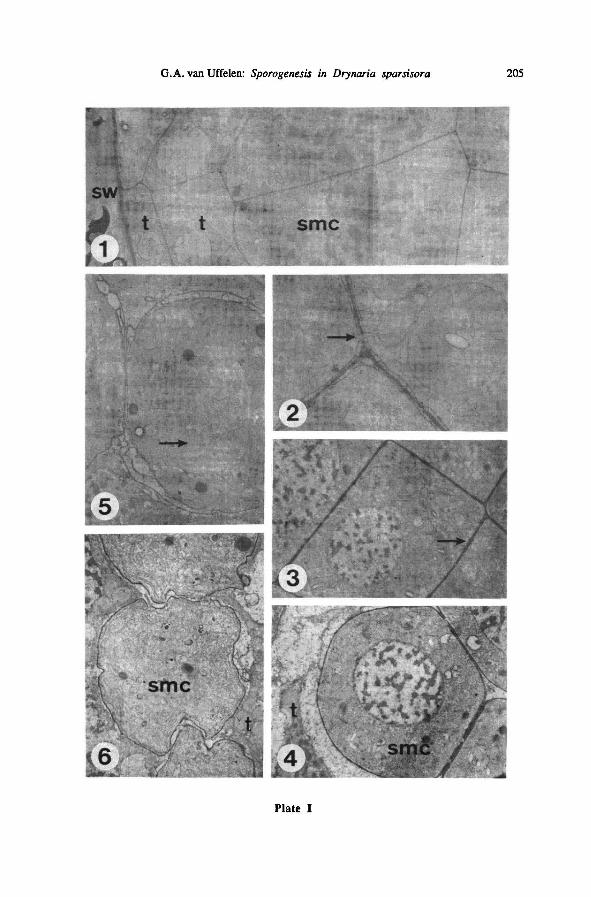
G.A. van Uffelen: Sporogenesis in Drynaria sparsisora 205\I
Plate I
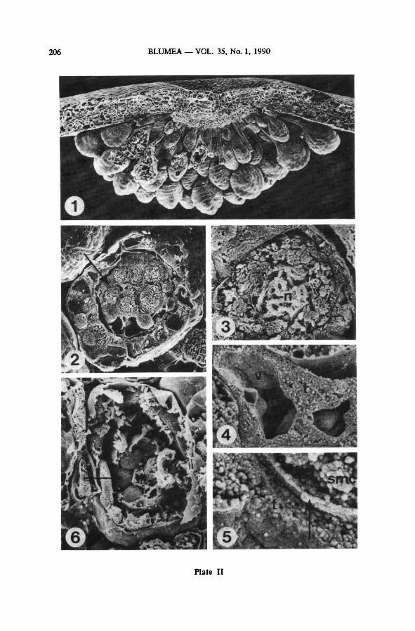
BLUMEA VOL. 35, No. 1, 1990206\II
Plate II
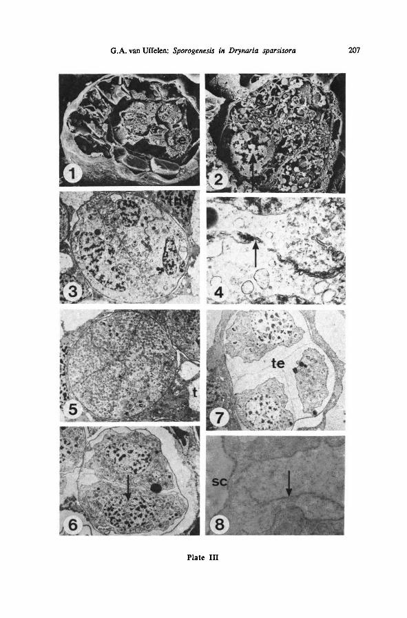
G.A. van Uffelen: Sporogenesis in Drynaria sparsisora 207\III
Plate III
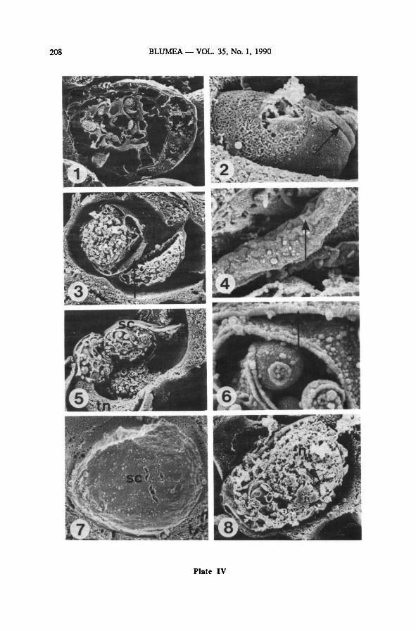
BLUMEA VOL. 35, No. 1, 1990208\IV
Plate IV
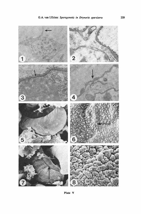
G.A. van Uffelen: Sporogenesis in Drynaria sparsisora 209\V
Plate V
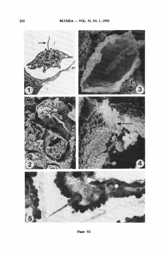
210\VI BLUMEA VOL. 35, No. 1, 1990
Plate VI
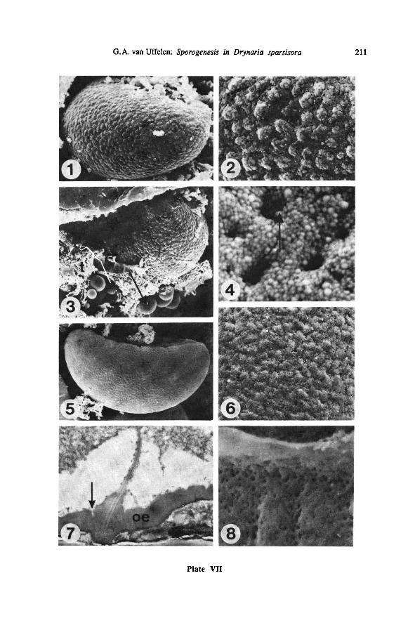
G.A. van Uffelen: Sporogenesis in Drynaria sparsisora 211\VII
Plate VII
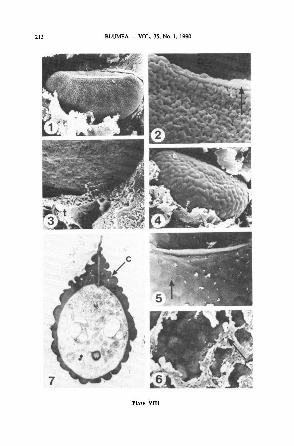
BLUMEA VOL. 35, No. 1, 1990212\VIII
Plate VIII
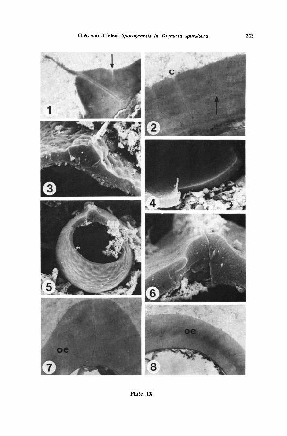
G.A. van Uffelen: Sporogenesis in Drynaria sparsisora 213\IX
Plate IX
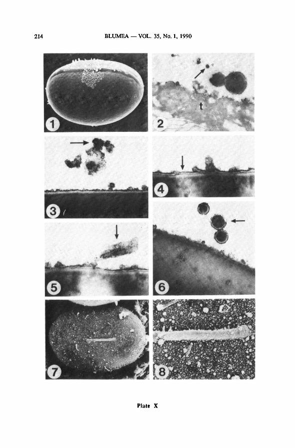
214\X BLUMEA VOL. 35, No. 1, 1990
Plate X
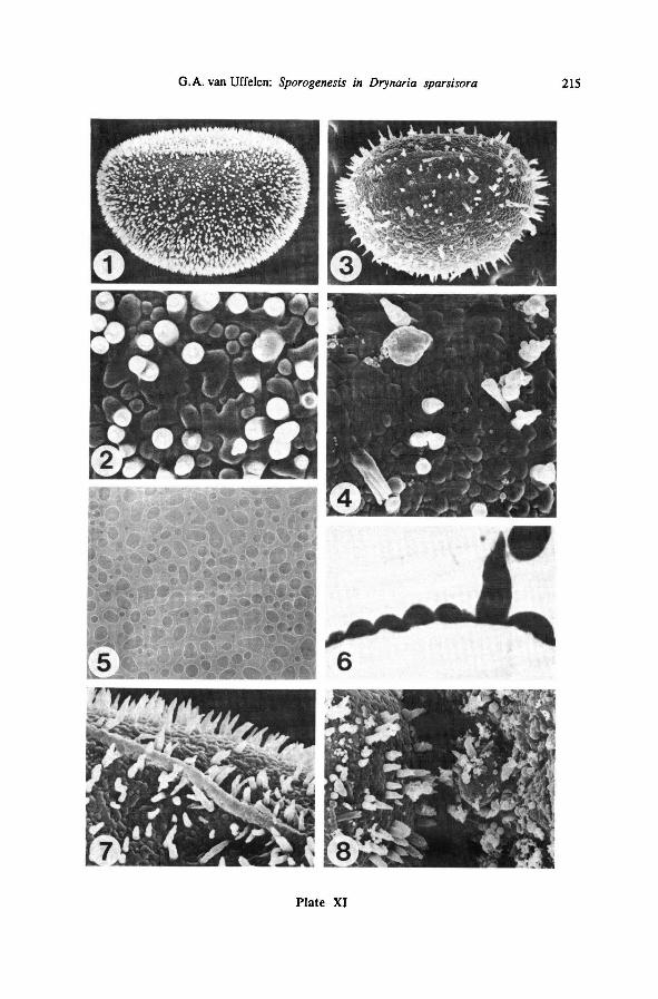
G.A. van Uffelen: Sporogenesis in Drynaria sparsisora 215\XI
Plate XI