Embed Size (px)
Citation preview
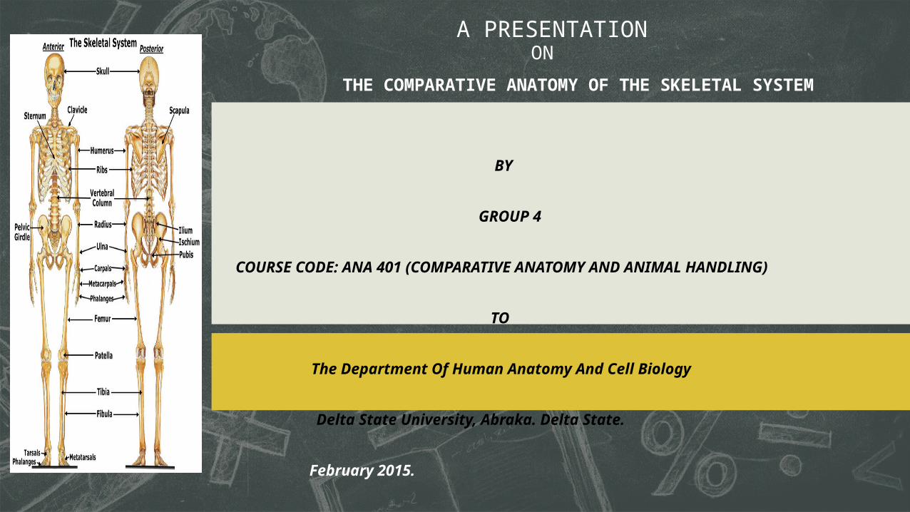
BY
GROUP 4 COURSE CODE: ANA 401 (COMPARATIVE ANATOMY AND ANIMAL HANDLING) TO
The Department Of Human Anatomy And Cell Biology
Delta State University, Abraka. Delta State.
February 2015.
A PRESENTATION
THE COMPARATIVE ANATOMY OF THE SKELETAL SYSTEM
ON
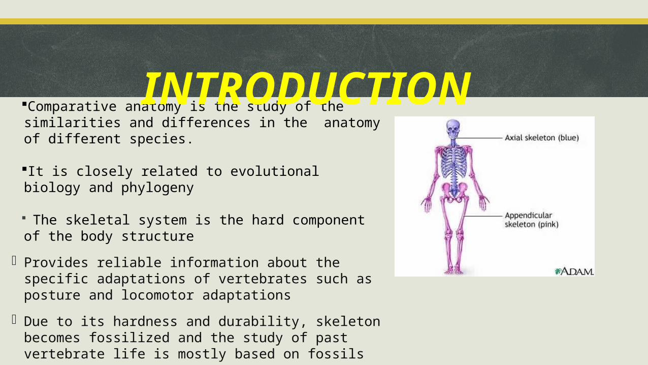
Comparative anatomy is the study of the similarities and differences in the anatomy of different species.
It is closely related to evolutional biology and phylogeny
The skeletal system is the hard component of the body structure
- Provides reliable information about the specific adaptations of vertebrates such as posture and locomotor adaptations
- Due to its hardness and durability, skeleton becomes fossilized and the study of past vertebrate life is mostly based on fossils
INTRODUCTION
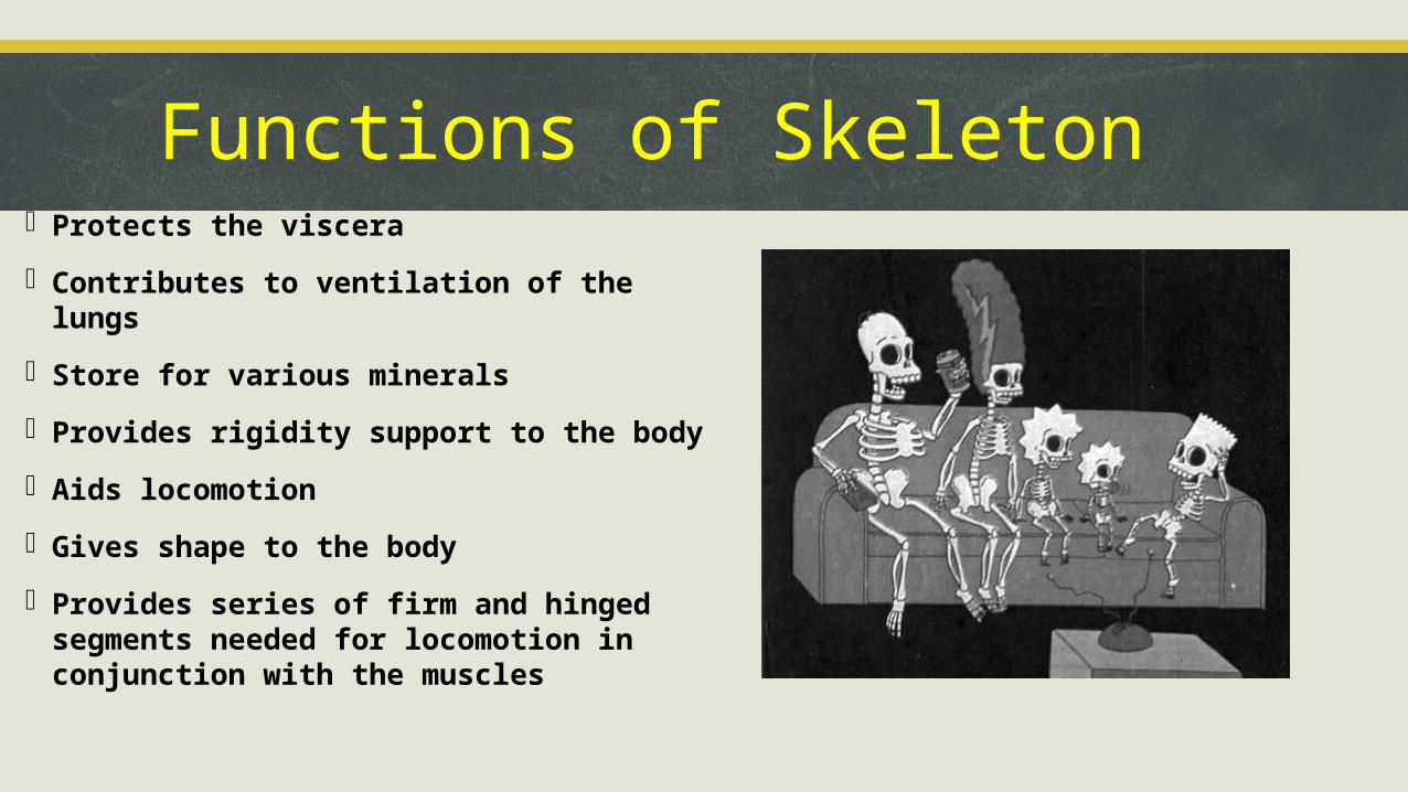
Functions of Skeleton- Protects the viscera
- Contributes to ventilation of the lungs
- Store for various minerals
- Provides rigidity support to the body
- Aids locomotion
- Gives shape to the body
- Provides series of firm and hinged segments needed for locomotion in conjunction with the muscles
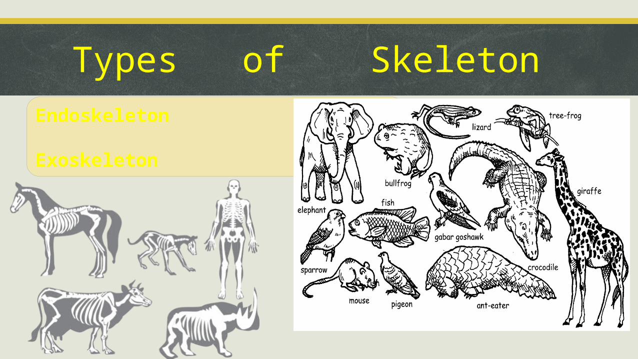
Types of SkeletonEndoskeleton
Exoskeleton
ORIGIN OF BONE
Earliest known fossils are fishes
Dates back to late Cambrian & early ordovician times (500mybp)
Found in scattered areas of N.America, Greenland & Spitsbegen
Small in size & varies; jawless & no paired fin
Head covered with a thick plate of Bone.
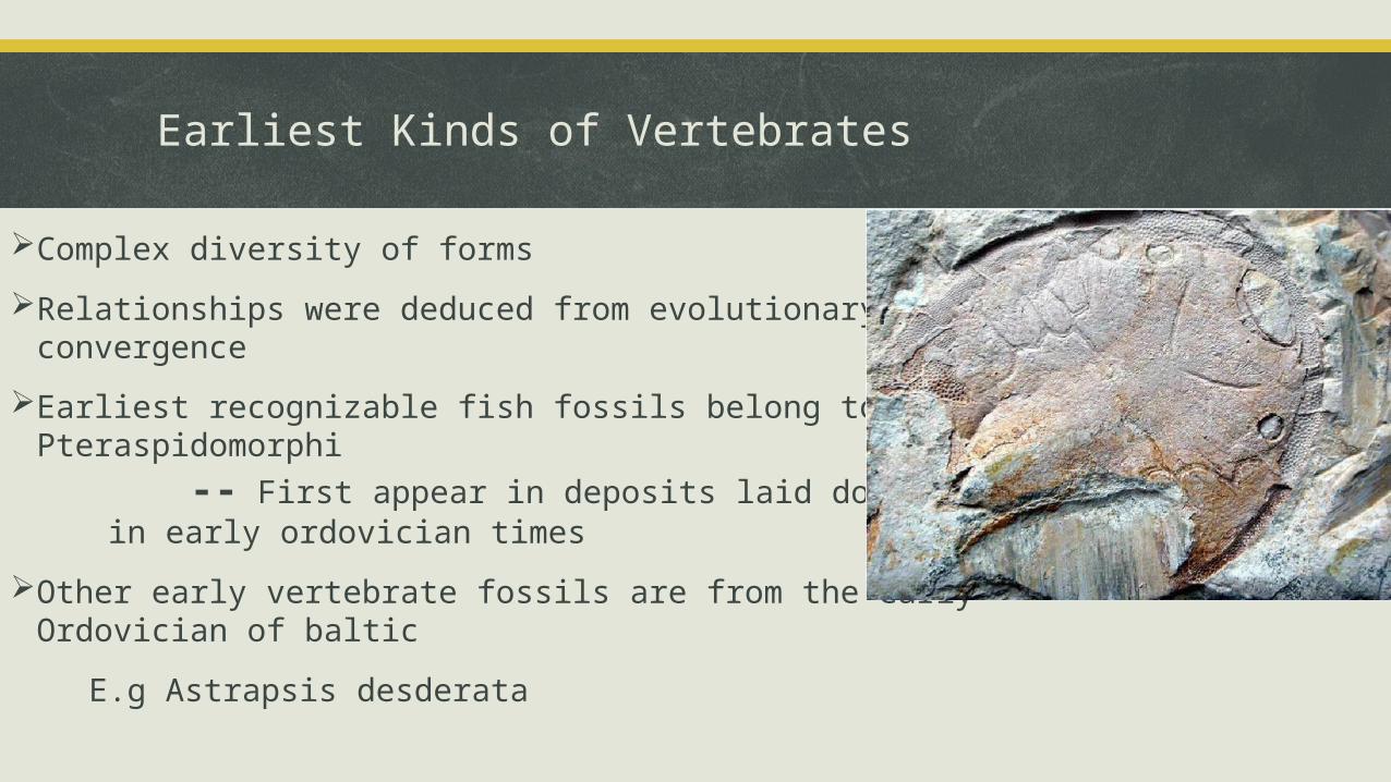
Earliest Kinds of Vertebrates
Complex diversity of forms
Relationships were deduced from evolutionary convergence
Earliest recognizable fish fossils belong to group – Pteraspidomorphi -- First appear in deposits laid down in early ordovician times
Other early vertebrate fossils are from the early Ordovician of baltic
E.g Astrapsis desderata

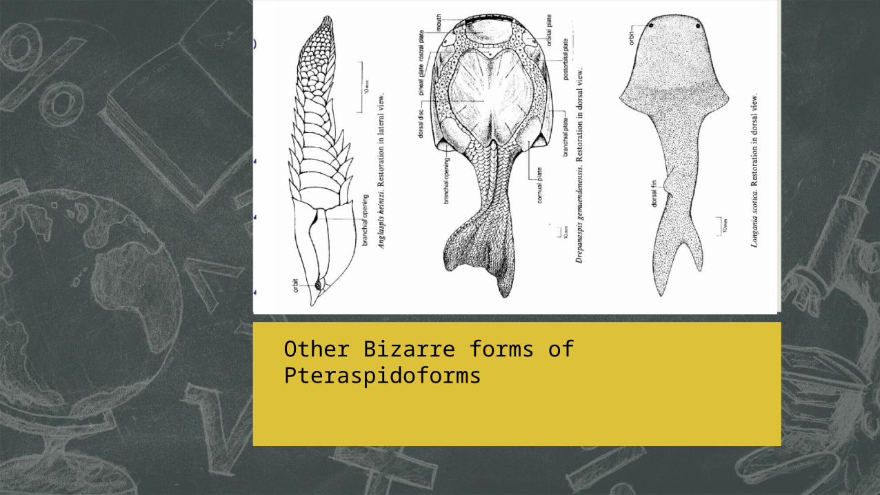
Other Bizarre forms of Pteraspidoforms
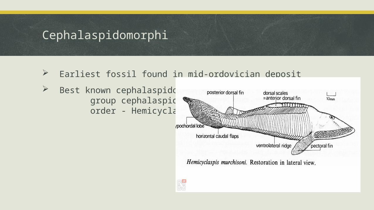
Cephalaspidomorphi
Earliest fossil found in mid-ordovician deposit
Best known cephalaspidomorphii group cephalaspidoforms order - Hemicyclapsis
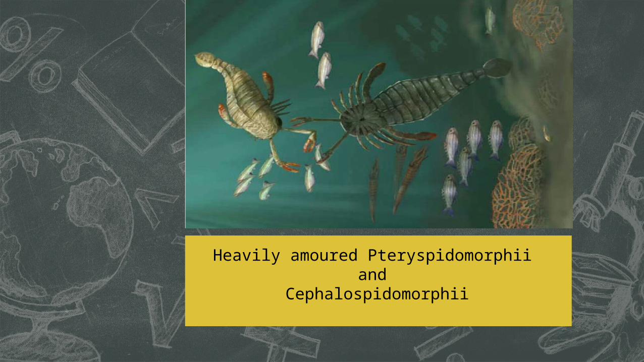
Heavily amoured Pteryspidomorphii and
Cephalospidomorphii
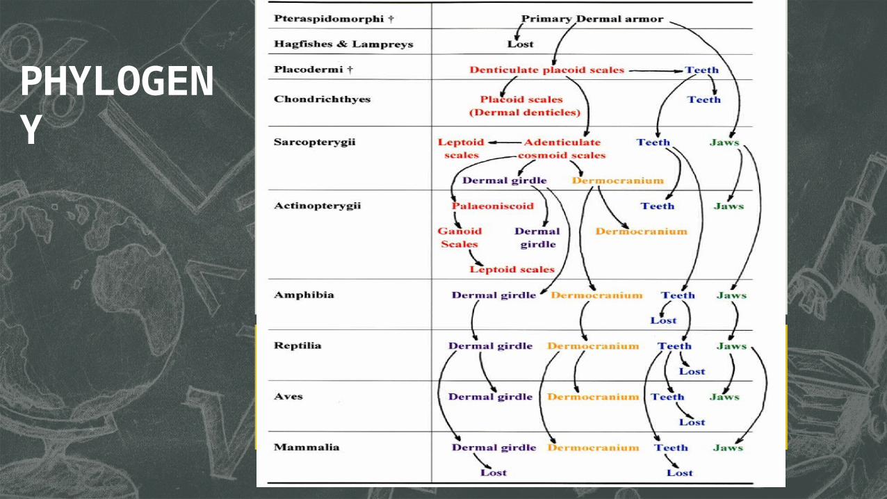
PHYLOGENY
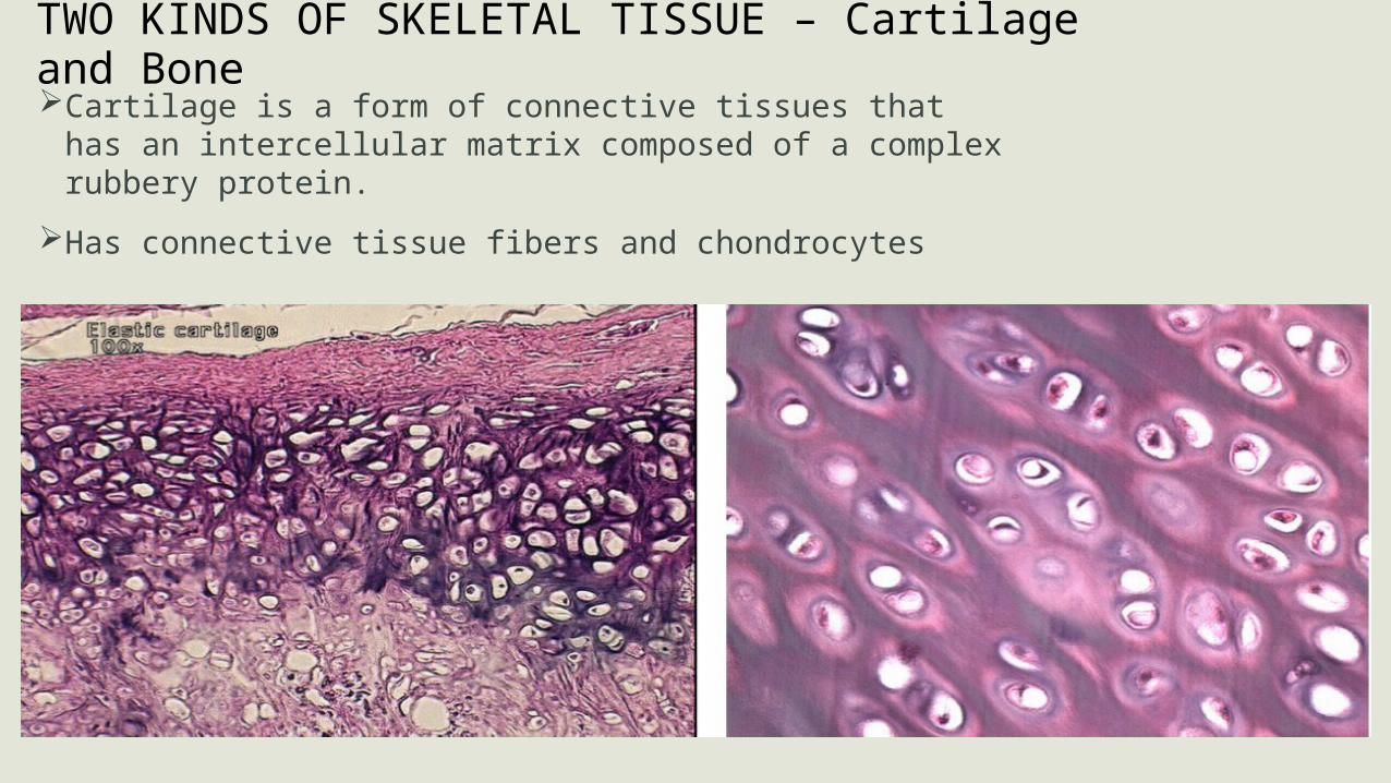
TWO KINDS OF SKELETAL TISSUE – Cartilage and BoneCartilage is a form of connective tissues that has an intercellular matrix
composed of a complex rubbery protein.
Has connective tissue fibers and chondrocytes
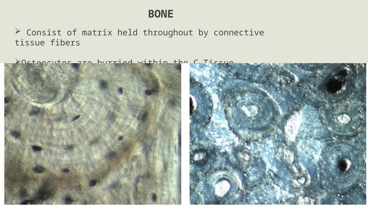
BONE Consist of matrix held throughout by connective tissue fibers
Osteocytes are burried within the C.Tissue
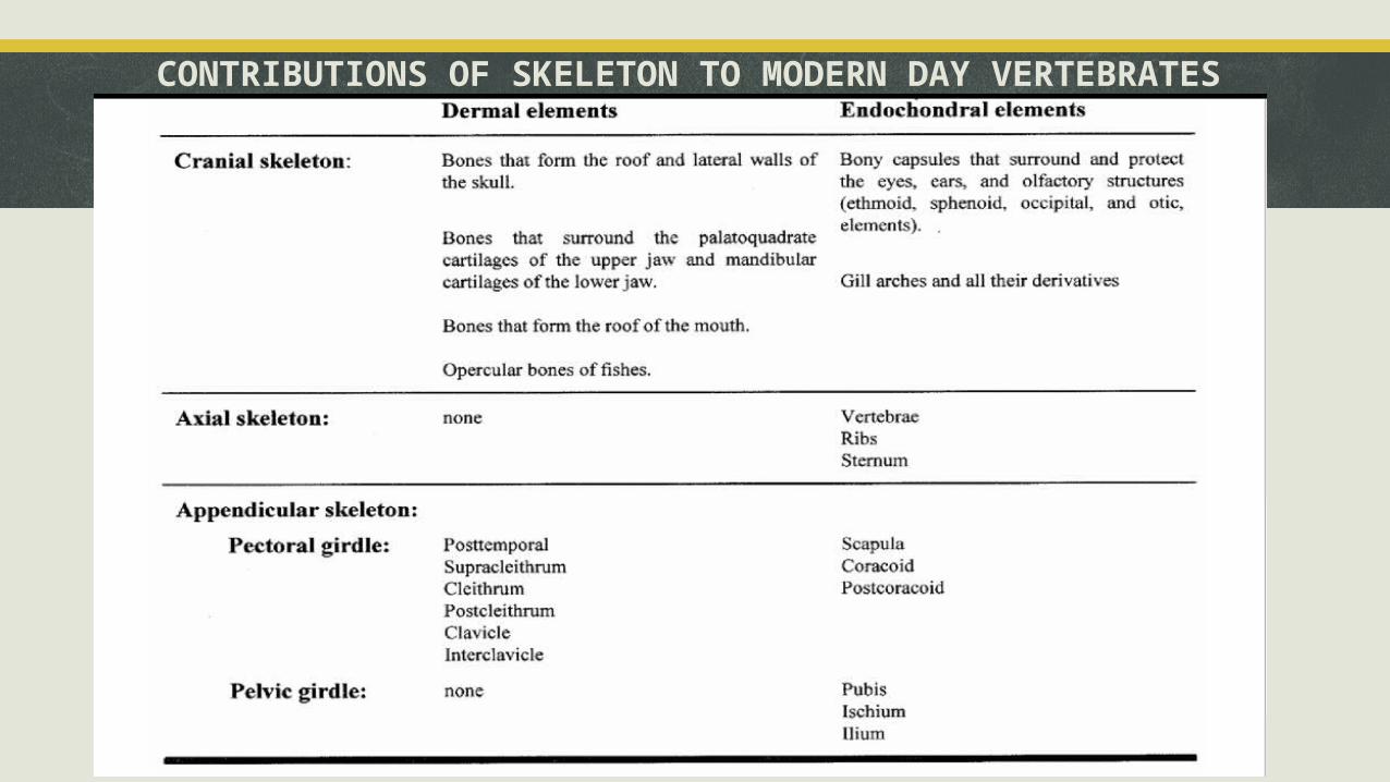
CONTRIBUTIONS OF SKELETON TO MODERN DAY VERTEBRATES
EMBRYOGENIC DEVELOPMENT OF THE SKELETON
Embryogenic skeleton development begins when the embryo consist of:
Ectoderm
Endoderm
Longitudinal Primitive streak (i) Notochord (gives rise to longitudinal axis of the embryo) (ii) Third basal layer – Mesoderm
The mesodermal sheet becomes differentiated into
• Paraxial node
• Lateral plate
• Sclerotome
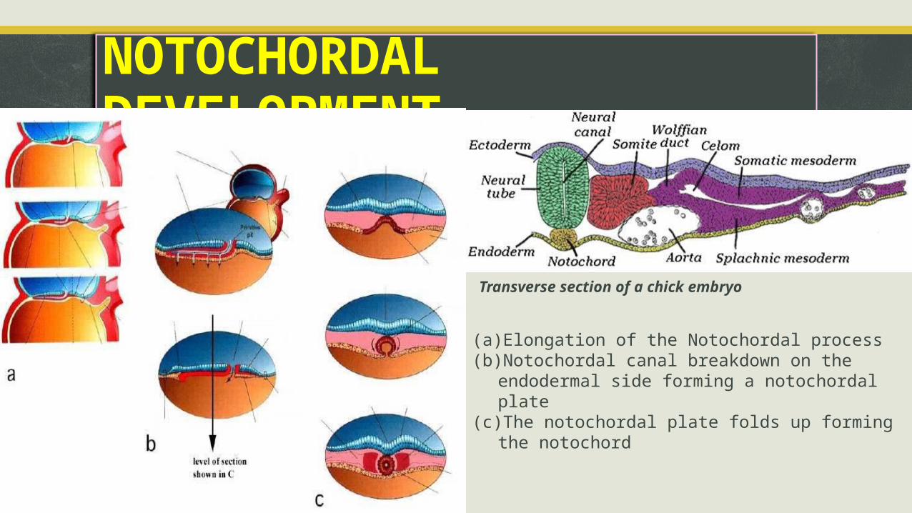
NOTOCHORDAL DEVELOPMENT
(a) Elongation of the Notochordal process(b) Notochordal canal breakdown on the endodermal side
forming a notochordal plate(c) The notochordal plate folds up forming the notochord
Transverse section of a chick embryo
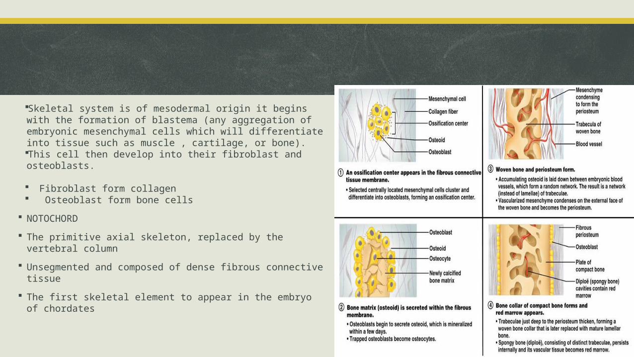
Skeletal system is of mesodermal origin it begins with the formation of blastema (any aggregation of embryonic mesenchymal cells which will differentiate into tissue such as muscle , cartilage, or bone). This cell then develop into their fibroblast and osteoblasts.
Fibroblast form collagen Osteoblast form bone cells
NOTOCHORD
The primitive axial skeleton, replaced by the vertebral column
Unsegmented and composed of dense fibrous connective tissue
The first skeletal element to appear in the embryo of chordates
Regional Classification of the Skeletal System
AXIAL and APPENDICULAR SKELETON: Axial is used to describe the portions of the skeleton that forms the central axis of the body. The appendicular skeleton consists of the Limbs and the girdles.
CRANIAL and POST-CRANIAL SKELETON: The cranial portion of the skeleton consists of the cranium, the mandibles and the hyoids. The post-cranial skeleton is everything else apart from the skull such as the vertebrae, ribs and the limbs bone.
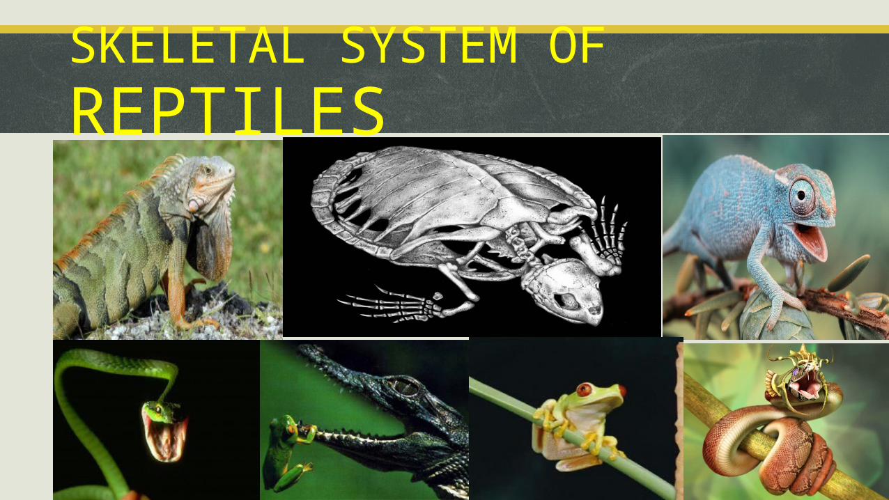
SKELETAL SYSTEM OF REPTILES
INTRODUCTION
Reptile are derived from latin world repto which means to creep
The first reptiles to evolve 300 million years ago were called Anapsids.
Reptiles are class of the chordate phylum.
Most of the reptiles have scaly skin and feet with claws on their toes.
Reptile have bilateral symmetry which means that the right sides of reptiles are mirror images of the left side.
Reptiles have basic axial skeleton with vertebral column. The skeletal system of reptile evolved for support and movement on land.
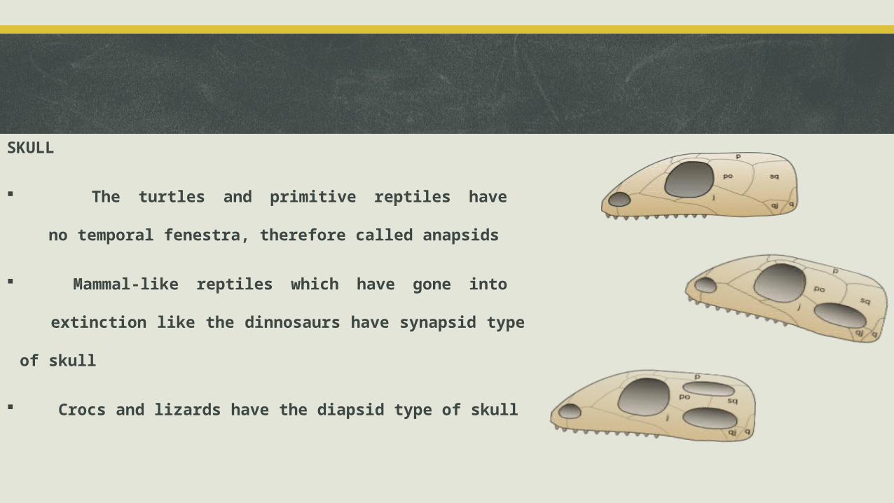
SKULL
The turtles and primitive reptiles have
no temporal fenestra, therefore called anapsids
Mammal-like reptiles which have gone into
extinction like the dinnosaurs have synapsid type of skull
Crocs and lizards have the diapsid type of skull
Skeletal systems in turtles
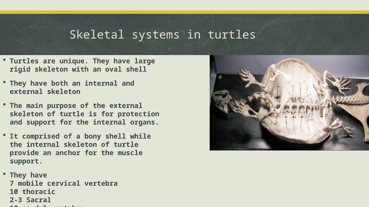
Turtles are unique. They have large rigid skeleton with an oval shell
They have both an internal and external skeleton
The main purpose of the external skeleton of turtle is for protection and support for the internal organs.
It comprised of a bony shell while the internal skeleton of turtle provide an anchor for the muscle support.
They have 7 mobile cervical vertebra10 thoracic2-3 Sacral12 caudal vertebra
The femur is the bony element of the thigh, the tibia and fibula are the bony
elements of the shank.
The pelvis is composed of 3 pairs of bones: pubis ischium and ilium. The pubis
and the ischium forms the ventrally positioned part of the pelvis.
The two ilia are oriented dorsoventrally, articulate with the sacral vertebrae
and attach the pelvis to the carapace via ligaments.
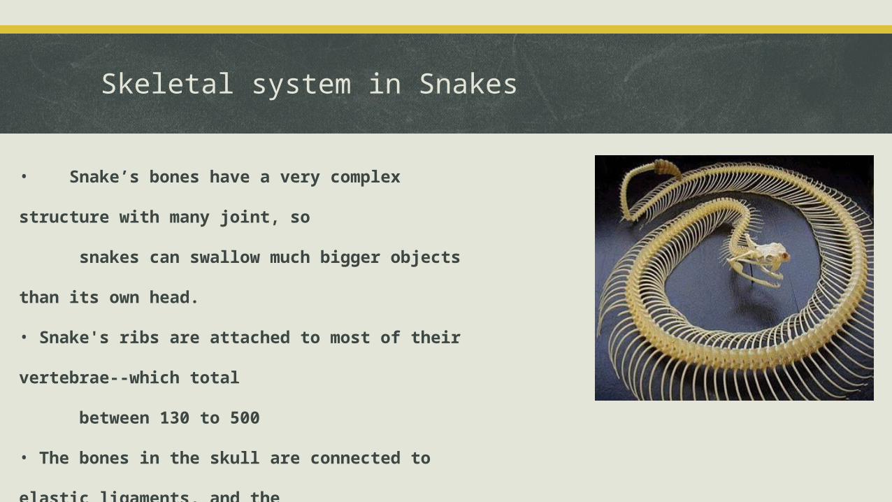
Skeletal system in Snakes
• Snake’s bones have a very complex structure with many joint, so
snakes can swallow much bigger objects than its own head.
• Snake's ribs are attached to most of their vertebrae--which total
between 130 to 500
• The bones in the skull are connected to elastic ligaments, and the
joints of the jaw are double hinged and positioned in the
posterior aspect of the skull
SKELETAL SYSTEM IN PISCESSCIENTIFIC CLASSIFICATION:
• Kingdom- Animalia
• Phylum- Chordata
• Class- Pisces
GROUPS
Jawless fish
Armoured fish
Cartilaginous fish
Ray-finned fish
Lobe-finned fish
• The skeleton which forms their support structure could either be made of cartilage, in cartilaginous fish,bone and cartilage in bony fish and the notochord in jawless fishes .
• The class Pisces have endoskeleton also referred to as internal skeleton, that inside the body as in mammals and aves.
• The main skeletal element is the vetebral column, composed of articulating vertebrae which are light weight but strong.
• The main external features of the fish are the fins which are composed of either bony or soft spins called rays which with the exception of caudal skin have no direct connection with the spine. They are supported by the muscle..
Nbgh
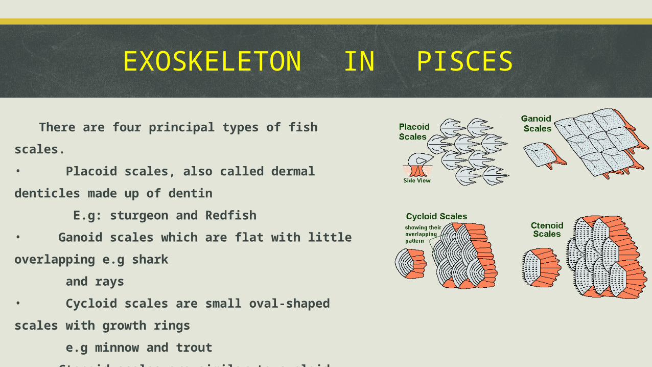
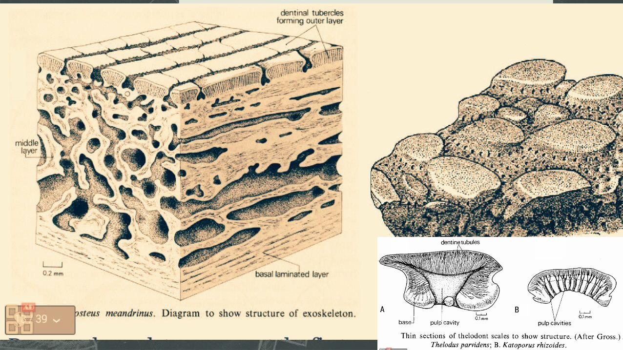
EXOSKELETON IN PISCES
There are four principal types of fish scales.
• Placoid scales, also called dermal denticles made up of dentin
E.g: sturgeon and Redfish
• Ganoid scales which are flat with little overlapping e.g shark
and rays
• Cycloid scales are small oval-shaped scales with growth rings
e.g minnow and trout
• Ctenoid scales are similar to cycloid scales but bear spines e.g
Perch
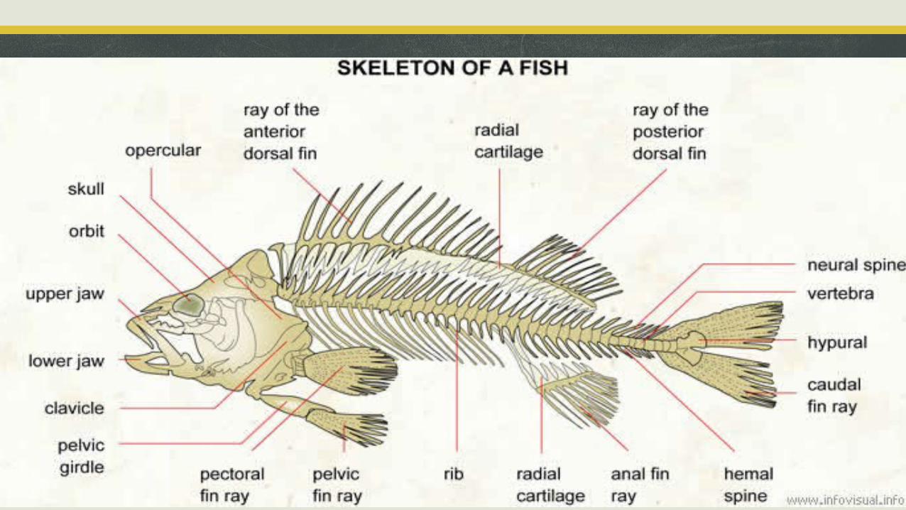
OPERCULAR: pair of bony plates covering the gill opening.
RAY OF THE ANTERIOR DORSAL FIN: each of the small bones forming the front fin on the back of a fish
RADIAL CARTILAGE: elastic substance of the radius.
RAY OF THE POSTERIOR DORSAL FIN: each of the small bones forming the rear fin on the back of a fish.
NEUTRAL SPINE: spines containing part of the nervous system
VERTEBRA: each of the bones forming the neural spine of a fish.
HYPURAL: bone to which are attached the spiny rays of the caudal fin of a fish.
CAUDAL FIN RAY: each of the small bone forming the tail fin of a fish
ANAL FIN RAY: each of the small bones forming the fin behind the anus of a fish
RADIAL CARTILAGE: elastic substance of the radius
RIB: each of the bones forming the thoracic cage
PELVIC FIN RAY: each of the bones forming the fin beneath the pelvic girdle
PECTORAL FIN RAY: each of the bones forming the chest fin.
PELVIC GIRDLE: set of bone forming the pelvis
CLAVICLE; shoulder bone
LOWER JAW; mandible
UPPER JAW; upper part of the mouth
ORBIT;cavity of the skull that contains the eye
SKULL; bony case of the brain of a fish.
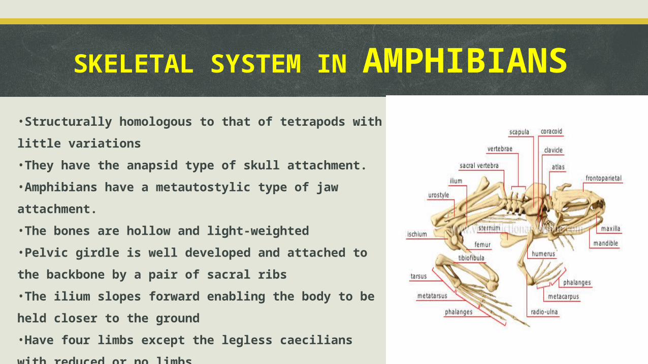
SKELETAL SYSTEM IN AMPHIBIANS•Structurally homologous to that of tetrapods with little variations
•They have the anapsid type of skull attachment.
•Amphibians have a metautostylic type of jaw attachment.
•The bones are hollow and light-weighted
•Pelvic girdle is well developed and attached to the backbone by a pair
of sacral ribs
•The ilium slopes forward enabling the body to be held closer to the
ground
•Have four limbs except the legless caecilians with reduced or no limbs
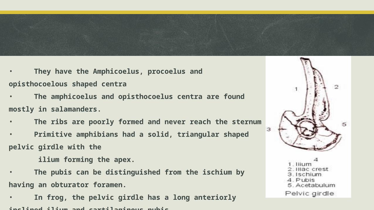
• They have the Amphicoelus, procoelus and opisthocoelous shaped centra
• The amphicoelus and opisthocoelus centra are found mostly in salamanders.
• The ribs are poorly formed and never reach the sternum
• Primitive amphibians had a solid, triangular shaped pelvic girdle with the
ilium forming the apex.
• The pubis can be distinguished from the ischium by having an obturator foramen.
• In frog, the pelvic girdle has a long anteriorly inclined ilium and cartilaginous pubis.
• All 3 bones take part in formation of the acetabulum
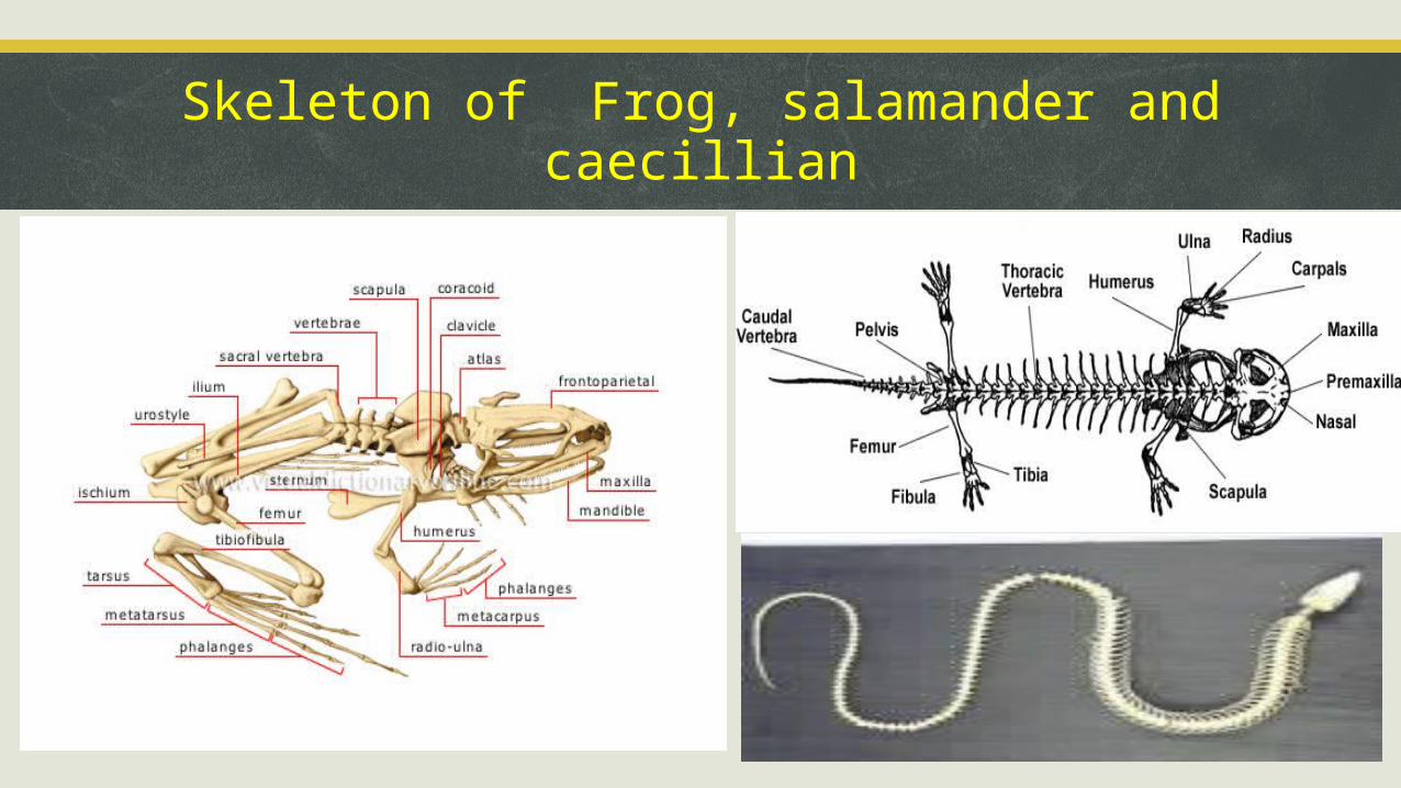
Skeleton of Frog, salamander and caecillian
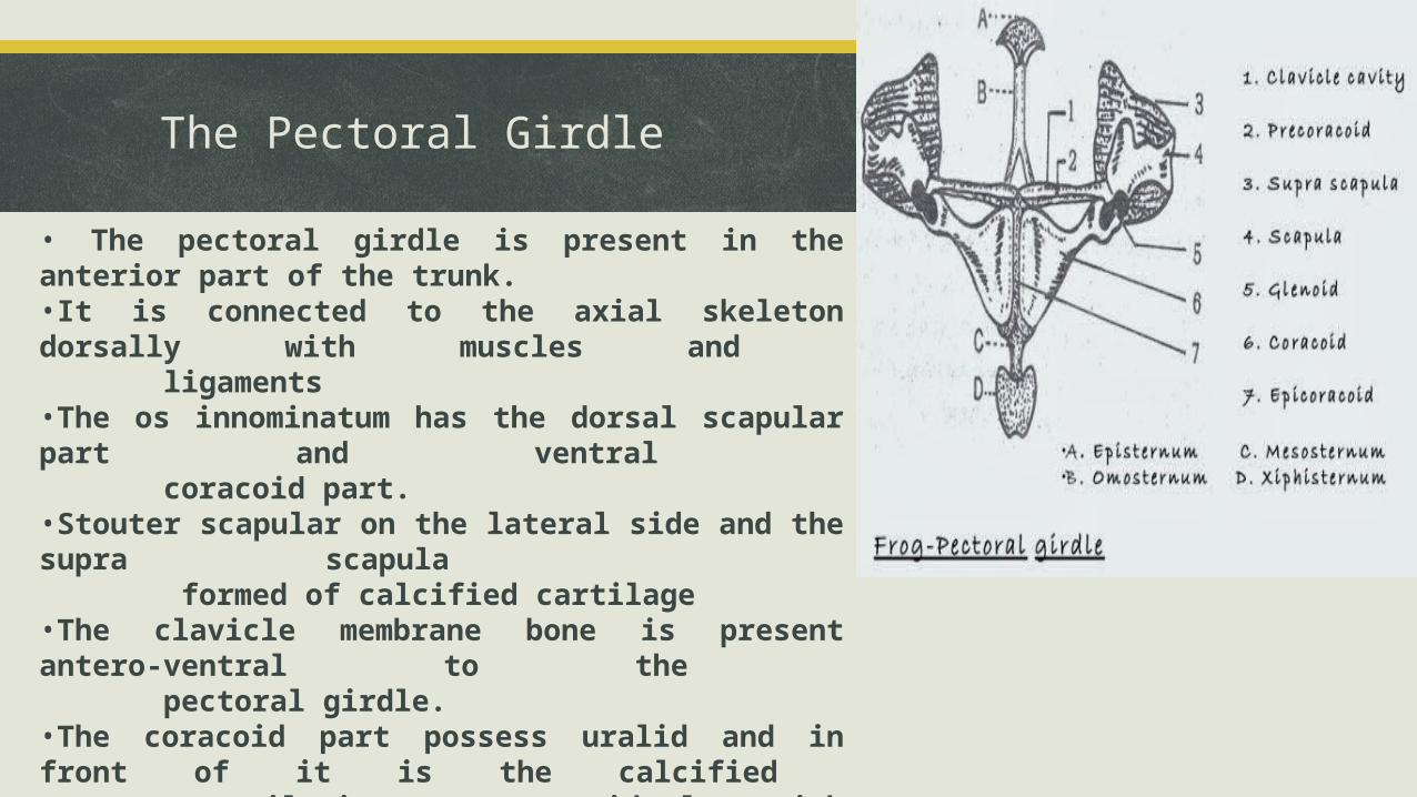
The Pectoral Girdle
• The pectoral girdle is present in the anterior part of the trunk.•It is connected to the axial skeleton dorsally with muscles and ligaments•The os innominatum has the dorsal scapular part and ventral coracoid part. •Stouter scapular on the lateral side and the supra scapula formed of calcified cartilage•The clavicle membrane bone is present antero-ventral to the pectoral girdle.•The coracoid part possess uralid and in front of it is the calcified cartilaginous precoracoid along with another strip cartilaginous epicoracoid bones•Closely associated epicoracoids
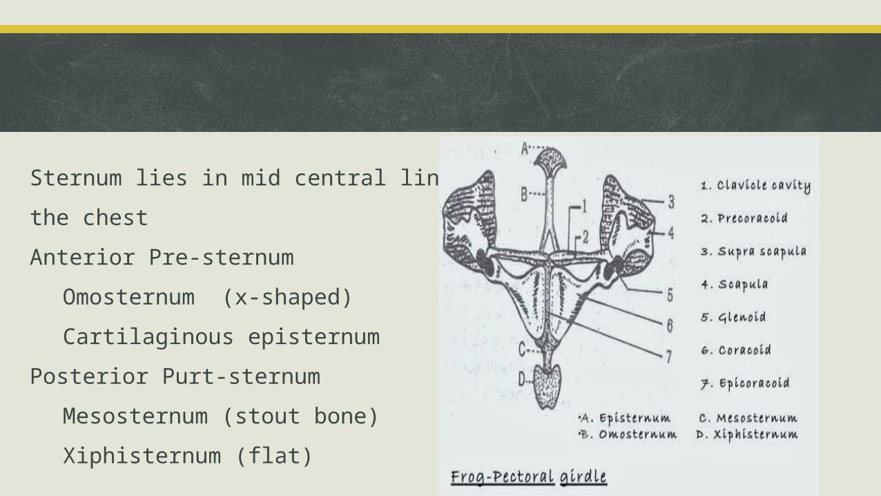
Sternum lies in mid central line of the chest
Anterior Pre-sternum
Omosternum (x-shaped)
Cartilaginous episternum
Posterior Purt-sternum
Mesosternum (stout bone)
Xiphisternum (flat)
SKELETAL SYSTEM OF AVES Aves (birds) are group of vertebrates that have wings, feathers, hollow bones and numerous other adaptations for an aerial life style.
ClassificationGroup and species
Skeleton•Aves inherited their basic skeleton from their reptilian ancestors.•compared with other vetebrates, birds have a body plan that shows many usual adaptations mostly to facilitate flight.
Skull: Skull bones in adult birds are fused and do not show cranial sutures•They have a diapsid skull which weight 1% of their total body weight.•A light toothless beak which replaces the bony, heavy toothed jaw of reptiles.
The Vertebrae• The spine of birds has cervical, thoracic, lumbar and caudal region with the number of cervical vertebrae highly variable and flexible.
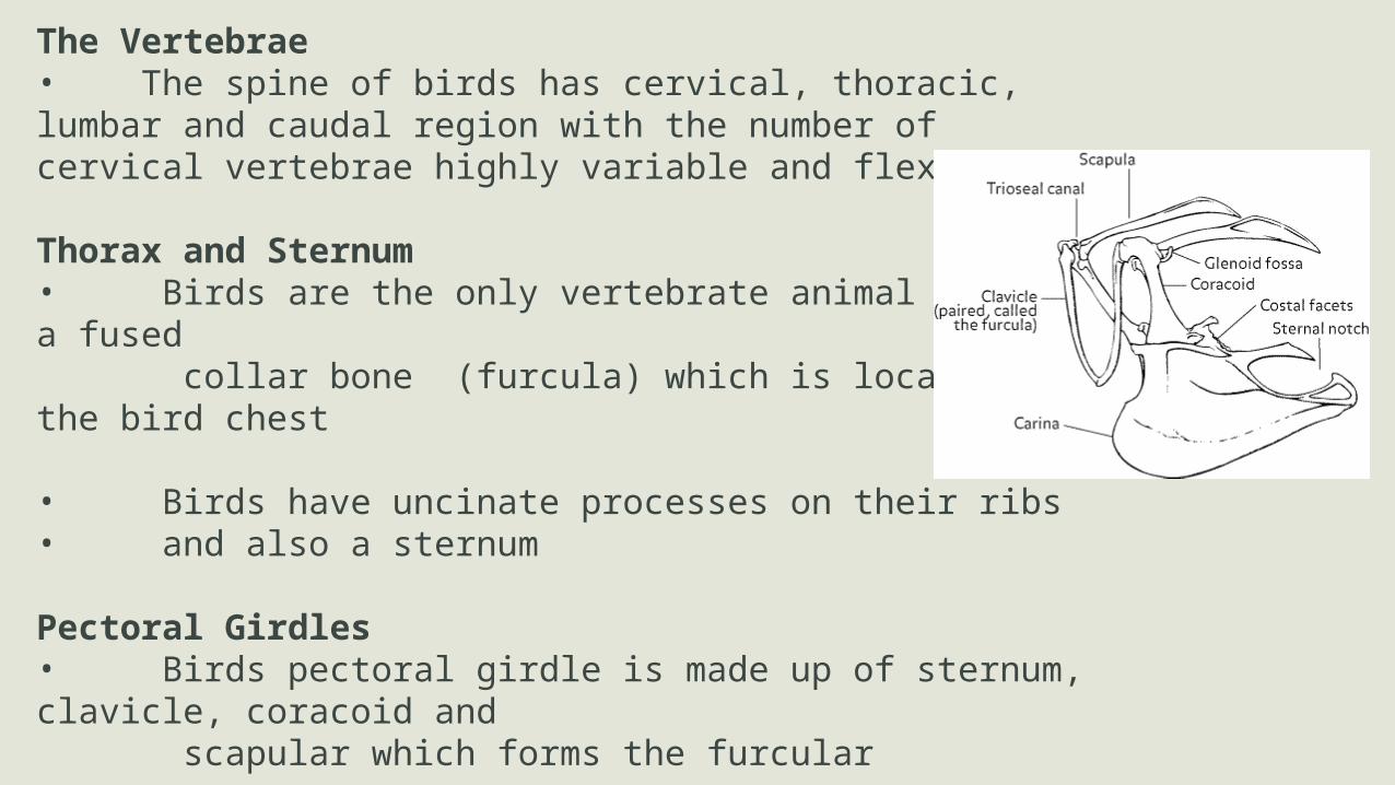
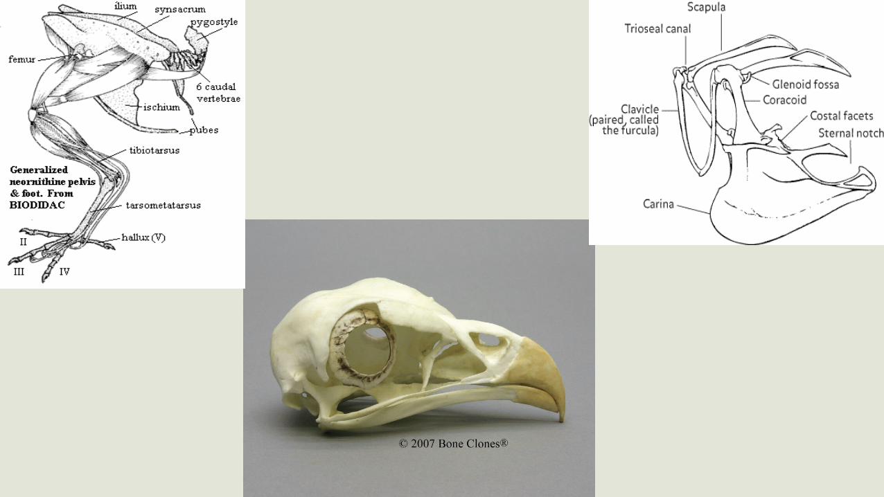
Thorax and Sternum• Birds are the only vertebrate animal to have a fused collar bone (furcula) which is located at the bird chest
• Birds have uncinate processes on their ribs • and also a sternum
Pectoral Girdles• Birds pectoral girdle is made up of sternum, clavicle, coracoid and scapular which forms the furcular
Pelvic Girdle• Is an extensive fusion of bones of the pelvic region to provide staff support for the legs in order to deal with the stress of take off and landing.• It consist of synsacrum, caudal vertebrae and at the end of the spinal column is the pygostle
Wings• The wing consist of the humerus which is short compared to the total length of the pulling and withstand the pulling of the flight muscles,• The radius and ulna which form support for the mid – wing• The carpometacarpus, which form the third major section of the bird wing.
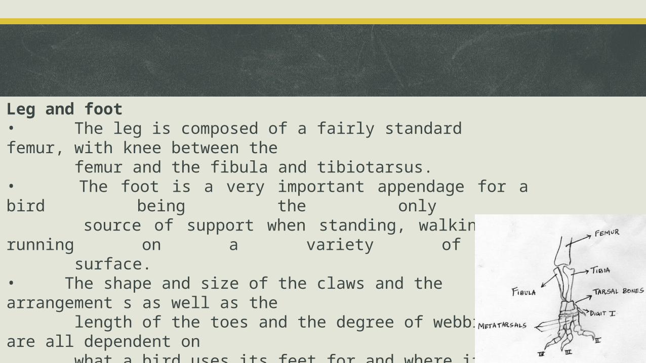
Leg and foot• The leg is composed of a fairly standard femur, with knee between the femur and the fibula and tibiotarsus.• The foot is a very important appendage for a bird being the only source of support when standing, walking and running on a variety of surface.• The shape and size of the claws and the arrangement s as well as the length of the toes and the degree of webbing are all dependent on what a bird uses its feet for and where it live.
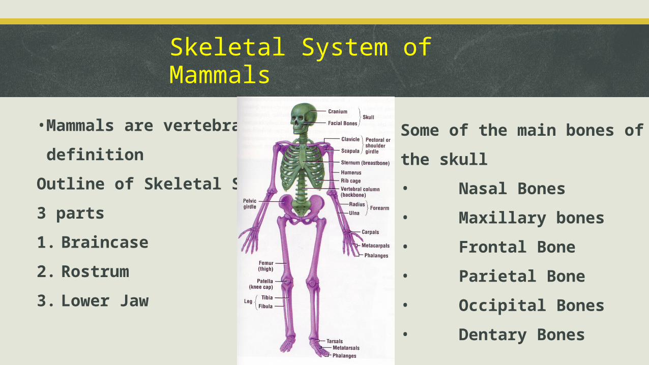
Skeletal System of Mammals
•Mammals are vertebrates by
definition
Outline of Skeletal Skull
3 parts
1. Braincase
2. Rostrum
3. Lower Jaw
Some of the main bones of the skull
• Nasal Bones
• Maxillary bones
• Frontal Bone
• Parietal Bone
• Occipital Bones
• Dentary Bones
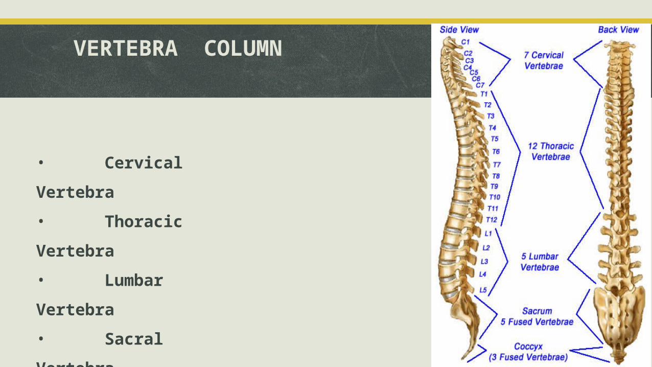
VERTEBRA COLUMN
• Cervical Vertebra
• Thoracic Vertebra
• Lumbar Vertebra
• Sacral Vertebra
• Caudal Vertebra
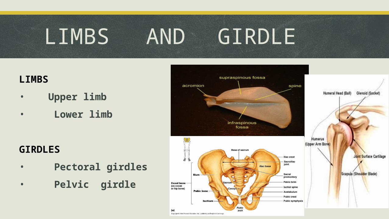
LIMBS AND GIRDLE
LIMBS
• Upper limb
• Lower limb
GIRDLES
• Pectoral girdles
• Pelvic girdle
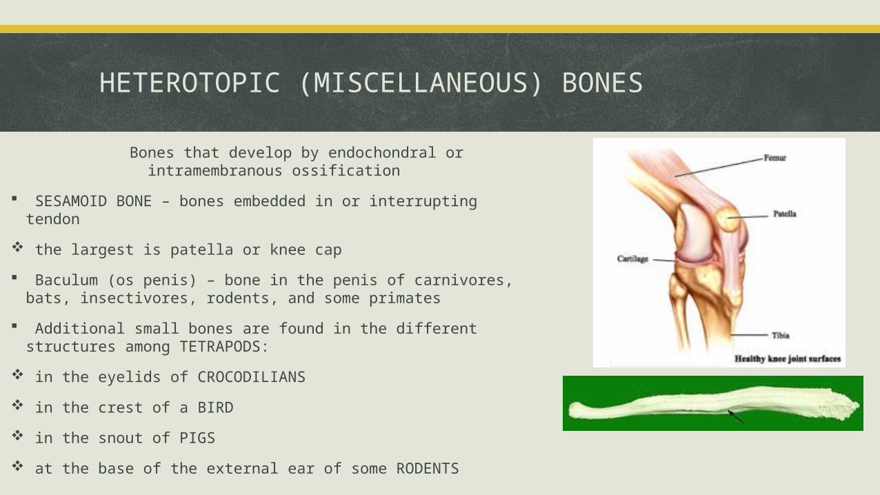
HETEROTOPIC (MISCELLANEOUS) BONES
Bones that develop by endochondral or intramembranous ossification
SESAMOID BONE – bones embedded in or interrupting tendon
the largest is patella or knee cap
Baculum (os penis) – bone in the penis of carnivores, bats, insectivores, rodents, and some primates
Additional small bones are found in the different structures among TETRAPODS:
in the eyelids of CROCODILIANS
in the crest of a BIRD
in the snout of PIGS
at the base of the external ear of some RODENTS

Similarities and differences in vertebrates
Comparative anatomy of the skeletal system
Organ system
Jawless fishes Cartilaginous fishes
Bony fishes amphibians reptiles birds Mammals
Skeletal system
They possess notochord no axial and appendicular skeleton
Axial and appendicular Made up of cartilages
Axial and appendicular made up of bones and cartilages
Axial and appendicular made up of bones and cartilages
Axial and appendicularmade up of bones and cartilages
Axial and appendicular made up of bones and cartilages
Axial and appendicular madew up of bones and cartilages
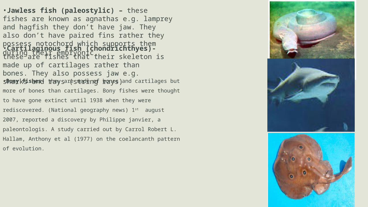
•Jawless fish (paleostylic) – these fishes are known as agnathas e.g. lamprey and hagfish they don’t have jaw. They also don’t have paired fins rather they possess notochord which supports them during their embryonic.
•Cartilaginous fish (chondrichthyes)- these are fishes that their skeleton is made up of cartilages rather than bones. They also possess jaw e.g. sharks and rays (string rays)
•Bony fishes- they are made of bones and cartilages but more of bones
than cartilages. Bony fishes were thought to have gone extinct until 1938
when they were rediscovered. (National geography news) 1st august
2007, reported a discovery by Philippe janvier, a paleontologis. A study
carried out by Carrol Robert L. Hallam, Anthony et al (1977) on the
coelancanth pattern of evolution.
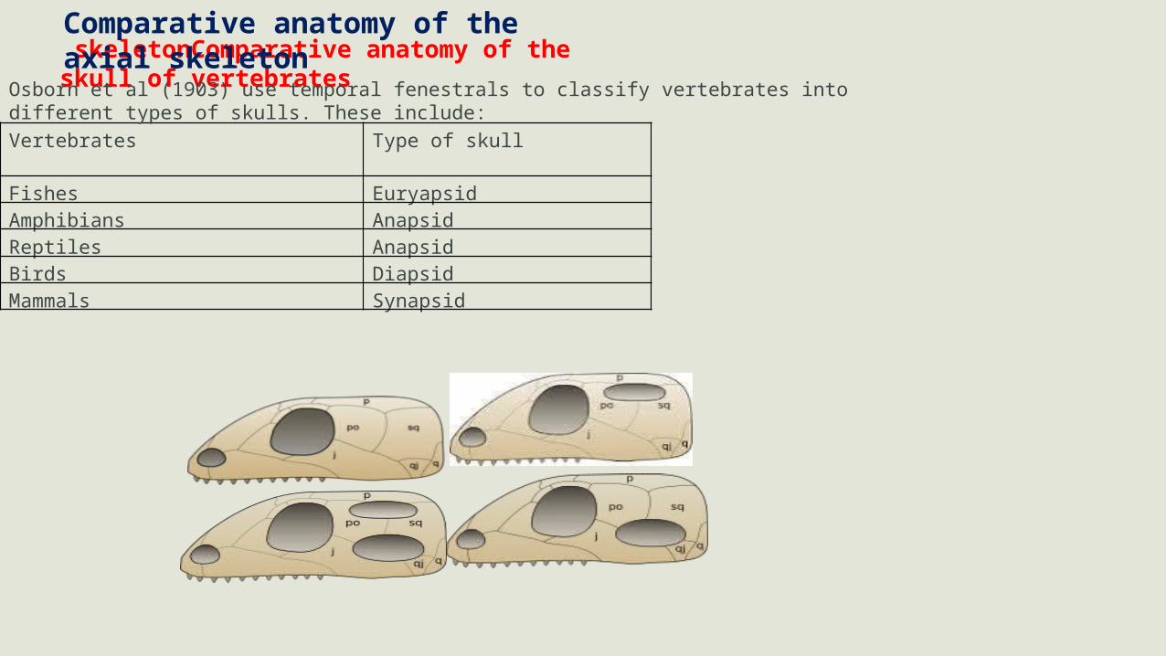
skeletonComparative anatomy of the skull of vertebrates
Vertebrates Type of skull
Fishes EuryapsidAmphibians Anapsid Reptiles AnapsidBirds DiapsidMammals Synapsid
Osborn et al (1903) use temporal fenestrals to classify vertebrates into different types of skulls. These include:
Comparative anatomy of the axial skeleton
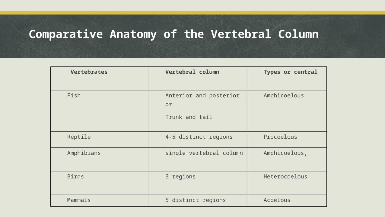
Vertebrates Vertebral column Types or central
Fish Anterior and posterior or
Trunk and tail
Amphicoelous
Reptile 4-5 distinct regions Procoelous
Amphibians single vertebral column Amphicoelous,
Birds 3 regions Heterocoelous
Mammals 5 distinct regions Acoelous
Comparative Anatomy of the Vertebral Column
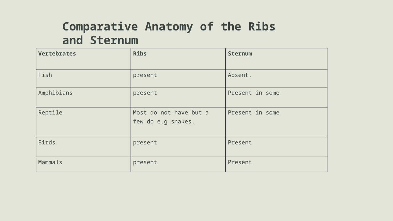
Vertebrates Ribs Sternum
Fish present Absent.
Amphibians present Present in some
Reptile Most do not have but afew do e.g snakes.
Present in some
Birds present Present
Mammals present Present
Comparative Anatomy of the Ribs and Sternum
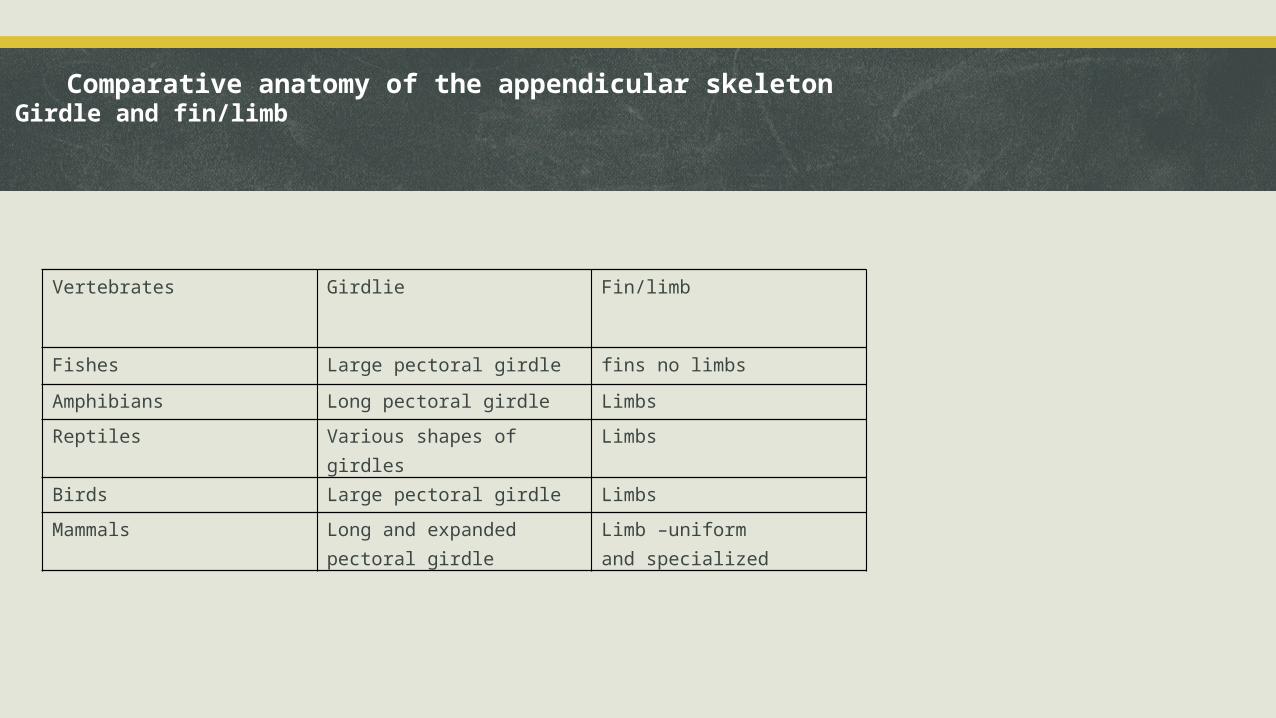
Vertebrates Girdlie Fin/limb
Fishes Large pectoral girdle fins no limbs
Amphibians Long pectoral girdle Limbs
Reptiles Various shapes of girdles Limbs
Birds Large pectoral girdle Limbs
Mammals Long and expanded pectoral girdle
Limb –uniform and specialized
Comparative anatomy of the appendicular skeleton Girdle and fin/limb
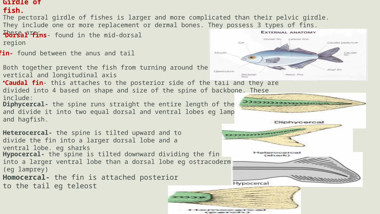
Girdle of fish.The pectoral girdle of fishes is larger and more complicated than their pelvic girdle. They include one or more replacement or dermal bones. They possess 3 types of fins. These are:
•Dorsal fins- found in the mid-dorsal region
•Ana fin- found between the anus and tail
Both together prevent the fish from turning around the vertical and longitudinal axis
•Caudal fin- this attaches to the posterior side of the tail and they are divided into 4 based on shape and size of the spine of backbone. These include:
Diphycercal- the spine runs straight the entire length of the fin and divide it into two equal dorsal and ventral lobes eg lamprey and hagfish.
Heterocercal- the spine is tilted upward and to divide the fin into a larger dorsal lobe and a ventral lobe. eg sharks
Hypocercal- the spine is tilted downward dividing the fin into a larger ventral lobe than a dorsal lobe eg ostracoderm (eg lamprey)
Homocercal- the fin is attached posterior to the tail eg teleost
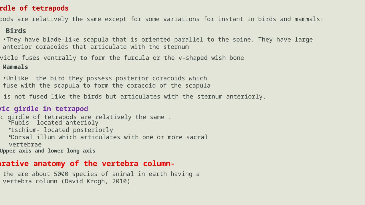
Girdle of tetrapodsGirdles in tetrapods are relatively the same except for some variations for instant in birds and mammals:
Birds•They have blade-like scapula that is oriented parallel to the spine. They have large anterior coracoids that articulate with the sternum
•The two clavicle fuses ventrally to form the furcula or the v-shaped wish boneMammals
•Unlike the bird they possess posterior coracoids which fuse with the scapula to form the coracoid of the scapula
•Their clavicle is not fused like the birds but articulates with the sternum anteriorly.
Pelvic girdle in tetrapodThe pelvic girdle of tetrapods are relatively the same .
•Pubis- located anterioly•Ischium- located posteriorly•Dorsal illum which articulates with one or more sacral vertebrae
Upper axis and lower long axis
Comparative anatomy of the vertebra column-the are about 5000 species of animal in earth having a vertebra column (David Krogh, 2010)
conclusion Comparative anatomy of the skeletal system is the study of the similarities and differences of the
skeletal system in vertebrates. These vertebrates includes, mammal, birds, reptile, amphibians and fish.
The skeletal system is made of bones and cartilages which forms the axial and appendicular skeleton.
Vertebrates skeleton is derived from the dermal amour of primitive jawless fishes
The skeletal system creates an awareness on the evolution of vertebrates and it is the basis for evolutionary studies.
ANATOMY.DELTA STATE UNIVERSITY, ABRAKA
Thank you