Embed Size (px)
Citation preview

ANAESTHETIC MANAGEMENT OF SUPRATENTORIAL SPACE OCCUPYING LESIONS

Anatomy of supratentorial space
Falx cerebri
Tentoriumcerebelli

SUPRATENTORIAL MASSES
1. INFECTION– Subdural abscess– Epidural abscess
2. HEMATOMA– SDH, EDH, Intracranial bleed
3. HYDROCEPHALUS4. NEOPLASMS5. ANEURYSMS & AV MALFORMATIONS

NEOPLASMS OF BRAIN
• PRIMARY (85%)• METASTASIS (12%)

SUPRATENTORIAL TUMOURS CLASSIFICATION
INTRA AXIALBRAIN
PARENCHYMA
GLIOMA(35%)-
ASTROCYTOMA
OLIGODENDROGLIO
MAPINEALOM
A
INTRAVENTRICULAR
EPENDYMOMA
CHOROID PLEXUS
PAPILLOMA
EXTRAAXIAL
MENINGIOMA(15%)PITUITARY
ADENOMA(8%)SCHWANOMMA
DERMOIDCRANIOPHARYNGIOM
ACHORDOMA

Meningioma• Slow growing benign tumor• Well circumscribed• Arise from arachnoid cap cells• Most common sites–near sagittal sinus,
falx cerebri, cerebral convexity• Good prognosis• Some tumors may recur

Astrocytoma• Low grade-young adults,good prognosis• Pilocytic-children ,good prognosis• Anaplastic-poorly differentiated,intermediate
prog• Glioblastoma multiforme

Glioblastoma multiforme
• 30% of all brain tumors in adults• Central necrosis and surrounding edema• Resection inadequate due to microscopic
infiltration of normal brain• Treatment surgical debulking + RT + Chemo• Life expectancy in the order of weeks

Oligodendroglioma
• Arise from myelin producing cells• Treatment- resection

Pituitary tumors
• Arise from cells of anterior pituitary• May occur with MEN 1
functional
• Hormone secreting• Microadenomas(<1
cm)
nonfunctional
• macroadenomas• Compression of ICA
cavernous sinus or 3rd nerve or optic chiasma
ACTH,TSH,FSH,LH,PRL

Clinical features Raised ICP—headache –
worst,bursting or throbbing,awaken from sleep,aggrevated by change of posture/cough/strain
Vomiting , papilloedema
Seizures
Focal neurologic signs-sensory deficits /hemiparesis/cranial nerve palsies

Frontal-hemiparesis subtle personality changes, cognitive dysfunction
Parietal-sensory changes
Temporal lobe-focal seizures
Sellar and parasellar-visual field changes Hypopituitarism Features of Cushing syndrome or acromegaly


Anaesthetic management of supratentorial trs
problems and concerns
1. Intracranial pathophysiology of trs2. Effect of anaesthetics on brain3. Surgical position & concurrent medications4. Measures to decrease ICP & brain bulk5. Complications


Pathophysiologic consideration of brain tumors
• Intracranial pressure• Munroe kellie doctrine
CSF COMPARTMENT
CELLULAR COMPARTMENT
ICP

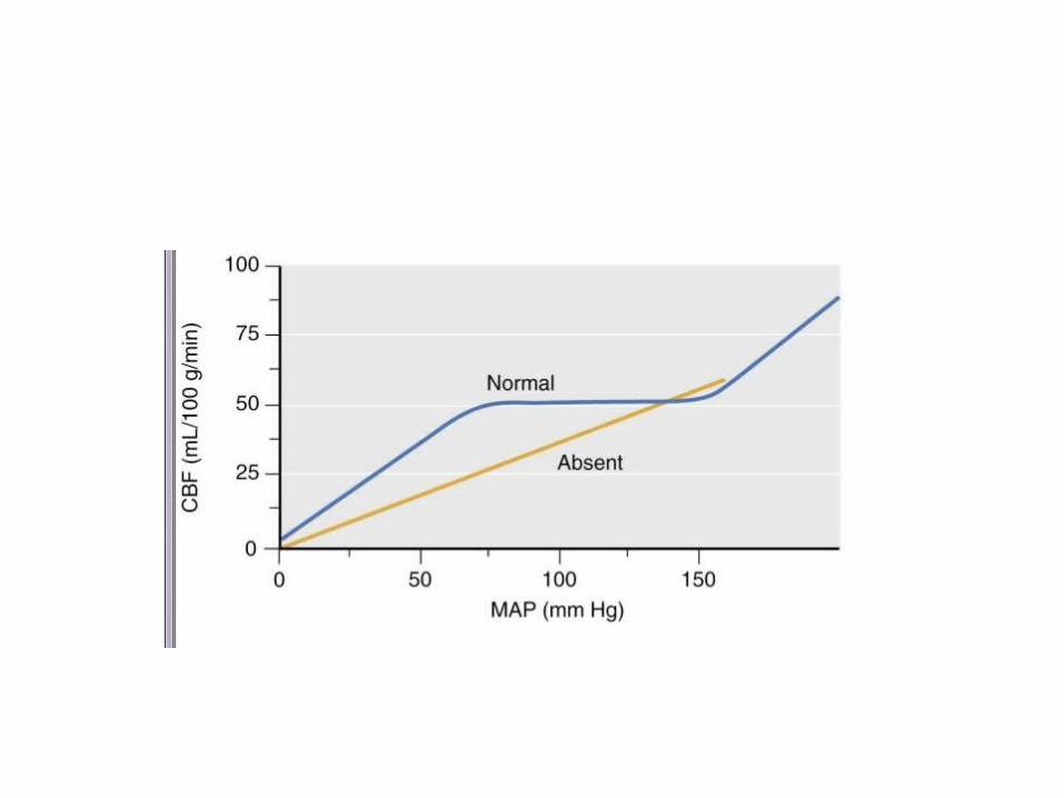
Increase in ICP leads to1. cerebral ischemia(CPP=MAP-ICP)2. Brain herniation
1. Subfalcine herniation2. Transtentorial3. Cerebellar 4. Transcalvareal

Subfalcine
Uncal or transtentorial
Cerebellar
Trans calvarial

Subfalcine• Asymmetrical supratentorial trs• Herniation across midline beneath falx• Anterior cerebral artery may be compressedTranstentorial• Central portion is forced out of supratentorial
compartment• DURETS HAEMORRHAGE-brain stem
haemorrhage

cerebellar herniation• Compression of 3rd cranial nerve• Dilated pupils,ptosis,lateral deviation of eye• Anisocoria-most important early sign

Recurrent Issues In Neuroanesthesia
• Brain relaxation/ control of ICP• Management of PaCO2• Management of arterial blood pressure• Use of steroids• Use of Osmotherapy and Diuretics• Use of anticonvulsants• Patient positioning• Pnemocephalus• Venous air embolism• Hypothermia• Monitoring• I/V fluid management• Glucose management• Emergence

Intraoperative Management
Anaesthetic goals• Maintain CPP• Prevent brain herniation• Maintain O2• Maintain CO2• Provide lax operative field• neuroprotection

Effect of anaesthetic drugsBARBITURATES PROPOFOL ETOMIDATE KETAMINE
CMRO2 CMRO2 CMRO2 CMRO2
CBF CBF CBF CBF
ICP ICP ICP ICP
Cerebral protection +CPP preserved CPP decreased No reduction in
CPPAutoregulation maintained
Autoregulation maintained
Autoregulation not evaluated
CO2 responsiveness preserved
CO2 responsiveness preserved
CO2 responsiveness preserved


• Order of vasodilatory potency• Halothane.>Enflurane>desflurane&Isoflurane>
Sevoflurane• CO2 responsiveness preserved• Autoregulation in response to rising arterial
pressure is impaired• Sevoflurane cause least impairment

Net CBF determining factors
• Conc of the anesthetic agent• Extent of previous CMR depression• Extent of blood pressure changes caused by
the previous or anesthetic induced autoregulation disturbances
• Simultaneous changes in Pa CO2 due to disease related impairment in CO2 responsiveness

N2O HALO ISO SEVO ENFLU DES
CMRO2
CBF
ICP
CSF production
decreas Nochange
increas increas
CSF absorption
decreas increas increas No change

N2O
• Can cause significant increase in CBF, CMR, & ICP
• Most extensive increase when used alone• With IV agents, CBF effect considerably
reduced(Thiopentone, Propofol, Benzodiazepines,
Narcotics)• With volatile agents, CBF increase is
exaggerated.

Muscle relaxants
• Succinylcholine– Transient increase in ICP– Caused by increased afferent signals from muscle
spindles– Prevented by deep anaesthesia, defasciculation
with NDMR

• NDMR– With histamine release(eg Atracurium,
mivacurium)Cause cerebral vasodilatation & increase ICP
– Simultaneous decrease in BP & cause reduction in CPP
– Laudanosine-metabolite of atracurium -seizures

• PancuroniumLarge bolus -> abrupt increase in BP -> if
autoregulation defective -> increase ICP VecuroniumNo variation in HR and BPPreferred competetive blocker

• Benzodiazepines– Modest parellel reduction in CBF & CMRO2– CO2 responsiveness preserved
Lignocaine • Dose related reduction in CMRO2 & CBF• Prevention or treatment of acute increase in ICP
(enotracheal suctioning)• Large dose - seizures

• Opioids– Modest reduction in CBF & CMR– Neuroexcitatory activities & seizures reported

Preoperative asessment
• History– Raised ICP- Headache, Nausea, vomiting, blurred
vision– Level of consciousness– History of seizures– Focal neurologic signs – sensory deficits,
hemiparesis, cranial nerve palsy– Medication – steroids, antiepileptics, mannitol

• Physical examination• Goal – Assess how much permanent & reversible
neurological damage is already present

• GCS score• Papilloedema• Cushings response
– HTN, bradycardia• Focal neurological signs-document hemiparesis, sensory deficits, cranial nerve palsy• Hydration status
– Ask about duration of bed rest– Fluid intake– Diuretics

Investigations
• BRE• URE• RBS• RFT• LFT• S.electrolytes• Coagulation profile• ECG all leads• CXRAY

• CT/ MRI– Look for size & location of tumor
• To asess surgical position• Potential for blood loss• Risk of air embolism
– Midline shift– Effacement of ventricles– Loss of sulci– Obliteration of cisterns– Cerebral edema– Hydrocephalus

Positioning • Common neurosurgical positions are– Supine– Lateral (park bench)– Semilateral (Janetta)– Prone– Sitting

Preoperative Preperation
• Preop steroids– Decreases edema– Decreases BBB pemeability– Improves the viscoelastic properties of intracranial
space– Clinical improvement within 24 hrs– Decreases ICP within 48-72 hrsARRANGE BLOOD
Premedication
Not premedicated outside operating room– Benzodiazepines like midazolam if no signs of
raised ICP H2 blockers & gastric prokinetic drugs

Anticonvulsants
• Intraop seizures—Cortical irritation, Cortical incision, Brain surface irritation by retractors
• Levetiracetam can be safely given even in TBI

• Preinduction monitors– NIBP– SpO2– ECG
• Vascular access– 2 large widebore peripheral IV lines under LA

• Indications of Central Venous access– Large vascular tumors– Proximity to major arteries or venous sinus– Extensive bone resection– Major cardiovascular compromise present– Vasoactive drugs are to be infused– Risk of venous air embolism

Arterial cannulation / LA• Preinduction is appropriate• Need for close monitoring & control CPP– (transducer at level of external auditory meatus /
circle of willis)• ABG• RBS or S. electrolytes

Induction
• Avoid ICP elevations• Preserve CPP• Adequate depth of anaesthesia

• Premedication – Fentanyl 1-2 ug/kg• Preoxygenation with 100% O2• Induction with TPS 3-6mg/kg or Propofol 1.25-
2.5mg/kg• Control ventilation (PaCO2~ 35mm Hg)

• Lignocaine 1.5mg/kg 90sec before intubation
• Succinyl choline (transient increase in ICP)Prevented by deep anaesthesia and NDMR
• Gentle laryngoscopy & intubation

• Maintenance• Maintain with propofol infusion & opiod till dura is
opened • N2O + O2 (50-70%)+ Propofol infusion 50-150
ug/kg)• Muscle relaxants Vecuronium is ideal• NDMR with histamine release is avoided• Volatile anesthetics may be used once dura open

Positioning
• Pin holder application Deepen with propofol 0.5mg/kg or TPS 1mg/kg or Fentanyl 1-3 ug/kg Or esmolol .5mg/kg or labetalol .075-.15 mg/kg along with local anesthetic infiltration • Pin insertion can be associated with venous air
embolism

• Mild head up positioning (15-300) to allow optimum venous drainage
• Secure ETT tightly• Severe flexion / lateral rotation of head should be avoided (at
least 2 finger space between chin & nearest bone)• If head is turned laterally, contralateral shoulder is elevated
with roll to prevent brachial plexus stretch injury

• Pressure points should be padded• Eyes taped to prevent corneal damage from exposure or
irrigation of fluid

Intra op ICP reduction
1. Hyperventilation– Lower pCO2 (1mm change in pCO2, CBF changes
by 1-2ml/100gm/min )- Cerebral ischemia in injured brain. ICP lowering effect is not sustained. The CSF pH and CBF returns to normal within 8- 12 hrs.
– Maintain PaCO2 30-35 mm Hg– If hyperventilated for long (ICU) make them
normocapnic slowly


1. Drugs -Osmotic diuretics – Mannitol– 0.25-1gm /kg over 10-15min prior to craniotomy– Effective for ~ 2hrs– Upper acceptable osmolality limit of 320mosm/LMechanism of action – Produce osmotic gradient that draws fluid out of brain
parenchyma– Removes ~ 90ml brain water at peak effectSide effects– Acute hypervolemia (due to vasodilatation)– Electrolyte imbalance

FrusemideMOA• Hastens excretion of water from intravascular
space and maintains osmotic gradient• Inhibit chloride channel and prevent
accumulation of idiogenic osmoles --prevents rebound oedema
• Dose .15-.3 mg/kg

CSF Drainage
• By either direct puncture of lateral ventricle by surgeon or lumbar spinal catheter by anaesthesiologist
• A/C brain herniation may occur
• Draining 10-20 ml CSF effectively reduces brain tension

Tight brain check listAre relevant pressures controlled JVP
Airway pressureArterial pressurePaCO2, PaO2
Is metabolic rate controlled Pain/arousalSeizures temperature
Are any potential vasodilators in use
N2OVolatile agentsCCBNitroprusside
Are there any unrecognized mass lesions
BloodAir with/without N2O
JVP•Extreme head rotation•Direct jugular compression•Head up posture•Airway obstruction•Bronchospasm•Straining •Coughing•Pneumothorax
Monitoring • PR, MAP, SPO2, ETCO2• U/O• CVP• Temp

Others• PNS• Precordial doppler, Trans esophageal ECHO• ICP monitor • EEG – CMRO2, depth of anaesthesia, cerebral
ischemia• Evoked potentials • Jugular venous bulb monitoring – determines
adequacy of cerebral perfusion & oxygenation

Fluid management • Aim---to maintain normovolemia, normotension Avoid reduction of serum osmolarity Keep hematocrit around 30 %• Glucose containing solutions to be avoided• Hyperglycemia -> increased lactate production ->
intracellular acidosis -> aggravate neuronal injury• Blood glucose <140-180 mg%• Normoglycemia not recommended- injured brain is the
state of hyperglycolysis.
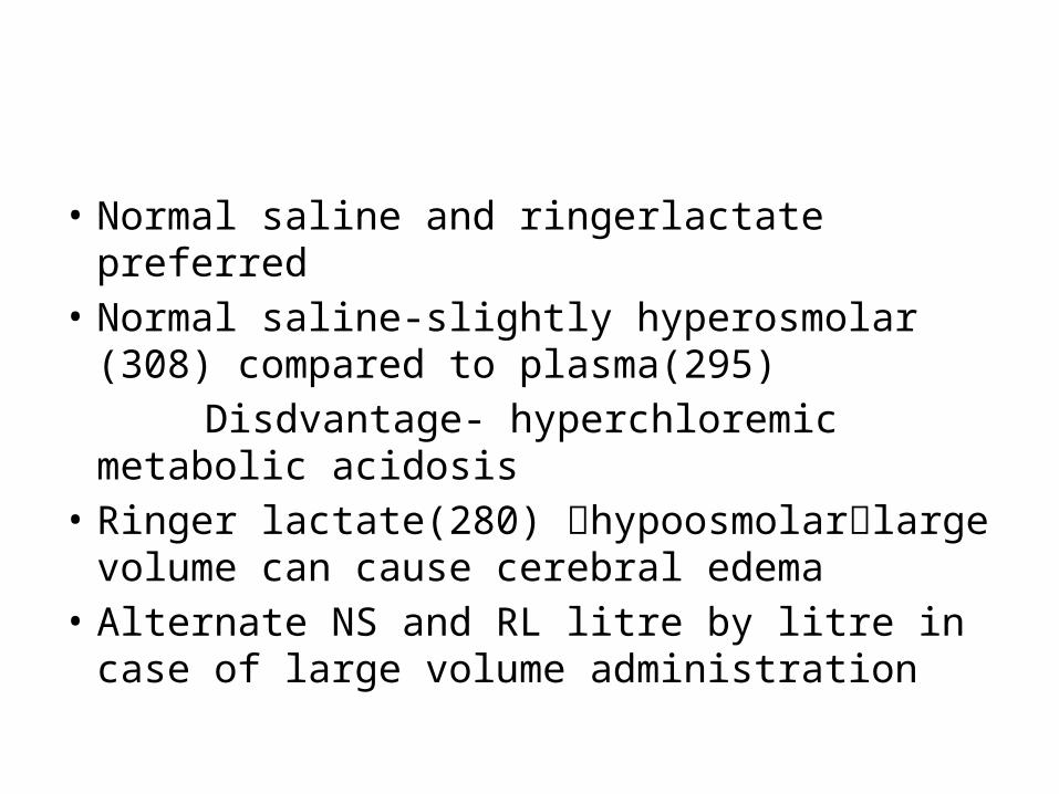
• Normal saline and ringerlactate preferred• Normal saline-slightly hyperosmolar (308)
compared to plasma(295) Disdvantage- hyperchloremic metabolic
acidosis• Ringer lactate(280) hypoosmolarlarge
volume can cause cerebral edema• Alternate NS and RL litre by litre in case of large
volume administration

Colloids
• TCMP gradient is mainly determined by osmolarity and only by a smaller grade by colloid oncotic pressure
• Albumin is a reasonable choice if colloid is required• Starch containing solutions producesI. Dilutional reduction of coagulation factorsII. Interferes directly with platelets and factor viii
complex.• Keep the dose limited to the manufacturers
recommendation

BP CONTROL
• Maintain cerebral perfusion pressure normal or high normal range
• CBF is low in many regions after TBI• Autoregulatory response may not be intact
after TBI/ SAH• Brain compressed under retractors regional
perfusion press will be low


Temperature
• Routine use of hypothermia not advocated Problems– dysrhythmias and coagulation
dysfunctionDeep brain temperature- esophageal, tympanic
membrane pulmonary artery jugular bulb temperature


Emergence
Goals• Smooth emergence• Maintain MAP, CMRO2, PaO2, PaCO2, Temp• Avoid factors that lead to intracranial bleeding– (coughing, bucking, intratracheal suctioning,
ventilator fight)• Pt should be calm, cooperative, & responsive
to verbal commands soon after emergence
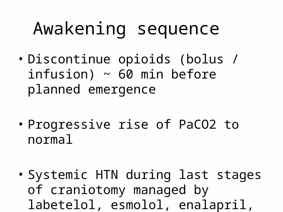
Awakening sequence
• Discontinue opioids (bolus / infusion) ~ 60 min before planned emergence
• Progressive rise of PaCO2 to normal
• Systemic HTN during last stages of craniotomy managed by labetelol, esmolol, enalapril, nicardipine, diltiazem, dexmeditomedine

• Stop volatile anesthetics during skin closure• Maintain with 02 +N2O with propofol either bolus
or infusion at rates of 25-100 ug/kg/min• Lignocaine 1.5mg/kg to be given as head dressing
begins, which decrease coughing & straining• Adequate suctioning• Antagonise muscle relaxant, Stop N2O• Extubate• Transfer to PACU

Indications for late emergence• Preop – pt obtunded• Inadequate airway control preop• Large risk of brain edema / raised ICP• Extensive surgery• Repeat surgery• Major glioblastoma surgery• Surgery involving/ close to vital areas• Surgery asso with significant brain ischemia
(long vascular clipping times, extensive retractor pressure)

Delayed emergence• If pt not awake enough to obey simple verbal
commands 20-30 min after pharmacologically adequate cessation of anaesthesia, non anesthetic causes of delayed emergence should be considered & ruled out like– Seizures– Cerebral edema– Intracranial hematoma– Pneumocephalus– Ischemia– Metabolic / electrolyte abnormalities

Complications
• Bleeding• Hemodynamic instability• Brain swelling• Venous air embolism• Frontal lobey• Abnormal water balance• Temperature disturbances

THANK YOU…

![CCT in Anaesthetics · 2019. 8. 16. · • Planned supratentorial and posterior fossa surgery [including vascular disease and tumours] • Emergency surgery for traumatic brain injury](https://img.dokumen.tips/doc/110x75/614455f2aa0cd638b460ca6d/cct-in-anaesthetics-2019-8-16-a-planned-supratentorial-and-posterior-fossa.jpg)