Embed Size (px)
Citation preview

Dr. Sarbesh Tiwari

The skull base represents a central and complex bone structure of the
skull that forms the floor of the cranial cavity on which the brain lies.
It separates brain from facial structures and suprahyoid neck.
Anatomical knowledge of this particular region is important for
under-standing several pathologic conditions as well as for planning
surgical procedures.
2Anatomy skull base

The human skull consists of three components:
(1) the membranous neurocranium, which constitutes the flat bones of
the skull,
(2) the cartilaginous neurocranium or chondrocranium which forms the
majority of the skull base, and
(3) the viscerocranium or facial skeleton.
3Anatomy skull base

The basicranium develops primarily from cartilage precursors, with a
small component from membranous bone.
The development of the cartilaginous skull base begins around the
40th day of gestation, with the conversion of mesenchyme into
cartilage.
Occipital sclerotomal mesenchyme concentrates around the
notochord and extends cephalically forming the floor of brain.
4Anatomy skull base

The parachordal cartilage – Around the notochord.
Sclerotomal cartilage – Occipital bone.
2 hypophyseal cartilage – Fuse to form basisphenoid cartilage.
2 presphenoid cartilage – body of sphenoid. ‘
Orbitosphenoid and Alisphenoid – wings of sphenoid.
5Anatomy skull base

6Anatomy skull base

The chondrocranium begins to form when the collections of mesenchyme accumulating around and in front of the notochord condense into cartilage.
These chondrification centers, termed the parachordal cartilages, form early in the seventh week adjacent to the rostral end of the notochord and contribute to the creation of the basal plate.
The parachordal cartilage fuse with the sclerotomes arising from the occipital somites surrounding the neural tube.
7Anatomy skull base
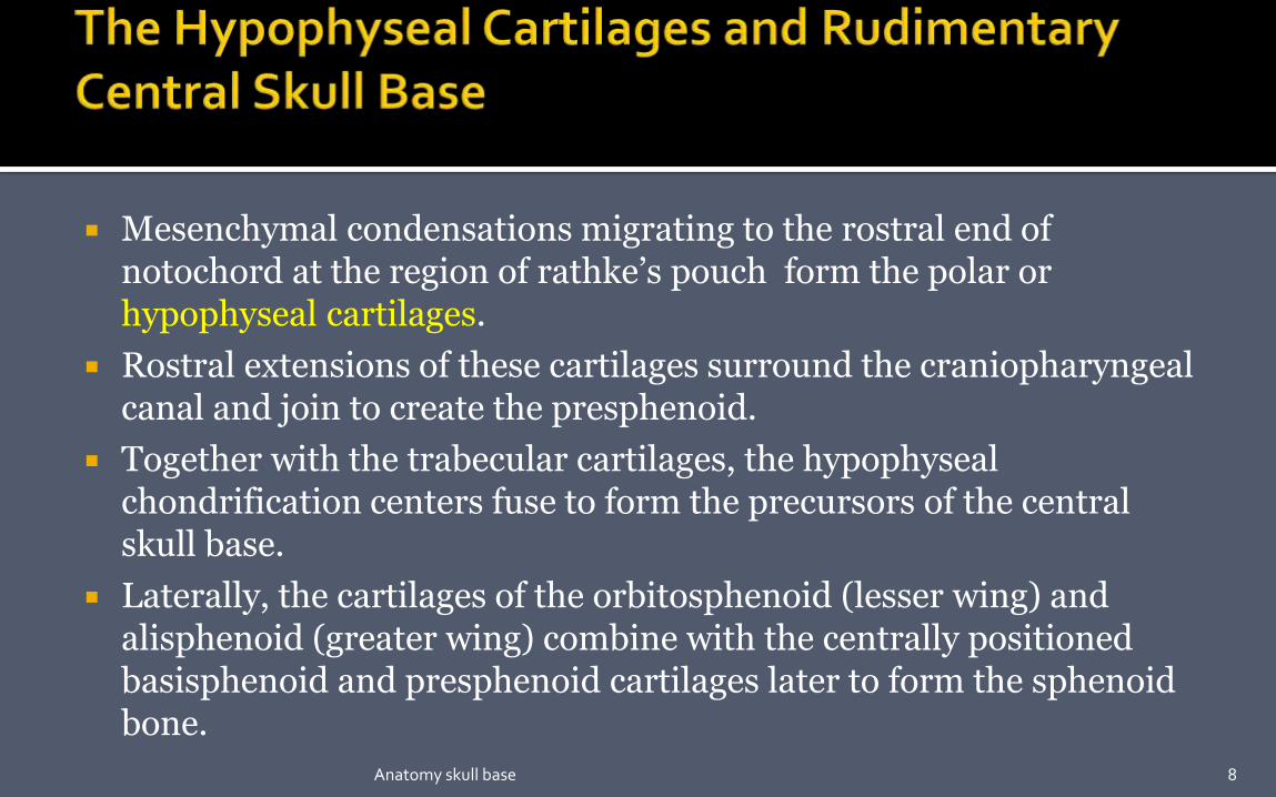
Mesenchymal condensations migrating to the rostral end of notochord at the region of rathke’s pouch form the polar or hypophyseal cartilages.
Rostral extensions of these cartilages surround the craniopharyngealcanal and join to create the presphenoid.
Together with the trabecular cartilages, the hypophyseal chondrification centers fuse to form the precursors of the central skull base.
Laterally, the cartilages of the orbitosphenoid (lesser wing) and alisphenoid (greater wing) combine with the centrally positioned basisphenoid and presphenoid cartilages later to form the sphenoid bone.
8Anatomy skull base

The capsular tissue surrounding the nasal placodes chondrifies along with the trabeculi cranii, ossifies into the ethmoid and inferior nasal concha bones.
The midline segments of these bones create the nasal septum, which remains cartilaginous postpartum and acts as functional matrix for later midface growth.
9Anatomy skull base

Tissue surrounding the otic placodes also chondrifies then fuses with the parachordal cartilages to eventually create the petrous and mastoid segments of the temporal bone.
10Anatomy skull base

11Anatomy skull base

Dorsal view of the chondrocranium, or
base of the skull, in the adult showing
bones formed by endochondral
ossification.
Bones that form rostral to the rostral
half of the sella turcica arise from
neural crest and constitute the
prechordal (in front of the notochord)
chondrocranium (blue).
Those forming posterior to this
landmark arise from paraxial
mesoderm (chordal chondrocranium)
(red).
12Anatomy skull base

13Anatomy skull base

The skull base is composed of
five bones:
(1) ethmoid,
(2) sphenoid,
(3) occipital ,
(4) paired temporal , and
(5) paired frontal bones.
Fossa :-
1. Anterior cranial fossa
2. Middle cranial fossa.
3. Posterior cranial fossa. 14Anatomy skull base

It forms the bottom of the anterior skull , separating the anterior cranial
fossa from the paranasal sinuses and the orbits.
The boundaries :-
1. Anterior border :- posterior wall of the frontal sinus.
2. Posterior border :- lesser wing of the sphenoid bone and anterior clinoid
processes.
3. Floor :- roof of the nasal cavity and ethmoid sinuses medially.
4. Lateral wall :- thick and strong orbital plates of the frontal bone.
15Anatomy skull base

16Anatomy skull base

Frontal crest :- It’s a midline bony ridge that projects upwards and provide attachment to the falx cerebri.
Crista galli :- (latin for cock’s comb) Provides the site for anterior most attachment of the falx cerebri.
Cribriform plate :- It is a sheet of bone which contains numerous small foramina – these transmit olfactory nerve fibres (CN I) into the nasal cavity.
Anterior ethmoidal foramen transmits the anterior ethmoidal artery, nerve and vein. Posterior ethmoidal foramen transmits the posterior ethmoidal artery, nerve and vein.
17Anatomy skull base

The CSB makes up the floor of the
middle cranial fossa.
The sphenoid bone contributes to the
most of the CSB.
Anterior border :-
- tuberculum sellae, anterior clinoid
process, posterior margin of lesser
wing of sphenoid & anterior superior
rim of greater wing of sphenoid.
Posterior border :-
- superior border of petrous part of
temporal bone and the dorsum sellae
of sphenoid.
18Anatomy skull base
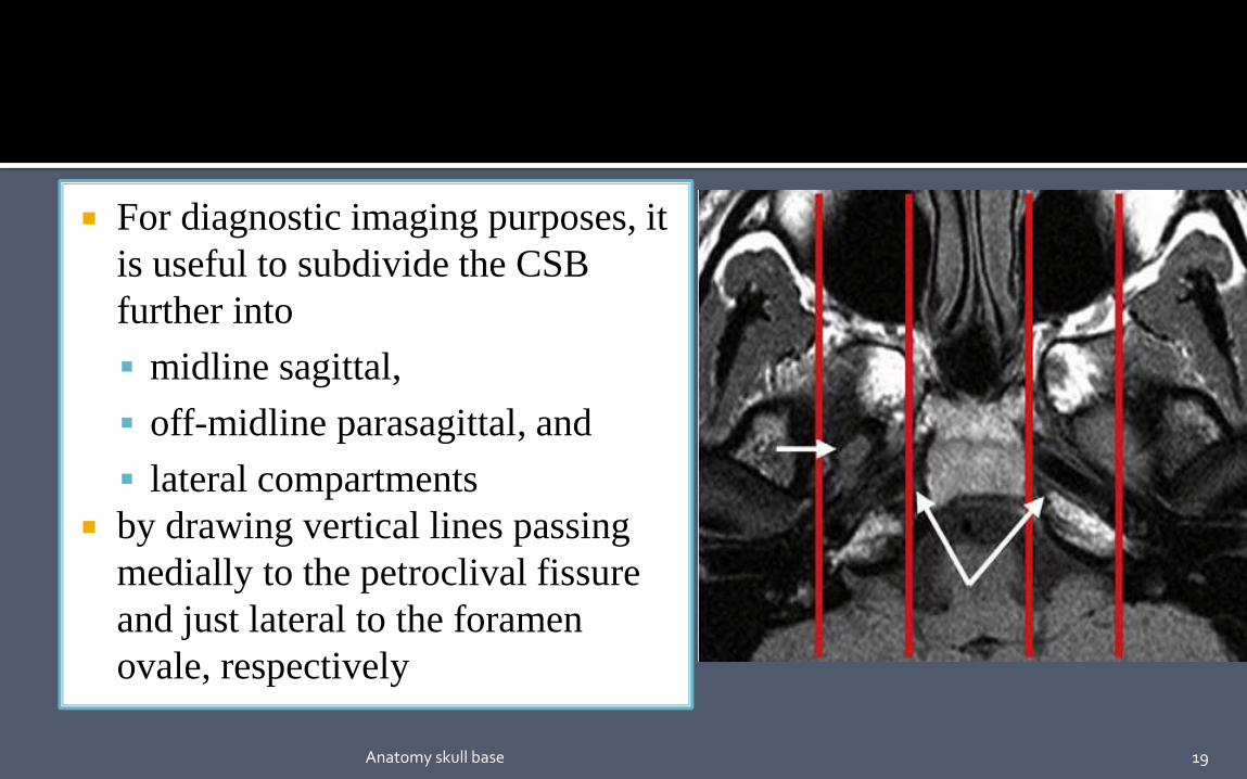
For diagnostic imaging purposes, it
is useful to subdivide the CSB
further into
midline sagittal,
off-midline parasagittal, and
lateral compartments
by drawing vertical lines passing
medially to the petroclival fissure
and just lateral to the foramen
ovale, respectively
19Anatomy skull base

Includes the body of the
sphenoid and the portion of the
clivus anterior to the spheno-
occipital synchondrosis
(basisphenoid),
contains the sphenoid sinus,
and
bordered superiorly by the sella
turcica and inferiorly by the
roof and posterior wall of the
nasopharynx20Anatomy skull base

It includes the petroclival synchondrosis, foramen lacerum, and
medial aspect of the greater sphenoid wing.
It is bordered superiorly and medially by the parasellar region
containing the cavernous sinus, superiorly and laterally by the basal
temporal lobes, and inferiorly by the parapharyngeal and masticator
spaces of the suprahyoid neck.
Many crucial neurovascular structures lay in this compartment ,
including cavernous sinus, superior orbital fissure, foramen
rotundum, vidian canal , and foramen lacerum.
21Anatomy skull base

formed by sphenoid triangle, squamous part of temporal bone, and
temporomandibular joint.
22Anatomy skull base

Anterior margin :- The posterior surface of the clivus.
Laterally – superiorly the posterior surface of the petrous part of temporal bone and inferiorly the condylar part of the occipital bone.
Posteriorly , the mastoid part of temporal bone and the squamous part of occipital bone.
Foramen magnum
23Anatomy skull base

The sphenoid bone is the foundation of the central skull base.
The shape of the sphenoid bone resembles that of a bird with wings
outstretched.
It consist of
a central body;
two sets of wings– the greater and lesser, which course laterally ;and
two pterygoid processes, which are directed inferiorly.
24Anatomy skull base
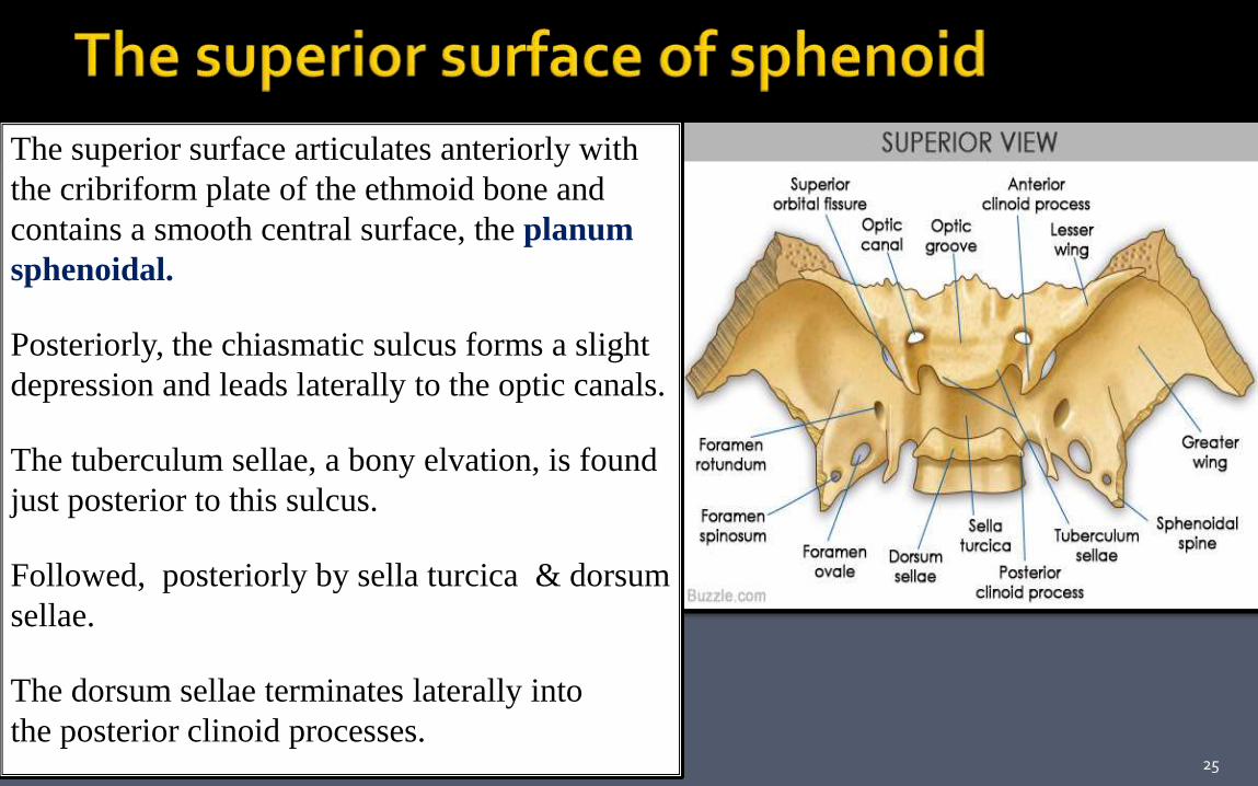
The superior surface articulates anteriorly with
the cribriform plate of the ethmoid bone and
contains a smooth central surface, the planum
sphenoidal.
Posteriorly, the chiasmatic sulcus forms a slight
depression and leads laterally to the optic canals.
The tuberculum sellae, a bony elvation, is found
just posterior to this sulcus.
Followed, posteriorly by sella turcica & dorsum
sellae.
The dorsum sellae terminates laterally into
the posterior clinoid processes.25Anatomy skull base

The anterior surface of the body of sphenoid
forms the roof & posterior wall of
nasopharynx.
„ The body houses the sphenoid sinus .
„ Lesser wings- forms medial portion of
orbital apex
„ Greater wings – course upward and
laterally from both sides of the sphenoid
body- forms floor of middle cranial fossa,
posterolateral orbit & lateral calvaria.
26Anatomy skull base

The pterygoid processes descend inferiorly from the sphenoid body.
The lateral pterygoid plate forms a portion of the medial wall of
the infratemporal fossa and provides attachment for the lateral
pterygoid muscle.
The medial pterygoid plate terminates inferiorly as a hook-like
process, the pterygoid hammlus around which the tendon of the
tensor veli palatini is slung.
Attachment to medial pterygoid muscles and pharyngobasilar
fascia.
27Anatomy skull base

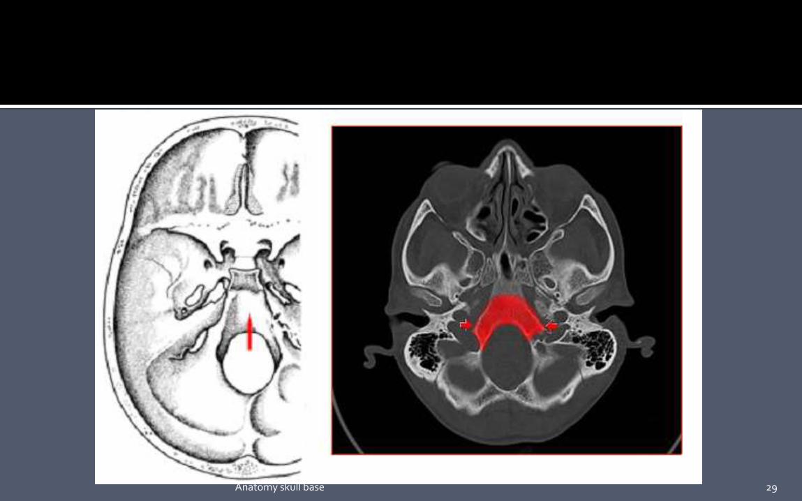
The clivus is that part of the skull base situated between the
foramen magnum and the dorsum sellae.
Formed from sphenoid and occipital bones.
The petroccipital fissure forms the anterior lateral margin of the
clivus, while the synchondrosis between the basioccipital and
exoccipital bones forms the posterior lateral margins.
Normal fat signals in adult (late teens) in MRI.
28Anatomy skull base
29Anatomy skull base

The cavernous sinuses are situated on each side of the body of the
sphenoid bone and extend from the superior orbital fissure anteriorly to
the petrous apex posteriorly.
Receives :-
Superior ophthalmic vein
Inferior ophthalmic vein
Sphenoparietal sinus.
-Drains into
Petrosal sinus.
Pterygoid plexus.
Basilar plexus. Contents
1. III, IV, V1, V2 & VI
2. ICA. 30Anatomy skull base

Dural invagination at posterior aspect of
cavernous sinus
„ Contains gasserian ganglion (trigeminal).
Dural layers demonstrate thin peripheral
enhancement.
In high resolution MR, 3 sensory divisions
of trigeminal nerve can be visualized
leaving the gasserian ganglion.
31Anatomy skull base

The optic canal is formed by the lesser
wing of sphenoid.
The contents are :-
Optic nerve .
Ophthalmic Artery.
Sympathetic fibers from carotid plexus.
32Anatomy skull base

The triangular superior orbital fissure is
bounded medially by the body of the sphenoid,
above by the lesser wing, and below by the
greater wing and is completed laterally by the
frontal bone as the greater and lesser wings
converge.
Optic strut separates the optic canal from the
superior orbital fissure.
The optic canal and the superior orbital
fissure together form the orbital apex.
33Anatomy skull base

Content –
•Nerves 3,4,1st division of 5 , 6.
•„Orbital branch of middle meningeal art; sympathetic
nerve; recurrent meningeal art, sup. ophthalmic vein.
34Anatomy skull base

Extends from PPF along orbital floor.
„Separates greater wings of the sphenoid from the maxilla.
Content –
Maxillary branch of trigeminal nerve. Infraorbital vessels. Emissary veins connecting inferior
ophthalmic vein to pterygoid venous plexus.
Zygomatic nerve.
35Anatomy skull base

Is actually a canal in the base of the greater sphenoid
wing, is situated just inferior and lateral to the
superior orbital fissure.
The canal extends obliquely forward and slightly
inferiorly, connecting the middle cranial fossa to the
pterygopalatine fossa.
The canal transmits the maxillary nerve ( V2) the
artery of the foramen Rotundum and emissary veins.
This foramen is best visualized by means of coronal
CT
36Anatomy skull base
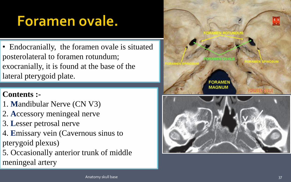
• Endocranially, the foramen ovale is situated
posterolateral to foramen rotundum;
exocranially, it is found at the base of the
lateral pterygoid plate.
Contents :-
1. Mandibular Nerve (CN V3)
2. Accessory meningeal nerve
3. Lesser petrosal nerve
4. Emissary vein (Cavernous sinus to
pterygoid plexus)
5. Occasionally anterior trunk of middle
meningeal artery
37Anatomy skull base

It is an aperture in the greater wing of the
sphenoid posterolateral to foramen ovale.
Contents :-
1. Middle meningeal artery & vein.
2. Emissary vein.
3. Nervous spinosus (Meningeal branch
of mandibular nerve)
38Anatomy skull base

The vidian canal (pterygoid canal) is located in
the floor of the sphenoid sinus at the junction
of the pterygoid process and the sphenoid body
connecting the pterygopalatine fossa anteriorly
and the foramen lacerum posteriorly.
Contents :-
- Vidian Artery ( Br. Of Maxillary
Artery).
- Vidian Nerve (greater superficial
petrosal nerve and deep petrosal nerve )
39Anatomy skull base
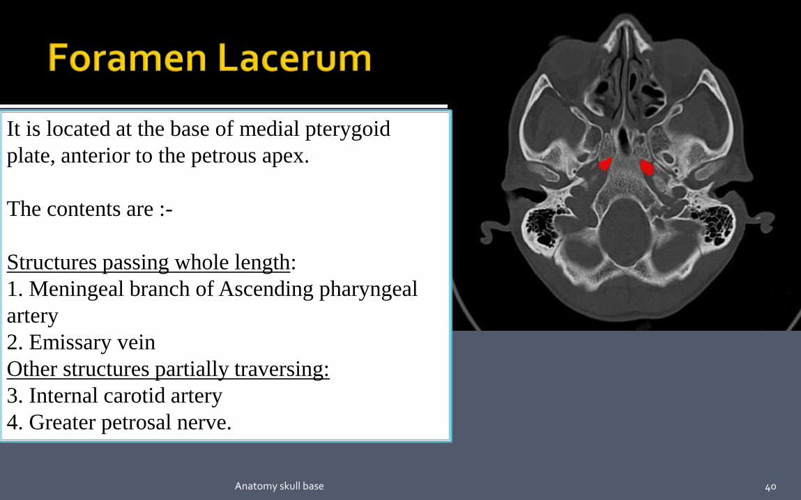
It is located at the base of medial pterygoid
plate, anterior to the petrous apex.
The contents are :-
Structures passing whole length:
1. Meningeal branch of Ascending pharyngeal
artery
2. Emissary vein
Other structures partially traversing:
3. Internal carotid artery
4. Greater petrosal nerve.
40Anatomy skull base

The carotid canal is a passage within the petrous
temporal bone and transmits the internal carotid
artery and sympathetic plexus.
The carotid canal is initially directed superiorly,
then turns anteromedially to reach up to
the petrous apex.
It approximately runs 2cm and opens to foramen
lacerum.
41Anatomy skull base
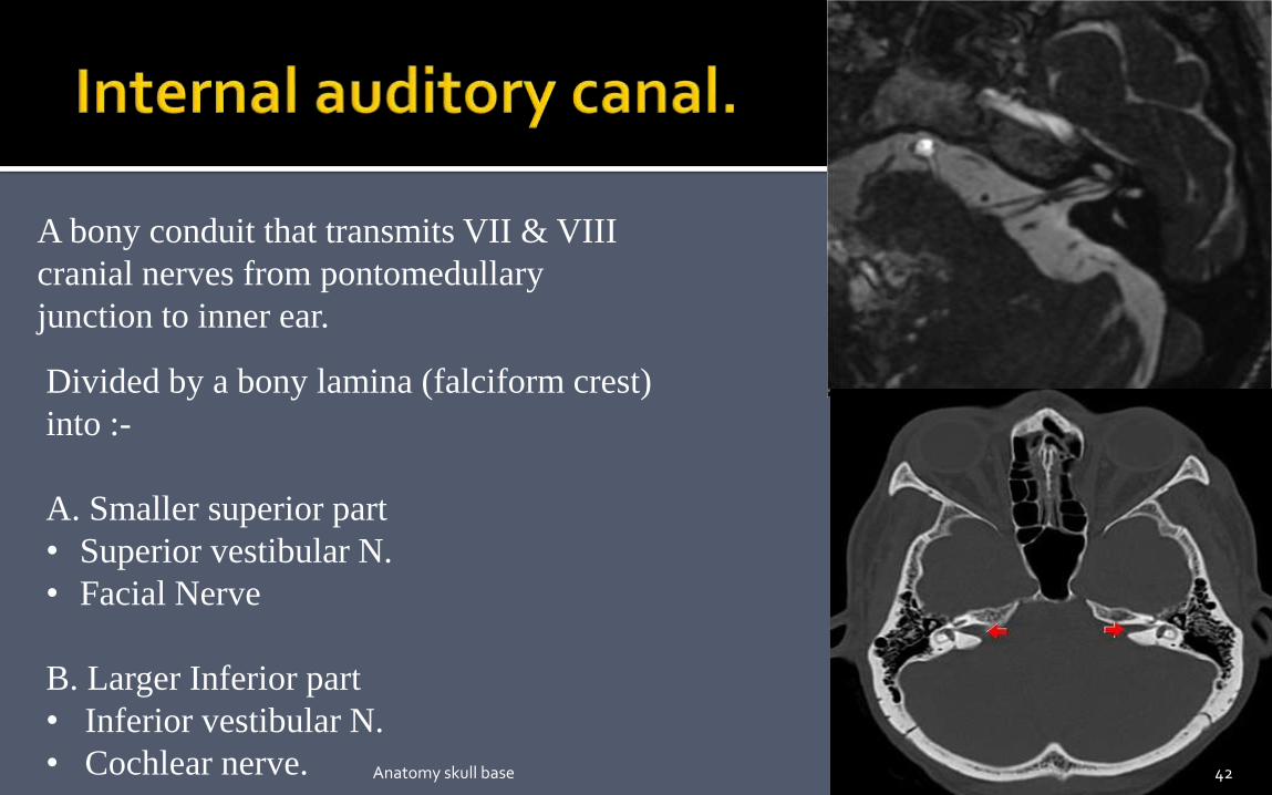
A bony conduit that transmits VII & VIII
cranial nerves from pontomedullary
junction to inner ear.
Divided by a bony lamina (falciform crest)
into :-
A. Smaller superior part
• Superior vestibular N.
• Facial Nerve
B. Larger Inferior part
• Inferior vestibular N.
• Cochlear nerve. 42Anatomy skull base

The jugular foramen is divided by a fibrous
or bony septum, the jugular spine, into:
A. the pars nervosa: smaller and
anteromedial-
i. Inferior petrosal sinus.
ii. Glossopharyngeal nerve with its
tymapnic branch (Jacobson’s Nerve).
B. the pars vascularis: larger and
posterolateral-
i. Jugular bulb.
ii. Vagus Nerve with its auricular
branch (Arnold’s nerve).
iii. Spinal accessory nerve 43Anatomy skull base

The appearance of the jugular foramen is anatomically variable, and
sometimes both cranial nerves IX and X traverse through the pars
nervosa.
The right jugular foramen is larger than the left in 75% of the
population.
When the roof of the jugular bulb is seen above the level of floor of
internal auditory canal , it is called a highriding jugular bulb, which is
more common on the right side.
This is a dangerous variant and compromises the exposure during
translabyrinthine surgery.
44Anatomy skull base

Located within occipital bone.
The hypoglossal (or anterior condyloid)
canal is a paired bone passage that runs
lateral to and slightly forward from the
posterior cranial fossa to the
nasopharyngeal carotid space.
It transmits the hypoglossal nerve.
Intracanalicular enhancement is always
present (emissary veins), with linear
filling defects ( nerve rootlets).
45Anatomy skull base
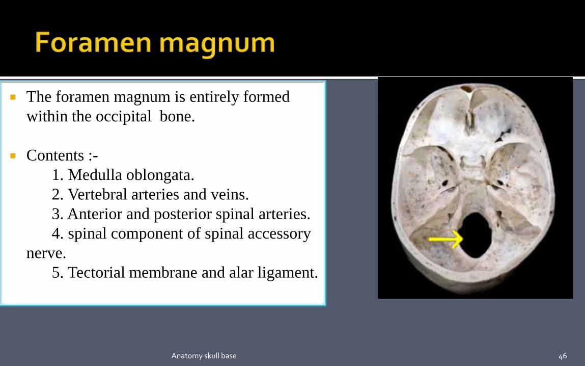
The foramen magnum is entirely formed
within the occipital bone.
Contents :-
1. Medulla oblongata.
2. Vertebral arteries and veins.
3. Anterior and posterior spinal arteries.
4. spinal component of spinal accessory
nerve.
5. Tectorial membrane and alar ligament.
46Anatomy skull base

47Anatomy skull base

48

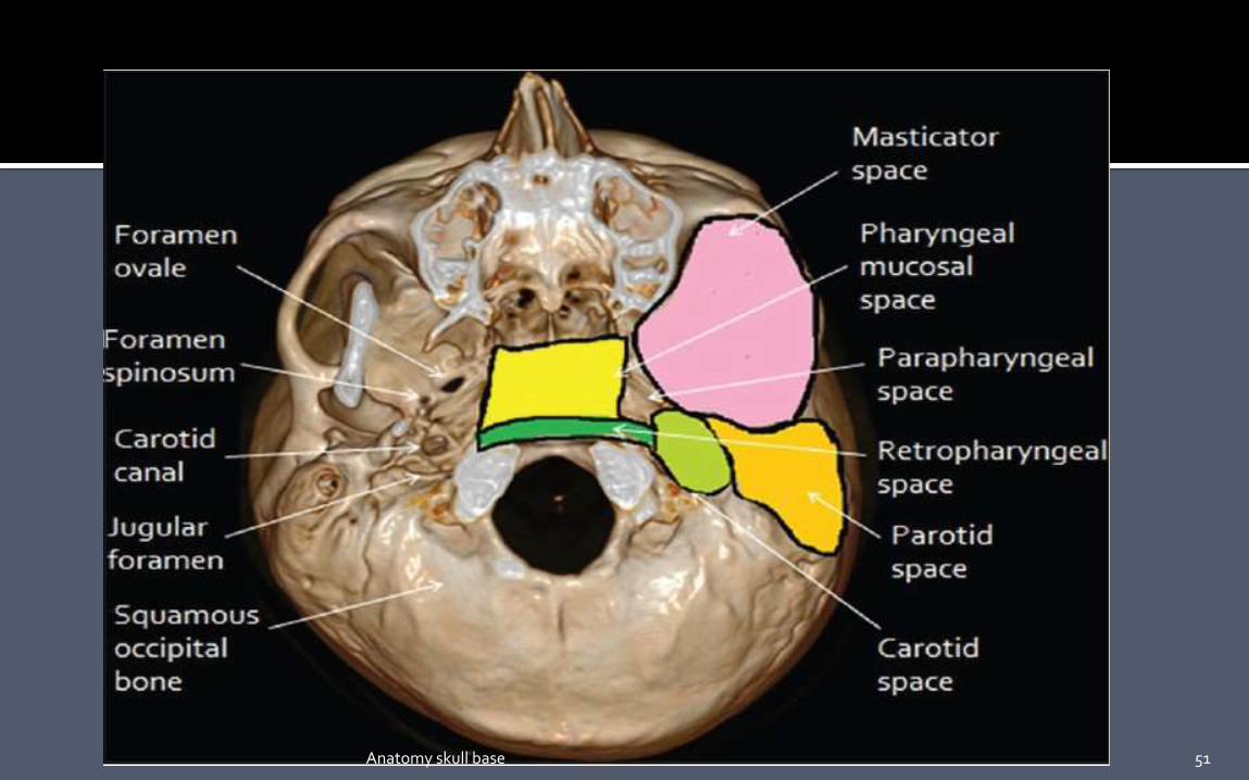
Parapharyngeal , masticator, carotid, and retropharyngeal spaces are
seen in close contact with the skull base along their cephalad aspect .
Parapharyngeal space extends caudally to the submandibular space
and cranially abuts the base skull. It contains fat within, which acts as
a medium for infection.
49Anatomy skull base

Masticator space connects the mandible to the skull base. Odontogenic
infections and oropharyngeal squamous cell carcinoma can tract along
masticator space to the base skull.
Intracranial extension of the tumor can occur
via third division of trigeminal nerve, mandibular nerve (perineural
spread) through the foramen ovale.
Vascular lesions such as jugular vein thrombosis and neural tumors such
as Schwannoma, Neurofibromas, and Paraganglioma are seen in the
carotid space.
50Anatomy skull base
51Anatomy skull base

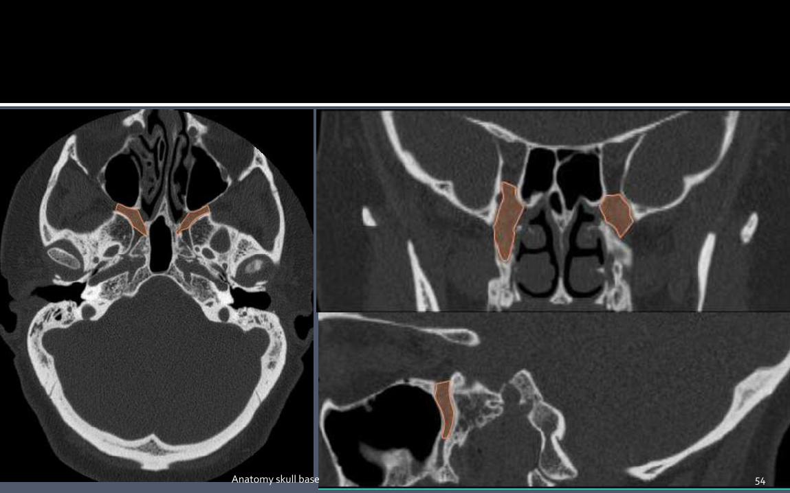
A fat filled space between the pterygoid
plates and the posterior wall of maxillary
sinus.
Shaped like an inverted pyramid.
Borders :-
The walls of the PPF are as follows:
Medial - perpendicular plate palatine bone.
Lateral - narrowing to pterygomaxillary fissure
Anterior - posterior wall of maxillary sinus
posterior - Medial and lateral pterygoid plates;
inferior aspect of lesser wing of sphenoid bone52Anatomy skull base

The PPF is an important pathway for the spread of neoplastic and infectious
processes:
Medially - communicates with the nasal cavity via the sphenopalatine foramen.
Laterally - communicates with the masticator space (or infratemporal fossa) via
the pterygomaxillary fissure.
Anteriorly - communicates with the orbit via the inferior orbital fissure.
Posteriorly and superiorly - communicates with the Meckel cave and cavernous sinus (of
the middle cranial fossa) via the foramen rotundum.
Posteriorly and inferiorly - communicates with the middle cranial fossa via the vidian
canal, which transmits the Vidian nerve.
Inferiorly - communicates with the palate via the greater and lesser palatine canals 53
54Anatomy skull base

The infratemporal fossa is the space between the skull base, lateral
pharyngeal wall and the ramus of mandible.
Boundaries :-
Lateral- Ramus and condylar process of the mandible.
„Medial- Lateral pterygoid plate.
Anterior – Posterolateral wall of maxilla.
Posterior – Carotid sheath.
„Superior - CRANIAL BASE (greater wing of the sphenoid bone).
„Inferior – Medial pterygoid muscles.
55Anatomy skull base

Pterygoid muscles, medial and Lateral
„ Maxillary artery and vein
„ Pterygoid plexus of veins
„ Mandibular division of trigeminal nerve
„ Otic ganglion
56Anatomy skull base

57Anatomy skull base

58Anatomy skull base

59Anatomy skull base

60Anatomy skull base

61Anatomy skull base
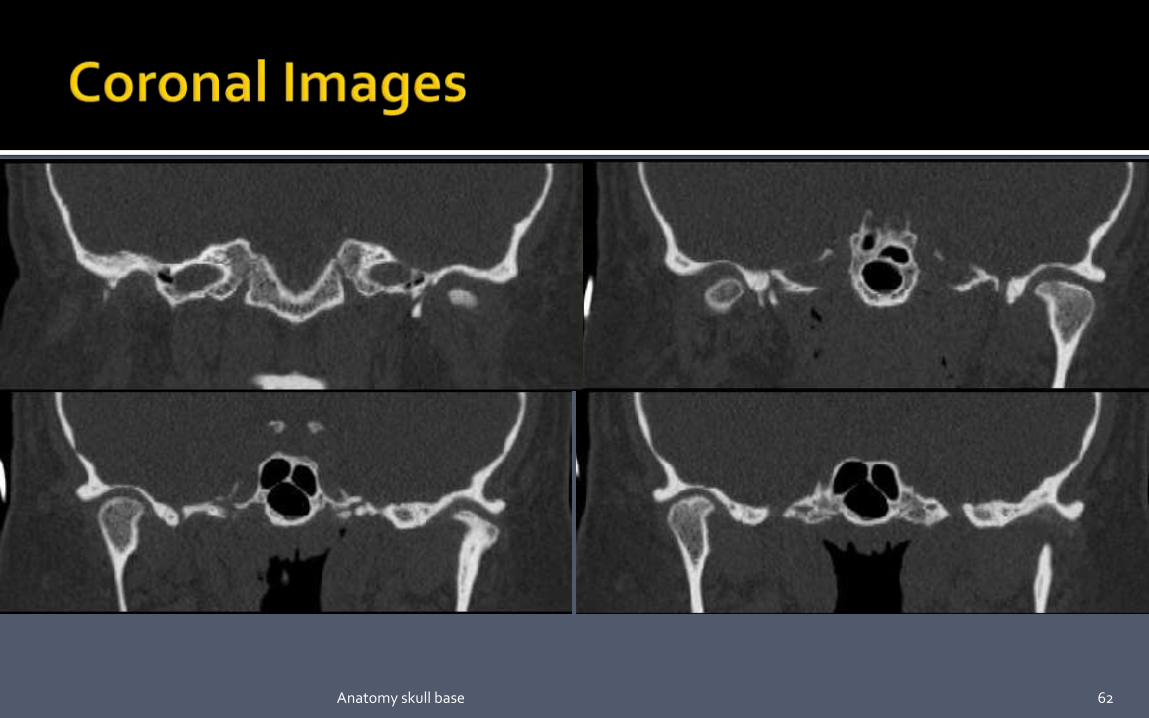
62Anatomy skull base

63Anatomy skull base
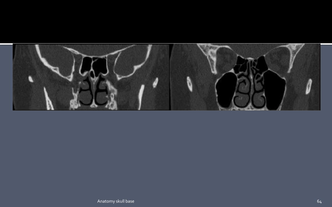
64Anatomy skull base

1. Imaging of skull base: Pictorial essay Abhijit A Raut , Prashant S Naphade, and Ashish
Chawla. Indian J Radiol Imaging. 2012 OctDec; 22(4): 305–316.
2. Imaging of the Anterior Skull Base :Hemant Parmar,MD, SachinGujar,MD et.al. Neuroimag Clin
N Am 19 (2009) 427–439.
3. CT and MR Imaging of the Central Skul Base. Part 1: Techniques, Embryologic Development, and
Anatomy. Fredj Lame, MD, Lyn Nadel, MD, Ira F. Braun, MD. RadloGraphics 190; 10:59 1-602
4. Imaging of the Central Skull Base Alexandra Borges, MD. Neuroimaging Clinics of North
America Volume 19, Issue 3, August 2009, Pages 441–468.
5. Skull base embryology: a multidisciplinary review. Di Ieva A1, Bruner E, Haider T et.al Childs
Nerv Syst. 2014 Jun;30(6):991-1000.
6. Som and curtin.
7. http://www.med.wayne.edu/diagRadiology/Anatomy_Modules/axialpages/Overview.html
THANK YOU 65Anatomy skull base

66Anatomy skull base