Embed Size (px)
Citation preview
Endoscopic Ultrasound (EUS):from A to Z
Jan 2015
Jason Klapman,MDDirector of EndoscopyMoffitt Cancer Center
• No relevant financial disclosures

Objectives
• Describe the main clinical uses of EUS
• Illustrate the role of EUS in the context of other modalities in the investigation of pancreatic/biliary disease
• Provide a perspective on how EUS advances may impact the conventional approach to GI disorders

What is EUS?
• convergence of US and endoscopy
• US probe at scope tip allows detailed views of GI tract wall and adjacent structures
• History: 1st published reports in 1980s, increasing clinical use since 1990s
EUS - fine needle aspiration (FNA)
QuickTime™ and aDV/DVCPRO - NTSC decompressor
are needed to see this picture.
QuickTime™ and aDV/DVCPRO - NTSC decompressor
are needed to see this picture.

EUS allows us to see...
• Esophagus: esophageal wall, mediastinal structures (aorta, heart, azygous vein, right/left pleura, mediastinal LN, etc.)
• Stomach: gastric wall, pancreas (body/tail), celiac vessels, liver, GB, spleen, left adrenal, left kidney
• Duodenum: duodenal wall, ampulla, pancreas (head/uncinate), CBD, GB, portal vein, right kidney
• Rectum: rectal wall, anal sphincter, perirectal structures (prostate, uterus), iliac vessels

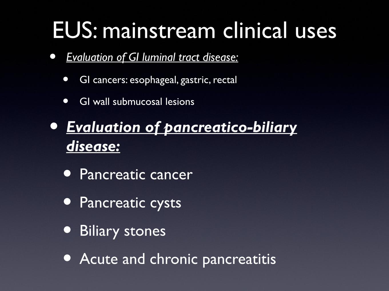
EUS: mainstream clinical uses
• Evaluation of GI luminal tract disease:
• GI cancers: esophageal, gastric, rectal
• GI wall submucosal lesions
• Evaluation of pancreatico-biliary disease:
• Known of suspected pancreatic cancer
• Pancreatic cysts
• Biliary stones
• Acute and chronic pancreatitis

EUS : normal GI tract wall
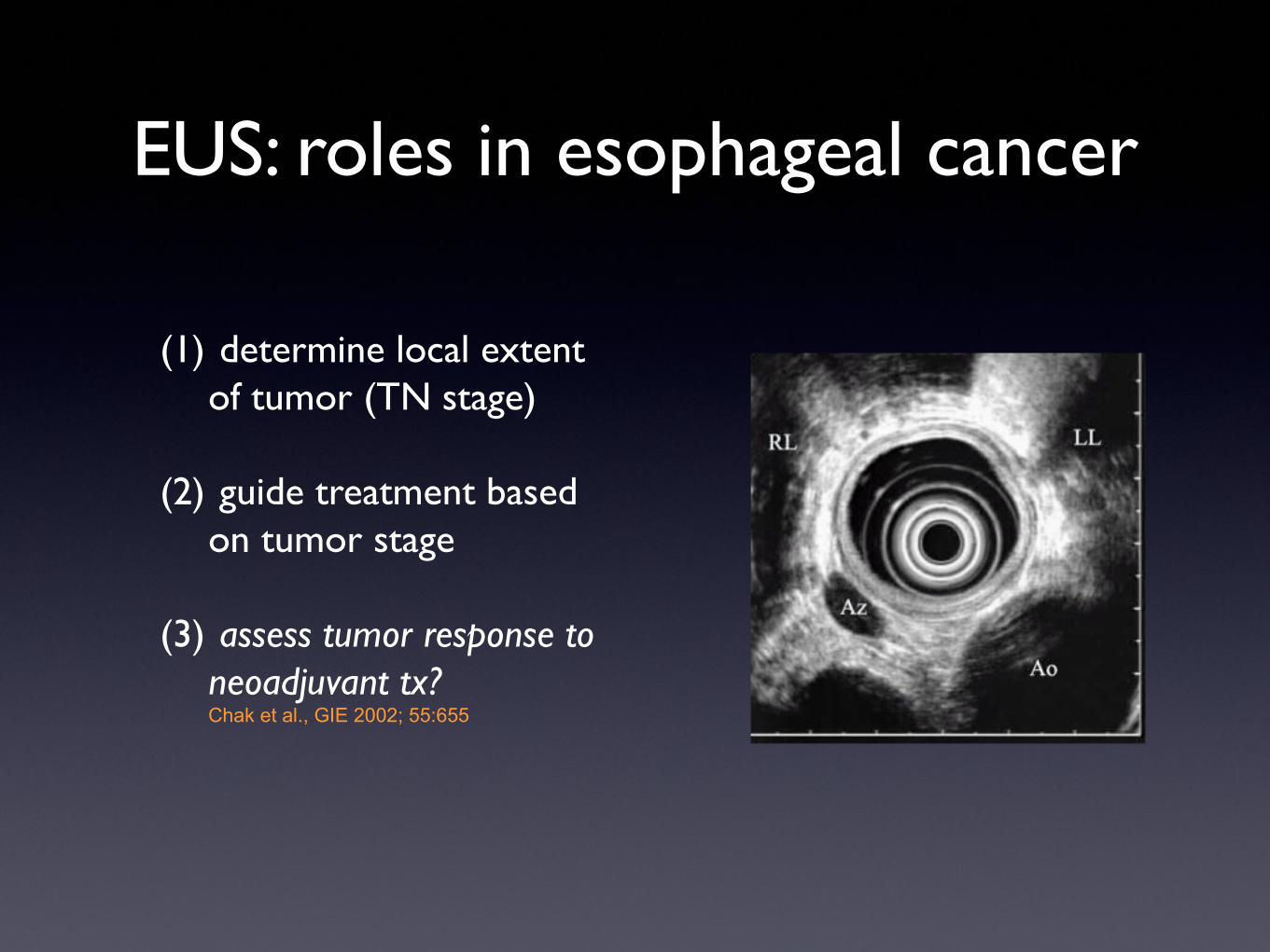
EUS: roles in esophageal cancer
(1) determine local extent of tumor (TN stage)
(2) guide treatment based on tumor stage
(3) assess tumor response to neoadjuvant tx? Chak et al., GIE 2002; 55:655

Esophageal cancer: stage-based treatment

Esophageal cancer - early stage
tumor stage T2: tumor invades into (but not through) esophageal wallPatient underwent esophagectomy

GE junction cancer: locally advanced
tumor stage T3N1: tumor invades through muscular wall + local LNPatient underwent preop chemoXRT followed by surgery

EUS-FNA of LN in esophageal cancer
•Technically feasible when tumor not adjacent to LN
•Increases N staging accuracy over EUS alone: 70 vs. 93%
•Wiersema. GIE 2001;53:751.
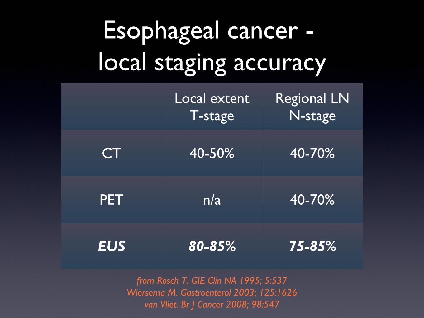
Esophageal cancer - local staging accuracy
Local extentT-stage
Regional LNN-stage
CT 40-50% 40-70%
PET n/a 40-70%
EUS 80-85% 75-85%
from Rosch T. GIE Clin NA 1995; 5:537Wiersema M. Gastroenterol 2003; 125:1626
van Vliet. Br J Cancer 2008; 98:547
EUS: roles in gastric malignancy
• guide treatment based on tumor stage
• early stage > surgery
• advanced stage > chemo, palliative surgery
• superficial lesions > endoscopic treatment
• tumor staging and follow-up of gastric lymphoma (MALToma)
• evaluation of suspected linitis plastica

EUS: roles in rectal cancer
(1) Guide treatment based on tumor stage (analogous to esophageal cancer)
(2) Post-operative surveillance:
• q3-6 months for patients that did not undergo aggressive surgical resection (e.g. mesorectal excision)
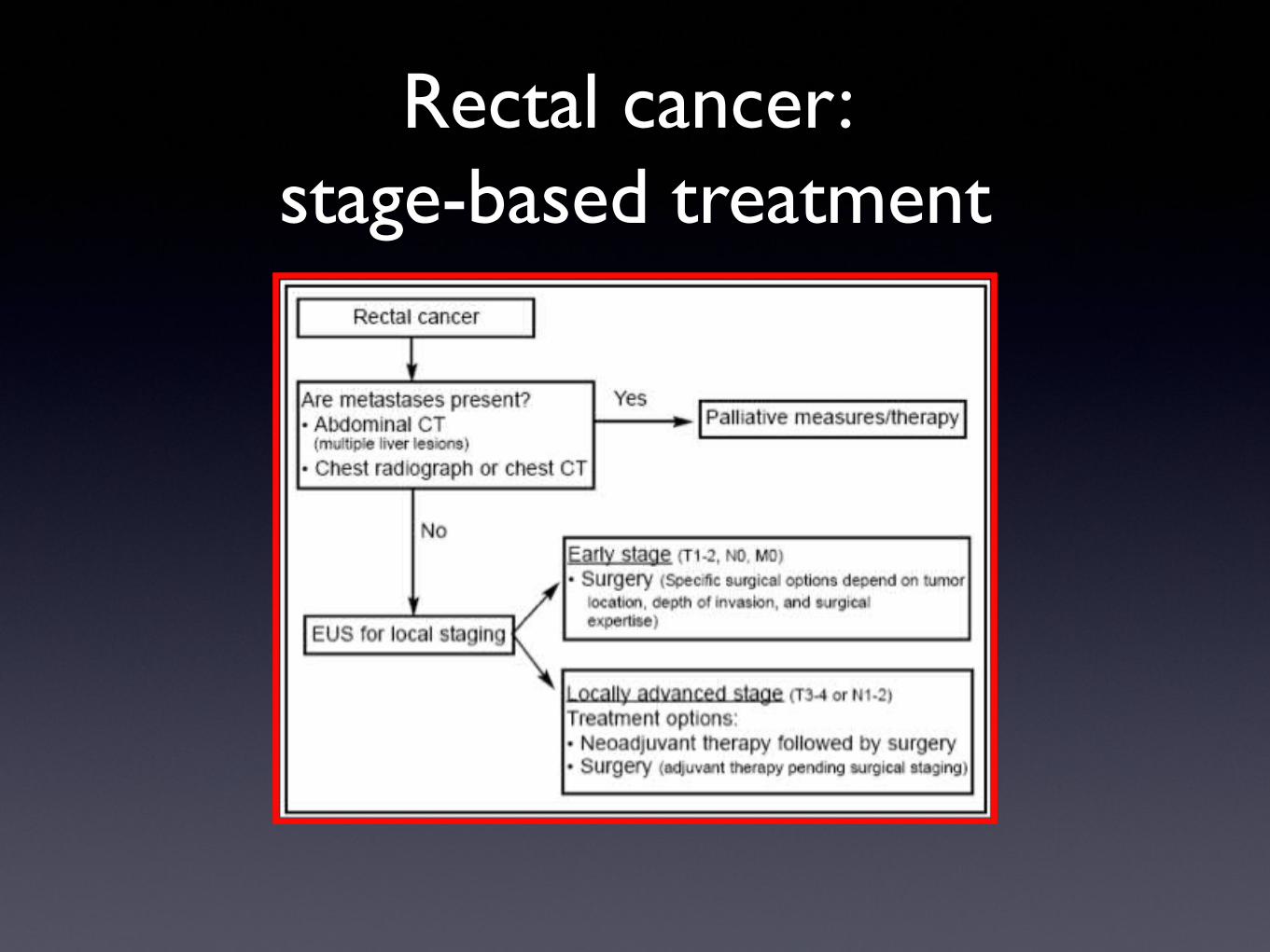
Rectal cancer: stage-based treatment
Rectal cancer : early stage lesion
QuickTime™ and aDV/DVCPRO - NTSC decompressor
are needed to see this picture.

Rectal cancer : locally advanced
T4- mass invades through rectal wall into prostatecandidate for neoadjuvant therapy
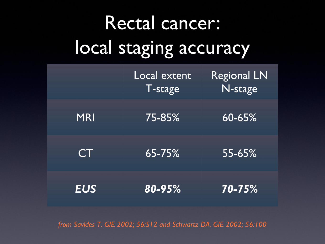
Rectal cancer:local staging accuracy
Local extentT-stage
Regional LNN-stage
MRI 75-85% 60-65%
CT 65-75% 55-65%
EUS 80-95% 70-75%
from Savides T. GIE 2002; 56:S12 and Schwartz DA. GIE 2002; 56:100
Summary:EUS for GI luminal cancers
• Determine local tumor extent (T and N stage)
• Guide treatment based on predicted tumor stage

Submucosal lesion at EGD
QuickTime™ and aDV/DVCPRO - NTSC decompressor
are needed to see this picture.
What is it?
Is it worrisome?
Surgery?
Differential Dx ???

Etiology of submucosal lesions by EUS
from Chak. GIE 2002; 56:S43
EUS: mainstream clinical uses• Evaluation of GI luminal tract disease:
• GI cancers: esophageal, gastric, rectal
• GI wall submucosal lesions
• Evaluation of pancreatico-biliary disease:
• Pancreatic cancer
• Pancreatic cysts
• Biliary stones
• Acute and chronic pancreatitis

Case:• 65 year-old male presents with a 20 lb.
unintentional wt loss over 3 mo, and 2 wk hx of jaundice. He denies abd pain or fevers. TB=12, DB=8, Alk Phos=650.
• An MRI/MRCP was obtained- moderate CBD dilation with “fullness” of the pancreatic head, no definite mass. The patient has done internet research, and asks if the next step is ERCP ?
What is the role of ERCP in suspected pancreatic CA?
Did the MRI miss a tumor? How often does that occur?
What is the role of EUS?
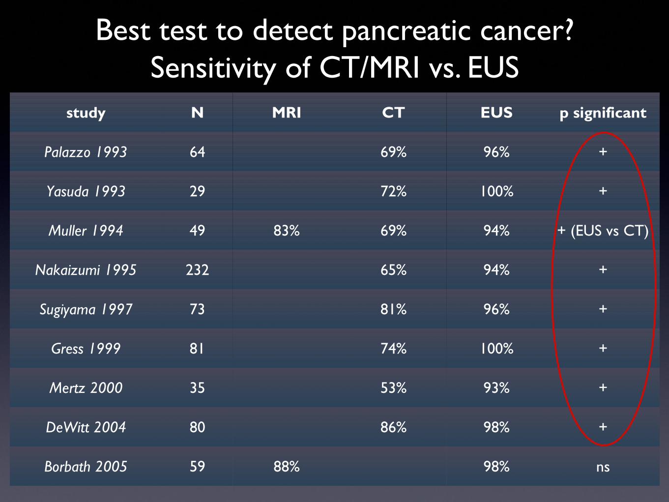
Best test to detect pancreatic cancer?Sensitivity of CT/MRI vs. EUS
study N MRI CT EUS p significant
Palazzo 1993 64 69% 96% +
Yasuda 1993 29 72% 100% +
Muller 1994 49 83% 69% 94% + (EUS vs CT)
Nakaizumi 1995 232 65% 94% +
Sugiyama 1997 73 81% 96% +
Gress 1999 81 74% 100% +
Mertz 2000 35 53% 93% +
DeWitt 2004 80 86% 98% +
Borbath 2005 59 88% 98% ns

Detection of small tumors< 2.5 - 3cm
study N sensitivity:CT EUS
Palazzo 1993 7 14% 100%
Muller 1994 15 53% 93%
DeWitt 2004 19 53% (MDCT) 89%
Main benefit of EUS over CT is detection of small lesions
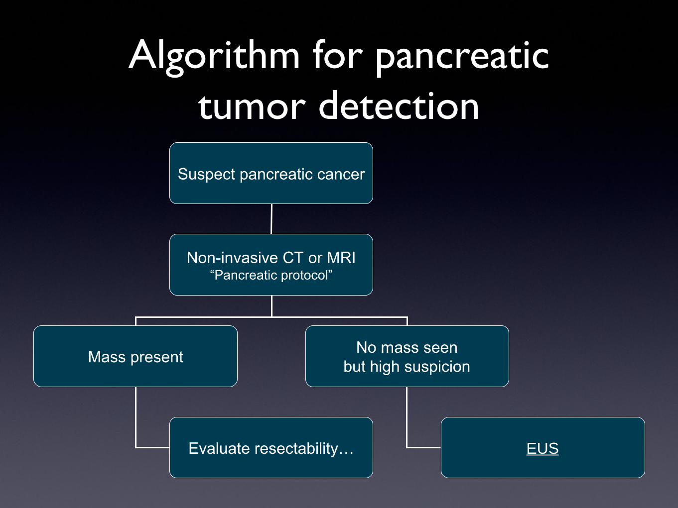
Algorithm for pancreatic tumor detection
Suspect pancreatic cancer
Non-invasive CT or MRI“Pancreatic protocol”
Mass presentNo mass seen
but high suspicion
EUSEvaluate resectability…
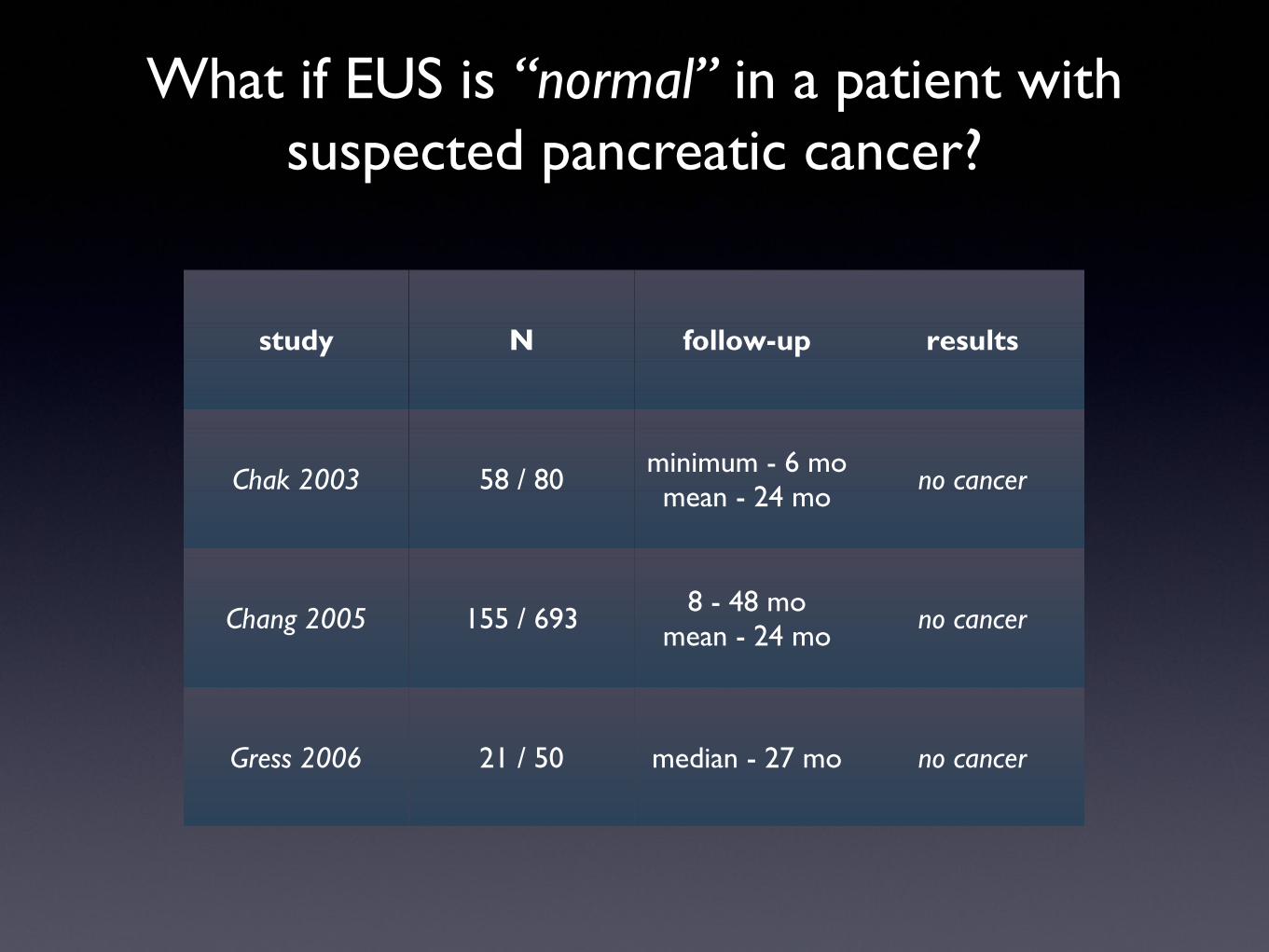
What if EUS is “normal” in a patient with suspected pancreatic cancer?
study N follow-up results
Chak 2003 58 / 80 minimum - 6 momean - 24 mo no cancer
Chang 2005 155 / 693 8 - 48 momean - 24 mo no cancer
Gress 2006 21 / 50 median - 27 mo no cancer
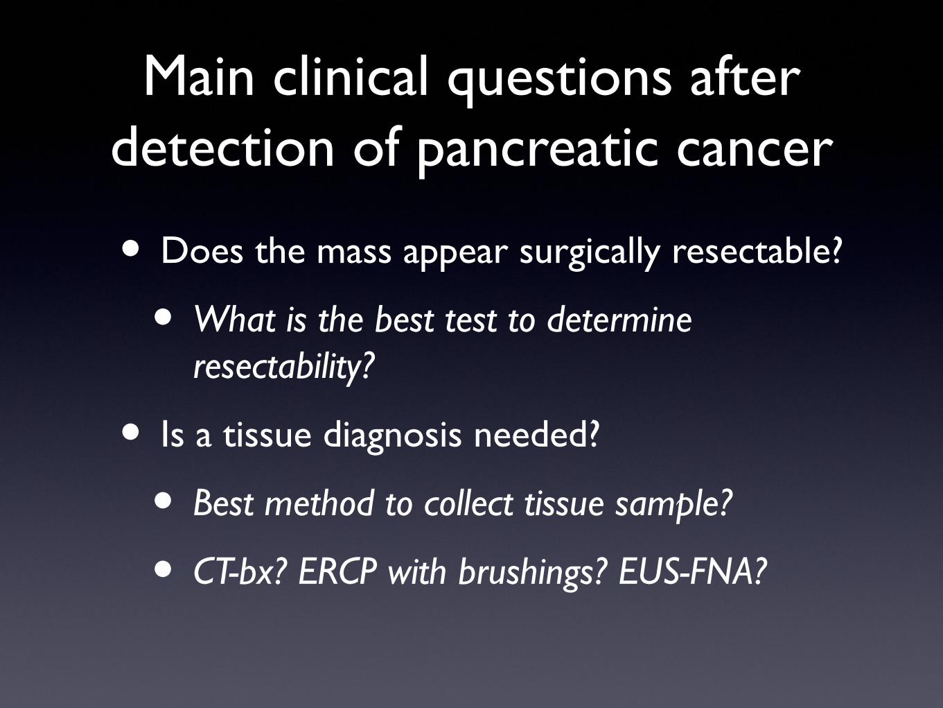
Main clinical questions after detection of pancreatic cancer
• Does the mass appear surgically resectable?
• What is the best test to determine resectability?
• Is a tissue diagnosis needed?
• Best method to collect tissue sample?
• CT-bx? ERCP with brushings? EUS-FNA?

Accuracy in assessing resectability in pancreatic cancer
study N MRI CT EUS p-value
Gress 1999 81 60% 93% <0.001
Ahmad 2000 63 77% 69% ns
Ramsay 2004 27 83% 76% 63% ns
Soriano 2004 62 75% 83% 67% ns
DeWitt 2004 53 77% 77% ns
CT/MRI + EUS may be more accurate than either aloneAhmad 2000, Soriano 2004
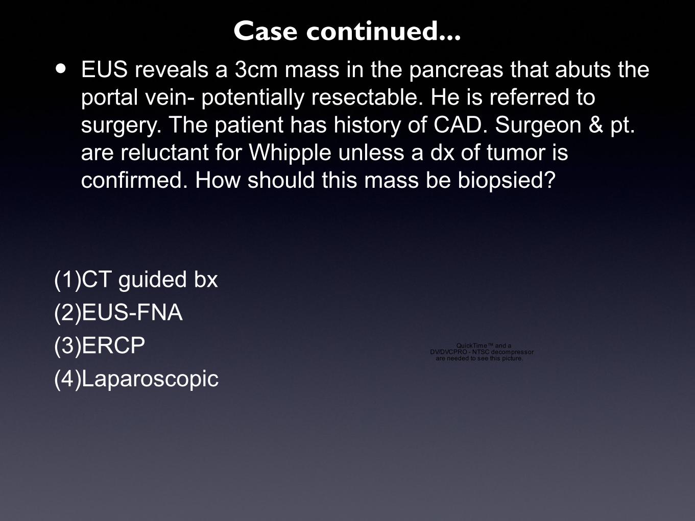
• EUS reveals a 3cm mass in the pancreas that abuts the portal vein- potentially resectable. He is referred to surgery. The patient has history of CAD. Surgeon & pt. are reluctant for Whipple unless a dx of tumor is confirmed. How should this mass be biopsied?
(1)CT guided bx
(2)EUS-FNA
(3)ERCP
(4)Laparoscopic
QuickTime™ and aDV/DVCPRO - NTSC decompressor
are needed to see this picture.
Case continued...
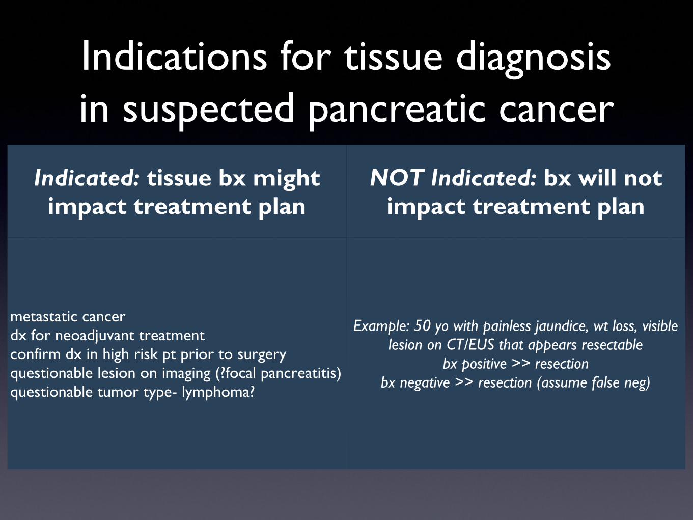
Indications for tissue diagnosis in suspected pancreatic cancer
Indicated: tissue bx might impact treatment plan
NOT Indicated: bx will not impact treatment plan
metastatic cancerdx for neoadjuvant treatmentconfirm dx in high risk pt prior to surgeryquestionable lesion on imaging (?focal pancreatitis)questionable tumor type- lymphoma?
Example: 50 yo with painless jaundice, wt loss, visible lesion on CT/EUS that appears resectable
bx positive >> resectionbx negative >> resection (assume false neg)
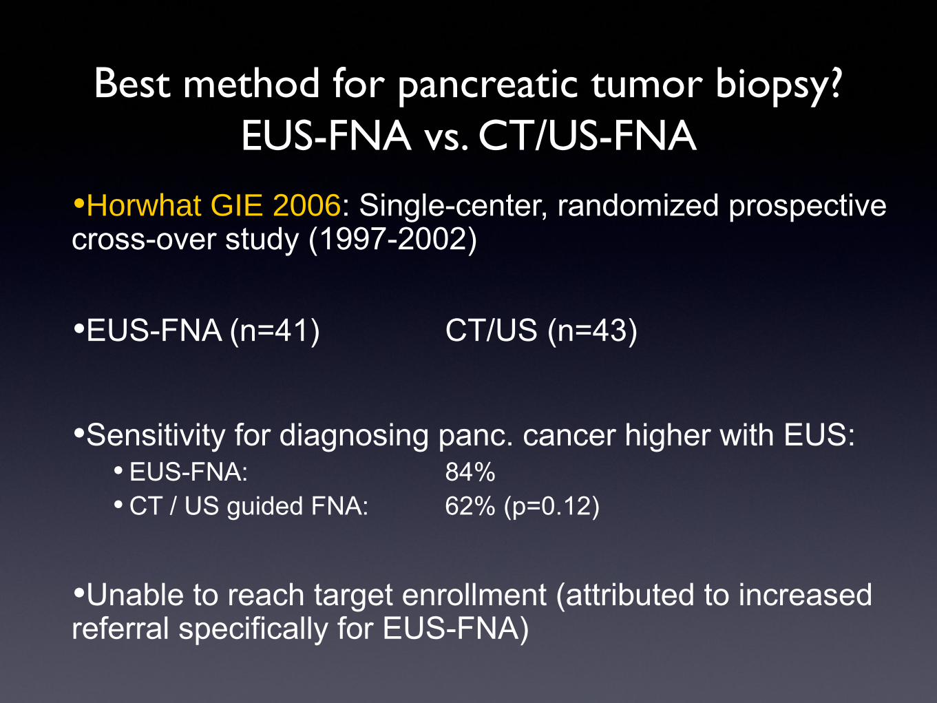
Best method for pancreatic tumor biopsy?EUS-FNA vs. CT/US-FNA
•Horwhat GIE 2006: Single-center, randomized prospective cross-over study (1997-2002)
•EUS-FNA (n=41) CT/US (n=43)
•Sensitivity for diagnosing panc. cancer higher with EUS:• EUS-FNA: 84%• CT / US guided FNA: 62% (p=0.12)
•Unable to reach target enrollment (attributed to increased referral specifically for EUS-FNA)

Complications of tissue samplingEUS vs. percutaneous-FNA
2% vs. 16% ; p=0.025Micames GIE 2003

Tissue sampling in pancreatic cancerEUS-FNA or ERCP?
sensitivityProcedure
related pancreatitis
EUS-FNA >85% 1-2%
ERCP 40-75% 3-5%
Fritscher-Ravens, AJG 2000Kochman, JCO 1997Jacobsen, GIE 2005Jailwala, GIE 2000Brugge, GIE 2010
From Brugge NEJM 1999; 341
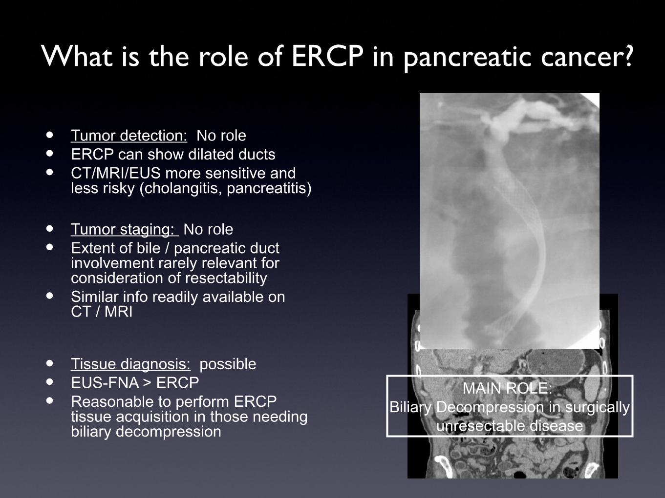
What is the role of ERCP in pancreatic cancer?
• Tumor detection: No role
• ERCP can show dilated ducts
• CT/MRI/EUS more sensitive and less risky (cholangitis, pancreatitis)
• Tumor staging: No role
• Extent of bile / pancreatic duct involvement rarely relevant for consideration of resectability
• Similar info readily available on CT / MRI
• Tissue diagnosis: possible
• EUS-FNA > ERCP
• Reasonable to perform ERCP tissue acquisition in those needing biliary decompression
MAIN ROLE: Biliary Decompression in surgically
unresectable disease
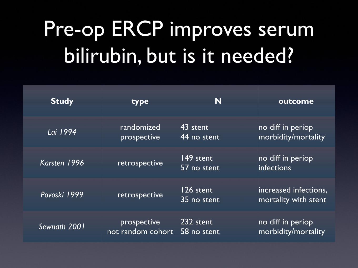
Pre-op ERCP improves serum bilirubin, but is it needed?
Study type N outcome
Lai 1994 randomizedprospective
43 stent44 no stent
no diff in periop morbidity/mortality
Karsten 1996 retrospective 149 stent57 no stent
no diff in periop infections
Povoski 1999 retrospective 126 stent35 no stent
increased infections, mortality with stent
Sewnath 2001 prospectivenot random cohort
232 stent58 no stent
no diff in periop morbidity/mortality

EUS: mainstream clinical uses
• Evaluation of GI luminal tract disease:
• GI cancers: esophageal, gastric, rectal
• GI wall submucosal lesions
• Evaluation of pancreatico-biliary disease:
• Pancreatic cancer
• Pancreatic cysts
• Biliary stones
• Acute and chronic pancreatitis
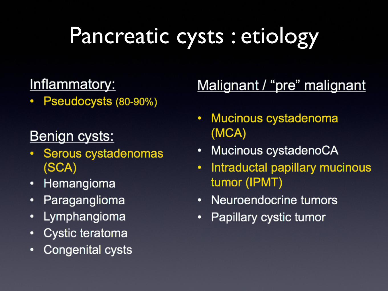
Pancreatic cysts : etiology

Pancreatic cyst dilemma:benign or potentially malignant?
Lesion EUS appearance
EUS-FNA
viscosity amylase CEA
pseudocyst internal debris low high low
serous cystadenoma microcysts low low low
IPMN dilated PD or side branches high high high
mucinous cystadenoma
macrocystic sepatated high low high
IPMN or MCA with CA
above with mural nodule, mass high high or low high
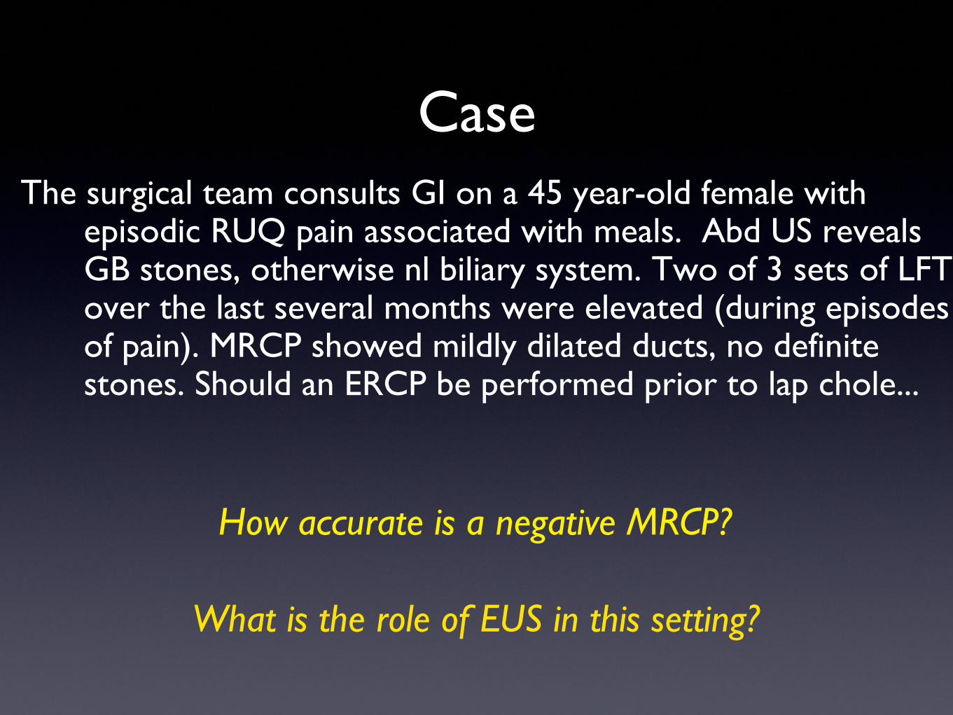
CaseThe surgical team consults GI on a 45 year-old female with
episodic RUQ pain associated with meals. Abd US reveals GB stones, otherwise nl biliary system. Two of 3 sets of LFTs over the last several months were elevated (during episodes of pain). MRCP showed mildly dilated ducts, no definite stones. Should an ERCP be performed prior to lap chole...
How accurate is a negative MRCP?
What is the role of EUS in this setting?
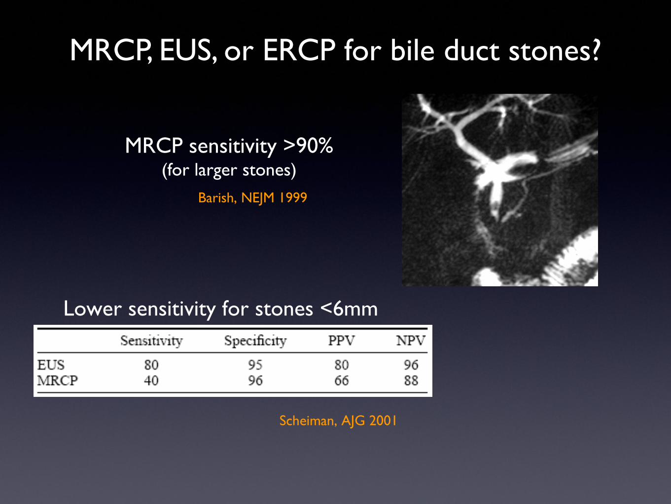
MRCP, EUS, or ERCP for bile duct stones?
MRCP sensitivity >90%(for larger stones)
Barish, NEJM 1999
Lower sensitivity for stones <6mm
Scheiman, AJG 2001
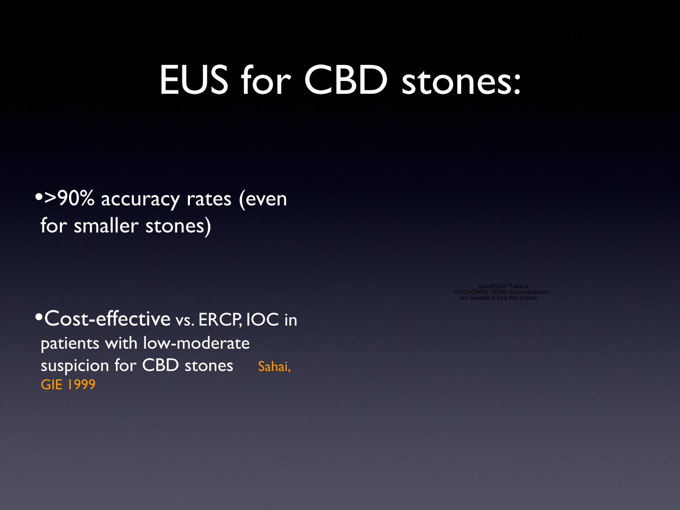
EUS for CBD stones:
•>90% accuracy rates (even for smaller stones)
•Cost-effective vs. ERCP, IOC in patients with low-moderate suspicion for CBD stones Sahai, GIE 1999
QuickTime™ and aDV/DVCPRO - NTSC decompressor
are needed to see this picture.

Evaluation of biliary stones:
MRCP EUS ERCP
Indications Low suspicion CBD stone
Low-mod. suspicion High suspicion, cholangitis, severe
GS pancreatitis
Detection rate > 90%(large stones)
> 90% Gold standard
Therapeutic No No, but can do immediate ERCP
Yes
Approximate CostMedicare 2007
$560 $780 $ 780-1530
Risks none <1% 4-10%
++
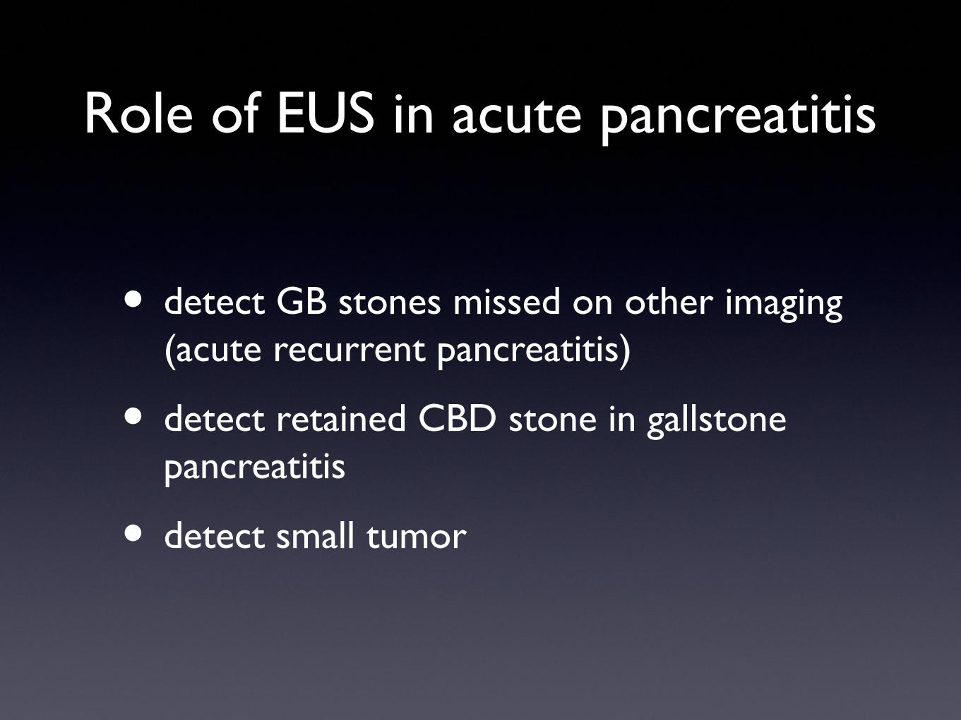
Role of EUS in acute pancreatitis
• detect GB stones missed on other imaging (acute recurrent pancreatitis)
• detect retained CBD stone in gallstone pancreatitis
• detect small tumor

EUS: criteria for chronic pancreatitis
abnl on ERCP
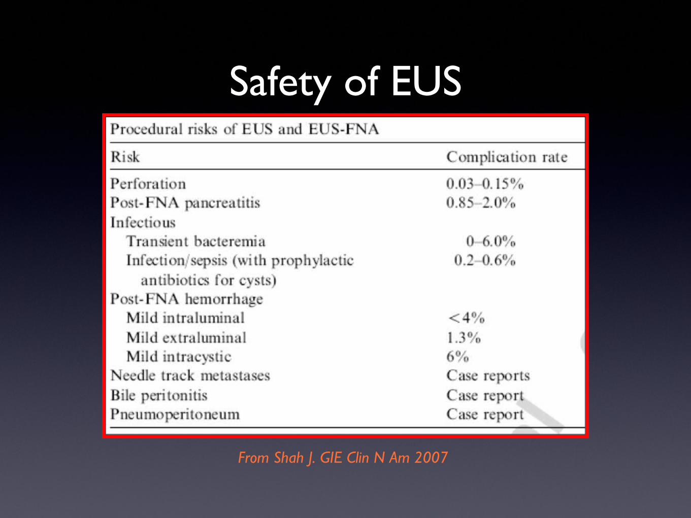
Safety of EUS
From Shah J. GIE Clin N Am 2007

New strategy in evaluating pancreatic/biliary disease : Single-session EUS + ERCP
• Perform EUS > immediate ERCP, if needed
• Optimize care:• combine high diag yield of EUS with high therapeutic success of ERCP
• minimize risks of unnecessary ERCP
• Limitations• requires specialized endo unit with fluoro + EUS
• needs endoscopist / assistants trained in both
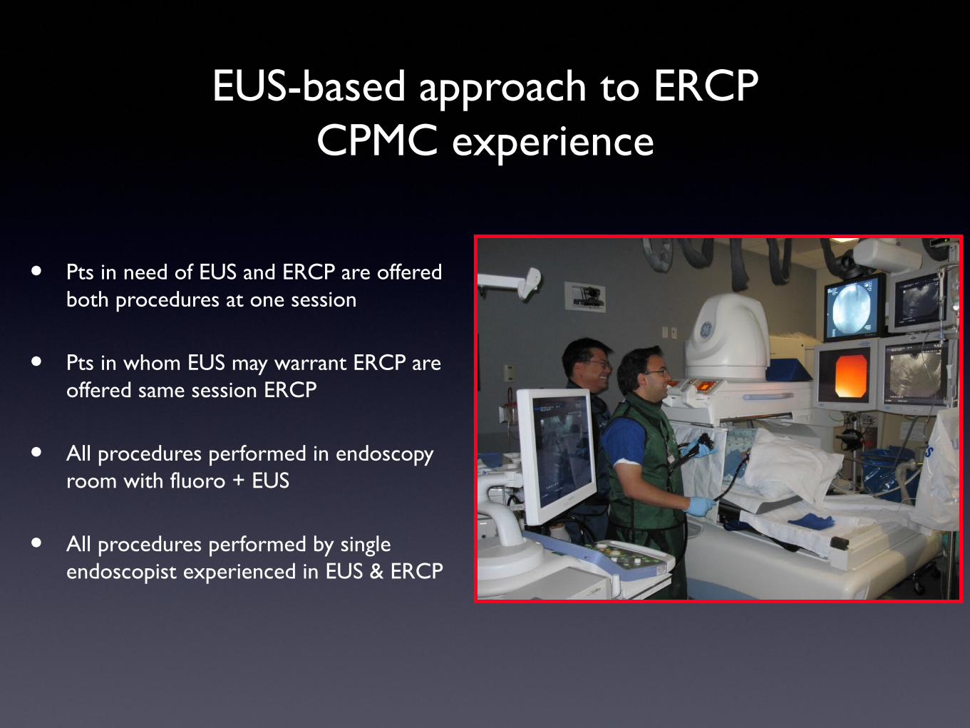
EUS-based approach to ERCPCPMC experience
• Pts in need of EUS and ERCP are offered both procedures at one session
• Pts in whom EUS may warrant ERCP are offered same session ERCP
• All procedures performed in endoscopy room with fluoro + EUS
• All procedures performed by single endoscopist experienced in EUS & ERCP
EUS-based ERCP: suspected CBD stones
• EUS “diagnostic cholangiogram”
• stone present > ERCP
• stone absent > no ERCP
• same session (one sedation)
QuickTime™ and aDV/DVCPRO - NTSC decompressor
are needed to see this picture.

EUS-based ERCP: suspected malignant obstruction
• mass present or not?
• immediate staging information- resectable?
• tissue sampling (EUS-FNA)
• decide need for ERCP and appropriate stent type
QuickTime™ and aDV/DVCPRO - NTSC decompressor
are needed to see this picture.
EUS : from A to ZSummary
• EUS has an established role in evaluating GI tract cancers, submucosal GI lesions, and a variety of pancreaticobiliary diseases
• Advances in EUS technology and treatment strategy are improving the diagnostic and therapeutic approach for patients with various types of GI disorders
?