Embed Size (px)
Citation preview

Practice Guidelines
for Management of
the Difficult Airway
An Updated Report by the American Society of Anesthesiologists Task
Force on Management of the Difficult Airway
2013; 118(2):251-270

Purposes of the Guidelines for
Difficult Airway Management
The purpose of these Guidelines is to
facilitate the management of the difficult
airway and to reduce the likelihood of
adverse outcomes.
The principal adverse outcomes associated
with the difficult airway include death,
brain injury, cardiopulmonary arrest,
unnecessary surgical airway, airway
trauma, and damage to the teeth.

Definition of
Difficult
Airway
Definition of Difficult Airway
The clinical situation in which a
conventionally trained anesthesiologist
experiences difficulty with facemask
ventilation of the upper airway, difficulty
with tracheal intubation, or both.
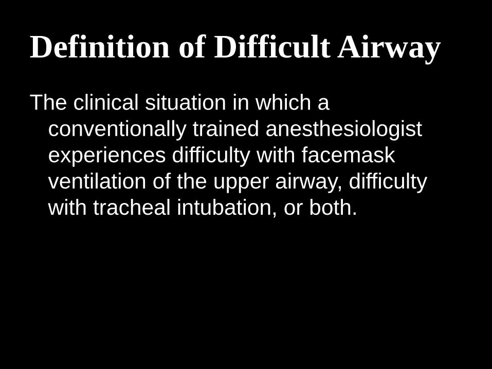
Difficult facemask or supraglottic airway (SGA)
ventilation (e.g., LMA, ILMA, laryngeal tube)
Signs of inadequate ventilation include absent or
inadequate chest movement, absent or inadequate breath
sounds, auscultatory signs of severe obstruction,
cyanosis, gastric air entry or dilatation, decreasing or
inadequate oxygen saturation (SpO2), absent or
inadequate exhaled CO2, absent or inadequate
spirometric measures of exhaled gas flow, and
hemodynamic changes associated with hypoxemia or
hypercarbia (e.g., hypertension, tachycardia, arrhythmia).
Difficult SGA placement
Difficult laryngoscopy
Difficult tracheal intubation
Failed intubation

Evaluation of
the Airway

Evaluation of the Airway
History
Physical Examination
Additional Evaluation

History
Age
Obesity
Obstructive sleep
apnea
History of snoring
Mediastinal
masses
Ankylosis
Degenerative
osteoarthritis
Subglottic stenosis
Lingual thyroid or
tonsillar hypertrophy
Treacher-Collins,
Pierre-Robin or
Down Syndrome

Physical Examination
Anatomical features of head and neck
Including: length of upper incisors, relation of maxillary and mandibular incisors during normal jaw closure and voluntary protrusion, interincisor distance, visibility of uvula, shape of palate, compliance of mandibular space, thyromental distance, length and thickness of neck, and range of motion of head and neck.

LEMON Assessment
Look externally
Evaluate the 3-3-2 rule
Mallampati score
Obstruction
Neck mobility

Additional Evaluation
CXR
CT
Fluoroscopy

Basic Preparation for
Difficult Airway
Management

Inform the patient (or responsible person) of the special risks and procedures pertaining to management of the difficult airway.
Ascertain that there is at least one additional individual who is immediately available to serve as an assistant in difficult airway management.
Administer facemask pre-oxygenation before initiating management of the difficult airway.
Actively pursue opportunities to deliver supplemental oxygen throughout the process of difficult airway management. Opportunities for supplemental oxygen administration include oxygen delivery by nasal cannulae, facemask, or LMA, insufflation.

Strategy for Intubation
of the Difficult Airway

Strategy for Intubation of the
Difficult Airway
Awake intubation
Video-assisted laryngoscopy
Intubating stylets or tube-changers
SGA for ventilation (e.g., LMA, laryngeal tube)
SGA for intubation (e.g., ILMA)
Rigid laryngoscopic blades of varying design and size
Fiberoptic-guided intubation
Lighted stylets or light wands

Assessment of the likelihood and anticipated clinical impact of 6 basic problems
1. Difficulty with patient cooperation or
consent
2. Difficult mask ventilation
3. Difficult SGA placement
4. Difficult laryngoscopy
5. Difficult intubation
6. Difficult surgical airway access

Consideration of the relative clinical merits and feasibility of 4 basic management choices
1. Awake intubation versus intubation after induction of general anesthesia
2. Noninvasive techniques versusinvasive techniques for the initial approach to intubation
3. Video-assisted laryngoscopy as an initial approach to intubation
4. Preservation versus ablation of spontaneous ventilation

Identification of a primary or preferred approach to
1. Awake intubation
2. The patient who can be adequately
ventilated but is difficult to intubate
3. The life-threatening situation in which
the patient cannot be ventilated or
intubated

Confirmation of tracheal intubation using capnography or end-tidal CO2 monitoring

Suggested Contents of the Portable Storage Unit for Difficult Airway Management
Rigid laryngoscope blades of alternate design and size from those routinely used; this may include a rigid fiberoptic laryngoscope
Video-laryngoscope
Tracheal tubes of assorted sizes
Tracheal tube guides. Examples include semi-rigid stylets, ventilating tube-changer, light wands, and forceps designed to manipulate the distal portion of the tracheal tube
Supra-glottic airways (e.g., LMA or ILMA of assorted sizes for noninvasive airway ventilation/intubation)
Flexible fiberoptic intubation equipment
Equipment suitable for emergency invasive airway access
An exhaled CO2 detector

Techniques for
Difficult Intubation
Awake intubation
Blind intubation (oral or
nasal)
Fiberoptic intubation
Intubating stylet or
tube-changer
Supra-glottic airway as
an intubating conduit
Laryngoscope blades of
varying design and size
Light wand
Video-laryngoscope
Techniques for
Difficult Ventilation
Intra-tracheal jet stylet
Invasive airway access
Supraglottic airway
Oral and naso-
pharyngeal airways
Rigid ventilating
bronchoscope
Two-person mask
ventilation