Vol. 120, No. 2, 1984 BIOCHEMICAL AND BIOPHYSICAL RESEARCH COMMUNICATIONS
April 30, 1984 Pages 411-419
CHANGES IN MITOCHONDRIAL PROTEINS DURING NEUROBLASTOMA DIFFERENTIATION
Jean-Luc VayssiPre, Francis Berthelot, Bernard Croizat and Franqois Gros
Laboratoire de Biochimie CeZlulaire, CoZlBge de France, 11, Place MarceZin BertheZot, 75231 Paris Cedex 05, France
Received March 14, 1984
SUMMARY. The evolution of three major mit-proteins was followed in neuro- blastoma cells cultured in different conditions of differentiation. 1 methyl cyclohexane carboxylic acid (CCA) was found to stimulate the synthesis of the three mit-protein markers. This result, compared to the effects of oligomycin, an inhibitor of mitochondrial function, favours the hypothesis that CCA induces in vitro neurogenesis through a general metabolic alteration.
Most of our information concerning brain metabolism emphazises
the high level of energy metabolism and oxygen consumption. The availability
of clonal cell lines of neuronal origin and of primary cultures has made it
possible to compare the energy metabolism of homogeneous cell populations
with that of the mixed cell population in the brain. Studies confirm that
02 consumption of neurons grown in culture is always higher than glial
respiration (I). Furthermore, the evolution of enzymatic activities in cell
culture indicate that differential modifications of the energy metabolism
occur in neuronal and glial elements (2). Neuronal maturation reflects a
shift of the energy metabolism towards a more aerobic form (2, 3).
Alternatively, the perfusion of anaesthetics in brain induces a reduction
of neuronal activity as well as of energy metabolism, oxygen consumption
and transmitter turnover (4, 5).
These considerations led us to further study the effects on mouse
neuroblastoma cells of a pharmacologically active substance, known for its
antianoxic effects on the rat brain (6) and which we had shown previously
to act as a potent neural differentiation inducer, the compound 1 methyl
cyclohexane carboxylic acid (CCA).
CCA promotes a significant increase in the level of cellular
energy metabolism in developing neuroblastoma in tissue culture conditions.
This increase is estimated by the cellular accumulation of the complex
between the phosphorylated [14C]deoxygl ucose and hexose phosphate
isomerase, a phenomenon which reflects glucose utilization, according to
Abbreviations: mit-protein, mitochondrial protein; mitpro, precursor of mitochondrial protein.
Vol. 120, No. 2, 1984 BIOCHEMICAL AND BIOPHYSICAL RESEARCH COMMUNICATIONS
Sokoloff (7). A considerably higher amount of radio-activity - 2.5 to
4 times - was found in CCA-treated cells, as compared to control,
undifferentiated cultures or to neuroblastoma induced by other treatments,
corresponding to a higher rate of deoxyglucose penetration and utilization
(8). This "burst" in cellular energy metabolism occurs prior to any
visible change in cell growth and morphology. It was thus postulated that
it could play a role in triggering the neuronal differentiation. It is
interesting to note that the drastic changes in glucose utilization
induced by CCA are specific of neuroblastoma cells and not found in other
systems, such as myoblast cultures.
These observations have drawn our attention to a possible general
relationship between mitochondrial function and neurogenesis, and led us
to examine more particularly the fate of mitochondrial (mit) proteins in
the course of neuroblastoma differentiation. Few data concerning the role
of mitochondria in neuronal function are available. Studies, reporting the
effects of anaesthetics on electroencephalogram, provide evidence for the
involvement of mitochondrial enzymes in the relationship between cerebral
metabolism and function (3, 9, 10, I!). Ultrastructural studies in the rat
brain also emphasize the fact that the number and morphology of
mitochondria undergo important changes in active neuronal cells (12).
As an approach to this study we have first attempted to
characterize some mit-protein markersinc-1300 neuroblastoma (NIE-115
clone). Those selected in this study have also been detected in the mouse
brain tissue. The present paper indicates that CCA, and to a much lesser
extent DMSO but not serum deprivation, strongly stimulates the synthesis
of mit-proteins supportingtheview that the strong CCA inducer effect
could be correlated with an interaction at the mitochondrial level. Brain
maturation is accompanied by an increased synthesis of mit-protein.
MATERIALS AND METHODS
Cell Culture
The NlE-115 and NS-20 clones from mouse neuroblastoma C-1300 were used. Culture conditions have been described in (13). Morphological differentiation was induced either by withdrawing serum from the culture medium or by addition, to a serum containing medium, of 0.1 % CCA for 3 days or 2 % DMSO for 7 days. Cultures were changed with fresh medium every 24 hours. Mitochondrial function was impaired by adding 0.6 nM oligomycin to the culture medium or 5 uM nonactin (Sigma) for18hours priortoharvesting.
Protein labeling and cell extracts
Cells werelabeled for 6 or 24 hours with [35S]methionine (25 uCi/ml medium) prior to harvesting. The cells extracts were prepared and analysed on ZD-electrophoresis as described in (14).
412
Vol. 120, No. 2, 1984 BIOCHEMICAL AND BIOPHYSICAL RESEARCH COMMUNICATIONS
Preparation of mit-proteins
Purification of mit-proteins was achieved according to Anderson (15). Cells prelabeled with 135Slmeth ionine were homogenized in 0.25 M sucrose/ 0.3 mM EDTA, pH 7.4, and spun at 600 x g for 10 min. The super- natant was centrifuged at 100,000 x g for 10 min, and the resulting pellet was washed twice. The last pellet was resuspended in 50 % (wt/wt) sucrose/ 0.3 mM EDTA and layered under a step gradient of 44.5 % and 41 X sucrose. This was centrifuged for 2 h at 45,000 rpm in a Beckman type 50 rotor. The material banding between 41 % and 44.5 % sucrose was diluted into 0.25 M sucrose/ 0.3 M EDTA, centrifuged at 10,000 x g for 10 min and the resulting pellet was dissolved, for protein analysis, in 8 M urea/ 20 % Nonidet P-401 2 % ampholines (LKB, 3.5- 10) / 1 % mercaptoethanol.
Peptide analysis
Peptide analysis was carried out according to Cleveland et al. (16). Sections containing mit-proteins were cut out from two-dimensional SDS gels overlaid with 20 ng of protease from StaphyZococcus aureus V8 diluted in Tris 0.125 M pH 6.8, 0.1 % SDS, EDTA 1 mM, 10 % glycerol. Digestion proceeded directly in the stacking gel during the subsequent electrophoresis.
RESULTS
Characterization of some neuroblastoma mitochondrial proteins
Two experimental procedures were used in order to identify mit-
proteins in neuroblastoma cells : i) purified mit-proteins were prepared
as described in the "Materials and Methods" and analysed on two-dimensional
electrophoresis ; ii) mitochondrial function was impaired in whole develop-
ing cells using two specific inhibitors, nonactin and oligomycin. The K +
ionophore, nonactin, is known to alter the electrochemical potential, thus
preventing both the translocation of mit-precursors into or across the
mitochondrial membranes and their cleavage into mature polypeptides.
Conversely, the addition to culture medium of oligomycin, an inhibitor of
phosphorylative oxydations, was shown, in the present work, to enhance
methionine incorporation into the mit-proteins, whose precursors are not
processed in the presence of nonactin. The combination of these procedures
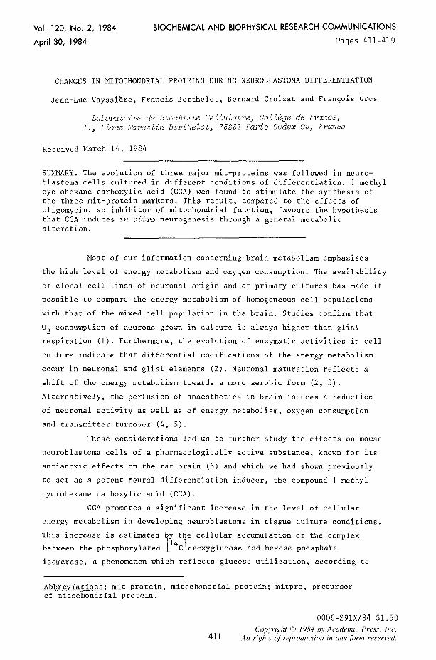
allowed us to select three mit-protein-markers (called 4, 5 and Cl
respectively). They appear as major spots on the electrophoregram of
purified neuroblastoma mitochondria, after appropriate lysis, as shown on
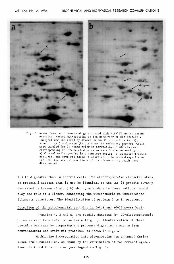
Fig. 1. Fig. 2a and 2b show the electrophoregrams of total cell proteins
from a) control cultures : arrows indicate positions of mature proteins 4,
5 and Cl ; b) nonactin-treated cultures : arrows point towards the virtual
positions of the corresponding proteins, which have disappeared.
Treatment of cultures with 5 uM nonactin for 18 hours before
harvesting resulted in the loss of the spot corresponding to protein 4
(apparent molecular weight : 52 kda ; pHi5.8) from the two-dimensional
pattern of labeled polypeptides. A new spot appeared which is likely to
correspond to the precursor (mitpro) of protein 4 (compare Fig. 2a to 2b).
413
Vol. 120, No. 2, 1984 BIOCHEMICAL AND BIOPHYSICAL RESEARCH COMMUNICATIONS
Fig. I Electrophoregram showing mit-proteins in a preparation from NlE-115 neuroblastoma cells grown in a complete medium (DIEM + 7.5 % fetal calf serum) and labeled during 6 hours prior to harvesting. An amount of radioactivity corresponding to 2.105 cts/min of 35S- labeled proteins was loaded on the gel.
The precursor is made by the nucleo-cytoplasmic genetic system (17). After
24 hours labeling prior to harvesting, methionine incorporation into
protein 4 from oligomycin-treated culture increased 1.5 fold relative to
the value found in control culture. Electrophoretic characteristics of this
protein suggest that it could be the 6 subunit of F1 ATPase (data not
shown).
The spot corresponding to protein Cl (apparent molecular weight :
68 kda ; pH i 5.8) in total extracts from methionine labeled control cultures
disappears upon pretreatment with nonactin. However, we were unable to
locate a putative precursor. (The latter might be present on the gel with
important molecular weight or(and) pH modifications). Methionine
incorporation into protein C 1 after oligomycin treatment increased 2.7 fold.
The synthesis of protein 5 (apparent molecular weight : kda ;
pHi 5.5) was also inhibited by nonactin in neuroblastoma cells and like in
the case for protein C , no candidate for a mitpro was identified on the
electrophoregram. Methionine incorporation after oligomycin treatment was
414
Vol. 120, No. 2, 1984 BIOCHEMICAL AND BIOPHYSICAL RESEARCH COMMUNICATIONS
Fig. 2 Areas from two-dimensional gels loaded with NIE-115 neuroblastoma extracts. Mature mit-proteins or the precursor of mit-protein 4 (mitpro) are indicated by arrows. cx and 13 isotubulins (a, B),
vimentin (Vi) and actin (A) are shown as reference markers. Cells were labeled for 24 hours prior to harvesting. 7.105 cts/ min corresponding to 35S-labeled proteins were loaded on each gel. a) Control cells growing in a complete medium. b) Nonactin-treated cultures. The drug was added 18 hours prior to harvesting. Arrows indicate the virtual positions of the mit-proteins which have disappeared.
1.3 fold greater than in control cells. The electrophoretic characteristics
of protein 5 suggest that it may be identical to the IEF 24 protein already
described by Larsen et a2. (18) which, according to these authors, would
play the role of a linker, connecting the mitochondria to intermediate
filaments structures. The identification of protein 5 is in progress.
Detection of the mitochondrial oroteins in fetal and adult mouse brain
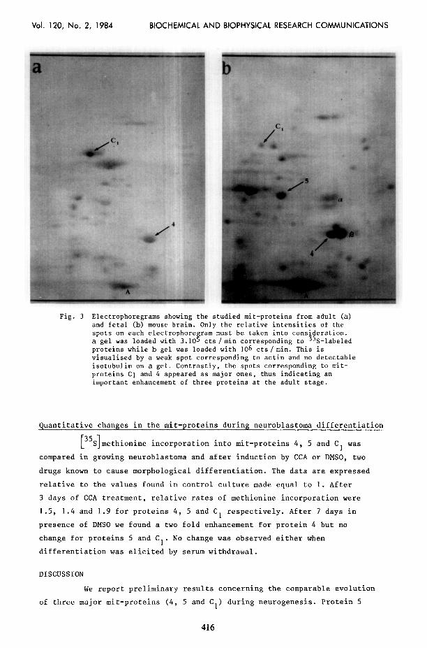
Proteins 4, 5 and C, are readily detected by 2D-electrophoresis
of an extract from fetal mouse brain (Fig. 3). Identification of these
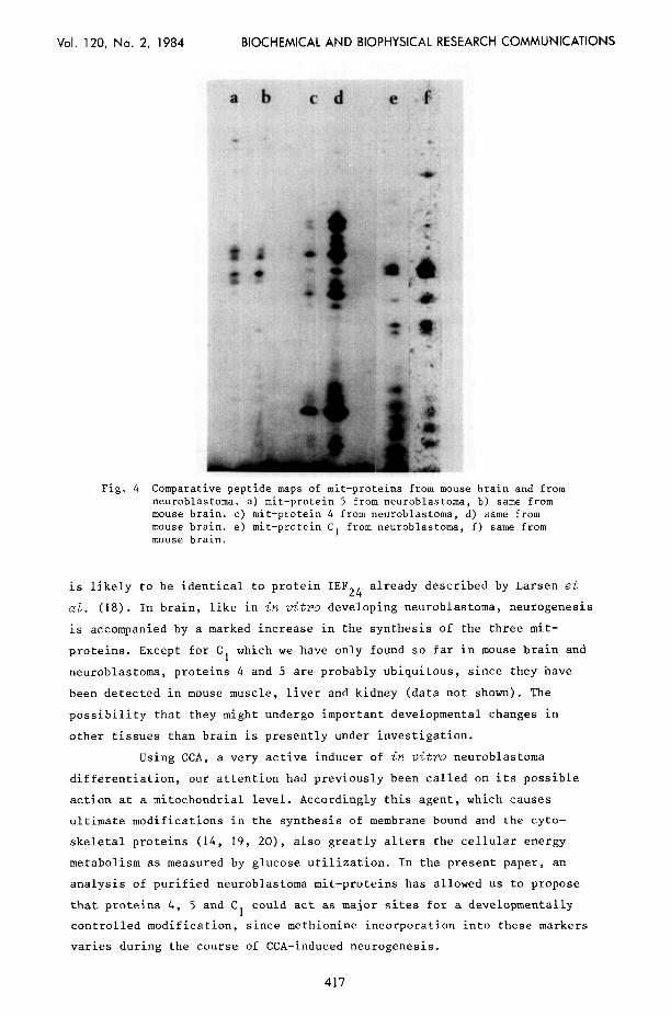
proteins was made by comparing the protease digestion products from
neuroblastoma and brain mit-proteins, as shown in Fig. 4.
Methionine incorporation into mit-proteins was enhanced during
mouse brain maturation, as shown by the examination of the autoradiograms
from adult and fetal brains (see legend to Fig. 3).
415
Vol. 120, No. 2, 1984 BIOCHEMICAL AND BIOPHYSICAL RESEARCH COMMUNICATIONS
Fig. 3 Electrophoregrams showing the studied mit-proteins from adult (a) and fetal (b) mouse brain. Only the relative intensities of the spots on each electrophoregram must be taken into consideration. a gel was loaded with 3.105 cts /min corresponding to 35S-labeled proteins while b gel was loaded with 106 cts/min. This is visualised by a weak spot corresponding to actin and no detectable isotubulin on a gel. Contrastly, the spots corresponding to mit- proteins Cl and 4 appeared as major ones, thus indicating an important enhancement of three proteins at the adult stage.
Quantitative changes in the mit-proteins during neuroblastoma differentiation
[ 1 35 S methionine incorporation into mit-proteins 4, 5 and Cl was
compared in growing neuroblastoma and after induction by CCA or DMSO, two
drugs known to cause morphological differentiation. The data are expressed
relative to the values found in control culture made equal to 1. After
3 days of CCA treatment, relative rates of methionine incorporation were
1.5, 1.4 and 1.9 for proteins 4, 5 and C1 respectively. After 7 days in
presence of DMSO we found a two fold enhancement for protein 4 but no
change for proteins 5 and C,. No change was observed either when
differentiation was elicited by serum withdrawal.
DISCUSSION
We report preliminary results concerning the comparable evolution
of three major mit-proteins (4, 5 and C,) during neurogenesis. Protein 5
416
Vol. 120, No. 2, 1984 BIOCHEMICAL AND BIOPHYSICAL RESEARCH COMMUNICATIONS
ab cd e 4
Fig. 4 Comparative peptide maps of mit-proteins from mouse brain and from neuroblastoma. a) mit-protein 5 from neuroblastoma, b) same from mouse brain. c) mit-protein 4 from neuroblastoma, d) same from mouse brain. mouse brain.
e) mit-protein C, from neuroblastoma, f) same from
is likely to be identical to protein IEF24 already described by Larsen et
~2. (18). In brain, like in in vitro developing neuroblastoma, neurogenesis
is accompanied by a marked increase in the synthesis of the three mit-
proteins. Except for Cl which we have only found so far in mouse brain and
neuroblastoma, proteins 4 and 5 are probably ubiquitous, since they have
been detected in mouse muscle, liver and kidney (data not shown). The
possibility that they might undergo important developmental changes in
other tissues than brain is presently under investigation.
Using CCA, a very active inducer of in tritro neuroblastoma
differentiation, our attention had previously been called on its possible
action at a mitochondrial level. Accordingly this agent, which causes
ultimate modifications in the synthesis of membrane bound and the cyto-
skeletal proteins (14, 19, 20), also greatly alters the cellular energy
metabolism as measured by glucose utilization. In the present paper, an
analysis of purified neuroblastoma mit-proteins has allowed us to propose
that protei.ns 4, 5 and Cl could act as major sites for a developmentally
controlled modification, since methionine incorporation into these markers
varies during the course of CCA-induced neurogenesis.
417
Vol. 120, No. 2, 1984 BIOCHEMICAL AND BIOPHYSICAL RESEARCH COMMUNICATIONS
The effect of various inhibitors of mitochondrial functions has
also been analyzed. Surprisingly oligomycin, which blocks the oxydative
phosphorylations, enhances the synthesis of the mit-protein markers, like
does CCA. It is interesting to note that oligomycin-treated cells exhibit
an increase in the methionine incorporation into some cytoskeletal
components like after CCA treatment (not shown here). It is not yet
possible however to clearly understand this correlation.
At present, the following points have been established during
CCA induced neurogenesis : i) 2-Deoxyglucose utilization is markedly
increased ; ii) methionine incorporation is enhanced into all the mit-
protein-markers studied. With the effects of oligomycin in mind, these
results support the idea that CCA would trigger the neuronal differentiation
through a general metabolic alteration causing perhaps, subsequently,
rearrangements in cytoskeleton and associated structures. This would end up
in morphological and biochemical phenotypes characteristic of mature
neurons.
The differences between the effects of CCA, DMSO and serum
withdrawal on the mitochondria emphasize the idea that "neuroblastoma
differentiation" can correspond to different series of inducing events.
Several paths may lead to a final state of morphologically differentiated
cell.
ACKNOWLEDGEMENTS
This work was supported by grants from SANOFI and DGRST. We thank Monique Basseville for her excellent technical assistance.
REFERENCES
1. Roth-Schechter, B.F., and Tholey, G. (1982) Neurochem. Res. 7, 329-337. 2. Tholey, G., Ledig, M., and Mandel, P. (1982) Neurochem. Res. 7, 27-36. 3. Tholey, G., Roth-Schechter, B.F., and Mandel, P.C. (1981) J. Neurochem.
36, 77-81. 4. Krieglstein, J., Sperling, G., and Stock, R. (1982) Neurochem. Res. 7,
737-748. 5. Hanin, I., and Jenden, D.J. (1969) Biochem. Pharmacol. 18, 837-845. 6. Simard, J., Ferrandes, B., Lacolle, J.Y., and Eymard, P. (1978) Satellite
Symposium on Cerebro-Vascular Diseases, Reims, France. 7. Sokoloff, L., Reivich, M., Kennedy, C., Des Rosiers, M.H., Patlak, C.S.,
Pettigrew, K.D., Sakurada, O., and Shinohara, M. (1977) J. Neurochem. 28, 897-916.
8. Croizat, B., Berthelot, F., Portier, M.M., Ohayon, H., and Gros, F. (1981) Biochem. Biophys. Res. Commun. 103, 1044-1051.
9. Dirks, G., Hanke, J., Krieglstein, J., Stock, R., and Wickop, G. (1980) J. Neurochem. 35, 311-317.
10. Gots, R.E., and Bessman, S.P. (1974) Arch. Biochem. Biophys. 163, 7-14. 11. Inui, M., and Ishibashi, S. (1979) J. Biochem. (Tokyo) 85, 1151-1156. 12. Vanneste, J., Vanneste, Ph., and Van den Bosch de Aguilar (1981) Acta
Neuropathol. (Berlin) 54, 83-87.
418
Vol. 120, No. 2, 1984 BIOCHEMICAL AND BIOPHYSICAL RESEARCH COMMUNICATIONS
13. Croizat, B., Berthelot, F., Felsani, A., and Gros, F. (1977) Eur. J. Biochem. 74, 405-412.
14. Portier, M.M., EddG, B., Berthelot, F., Croizat, B., and Gros, F. (1980) Biochem. Biophys. Res. Commun. 96, 1610-1618.
15. Anderson, L. (1981) Proc. Natl. Acad. Sci. USA 78, 2407-2411. 16. Cleveland, D.W., Fischer, S.G., Kirschner, M.W., and LaEmmli, U.K. (1977)
J. Biol. Chem. 252, 1102-1106. 17. Vayssisre, J.L., Berthelot, F., Portier, M.M., Denoulet, Ph., Croizat, B.,
and Gros, F. (1983) Communication to International Symposium on Contractile Proteins, Sassari (Italy). In press.
18. Moose-Larsen, P., Bravo, R., Fey, S.J., Small, J.U., and Celis, J.E. (1982) Cell 31, 681-682.
19. Gros, F., Croizat, B., Portier, M.M., Berthelot, F., and Felsani, A. (1982) in : Molecular Genetic Neurosciences (eds. Schmidt, F.O., Bird, S.J., and Bloom, F.E.), pp. 335-347, Raven Press, New York.
20. Portier, M.M., Croizat, B., and Gros, 9. (1982) FEBS Lett. 146, 283-288.
419
Recommended


