Embed Size (px)
Citation preview

Mohammad Sazzad HossenB.Optom, M.Optom
Consultant OptometristAd-din Medical College Hospital
Visiting Faculty: UniSA, Dhaka
Systemic disease and the eye

Common systemic diseases Common systemic diseases affecting the eyeaffecting the eye
InfectiousInfectious ToxoplasmosisToxoplasmosis ToxocariasisToxocariasis TBTB SyphilisSyphilis LeprosyLeprosy HIVHIV CMVCMV
Non-infectiousNon-infectious Endocrine – diabetes, Endocrine – diabetes,
thyroidthyroid Connective tissue Connective tissue
disease – disease – RA/SLE/Wegeners/PANRA/SLE/Wegeners/PAN/Systemic sclerosis/Systemic sclerosis
Vasculitides (GCA)Vasculitides (GCA) SarcoidosisSarcoidosis Behcet’s DiseaseBehcet’s Disease Vogt Koyanagi Harada Vogt Koyanagi Harada
syndromesyndrome Phakomatoses Phakomatoses

DIABETIC RETINOPATHY
1. Adverse risk factors2. Pathogenesis
5. Clinically significant macular oedema
6. Preproliferative diabetic retinopathy
3. Background diabetic retinopathy
4. Diabetic maculopathies• Focal• Diffuse• Ischaemic
7. Proliferative diabetic retinopathy

Adverse Risk Factors
1. Long duration of diabetes
• Obesity• Hyperlipidaemia
2. Poor metabolic control
3. Pregnancy
4. Hypertension
5. Renal disease
6. Other
• Smoking• Anaemia

Location of lesions in background diabetic retinopathy

Signs of background diabetic retinopathy
Microaneurysms usually temporal to fovea
Intraretinal dot and blot haemorrhages
Hard exudates frequentlyarranged in clumps or rings
Retinal oedema seen asthickening on biomicroscopy

Preproliferative diabetic retinopathy
Treatment - not required but watch for proliferative disease
• Cotton-wool spots• Venous irregularities
• Dark blot haemorrhages• Intraretinal microvascular abnormalities (IRMA)
Signs
Proliferative diabetic retinopathy
• Flat or elevated• Severity determined by comparing with area of disc
Neovascularization
Neovascularization of disc = NVD
• Affects 5-10% of diabetics• IDD at increased risk (60% after 30 years)
Neovascularization elsewhere = NVE

• Spot size (200-500 m) depends on contact lens magnification
• Gentle intensity burn (0.10-0.05 sec)
• Follow-up 4 to 8 weeks
• Area covered by complete PRP• Initial treatment is 2000-3000 burns
Laser panretinal photocoagulation

Retinal Vein OcclusionRetinal Vein Occlusion
Second most common cause of vascular-related visual loss.
Risk factors: hypertension, age, blood dyscrasias (OCP,HRT) and vasculitis (Behcets,sarcoidosis,AIDS,SLE)
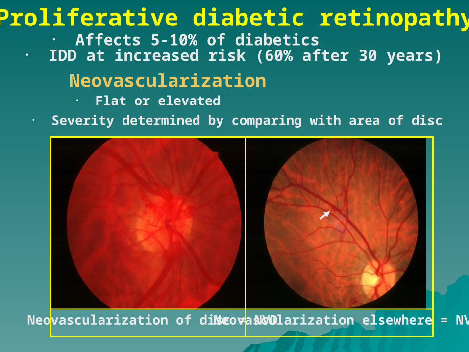
Retinal Artery OcclusionRetinal Artery Occlusion
Risk factors: Carotid artery atherosclerosis (CRAO), carotid emboli (BRAO), vasculitis (GCA,SLE,PAN), coagulopathy.
OCULAR EMERGENCY - Immediate referral to ophthalmologist

1. Soft tissue involvement• Periorbital and lid swelling• Conjunctival hyperaemia• Chemosis
• Superior limbic keratoconjunctivitis
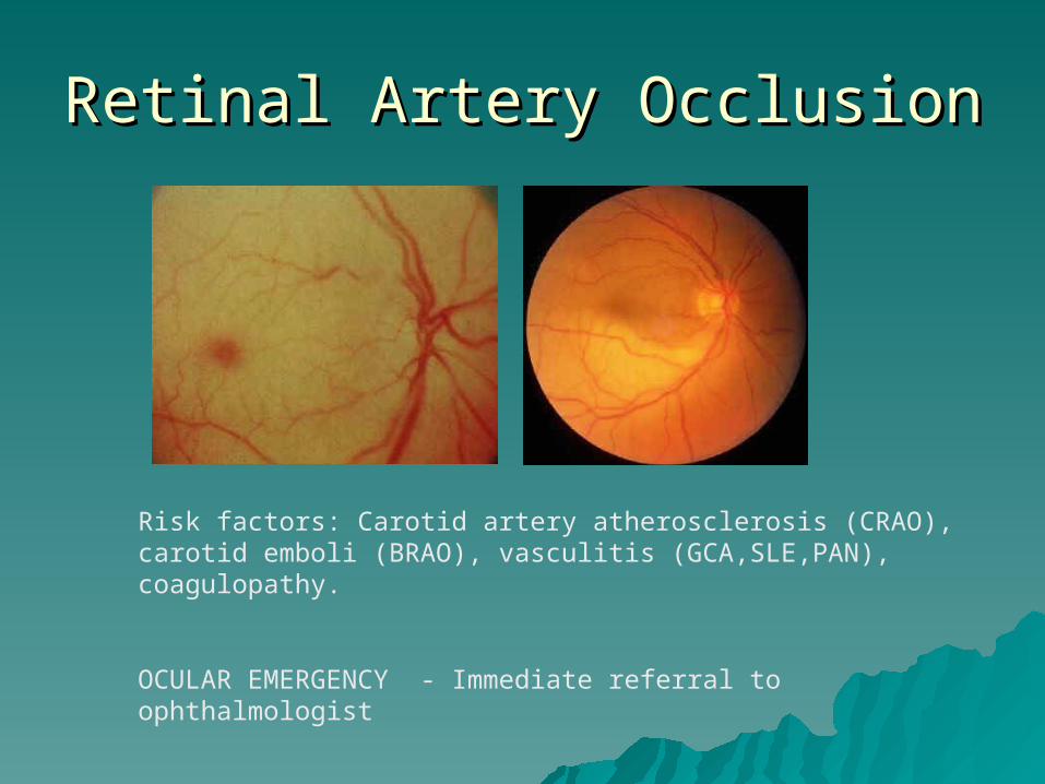
2. Eyelid retraction
3. Proptosis
4. Optic neuropathy
5. Restrictive myopathy
THYROID EYE DISEASE

Soft tissue involvementPeriorbital and lid swelling
Chemosis
Conjunctival hyperaemia
Superior limbic keratoconjunctivitis
Signs of eyelid retraction Occurs in about 50%
• Bilateral lid retraction • No associated proptosis
• Bilateral lid retraction • Bilateral proptosis
• Lid lag in downgaze • Unilateral lid retraction • Unilateral proptosis

Proptosis
Treatment options • Systemic steroids • Radiotherapy • Surgical decompression
• Occurs in about 50% • Uninfluenced by treatment of hyperthyroidism
Axial and permanent in about 70% May be associated with choroidal folds

Optic neuropathy• Occurs in about 5% • Early defective colour vision • Usually normal disc appearance
Caused by optic nerve compression at orbital apex by enlarged recti
Often occurs in absence of significant proptosis

• Occurs in about 40% • Due to fibrotic contracture
Restrictive myopathy
Elevation defect - most common Abduction defect - less common
Depression defect - uncommon Adduction defect - rare

SARCOIDOSISSARCOIDOSIS
Idiopathic multisystem disorderIdiopathic multisystem disorder Characterised by non-caseating Characterised by non-caseating
granulomatagranulomata More common in women 20-50 yrsMore common in women 20-50 yrs More common in blacks and AsiansMore common in blacks and Asians ? Related to mycobacteria? Related to mycobacteria
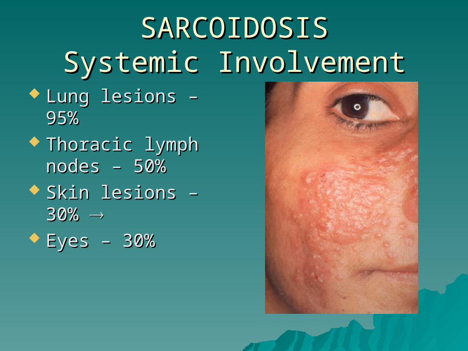
SARCOIDOSISSARCOIDOSISSystemic InvolvementSystemic Involvement
Lung lesions – 95%Lung lesions – 95% Thoracic lymph Thoracic lymph
nodes – 50%nodes – 50% Skin lesions – 30% Skin lesions – 30%
Eyes – 30%Eyes – 30%
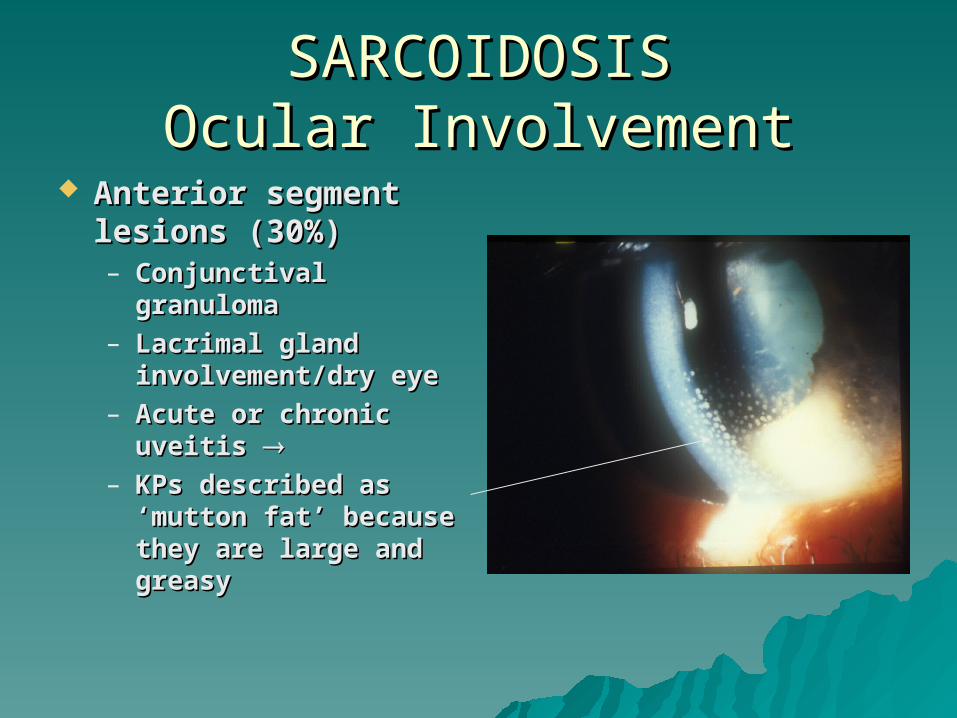
SARCOIDOSISSARCOIDOSISOcular InvolvementOcular Involvement
Anterior segment Anterior segment lesions (30%)lesions (30%)– Conjunctival Conjunctival
granulomagranuloma– Lacrimal gland Lacrimal gland
involvement/dry eyeinvolvement/dry eye– Acute or chronic Acute or chronic
uveitis uveitis – KPs described as KPs described as
‘mutton fat’ because ‘mutton fat’ because they are large and they are large and greasygreasy

SARCOIDOSISSARCOIDOSISOcular InvolvementOcular Involvement
Posterior segment Posterior segment lesions (20%)lesions (20%)– Patchy venous Patchy venous
sheathingsheathing– Cellular infiltrate Cellular infiltrate
around vesselsaround vessels– Chorioretinal Chorioretinal
granulonmasgranulonmas– Vasculitis including Vasculitis including
occlusive causing:-occlusive causing:-– NeovascularisationNeovascularisation– Infiltrate in vitreous Infiltrate in vitreous
(vitritis) including (vitritis) including cell clumps cell clumps (snowballs)(snowballs)

SARCOIDOSISSARCOIDOSISOcular InvolvementOcular Involvement
Sheathing of the Sheathing of the retinal veinsretinal veins
Fluorescein Fluorescein angiography angiography showing leakage showing leakage and staining at and staining at sites of sheathingsites of sheathing

SARCOIDOSISSARCOIDOSISGranuloma in FundusGranuloma in Fundus
Retinal and pre-Retinal and pre-retinalretinal
Choroidal Choroidal

SARCOIDOSISSARCOIDOSISGranuloma in FundusGranuloma in Fundus
Optic nerve head Optic nerve head granulomagranuloma
Normal optic nerve Normal optic nerve headhead

SARCOIDOSISSARCOIDOSISSystemic SignsSystemic Signs
Lupus pernio Lupus pernio affecting the nose – affecting the nose – a chronic a chronic progressive progressive cutaneous sarcoid cutaneous sarcoid that most that most commonly affects commonly affects face and earsface and ears

SARCOIDOSISSARCOIDOSISSystemic signsSystemic signs
Facial palsyFacial palsy
Salivary gland Salivary gland enlargementenlargement

SARCOIDOSISSARCOIDOSISSystemic signsSystemic signs
Hilar adenopathy Hilar adenopathy on chest x-rayon chest x-ray
Lung infiltrateLung infiltrate
Erythema nodosumErythema nodosum
ArthritisArthritis

SARCOIDOSISSARCOIDOSISInvestigations (1)Investigations (1)
CXR – to detect CXR – to detect pulmonary signspulmonary signs
Bilateral hilar Bilateral hilar lymph-adenopathylymph-adenopathy
Pulmonary mottlingPulmonary mottling
SARCOIDOSISSARCOIDOSISInvestigations (2)Investigations (2)
Serum angiotensin-converting Serum angiotensin-converting enzyme (ACE) – elevated in active enzyme (ACE) – elevated in active sarcoidosissarcoidosis
Mantoux test – caution in patients Mantoux test – caution in patients who have had BCG vaccination. Test who have had BCG vaccination. Test may be negativemay be negative
Lung function testsLung function tests

SARCOIDOSISSARCOIDOSISInvestigations (3)Investigations (3)
Gallium scan Gallium scan showing increased showing increased uptake in the uptake in the lacrimal and lacrimal and parotid glands and parotid glands and pulmonary regions pulmonary regions in a patient with in a patient with active sarcoidosisactive sarcoidosis

SARCOIDOSISSARCOIDOSISTreatmentTreatment
Systemic steroids may be necessary Systemic steroids may be necessary in patients with posterior segment in patients with posterior segment disease where vision is threatened, disease where vision is threatened, especially if optic nerve is involvedespecially if optic nerve is involved

PHACOMATOSES
1. Neurofibromatosis
2. Tuberous sclerosis (Bourneville disease)
3. von-Hippel-Lindau syndrome
4. Sturge-Weber syndrome
• Type I (NF-1) - von Recklinghausen disease
• Type II (NF-2) - bilateral acoustic neuromas

Neurofibromatosis type-1 - (NF-1)
Appear during first year of life
Café-au-lait spots
• Most common phacomatosis
Increase in size and number throughout childhood
• Affects 1:4000 individuals• Presents in childhood
• Gene localized to chromosome 17q11
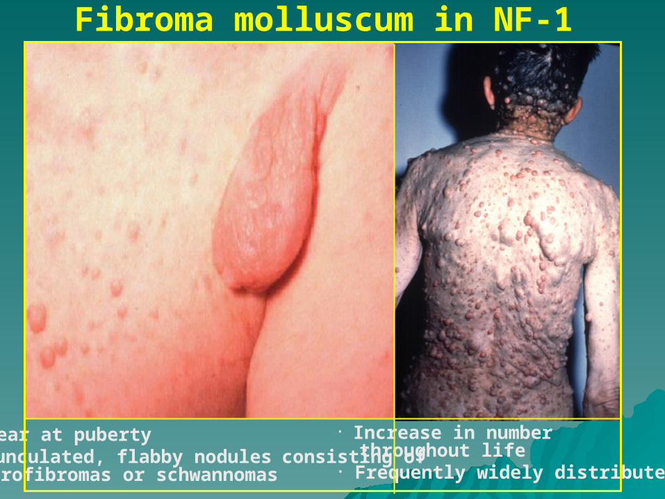
Fibroma molluscum in NF-1
• Appear at puberty• Pedunculated, flabby nodules consisting of neurofibromas or schwannomas
• Increase in number throughout life• Frequently widely distributed
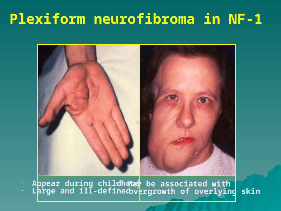
Plexiform neurofibroma in NF-1
• May be associated with overgrowth of overlying skin
• Appear during childhood• Large and ill-defined
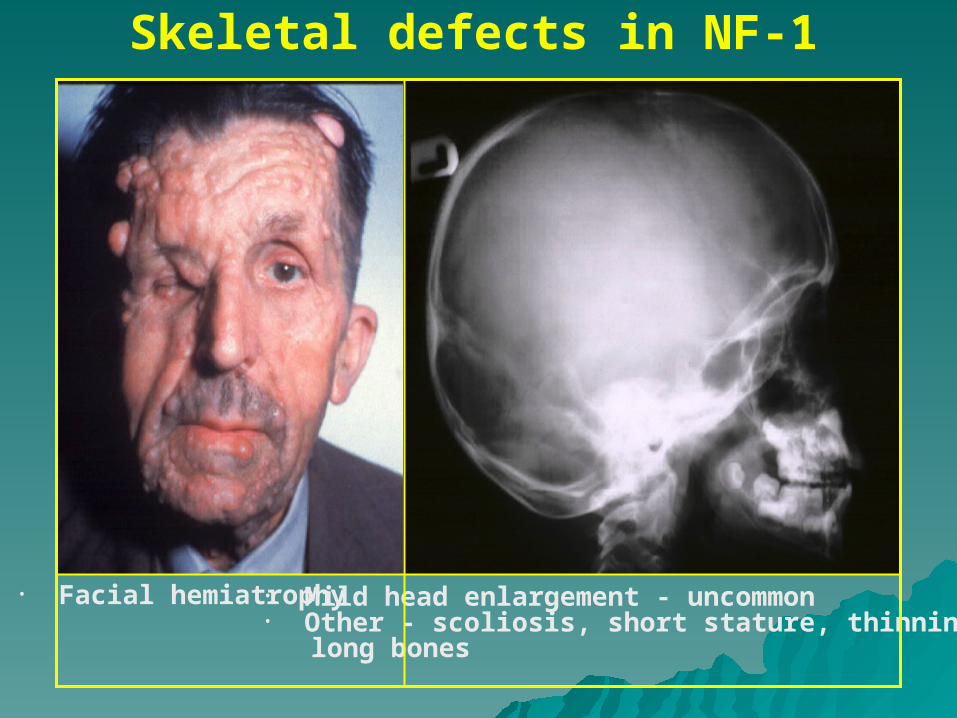
Skeletal defects in NF-1
• Mild head enlargement - uncommon• Other - scoliosis, short stature, thinning of long bones
• Facial hemiatrophy

Orbital lesions in NF-1Spheno-orbital encephaloceleOptic nerve glioma in about 15%
• Sagittal MRI scan of optic nerve glioma invading hypothalamus
• Glioma may be unilateral or bilateral
• Axial CT scan of congenital absence of left greater wing of sphenoid bone
• Causes pulsating proptosis without bruit
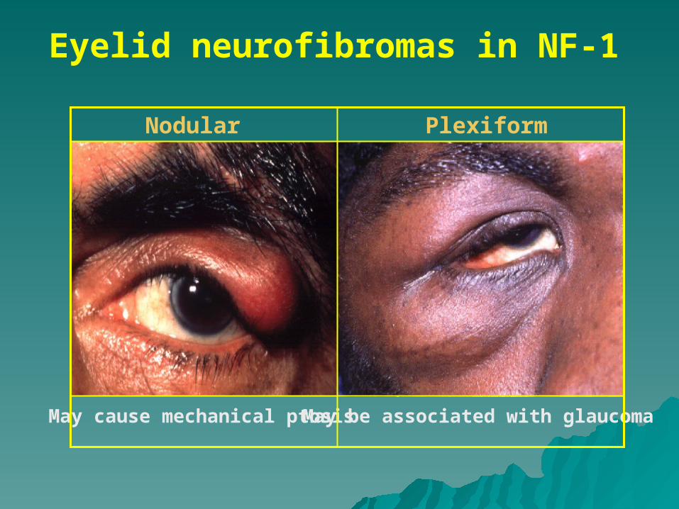
Eyelid neurofibromas in NF-1
Nodular Plexiform
May cause mechanical ptosis May be associated with glaucoma
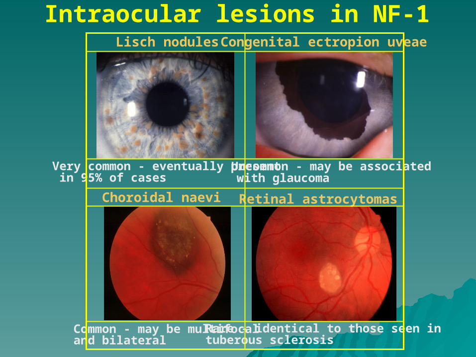
Intraocular lesions in NF-1Lisch nodules
Very common - eventually present in 95% of cases
Congenital ectropion uveae
Uncommon - may be associated with glaucoma
Retinal astrocytomas
Rare - identical to those seen intuberous sclerosis
Choroidal naevi
Common - may be multifocal and bilateral

Ocular features of NF-2
Common - combined hamartomas of RPE and retina
Very common -presenile cataract

Tuberous sclerosis (Bourneville disease)
• Diffuse thickening over lumbar region• Present in 40%
Shagreen patches
• Autosomal dominant• Triad - mental handicap, epilepsy, adenoma sebaceum
Adenoma sebaceum
• Around nose and cheeks• Appear after age 1 and slowly enlarge
Ash leaf spots
• Hypopigmented skin patches • In infants best detected using ultraviolet light (Wood’s lamp)

Systemic hamartomas in tuberous sclerosisAstrocytic cerebral hamartomas
• Slow-growing periventricular tumours• May cause hydrocephalus, epilepsy and mental retardation
• Usually asymptomatic and innocuous• Kidneys (angiomyolipoma), heart (rhabdomyoma)
Visceral and subungual hamartomas
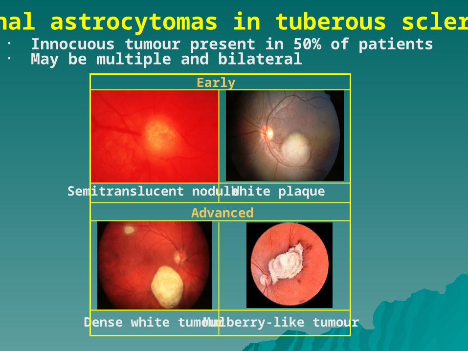
Retinal astrocytomas in tuberous scleritis
Dense white tumour Mulberry-like tumour
Early
• Innocuous tumour present in 50% of patients• May be multiple and bilateral
Semitranslucent nodule White plaque
Advanced

Systemic features of v-H-L syndromeAutosomal dominant
• Tumours - renal carcinoma and phaeochromocytoma
• Cysts - kidneys, liver, pancreas, epididymis, ovary and lungs
• Polycythaemia
CNS Haemangioblastoma
MRI of spinal cord tumour
Angiogram of cerebellar tumour
Visceral tumours

Retinal capillary haemangioma in v-H-L syndrome
Round orange-red mass
Early
• Vision-threatening tumour present in 50% of patients• May be multiple and bilateral
Tiny lesion between arteriole and venuole
Small red nodule
Associated dilatation and tortuosity of feeder vessels
Advanced

Systemic features of Sturge-Weber syndrome
• Congenital, does not blanche with pressure• Associated with ipsilateral glaucoma in 30% of cases
Naevus flammeus
• CT scan showing left parietal haemangioma• Complications - mental handicap, epilepsy and hemiparesis
Meningeal haemangioma

Ocular features of Sturge-Weber syndrome
Normal eye
Buphthalmos in 60%May be associated with episcleral haemangioma
Affected eye
Diffuse choroidal haemangioma
Glaucoma

Peripheral corneal involvement in rheumatoid arthritis
• Chronic and asymptomatic• Circumferential thinning with intact epithelium (‘contact lens cornea’)
• Acute and painful• Circumferential ulceration and infiltration
Treatment - systemic steroids and/or cytotoxic drugs
Without inflammation With inflammation

Peripheral corneal involvement in Wegener granulomatosis and polyarteritis nodosa
Circumferential and central ulceration similar to Mooren ulcer
Unlike Mooren ulcer sclera may alsobecome involved
Treatment - systemic steroids and cyclophosphamide
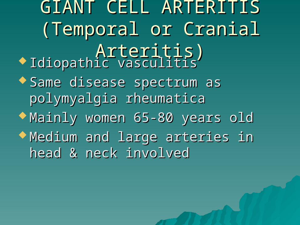
GIANT CELL ARTERITISGIANT CELL ARTERITIS(Temporal or Cranial Arteritis)(Temporal or Cranial Arteritis)
Idiopathic vasculitisIdiopathic vasculitis Same disease spectrum as Same disease spectrum as
polymyalgia rheumaticapolymyalgia rheumatica Mainly women 65-80 years oldMainly women 65-80 years old Medium and large arteries in head & Medium and large arteries in head &
neck involvedneck involved
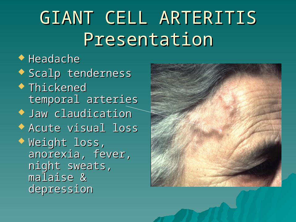
GIANT CELL ARTERITISGIANT CELL ARTERITISPresentationPresentation
HeadacheHeadache Scalp tendernessScalp tenderness Thickened temporal Thickened temporal
arteriesarteries Jaw claudicationJaw claudication Acute visual lossAcute visual loss Weight loss, Weight loss,
anorexia, fever, anorexia, fever, night sweats, night sweats, malaise & malaise & depressiondepression

GIANT CELL ARTERITISGIANT CELL ARTERITISOcular ComplicationsOcular Complications
Transient monocular Transient monocular visual loss visual loss (amaurosis fugax)(amaurosis fugax)
Visual loss due toVisual loss due to– Central retinal Central retinal
artery occlusion artery occlusion (CRAO) or (CRAO) or
– Anterior ischaemic Anterior ischaemic optic neuropathy optic neuropathy (AION)(AION)
Visual field defectsVisual field defects

GIANT CELL ARTERITISGIANT CELL ARTERITISManagementManagement
ESR if suspectedESR if suspected Start high dose steroids immediately Start high dose steroids immediately
to prevent stroke or second eye to prevent stroke or second eye involvementinvolvement
Temporal artery biopsy within a week Temporal artery biopsy within a week of starting steroidsof starting steroids
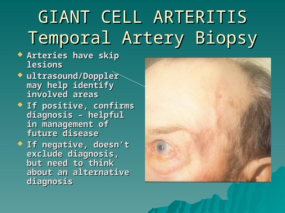
GIANT CELL ARTERITIS GIANT CELL ARTERITIS Temporal Artery BiopsyTemporal Artery Biopsy
Arteries have skip Arteries have skip lesionslesions
ultrasound/Doppler ultrasound/Doppler may help identify may help identify involved areasinvolved areas
If positive, confirms If positive, confirms diagnosis – helpful in diagnosis – helpful in management of management of future diseasefuture disease
If negative, doesn’t If negative, doesn’t exclude diagnosis, exclude diagnosis, but need to think but need to think about an alternative about an alternative diagnosisdiagnosis

GIANT CELL ARTERITISGIANT CELL ARTERITISHistopathologyHistopathology
Granulomatous cell Granulomatous cell infiltrationinfiltration
Giant cellsGiant cells Disruption of Disruption of
internal elastic internal elastic laminalamina
Proliferation of Proliferation of intimaintima
Occlusion of lumenOcclusion of lumen

GIANT CELL ARTERITISGIANT CELL ARTERITISTreatmentTreatment
Intravenous and oral steroids – Intravenous and oral steroids – prolonged course of steroids often prolonged course of steroids often necessarynecessary
Ocular manifestations Ocular manifestations of HIV infectionof HIV infection

IntroductionIntroduction
AIDS is an infectious disease caused by the gradual AIDS is an infectious disease caused by the gradual decrease in decrease in CD4+ T lymphocytes causing causing subsequent opportunistic infections and neoplasia. subsequent opportunistic infections and neoplasia. It is a blood borne and sexually transmitted It is a blood borne and sexually transmitted infection caused by the HIV (Human infection caused by the HIV (Human Immunodeficiency Virus)Immunodeficiency Virus)
Approximately 36 million persons around the world Approximately 36 million persons around the world are infected. Up to 70% of patients infected with are infected. Up to 70% of patients infected with HIV will develop some form of ocular involvement, HIV will develop some form of ocular involvement, ie: direct infection by HIV,opportunistic infections ie: direct infection by HIV,opportunistic infections and neoplasia.and neoplasia.
HIV infection progresses though HIV infection progresses though different phases


Ophthalmic Manifestations of HIV InfectionOphthalmic Manifestations of HIV Infection
AROUND THE EYEAROUND THE EYE– Molluscum Molluscum
ContagiosumContagiosum– Herpes Zoster Ophthal
micus– Kaposi’s Sarcoma– Conjunctival Squamous
Cell Carcinoma– Trichomegaly
FRONT OF THE EYEFRONT OF THE EYE– Dry Eye– Anterior Uveitis
BACK OF THE EYEBACK OF THE EYE– Retinal Microvasculopat
hy– CMV Retinitis– Acute Retinal Necrosis– Progressive Outer Retin
al Necrosis– Toxoplasmosis Retinoch
oroiditis– Syphilis Retinitis– Candida albicans endop
hthalmitis NEURO-OPHTHALMICNEURO-OPHTHALMIC

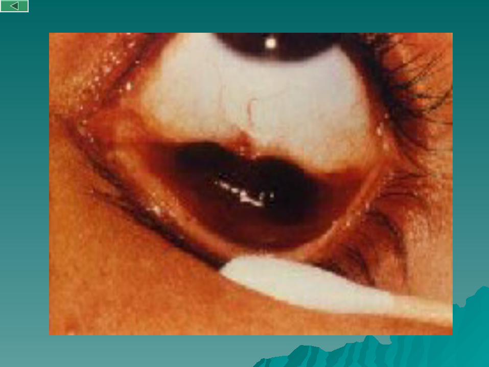
Molluscum ContagiosumMolluscum Contagiosum
Molluscum contagiosum is a Molluscum contagiosum is a viral infection of the skin.viral infection of the skin.
Affects up to 20% of Affects up to 20% of symptomatic HIV infected symptomatic HIV infected patients.patients.
Clinically appears like Clinically appears like painless, small, umbilicated painless, small, umbilicated nodules, which produce a nodules, which produce a waxy discharge when waxy discharge when pressured.pressured.
Treatment consists on Treatment consists on excision of the lesion, excision of the lesion, curettage or cryotherapycurettage or cryotherapy

Herpes Zoster OphthalmicusHerpes Zoster Ophthalmicus
Due to the reactivation of a latent infection by Due to the reactivation of a latent infection by Varicella Zoster Virus in the dorsal root of Varicella Zoster Virus in the dorsal root of trigeminal nerve ganglion.trigeminal nerve ganglion.
It manifests with a maculo-papulo-vesicular rash It manifests with a maculo-papulo-vesicular rash which often is preceded by pain. Usually involves which often is preceded by pain. Usually involves the upper lid and does not cross the midline the upper lid and does not cross the midline
Treatment consists on oral Aciclovir 800mg 5 Treatment consists on oral Aciclovir 800mg 5 times /day. In immunocompromised patients times /day. In immunocompromised patients Aciclovir is given intravenously for two weeks. Aciclovir is given intravenously for two weeks. Ocular manifestations such as anterior uveitis, Ocular manifestations such as anterior uveitis, are treated with topical steroids and mydriatics.are treated with topical steroids and mydriatics.


Kaposi’s SarcomaKaposi’s Sarcoma
Kaposi’s sarcoma is a vascular neoplasm which is almost Kaposi’s sarcoma is a vascular neoplasm which is almost exclusively seen in patients with AIDS.exclusively seen in patients with AIDS.
KS is the commonest anterior segment lesion seen in AIDS; KS is the commonest anterior segment lesion seen in AIDS; appears as a violaceous non-tender nodule on the eyelid or appears as a violaceous non-tender nodule on the eyelid or conjunctiva.conjunctiva.
Typically KS involves only the skin but when there is a Typically KS involves only the skin but when there is a reduced CD4 count it can progress rapidly to other sites reduced CD4 count it can progress rapidly to other sites such as the gastrointestinal tract and CNSsuch as the gastrointestinal tract and CNS
Treatment of ocular adnexal KS may be necessary for Treatment of ocular adnexal KS may be necessary for cosmesis and to relieve functional difficulties. The mainstay cosmesis and to relieve functional difficulties. The mainstay of treatment is radiotherapy. Other options include of treatment is radiotherapy. Other options include cryotherapy or chemotherapy.cryotherapy or chemotherapy.
Conjunctival Squamous Cell CarcinomaConjunctival Squamous Cell Carcinoma
Squamous cell carcinoma (SCC) is the third most Squamous cell carcinoma (SCC) is the third most common neoplasm associated to HIV infection. common neoplasm associated to HIV infection. This may be due to an interaction between HIV, This may be due to an interaction between HIV, sunlight and Human Papilloma Virus infection.sunlight and Human Papilloma Virus infection.
SCC appears as a pink, gelatinous growth, usually SCC appears as a pink, gelatinous growth, usually in the interpalpebral area. Often an engorgedin the interpalpebral area. Often an engorged blood vessel feeding the tumour is seen. It may blood vessel feeding the tumour is seen. It may extend onto the cornea, but deep invasion and extend onto the cornea, but deep invasion and metastasis are rare. metastasis are rare.
The treatment of choice is local excision and The treatment of choice is local excision and cryotherapy but the presence of orbital invasion cryotherapy but the presence of orbital invasion is an indication of exenterationis an indication of exenteration


TrichomegalyTrichomegaly
Trichomegaly or Trichomegaly or hypertrichosis is an hypertrichosis is an exaggerated growth of exaggerated growth of the eye lashes found the eye lashes found in the later stages of in the later stages of the diseasethe disease
The cause is not The cause is not knownknown
When symptomatic or When symptomatic or for cosmetic reasons for cosmetic reasons the eyelashes can be the eyelashes can be trimmed or pluckedtrimmed or plucked

Dry EyeDry Eye Sicca syndrome is Sicca syndrome is
frequent among frequent among patients with HIV patients with HIV infectioninfection
Patients complain of Patients complain of burning uncomfortable burning uncomfortable red eyes.red eyes.
There are several There are several causes of dry eye in causes of dry eye in HIV infection from HIV infection from blepharitis to blepharitis to destruction of the destruction of the lacrimal glands.lacrimal glands.
Treatment is with tear Treatment is with tear supplements supplements

Anterior UveitisAnterior Uveitis HIV related anteriorHIV related anterior uveitis can uveitis can
be:be:– Direct manifestation of the Direct manifestation of the
human immunodeficiency virus human immunodeficiency virus infectioninfection
– autoimmnune in originautoimmnune in origin– drug induced ie: rifabutin, drug induced ie: rifabutin,
secondary to direct toxic effect secondary to direct toxic effect upon the non-pigmented upon the non-pigmented epithelium of the ciliary body epithelium of the ciliary body
– Any of the different infections Any of the different infections associated with AIDS, ie: Herpes associated with AIDS, ie: Herpes Zoster Virus, Herpes Simplex Zoster Virus, Herpes Simplex Virus, Cytomegalovirus, Virus, Cytomegalovirus, Toxoplasma gondii, SyphilisToxoplasma gondii, Syphilis
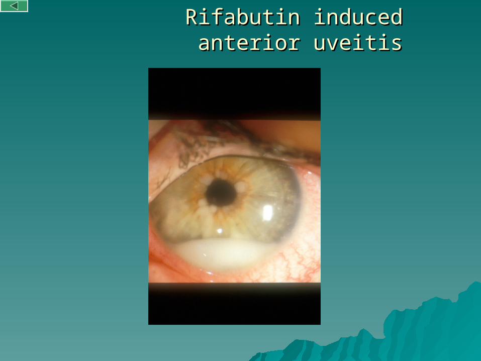
Rifabutin induced anterior uveitisRifabutin induced anterior uveitis
Retinal microvasculitisRetinal microvasculitis
Retinal microvasculopathy occurs in more than half of the Retinal microvasculopathy occurs in more than half of the patients with HIVpatients with HIV
It is seen as transient cotton wool spots (CWS), intra-retinal It is seen as transient cotton wool spots (CWS), intra-retinal haemorrhages and microaneurysm, which occurs in 50-70% haemorrhages and microaneurysm, which occurs in 50-70% of patients. It is usually asymptomatic.of patients. It is usually asymptomatic.
It has an unclear pathogenesis, but it is thought to be HIV It has an unclear pathogenesis, but it is thought to be HIV infection of retinal vascular cells.infection of retinal vascular cells.
In an otherwise healthy individual the presence of CWS, In an otherwise healthy individual the presence of CWS, should be differentiated from other forms of retinopathy, should be differentiated from other forms of retinopathy, such as diabetic or hypertensive retinopathy. Serological such as diabetic or hypertensive retinopathy. Serological test for HIV will confirm the diagnosistest for HIV will confirm the diagnosis
Treatment is based in delaying the progression of the Treatment is based in delaying the progression of the disease associated with HIVdisease associated with HIV
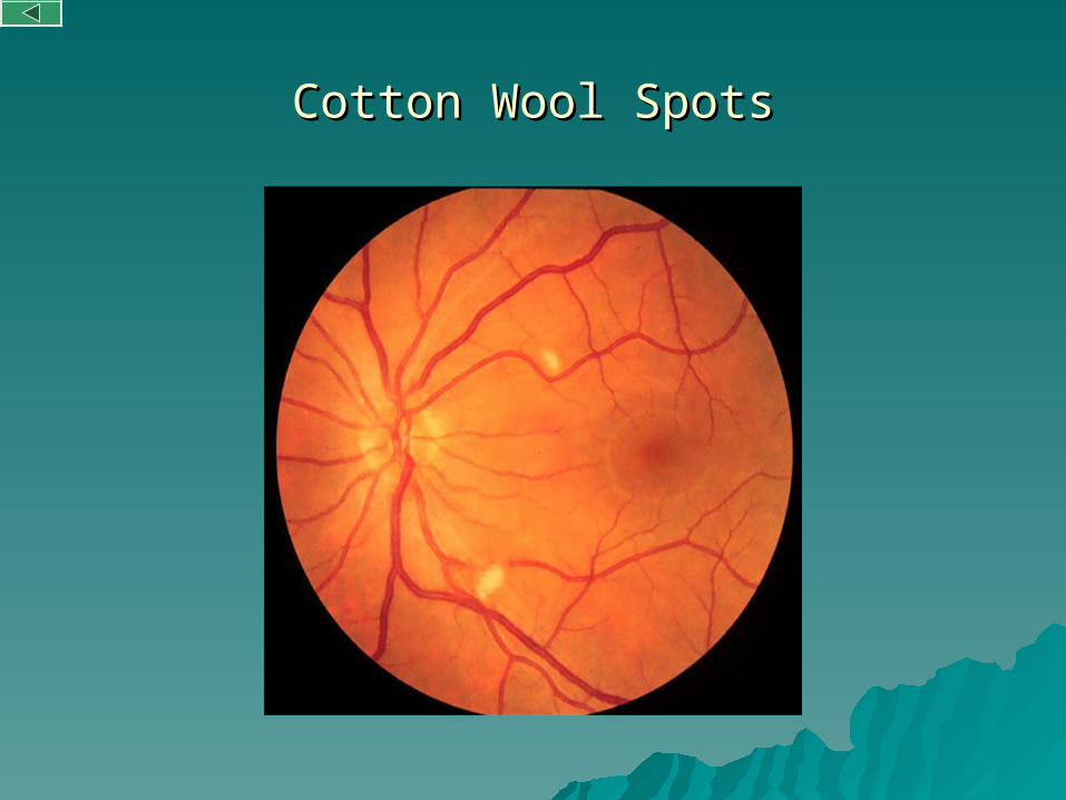
Cotton Wool SpotsCotton Wool Spots
CMV RetinitisCMV Retinitis
IntroductionIntroduction– CMV Retinitis is the commonest intraocular ocular CMV Retinitis is the commonest intraocular ocular
opportunistic infection seen in patients with AIDSopportunistic infection seen in patients with AIDS– Antibodies are found in almost 95% of adults, Antibodies are found in almost 95% of adults,
causing a trivial illness in immunocompetent adults, causing a trivial illness in immunocompetent adults, however severe immunosuppression causes viral however severe immunosuppression causes viral reactivation and tissue invasive diseasereactivation and tissue invasive disease
PathogenesisPathogenesis– Reactivation from extraocular sites leads to seeding Reactivation from extraocular sites leads to seeding
in other sites such as the retinain other sites such as the retina EpidemiologyEpidemiology
– The number of newly diagnosed cases of CMVR has The number of newly diagnosed cases of CMVR has decreased since the introduction of the HAARTdecreased since the introduction of the HAART
Highly Active Antiretroviral TherapyHighly Active Antiretroviral Therapy

CMV RetinitisCMV Retinitis
Clinical manifestationsClinical manifestations– Patients may complain of minor visual symptoms such as floaters, Patients may complain of minor visual symptoms such as floaters,
flashing lights or mild blurred vision, or be totally asymptomatic.flashing lights or mild blurred vision, or be totally asymptomatic.– It presents with a wide range of clinical appearances. From cotton It presents with a wide range of clinical appearances. From cotton
wool spots which may look like HIV Retinopathy to confluent areas of wool spots which may look like HIV Retinopathy to confluent areas of full thickness retinal necrosis and vasculitis. CMVR can progress in a full thickness retinal necrosis and vasculitis. CMVR can progress in a “brushfire” pattern from the active edge of an active lesion. The “brushfire” pattern from the active edge of an active lesion. The retinal vessels in an affected area show attenuation, becoming ghost retinal vessels in an affected area show attenuation, becoming ghost vessels eventually.vessels eventually.
TreatmentTreatment– The treatment of CMVR in patients with AIDS requires the use of The treatment of CMVR in patients with AIDS requires the use of
specific antiviral agents, ganciclovir, foscarnet or cidovir in specific antiviral agents, ganciclovir, foscarnet or cidovir in conjunction with HAART.conjunction with HAART.
– These treatments can be administered orally, intravenously or These treatments can be administered orally, intravenously or intravitreally. Systemic treatment has the advantage of treating intravitreally. Systemic treatment has the advantage of treating infection elsewhere in the body as well as the other eye but has the infection elsewhere in the body as well as the other eye but has the disadvantages of systemic side effects.disadvantages of systemic side effects.
– Intravitreal implants release the drug over a six-month period, Intravitreal implants release the drug over a six-month period, achieving prolonged high intravitreal levelsof drug.achieving prolonged high intravitreal levelsof drug.

CMV RetinitisCMV Retinitis

Acute Retinal NecrosisAcute Retinal Necrosis
ARN is a confluent peripheral whitening of the retina with ARN is a confluent peripheral whitening of the retina with marked vitritis and blood vessel closure. Optic neuritis marked vitritis and blood vessel closure. Optic neuritis and retinal detachment are frequent complications.and retinal detachment are frequent complications.
ARN is usually due to Varicella-Zoster infection, but it can ARN is usually due to Varicella-Zoster infection, but it can also be caused by Herpes Simplex virus or also be caused by Herpes Simplex virus or Cytomegalovirus.Cytomegalovirus.
Initially described in the immunocompetent, it has also Initially described in the immunocompetent, it has also been described in the immunosuppressed.been described in the immunosuppressed.
The diagnosis is mainly clinical and is confirmed by PCR The diagnosis is mainly clinical and is confirmed by PCR assays on vitreous samples.assays on vitreous samples.
Patients are treated with high doses of intravenous Patients are treated with high doses of intravenous aciclovir or famciclovir, combined with laser treatment to aciclovir or famciclovir, combined with laser treatment to prevent retinal detachment.prevent retinal detachment.

Acute Retinal NecrosisAcute Retinal Necrosis

Progressive Outer Retinal Progressive Outer Retinal NecrosisNecrosis
(Varicella-Zoster Retinitis)(Varicella-Zoster Retinitis) PORN is a devastating viral retinitis caused by Varicella-PORN is a devastating viral retinitis caused by Varicella-
Zoster virus, without vitritis or retinal vasculitis.Zoster virus, without vitritis or retinal vasculitis. The retinitis can be located anywhere but it is common The retinitis can be located anywhere but it is common
for the lesions to coalesce and spread posteriorly in a for the lesions to coalesce and spread posteriorly in a rapid fashion.rapid fashion.
The main symptom is rapid loss of vision.The retina The main symptom is rapid loss of vision.The retina shows typically a white lesion with no haemorrhages or shows typically a white lesion with no haemorrhages or exudates.exudates.
Treatment is often unsatisfactory and usually requires Treatment is often unsatisfactory and usually requires combination of Ganciclovir and Aciclovir. The prognosis combination of Ganciclovir and Aciclovir. The prognosis is very poor and retinal detachment is common. is very poor and retinal detachment is common. Resolution may leave a white plaque with the Resolution may leave a white plaque with the appearance of “cracked mud”.appearance of “cracked mud”.


Toxoplasma RetinochoroiditisToxoplasma Retinochoroiditis
Toxoplasmosis retinochoroiditis is an uncommon Toxoplasmosis retinochoroiditis is an uncommon infection of the eye in AIDS. Ocular toxoplasmosis infection of the eye in AIDS. Ocular toxoplasmosis in HIV positive patients is different in appearance in HIV positive patients is different in appearance from immunocompetent patients. Unlike in from immunocompetent patients. Unlike in immunocompetent patients, HIV infected patients immunocompetent patients, HIV infected patients often have bilateral and multifocal disease often have bilateral and multifocal disease associated with anterior uveitis and vitritis but associated with anterior uveitis and vitritis but unlike immunocompetent patients, in HIV infected unlike immunocompetent patients, in HIV infected patients often have with no pigmented scars patients often have with no pigmented scars adjacent to the areas of retinal necrosis. adjacent to the areas of retinal necrosis. Toxoplasmosis in immunocompromised patients is Toxoplasmosis in immunocompromised patients is not self-limiting as it is in imunocompetent patients.not self-limiting as it is in imunocompetent patients.

Toxoplasma RetinochoroiditisToxoplasma Retinochoroiditis
When testing patients for antibodies to When testing patients for antibodies to toxoplasmosis both IgG and IgM levels may be toxoplasmosis both IgG and IgM levels may be raised, but in immunocompromised patients raised, but in immunocompromised patients these tests may be negative. these tests may be negative.
Treatment in immunocompromised patients Treatment in immunocompromised patients consists in the association of sulphadiazine or consists in the association of sulphadiazine or clindamycin, pyrimethamine and folinic acid clindamycin, pyrimethamine and folinic acid (triple therapy).(triple therapy).
Long term maintenance treatment may be Long term maintenance treatment may be needed in order to prevent relapses.needed in order to prevent relapses.
Often associated with toxoplasma lesions in the Often associated with toxoplasma lesions in the Central Nervous System.Central Nervous System.

MRI T1 showing an uniformly enhancing lesion in the midbrain
One week later, the lesion showing ring enhancement

ImmunocompetentImmunocompetent ImmunocompromisedImmunocompromised

Syphilis RetinitisSyphilis Retinitis
There is a strong association between There is a strong association between syphilis and HIV infection.syphilis and HIV infection.
It can manifest as a retinitis with dense It can manifest as a retinitis with dense vitritis, retinal vasculitis, serous retinal vitritis, retinal vasculitis, serous retinal detachment or neuroretinitis, as well as other detachment or neuroretinitis, as well as other types of ocular involvement such as, types of ocular involvement such as, conjunctivitis, anterior uveitis, cranial nerve conjunctivitis, anterior uveitis, cranial nerve palsies and optic neuritis.palsies and optic neuritis.
Treatment consists in high dose of Treatment consists in high dose of intravenous Penicillin for 2 weeks.intravenous Penicillin for 2 weeks.


Candida albicans Candida albicans endophthalmitisendophthalmitis
Infection with candida albicans is rare. Candida Infection with candida albicans is rare. Candida albicans is the commonest cause of fungal albicans is the commonest cause of fungal endophthalmitisendophthalmitis
Affected patients usually have a history of drug Affected patients usually have a history of drug abuse or indwelling central linesabuse or indwelling central lines
In the initial stages, floaters are the main In the initial stages, floaters are the main symptom. As the condition progresses, whitish symptom. As the condition progresses, whitish “puff-balls” and vitreous strands develop. Later, “puff-balls” and vitreous strands develop. Later, similar infiltrates appear in the choroid and retinasimilar infiltrates appear in the choroid and retina
The treatment depends on the severity of the The treatment depends on the severity of the ocular involvement and systemic disease. The ocular involvement and systemic disease. The original foci should be removed. The drugs of original foci should be removed. The drugs of choice are Amphotericine B and Fluconazolchoice are Amphotericine B and Fluconazol

Candida albicans Candida albicans endophthalmitisendophthalmitis

GlossaryGlossary
CD4CD4: Director of the immune response. When activated it : Director of the immune response. When activated it releases releases cytokines which in turn will activate the immune cytokines which in turn will activate the immune systemsystem
Cotton Wool SpotsCotton Wool Spots: Light-coloured deposits in the retina : Light-coloured deposits in the retina secondary secondary to infarcts of the nerve fibre layer to infarcts of the nerve fibre layer
HAARTHAART: Highly Active Antiretroviral Therapy: Highly Active Antiretroviral Therapy ImmunoblogulinImmunoblogulin: Protein in charge of fighting foreign : Protein in charge of fighting foreign
substances in substances in our body. our body. IgG IgG is the commonest type of is the commonest type of immunoglobulin and immunoglobulin and IgMIgM is the earliest class of is the earliest class of immunoglobulin.immunoglobulin.
PCRPCR: Polymerase Chain Reaction is a technique used to make : Polymerase Chain Reaction is a technique used to make numerous copies of an specific portion of DNAnumerous copies of an specific portion of DNA
VDRLVDRL: Venereal Disease Research Laboratory. The test : Venereal Disease Research Laboratory. The test becomes becomes negative after successful treatment of the disease. negative after successful treatment of the disease.

Questions?


![AAU-Hematologic Aspect of Systemic Disease[1]](https://img.dokumen.tips/doc/110x75/546874efb4af9f533f8b5d3f/aau-hematologic-aspect-of-systemic-disease1.jpg)