Embed Size (px)
Citation preview

10/15/2013
1
Retinal Findings with Systemic
Disease
Jeffry D. Gerson, O.D., F.A.A.O.
Shawnee, KS
Disclosure
I have been on advisory boards/a consultant
to/received honoraria from/ or been on speakers
bureau list of the following:
Allergan, Alcon, Arctic Dx, Bausch & Lomb, Carl Zeiss
Meditec, Freedom Meditech, Optos, Optovue, VSP,
ZeaVision
These affiliations will have no
affect on the content of this lecture
Course Objectives
Discuss Ophthalmic tests for evaluating
retina
Discuss systemic conditions that affect
retina, and how we factor into patient care
Discuss findings associated with systemic
diseases, both common and uncommon
Know when to refer, and to whom

10/15/2013
2
Antioxidants
Do you drink coffee?
Over 50% of Americans drink coffee
Is this important?
Coffee is leading source (by far) for antioxidant intake in the US diet!!1
Neither coffee nor caffeine intake were associated with early AMD per BDES
Beware:
COFFEE and DOUGHNUT Maculopathy2 1. As reported by American Chemical Society 8/05
2. Kerrison J.B. et.al. Coffee and Doughnut Maculopathy: Acute Ring Scotomas. BJO.2000 Feb;84(2):158-64.
The Relationship of Coffee Consumption with
Mortality Ann Intern Med 2008:148:904-14 2 Cohorts
41,736 men Hx Professionals FUp Study – 18 years
86,214 women Nurse’s Hx Study - 24 years
Results
After adjustment for age, smoking, other CVDz and CA risk factors
Men
<1 cup / month 1.07
1 c/m – 4 cups/w 1.02
5-7 cups / week 0.97
2-3 cups / day 0.93
4-5 cups / day 0.80
> 6 cups / day 0.74
P<0.001 for trend
and independent
of caffeine intake
Medical optometry: A different kind
of “liability”

10/15/2013
3
More Than Meets the Eye
Macula off retinal detachment OD
LP vision
Systemic health: good?
Meds: Valium, Oxycodone, Methadone, Elavil
Tx: Vitrectomy and Scleral Buckle
Post op: Corneal Abrasion and HM
How did the abrasion happen???
Bottle Top
What does
this mean to
you?
Clinical exam
**BIO best for clinical exam
of peripheral retina
Condensing lens at slit lamp
ideal for magnified posterior
pole views
Dynamic clinical exam
Require examiner to
document
Some things best seen
clinically, and unable to be
seen with imaging devices
10/15/2013
4
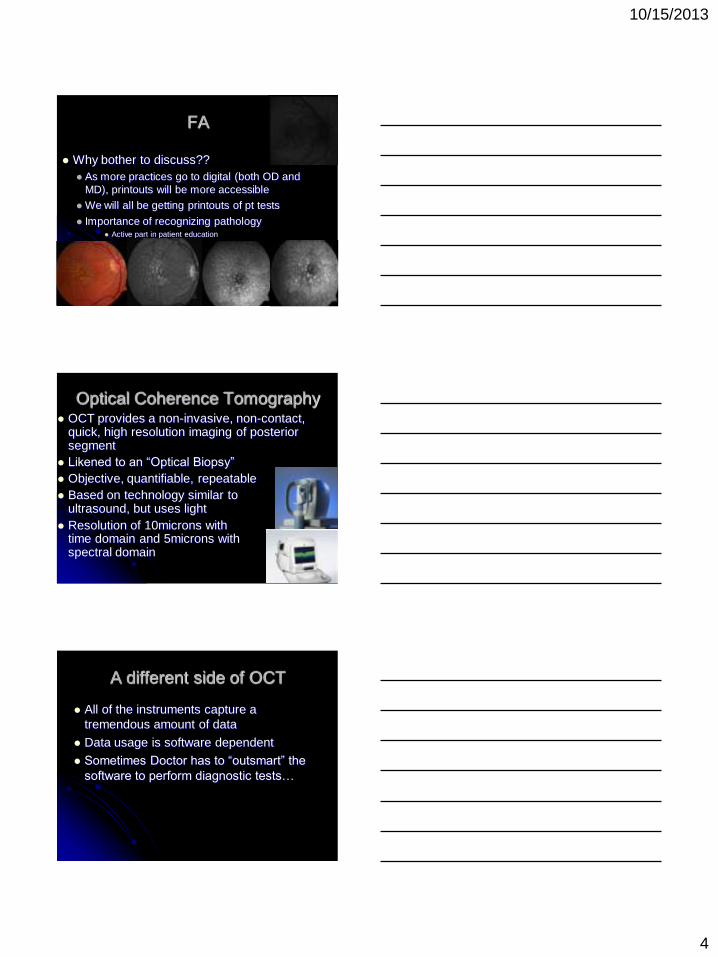
FA
Why bother to discuss??
As more practices go to digital (both OD and
MD), printouts will be more accessible
We will all be getting printouts of pt tests
Importance of recognizing pathology Active part in patient education
Optical Coherence Tomography OCT provides a non-invasive, non-contact,
quick, high resolution imaging of posterior segment
Likened to an “Optical Biopsy”
Objective, quantifiable, repeatable
Based on technology similar to ultrasound, but uses light
Resolution of 10microns with time domain and 5microns with spectral domain
A different side of OCT
All of the instruments capture a
tremendous amount of data
Data usage is software dependent
Sometimes Doctor has to “outsmart” the
software to perform diagnostic tests…

10/15/2013
5
En-Face or “C-scan” technology
Cystic
spaces
in CME
Advanced visualization
20/60
“Cataracts worse x 6
mos”
ERM causing macular
thickening
Surface tension/folds evident on
en-face / c-scan
Choroid Ant. Surface RPE Ant. Aspect of CNV
Virtual Fluorescein Angiography

10/15/2013
6
SDOCT Capabilities….
Healthy patient??...
32 yo male
2-3 month history of cough, dyspnea, chills, malaise
Recently returned from International travel
Lives in Midwest
Health care professional
No improvement with antibiotics and PO prednisone
Abnormal chest x-ray
Good vision
Referred to Pulmonologist
Chest X-ray
Calcified Granulomas
Differentials?
TB
Sarcoid
Histoplasmosis
Lymphoma

10/15/2013
7
Case continued
CT ordered with contrast
Labs ordered CBC Normal
Normal Liver function
ESR 46 mm/hr
Negative TB skin test
ACE 44 U/L (7-46)
Histo Mycelial Ab Normal
Histo Anti H Ab 1:32
Histoplasmosis Treatment:
Sporanox (Itraconazole) 200mg BID x 1 mo
100mg BID x 2 mo
Aside:
Value of prescription drug coverage!
Importance of good doctor patient relationship!!!
In case you were wondering, Histo has remained quiet, with no radiologic changes as of 4/06
Systemic Histoplasmosis
Caused by Histoplasma capsulatum, a dimorphic fungus, that turns into a yeast at body temperature
Endemic to Ohio, Mississippi, and Missouri River valleys
Aerosolized fragments result in alveolar deposition
Most infected people are asymptomatic
Can involve CNS, liver, spleen, eyes, rheumatologic system, and hematologic system

10/15/2013
8
Histoplasmosis cont.
Symptoms can occur 3-14 days after exposure
Approximately 250,000 infected annually
Clinical manifestations in less than 5%
About 90% with acute pulmonary histo are asymptomatic
Enlarged hilar and mediastinal lymph nodes in 5-10% of patients
Affects males 4:1
Progressive disseminated histo mostly occurs in immunocompromised patients ex: AIDS
Good summary article: Trevino & Salvat:Preventing Reactivation of OHS. Optometry 1/06
Testing
CBC generally normal
Sputum cultures yield positive results in only 10-15% of acute pulmonary histo
Complement fixing antibodies Greater than 1:32 suggests active
Positive 5-15% of within 3 wks of exposure
Positive 75-95% at 6wks
Immunoprecipitating antibodies Anti-M detected in 50-80%, and remains elevated for years
Anti-H detected in 10-20% and becomes undetectable after 6mos. This antibody is most specific for active histo
Imaging studies Chest X-ray
CT scan
HLA-B7, HLA-DR2 and may be elevated more in people with CNVM
Treatment
No treatment needed if asymptomatic
Treatment if symptomatic, or progressive
Treatments Amphotericin B: drug of choice for overwhelming
active histo, administered by IV
Itraconazole: Fungistatic, very active against Histo, minimal side affects Liver functions must be monitored
Approximately 86% success when treating > 2mos
Ketoconazole: Fungistatic, well tolerated, does not cross blood/brain barrier

10/15/2013
9
(P)OHS (Presumed) ocular
histoplasmosis syndrome
Not previously found post-ennucleation in patients with typical POHS
Has been found in eye of patients with known Histo
Approx 1-10% pts. In endemic areas have ocular involvement, usually asymptomatic
10% will be bilateral
OHS
Histo Spots
Atrophic yellowish white scars from previous multifocal or disseminated choroiditis
Can form streaks
Peripapillary Atrophy
May represent atrophied granulomas that formed during active infective stage of
Neovascular membranes can form here, and involve macula
Macular Involvement CNVM tend to form in
area of pre-existing histo spot
May be immune reaction against H. capsulatum
May be due to weakened Bruch’s membrane
10% become bilateral at 5 yrs, and 20% at 10yrs
81% with disciform macular scarring have pulmonary calcifications
OHS

10/15/2013
10
Treating CNVM from Histo
MPS
Argon laser to entire lesion effective if extrafoveal with 8%
recurrence
Krypton laser if juxtafoveal with 23% recurrence
Submacular Surgery (SST)
Benefit seen in surgical group if entering acuity worse than 20/100
(76% vs 50% same or better)
More recently shown beneficial with PPCNVM1: different histopath
Pt experience no better with surg in any group2
PDT
>50% remain equal or show improvement
No cases of severe vision loss as has been reported as has been
with AMD patients
Anti-VEGF Therapy
1. Thomas, Matt at Barnes Retina in St. Louis 3/2008 2. Surg vs observ with
POHS CNVM. SST group. Arch Ophth 12/08
Central “Spot”
50yo female referred in with a “spot” in the center of her vision
Present for 1-2 wks
Referring OD noticed abnormality
VA 20/20 OU
Denies High stress or type “A” personality
Central Serous Choroidopathy
Characterized by breakdown of the outer retinal barrier, with leakage of fluid through a defect in the RPE into the subretinal space, resulting in a neurosensory detachment
Often times associated with high stress +/- ED (Emotional Distress) may be related1
FA or OCT must be done to rule out CNVM
Other systemic associations Use of corticosteroids* (Well documented in literature),
pregnancy, increased adrenaline level, hemodialysis, collagen vascular disease, and hypertension
Treatment?
Letter of diagnosis to PCP to make aware
1. Conrad et al. Alexithymia and emotional distress in ICSC. Psychosomatics. 2007 Nov-Dec;48(6):489-95

10/15/2013
11
ICSC
Newer treatments proposed: PDT
Success in multiple studies1
IVTA May prevent leakage
Not study proven and counterintuitive
Anti-VEGF
Is it too easy to be successful with new treatments??
PDT for RPE leaks in CSC. Ober, M et al. Ophthalmology. Dec. 2005.
Central Serous and Steroids
How would you know about steroid use?
What kinds of steroids
I have had cases of cream/ointment, oral
Could hormones have same affect?
Patient on Androgel for “Low T”
Case Study
44 yo native-american
male
Recent awareness of
central blind spot
20/25 OU
Diagnosis?
Solar Maculopathy
Systemic assoc???

10/15/2013
12
Solar Maculopathy
Bilateral yellowish spot in fovea with surrounding
hyperpigmentation and OCT shows loss of cells at
RPE layer
Retinal phototoxicity vs photocoagulation
Often happens in patients who use drugs or are on
psychotropic meds
Sun gazing while “on drugs” or brief exposure with pharm.
Dilated pupils
No other systemic associations
Case Study cont.
Take a closer look at
the ONH
What is this?
No PEPS
Idiopathic
Warned of possibility
of future CNVM
Case study 32yo female
Good health
20/20 OU
“retinal changes”

10/15/2013
13
Angiod Streaks
• Diagnosis: Angioid Streaks
• Treatment: yearly exams, and home monitor with Amsler grid
• Note: proximity of Angioid streak to fovea
• Over 50% of Angioid streak patients have associated systemic disorders
Angioid Streaks
Represent breaks in an abnormal Bruch’s Membrane that may present spontaneously or as result of trauma
Eventual RPE and choriocapillaris degeneration
Generally radiate out from ONH, bilateral
Color depends on fundus color and degree of RPE atrophy Red: Lightly colored fundi, reflect underlying choroid
Brown: Darker pigmented fundi
Orange: Specific type of RPE mottling
Angioid Streaks: associated systemic conditions
Pseudoxanthoma Elasticum 80-90% have angioid streaks
Degeneration of collagen
Most common systemic
Paget’s Disease 8-15% have angioid streaks
Metabolic bone disease
Sickle Cell Disease <6% have angioid streaks
Ehler’s-Danlos Syndrome Skin fragility, joint hyperextensibility
Diabetes
Others: maybe coincidental
PEPSI

10/15/2013
14
Angioid Streaks
Not problematic unless get CNVM
If CNVM, standard is thermal laser, but >75%
recur
Monitor with Amsler grid
Case of Missing Labs
RM is a 46 year old Caucasian male
Referred for retinal changes, questionable
macular edema
Last physical 2-3 years prior
“No systemic health problems”, no
medications
Paramedic
Note: Not a very healthy looking patient
“Healthy” Paramedic cont.
Visual acuity: OD: 20/100 OS: 20/30
Pupils, CVF, Amsler all normal
Anterior segment: Normal, no iris changes
Fundus exam: Widespread microaneurysms, several cotton wool spots, vascular
engorgement and crossings, dot and flame hemorrhages in post-pole and equatorially
Macular edema present OD, and possibly OS
Fluorescein Angiogram ordered Above changes noted, significant leakage in OD macula. Limited
change to macula OS
TX: Focal laser recommended
TX Cont: Letter sent to PCP telling of findings, recommend blood workup for DM and other vascular problems
10/15/2013
15
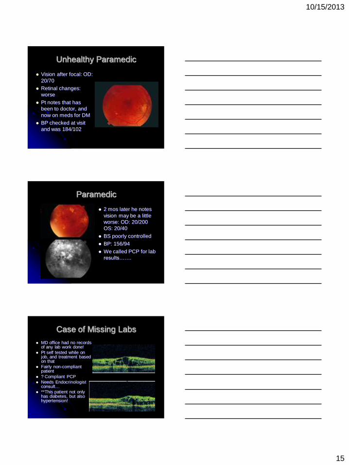
Unhealthy Paramedic
Vision after focal: OD:
20/70
Retinal changes:
worse
Pt notes that has
been to doctor, and
now on meds for DM
BP checked at visit
and was 184/102
Paramedic
2 mos later he notes
vision may be a little
worse: OD: 20/200
OS: 20/40
BS poorly controlled
BP: 156/94
We called PCP for lab
results…….
Case of Missing Labs
MD office had no records of any lab work done!
Pt self tested while on job, and treatment based on that
Fairly non-compliant patient
? Compliant PCP
Needs Endocrinologist consult…
**This patient not only has diabetes, but also hypertension!
10/15/2013
16
Diabetes
2 types
Type 1 (previously insulin dependent)
Beta cell destruction leading to absolute insulin deficiency
Glucose stays in blood since can not enter insulin dependent
tissues
Type 2 (previously non-insulin dependent)
Peripheral insulin resistance, maybe relative insulin
deficiency or secretory defect
Treatment to decrease hepatic glucose production &/or
decrease peripheral insulin resistance
May become insulin dependent
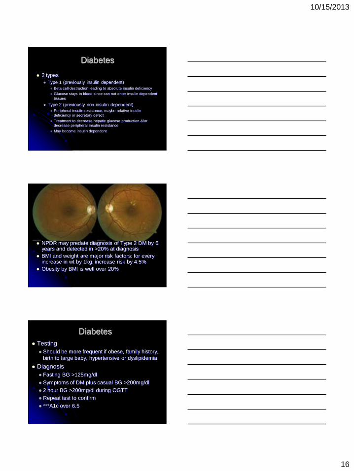
Diabetes
Lifetime risk of DM for Caucasian individuals born in 2000 is 32.8% for males and 38.5% for women (approx 20% more for hispanic)
DM affects approximately 1:16 Americans, and approx 1/3- 1/2 unaware they have DM
NPDR may predate diagnosis of Type 2 DM by 6 years and detected in >20% at diagnosis
BMI and weight are major risk factors: for every increase in wt by 1kg, increase risk by 4.5%
Obesity by BMI is well over 20%
Diabetes
Testing
Should be more frequent if obese, family history,
birth to large baby, hypertensive or dyslipidemia
Diagnosis
Fasting BG >125mg/dl
Symptoms of DM plus casual BG >200mg/dl
2 hour BG >200mg/dl during OGTT
Repeat test to confirm
***A1c over 6.5
10/15/2013
17
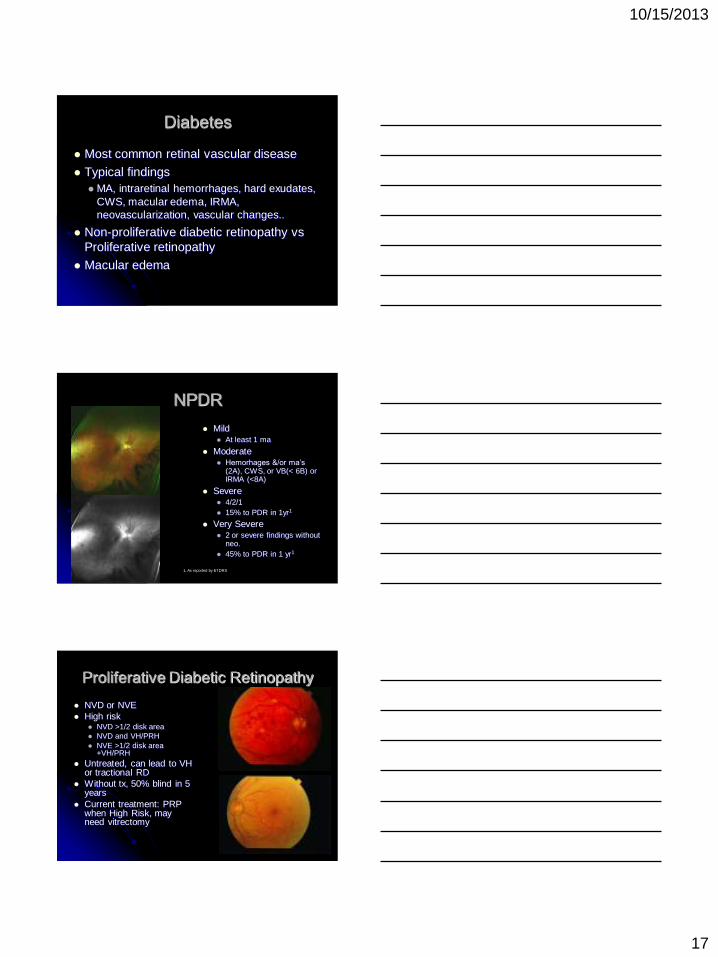
Diabetes
Most common retinal vascular disease
Typical findings
MA, intraretinal hemorrhages, hard exudates,
CWS, macular edema, IRMA,
neovascularization, vascular changes..
Non-proliferative diabetic retinopathy vs
Proliferative retinopathy
Macular edema
NPDR
Mild At least 1 ma
Moderate Hemorhages &/or ma’s
(2A), CWS, or VB(< 6B) or IRMA (<8A)
Severe 4/2/1
15% to PDR in 1yr1
Very Severe 2 or severe findings without
neo.
45% to PDR in 1 yr1
1. As reported by ETDRS
Proliferative Diabetic Retinopathy
NVD or NVE
High risk NVD >1/2 disk area
NVD and VH/PRH
NVE >1/2 disk area +VH/PRH
Untreated, can lead to VH or tractional RD
Without tx, 50% blind in 5 years
Current treatment: PRP when High Risk, may need vitrectomy

10/15/2013
18
Macular Edema
3 criteria Thickening <1/3DD from
center of macula
Heme/exudate with thickening of adjacent retina <1/3dd from center of macula
Thickening >1dd size within 1dd center
Current treatment: Grid/Focal laser
Investigational treatment: IVTA
Diabetic Retinopathy Study
Randomized, prospective to evaluate PRP
Primary outcome was severe vision loss
defined as 5/200
Demonstrated 50% decrease in SVL in
PRP group
Recommendation: PRP
Complication: 11% lost 1 or more lines of
acuity, and 5% had visual field loss
Early Treatment for Diabetic Retinopathy Study
Evaluated PRP and aspirin in pts with less than
HR PDR OU, laser for DME
Outcome was Moderate VL (doubling of visual
angle)
Results:
>50% less MVL with laser for CSME
PRP for PDR, not needed earlier, but may be
beneficial for Type 2
ASA 650mg did not alter retinopathy, VA or VH, or
rates of vitrectomy

10/15/2013
19
Diabetic Retinopathy Vitrectomy Study
Is early vitrectomy beneficial?
20/40 was more common in early-
vitrectomy group (1-6 mos.)
Benefit seen in eyes with most severe
disease
In regards to VH, clear
benefit to type 1, but not
to type 2
Today: 25g vitrectomy
Intravitreal Steroid for
DME…The Next “Best Thing” NOT……..
Published paper shows that traditional focal
laser better for CSME than 2 different doses of
steroid injection1
At 2 yrs, focal more effective and less side
affects than injection: in general
Just as convincing at 3yrs2 IVT stable vs laser gain!
Subgroup:
Thicker OCT better with Laser
Worse VA than 20/200, better with 4mg steroid
1. IVTA vs focal for DME. DRCR.net. Ophth 9/08. 2. 3yr f/u on laser vs IVT for DME. DRCR. Arch Ophth 3/09.
Lucentis DRCR.net investigated Lucentis vs laser and/or
steroid n= 691 people (~850 eyes).
Grps (success is 20/20 or <250microns @ 1yr)
1: sham injection + prompt laser treatment
2: Lucentis + prompt laser (8/13)
3: Lucentis + deferred laser treatment (≥24 weeks (9/13)
4: IVK + prompt laser (3/4)
Success: 32%, 64%, 52%, 56%
Lucentis gained 9 letters vs 3 in laser v 4 w steriod
Steroid better than laser for OCT, but not VA
Approx 30% Lucentis + 3 lines vs 15% w laser
Elman et al. Lucentis in DME. Ophthal 4/10

10/15/2013
20
Diabetes Control and Complications Trial &
UK Prospective Diabetes Study Pts randomized to conventional or
intense control
Showed slower progression for intense control group
For those with no NPDR at start, if intense, then 76% less devel. of retinopathy
If A1c down by 2%, PDR would decrease by 50%
Decrease in A1C by 1 %:
14% decrease in MI
12% decrease in stroke
37% decrease in microvascular dz
21% decrease in any DM endpoint
DCCT reported relationship of A1C and avg. Glucose
%HbA1C Avg. Glucose (mg/dL)
4.0 60
5.0 90
6.0 120
7.0 150
8.0 180
9.0 210
10.0 240
11.0 270
Control group in DCCT: 9-10%
Strict control group: 7%
Sources: NEJM 329:977-986 1993 UKPDS: Lancet 352:837-853,1998
What We Need to Know
The Diabetes Prevention Program (DPP) showed that lifestyle modification lowers the risk of developing T2DM in high-risk patients by 58% over 4 yrs.
Walking 150 minutes per week
Metformin reduced the risk by 30%
Exercise was twice as
effective as drug
Diabetes Prevention Program
The DPP Ten Years Out :
• 38% reduced risk of with lifestyle modification
•17% reduced risk with metformin
Hemoblobin A1c
Importance of A1c
monitoring
Critical to disease
control and
prevention of
problems
Does a patient know
their last reading?
Good, bad, or worse
response
In office testing
www.a1cnow.com

10/15/2013
21
The Challenge for OD’s in
Diabetes Care….
Those Who Aren’t What We Call Them
Calling someone “a diabetic” is existentially diminutive
Few people want to be known AS or even BY their health status
Why don’t we say ‘he is a macular degenerate,’ or ‘she is cancerous’?
Don’t Substitute a Part Of Any Person
For the Whole Person
You never know..
Diagnosed with
T2DM 2 wks ago
Vision not good,
Endo said due to
BS fluctuation
20/50
10/15/2013
22
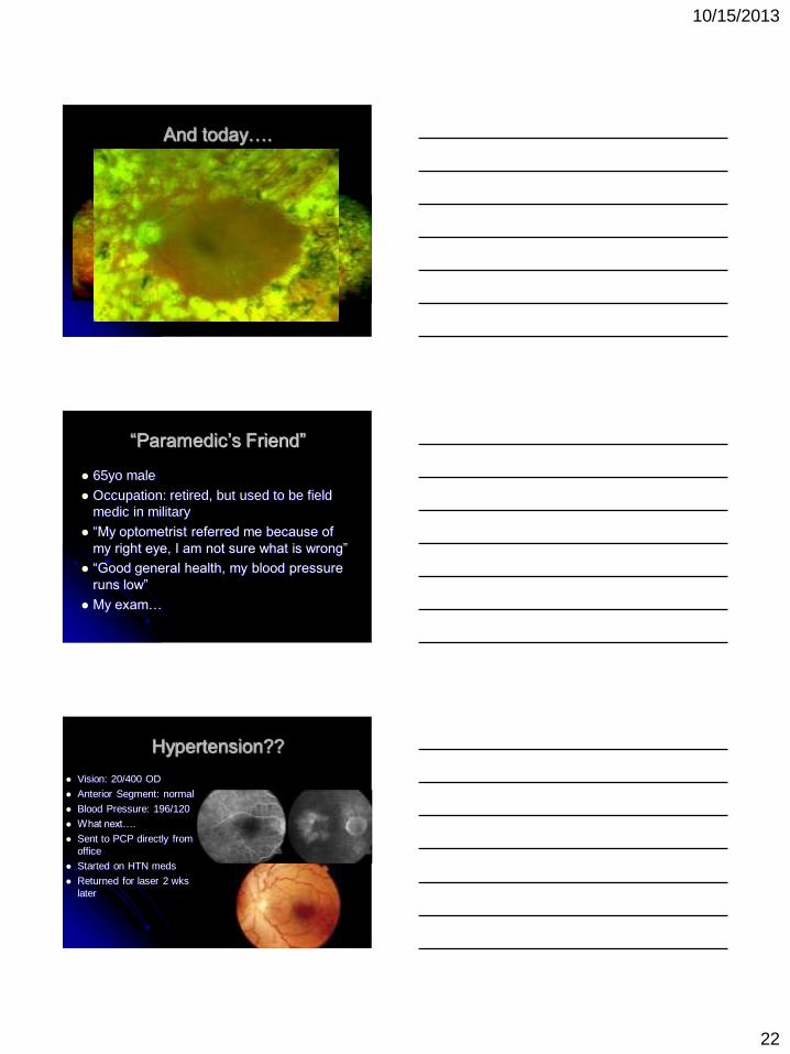
And today….
“Paramedic’s Friend”
65yo male
Occupation: retired, but used to be field
medic in military
“My optometrist referred me because of
my right eye, I am not sure what is wrong”
“Good general health, my blood pressure
runs low”
My exam…
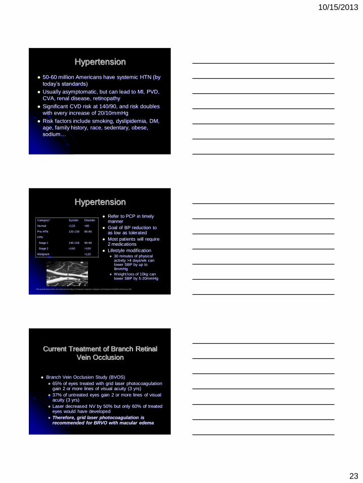
Hypertension??
Vision: 20/400 OD
Anterior Segment: normal
Blood Pressure: 196/120
What next….
Sent to PCP directly from
office
Started on HTN meds
Returned for laser 2 wks
later
10/15/2013
23
Hypertension
50-60 million Americans have systemic HTN (by
today’s standards)
Usually asymptomatic, but can lead to MI, PVD,
CVA, renal disease, retinopathy
Significant CVD risk at 140/90, and risk doubles
with every increase of 20/10mmHg
Risk factors include smoking, dyslipidemia, DM,
age, family history, race, sedentary, obese,
sodium…
Hypertension
Category* Systolic Diastolic
Normal <120 <80
Pre-HTN 120-139 80-89
HTN
Stage 1 140-159 90-99
Stage 2 >160 >100
Malignant >120
Refer to PCP in timely manner
Goal of BP reduction to as low as tolerated
Most patients will require 2 medications
Lifestyle modification 30 minutes of physical
activity >4 days/wk can lower SBP by up to 9mmHg
Weight loss of 10kg can lower SBP by 5-20mmHg
*The Seventh Report of the Joint National Committee on Prevention, Detection, Evaluation, and Treatment of High Blood Pressure, NIH
Current Treatment of Branch Retinal
Vein Occlusion
Branch Vein Occlusion Study (BVOS)
65% of eyes treated with grid laser photocoagulation gain 2 or more lines of visual acuity (3 yrs)
37% of untreated eyes gain 2 or more lines of visual acuity (3 yrs)
Laser decreased NV by 50% but only 60% of treated eyes would have developed
Therefore, grid laser photocoagulation is recommended for BRVO with macular edema

10/15/2013
24
Current Treatment of Central Retinal
Vein Occlusion
Central Vein Occlusion Study (CVOS)
Grid laser photocoagulation reduces angiographic evidence of
macular edema
Final median visual acuity in treated eyes was 20/200 (3 yrs)
Final median visual acuity in untreated eyes was 20/160 (3 yrs)
With or without treatment, approx. 33% Lose 3 lines of VA at 3
years
PRP did not prevent iris NV
Therefore, grid laser photocoagulation is NOT
recommended for CRVO, unless NV develops
So, what do we do now???
CRUISE: Luncentis for CRVO
BRAVO: Lucentis for BRVO
SCORE for BRVO
SCORE for CRVO
Dex
Why studies are needed
“When you have a hammer, everything looks like a nail”
Jost Jonas, M.D.

10/15/2013
25
Hemorrhage everywhere!
68 yo female
Dramatic decrease in
vision 1 wk prior due
to Vitreous Heme
Exam as seen after
VH resolution
Diagnosis and
Treatment?
Macroaneurysm
1mo and 5 mo s/p focal laser
VA returned to 20/20
Blood Pressure at initial visit:
186/98
Hypertension is prime concern if macro-a seen, secondary concern of diabetes
RAM
Most commonly in 6th or 7th decade of life
Usually women, and only 10% bilateral
Hypertension is prime systemic assoc. (2/3)
Must also rule out cardiovascular disease,
including increased cholesterol/lipid levels, and
diabetes
Communication to PCP

10/15/2013
26
Dangers of Addiction
38 yo male
Healthy
No meds, but…
Viagra PRN
Frequent Alcohol
20/20 OD, 20/30 OS
Ant Seg healthy
Retina OS as seen
Diagnosis?
Valsalva Not generally associated
with systemic disease, but… More common in people
with DM, HTN, and sickle cell
Typical ocular findings: Pre-retinal heme, sub-
hyaloid heme
Caused by sudden raise in intrathoracic pressure, which leads to Increased intraocular venous pressure
Causes break in macular capillary
Valsalva Maculopathy
Common causes:
Vomit, cough, sneeze,
constipation, exertion
Often seen with
alcoholism, bulemia and
GI problems
Tend to resolve on own
No long lasting damage
What caused condition
in this patient?
“Drunken Pumpkin”

10/15/2013
27
What do you think when you see
this clinical image?
OD Macula OS Macula
How about this Visual Field??
Pt. AM exam findings
Pt AM is a 47yo female that has been on Plaquenil 200mg BID x 1 yr, weights approx 120lbs
Being seen by request of her rheumatologist for screening for Plaquenil toxicity
Vision corrects to 20/20 in both eyes
Pupils and screening Matrix VF are normal
Contrast is normal at 1.25% OU and color is normal
MPOD is .31 OD and .38 OS
IOP 18/17mmHg
Schirmer is 0mm in both eyes w/ dry eye sx

10/15/2013
28
Further findings: Cross Hair OCT:
Worried yet??
OD
OS
No apparent PIL degredation seen
Inner Retinal Thickness: Still all
normal
GCC and RNFL
GCC Comparison RNFL Comparison

10/15/2013
29
Full Retina
Significance
OD and OS
Note:
significantly
ring thinning
around OD
macula
New Guidelines per AAO (the
other one) Risk increases sharply to 1% at 5-7yrs or
cumulative dose of 1000g (usual dose 400mg/d HCQ or 250mg/d CQ)
New screening guidelines include baseline exam and then annually at 5yrs
Objective tests: mfERG or FAF or SDOCT
Subjective test: 10-2
Fundus exam still important, but findings are generally late stage
Recommendations screening for CQ and HCQ Retinop.Marmor et al. Ophthalmology 2/11
This is the question
When looking at the scans for this patient,
can we tell if this is Plaquenil toxicity vs
other macular abnormality?
Is it likely to see such asymmetric changes
due to Plaquenil?
Cumulative dose is low, at only
approximately 150,000mg (well below
hypothesized “tipping point” of
1,000,000mg)

10/15/2013
30
Patient
PIL Present ELM Present
Normal
PIL Present ELM Present
A horizontal section through the fovea of the left eye reveals similar findings as displayed previously in the right
eye.
A small, but intact, PIL is present under the fovea and a perifoveal absence of the PIL is documented. With loss of the PIL, the intact external limiting membrane (ELM) appears to drape over the missing tissue.
PIL Missing
An obvious example of loss of PIL
Drug Induced Maculopathies
Tamoxifen
1-6% incidence
Related to total dose (10g)
or daily dose
Can happen very acutely
Often improve after
discontinue drug
Tamoxifen vs Evista
STAR Trial: shows that Evista (approved for
prevention and treatment of osteoporosis) may
be as effective in Breast CA prevention as
Tamoxifen in high risk post-menopausal women
Evista was equally preventative with less side effects
(Decrease CA by 50% in both groups)
Evista had 38% less uterine ca and 29% fewer blood
clots
20% reduced rate of cataracts and
no retinal findings
National Cancer Institute April 2006

10/15/2013
31
62yo Female 20/20 OU
20/20 OU
Anterior seg normal
IOP, VF, pupils WNL
No current meds
???What is this
Nevus
Usually flat lesions of choroid,
may have minimal elevation
May develop drusen
Estimated to be in 6-10% by
Blue Mountain Eye Study
Recent pub. stating 2.1%1
May be pigmented or
amelanotic
Observation for growth critical
Ophthalmology Oct 2005 Singh
et al
Estimate 8.64 million in US
with nevus
Estimate conversion to
melanoma to be 1/8845 1. Greenstein et al. Prevalence of nevi. Ophthal. 12/11.
Metastatic Disease
Cancer is 2nd leading cause of death in US
Choroidal met is most common ocular malignancy
As high as 34% with choroidal met, have no previous dx of cancer
Most common primary site is lung, followed by breast
Despite rise in dermal melanoma, no rise in choroidal melanoma seen
PET/CT scans most effective for detecting systemic met. BJO Sept. 2005
10/15/2013
32
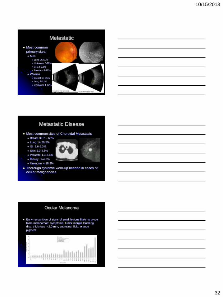
Metastatic
Most common
primary sites:
Men
Lung 26-50%
Unknown 6-29%
GI 3.5-12%
Prostate 3-12%
Women
Breast 68-85%
Lung 8-12%
Unknown 4-12%
Info_B:HMHMASS G=79dB DYN=60dB
+Dist~11.4mm <>Dist Off Info_B:VMAPMASS G=79dB
+Dist~3.3mm <>Dist Off
Metastatic Disease
Most common sites of Choroidal Metastasis
Breast 39.7 – 65%
Lung 14-29.5%
GI 2.6-6.3%
Skin 2.0-4.5%
Prostate 1.3-3.6%
Kidney .9-4.0%
Unknown 4-18.3%
Thorough systemic work-up needed in cases of
ocular malignancies
Ocular Melanoma
Early recognition of signs of small lesions likely to prove
to be melanomas: symptoms, tumor margin touching
disc, thickness > 2.0 mm, subretinal fluid, orange
pigment
10/15/2013
33
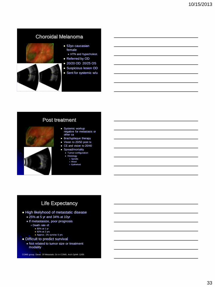
Choroidal Melanoma
53yo caucasian
female
HTN and hypecholest.
Referred by OD
20/20 OD 20/25 OS
Suspicious lesion OD
Sent for systemic w/u
Post treatment
Systemic workup negative for metastasis or other ca
Brachyplaque therapy
Vision to 20/50 post tx
CE and vision to 20/40
Spread/mortality Tumor configuration
Histology
Spindle
Mixed
Epithelioid
Life Expectancy
High likelyhood of metastatic disease
25% at 5 yr and 34% at 10yr
If metastasize, poor prognosis Death rate of:
80% at 1 yr
92% at 2 yrs
Approx. 1% survive 5 yrs
Difficult to predict survival
Not related to tumor size or treatment modality
COMS group. Devel. Of Metastatic Dz in COMS. Arch Ophth 12/05

10/15/2013
34
Familial Adenomatous Polyposis
(FAP)
Rare: 2.3-3.2/100,000
Avg onset at 16yo
Without Colectomy, colon cancer inevitable
Autosomal dominant 75-80% have affected parent
78-88% have 4 or more fundus lesions
Retinal Consult 37 year old female
Vision 20/40 OS
No pain or pain with
movements
No APD
Normal Anterior
segment exam
Recent ER visit for
LOV
Then went to Ophthal.
Either MS, Diabetes or
nothing…wait and see
Further History:
Previous episodes of vision “Graying”
Unable to take hot showers
Electric like impulses through arms/back
Numbness in fingers
Clumsy walking
Decreased contrast/color OS
10/15/2013
35
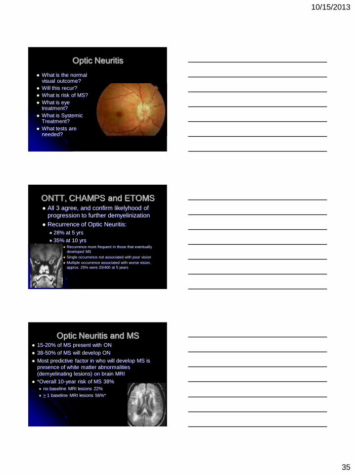
Optic Neuritis
What is the normal visual outcome?
Will this recur?
What is risk of MS?
What is eye treatment?
What is Systemic Treatment?
What tests are needed?
ONTT, CHAMPS and ETOMS All 3 agree, and confirm likelyhood of
progression to further demyelinization
Recurrence of Optic Neuritis:
28% at 5 yrs
35% at 10 yrs Recurrence more frequent in those that eventually
developed MS
Single occurrence not associated with poor vision
Multiple occurrence associated with worse vision,
approx. 25% were 20/400 at 5 years
Optic Neuritis and MS 15-20% of MS present with ON
38-50% of MS will develop ON
Most predictive factor in who will develop MS is
presence of white matter abnormalities
(demyelinating lesions) on brain MRI
*Overall 10-year risk of MS 38%
no baseline MRI lesions 22%
> 1 baseline MRI lesions 56%*

10/15/2013
36
Treatment?
Oral steroids alone not affective
At 3 years, MS risk for IV vs PO vs Placebo 17% vs 21% vs 25%
IV methylprednisilone x 3 days followed by 11 days of oral pred.
Treatment with IMA? 12,000/yr with wkly/daily
injections and side effects
Interferon Retinopathy1
*NEW ORAL TX!!!**
Retinopathy of MS on Interferon. Saito.et al. MS: April 07
OCT: Predictive value
RNFL thickness may be able to be
predictive as to MS or level of vision loss
RNFL thickness signif. reduced in MS eyes
Disease free thickness>MS = fellow of ON >
MS w ON
Lower visual function with less RNFL
Avg. RNFL thickness declined with
increased neuro. impair. and disability
Fisher et al. RNFL in MS. Ophthal 2/06
Lattice Degeneration…
30 year old male referred for evaluation of
lattice degeneration and atrophic holes
Very healthy athlete, no medications
Exam findings:
VA: 20/20 OU
Anterior segment healthy
Peripheral retina: Lattice with holes
Posterior pole..

10/15/2013
37
Plaques
Several Hollenhorst
Plaques
Further questioning: No
cardiovascular or carotid
disease
Treatment: Laser to
lattice and holes
Referral: To PCP for
cardio and carotid work-
up
Pt lost to follow up
Hollenhorst Plaques
Landmark article in AJO January 1973
Carotid disease and heart disease about
same incidence at time of plaque seen
Patients 4x more likely to die of MI than CVA
If embolus, mortality 54% over 7 years (2x
that of age matched norms)
Referral to PCP or internist
Artery Occlusion
Historically felt than 5% develop NV Duker et al 1991: 18.2% NVI, 15.2% NVG
Hayreh: mean to NVI 5.5 weeks Can develop NVI without carotid disease
Inner retinal cell death, but outer layers spared, and have high O2 demand
Treatment PRP when NVI
Acute treatment AC paracentesis, massage, carbogen…
Accupunture1: marked visual improvement in 25%
TPA (EAGLE study in Europe)
Referral to PCP or internist for treatment of underlying systemic disease
Article in Sept 06 AJO by S.S. Hayreh
1. Zheng J, Chen Z, Wang W, Xia S: Acupuncture in retinal arterial occlusion. Chin Med J (Engl) 94: 175, 1981

10/15/2013
38
Just last month
42yo healthy
Caucasian female
Work-in appt for
“flashes in vision”
1 mo ago exam,
completely normal
exam
Last month
What is it??
Normal CBC, PT, PTT, ANA, SED, CRP, B12, A1c,
Ferritin,VWF, factor 5, high LDL and Cholesterol, BP
118/84
Is AMD strictly an ocular
disease with no systemic
associations? NO
Several different theories and factors that
point to AMD being systemically related
“Systemic” treatments may be beneficial
Nutrition modification is an easy way to
treat systemically

10/15/2013
39
Remember Pablo….Vision is
important Can we allow our patients to see like
this…regardless of ocular pathology?
So now you are ready to “treat”
systemic disease, but….. What is the most important thing we can
do for our patients (in their “eyes”)
CORRECT VISION!
That is why they come to us
Majority of vision impairment in diabetes is from
lack of refraction!1,2
Practice the “Optometric Model”
Combining medical and optical “treatment”
1. Klein et al. VI Prevalence (WESDR) Ophth. 10/09. 2. Zhang et al. DM and VI. Arch of
Ophth. 10/09.
Thank You
Online Resources
www.theretinaexchange.com
www.retinalphysician.com
www.pubmed.com
www.optometricretinasociety.org
www.optos.com