Embed Size (px)
Citation preview

PRIMARY LEIOMYOSARCOMA OF IVC
A RARE ENTITY
SURGERY UNIT- II

60 year old female presented with six months history of,
vague right loin pain
Loss of weight and appetite-10kg in
6 months
weakness

No h/o hematuria
No h/o fever
No h/o altered bowel habits
No h/o walking difficulties
H/o B/L knee replacement for arthritis 2years back.
H/o lap.cholecystectomy -one year back

Moderately built.
pale.
Pedal Edema +
P/A
Few dilated superficial veins over lower abdomen and upper thighs.
no lump found.

Hb – 9.7g/l
Tlc -8700
Plt -124000
Urea -45
Cr -1.1mg/dl
Sr .bil-0.9mg /dl
Sgot/pt -27/23
Sr.Alp -98 iu /dl
Coagulation profile-normal


Usg abdomen
6*7 cm heteroechoic lobulatedretroperitoneal mass lesion adjacent to renal part of IVC on right side with partial luminal compression
? RP mass with IVC thrombus
? IVC tumor
Suggested Usg doppler

IVC doppler revealed compressed juxta renal IVC with reduced upward flow
?thrombus
?tumor
CECT abdomen was advised.


Malignant spindle cell tumor
IHC-Positive for desmin and smooth muscle actin and negative for s-100,c-kit
? Leiomyosarcoma-retroperitoneum.

Exploratory laparotomy and proceed
Intra operatively :
Hard infiltrating tumor arising from juxtarenal ivc which was extending about 5cm above and 3cm below the Rt renal vein.
Rt renal vessels were completely encased by the tumor.
Aorta free


The liver was mobilized to expose the sub hepatic part of the IVC to achieve proximal clearance of the tumor.
The infra-diaphragmatic aorta was slung and a clamp was placed at the level superiorly to control inflow.
Left renal vein was ligated and transected from IVC.
The patient was heparinised and the tumor resecteden block along with a right nephrectomy after applying proximal and distal clamps to the IVC

A Dacron 18mm interposition graft was used to reconstruct the IVC.
The left renal vein was re-implanted in an end to side manner into the Dacron graft.


Stable, Good urine output
NPO for 4days
On anticoagulation therapy
Discharged on day 15.


Doxorubicin based chemotherapy.
Radiotherapy also planned.
Now on 6 months
follow-up without any complaints.


• Extremely rare tumor with fewer then 200 cases reported in literature since 1871 *
• First reported case : Perl in 1871• Documented in the surgical literature mostly
as case reports rather than case series.• Invariably malignant• Prognosis depends on early diagnosis and
management
* Mingoli A, Cavallaro A, Sapienza P, Di Marzo L, Feldhaus RJ, Cavallari N:International registry of inferior vena cava leiomyosarcoma: analysis of a world series on 218 patients. Anticancer Res 1996, 16(5B):3201-3205.

Origin – arises from the smooth muscle cells of the media of the cava
Venous leiomyosarcoma occur 5 times more often than arterial ones
Among all the veins 50% originating in the IVC.


Encapsulated, consisting of lobulated whorls
Histologically demonstrates spindle shaped bundles of cells with high mitotic activity
Positive staining for desmin,HHF35,vimentin and smooth muscle actin

PRIMARY TUMOR
Arises directly from the vessel wall.
SECONDARY TUMOR
Arises from adjacent retroperitoneal
structures and invades IVC.
RCC
Pheochromocytoma
Testicular tumor etc

Depending on site of IVC involved:
Supra hepatic IVC tumor (24%)
Arises from above hepatic vein origin
to right atrium.
Very difficult to operate
Poor prognosis.

Retro hepatic IVC tumor (42% ) :
Arises from middle part of IVC (from
renal vein origin to hepatic vein
origin).
Most common site of primary tumor
Lower IVC tumor (34%)
Arises from lower part of IVC
(below renal vein draining into IVC )

Pathologically *
(1) Primarily Exophytic tumor – 62%(2)Primarily Intraluminal tumor-5%(3) Combined -33%
(difficult to differentiate from secondary tumor arising from retro peritoneum and invading IVC)
*Hartman DS, Hayes WS, Choyke PL,Tibbets GP. From the archives of the AFIP. Leiomyosarcoma of the retroperitoneum and inferior vena cava: radiologic-pathologic correlation. Radio graphics 1992;12:1203–1220.

Usually asymptomatic in early stage thus delaying diagnosis.
More common in females M:F=1:5
Usually presents at sixth- seventh decade

Symptoms and signs are nonspecific-
• Abdominal Pain (66%)
• Abdominal mass (48%)
• Lower limb edema (39%)
• Budd – Chiari syndrome (22%)
• Others- fever, weakness , anorexia , nocturnal sweating and dyspnoea
Bower TC, Stanson A. Diagnosis and management of tumors of the inferior vena cava. Vascular surgery, 5thed. Philadelphia: WB Saunders, 2000: 2077–2091.

Upper IVC involvement=Budd chiarisyndrome
Middle IVC involvement=Nephrotic syndrome
Infrarenal involvement=Lower extremity edema

Mainly haematogenous
To liver,lung and brain
In advanced stage may spread through lymphatics

ULTRASONOGRAPHY
Initial screening test
Doppler USG helps to assess vascularity oftumor
It also helps to differentiate IVC tumorfrom Intraluminal thrombus.

Fig 1- USG showing Intraluminal IVC tumor
Fig-2 colour doppler usg showing slow resistance flow in intraluminal tumor
Fig 1- USG showing Intraluminal IVC tumor
Fig-2 colour doppler usgshowing slow resistance flow in intraluminal tumor


It is useful screening test to diagnose IVC tumor.
CT guided biopsy can be taken from exophytic component.
However sometimes it becomes difficult on CT to differentiate between primary and secondary IVC tumor.

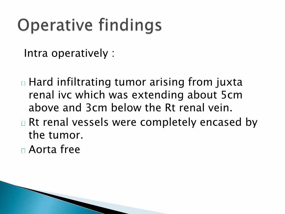
Fig- Contrast CT scan showing heterogenouslyenhanced mass in the IVC.

Advantage includes-
• Allows multi-planer imaging
• This gives highly accurate assessment of relationship of tumor with adjacent structures.
• Vessel patency can be assessed using flow void or flow enhancement properties.


The cephalo caudalextent of tumor withinthe cava, and thuspotential resectability canalso be determined.
Fat suppression andGadolinium enhancementfuther increases qualityof MRI imaging.

Invasive technique
Intraluminal tumor – on Cavography, the IVC is dilated and the tumor is detected as a mass dilating and filling the lumen.
Exophytic tumor is detected as a mass surrounding the IVC that may protrude into the lumen.

Primary leiomyosarcoma of IVC are slow growing tumor and are invariably malignant.
Usually diagnosed late due to nonspecific symptoms.
Surgical resection is treatment of choice whenever tumor is resectable.
Even if unresectable , surgery gives best palliative treatment.
Even on recurrence sx is the only option
Chemotherapy and radiotherapy- role is controversial .

Limited studies about surgical management and limited data on long term survival after surgery.
Complete surgical resection with a one cm of tumor free margin is considered treatment of choice
Hemant D, Krantikumar R, Amita J, Chawla A, Ranjeet N: Primary Leiomyosarcoma of inferior vena cava, a rare entity: Imaging features. Austral as Radiol 2001, 45(4):448-451

Ligation of IVC associated with severe limb edema due to disruption of collaterals.
Current recommendation is to use prosthetic graft for reconstruction of the IVC .
Ring enforced PTFE or Dacron graft are commonly used Prosthesis for IVC reconstruction.
Sarkar R, Eilber FR, Gelabert HA, Quinones-Baldrich WJ. Prosthetic
replacement of the inferior vena cava for malignancy. J Vasc Surg 1998; 28: 75–83.

International registry of IVC leiomyosarcoma analyzed 218 patients.
A radical tumor resection was undertaken in 134 (61.5%) patients, 26 (11.9%) had a palliative resection, and 58 (26.6%) were inoperable.
Radical tumor resection was associated with better 5- and 10-year survival rates (49.4% and 29.5%) when compared to patients undergoing palliative resection or those who were inoperable
Mingoli A , Cavallaro A, Sapienza P, Di Marzo L, Feldhaus RJ, CavallariN . International registry of inferior vena cava Leiomyosarcoma. Analysis of World Series of 218 patients. Anticancer Res 1996;16: 3201-3206

Hollenbeck et al reported 25 patients ofprimary IVC tumor treated between 1982and 2002.
Study showed that patients undergoingcomplete resection had 3- and 5-yearsurvival rates of 76% and 33% respectively.
Hollenbeck ST, Grobmeyer SR, Kent KC, Brennan MF: Surgicaltreatment and outcomes of patients with primary inferior vena cavaLeiomyosarcoma. J Am Coll Surg 2003, 197(4):575-579.

Role still controversial
Adriamycin/ Ifosfamide based regimen are commonly used.
However no case series study to suggest its exact

Less information about its role
Some study suggest radiotherapy reduces the recurrence rate.

Poor Prognostic Factors:
• Upper IVC involvement
• Lower limb edema
• Budd- chiari syndrome
• IVC occlusion
• Distant metastasis

Primary leiomyosarcoma a rare tumor which are invariably malignant.
Slow growing tumor with delayed nonspecific presentation.
MRI is investigation of choice
Sometime difficult to differentiate between primary and secondary IVC tumor
Surgery is treatment of choice , even for palliation .
Role of chemotherapy and radiotherapy controversial.

THANK YOU !