Embed Size (px)
Citation preview


BIOPHARMACEUTICALSBIOCHEMISTRY AND BIOTECHNOLOGY
Second Edition
Gary Walsh
Industrial Biochemistry ProgrammeCES Department
University of Limerick, Ireland


BIOPHARMACEUTICALSBIOCHEMISTRY AND BIOTECHNOLOGY
Second Edition

BIOPHARMACEUTICALSBIOCHEMISTRY AND BIOTECHNOLOGY
Second Edition
Gary Walsh
Industrial Biochemistry ProgrammeCES Department
University of Limerick, Ireland
First Edition 1998 u John Wiley & Sons, Ltd
Copyright u 2003 John Wiley & Sons Ltd, The Atrium, Southern Gate, Chichester,West Sussex PO19 8SQ, England
Telephone (+44) 1243 779777
E-mail (for orders and customer service enquiries): [email protected] our Home Page on www.wileyeurope.com or www.wiley.com
All Rights Reserved. No part of this publication may be reproduced, stored in a retrieval system ortransmitted in any form or by any means, electronic, mechanical, photocopying, recording, scanning orotherwise, except under the terms of the Copyright, Designs and Patents Act 1988 or under the terms of alicence issued by the Copyright Licensing Agency Ltd, 90 Tottenham Court Road, London W1T 4LP, UK,without the permission in writing of the Publisher. Requests to the Publisher should be addressed to thePermissions Department, John Wiley & Sons Ltd, The Atrium, Southern Gate, Chichester, West SussexPO19 8SQ, England, or emailed to [email protected], or faxed to (+44) 1243 770620.
This publication is designed to provide accurate and authoritative information in regard to the subjectmatter covered. It is sold on the understanding that the Publisher is not engaged in rendering professionalservices. If professional advice or other expert assistance is required, the services of a competentprofessional should be sought.
Other Wiley Editorial Offices
John Wiley & Sons Inc., 111 River Street, Hoboken, NJ 07030, USA
Jossey-Bass, 989 Market Street, San Francisco, CA 94103-1741, USA
Wiley-VCH Verlag GmbH, Boschstr. 12, D-69469 Weinheim, Germany
John Wiley & Sons Australia Ltd, 33 Park Road, Milton, Queensland 4064, Australia
John Wiley & Sons (Asia) Pte Ltd, 2 Clementi Loop #02-01, Jin Xing Distripark, Singapore 129809
John Wiley & Sons Canada Ltd, 22 Worcester Road, Etobicoke, Ontario, Canada M9W 1L1
Wiley also publishes its books in a variety of electronic formats. Some content that appears in print maynot be available in electronic books.
British Library Cataloguing in Publication Data
A catalogue record for this book is available from the British Library
ISBN 0 470 84326 8 (ppc)ISBN 0 470 84327 6 (pbk)
Typeset by Dobbie Typesetting Ltd, Tavistock, DevonPrinted and bound in Great Britain by Antony Rowe Ltd, Chippenham, WiltsThis book is printed on acid-free paper responsibly manufactured from sustainable forestry in which atleast two trees are planted for each one used for paper production.

I dedicate this book to my beautiful son Shane, born during therevision of Chapter 6. I include his photograph in the hope that aHollywood producer, looking for a child film star, will spot it andimmediately offer to make us suitably rich. In the future, I also hopeto use it to embarrass him during his teenage years, by showing it to
all his cool, sophisticated friends.

Contents
Preface xvii
Chapter 1 Pharmaceuticals, biologics and biopharmaceuticals 1
Introduction to pharmaceutical products 1Biopharmaceuticals and pharmaceutical biotechnology 1History of the pharmaceutical industry 3The age of biopharmaceuticals 5Biopharmaceuticals: current status and future prospects 8Traditional pharmaceuticals of biological origin 12Pharmaceuticals of animal origin 13
The sex hormones 14The androgens 14Oestrogens 15Progesterone and progestogens 17
Corticosteroids 19Catecholamines 21Prostaglandins 23
Pharmaceutical substances of plant origin 27Alkaloids 28
Atropine and scopalamine 28Morphine and cocaine 29Additional plant alkaloids 30Ergot alkaloids 30
Flavonoids, xanthines and terpenoids 30Cardiac glycosides and coumarins 33Aspirin 33
Pharmaceutical substances of microbial origin 33The macrolides and ansamycins 38Peptide and other antibiotics 39
Conclusion 39Further reading 40
Chapter 2 The drug development process 43
Drug discovery 44The impact of genomics and related technologies upon drug discovery 45
Gene chips 47Proteomics 49Structural genomics 50Pharmacogenetics 51
Plants as a source of drugs 52Microbial drugs 53Rational drug design 54Combinatorial approaches to drug discovery 56Initial product characterization 57
Patenting 57What is a patent and what is patentable? 57Patent types 62The patent application 63Patenting in biotechnology 64
Delivery of biopharmaceuticals 66Oral delivery systems 66Pulmonary delivery 67Nasal, transmucosal and transdermal delivery systems 68
Pre-clinical trials 69Pharmacokinetics and pharmacodynamics 69Toxicity studies 71Reproductive toxicity and teratogenicity 71Mutagenicity, carcinogenicity and other tests 72
Clinical trials 73Clinical trial design 75Trial size and study population 75Randomized control studies 76Additional trial designs 76
The role and remit of regulatory authorities 78The Food and Drug Administration 78
The investigational new drug application 80The new drug application 82
European regulations 84National regulatory authorities 84The EMEA and the new EU drug approval systems 85The centralized procedure 86Mutual recognition 88
Drug registration in Japan 88World harmonization of drug approvals 89
Conclusion 89Further reading 89
Chapter 3 The drug manufacturing process 93
International pharmacopoeia 93Martindale, the Extra Pharmacopoeia 94
Guides to good manufacturing practice 94
viii CONTENTS
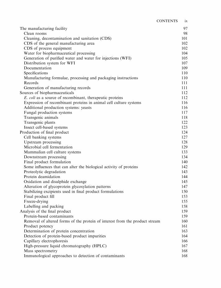
The manufacturing facility 97Clean rooms 98Cleaning, decontamination and sanitation (CDS) 101CDS of the general manufacturing area 102CDS of process equipment 102Water for biopharmaceutical processing 104Generation of purified water and water for injections (WFI) 105Distribution system for WFI 107Documentation 109Specifications 110Manufacturing formulae, processing and packaging instructions 110Records 111Generation of manufacturing records 111
Sources of biopharmaceuticals 112E. coli as a source of recombinant, therapeutic proteins 112Expression of recombinant proteins in animal cell culture systems 116Additional production systems: yeasts 116Fungal production systems 117Transgenic animals 118Transgenic plants 122Insect cell-based systems 123
Production of final product 124Cell banking systems 127Upstream processing 128Microbial cell fermentation 129Mammalian cell culture systems 133Downstream processing 134Final product formulation 140Some influences that can alter the biological activity of proteins 142Proteolytic degradation 143Protein deamidation 144Oxidation and disulphide exchange 145Alteration of glycoprotein glycosylation patterns 147Stabilizing excipients used in final product formulations 150Final product fill 153Freeze-drying 155Labelling and packing 158
Analysis of the final product 159Protein-based contaminants 159Removal of altered forms of the protein of interest from the product stream 160Product potency 161Determination of protein concentration 163Detection of protein-based product impurities 164Capillary electrophoresis 166High-pressure liquid chromatography (HPLC) 167Mass spectrometry 168Immunological approaches to detection of contaminants 168
CONTENTS ix
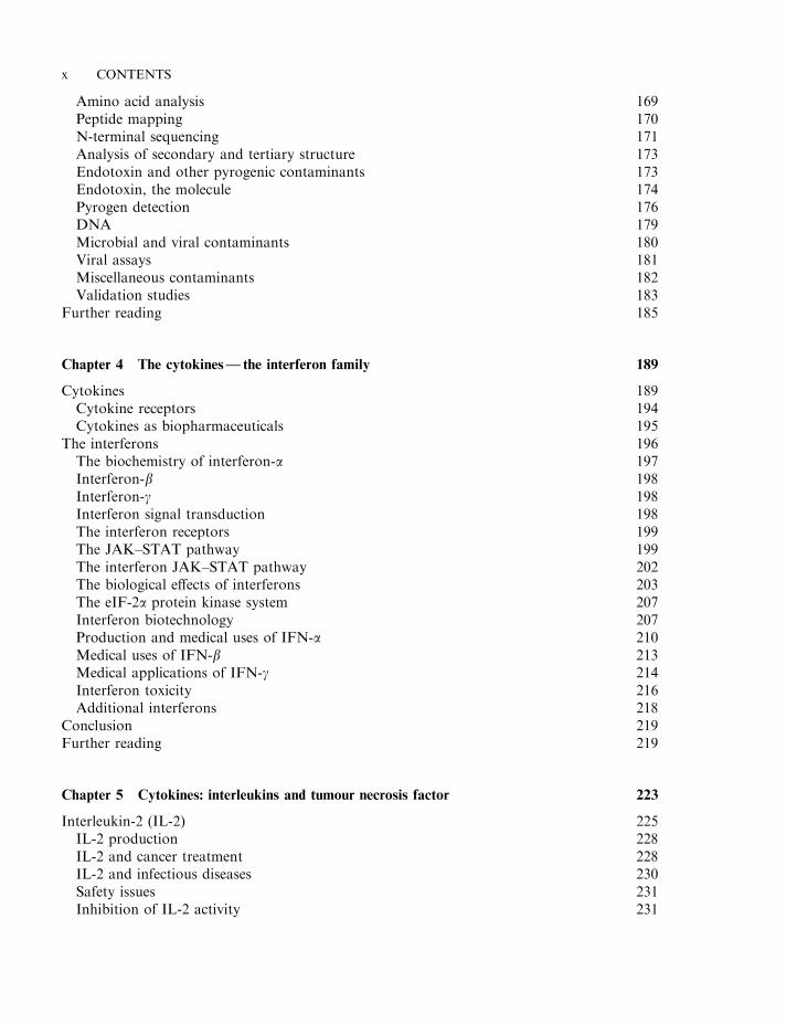
Amino acid analysis 169Peptide mapping 170N-terminal sequencing 171Analysis of secondary and tertiary structure 173Endotoxin and other pyrogenic contaminants 173Endotoxin, the molecule 174Pyrogen detection 176DNA 179Microbial and viral contaminants 180Viral assays 181Miscellaneous contaminants 182Validation studies 183
Further reading 185
Chapter 4 The cytokines— the interferon family 189
Cytokines 189Cytokine receptors 194Cytokines as biopharmaceuticals 195
The interferons 196The biochemistry of interferon-a 197Interferon-b 198Interferon-g 198Interferon signal transduction 198The interferon receptors 199The JAK–STAT pathway 199The interferon JAK–STAT pathway 202The biological effects of interferons 203The eIF-2a protein kinase system 207Interferon biotechnology 207Production and medical uses of IFN-a 210Medical uses of IFN-b 213Medical applications of IFN-g 214Interferon toxicity 216Additional interferons 218
Conclusion 219Further reading 219
Chapter 5 Cytokines: interleukins and tumour necrosis factor 223
Interleukin-2 (IL-2) 225IL-2 production 228IL-2 and cancer treatment 228IL-2 and infectious diseases 230Safety issues 231Inhibition of IL-2 activity 231
x CONTENTS
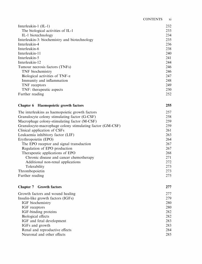
Interleukin-1 (IL-1) 232The biological activities of IL-1 233IL-1 biotechnology 234
Interleukin-3: biochemistry and biotechnology 235Interleukin-4 236Interleukin-6 238Interleukin-11 240Interleukin-5 241Interleukin-12 244Tumour necrosis factors (TNFs) 246TNF biochemistry 246Biological activities of TNF-a 247Immunity and inflammation 248TNF receptors 249TNF: therapeutic aspects 250
Further reading 252
Chapter 6 Haemopoietic growth factors 255
The interleukins as haemopoietic growth factors 257Granulocyte colony stimulating factor (G-CSF) 258Macrophage colony-stimulating factor (M-CSF) 259Granulocyte-macrophage colony stimulating factor (GM-CSF) 259Clinical application of CSFs 261Leukaemia inhibitory factor (LIF) 263Erythropoietin (EPO) 264The EPO receptor and signal transduction 267Regulation of EPO production 267Therapeutic applications of EPO 268
Chronic disease and cancer chemotherapy 271Additional non-renal applications 272Tolerability 273
Thrombopoietin 273Further reading 275
Chapter 7 Growth factors 277
Growth factors and wound healing 277Insulin-like growth factors (IGFs) 279IGF biochemistry 280IGF receptors 280IGF-binding proteins 282Biological effects 282IGF and fetal development 283IGFs and growth 283Renal and reproductive effects 284Neuronal and other effects 285
CONTENTS xi
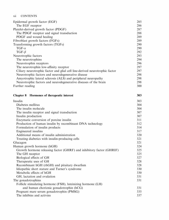
Epidermal growth factor (EGF) 285The EGF receptor 286
Platelet-derived growth factor (PDGF) 287The PDGF receptor and signal transduction 288PDGF and wound healing 289
Fibroblast growth factors (FGFs) 289Transforming growth factors (TGFs) 290
TGF-a 290TGF-b 292
Neurotrophic factors 293The neurotrophins 294Neurotrophin receptors 296The neurotrophin low-affinity receptor 297Ciliary neurotrophic factor and glial cell line-derived neurotrophic factor 297Neurotrophic factors and neurodegenerative disease 298Amyotrophic lateral sclerosis (ALS) and peripheral neuropathy 298Neurotrophic factors and neurodegenerative diseases of the brain 298
Further reading 300
Chapter 8 Hormones of therapeutic interest 303
Insulin 303Diabetes mellitus 304The insulin molecule 304The insulin receptor and signal transduction 307Insulin production 307Enzymatic conversion of porcine insulin 311Production of human insulin by recombinant DNA technology 312Formulation of insulin products 314Engineered insulins 317Additional means of insulin administration 320Treating diabetics with insulin-producing cells 321
Glucagon 321Human growth hormone (hGH) 324
Growth hormone releasing factor (GHRF) and inhibitory factor (GHRIF) 325The GH receptor 325Biological effects of GH 327Therapeutic uses of GH 328Recombinant hGH (rhGH) and pituitary dwarfism 328Idiopathic short stature and Turner’s syndrome 330Metabolic effects of hGH 330GH, lactation and ovulation 331
The gonadotrophins 331Follicle stimulating hormone (FSH), luteinizing hormone (LH)
and human chorionic gonadotrophin (hCG) 331Pregnant mare serum gonadotrophin (PMSG) 335The inhibins and activins 337
xii CONTENTS
LHRH and regulation of gonadotrophin production 338Medical and veterinary applications of gonadotrophins 339Sources and medical uses of FSH, LH and hCG 340Recombinant gonadotrophins 342Veterinary uses of gonadotrophins 344Gonadotrophin releasing hormone (GnRH) 345Additional recombinant hormones now approved 345
Conclusions 348Further reading 348
Chapter 9 Blood products and therapeutic enzymes 351
Disease transmission 351Whole blood 353Platelets and red blood cells 353Blood substitutes 353Dextrans 354Albumin 355Gelatin 357Oxygen-carrying blood substitutes 357
Haemostasis 358The coagulation pathway 358Terminal steps of coagulation pathway 361Clotting disorders 365Factor VIII and haemophilia 366
Production of factor VIII 368Factors IX, VIIa and XIII 371Anticoagulants 372
Heparin 372Vitamin K antimetabolites 375Hirudin 375
Antithrombin 379Thrombolytic agents 380
Tissue plasminogen activator (tPA) 381First-generation tPA 383Engineered tPA 383
Streptokinase 385Urokinase 386Staphylokinase 386a1-Antitrypsin 388
Enzymes of therapeutic value 389Asparaginase 390DNase 392Glucocerebrosidase 393a-Galactosidase and urate oxidase 395Superoxide dismutase 397
CONTENTS xiii
Debriding agents 397Digestive aids 398Lactase 400
Further reading 400
Chapter 10 Antibodies, vaccines and adjuvants 403
Polyclonal antibody preparations 403Anti-D immunoglobulin 406Normal immunoglobulins 407Hepatitis B and tetanus immunoglobulin 407Snake and spider antivenins 408
Monoclonal antibodies 409Production of monoclonals via hybridoma technology 411Antibody screening: phage display technology 412Therapeutic application of monoclonal antibodies 414Tumour immunology 415
Antibody-based strategies for tumour detection/destruction 417Drug-based tumour immunotherapy 424First-generation anti-tumour antibodies: clinical disappointment 426Tumour-associated antigens 426
Antigenicity of murine monoclonals 428Chimaeric and humanized antibodies 429Antibody fragments 432Additional therapeutic applications of monoclonal antibodies 433
Cardiovascular and related disease 433Infectious diseases 433Autoimmune disease 434Transplantation 434
Vaccine technology 435Traditional vaccine preparations 436
Attenuated, dead or inactivated bacteria 438Attenuated and inactivated viral vaccines 439Toxoids, antigen-based and other vaccine preparations 440
The impact of genetic engineering on vaccine technology 441Peptide vaccines 444Vaccine vectors 445
Development of an AIDS vaccine 447Difficulties associated with vaccine development 450
AIDS vaccines in clinical trials 450Cancer vaccines 452Recombinant veterinary vaccines 452Adjuvant technology 453Adjuvant mode of action 455
Mineral-based adjuvants 455Oil-based emulsion adjuvants 455
xiv CONTENTS
Bacteria/bacterial products as adjuvants 457Additional adjuvants 458
Further reading 460
Chapter 11 Nucleic acid therapeutics 463
Gene therapy 463Basic approach to gene therapy 464Some additional questions 467Vectors used in gene therapy 468
Retroviral vectors 468Additional viral-based vectors 472Manufacture of viral vectors 474Non-viral vectors 476
Manufacture of plasmid DNA 480Gene therapy and genetic disease 482Gene therapy and cancer 485Gene therapy and AIDS 486Gene-based vaccines 488Gene therapy: some additional considerations 488
Anti-sense technology 488Anti-sense oligonucleotides 490Uses, advantages and disadvantages of ‘oligos’ 491Delivery and cellular uptake of oligonucleotides 493Manufacture of oligonucleotides 493Vitravene, an approved antisense agent 494Antigene sequences and ribozymes 494
Conclusion 495Further reading 496
Appendix 1 Biopharmaceuticals thus far approved in the USA or European Union 499
Appendix 2 Some Internet addresses relevant to the biopharmaceutical sector 509
Appendix 3 Two selected monographs reproduced from the European Pharmacopoeiawith permission from the European Commission:I. Products of recombinant DNA technology 515II. Interferon a-2 concentrated solution 520
Appendix 4 Manufacture of biological medicinal products for human use. (Annex 2from The Rules Governing Medicinal Products in the European Community,Vol. 4, Good Manufacturing Practice for Medicinal Products) 527
Index 533
CONTENTS xv

Preface
Advances in our understanding of the molecular principles underlining both health and diseasehas revealed the existence of many regulatory polypeptides of significant medical potential. Thefact that such polypeptides are produced naturally within the body only in minute quantitiesinitially precluded their large-scale medical application. The development in the 1970s of thetwin techniques of genetic engineering and hybridoma technology marked the birth of themodern biotech era. These techniques facilitate the large-scale production of virtually anyprotein, and proteins of medical interest produced by these methodologies have been coined‘biopharmaceuticals’. More recent developments in biomedical research highlights the clinicalpotential of nucleic acid-based therapeutic agents. Gene therapy and anti-sense technology arelikely to become a medical reality within a decade. The term ‘biopharmaceutical’ now alsoincorporates the polynucleotide sequences utilized for such purposes.
This book attempts to provide a balanced overview of the biopharmaceutical industry, notonly in terms of categorizing the products currently available, but also illustrating how thesedrugs are produced and brought to market. Chapter 1 serves as an introduction to the topic, andalso focuses upon several ‘traditional’ pharmaceutical substances isolated (initially at least) frombiological sources. This serves as a backdrop for the remaining chapters, which focus almostexclusively upon recently developed biopharmaceutical products. The major emphasis is placedupon polypeptide-based therapeutic agents, while the potential of nucleic acid-based drugs isdiscussed in the final chapter.
In preparing the latest edition of this textbook, I highlight the latest developments within thesector, provide a greater focus upon actual commercial products thus far approved and howthey are manufactured, and I include substantial new sections detailing biopharmaceutical drugdelivery and how advances in genomics and proteomics will likely impact upon (bio)pharma-ceutical drug development.
The major target audience is that of advanced undergraduates or postgraduate studentspursuing courses in relevant aspects of the biological sciences. The book should proveparticularly interesting to students undertaking programmes in biotechnology, biochemistry, thepharmaceutical sciences, medicine or any related biomedical subject. A significant additionaltarget audience are those already employed in the (bio)pharmaceutical sector, who wish to gaina better overview of the industry in which they work.
The successful completion of this text has been made possible by the assistance of severalpeople to whom I owe a depth of gratitude. Chief amongst these is Sandy Lawson, who appearsto be able to read my mind as well as my handwriting. Thank you to Nancy, my beautiful wife,who suffered most from my becoming a social recluse during the preparation of this text. Thankyou, Nancy, for not carrying out your threat to burn the manuscript on various occasions, andfor helping with the proof-reading. I am also very grateful to the staff of John Wiley and SonsLtd for the professionalism and efficiency they exhibited while bringing this book through the

publication process. The assistance of companies who provided information and photographsfor inclusion in the text is also gratefully acknowledged, as is the cooperation of those publisherswho granted me permission to include certain copyrighted material. Finally, a word ofappreciation to all my colleagues at Limerick, who continue to make our university such a greatplace to work.
Gary WalshLimerick, November 2002
xviii PREFACE
Chapter 1
Pharmaceuticals, biologics
and biopharmaceuticals
INTRODUCTION TO PHARMACEUTICAL PRODUCTS
Pharmaceutical substances form the backbone of modern medicinal therapy. Most traditionalpharmaceuticals are low molecular mass organic chemicals (Table 1.1). Although some (e.g.aspirin) were originally isolated from biological sources, most are now manufactured by directchemical synthesis. Two types of manufacturing companies thus comprise the ‘traditional’pharmaceutical sector; the chemical synthesis plants, which manufacture the raw chemicalingredients in bulk quantities, and the finished product pharmaceutical facilities, which purchasethese raw bulk ingredients, formulate them into final pharmaceutical products, and supply theseproducts to the end-user.
In addition to chemical-based drugs, a range of pharmaceutical substances (e.g. hormonesand blood products) are produced by or extracted from biological sources. Such products, somemajor examples of which are listed in Table 1.2, may thus be described as products ofbiotechnology. In some instances, categorizing pharmaceuticals as products of biotechnology orchemical synthesis becomes somewhat artificial, e.g. certain semi-synthetic antibiotics areproduced by chemical modification of natural antibiotics produced by fermentationtechnology.
BIOPHARMACEUTICALS AND PHARMACEUTICALBIOTECHNOLOGY
Terms such as ‘biologic’, ‘biopharmaceutical’ and ‘products of pharmaceutical biotechnology’or ‘biotechnology medicines’ have now become an accepted part of the pharmaceuticalliterature. However, these terms are sometimes used interchangeably and can mean differentthings to different people.
While it might be assumed that ‘biologic’ refers to any pharmaceutical product produced bybiotechnological endeavour, its definition is more limited. In pharmaceutical circles, ‘biologic’
Biopharmaceuticals: Biochemistry and Biotechnology, Second Edition. Gary WalshJohn Wiley & Sons Ltd: ISBN 0 470 84326 8 (ppc), ISBN 0 470 84327 6 (pbk)
generally refers to medicinal products derived from blood, as well as vaccines, toxins andallergen products. Thus, some traditional biotechnology-derived pharmaceutical products (e.g.hormones, antibiotics and plant metabolites) fall outside the strict definition.
The term ‘biopharmaceutical’ was first used in the 1980s and came to describe a class oftherapeutic protein produced by modern biotechnological techniques, specifically via geneticengineering or (in the case of monoclonal antibodies) by hybridoma technology. This usageequated the term ‘biopharmaceutical’ with ‘therapeutic protein synthesized in engineered (non-naturally occurring) biological systems’. More recently, however, nucleic acids used forpurposes of gene therapy and antisense technology (Chapter 11) have come to the fore and theytoo are generally referred to as ‘biopharmaceuticals’. Moreover, several recently approvedproteins are used for in vivo diagnostic as opposed to therapeutic purposes. Throughout thisbook therefore, the term ‘biopharmaceutical’ refers to protein or nucleic acid basedpharmaceutical substances used for therapeutic or in vivo diagnostic purposes, which areproduced by means other than direct extraction from natural (non-engineered) biologicalsources (Tables 1.3 and 1.4).
As used herein, ‘biotechnology medicines’ or ‘products of pharmaceutical biotechnology’ areafforded a much broader definition. Unlike the term ‘biopharmaceutical’, the term
2 BIOPHARMACEUTICALS
Table 1.1. Some traditional pharmaceutical substances which are generally produced by direct chemicalsynthesis
Drug Molecular formula Molecular mass Therapeutic indication
Acetaminophen(paracetamol)
C8H9NO2 151.16 Analgesic
Ketamine C13H16ClNO 237.74 AnaestheticLevamisole C11H12N2S 204.31 AnthelminticDiazoxide C8H7ClN2O2S 230.7 Anti-hypertensiveAcyclovir C8H11N5O3 225.2 Anti-viral agentZidovudine C10H13N5O4 267.2 Anti-viral agentDexamethasone C22H29FO5 392.5 Anti-inflammatory and
immunosuppressive agentMisoprostol C22H38O5 382.5 Anti-ulcer agentCimetidine C10H16N6 252.3 Anti-ulcer agent
Table 1.2. Some pharmaceuticals which were traditionally obtained by direct extraction from biologicalsource material. Many of the protein-based pharmaceuticals mentioned below are now also produced bygenetic engineering
Substance Medical application
Blood products (e.g. coagulation factors) Treatment of blood disorders such as haemophilia A or BVaccines Vaccination against various diseasesAntibodies Passive immunization against various diseasesInsulin Treatment of diabetes mellitusEnzymes Thrombolytic agents, digestive aids, debriding agents
(i.e. cleansing of wounds)Antibiotics Treatment against various infectious agentsPlant extracts (e.g. alkaloids) Various, including pain relief
‘biotechnology’ has a much broader and long-established meaning. Essentially, it refers to theuse of biological systems (e.g. cells or tissues) or biological molecules (e.g. enzymes orantibodies) for or in the manufacture of commercial products. Therefore, the term is equallyapplicable to long-established biological processes, such as brewing, and more modernprocesses, such as genetic engineering. As such, the term ‘biotechnology medicine’ is definedhere as ‘any pharmaceutical product used for a therapeutic or in vivo diagnostic purpose, whichis produced in full or in part by either traditional or modern biotechnological means’. Suchproducts encompass, for example, antibiotics extracted from fungi, therapeutic proteinsextracted from native source material (e.g. insulin from pig pancreas) and products produced bygenetic engineering (e.g. recombinant insulin) (Tables 1.3 and 1.4).
HISTORY OF THE PHARMACEUTICAL INDUSTRY
The pharmaceutical industry, as we now know it, is barely 60 years old. From very modestbeginnings it has grown rapidly, reaching an estimated value of $100 billion by the mid-1980s.Its current value is likely double this figure or more. There are well in excess of 10 000pharmaceutical companies in existence, although only about 100 of these can claim to be of trueinternational significance. These companies manufacture in excess of 5000 individualpharmaceutical substances used routinely in medicine.
The first stages of development of the modern pharmaceutical industry can be traced back tothe turn of the twentieth century. At that time (apart from folk cures), the medical communityhad at their disposal only four drugs that were effective in treating specific diseases:
. Digitalis, extracted from foxglove, was known to stimulate heart muscle and hence was usedto treat various heart conditions.
. Quinine, obtained from the barks/roots of a plant (Cinchona sp.), was used to treatmalaria.
. Pecacuanha (active ingredient is a mixture of alkaloids), used for treating dysentery, wasobtained from the bark/roots of the plant species Cephaelis.
. Mercury, for the treatment of syphilis.
PHARMACEUTICALS, BIOLOGICS AND BIOPHARMACEUTICALS 3
Table 1.3. A summary of the definition of the terms ‘biologic’, ‘biopharmaceutical’ and ‘biotechnologymedicine’ as used throughout this book. Reprinted from European Journal of Pharmaceutical Sciences,vol 15, Walsh, Biopharmaceuticals and Biotechnology, p 135–138, s2002, with permission from ElsevierScience
Biopharmaceutical A protein or nucleic acid based pharmaceutical substance used fortherapeutic or in vivo diagnostic purposes, which is produced bymeans other than direct extraction from a native (non-engineered)biological source
Biotechnology medicine/product of pharmaceuticalbiotechnology
Any pharmaceutical product used for therapeutic or in vivo diagnosticpurposes, which is produced in full or in part by biotechnologicalmeans
Biologic A virus, therapeutic serum, toxin, antitoxin, vaccine, blood, bloodcomponent or derivative, allergenic product or analogous product,or arsphenamine or its derivatives or any other trivalent organicarsenic compound applicable to the prevention, cure or treatment ofdisease or conditions of human beings
The lack of appropriate safe and effective medicines contributed in no small way to the low lifeexpectancy characteristic of those times.
Developments in biology (particularly the growing realization of the microbiological basis ofmany diseases), as well as a developing appreciation of the principles of organic chemistry,helped underpin future innovation in the fledgling pharmaceutical industry. The successfulsynthesis of various artificial dyes, which proved to be therapeutically useful, led to theformation of pharmaceutical/chemical companies such as Bayer and Hoechst in the late 1800s,e.g. scientists at Bayer succeeded in synthesizing aspirin in 1895.
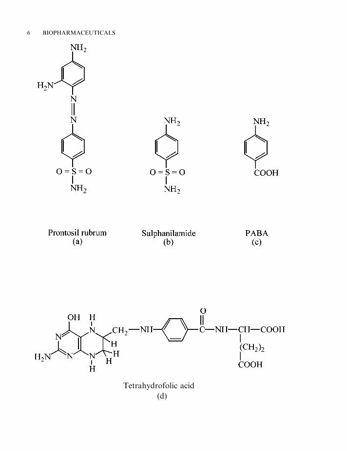
Despite these early advances, it was not until the 1930s that the pharmaceutical industrybegan to develop in earnest. The initial landmark discovery of this era was probably thediscovery and chemical synthesis of the sulpha drugs. These are a group of related moleculesderived from the red dye, Prontosil rubrum. These drugs proved effective in the treatment of awide variety of bacterial infections (Figure 1.1). Although it was first used therapeutically in theearly 1920s, large-scale industrial production of insulin also commenced in the 1930s.
The medical success of these drugs gave new emphasis to the pharmaceutical industry, which
4 BIOPHARMACEUTICALS
Table 1.4. The categorization of pharmaceutically significant biological molecules using the indicateddefinitions as listed in Table 1.3. Reproduced in modified form from European Journal ofPharmaceutical Sciences, vol 15, Walsh, Biopharmaceuticals and Biotechnology, p 135–138, s2002,with permission from Elsevier Science
Pharmaceutical product Biopharmaceutical? Biotechnology medicine? Biologic?
Recombinant protein Yes Yes NoMonoclonal antibody Yes Yes NoProteins obtained by directextraction from nativesource (e.g. blood derivedclotting factors)
No Yes Some (e.g. bloodfactors andpolyclonalantibodies)
Gene therapy products Yes Yes NoAntisense oligonucleotidesmanufactured by directchemical synthesis
Yes No No
Antisense oligonucleotidesproduced by enzymaticsynthesis
Yes Yes No
Peptides manufactured bydirect chemical synthesis
No No No
Peptides, if obtained bydirect extraction fromnative producer source
No Yes No
Antibiotics obtained by directextraction from nativeproducer, or bysemi-synthesis
No Yes No
Plant-based productsobtained by direct extractionfrom a native producer,or by semi-synthesis(e.g. taxol)
No Yes No
Cell/tissue-based therapeuticagents
No Yes No
was boosted further by the commencement of industrial-scale penicillin manufacture in the early1940s. Around this time, many of the current leading pharmaceutical companies (or theirforerunners) were founded. Examples include Ciba Geigy, Eli Lilly, Wellcome, Glaxo andRoche. Over the next two to three decades, these companies developed drugs such astetracyclines, corticosteroids, oral contraceptives, antidepressants and many more. Most ofthese pharmaceutical substances are manufactured by direct chemical synthesis.
THE AGE OF BIOPHARMACEUTICALS
Biomedical research continues to broaden our understanding of the molecular mechanismsunderlining both health and disease. Research undertaken since the 1950s has pinpointed a hostof proteins produced naturally in the body which have obvious therapeutic applications.Examples include the interferons, and interleukins, which regulate the immune response; growthfactors such as erythropoietin, which stimulates red blood cell production; and neurotrophicfactors, which regulate the development and maintenance of neural tissue.
While the pharmaceutical potential of these regulatory molecules was generally appreciated,their widespread medical application was in most cases rendered impractical due to the tinyquantities in which they were naturally produced. The advent of recombinant DNA technology(genetic engineering) and monoclonal antibody technology (hybridoma technology) overcamemany such difficulties, and marked the beginning of a new era of the pharmaceutical sciences.
Recombinant DNA technology has had a four-fold positive impact upon the production ofpharmaceutically important proteins:
. It overcomes the problem of source availability. Many proteins of therapeutic potential areproduced naturally in the body in minute quantities. Examples include interferons (Chapter4), interleukins (Chapter 5) and colony stimulating factors (Chapter 6). This renderedimpractical their direct extraction from native source material in quantities sufficient to meetlikely clinical demand. Recombinant production (Chapter 3) allows the manufacture of anyprotein in whatever quantity it is required.
. It overcomes problems of product safety. Direct extraction of product from some nativebiological sources has, in the past, led to the unwitting transmission of disease. Examplesinclude the transmission of blood-borne pathogens such as hepatitis B, C and HIV viainfected blood products and the transmission of Creutzfeldt–Jakob disease to personsreceiving human growth hormone preparations derived from human pituitaries.
. It provides an alternative to direct extraction from inappropriate/dangerous source material. Anumber of therapeutic proteins have traditionally been extracted from human urine. Thefertility hormone FSH, for example, is obtained from the urine of post-menopausal women,while a related hormone, hCG, is extracted from the urine of pregnant women (Chapter 8).Urine is not considered a particularly desirable source of pharmaceutical products. Whileseveral products obtained from this source remain on the market, recombinant forms havenow also been approved. Other potential biopharmaceuticals are produced naturally indownright dangerous sources. Ancrod, for example, is a protein displaying anti-coagulantactivity (Chapter 9) and, hence, is of potential clinical use; however, it is produced naturallyby the Malaysian pit viper. While retrieval by milking snake venom is possible, and indeedmay be quite an exciting procedure, recombinant production in less dangerous organisms,such as Escherichia coli or Saccharomyces cerevisiae, would be considered preferable by most.
. It facilitates the generation of engineered therapeutic proteins displaying some clinical
PHARMACEUTICALS, BIOLOGICS AND BIOPHARMACEUTICALS 5
6 BIOPHARMACEUTICALS
Tetrahydrofolic acid
(d)
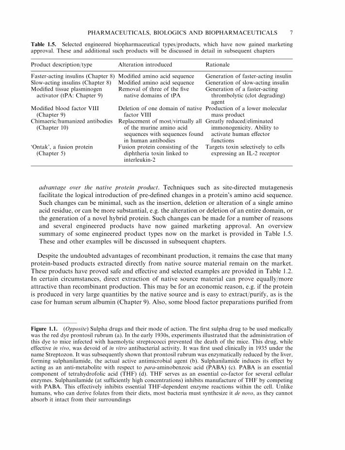
advantage over the native protein product. Techniques such as site-directed mutagenesisfacilitate the logical introduction of pre-defined changes in a protein’s amino acid sequence.Such changes can be minimal, such as the insertion, deletion or alteration of a single aminoacid residue, or can be more substantial, e.g. the alteration or deletion of an entire domain, orthe generation of a novel hybrid protein. Such changes can be made for a number of reasonsand several engineered products have now gained marketing approval. An overviewsummary of some engineered product types now on the market is provided in Table 1.5.These and other examples will be discussed in subsequent chapters.
Despite the undoubted advantages of recombinant production, it remains the case that manyprotein-based products extracted directly from native source material remain on the market.These products have proved safe and effective and selected examples are provided in Table 1.2.In certain circumstances, direct extraction of native source material can prove equally/moreattractive than recombinant production. This may be for an economic reason, e.g. if the proteinis produced in very large quantities by the native source and is easy to extract/purify, as is thecase for human serum albumin (Chapter 9). Also, some blood factor preparations purified from
PHARMACEUTICALS, BIOLOGICS AND BIOPHARMACEUTICALS 7
Table 1.5. Selected engineered biopharmaceutical types/products, which have now gained marketingapproval. These and additional such products will be discussed in detail in subsequent chapters
Product description/type Alteration introduced Rationale
Faster-acting insulins (Chapter 8) Modified amino acid sequence Generation of faster-acting insulinSlow-acting insulins (Chapter 8) Modified amino acid sequence Generation of slow-acting insulinModified tissue plasminogenactivator (tPA: Chapter 9)
Removal of three of the fivenative domains of tPA
Generation of a faster-actingthrombolytic (clot degrading)agent
Modified blood factor VIII(Chapter 9)
Deletion of one domain of nativefactor VIII
Production of a lower molecularmass product
Chimaeric/humanized antibodies(Chapter 10)
Replacement of most/virtually allof the murine amino acidsequences with sequences foundin human antibodies
Greatly reduced/eliminatedimmonogenicity. Ability toactivate human effectorfunctions
‘Ontak’, a fusion protein(Chapter 5)
Fusion protein consisting of thediphtheria toxin linked tointerleukin-2
Targets toxin selectively to cellsexpressing an IL-2 receptor
Figure 1.1. (Opposite) Sulpha drugs and their mode of action. The first sulpha drug to be used medicallywas the red dye prontosil rubrum (a). In the early 1930s, experiments illustrated that the administration ofthis dye to mice infected with haemolytic streptococci prevented the death of the mice. This drug, whileeffective in vivo, was devoid of in vitro antibacterial activity. It was first used clinically in 1935 under thename Streptozon. It was subsequently shown that prontosil rubrum was enzymatically reduced by the liver,forming sulphanilamide, the actual active antimicrobial agent (b). Sulphanilamide induces its effect byacting as an anti-metabolite with respect to para-aminobenzoic acid (PABA) (c). PABA is an essentialcomponent of tetrahydrofolic acid (THF) (d). THF serves as an essential co-factor for several cellularenzymes. Sulphanilamide (at sufficiently high concentrations) inhibits manufacture of THF by competingwith PABA. This effectively inhibits essential THF-dependent enzyme reactions within the cell. Unlikehumans, who can derive folates from their diets, most bacteria must synthesize it de novo, as they cannotabsorb it intact from their surroundings
donor blood actually contain several different blood factors and hence can be used to treatseveral haemophilia patient types. Recombinant blood factor preparations, on the other hand,contain but a single blood factor and hence can be used to treat only one haemophilia type(Chapter 9).
The advent of genetic engineering and monoclonal antibody technology underpinned theestablishment of literally hundreds of start-up biopharmaceutical (biotechnology) companies inthe late 1970s and early 1980s. The bulk of these companies were founded in the USA, withsmaller numbers of start-ups emanating from Europe and other world regions.
Many of these fledgling companies were founded by academics/technical experts who soughtto take commercial advantage of developments in the biotechnological arena. These companieswere largely financed by speculative monies attracted by the hype associated with theestablishment of the modern biotech era. While most of these early companies displayedsignificant technical expertise, the vast majority lacked experience in the practicalities of thedrug development process (Chapter 2). Most of the well-established large pharmaceuticalcompanies, on the other hand, were slow to invest heavily in biotech research and development.However, as the actual and potential therapeutic significance of biopharmaceuticals became evident,many of these companies did diversify into this area. Most either purchased small establishedbiopharmaceutical concerns or formed strategic alliances with them. An example was the long-term alliance formed by Genentech (see later) and the well-established pharmaceutical company,Eli Lilly. Genentech developed recombinant human insulin, which was then marketed by EliLilly under the trade name, Humulin. The merger of biotech capability with pharmaceuticalexperience helped accelerate development of the biopharmaceutical sector.
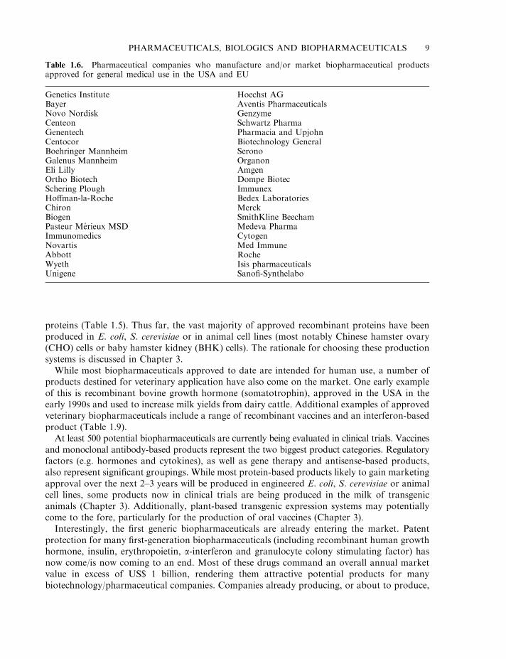
Many of the earlier biopharmaceutical companies no longer exist. The overall level ofspeculative finance available was not sufficient to sustain them all long-term (it can take 6–10years and $200–500 million to develop a single drug; Chapter 2). Furthermore, the promise andhype of biotechnology sometimes exceeded its ability to actually deliver a final product. Somebiopharmaceutical substances showed little efficacy in treating their target condition, and/orexhibited unacceptable side effects. Mergers and acquisitions also led to the disappearance ofseveral biopharmaceutical concerns. Table 1.6 lists the major pharmaceutical concerns whichnow manufacture/market biopharmaceuticals approved for general medical use. Box 1.1provides a profle of three well-established dedicated biopharmaceutical companies.
BIOPHARMACEUTICALS: CURRENT STATUS AND FUTUREPROSPECTS
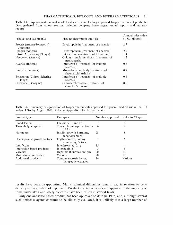
By mid-2002, some 120 biopharmaceutical products had gained marketing approval in the USAand/or EU. Collectively, these represent a global biopharmaceutical market in the region of $15billion (Table 1.7). A detailed list of the approved products is provided in Appendix 1. Theproducts include a range of hormones, blood factors and thrombolytic agents, as well asvaccines and monoclonal antibodies (Table 1.8). All but one are protein-based therapeuticagents. The exception is Vitravene, an antisense oligonucleotide (Chapter 11), first approved inthe USA in 1998. Many additional nucleic acid-based products for use in gene therapy orantisense technology (Chapter 11) are currently in clinical trials.
Many of the initial biopharmaceuticals approved were simple replacement proteins (e.g.blood factors and human insulin). The ability to logically alter the amino acid sequence of aprotein, coupled to an increased understanding of the relationship between protein structureand function has facilitated the more recent introduction of several engineered therapeutic
8 BIOPHARMACEUTICALS
proteins (Table 1.5). Thus far, the vast majority of approved recombinant proteins have beenproduced in E. coli, S. cerevisiae or in animal cell lines (most notably Chinese hamster ovary(CHO) cells or baby hamster kidney (BHK) cells). The rationale for choosing these productionsystems is discussed in Chapter 3.
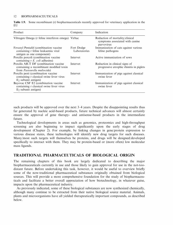
While most biopharmaceuticals approved to date are intended for human use, a number ofproducts destined for veterinary application have also come on the market. One early exampleof this is recombinant bovine growth hormone (somatotrophin), approved in the USA in theearly 1990s and used to increase milk yields from dairy cattle. Additional examples of approvedveterinary biopharmaceuticals include a range of recombinant vaccines and an interferon-basedproduct (Table 1.9).
At least 500 potential biopharmaceuticals are currently being evaluated in clinical trials. Vaccinesand monoclonal antibody-based products represent the two biggest product categories. Regulatoryfactors (e.g. hormones and cytokines), as well as gene therapy and antisense-based products,also represent significant groupings. While most protein-based products likely to gain marketingapproval over the next 2–3 years will be produced in engineered E. coli, S. cerevisiae or animalcell lines, some products now in clinical trials are being produced in the milk of transgenicanimals (Chapter 3). Additionally, plant-based transgenic expression systems may potentiallycome to the fore, particularly for the production of oral vaccines (Chapter 3).
Interestingly, the first generic biopharmaceuticals are already entering the market. Patentprotection for many first-generation biopharmaceuticals (including recombinant human growthhormone, insulin, erythropoietin, a-interferon and granulocyte colony stimulating factor) hasnow come/is now coming to an end. Most of these drugs command an overall annual marketvalue in excess of US$ 1 billion, rendering them attractive potential products for manybiotechnology/pharmaceutical companies. Companies already producing, or about to produce,
PHARMACEUTICALS, BIOLOGICS AND BIOPHARMACEUTICALS 9
Table 1.6. Pharmaceutical companies who manufacture and/or market biopharmaceutical productsapproved for general medical use in the USA and EU
Genetics Institute Hoechst AGBayer Aventis PharmaceuticalsNovo Nordisk GenzymeCenteon Schwartz PharmaGenentech Pharmacia and UpjohnCentocor Biotechnology GeneralBoehringer Mannheim SeronoGalenus Mannheim OrganonEli Lilly AmgenOrtho Biotech Dompe BiotecSchering Plough ImmunexHoffman-la-Roche Bedex LaboratoriesChiron MerckBiogen SmithKline BeechamPasteur Merieux MSD Medeva PharmaImmunomedics CytogenNovartis Med ImmuneAbbott RocheWyeth Isis pharmaceuticalsUnigene Sanofi-Synthelabo
generic biopharmaceuticals include Genemedix (UK), Sicor and Ivax (USA), Congene andMicrobix (Canada) and BioGenerix (Germany); e.g. Genemedix secured approval for sale of arecombinant colony-stimulating factor in China in 2001 and is also commencing themanufacture of recombinant erythropoietin; Sicor currently markets human growth hormoneand interferon-a in Eastern Europe and various developing nations. The widespread approvaland marketing of generic biopharmaceuticals in regions such as the EU and USA is, however,unlikely to occur in the near future, mainly due to regulatory issues.
To date (mid-2002) no gene therapy based product has thus far been approved for generalmedical use (Chapter 11). Although gene therapy trials were initiated as far back as 1990, the
10 BIOPHARMACEUTICALS
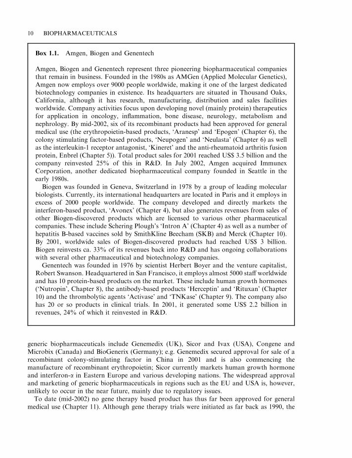
Box 1.1. Amgen, Biogen and Genentech
Amgen, Biogen and Genentech represent three pioneering biopharmaceutical companiesthat remain in business. Founded in the 1980s as AMGen (Applied Molecular Genetics),Amgen now employs over 9000 people worldwide, making it one of the largest dedicatedbiotechnology companies in existence. Its headquarters are situated in Thousand Oaks,California, although it has research, manufacturing, distribution and sales facilitiesworldwide. Company activities focus upon developing novel (mainly protein) therapeuticsfor application in oncology, inflammation, bone disease, neurology, metabolism andnephrology. By mid-2002, six of its recombinant products had been approved for generalmedical use (the erythropoietin-based products, ‘Aranesp’ and ‘Epogen’ (Chapter 6), thecolony stimulating factor-based products, ‘Neupogen’ and ‘Neulasta’ (Chapter 6) as wellas the interleukin-1 receptor antagonist, ‘Kineret’ and the anti-rheumatoid arthritis fusionprotein, Enbrel (Chapter 5)). Total product sales for 2001 reached US$ 3.5 billion and thecompany reinvested 25% of this in R&D. In July 2002, Amgen acquired ImmunexCorporation, another dedicated biopharmaceutical company founded in Seattle in theearly 1980s.
Biogen was founded in Geneva, Switzerland in 1978 by a group of leading molecularbiologists. Currently, its international headquarters are located in Paris and it employs inexcess of 2000 people worldwide. The company developed and directly markets theinterferon-based product, ‘Avonex’ (Chapter 4), but also generates revenues from sales ofother Biogen-discovered products which are licensed to various other pharmaceuticalcompanies. These include Schering Plough’s ‘Intron A’ (Chapter 4) as well as a number ofhepatitis B-based vaccines sold by SmithKline Beecham (SKB) and Merck (Chapter 10).By 2001, worldwide sales of Biogen-discovered products had reached US$ 3 billion.Biogen reinvests ca. 33% of its revenues back into R&D and has ongoing collaborationswith several other pharmaceutical and biotechnology companies.
Genentech was founded in 1976 by scientist Herbert Boyer and the venture capitalist,Robert Swanson. Headquartered in San Francisco, it employs almost 5000 staff worldwideand has 10 protein-based products on the market. These include human growth hormones(‘Nutropin’, Chapter 8), the antibody-based products ‘Herceptin’ and ‘Rituxan’ (Chapter10) and the thrombolytic agents ‘Activase’ and ‘TNKase’ (Chapter 9). The company alsohas 20 or so products in clinical trials. In 2001, it generated some US$ 2.2 billion inrevenues, 24% of which it reinvested in R&D.
results have been disappointing. Many technical difficulties remain, e.g. in relation to genedelivery and regulation of expression. Product effectiveness was not apparent in the majority oftrials undertaken and safety concerns have been raised in several trials.
Only one antisense-based product has been approved to date (in 1998) and, although severalsuch antisense agents continue to be clinically evaluated, it is unlikely that a large number of
PHARMACEUTICALS, BIOLOGICS AND BIOPHARMACEUTICALS 11
Table 1.7. Approximate annual market values of some leading approved biopharmaceutical products.Data gathered from various sources, including company home pages, annual reports and industryreports
Product and (Company) Product description and (use)Annual sales value(US$, billions)
Procrit (Amgen/Johnson &Johnson)
Erythropoietin (treatment of anaemia) 2.7
Epogen (Amgen) Erythropoietin (treatment of anaemia) 2.0Intron A (Schering Plough) Interferon-a (treatment of leukaemia) 1.4Neupogen (Amgen) Colony stimulating factor (treatment of
neutropenia)1.2
Avonex (Biogen) Interferon-b (treatment of multiplesclerosis)
0.8
Embrel (Immunex) Monoclonal antibody (treatment ofrheumatoid arthritis)
0.7
Betasteron (Chiron/ScheringPlough)
Interferon-b (treatment of multiplesclerosis)
0.6
Cerezyme (Genzyme) Glucocerebrosidase (treatment ofGaucher’s disease)
0.5
Table 1.8. Summary categorization of biopharmaceuticals approved for general medical use in the EUand/or USA by August 2002. Refer to Appendix 1 for further details
Product type Examples Number approved Refer to Chapter
Blood factors Factors VIII and IX 7 9Thrombolytic agents Tissue plasminogen activator
(tPA)6 9
Hormones Insulin, growth hormone,gonadotrophins
28 8
Haemapoietic growth factors Erythropoietin, colonystimulating factors
7 6
Interferons Interferons-a, -b, -g 15 4Interleukin-based products Interleukin-2 3 5Vaccines Hepatitis B surface antigen 20 10Monoclonal antibodies Various 20 10Additional products Tumour necrosis factor,
therapeutic enzymes14 Various
such products will be approved over the next 3–4 years. Despite the disappointing results thusfar generated by nucleic acid-based products, future technical advances will almost certainlyensure the approval of gene therapy- and antisense-based products in the intermediatefuture.
Technological developments in areas such as genomics, proteomics and high-throughputscreening are also beginning to impact significantly upon the early stages of drugdevelopment (Chapter 2). For example, by linking changes in gene/protein expression tovarious disease states, these technologies will identify new drug targets for such diseases.Many/most such targets will themselves be proteins, and drugs will be designed/developedspecifically to interact with them. They may be protein-based or (more often) low molecularmass ligands.
TRADITIONAL PHARMACEUTICALS OF BIOLOGICAL ORIGIN
The remaining chapters of this book are largely dedicated to describing the majorbiopharmaceuticals currently in use and those likely to gain approval for use in the not-too-distant future. Before undertaking this task, however, it would be useful to overview brieflysome of the now-traditional pharmaceutical substances originally obtained from biologicalsources. This will provide a more comprehensive foundation for the study of biopharmaceu-ticals and facilitate a better overall appreciation of how biotechnology, in whatever guise,impacts upon the pharmaceutical industry.
As previously indicated, some of these biological substances are now synthesized chemically,although many continue to be extracted from their native biological source material. Animals,plants and microorganisms have all yielded therapeutically important compounds, as describedbelow.
12 BIOPHARMACEUTICALS
Table 1.9. Some recombinant (r) biopharmaceuticals recently approved for veterinary application in theEU
Product Company Indication
Vibragen Omega (r feline interferon omega) Virbac Reduction of mortality/clinicalsymptoms associated with canineparvovirus
Fevaxyl Pentafel (combination vaccinecontaining r feline leukaemia viralantigen as one component)
Fort DodgeLaboratories
Immunization of cats against variousfeline pathogens
Porcilis porcoli (combination vaccinecontaining r E. coli adhesins)
Intervet Active immunization of sows
Porcilis AR-T DF (combination vaccinecontaining a recombinant modified toxinfrom Pasteurella multocida)
Intervet Reduction in clinical signs ofprogressive atrophic rhinitis in piglets
Porcilis pesti (combination vaccinecontaining r classical swine fever virusE2 subunit antigen)
Intervet Immunization of pigs against classicalswine fever
Bayovac CSF E2 (combination vaccinecontaining r classical swine fever virusE2 subunit antigen)
Intervet Immunization of pigs against classicalswine fever
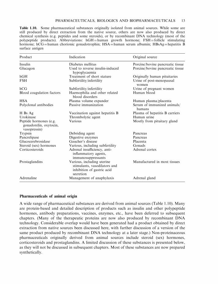
Pharmaceuticals of animal origin
A wide range of pharmaceutical substances are derived from animal sources (Table 1.10). Manyare protein-based and detailed description of products such as insulin and other polypeptidehormones, antibody preparations, vaccines, enzymes, etc., have been deferred to subsequentchapters. (Many of the therapeutic proteins are now also produced by recombinant DNAtechnology. Considerable overlap would have been generated had a product obtained by directextraction from native sources been discussed here, with further discussion of a version of thesame product produced by recombinant DNA technology at a later stage.) Non-proteinaceouspharmaceuticals originally derived from animal sources include steroid (sex) hormones,corticosteroids and prostaglandins. A limited discussion of these substances is presented below,as they will not be discussed in subsequent chapters. Most of these substances are now preparedsynthetically.
PHARMACEUTICALS, BIOLOGICS AND BIOPHARMACEUTICALS 13
Table 1.10. Some pharmaceutical substances originally isolated from animal sources. While some arestill produced by direct extraction from the native source, others are now also produced by directchemical synthesis (e.g. peptides and some steroids), or by recombinant DNA technology (most of thepolypeptide products). Abbreviations: hGH¼human growth hormone; FSH¼ follicle stimulatinghormone; hCG¼human chorionic gonadotrophin; HSA¼human serum albumin; HBsAg¼hepatitis Bsurface antigen
Product Indication Original source
Insulin Diabetes mellitus Porcine/bovine pancreatic tissueGlucagon Used to reverse insulin-induced
hypoglycaemiaPorcine/bovine pancreatic tissue
hGH Treatment of short stature Originally human pituitariesFSH Subfertility/infertility Urine of post-menopausal
womenhCG Subfertility/infertility Urine of pregnant womenBlood coagulation factors Haemophilia and other related
blood disordersHuman blood
HSA Plasma volume expander Human plasma/placentaPolyclonal antibodies Passive immunization Serum of immunized animals/
humansH Bs Ag Vaccination against hepatitis B Plasma of hepatitis B carriersUrokinase Thrombolytic agent Human urinePeptide hormones (e.g.gonadorelin, oxytocin,vasopressin)
Various Mostly from pituitary gland
Trypsin Debriding agent PancreasPancrelipase Digestive enzymes PancreasGlucocerebrosidase Gaucher’s disease PlacentaSteroid (sex) hormones Various, including subfertility GonadsCorticosteroids Adrenal insufficiency, anti-
inflammatory agents,immunosuppressants
Adrenal cortex
Prostaglandins Various, including uterinestimulants, vasodilators andinhibition of gastric acidsecretion
Manufactured in most tissues
Adrenaline Management of anaphylaxis Adrenal gland
The sex hormones
The male and female gonads, as well as the placenta of pregnant females and, to a lesser extent,the adrenal cortex, produce a range of steroid hormones which regulate the development andmaintenance of reproductive and related functions. As such, these steroid sex hormones havefound medical application in the treatment of various reproductive dysfunctions.
While these steroids directly regulate sexual function, their synthesis and release are, in turn,controlled by gonadotropins—polypeptide hormones produced by the pituitary gland. Thebiology and medical applications of the gonadotropins are outlined in Chapter 8. Sex hormonesproduced naturally may be classified into one of three groups:
. the androgens;
. the oestrogens;
. progesterone.
While these steroids can be extracted directly from human tissue, in most instances they can alsobe synthesized chemically. Direct chemical synthesis methodology has also facilitated thedevelopment of synthetic steroid analogues. Many such analogues exhibit therapeuticadvantages over the native hormone, e.g. they may be more potent, be absorbed intact fromthe digestive tract, or exhibit a longer duration of action in the body. The majority of sex steroidhormones now used clinically are chemically synthesized.
The androgens The androgens are the main male sex hormones. They are produced by theLeydig cells of the testes, as well as in the adrenals. They are also produced by the femaleovary. As in the case of all other steroid hormones, the androgens are synthesized in thebody, using cholesterol as their ultimate biosynthetic precursor. The major androgenproduced by the testes is testosterone, of which 4–10mg is secreted daily into the bloodstreamby healthy young men. Testosterone synthesis is stimulated by the gonadotrophin, luteinizinghormone.
Testosterone is transported in the blood bound to transport proteins, the most important ofwhich are albumin (a non-specific carrier) and testosterone–oestradiol binding globulin(TEBG), a 40 kDa polypeptide which binds testosterone and oestrogens with high affinity.
Testosterone and other androgens induce their characteristic biological effects via binding to aspecific intracellular receptor. These hormones promote:
. induction of sperm production;
. development/maintenance of male secondary sexual characteristics;
. general anabolic (growth-promoting) effects;
. regulation of gonadotrophin secretion.
The actions of androgens are often antagonized by oestrogens, and vice versa. This forms thebasis of androgen administration in some forms of breast cancer, and oestrogen administrationin the treatment of prostate cancer. Anti-androgenic compounds have also been synthesized.These antagonize androgen action due to their ability to compete with androgens for binding tothe receptor.
Androgens are used medically as replacement therapy in male hypogonadal disorders (i.e.impaired functioning of the testes). They are administered to adolescent males displayingdelayed puberty to promote an increase in the size of the scrotum and other sexual organs.Androgens are also sometimes administered to females, particularly in the management of some
14 BIOPHARMACEUTICALS
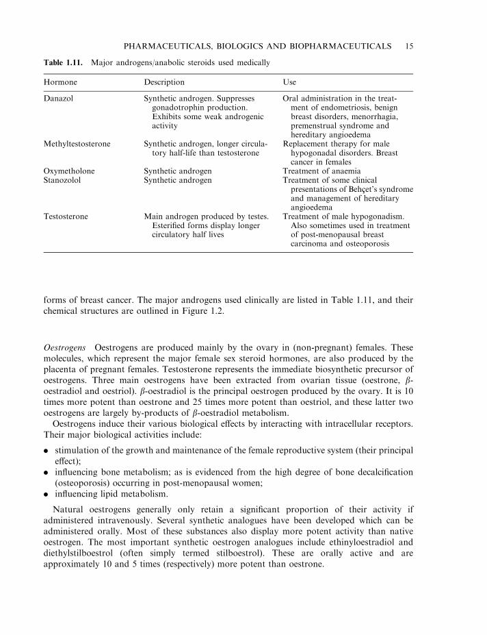
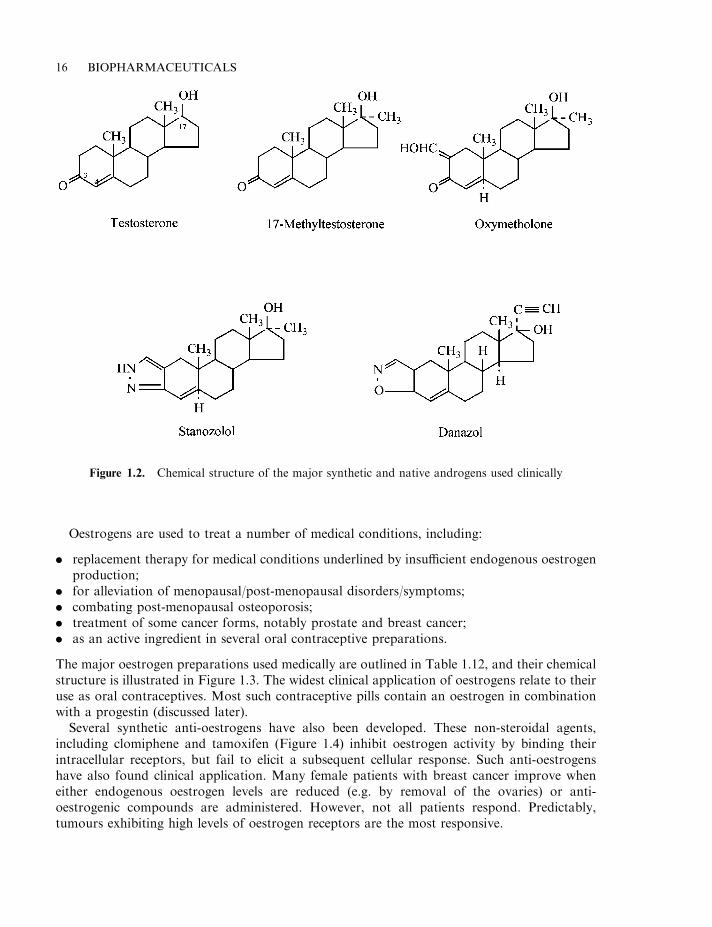
forms of breast cancer. The major androgens used clinically are listed in Table 1.11, and theirchemical structures are outlined in Figure 1.2.
Oestrogens Oestrogens are produced mainly by the ovary in (non-pregnant) females. Thesemolecules, which represent the major female sex steroid hormones, are also produced by theplacenta of pregnant females. Testosterone represents the immediate biosynthetic precursor ofoestrogens. Three main oestrogens have been extracted from ovarian tissue (oestrone, b-oestradiol and oestriol). b-oestradiol is the principal oestrogen produced by the ovary. It is 10times more potent than oestrone and 25 times more potent than oestriol, and these latter twooestrogens are largely by-products of b-oestradiol metabolism.
Oestrogens induce their various biological effects by interacting with intracellular receptors.Their major biological activities include:
. stimulation of the growth and maintenance of the female reproductive system (their principaleffect);
. influencing bone metabolism; as is evidenced from the high degree of bone decalcification(osteoporosis) occurring in post-menopausal women;
. influencing lipid metabolism.
Natural oestrogens generally only retain a significant proportion of their activity ifadministered intravenously. Several synthetic analogues have been developed which can beadministered orally. Most of these substances also display more potent activity than nativeoestrogen. The most important synthetic oestrogen analogues include ethinyloestradiol anddiethylstilboestrol (often simply termed stilboestrol). These are orally active and areapproximately 10 and 5 times (respectively) more potent than oestrone.
PHARMACEUTICALS, BIOLOGICS AND BIOPHARMACEUTICALS 15
Table 1.11. Major androgens/anabolic steroids used medically
Hormone Description Use
Danazol Synthetic androgen. Suppressesgonadotrophin production.Exhibits some weak androgenicactivity
Oral administration in the treat-ment of endometriosis, benignbreast disorders, menorrhagia,premenstrual syndrome andhereditary angioedema
Methyltestosterone Synthetic androgen, longer circula-tory half-life than testosterone
Replacement therapy for malehypogonadal disorders. Breastcancer in females
Oxymetholone Synthetic androgen Treatment of anaemiaStanozolol Synthetic androgen Treatment of some clinical
presentations of Behcet’s syndromeand management of hereditaryangioedema
Testosterone Main androgen produced by testes.Esterified forms display longercirculatory half lives
Treatment of male hypogonadism.Also sometimes used in treatmentof post-menopausal breastcarcinoma and osteoporosis
Oestrogens are used to treat a number of medical conditions, including:
. replacement therapy for medical conditions underlined by insufficient endogenous oestrogenproduction;
. for alleviation of menopausal/post-menopausal disorders/symptoms;
. combating post-menopausal osteoporosis;
. treatment of some cancer forms, notably prostate and breast cancer;
. as an active ingredient in several oral contraceptive preparations.
The major oestrogen preparations used medically are outlined in Table 1.12, and their chemicalstructure is illustrated in Figure 1.3. The widest clinical application of oestrogens relate to theiruse as oral contraceptives. Most such contraceptive pills contain an oestrogen in combinationwith a progestin (discussed later).
Several synthetic anti-oestrogens have also been developed. These non-steroidal agents,including clomiphene and tamoxifen (Figure 1.4) inhibit oestrogen activity by binding theirintracellular receptors, but fail to elicit a subsequent cellular response. Such anti-oestrogenshave also found clinical application. Many female patients with breast cancer improve wheneither endogenous oestrogen levels are reduced (e.g. by removal of the ovaries) or anti-oestrogenic compounds are administered. However, not all patients respond. Predictably,tumours exhibiting high levels of oestrogen receptors are the most responsive.
16 BIOPHARMACEUTICALS
Figure 1.2. Chemical structure of the major synthetic and native androgens used clinically
Progesterone and progestogens Progesterone is the main hormone produced by the corpusluteum during the second half of the female menstrual cycle (Chapter 8). It acts upon theendometrial tissue (lining of the womb). Under its influence, the endometrium begins to produceand secrete mucus, which is an essential prerequisite for subsequent implantation of a fertilizedegg. In pregnant females, progesterone is also synthesized by the placenta, and its continuedproduction is essential for maintenance of the pregnant state. Administration of progesteroneto a non-pregnant female prevents ovulation and, as such, the progestogens (discussed below)are used as contraceptive agents. Progesterone also stimulates breast growth, and isimmunosuppressive at high doses.
Only minor quantities of intact biologically active progesterone are absorbed if the hormoneis given orally. Progestogens are synthetic compounds which display actions similar to that ofprogesterone. Many progestogens are more potent than progesterone itself and can be absorbedintact when administered orally.
Progesterone, and particularly progestogens, are used for a number of therapeutic purposes,including:
. treatment of menstrual disorders;
. treatment of endometriosis (the presence of tissue similar to the endometrium at other sites inthe pelvis);
. management of some breast and endometrial cancers;
. hormone replacement therapy, where they are used in combination with oestrogens;
. contraceptive agents, usually in combination with oestrogens.
PHARMACEUTICALS, BIOLOGICS AND BIOPHARMACEUTICALS 17
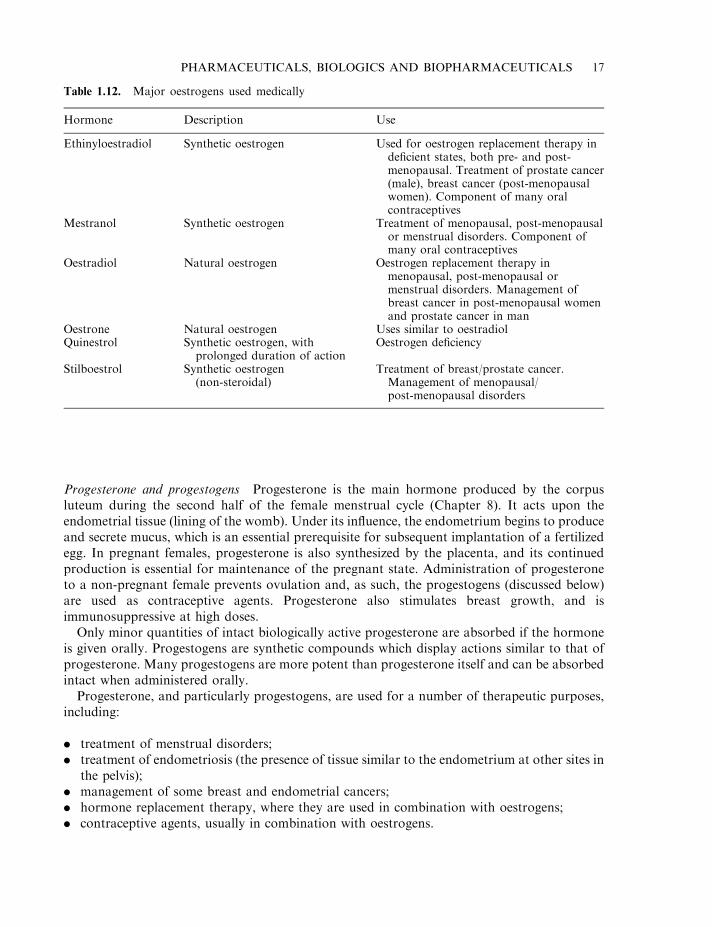
Table 1.12. Major oestrogens used medically
Hormone Description Use
Ethinyloestradiol Synthetic oestrogen Used for oestrogen replacement therapy indeficient states, both pre- and post-menopausal. Treatment of prostate cancer(male), breast cancer (post-menopausalwomen). Component of many oralcontraceptives
Mestranol Synthetic oestrogen Treatment of menopausal, post-menopausalor menstrual disorders. Component ofmany oral contraceptives
Oestradiol Natural oestrogen Oestrogen replacement therapy inmenopausal, post-menopausal ormenstrual disorders. Management ofbreast cancer in post-menopausal womenand prostate cancer in man
Oestrone Natural oestrogen Uses similar to oestradiolQuinestrol Synthetic oestrogen, with
prolonged duration of actionOestrogen deficiency
Stilboestrol Synthetic oestrogen(non-steroidal)
Treatment of breast/prostate cancer.Management of menopausal/post-menopausal disorders
18 BIOPHARMACEUTICALS
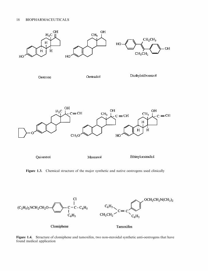
Figure 1.3. Chemical structure of the major synthetic and native oestrogens used clinically
Figure 1.4. Structure of clomiphene and tamoxifen, two non-steroidal synthetic anti-oestrogens that havefound medical application
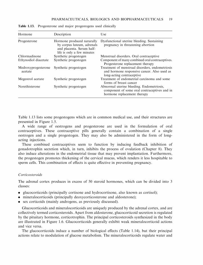
Table 1.13 lists some progestogens which are in common medical use, and their structures arepresented in Figure 1.5.
A wide range of oestrogens and progesterone are used in the formulation of oralcontraceptives. These contraceptive pills generally contain a combination of a singleoestrogen and a single progestogen. They may also be administered in the form of long-acting injections.
These combined contraceptives seem to function by inducing feedback inhibition ofgonadotrophin secretion which, in turn, inhibits the process of ovulation (Chapter 8). Theyalso induce alterations in the endometrial tissue that may prevent implantation. Furthermore,the progestogen promotes thickening of the cervical mucus, which renders it less hospitable tosperm cells. This combination of effects is quite effective in preventing pregnancy.
Corticosteroids
The adrenal cortex produces in excess of 50 steroid hormones, which can be divided into 3classes:
. glucocorticoids (principally cortisone and hydrocortisone, also known as cortisol);
. mineralocorticoids (principally deoxycorticosterone and aldosterone);
. sex corticoids (mainly androgens, as previously discussed).
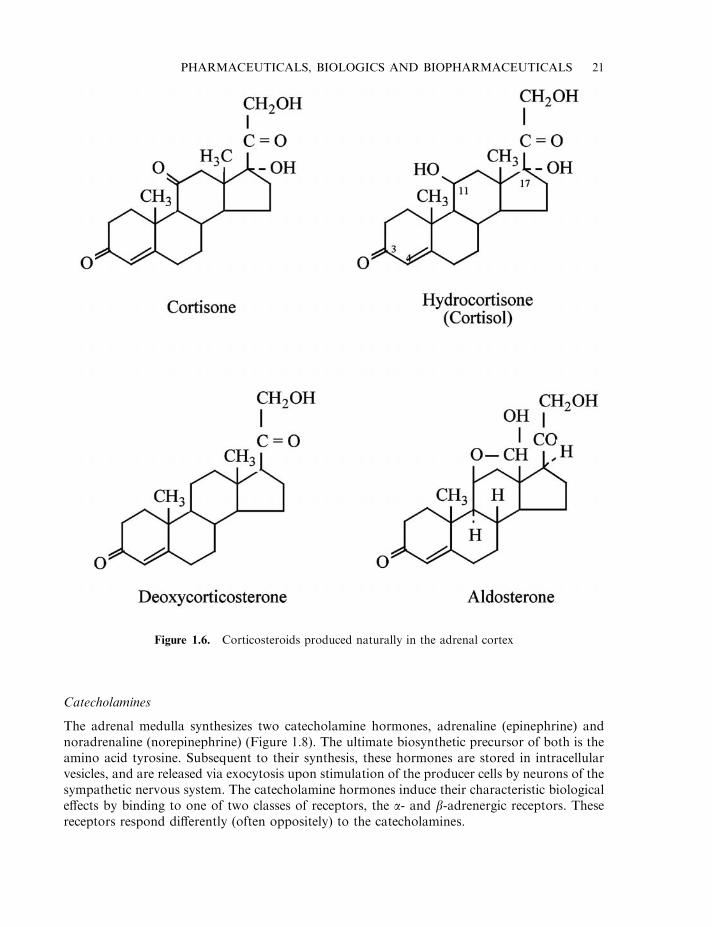
Glucocorticoids and mineralocorticoids are uniquely produced by the adrenal cortex, and arecollectively termed corticosteroids. Apart from aldosterone, glucocorticoid secretion is regulatedby the pituitary hormone, corticotrophin. The principal corticosteroids synthesized in the bodyare illustrated in Figure 1.6. Glucocorticoids generally exhibit weak mineralocorticoid actionsand vice versa.
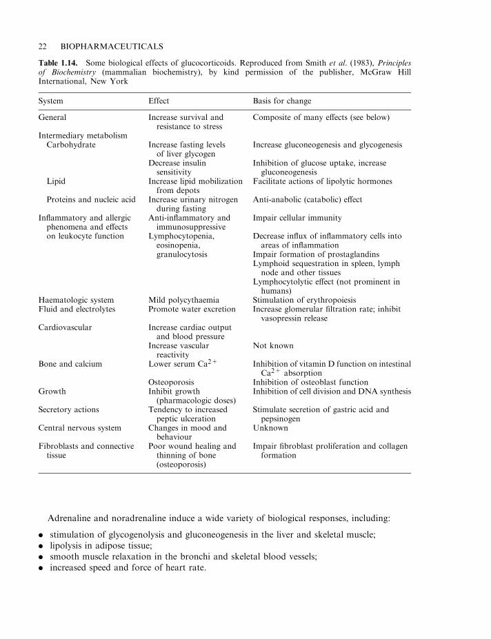
The glucocorticoids induce a number of biological effects (Table 1.14), but their principalactions relate to modulation of glucose metabolism. The mineralocorticoids regulate water and
PHARMACEUTICALS, BIOLOGICS AND BIOPHARMACEUTICALS 19
Table 1.13. Progesterone and major progestogens used clinically
Hormone Description Use
Progesterone Hormone produced naturallyby corpus luteum, adrenalsand placenta. Serum half-life is only a few minutes
Dysfunctional uterine bleeding. Sustainingpregnancy in threatening abortion
Chlormadinone Synthetic progestogen Menstrual disorders. Oral contraceptiveEthynodiol diacetate Synthetic progestogen Component ofmany combined oral contraceptives.
Progesterone replacement therapyMedroxyprogesteroneacetate
Synthetic progestogen Treatment of menstrual disorders, endometriosisand hormone responsive cancer. Also used aslong-acting contraceptive
Megestrol acetate Synthetic progestogen Treatment of endometrial carcinoma and someforms of breast cancer
Norethisterone Synthetic progestogen Abnormal uterine bleeding. Endometriosis,component of some oral contraceptives and inhormone replacement therapy
electrolyte metabolism. They generally increase renal reabsorption of Na+, and promote Na+–K+–H+ exchange. This typically results in increased serum Na+ concentrations and decreasedserum K+ concentrations. Elevated blood pressure is also usually induced.
Various synthetic corticosteroids have also been developed. Some display greater potencythan the native steroids, while others exhibit glucocorticoid activity with little associatedmineralocorticoid effects, or vice versa. The major glucocorticoids used clinically are synthetic.They are usually employed as:
. replacement therapy in cases of adrenal insufficiency;
. anti-inflammatory agents;
. immunosuppressive agents.
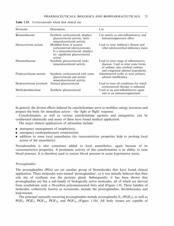
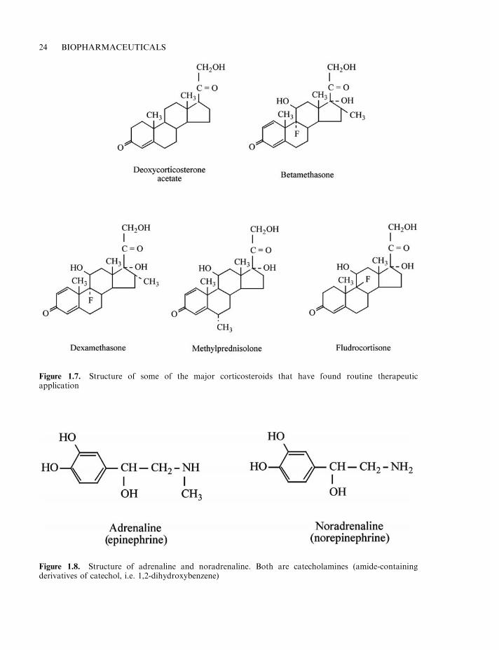
Examples of the corticosteroids used most commonly in the clinic are presented in Table 1.15and Figure 1.7.
20 BIOPHARMACEUTICALS
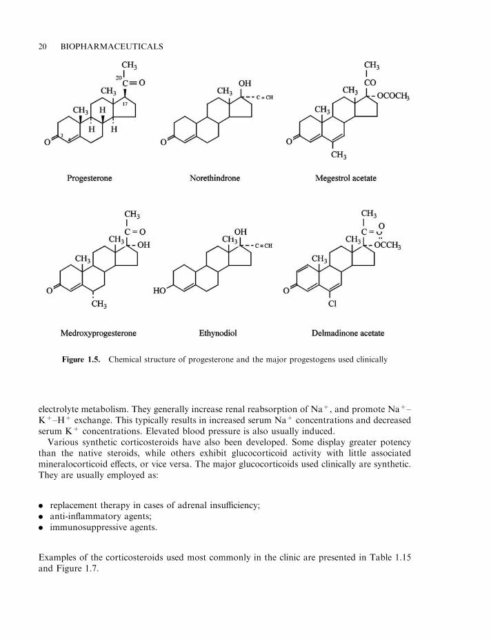
Figure 1.5. Chemical structure of progesterone and the major progestogens used clinically
Catecholamines
The adrenal medulla synthesizes two catecholamine hormones, adrenaline (epinephrine) andnoradrenaline (norepinephrine) (Figure 1.8). The ultimate biosynthetic precursor of both is theamino acid tyrosine. Subsequent to their synthesis, these hormones are stored in intracellularvesicles, and are released via exocytosis upon stimulation of the producer cells by neurons of thesympathetic nervous system. The catecholamine hormones induce their characteristic biologicaleffects by binding to one of two classes of receptors, the a- and b-adrenergic receptors. Thesereceptors respond differently (often oppositely) to the catecholamines.
PHARMACEUTICALS, BIOLOGICS AND BIOPHARMACEUTICALS 21
Figure 1.6. Corticosteroids produced naturally in the adrenal cortex
Adrenaline and noradrenaline induce a wide variety of biological responses, including:
. stimulation of glycogenolysis and gluconeogenesis in the liver and skeletal muscle;
. lipolysis in adipose tissue;
. smooth muscle relaxation in the bronchi and skeletal blood vessels;
. increased speed and force of heart rate.
22 BIOPHARMACEUTICALS
Table 1.14. Some biological effects of glucocorticoids. Reproduced from Smith et al. (1983), Principlesof Biochemistry (mammalian biochemistry), by kind permission of the publisher, McGraw HillInternational, New York
System Effect Basis for change
General Increase survival andresistance to stress
Composite of many effects (see below)
Intermediary metabolismCarbohydrate Increase fasting levels
of liver glycogenIncrease gluconeogenesis and glycogenesis
Decrease insulinsensitivity
Inhibition of glucose uptake, increasegluconeogenesis
Lipid Increase lipid mobilizationfrom depots
Facilitate actions of lipolytic hormones
Proteins and nucleic acid Increase urinary nitrogenduring fasting
Anti-anabolic (catabolic) effect
Inflammatory and allergicphenomena and effects
Anti-inflammatory andimmunosuppressive
Impair cellular immunity
on leukocyte function Lymphocytopenia,eosinopenia,granulocytosis
Decrease influx of inflammatory cells intoareas of inflammation
Impair formation of prostaglandinsLymphoid sequestration in spleen, lymphnode and other tissues
Lymphocytolytic effect (not prominent inhumans)
Haematologic system Mild polycythaemia Stimulation of erythropoiesisFluid and electrolytes Promote water excretion Increase glomerular filtration rate; inhibit
vasopressin releaseCardiovascular Increase cardiac output
and blood pressureIncrease vascularreactivity
Not known
Bone and calcium Lower serum Ca2+ Inhibition of vitamin D function on intestinalCa2+ absorption
Osteoporosis Inhibition of osteoblast functionGrowth Inhibit growth
(pharmacologic doses)Inhibition of cell division and DNA synthesis
Secretory actions Tendency to increasedpeptic ulceration
Stimulate secretion of gastric acid andpepsinogen
Central nervous system Changes in mood andbehaviour
Unknown
Fibroblasts and connectivetissue
Poor wound healing andthinning of bone(osteoporosis)
Impair fibroblast proliferation and collagenformation
In general, the diverse effects induced by catecholamines serve to mobilize energy resources andprepare the body for immediate action— the ‘fight or flight’ response.
Catecholamines, as well as various catecholamine agonists and antagonists, can besynthesized chemically and many of these have found medical application.
The major clinical applications of adrenaline include:
. emergency management of anaphylaxis;
. emergency cardiopulmonary resuscitation;
. addition to some local anaesthetics (its vasoconstrictor properties help to prolong localaction of the anaesthetic).
Noradrenaline is also sometimes added to local anaesthetics, again because of itsvasoconstrictive properties. A prominent activity of this catecholamine is its ability to raiseblood pressure. It is therefore used to restore blood pressure in acute hypotensive states.
Prostaglandins
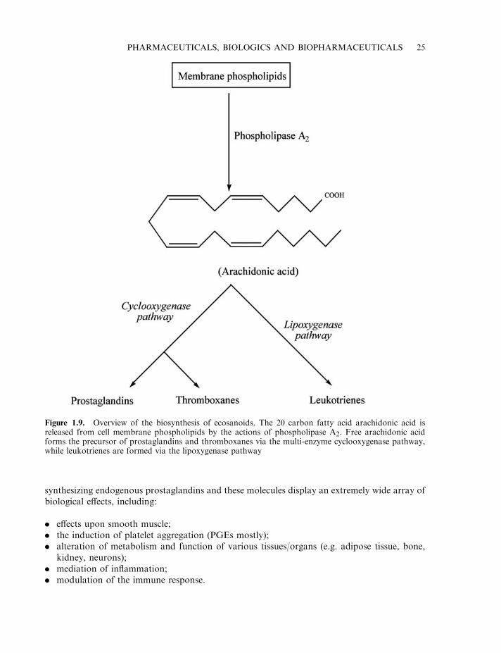
The prostaglandins (PGs) are yet another group of biomolecules that have found clinicalapplication. These molecules were named ‘prostaglandins’, as it was initially believed that theirsole site of synthesis was the prostate gland. Subsequently, it has been shown thatprostaglandins are but a sub-family of biologically active molecules, all of which are derivedfrom arachidonic acid, a 20-carbon polyunsaturated fatty acid (Figure 1.9). These families ofmolecules, collectively known as ecosanoids, include the prostaglandins, thromboxanes andleukotrienes.
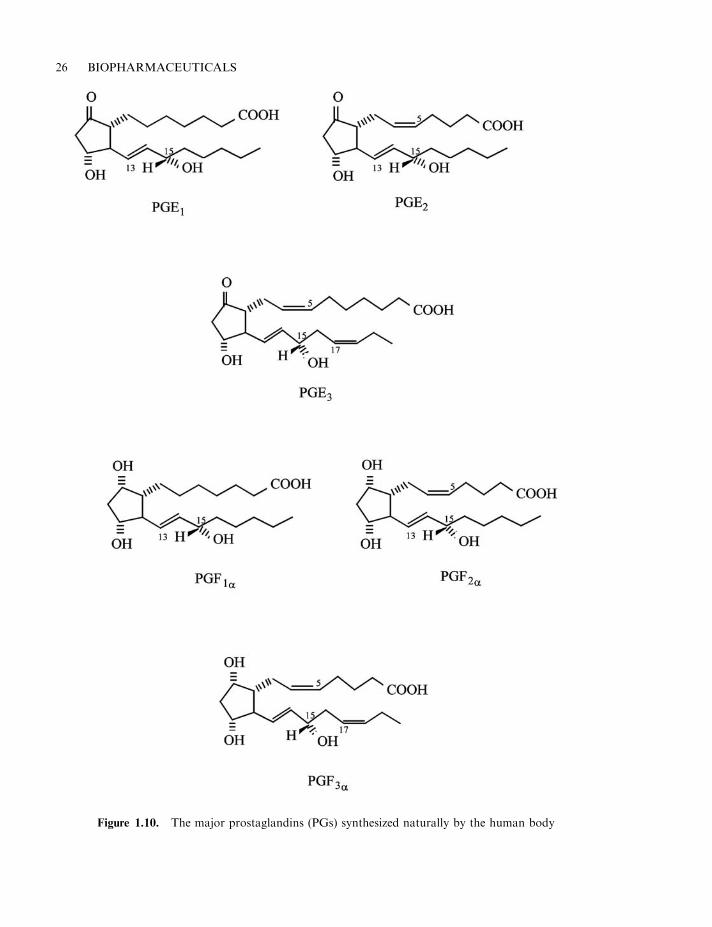
The principal naturally-occurring prostaglandins include prostaglandin E1 (PGE1), as well asPGE2, PGE3, PGF1a, PGF2a and PGF3a (Figure 1.10). All body tissues are capable of
PHARMACEUTICALS, BIOLOGICS AND BIOPHARMACEUTICALS 23
Table 1.15. Corticosteroids which find clinical use
Hormone Description Use
Betamethasone Synthetic corticosteroid, displaysglucocorticoid activity, lacksmineralocorticoid activity
Use mainly as anti-inflammatory andimmunosuppressive effect
Deoxycortone acetate Modified form of naturalcorticosteroid (deoxycortone).Is a mineralocorticoid, displaysno significant glucocorticoidaction
Used to treat Addison’s disease andother adrenocortical deficiency states
Dexamethasone Synthetic glucocorticoid, lacksmineralocorticoid activity
Used to treat range of inflammatorydiseases. Used to treat some formsof asthma, also cerebral oedemaand congenital adrenal hyperplasia
Fludrocortisone acetate Synthetic corticosteroid with someglucocorticoid and potentmineralocorticoid activity
Administered orally to treat primaryadrenal insufficiency
Hydrocortisone (cortisol) Natural glucocorticoid Used to treat all conditions for whichcorticosteroid therapy is indicated
Methylprednisolone Synthetic glucocorticoid Used as an anti-inflammatory agentand as an immunosuppressant
24 BIOPHARMACEUTICALS
Figure 1.7. Structure of some of the major corticosteroids that have found routine therapeuticapplication
Figure 1.8. Structure of adrenaline and noradrenaline. Both are catecholamines (amide-containingderivatives of catechol, i.e. 1,2-dihydroxybenzene)
synthesizing endogenous prostaglandins and these molecules display an extremely wide array ofbiological effects, including:
. effects upon smooth muscle;
. the induction of platelet aggregation (PGEs mostly);
. alteration of metabolism and function of various tissues/organs (e.g. adipose tissue, bone,kidney, neurons);
. mediation of inflammation;
. modulation of the immune response.
PHARMACEUTICALS, BIOLOGICS AND BIOPHARMACEUTICALS 25
Figure 1.9. Overview of the biosynthesis of ecosanoids. The 20 carbon fatty acid arachidonic acid isreleased from cell membrane phospholipids by the actions of phospholipase A2. Free arachidonic acidforms the precursor of prostaglandins and thromboxanes via the multi-enzyme cyclooxygenase pathway,while leukotrienes are formed via the lipoxygenase pathway
26 BIOPHARMACEUTICALS
Figure 1.10. The major prostaglandins (PGs) synthesized naturally by the human body
Their actions on smooth muscle, in particular, can be complex. For example, both PGEs andPGFs induce contraction of uterine and gastrointestinal smooth muscle. On the other hand,PGEs stimulate dilation of vascular and bronchial smooth muscle, whereas PGFs induceconstriction of these muscle types.
Although prostaglandins in some ways resemble hormones, they fail to fit the classicaldefinition of a hormone in several respects, e.g. their synthesis is not localized to aspecialized gland and they act primarily in a paracrine rather than true endocrinefashion. The diverse biological actions of the prostaglandins underpin their diverse clinicalapplications. Synthetic analogues have also been developed which display some clinicaladvantage over native PGs (e.g. greater stability, longer duration of action, more specificbiological effects). The major clinical uses of native or synthetic prostaglandins can be groupedas follows:
. Obstetrics and gynaecology: several prostaglandin preparations (mainly E2, F2a and theiranalogues) are used to induce uterine contraction. The purpose can be either to induce labouras part of the normal childbirth procedure, or to terminate a pregnancy;
. Induction of vasodilation and inhibition of platelet aggregation: prostaglandin E1 and itsanalogues are most commonly employed and are used to treat infants with congenital heartdisease;
. Inhibition of gastric acid secretion: an effect promoted in particular by PGE1 and itsanalogues.
In some circumstances, inhibition of endogenous prostaglandin synthesis can represent adesirable medical goal. This is often achieved by administration of drugs capable of inhibitingthe cyclooxygenase system. Aspirin is a well known example of such a drug, and it is by thismechanism that it induces its analgesic and anti-inflammatory effects.
Pharmaceutical substances of plant origin
The vast bulk of early medicinal substances were plant-derived. An estimated 3 billion peopleworldwide continue to use traditional plant medicines as their primary form of healthcare. Atleast 25% of all prescription drugs sold in North America contain active substances which wereoriginally isolated from plants (or are modified forms of chemicals originally isolated fromplants). Many of these were discovered by a targeted knowledge-based ethnobotanicalapproach. Researchers simply recorded plant-based medical cures for specific diseases andthen analysed the plants for their active ingredients. Plants produce a wide array of bioactivemolecules via secondary metabolic pathways. Most of these probably evolved as chemicaldefence agents against infections or predators.
While some medicinal substances continue to be directly extracted from plant material, inmany instances plant-derived drugs can now be manufactured, at least in part, by directchemical synthesis. In addition, chemical modification of many of these plant ‘lead’ drugs haveyielded a range of additional therapeutic substances.
The bulk of plant-derived medicines can be categorized into a number of chemical families,including alkaloids, flavonoids, terpenes and terpenoids, steroids (e.g. cardiac glycosides), aswell as coumarins, quinines, salicylates and xanthines. A list of some better-known plant-derived drugs is presented in Table 1.16.
PHARMACEUTICALS, BIOLOGICS AND BIOPHARMACEUTICALS 27
Alkaloids
Alkaloids may be classified as relatively low molecular mass bases containing one or morenitrogen atoms, often present in a ring system. They are found mainly in plants (from whichover 6000 different alkaloids have been extracted). They are especially abundant in floweringplants, particularly lupins, poppies, tobacco and potatoes. They also are synthesized by someanimals, insects, marine organisms and microorganisms. Most producers simultaneouslysynthesize several distinct alkaloids.
The solubility of these substances in organic solvents facilitates their initial extraction andpurification from plant material using, for example, petroleum ether. Subsequent chromato-graphic fractionation facilitates separation of individual alkaloid components. Many alkaloidsare poisonous (and have been used for this purpose), although at lower concentrations they maybe useful therapeutic agents. Several of the best known alkaloid-based drugs are discussedbelow. The chemical structure of most of these is presented in Figure 1.11.
Atropine and scopalamine Atropine is found in the berries of the weeds deadly nightshade andblack nightshade. It is also synthesized in the leaves and roots of Hyoscyamus muticusi. At high
28 BIOPHARMACEUTICALS
Table 1.16. Some drugs of plant origin
Drug Chemical type Indication Plant producer
Aspirin Salicylate Analgesic, anti-inflammatory Salix alba (white willow tree)and Filipendula ulmaria(meadowsweet)
Atropine Alkaloid Pupil dilator Atropa belladonna (deadlynightshade)
Caffeine Xanthine Increases mental alertness Camellia sinensisCocaine Alkaloid Ophthalmic anaesthetic Erythoxylum coca (coca leaves)Codeine Alkaloid Analgesic, cough suppressor Papaver somniferum (opium
poppy)Dicoumarol Coumarin Anti-coagulant Melilotus officinalisDigoxin Steroid Increases heart muscle contraction Digitalis purpurea (purple
foxglove)Digitoxin Steroid Increases heart muscle contraction Digitalis purpureaIpecac Alkaloid Induces vomiting Psychotria ipecacuanhaMorphine Alkaloid Analgesic Papaver somniferum (opium
poppy)Pseudoephedrine Alkaloid Clears nasal congestion Ephedra sinicaQuinine Alkaloid Malaria Cinchona pubescens (fever tree)Reserpine Alkaloid Antihypertensive (reduces blood
pressure)Rauvolfia serpentina (Indiansnakeroot)
Scopalamine Alkaloid Motion sickness Datura stramonium (Jimsonweed)
Taxol Terpenoid Ovarian, breast cancer Taxus brevifolia (western yewtree)
Theophylline Xanthine Anti-asthmatic, diuretic Camellia sinensisVinblastine Alkaloid Hodgkin’s disease Catharanthus roseus (rosy
periwinkle)Vincristine Alkaloid Leukaemia Catharanthus roseus
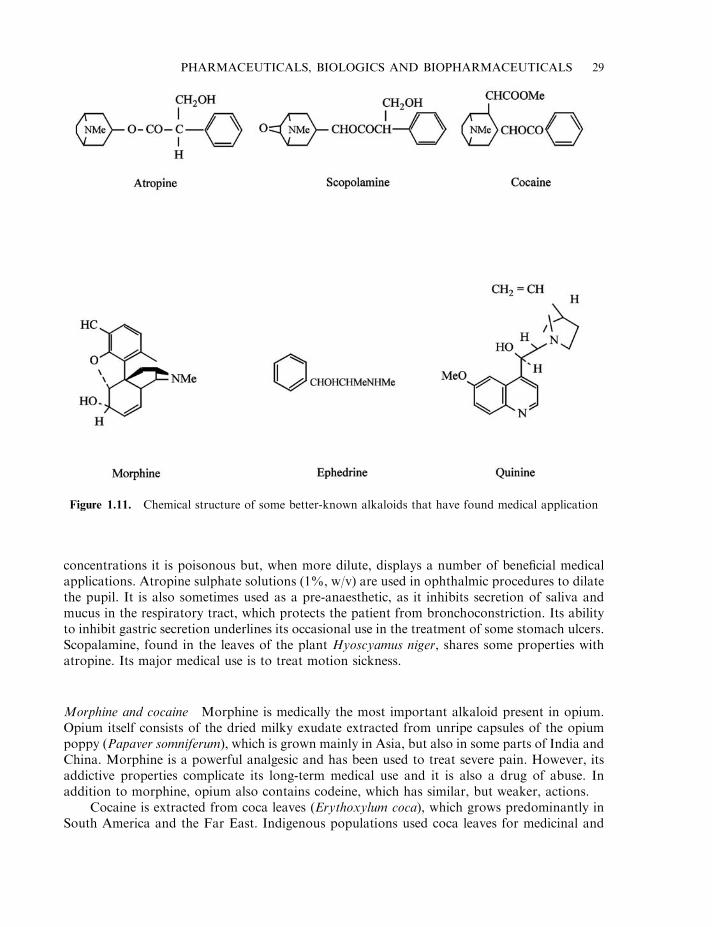
concentrations it is poisonous but, when more dilute, displays a number of beneficial medicalapplications. Atropine sulphate solutions (1%, w/v) are used in ophthalmic procedures to dilatethe pupil. It is also sometimes used as a pre-anaesthetic, as it inhibits secretion of saliva andmucus in the respiratory tract, which protects the patient from bronchoconstriction. Its abilityto inhibit gastric secretion underlines its occasional use in the treatment of some stomach ulcers.Scopalamine, found in the leaves of the plant Hyoscyamus niger, shares some properties withatropine. Its major medical use is to treat motion sickness.
Morphine and cocaine Morphine is medically the most important alkaloid present in opium.Opium itself consists of the dried milky exudate extracted from unripe capsules of the opiumpoppy (Papaver somniferum), which is grown mainly in Asia, but also in some parts of India andChina. Morphine is a powerful analgesic and has been used to treat severe pain. However, itsaddictive properties complicate its long-term medical use and it is also a drug of abuse. Inaddition to morphine, opium also contains codeine, which has similar, but weaker, actions.
Cocaine is extracted from coca leaves (Erythoxylum coca), which grows predominantly inSouth America and the Far East. Indigenous populations used coca leaves for medicinal and
PHARMACEUTICALS, BIOLOGICS AND BIOPHARMACEUTICALS 29
Figure 1.11. Chemical structure of some better-known alkaloids that have found medical application
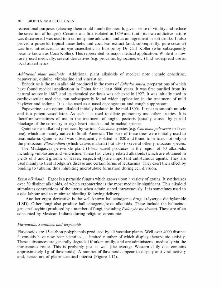
recreational purposes (chewing them could numb the mouth, give a sense of vitality and reducethe sensation of hunger). Cocaine was first isolated in 1859 and (until its own addictive naturewas discovered) was used to treat morphine addiction and as an ingredient in soft drinks. It alsoproved a powerful topical anaesthetic and coca leaf extract (and, subsequently, pure cocaine)was first introduced as an eye anaesthetic in Europe by Dr Carl Koller (who subsequentlybecame known as Coca Koller). This represented its major medical application. While it is nowrarely used medically, several derivatives (e.g. procaine, lignocaine, etc.) find widespread use aslocal anaesthetics.
Additional plant alkaloids Additional plant alkaloids of medical note include ephedrine,papaverine, quinine, vinblastine and vincristine.
Ephedrine is the main alkaloid produced in the roots of Ephedra sinica, preparations of whichhave found medical application in China for at least 5000 years. It was first purified from itsnatural source in 1887, and its chemical synthesis was achieved in 1927. It was initially used incardiovascular medicine, but subsequently found wider application in the treatment of mildhayfever and asthma. It is also used as a nasal decongestant and cough suppressant.
Papaverine is an opium alkaloid initially isolated in the mid-1800s. It relaxes smooth muscleand is a potent vasodilator. As such it is used to dilate pulmonary and other arteries. It istherefore sometimes of use in the treatment of angina pectoris (usually caused by partialblockage of the coronary artery), heart attacks and bronchial spasms.
Quinine is an alkaloid produced by various Cinchona species (e.g. Cinchona pubescens or fevertree), which are mainly native to South America. The bark of these trees were initially used totreat malaria. Quinine itself was subsequently isolated in 1820 and found to be toxic not only tothe protozoan Plasmodium (which causes malaria) but also to several other protozoan species.
The Madagascar periwinkle plant (Vinca rosea) produces in the region of 60 alkaloids,including vinblastine and vincristine. These two closely related alkaloids (which are obtained inyields of 3 and 2 g/tonne of leaves, respectively) are important anti-tumour agents. They areused mainly to treat Hodgkin’s disease and certain forms of leukaemia. They exert their effect bybinding to tubulin, thus inhibiting microtubule formation during cell division.
Ergot alkaloids Ergot is a parasitic fungus which grows upon a variety of grains. It synthesizesover 30 distinct alkaloids, of which ergometrine is the most medically significant. This alkaloidstimulates contractions of the uterus when administered intravenously. It is sometimes used toassist labour and to minimize bleeding following delivery.
Another ergot derivative is the well known hallucinogenic drug, D-lysergic diethylamide(LSD). Other fungi also produce hallucinogenic/toxic alkaloids. These include the hallucino-genic psilocybin (produced by a number of fungi, including Psilocybe mexicana). These are oftenconsumed by Mexican Indians during religious ceremonies.
Flavonoids, xanthines and terpenoids
Flavonoids are 15-carbon polyphenols produced by all vascular plants. Well over 4000 distinctflavonoids have now been identified, a limited number of which display therapeutic activity.These substances are generally degraded if taken orally, and are administered medically via theintravenous route. This is probably just as well (the average Western daily diet containsapproximately 1 g of flavonoids). A number of flavonoids appear to display anti-viral activityand, hence, are of pharmaceutical interest (Figure 1.12).
30 BIOPHARMACEUTICALS
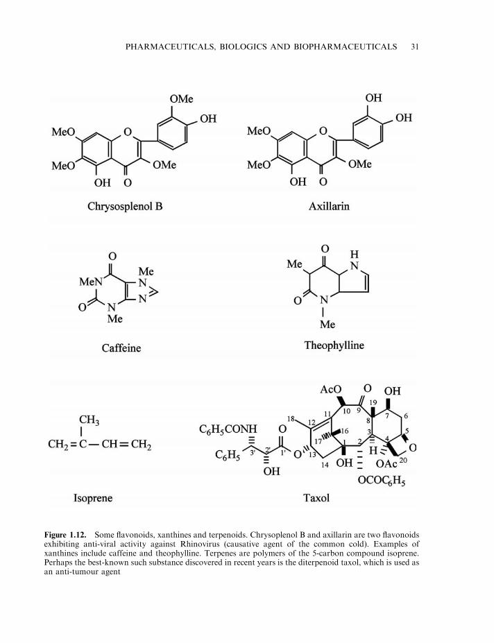
PHARMACEUTICALS, BIOLOGICS AND BIOPHARMACEUTICALS 31
Figure 1.12. Some flavonoids, xanthines and terpenoids. Chrysoplenol B and axillarin are two flavonoidsexhibiting anti-viral activity against Rhinovirus (causative agent of the common cold). Examples ofxanthines include caffeine and theophylline. Terpenes are polymers of the 5-carbon compound isoprene.Perhaps the best-known such substance discovered in recent years is the diterpenoid taxol, which is used asan anti-tumour agent
The principal xanthines of medical interest include caffeine, theophylline and aminophylline.Caffeine is synthesized by several plants and was originally isolated from tea in 1838. It is amethylxanthine (Figure 1.12) which stimulates the central nervous system, increasing mentalalertness. It also acts as a diuretic and stimulates gastric acid secretion. It is absorbed upon oraladministration and is frequently included in drugs containing an analgesic, such as aspirin orparacetamol.
Theophilline is also a minor constituent of tea, but is prepared by direct chemical synthesis formedical use. It functions to relax smooth muscle and, therefore, can be used as a bronchodilatorin the treatment of asthma and bronchitis. Aminophylline is a derivative of theophilline(theophylline ethylenediamine), which is often used in place of theophilline due to its greateraqueous solubility.
Terpenes are polymers of the 5-carbon compound isoprene (Figure 1.12) and, as such,generally display properties similar to those of hydrocarbons. Terpenoids are substitutedterpenes (i.e. contain additional chemical groups, such as an alcohol, phenols, aldehydes,ketones, etc.). Only a few such substances could be regarded as true drugs. Terpenes, such aslimonene, menthol and camphor, form components of various essential oils with pseudo-pharmaceutical uses. A number of these molecules, however, exhibit anti-tumour activity, ofwhich taxol is by far the most important.
The diterpenoid taxol (Figure 1.12) was first isolated from the pacific yew tree (Taxusbrevifolia) in the late 1960s. Its complete structure was elucidated by 1971. Difficulties associatedwith the subsequent development of taxol as a useful drug mirror those encountered during thedevelopment of many plant-derived metabolites as drug products. Its low solubility made taxoldifficult to formulate into a stable product, and its low natural abundance required large-scaleextraction from its native source.
Despite such difficulties, encouraging in vitro bioassay results against transformed cell linesfuelled pre-clinical studies aimed at assessing taxol as an anti-cancer agent. Initial clinical trialsin humans commenced in 1983 using a product formulated as an emulsion in a modified castoroil. Initial difficulties associated with allergic reactions against the oil were largely overcome bymodifying the treatment regimen used. Large-scale clinical trials proved the efficacy of taxol asan anti-cancer agent, and it was approved for use in the treatment of ovarian cancer by the USFood and Drug Administration (FDA) in 1992.
Direct extraction from the bark of T. brevifolia yielded virtually all of the taxol used clinicallyup to almost the mid-1990s. The yield of active principle was in the range 0.007–0.014%. Hugequantities of bark were thus required to sustain taxol production (almost 30 000 kg bark wereextracted in 1989 to meet requirements during large-scale clinical trials). A major (late)intermediate in the biosynthesis of taxol is 10-decacetylbaccatin (10-DAB). This can be obtainedfrom the leaves (needles) of many species of yew, and at concentrations in excess of 0.1%.Chemical methods have been developed allowing synthesis of taxol from 10-DAB, and much ofthe taxol now used therapeutically is produced in this way. Semi-synthesis of taxol alsofacilitates generation of taxol analogues, some of which have also generated clinical interest.Although semi-synthesis of taxol is relatively straightforward, its total de novo synthesis isextremely complex. The cost of achieving de novo synthesis ensures that this approach will notbe adopted for commercial production of this drug.
An alternative route of taxol production under investigation entails the use of plant cellculture techniques. Plant cell culture is considered to be an economically viable production routefor plant-derived drugs, if the drug commands a market value in excess of $1000–2000/kg. Whilemany commonly used plant-derived metabolites fall into this category, plant cell culture has not
32 BIOPHARMACEUTICALS
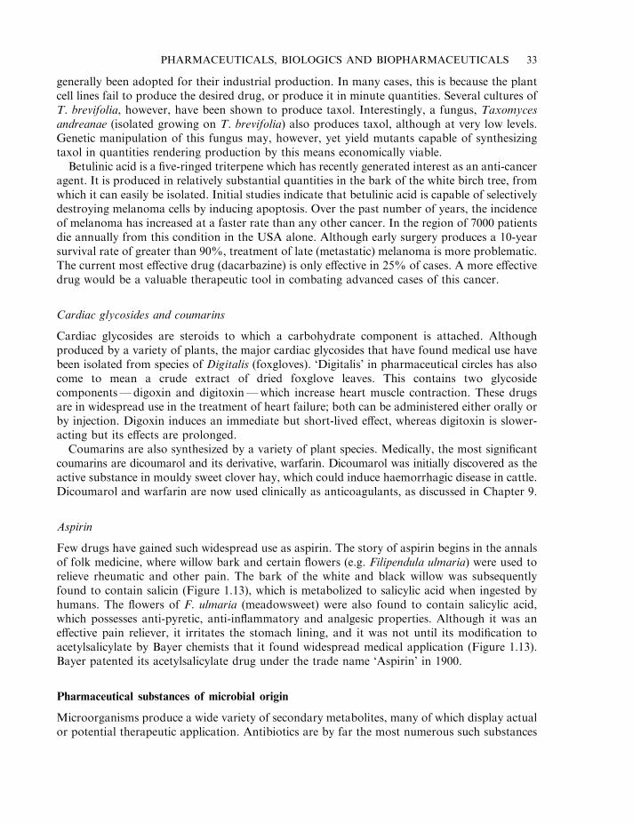
generally been adopted for their industrial production. In many cases, this is because the plantcell lines fail to produce the desired drug, or produce it in minute quantities. Several cultures ofT. brevifolia, however, have been shown to produce taxol. Interestingly, a fungus, Taxomycesandreanae (isolated growing on T. brevifolia) also produces taxol, although at very low levels.Genetic manipulation of this fungus may, however, yet yield mutants capable of synthesizingtaxol in quantities rendering production by this means economically viable.
Betulinic acid is a five-ringed triterpene which has recently generated interest as an anti-canceragent. It is produced in relatively substantial quantities in the bark of the white birch tree, fromwhich it can easily be isolated. Initial studies indicate that betulinic acid is capable of selectivelydestroying melanoma cells by inducing apoptosis. Over the past number of years, the incidenceof melanoma has increased at a faster rate than any other cancer. In the region of 7000 patientsdie annually from this condition in the USA alone. Although early surgery produces a 10-yearsurvival rate of greater than 90%, treatment of late (metastatic) melanoma is more problematic.The current most effective drug (dacarbazine) is only effective in 25% of cases. A more effectivedrug would be a valuable therapeutic tool in combating advanced cases of this cancer.
Cardiac glycosides and coumarins
Cardiac glycosides are steroids to which a carbohydrate component is attached. Althoughproduced by a variety of plants, the major cardiac glycosides that have found medical use havebeen isolated from species of Digitalis (foxgloves). ‘Digitalis’ in pharmaceutical circles has alsocome to mean a crude extract of dried foxglove leaves. This contains two glycosidecomponents—digoxin and digitoxin—which increase heart muscle contraction. These drugsare in widespread use in the treatment of heart failure; both can be administered either orally orby injection. Digoxin induces an immediate but short-lived effect, whereas digitoxin is slower-acting but its effects are prolonged.
Coumarins are also synthesized by a variety of plant species. Medically, the most significantcoumarins are dicoumarol and its derivative, warfarin. Dicoumarol was initially discovered as theactive substance in mouldy sweet clover hay, which could induce haemorrhagic disease in cattle.Dicoumarol and warfarin are now used clinically as anticoagulants, as discussed in Chapter 9.
Aspirin
Few drugs have gained such widespread use as aspirin. The story of aspirin begins in the annalsof folk medicine, where willow bark and certain flowers (e.g. Filipendula ulmaria) were used torelieve rheumatic and other pain. The bark of the white and black willow was subsequentlyfound to contain salicin (Figure 1.13), which is metabolized to salicylic acid when ingested byhumans. The flowers of F. ulmaria (meadowsweet) were also found to contain salicylic acid,which possesses anti-pyretic, anti-inflammatory and analgesic properties. Although it was aneffective pain reliever, it irritates the stomach lining, and it was not until its modification toacetylsalicylate by Bayer chemists that it found widespread medical application (Figure 1.13).Bayer patented its acetylsalicylate drug under the trade name ‘Aspirin’ in 1900.
Pharmaceutical substances of microbial origin
Microorganisms produce a wide variety of secondary metabolites, many of which display actualor potential therapeutic application. Antibiotics are by far the most numerous such substances
PHARMACEUTICALS, BIOLOGICS AND BIOPHARMACEUTICALS 33
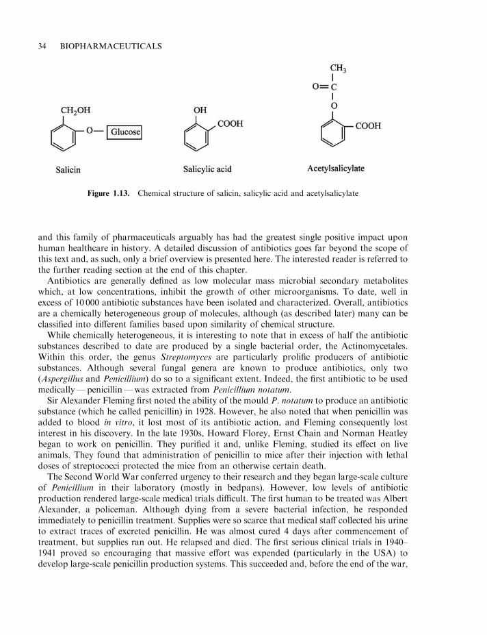
and this family of pharmaceuticals arguably has had the greatest single positive impact uponhuman healthcare in history. A detailed discussion of antibiotics goes far beyond the scope ofthis text and, as such, only a brief overview is presented here. The interested reader is referred tothe further reading section at the end of this chapter.
Antibiotics are generally defined as low molecular mass microbial secondary metaboliteswhich, at low concentrations, inhibit the growth of other microorganisms. To date, well inexcess of 10 000 antibiotic substances have been isolated and characterized. Overall, antibioticsare a chemically heterogeneous group of molecules, although (as described later) many can beclassified into different families based upon similarity of chemical structure.
While chemically heterogeneous, it is interesting to note that in excess of half the antibioticsubstances described to date are produced by a single bacterial order, the Actinomycetales.Within this order, the genus Streptomyces are particularly prolific producers of antibioticsubstances. Although several fungal genera are known to produce antibiotics, only two(Aspergillus and Penicillium) do so to a significant extent. Indeed, the first antibiotic to be usedmedically—penicillin—was extracted from Penicillium notatum.
Sir Alexander Fleming first noted the ability of the mould P. notatum to produce an antibioticsubstance (which he called penicillin) in 1928. However, he also noted that when penicillin wasadded to blood in vitro, it lost most of its antibiotic action, and Fleming consequently lostinterest in his discovery. In the late 1930s, Howard Florey, Ernst Chain and Norman Heatleybegan to work on penicillin. They purified it and, unlike Fleming, studied its effect on liveanimals. They found that administration of penicillin to mice after their injection with lethaldoses of streptococci protected the mice from an otherwise certain death.
The Second World War conferred urgency to their research and they began large-scale cultureof Penicillium in their laboratory (mostly in bedpans). However, low levels of antibioticproduction rendered large-scale medical trials difficult. The first human to be treated was AlbertAlexander, a policeman. Although dying from a severe bacterial infection, he respondedimmediately to penicillin treatment. Supplies were so scarce that medical staff collected his urineto extract traces of excreted penicillin. He was almost cured 4 days after commencement oftreatment, but supplies ran out. He relapsed and died. The first serious clinical trials in 1940–1941 proved so encouraging that massive effort was expended (particularly in the USA) todevelop large-scale penicillin production systems. This succeeded and, before the end of the war,
34 BIOPHARMACEUTICALS
Figure 1.13. Chemical structure of salicin, salicylic acid and acetylsalicylate
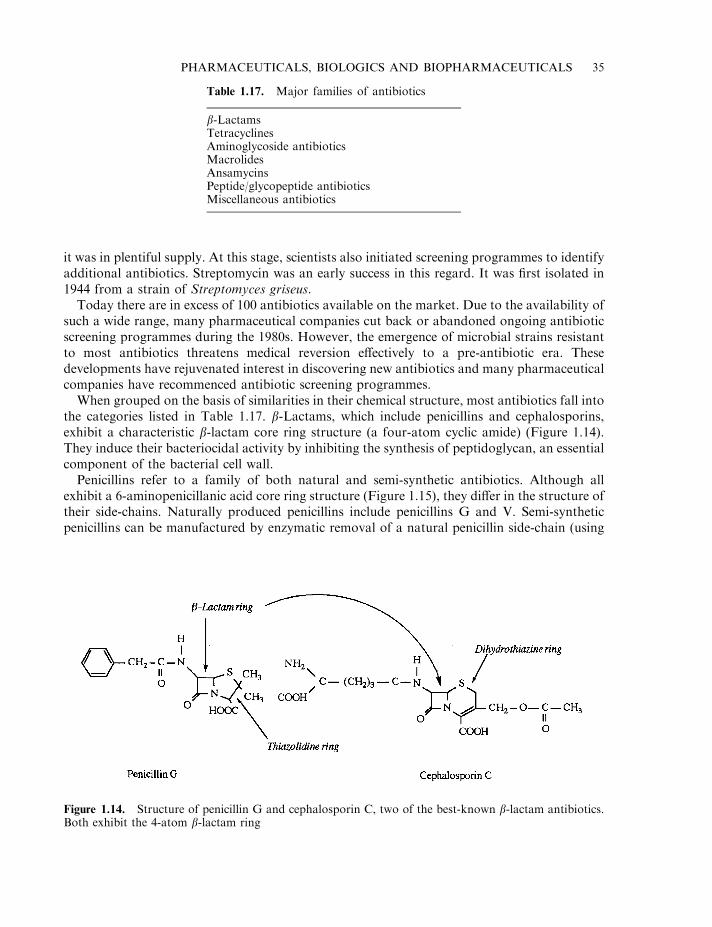
it was in plentiful supply. At this stage, scientists also initiated screening programmes to identifyadditional antibiotics. Streptomycin was an early success in this regard. It was first isolated in1944 from a strain of Streptomyces griseus.
Today there are in excess of 100 antibiotics available on the market. Due to the availability ofsuch a wide range, many pharmaceutical companies cut back or abandoned ongoing antibioticscreening programmes during the 1980s. However, the emergence of microbial strains resistantto most antibiotics threatens medical reversion effectively to a pre-antibiotic era. Thesedevelopments have rejuvenated interest in discovering new antibiotics and many pharmaceuticalcompanies have recommenced antibiotic screening programmes.
When grouped on the basis of similarities in their chemical structure, most antibiotics fall intothe categories listed in Table 1.17. b-Lactams, which include penicillins and cephalosporins,exhibit a characteristic b-lactam core ring structure (a four-atom cyclic amide) (Figure 1.14).They induce their bacteriocidal activity by inhibiting the synthesis of peptidoglycan, an essentialcomponent of the bacterial cell wall.
Penicillins refer to a family of both natural and semi-synthetic antibiotics. Although allexhibit a 6-aminopenicillanic acid core ring structure (Figure 1.15), they differ in the structure oftheir side-chains. Naturally produced penicillins include penicillins G and V. Semi-syntheticpenicillins can be manufactured by enzymatic removal of a natural penicillin side-chain (using
PHARMACEUTICALS, BIOLOGICS AND BIOPHARMACEUTICALS 35
Figure 1.14. Structure of penicillin G and cephalosporin C, two of the best-known b-lactam antibiotics.Both exhibit the 4-atom b-lactam ring
Table 1.17. Major families of antibiotics
b-LactamsTetracyclinesAminoglycoside antibioticsMacrolidesAnsamycinsPeptide/glycopeptide antibioticsMiscellaneous antibiotics
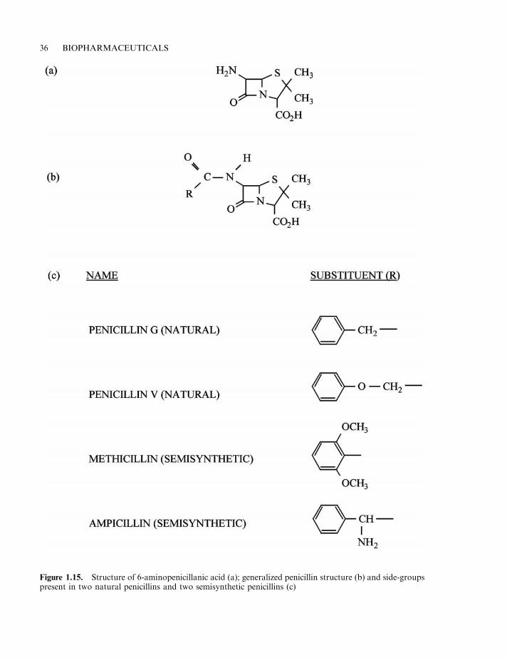
36 BIOPHARMACEUTICALS
Figure 1.15. Structure of 6-aminopenicillanic acid (a); generalized penicillin structure (b) and side-groupspresent in two natural penicillins and two semisynthetic penicillins (c)
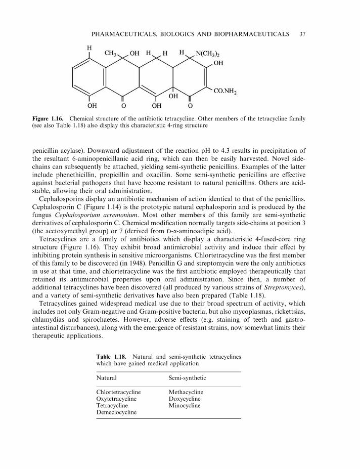
penicillin acylase). Downward adjustment of the reaction pH to 4.3 results in precipitation ofthe resultant 6-aminopenicillanic acid ring, which can then be easily harvested. Novel side-chains can subsequently be attached, yielding semi-synthetic penicillins. Examples of the latterinclude phenethicillin, propicillin and oxacillin. Some semi-synthetic penicillins are effectiveagainst bacterial pathogens that have become resistant to natural penicillins. Others are acid-stable, allowing their oral administration.
Cephalosporins display an antibiotic mechanism of action identical to that of the penicillins.Cephalosporin C (Figure 1.14) is the prototypic natural cephalosporin and is produced by thefungus Cephalosporium acremonium. Most other members of this family are semi-syntheticderivatives of cephalosporin C. Chemical modification normally targets side-chains at position 3(the acetoxymethyl group) or 7 (derived from D-a-aminoadipic acid).
Tetracyclines are a family of antibiotics which display a characteristic 4-fused-core ringstructure (Figure 1.16). They exhibit broad antimicrobial activity and induce their effect byinhibiting protein synthesis in sensitive microorganisms. Chlortetracycline was the first memberof this family to be discovered (in 1948). Penicillin G and streptomycin were the only antibioticsin use at that time, and chlortetracycline was the first antibiotic employed therapeutically thatretained its antimicrobial properties upon oral administration. Since then, a number ofadditional tetracyclines have been discovered (all produced by various strains of Streptomyces),and a variety of semi-synthetic derivatives have also been prepared (Table 1.18).
Tetracyclines gained widespread medical use due to their broad spectrum of activity, whichincludes not only Gram-negative and Gram-positive bacteria, but also mycoplasmas, rickettsias,chlamydias and spirochaetes. However, adverse effects (e.g. staining of teeth and gastro-intestinal disturbances), along with the emergence of resistant strains, now somewhat limits theirtherapeutic applications.
PHARMACEUTICALS, BIOLOGICS AND BIOPHARMACEUTICALS 37
Figure 1.16. Chemical structure of the antibiotic tetracycline. Other members of the tetracycline family(see also Table 1.18) also display this characteristic 4-ring structure
Table 1.18. Natural and semi-synthetic tetracyclineswhich have gained medical application
Natural Semi-synthetic
Chlortetracycline MethacyclineOxytetracycline DoxycyclineTetracycline MinocyclineDemeclocycline
The aminoglycosides are a closely related family of antibiotics produced almost exclusively bymembers of the genus Streptomyces and Micromonospora (Table 1.19). Most are polycationiccompounds, composed of a cyclic amino alcohol to which amino sugars are attached. They allinduce their bacteriocidal effect by inhibiting protein synthesis (apparently by binding to the 30 Sand, to some extent, the 50 S, ribosomal subunits). Most are orally inactive, generallynecessitating their parenteral administration.
The aminoglycosides are most active against Gram-negative rods. Streptomycin was the firstaminoglycoside to be used clinically. Another notable member of this family, gentamicin, wasfirst purified from a culture of Micromonospora purpurea in 1963. Its activity againstPseudomonas aeruginosa and Serratia marcescens renders it useful in the treatment of these(often life-threatening) infections.
The macrolides and ansamycins
The macrolides are a large group of antibiotics. They are characterized by a core ring structurecontaining 12 or more carbon atoms (closed by a lactone group), to which one or more sugarsare attached. The core ring of most anti-bacterial macrolides consists of 14 or 16 carbon atoms,while that of the larger anti-fungal and anti-protozoal macrolides contain up to 30 carbons. Thisfamily of antibiotics are produced predominantly by various species of Streptomyces.Antibacterial macrolides induce their effects by inhibiting bacterial synthesis (anti-fungal/protozoal macrolides appear to function by interfering with sterols, thus compromisingmembrane structure). The only member of this family that enjoys widespread therapeutic use iserythromycin, which was discovered in 1952.
Ansamycins, like the macrolides, are synthesized by condensation of a number of acetate andpropionate units. These antibiotics, which are produced by several genera of the Actinomy-cetales, display a characteristic core aromatic ring structure. Amongst the best-known familymembers are the rifamycins, which are particularly active against Gram-positive bacteria andmycobacteria. They have been used, for example, in the treatment of Mycobacterium tuberculosis.
38 BIOPHARMACEUTICALS
Table 1.19. Some aminoglycoside antibiotics which have gainedsignificant therapeutic application. Producer microorganisms arelisted in brackets. In addition to naturally produced aminoglyco-sides, a number of semi-synthetic derivatives have also foundmedical application. Examples include amikacin, a semi-syntheticderivative of kanamycin and netilmicin, an N-ethyl derivative ofsissomicin
Streptomycin (Streptomyces griseus)Tobramycin (Streptomyces tenebrarius)Framycetin (Streptomyces spp.)Neomycin (Streptomyces spp.)Kanamycin (Streptomyces spp.)Paromycin (Streptomyces spp.)Gentamicin (Micromonospora purpurea)Sissomicin (Micromonospora spp.)
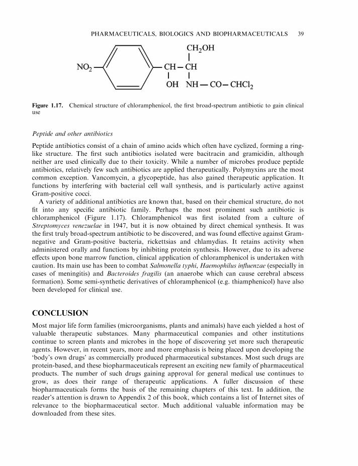
Peptide and other antibiotics
Peptide antibiotics consist of a chain of amino acids which often have cyclized, forming a ring-like structure. The first such antibiotics isolated were bacitracin and gramicidin, althoughneither are used clinically due to their toxicity. While a number of microbes produce peptideantibiotics, relatively few such antibiotics are applied therapeutically. Polymyxins are the mostcommon exception. Vancomycin, a glycopeptide, has also gained therapeutic application. Itfunctions by interfering with bacterial cell wall synthesis, and is particularly active againstGram-positive cocci.
A variety of additional antibiotics are known that, based on their chemical structure, do notfit into any specific antibiotic family. Perhaps the most prominent such antibiotic ischloramphenicol (Figure 1.17). Chloramphenicol was first isolated from a culture ofStreptomyces venezuelae in 1947, but it is now obtained by direct chemical synthesis. It wasthe first truly broad-spectrum antibiotic to be discovered, and was found effective against Gram-negative and Gram-positive bacteria, rickettsias and chlamydias. It retains activity whenadministered orally and functions by inhibiting protein synthesis. However, due to its adverseeffects upon bone marrow function, clinical application of chloramphenicol is undertaken withcaution. Its main use has been to combat Salmonella typhi, Haemophilus influenzae (especially incases of meningitis) and Bacteroides fragilis (an anaerobe which can cause cerebral abscessformation). Some semi-synthetic derivatives of chloramphenicol (e.g. thiamphenicol) have alsobeen developed for clinical use.
CONCLUSION
Most major life form families (microorganisms, plants and animals) have each yielded a host ofvaluable therapeutic substances. Many pharmaceutical companies and other institutionscontinue to screen plants and microbes in the hope of discovering yet more such therapeuticagents. However, in recent years, more and more emphasis is being placed upon developing the‘body’s own drugs’ as commercially produced pharmaceutical substances. Most such drugs areprotein-based, and these biopharmaceuticals represent an exciting new family of pharmaceuticalproducts. The number of such drugs gaining approval for general medical use continues togrow, as does their range of therapeutic applications. A fuller discussion of thesebiopharmaceuticals forms the basis of the remaining chapters of this text. In addition, thereader’s attention is drawn to Appendix 2 of this book, which contains a list of Internet sites ofrelevance to the biopharmaceutical sector. Much additional valuable information may bedownloaded from these sites.
PHARMACEUTICALS, BIOLOGICS AND BIOPHARMACEUTICALS 39
Figure 1.17. Chemical structure of chloramphenicol, the first broad-spectrum antibiotic to gain clinicaluse
FURTHER READING
Books
Buckingham, J. (1996). Dictionary of Natural Products. Chapman & Hall, London.Crommelin, D. & Sindelar, R. (2002) Pharmaceutical Biotechnology, 2nd Edn. Taylor & Francis, London.Goldberg, R. (2001). Pharmaceutical Medicine, Biotechnology and European Law. Cambridge University Press,
Cambridge.Grindley, J. & Ogden, J. (2000). Understanding Biopharmaceuticals. Manufacturing and Regulatory Issues. Interpharm
Press, Denver, CO.Kucers, A. (1997). Use of Antibiotics: A Clinical Review of Antibacterial, Antifungal and Antiviral Drugs, 5th edn.
Butterworth–Heinemann, Oxford.Lincini, G. (1994). Biotechnology of Antibiotics and Other Bioactive Microbial Metabolites. Plenum, New York.Lubiniecki, A. (1994). Regulatory Practice for Biopharmaceutical Production. Wiley, Chichester.Mann, J. (1998). Bacteria and Antibacterial Agents. Oxford University Press, Oxford.Manske, R. (1999). Alkaloids. Academic Press, London.Oxender, D. & Post, L. (1999). Novel Therapeutics from Modern Biotechnology. Springer-Verlag, Berlin.Pezzuto, J. (1993). Biotechnology and Pharmacy. Chapman & Hall, London.Reese, R. (2000). Handbook of Antibiotics. Lippincott, Williams & Wilkins, Philadelphia, PA.Roberts, M. (1998). Alkaloids. Plenum, New York.Smith, E. et al. (1983). Mammalian biochemistry. In Principles of Biochemistry. McGraw Hill, New York.Strohl, W. (1997). Biotechnology of Antibiotics. Marcel Dekker, New York.Walsh, G. & Murphy, B. (Eds) (1998). Biopharmaceuticals: An Industrial Perspective. Kluwer Academic, Dordrecht.
Articles
General biopharmaceutical
Drews, J. (1993). Into the twenty-first century. Biotechnology and the pharmaceutical industry in the next 10 years. Bio/Technology 11, 516–520.
Olson, E. & Ratzkin, B. (1999). Pharmaceutical biotechnology. Curr. Opin. Biotechnol. 10, 525–527.Walsh, G. (2000). Biopharmaceutical benchmarks. Nature Biotechnol. 18, 831–833.Walsh, G. (2002). Biopharmaceuticals and biotechnology medicines: an issue of nomenclature. Eur. J. Pharmaceut. Sci.
15, 135–138.Weng, Z. & DeLisi, C. (2000). Protein therapeutics: promises and challenges of the twenty-first century. Trends
Biotechnol. 20(1), 29–36.
Natural products
Attaurrahman-Choudhary, M. (1997). Diterpenoid and steroidal alkaloids. Nat. Prod. Rep. 14(2), 191–203.Billstein, S. (1994). How the pharmaceutical industry brings an antibiotic drug to the market in the United States.
Antimicrob. Agents Chemother. 38(12), 2679–2682.Bruce, N. et al. (1995). Engineering pathways for transformations of morphine alkaloids. Trends Biotechnol. 13,
200–205.Chopra, I. et al. (1997). The search for anti-microbial agents effective against bacteria resistant to multiple antibiotics.
Antimicrob. Agents Chemother. 41(3), 497–503.Cordell, G. et al. (2001). The potential of alkaloids in drug discovery. Phytother. Res. 15(3), 183–205.Dougherty, T. et al. (2002). Microbial genomics and novel antibiotic discovery. New Technol. New Drugs Curr.
Pharmaceut. Design 8(13), 1119–1135.Facchini, P. (2001). Alkaloid biosynthesis in plants: biochemistry, cell biology, molecular regulation and metabolic
engineering applications. Ann. Rev. Plant Physiol. Plant Mol. Biol. 52, 29–66.Flieger, M. et al. (1997). Ergot alkaloids— sources, structures and analytical methods. Folia Microbiol. 42(1), 3–29.Glass, J. et al. (2002). Streptococcus pneumoniae as a genomics platform for broad spectrum antibiotic discovery. Curr.
Opin. Microbiol. 5(3), 338–342.Gournelis, D. et al. (1997). Cyclopeptide alkaloids. Nat. Prod. Rep. 14(1), 75–82.Hancock, R. (1997). Peptide antibiotics. Lancet 349(9049), 418–422.Kalant, H. (1997). Opium revisited—a brief review of its nature, composition, non-medical use and relative risks.
Addiction 92(3), 267–277.
40 BIOPHARMACEUTICALS
Kaufmann, C. & Carver, P. (1997). Antifungal agents in the 1990s—current status and future developments. Drugs53(4), 539–549.
Krohn, K. & Rohr, J. (1997). Angucyclines— total synthesis, new structures and biosynthetic studies of an emerging newclass of antibiotics. Topics Curr. Chem. 188, 127–195.
McManus, M. (1997). Mechanisms of bacterial resistance to anti-microbial agents. Am. J. Health Syst. Pharm. 54(12),1420–1433.
Marston, A. et al. (1993). Search for antifungal, molluscicidal and larvicidal compounds from African medicinal plants.J. Ethnopharmacol. 38, 215–233.
Michael, J.P. (1997). Quinoline, quinazoline and acridone alkaloids. Nat. Prod. Rep. 14(1), 11–20.Nicolas, P. &Mor, A. (1995). Peptides as weapons against microorganisms in the chemical defence system of vertebrates.
Ann. Rev. Microbiol. 49, 277–304.Normark, B. & Normark, S. (2002). Evolution and spread of antibiotic resistance. J. Intern. Med. 252(2), 91–106.Tudzynski, P. et al. (2001). Biotechnology and genetics of ergot alkaloids. Appl. Microbiol. Biotechnol. 57(5–6),
593–605.Walsh, C. (2002). Combinatorial biosynthesis of antibiotics, challenges and opportunities. Chembiochem. 3(2–3),
125–134.
PHARMACEUTICALS, BIOLOGICS AND BIOPHARMACEUTICALS 41

Chapter 2
The drug development process
In this chapter, the life history of a successful drug will be outlined (summarized in Figure 2.1).A number of different strategies are adopted by the pharmaceutical industry in their efforts toidentify new drug products. These approaches range from random screening of a wide range ofbiological materials to knowledge-based drug identification. Once a potential new drug has beenidentified, it is then subjected to a range of tests (both in vitro and in animals) in order tocharacterize it in terms of its likely safety and effectiveness in treating its target disease.
After completing such pre-clinical trials, the developing company apply to the appropriategovernment-appointed agency (e.g. the FDA in the USA) for approval to commence clinicaltrials (i.e. to test the drug in humans). Clinical trials are required to prove that the drug is safeand effective when administered to human patients, and these trials may take 5 years or more tocomplete. Once the drug has been characterized, and perhaps early clinical work is under way,the drug is normally patented by the developing company, in order to ensure that it receivesmaximal commercial benefit from the discovery.
Upon completion of clinical trials, the developing company collates all the pre-clinical andclinical data they have generated, as well as additional pertinent information, e.g. details of theexact production process used to make the drug. They submit this information as a dossier (amulti-volume work) to the regulatory authorities. Regulatory scientific officers then assess theinformation provided and decide (largely on criteria of drug safety and efficacy) whether thedrug should be approved for general medical use.
If marketing approval is granted, the company can sell the product from then on. As the drughas been patented, they will have no competition for a number of years at least. However, inorder to sell the product, a manufacturing facility is required, and the company will also have togain manufacturing approval from the regulatory authorities. In order to gain a manufacturinglicence, a regulatory inspector will review the proposed manufacturing facility. The regulatoryauthority will only grant the company a manufacturing licence if they are satisfied that everyaspect of the manufacturing process is conducive to consistently producing a safe and effectiveproduct.
Regulatory involvement does not end even at this point. Post-marketing surveillance isgenerally undertaken, with the company being obliged to report any subsequent drug-inducedside effects/adverse reactions. The regulatory authority will also inspect the manufacturing facilityfrom time to time in order to ensure that satisfactory manufacturing standards are maintained.
Biopharmaceuticals: Biochemistry and Biotechnology, Second Edition by Gary WalshJohn Wiley & Sons Ltd: ISBN 0 470 84326 8 (ppc), ISBN 0 470 84327 6 (pbk)
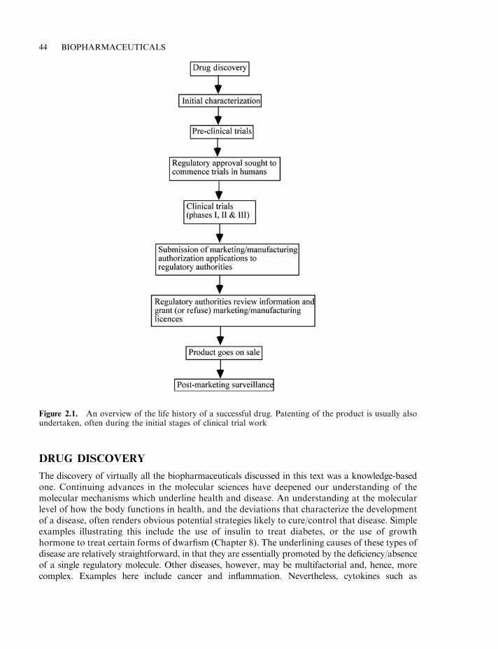
DRUG DISCOVERY
The discovery of virtually all the biopharmaceuticals discussed in this text was a knowledge-basedone. Continuing advances in the molecular sciences have deepened our understanding of themolecular mechanisms which underline health and disease. An understanding at the molecularlevel of how the body functions in health, and the deviations that characterize the developmentof a disease, often renders obvious potential strategies likely to cure/control that disease. Simpleexamples illustrating this include the use of insulin to treat diabetes, or the use of growthhormone to treat certain forms of dwarfism (Chapter 8). The underlining causes of these types ofdisease are relatively straightforward, in that they are essentially promoted by the deficiency/absenceof a single regulatory molecule. Other diseases, however, may be multifactorial and, hence, morecomplex. Examples here include cancer and inflammation. Nevertheless, cytokines such as
44 BIOPHARMACEUTICALS
Figure 2.1. An overview of the life history of a successful drug. Patenting of the product is usually alsoundertaken, often during the initial stages of clinical trial work
interferons and interleukins, known to stimulate the immune response/regulate inflammation, haveproved to be therapeutically useful in treating several such complex diseases (Chapters 4 and 5).
An understanding, at the molecular level, of the actions of various regulatory proteins, or theprogression of a specific disease does not, however, automatically translate into pinpointing aneffective treatment strategy. The physiological responses induced by the potential biopharma-ceutical in vitro (or in animal models) may not accurately predict the physiological responsesseen when the product is administered to a diseased human. For example, many of the mostpromising biopharmaceutical therapeutic agents (e.g. virtually all the cytokines; Chapter 4),display multiple activities on different cell populations. This makes it difficult, if not impossible,to predict what the overall effect administration of any biopharmaceutical will have on thewhole body, hence the requirement for clinical trials.
In other cases, the widespread application of a biopharmaceutical may be hindered by theoccurrence of relatively toxic side effects (as is the case with tumour necrosis factor a (TNF-a),Chapter 5). Finally, some biomolecules have been discovered and purified because of acharacteristic biological activity which, subsequently, was found not to be the molecule’sprimary biological activity. TNF-a again serves as an example. It was first noted because of itscytotoxic effects on some cancer cell types in vitro. Subsequently, trials assessing its therapeuticapplication in cancer proved disappointing, due not only to its toxic side effects but also to itsmoderate, at best, cytotoxic effect on many cancer cell types in vivo. TNF’s major biologicalactivity in vivo is now known to be as a regulator of the inflammatory response.
In summary, the ‘discovery’ of biopharmaceuticals, in most cases, merely relates to the logicalapplication of our rapidly increasing knowledge of the biochemical basis of how the bodyfunctions. These substances could be accurately described as being the body’s own pharmaceuticals.Moreover, rapidly expanding areas of research, such as genomics and proteomics, will likelyhasten the discovery of many more such products, as discussed below.
While biopharmaceuticals are typically proteins derived from the human body, mostconventional drugs have been obtained from sources outside the body (e.g. plant and microbialmetabolites, synthetic chemicals, etc.). Although they do not form the focus of this text, a briefoverview of strategies adopted in the discovery of such ‘non-biopharmaceutical’ drugs isappropriate, and is summarized later in this chapter.
The impact of genomics and related technologies upon drug discovery
The term ‘genomics’ refers to the systematic study of the entire genome of an organism. Its coreaim is to sequence the entire DNA complement of the cell and to physically map the genomearrangement (assign exact positions in the genome to the various genes/non-coding regions).Prior to the 1990s, the sequencing and study of a single gene represented a significant task.However, improvements in sequencing technologies and the development of more highlyautomated hardware systems now render DNA sequencing considerably faster, cheaper andmore accurate. Modern sequencing systems can sequence in excess of 1000 bases/h. Suchinnovations underpin the ‘high-throughput’ sequencing necessary to evaluate an entire genomesequence within a reasonable time frame. As a result, the genomes of almost 70 microorganismshave thus far been completely or almost completely sequenced (Table 2.1). In addition, variouspublic and private bodies are currently sequencing the genomes of various plants and animals,including those of wheat, barley, chicken, dog, cow, pig, sheep, mouse and rat. The human genomeproject commenced in 1990, with an initial target completion date set at 2005. A ‘rough draft’ waspublished in February 2001, with the final completed draft expected in 2003. The total human
THE DRUG DEVELOPMENT PROCESS 45
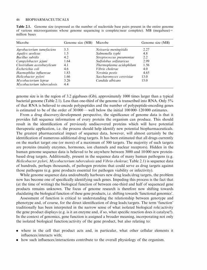
genome size is in the region of 3.2 gigabases (Gb), approximately 1000 times larger than a typicalbacterial genome (Table 2.1). Less than one-third of the genome is transcribed into RNA. Only 5%of that RNA is believed to encode polypeptides and the number of polypeptide-encoding genesis estimated to be of the order of 30 000—well below the initial 100 000–120 000 estimates.
From a drug discovery/development perspective, the significance of genome data is that itprovides full sequence information of every protein the organism can produce. This shouldresult in the identification of previously undiscovered proteins which will have potentialtherapeutic application, i.e. the process should help identify new potential biopharmaceuticals.The greatest pharmaceutical impact of sequence data, however, will almost certainly be theidentification of numerous additional drug targets. It has been estimated that all drugs currentlyon the market target one (or more) of a maximum of 500 targets. The majority of such targetsare proteins (mainly enzymes, hormones, ion channels and nuclear receptors). Hidden in thehuman genome sequence data is believed to be anywhere between 3000 and 10 000 new protein-based drug targets. Additionally, present in the sequence data of many human pathogens (e.g.Helicobacter pylori, Mycobacterium tuberculosis and Vibrio cholerae; Table 2.1) is sequence dataof hundreds, perhaps thousands, of pathogen proteins that could serve as drug targets againstthose pathogens (e.g. gene products essential for pathogen viability or infectivity).
While genome sequence data undoubtedly harbours new drug leads/drug targets, the problemnow has become one of specifically identifying such genes. Impeding this process is the fact that(at the time of writing) the biological function of between one-third and half of sequenced geneproducts remains unknown. The focus of genome research is therefore now shifting towardselucidating the biological function of these gene products, i.e. shifting towards ‘functional genomics’.
Assessment of function is critical to understanding the relationship between genotype andphenotype and, of course, for the direct identification of drug leads/targets. The term ‘function’traditionally has been interpreted in the narrow sense of what isolated biological role/activitythe gene product displays (e.g. is it an enzyme and, if so, what specific reaction does it catalyse?).In the context of genomics, gene function is assigned a broader meaning, incorporating not onlythe isolated biological function/activity of the gene product, but also relating to:
. where in the cell that product acts and, in particular, what other cellular elements itinfluences/interacts with;
. how such influences/interactions contribute to the overall physiology of the organism.
46 BIOPHARMACEUTICALS
Table 2.1. Genome size (expressed as the number of nucleotide base pairs present in the entire genomeof various microorganisms whose genome sequencing is complete/near complete). MB (megabase)=million bases
Microbe Genome size (MB) Microbe Genome size (MB)
Agrobacterium tumefaciens 5.3 Neisseria meningitidis 2.27Aquifex aeolicus 1.5 Salmonella typhi 4.8Bacillus subtilis 4.2 Streptococcus pneumoniae 2.2Campylobacter jejuni 1.64 Sulfolobus solfataricus 2.99Clostridium acetobutylicum 4.1 Thermoplasma acidophilum 1.56Escherichia coli 4.6 Vibrio cholerae 4.0Haemophilus influenzae 1.83 Yersinia pestis 4.65Helicobacter pylori 1.66 Saccharomyces cerevisiae 13.0Mycobacterium leprae 3.26 Candida albicans 15.0Mycobacterium tuberculosis 4.4
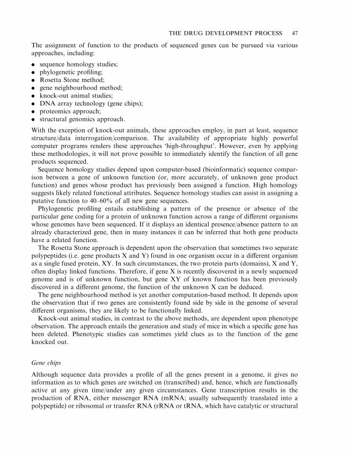
The assignment of function to the products of sequenced genes can be pursued via variousapproaches, including:
. sequence homology studies;
. phylogenetic profiling;
. Rosetta Stone method;
. gene neighbourhood method;
. knock-out animal studies;
. DNA array technology (gene chips);
. proteomics approach;
. structural genomics approach.
With the exception of knock-out animals, these approaches employ, in part at least, sequencestructure/data interrogation/comparison. The availability of appropriate highly powerfulcomputer programs renders these approaches ‘high-throughput’. However, even by applyingthese methodologies, it will not prove possible to immediately identify the function of all geneproducts sequenced.
Sequence homology studies depend upon computer-based (bioinformatic) sequence compar-ison between a gene of unknown function (or, more accurately, of unknown gene productfunction) and genes whose product has previously been assigned a function. High homologysuggests likely related functional attributes. Sequence homology studies can assist in assigning aputative function to 40–60% of all new gene sequences.
Phylogenetic profiling entails establishing a pattern of the presence or absence of theparticular gene coding for a protein of unknown function across a range of different organismswhose genomes have been sequenced. If it displays an identical presence/absence pattern to analready characterized gene, then in many instances it can be inferred that both gene productshave a related function.
The Rosetta Stone approach is dependent upon the observation that sometimes two separatepolypeptides (i.e. gene products X and Y) found in one organism occur in a different organismas a single fused protein, XY. In such circumstances, the two protein parts (domains), X and Y,often display linked functions. Therefore, if gene X is recently discovered in a newly sequencedgenome and is of unknown function, but gene XY of known function has been previouslydiscovered in a different genome, the function of the unknown X can be deduced.
The gene neighbourhood method is yet another computation-based method. It depends uponthe observation that if two genes are consistently found side by side in the genome of severaldifferent organisms, they are likely to be functionally linked.
Knock-out animal studies, in contrast to the above methods, are dependent upon phenotypeobservation. The approach entails the generation and study of mice in which a specific gene hasbeen deleted. Phenotypic studies can sometimes yield clues as to the function of the geneknocked out.
Gene chips
Although sequence data provides a profile of all the genes present in a genome, it gives noinformation as to which genes are switched on (transcribed) and, hence, which are functionallyactive at any given time/under any given circumstances. Gene transcription results in theproduction of RNA, either messenger RNA (mRNA; usually subsequently translated into apolypeptide) or ribosomal or transfer RNA (rRNA or tRNA, which have catalytic or structural
THE DRUG DEVELOPMENT PROCESS 47
functions). The study of under which circumstances an RNA species is expressed/not expressedin the cell/organism can provide clues as to the biological function of the RNA (or, in the case ofmRNA, the function of the final polypeptide product). Furthermore, in the context of druglead/target discovery, the conditions under which a specific mRNA is produced can also point toputative biopharmaceuticals/drug targets. For example, if a particular mRNA is only producedby a cancer cell, that mRNA (or, more commonly, its polypeptide product) may represent agood target for a novel anti-cancer drug.
Levels of RNA (usually specific mRNAs) in a cell can be measured by well-establishedtechniques such as Northern blot analysis or by polymerase chain reaction (PCR) analysis.However, the recent advent of DNA microarray technology has converted the identificationand measurement of specific mRNAs (or other RNAs if required) into a ‘high-throughput’process. DNA arrays are also termed ‘oligonucleotide arrays’, ‘gene chip arrays’ or simply,‘chips’.
The technique is based upon the ability to anchor nucleic acid sequences (usually DNA-based) on plastic/glass surfaces at very high density. Standard griding robots can put on upto 250 000 different short oligonucleotide probes or 10 000 full-length complementary DNA(cDNA) sequences per cm2 of surface. Probe sequences are generally produced/designed fromgenome sequence data and hence chip production is often referred to as ‘downloading thegenome on a chip’. RNA can be extracted from a cell and probed with the chip. Anycomplementary RNA sequences present will hybridize with the appropriate immobilizedchip sequence (Figure 2.2). Hybridization is detectable as the RNA species are firstlabelled. Hybridization patterns obviously yield critical information regarding geneexpression.
48 BIOPHARMACEUTICALS
Figure 2.2. Generalized outline of a gene chip. In this example, short oligonucleotide sequences areattached to the anchoring surface (only the outer rows are shown). Each probe displays a differentnucleotide sequence, and the sequences used are usually based upon genome sequence information. Thesequence of one such probe is shown as AGGCA. By incubating the chip, e.g. with total cellular mRNA,under appropriate conditions, any mRNA with a complementary sequence (UCCGU in the case of theprobe sequence shown) will hybridize with the probes. In reality, probes will have longer sequences thanthe one shown above
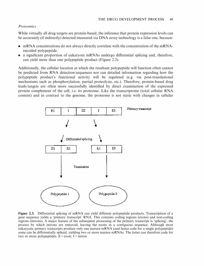
Proteomics
While virtually all drug targets are protein-based, the inference that protein expression levels canbe accurately (if indirectly) detected/measured via DNA array technology is a false one, because:
. mRNA concentrations do not always directly correlate with the concentration of the mRNA-encoded polypeptide.
. a significant proportion of eukaryote mRNAs undergo differential splicing and, therefore,can yield more than one polypeptide product (Figure 2.3).
Additionally, the cellular location at which the resultant polypeptide will function often cannotbe predicted from RNA detection/sequences nor can detailed information regarding how thepolypeptide product’s functional activity will be regulated (e.g. via post-translationalmechanisms such as phosphorylation, partial proteolysis, etc.). Therefore, protein-based drugleads/targets are often more successfully identified by direct examination of the expressedprotein complement of the cell, i.e. its proteome. Like the transcriptome (total cellular RNAcontent) and in contrast to the genome, the proteome is not static with changes in cellular
THE DRUG DEVELOPMENT PROCESS 49
Figure 2.3. Differential splicing of mRNA can yield different polypeptide products. Transcription of agene sequence yields a ‘primary transcript’ RNA. This contains coding regions (exons) and non-codingregions (introns). A major feature of the subsequent processing of the primary transcript is ‘splicing’, theprocess by which introns are removed, leaving the exons in a contiguous sequence. Although mosteukaryotic primary transcripts produce only one mature mRNA (and hence code for a single polypeptide)some can be differentially spliced, yielding two or more mature mRNAs. The latter can therefore code fortwo or more polypeptides. E=exon; I=intron
conditions triggering changes in cellular protein profiles/concentrations. This field of study istermed ‘proteomics’.
Proteomics is, therefore, closely aligned to functional genomics and entails the systematic andcomprehensive analysis of the proteins expressed in the cell and their function. Classicalproteomic studies generally entailed initial extraction of the total protein content from the targetcell/tissue, followed by separation of the proteins therein using two-dimensional (2-D)electrophoresis (Chapter 3). Isolated protein ‘spots’ could then be eluted from the electrophoreticgel and subjected to further analysis; mainly to Edman degradation, in order to generate partialamino acid sequence data. The sequence data could then be used to interrogate protein sequencedatabanks, e.g. in order to assign putative function by sequence homology searches (Figure 2.4).2-D electrophoresis, however, is generally capable of resolving no more than 2000 differentproteins, and proteins expressed at low levels may not be detected at all if their gel concentrationis below the (protein) staining threshold. The latter point can be particularly significant in thecontext of drug/target identification, as most such targets are likely to be kinases and otherregulatory proteins which are generally expressed within cells at very low levels.
More recently, high-resolution chromatographic techniques (particularly reverse-phase andion exchanged-based HPLC) have been applied in the separation of proteome proteins and high-resolution mass spectrometry is being employed to aid high-throughput sequence determination.
Structural genomics
Related to the discipline of proteomics is that of structural genomics. The latter focuses uponthe large-scale, systematic study of gene product structure. While this embraces rRNA andtRNA, in practice the field focuses upon protein structure. The basic approach to structuralgenomics entails the cloning and recombinant expression of cellular proteins, followed by theirpurification and three-dimensional (3-D) structural analysis. High-resolution determination of aprotein’s structure is amongst the most challenging of molecular investigations. By the year2000, protein structure databanks housed in the region of 12 000 entries. However, suchdatabanks are highly redundant, often containing multiple entries describing variants of thesame molecule. For example, in excess of 50 different structures of ‘insulin’ have been deposited(e.g. both native and mutated/engineered forms from various species, as well as insulins in variouspolymeric forms and in the presence of various stabilizers and other chemicals). In reality, by theyear 2000, the 3-D structure of approximately 2000 truly different proteins had been resolved.
Until quite recently, X-ray crystallography was the technique used almost exclusively toresolve the 3-D structure of proteins. As well as itself being technically challenging, a majorlimitation of X-ray crystallography is the requirement for the target protein in crystalline form.It has thus far proved difficult or impossible to induce the majority of proteins to crystallize.Nuclear magnetic resonance (NMR) is an analytical technique which can also be used todetermine the three-dimensional structure of a molecule without the necessity for crystallization.For many years, even the most powerful NMR machines could resolve the 3-D structure of onlyrelatively small proteins (less than 20–25 kDa). However, recent analytical advances now renderit possible to successfully analyse much larger proteins by this technique.
The ultimate goal of structural genomics is to provide a complete 3-D description of any geneproduct. Also, as the structures of more and more proteins of known function are elucidated, itshould become increasingly possible to link specific functional attributes to specific structuralattributes. As such, it may prove ultimately feasible to predict protein function if its structure isknown, and vice versa.
50 BIOPHARMACEUTICALS
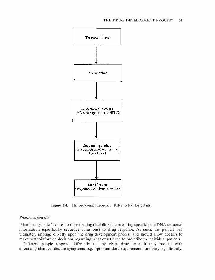
Pharmacogenetics
‘Pharmacogenetics’ relates to the emerging discipline of correlating specific gene DNA sequenceinformation (specifically sequence variations) to drug response. As such, the pursuit willultimately impinge directly upon the drug development process and should allow doctors tomake better-informed decisions regarding what exact drug to prescribe to individual patients.
Different people respond differently to any given drug, even if they present withessentially identical disease symptoms, e.g. optimum dose requirements can vary significantly.
THE DRUG DEVELOPMENT PROCESS 51
Figure 2.4. The proteomics approach. Refer to text for details
Furthermore, not all patients respond positively to a specific drug (e.g. interferon-b is ofclinical benefit to only one in three multiple sclerosis patients; see Chapter 4). The range andseverity of adverse effects induced by a drug can also vary significantly within a patientpopulation base.
While the basis of such differential responses can sometimes be non-genetic (e.g. general stateof health, etc), genetic variation amongst individuals remains the predominant factor. While allhumans display almost identical genome sequences, some differences are evident. The mostprominent widespread-type variations amongst individuals are known as single nucleotidepolymorphisms (SNPs, sometimes pronounced ‘snips’). SNPs occur in the general population atan average incidence of one in every 1000 nucleotide bases and hence the entire human genomeharbours 3 million or so. SNPs are not mutations; the latter arise more infrequently, are morediverse and are generally caused by spontaneous/mutagen-induced mistakes in DNA repair/replication. SNPs occurring in structural genes/gene regulatory sequences can alter amino acidsequence/expression levels of a protein and hence affect its functional attributes. SNPs largelyaccount for natural physical variations evident in the human population (e.g. height, colour ofeyes, etc.).
The presence of a SNP within the regulatory or structural regions of a gene coding for aprotein which interacts with a drug could obviously influence the effect of the drug on the body.In this context, the protein product could, for example, be the drug target or perhaps an enzymeinvolved in metabolizing the drug.
The identification and characterization of SNPs within human genomes is, therefore, of bothacademic and applied interest. Several research groups continue to map human SNPs and over1.5 million have thus far been identified.
By identifying and comparing SNP patterns from a group of patients responsive to aparticular drug with patterns displayed by a group of unresponsive patients, it may be possibleto identify specific SNP characteristics linked to drug efficacy. In the same way, SNP patterns orcharacteristics associated with adverse reactions (or even a predisposition to a disease) may beuncovered. This could usher in a new era of drug therapy, where drug treatment could betailored to the individual patient. Furthermore, different drugs could be developed with theforeknowledge that each would be efficacious when administered to specific (SNP-determined)patient sub-types. A (distant) futuristic scenario could be visualized where all individuals couldcarry chips encoded with SNP details relating to their specific genome, allowing medical staff tochoose the most appropriate drugs to prescribe in any given circumstance.
Linking specific genetic determinants to many diseases, however, is unlikely to be asstraightforward as implied thus far. The progress of most diseases, and the relative effectivenessof allied drug treatment, is dependent upon many factors, including the interplay of multiplegene products. ‘Environmental’ factors, such as patient age, sex and general health, also play aprominent role.
The term ‘pharmacogenomics’ is one which has entered the ‘genomic’ vocabulary. Althoughsometimes used almost interchangeably with pharmacogenetics, it more specifically refers tostudying the pattern of expression of gene products involved in a drug response.
Plants as a source of drugs
Traditionally, drug discovery programmes within the pharmaceutical industry relied heavilyupon screening various biological specimens for potential drugs. Prior to the 1950s, the vastbulk of drug substances discovered were initially extracted from vascular plants (see also
52 BIOPHARMACEUTICALS
Chapter 1). Examples include digoxin and digitoxin, originally isolated from the foxglove (amember of the genus Digitalis), as well as aspirin, codeine and taxol.
Today, well over 100 drugs (accounting for 25% of all prescriptions issued in the USA), wereinitially isolated from vascular plants. While some are still extracted from their native source,most are now obtained more cheaply and easily by direct chemical synthesis or semi-synthesis.
Plants are a rich potential source of drugs as they produce a vast array of novel bioactivemolecules, many of which probably serve as chemical defences against infection or predation. Inaddition, the variety of different plant species present on the earth is staggering. There exist wellover 265 000 flowering species alone, of which less that 1% have, thus far, been screened for thepresence of any bioactive molecules of potential therapeutic use.
Two screening approaches may be adopted with respect to plants. The most straightforwardentails random collection of vegetation in areas supporting a diversity of plant growth.Although there have been a few notable successes recorded (e.g. taxol) by pursuing thisapproach, the success rate of identifying a new useful drug is quite low.
Targeted screening approaches specifically zone in upon plants more likely to containbioactive molecules in the first place. For example, plants which seem to be immune frompredation may well be producing substances toxic to, for example, insects. These in turn are alsolikely to have some effect on human cells.
A more commonly employed targeted search strategy is that of the ethnobotanical approach(ethnobotany is the study of the relationships between plants and people). This entailsinteraction of the drug discoverer with indigenous communities in areas where herbal or plant-based medicines form the basis of therapeutic intervention. Researchers simply collect plantspecimens used as local cures. Furthermore if, for example, interest is focused upon discoveringan antiviral agent, plants used in the treatment of diseases known to be caused by viruses aregiven special attention (in some instances, scientists have even used non-human guides intargeting certain plants, e.g. some have studied the plant types fed to sick monkeys by othermembers of the monkey troop).
The collection procedure itself is straightforward. After cataloguing and identification, 1–2 kgof the plant material is dried, or stored in alcohol and brought back to the lab. The plantmaterial is crushed and extracted with various solvents (most plant-derived bioactive moleculesare low molecular mass substances, soluble in organic solvents of varying polarity). Afterremoval of the solvent, the extracts are screened for desirable biological activities (e.g. inhibitionof microbial growth, selective toxicity towards various human cancer cell lines, etc.).
If an interesting activity is described, larger quantities (10–100 kg) of the plant material arecollected, from which chemists purify and characterize the active principle. The active principleis known as a ‘lead compound’. Chemists will then usually attempt to modify the leadcompound in order to render it more therapeutically useful (e.g. make it more potent, orperhaps increase its hydrophobicity so that it can pass through biological membranes). This isthen subjected to further pre-clinical trials, and chemists determine whether an economicallyfeasible method, allowing the drug’s chemical synthesis, can be developed.
Microbial drugs
Microorganisms, particularly bacteria and fungi, have also proved to be a rich source ofbioactive molecules such as antibiotics and anticancer agents (Chapter 1). Again, as in the caseof plants, an incredibly rich array of microbial species inhabit the earth and their characteristicmetabolic flexibility generates an enormous bank of potential pharmaceutical products. Most
THE DRUG DEVELOPMENT PROCESS 53
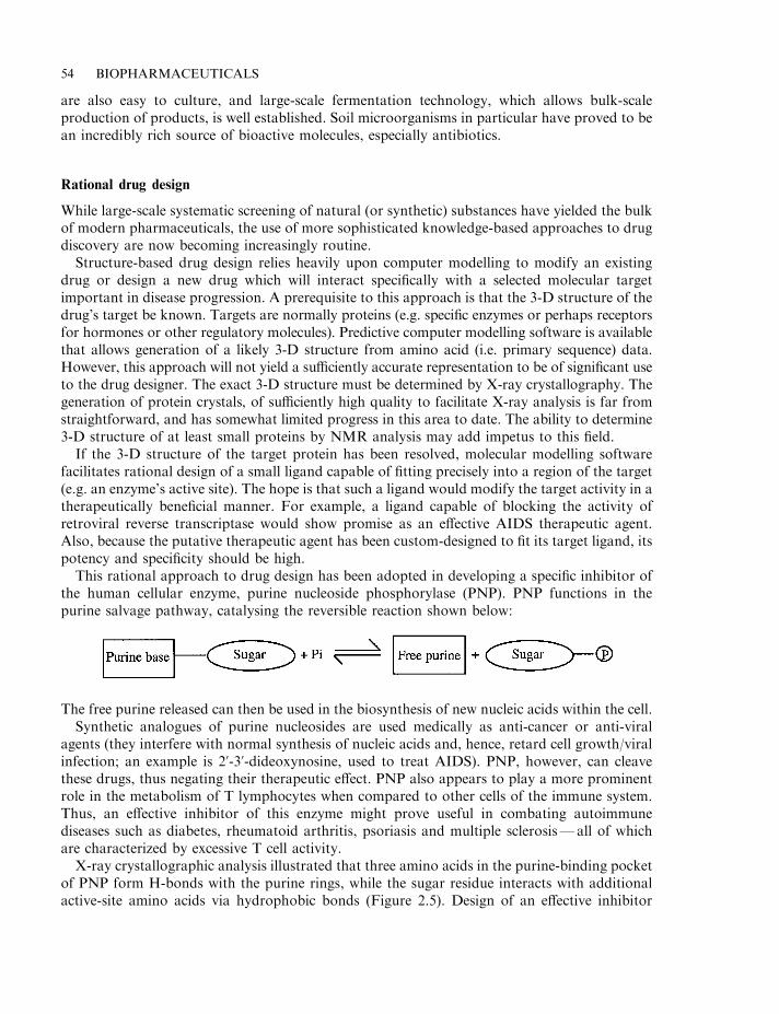
are also easy to culture, and large-scale fermentation technology, which allows bulk-scaleproduction of products, is well established. Soil microorganisms in particular have proved to bean incredibly rich source of bioactive molecules, especially antibiotics.
Rational drug design
While large-scale systematic screening of natural (or synthetic) substances have yielded the bulkof modern pharmaceuticals, the use of more sophisticated knowledge-based approaches to drugdiscovery are now becoming increasingly routine.
Structure-based drug design relies heavily upon computer modelling to modify an existingdrug or design a new drug which will interact specifically with a selected molecular targetimportant in disease progression. A prerequisite to this approach is that the 3-D structure of thedrug’s target be known. Targets are normally proteins (e.g. specific enzymes or perhaps receptorsfor hormones or other regulatory molecules). Predictive computer modelling software is availablethat allows generation of a likely 3-D structure from amino acid (i.e. primary sequence) data.However, this approach will not yield a sufficiently accurate representation to be of significant useto the drug designer. The exact 3-D structure must be determined by X-ray crystallography. Thegeneration of protein crystals, of sufficiently high quality to facilitate X-ray analysis is far fromstraightforward, and has somewhat limited progress in this area to date. The ability to determine3-D structure of at least small proteins by NMR analysis may add impetus to this field.
If the 3-D structure of the target protein has been resolved, molecular modelling softwarefacilitates rational design of a small ligand capable of fitting precisely into a region of the target(e.g. an enzyme’s active site). The hope is that such a ligand would modify the target activity in atherapeutically beneficial manner. For example, a ligand capable of blocking the activity ofretroviral reverse transcriptase would show promise as an effective AIDS therapeutic agent.Also, because the putative therapeutic agent has been custom-designed to fit its target ligand, itspotency and specificity should be high.
This rational approach to drug design has been adopted in developing a specific inhibitor ofthe human cellular enzyme, purine nucleoside phosphorylase (PNP). PNP functions in thepurine salvage pathway, catalysing the reversible reaction shown below:
The free purine released can then be used in the biosynthesis of new nucleic acids within the cell.Synthetic analogues of purine nucleosides are used medically as anti-cancer or anti-viral
agents (they interfere with normal synthesis of nucleic acids and, hence, retard cell growth/viralinfection; an example is 2’-3’-dideoxynosine, used to treat AIDS). PNP, however, can cleavethese drugs, thus negating their therapeutic effect. PNP also appears to play a more prominentrole in the metabolism of T lymphocytes when compared to other cells of the immune system.Thus, an effective inhibitor of this enzyme might prove useful in combating autoimmunediseases such as diabetes, rheumatoid arthritis, psoriasis and multiple sclerosis—all of whichare characterized by excessive T cell activity.
X-ray crystallographic analysis illustrated that three amino acids in the purine-binding pocketof PNP form H-bonds with the purine rings, while the sugar residue interacts with additionalactive-site amino acids via hydrophobic bonds (Figure 2.5). Design of an effective inhibitor
54 BIOPHARMACEUTICALS
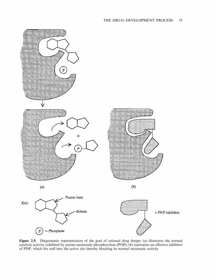
THE DRUG DEVELOPMENT PROCESS 55
Figure 2.5. Diagramatic representation of the goal of rational drug design: (a) illustrates the normalcatalytic activity exhibited by purine nucleoside phosphorylase (PNP); (b) represents an effective inhibitorof PNP, which fits well into the active site thereby blocking its normal enzymatic activity
centred around developing a molecule that fitted into the active site, and engaged in strongerH-bonding or additional hydrophobic interactions with the active site residues.
Without using computer modelling, identification of a potent inhibitor would, on average,require screening of hundreds of thousands of candidates, take up to 10 years and cost severalmillion dollars. With computer modelling, the time and cost are cut to a fraction of this and, inthis case, less than 60 compounds were prepared/modified to yield a highly effective inhibitor,BCX-34. This inhibitor molecule contains a 9-deazaguanine group (to fill the sugar binding site)and an acetate group (to fill the phosphate binding site). It is 100 times more potent than thebest inhibitor previously identified by classical techniques, and it has performed well in initialclinical trials in treating psoriasis and cutaneous T cell lymphoma.
Combinatorial approaches to drug discovery
The concept of rational drug design drew emphasis away from the traditional random screeningapproach to drug discovery. However, the evelopment of techniques capable of generating largenumbers of novel synthetic chemicals (combinatorial libraries), coupled with high-throughputscreening methods, has generated fresh interest in the random screening approach.
Libraries can be generated relatively inexpensively and in a short time period. While manylarger pharmaceutical companies maintain libraries of several hundred thousand natural andsynthetic compounds, combinatorial chemistry can generate millions of compounds withrelative ease. Peptides, for example, play various regulatory roles in the body, and several enjoytherapeutic application (e.g. GnRH; Chapter 8). Synthetic peptide libraries, containing peptidesdisplaying millions of amino acid sequence combinations, can now be generated bycombinatorial chemistry with minimal requirement for expensive equipment. Two approachescan be followed to generate a combinatorial peptide library: ‘split synthesis’ and ‘T-bagsynthesis’.
The split level approach, for example, results in the creation of a large peptide library in whichthe peptides are grown on small synthetic beads. Peptides grown on any single bead will all be ofidentical amino acid sequences. Individual amino acids can be coupled to the growing chain bystraightforward solid phase peptide synthesis techniques (Box 2.1). In the split-level approach, apool of beads are equally distributed into separate reaction vessels, each containing a singleamino acid in solution. After chemical coupling to the beads, the beads are recovered, pooledand randomly distributed into the reaction vessels once more. This cycle can be repeated severaltimes to extend the peptide chain. A simplified example is provided in Figure 2.6. While theexample presented in Figure 2.6 is straightforward, a hexapeptide library containing everypossible combination of all 20 commonly occurring amino acids would contain 64 million (206)different peptide species. Such a library, although more cumbersome, would be generated usingexactly the same strategy.
The combinatorial approach is characterized not only by rapid synthesis of vast peptidelibraries but also by rapid screening of these libraries (i.e. screening of the library to locate anypeptides capable of binding to a ligand of interest—perhaps an enzyme or a hormone receptor).The soluble ligand is first labelled with an easily visible tag, often a fluorescent tag. This is thenadded to the beads. Binding of the ligand to a particular peptide will effectively result in stainingof the bead to which the peptide is attached. This bead can then be physically separated from theother beads, e.g. using a microforceps. The isolated bead is then washed in 8M guanidinehydrochloride (to remove the screening ligand) and the sequence of the attached peptide is thenelucidated using a microsequencer. Typically, a bead will have 50–200 pmol of peptide attached,
56 BIOPHARMACEUTICALS

while the lower limit of sensitivity of most microsequencers is of the order of 5 pmol. Once itsamino acid sequence is elucidated, it can be synthesized in large quantities for further study.
A library containing several million beads can be screened in a single afternoon. Furthermore,the library is reusable, as it may be washed in 8M guanidine hydrochloride and then re-screenedusing a different probe. This split synthesis approach displays the ability to generate peptidelibraries of incredible variety, variety that can be further expanded by incorporation of, forexample, D-amino acids or rarely occurring amino acids.
Overall, therefore, various approaches may be adopted in the quest to discover new drugs.The approach generally adopted in biopharmacentical discovery differs from most otherapproaches in that biopharmaceuticals are produced naturally in the body. Discovery of abiopharmaceutical product, therefore, becomes a function of an increased knowledge of how thebody itself functions. After its initial discovery, the physicochemical and biologicalcharacteristics of the potential drug can then be studied.
Initial product characterization
The physicochemical and other properties of any newly identified drug must be extensivelycharacterized prior to its entry into clinical trials. As the vast bulk of biopharmaceuticals areproteins, a summary overview of the approach taken to initial characterization of thesebiomolecules is presented. A prerequisite to such characterization is initial purification of theprotein. Purification to homogeneity usually requires a combination of three or more high-resolution chromatographic steps. The purification protocol is designed carefully, as it usuallyforms the basis of subsequent pilot and process-scale purification systems. The purified productis then subjected to a battery of tests, which aim to characterize it fully. Moreover, once thesecharacteristics have been defined, they form the basis of many of the quality control (QC)identity tests routinely performed on the product during its subsequent commercialmanufacture. As these identity tests are discussed in detail in Chapter 3, only an abbreviatedoverview is presented here, in the form of Figure 2.7.
In addition to the studies listed in Figure 2.7, stability characteristics of the protein, e.g. withregard to temperature, pH and incubation with various potential excipients, are undertaken.Such information is required in order to identify a suitable final product formulation, and togive an early indication of the likely useful shelf-life of the product.
PATENTING
The discovery and initial characterization of any substance of potential pharmaceuticalapplication is followed by its patenting. The more detail given relevant to the drug’sphysicochemical characteristics, a method of synthesis and its biological effects, the better thechances of successfully securing a patent. Thus patenting may not take place until pre-clinicaltrials and phase I clinical trials are completed. Patenting, once successfully completed, does notgrant the patent holder an automatic right to utilize/sell the patented product— it must first beproved safe and effective in subsequent clinical trials and then be approved for general medicaluse by the relevant regulatory authorities.
What is a patent and what is patentable?
A patent may be described as a monopoly granted by a government to an inventor, such thatonly the inventor may exploit the invention/innovation for a fixed period of time (up to 20
THE DRUG DEVELOPMENT PROCESS 57
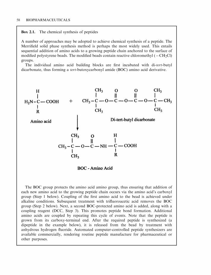
58 BIOPHARMACEUTICALS
Box 2.1. The chemical synthesis of peptides
A number of approaches may be adopted to achieve chemical synthesis of a peptide. TheMerrifield solid phase synthesis method is perhaps the most widely used. This entailssequential addition of amino acids to a growing peptide chain anchored to the surface ofmodified polystyrene beads. The modified beads contain reactive chloromethyl (7CH2Cl)groups.
The individual amino acid building blocks are first incubated with di-tert-butyldicarbonate, thus forming a tert-butoxycarbonyl amide (BOC) amino acid derivative.
The BOC group protects the amino acid amino group, thus ensuring that addition ofeach new amino acid to the growing peptide chain occurs via the amino acid’s carboxylgroup (Step 1 below). Coupling of the first amino acid to the bead is achieved underalkaline conditions. Subsequent treatment with trifluoroacetic acid removes the BOCgroup (Step 2 below). Next, a second BOC-protected amino acid is added, along with acoupling reagent (DCC, Step 3). This promotes peptide bond formation. Additionalamino acids are coupled by repeating this cycle of events. Note that the peptide isgrown from its carboxy-terminal end. After the required peptide is synthesized (adipeptide in the example below), it is released from the bead by treatment withanhydrous hydrogen fluoride. Automated computer-controlled peptide synthesizers areavailable commercially, rendering routine peptide manufacture for pharmaceutical orother purposes.
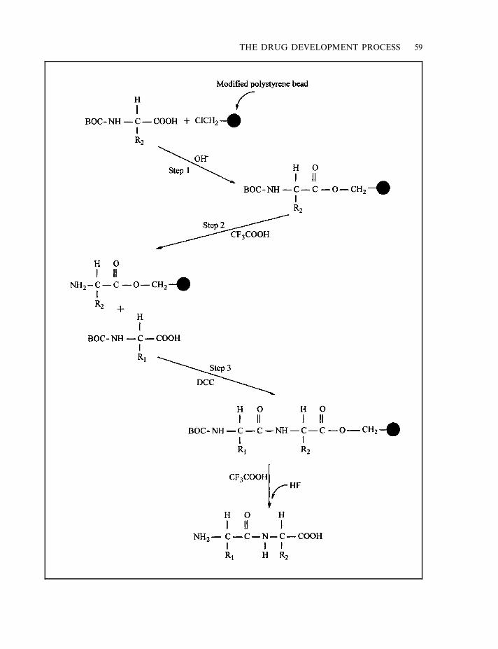
THE DRUG DEVELOPMENT PROCESS 59
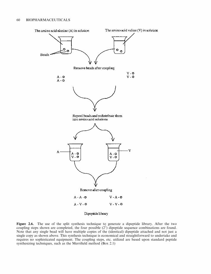
60 BIOPHARMACEUTICALS
Figure 2.6. The use of the split synthesis technique to generate a dipeptide library. After the twocoupling steps shown are completed, the four possible (22) dipeptide sequence combinations are found.Note that any single bead will have multiple copies of the (identical) dipeptide attached and not just asingle copy as shown above. This synthesis technique is economical and straightforward to undertake andrequires no sophisticated equipment. The coupling steps, etc. utilized are based upon standard peptidesynthesizing techniques, such as the Merrifield method (Box 2.1)
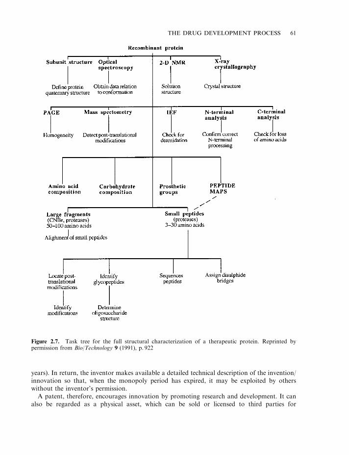
years). In return, the inventor makes available a detailed technical description of the invention/innovation so that, when the monopoly period has expired, it may be exploited by otherswithout the inventor’s permission.
A patent, therefore, encourages innovation by promoting research and development. It canalso be regarded as a physical asset, which can be sold or licensed to third parties for
THE DRUG DEVELOPMENT PROCESS 61
Figure 2.7. Task tree for the full structural characterization of a therapeutic protein. Reprinted bypermission from Bio/Technology 9 (1991), p. 922
cash. Patents also represent a unique source of technical information regarding the patentedproduct.
The philosophy underlining patent law is fairly similar throughout the world. Thus, althoughthere is no worldwide patenting office, patent practice in different world regions is often quitesimilar. This is fortuitous, as there is a growing tendency towards world harmonization ofpatent law, fuelled by multinational trade agreements.
In order to be considered patentable, an invention/innovation must satisfy several criteria, themost important four of which are:
. novelty;
. non-obviousness;
. sufficiency of disclosure;
. utility.
To be novel, the invention/innovation should not be what is described as ‘prior art’, i.e. itshould not be something already known or described previously. Prior disclosure of theinvention/innovation, e.g. by publishing its details in a scientific (or other) journal, or a verbaldescription given, e.g. at a scientific meeting, puts this innovation in the public arena. This caneffectively make it ‘prior art’. While this can hinder a subsequent patent application, it may notbe entirely fatal. To preclude a successful subsequent patent application, prior publicationusually must be ‘enabling’, i.e. the detail given must be sufficient to allow an average person,familiar with the discipline relating to the patent (i.e. ‘a person with ordinary skill andknowledge in the art’), to repeat the experiment or process described. A bare prior disclosure,which describes the innovation but not to an enabling level, will not normally prevent itssubsequent patenting. Patenting law in relation to this point is more flexible in the USA. Here,even in the case of an existing enabling publication, patenting is usually possible as long as thepatent is filed in under 1 year after the date of that publication.
The USA, unlike many other world regions, also adopts the ‘first to invent’ principle. Putsimply (although patent disputes are rarely simple), if two inventors file similar patents, thepatent will be granted to the one who proves that he/she was the first to ‘invent’ the ‘product’,even if he/she was not the first to file for a patent. Proving you were the first to invent can becomplex and usually hinges around the availability of full and detailed laboratory notebooks orother records as to how and when the invention was made.
Non-obviousness (inventiveness) means that the invention/innovation process must not besomething that would be immediately obvious to somebody skilled in the art. Non-obviousness,although it sounds straightforward, is often a difficult concept to apply in practice. Obviousnesscould be described as a simple and logical progression of prior art, thus non-obviousnessrequires an additional ingredient of inspiration or often chanced good luck.
Sufficiency of disclosure is a more straightforward requirement. Sufficient technical detailmust be provided in the patent application such that somebody of ordinary technical skill in thearea could reproduce/repeat the innovation. Utility or industrial applicability is the last majorprerequisite to patenting. This simply means that the innovation must have some applied use.
Patent types
Patent types can be subdivided into ‘Product’, ‘Process’ and ‘Use’ patents. In the first case, aspecific substance is patented (e.g. a revolutionary new car engine, a new cytokine withapplicability in cancer treatment, a novel microorganism capable of degrading oil, etc.). In the
62 BIOPHARMACEUTICALS
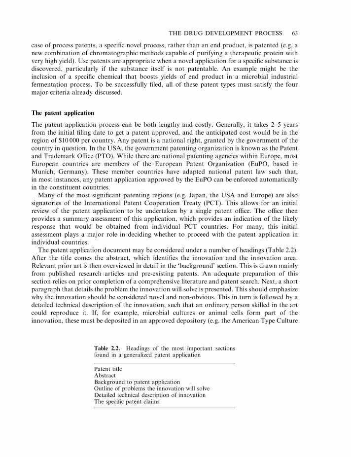
case of process patents, a specific novel process, rather than an end product, is patented (e.g. anew combination of chromatographic methods capable of purifying a therapeutic protein withvery high yield). Use patents are appropriate when a novel application for a specific substance isdiscovered, particularly if the substance itself is not patentable. An example might be theinclusion of a specific chemical that boosts yields of end product in a microbial industrialfermentation process. To be successfully filed, all of these patent types must satisfy the fourmajor criteria already discussed.
The patent application
The patent application process can be both lengthy and costly. Generally, it takes 2–5 yearsfrom the initial filing date to get a patent approved, and the anticipated cost would be in theregion of $10 000 per country. Any patent is a national right, granted by the government of thecountry in question. In the USA, the government patenting organization is known as the Patentand Trademark Office (PTO). While there are national patenting agencies within Europe, mostEuropean countries are members of the European Patent Organization (EuPO, based inMunich, Germany). These member countries have adapted national patent law such that,in most instances, any patent application approved by the EuPO can be enforced automaticallyin the constituent countries.
Many of the most significant patenting regions (e.g. Japan, the USA and Europe) are alsosignatories of the International Patent Cooperation Treaty (PCT). This allows for an initialreview of the patent application to be undertaken by a single patent office. The office thenprovides a summary assessment of this application, which provides an indication of the likelyresponse that would be obtained from individual PCT countries. For many, this initialassessment plays a major role in deciding whether to proceed with the patent application inindividual countries.
The patent application document may be considered under a number of headings (Table 2.2).After the title comes the abstract, which identifies the innovation and the innovation area.Relevant prior art is then overviewed in detail in the ‘background’ section. This is drawn mainlyfrom published research articles and pre-existing patents. An adequate preparation of thissection relies on prior completion of a comprehensive literature and patent search. Next, a shortparagraph that details the problem the innovation will solve is presented. This should emphasizewhy the innovation should be considered novel and non-obvious. This in turn is followed by adetailed technical description of the innovation, such that an ordinary person skilled in the artcould reproduce it. If, for example, microbial cultures or animal cells form part of theinnovation, these must be deposited in an approved depository (e.g. the American Type Culture
THE DRUG DEVELOPMENT PROCESS 63
Table 2.2. Headings of the most important sectionsfound in a generalized patent application
Patent titleAbstractBackground to patent applicationOutline of problems the innovation will solveDetailed technical description of innovationThe specific patent claims
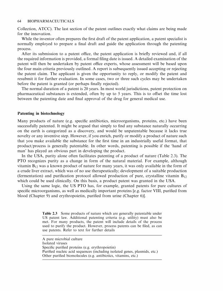
Collection, ATCC). The last section of the patent outlines exactly what claims are being madefor the innovation.
While the inventor often prepares the first draft of the patent application, a patent specialist isnormally employed to prepare a final draft and guide the application through the patentingprocess.
After its submission to a patent office, the patent application is briefly reviewed and, if allthe required information is provided, a formal filing date is issued. A detailed examination of thepatent will then be undertaken by patent office experts, whose assessment will be based uponthe four main criteria previously outlined. A report is subsequently issued accepting or rejectingthe patent claim. The applicant is given the opportunity to reply, or modify the patent andresubmit it for further evaluation. In some cases, two or three such cycles may be undertakenbefore the patent is granted (or perhaps finally rejected).
The normal duration of a patent is 20 years. In most world jurisdictions, patent protection onpharmaceutical substances is extended, often by up to 5 years. This is to offset the time lostbetween the patenting date and final approval of the drug for general medical use.
Patenting in biotechnology
Many products of nature (e.g. specific antibiotics, microorganisms, proteins, etc.) have beensuccessfully patented. It might be argued that simply to find any substance naturally occurringon the earth is categorized as a discovery, and would be unpatentable because it lacks truenovelty or any inventive step. However, if you enrich, purify or modify a product of nature suchthat you make available the substance for the first time in an industrially useful format, thatproduct/process is generally patentable. In other words, patenting is possible if the ‘hand ofman’ has played an obvious part in developing the product.
In the USA, purity alone often facilitates patenting of a product of nature (Table 2.3). ThePTO recognizes purity as a change in form of the natural material. For example, althoughvitamin B12 was a known product of nature for many years, it was only available in the form ofa crude liver extract, which was of no use therapeutically; development of a suitable production(fermentation) and purification protocol allowed production of pure, crystalline vitamin B12
which could be used clinically. On this basis, a product patent was granted in the USA.Using the same logic, the US PTO has, for example, granted patents for pure cultures of
specific microorganisms, as well as medically important proteins [e.g. factor VIII, purified fromblood (Chapter 9) and erythropoietin, purified from urine (Chapter 6)].
64 BIOPHARMACEUTICALS
Table 2.3 Some products of nature which are generally patentable underUS patent law. Additional patenting criteria (e.g. utility) must also bemet. For many products, the patent will include details of the processused to purify the product. However, process patents can be filed, as canuse patents. Refer to text for further details
A pure microbial cultureIsolated virusesSpecific purified proteins (e.g. erythropoietin)Purified nucleic acid sequences (including isolated genes, plasmids, etc.)Other purified biomolecules (e.g. antibiotics, vitamins, etc.)
Rapid technological advances in the biological sciences raises complex patenting issues, andpatenting law as applied to modern biotechnology is still evolving.
In the late 1980s, the PTO confirmed that they would consider issuing patents for non-humanmulticellular organisms, including animals. The first transgenic animal was patented in 1988 byHarvard University. The ‘Harvard mouse’ carried a gene which made it more susceptible tocancer and, hence, more sensitive in detecting possible carcinogens.
Another area of biotechnology patent law relates to the patenting of genes and DNAsequences. Thus far, patents have been issued for some human genes, largely on the basis of theuse of their cloned products (e.g. erythropoietin and tissue plasminogen activator). The humangenome project continues to make available vast quantities of DNA sequence information—ofboth genes and non-coding regions. Moreover, the function of the bulk of the putative genessequences at the moment remain unknown.
Most of the groups working on this project are funded by public bodies. Some sequencinggroups, however, are funded by private companies. These wish to see a payback on theirinvestment. Gaining a patent for a nucleotide sequence that is of medical or diagnostic usewould provide very significant payback potential.
Patenting DNA sequences came under heavy legal and public scrutiny in 1992, when the USNational Institutes of Health (NIH) filed a patent application on partial human cDNAsequences of unknown function. This patent was rejected, and the consensus has emerged thatpatent protection should only be considered for nucleotide sequences that can be used forspecific purposes, e.g. for a sequence which can serve as a diagnostic marker or codes for aprotein product of medical value. This appears to be a reasonable approach, as it balances issuesof public interest with encouraging innovation in the area.
The issue of patenting genetic material or transgenic plants or animals remains a contentiousone. The debate is not confined to technical and legal argument—ethical and politicalissues, including public opinion, also impinge on the decision-making process. The increasingtechnical complexity and sophistication of the biological principles and processes upon whichbiotechnological innovations are based also render resolution of legal patenting issues moredifficult.
A major step in clarifying EU-wide law with regard to patenting in biotechnology stems fromthe introduction of the 1998 European patent directive. This directive (EU law) confirms thatbiological materials (e.g. specific cells, proteins, genes, nucleotide sequences, antibiotics, etc.)which previously existed in nature, are potentially patentable. However, in order to actually bepatentable, they must (a) be isolated/purified from their natural environment and/or beproduced via a technical process (e.g. recombinant DNA technology in the case of recombinantproteins), and (b) they must conform to the general patentability principles regarding novelty,non-obviousness, utility and sufficiency of disclosure. The ‘utility’ condition, therefore, in effectprevents patenting of gene/genome sequences of unknown function. The directive also prohibitsthe possibility of patenting inventions if their exploitation would be contrary to public order ormorality. Thus, it is not possible to patent:
. the human body;
. the cloning of humans;
. the use of human embryos for commercial purposes;
. modifying germ line identity in humans;
. modifying the genetic complement of an animal if the modifications cause suffering withoutresultant substantial medical benefits to the animal or to humans.
THE DRUG DEVELOPMENT PROCESS 65
DELIVERY OF BIOPHARMACEUTICALS
An important issue, which must be addressed during the pre-clinical phase of the drugdevelopment process, relates to the route by which the drug will be delivered or administered.To date, the vast majority of biopharmaceuticals approved for general medical use areadministered by direct injection (i.e. parenterally), usually either intravenously (i.v.),subcutaneously (s.c.—directly under the skin) or intramuscularly (i.m.— into muscle tissue);s.c. or i.m. administration is generally followed by slow release of the drug from its depot siteinto the bloodstream. Amongst the few exceptions to this parenteral route are the enzymeDNase, used to treat cystic fibrosis (Chapter 9), and platelet-derived growth factor (PDGF),used to treat certain skin ulcers (Chapter 7). However, neither of these products is required toreach the bloodstream in order to achieve its therapeutic effect. In fact, in each case, the deliverysystem delivers the biopharmaceutical directly to its site of action (DNase is delivered directly tothe lungs via aerosol inhalation, while PDGF is applied topically—directly on the ulcersurface—as a gel).
Parenteral administration is not perceived as a problem in the context of drugs that areadministered infrequently, or as a once-off dose to a patient. However, in the case of productsadministered frequently or daily (e.g. insulin to diabetics), non-parenteral delivery routes wouldbe preferred. Such routes would be more convenient, less invasive, less painful and generallywould achieve better patient compliance. Alternative potential delivery routes include oral,nasal, transmucosal, transdermal or pulmonary routes. While such routes have proved possiblein the context of many drugs, routine administration of biopharmaceuticals by such means hasproved to be technically challenging. Obstacles encountered include their high molecular mass,their susceptibility to enzymatic inactivation and their potential to aggregate.
Oral delivery systems
Oral delivery is usually the preferred system for drug delivery, due to its convenience and thehigh level of associated patient compliance generally attained. Biopharmaceutical delivery viathis route has proved problematic for a number of reasons:
. inactivation due to stomach acid. Prior to consumption of a meal, stomach pH is usuallybelow 2.0. Although the buffering action of food can increase the pH to neutrality, theassociated stimulation of stomach acid secretion subsequently reduces the ambient pH backdown to 3.0–3.5. Virtually all biopharmaceuticals are acid-labile and are inactivated at lowpH values;
. inactivation due to digestive proteases. Therapeutic proteins would represent potentialtargets for digestive proteases such as pepsin, trypsin and chymotrypsin;
. their (relatively) large size and hydrophilic nature renders difficult the passage of intactbiopharmaceuticals across the intestinal mucosa;
. orally absorbed drugs are subjected to first-pass metabolism. Upon entry into thebloodstream, the first organ encountered is the liver, which usually removes a significantproportion of absorbed drugs from circulation.
Given suchdifficulties, it is not unsurprising that bioavailabilities below1%areoften recorded inthe context of oral biopharmaceutical drug delivery. Strategies pursued to improve bioavailabilityinclude physically protecting the drug via encapsulation, formulation as microemulsions/microparticulates, as well as inclusion of protease inhibitors and permeability enhancers.
66 BIOPHARMACEUTICALS
Encapsulation within an enteric coat (resistant to low pH values) protects the product duringstomach transit. Microcapsules/spheres utilized have been made from various polymericsubstances, including cellulose, polyvinyl alcohol, polymethylacrylates and polystyrene.Delivery systems based upon the use of liposomes and cyclodextrin-protective coats have alsobeen developed. Included in some such systems also are protease inhibitors, such as aprotininand ovomucoids. Permeation enhancers employed are usually detergent-based substances,which can enhance absorption through the gastrointestinal lining.
More recently, increasing research attention has focused upon the use of ‘mucoadhesivedelivery systems’ in which the biopharmaceutical is formulated with/encapsulated in moleculeswhich interact with the intestinal mucosa membranes. The strategy is obviously to retain thedrug at the absorbing surface for a prolonged period. Non-specific (charge-based) interactionscan be achieved by the use of polyacrylic acid, while more biospecific interactions are achievedby using selected lectins or bacterial adhesion proteins. Despite intensive efforts, however, thesuccessful delivery of biopharmaceuticals via the oral route remains some way off.
Pulmonary delivery
Pulmonary delivery currently represents the most promising alternative to parenteral deliverysystems for biopharmaceuticals. While the lung is not particularly permeable to solutes of lowmolecular mass (e.g. sucrose or urea), macromolecules can be absorbed into the blood via thelungs surprisingly well. In fact, pulmonary macromolecular absorption generally appears to beinversely related to molecular mass, up to a mass of about 500 kDa. Many peptides/proteinsdelivered to the deep lung are detected in the blood within minutes and bioavailabilitiesapproaching or exceeding 50% (relative to subcutaneous injection) have been reported fortherapeutic proteins such as colony-stimulating factors and some interferons. Although notcompletely understood, such high pulmonary bioavailability may stem from:
. the lungs’ very large surface area;
. their low surface fluid volume;
. thin diffusional layer;
. relatively slow cell surface clearance;
. the presence of proteolytic inhibitors.
Additional advantages associated with the pulmonary route include:
. the avoidance of first-pass metabolism;
. the availability of reliable, metered nebulizer-based delivery systems capable of accuratedosage delivery, in either powder or liquid form;
. levels of absorption achieved without the need to include penetration enhancers, which aregenerally too irritating for long-term use.
Although obviously occurring in practice, macromolecules absorbed via the pulmonary routemust cross a number of biological barriers to get into the blood. These are:
. a protective monolayer of insoluble phospholipid, termed ‘lung surfactant’, and itsunderlying surface lining fluid, which lies immediately above the lung epithelial cells;
. the epithelial cells lining the lung;
. the interstitium (an extracellular space) and the basement membrane, composed of a layer ofinterstitial fibrous material;
THE DRUG DEVELOPMENT PROCESS 67
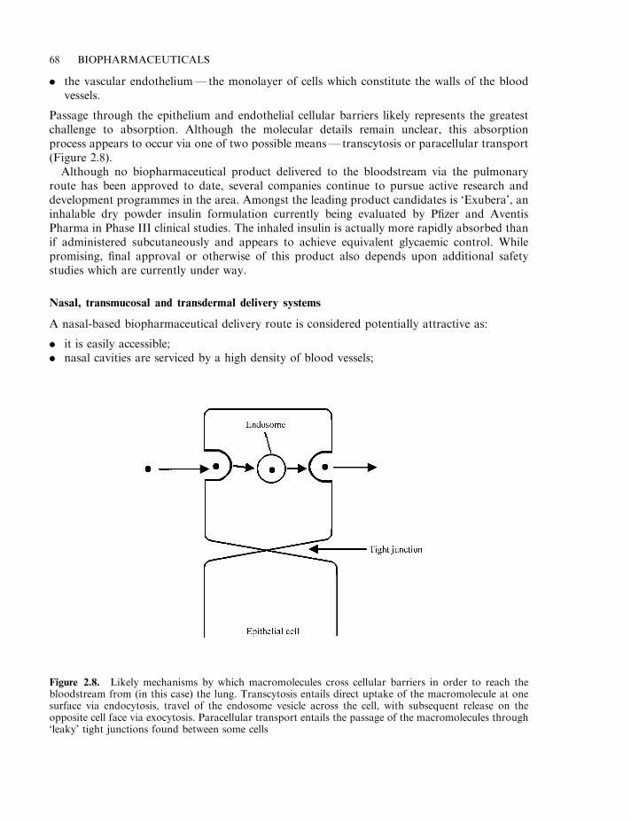
. the vascular endothelium—the monolayer of cells which constitute the walls of the bloodvessels.
Passage through the epithelium and endothelial cellular barriers likely represents the greatestchallenge to absorption. Although the molecular details remain unclear, this absorptionprocess appears to occur via one of two possible means— transcytosis or paracellular transport(Figure 2.8).
Although no biopharmaceutical product delivered to the bloodstream via the pulmonaryroute has been approved to date, several companies continue to pursue active research anddevelopment programmes in the area. Amongst the leading product candidates is ‘Exubera’, aninhalable dry powder insulin formulation currently being evaluated by Pfizer and AventisPharma in Phase III clinical studies. The inhaled insulin is actually more rapidly absorbed thanif administered subcutaneously and appears to achieve equivalent glycaemic control. Whilepromising, final approval or otherwise of this product also depends upon additional safetystudies which are currently under way.
Nasal, transmucosal and transdermal delivery systems
A nasal-based biopharmaceutical delivery route is considered potentially attractive as:
. it is easily accessible;
. nasal cavities are serviced by a high density of blood vessels;
68 BIOPHARMACEUTICALS
Figure 2.8. Likely mechanisms by which macromolecules cross cellular barriers in order to reach thebloodstream from (in this case) the lung. Transcytosis entails direct uptake of the macromolecule at onesurface via endocytosis, travel of the endosome vesicle across the cell, with subsequent release on theopposite cell face via exocytosis. Paracellular transport entails the passage of the macromolecules through‘leaky’ tight junctions found between some cells
. nasal microvilli generate a large potential absorption surface area;
. nasal delivery ensures the drug by-passes first-pass metabolism.
However, the route does display some disadvantages, including:
. clearance of a proportion of administered drug occurs due to its deposition upon the nasalmucous blanket, which is constantly cleared by ciliary action;
. the existence of extracellular nasal proteases/peptidases;
. low uptake rates for larger peptides/polypeptides.
Peptide/protein uptake rates across the nasal epithelia are dependent upon molecular mass.Relatively small peptides, such as oxytocin, desmopressin and LHRH analogues, cross relativelyeasily and several such products used medically are routinely delivered nasally. Larger molecules(of molecular mass greater than 10 kDa) generally do not cross the epithelial barrier without theconcurrent administration of detergent-like uptake enhancers. Long-term use of enhancers isprohibited due to their damaging cellular effects.
Research efforts also continue to explore mucosal delivery of peptides/proteins via the buccal,vaginal and rectal routes. Again, bioavailabilities recorded are low with modest increasesobserved upon inclusion of permeation enhancers. Additional barriers also exist, e.g. relating tolow surface areas, relatively rapid clearance from the mouth (buccal) cavity and the cyclicchanges characteristic of vaginal tissue. Various strategies have been adopted in an attempt toachieve biopharmaceutical delivery across the skin (transdermal systems). Most have met with,at best, modest success thus far. Strategies employed include the use of a jet of helium or theapplication of a low voltage to accelerate proteins through the skin.
PRE-CLINICAL TRIALS
In order to gain approval for general medical use, the quality, safety and efficacy of any productmust be demonstrated. Demonstration of conformance to these requirements, particularlysafety and efficacy, is largely attained by undertaking clinical trials. However, preliminary data,especially safety data, must be obtained prior to the drug’s administration to human volunteers.Regulatory authority approval to commence clinical trials is based largely upon pre-clinicalpharmacological and toxicological assessment of the potential new drug in animals. Such pre-clinical studies can take up to 3 years to complete, and at a cost of anywhere between $10million and $30 million. On average, approximately 10% of potential new drugs survive pre-clinical trials.
In many instances, there is no strict set of rules governing the range of tests that must beundertaken during pre-clinical studies. However, guidelines are usually provided by regulatoryauthorities. The range of studies generally undertaken with regard to traditional chemical-basedpharmaceuticals are discussed below (see also Table 2.4). Most of these tests are equallyapplicable to biopharmaceutical products.
Pharmacokinetics and pharmacodynamics
Pharmacology may be described as the study of the properties of drugs and how they interactwith/affect the body. Within this broad discipline exist (somewhat artificial) sub-disciplines,including pharmacokinetics and pharmacodynamics.
THE DRUG DEVELOPMENT PROCESS 69
Pharmacokinetics relates to the fate of a drug in the body, particularly its ADME, i.e.:
. absorption into the body;
. distribution within the body;
. metabolism by the body;
. excretion from the body;
The results of such studies not only help to identify any toxic effects but also point to the mostappropriate method of drug administration, as well as the most likely effective dosage regime toemploy. Generally, ADME studies are undertaken in two species, usually rats and dogs, andstudies are repeated at various different dosage levels. All studies are undertaken in both malesand females.
If initial clinical trials reveal differences in human versus animal model pharmacokineticprofiles, additional pharmacokinetic studies may be necessary using primates.
Pharmacodynamic studies deal more specifically with how the drug brings about itscharacteristic effects. Emphasis in such studies is often placed upon how a drug interacts with acell/organ type, the effects and side effects it induces, and observed dose–response curves.
Bioavailability and bioequivalence are also usually assessed in animals. Such studiesare undertaken as part of pharmacokinetic and/or pharmacodynamic studies. Bioavailabilityrelates to the proportion of a drug that actually reaches its site of action after administration.As most biopharmaceuticals are delivered parenterally (e.g. by injection), their bioavailabilityis virtually 100%. On the other hand, administration of biopharmaceuticals by mouth would,in most instances, yield a bioavailability at or near 0%. Bioavailability studies would berendered more complex if, for example, a therapeutic peptide was being administeredintranasally.
Bioequivalence studies come into play if any change in product production/delivery systems isbeing contemplated. These studies would seek to identify if such modifications still yield aproduct equivalent to the original one in terms of safety and efficacy. Modifications couldinclude an altered formulation or method of administration, dosage regimes, etc.
70 BIOPHARMACEUTICALS
Table 2.4. The range of major tests undertaken on a potential newdrug during pre-clinical trials. The emphasis at this stage of thedrug development process is upon assessing safety. Satisfactorypharmacological, and particularly toxicological, results must beobtained before any regulatory authority will permit commence-ment of human trials
Pharmacokinetic profilePharmacodynamic profileBioequivalence and bioavailabilityAcute toxicityChronic toxicityReproductive toxicity and teratogenicityMutagenicityCarcinogenicityImmunotoxicityLocal tolerance
Toxicity studies
Comprehensive toxicity studies are carried out by animal testing in order to ascertain whetherthe product exhibits any short-term or long-term toxicity. Acute toxicity is usually assessed byadministration of a single high dose of the test drug to rodents. Both rats and mice (male andfemale) are usually employed. The test material is administered by two means, one of whichshould represent the proposed therapeutic method of administration. The animals are thenmonitored for 7–14 days, with all fatalities undergoing extensive post-mortem analysis.
Earlier studies demanded calculation of an LD50 value (i.e. the quantity of the drug requiredto cause death of 50% of the test animals). Such studies required large quantities of animals,were expensive, and attracted much attention from animal welfare groups. Their physiologicalrelevance to humans was often also questioned. Nowadays, in most world regions, calculationof the approximate lethal dose is sufficient.
Chronic toxicity studies also require large numbers of animals and, in some instances, can lastfor up to 2 years. Most chronic toxicity studies demand daily administration of the test drug(parenterally for most biopharmaceuticals). Studies lasting 1–4 weeks are initially carried out inorder to, for example, assess drug levels required to induce an observable toxic effect. The mainstudies are then initiated and generally involve administration of the drug at three differentdosage levels. The highest level should ideally induce a mild but observable toxic effect, while thelowest level should not induce any ill effects. The studies are normally carried out in twodifferent species—usually rats and dogs, and using both males and females. All animals aresubjected to routine clinical examination and periodic analyses, e.g. of blood and urine, areundertaken. Extensive pathological examination of all animals is undertaken at the end of thestudy.
The duration of such toxicity tests varies. In the USA, the FDA usually recommends a periodof up to 2 years, while in Europe the recommended duration is usually much shorter. Chronictoxicity studies of biopharmaceuticals can also be complicated by their likely stimulation of animmune response in the recipient animals.
Reproductive toxicity and teratogenicity
All reproductive studies entail ongoing administration of the proposed drug at three differentdosage levels (ranging from non-toxic to slightly toxic) to different groups of the chosen targetspecies (usually rodents). Fertility studies aim to assess the nature of any effect of the substanceon male or female reproductive function. The drug is administered to males for at least 60 days(one full spermatogenesis cycle). Females are dosed for at least 14 days before they are mated.Specific tests carried out include assessment of male spermatogenesis, female folliculardevelopment, as well as fertilization, implantation and early fetal development.
These reproductive toxicity studies complement teratogenicity studies, which aim to assesswhether the drug promotes any developmental abnormalities in the fetus (a teratogen is anysubstance/agent which can induce fetal developmental abnormalities; examples include alcohol,radiation and some viruses). Daily doses of the drug are administered to pregnant females of atleast two species (usually rats and rabbits). The animals are sacrificed close to term and a fullautopsy on the mother and fetus ensues. Post-natal toxicity evaluation often forms an extensionof such studies. This entails administration of the drug to females both during and afterpregnancy, with assessment of mother and progeny not only during pregnancy, but also duringthe lactation period.
THE DRUG DEVELOPMENT PROCESS 71
Mutagenicity, carcinogenicity and other tests
Mutagenicity tests aim to determine whether the proposed drug is capable of inducing DNAdamage, either by inducing alterations in chromosomal structure or by promoting changes innucleotide base sequence. While mutagenicity tests are prudent and necessary in the case ofchemical-based drugs, they are less so for most biopharmaceutical substances. In many cases,biopharmaceutical mutagenicity testing is likely to focus more so on any novel excipients addedto the final product, rather than the biopharmaceutical itself (‘excipient’ refers to any substancesother than the active ingredient, present in the final drug formulation).
Mutagenicity tests are usually carried out in vitro and in vivo, often using both prokaryoticand eukaryotic organisms. A well-known example is the Ames test, which assesses the ability ofa drug to induce mutation reversions in E. coli and Salmonella typhimurium.
Longer-term carcinogenicity tests are undertaken, particularly if (a) the product’s likelytherapeutic indication will necessitate its administration over prolonged periods (a few weeks ormore) or (b) if there is any reason to suspect that the active ingredient or other constituentscould be carcinogenic. These tests normally entail ongoing administration of the product torodents at various dosage levels for periods of up to (or above) 2 years.
Some additional animal investigations are also undertaken during pre-clinical trials. Theseinclude immunotoxicity and local toxicity tests. Again, for many biopharmaceuticals,immunotoxicity tests (i.e. the product’s ability to induce an allergic or hypersensitive response)are often inappropriate or impractical. The regulatory guidelines suggest that further studiesshould be carried out if a biotechnology drug is found capable of inducing an immune response.However, many of the most prominent biopharmaceuticals (e.g. cytokines) actually function tomodulate immunological activities in the first place.
Many drugs are administered to localized areas within the body by, for example,subcutaneous (s.c.) or intramuscular (i.m.) injection. Local toxicity tests appraise whetherthere is any associated toxicity at/surrounding the site of injection. Predictably, these aregenerally carried out by s.c. or i.m. injection of product to test animals, followed by observationof the site of injection. The exact cause of any adverse response noted (i.e. active ingredient orexcipient) is usually determined by their separate subsequent administration.
Pre-clinical pharmacological and toxicological assessment entails the use of thousands ofanimals. This is both costly and, in many cases, politically contentious. Attempts have beenmade to develop alternatives to using animals for toxicity tests, and these have mainly centredaround animal cell culture systems. A whole range of animal and human cell types may becultured, at least transiently, in vitro. Large-scale and fairly rapid screening can be undertakenby, for example, microculture of the target animal cells in microtitre plates, followed by additionof the drug and an indicator molecule.
The indicator molecule serves to assess the state of health of the cultured cells. The dye,neutral red, is often used (healthy cells assimilate the dye, dead cells do not). The majordrawback to such systems is that they do not reflect the complexities of living animals and,hence, may not accurately reflect likely results of whole-body toxicity studies. Regulatoryauthorities are (rightly) slow to allow replacement of animal-based test protocols until thereplacement system has been shown to be reliable and is fully validated.
The exact range of pre-clinical tests that regulatory authorities suggest be undertaken forbiopharmaceutical substances remains flexible. Generally, only a sub-group of the standard testsfor chemical-based drugs are appropriate. Biopharmaceuticals pose several particulardifficulties, especially in relation to pre-clinical toxicological assessment. These difficultiesstem from several factors (some of which have already been mentioned), including:
72 BIOPHARMACEUTICALS
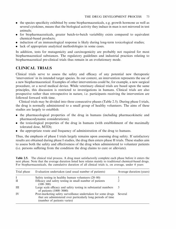
. the species specificity exhibited by some biopharmaceuticals, e.g. growth hormone as well asseveral cytokines, means that the biological activity they induce in man is not mirrored in testanimals;
. for biopharmaceuticals, greater batch-to-batch variability exists compared to equivalentchemical-based products;
. induction of an immunological response is likely during long-term toxicological studies;
. lack of appropriate analytical methodologies in some cases.
In addition, tests for mutagenicity and carcinogenicity are probably not required for mostbiopharmaceutical substances. The regulatory guidelines and industrial practices relating tobiopharmaceutical pre-clinical trials thus remain in an evolutionary mode.
CLINICAL TRIALS
Clinical trials serve to assess the safety and efficacy of any potential new therapeutic‘intervention’ in its intended target species. In our context, an intervention represents the use ofa new biopharmaceutical. Examples of other interventions could be, for example, a new surgicalprocedure, or a novel medical device. While veterinary clinical trials are based upon the sameprinciples, this discussion is restricted to investigations in humans. Clinical trials are alsoprospective rather than retrospective in nature, i.e. participants receiving the intervention arefollowed forward with time.
Clinical trials may be divided into three consecutive phases (Table 2.5). During phase I trials,the drug is normally administered to a small group of healthy volunteers. The aims of thesestudies are largely to establish:
. the pharmacological properties of the drug in humans (including pharmacokinetic andpharmacodynamic considerations);
. the toxicological properties of the drug in humans (with establishment of the maximallytolerated dose, MTD);
. the appropriate route and frequency of administration of the drug to humans.
Thus, the emphasis of phase I trials largely remains upon assessing drug safety. If satisfactoryresults are obtained during phase I studies, the drug then enters phase II trials. These studies aimto assess both the safety and effectiveness of the drug when administered to volunteer patients(i.e. persons suffering from the condition the drug claims to cure or alleviate).
THE DRUG DEVELOPMENT PROCESS 73
Table 2.5. The clinical trial process. A drug must satisfactorily complete each phase before it enters thenext phase. Note that the average duration listed here relates mainly to traditional chemical-based drugs.For biopharmaceuticals, the cumulative duration of all clinical trials is, on average, under 4 years
Trial phase Evaluation undertaken (and usual number of patients) Average duration (years)
I Safety testing in healthy human volunteers (20–80) 1II Efficacy and safety testing in small number of patients
(100–300)2
III Large scale efficacy and safety testing in substantial numbersof patients (1000–3000)
3
IV Post-marketing safety surveillance undertaken for some drugsthat are administered over particularly long periods of time(number of patients varies)
Several
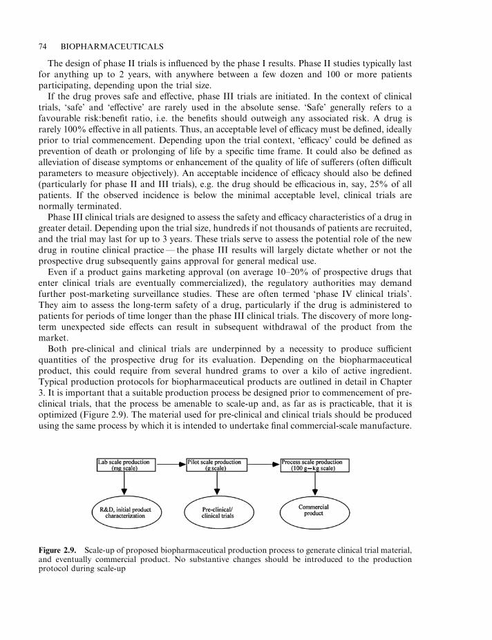
The design of phase II trials is influenced by the phase I results. Phase II studies typically lastfor anything up to 2 years, with anywhere between a few dozen and 100 or more patientsparticipating, depending upon the trial size.
If the drug proves safe and effective, phase III trials are initiated. In the context of clinicaltrials, ‘safe’ and ‘effective’ are rarely used in the absolute sense. ‘Safe’ generally refers to afavourable risk:benefit ratio, i.e. the benefits should outweigh any associated risk. A drug israrely 100% effective in all patients. Thus, an acceptable level of efficacy must be defined, ideallyprior to trial commencement. Depending upon the trial context, ‘efficacy’ could be defined asprevention of death or prolonging of life by a specific time frame. It could also be defined asalleviation of disease symptoms or enhancement of the quality of life of sufferers (often difficultparameters to measure objectively). An acceptable incidence of efficacy should also be defined(particularly for phase II and III trials), e.g. the drug should be efficacious in, say, 25% of allpatients. If the observed incidence is below the minimal acceptable level, clinical trials arenormally terminated.
Phase III clinical trials are designed to assess the safety and efficacy characteristics of a drug ingreater detail. Depending upon the trial size, hundreds if not thousands of patients are recruited,and the trial may last for up to 3 years. These trials serve to assess the potential role of the newdrug in routine clinical practice— the phase III results will largely dictate whether or not theprospective drug subsequently gains approval for general medical use.
Even if a product gains marketing approval (on average 10–20% of prospective drugs thatenter clinical trials are eventually commercialized), the regulatory authorities may demandfurther post-marketing surveillance studies. These are often termed ‘phase IV clinical trials’.They aim to assess the long-term safety of a drug, particularly if the drug is administered topatients for periods of time longer than the phase III clinical trials. The discovery of more long-term unexpected side effects can result in subsequent withdrawal of the product from themarket.
Both pre-clinical and clinical trials are underpinned by a necessity to produce sufficientquantities of the prospective drug for its evaluation. Depending on the biopharmaceuticalproduct, this could require from several hundred grams to over a kilo of active ingredient.Typical production protocols for biopharmaceutical products are outlined in detail in Chapter3. It is important that a suitable production process be designed prior to commencement of pre-clinical trials, that the process be amenable to scale-up and, as far as is practicable, that it isoptimized (Figure 2.9). The material used for pre-clinical and clinical trials should be producedusing the same process by which it is intended to undertake final commercial-scale manufacture.
74 BIOPHARMACEUTICALS
Figure 2.9. Scale-up of proposed biopharmaceutical production process to generate clinical trial material,and eventually commercial product. No substantive changes should be introduced to the productionprotocol during scale-up
Extensive early development work is thus essential. Any significant deviation from theproduction protocol, used to generate the trial material, could invalidate all the clinical trialresults with respect to the proposed commercialized product (changes in the production processcould potentially change the final product characteristics—both the active ingredient andcontaminant profile).
Clinical trial design
Proper and comprehensive planning of a clinical trial is essential to the successful developmentof any drug. Clinical trial design is a subject whose scope is too broad to be undertaken in thistext, and only a brief overview of the subject is presented below. The interested reader is referredto the Further Reading section at the end of this chapter. The general principles presented beloware relevant to phase II, and particularly phase III, clinical trials. As the bulk of the estimated$300–500 million required to develop a drug is spent on clinical trials, a poorly planned and/orexecuted clinical trial can be very costly to the drug developer.
The first issue to be considered when developing a trial protocol is to define precisely whatquestions the trial results should be capable of answering. As previously discussed, the terms‘safety’ and ‘efficacy’ are difficult to define in a therapeutic context. An acceptable meaning ofthese concepts, however, should be committed to paper prior to planning of the trial.
Trial size and study population
A clinical trial must obviously have a control group against which the test (intervention) groupcan be compared. The control group may receive (a) no intervention at all; (b) a placebo (i.e. asubstance such as saline, which will have no pharmacological or other effect); (c) the therapymost commonly used at that time to combat the target disease/condition.
The size of the trial will be limited by a number of factors, including:
. economic considerations (level of supporting financial resources);
. size of population with target condition;
. size of population with target condition after additional trial criteria have been imposed (e.g.specific age bracket, lack of complicating medical conditions, etc.);
. size of eligible population willing to participate in the trial.
While a comprehensive phase III trial would require at least several hundred patients, smallertrials would suffice if, for example:
. the target disease is very serious/fatal;
. there are no existing acceptable alternative treatments;
. the target disease population is quite small;
. the new drug is clearly effective and exhibits little toxicity.
Choosing the study population is obviously critical to adequate trial design. The specific criteriaof patient eligibility should be clearly pre-defined as part of the primary question the trial strivesto answer.
A number of trial design types may be used (Table 2.6), each having its own uniqueadvantages and disadvantages. The most scientifically pure is a randomized, double-blind trial(see later). However, in many instances, alternative trial designs are chosen based on ethical orother grounds. In most cases, two groups are considered: control and test. However, thesedesigns can be adapted to facilitate more complex sub-grouping.
THE DRUG DEVELOPMENT PROCESS 75
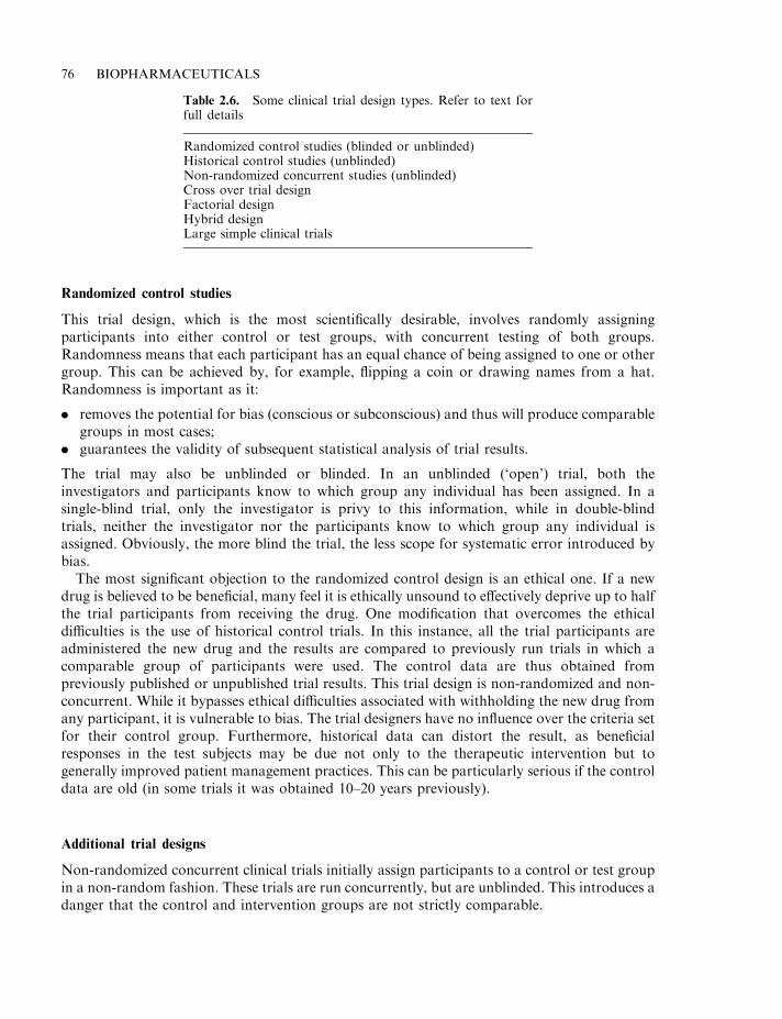
Randomized control studies
This trial design, which is the most scientifically desirable, involves randomly assigningparticipants into either control or test groups, with concurrent testing of both groups.Randomness means that each participant has an equal chance of being assigned to one or othergroup. This can be achieved by, for example, flipping a coin or drawing names from a hat.Randomness is important as it:
. removes the potential for bias (conscious or subconscious) and thus will produce comparablegroups in most cases;
. guarantees the validity of subsequent statistical analysis of trial results.
The trial may also be unblinded or blinded. In an unblinded (‘open’) trial, both theinvestigators and participants know to which group any individual has been assigned. In asingle-blind trial, only the investigator is privy to this information, while in double-blindtrials, neither the investigator nor the participants know to which group any individual isassigned. Obviously, the more blind the trial, the less scope for systematic error introduced bybias.
The most significant objection to the randomized control design is an ethical one. If a newdrug is believed to be beneficial, many feel it is ethically unsound to effectively deprive up to halfthe trial participants from receiving the drug. One modification that overcomes the ethicaldifficulties is the use of historical control trials. In this instance, all the trial participants areadministered the new drug and the results are compared to previously run trials in which acomparable group of participants were used. The control data are thus obtained frompreviously published or unpublished trial results. This trial design is non-randomized and non-concurrent. While it bypasses ethical difficulties associated with withholding the new drug fromany participant, it is vulnerable to bias. The trial designers have no influence over the criteria setfor their control group. Furthermore, historical data can distort the result, as beneficialresponses in the test subjects may be due not only to the therapeutic intervention but togenerally improved patient management practices. This can be particularly serious if the controldata are old (in some trials it was obtained 10–20 years previously).
Additional trial designs
Non-randomized concurrent clinical trials initially assign participants to a control or test groupin a non-random fashion. These trials are run concurrently, but are unblinded. This introduces adanger that the control and intervention groups are not strictly comparable.
76 BIOPHARMACEUTICALS
Table 2.6. Some clinical trial design types. Refer to text forfull details
Randomized control studies (blinded or unblinded)Historical control studies (unblinded)Non-randomized concurrent studies (unblinded)Cross over trial designFactorial designHybrid designLarge simple clinical trials
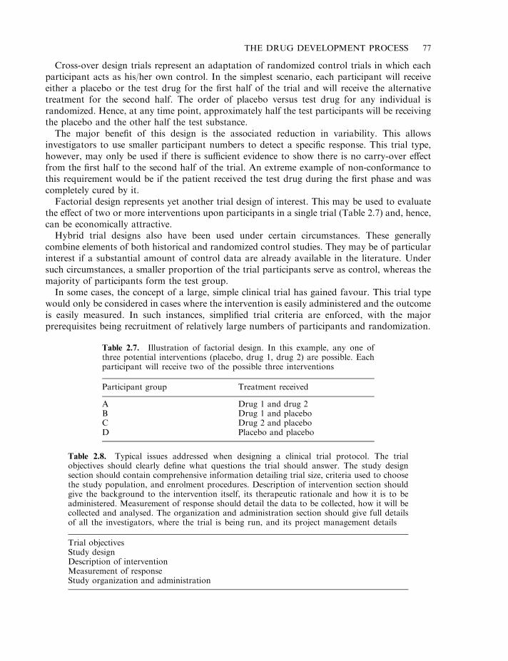
Cross-over design trials represent an adaptation of randomized control trials in which eachparticipant acts as his/her own control. In the simplest scenario, each participant will receiveeither a placebo or the test drug for the first half of the trial and will receive the alternativetreatment for the second half. The order of placebo versus test drug for any individual israndomized. Hence, at any time point, approximately half the test participants will be receivingthe placebo and the other half the test substance.
The major benefit of this design is the associated reduction in variability. This allowsinvestigators to use smaller participant numbers to detect a specific response. This trial type,however, may only be used if there is sufficient evidence to show there is no carry-over effectfrom the first half to the second half of the trial. An extreme example of non-conformance tothis requirement would be if the patient received the test drug during the first phase and wascompletely cured by it.
Factorial design represents yet another trial design of interest. This may be used to evaluatethe effect of two or more interventions upon participants in a single trial (Table 2.7) and, hence,can be economically attractive.
Hybrid trial designs also have been used under certain circumstances. These generallycombine elements of both historical and randomized control studies. They may be of particularinterest if a substantial amount of control data are already available in the literature. Undersuch circumstances, a smaller proportion of the trial participants serve as control, whereas themajority of participants form the test group.
In some cases, the concept of a large, simple clinical trial has gained favour. This trial typewould only be considered in cases where the intervention is easily administered and the outcomeis easily measured. In such instances, simplified trial criteria are enforced, with the majorprerequisites being recruitment of relatively large numbers of participants and randomization.
THE DRUG DEVELOPMENT PROCESS 77
Table 2.7. Illustration of factorial design. In this example, any one ofthree potential interventions (placebo, drug 1, drug 2) are possible. Eachparticipant will receive two of the possible three interventions
Participant group Treatment received
A Drug 1 and drug 2B Drug 1 and placeboC Drug 2 and placeboD Placebo and placebo
Table 2.8. Typical issues addressed when designing a clinical trial protocol. The trialobjectives should clearly define what questions the trial should answer. The study designsection should contain comprehensive information detailing trial size, criteria used to choosethe study population, and enrolment procedures. Description of intervention section shouldgive the background to the intervention itself, its therapeutic rationale and how it is to beadministered. Measurement of response should detail the data to be collected, how it will becollected and analysed. The organization and administration section should give full detailsof all the investigators, where the trial is being run, and its project management details
Trial objectivesStudy designDescription of interventionMeasurement of responseStudy organization and administration
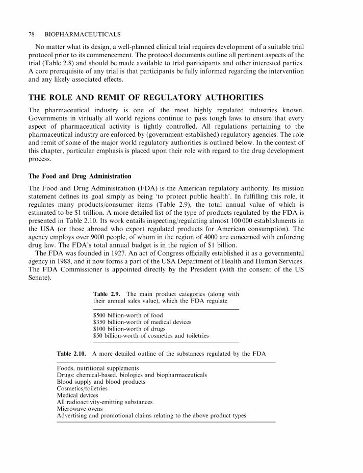
No matter what its design, a well-planned clinical trial requires development of a suitable trialprotocol prior to its commencement. The protocol documents outline all pertinent aspects of thetrial (Table 2.8) and should be made available to trial participants and other interested parties.A core prerequisite of any trial is that participants be fully informed regarding the interventionand any likely associated effects.
THE ROLE AND REMIT OF REGULATORY AUTHORITIES
The pharmaceutical industry is one of the most highly regulated industries known.Governments in virtually all world regions continue to pass tough laws to ensure that everyaspect of pharmaceutical activity is tightly controlled. All regulations pertaining to thepharmaceutical industry are enforced by (government-established) regulatory agencies. The roleand remit of some of the major world regulatory authorities is outlined below. In the context ofthis chapter, particular emphasis is placed upon their role with regard to the drug developmentprocess.
The Food and Drug Administration
The Food and Drug Administration (FDA) is the American regulatory authority. Its missionstatement defines its goal simply as being ‘to protect public health’. In fulfilling this role, itregulates many products/consumer items (Table 2.9), the total annual value of which isestimated to be $1 trillion. A more detailed list of the type of products regulated by the FDA ispresented in Table 2.10. Its work entails inspecting/regulating almost 100 000 establishments inthe USA (or those abroad who export regulated products for American consumption). Theagency employs over 9000 people, of whom in the region of 4000 are concerned with enforcingdrug law. The FDA’s total annual budget is in the region of $1 billion.
The FDA was founded in 1927. An act of Congress officially established it as a governmentalagency in 1988, and it now forms a part of the USA Department of Health and Human Services.The FDA Commissioner is appointed directly by the President (with the consent of the USSenate).
78 BIOPHARMACEUTICALS
Table 2.9. The main product categories (along withtheir annual sales value), which the FDA regulate
$500 billion-worth of food$350 billion-worth of medical devices$100 billion-worth of drugs$50 billion-worth of cosmetics and toiletries
Table 2.10. A more detailed outline of the substances regulated by the FDA
Foods, nutritional supplementsDrugs: chemical-based, biologics and biopharmaceuticalsBlood supply and blood productsCosmetics/toiletriesMedical devicesAll radioactivity-emitting substancesMicrowave ovensAdvertising and promotional claims relating to the above product types
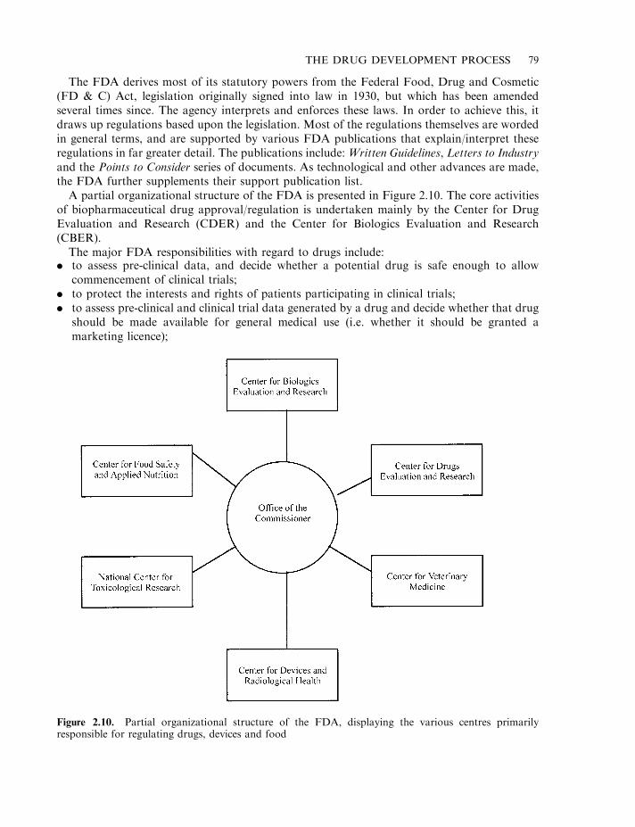
The FDA derives most of its statutory powers from the Federal Food, Drug and Cosmetic(FD & C) Act, legislation originally signed into law in 1930, but which has been amendedseveral times since. The agency interprets and enforces these laws. In order to achieve this, itdraws up regulations based upon the legislation. Most of the regulations themselves are wordedin general terms, and are supported by various FDA publications that explain/interpret theseregulations in far greater detail. The publications include: Written Guidelines, Letters to Industryand the Points to Consider series of documents. As technological and other advances are made,the FDA further supplements their support publication list.
A partial organizational structure of the FDA is presented in Figure 2.10. The core activitiesof biopharmaceutical drug approval/regulation is undertaken mainly by the Center for DrugEvaluation and Research (CDER) and the Center for Biologics Evaluation and Research(CBER).
The major FDA responsibilities with regard to drugs include:. to assess pre-clinical data, and decide whether a potential drug is safe enough to allow
commencement of clinical trials;. to protect the interests and rights of patients participating in clinical trials;. to assess pre-clinical and clinical trial data generated by a drug and decide whether that drug
should be made available for general medical use (i.e. whether it should be granted amarketing licence);
THE DRUG DEVELOPMENT PROCESS 79
Figure 2.10. Partial organizational structure of the FDA, displaying the various centres primarilyresponsible for regulating drugs, devices and food
. to oversee the manufacture of safe effective drugs (inspect and approve drug manufacturingfacilities on the basis of compliance to the principles of good manufacturing practice asapplied to pharmaceuticals);
. to ensure the safety of the US blood supply.In relation to the drug development process, CDER oversees and regulates the development
and marketing approval of mainly chemical-based drugs. CBER is more concerned withbiologics. ‘Biologic’ has traditionally been defined in a narrow sense and has been taken to referto vaccines and viruses, blood and blood products, as well as antiserum, toxins and antitoxinsused for therapeutic purposes. Because of this, many established pharmaceutical products (e.g.microbial metabolites and hormones) have come under appraisal by CDER, even though onemight initially assume they would come under the biologics umbrella (Table 2.11).
Biopharmaceutical products have not fallen neatly into either category and the decision torefer any biotech-derived drug to CBER or CDER is taken on a case-by-case basis. Tissueplasminogen activator (tPA; Chapter 9), for example, is licensed as a biologic, whereaserythropoietin (EPO; Chapter 6) comes under the auspices of CDER. The majority ofbiopharmaceuticals, however, are assessed by CBER.
The criteria used by CBER and CDER regulators in assessing product performance duringthe drug development process are similar, i.e. safety, quality and efficacy. However, theadministrative details can vary in both name and content. Upon concluding pre-clinical trials,all the data generated regarding any potential new drug are compiled in a dossier and submittedto CDER or CBER in the form of an investigational new drug application (IND application).The FDA assesses the application, and if it does not object within a specific time frame (usually30 days), clinical trials can begin. The FDA usually meet with the drug developers at variousstages, to be updated and often to give informal guidance/advice. Once clinical trials have beencompleted, all the data generated during the entire development process are compiled in a multi-volume dossier.
The dossier submitted to CDER is known as a new drug application (NDA) which, ifapproved, allows the drug to be marketed. If the drug is a CBER-regulated one, a biologicslicence application (BLA) is submitted.
The investigational new drug application
An investigational new drug (IND) is a new chemical-based, biologic or biopharmaceuticalsubstance for which the FDA has given approval to undergo clinical trials. An IND application
80 BIOPHARMACEUTICALS
Table 2.11 Some traditional pharmaceutical products produced by/extracted from biological sources,which come under the auspices of CDER for regulatory purposes. The specific reviewing divisions withinCDER which deal with these products are also listed
Product Reviewing division
Most antibiotics Division of anti-infective drug productsAnticoagulants, fibrinolytics, prostaglandins Division of gastrointestinal and coagulation drug
productsEnzymes Division of medical imaging, surgical and dental drug
productsInsulin, somatostatin, growth hormone,gonadotrophins, vasopressin, oxytocin
Division of metabolic and endocrine drug products
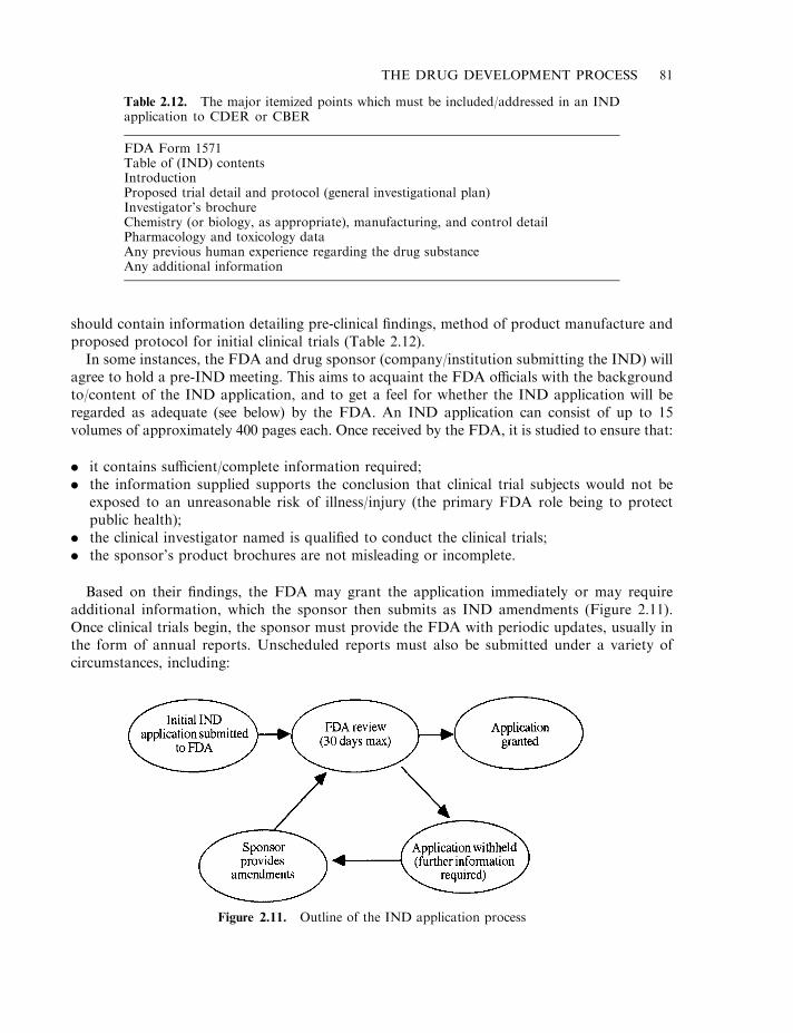
should contain information detailing pre-clinical findings, method of product manufacture andproposed protocol for initial clinical trials (Table 2.12).
In some instances, the FDA and drug sponsor (company/institution submitting the IND) willagree to hold a pre-IND meeting. This aims to acquaint the FDA officials with the backgroundto/content of the IND application, and to get a feel for whether the IND application will beregarded as adequate (see below) by the FDA. An IND application can consist of up to 15volumes of approximately 400 pages each. Once received by the FDA, it is studied to ensure that:
. it contains sufficient/complete information required;
. the information supplied supports the conclusion that clinical trial subjects would not beexposed to an unreasonable risk of illness/injury (the primary FDA role being to protectpublic health);
. the clinical investigator named is qualified to conduct the clinical trials;
. the sponsor’s product brochures are not misleading or incomplete.
Based on their findings, the FDA may grant the application immediately or may requireadditional information, which the sponsor then submits as IND amendments (Figure 2.11).Once clinical trials begin, the sponsor must provide the FDA with periodic updates, usually inthe form of annual reports. Unscheduled reports must also be submitted under a variety ofcircumstances, including:
THE DRUG DEVELOPMENT PROCESS 81
Table 2.12. The major itemized points which must be included/addressed in an INDapplication to CDER or CBER
FDA Form 1571Table of (IND) contentsIntroductionProposed trial detail and protocol (general investigational plan)Investigator’s brochureChemistry (or biology, as appropriate), manufacturing, and control detailPharmacology and toxicology dataAny previous human experience regarding the drug substanceAny additional information
Figure 2.11. Outline of the IND application process
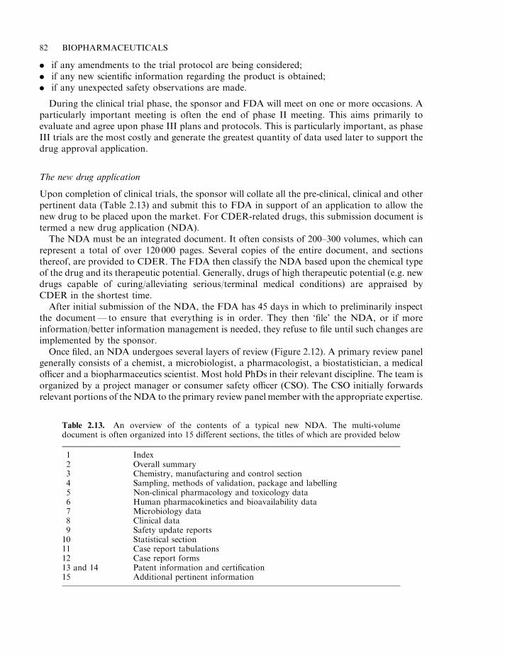
. if any amendments to the trial protocol are being considered;
. if any new scientific information regarding the product is obtained;
. if any unexpected safety observations are made.
During the clinical trial phase, the sponsor and FDA will meet on one or more occasions. Aparticularly important meeting is often the end of phase II meeting. This aims primarily toevaluate and agree upon phase III plans and protocols. This is particularly important, as phaseIII trials are the most costly and generate the greatest quantity of data used later to support thedrug approval application.
The new drug application
Upon completion of clinical trials, the sponsor will collate all the pre-clinical, clinical and otherpertinent data (Table 2.13) and submit this to FDA in support of an application to allow thenew drug to be placed upon the market. For CDER-related drugs, this submission document istermed a new drug application (NDA).
The NDA must be an integrated document. It often consists of 200–300 volumes, which canrepresent a total of over 120 000 pages. Several copies of the entire document, and sectionsthereof, are provided to CDER. The FDA then classify the NDA based upon the chemical typeof the drug and its therapeutic potential. Generally, drugs of high therapeutic potential (e.g. newdrugs capable of curing/alleviating serious/terminal medical conditions) are appraised byCDER in the shortest time.
After initial submission of the NDA, the FDA has 45 days in which to preliminarily inspectthe document— to ensure that everything is in order. They then ‘file’ the NDA, or if moreinformation/better information management is needed, they refuse to file until such changes areimplemented by the sponsor.
Once filed, an NDA undergoes several layers of review (Figure 2.12). A primary review panelgenerally consists of a chemist, a microbiologist, a pharmacologist, a biostatistician, a medicalofficer and a biopharmaceutics scientist. Most hold PhDs in their relevant discipline. The team isorganized by a project manager or consumer safety officer (CSO). The CSO initially forwardsrelevant portions of the NDA to the primary review panel member with the appropriate expertise.
82 BIOPHARMACEUTICALS
Table 2.13. An overview of the contents of a typical new NDA. The multi-volumedocument is often organized into 15 different sections, the titles of which are provided below
1 Index2 Overall summary3 Chemistry, manufacturing and control section4 Sampling, methods of validation, package and labelling5 Non-clinical pharmacology and toxicology data6 Human pharmacokinetics and bioavailability data7 Microbiology data8 Clinical data9 Safety update reports10 Statistical section11 Case report tabulations12 Case report forms13 and 14 Patent information and certification15 Additional pertinent information
Each reviewer then prepares a review report. This is forwarded to their supervisory officers,who undertake a second review. All of the reports are then sent to the division director who, inturn, recommends rejection or approval, or asks the sponsor to provide more information. Onaverage, this entire process takes 12 months.
Even when the NDA is approved and the product goes on sale, the sponsor must provide theFDA with further occasional reports. These can be in the form of scheduled annual reports, butunscheduled reports are also required in instances such as the occurrence of an unexpectedadverse response to the drug.
A similar general approach is taken by CBER with regard to drugs being developed undertheir auspices. The CBER ‘licensing process’ for a new drug consists of three phases: the INDphase (already discussed), the pre-marketing approval phase (licensure phase) and the post-marketing surveillance phase. The pre-marketing approval phase (i.e. clinical trial phase) aimsto generate data which proves the potency, purity and safety of the product. Upon completionof clinical trials, the sponsor collates the data generated and submits it to the FDA in the formof a biologics licence application, which must provide a comprehensive description of both theproduct and product manufacture (including methods of QC analysis, product stability data,labelling data and, of course, safety and efficacy data).
A small number of biotechnology products are classified as medical devices and hence areregulated by the Center for Devices and Radiological Health. The first approved biotechproduct to come under the auspices of the CDRH was OP-1 implant. Marketed by Stryker
THE DRUG DEVELOPMENT PROCESS 83
Figure 2.12. The CDER review process for a typical NDA. In addition to the review stages described, theFDA also may consult with a technical advisory committee. The members of the advisory committee arenot routinely involved in IND or NDA assessment. The FDA is not obliged to follow any advice given bythe advisory committee, but it generally does so
Biotech, OP-1 implant is a sterile powder composed of recombinant human osteogenic protein-1(OP-1) along with bovine collagen. It is used to treat fractured bones that fail to heal. Theproduct is mixed with sterile saline immediately before application, which entails surgicalinsertion of the paste into the fracture.
European regulations
The overall philosophy behind granting a marketing authorization for a new drug is broadlysimilar in the USA and Europe. There are, however, major differences in the systems by whichthese philosophies are implemented in the two regions.
The European Union (EU) is currently composed of 15 member states and a total populationof 371 million (compared with 249 million in the USA and 124 million in Japan— the other twoworld pharmaceutical markets). The total European pharmaceutical market value stands at e70billion, representing almost 33% of world sales (the US and Japanese figures are 31% and 21%,respectively). Annual expenditure on European pharmaceutical R&D stands at about e17billion.
Prominent within the EU organizational structure is the European Commission. TheCommission is composed of 20 commissioners (with at least one being from each member state)and several thousand civil servants. The major function of the European Commission is topropose new EU legislation and to ensure enforcement of existing legislation (European druglaw, therefore, comes under the auspices of the Commission).
Two forms of legal instrument can be issued from the centralized European authorities: a‘regulation’ and a ‘directive’. A regulation is a strong legal instruction which, once passed, mustbe implemented immediately, and without modification, by national governments. A directive isa looser legal term, and provides an individual member state with 18 months to translate theflavour of that law into national law. Pharmaceutical law within the EU has been shaped byboth directives and regulations, as discussed later.
National regulatory authorities
In all European countries, there exist national regulatory authorities. These authorities areappointed by the government of the country in question and are usually located in the nationalMinistry of Health. They serve to apply national and European law with regard to the drugdevelopment process. In many countries, different arms of the regulatory authorities areresponsible for authorizing and assessing clinical trials, assessing the resultant drug dossier anddeciding on that basis whether or not to grant a (national) marketing authorization/productlicence. They are also often responsible for issuing manufacturing licences to companies(Chapter 3).
In the past, a company wishing to gain a marketing licence within Europe usually applied forseparate marketing authorizations on a country-by-country basis. This entailed significantduplication of effort, because:
. the drug dossiers needed to be translated into various European languages;
. national laws often differed and, hence, different expectations/dossier requirements wereassociated with different countries;
. the time scale taken for dossier assessment varied from country to country.
Attempts to harmonize European pharmaceutical laws were accelerated in the 1980s. From1985 onwards, a substantial number of European pharmaceutical directives have been adopted.
84 BIOPHARMACEUTICALS
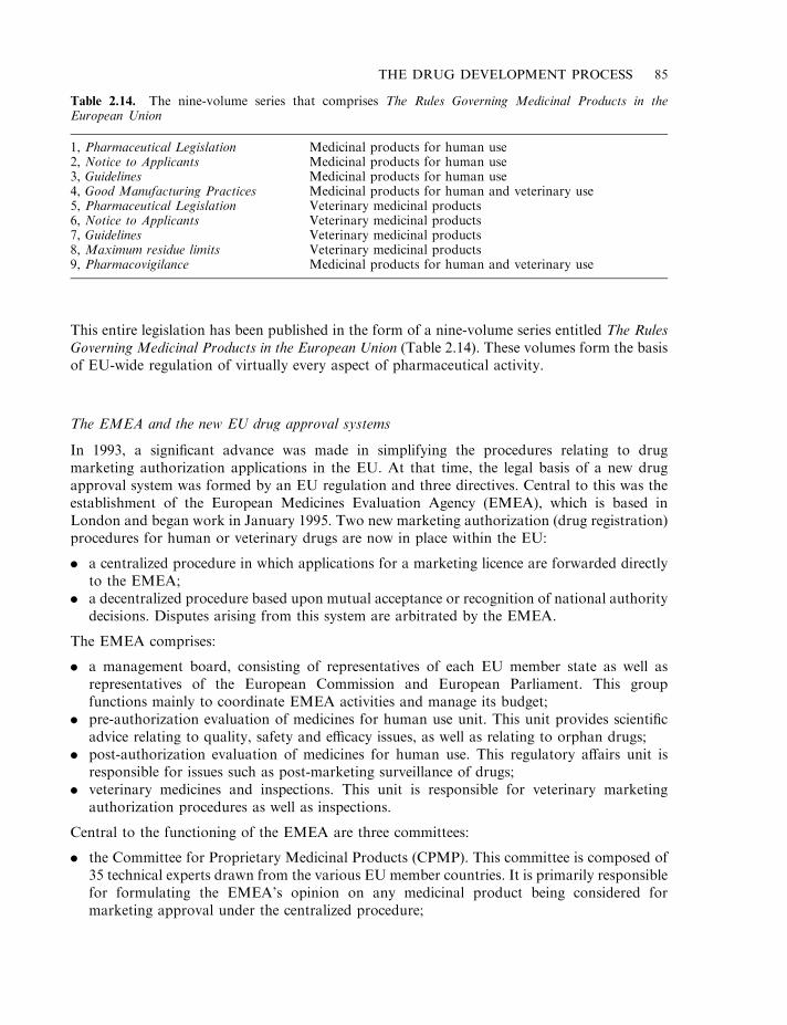
This entire legislation has been published in the form of a nine-volume series entitled The RulesGoverning Medicinal Products in the European Union (Table 2.14). These volumes form the basisof EU-wide regulation of virtually every aspect of pharmaceutical activity.
The EMEA and the new EU drug approval systems
In 1993, a significant advance was made in simplifying the procedures relating to drugmarketing authorization applications in the EU. At that time, the legal basis of a new drugapproval system was formed by an EU regulation and three directives. Central to this was theestablishment of the European Medicines Evaluation Agency (EMEA), which is based inLondon and began work in January 1995. Two new marketing authorization (drug registration)procedures for human or veterinary drugs are now in place within the EU:
. a centralized procedure in which applications for a marketing licence are forwarded directlyto the EMEA;
. a decentralized procedure based upon mutual acceptance or recognition of national authoritydecisions. Disputes arising from this system are arbitrated by the EMEA.
The EMEA comprises:
. a management board, consisting of representatives of each EU member state as well asrepresentatives of the European Commission and European Parliament. This groupfunctions mainly to coordinate EMEA activities and manage its budget;
. pre-authorization evaluation of medicines for human use unit. This unit provides scientificadvice relating to quality, safety and efficacy issues, as well as relating to orphan drugs;
. post-authorization evaluation of medicines for human use. This regulatory affairs unit isresponsible for issues such as post-marketing surveillance of drugs;
. veterinary medicines and inspections. This unit is responsible for veterinary marketingauthorization procedures as well as inspections.
Central to the functioning of the EMEA are three committees:
. the Committee for Proprietary Medicinal Products (CPMP). This committee is composed of35 technical experts drawn from the various EU member countries. It is primarily responsiblefor formulating the EMEA’s opinion on any medicinal product being considered formarketing approval under the centralized procedure;
THE DRUG DEVELOPMENT PROCESS 85
Table 2.14. The nine-volume series that comprises The Rules Governing Medicinal Products in theEuropean Union
1, Pharmaceutical Legislation Medicinal products for human use2, Notice to Applicants Medicinal products for human use3, Guidelines Medicinal products for human use4, Good Manufacturing Practices Medicinal products for human and veterinary use5, Pharmaceutical Legislation Veterinary medicinal products6, Notice to Applicants Veterinary medicinal products7, Guidelines Veterinary medicinal products8, Maximum residue limits Veterinary medicinal products9, Pharmacovigilance Medicinal products for human and veterinary use
. the Committee for Veterinary Medicinal Products (CVMP), whose structure and role issimilar to the CPMP except that it is concerned with animal medicines;
. the Committee for Orphan Medicinal Products (COMP), composed of a representative fromeach EU member state, as well as EMEA and patient representatives. The COMP assessesapplications relating to experimental medicines being granted ‘orphan’ status. Orphanmedicines are those intended to treat rare diseases, and orphan designation results in areduction of the fees charged by the EMEA when assessing marketing authorizationapplications.
Unlike the FDA, the EMEA itself does not directly undertake appraisals of drugdossiers submitted to support marketing authorization applications under thecentralized procedure. Instead (as discussed in detail below), they forward the dossier toselected national EU regulatory bodies, who undertake the appraisal, and the EMEA makes arecommendation to approve (or not) the application, based upon the national body’s report.The overall role of the EMEA is thus to coordinate and manage the new system. The EMEA’sannual budget is of the order of e70 million. The key objectives of the EMEA may besummarized as:
. protection of public and animal health by ensuring the quality, safety and efficacy ofmedicinal products for human and veterinary use;
. to strengthen the ideal of a single European market for human and veterinarypharmaceuticals;
. to support the European pharmaceutical industry as part of the EU industrial policy.
The centralized procedure
Under the centralized procedure, applications are accepted with regard to (a) products ofbiotechnology; (b) new chemical entities (NCEs: drugs in which the active ingredient is new).Biotech products are grouped as ‘list A’, NCEs as ‘list B’. Marketing approval application forbiotech products must be considered under the centralized procedure, whereas NCEs can beconsidered under centralized or decentralized mechanisms.
Upon receipt of a marketing authorization application under centralized procedures, theEMEA staff carry out an initial appraisal to ensure that it is complete and has been compiled inaccordance with the appropriate EU guidelines (Box 2.2). This appraisal must be completedwithin 10 days, at which time (if the application is in order), it is given a filing date. The sponsoralso pays an appropriate fee. The EMEA then has 210 days to consider the application. In thecase of human drugs, the application immediately comes before the CPMP (which convenes 2–3days a month).
Two rapporteurs are then appointed (committee members who are responsible for gettingthe application assessed). The rapporteurs generally arrange to have the application assessedby their respective home national regulatory authority. Once assessment is complete, thereports are presented via the rapporteur to the CPMP. After discussion, the CPMP issue an‘opinion’ (i.e. a recommendation that the application be accepted, or not). This opinion isthen forwarded to the European Commission, who have another 90 days to consider it. Theyusually accept the opinion and ‘convert’ it into a decision (they—and not the EMEAdirectly—have the authority to issue a marketing authorization). The single marketingauthorization covers the entire EU. Once granted it is valid for 5 years after which it must berenewed. The total time in which a decision must be taken is 300 days (210+90), which is
86 BIOPHARMACEUTICALS
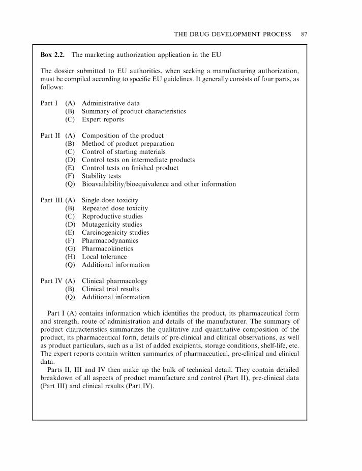
THE DRUG DEVELOPMENT PROCESS 87
Box 2.2. The marketing authorization application in the EU
The dossier submitted to EU authorities, when seeking a manufacturing authorization,must be compiled according to specific EU guidelines. It generally consists of four parts, asfollows:
Part I (A) Administrative data(B) Summary of product characteristics(C) Expert reports
Part II (A) Composition of the product(B) Method of product preparation(C) Control of starting materials(D) Control tests on intermediate products(E) Control tests on finished product(F) Stability tests(Q) Bioavailability/bioequivalence and other information
Part III (A) Single dose toxicity(B) Repeated dose toxicity(C) Reproductive studies(D) Mutagenicity studies(E) Carcinogenicity studies(F) Pharmacodynamics(G) Pharmacokinetics(H) Local tolerance(Q) Additional information
Part IV (A) Clinical pharmacology(B) Clinical trial results(Q) Additional information
Part I (A) contains information which identifies the product, its pharmaceutical formand strength, route of administration and details of the manufacturer. The summary ofproduct characteristics summarizes the qualitative and quantitative composition of theproduct, its pharmaceutical form, details of pre-clinical and clinical observations, as wellas product particulars, such as a list of added excipients, storage conditions, shelf-life, etc.The expert reports contain written summaries of pharmaceutical, pre-clinical and clinicaldata.
Parts II, III and IV then make up the bulk of technical detail. They contain detailedbreakdown of all aspects of product manufacture and control (Part II), pre-clinical data(Part III) and clinical results (Part IV).
considerably shorter than the average approval times in many other world regions. The 300day deadline only applies in situations where the CPMP do not require any additionalinformation from the sponsor. In many cases, additional information is required and whenthis occurs, the 300 day ‘clock’ stops until the additional information is received.
Mutual recognition
The mutual recognition procedure is an alternative means by which a marketing authorizationmay be sought. It is open to all drug types except products of biotechnology. Briefly, if thisprocedure is adopted by a sponsor, the sponsor applies for a marketing licence, not to theEMEA but to a specific national regulatory authority (chosen by the sponsor). The nationalauthority then has 210 days to assess the application.
If adopted by the national authority in question, the sponsor can seek marketing licences inother countries on that basis. For this bilateral phase, other states in which marketingauthorization are sought have 60 days in which to review the application. The theory, of course,is that no substantive difficulty should arise at this stage, as all countries are working to the sameset of standards as laid down in The Rules Governing Medicinal Products in the European Union.
A further 30 days is set aside in which any difficulties that arise may be resolved. The totalapplication duration is 300 days. If one or more states refuse to grant the marketingauthorization (i.e. mutual recognition breaks down), the difficulties are referred back to theEMEA. The CPMP will then make a decision (‘opinion’), which is sent to the EuropeanCommission. The Commission, taking into account the CPMP opinion, will make a finaldecision, which is a binding one.
Drug registration in Japan
The Japanese are the greatest consumers of pharmaceutical products per capita in the world.The Ministry of Health and Welfare in Japan has overall responsibility to implement Japanesepharmaceutical law. Within the department is the Pharmaceutical Affairs Bureau (PAB), whichexercises this authority.
There are three basic steps in the Japanese regulatory process:
. approval (‘shonin’) must be obtained to manufacture or import a drug;
. a licence (‘kyoka’) must also be obtained;
. an official price for the drug must be set.
The PAB undertakes drug dossier evaluations, a process which normally takes 18 months. Theapproval requirements/process for pharmaceuticals (including biopharmaceuticals) are, inbroad terms, quite similar to those in the USA. PAB have issued specific requirements(Notification 243) for submission of recombinant protein drugs.
Applications are initially carefully checked by a single regulatory examiner to ensureconformance to guidelines. Subsequently, the application is reviewed in detail by a sub-committeeof specialists. Clear evidence of safety, quality and efficacy are required prior to approval.
In addition, the Japanese normally insist that at least some clinical trials be carried out inJapan itself. This position is adopted due, for example, to differences in the body size andmetabolism of the Japanese compared to US or European citizens. Also, the quantity of activeingredient present in Japanese drugs is lower than in many other world regions. Hence, trialsmust be undertaken to prove product efficacy under intended Japanese usage conditions.
88 BIOPHARMACEUTICALS
World harmonization of drug approvals
There is a growing trend in the global pharmaceutical industry towards internationalization.Increasingly, mergers and other strategic alliances are a feature of world pharmaceuticalactivity. Many companies are developing drugs which they aim to register in several worldregions. Differences in regulatory practices and requirements in these different regionsconsiderably complicates this process.
Development of harmonized requirements for world drug registrations would be ofconsiderable benefit to pharmaceutical companies and to patients for whom many drugswould be available much more quickly.
The ICH process (International Conference on Harmonization of technical requirements forregistration of pharmaceuticals for human use) brings together experts from both the regulatoryauthorities and pharmaceutical industries based in the EU, USA and Japan. The aim is toachieve greater harmonization of technical/guidelines relating to product registration in theseworld regions, and considerable progress has been made in this regard over the last decade. The‘members’ comprising ICH are experts from the European Commission, the EuropeanFederation of Pharmaceutical Industries Association (EFPIA), the Japanese Ministry of Healthand Welfare, the Japanese Pharmaceutical Manufacturers Assocaition (JPMA), the FDA andthe Pharmaceutical Research and Manufacturers of America (PhRMA). The ICH process issupported by an ICH secretariat, based in Geneva, Switzerland.
CONCLUSION
The drug discovery and development process is a long and expensive one. A wide range ofstrategies may be adopted in the quest for identifying new therapeutic substances. Mostbiopharmaceuticals, however, have been discovered directly as a consequence of an increasedunderstanding of the molecular mechanisms underlining how the body functions, in both healthand disease.
Before any newly discovered drug is placed on the market, it must undergo extensive testing inorder to assure that it is both safe and effective in achieving its claimed therapeutic effect. Thedata generated by these tests (i.e. pre-clinical and clinical trials), are then appraised byindependent, government-appointed regulatory agencies, who ultimately decide whether a drugshould gain a marketing license. While the drug development process may seem cumbersomeand protracted, the cautious attitude adopted by regulatory authorities has served the publicwell in ensuring that only drugs of the highest quality finally come onto the market.
FURTHER READING
Books
Adjei, H. (1997). Inhalation Delivery of Therapeutic Peptides and Proteins. J.A. Majors Co., Atlanta, GA.Ansel, H. (1999). Pharmaceutical Dosage Forms and Drug Delivery Systems. Lippincott Williams & Wilkins,
Philadelphia, PA.Askari, F. (2003). Beyond the Genome—The Proteomics Revolution. Prometheus, Frome, UK.Crommelin, D. (2002). Pharmaceutical Biotechnology. Routledge, London.De Jong, M. (1998). FAQs on EU Pharmaceutical Regulatory Affairs. Brookwood Medical, Richmond, UK.Desalli, R. (2002). The Genomics Revolution. Joseph Henry Press.Ferraiolo, B. (1992). Protein Pharmacokinetics and Metabolism. Plenum, New York.Giffels, J. (1996). Clinical Trials. Demos Medical, London.
THE DRUG DEVELOPMENT PROCESS 89
Goldberg, R. (2001). Pharmaceutical Medicine, Biotechnology and European Patent Law. Cambridge University Press,Cambridge.
Grindley, J. & Ogden, J. (2000). Understanding Biopharmaceuticals. Manufacturing and Regulatory Issues. Interpharm,Denver, CO.
Hunt, S. (2000). Functional Genomics. Oxford University Press, Oxford.Knight, H. (2001). Patent Strategy. Wiley, Chichester.Krogsgaard, L. (2002). Textbook of Drug Design and Discovery. Taylor and Francis, London.McNally, E. (Ed), (2000). Protein Formulation and Delivery. Marcel Dekker, New York.Oxender, D. & Post, L. (1999). Novel Therapeutics from Modern Biotechnology. Springer-Verlag, Berlin.Pennington, S. (2000). Proteomics. BIOS Scientific, Oxford.Poste, G. (1999). The Impact of Genomics on Healthcare. Royal Society of Medicine Press, London.Scolnick, E. (2001). Drug Discovery and Design. Academic Press, London.Venter, J. (2000). From Genome to Therapy. Wiley, Chichester.
Articles
Clinical trials and regulatory authorities
Beatrice, M. (1997). FDA regulatory reform. Curr. Opin. Biotechnol. 18(3), 370–378.Crooke, S. (1995). Comprehensive reform of the new drug regulatory process. Bio/Technology 13, 25–29.Graffeo, A. (1994). The do’s and don’ts of preclinical development. Bio/Technology 12, 865.Helms, P. (2002). Real world pragmatic clinical trials: what are they and what do they tell us? Pediat. Allergy Immunol.
13(1), 4–9.Jefferys, D. & Jones, K. (1995). EMEA and the new pharmaceutical procedures for Europe. Eur. J. Clin. Pharmacol. 47,
471–476.Kaitin, K. (1997). FDA reform: setting the stage for efforts to reform the agency. Drug Inf. J. 31, 27–33.Kessler, D. & Feiden, K. (1995). Faster evaluation of vital drugs. Sci. Am. March, 26–32.Lubiniecki, A. (1997). Potential influence of international harmonization of pharmaceutical regulations on
biopharmaceutical development. Curr. Opin. Biotechnol. 8(3), 350–356.Pignatti, F et al. (2002). Clinical trials for registration in the European Union: the EMEA 5-year experience in oncology.
Crit. Rev. Oncol. Haematol. 42(2), 123–135.Sauer, F. (1997). A new and fast drug approval system in Europe. Drug Inf. J. 31, 1–6.Walsh, G. (1999). Drug approval in Europe. Nature Biotechnol. 17, 237–240
Patenting
Allison, J. & Lemley, M. (2002). The growing complexity of the United States patenting system. Boston Univ. Law Rev.82(1), 77–144.
Barton, J. (1991). Patenting life. Sci. Am. March, 18–24.Berks, A. (1994). Patent information in biotechnology. Trends Biotechnol. 12, 352–364.Crespi, R. (1993). Biotechnological inventions and the patent law: outstanding issues. Biotechnol. Genet. Eng. Rev. 11,
229–261.Crespi, R. (1994). Protecting biotechnological inventions. Trends Biotechnol. 12, 395–397.Johnson, E. (1996). A benchside guide to patents and patenting. Nature Biotechnol. 14, 288–291.Orr, R. & O’Neill, C. (2000). Patent review: therapeutic applications of antisense oligonucleotides, 1999–2000. Curr.
Opin. Mol. Therapeut. 2(3), 325–331.
Drug development
Bugg, C. et al. (1993). Drugs by design. Sci. Am. December, 92–98.Cox, P. & Balick, M. (1994). The ethanobotanical approach to drug discovery. Sci. Am. June, 60–65.Drews, J. (2000). Drug discovery: a historical perspective. Science 287(5460), 1960–1964.Dykes, C. (1996). Genes, disease and medicine. Br. J. Clin. Pharmacol. 42(6), 683–695.Ecker, D. & Crooke, S. (1995). Combinatorial drug discovery: which methods will produce greatest value? Bio/
Technology 13, 351–359.Geisow, M. J. (1991) Characterizing recombinant proteins. Bio/Technology, 9(10), 921–922.Hodgson, J. (1993). Pharmaceutical screening: from off the wall to off the shelf. Bio/Technol. 11, 683–688.Lowman, H. (1997). Bacteriophage display and discovery of peptide leads for drug development. Ann. Rev. Biophys.
Biomol. Struct. 26, 401–424.
90 BIOPHARMACEUTICALS
Medynski, D. (1994). Synthetic peptide combinatorial libraries. Bio/Technology 12, 709–710.Saragovi, H. et al. (1992). Loops and secondary structure mimetics: development and applications in basic science and
rational drug discovery. Bio/Technology 10, 773–777.Scicinski, J. (1995). Chemical libraries in drug discovery. Trends Biotechnol 13, 246–247.
Genomics, proteomics and related technologies
Baba, Y. (2001). Development of novel biomedicine based on genome science. Eur. J. Pharmaceut. Sci. 13(1), 3–4.Brenner, S. (2001). A tour of structural genomics. Nature Rev. Genet. 2(10), 801–809.Cunningham, M. (2000) Genomics and proteomics, the new millennium of drug discovery and development. J.
Pharmacol. Toxicol. Methods 44(1), 291–300.Debouck, C. & Goodfellow, P. (1999). DNA microarrays in drug discovery and development. Nature Genet. 21, 48–50.Gabig, M. &Wegrzyn, G. (2001). An introduction to DNA chips: principles, technology, applications and analysis. Acta
Biochim. Polon. 48(3), 615–622.Jain, K. (2001). Proteomics: new technologies and their applications. Drug Discovery Today 6(9), 457–459.Jain, K. (2001). Proteomics: delivering new routes to drug discovery, part 2. Drug Discovery Today 6(16), 829–832.Kassel, D. (2001). Combinatorial chemistry and mass spectrometry in the twenty-first century drug development
laboratory. Chem. Rev. 101(2), 255–267.Lesley, S. (2001). High-throughput proteomics: protein expression and purification in the post-genomic world. Protein
Expr. Purif. 22(2), 159–164.McLeod, H. & Evans, W. (2001). Pharmacogenomics: unlocking the human genome for better drug therapy. Ann. Rev.
Pharmacol. Toxicol. 41, 101–121.Page, M. et al. (1999). Proteomics: a major new technology for the drug discovery process. Drug Discovery Today 4(2),
55–62.Ramsay, G. (1998) DNA chips: state of the art. Nature Biotechnol. 16(1), 40–44.Roses, A. (2000). Pharmacogenetics and the practice of medicine. Nature 405(6788), 857–865.Schmitz, G. et al. (2001). Pharmacogenomics: implications for laboratory medicine. Clin. Chim. Acta 308(1–2), 43–53.Searls, D. (2000). Using bioinformatics in gene and drug discovery. Drug Discovery Today 5(4), 135–143.Terstappen, G. & Reggiani, A. (2001). In silico research in drug discovery. Trends Pharmacol. Sci. 22(1), 23–26.Wamg, J. & Hewick, R. (1999). Proteomics in drug discovery. Drug Discovery Today 4(3), 129–133.Wieczorek, S. & Tsongalis, G. (2001). Pharmacogenomics: will it change the field of medicine? Clin. Chim. Acta 308(1–2),
1–8.Wilgenbus, K. & Lichter, P. (1999). DNA chip technology ante portas. J. Mol. Med. 77(11), 761–768.Wolf, C. et al. (2000). Science, medicine and the future—pharmacogenetics. Br. Med. J. 320(7240), 987–990.
Drug delivery
Davis, S. (2001). Nasal vaccines. Adv. Drug Delivery Rev. 51, 21–42.Kompella, U. et al. (2001). Delivery systems for penetration enhancement of peptide and protein drugs: design
considerations. Adv. Drug Delivery Rev. 46, 211–245.Patton, J. et. al. (1999). Inhaled insulin. Adv. Drug Delivery Rev. 35, 235–247.Patton, J. (1996). Mechanism of macromolecule absorption by the lungs. Adv. Drug Delivery Rev. 19(1), 3–36.
THE DRUG DEVELOPMENT PROCESS 91


Chapter 3
The drug manufacturing process
The manufacture of pharmaceutical substances is one of the most highly regulated andrigorously controlled manufacturing processes known. In order to gain a manufacturing licence,the producer must prove to the regulatory authorities that not only is the product itself safe andeffective, but that all aspects of the proposed manufacturing process comply to the highest safetyand quality standards. Various elements contribute to the safe manufacture of qualitypharmaceutical products. These include:
. the design and layout of the manufacturing facility;
. raw materials utilized in the manufacturing process;
. the manufacturing process itself;
. the training and commitment of personnel involved in all aspects of the manufacturingoperation;
. the existence of a regulatory framework which assures the establishment and maintenance ofthe highest quality standards with regard to all aspects of manufacturing.
This chapter aims to overview the manufacturing process. It concerns itself with four majorthemes: (a) a description of the infrastructure of a typical manufacturing facility, and somerelevant operational issues—much of the detail presented in this section is equally applicable tofacilities manufacturing non-biological-based pharmaceutical products; (b) sources of biophar-maceuticals; (c) upstream and downstream processing of biopharmaceutical products; and (d)analysis of the final biopharmaceutical product. Before delving into specific aspects ofpharmaceutical manufacturing, various publications, such as international pharmacopoeias andguides to good manufacturing practice for medicinal products, will be discussed. Thesepublications play a central role in establishing criteria which guarantee the consistentproduction of safe and effective drugs.
INTERNATIONAL PHARMACOPOEIA
Many thousands of pharmaceutical substances are routinely manufactured by the pharmaceu-tical industry. Two of the most important determinants of final product safety and efficacy are:(a) the standard of raw materials used in the manufacturing process; and (b) the standard (i.e.specification) to which the final product is manufactured. Most pharmaceutical substances are
Biopharmaceuticals: Biochemistry and Biotechnology, Second Edition by Gary WalshJohn Wiley & Sons Ltd: ISBN 0 470 84326 8 (ppc), ISBN 0 470 84327 6 (pbk)
manufactured to exacting specifications laid down in publications termed ‘pharmacopoeias’.There are more than two dozen pharmacopoeias published world-wide, most notably the UnitedStates Pharmacopoeia (USP), the European Pharmacopoeia (Eur. Ph.) and the JapanesePharmacopoeia. The products listed in these international pharmacopoeias are invariably genericdrugs (i.e. drugs no longer patent-protected, which can be manufactured in any pharmaceuticalfacility holding the appropriate manufacturing licence). The vast bulk of such substances aretraditional chemical-based drugs, and biological substances such as insulin and various bloodproducts. Two sample monographs from the European Pharmacopoeia are reproduced inAppendix 3. Future editions of such pharmacopoeias are likely to include a growing number ofbiopharmaceuticals, particularly as many of these begin to lose their patent protection.
Martindale, The Extra Pharmacopoeia
Martindale, The Extra Pharmacopoeia (often simply referred to as ‘Martindale’) represents anadditional publication of relevance to the pharmaceutical industry. Unlike the pharmacopoeiasdiscussed above, Martindale is not a book of standards. The aim of this encyclopaedicpublication is to provide concise, unbiased information (largely summarized from the peer-reviewed literature) regarding drugs of clinical interest.
The first edition of Martindale was published in 1883 by William Martindale and the 30thedition was published in 1993. It contains information in monograph format on over 5000 drugsin clinical use. The vast bulk of substances described are chemical-based pharmaceuticals, aswell as traditional biological substances such as antibiotics, certain hormones and bloodproducts. Recent editions, however, carry increasing numbers of monographs detailingbiopharmaceuticals—a reflection of their growing importance in the pharmaceutical industry.
Martindale is largely organized into chapters that detail groups of drugs having similarclinical uses or actions (Table 3.1). The information presented in a monograph detailing anyparticular drug will usually include:
. its physiochemical characteristics;
. absorption and fate;
. uses and appropriate mode of administration;
. adverse/side effects;
. suitable dosage levels.
In addition, summaries of published papers/reviews of the substance in question are included.Because of its clinical emphasis, Martindale represents a valuable drug information source topharmacists and clinicians, but also provides much relevant drug information to personnelengaged in pharmaceutical manufacturing.
GUIDES TO GOOD MANUFACTURING PRACTICE
All aspects of pharmaceutical manufacture must comply with the most rigorous standards toensure consistent production of a safe, effective product. The principles underlining suchstandards are summarized in publications which detail good manufacturing practice (GMP).Pharmaceutical manufacturers must be familiar with the principles laid down in thesepublications and they are legally obliged to ensure adoption of these principles to their specificmanufacturing process. Regulatory authority personnel will assess compliance of themanufacturer with these principles by undertaking regular inspections of the facility. The
94 BIOPHARMACEUTICALS

subsequent granting/renewing (or refusing/revoking) of a manufacturing licence depends largelyupon the level of compliance found during the inspection.
Although separate guides to pharmaceutical GMP are published in different world regions theprinciples outlined in them all are largely similar. In Europe, for example, the European Union(EU) publishes the EU Guide to Good Manufacturing Practice for Medicinal Products. This guideconsists of a number of chapters, each of which is concerned with a specific aspect ofpharmaceutical manufacture (Table 3.2). The principles therein often appear little more thancommon-sense guidelines, e.g. the principles outlined in the chapter detailing GMP in relation topersonnel could be summarized as:
. an adequate number of sufficiently qualified, experienced personnel must be employed by themanufacturer;
. key personnel, such as the heads of production and quality control, must be independent ofeach other;
THE DRUG MANUFACTURING PROCESS 95
Table 3.1. List of the major headings under which various drugs are described in Martindale, The ExtraPharmacopoeia
Analgesics and anti-inflammatoryagents
Cardiac inotropic agents Non-ionic surfactants
Anthelmintics Chelating agents, antidotesand antagonists
Nutritional agents and vitamins
Anti-arrhythmic agents Colouring agents Opioid analgesicsAnti-bacterial agents Contrast media Organic solventsAnti-coagulants Corticosteroids Paraffins and similar basesAnti-depressants Cough suppressants,
expectorants and mucolyticsParasympathomimetics
Anti-diabetic agents Dermatological agents Pesticides and repellantsAnti-epileptics Diagnostic agents PreservativesAnti-fungal agents Disinfectants Prophylactic anti-asthma agentsAnti-gout agents Diuretics ProstaglandinsAnti-hypertensive agents Dopaminergic anti-
parkinsonian agentsRadiopharmaceuticals
Anti-malarials Electrolytes Sex hormonesAnti-migraine agents Gases Skeletal muscle relaxantsAnti-muscarinic agents Gastrointestinal agents Soaps and other anionic
surfactantsAnti-neoplastic agents andimmunosuppressants
General anaesthetics Stabilizing and suspending agents
Anti-protozoal agents Haemostatics Stimulants and anorecticsAnti-thyroid agents Histamine H1-receptor
antagonistsSunscreen agents
Anti-viral agents Hypothalamic and pituitaryhormones
Sympathomimetics
Anxiolytic sedatives, hypnotics andneuroleptics
Iodine and iodides Thrombolytic agents
b-Adrenoceptor blockingagents
Iron and iron components
Thyroid agents Blood and blood productsLipid-regulating agents Vaccines, immunoglobulins
and antiseraBlood substitutes and plasmaexpanders
Local anaesthetics Vasodilators Calcium-regulating agentsNitrates and other anti-angina agents Xanthines
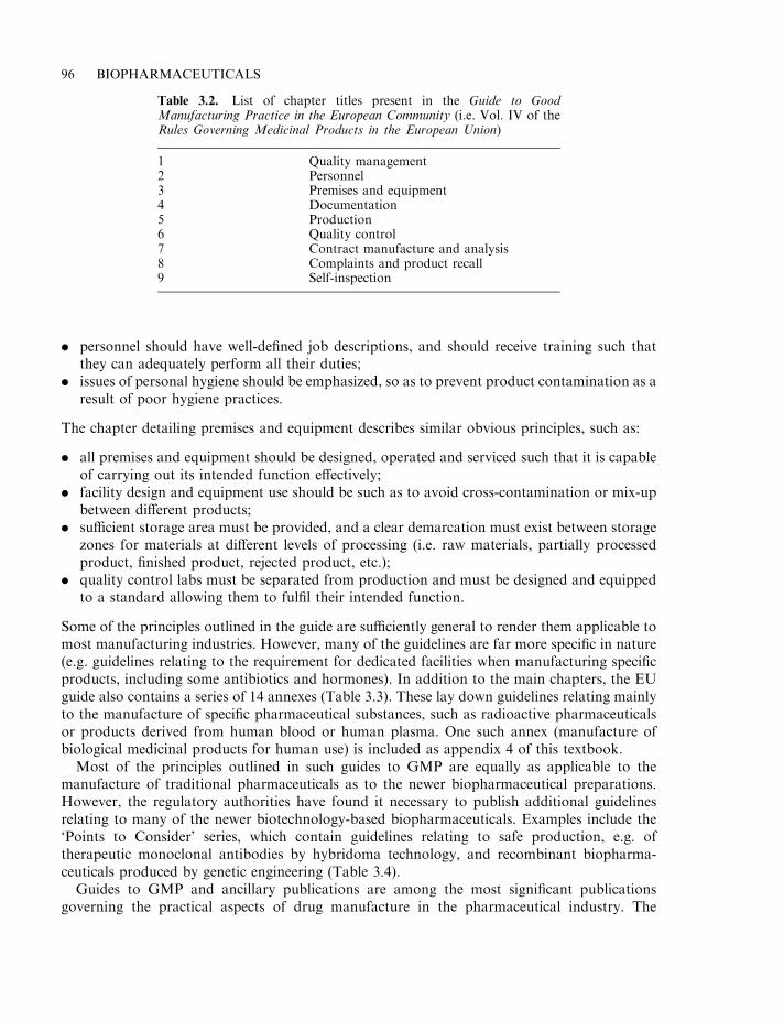
. personnel should have well-defined job descriptions, and should receive training such thatthey can adequately perform all their duties;
. issues of personal hygiene should be emphasized, so as to prevent product contamination as aresult of poor hygiene practices.
The chapter detailing premises and equipment describes similar obvious principles, such as:
. all premises and equipment should be designed, operated and serviced such that it is capableof carrying out its intended function effectively;
. facility design and equipment use should be such as to avoid cross-contamination or mix-upbetween different products;
. sufficient storage area must be provided, and a clear demarcation must exist between storagezones for materials at different levels of processing (i.e. raw materials, partially processedproduct, finished product, rejected product, etc.);
. quality control labs must be separated from production and must be designed and equippedto a standard allowing them to fulfil their intended function.
Some of the principles outlined in the guide are sufficiently general to render them applicable tomost manufacturing industries. However, many of the guidelines are far more specific in nature(e.g. guidelines relating to the requirement for dedicated facilities when manufacturing specificproducts, including some antibiotics and hormones). In addition to the main chapters, the EUguide also contains a series of 14 annexes (Table 3.3). These lay down guidelines relating mainlyto the manufacture of specific pharmaceutical substances, such as radioactive pharmaceuticalsor products derived from human blood or human plasma. One such annex (manufacture ofbiological medicinal products for human use) is included as appendix 4 of this textbook.
Most of the principles outlined in such guides to GMP are equally as applicable to themanufacture of traditional pharmaceuticals as to the newer biopharmaceutical preparations.However, the regulatory authorities have found it necessary to publish additional guidelinesrelating to many of the newer biotechnology-based biopharmaceuticals. Examples include the‘Points to Consider’ series, which contain guidelines relating to safe production, e.g. oftherapeutic monoclonal antibodies by hybridoma technology, and recombinant biopharma-ceuticals produced by genetic engineering (Table 3.4).
Guides to GMP and ancillary publications are among the most significant publicationsgoverning the practical aspects of drug manufacture in the pharmaceutical industry. The
96 BIOPHARMACEUTICALS
Table 3.2. List of chapter titles present in the Guide to GoodManufacturing Practice in the European Community (i.e. Vol. IV of theRules Governing Medicinal Products in the European Union)
1 Quality management2 Personnel3 Premises and equipment4 Documentation5 Production6 Quality control7 Contract manufacture and analysis8 Complaints and product recall9 Self-inspection
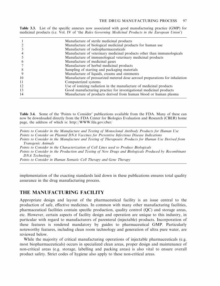
implementation of the exacting standards laid down in these publications ensures total qualityassurance in the drug manufacturing process.
THE MANUFACTURING FACILITY
Appropriate design and layout of the pharmaceutical facility is an issue central to theproduction of safe, effective medicines. In common with many other manufacturing facilities,pharmaceutical facilities contain specific production, quality control (QC) and storage areas,etc. However, certain aspects of facility design and operation are unique to this industry, inparticular with regard to manufacturers of parenteral (injectable) products. Incorporation ofthese features is rendered mandatory by guides to pharmaceutical GMP. Particularlynoteworthy features, including clean room technology and generation of ultra pure water, arereviewed below.
While the majority of critical manufacturing operations of injectable pharmaceuticals (e.g.most biopharmaceuticals) occurs in specialized clean areas, proper design and maintenance ofnon-critical areas (e.g. storage, labelling and packing areas) is also vital to ensure overallproduct safety. Strict codes of hygiene also apply to these non-critical areas.
THE DRUG MANUFACTURING PROCESS 97
Table 3.3. List of the specific annexes now associated with good manufacturing practice (GMP) formedicinal products (i.e. Vol. IV of ‘the Rules Governing Medicinal Products in the European Union’)
1 Manufacture of sterile medicinal products2 Manufacture of biological medicinal products for human use3 Manufacture of radiopharmaceuticals4 Manufacture of veterinary medicinal products other than immunologicals5 Manufacture of immunological veterinary medicinal products6 Manufacture of medicinal gases7 Manufacture of herbal medicinal products8 Sampling of starting and packaging materials9 Manufacture of liquids, creams and ointments10 Manufacture of pressurized metered dose aerosol preparations for inhalation11 Computerized systems12 Use of ionizing radiation in the manufacture of medicinal products13 Good manufacturing practice for investigational medicinal products14 Manufacture of products derived from human blood or human plasma
Table 3.4. Some of the ‘Points to Consider’ publications available from the FDA. Many of these cannow be downloaded directly from the FDA Center for Biologics Evaluation and Research (CBER) homepage, the address of which is: http://WWW.fda.gov/cber/
Points to Consider in the Manufacture and Testing of Monoclonal Antibody Products for Human UsePoints to Consider on Plasmid DNA Vaccines for Preventive Infectious Disease IndicationsPoints to Consider in the Manufacture and Testing of Therapeutic Products for Human Use Derived fromTransgenic Animals
Points to Consider in the Characterization of Cell Lines used to Produce BiologicalsPoints to Consider in the Production and Testing of New Drugs and Biologicals Produced by RecombinantDNA Technology
Points to Consider in Human Somatic Cell Therapy and Gene Therapy
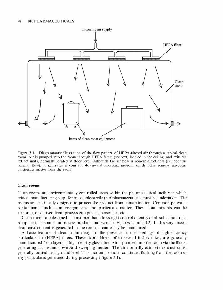
Clean rooms
Clean rooms are environmentally controlled areas within the pharmaceutical facility in whichcritical manufacturing steps for injectable/sterile (bio)pharmaceuticals must be undertaken. Therooms are specifically designed to protect the product from contamination. Common potentialcontaminants include microorganisms and particulate matter. These contaminants can beairborne, or derived from process equipment, personnel, etc.
Clean rooms are designed in a manner that allows tight control of entry of all substances (e.g.equipment, personnel, in-process product, and even air; Figures 3.1 and 3.2). In this way, once aclean environment is generated in the room, it can easily be maintained.
A basic feature of clean room design is the presence in their ceilings of high-efficiencyparticulate air (HEPA) filters. These depth filters, often several inches thick, are generallymanufactured from layers of high-density glass fibre. Air is pumped into the room via the filters,generating a constant downward sweeping motion. The air normally exits via exhaust units,generally located near ground level. This motion promotes continued flushing from the room ofany particulates generated during processing (Figure 3.1).
98 BIOPHARMACEUTICALS
Figure 3.1. Diagrammatic illustration of the flow pattern of HEPA-filtered air through a typical cleanroom. Air is pumped into the room through HEPA filters (see text) located in the ceiling, and exits viaextract units, normally located at floor level. Although the air flow is non-unidirectional (i.e. not truelaminar flow), it generates a constant downward sweeping motion, which helps remove air-borneparticulate matter from the room
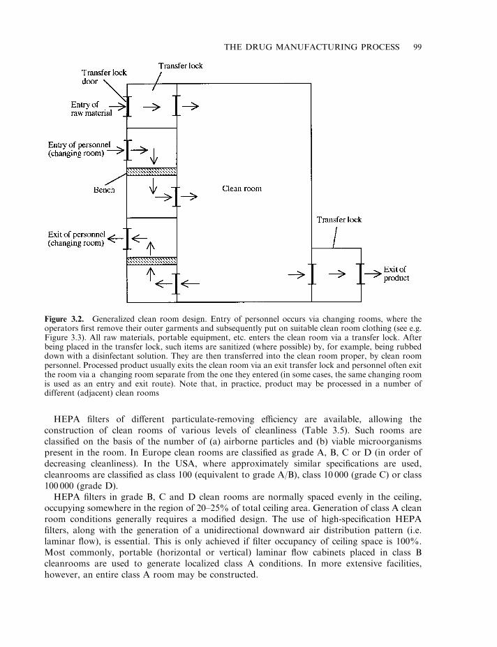
HEPA filters of different particulate-removing efficiency are available, allowing theconstruction of clean rooms of various levels of cleanliness (Table 3.5). Such rooms areclassified on the basis of the number of (a) airborne particles and (b) viable microorganismspresent in the room. In Europe clean rooms are classified as grade A, B, C or D (in order ofdecreasing cleanliness). In the USA, where approximately similar specifications are used,cleanrooms are classified as class 100 (equivalent to grade A/B), class 10 000 (grade C) or class100 000 (grade D).
HEPA filters in grade B, C and D clean rooms are normally spaced evenly in the ceiling,occupying somewhere in the region of 20–25% of total ceiling area. Generation of class A cleanroom conditions generally requires a modified design. The use of high-specification HEPAfilters, along with the generation of a unidirectional downward air distribution pattern (i.e.laminar flow), is essential. This is only achieved if filter occupancy of ceiling space is 100%.Most commonly, portable (horizontal or vertical) laminar flow cabinets placed in class Bcleanrooms are used to generate localized class A conditions. In more extensive facilities,however, an entire class A room may be constructed.
THE DRUG MANUFACTURING PROCESS 99
Figure 3.2. Generalized clean room design. Entry of personnel occurs via changing rooms, where theoperators first remove their outer garments and subsequently put on suitable clean room clothing (see e.g.Figure 3.3). All raw materials, portable equipment, etc. enters the clean room via a transfer lock. Afterbeing placed in the transfer lock, such items are sanitized (where possible) by, for example, being rubbeddown with a disinfectant solution. They are then transferred into the clean room proper, by clean roompersonnel. Processed product usually exits the clean room via an exit transfer lock and personnel often exitthe room via a changing room separate from the one they entered (in some cases, the same changing roomis used as an entry and exit route). Note that, in practice, product may be processed in a number ofdifferent (adjacent) clean rooms
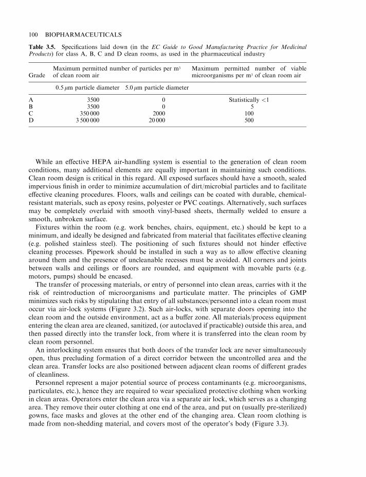
While an effective HEPA air-handling system is essential to the generation of clean roomconditions, many additional elements are equally important in maintaining such conditions.Clean room design is critical in this regard. All exposed surfaces should have a smooth, sealedimpervious finish in order to minimize accumulation of dirt/microbial particles and to facilitateeffective cleaning procedures. Floors, walls and ceilings can be coated with durable, chemical-resistant materials, such as epoxy resins, polyester or PVC coatings. Alternatively, such surfacesmay be completely overlaid with smooth vinyl-based sheets, thermally welded to ensure asmooth, unbroken surface.
Fixtures within the room (e.g. work benches, chairs, equipment, etc.) should be kept to aminimum, and ideally be designed and fabricated from material that facilitates effective cleaning(e.g. polished stainless steel). The positioning of such fixtures should not hinder effectivecleaning processes. Pipework should be installed in such a way as to allow effective cleaningaround them and the presence of uncleanable recesses must be avoided. All corners and jointsbetween walls and ceilings or floors are rounded, and equipment with movable parts (e.g.motors, pumps) should be encased.
The transfer of processing materials, or entry of personnel into clean areas, carries with it therisk of reintroduction of microorganisms and particulate matter. The principles of GMPminimizes such risks by stipulating that entry of all substances/personnel into a clean room mustoccur via air-lock systems (Figure 3.2). Such air-locks, with separate doors opening into theclean room and the outside environment, act as a buffer zone. All materials/process equipmententering the clean area are cleaned, sanitized, (or autoclaved if practicable) outside this area, andthen passed directly into the transfer lock, from where it is transferred into the clean room byclean room personnel.
An interlocking system ensures that both doors of the transfer lock are never simultaneouslyopen, thus precluding formation of a direct corridor between the uncontrolled area and theclean area. Transfer locks are also positioned between adjacent clean rooms of different gradesof cleanliness.
Personnel represent a major potential source of process contaminants (e.g. microorganisms,particulates, etc.), hence they are required to wear specialized protective clothing when workingin clean areas. Operators enter the clean area via a separate air lock, which serves as a changingarea. They remove their outer clothing at one end of the area, and put on (usually pre-sterilized)gowns, face masks and gloves at the other end of the changing area. Clean room clothing ismade from non-shedding material, and covers most of the operator’s body (Figure 3.3).
100 BIOPHARMACEUTICALS
Table 3.5. Specifications laid down (in the EC Guide to Good Manufacturing Practice for MedicinalProducts) for class A, B, C and D clean rooms, as used in the pharmaceutical industry
GradeMaximum permitted number of particles per m3
of clean room airMaximum permitted number of viablemicroorganisms per m3 of clean room air
0.5 mm particle diameter 5.0mm particle diameter
A 3500 0 Statistically <1B 3500 0 5C 350 000 2000 100D 3 500 000 20 000 500
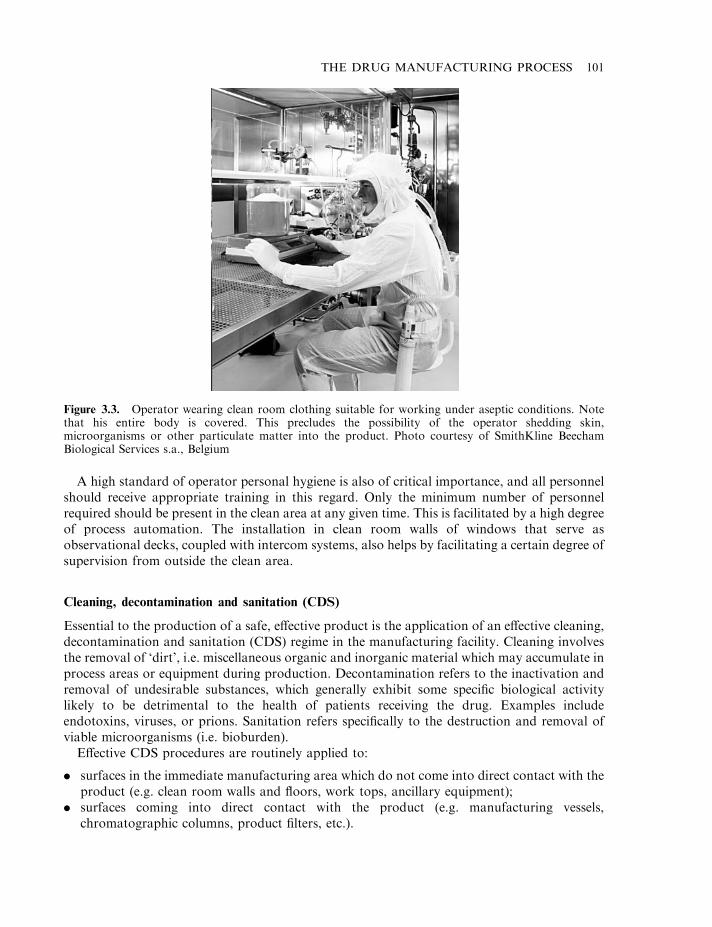
A high standard of operator personal hygiene is also of critical importance, and all personnelshould receive appropriate training in this regard. Only the minimum number of personnelrequired should be present in the clean area at any given time. This is facilitated by a high degreeof process automation. The installation in clean room walls of windows that serve asobservational decks, coupled with intercom systems, also helps by facilitating a certain degree ofsupervision from outside the clean area.
Cleaning, decontamination and sanitation (CDS)
Essential to the production of a safe, effective product is the application of an effective cleaning,decontamination and sanitation (CDS) regime in the manufacturing facility. Cleaning involvesthe removal of ‘dirt’, i.e. miscellaneous organic and inorganic material which may accumulate inprocess areas or equipment during production. Decontamination refers to the inactivation andremoval of undesirable substances, which generally exhibit some specific biological activitylikely to be detrimental to the health of patients receiving the drug. Examples includeendotoxins, viruses, or prions. Sanitation refers specifically to the destruction and removal ofviable microorganisms (i.e. bioburden).
Effective CDS procedures are routinely applied to:
. surfaces in the immediate manufacturing area which do not come into direct contact with theproduct (e.g. clean room walls and floors, work tops, ancillary equipment);
. surfaces coming into direct contact with the product (e.g. manufacturing vessels,chromatographic columns, product filters, etc.).
THE DRUG MANUFACTURING PROCESS 101
Figure 3.3. Operator wearing clean room clothing suitable for working under aseptic conditions. Notethat his entire body is covered. This precludes the possibility of the operator shedding skin,microorganisms or other particulate matter into the product. Photo courtesy of SmithKline BeechamBiological Services s.a., Belgium
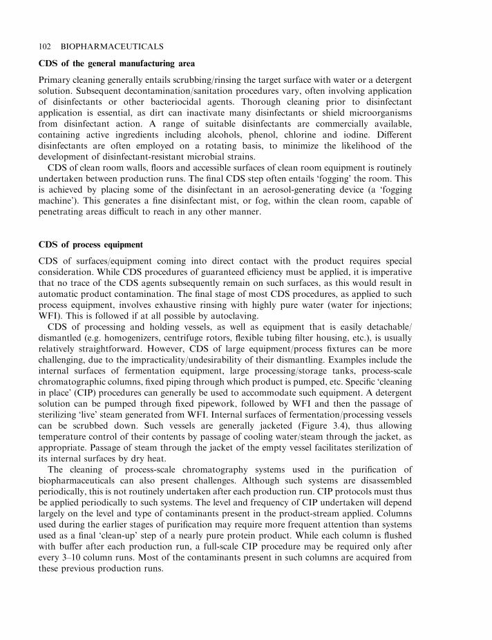
CDS of the general manufacturing area
Primary cleaning generally entails scrubbing/rinsing the target surface with water or a detergentsolution. Subsequent decontamination/sanitation procedures vary, often involving applicationof disinfectants or other bacteriocidal agents. Thorough cleaning prior to disinfectantapplication is essential, as dirt can inactivate many disinfectants or shield microorganismsfrom disinfectant action. A range of suitable disinfectants are commercially available,containing active ingredients including alcohols, phenol, chlorine and iodine. Differentdisinfectants are often employed on a rotating basis, to minimize the likelihood of thedevelopment of disinfectant-resistant microbial strains.
CDS of clean room walls, floors and accessible surfaces of clean room equipment is routinelyundertaken between production runs. The final CDS step often entails ‘fogging’ the room. Thisis achieved by placing some of the disinfectant in an aerosol-generating device (a ‘foggingmachine’). This generates a fine disinfectant mist, or fog, within the clean room, capable ofpenetrating areas difficult to reach in any other manner.
CDS of process equipment
CDS of surfaces/equipment coming into direct contact with the product requires specialconsideration. While CDS procedures of guaranteed efficiency must be applied, it is imperativethat no trace of the CDS agents subsequently remain on such surfaces, as this would result inautomatic product contamination. The final stage of most CDS procedures, as applied to suchprocess equipment, involves exhaustive rinsing with highly pure water (water for injections;WFI). This is followed if at all possible by autoclaving.
CDS of processing and holding vessels, as well as equipment that is easily detachable/dismantled (e.g. homogenizers, centrifuge rotors, flexible tubing filter housing, etc.), is usuallyrelatively straightforward. However, CDS of large equipment/process fixtures can be morechallenging, due to the impracticality/undesirability of their dismantling. Examples include theinternal surfaces of fermentation equipment, large processing/storage tanks, process-scalechromatographic columns, fixed piping through which product is pumped, etc. Specific ‘cleaningin place’ (CIP) procedures can generally be used to accommodate such equipment. A detergentsolution can be pumped through fixed pipework, followed by WFI and then the passage ofsterilizing ‘live’ steam generated from WFI. Internal surfaces of fermentation/processing vesselscan be scrubbed down. Such vessels are generally jacketed (Figure 3.4), thus allowingtemperature control of their contents by passage of cooling water/steam through the jacket, asappropriate. Passage of steam through the jacket of the empty vessel facilitates sterilization ofits internal surfaces by dry heat.
The cleaning of process-scale chromatography systems used in the purification ofbiopharmaceuticals can also present challenges. Although such systems are disassembledperiodically, this is not routinely undertaken after each production run. CIP protocols must thusbe applied periodically to such systems. The level and frequency of CIP undertaken will dependlargely on the level and type of contaminants present in the product-stream applied. Columnsused during the earlier stages of purification may require more frequent attention than systemsused as a final ‘clean-up’ step of a nearly pure protein product. While each column is flushedwith buffer after each production run, a full-scale CIP procedure may be required only afterevery 3–10 column runs. Most of the contaminants present in such columns are acquired fromthese previous production runs.
102 BIOPHARMACEUTICALS
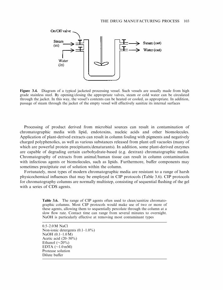
Processing of product derived from microbial sources can result in contamination ofchromatographic media with lipid, endotoxins, nucleic acids and other biomolecules.Application of plant-derived extracts can result in column fouling with pigments and negativelycharged polyphenolics, as well as various substances released from plant cell vacuoles (many ofwhich are powerful protein precipitants/denaturants). In addition, some plant-derived enzymesare capable of degrading certain carbohydrate-based (e.g. dextran) chromatographic media.Chromatography of extracts from animal/human tissue can result in column contaminationwith infectious agents or biomolecules, such as lipids. Furthermore, buffer components maysometimes precipitate out of solution within the column.
Fortunately, most types of modern chromatographic media are resistant to a range of harshphysicochemical influences that may be employed in CIP protocols (Table 3.6). CIP protocolsfor chromatography columns are normally multistep, consisting of sequential flushing of the gelwith a series of CDS agents.
THE DRUG MANUFACTURING PROCESS 103
Figure 3.4. Diagram of a typical jacketed processing vessel. Such vessels are usually made from highgrade stainless steel. By opening/closing the appropriate valves, steam or cold water can be circulatedthrough the jacket. In this way, the vessel’s contents can be heated or cooled, as appropriate. In addition,passage of steam through the jacket of the empty vessel will effectively sanitize its internal surfaces
Table 3.6. The range of CIP agents often used to clean/sanitize chromato-graphic columns. Most CIP protocols would make use of two or more ofthese agents, allowing them to sequentially percolate through the column at aslow flow rate. Contact time can range from several minutes to overnight.NaOH is particularly effective at removing most contaminant types
0.5–2.0M NaClNon-ionic detergents (0.1–1.0%)NaOH (0.1–1.0M)Acetic acid (20–50%)Ethanol (�20%)EDTA (�1.0mM)Protease solutionDilute buffer
Application of concentrated solutions of neutral salts (e.g. KCl or NaCl) is often effective inremoving precipitated/aggregated proteins, or other material retained in the column via ionicinteraction with the media. The use of buffers containing EDTA or other chelating agents helpsremove any metal ions associated with the gel. Use of (dilute) detergent solutions is effective inremoving lipid and a whole range of other contaminants. Solvent-containing (e.g. ethanol,butanol, isopropanol) buffers may also be used in this regard. Increasing the columntemperature to 50–608C may sometimes be considered, particularly if lipid appears to be amajor column contaminant (most lipids liquefy at such temperatures).
Sodium hydroxide is one of the most extensively used chromatography CIP agents. It isreadily available, inexpensive and effective. It is usually applied to a column at strengths of up to1.0M. At such concentrations, it quickly removes/destroys most contaminants, includingmicroorganisms and viruses. It will also degrade endotoxin (discussed later) within minutes.
Most types of chromatographic media can withstand incubation with NaOH for prolongedperiods. This allows CDS efficiency to be maximized by retaining NaOH in the column for timeperiods of the order of 30–60 minutes (silica gel is an exception, as it is quickly destroyed at pHvalues greater than 8). The chromatographic column is subsequently rinsed exhaustively bypumping WFI (or buffer made with WFI) through, until the column effluent is free from alltraces of NaOH. Prolonged exposure of the chromatographic media/column parts to residualNaOH could promote column deterioration, and obviously could contaminate/inactivate theprotein stream in the next production run.
Chromatographic systems are also designed to facilitate effective CIP. Internal surfaces of thecolumn, its valves and piping, are smooth, impervious and devoid of recesses which couldharbour microorganisms or other contaminants.
Periodic system disassembly allows more extensive CDS procedures to be undertaken. Mostcolumns are manufactured from glass, or more usually, tough plastic or stainless steel. After athorough cleaning of all disassembled components, sterilization by autoclaving is usuallyundertaken prior to re-assembly. Most chromatographic media likewise can be autoclavedbefore column re-pouring.
CIP of the ring main systems used to store and circulate WFI and purified water around thepharmaceutical plant is also routinely undertaken (see next section). This normally entailsemptying the ring main systems (including reservoirs), opening all the outlet valves, andsubsequently pumping sterile steam through all pipework. This is generally sufficient tophysically dislodge any traces of trapped particulate matter or biological agents harboured inthe system.
Water for biopharmaceutical processing
Water represents one of the most important raw materials used in biopharmaceuticalmanufacture. It is used as a basic ingredient of fermentation media, and in the manufactureof buffers used throughout product extraction and purification. It represents the solvent inwhich biopharmaceutical products sold in liquid form are dissolved, and in which freeze-driedbiopharmaceuticals will be reconstituted immediately prior to use. It is also used for ancillaryprocesses, such as the cleaning of equipment, piping and product-holding tanks. It isadditionally used to clean/rinse the vials into which the final product is filled.
It has been estimated that up to 30 000 litres of water is required to support the production of1 kg of a recombinant biopharmaceutical produced in a microbial system. It is not surprising,therefore, that the generation of water of suitable purity for processing is central to the
104 BIOPHARMACEUTICALS
successful operation of any (bio)pharmaceutical facility. The use of potable water (i.e. water ofdrinking standard) is limited to tasks such as routine cleaning of non-critical areas/processequipment. Such water must be subject to further in-house purification prior to its use in themanufacturing process. Water of two different levels of quality is usually required. These aretermed ‘purified water’ and ‘water for injections’ (WFI). These are distinguished on the basis ofthe range and quantity of allowable contaminants, with WFI being the most pure. Specificcriteria defining levels and types of contaminants permissible in both (along with guidelines as tohow the water must be produced) are outlined in international pharmacopoeias.
Purified water has a number of limited uses in pharmaceutical manufacturing. It is often usedas the solvent in the manufacture of aqueous-based oral products (e.g. cough mixtures,veterinary de-wormers, etc.). It is not intended to be used as a solvent in the downstreamprocessing of parenteral products— the category into which almost all biopharmaceuticals fall.It is used for primary cleaning of some process equipment/clean room floors, particularly inclass D or C clean areas, and for the generation of steam in the facilities’ autoclaves. Inbiopharmaceutical processing, purified water is often used in the generation of fermentationmedia used to culture biopharmaceutical-producing recombinant microorganisms. Its use insubsequent downstream processing is precluded, as its specifications allows the presence ofmany contaminants which downstream processing aims to minimize or eliminate from theproduct.
Water for injection finds extensive application in biopharmaceutical manufacturing.Although there is no regulatory requirement to do so, some manufacturers use WFI whenmaking microbial fermentation media. It is also commonly used in making culture media usedin the process-scale propagation of biopharmaceutical-producing mammalian cell lines. Lowinitial bioburden in WFI renders its sterilization by filtration more straightforward.Furthermore, mammalian cells can be particularly sensitive to some potential watercontaminants. The presence of even low concentrations of heavy metals, for example, canadversely effect the growth/product-producing characteristics of these cells.
WFI is the grade of water used exclusively in all downstream biopharmaceutical processingprocedures, ranging from making extraction/homogenization/chromatography buffers torinsing process equipment coming into direct contact with the product.
Generation of purified water and water for injections (WFI)
Purified water and WFI are generated from potable water. While the main techniques by whichthey are produced are specified by pharmacopoeias, pre-treatment of the incoming potablewater will vary, and is often dictated by the range of contaminants found in this water.
While some companies may have a private source of potable water, most obtain incomingwater from local municipal authorities. This water is sure to contain various levels of severalpotential contaminants (Table 3.7). A multi-step purification procedure is then undertaken,which usually contains some or all of the following steps (see also Figure 3.5):
. the incoming water is collected in a storage or ‘break’ tank, from where it is pumped througha depth filter, organic trap and carbon filter. The depth filter often contains a mixture ofgranular anthracite, washed sand, and gravel. Solids and colloidal material are retained asthe water percolates through the filter bed. The bed may be regenerated every few days bybackwashing (i.e. reversing the direction of water flow through the bed). The organic trap
THE DRUG MANUFACTURING PROCESS 105
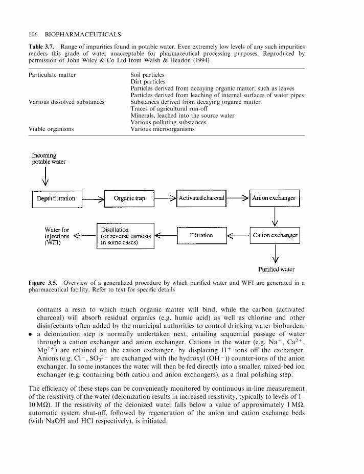
contains a resin to which much organic matter will bind, while the carbon (activatedcharcoal) will absorb residual organics (e.g. humic acid) as well as chlorine and otherdisinfectants often added by the municipal authorities to control drinking water bioburden;
. a deionization step is normally undertaken next, entailing sequential passage of waterthrough a cation exchanger and anion exchanger. Cations in the water (e.g. Na+, Ca2+,Mg2+) are retained on the cation exchanger, by displacing H+ ions off the exchanger.Anions (e.g. Cl7, SO3
27 are exchanged with the hydroxyl (OH7)) counter-ions of the anionexchanger. In some instances the water will then be fed directly into a smaller, mixed-bed ionexchanger (e.g. containing both cation and anion exchangers), as a final polishing step.
The efficiency of these steps can be conveniently monitored by continuous in-line measurementof the resistivity of the water (deionization results in increased resistivity, typically to levels of 1–10MO). If the resistivity of the deionized water falls below a value of approximately 1MO,automatic system shut-off, followed by regeneration of the anion and cation exchange beds(with NaOH and HCl respectively), is initiated.
106 BIOPHARMACEUTICALS
Table 3.7. Range of impurities found in potable water. Even extremely low levels of any such impuritiesrenders this grade of water unacceptable for pharmaceutical processing purposes. Reproduced bypermission of John Wiley & Co Ltd from Walsh & Headon (1994)
Particulate matter Soil particlesDirt particlesParticles derived from decaying organic matter, such as leavesParticles derived from leaching of internal surfaces of water pipes
Various dissolved substances Substances derived from decaying organic matterTraces of agricultural run-offMinerals, leached into the source waterVarious polluting substances
Viable organisms Various microorganisms
Figure 3.5. Overview of a generalized procedure by which purified water and WFI are generated in apharmaceutical facility. Refer to text for specific details
Deionizers fail to remove microbial contaminants, and exchange resin beds can often actuallyharbour microorganisms, contaminating the water. For this reason, filtration or UV treatmentoften constitutes the next purification step. One such treatment configuration entails pumpingthe deionized water through a 0.45 mm filter, followed by exposure to UV (at 254 nm). This maybe followed by yet another filtration step through a 0.22 mm filter to remove UV-inactivatedmicroorganisms. In some systems the 0.22 mm filter may even be replaced by ultrafilters capableof retaining molecules whose molecular mass is significantly greater than 10 kDa. Such systemswill remove not only bacteria but also many bacterial products, including endotoxin andadditional organic and colloidal material.
Deionized water often meets the pharmacopoeial criteria laid down for ‘purified water’.Sometimes, however, further purification may be necessary to attain this standard. This oftenentails a distillation or reverse-osmosis step. Deionized water will, however, not meet thepharmacopoeial requirements for WFI. WFI is best generated by distillation of deionized water.Distillation entails converting water to vapour by heat, followed by passing over a condenser,which results in condensation of pure water. Dissolved minerals and most organics are notvolatile at 1008C.
Reverse osmosis (RO) entails the use of a semi-permeable membrane (permeable to thesolvent, i.e. water molecules, but impermeable to solutes, i.e. contaminants). Osmosis describesthe movement of solvent molecules across such a membrane from a solution of lower soluteconcentration to one of higher solute concentration. The force promoting this movement istermed ‘osmotic pressure’. During reverse osmosis, a pressure greater than the natural osmoticpressure is applied to the system from the higher solute concentration side, causing solventmolecules to flow in the opposite direction.
RO membranes are manufactured from polymers such as cellulose acetate or polyamides. Asingle pass of water through such membranes can remove 95% of all dissolved solids and 99%of microorganisms and endotoxins. Double RO systems are often employed, increasing theefficiency of the process still further, and providing some element of protection against theconsequences of puncture of one of the membranes. While RO systems are less expensive thandistillation, many regard distillation as being a safer option. Pin-head punctures of ROmembranes can be hard to detect. Membranes are also susceptible to microbial colonization andcannot be exposed to high temperatures, which renders effective sanitation more difficult.
Distribution system for WFI
Upon its manufacture, WFI is fed into a sealed storage vessel, often made from stainless steel.The water is circulated via a series of pipework throughout the building, and from which anumber of outlet valves are available. The pipework leads back to the storage tank, allowingconstant recirculation of the water throughout the facility. Because of this it is known as a ringmain or loop system (Figure 3.6).
As pharmacopoeial specifications preclude the addition of sanitizing agents such as chlorine,maintenance of WFI within microbiological specifications requires special attention. Whilecirculating, the WFI is maintained at a flow rate of the order 9 ft/s. This ensures constantturbulent flow, discouraging microbial attachment to internal surfaces of the distribution pipes.The WFI is also constantly maintained at temperatures of the order of 858C, again todiscourage microbial growth.
Water collected from the WFI hot loop is generally allowed to cool before use forbiopharmaceutical processing (hot buffers would not only potentially denature the protein
THE DRUG MANUFACTURING PROCESS 107
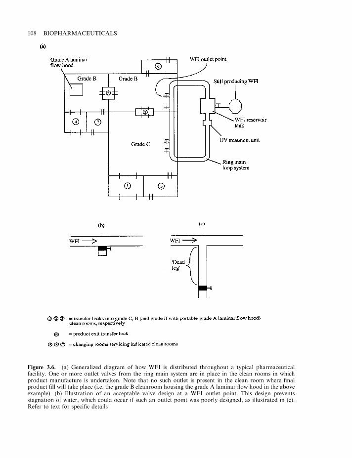
108 BIOPHARMACEUTICALS
Figure 3.6. (a) Generalized diagram of how WFI is distributed throughout a typical pharmaceuticalfacility. One or more outlet valves from the ring main system are in place in the clean rooms in whichproduct manufacture is undertaken. Note that no such outlet is present in the clean room where finalproduct fill will take place (i.e. the grade B cleanroom housing the grade A laminar flow hood in the aboveexample). (b) Illustration of an acceptable valve design at a WFI outlet point. This design preventsstagnation of water, which could occur if such an outlet point was poorly designed, as illustrated in (c).Refer to text for specific details
product, but their pH values could change quite dramatically when cooled from 858C to 258C).In some instances a separate loop system is constructed in which WFI is circulated at ambienttemperature allowing its immediate use in processing operations. To maintain water quality,however, this entire circulating system must be emptied and sanitized every 24 h.
Careful design of circulating systems also helps to maintain the microbiological quality ofWFI. Circulating pipework lengths are fused together by welding, as opposed to the use ofthreaded fittings, which could harbour bacteria. ‘Dead-legs’ (areas where water could stagnate,e.g. at water outlet points), are avoided, and bends in pipes are smooth and curving, as opposedto the use of abrupt T-junctions. UV cells are also fitted on-line in the system, subjectingcirculating water to their continual bactericidal influence.
Upon initial installation, the pipework is cleaned by passage of detergent or other cleaningagents, followed by a water rinse. ‘Passivation’ (exposure of the internal pipework surfaces tochemical agents, rendering the surface less reactive subsequently) is then undertaken, usually byoxidation using nitric acid or certain organic acids.
WFI is quite corrosive, especially at 858C, and it can promote leeching from even high-gradestainless steel piping. Addition of ozone to the WFI can alleviate this, as the ozone’s microcidalproperties facilitate prolonged storage/circulation of the water at 258C. Other innovations inthis field include replacement of stainless steel pipework with chemically inert plastics. However,extensive tests need to be undertaken in order to prove that WFI cannot leach potentiallydangerous substances from these plastics before their use will become routine.
Loop systems distributing purified water also exist in most facilities, with their design beingsomewhat similar to WFI systems. The more relaxed microbiological specifications relating topurified water renders the design and operation of such systems less complex. Purified water isgenerally circulated at ambient temperatures, via stainless steel or sometimes plastic-basedtubing.
Samples of WFI and purified water are usually collected daily by quality control personnel,and tested for conformance to specification. Failure to meet specification results in the systembeing emptied and fully sanitized, before generation of fresh water.
Documentation
Adequate documentation forms an essential part of good manufacturing practice. For thisreason, every aspect of pharmaceutical manufacture is characterized by the existence ofextensive associated documentation. This is essential in order to:
. help prevent errors/misunderstandings associated with verbal communication;
. facilitate the tracing of the manufacturing history of any batch of product;
. ensure reproducibility in all aspects of pharmaceutical manufacture.
Most documents associated with pharmaceutical manufacturing fall into one of fourcategories:
. standard operating procedures (SOPs);
. specifications;
. manufacturing formulae, processing and packaging instructions;
. records.
Great care is taken when initially preparing such documents to ensure they are written/worded in a clear and unambiguous fashion. They are usually written by supervisory personnel,
THE DRUG MANUFACTURING PROCESS 109
and inspected by senior technical personnel (often the production or QC manager or both),before their final approval for general use. Such documents are reviewed regularly, and updatedas required.
SOPs are documents which detail how staff should undertake particular procedures orprocesses. These procedures/processes are usually of a general nature, often being independentof any one pharmaceutical product. Many SOPs fall into one of several general categories,including:
. SOPs detailing step-by-step operational procedures for specific items of equipment (e.g.autoclaves, homogenizers, freeze-dryers, pH meters, product labelling machines, etc.);
. SOPs detailing maintenance/validation procedures for specific items of equipment or facilityareas, e.g. SOPs detailing CDS of clean rooms;
. SOPs relating directly to personnel (e.g. step-by-step procedures undertaken when gowning-up before entering a clean room);
. SOPs relating to testing/analysis (e.g. procedures detailing how to properly sample rawmaterials/finished product for QC analysis, SOPs relating to the routine sampling and testingof WFI from the ring main system, etc.).
Specifications
Specifications are normally written by QC personnel. They detail the exact qualitative andquantitative requirements to which individual raw materials or product must conform. Forexample, specifications for chemical raw materials will set strict criteria relating to thepercentage active ingredients present, permitted levels of named contaminants, etc. Specifica-tions for packing materials will, for example, lay down exact dimensions of product packagingcartons; specifications for product labels will detail label dimensions and exact details of labeltext, etc. Specifications for all raw materials are sent to raw material suppliers and, upon theirdelivery, QC personnel will ensure that these raw materials meet their specifications before beingreleased to production (the raw materials are held in ‘quarantine’ prior to their approval). Finalproduct specifications will also be prepared. As most products are manufactured to conformwith pharmacopoeial requirements, many of the specifications set for raw materials/finishedproduct are simply transcribed from the appropriate pharmacopoeia.
Manufacturing formulae, processing and packaging instructions
Manufacturing formulae should clearly indicate the product name, potency or strength, andexact batch size. It lists each of the starting raw materials required, and the quantity in whicheach is required. The processing instructions should contain step-by-step manufacturinginstructions. The detail given should be sufficient to allow a technically competent person,unfamiliar with the process, to successfully undertake the manufacturing procedure.
The processing instructions should also indicate the principal items of equipment utilizedduring manufacture, and the precise location in which each step should be undertaken (e.g. in aspecific clean room, etc.). It will also list any specific precautions which must be observed duringmanufacture (e.g. precautions to protect the product from, perhaps, excessive heating, or toprotect the operator from any potentially dangerous product constituent). Each product willalso have its own labelling and packing instructions, indicating:
. the label to be used, and its exact text;
110 BIOPHARMACEUTICALS
. exact packing instructions (e.g. how many units of product per pack, how may packs pershipping carton, etc.).
A copy of the label to be used is generally attached to the documents, to allow the supervisorand operators to verify easily that the correct label has been dispensed for the product inquestion.
Records
Maintenance of adequate and accurate records forms an essential part of GMP. For any givenbatch of product, records relating to every aspect of manufacture of that batch will be retained.These records will include:
. specification results obtained on all raw materials;
. batch manufacturing, processing and packaging records;
. QC analysis results of bulk and finished product.
These records, along with samples of finished product, must be retained in the facility for atleast 1 year after the expiry of that batch. Should any difficulty arise regarding thefinished product, the records should allow tracing back of all direct manufacturing steps, aswell as indirect procedures which might influence the quality/safety of the product.During inspections, regulatory inspectors usually examine the records relating to a number ofrandomly chosen batches in detail, in order to help them assess ongoing adherence to GMP inthe facility.
Generation of manufacturing records
Prior to commencement of the manufacturing of a specific product, production personnel willprint/photocopy the manufacturing formulae, processing and packaging instructions associatedwith that product. The responsible individual fills in the batch number in the space provided (abatch number is a unique combination of numbers and/or letters assigned to that batch, in orderto distinguish it from all other batches). This photocopied document forms the blueprint formanufacture of that batch. A space is provided after each manufacturing/packing instruction.When that step is completed, the responsible operator initials the space and includes the exacttime and date undertaken. This forms a detailed record of manufacture.
Additional supporting documents are also included in the manufacturing records. These mayinclude computerized print-outs from weighing equipment used to dispense chemical rawmaterials, or recorder charts obtained, e.g. from a freeze-drier upon completion of freeze-dryingthat batch of product.
QC records relating to raw materials, in-process and final product are generated in much thesame way—by printing/photocopying originals and filling in the test results obtained.
Advances in information technology are now impacting upon the pharmaceutical industry.Many documents are now maintained in electronic format. In fact, some regard it as likely thatin the future ‘paperless facilities’ will become commonplace, with all documentation beingcomputerized. Several aspects of such electronic document maintenance deserve specialattention. Adequate back-up files should always be retained. Also, restricted access tocomputerized systems is required to ensure that data/documentation is only entered/amendedby persons authorized to do so.
THE DRUG MANUFACTURING PROCESS 111
SOURCES OF BIOPHARMACEUTICALS
The bulk of biopharmaceuticals currently on the market are produced by genetic engineeringusing various recombinant expression systems. While a wide range of potential proteinproduction systems are available (Table 3.8), most of the recombinant proteins that have gainedmarketing approval to date are produced either in recombinant Eschericia coli or inrecombinant mammalian cell lines (Table 3.9). Such recombinant systems are invariablyconstructed by the introduction of a gene or cDNA coding for the protein of interest into a well-characterized strain of the chosen producer cell. Examples include E. coli K12 and Chinesehamster ovary strain K1 (CHO-K1). Gene/cDNA transfer is normally achieved by using anappropriate expression plasmid, or other standard gene-manipulating techniques. Eachrecombinant production system displays its own unique set of advantages and disadvantages,as described below.
E. coli as a source of recombinant, therapeutic proteins
Many microorganisms represent attractive potential production systems for therapeuticproteins. They can usually be cultured in large quantities, inexpensively and in a short time,by standard methods of fermentation. Production facilities can be constructed in any worldregion, and the scale of production can be varied as required.
The expression of recombinant proteins in cells in which they do not naturally occur is termed‘heterologous protein production’. By far the most common microbial species used to produce
112 BIOPHARMACEUTICALS
Table 3.8. Expression systems that are/could potentially be used forthe production of recombinant biopharmaceutical products (CHO¼Chinesehamster ovary; BHK¼baby hamster kidney)
E. coli (and additional prokaryotic systems, e.g. Bacilli)Yeast (particularly Saccharomyces cerevisiae)Fungi (particularly Aspergillus)Animal cell culture (particularly CHO and BHK cell lines)Transgenic animals (focus thus far is upon sheep and goats)Plant-based expression systems (various)Insect cell culture systems
Table 3.9. Some biopharmaceuticals currently on the market that are produced by genetic engineeringin either E. coli or animal cells. CHO=Chinese hamster ovary cells; BHK=baby hamster kidney cells
Biopharmaceutical product Source Biopharmaceutical product Source
Tissue plasminogen activator (tPA) E. coli, CHO Follicle-stimulating hormone (FSH) CHOInsulin E. coli Interferon-b CHO
Erythropoietin CHOInterferon-a E. coli Glucocerebrosidase CHOInterferon-g E. coli Factor VIIa BHKInterleukin-2 (IL-2) E. coliGranulocyte colony-stimulating factor(G-CSF)
E. coli
Human growth hormone (hGH) E. coli
heterologous proteins of therapeutic interest is Eschericia coli. The first biopharmaceuticalproduced by genetic engineering to gain marketing approval (in 1982) was recombinant humaninsulin (trade name Humulin), produced in E. coli. An example of a more recently approvedbiopharmaceutical which is produced in E. coli is that of Ecokinase, a recombinant tissueplasminogen activator (tPA, Chapter 9), approved for sale in the EU in 1996. Many additionalexamples are provided in subsequent chapters.
As a recombinant production system, E. coli displays a number of advantages, which include:
. E. coli has long served as the model system for studies relating to prokaryotic genetics. Itsmolecular biology is thus well characterized;
. high levels of expression of heterologous proteins can be achieved in recombinant E. coli(Table 3.10). Modern, high-expression promoters can routinely ensure that levels ofexpression of the recombinant protein reach up to 30% total cellular protein;
. E. coli cells grow rapidly on relatively simple and inexpensive media, and the appropriatefermentation technology is well established.
These advantages, particularly the ease of genetic manipulation, rendered E. coli the primarybiopharmaceutical production system for many years. However, E. coli also displays a numberof drawbacks as a biopharmaceutical producer, including:
. heterologous proteins accumulate intracellularly;
. inability to undertake post-translational modifications (particularly glycosylation) ofproteins;
. the presence of lipopolysaccharide on its surface.
The vast bulk of proteins synthesized naturally by E. coli (i.e. its homologous proteins) areintracellular. Few are exported to the periplasmic space, or are released as true extracellularproteins. Heterologous proteins expressed in E. coli thus invariably accumulate in the cellcytoplasm. Intracellular protein production complicates downstream processing (relative toextracellular production) because:
. additional primary processing steps are required, i.e. cellular homogenization, withsubsequent removal of cell debris by centrifugation or filtration;
. more extensive chromatographic purification is required in order to separate the protein ofinterest from the several thousand additional homologous proteins produced by the E. colicells.
THE DRUG MANUFACTURING PROCESS 113
Table 3.10. Levels of expression of various biopharmaceuticalsproduced in recombinant E. coli cells
BiopharmaceuticalLevel of expression
(% of total cellular protein)
Interferon-g 25Insulin 20Interferon-b 15Tumour necrosis factor 15a1-Antitrypsin 15Interleukin 2 10Human growth hormone 5
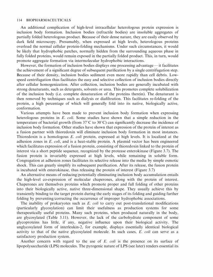
An additional complication of high-level intracellular heterologous protein expression isinclusion body formation. Inclusion bodies (refractile bodies) are insoluble aggregates ofpartially folded heterologous product. Because of their dense nature, they are easily observed bydark field microscopy. Presumably, when expressed at high levels, heterologous proteinsoverload the normal cellular protein-folding mechanisms. Under such circumstances, it wouldbe likely that hydrophobic patches, normally hidden from the surrounding aqueous phase infully folded proteins, would remain exposed in the partially folded product. This, in turn, wouldpromote aggregate formation via intermolecular hydrophobic interactions.
However, the formation of inclusion bodies displays one processing advantage— it facilitatesthe achievement of a significant degree of subsequent purification by a single centrifugation step.Because of their density, inclusion bodies sediment even more rapidly than cell debris. Low-speed centrifugation thus facilitates the easy and selective collection of inclusion bodies directlyafter cellular homogenization. After collection, inclusion bodies are generally incubated withstrong denaturants, such as detergents, solvents or urea. This promotes complete solubilizationof the inclusion body (i.e. complete denaturation of the proteins therein). The denaturant isthen removed by techniques such as dialysis or diafiltration. This facilitates re-folding of theprotein, a high percentage of which will generally fold into its native, biologically active,conformation.
Various attempts have been made to prevent inclusion body formation when expressingheterologous proteins in E. coli. Some studies have shown that a simple reduction in thetemperature of bacterial growth (from 378C to 308C) can significantly decrease the incidence ofinclusion body formation. Other studies have shown that expression of the protein of interest asa fusion partner with thioredoxin will eliminate inclusion body formation in most instances.Thioredoxin is a homologous E. coli protein, expressed at high levels. It is localized at theadhesion zones in E. coli, and is a heat-stable protein. A plasmid vector has been engineeredwhich facilitates expression of a fusion protein, consisting of thioredoxin linked to the protein ofinterest via a short peptide sequence, recognized by the protease enterokinase (Figure 3.7). Thefusion protein is invariably expressed at high levels, while remaining in soluble form.Congregation at adhesion zones facilitates its selective release into the media by simple osmoticshock. This can greatly simplify its subsequent purification. After its release, the fusion proteinis incubated with enterokinase, thus releasing the protein of interest (Figure 3.7)
An alternative means of reducing/potentially eliminating inclusion body accumulation entailsthe high-level co-expression of molecular chaperones, along with the protein of interest.Chaperones are themselves proteins which promote proper and full folding of other proteinsinto their biologically active, native three-dimensional shape. They usually achieve this bytransiently binding to the target protein during the early stages of its folding and guiding furtherfolding by preventing/correcting the occurrence of improper hydrophobic associations.
The inability of prokaryotes such as E. coli to carry out post-translational modifications(particularly glycosylation) can limit their usefulness as production systems for sometherapeutically useful proteins. Many such proteins, when produced naturally in the body,are glycosylated (Table 3.11). However, the lack of the carbohydrate component of someglycoproteins has little, if any, negative influence upon their biological activity. Theunglycosylated form of interleukin-2, for example, displays essentially identical biologicalactivity to that of the native glycosylated molecule. In such cases, E. coli can serve as asatisfactory production system.
Another concern with regard to the use of E. coli is the presence on its surface oflipopolysaccharide (LPS) molecules. The pyrogenic nature of LPS (see later) renders essential its
114 BIOPHARMACEUTICALS
THE DRUG MANUFACTURING PROCESS 115
Figure 3.7. High level expression of a protein of interest in E. coli in soluble form by using the engineered‘thiofusion’ expression system. Refer to text for specific details
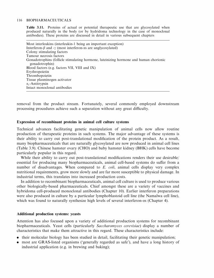
removal from the product stream. Fortunately, several commonly employed downstreamprocessing procedures achieve such a separation without any great difficulty.
Expression of recombinant proteins in animal cell culture systems
Technical advances facilitating genetic manipulation of animal cells now allow routineproduction of therapeutic proteins in such systems. The major advantage of these systems istheir ability to carry out post-translational modification of the protein product. As a result,many biopharmaceuticals that are naturally glycosylated are now produced in animal cell lines(Table 3.9). Chinese hamster ovary (CHO) and baby hamster kidney (BHK) cells have becomeparticularly popular in this regard.
While their ability to carry out post-translational modifications renders their use desirable/essential for producing many biopharmaceuticals, animal cell-based systems do suffer from anumber of disadvantages. When compared to E. coli, animal cells display very complexnutritional requirements, grow more slowly and are far more susceptible to physical damage. Inindustrial terms, this translates into increased production costs.
In addition to recombinant biopharmaceuticals, animal cell culture is used to produce variousother biologically-based pharmaceuticals. Chief amongst these are a variety of vaccines andhybridoma cell-produced monoclonal antibodies (Chapter 10). Earlier interferon preparationswere also produced in culture by a particular lymphoblastoid cell line (the Namalwa cell line),which was found to naturally synthesize high levels of several interferon-as (Chapter 4).
Additional production systems: yeasts
Attention has also focused upon a variety of additional production systems for recombinantbiopharmaceuticals. Yeast cells (particularly Saccharomyces cerevisiae) display a number ofcharacteristics that make them attractive in this regard. These characteristics include:
. their molecular biology has been studied in detail, facilitating their genetic manipulation;
. most are GRAS-listed organisms (‘generally regarded as safe’), and have a long history ofindustrial application (e.g. in brewing and baking);
116 BIOPHARMACEUTICALS
Table 3.11. Proteins of actual or potential therapeutic use that are glycosylated whenproduced naturally in the body (or by hydridoma technology in the case of monoclonalantibodies). These proteins are discussed in detail in various subsequent chapters
Most interleukins (interleukin-1 being an important exception)Interferon-b and -g (most interferon-as are unglycosylated)Colony stimulating factorsTumour necrosis factorsGonadotrophins (follicle stimulating hormone, luteinizing hormone and human chorionicgonadotrophin)
Blood factors (e.g. factors VII, VIII and IX)ErythropoietinThrombopoietinTissue plasminogen activatora1-AntitrypsinIntact monoclonal antibodies
. they grow relatively quickly in relatively inexpensive media, and their tough outer cell wallprotects them from physical damage;
. suitable industrial scale fermentation equipment/technology is already available;
. they possess the ability to carry out post-translational modifications of proteins.
The practical potential of yeast-based production systems has been confirmed by the successfulexpression of a whole range of proteins of therapeutic interest in such systems. However, anumber of disadvantages relating to heterologous protein production in yeast have beenrecognized. These include:
. although capable of glycosylating heterologous human proteins, the glycosylation patternusually varies somewhat from the pattern observed in the native glycoprotein (when isolatedfrom its natural source, or when expressed in recombinant animal cell culture systems);
. in most instances, expression levels of heterologous proteins remain less than 5% of totalcellular protein. This is significantly lower than expression levels typically achieved inrecombinant E. coli systems.
Despite such potential disadvantages, several recombinant biopharmaceuticals now approvedfor general medical use are produced in yeast (S. cerevisiae)-based systems (Table 3.12).Interestingly, most such products are not glycosylated. The oligosaccharide component ofglycoproteins produced in yeasts generally contain high levels of mannose. Such high mannose-type glycosylation patterns generally trigger their rapid clearance from the blood stream. Suchproducts, therefore, would be expected to display a short half-life when parenterallyadministered to man.
Fungal production systems
Fungi have elicited interest as heterologous protein producers, as many have a long history ofuse in the production of various industrial enzymes, such as a-amylase and glucoamylase.Suitable fermentation technology therefore already exists. In general, fungi are capable ofhigh-level expression of various proteins, many of which they secrete into their extracellularmedia. The extracellular production of a biopharmaceutical would be distinctly advantageousin terms of subsequent downstream processing. Fungi also possess the ability to carry out
THE DRUG MANUFACTURING PROCESS 117
Table 3.12. Recombinant therapeutic proteins approved for general medical use that are produced inSaccharomycese cerevisiae. All are subsequently discussed in the chapter indicated
Trade name Description Use Refer toChapter
Novolog Engineered short-acting insulin Diabetes mellitus 8Leukine Colony stimulating factor (GM-CSF) Bone marrow
transplantation6
Recombivax, Comvax, Engerix B,Tritanrix-HB, Infanrix, Twinrix,Primavax, Hexavax
All vaccine preparations containingrHBsAg as one component
Vaccination 10
Revasc, Refludan Hirudin Anticoagulant 9Fasturtec Urate oxidase Hyperuricaemia 9Regranex Platelet-derived growth factor Diabetic ulcers 7
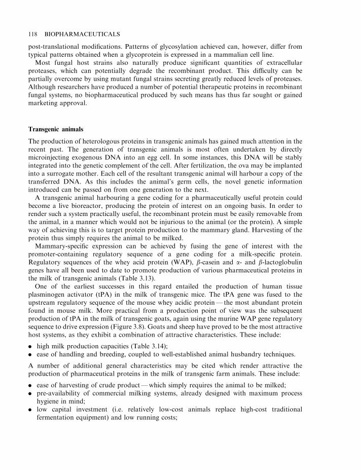
post-translational modifications. Patterns of glycosylation achieved can, however, differ fromtypical patterns obtained when a glycoprotein is expressed in a mammalian cell line.
Most fungal host strains also naturally produce significant quantities of extracellularproteases, which can potentially degrade the recombinant product. This difficulty can bepartially overcome by using mutant fungal strains secreting greatly reduced levels of proteases.Although researchers have produced a number of potential therapeutic proteins in recombinantfungal systems, no biopharmaceutical produced by such means has thus far sought or gainedmarketing approval.
Transgenic animals
The production of heterologous proteins in transgenic animals has gained much attention in therecent past. The generation of transgenic animals is most often undertaken by directlymicroinjecting exogenous DNA into an egg cell. In some instances, this DNA will be stablyintegrated into the genetic complement of the cell. After fertilization, the ova may be implantedinto a surrogate mother. Each cell of the resultant transgenic animal will harbour a copy of thetransferred DNA. As this includes the animal’s germ cells, the novel genetic informationintroduced can be passed on from one generation to the next.
A transgenic animal harbouring a gene coding for a pharmaceutically useful protein couldbecome a live bioreactor, producing the protein of interest on an ongoing basis. In order torender such a system practically useful, the recombinant protein must be easily removable fromthe animal, in a manner which would not be injurious to the animal (or the protein). A simpleway of achieving this is to target protein production to the mammary gland. Harvesting of theprotein thus simply requires the animal to be milked.
Mammary-specific expression can be achieved by fusing the gene of interest with thepromoter-containing regulatory sequence of a gene coding for a milk-specific protein.Regulatory sequences of the whey acid protein (WAP), b-casein and a- and b-lactoglobulingenes have all been used to date to promote production of various pharmaceutical proteins inthe milk of transgenic animals (Table 3.13).
One of the earliest successes in this regard entailed the production of human tissueplasminogen activator (tPA) in the milk of transgenic mice. The tPA gene was fused to theupstream regulatory sequence of the mouse whey acidic protein— the most abundant proteinfound in mouse milk. More practical from a production point of view was the subsequentproduction of tPA in the milk of transgenic goats, again using the murine WAP gene regulatorysequence to drive expression (Figure 3.8). Goats and sheep have proved to be the most attractivehost systems, as they exhibit a combination of attractive characteristics. These include:
. high milk production capacities (Table 3.14);
. ease of handling and breeding, coupled to well-established animal husbandry techniques.
A number of additional general characteristics may be cited which render attractive theproduction of pharmaceutical proteins in the milk of transgenic farm animals. These include:
. ease of harvesting of crude product—which simply requires the animal to be milked;
. pre-availability of commercial milking systems, already designed with maximum processhygiene in mind;
. low capital investment (i.e. relatively low-cost animals replace high-cost traditionalfermentation equipment) and low running costs;
118 BIOPHARMACEUTICALS
. high expression levels of proteins are potentially attained. In many instances, the level ofexpression exceeds 1 g protein/litre milk. In one case, initial expression levels of 60 g/l wereobserved, which stabilized at 35 g/l as lactation continued (the expression of the a1-antitrypsin gene, under the influence of the ovine b-lactoglobulin promoter, in a transgenicsheep). Even at expression levels of 1 g/l, one transgenic goat would produce a similarquantity of product in 1 day as would likely be recoverable from a 50–100 l bioreactorsystem;
. ongoing supply of product is guaranteed (by breeding);
. milk is biochemically well characterized, and the physicochemical properties of the majornative milk proteins of various species are well known. This helps rational development ofappropriate downstream processing protocols (Table 3.15).
Despite the attractiveness of this system, a number of issues remain to be resolved before it isbroadly accepted by the industry. These include:
. variability of expression levels. While in many cases expression levels of heterologousproteins exceed 1 g/l, in some instances expression levels as low as 1.0mg/l have beenobtained;
. characterization of the exact nature of the post-translational modifications the mammarysystem is capable of undertaking, e.g. the carbohydrate composition of tPA produced in thissystem differs from the recombinant enzyme produced in murine cell culture systems;
. significant time lag between the generation of a transgenic embryo and commencement ofroutine product manufacture. Once a viable embryo containing the inserted desired gene isgenerated, it must firstly be brought to term. This gestation period ranges from 1 month forrabbits to 9 months for cows. The transgenic animal must then reach sexual maturity beforebreeding (5 months for rabbits, 15 months for cows). Before they begin to lactate (i.e.produce the recombinant product), they must breed successfully and bring their offspring toterm. The overall time lag to routine manufacture can, therefore, be almost 3 years in the caseof cows or 7 months in the case of rabbits. Furthermore, if the original transgenic embryoturns out to be male, a further delay is encountered as this male must breed in order to pass
THE DRUG MANUFACTURING PROCESS 119
Table 3.13. Proteins of actual/potential therapeutic use that have been produced inthe milk of transgenic animals
Protein Animal species Expression levels in milk
tPA Goat 6 g/lInterleukin-2 Rabbit 0.5mg/lFactor VIII Pig 3mg/lFactor IX Sheep 1 g/la1-Antitrypsin Goat 20 g/lFibrinogen Sheep 5 g/lErythropoietin Rabbit 50mg/lAntithrombin III Goat 14 g/lHuman a-lactalbumin Cow 2.5 g/lInsulin-like growth factor I Rabbit 1 g/lProtein C Pig 1 g/lGrowth hormone Rabbit 50mg/l
on the desired gene to daughter animals—who will then eventually produce the desiredproduct in their milk.
Another general disadvantage of this approach relates to the use of the micro-injectiontechnique to introduce the desired gene into the pronucleus of the fertilized egg. This approach
120 BIOPHARMACEUTICALS
Figure 3.8. The production and purification of tPA from the milk of transgenic goats (WAP=murinewhey acid). The downstream processing procedure yielded in excess of an 8000-fold purification factor withan overall product yield of 25%. The product was greater than 98% pure, as judged by SDS-electrophoresis
is inefficient and time-consuming. There is no control over issues such as if/where in the hostgenomes the injected gene will integrate. Overall, only a modest proportion of manipulatedembryos will culminate in the generation of a healthy biopharmaceutical-producing animal.
A number of alternative approaches are being developed which may overcome some of theseissues. Replication-defective retroviral vectors are available which will more consistently (a)deliver a chosen gene into cells and (b) ensure chromosomal integration of the gene. A secondinnovation is the application of nuclear transfer technology.
Nuclear transfer entails substituting the genetic information present in an unfertilized eggwith donor genetic information. The best-known product of this technology is ‘Dolly’ the sheep,produced by substituting the nucleus of a sheep egg with a nucleus obtained from an adult sheepcell (genetically, therefore, Dolly was a clone of the original ‘donor’ sheep). An extension ofthis technology applicable to biopharmaceutical manufacture entails using a donor cellnucleus previously genetically manipulated so as to harbour a gene coding for thebiopharmaceutical of choice. The technical viability of this approach was proved in the late1990s upon the birth of two transgenic sheep, ‘Polly’ and ‘Molly’. The donor nucleus used togenerate these sheep harboured an inserted (human) blood factor IX gene under the control of amilk protein promoter. Both now produce significant quantities of human factor IX in theirmilk.
At the time of writing, no therapeutic protein produced in the milk of transgenic animals hadbeen approved for general medical use. A number of companies, however, continue to pursuethis strategy. These include GTC Biotherapeutics USA (formerly Genzyme Transgenics) andPPL Therapeutics (Scotland). a1-Antitrypsin, antithrombin and a range of monoclonalantibody-based products produced via transgenic technology continue to be evaluated bythese companies.
In addition to milk, a range of recombinant proteins have been expressed in various othertargeted tissues/fluids of transgenic animals. Antibodies and other proteins have been producedin the blood of transgenic pigs and rabbits. This mode of production is, however, unlikely to bepursued industrially for a number of reasons:
THE DRUG MANUFACTURING PROCESS 121
Table 3.14. Typical annual milk yields (litres) as well as time lapse between generation ofthe transgene embryo and first product harvest (first lactation) of indicated species
Species Annual milk yield (l) Time to first production batch (months)
Cow 6000–9000 33–36Goat 700–800 18–20Sheep 400–500 18–20Pig 250–300 16–17Rabbit 4–5 7
Table 3.15. Some physicochemical properties of the major (bovine) milk proteins
Protein Caseins b-Lactoglobulin a-Lactalbumin Serum albumin IgG
Concentration (g/l) 25 (Total) 2–4 0.5–1.5 0.4 0.5–1.0Mass (kDa) 20–25 18 14 66 150Phosphorylated? Yes No No No NoIsoelectric point Vary 5.2 4.2–4.8 4.7–4.9 5.5–8.3
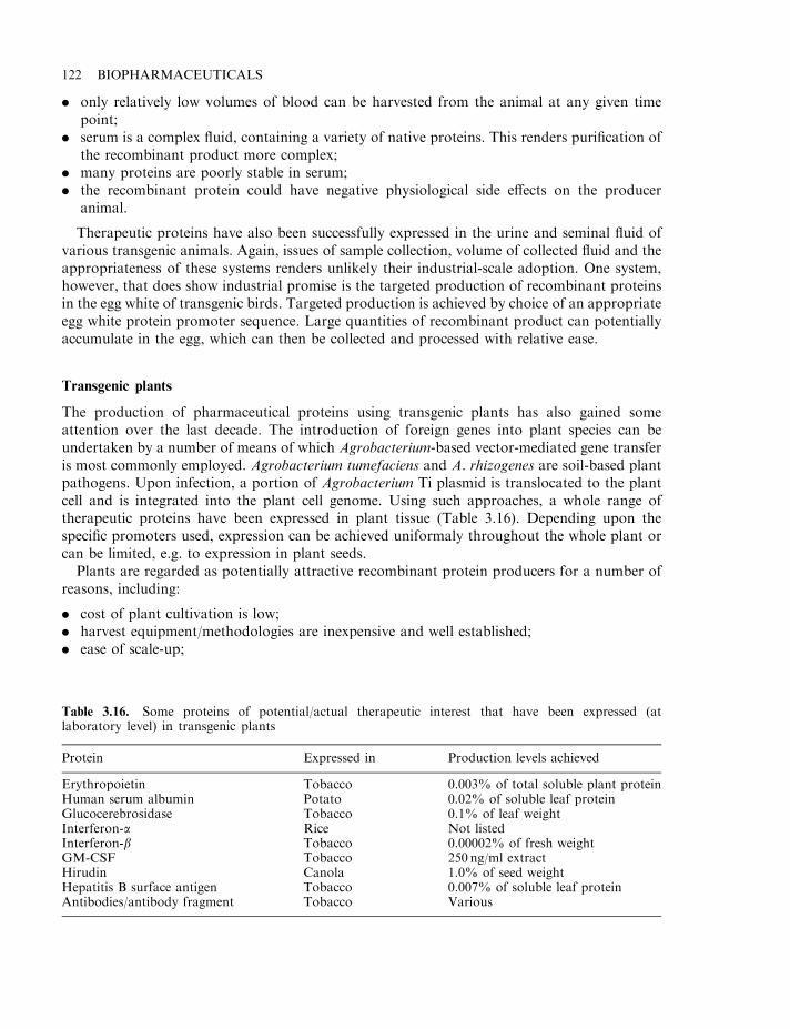
. only relatively low volumes of blood can be harvested from the animal at any given timepoint;
. serum is a complex fluid, containing a variety of native proteins. This renders purification ofthe recombinant product more complex;
. many proteins are poorly stable in serum;
. the recombinant protein could have negative physiological side effects on the produceranimal.
Therapeutic proteins have also been successfully expressed in the urine and seminal fluid ofvarious transgenic animals. Again, issues of sample collection, volume of collected fluid and theappropriateness of these systems renders unlikely their industrial-scale adoption. One system,however, that does show industrial promise is the targeted production of recombinant proteinsin the egg white of transgenic birds. Targeted production is achieved by choice of an appropriateegg white protein promoter sequence. Large quantities of recombinant product can potentiallyaccumulate in the egg, which can then be collected and processed with relative ease.
Transgenic plants
The production of pharmaceutical proteins using transgenic plants has also gained someattention over the last decade. The introduction of foreign genes into plant species can beundertaken by a number of means of which Agrobacterium-based vector-mediated gene transferis most commonly employed. Agrobacterium tumefaciens and A. rhizogenes are soil-based plantpathogens. Upon infection, a portion of Agrobacterium Ti plasmid is translocated to the plantcell and is integrated into the plant cell genome. Using such approaches, a whole range oftherapeutic proteins have been expressed in plant tissue (Table 3.16). Depending upon thespecific promoters used, expression can be achieved uniformaly throughout the whole plant orcan be limited, e.g. to expression in plant seeds.
Plants are regarded as potentially attractive recombinant protein producers for a number ofreasons, including:
. cost of plant cultivation is low;
. harvest equipment/methodologies are inexpensive and well established;
. ease of scale-up;
122 BIOPHARMACEUTICALS
Table 3.16. Some proteins of potential/actual therapeutic interest that have been expressed (atlaboratory level) in transgenic plants
Protein Expressed in Production levels achieved
Erythropoietin Tobacco 0.003% of total soluble plant proteinHuman serum albumin Potato 0.02% of soluble leaf proteinGlucocerebrosidase Tobacco 0.1% of leaf weightInterferon-a Rice Not listedInterferon-b Tobacco 0.00002% of fresh weightGM-CSF Tobacco 250 ng/ml extractHirudin Canola 1.0% of seed weightHepatitis B surface antigen Tobacco 0.007% of soluble leaf proteinAntibodies/antibody fragment Tobacco Various
. proteins expressed in seeds are generally stable in the seed for prolonged periods of time(often years);
. plant-based systems are free of human pathogens (e.g. HIV).
However, a number of potential disadvantages are also associated with the use of plant-basedexpression systems, including:
. variable/low expression levels sometimes achieved;
. potential occurrence of post-translational gene silencing (a sequence-specific mRNAdegradation mechanism);
. glycosylation patterns achieved differ significantly from native human protein glycosylationpatterns;
. seasonal/geographical nature of plant growth.
For these reasons, as well as the fact that additional tried and tested expression systems arealready available, production of recombinant therapeutic proteins in transgenic plant systemshas not as yet impacted significantly on the industry.
The most likely focus of future industry interest in this area concerns the production of oralvaccines in edible plants or fruit, such as tomatoes and bananas. Animal studies have clearlyshown that ingestion of transgenic plant tissue expressing recombinant sub-unit vaccines (seeChapter 10 for a discussion of sub-unit vaccines) induces the production of antigen-specificantibody responses, not only in mucosal secretions but also in the serum. The approach iselegant in that direct consumption of the plant material provides an inexpensive, efficient andtechnically straightforward mode of large-scale vaccine delivery, particularly in poorer worldregions. However, several hurdles hindering the widespread application of this technologyinclude:
. the immunogenicity of orally administered vaccines can vary widely;
. the stability of antigens in the digestive tract varies widely;
. the genetics of many potential systems remain poorly characterized, leading to inefficienttransformation systems and low expression levels.
Insect cell-based systems
A wide range of proteins have been produced at laboratory scale in recombinant insect cellculture systems. The approach generally entails the infection of cultured insect cells with anengineered baculovirus (a viral family that naturally infects insects) carrying the gene coding forthe desired protein, placed under the influence of a powerful viral promoter. Amongst thesystems most commonly employed are:
. the silkworm virus Bombyx mori nuclear polyhedrovirus (BmNPV), in conjunction withcultured silkworm cells (i.e. Bombyx mori cells) or;
. the virus Autographa californica nuclear polyhedrovirus (AcNPV), in conjunction withcultured armyworm cells (Spodoptera frugiperda cells).
Baculovirus/insect cell-based systems are cited as having a number of advantages, including:
. high-level intracellular recombinant protein expression. The use of powerful viral promoterssuch as promoters derived from the viral polyhedrin or P10 genes can drive recombinantprotein expression levels to 30–50% of total intracellular protein;
THE DRUG MANUFACTURING PROCESS 123
. insect cells can be cultured more rapidly and using less expensive media than mammalian celllines;
. human pathogens (e.g. HIV) do not generally infect insect cell lines.
However, a number of disadvantages are also associated with this production system, including:
. targeted extracellular recombinant production generally results in low-level extracellularaccumulation of the desired protein (often in the mg/l range). Extracellular productionsimplifies subsequent downstream processing, as discussed later in this chapter;
. post-translational modifications, in particular glycosylation patterns, can be incomplete and/or can differ very significantly from the patterns associated with native human glycoproteins.
Therapeutic proteins successfully produced on a laboratory scale in insect cell lines includehepatitis B surface antigen, interferon-g and tissue plasminogen activator (tPA). To date, notherapeutic product produced by such means has been approved for human use. Two veterinaryvaccines, however, have; ‘Bayovac CSF E2’ and ‘Porcilis Pesti’ are both subunit vaccines(Chapter 10) containing the E2 surface antigen protein of classical swine fever virus as activeingredient. The vaccines are administered to pigs in order to immunize against classical swinefever and an overview of their manufacture is provided in Figure 3.9.
An alternative insect cell-based system used to achieve recombinant protein productionentails the use of live insects. Most commonly, live caterpillars or silkworms are injected withthe engineered baculovirus vector, effectively turning the whole insect into a live bioreactor. Oneveterinary biopharmaceutical, Vibragen Omega, is manufactured using this approach and anoverview of its manufacture is outlined in Figure 3.10. Briefly, whole live silkworms areintroduced into pre-sterilized cabinets and reared on laboratory media. After 2 days, eachsilkworm is innoculated with engineered virus, using an automatic microdispenser. Thisengineered silkworm polyhedrosis virus harbours a copy of cDNA coding for feline interferon-o. During the subsequent 5 days of rearing, a viral infection is established and hencerecombinant protein synthesis occurs within the silkworms. After acid extraction, neutralizationand clarification, the recombinant product is purified chromatographically. A two-step affinityprocedure using blue sepharose dye affinity and copper chelate sepharose chromatography isemployed (see later in this chapter). After a gel-filtration step, excipients (sorbitol and gelatin)are added and the product is freeze-dried after filling into glass vials.
PRODUCTION OF FINAL PRODUCT
This section briefly overviews how biopharmaceutical substances are produced in abiopharmaceutical/biotech manufacturing facility. As the vast bulk of biopharmaceuticals areproteins synthesized in recombinant prokaryotic (e.g. E. coli) or eukaryotic (e.g. mammaliancells) production systems, attention will focus specifically upon these.
As defined in this text, the production process is deemed to commence when a single vial ofthe working cell bank system (see later) is taken from storage, and the cells therein cultured inorder to initiate the biosynthesis of a batch of product (Figure 3.11). The production process isdeemed to be complete only when the final product is filled in its final containers and thosecontainers have been labelled and placed in their final product packaging.
Production can be divided into ‘upstream’ and ‘downstream’ processing (Figure 3.11).Upstream processing refers to the initial fermentation process, which results in the initialgeneration of product. Downstream processing refers to the actual purification of the protein
124 BIOPHARMACEUTICALS
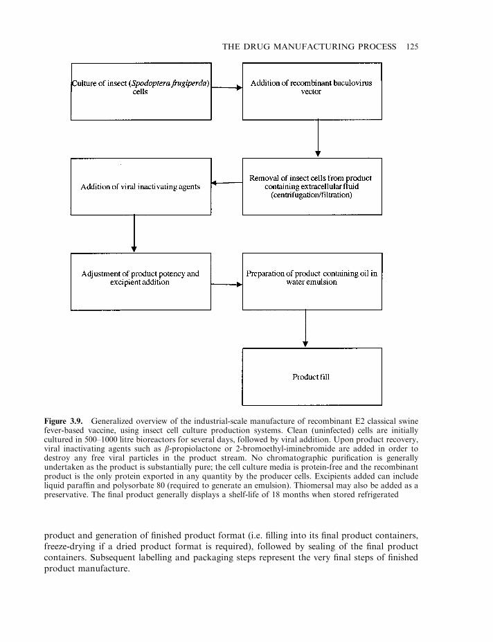
product and generation of finished product format (i.e. filling into its final product containers,freeze-drying if a dried product format is required), followed by sealing of the final productcontainers. Subsequent labelling and packaging steps represent the very final steps of finishedproduct manufacture.
THE DRUG MANUFACTURING PROCESS 125
Figure 3.9. Generalized overview of the industrial-scale manufacture of recombinant E2 classical swinefever-based vaccine, using insect cell culture production systems. Clean (uninfected) cells are initiallycultured in 500–1000 litre bioreactors for several days, followed by viral addition. Upon product recovery,viral inactivating agents such as b-propiolactone or 2-bromoethyl-iminebromide are added in order todestroy any free viral particles in the product stream. No chromatographic purification is generallyundertaken as the product is substantially pure; the cell culture media is protein-free and the recombinantproduct is the only protein exported in any quantity by the producer cells. Excipients added can includeliquid paraffin and polysorbate 80 (required to generate an emulsion). Thiomersal may also be added as apreservative. The final product generally displays a shelf-life of 18 months when stored refrigerated
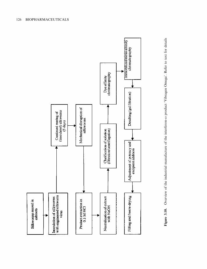
126 BIOPHARMACEUTICALS
Figure
3.10.
Overview
oftheindustrialmanufacture
oftheinterferon-o
product
‘Vibragen
Omega’.Refer
totextfordetails

Cell banking systems
Recombinant biopharmaceutical production cell lines are most often initially constructed by theintroduction into these cells of a plasmid housing a nucleotide sequence coding for the protein ofinterest. After culture, the resultant product-producing cell line is generally aliquoted into smallamounts, which are placed in ampoules and subsequently immersed in liquid nitrogen. Thecontent of all the ampoules is therefore identical, and the cells are effectively preserved forindefinite periods when stored under liquid nitrogen. This batch of cryopreserved ampoulesform a ‘cell bank’ system, whereby one ampoule is thawed and the cells therein cultured in orderto seed, for example, a single production run. This concept is applied to both prokaryotic andeukaryotic biopharmaceutical-producing cells.
The cell bank’s construction design is normally two-tiered, consisting of a ‘master cell bank’and a ‘working cell bank’ (Figure 3.12). The master cell bank is constructed first, directly from aculture of the newly constructed production cell line. It can consist of several hundredindividually stored ampoules. These ampoules are not used to directly seed a production batch.Instead, they are used, as required, to generate a working cell bank. The generation of a singleworking cell bank normally entails thawing a single master cell bank ampoule, culturing of thecells therein, with their subsequent aliquoting into multiple ampoules. These ampoules are thencryopreserved and form the working cell bank. When a single batch of new product is required,one ampoule from the working cell bank is thawed and used to seed that batch. When all the
THE DRUG MANUFACTURING PROCESS 127
Figure 3.11. Overview of the production process for a biopharmaceutical product. Refer to text forspecific details
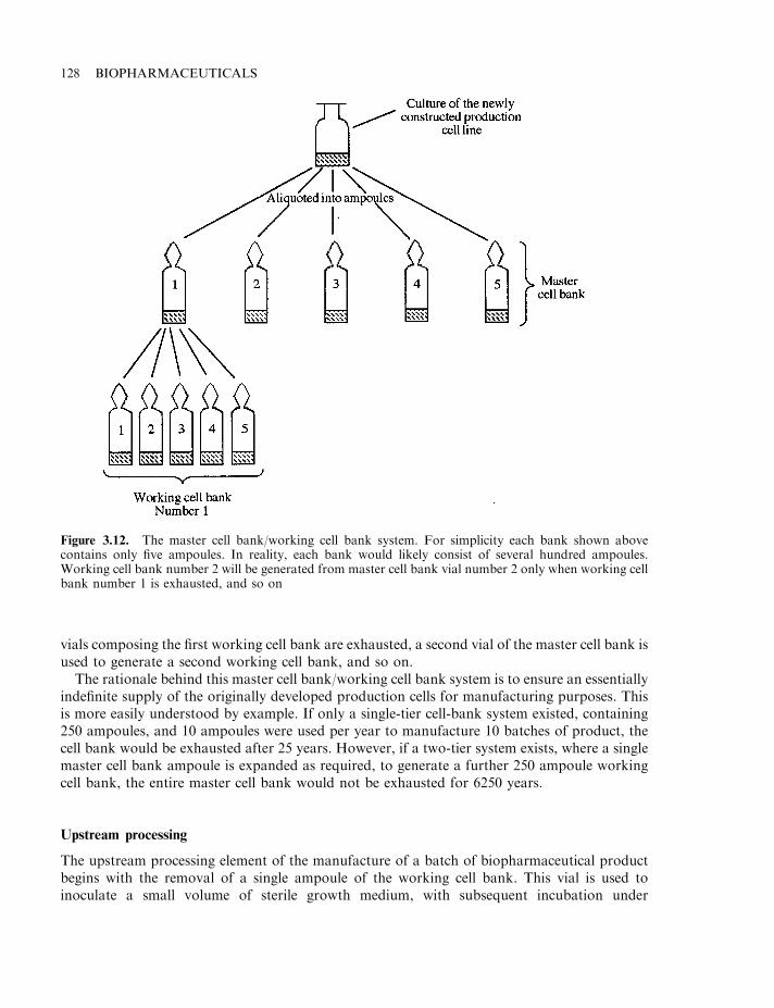
vials composing the first working cell bank are exhausted, a second vial of the master cell bank isused to generate a second working cell bank, and so on.
The rationale behind this master cell bank/working cell bank system is to ensure an essentiallyindefinite supply of the originally developed production cells for manufacturing purposes. Thisis more easily understood by example. If only a single-tier cell-bank system existed, containing250 ampoules, and 10 ampoules were used per year to manufacture 10 batches of product, thecell bank would be exhausted after 25 years. However, if a two-tier system exists, where a singlemaster cell bank ampoule is expanded as required, to generate a further 250 ampoule workingcell bank, the entire master cell bank would not be exhausted for 6250 years.
Upstream processing
The upstream processing element of the manufacture of a batch of biopharmaceutical productbegins with the removal of a single ampoule of the working cell bank. This vial is used toinoculate a small volume of sterile growth medium, with subsequent incubation under
128 BIOPHARMACEUTICALS
Figure 3.12. The master cell bank/working cell bank system. For simplicity each bank shown abovecontains only five ampoules. In reality, each bank would likely consist of several hundred ampoules.Working cell bank number 2 will be generated from master cell bank vial number 2 only when working cellbank number 1 is exhausted, and so on
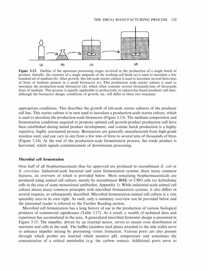
appropriate conditions. This describes the growth of lab-scale starter cultures of the producercell line. This starter culture is in turn used to inoculate a production-scale starter culture, whichis used to inoculate the production-scale bioreactor (Figure 3.13). The medium composition andfermentation conditions required to promote optimal cell growth/product production will havebeen established during initial product development, and routine batch production is a highlyrepetitive, highly automated process. Bioreactors are generally manufactured from high-gradestainless steel, and can vary in size from a few tens of litres to several tens of thousands of litres(Figure 3.14). At the end of the production-scale fermentation process, the crude product isharvested, which signals commencement of downstream processing.
Microbial cell fermentation
Over half of all biopharmacuticals thus far approved are produced in recombinant E. coli orS. cerevisiae. Industrial-scale bacterial and yeast fermentation systems share many commonfeatures, an overview of which is provided below. Most remaining biopharmaceuticals areproduced using animal cell culture, mainly by recombinant BHK or CHO cells (or hybridomacells in the case of some monoclonal antibodies; Appendix 1). While industrial-scale animal cellculture shares many common principles with microbial fermentation systems, it also differs inseveral respects, as subsequently described. Microbial fermentation/animal cell culture is a vastspeciality area in its own right. As such, only a summary overview can be provided below andthe interested reader is referred to the Further Reading section.
Microbial cell fermentation has a long history of use in the production of various biologicalproducts of commercial significance (Table 3.17). As a result, a wealth of technical data andexperience has accumulated in the area. A generalized microbial fermenter design is presented inFigure 3.15. The impeller, driven by an external motor, serves to ensure even distribution ofnutrients and cells in the tank. The baffles (stainless steel plates attached to the side walls) serveto enhance impeller mixing by preventing vortex formation. Various ports are also presentthrough which probes are inserted which monitor pH, temperature and sometimes theconcentration of a critical metabolite (e.g. the carbon source). Additional ports serve to
THE DRUG MANUFACTURING PROCESS 129
Figure 3.13. Outline of the upstream processing stages involved in the production of a single batch ofproduct. Initially, the contents of a single ampoule of the working cell bank (a) is used to inoculate a fewhundred ml of medium (b). After growth, this lab-scale starter culture is used to inoculate several litres/tensof litres of medium present in a small bioreactor (c). This production scale starter culture is used toinoculate the production-scale bioreactor (d), which often contains several thousands/tens of thousandslitres of medium. This process is equally applicable to prokaryotic or eukaryotic-based producer cell lines,although the bioreactor design, conditions of growth, etc. will differ in these two instances
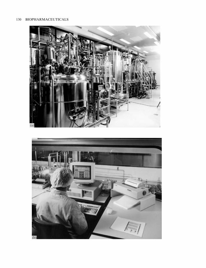
130 BIOPHARMACEUTICALS
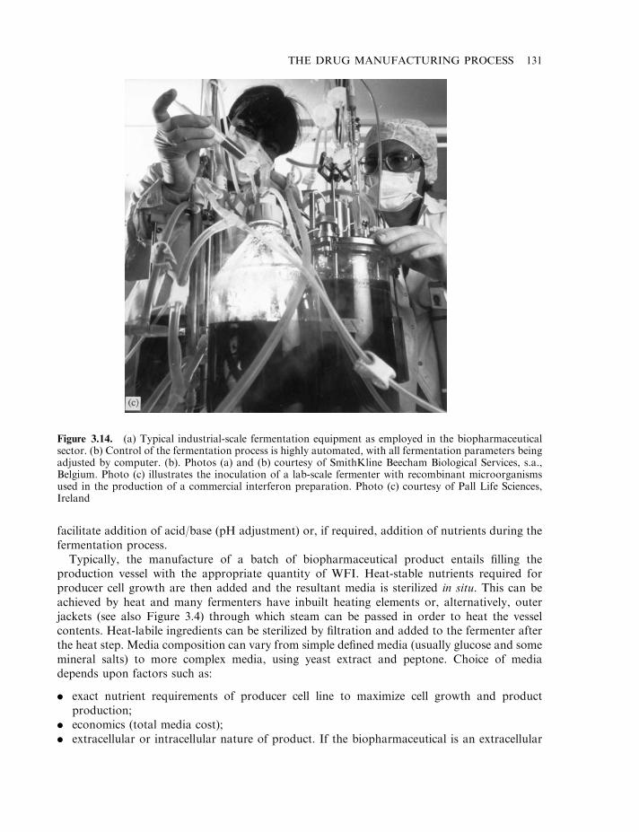
facilitate addition of acid/base (pH adjustment) or, if required, addition of nutrients during thefermentation process.
Typically, the manufacture of a batch of biopharmaceutical product entails filling theproduction vessel with the appropriate quantity of WFI. Heat-stable nutrients required forproducer cell growth are then added and the resultant media is sterilized in situ. This can beachieved by heat and many fermenters have inbuilt heating elements or, alternatively, outerjackets (see also Figure 3.4) through which steam can be passed in order to heat the vesselcontents. Heat-labile ingredients can be sterilized by filtration and added to the fermenter afterthe heat step. Media composition can vary from simple defined media (usually glucose and somemineral salts) to more complex media, using yeast extract and peptone. Choice of mediadepends upon factors such as:
. exact nutrient requirements of producer cell line to maximize cell growth and productproduction;
. economics (total media cost);
. extracellular or intracellular nature of product. If the biopharmaceutical is an extracellular
THE DRUG MANUFACTURING PROCESS 131
Figure 3.14. (a) Typical industrial-scale fermentation equipment as employed in the biopharmaceuticalsector. (b) Control of the fermentation process is highly automated, with all fermentation parameters beingadjusted by computer. (b). Photos (a) and (b) courtesy of SmithKline Beecham Biological Services, s.a.,Belgium. Photo (c) illustrates the inoculation of a lab-scale fermenter with recombinant microorganismsused in the production of a commercial interferon preparation. Photo (c) courtesy of Pall Life Sciences,Ireland
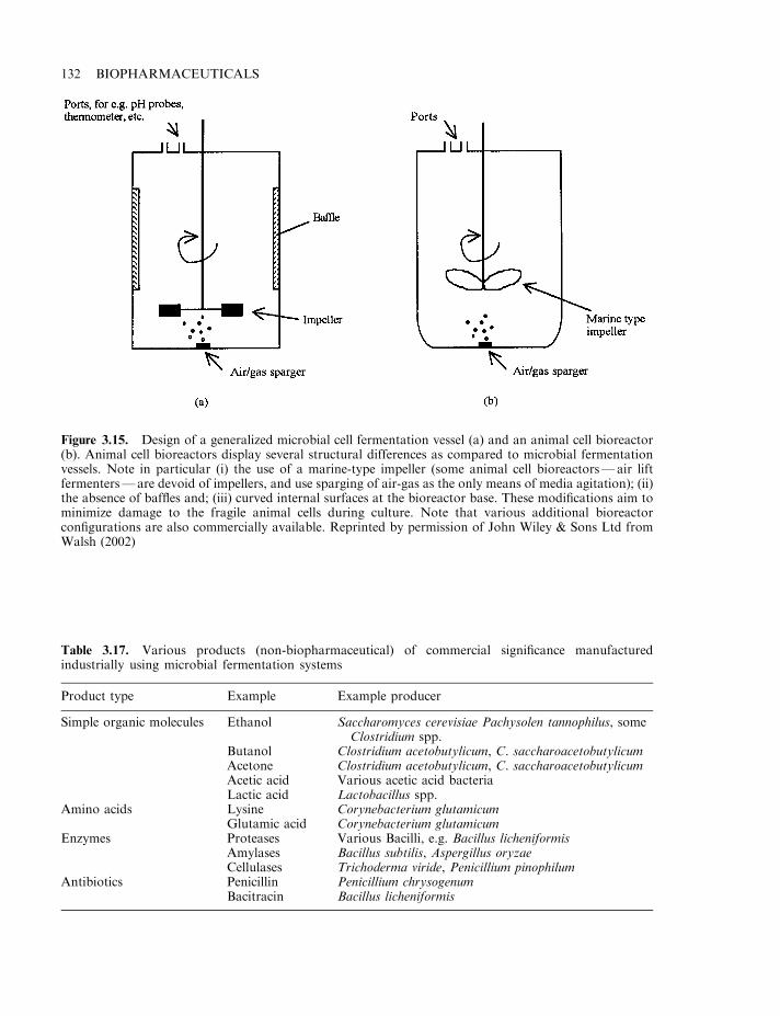
132 BIOPHARMACEUTICALS
Figure 3.15. Design of a generalized microbial cell fermentation vessel (a) and an animal cell bioreactor(b). Animal cell bioreactors display several structural differences as compared to microbial fermentationvessels. Note in particular (i) the use of a marine-type impeller (some animal cell bioreactors—air liftfermenters—are devoid of impellers, and use sparging of air-gas as the only means of media agitation); (ii)the absence of baffles and; (iii) curved internal surfaces at the bioreactor base. These modifications aim tominimize damage to the fragile animal cells during culture. Note that various additional bioreactorconfigurations are also commercially available. Reprinted by permission of John Wiley & Sons Ltd fromWalsh (2002)
Table 3.17. Various products (non-biopharmaceutical) of commercial significance manufacturedindustrially using microbial fermentation systems
Product type Example Example producer
Simple organic molecules Ethanol Saccharomyces cerevisiae Pachysolen tannophilus, someClostridium spp.
Butanol Clostridium acetobutylicum, C. saccharoacetobutylicumAcetone Clostridium acetobutylicum, C. saccharoacetobutylicumAcetic acid Various acetic acid bacteriaLactic acid Lactobacillus spp.
Amino acids Lysine Corynebacterium glutamicumGlutamic acid Corynebacterium glutamicum
Enzymes Proteases Various Bacilli, e.g. Bacillus licheniformisAmylases Bacillus subtilis, Aspergillus oryzaeCellulases Trichoderma viride, Penicillium pinophilum
Antibiotics Penicillin Penicillium chrysogenumBacitracin Bacillus licheniformis
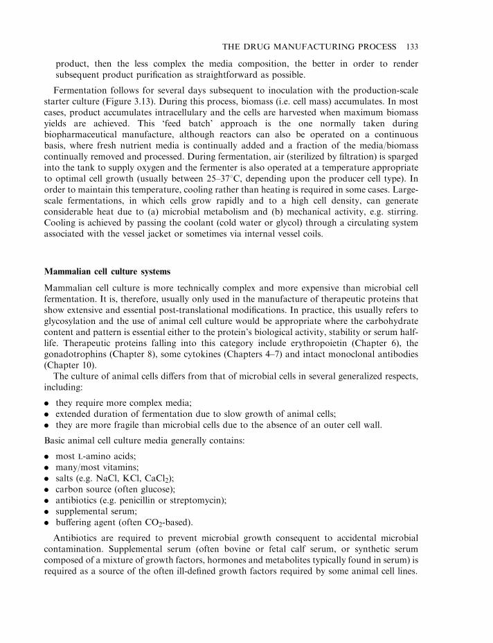
product, then the less complex the media composition, the better in order to rendersubsequent product purification as straightforward as possible.
Fermentation follows for several days subsequent to inoculation with the production-scalestarter culture (Figure 3.13). During this process, biomass (i.e. cell mass) accumulates. In mostcases, product accumulates intracellulary and the cells are harvested when maximum biomassyields are achieved. This ‘feed batch’ approach is the one normally taken duringbiopharmaceutical manufacture, although reactors can also be operated on a continuousbasis, where fresh nutrient media is continually added and a fraction of the media/biomasscontinually removed and processed. During fermentation, air (sterilized by filtration) is spargedinto the tank to supply oxygen and the fermenter is also operated at a temperature appropriateto optimal cell growth (usually between 25–378C, depending upon the producer cell type). Inorder to maintain this temperature, cooling rather than heating is required in some cases. Large-scale fermentations, in which cells grow rapidly and to a high cell density, can generateconsiderable heat due to (a) microbial metabolism and (b) mechanical activity, e.g. stirring.Cooling is achieved by passing the coolant (cold water or glycol) through a circulating systemassociated with the vessel jacket or sometimes via internal vessel coils.
Mammalian cell culture systems
Mammalian cell culture is more technically complex and more expensive than microbial cellfermentation. It is, therefore, usually only used in the manufacture of therapeutic proteins thatshow extensive and essential post-translational modifications. In practice, this usually refers toglycosylation and the use of animal cell culture would be appropriate where the carbohydratecontent and pattern is essential either to the protein’s biological activity, stability or serum half-life. Therapeutic proteins falling into this category include erythropoietin (Chapter 6), thegonadotrophins (Chapter 8), some cytokines (Chapters 4–7) and intact monoclonal antibodies(Chapter 10).
The culture of animal cells differs from that of microbial cells in several generalized respects,including:
. they require more complex media;
. extended duration of fermentation due to slow growth of animal cells;
. they are more fragile than microbial cells due to the absence of an outer cell wall.
Basic animal cell culture media generally contains:
. most L-amino acids;
. many/most vitamins;
. salts (e.g. NaCl, KCl, CaCl2);
. carbon source (often glucose);
. antibiotics (e.g. penicillin or streptomycin);
. supplemental serum;
. buffering agent (often CO2-based).
Antibiotics are required to prevent microbial growth consequent to accidental microbialcontamination. Supplemental serum (often bovine or fetal calf serum, or synthetic serumcomposed of a mixture of growth factors, hormones and metabolites typically found in serum) isrequired as a source of the often ill-defined growth factors required by some animal cell lines.
THE DRUG MANUFACTURING PROCESS 133
The media constituents, several of which are heat-labile, are generally dissolved in WFI andfilter-sterilized into the pre-sterile animal cell reactor. Reactor design (and operation) differssomewhat from microbial fermentations, mainly with a view to minimizing damage to the morefragile cells during cell culture (Figure 3.15). Although the generalized reactor design presentedin Figure 3.15 is commonly employed on industrial scale, alternative reactor configurations arealso available. These include hollow-fibre systems as well as the classical roller-bottle systems.Roller bottles are still used in the industrial production of some vaccines, erythropoietin andgrowth hormone-based products. Roller bottles are cylindrical bottles which are partially filledwith media, placed on their sides and mechanically rolled during cell culture. This system isgentle on the cells and the rolling action ensures homogeneity in the culture media and efficientoxygen transfer. The major disadvantage associated with applying roller-bottle technology onan industrial scale is that many thousands of bottles are required to produce a single batch ofproduct.
Different animal cell types display different properties pertinent to their successful culture.Those used to manufacture biopharmaceuticals are invariably continuous (transformed) celllines. Such cells will grow relatively vigorously and easily in submerged culture systems, be theyroller bottle- or bioreactor-based.
Unlike transformed cell lines, non-continuous cell lines generally:
. display anchorage dependence (i.e. will only grow and divide when attached to a solidsubstratum; continuous cell lines will grow in free suspension);
. grow as a monolayer;
. exhibit contact inhibition (physical contact between individual cells inhibits further division);
. display a finite lifespan, i.e. die, generally after 50–100 cell divisions, even when culturedunder ideal conditions;
. display longer population doubling times and grow to lower cell densities when compared tocontinuous cell lines;
. usually have more complex media requirements.
Many of these properties would obviously limit applicability of non-continuous cell lines in theindustrial-scale production of recombinant proteins. However, such cell types are routinelycultured for research purposes, toxicity testing, etc.
The anchorage-dependent growth properties of such non-continuous cell lines impacts uponhow they are cultured, both at lab and industrial scale. If grown in roller bottles/other low-volume containers, cells grow attached to the internal walls of the vessel. Large-scale culture canbe undertaken in submerged-type vessels, such as that described in Figure 3.15(b) in conjunctionwith the use of microcarrier beads. Microcarriers are solid or sometimes porous sphericalparticles approximately 200 mm in diameter, manufactured from such materials as collagen,dextran or plastic. They display densities slightly greater than water, such that gentle mixingwithin the animal cell bioreactor is sufficient to maintain the beads in suspension and evenlydistributed throughout the media. Anchorage-dependent cells can attach to, and grow on, thebeads’ outer surfaces/outer pores.
Downstream processing
An overview of the steps normally undertaken during downstream processing is presented inFigure 3.16. Details of the exact steps undertaken during the downstream processing of anyspecific biopharmaceutical product are usually considered highly confidential by the
134 BIOPHARMACEUTICALS
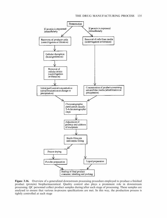
THE DRUG MANUFACTURING PROCESS 135
Figure 3.16. Overview of a generalized downstream processing procedure employed to produce a finishedproduct (protein) biopharmaceutical. Quality control also plays a prominent role in downstreamprocessing. QC personnel collect product samples during/after each stage of processing. These samples areanalysed to ensure that various in-process specifications are met. In this way, the production process istightly controlled at each stage
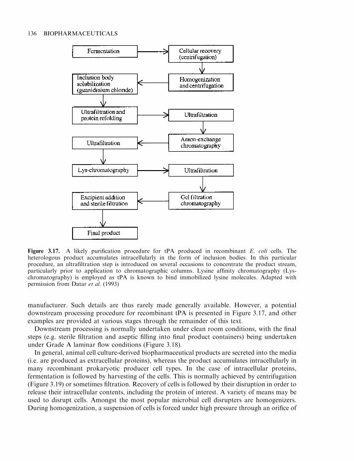
manufacturer. Such details are thus rarely made generally available. However, a potentialdownstream processing procedure for recombinant tPA is presented in Figure 3.17, and otherexamples are provided at various stages through the remainder of this text.
Downstream processing is normally undertaken under clean room conditions, with the finalsteps (e.g. sterile filtration and aseptic filling into final product containers) being undertakenunder Grade A laminar flow conditions (Figure 3.18).
In general, animal cell culture-derived biopharmaceutical products are secreted into the media(i.e. are produced as extracellular proteins), whereas the product accumulates intracellularly inmany recombinant prokaryotic producer cell types. In the case of intracellular proteins,fermentation is followed by harvesting of the cells. This is normally achieved by centrifugation(Figure 3.19) or sometimes filtration. Recovery of cells is followed by their disruption in order torelease their intracellular contents, including the protein of interest. A variety of means may beused to disrupt cells. Amongst the most popular microbial cell disrupters are homogenizers.During homogenization, a suspension of cells is forced under high pressure through an orifice of
136 BIOPHARMACEUTICALS
Figure 3.17. A likely purification procedure for tPA produced in recombinant E. coli cells. Theheterologous product accumulates intracellularly in the form of inclusion bodies. In this particularprocedure, an ultrafiltration step is introduced on several occasions to concentrate the product stream,particularly prior to application to chromatographic columns. Lysine affinity chromatography (Lys-chromatography) is employed as tPA is known to bind immobilized lysine molecules. Adapted withpermission from Datar et al. (1993)
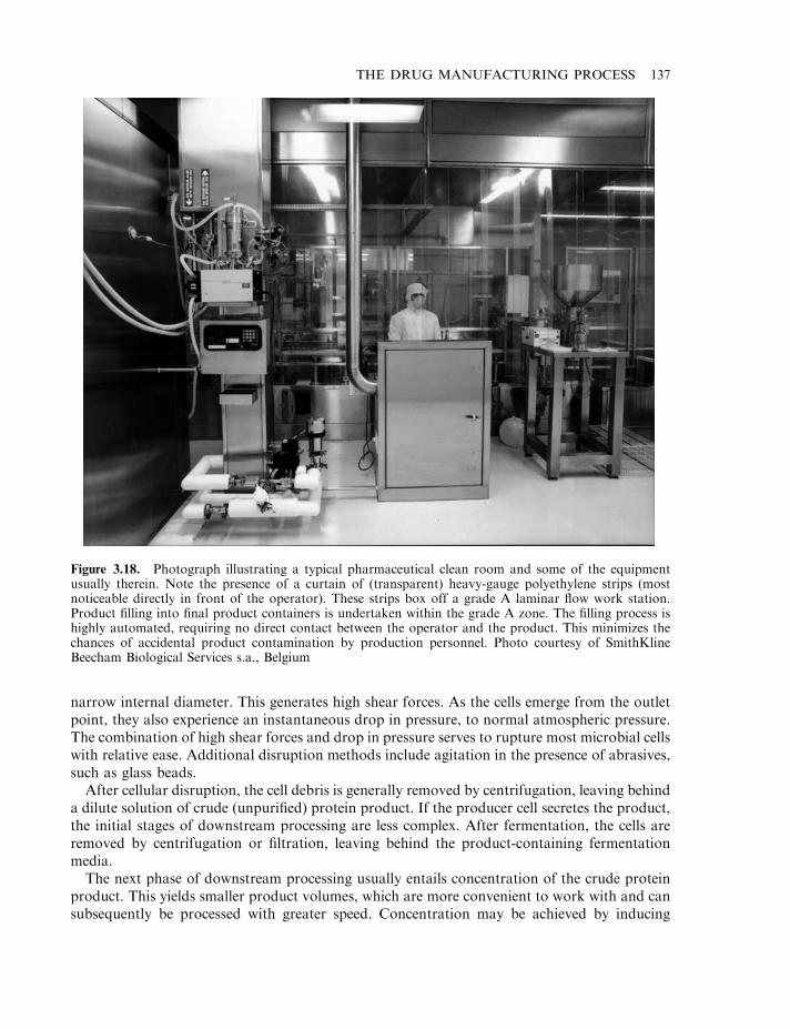
narrow internal diameter. This generates high shear forces. As the cells emerge from the outletpoint, they also experience an instantaneous drop in pressure, to normal atmospheric pressure.The combination of high shear forces and drop in pressure serves to rupture most microbial cellswith relative ease. Additional disruption methods include agitation in the presence of abrasives,such as glass beads.
After cellular disruption, the cell debris is generally removed by centrifugation, leaving behinda dilute solution of crude (unpurified) protein product. If the producer cell secretes the product,the initial stages of downstream processing are less complex. After fermentation, the cells areremoved by centrifugation or filtration, leaving behind the product-containing fermentationmedia.
The next phase of downstream processing usually entails concentration of the crude proteinproduct. This yields smaller product volumes, which are more convenient to work with and cansubsequently be processed with greater speed. Concentration may be achieved by inducing
THE DRUG MANUFACTURING PROCESS 137
Figure 3.18. Photograph illustrating a typical pharmaceutical clean room and some of the equipmentusually therein. Note the presence of a curtain of (transparent) heavy-gauge polyethylene strips (mostnoticeable directly in front of the operator). These strips box off a grade A laminar flow work station.Product filling into final product containers is undertaken within the grade A zone. The filling process ishighly automated, requiring no direct contact between the operator and the product. This minimizes thechances of accidental product contamination by production personnel. Photo courtesy of SmithKlineBeecham Biological Services s.a., Belgium
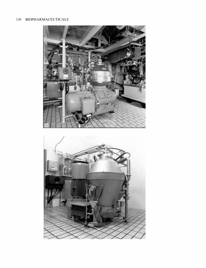
138 BIOPHARMACEUTICALS
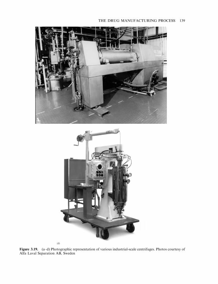
THE DRUG MANUFACTURING PROCESS 139
Figure 3.19. (a–d) Photographic representation of various industrial-scale centrifuges. Photos courtesy ofAlfa Laval Separation AB, Sweden
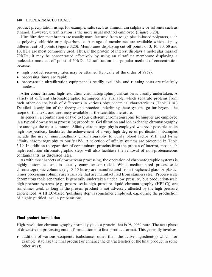
product precipitation using, for example, salts such as ammonium sulphate or solvents such asethanol. However, ultrafiltration is the more usual method employed (Figure 3.20).
Ultrafiltration membranes are usually manufactured from tough plastic-based polymers, suchas polyvinyl chloride or polycarbonate. A range of membranes are available which displaydifferent cut-off points (Figure 3.20). Membranes displaying cut-off points of 3, 10, 30, 50 and100 kDa are most commonly used. Thus, if the protein of interest displays a molecular mass of70 kDa, it may be concentrated effectively by using an ultrafilter membrane displaying amolecular mass cut-off point of 50 kDa. Ultrafiltration is a popular method of concentrationbecause:
. high product recovery rates may be attained (typically of the order of 99%);
. processing times are rapid;
. process-scale ultrafiltration equipment is readily available, and running costs are relativelymodest.
After concentration, high-resolution chromatographic purification is usually undertaken. Avariety of different chromatographic techniques are available, which separate proteins fromeach other on the basis of differences in various physiochemical characteristics (Table 3.18.)Detailed description of the theory and practice underlining these systems go far beyond thescope of this text, and are freely available in the scientific literature.
In general, a combination of two to four different chromatographic techniques are employedin a typical downstream processing procedure. Gel filtration and ion exchange chromatographyare amongst the most common. Affinity chromatography is employed wherever possible, as itshigh biospecificity facilitates the achievement of a very high degree of purification. Examplesinclude the use of immunoaffinity chromatography to purify blood factor VIII and lysineaffinity chromatography to purify tPA. A selection of affinity systems are presented in Table3.19. In addition to separation of contaminant proteins from the protein of interest, most suchhigh-resolution chromatographic steps will also facilitate the removal of non-proteinaceouscontaminants, as discussed later.
As with most aspects of downstream processing, the operation of chromatographic systems ishighly automated and is usually computer-controlled. While medium-sized process-scalechromatographic columns (e.g. 5–15 litres) are manufactured from toughened glass or plastic,larger processing columns are available that are manufactured from stainless steel. Process-scalechromatographic separation is generally undertaken under low pressure, but production-scalehigh-pressure systems (e.g. process-scale high pressure liquid chromatography (HPLC)) aresometimes used, as long as the protein product is not adversely affected by the high pressureexperienced. A HPLC-based ‘polishing step’ is sometimes employed, e.g. during the productionof highly purified insulin preparations.
Final product formulation
High-resolution chromatography normally yields a protein that is 98–99% pure. The next phaseof downstream processing entails formulation into final product format. This generally involves:
. addition of various excipients (substances other than the active ingredient(s) which, forexample, stabilize the final product or enhance the characteristics of the final product in someother way);
140 BIOPHARMACEUTICALS
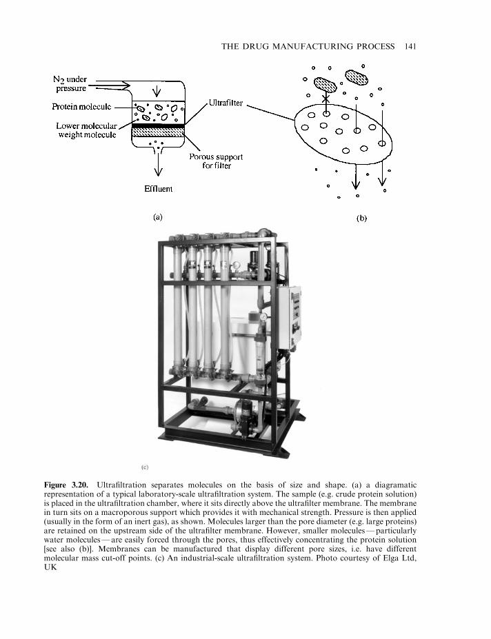
THE DRUG MANUFACTURING PROCESS 141
Figure 3.20. Ultrafiltration separates molecules on the basis of size and shape. (a) a diagramaticrepresentation of a typical laboratory-scale ultrafiltration system. The sample (e.g. crude protein solution)is placed in the ultrafiltration chamber, where it sits directly above the ultrafilter membrane. The membranein turn sits on a macroporous support which provides it with mechanical strength. Pressure is then applied(usually in the form of an inert gas), as shown. Molecules larger than the pore diameter (e.g. large proteins)are retained on the upstream side of the ultrafilter membrane. However, smaller molecules—particularlywater molecules—are easily forced through the pores, thus effectively concentrating the protein solution[see also (b)]. Membranes can be manufactured that display different pore sizes, i.e. have differentmolecular mass cut-off points. (c) An industrial-scale ultrafiltration system. Photo courtesy of Elga Ltd,UK
. filtration of the final product through a 0.22 mm absolute filter in order to generate sterileproduct, followed by its aseptic filling into final product containers;
. freeze-drying (lyophilization) if the product is to be marketed in a powdered format.
The decision to market the product in liquid or powder form is often dictated by how stablethe protein is in solution. This in turn must be determined experimentally, as there is no way topredict the outcome for any particular protein. Some proteins may remain stable for months oreven years in solution, particularly if stabilizing excipients are added and the solution isrefrigerated. Other proteins, particularly when purified, may retain biological activity for only amatter of hours or days when in aqueous solution.
Some influences that can alter the biological activity of proteins
A number of different influences can denature or otherwise modify proteins, rendering them lessactive/inactive. As all protein products are marketed on an activity basis, every precaution must
142 BIOPHARMACEUTICALS
Table 3.18. Various chromatographic techniques that may be used to separate proteins from eachother. The basis upon which separation is achieved is also listed
Chromatographic technique Basis of separation
Ion-exchange Differences in protein surface charge at a given pHGel-filtration Differences in size/shape of different proteinsHydrophobic interaction chromatography(HIC)
Differences in the size and extent of hydrophobic patches onthe surface of proteins
Affinity chromatography Ability of a protein to bind in a bio-specific (selective)manner to a chosen immobilized ligand
Metal chelate chromatography Ability of certain proteins to complex with zinc and copperHydroxyapatite chromatography Mechanism not fully understood. Involves ability of some
proteins to bind to calcium and phosphate ions on thesurface of hydroxyapatite crystals
Table 3.19. Some forms of affinity chromatography that may be employed to purify a selected protein.Virtually all proteins carry out their biological effects by interacting in some way with other molecules,which can be given the general title ‘ligands’. This interaction is often quite biospecific. Immobilizationof such ligands (or molecules which mimic ligands) onto chromatographic beads thus facilitates selectivepurification of the molecule of interest
Affinity system Application
Protein A chromatography Protein A, produced by Staphylococcus aureus, binds IgG. It is usedextensively in antibody purification protocols
Lectin affinity chromatography Lectins are a group of proteins capable of binding carbohydrates.Lectin affinity chromatography may be used to purify glycoproteins
Immunoaffinity chromatography Immobilized antibodies may be used as affinity absorbants for theantigen that stimulated their production (e.g. purification of factorVIII using immobilized anti-factor VIII antibodies)
Dye affinity chromatography Purification of proteins that display ability to interact tightly withselected dyes
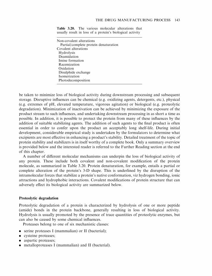
be taken to minimize loss of biological activity during downstream processing and subsequentstorage. Disruptive influences can be chemical (e.g. oxidizing agents, detergents, etc.), physical(e.g. extremes of pH, elevated temperature, vigorous agitation) or biological (e.g. proteolyticdegradation). Minimization of inactivation can be achieved by minimizing the exposure of theproduct stream to such influences, and undertaking downstream processing in as short a time aspossible. In addition, it is possible to protect the protein from many of these influences by theaddition of suitable stabilizing agents. The addition of such agents to the final product is oftenessential in order to confer upon the product an acceptably long shelf-life. During initialdevelopment, considerable empirical study is undertaken by the formulators to determine whatexcipients are most effective in enhancing a product’s stability. Detailed treatment of the topic ofprotein stability and stabilizers is in itself worthy of a complete book. Only a summary overviewis provided below and the interested reader is referred to the Further Reading section at the endof this chapter.
A number of different molecular mechanisms can underpin the loss of biological activity ofany protein. These include both covalent and non-covalent modification of the proteinmolecule, as summarized in Table 3.20. Protein denaturation, for example, entails a partial orcomplete alteration of the protein’s 3-D shape. This is underlined by the disruption of theintramolecular forces that stabilize a protein’s native conformation, viz hydrogen bonding, ionicattractions and hydrophobic interactions. Covalent modifications of protein structure that canadversely effect its biological activity are summarized below.
Proteolytic degradation
Proteolytic degradation of a protein is characterized by hydrolysis of one or more peptide(amide) bonds in the protein backbone, generally resulting in loss of biological activity.Hydrolysis is usually promoted by the presence of trace quantities of proteolytic enzymes, butcan also be caused by some chemical influences.
Proteases belong to one of six mechanistic classes:
. serine proteases I (mammalian) or II (bacterial);
. cysteine proteases;
. aspartic proteases;
. metalloproteases I (mammalian) and II (bacterial).
THE DRUG MANUFACTURING PROCESS 143
Table 3.20. The various molecular alterations thatusually result in loss of a protein’s biological activity
Non-covalent alterationsPartial/complete protein denaturation
Covalent alterationsHydrolysisDeamidationImine formationRacemizationOxidationDisulphide exchangeIsomerizationPhotodecomposition
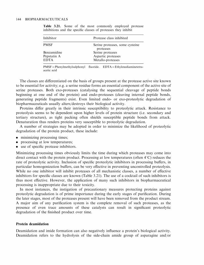
The classes are differentiated on the basis of groups present at the protease active site knownto be essential for activity; e.g. a serine residue forms an essential component of the active site ofserine proteases. Both exo-proteases (catalysing the sequential cleavage of peptide bondsbeginning at one end of the protein) and endo-proteases (cleaving internal peptide bonds,generating peptide fragments) exist. Even limited endo- or exo-proteolytic degradation ofbiopharmaceuticals usually alters/destroys their biological activity.
Proteins differ greatly in their intrinsic susceptibility to proteolytic attack. Resistance toproteolysis seems to be dependent upon higher levels of protein structure (i.e. secondary andtertiary structure), as tight packing often shields susceptible peptide bonds from attack.Denaturation thus renders proteins very susceptible to proteolytic degradation.
A number of strategies may be adopted in order to minimize the likelihood of proteolyticdegradation of the protein product, these include:
. minimizing processing times;
. processing at low temperatures;
. use of specific protease inhibitors.
Minimizing processing times obviously limits the time during which proteases may come intodirect contact with the protein product. Processing at low temperatures (often 48C) reduces therate of proteolytic activity. Inclusion of specific proteolytic inhibitors in processing buffers, inparticular homogenization buffers, can be very effective in preventing uncontrolled proteolysis.While no one inhibitor will inhibit proteases of all mechanistic classes, a number of effectiveinhibitors for specific classes are known (Table 3.21). The use of a cocktail of such inhibitors isthus most effective. However, the application of many such inhibitors in biopharmaceuticalprocessing is inappropriate due to their toxicity.
In most instances, the instigation of precautionary measures protecting proteins againstproteolytic degradation is of prime importance during the early stages of purification. Duringthe later stages, most of the proteases present will have been removed from the product stream.A major aim of any purification system is the complete removal of such proteases, as thepresence of even trace amounts of these catalysts can result in significant proteolyticdegradation of the finished product over time.
Protein deamidation
Deamidation and imide formation can also negatively influence a protein’s biological activity.Deamidation refers to the hydrolysis of the side-chain amide group of asparagine and/or
144 BIOPHARMACEUTICALS
Table 3.21. Some of the most commonly employed proteaseinhibitions and the specific classes of proteases they inhibit
Inhibitor Protease class inhibited
PMSF Serine proteases, some cysteineproteases
Benzamidine Serine proteasesPepstatin A Aspartic proteasesEDTA Metallo-proteases
PMSF¼Phenylmethylsulphonyl fluoride. EDTA¼Ethylenediaminetetra-acetic acid
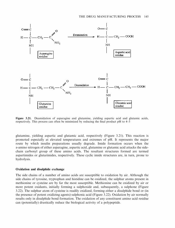
glutamine, yielding aspartic and glutamic acid, respectively (Figure 3.21). This reaction ispromoted especially at elevated temperatures and extremes of pH. It represents the majorroute by which insulin preparations usually degrade. Imide formation occurs when thea-amino nitrogen of either asparagine, aspartic acid, glutamine or glutamic acid attacks the side-chain carbonyl group of these amino acids. The resultant structures formed are termedaspartimides or glutarimides, respectively. These cyclic imide structures are, in turn, prone tohydrolysis.
Oxidation and disulphide exchange
The side chains of a number of amino acids are susceptible to oxidation by air. Although theside chains of tyrosine, tryptophan and histidine can be oxidized, the sulphur atoms present inmethionine or cysteine are by far the most susceptible. Methionine can be oxidized by air ormore potent oxidants, initially forming a sulphoxide and, subsequently, a sulphone (Figure3.22). The sulphur atom of cysteine is readily oxidized, forming either a disulphide bond or (inthe presence of potent oxidizing agents) sulphonic acid (Figure 3.22). Oxidation by air normallyresults only in disulphide bond formation. The oxidation of any constituent amino acid residuecan (potentially) drastically reduce the biological activity of a polypeptide.
THE DRUG MANUFACTURING PROCESS 145
Figure 3.21. Deamidation of asparagine and glutamine, yielding aspartic acid and glutamic acids,respectively. This process can often be minimized by reducing the final product pH to 4–5
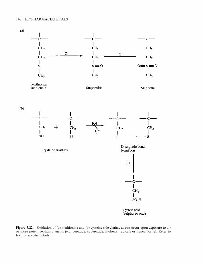
146 BIOPHARMACEUTICALS
Figure 3.22. Oxidation of (a) methionine and (b) cysteine side-chains, as can occur upon exposure to airor more potent oxidizing agents (e.g. peroxide, superoxide, hydroxyl radicals or hypochlorite). Refer totext for specific details
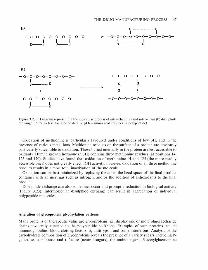
Oxidation of methionine is particularly favoured under conditions of low pH, and in thepresence of various metal ions. Methionine residues on the surface of a protein are obviouslyparticularly susceptible to oxidation. Those buried internally in the protein are less accessible tooxidants. Human growth hormone (hGH) contains three methionine residues (at positions 14,125 and 170). Studies have found that oxidation of methionine 14 and 125 (the more readilyaccessible ones) does not greatly effect hGH activity; however, oxidation of all three methionineresidues results in almost total inactivation of the molecule.
Oxidation can be best minimized by replacing the air in the head space of the final productcontainer with an inert gas such as nitrogen, and/or the addition of antioxidants to the finalproduct.
Disulphide exchange can also sometimes occur and prompt a reduction in biological activity(Figure 3.23). Intermolecular disulphide exchange can result in aggregation of individualpolypeptide molecules.
Alteration of glycoprotein glycosylation patterns
Many proteins of therapeutic value are glycoproteins, i.e. display one or more oligosaccharidechains covalently attached to the polypeptide backbone. Examples of such proteins includeimmunoglobulins, blood clotting factors, a1-antitrypsin and some interferons. Analysis of thecarbohydrate composition of glycoproteins reveals the presence of a variety sugars, including D-galactose, D-mannose and L-fucose (neutral sugars), the amino-sugars; N-acetylglucosamine
THE DRUG MANUFACTURING PROCESS 147
Figure 3.23. Diagram representing the molecular process of intra-chain (a) and inter-chain (b) disulphideexchange. Refer to text for specific details. (-O-¼amino acid residues in polypeptide)
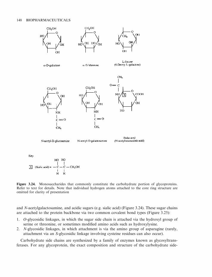
and N-acetylgalactosamine, and acidic sugars (e.g. sialic acid) (Figure 3.24). These sugar chainsare attached to the protein backbone via two common covalent bond types (Figure 3.25):
1. O-glycosidic linkages, in which the sugar side chain is attached via the hydroxyl group ofserine or threonine, or sometimes modified amino acids such as hydroxylysine.
2. N-glycosidic linkages, in which attachment is via the amino group of asparagine (rarely,attachment via an S-glycosidic linkage involving cysteine residues can also occur).
Carbohydrate side chains are synthesized by a family of enzymes known as glycosyltrans-ferases. For any glycoprotein, the exact composition and structure of the carbohydrate side-
148 BIOPHARMACEUTICALS
Figure 3.24. Monosaccharides that commonly constitute the carbohydrate portion of glycoproteins.Refer to text for details. Note that individual hydrogen atoms attached to the core ring structure areomitted for clarity of presentation
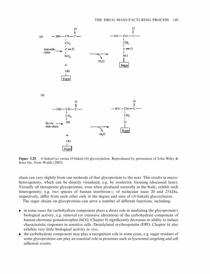
chain can vary slightly from one molecule of that glycoprotein to the next. This results in micro-heterogeneity, which can be directly visualized, e.g. by isoelectric focusing (discussed later).Virtually all therapeutic glycoproteins, even when produced naturally in the body, exhibit suchheterogeneity; e.g. two species of human interferon-g, of molecular mass 20 and 25 kDa,respectively, differ from each other only in the degree and sites of (N-linked) glycosylation.
The sugar chains on glycoproteins can serve a number of different functions, including:
. in some cases the carbohydrate component plays a direct role in mediating the glycoprotein’sbiological activity, e.g. removal (or extensive alteration) of the carbohydrate component ofhuman chorionic gonadotrophin (hCG; Chapter 8) significantly decreases its ability to inducecharacteristic responses in sensitive cells. Desialylated erythropoietin (EPO; Chapter 6) alsoexhibits very little biological activity in vivo;
. the carbohydrate component may play a recognition role in some cases, e.g. sugar residues ofsome glycoproteins can play an essential role in processes such as lysosomal targeting and celladhesion events;
THE DRUG MANUFACTURING PROCESS 149
Figure 3.25. N-linked (a) versus O-linked (b) glycosylation. Reproduced by permission of John Wiley &Sons Inc. from Walsh (2002)
. the carbohydrate component serves to substantially increase the solubility of severalglycoproteins;
. the carbohydrate component helps regulate the biological half-life of some serumglycoproteins. It increases the serum half-life of some glycoproteins by decreasing the rateof clearance from the serum. Furthermore, modification/partial degradation (i.e. asialogly-coproteins) of the carbohydrate component can accelerate removal of the protein from theserum;
. the carbohydrate component probably exerts a direct stabilizing influence upon theconformation of many glycoproteins.
Alteration of its carbohydrate component can therefore potentially affect the biologicalactivity of the glycoprotein, or alter its immunological properties. In practice, however,expression of many normally glycosylated proteins in E. coli generates a protein product whosebiological characteristics are indistinguishable from the native product. Furthermore, theglycosylation patterns obtained when human glycoproteins are expressed in non-humaneukaryotic expression systems (e.g. animal cell culture) are usually distinct from theglycosylation pattern associated with the native human protein. The glycosylation pattern ofhuman tPA produced in transgenic animals, for example, is different from the pattern obtainedwhen the same gene is expressed in a recombinant mouse cell line. Both these patterns are inturn different from the native human pattern. The tPA from all sources is, however, safe andeffective. The clinical significance of altered glycosylation patterns/micro-heterogeneity is bestdetermined by clinical trials. If the product is found to be safe and effective, then routine end-product QC analysis for carbohydrate-based micro-heterogeneity is carried out, more todetermine batch-to-batch consistency (which is desirable) than to detect microheterogeneity perse.
Most glycoprotein biopharmaceuticals will exhibit microheterogeneity. Isoelectric focusing oftypical batches of the therapeutic monoclonal antibody OKT-3 (Chapter 10) reveals at least fourbands, representing four product variations that differ slightly in their carbohydrate content.Likewise, isoelectric focusing of batches of erythropoietin (40 kDa glycoprotein hormone, up to50% of which is carbohydrate; Chapter 6) typically reveals up to six bands, which again differonly in their carbohydrate content. In any such instance, batch rejection would only beconsidered if strongly atypical heterogeneity was observed.
Large deviations from normal batch-to-batch glycosylation pattern is most likely caused bythe presence of a glycosidase capable of enzymatic degradation of the carbohydrate side-chains.The likelihood of such eventualities may be minimized by carrying out downstream processingat low temperatures, and as quickly as possible.
Stabilizing excipients used in final product formulations
A range of various substances may be added to a purified (polypeptide) biopharmaceuticalproduct in order to stabilize that product (Table 3.22). Such agents can stabilize proteins in anumber of different ways. Specific examples include:
. serum albumin. Addition of serum albumin has been shown to stabilize various differentpolypeptides (Table 3.23). Human serum albumin (HSA) is often employed in the case ofbiopharmaceuticals destined for parenteral administration to humans. In many cases, it isused in combination with additional stabilizers, including amino acids (mainly glycine) andcarbohydrates. Serum albumin itself is quite a stable molecule, capable of withstanding
150 BIOPHARMACEUTICALS
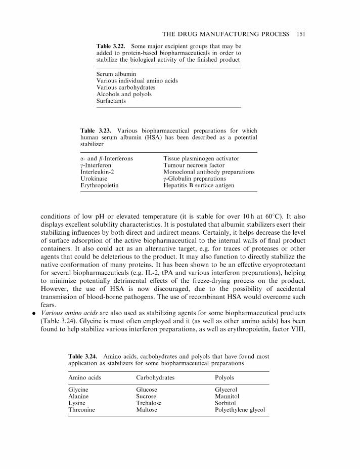
conditions of low pH or elevated temperature (it is stable for over 10 h at 608C). It alsodisplays excellent solubility characteristics. It is postulated that albumin stabilizers exert theirstabilizing influences by both direct and indirect means. Certainly, it helps decrease the levelof surface adsorption of the active biopharmaceutical to the internal walls of final productcontainers. It also could act as an alternative target, e.g. for traces of proteases or otheragents that could be deleterious to the product. It may also function to directly stabilize thenative conformation of many proteins. It has been shown to be an effective cryoprotectantfor several biopharmaceuticals (e.g. IL-2, tPA and various interferon preparations), helpingto minimize potentially detrimental effects of the freeze-drying process on the product.However, the use of HSA is now discouraged, due to the possibility of accidentaltransmission of blood-borne pathogens. The use of recombinant HSA would overcome suchfears.
. Various amino acids are also used as stabilizing agents for some biopharmaceutical products(Table 3.24). Glycine is most often employed and it (as well as other amino acids) has beenfound to help stabilize various interferon preparations, as well as erythropoietin, factor VIII,
THE DRUG MANUFACTURING PROCESS 151
Table 3.22. Some major excipient groups that may beadded to protein-based biopharmaceuticals in order tostabilize the biological activity of the finished product
Serum albuminVarious individual amino acidsVarious carbohydratesAlcohols and polyolsSurfactants
Table 3.23. Various biopharmaceutical preparations for whichhuman serum albumin (HSA) has been described as a potentialstabilizer
a- and b-Interferons Tissue plasminogen activatorg-Interferon Tumour necrosis factorInterleukin-2 Monoclonal antibody preparationsUrokinase g-Globulin preparationsErythropoietin Hepatitis B surface antigen
Table 3.24. Amino acids, carbohydrates and polyols that have found mostapplication as stabilizers for some biopharmaceutical preparations
Amino acids Carbohydrates Polyols
Glycine Glucose GlycerolAlanine Sucrose MannitolLysine Trehalose SorbitolThreonine Maltose Polyethylene glycol
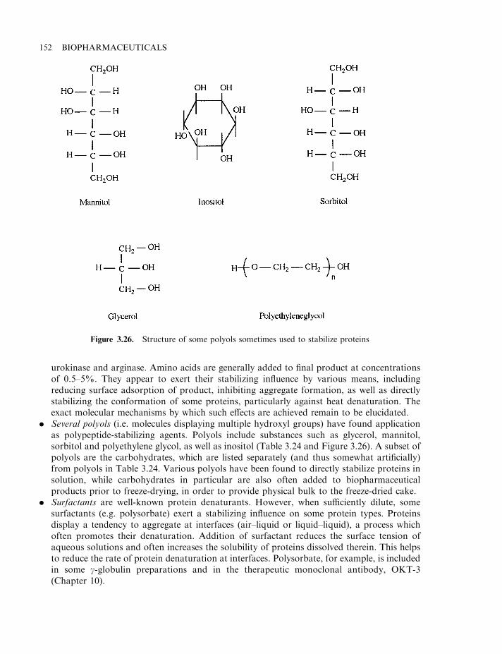
urokinase and arginase. Amino acids are generally added to final product at concentrationsof 0.5–5%. They appear to exert their stabilizing influence by various means, includingreducing surface adsorption of product, inhibiting aggregate formation, as well as directlystabilizing the conformation of some proteins, particularly against heat denaturation. Theexact molecular mechanisms by which such effects are achieved remain to be elucidated.
. Several polyols (i.e. molecules displaying multiple hydroxyl groups) have found applicationas polypeptide-stabilizing agents. Polyols include substances such as glycerol, mannitol,sorbitol and polyethylene glycol, as well as inositol (Table 3.24 and Figure 3.26). A subset ofpolyols are the carbohydrates, which are listed separately (and thus somewhat artificially)from polyols in Table 3.24. Various polyols have been found to directly stabilize proteins insolution, while carbohydrates in particular are also often added to biopharmaceuticalproducts prior to freeze-drying, in order to provide physical bulk to the freeze-dried cake.
. Surfactants are well-known protein denaturants. However, when sufficiently dilute, somesurfactants (e.g. polysorbate) exert a stabilizing influence on some protein types. Proteinsdisplay a tendency to aggregate at interfaces (air–liquid or liquid–liquid), a process whichoften promotes their denaturation. Addition of surfactant reduces the surface tension ofaqueous solutions and often increases the solubility of proteins dissolved therein. This helpsto reduce the rate of protein denaturation at interfaces. Polysorbate, for example, is includedin some g-globulin preparations and in the therapeutic monoclonal antibody, OKT-3(Chapter 10).
152 BIOPHARMACEUTICALS
Figure 3.26. Structure of some polyols sometimes used to stabilize proteins
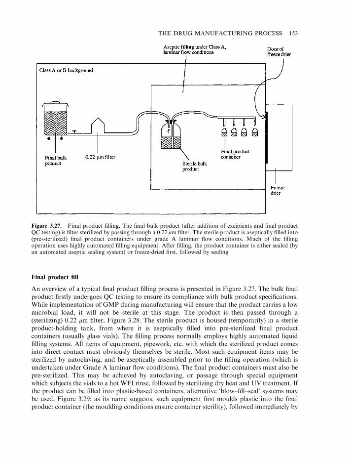
Final product fill
An overview of a typical final product filling process is presented in Figure 3.27. The bulk finalproduct firstly undergoes QC testing to ensure its compliance with bulk product specifications.While implementation of GMP during manufacturing will ensure that the product carries a lowmicrobial load, it will not be sterile at this stage. The product is then passed through a(sterilizing) 0.22 mm filter, Figure 3.28. The sterile product is housed (temporarily) in a sterileproduct-holding tank, from where it is aseptically filled into pre-sterilized final productcontainers (usually glass vials). The filling process normally employs highly automated liquidfilling systems. All items of equipment, pipework, etc. with which the sterilized product comesinto direct contact must obviously themselves be sterile. Most such equipment items may besterilized by autoclaving, and be aseptically assembled prior to the filling operation (which isundertaken under Grade A laminar flow conditions). The final product containers must also bepre-sterilized. This may be achieved by autoclaving, or passage through special equipmentwhich subjects the vials to a hot WFI rinse, followed by sterilizing dry heat and UV treatment. Ifthe product can be filled into plastic-based containers, alternative ‘blow–fill–seal’ systems maybe used, Figure 3.29; as its name suggests, such equipment first moulds plastic into the finalproduct container (the moulding conditions ensure container sterility), followed immediately by
THE DRUG MANUFACTURING PROCESS 153
Figure 3.27. Final product filling. The final bulk product (after addition of excipients and final productQC testing) is filter sterilized by passing through a 0.22mm filter. The sterile product is aseptically filled into(pre-sterilized) final product containers under grade A laminar flow conditions. Much of the fillingoperation uses highly automated filling equipment. After filling, the product container is either sealed (byan automated aseptic sealing system) or freeze-dried first, followed by sealing
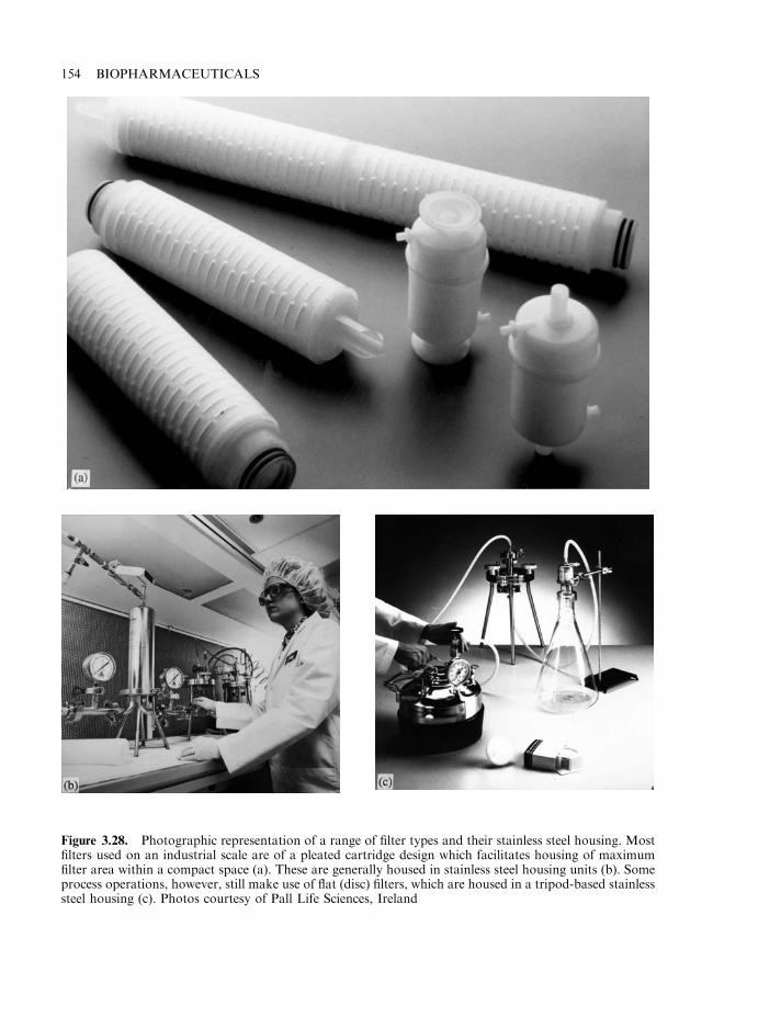
154 BIOPHARMACEUTICALS
Figure 3.28. Photographic representation of a range of filter types and their stainless steel housing. Mostfilters used on an industrial scale are of a pleated cartridge design which facilitates housing of maximumfilter area within a compact space (a). These are generally housed in stainless steel housing units (b). Someprocess operations, however, still make use of flat (disc) filters, which are housed in a tripod-based stainlesssteel housing (c). Photos courtesy of Pall Life Sciences, Ireland
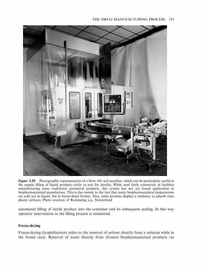
automated filling of sterile product into the container and its subsequent sealing. In this wayoperator intervention in the filling process is minimized.
Freeze-drying
Freeze-drying (lyophilization) refers to the removal of solvent directly from a solution while inthe frozen state. Removal of water directly from (frozen) biopharmaceutical products via
THE DRUG MANUFACTURING PROCESS 155
Figure 3.29. Photographic representation of a blow–fill–seal machine, which can be particularly useful inthe aseptic filling of liquid products (refer to text for details). While used fairly extensively in facilitiesmanufacturing some traditional parenteral products, this system has not yet found application inbiopharmaceutical manufacture. This is due mainly to the fact that many biopharmaceutical preparationsare sold not in liquid, but in freeze-dried format. Also, some proteins display a tendancy to adsorb ontoplastic surfaces. Photo courtesy of Rommelag a.g., Switzerland
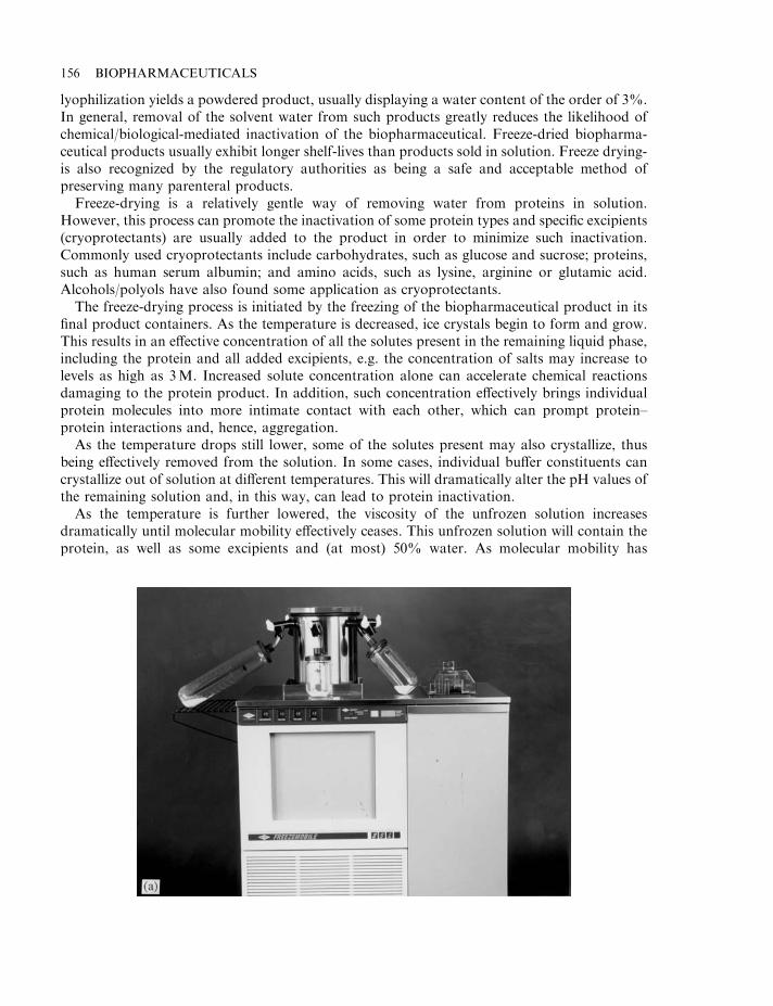
lyophilization yields a powdered product, usually displaying a water content of the order of 3%.In general, removal of the solvent water from such products greatly reduces the likelihood ofchemical/biological-mediated inactivation of the biopharmaceutical. Freeze-dried biopharma-ceutical products usually exhibit longer shelf-lives than products sold in solution. Freeze drying-is also recognized by the regulatory authorities as being a safe and acceptable method ofpreserving many parenteral products.
Freeze-drying is a relatively gentle way of removing water from proteins in solution.However, this process can promote the inactivation of some protein types and specific excipients(cryoprotectants) are usually added to the product in order to minimize such inactivation.Commonly used cryoprotectants include carbohydrates, such as glucose and sucrose; proteins,such as human serum albumin; and amino acids, such as lysine, arginine or glutamic acid.Alcohols/polyols have also found some application as cryoprotectants.
The freeze-drying process is initiated by the freezing of the biopharmaceutical product in itsfinal product containers. As the temperature is decreased, ice crystals begin to form and grow.This results in an effective concentration of all the solutes present in the remaining liquid phase,including the protein and all added excipients, e.g. the concentration of salts may increase tolevels as high as 3M. Increased solute concentration alone can accelerate chemical reactionsdamaging to the protein product. In addition, such concentration effectively brings individualprotein molecules into more intimate contact with each other, which can prompt protein–protein interactions and, hence, aggregation.
As the temperature drops still lower, some of the solutes present may also crystallize, thusbeing effectively removed from the solution. In some cases, individual buffer constituents cancrystallize out of solution at different temperatures. This will dramatically alter the pH values ofthe remaining solution and, in this way, can lead to protein inactivation.
As the temperature is further lowered, the viscosity of the unfrozen solution increasesdramatically until molecular mobility effectively ceases. This unfrozen solution will contain theprotein, as well as some excipients and (at most) 50% water. As molecular mobility has
156 BIOPHARMACEUTICALS
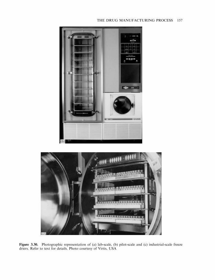
THE DRUG MANUFACTURING PROCESS 157
Figure 3.30. Photographic representation of (a) lab-scale, (b) pilot-scale and (c) industrial-scale freezedriers. Refer to text for details. Photo courtesy of Virtis, USA
effectively stopped, chemical reactivity also all but ceases. The consistency of this ‘solution’ isthat of glass, and the temperature at which this is attained is called the glass transitiontemperature, Tg’. For most protein solutions, Tg’ values reside between 7408C and 7608C.The primary aim of the initial stages of the freeze-drying process is to decrease the producttemperature below that of its Tg’ value as quickly as possible.
The next phase of the freeze-drying process entails the application of a vacuum to the system.When the vacuum is established, the temperature is increased, usually to temperatures slightly inexcess of 08C. This promotes sublimation of the crystalline water, leaving behind a powderedcake of dried material. Once satisfactory drying has been achieved, the product container issealed.
The drying chamber of industrial-scale freeze-dryers usually opens into a clean room (Figure3.27). This facilitates direct transfer of the product-containing vials into the chamber.Immediately prior to filling, rubber stoppers are usually partially inserted into the mouth ofeach vial in such a way as not to hinder the outward flow of water vapour during the freeze-drying process. The drying chamber normally contains several rows of shelves, each of whichcan accommodate several thousand vials (Figure 3.30). These shelves are wired to allow theirelectrical heating and cooling and their upward or downward movement. After the freeze-dryingcycle is complete (which can take 3 days or more), the shelves are then moved upwards. As eachshelf moves up, the partially-inserted rubber seals are inserted fully into the vial mouth as theycome in contact with the base plate of the shelf immediately above them. After productrecovery, the empty chamber is closed and is then heat-sterilized (using its own chamber-heatingmechanism). The freeze-drier is then ready to accept its next load.
Labelling and packing
After the product has been filled (and sealed) in its final product container, it is immediatelyplaced under quarantine. QC personnel then remove representative samples of the product andcarry out tests to ensure conformance to final product specification. The most importantspecifications will relate to product potency, sterility and final volume fill, as well as the absenceof endotoxin or other potentially toxic substances. Detection and quantification of excipientsadded will also be undertaken.
Only after QC personnel are satisfied that the product meets these specifications will it belabelled and packed. These operations are highly automated. Labelling, in particular, deservesspecial attention. Mislabelling of product remains one of the most common reasons for productrecall. This can occur relatively easily, particularly if the facility manufactures several differentproducts, or even a single product at several different strengths. Information presented on alabel should normally include:
. name and strength/potency of the product;
. specific batch number of the product;
. date of manufacture and expiry date;
. storage conditions required.
Additional information often presented includes the name of the manufacturer, a list ofexcipients included and a brief summary of the correct mode of product usage.
When a batch of product is labelled and packed, and QC personnel are satisfied that labellingand packing are completed to specification, the QC manager will write and sign a Certificate ofAnalysis. This details the pre-defined product specifications and confirms conformance of the
158 BIOPHARMACEUTICALS
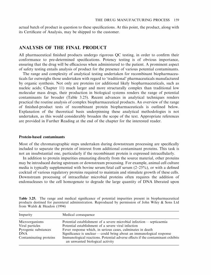
actual batch of product in question to these specifications. At this point, the product, along withits Certificate of Analysis, may be shipped to the customer.
ANALYSIS OF THE FINAL PRODUCT
All pharmaceutical finished products undergo rigorous QC testing, in order to confirm theirconformance to pre-determined specifications. Potency testing is of obvious importance,ensuring that the drug will be efficacious when administered to the patient. A prominent aspectof safety testing entails analysis of product for the presence of various potential contaminants.
The range and complexity of analytical testing undertaken for recombinant biopharmaceu-ticals far outweighs those undertaken with regard to ‘traditional’ pharmaceuticals manufacturedby organic synthesis. Not only are proteins (or additional likely biopharmaceuticals, such asnucleic acids; Chapter 11) much larger and more structurally complex than traditional lowmolecular mass drugs, their production in biological systems renders the range of potentialcontaminants far broader (Table 3.25). Recent advances in analytical techniques renderspractical the routine analysis of complex biopharmaceutical products. An overview of the rangeof finished-product tests of recombinant protein biopharmaceuticals is outlined below.Explanation of the theoretical basis underpinning these analytical methodologies is notundertaken, as this would considerably broaden the scope of the text. Appropriate referencesare provided in Further Reading at the end of the chapter for the interested reader.
Protein-based contaminants
Most of the chromatographic steps undertaken during downstream processing are specificallyincluded to separate the protein of interest from additional contaminant proteins. This task isnot an insubstantial one, particularly if the recombinant protein is expressed intracellularly.
In addition to protein impurities emanating directly from the source material, other proteinsmay be introduced during upstream or downstream processing. For example, animal cell culturemedia is typically supplemented with bovine serum/fetal calf serum (2–25%), or with a definedcocktail of various regulatory proteins required to maintain and stimulate growth of these cells.Downstream processing of intracellular microbial proteins often requires the addition ofendonucleases to the cell homogenate to degrade the large quantity of DNA liberated upon
THE DRUG MANUFACTURING PROCESS 159
Table 3.25. The range and medical significance of potential impurities present in biopharmaceuticalproducts destined for parenteral administration. Reproduced by permission of John Wiley & Sons Ltdfrom Walsh & Headon (1994)
Impurity Medical consequence
Microorganisms Potential establishment of a severe microbial infection—septicaemiaViral particles Potential establishment of a severe viral infectionPyrogenic substances Fever response which, in serious cases, culminates in deathDNA Significance is unclear—could bring about an immunological responseContaminating proteins Immunological reactions. Potential adverse effects if the contaminant exhibits
an unwanted biological activity
cellular disruption (DNA promotes increased solution viscosity, rendering processing difficult;viscosity, being a function of the DNA’s molecular mass, is reduced upon nuclease treatment).
Minor amounts of protein could also potentially enter the product stream from additionalsources, e.g. protein shed from production personnel. Implementation of GMP should,however, minimize contamination from such sources.
The clinical significance of protein-based impurities relates to: (a) their potential biologicalactivities; and (b) their antigenicity. While some contaminants may display no undesirablebiological activity, others may exhibit activities deleterious to either the product itself (e.g.proteases which could modify/degrade the product) or the recipient patient (e.g. the presence ofcontaminating toxins).
Their inherent immunogenicity also renders likely an immunological reaction against protein-based impurities upon product administration to the recipient patient. This is particularly truein the case of products produced in microbial or other recombinant systems (i.e. mostbiopharmaceuticals). While the product itself is likely to be non-immunogenic (being coded forby a human gene), contaminant proteins will be endogenous to the host cell, and hence foreignto the human body. Administration of the product can elicit an immune response against thecontaminant. This is particularly likely if a requirement exists for ongoing, repeat productadministration (e.g. administration of recombinant insulin). Immunological activation of thistype could also potentially (and more seriously) have a sensitizing effect on the recipient againstthe actual protein product.
In addition to distinct gene products, modified forms of the protein of interest are alsoconsidered impurities, rendering desirable their removal from the product stream. While somesuch modified forms may be innocuous, others may not. Modified product ‘impurities’ maycompromise the product in a number of ways, e.g:
. biologically inactive forms of the product will reduce overall product potency;
. some modified product forms remain biologically active but exhibit modified pharmacoki-netic characteristics (i.e. timing and duration of drug action);
. modified product forms may be immunogenic.
Altered forms of the protein of interest can be generated in a number of ways by covalent andnon-covalent modifications (see e.g. Table 3.20).
Removal of altered forms of the protein of interest from the product stream
Modification of any protein will generally alter some aspect of its physicochemicalcharacteristics. This facilitates removal of the modified form by standard chromatographictechniques during downstream processing. Most downstream procedures for protein-basedbiopharmaceuticals include both gel-filtration and ion-exchange steps. Aggregated forms of theproduct will be effectively removed by gel-filtration (because they now exhibit a molecular massgreater by several orders of magnitude than the native product). This technique will equallyefficiently remove extensively proteolysed forms of the product. Glycoprotein variants whosecarbohydrate moieties have been extensively degraded will also likely be removed by gel-filtration (or ion-exchange) chromatography. Deamidation and oxidation will generate productvariants with altered surface charge characteristics, often rendering their removal by ion-exchange relatively straightforward. Incorrect disulphide bond formation, partial denaturationand limited proteolysis can also alter the shape and surface charge of proteins, facilitating their
160 BIOPHARMACEUTICALS
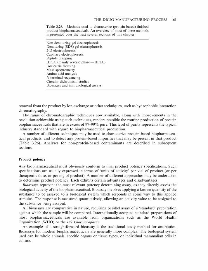
removal from the product by ion-exchange or other techniques, such as hydrophobic interactionchromatography.
The range of chromatographic techniques now available, along with improvements in theresolution achievable using such techniques, renders possible the routine production of proteinbiopharmaceuticals that are in excess of 97–99% pure. This level of purity represents the typicalindustry standard with regard to biopharmaceutical production.
A number of different techniques may be used to characterize protein-based biopharmaceu-tical products, and to detect any protein-based impurities that may be present in that product(Table 3.26). Analyses for non-protein-based contaminants are described in subsequentsections.
Product potency
Any biopharmaceutical must obviously conform to final product potency specifications. Suchspecifications are usually expressed in terms of ‘units of activity’ per vial of product (or pertherapeutic dose, or per mg of product). A number of different approaches may be undertakento determine product potency. Each exhibits certain advantages and disadvantages.
Bioassays represent the most relevant potency-determining assay, as they directly assess thebiological activity of the biopharmaceutical. Bioassay involves applying a known quantity of thesubstance to be assayed to a biological system which responds in some way to this appliedstimulus. The response is measured quantitatively, allowing an activity value to be assigned tothe substance being assayed.
All bioassays are comparative in nature, requiring parallel assay of a ‘standard’ preparationagainst which the sample will be compared. Internationally accepted standard preparations ofmost biopharmaceuticals are available from organizations such as the World HealthOrganization (WHO) or the US Pharmacopoeia.
An example of a straightforward bioassay is the traditional assay method for antibiotics.Bioassays for modern biopharmaceuticals are generally more complex. The biological systemused can be whole animals, specific organs or tissue types, or individual mammalian cells inculture.
THE DRUG MANUFACTURING PROCESS 161
Table 3.26. Methods used to characterize (protein-based) finishedproduct biopharmaceuticals. An overview of most of these methodsis presented over the next several sections of this chapter
Non-denaturing gel electrophoresisDenaturing (SDS) gel electrophoresis2-D electrophoresisCapillary electrophoresisPeptide mappingHPLC (mainly reverse phase—HPLC)Isoelectric focusingMass spectrometryAmino acid analysisN-terminal sequencingCircular dichromism studiesBioassays and immunological assays
Bioassays of related substances can be quite similar in design. Specific growth factors, forexample, stimulate the accelerated growth of specific animal cell lines. Relevant bioassays can beundertaken by incubation of the growth factor-containing sample with a culture of the relevantsensitive cells and radiolabelled nucleotide precursors. After an appropriate time period, thelevel of radioactivity incorporated into the DNA of the cells is measured. This is a measure ofthe bioactivity of the growth factor.
The most popular bioassay of erythropoietin (EPO) involves a mouse-based bioassay [EPOstimulates red blood cell (RBC) production, making it useful in the treatment of certain forms ofanaemia; Chapter 6]. Basically, the EPO-containing sample is administered to mice, along withradioactive iron (57Fe). Subsequent measurement of the rate of incorporation of radioactivityinto proliferating RBCs is undertaken (the greater the stimulation of RBC proliferation, themore iron is taken up for haemoglobin synthesis).
One of the most popular bioassays for interferons is termed the ‘cytopathic effect inhibitionassay’. This assay is based upon the ability of many interferons to render animal cells resistant toviral attack. It entails incubation of the interferon preparation with cells sensitive to destructionby a specific virus. That virus is then subsequently added, and the percentage of cells that survivethereafter is proportional to the levels of interferon present in the assay sample. Viable cells canassimilate certain dyes, such as neutral red. Addition of the dye, followed by spectrophotometricquantitation of the amount of dye assimilated, can thus be used to quantitfy percentage cellsurvival. This type of assay can be scaled down to run in a single well of a microtitre plate. Thisfacilitates automated assay of a large number of samples with relative ease.
While bioassays directly assess product potency (i.e. activity), they suffer from a number ofdrawbacks, including:
. Lack of precision. The complex nature of any biological system, be it an entire animal or anindividual cell, often results in the responses observed being influenced by factors such as themetabolic status of individual cells, or (in the case of whole animals) sub-clinical infections,stress levels induced by human handling, etc.
. Time. Most bioassays take days, and in some cases weeks, to run. This can render routinebioassays difficult, and impractical to undertake as a quick QC potency test duringdownstream processing.
. Cost. Most bioassay systems, particularly those involving whole animals, are extremelyexpensive to undertake.
Because of such difficulties, alternative assays have been investigated, and sometimes are used inconjunction with, or instead of, bioassays. The most popular alternative assay systems are theimmunoassays.
Immunoassays employ monoclonal or polyclonal antibody preparations to detect andquantify the product. The specificity of antibody–antigen interaction ensures good assayprecision. The use of conjugated radiolabels (radioimmunoassay; RIA) or enzymes (enzymeimmunoassay: EIA) to allow detection of antigen–antibody binding renders such assays verysensitive. Furthermore, when compared to bioassays, immunoassays are rapid (undertaken inminutes to hours), inexpensive and straightforward to undertake.
The obvious disadvantage of immunoassays is that immunological reactivity cannot beguaranteed to correlate directly with biological activity. Relatively minor modifications of theprotein product, while having a profound influence on its biological activity, may have little orno influence on its ability to bind antibody.
162 BIOPHARMACEUTICALS
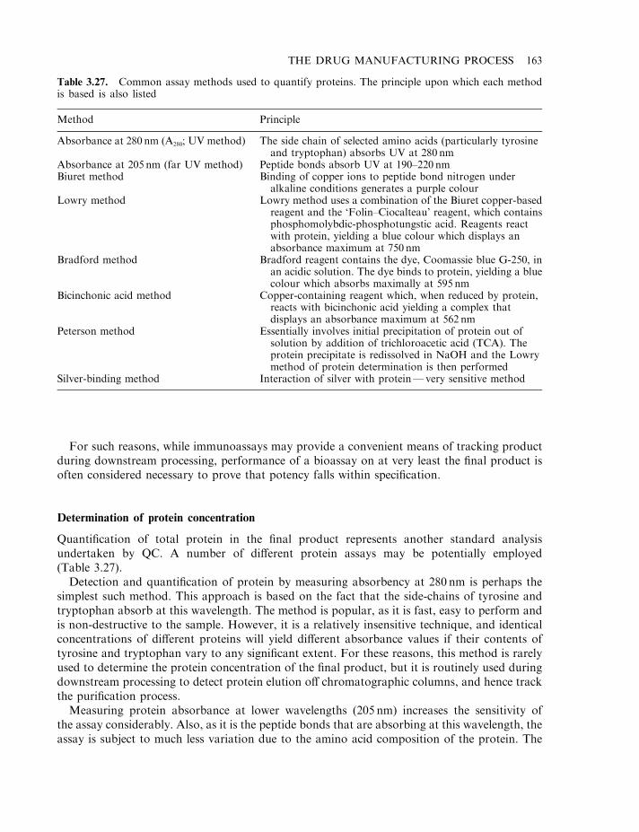
For such reasons, while immunoassays may provide a convenient means of tracking productduring downstream processing, performance of a bioassay on at very least the final product isoften considered necessary to prove that potency falls within specification.
Determination of protein concentration
Quantification of total protein in the final product represents another standard analysisundertaken by QC. A number of different protein assays may be potentially employed(Table 3.27).
Detection and quantification of protein by measuring absorbency at 280 nm is perhaps thesimplest such method. This approach is based on the fact that the side-chains of tyrosine andtryptophan absorb at this wavelength. The method is popular, as it is fast, easy to perform andis non-destructive to the sample. However, it is a relatively insensitive technique, and identicalconcentrations of different proteins will yield different absorbance values if their contents oftyrosine and tryptophan vary to any significant extent. For these reasons, this method is rarelyused to determine the protein concentration of the final product, but it is routinely used duringdownstream processing to detect protein elution off chromatographic columns, and hence trackthe purification process.
Measuring protein absorbance at lower wavelengths (205 nm) increases the sensitivity ofthe assay considerably. Also, as it is the peptide bonds that are absorbing at this wavelength, theassay is subject to much less variation due to the amino acid composition of the protein. The
THE DRUG MANUFACTURING PROCESS 163
Table 3.27. Common assay methods used to quantify proteins. The principle upon which each methodis based is also listed
Method Principle
Absorbance at 280 nm (A280; UV method) The side chain of selected amino acids (particularly tyrosineand tryptophan) absorbs UV at 280 nm
Absorbance at 205 nm (far UV method) Peptide bonds absorb UV at 190–220 nmBiuret method Binding of copper ions to peptide bond nitrogen under
alkaline conditions generates a purple colourLowry method Lowry method uses a combination of the Biuret copper-based
reagent and the ‘Folin–Ciocalteau’ reagent, which containsphosphomolybdic-phosphotungstic acid. Reagents reactwith protein, yielding a blue colour which displays anabsorbance maximum at 750 nm
Bradford method Bradford reagent contains the dye, Coomassie blue G-250, inan acidic solution. The dye binds to protein, yielding a bluecolour which absorbs maximally at 595 nm
Bicinchonic acid method Copper-containing reagent which, when reduced by protein,reacts with bicinchonic acid yielding a complex thatdisplays an absorbance maximum at 562 nm
Peterson method Essentially involves initial precipitation of protein out ofsolution by addition of trichloroacetic acid (TCA). Theprotein precipitate is redissolved in NaOH and the Lowrymethod of protein determination is then performed
Silver-binding method Interaction of silver with protein—very sensitive method
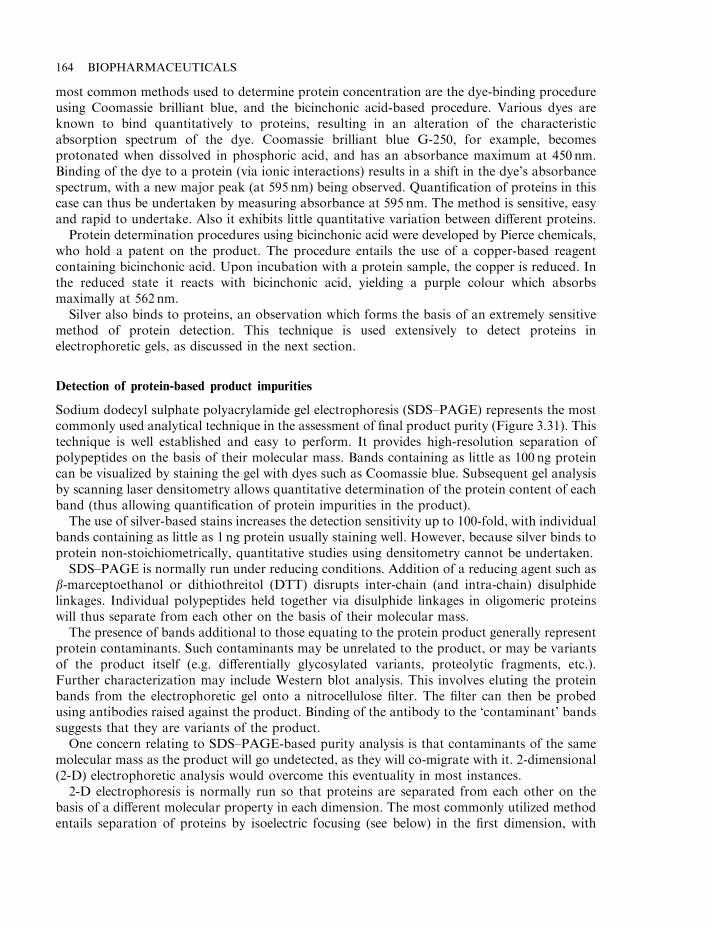
most common methods used to determine protein concentration are the dye-binding procedureusing Coomassie brilliant blue, and the bicinchonic acid-based procedure. Various dyes areknown to bind quantitatively to proteins, resulting in an alteration of the characteristicabsorption spectrum of the dye. Coomassie brilliant blue G-250, for example, becomesprotonated when dissolved in phosphoric acid, and has an absorbance maximum at 450 nm.Binding of the dye to a protein (via ionic interactions) results in a shift in the dye’s absorbancespectrum, with a new major peak (at 595 nm) being observed. Quantification of proteins in thiscase can thus be undertaken by measuring absorbance at 595 nm. The method is sensitive, easyand rapid to undertake. Also it exhibits little quantitative variation between different proteins.
Protein determination procedures using bicinchonic acid were developed by Pierce chemicals,who hold a patent on the product. The procedure entails the use of a copper-based reagentcontaining bicinchonic acid. Upon incubation with a protein sample, the copper is reduced. Inthe reduced state it reacts with bicinchonic acid, yielding a purple colour which absorbsmaximally at 562 nm.
Silver also binds to proteins, an observation which forms the basis of an extremely sensitivemethod of protein detection. This technique is used extensively to detect proteins inelectrophoretic gels, as discussed in the next section.
Detection of protein-based product impurities
Sodium dodecyl sulphate polyacrylamide gel electrophoresis (SDS–PAGE) represents the mostcommonly used analytical technique in the assessment of final product purity (Figure 3.31). Thistechnique is well established and easy to perform. It provides high-resolution separation ofpolypeptides on the basis of their molecular mass. Bands containing as little as 100 ng proteincan be visualized by staining the gel with dyes such as Coomassie blue. Subsequent gel analysisby scanning laser densitometry allows quantitative determination of the protein content of eachband (thus allowing quantification of protein impurities in the product).
The use of silver-based stains increases the detection sensitivity up to 100-fold, with individualbands containing as little as 1 ng protein usually staining well. However, because silver binds toprotein non-stoichiometrically, quantitative studies using densitometry cannot be undertaken.
SDS–PAGE is normally run under reducing conditions. Addition of a reducing agent such asb-marceptoethanol or dithiothreitol (DTT) disrupts inter-chain (and intra-chain) disulphidelinkages. Individual polypeptides held together via disulphide linkages in oligomeric proteinswill thus separate from each other on the basis of their molecular mass.
The presence of bands additional to those equating to the protein product generally representprotein contaminants. Such contaminants may be unrelated to the product, or may be variantsof the product itself (e.g. differentially glycosylated variants, proteolytic fragments, etc.).Further characterization may include Western blot analysis. This involves eluting the proteinbands from the electrophoretic gel onto a nitrocellulose filter. The filter can then be probedusing antibodies raised against the product. Binding of the antibody to the ‘contaminant’ bandssuggests that they are variants of the product.
One concern relating to SDS–PAGE-based purity analysis is that contaminants of the samemolecular mass as the product will go undetected, as they will co-migrate with it. 2-dimensional(2-D) electrophoretic analysis would overcome this eventuality in most instances.
2-D electrophoresis is normally run so that proteins are separated from each other on thebasis of a different molecular property in each dimension. The most commonly utilized methodentails separation of proteins by isoelectric focusing (see below) in the first dimension, with
164 BIOPHARMACEUTICALS
separation in the second dimension being undertaken in the presence of SDS, thus promotingband separation on the basis of protein size. Modified electrophoresis equipment which renders2-D electrophoretic separation routine is freely available. Application of biopharmaceuticalfinished products to such systems allows rigorous analysis of purity.
Isoelectric focusing (IEF) entails setting up a pH gradient along the length of anelectrophoretic gel. Applied proteins will migrate under the influence of an electric field, untilthey reach a point in the gel at which the pH equals the protein’s isoelectric point (pI; the pH atwhich the protein exhibits no overall net charge—only species with a net charge will moveunder the influence of an electric field). IEF thus separates proteins on the basis of chargecharacteristics.
This technique is also utilized in the biopharmaceutical industry to determine producthomogeneity. Homogeneity is best indicated by the appearance in the gel of a single proteinband, exhibiting the predicted pI value. Interpretation of the meaning of multiple bands,however, is less straightforward, particularly if the protein is glycosylated (the bands can also bestained for the presence of carbohydrates). Glycoproteins varying slightly in their carbohydratecontent will vary in their sialic acid content, and hence exhibit slightly different pI values. In
THE DRUG MANUFACTURING PROCESS 165
Figure 3.31. Separation of proteins by SDS–PAGE. Protein samples are incubated with SDS (as well asreducing agents, which disrupt disulphide linkages). The electric field is applied across the gel after theprotein samples to be analysed are loaded into the gel wells. The rate of protein migration towards theanode is dependent upon protein size. After electrophoresis is complete, individual protein bands may bevisualized by staining with a protein-binding dye (a). If one well is loaded with a mixture of proteins, eachof known molecular mass, a standard curve relating distance migrated to molecular mass can beconstructed (b). This allows estimation of the molecular mass of the purified protein. Reproduced bypermission of John Wiley & Sons Inc. from Walsh (2002)
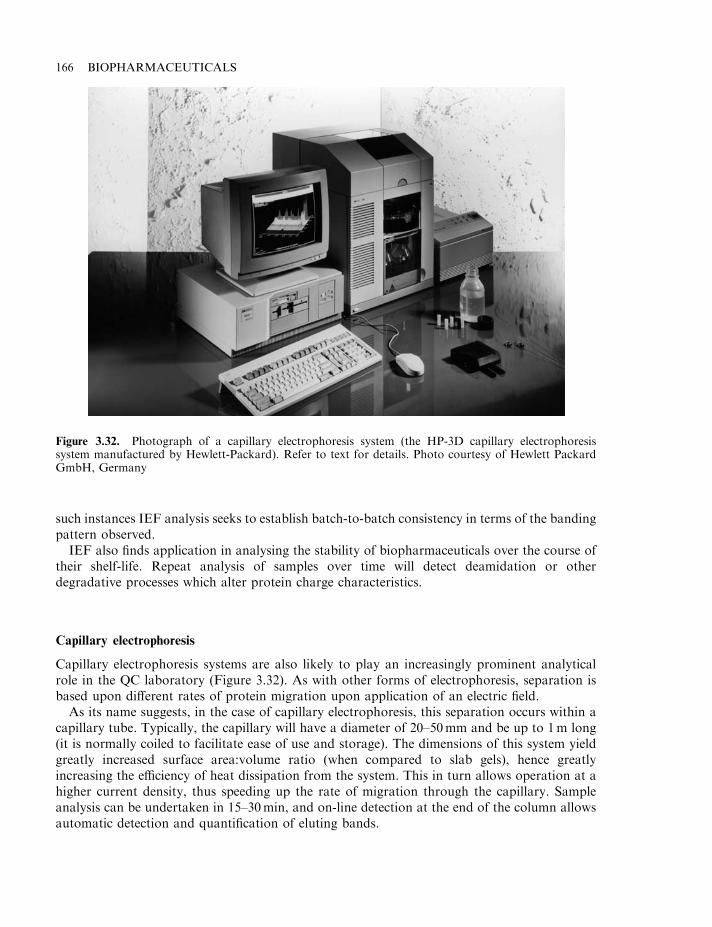
such instances IEF analysis seeks to establish batch-to-batch consistency in terms of the bandingpattern observed.
IEF also finds application in analysing the stability of biopharmaceuticals over the course oftheir shelf-life. Repeat analysis of samples over time will detect deamidation or otherdegradative processes which alter protein charge characteristics.
Capillary electrophoresis
Capillary electrophoresis systems are also likely to play an increasingly prominent analyticalrole in the QC laboratory (Figure 3.32). As with other forms of electrophoresis, separation isbased upon different rates of protein migration upon application of an electric field.
As its name suggests, in the case of capillary electrophoresis, this separation occurs within acapillary tube. Typically, the capillary will have a diameter of 20–50mm and be up to 1m long(it is normally coiled to facilitate ease of use and storage). The dimensions of this system yieldgreatly increased surface area:volume ratio (when compared to slab gels), hence greatlyincreasing the efficiency of heat dissipation from the system. This in turn allows operation at ahigher current density, thus speeding up the rate of migration through the capillary. Sampleanalysis can be undertaken in 15–30min, and on-line detection at the end of the column allowsautomatic detection and quantification of eluting bands.
166 BIOPHARMACEUTICALS
Figure 3.32. Photograph of a capillary electrophoresis system (the HP-3D capillary electrophoresissystem manufactured by Hewlett-Packard). Refer to text for details. Photo courtesy of Hewlett PackardGmbH, Germany
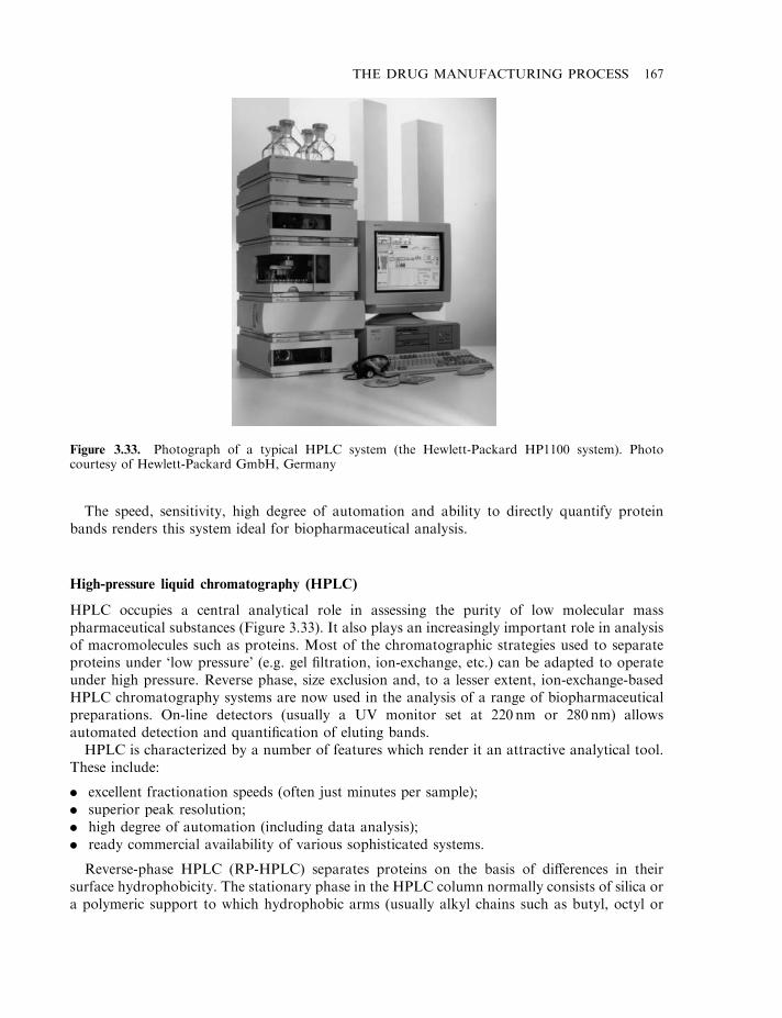
The speed, sensitivity, high degree of automation and ability to directly quantify proteinbands renders this system ideal for biopharmaceutical analysis.
High-pressure liquid chromatography (HPLC)
HPLC occupies a central analytical role in assessing the purity of low molecular masspharmaceutical substances (Figure 3.33). It also plays an increasingly important role in analysisof macromolecules such as proteins. Most of the chromatographic strategies used to separateproteins under ‘low pressure’ (e.g. gel filtration, ion-exchange, etc.) can be adapted to operateunder high pressure. Reverse phase, size exclusion and, to a lesser extent, ion-exchange-basedHPLC chromatography systems are now used in the analysis of a range of biopharmaceuticalpreparations. On-line detectors (usually a UV monitor set at 220 nm or 280 nm) allowsautomated detection and quantification of eluting bands.
HPLC is characterized by a number of features which render it an attractive analytical tool.These include:
. excellent fractionation speeds (often just minutes per sample);
. superior peak resolution;
. high degree of automation (including data analysis);
. ready commercial availability of various sophisticated systems.
Reverse-phase HPLC (RP-HPLC) separates proteins on the basis of differences in theirsurface hydrophobicity. The stationary phase in the HPLC column normally consists of silica ora polymeric support to which hydrophobic arms (usually alkyl chains such as butyl, octyl or
THE DRUG MANUFACTURING PROCESS 167
Figure 3.33. Photograph of a typical HPLC system (the Hewlett-Packard HP1100 system). Photocourtesy of Hewlett-Packard GmbH, Germany
octadecyl groups) have been attached. Reverse-phase systems have proved themselves to be aparticularly powerful analytical technique, capable of separating very similar molecules,displaying only minor differences in hydrophobicity. In some instances, a single amino acidsubstitution or the removal of a single amino acid from the end of a polypeptide chain can bedetected by RP-HPLC. In most instances modifications such as deamidation will also causepeak shifts. Such systems, therefore, may be used to detect impurities, be they related orunrelated to the protein product. RP-HPLC finds extensive application in analysis of insulinpreparations. Modified forms or insulin polymers are easily distinguishable from native insulinon reverse-phase columns.
While RP-HPLC has proved its analytical usefulness, its routine application to analysis ofspecific protein preparations should be undertaken only after extensive validation studies.HPLC in general can have a denaturing influence on many proteins (especially larger, complexproteins). Reverse-phase systems can be particularly harsh, as interaction with the highlyhydrophobic stationary phase can induce irreversible protein denaturation. Denaturation wouldresult in the generation of artifactual peaks on the chromatogram.
Size exclusion HPLC (SE-HPLC) separates proteins on the basis of size and shape. As mostsoluble proteins are globular (i.e. roughly spherical in shape), in most instances separation isessentially achieved on the basis of molecular mass. Commonly used SE-HPLC stationaryphases include silica-based supports and cross-linked agarose of defined pore size. Size exclusionsystems are most often used to analyse product for the presence of dimers or higher molecularmass aggregates of itself, as well as proteolysed product variants.
Calibration with standards allows accurate determination of the molecular mass of theproduct itself, as well as any impurities. Batch-to-batch variation can also be assessed bycomparison of chromatograms from different product runs.
Ion-exchange chromatography (both cation and anion) can also be undertaken in HPLCformat. Although not as extensively employed as RP or SE systems, ion-exchange-basedsystems are of use in analysing for impurities unrelated to the product, as well as detecting andquantifying deamidated forms.
Mass spectrometry
Recent advances in the field of mass spectrometry now extends the applicability of this methodto the analysis of macromolecules, such as proteins. Using electrospray mass spectrometry, it isnow possible to determine the molecular mass of many proteins to within an accuracy of+0.01%. A protein variant missing a single amino acid residue can easily be distinguished fromthe native protein in many instances. Although this is a very powerful technique, analysis of theresults obtained can sometimes be less than straightforward. Glycoproteins, for example, yieldextremely complex spectra (due to their natural heterogeneity), making the significance of thefindings hard to interpret.
Immunological approaches to detection of contaminants
Most recombinant biopharmaceuticals are produced in microbial or mammalian cell lines.Thus, although the product is derived from a human gene, all product-unrelated contaminantswill be derived from the producer organism. These non-self-proteins are likely to be highlyimmunogenic in humans, rendering their removal from the product stream especially important.Immunoassays may be conveniently used to detect and quantify non-product-related impurities
168 BIOPHARMACEUTICALS

in the final preparation (immunoassays generally may not be used to determine levels ofproduct-related impurities, as antibodies raised against such impurities would almost certainlycross-react with the product itself).
The strategy usually employed to develop such immunoassays is termed the ‘blank runapproach’. This entails constructing a host cell identical in all respects to the natural producercell, except that it lacks the gene coding for the desired product. This blank producer cell is thensubjected to upstream processing procedures identical to those undertaken with the normalproducer cell. Cellular extracts are subsequently subjected to the normal product purificationprocess, but only to a stage immediately prior to the final purification steps. This produces anarray of proteins which could co-purify with the final product. These proteins (of which theremay be up to 200, as determined by 2-D electrophoric analysis) are used to immunize horses,goats or other suitable animals. Polyclonal antibody preparations capable of binding specificallyto these proteins are therefore produced. Purification of the antibodies allows theirincorporation in radioimmunoassay or enzyme-based immunoassay systems, which maysubsequently be used to probe the product. Such multi-antigen assay systems will detect the sumtotal of host cell-derived impurities present in the product. Immunoassays identifying a singlepotential contaminant can also be developed.
Immunoassays have found widespread application in detecting and quantifying productimpurities. These assays are extremely specific and very sensitive, often detecting target antigendown to p.p.m. levels. Many immunoassays are available commercially and companies existwhich will rapidly develop tailor-made immunoassay systems for biopharmaceutical analysis.
Application of the analytical techniques discussed thus far focuses upon detection ofproteinaceous impurities. A variety of additional tests are undertaken which focus upon theactive substance itself. These tests aim to confirm that the presumed active substance observedby electrophoresis, HPLC, etc. is indeed the active substance, and that its primary sequence (andto a lesser extent, higher orders of structure) conform to licensed product specification. Testsperformed to verify the product identity include amino acid analysis, peptide mapping, N-terminal sequencing and spectrophotometric analyses.
Amino acid analysis
Amino acid analysis remains a characterization technique undertaken in many laboratories,particularly if the product is a peptide or small polypeptide (molecular mass 410 000Da). Thestrategy is simple—determine the range and quantity of amino acids present in the product andcompare the results obtained with the expected (theoretical) values. The results should becomparable.
The peptide/polypeptide product is usually hydrolysed by incubation with 6N HCl atelevated temperatures (1108C), under vacuum, for extended periods (12–24 h). The constituentamino acids are separated from each other by ion-exchange chromatography, and identified bycomparison with standard amino acid preparations. Reaction with ninhydrin allows subsequentquantification of each amino acid present.
While this technique is relatively straightforward and automated amino acid analysers arecommercially available, it is subject to a number of disadvantages that limit its usefulness inbiopharmaceutical analysis. These include:
. hydrolysis conditions can destroy/modify certain amino acid residues, particularlytryptophan, but also serine, threonine and tyrosine;
THE DRUG MANUFACTURING PROCESS 169
. the method is semi-quantitative rather than quantitative;
. sensitivity is at best moderate; low-level contaminants may go undetected (i.e. notsignificantly alter the amino acid profile obtained), particularly if the product is a highmolecular mass protein.
These disadvantages, along with the availability of alternative characterization methodologies,limit application of this technique in biopharmaceutical analysis.
Peptide mapping
A major concern relating to biopharmaceuticals produced in high-expression recombinantsystems is the potential occurrence of point mutations in the product’s gene, leading to analtered primary structure (i.e. amino acid sequence). Errors in gene transcription or translationcould also have similar consequences. The only procedure guaranteed to detect suchalterations is full sequencing of a sample of each batch of the protein; a considerable technicalchallenge. Although partial protein sequencing is normally undertaken (see later) the approachmost commonly used to detect alterations in amino acid sequence is peptide (fingerprint)mapping.
Peptide mapping entails exposure of the protein product to a reagent which promoteshydrolysis of peptide bonds at specific points along the protein backbone. This generates a seriesof peptide fragments. These fragments can be separated from each other by a variety oftechniques, including one- or two-dimensional electrophoresis and, in particular, RP-HPLC. Astandardized sample of the protein product, when subjected to this procedure, will yield acharacteristic peptide fingerprint, or map, with which the peptide maps obtained witheach batch of product can subsequently be compared. If the peptides generated arerelatively short, a change in a single amino acid residue is likely to alter thepeptide’s physicochemical properties sufficiently to alter its position within the peptide map(Figure 3.34). In this way single (or multiple) amino acid substitutions, deletions, insertions ormodifications can usually be detected. This technique plays an important role in monitoringbatch-to-batch consistency of the product, and also obviously can confirm the identity of theactual product.
The choice of reagent used to fragment the protein is critical to the success of this approach. Ifa reagent generates only a few very large peptides, a single amino acid alteration in one suchpeptide will be more difficult to detect than if it occurred in a much smaller peptide fragment. Onthe other hand, generation of a large number of very short peptides can be counter-productive,as it may prove difficult to resolve all the peptides from each other by subsequentchromatography. Generation of peptide fragments containing an average of 7–14 aminoacids is most desirable.
The most commonly utilized chemical cleavage agent is cyanogen bromide (it cleaves thepeptide bond on the carboxyl side of methionine residues). V8 protease, produced by certainstaphylococci, along with trypsin, are two of the more commonly used proteolytic-basedfragmentation agents.
Knowledge of the full amino acid sequence of the protein usually renders possible pre-determination of the most suitable fragmentation agent for any protein. The amino acidsequence of human growth hormone, for example, harbours 20 potential trypsin cleavage sites.Under some circumstances it may be possible to use a combination of fragmentation agents togenerate peptides of optimal length.
170 BIOPHARMACEUTICALS

N-terminal sequencing
N-terminal sequencing of the first 20–30 amino acid residues of the protein product has becomea popular quality control test for finished biopharmaceutical products. The technique is usefulas it:
. positively identifies the protein;
. confirms (or otherwise) the accuracy of the amino acid sequence of at least the N-terminus ofthe protein;
. readily identifies the presence of modified forms of the product in which one or more aminoacids are missing from the N-terminus.
N-terminal sequencing is normally undertaken by Edman degradation (Figure 3.35). Althoughthis technique was developed in the 1950s, advances in analytical methodologies now facilitate
THE DRUG MANUFACTURING PROCESS 171
Figure 3.34. Generation of a peptide map. In this simple example, the protein to be analysed is treatedwith a fragmentation agent, e.g. trypsin (a). In this case, five fragments are generated. The digest is thenapplied to a sheet of chromatography paper (b) at the point marked ‘origin’. The peptides are thenseparated from each other in the first (vertical) dimension by paper chromatography. Subsequently,electrophoresis is undertaken (in the horizontal direction). The separated peptide fragments may bevisualized, e.g. by staining with ninhydrin. 2-D separation of the peptides is far more likely to completelyresolve each peptide from the others. In the case above, for example, chromatography (in the verticaldimension) alone would not have been sufficient to fully resolve peptides 1 and 3. Duringbiopharmaceutical production, each batch of the recombinant protein produced should yield identicalpeptide maps. Any mutation which alters the protein’s primary structure (i.e. amino acid sequence) shouldresult in at least one fragment adopting an altered position in the peptide map
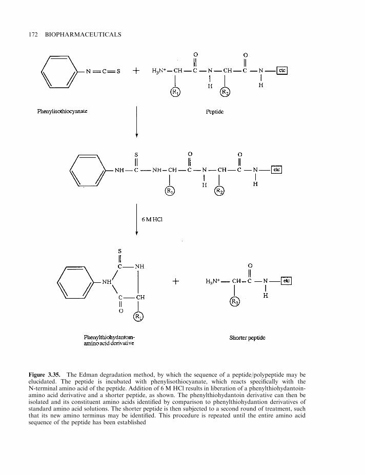
172 BIOPHARMACEUTICALS
Figure 3.35. The Edman degradation method, by which the sequence of a peptide/polypeptide may beelucidated. The peptide is incubated with phenylisothiocyanate, which reacts specifically with theN-terminal amino acid of the peptide. Addition of 6 M HCl results in liberation of a phenylthiohydantoin-amino acid derivative and a shorter peptide, as shown. The phenylthiohydantoin derivative can then beisolated and its constituent amino acids identified by comparison to phenylthiohydantion derivatives ofstandard amino acid solutions. The shorter peptide is then subjected to a second round of treatment, suchthat its new amino terminus may be identified. This procedure is repeated until the entire amino acidsequence of the peptide has been established
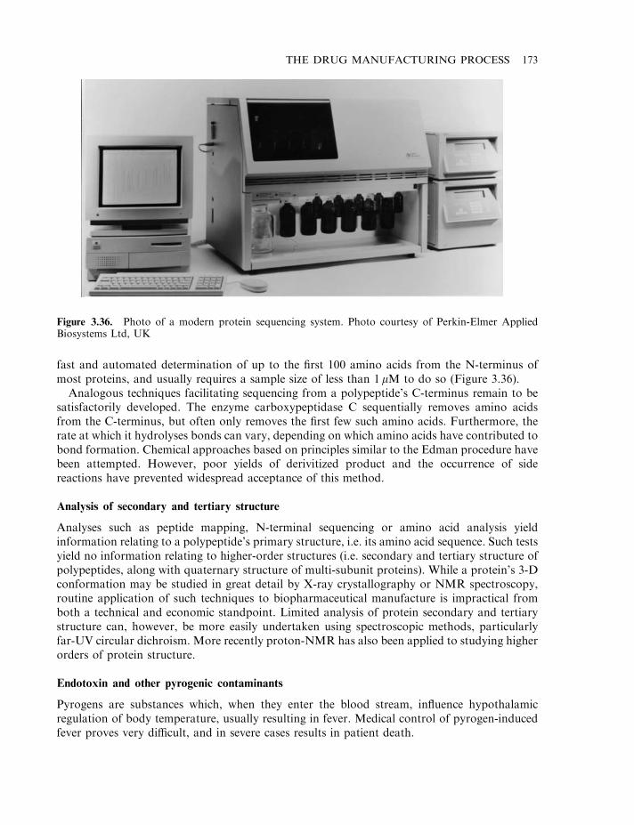
fast and automated determination of up to the first 100 amino acids from the N-terminus ofmost proteins, and usually requires a sample size of less than 1 mM to do so (Figure 3.36).
Analogous techniques facilitating sequencing from a polypeptide’s C-terminus remain to besatisfactorily developed. The enzyme carboxypeptidase C sequentially removes amino acidsfrom the C-terminus, but often only removes the first few such amino acids. Furthermore, therate at which it hydrolyses bonds can vary, depending on which amino acids have contributed tobond formation. Chemical approaches based on principles similar to the Edman procedure havebeen attempted. However, poor yields of derivitized product and the occurrence of sidereactions have prevented widespread acceptance of this method.
Analysis of secondary and tertiary structure
Analyses such as peptide mapping, N-terminal sequencing or amino acid analysis yieldinformation relating to a polypeptide’s primary structure, i.e. its amino acid sequence. Such testsyield no information relating to higher-order structures (i.e. secondary and tertiary structure ofpolypeptides, along with quaternary structure of multi-subunit proteins). While a protein’s 3-Dconformation may be studied in great detail by X-ray crystallography or NMR spectroscopy,routine application of such techniques to biopharmaceutical manufacture is impractical fromboth a technical and economic standpoint. Limited analysis of protein secondary and tertiarystructure can, however, be more easily undertaken using spectroscopic methods, particularlyfar-UV circular dichroism. More recently proton-NMR has also been applied to studying higherorders of protein structure.
Endotoxin and other pyrogenic contaminants
Pyrogens are substances which, when they enter the blood stream, influence hypothalamicregulation of body temperature, usually resulting in fever. Medical control of pyrogen-inducedfever proves very difficult, and in severe cases results in patient death.
THE DRUG MANUFACTURING PROCESS 173
Figure 3.36. Photo of a modern protein sequencing system. Photo courtesy of Perkin-Elmer AppliedBiosystems Ltd, UK
Pyrogens represent a diverse group of substances, including various chemicals, particulatematter and endotoxin (lipopolysaccharide, LPS—a molecule derived from the outer membraneof Gram-negative bacteria). Such Gram-negative organisms harbour 3–4 million LPS moleculeson their surface, representing in the region of 75% of their outer membrane surface area. Gram-negative bacteria clinically significant in human medicine include E. coli, Haemophilusinfluenzae, Salmonella enterica, Klebsiella pneumoniae, Bordetella pertussis, Pseudomonasaeruginosa, Chylamydia psittaci and Legionella pneumophila.
In many instances the influence of pyrogens on body temperature is indirect, e.g. entry ofendotoxin into the bloodstream stimulates the production of interleukin 1 (IL-1; Chapter 5) bymacrophages. It is the IL-1 that directly initiates the fever response (hence its alternative name,‘endogenous pyrogen’).
While entry of any pyrogenic substance into the bloodstream can have serious medicalconsequences, endotoxin receives most attention because of its ubiquitous nature. It is thereforethe pyrogen most likely to contaminate parenteral (bio)pharmaceutical products. Effectiveimplementation of GMP minimizes the likelihood of product contamination by pyrogens, e.g.GMP dictates that chemical reagents used in the manufacture of process buffers be extremelypure. Such raw materials are therefore unlikely to contain chemical contaminants displayingpyrogenic activity. Furthermore, GMP encourages filtration of virtually all parenteral productsthrough a 0.45 mm or 0.22 mm filter at points during processing and prior to filling in finalproduct containers (even if the product can subsequently be sterilized by autoclaving). Filtrationensures removal of all particulate matter from the product. In addition, most final productcontainers are rendered particle-free immediately prior to filling by an automatic pre-rinse usingWFI. As an additional safeguard, the final product will usually be subject to a particulate mattertest by QC before final product release. The simplest format for such a test could involve visualinspection of vial contents, although specific particle detecting and counting equipment is moreroutinely used.
Contamination of the final product with endotoxin is more difficult to control because:
. many recombinant biopharmaceuticals are produced in Gram-negative bacterial systems,thus the product source is also a source of endotoxin;
. despite rigorous implementation of GMP, most biopharmaceutical preparations will becontaminated with low levels of Gram-negative bacteria at some stage of manufacture. Thesebacteria shed endotoxin into the product stream, which is not removed during subsequentbacterial filtration steps. This is one of many reasons why GMP dictates that the level ofbioburden in the product stream should be minimized at all stages of manufacture;
. the heat-stability exhibited by endotoxin (see next section) means that autoclaving of processequipment will not destroy endotoxin present on such equipment;
. adverse medical reactions caused by endotoxin are witnessed in humans at dosage rates aslow as 0.5 ng/kg body weight.
Endotoxin, the molecule
The structural detail of a generalized endotoxin (LPS) molecule is presented in Figure 3.37. Asits name suggests, LPS consists of a complex polysaccharide component linked to a lipid (lipidA) moiety. The polysaccharide moiety is generally composed of 50 or more monosaccharideunits linked by glycosidic bonds. Sugar moieties often found in LPS include glucose,glucosamine, mannose and galactose, as well as more extensive structures such as
174 BIOPHARMACEUTICALS
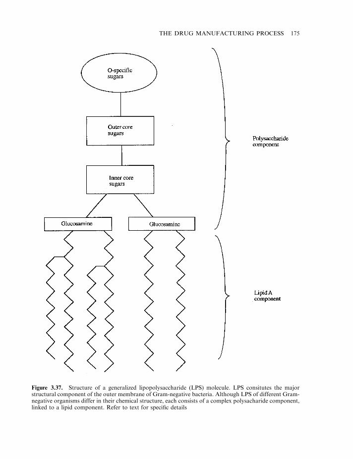
THE DRUG MANUFACTURING PROCESS 175
Figure 3.37. Structure of a generalized lipopolysaccharide (LPS) molecule. LPS consitutes the majorstructural component of the outer membrane of Gram-negative bacteria. Although LPS of different Gram-negative organisms differ in their chemical structure, each consists of a complex polysacharide component,linked to a lipid component. Refer to text for specific details
L-glycero-mannoheptose. The polysaccharide component of LPS may be divided into severalstructural domains. The inner (core) domains vary relatively little between LPS moleculesisolated from different Gram-negative bacteria. The outer (O-specific) domain is usuallybacterial strain-specific.
Most of the LPS biological activity (pyrogenicity) is associated with its lipid A moiety. Thisusually consists of six or more fatty acids attached directly to sugars such as glucosamine.Again, as is the case in relation to the carbohydrate component, lipid A moieties of LPS isolatedfrom different bacteria can vary somewhat. The structure of E. coli’s lipid A has been studied ingreatest detail. Its exact structure has been elucidated, and it can be chemically synthesized.
Pyrogen detection
Pyrogens may be detected in parenteral preparations (or other substances) by a number ofmethods. Two such methods are widely employed in the pharmaceutical industry.
Historically, the rabbit pyrogen test constituted the most widely used method. This entailsparenteral administration of the product to a group of healthy rabbits, with subsequentmonitoring of rabbit temperature using rectal probes. Increased rabbit temperature above acertain point suggests the presence of pyrogenic substances. The basic rabbit method, asoutlined in the European Pharmacopoeia, entails initial administration of the product to threerabbits. The product is considered to have passed the test if the total (summed) increase of thetemperature of all three animals is less than 1.158C. If the total increase recorded is greater than2.658C the product has failed. However, if the response observed falls between these two limits,the result is considered inconclusive, and the test must be repeated using a further batch ofanimals.
This test is popular because it detects a wide spectrum of pyrogenic substances. However, it isalso subject to a number of disadvantages, including:
. it is expensive (there is a requirement for animals, animal facilities and animal technicians);
. excitation/poor handling of the rabbits can affect the results obtained, usually prompting afalse-positive result;
. sub-clinical infection/poor overall animal health can also lead to false-positive results;
. use of different rabbit colonies/breeds can yield variable results.
Another issue of relevance is that certain biopharmaceuticals (e.g. cytokines such as 1L-1 andTNF; Chapter 5), themselves, induce a natural pyrogenic response. This rules out use of therabbit-based assay for detection of exogenous pyrogens in such products. Such difficulties haveled to the increased use of an in vitro assay; the Limulus amoebocyte lysate (LAL) test. This isbased upon endotoxin-stimulated coagulation of amoebocyte lysate obtained from horseshoecrabs. This test is now the most widely used assay for the detection of endotoxins inbiopharmaceutical and other pharmaceutical preparations.
Development of the LAL assay was based upon the observation that the presence of Gram-negative bacteria in the vascular system of the American horseshoe crab, (Limulus polyphemus),resulted in the clotting of its blood. Tests on fractionated blood showed the factor responsiblefor coagulation resided within the crab’s circulating blood cells, the amoebocytes. Furtherresearch revealed that the bacterial agent responsible for initiation of clot formation wasendotoxin.
The endotoxin molecule activates a coagulation cascade quite similar in design to themammalian blood coagulation cascade (Figure 3.38). Activation of the cascade also requires the
176 BIOPHARMACEUTICALS
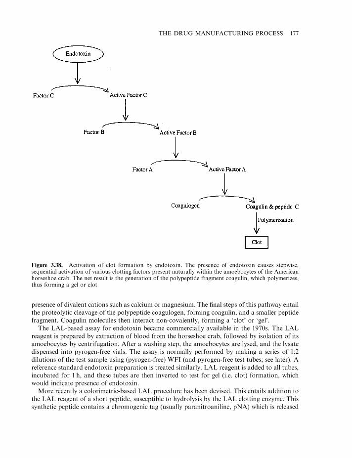
presence of divalent cations such as calcium or magnesium. The final steps of this pathway entailthe proteolytic cleavage of the polypeptide coagulogen, forming coagulin, and a smaller peptidefragment. Coagulin molecules then interact non-covalently, forming a ‘clot’ or ‘gel’.
The LAL-based assay for endotoxin became commercially available in the 1970s. The LALreagent is prepared by extraction of blood from the horseshoe crab, followed by isolation of itsamoebocytes by centrifugation. After a washing step, the amoebocytes are lysed, and the lysatedispensed into pyrogen-free vials. The assay is normally performed by making a series of 1:2dilutions of the test sample using (pyrogen-free) WFI (and pyrogen-free test tubes; see later). Areference standard endotoxin preparation is treated similarly. LAL reagent is added to all tubes,incubated for 1 h, and these tubes are then inverted to test for gel (i.e. clot) formation, whichwould indicate presence of endotoxin.
More recently a colorimetric-based LAL procedure has been devised. This entails addition tothe LAL reagent of a short peptide, susceptible to hydrolysis by the LAL clotting enzyme. Thissynthetic peptide contains a chromogenic tag (usually paranitroaniline, pNA) which is released
THE DRUG MANUFACTURING PROCESS 177
Figure 3.38. Activation of clot formation by endotoxin. The presence of endotoxin causes stepwise,sequential activation of various clotting factors present naturally within the amoebocytes of the Americanhorseshoe crab. The net result is the generation of the polypeptide fragment coagulin, which polymerizes,thus forming a gel or clot
CC
free into solution by the clotting enzyme. This allows spectrophotometric analysis of the testsample, facilitating more accurate end-point determination.
The LAL system displays several advantages when compared to the rabbit test, most notably:
. sensitivity—endotoxin levels as low as a few picograms (pg) per ml of sample assayed will bedetected;
. cost—the assay is far less expensive than the rabbit assay;
. speed—depending upon the format used, the LAL assay may be conducted within 15–60min.
Its major disadvantage is its selectivity— it only detects endotoxin-based pyrogens. In practice,however, endotoxin represents the pyrogen by far the most likely to be present inpharmaceutical products. The LAL method is used extensively within the industry. It is usednot only to detect endotoxin in finished parenteral preparations, but also in WFI and inbiological fluids such as serum or cerebrospinal fluid.
Before the LAL assay is routinely used to detect/quantify endotoxin in any product, itseffective functioning in the presence of that product must be demonstrated by validation studies.Such studies are required to prove that the product (or, more likely, excipients present in theproduct) do not interfere with the rate/extent of clot formation (i.e. are neither inhibitors noractivators of the LAL-based enzymes). LAL enzyme inhibition could facilitate false-negativeresults upon sample assay. Validation studies entail, for example, observing the effect of spikingendotoxin-negative product with known quantities of endotoxin, or spiking endotoxin withvarying quantities of product, before assay with the LAL reagents.
All ancillary reagents used in the LAL assay system (e.g. WFI, test tubes, pipette tips forliquid transfer, etc.) must obviously be endotoxin-free. Such items can be rendered endotoxin-free by heat. Its heat-stable nature, however, renders necessary very vigorous heating in order todestroy contaminant endotoxin. A single autoclave cycle is insufficient, with total destructionrequiring three consecutive autoclave cycles. Dry heat may also be used (1808C for 3 h or 2408Cfor 1 h).
GMP requires that, where practicable, process equipment coming into direct contact with thebiopharmaceutical product stream should be rendered endotoxin-free (depyrogenated) beforeuse. Autoclaving, steam or dry heat can effectively be used on many process vessels, pipework,etc., which are usually manufactured from stainless steel or other heat-resistant material. Suchan approach is not routinely practicable in the case of some items of process equipment, such aschromatographic systems. Fortunately, endotoxin is sensitive to strongly alkaline conditions,thus routine CIP of chromatographic systems using 1M NaOH represents an effectivedepyrogenation step. More gentle approaches, such as exhaustive rinsing with WFI (until anLAL test shows the eluate to be endotoxin-free) can also be surprisingly effective.
It is generally unnecessary to introduce specific measures aimed at endotoxin removal fromthe product during downstream processing. Endotoxin present in the earlier stages ofproduction are often effectively removed from the product during chromatographic fractiona-tion. The endotoxin molecule’s highly negative charge often facilitates its effective removal fromthe product stream by ion-exchange chromatography. Gel filtration chromatography also servesto remove endotoxin from the product. While individual lipopolysaccharide molecules exhibitan average molecular mass of less than 20 kDa, these molecules aggregate in aqueousenvironments, generating supramolecular structures of molecular mass 100–1 000 kDa.
The molecular mass of most biopharmaceuticals is considerably less than 100 kDa (Table3.28). The proteins would thus elute from gel-filtration columns much later than contaminating
178 BIOPHARMACEUTICALS
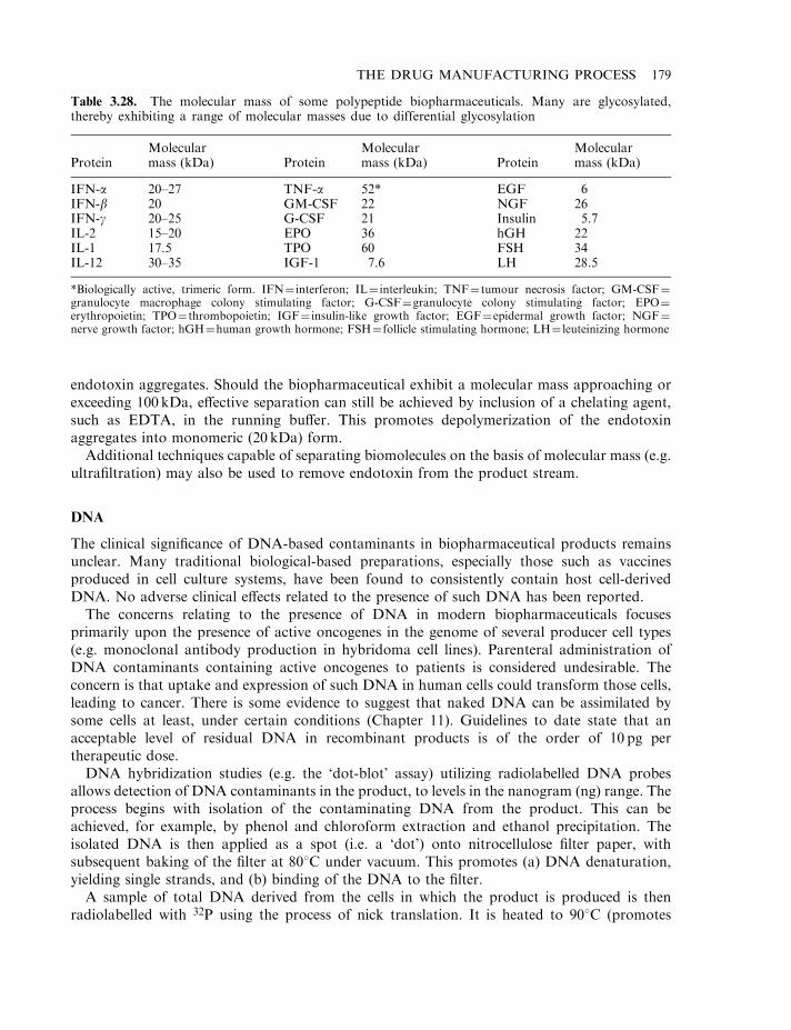
endotoxin aggregates. Should the biopharmaceutical exhibit a molecular mass approaching orexceeding 100 kDa, effective separation can still be achieved by inclusion of a chelating agent,such as EDTA, in the running buffer. This promotes depolymerization of the endotoxinaggregates into monomeric (20 kDa) form.
Additional techniques capable of separating biomolecules on the basis of molecular mass (e.g.ultrafiltration) may also be used to remove endotoxin from the product stream.
DNA
The clinical significance of DNA-based contaminants in biopharmaceutical products remainsunclear. Many traditional biological-based preparations, especially those such as vaccinesproduced in cell culture systems, have been found to consistently contain host cell-derivedDNA. No adverse clinical effects related to the presence of such DNA has been reported.
The concerns relating to the presence of DNA in modern biopharmaceuticals focusesprimarily upon the presence of active oncogenes in the genome of several producer cell types(e.g. monoclonal antibody production in hybridoma cell lines). Parenteral administration ofDNA contaminants containing active oncogenes to patients is considered undesirable. Theconcern is that uptake and expression of such DNA in human cells could transform those cells,leading to cancer. There is some evidence to suggest that naked DNA can be assimilated bysome cells at least, under certain conditions (Chapter 11). Guidelines to date state that anacceptable level of residual DNA in recombinant products is of the order of 10 pg pertherapeutic dose.
DNA hybridization studies (e.g. the ‘dot-blot’ assay) utilizing radiolabelled DNA probesallows detection of DNA contaminants in the product, to levels in the nanogram (ng) range. Theprocess begins with isolation of the contaminating DNA from the product. This can beachieved, for example, by phenol and chloroform extraction and ethanol precipitation. Theisolated DNA is then applied as a spot (i.e. a ‘dot’) onto nitrocellulose filter paper, withsubsequent baking of the filter at 808C under vacuum. This promotes (a) DNA denaturation,yielding single strands, and (b) binding of the DNA to the filter.
A sample of total DNA derived from the cells in which the product is produced is thenradiolabelled with 32P using the process of nick translation. It is heated to 908C (promotes
THE DRUG MANUFACTURING PROCESS 179
Table 3.28. The molecular mass of some polypeptide biopharmaceuticals. Many are glycosylated,thereby exhibiting a range of molecular masses due to differential glycosylation
ProteinMolecularmass (kDa) Protein
Molecularmass (kDa) Protein
Molecularmass (kDa)
IFN-a 20–27 TNF-a 52* EGF 6IFN-b 20 GM-CSF 22 NGF 26IFN-g 20–25 G-CSF 21 Insulin 5.7IL-2 15–20 EPO 36 hGH 22IL-1 17.5 TPO 60 FSH 34IL-12 30–35 IGF-1 7.6 LH 28.5
*Biologically active, trimeric form. IFN¼ interferon; IL¼ interleukin; TNF¼ tumour necrosis factor; GM-CSF¼granulocyte macrophage colony stimulating factor; G-CSF¼granulocyte colony stimulating factor; EPO¼erythropoietin; TPO¼ thrombopoietin; IGF¼ insulin-like growth factor; EGF¼epidermal growth factor; NGF¼nerve growth factor; hGH¼human growth hormone; FSH¼ follicle stimulating hormone; LH¼ leuteinizing hormone
denaturation, forming single strands) and incubated with the baked filter for several hours at408C. Lowering the temperature allows re-annealing of single strands via complementary base-pairing to occur. Labelled DNA will re-anneal with any complementary DNA strandsimmobilized on the filter. After the filter is washed (to remove non-specifically boundradiolabelled probe) it is subjected to autoradiography, which allows detection of any boundprobe.
Quantification of the DNA isolated from the product involves concurrent inclusion in thedot-blot assay of a set of spots, containing known quantities of DNA and derived from theproducer cell. After autoradiography, the intensity of the test spot is compared with thestandards.
In many instances there is little need to incorporate specific DNA removal steps duringdownstream processing. Endogenous nucleases liberated upon cellular homogenization comeinto direct contact with cellular DNA, resulting in its degradation. Commercial DNases aresometimes added to crude homogenate to reduce DNA-associated product viscosity. Mostchromatographic steps are also effective in separating DNA from the product stream. Ion-exchange chromatography is particularly effective, as DNA exhibits a large overall negativecharge (due to the phosphate constituent of its nucleotide backbone).
Microbial and viral contaminants
Finished-product biopharmaceuticals, along with other pharmaceuticals intended for parenteraladministration, must be sterile (the one exception being live bacterial vaccines). The presence ofmicroorganisms in the final product is unacceptable for a number of reasons:
. parenteral administration of contaminated product would likely lead to the establishment ofa severe infection in the recipient patient;
. microorganisms may be capable of metabolizing the product itself, thus reducing its potency.This is particularly true of protein-based biopharmaceuticals, as most microbes produce anarray of extracellular proteases;
. microbial-derived substances secreted into the product could adversely effect the recipient’shealth. Examples include endotoxin secreted from Gram-negative bacteria, or microbialproteins which would stimulate an immune response.
Terminal sterilization by autoclaving guarantees product sterility. Heat sterilization, however,is not a viable option in the case of biopharmaceuticals. Sterilization of biopharmaceuticals byfiltration, followed by aseptic filling into a sterile final-product container, inherently carries agreater risk of product contamination. Finished-product sterility testing of such preparationsthus represents one of the most critical product tests undertaken by QC. Specific guidelinesrelating to sterility testing of finished products are given in international pharmacopoeias.
Biopharmaceutical products are also subjected to screening for the presence of viral particlesprior to final product release. Although viruses could be introduced, e.g. via infected personnelduring downstream processing, proper implementation of GMP minimizes such risk. Any viralparticles found in the finished product are most likely derived from raw material sources.Examples could include HIV or hepatitis viruses present in blood used in the manufacture ofblood products. Such raw materials must be screened before processing for the presence of likelyviral contaminants.
A variety of murine (mouse) and other mammalian cell lines have become popular hostsystems for the production of recombinant human biopharmaceuticals. Moreover, most
180 BIOPHARMACEUTICALS
monoclonal antibodies used for therapeutic purposes are produced by murine-derivedhybridoma cells. These cell lines are sensitive to infection by various viral particles. Producercell lines are screened during product development studies to ensure freedom from a variety ofpathogenic advantageous agents, including various species of bacteria, fungi, yeasts,mycoplasma, protozoa, parasites, viruses and prions. Suitable microbiological precautionsmust subsequently be undertaken to prevent producer cell banks from becoming contaminatedwith such pathogens.
Removal of viruses from the product stream can be achieved in a number of ways. Thephysicochemical properties of viral particles differ greatly from most proteins, ensuring thateffective fractionation is automatically achieved by most chromatographic techniques. Gel-filtration chromatography, for example, effectively separates viral particles from most proteinson the basis of differences in size.
In addition to chromatographic separation, specific downstream processing steps may beundertaken which are specifically aimed at removal or inactivation of viral particles potentiallypresent in the product stream. Significantly, many are ‘blanket’ procedures, equally capable ofremoving known or potentially likely viral contaminants, and any uncharacterized/undetectedviruses. Filtration through a 0.22 mm filter effectively removes microbial agents from the productstream, but fails to remove most viral types. Repeat filtration through a 0.1 mm filter appearsmore effective in this regard. Alternatively, incorporation of an ultrafiltration step (preferably atthe terminal stages of downstream processing) also proves effective.
Incorporation of downstream processing steps known to inactivate a wide variety of viraltypes provides further assurance that the final product is unlikely to harbour active virus.Heating and irradiation are amongst the two most popular such approaches. Heating theproduct to 40–608C for several hours inactivates a broad range of viruses. Manybiopharmaceuticals can be heated to such temperatures without being denatured themselves.Such an approach has been used extensively to inactivate blood-borne viruses in bloodproducts. Exposure of product to controlled levels of UV radiation can also be quite effective,while having no adverse effect on the product itself.
Viral assays
A range of assay techniques may be used to detect and quantify viral contaminants in both rawmaterials and finished biopharmaceutical products. No generic assay exists that is capable ofdetecting all viral types potentially present in a given sample. Viral assays currently availablewill detect only a specific virus, or at best a family of closely related viruses. The strategyadopted, therefore, usually entails screening product for viral particles known to be capable ofinfecting the biopharmaceutical source material. Such assays will not normally detect newlyevolved viral strains, or uncharacterized/unknown viral contaminants. This fact underlines theimportance of including at least one step in downstream processing which is likely toindiscriminately inactivate or remove viruses from the product. This acts as a safety net.
Current viral assays fall into one of three categories:
. immunoassays;
. assays based on viral DNA probes;
. bioassays.
Generation of antibodies that can recognize and bind to specific viruses is straightforward. Asample of live or attenuated virus, or a purified component of the viral caspid, can be injected
THE DRUG MANUFACTURING PROCESS 181
into animals to stimulate polyclonal antibody production (or to facilitate monoclonal antibodyproduction by hybridoma technology). Harvested antibodies are then employed to developspecific immunoassays which can be used to routinely screen test samples for the presence ofthat specific virus. Immunoassays capable of detecting a wide range of viruses are availablecommercially. The sensitivity, ease, speed and relative inexpensiveness of these assays renderthem particularly attractive.
An alternative assay format entails the use of virus-specific DNA probes. These can be used toscreen the biopharmaceutical product for the presence of viral DNA. The assay strategy issimilar to the dot-blot assays used to detect host cell derived DNA contaminants, as discussedearlier.
Viral bioassays of various different formats have also been developed. One format entailsincubation of the final product with cell lines sensitive to a range of viruses. The cells aresubsequently monitored for cytopathic effects or other obvious signs of viral infection.
A range of mouse, rabbit or hamster antibody production tests (MAPs, RAPs or HAPs) mayalso be undertaken. These bioassays entail administration of the product to a test animal. Anyviral agents present will elicit the production of anti-viral antibodies in that animal. Serumsamples (withdrawn from the animal approximately 4 weeks after product administration) arescreened for the presence of antibodies recognizing a range of viral antigens. This can beachieved by enzyme immunoassay, in which immobilized antigen is used to screen for the virus-specific antibodies. These assay systems are extremely sensitive, as minute quantities of viralantigen will elicit strong antibody production. A single serum sample can also be screened forantibodies specific to a wide range of viral particles. Time and expense factors, however, militateagainst this particular assay format.
Miscellaneous contaminants
In addition to those already discussed, biopharmaceutical products may harbour othercontaminants, some of which may be intentionally added to the product-stream during theinitial stages of downstream processing. Examples could include buffer components,precipitants (ethanol or other solvents, salts, etc.), proteolytic inhibitors, glycerol, anti-foamagents, etc. In addition to these, other contaminants may enter the product during downstreamprocessing in a less controlled way. Examples could include metal ions leached from product-holding tanks/pipework, or breakdown products leaking from chromatographic media. Thefinal product containers must also be chosen carefully. They must be chemically inert, and be ofsuitable quality to eliminate the possibility of leaching of any substance from the containerduring product storage. For this reason high-quality glass vials are often used.
In some instances it may be necessary to demonstrate that all traces of specific contaminantshave been removed prior to final product filling. This would be true, for example, of manyproteolytic inhibitors added during the initial stages of downstream processing to preventproteolysis by endogenous proteases. Some such inhibitors may be inherently toxic, and manycould (inappropriately) inhibit endogenous proteases of the recipient patient.
Demonstration of the absence (from the product) of breakdown products from chromato-graphic columns may be necessary in certain instances. This is particularly true with regard tosome affinity chromatography columns. Various chemical-coupling methods may be used toattach affinity ligands to the chromatographic support material. Some such procedures entailthe use of toxic reagents which, if not entirely removed after coupling, could leach into theproduct. In some cases, ligands can also subsequently leach from the columns, particularly after
182 BIOPHARMACEUTICALS
sustained usage or over-vigorous sanitation procedures. Improvements in the chemical stabilityof modern chromatographic media have, however, reduced such difficulties, and mostmanufacturers have carried out extensive validation studies regarding the stability of theirproducts.
Sophisticated analytical methodologies facilitate detection of vanishingly low levels of manycontaminants in biopharmaceutical preparations. The possibility exists, however, thatuncharacterized contaminants may persist, remaining undetected in the final product. As anadditional safety measure, finished products are often subjected to ‘abnormal toxicity’ or‘general safety’ tests. Standardized protocols for such tests are outlined in various internationalpharmacopoeias. These normally entail parenteral administration of the product to at least fivehealthy mice. The animals are placed under observation for 48 h, and should exhibit no ill-effects(other than expected symptoms). The death or illness of one or more animals signals arequirement for further investigation, usually using a larger number of animals. Such toxicitytesting represents a safety net, designed to expose any unexpected activities in the product whichcould compromise the health of the recipient.
Validation studies
Validation can be defined as ‘the act of proving that any procedure, process, equipment,material, activity or system leads to the expected results’. Routine and adequate validationstudies form a core principle of GMP as applied to (bio)pharmaceutical manufacture, as suchstudies help assure the overall safety of the finished product (Box 3.1).
All validation procedures must be carefully designed and fully documented in written format(Box 3.1). The results of all validation studies undertaken must also be documented, andretained in the plant files. As part of their routine inspection of manufacturing facilities,regulatory personnel will usually inspect a sample of these records, to ensure conformance toGMP.
Validation studies encompass all aspects of (bio)pharmaceutical manufacture. All new itemsof equipment must be validated before being routinely used. Initial validation studies should becomprehensive, with follow-up validation studies being undertaken at appropriate time intervals(e.g. daily, weekly or monthly). It is considered judicious to validate older items of equipmentwith increased frequency. Such studies can forewarn the manufacturer of impending equipmentfailure. Some validatory studies are straightforward, e.g. validation of weighing equipmentsimply entails weighing standardized weights. Autoclaves may be validated by placing externaltemperature probes at various points in the autoclave chamber during a routine autoclave run.Validation studies should confirm that all areas within the chamber reach the requiredtemperature for the required time.
Periodic validation of clean room HEPA filters is also an essential part of GMP. After theirinstallation, HEPA filters are subjected to a leak test. Particle counters are also used to validateclean room conditions. A particle counter is a vacuum cleaner-like machine capable of suckingair from its surroundings at constant velocity, and passing it through a counting chamber. Thenumber of particles per m3 of air tested can easily be determined. Furthermore, passage of theair through a 0.2 mm filter housed in the counter will trap all airborne microorganisms. Byplacing the filter on the surface of a nutrient agar-containing Petri dish, trapped microorganismswill grow as colonies, allowing determination of the microbial load per m3 of air.
In addition to equipment, many processes/procedures undertaken during pharmaceuticalmanufacture are also subject to periodic validation studies. Validation of biopharmaceutical
THE DRUG MANUFACTURING PROCESS 183
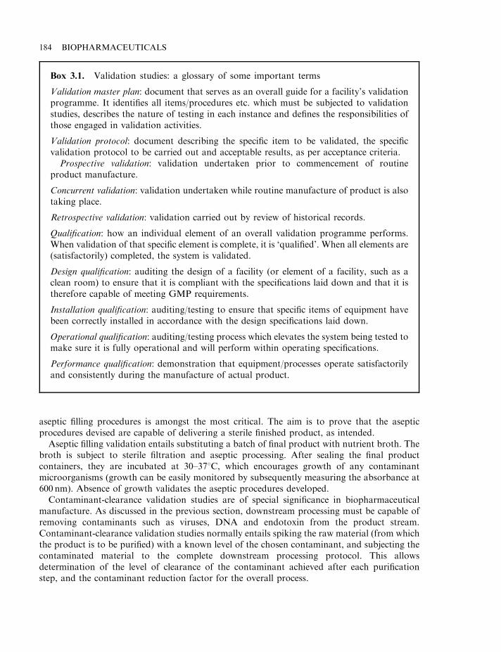
aseptic filling procedures is amongst the most critical. The aim is to prove that the asepticprocedures devised are capable of delivering a sterile finished product, as intended.
Aseptic filling validation entails substituting a batch of final product with nutrient broth. Thebroth is subject to sterile filtration and aseptic processing. After sealing the final productcontainers, they are incubated at 30–378C, which encourages growth of any contaminantmicroorganisms (growth can be easily monitored by subsequently measuring the absorbance at600 nm). Absence of growth validates the aseptic procedures developed.
Contaminant-clearance validation studies are of special significance in biopharmaceuticalmanufacture. As discussed in the previous section, downstream processing must be capable ofremoving contaminants such as viruses, DNA and endotoxin from the product stream.Contaminant-clearance validation studies normally entails spiking the raw material (from whichthe product is to be purified) with a known level of the chosen contaminant, and subjecting thecontaminated material to the complete downstream processing protocol. This allowsdetermination of the level of clearance of the contaminant achieved after each purificationstep, and the contaminant reduction factor for the overall process.
184 BIOPHARMACEUTICALS
Box 3.1. Validation studies: a glossary of some important terms
Validation master plan: document that serves as an overall guide for a facility’s validationprogramme. It identifies all items/procedures etc. which must be subjected to validationstudies, describes the nature of testing in each instance and defines the responsibilities ofthose engaged in validation activities.
Validation protocol: document describing the specific item to be validated, the specificvalidation protocol to be carried out and acceptable results, as per acceptance criteria.
Prospective validation: validation undertaken prior to commencement of routineproduct manufacture.
Concurrent validation: validation undertaken while routine manufacture of product is alsotaking place.
Retrospective validation: validation carried out by review of historical records.
Qualification: how an individual element of an overall validation programme performs.When validation of that specific element is complete, it is ‘qualified’. When all elements are(satisfactorily) completed, the system is validated.
Design qualification: auditing the design of a facility (or element of a facility, such as aclean room) to ensure that it is compliant with the specifications laid down and that it istherefore capable of meeting GMP requirements.
Installation qualification: auditing/testing to ensure that specific items of equipment havebeen correctly installed in accordance with the design specifications laid down.
Operational qualification: auditing/testing process which elevates the system being tested tomake sure it is fully operational and will perform within operating specifications.
Performance qualification: demonstration that equipment/processes operate satisfactorilyand consistently during the manufacture of actual product.
Viral clearance studies, for example, are typically undertaken by spiking the raw material witha mixture of at least three different viral species, preferably ones that represent likely productcontaminants, and for which straightforward assay systems are available. Loading levels of upto 161010 viral particles are commonly used. The cumulative viral removal/inactivationobserved should render the likelihood of a single viral particle remaining in a single therapeuticdose of product being greater than one in a million.
A similar strategy is adopted when undertaking DNA clearance studies. The starting materialis spiked with radiolabelled DNA and then subjected to downstream processing. The level ofresidual DNA remaining in the product stream after each step can easily be determined bymonitoring for radioactivity.
The quantity of DNA used to spike the product should ideally be somewhat in excess of thelevels of DNA normally associated with the product prior to its purification. However, spikingof the product with a vast excess of DNA is counter-productive in that it may render subsequentdownstream processing unrepresentative of standard production runs.
For more comprehensive validation studies, the molecular mass profile of the DNA spikeshould roughly approximate to the molecular mass range of endogenous contaminant DNA inthe crude product. Obviously, the true DNA clearance rate attained by downstream processingprocedures (e.g. gel-filtration) will depend to some extent on the molecular mass characteristicsof the contaminant DNA.
Other manufacturing procedures requiring validation include cleaning, decontamination andsanitation (CDS) procedures developed for specific items of equipment/processing areas. Ofparticular importance is the ability of such procedures to remove bioburden. This may beassessed by monitoring levels of microbial contamination before and after application of CDSprotocols, to the equipment item in question.
FURTHER READING
Books
Butler, M. (1996). Animal Cell Culture and Technology. The Basics. IRL Press, Oxford.Carpenter, J. (2002). Rational Design of Stable Protein Formulations. Kluwer Academic, Dordrecht.Dass, C. (2000). Principles and Practice of Biological Mass Spectrometry. Wiley, Chichester.Desai, M. (2000). Downstream Protein Processing Methods. Humana, New York.Flickinger, M. (1999). The Encyclopedia of Bioprocess Technology. Wiley, Chichester.Frokjaer, S. (2000). Pharmaceutical Formulation Development of Peptides and Proteins. Taylor and Francis, London.Grindley, J. & Ogden, J. (2000). Understanding Biopharmaceuticals. Manufacturing and Regulatory Issues. Interpharm
Press, Denver, CO.Harris, E. (2000). Protein Purification Applications. Oxford University Press, Oxford.Janson, J. (1998). Protein Purification. Wiley, Chichester.Kellner, R. (1999). Microcharacterization of Proteins. Wiley, Chichester.Martindale, The Extra Pharmacopoeia, 31st edn (1996). Rittenhouse Book Distributors, USA.Merten, O. (2001). Recombinant Protein Production with Prokaryotic and Eucaryotic Cells. Kluwer Academic,
Dordrecht.Oxender, D. & Post, L. (1999). Novel Therapeutics from Modern Biotechnology. Springer-Verlag, Berlin.Ramstorp, M. (2000). Contamination Control and Cleanroom Technology. Wiley, Chichester.Roe, S. (2001). Protein Purification Techniques. Oxford University Press, Oxford.The European Pharmacopoeia, Vol. 4 (2002). Council of Europe, Strasbourg.The Pharmaceutical Codex, Vol. 12 (1994). Pharmaceutical Press, Wallingford, UK.The Rules Governing Medicinal Products in the European Community. (a multi-volume work; various publication dates).
European Commission, Brussels.The United States Pharmacopoeia, 26-NF 21 (2003). United States Pharmacopoeial Convention, USA.Venn, R. (2000). Principles and Practice of Bioanalysis. Taylor and Francis, London.Walsh, G. & Headon, D. (1994). Protein Biotechnology. Wiley, Chichester.
THE DRUG MANUFACTURING PROCESS 185
Walsh, G. (2002). Proteins: Biochemistry and Biotechnology. Wiley, Chichester.Whyte, W. (2001). Cleanroom Technology. Wiley, Chichester.
Articles
Sources of biopharmaceuticals and upstream processing
Baneyx, F. (1999). Recombinant protein expression in E. coli. Curr. Opin. Biotechnol. 10, 411–421.Carrio, M. & Villaverde, A. (2002). Construction and deconstruction of bacterial inclusion bodies. J. Biotechnol. 96(1),
3–12.Datar, R. et al. (1993). Process economics of animal cell and bacterial fermentations: a case study analysis of tissue
plasminogen activator. Bio/Technology 11, 340–357.Fischer, R. et al. (1999). Towards molecular farming in the future: using plant cell suspension cultures as bioreactors.
Biotechnol Appl. Biochem. 30, 109–112.Helmrich, A. & Barnes, D. (1998). Animal cell culture equipment and techniques. Methods Cell Biol. 57, 3–17.Houdebine, L. (2000). Transgenic animal bioreactors. Transgen. Res. 9(4–5), 305–320.Hu, W. & Peshwa, M. (1993). Mammalian cells for pharmaceutical manufacturing. Am. Soc. Microbiol. News 59, 65–68.Larrick, J. & Thomas, D. (2001). Producing proteins in transgenic plants and animals. Curr. Opin. Biotechnol. 12(4),
411–418.La Vallie, E. et al. (1993). A thioredoxin gene fusion expression system that circumvents inclusion body formation in the
E. coli cytoplasm. Bio/Technology 11, 187–193.Mason, H. et al. (2002). Edible plant vaccines: applications for prophylactic and therapeutic molecular medicine. Trends
Mol. Med. 8(7), 324–329.Punt, P. et al. (2002). Filamentous fungi as cell factories for heterologous protein production. Trends Biotechnol. 20, 5,
200–206.Rosen, J. et al. (1996). The mammary gland as a bioreactor: factors resulting in the efficient expression of milk protein-
based transgenes. Am. J. Clin. Nutr. 63, 627S–632S.Varley, J & Birch, J. (1999). Reactor design for large scale suspension animal cell culture. Cytotechnology 29(3), 177–205.Wright, G. et al. (1991). High level expression of active human a1-antitrypsin in the milk of transgenic sheep. Bio/
Technology 9, 830–834.
Purification, characterization, formulation
Arakawa, T. et al. (2001). Factors effecting short-term and long-term stabilities of proteins. Adv. Drug Delivery Rev. 46,307–326.
Bernard, A. et al. (1996). Downstream processing of insect cultures. Cytotechnology 20(1–3), 239–257.Cleland, J. et al. (1993). The development of stable protein formulations: a close look at protein aggregation,
deamidation and oxidation. Crit. Rev. Therapeut. Drug Carrier Syst. 10(4), 307–377.Ding, J. & Ho, B. (2001). A new era in pyrogen testing. Trends Biotechnol. 18(8), 277–280Geisow, M. (1991). Characterizing recombinant proteins. Bio/Technology 9, 921–924.Hoglund, M. (1998). Glycosylated and non-glycosylated recombinant human granulocyte colony stimulating factor
(rhG-CSF)—what is the difference? Med. Oncol. 15(4), 229–233.Hu, S. & Dovichi, N. (2002). Capillary electrophoresis for the analysis of biopolymers. Anal. Chem. 74(12) 2833–2850.Kakehi, K. et al. (2002). Analysis of glycoproteins and the oligosaccharides thereof by high performance capillary
electrophoresis— significance in regulatory studies on biopharmaceutical products. Bio Chromatogr. 16(2), 103–115.Keller, K. et al. (2001). The bioseparation needs for tomorrow. Trends Biotechnol. 19(11), 438–441.Lee, J. (2000). Biopharmaceutical formulation. Curr. Opin. Biotechnol. 11(1), 81–84.Mann, M. et al. (2001). Analysis of proteins and proteomes by mass spectrometry. Ann Rev. Biochem. 70, 437–473.Ullao-Aguirre, A. et. al. (1999). Role of glycosylation in function of follicle-stimulating hormone. Endocrinology 11(3),
205–215.Wang, W. (2000). Lyophilization and development of solid protein pharmaceuticals. International J. Pharmaceut. 203(1–
2), 1–60.Wei, W. (1999). Instability, stabilization and formulation of liquid protein pharmaceuticals. Int. J. Pharmaceut. 185(2),
129–188.Willey, K. (1999). An elusive role for glycosylation in the structure and function of reproductive hormones. Hum.
Reprod. Update 5(4), 330–335.
186 BIOPHARMACEUTICALS

Regulation and validation
Chew, N. (1993). Validation of biopharmaceutical processes. Pharmaceut. Technol. Eur. 5(11), 34–39.Dabbah, R. & Grady, L. (1998). Pharmacopoeial harmonization in biotechnology. Curr. Opin. Biotechnol. 9, 307–311.Darling, A. (2002). Validation of biopharmaceutical purification processes for virus clearance evaluation. Mol.
Biotechnol. 21(1), 57–83.Glennon, B. (1997). Control system validation in multipurpose biopharmaceutical facilities. J. Biotechnol. 59(1–2),
53–61.Lubiniecki, A. (1998). Biopharmaceutical regulation—progress and challenges. Curr. Opin. Biotechnol. 9, 305–306.Seamon, K. (1998). Specifications for biotechnology-derived protein drugs. Curr. Opin. Biotechnol. 9, 319–325.
THE DRUG MANUFACTURING PROCESS 187

Chapter 4
The cytokines—
the interferon family
CYTOKINES
Cytokines are a diverse group of regulatory proteins or glycoproteins whose classificationremains somewhat confusing, (Table 4.1). These molecules are normally produced in minutequantities by the body. They act as chemical communicators between various cells, inducingtheir effect by binding to specific cell surface receptors, thereby triggering various intracellularsignal transduction events.
Most cytokines act upon or are produced by leukocytes (white blood cells), which constitutethe immune and inflammatory systems (Box 4.1). They thus play a central role in regulatingboth immune and inflammatory function and related processes, such as haematopoiesis (theproduction of blood cells from haematopoietic stem cells in the adult bone marrow) and woundhealing. Indeed, several immunosuppressive and anti-inflammatory drugs are now known toinduce their biological effects by regulating the production of several cytokines.
The term ‘cytokine’ was first introduced in the mid-1970s. It was applied to polypeptidegrowth factors controlling the differentiation and regulation of cells of the immune system. Theinterferons (IFNs) and interleukins (ILs) represented the major polypeptide families classified ascytokines at that time. Additional classification terms were also introduced, including;lymphokines [cytokines such as interleukin-2 (IL-2) and interferon-g (IFN-g), produced bylymphocytes] and monokines [cytokines such as tumour necrosis factor-a (TNF-a) produced bymonocytes]. However, classification on the basis of producing cell types also provedinappropriate, as most cytokines are produced by a range of cell types, e.g. both lymphocytesand monocytes produce IFN-a.
Initial classification of some cytokines was also undertaken on the basis of the specificbiological activity by which the cytokine was first discovered, e.g. TNF exhibited cytotoxiceffects on some cancer cell lines, colony stimulating factors (CSFs) promoted the growth in vitroof various leukocytes in clumps or colonies. This, too, proved an unsatisfactory classificationmechanism, as it was subsequently shown that most cytokines display a range of biological
Biopharmaceuticals: Biochemistry and Biotechnology, Second Edition by Gary WalshJohn Wiley & Sons Ltd: ISBN 0 470 84326 8 (ppc), ISBN 0 470 84327 6 (pbk)
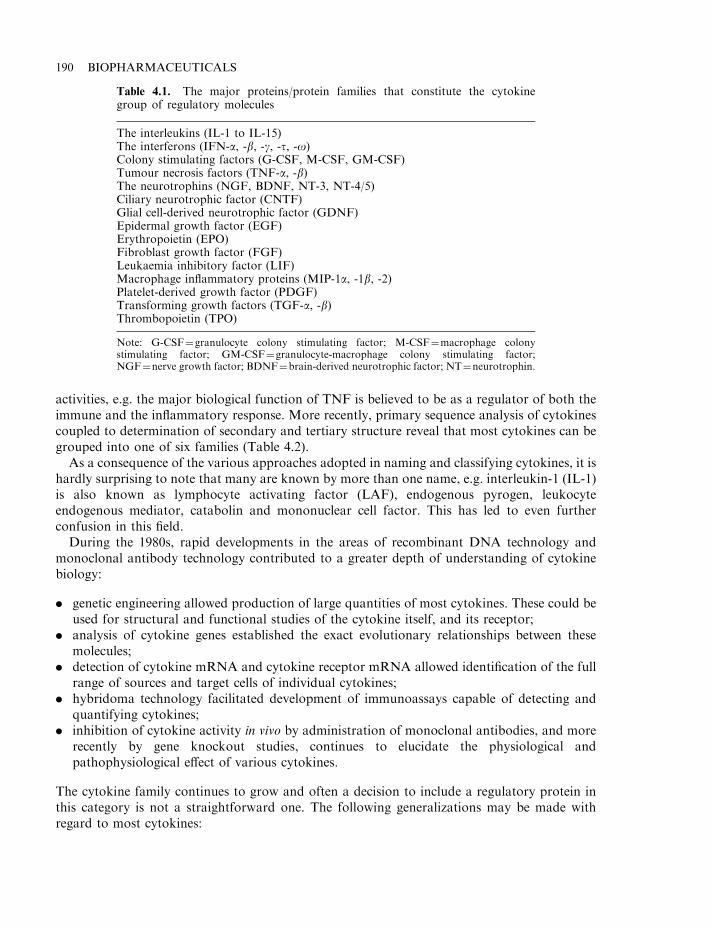
activities, e.g. the major biological function of TNF is believed to be as a regulator of both theimmune and the inflammatory response. More recently, primary sequence analysis of cytokinescoupled to determination of secondary and tertiary structure reveal that most cytokines can begrouped into one of six families (Table 4.2).
As a consequence of the various approaches adopted in naming and classifying cytokines, it ishardly surprising to note that many are known by more than one name, e.g. interleukin-1 (IL-1)is also known as lymphocyte activating factor (LAF), endogenous pyrogen, leukocyteendogenous mediator, catabolin and mononuclear cell factor. This has led to even furtherconfusion in this field.
During the 1980s, rapid developments in the areas of recombinant DNA technology andmonoclonal antibody technology contributed to a greater depth of understanding of cytokinebiology:
. genetic engineering allowed production of large quantities of most cytokines. These could beused for structural and functional studies of the cytokine itself, and its receptor;
. analysis of cytokine genes established the exact evolutionary relationships between thesemolecules;
. detection of cytokine mRNA and cytokine receptor mRNA allowed identification of the fullrange of sources and target cells of individual cytokines;
. hybridoma technology facilitated development of immunoassays capable of detecting andquantifying cytokines;
. inhibition of cytokine activity in vivo by administration of monoclonal antibodies, and morerecently by gene knockout studies, continues to elucidate the physiological andpathophysiological effect of various cytokines.
The cytokine family continues to grow and often a decision to include a regulatory protein inthis category is not a straightforward one. The following generalizations may be made withregard to most cytokines:
190 BIOPHARMACEUTICALS
Table 4.1. The major proteins/protein families that constitute the cytokinegroup of regulatory molecules
The interleukins (IL-1 to IL-15)The interferons (IFN-a, -b, -g, -t, -o)Colony stimulating factors (G-CSF, M-CSF, GM-CSF)Tumour necrosis factors (TNF-a, -b)The neurotrophins (NGF, BDNF, NT-3, NT-4/5)Ciliary neurotrophic factor (CNTF)Glial cell-derived neurotrophic factor (GDNF)Epidermal growth factor (EGF)Erythropoietin (EPO)Fibroblast growth factor (FGF)Leukaemia inhibitory factor (LIF)Macrophage inflammatory proteins (MIP-1a, -1b, -2)Platelet-derived growth factor (PDGF)Transforming growth factors (TGF-a, -b)Thrombopoietin (TPO)
Note: G-CSF¼granulocyte colony stimulating factor; M-CSF¼macrophage colonystimulating factor; GM-CSF¼granulocyte-macrophage colony stimulating factor;NGF¼nerve growth factor; BDNF¼brain-derived neurotrophic factor; NT¼neurotrophin.
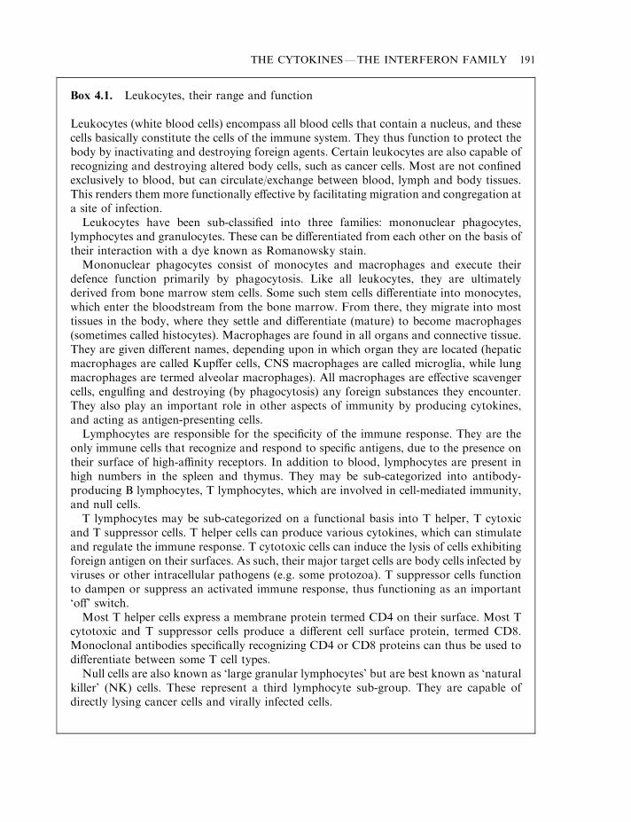
THE CYTOKINES—THE INTERFERON FAMILY 191
Box 4.1. Leukocytes, their range and function
Leukocytes (white blood cells) encompass all blood cells that contain a nucleus, and thesecells basically constitute the cells of the immune system. They thus function to protect thebody by inactivating and destroying foreign agents. Certain leukocytes are also capable ofrecognizing and destroying altered body cells, such as cancer cells. Most are not confinedexclusively to blood, but can circulate/exchange between blood, lymph and body tissues.This renders them more functionally effective by facilitating migration and congregation ata site of infection.
Leukocytes have been sub-classified into three families: mononuclear phagocytes,lymphocytes and granulocytes. These can be differentiated from each other on the basis oftheir interaction with a dye known as Romanowsky stain.
Mononuclear phagocytes consist of monocytes and macrophages and execute theirdefence function primarily by phagocytosis. Like all leukocytes, they are ultimatelyderived from bone marrow stem cells. Some such stem cells differentiate into monocytes,which enter the bloodstream from the bone marrow. From there, they migrate into mosttissues in the body, where they settle and differentiate (mature) to become macrophages(sometimes called histocytes). Macrophages are found in all organs and connective tissue.They are given different names, depending upon in which organ they are located (hepaticmacrophages are called Kupffer cells, CNS macrophages are called microglia, while lungmacrophages are termed alveolar macrophages). All macrophages are effective scavengercells, engulfing and destroying (by phagocytosis) any foreign substances they encounter.They also play an important role in other aspects of immunity by producing cytokines,and acting as antigen-presenting cells.
Lymphocytes are responsible for the specificity of the immune response. They are theonly immune cells that recognize and respond to specific antigens, due to the presence ontheir surface of high-affinity receptors. In addition to blood, lymphocytes are present inhigh numbers in the spleen and thymus. They may be sub-categorized into antibody-producing B lymphocytes, T lymphocytes, which are involved in cell-mediated immunity,and null cells.
T lymphocytes may be sub-categorized on a functional basis into T helper, T cytoxicand T suppressor cells. T helper cells can produce various cytokines, which can stimulateand regulate the immune response. T cytotoxic cells can induce the lysis of cells exhibitingforeign antigen on their surfaces. As such, their major target cells are body cells infected byviruses or other intracellular pathogens (e.g. some protozoa). T suppressor cells functionto dampen or suppress an activated immune response, thus functioning as an important‘off’ switch.
Most T helper cells express a membrane protein termed CD4 on their surface. Most Tcytotoxic and T suppressor cells produce a different cell surface protein, termed CD8.Monoclonal antibodies specifically recognizing CD4 or CD8 proteins can thus be used todifferentiate between some T cell types.
Null cells are also known as ‘large granular lymphocytes’ but are best known as ‘naturalkiller’ (NK) cells. These represent a third lymphocyte sub-group. They are capable ofdirectly lysing cancer cells and virally infected cells.
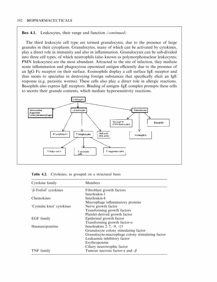
192 BIOPHARMACEUTICALS
Box 4.1. Leukocytes, their range and function (continued)
The third leukocyte cell type are termed granulocytes, due to the presence of largegranules in their cytoplasm. Granulocytes, many of which can be activated by cytokines,play a direct role in immunity and also in inflammation. Granulocytes can be sub-dividedinto three cell types, of which neutrophils (also known as polymorphonuclear leukocytes;PMN leukocytes) are the most abundant. Attracted to the site of infection, they mediateacute inflammation and phagocytose opsonized antigen efficiently due to the presence ofan IgG Fc receptor on their surface. Eosinophils display a cell surface IgE receptor andthus seems to specialize in destroying foreign substances that specifically elicit an IgEresponse (e.g. parasitic worms). These cells also play a direct role in allergic reactions.Basophils also express IgE receptors. Binding of antigen–IgE complex prompts these cellsto secrete their granule contents, which mediate hypersensitivity reactions.
Table 4.2. Cytokines, as grouped on a structural basis
Cytokine family Members
‘b-Trefoil’ cytokines Fibroblast growth factorsInterleukin-1
Chemokines Interleukin-8Macrophage inflammatory proteins
‘Cysteine knot’ cytokines Nerve growth factorTransforming growth factorsPlatelet-derived growth factor
EGF family Epidermal growth factorTransforming growth factor-a
Haematopoietins Interleukins 2–7, -9, -13Granulocyte colony stimulating factorGranulocyte-macrophage colony stimulating factorLeukaemia inhibitory factorErythropoietinCiliary neurotrophic factor
TNF family Tumour necrosis factor-a and -b
. They are very potent regulatory molecules, inducing their characteristic effects at nanomolar(nM) to picomolar (pM) concentrations.
. Most cytokines are produced by a variety of cell types, which may be leukocytes or non-leukocytes, e.g. IL-1 is produced by a wide range of cells including leukocytes such asmonocytes, macrophages, natural killer cells (NK cells), B and T lymphocytes, and non-leukocytes such as smooth muscle cells, vascular endothelial cells (a single layer of cells liningblood vessels), fibroblasts (cells found in connective tissue that produce ground substanceand collagen fibre precursors), astrocytes (non-neural cells found in the central nervoussystem) and chondrocytes (cells embedded in the matrix of cartilage).
. Many cell types can produce more than one cytokine, e.g. lymphocytes produce a wide rangeof ILs, CSFs, TNF, IFN-a and IFN-g. Fibroblasts can produce IL-1, IL-6, IL-8, and IL-11,CSFs, INF-b and TNF.
. Many cytokines play a regulatory role in processes other that immunity and inflammation.Neurotrophic factors such as nerve growth factor (NGF) and brain derived neurotrophicfactor (BDNF) regulate growth, development and maintenance of various neuralpopulations in the central and peripheral nervous system. Erythropoietin stimulates theproduction of red blood cells from erythroid precursors in the bone marrow.
. Most cytokines are pleiotropic, i.e. can effect a variety of cell types. Moreover, the effect acytokine has on one cell type may be the same or different to its effect on a different cell type,e.g. IL-1 can induce fever, hypotension and an acute phase response. Granulocyte-colonystimulating factor (G-CSF) is a growth factor for neutrophils, but is also involved instimulating migration of endothelial cells and growth of haematopoietic cells. IFN-gstimulates activation and growth of T and B lymphocytes, macrophages, NK cells,fibroblasts and endothelial cells. It also displays weak anti-proliferative activity with somecell types.
. Most cytokines are inducable, and are secreted by their producer cell, e.g. induction of IL-2synthesis and release by T-lymphocytes is promoted by binding of IL-1 to its receptor on thesurface of T cells. IFN-as are induced by viral intrusion into the body. In general, potentcytokine inducers include infectious agents, tissue injury and toxic stimuli. The body’s maindefence against such agents, of course, lies with the immune system and inflammation. Uponbinding to target cells, cytokines can often induce the target cell to synthesize and release avariety of additional cytokines.
. In contrast, some cytokines (e.g. some CSFs and erythropoietin) appear to be expressedconstitutively. In yet other instances, cytokines such as platelet-derived growth factor andtransforming growth factor (TGF)-b are stored in cytoplasmic granules and can be rapidlyreleased in response to appropriate stimuli. Other cytokines (mainly ones with growth factoractivity, e.g. TGF-b, fibroblast growth factor and IL-1) are found bound to the extracellularmatrix in connective tissue, bone and skin. These are released, bringing about a biologicalresponse upon tissue injury.
. Many cytokines exhibit redundancy, i.e. two or more cytokines can induce a similarbiological effect. Examples include TNF-a and -b , both of which bind to the same receptorand induce very similar if not identical biological responses. This is also true of theinterferon-a family of proteins and interferon-b, all of which bind the same receptor.
Although all cytokines are polypeptide regulatory factors, not all polypeptide regulatoryfactors are classified as cytokines. Classical polypeptide hormones such as insulin, folliclestimulating hormone (FSH) and growth hormone (GH) are not considered members of the
THE CYTOKINES—THE INTERFERON FAMILY 193
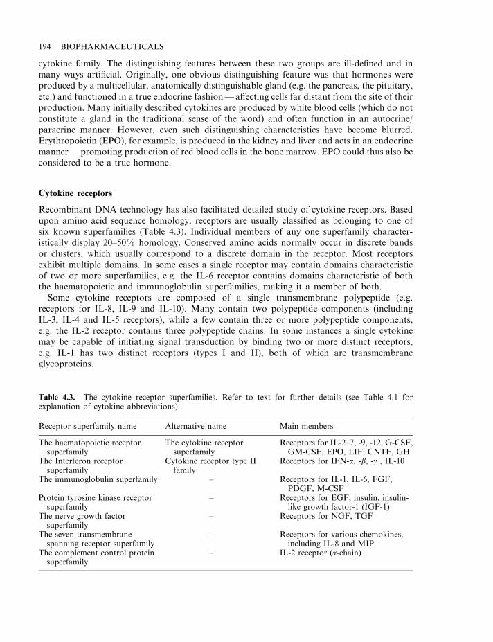
cytokine family. The distinguishing features between these two groups are ill-defined and inmany ways artificial. Originally, one obvious distinguishing feature was that hormones wereproduced by a multicellular, anatomically distinguishable gland (e.g. the pancreas, the pituitary,etc.) and functioned in a true endocrine fashion—affecting cells far distant from the site of theirproduction. Many initially described cytokines are produced by white blood cells (which do notconstitute a gland in the traditional sense of the word) and often function in an autocrine/paracrine manner. However, even such distinguishing characteristics have become blurred.Erythropoietin (EPO), for example, is produced in the kidney and liver and acts in an endocrinemanner—promoting production of red blood cells in the bone marrow. EPO could thus also beconsidered to be a true hormone.
Cytokine receptors
Recombinant DNA technology has also facilitated detailed study of cytokine receptors. Basedupon amino acid sequence homology, receptors are usually classified as belonging to one ofsix known superfamilies (Table 4.3). Individual members of any one superfamily character-istically display 20–50% homology. Conserved amino acids normally occur in discrete bandsor clusters, which usually correspond to a discrete domain in the receptor. Most receptorsexhibit multiple domains. In some cases a single receptor may contain domains characteristicof two or more superfamilies, e.g. the IL-6 receptor contains domains characteristic of boththe haematopoietic and immunoglobulin superfamilies, making it a member of both.
Some cytokine receptors are composed of a single transmembrane polypeptide (e.g.receptors for IL-8, IL-9 and IL-10). Many contain two polypeptide components (includingIL-3, IL-4 and IL-5 receptors), while a few contain three or more polypeptide components,e.g. the IL-2 receptor contains three polypeptide chains. In some instances a single cytokinemay be capable of initiating signal transduction by binding two or more distinct receptors,e.g. IL-1 has two distinct receptors (types I and II), both of which are transmembraneglycoproteins.
194 BIOPHARMACEUTICALS
Table 4.3. The cytokine receptor superfamilies. Refer to text for further details (see Table 4.1 forexplanation of cytokine abbreviations)
Receptor superfamily name Alternative name Main members
The haematopoietic receptorsuperfamily
The cytokine receptorsuperfamily
Receptors for IL-2–7, -9, -12, G-CSF,GM-CSF, EPO, LIF, CNTF, GH
The Interferon receptorsuperfamily
Cytokine receptor type IIfamily
Receptors for IFN-a, -b, -g , IL-10
The immunoglobulin superfamily – Receptors for IL-1, IL-6, FGF,PDGF, M-CSF
Protein tyrosine kinase receptorsuperfamily
– Receptors for EGF, insulin, insulin-like growth factor-1 (IGF-1)
The nerve growth factorsuperfamily
– Receptors for NGF, TGF
The seven transmembranespanning receptor superfamily
– Receptors for various chemokines,including IL-8 and MIP
The complement control proteinsuperfamily
– IL-2 receptor (a-chain)
In many cases where a receptor consists of multiple polypeptides, one of those polypeptides(which will be unique to that receptor) will interact directly with the ligand. The additionalpolypeptide(s), responsible for initiation of signal transduction, may be shared by a number ofreceptors. This explains the pleiotrophy exhibited by many cytokines (Figure 4.1).
Some cytokine receptors can directly initiate signal transduction upon binding of ligand. Inother cases additional elements are involved. For many receptors the exact intracellular eventstriggered upon ligand binding remain unelucidated. However, the molecular details of signaltransduction pathways for others are now understood. Specific details of several cytokinereceptors, along with their modes of signal transduction, will be discussed in the followingchapters.
Cytokines as biopharmaceuticals
Cytokines constitute the single most important group of biopharmaceutical substances. Ascoordinators of the immune and inflammatory response, manipulation of cytokine activity canhave a major influence on the body’s response to a variety of medical conditions.Administration of certain cytokines can enhance the immune response against a wide rangeof infectious agents and cancer cells. Erythropoietin has proved effective in stimulating redblood cell production in anaemic persons. Growth factors have obvious potential in promotingwound healing. Neurotrophic factors display some clinical promise in the abatement of certainneurodegenerative diseases.
THE CYTOKINES—THE INTERFERON FAMILY 195
Figure 4.1. Cytokine receptors usually display a unique cytokine (‘ligand’)-binding domain, but shareadditional receptor components, which are normally responsible for signal transduction. This explains themolecular basis of pleiotropy. IL-6, IL-11 and leukaemia inhibitory factor (LIF), for example, are allcomposed of a distinct ligand-specific binding domain, and a separate subunit (gp130). gp130 is responsiblefor initiating signal transduction and is identical in all three receptors. This is depicted schematically above
A better understanding of the molecular principles underlining cytokine biology may alsoprovide new knowledge-based strategies aimed at defeating certain viral pathogens. Thesepathogens appear to successfully establish an infection, at least in part, by producing specificproteins which thwart the normal cytokine-based immunological response. The Cowpox virus,for example, produces an IL-1-binding protein, while Shope fibroma virus produces a TNF-binding protein. The Epstein–Barr virus, on the other hand, produces a protein homologous toIL-10.
A variety of medical conditions are now believed to be caused or exacerbated byoverproduction of certain cytokines in the body. A variety of pro-inflammatory cytokines,including IL-6 and IL-8 as well as TNF, have been implicated in the pathogenesis of both septicshock and rheumatoid arthritis. Inhibiting the biological activity of such cytokines may provideeffective therapies for such conditions. This may be achieved by administration of monoclonalantibodies raised against the target cytokine, or administration of soluble forms of its receptorwhich will compete with cell surface receptors for cytokine binding.
Some cytokines have already gained approval for medical use. Many more are currentlyundergoing clinical or pre-clinical trials. Over the next few chapters, the biology and potentialmedical applications of these cytokines will be discussed in detail. The remainder of this chapterconcerns itself with the prototypic cytokine family— the interferons.
THE INTERFERONS
Interferons (IFNs) were the first family of cytokines to be discovered. In 1957 researchersobserved that if susceptible animal cells were exposed to a colonizing virus, these cellsimmediately become resistant to attack by other viruses. This resistance was induced by asubstance secreted by virally-infected cells, which was named ‘interferon’ (IFN). Subsequently ithas been shown that most species actually produce a whole range of interferons. Humansproduce at least three distinct classes, IFN-a, IFN-b and IFN-g (Table 4.4). These interferonsare produced by a variety of different cell types, and exhibit a wide range of biological effects,including:
. induction of cellular resistance to viral attack;
. regulation of most aspects of immune function;
. regulation of growth and differentiation of many cell types;
. sustenance of early phases of pregnancy in some animal species.
No one IFN will display all of these biological activities. Effects are initiated by the binding ofthe IFN to its specific cell surface receptor present in the plasma membrane of sensitive cells.
196 BIOPHARMACEUTICALS
Table 4.4. Human interferons (IFNs) and the cells that produce them
Interferonfamily Additional name
No of distinctIFNs in family Producing cells
IFN-a Leukocyte IFN, B cell IFN,lymphoblast IFN
415 Lymphocytes, monocytes, macrophages
IFN-b Fibroblast IFN, IFN-b-1* 1 Fibroblasts, some epithelial cellsIFN-g Immune IFN, T cell IFN 1 T-lymphocytes, NK cells
*Originally a second cytokine was called IFN-b-2, but this was subsequently found to be actually IL-6.
IFN-a and -b display significant amino acid sequence homology (30%), bind to the samereceptor, induce similar biological activities and are acid-stable. For these reasons, IFN-a andIFN-b are sometimes collectively referred to as ‘type I interferons’, or ‘acid-stable interferons’.IFN-g is evolutionarily distinct from the other interferons; it binds to a separate receptor andinduces a different range of biological activities, and it is thus often referred to as type IIinterferon.
Due to their biological activities, most interferons are of actual or likely use in the treatmentof many medical conditions, including:
. augmentation of the immune response against infectious agents (viral, bacterial, protozoan,etc.);
. treatment of some autoimmune conditions;
. treatment of certain cancer types.
Interferons may be detected and quantified using various bioassays or by immunoassaysystems. While such assays were available, subsequent purification, characterization andmedical utilization of IFNs initially proved difficult due to the tiny quantities in which theseregulatory proteins are produced naturally by the body. By the early 1970s, advances in animalcell culture technology, along with the identification of cells producing increased concentrationsof IFNs, made some (mostly IFN-as) available in reasonable quantities. It was not until theadvent of genetic engineering, however, that all IFNs could be produced in quantities sufficientto satisfy demand for both pure and applied purposes.
The biochemistry of interferon-a
For many years after its initial discovery, it was assumed that IFN-a represented a single geneproduct. It is now known that virtually all species produced multiple, closely related IFN-as.Purification studies from the 1970s using high-resolution chromatographic techniques (mainlyion-exchange and gel-filtration chromatography, immunoaffinity chromatography and iso-electric focusing) first elucidated this fact.
In humans at least 24 related genes or pseudo-genes exist, which code for the production of atleast 16 distinct mature IFN-as. These can be assigned to one of two families, types I and II.Humans are capable of synthesizing at least 15 type I IFN-as and a single type II IFN-a.
Most mature type I IFN-as contain 166 amino acids (one contains 165), while type II IFN-a iscomposed of 172 amino acids. All are initially synthesized containing an additional 23 aminoacid signal peptide. Based upon amino acid sequence data, the predicted molecular mass of allIFN-as is in the 19–20 kDa range. SDS–PAGE analysis, however, reveals observed molecularmasses up to 27 kDa. Isoelectric points determined by isoelectric focusing range between 5 and6.5. The heterogeneity observed is most likely due to O-linked glycosylation, although severalIFN-as are not glycosylated. Some IFN-as also exhibit natural heterogeneity due to limitedproteolytic processing at the carboxyl terminus.
Individual IFN-as generally exhibit in excess of 70% amino acid homology with each other.They are rich in leucine and glutamic acid, and display conserved cysteines (usually at positions1, 29, 99 and 139). These generally form two disulphide bonds in the mature molecule. Theirtertiary structures are similar, containing several a-helical segments, but appear devoid of b-sheets.
Individual members of the IFN-a family each have an identifying name. In most cases nameswere assigned by placing a letter after the ‘a’, (i.e. IFN-aA, IFN-aB, etc.). However,
THE CYTOKINES—THE INTERFERON FAMILY 197
some exceptions exist, which contain a number or a number and letter, e.g. IFN-a7, IFN-a8,IFN-a2B. Just to ensure total confusion, several are known by two different names, e.g. IFN-a7is also known as IFN-aJ1.
Interferon-b
IFN-b, normally produced by fibroblasts, was the first interferon to be purified. Humanssynthesize a single IFN-b molecule containing 166 amino acid residues, which exhibits 30%sequence homology to IFN-as. The mature molecule exhibits a single disulphide bond, and is aglycoprotein of molecular mass in excess of 20 kDa. The carbohydrate side-chain is attached viaan N-linked glycosidic bond to asparigine residue 80. The carbohydrate moiety facilitatespartial purification by lectin affinity chromatography. Immunoaffinity chromatography usingmonoclonal antibodies raised against IFN-b, as well as dye affinity chromatography, has alsobeen employed in its purification. IFN-b’s tertiary structure is dominated by five a-helicalsegments, three of which lie parallel to each other, with the remaining two being anti-parallel tothese.
Interferon-c
IFN-g is usually referred to as ‘immune’ interferon. It was initially purified from humanperipheral blood lymphocytes using techniques including concanavalin-A and dye affinitychromatography, as well as gel filtration chromatography. This IFN is produced predominantlyby lymphocytes. Its synthesis by these cells is reduced when they come into contact withpresented antigen. Additional cytokines, including IL-2 and IL-12 can also induce IFN-gproduction under certain circumstances. A single IFN-g gene exists, located on humanchromosome number 12. It displays little evolutionary homology to type I IFN genes. Themature polypeptide contains 143 amino acids with a predicted molecular mass of 17 kDa. SDS–PAGE analysis reveals three bands of molecular mass 16–17, 20 and 25 kDa, arising because ofdifferential glycosylation. The 20 kDa band is glycosylated at asparagine 97, while the 25 kDaspecies is glycosylated at asparagines 25 and 97. In addition, mature IFN-g exhibits naturalheterogeneity at its carboxyl terminus due to proteolytic processing (five truncated forms havebeen identified). The molecule’s tertiary structure consists of six a-helical segments linked bynon-helical regions.
Gel filtration analysis reveals bands of molecular mass 40–70 kDa. These represent dimers(and some multimers) of the IFN-g polypeptide. Its biologically active form appears to be ahomodimer in which the two subunits are associated in an anti-parallel manner.
Interferon signal transduction
All interferons mediate their biological effect by binding to high-affinity cell surface receptors.Binding is followed by initiation of signal transduction, culminating in an altered level ofexpression of several IFN-responsive genes. While both positive and negative regulation exists,thus far positive regulation (upregulation) of gene expression has been studied in greatest detail.
All IFN-stimulated genes are characterized by the upstream presence of an interferon-stimulated response element (ISRE). Signal transduction culminates in the binding of specificregulatory factors to the ISRE, which stimulates RNA polymerase II-mediated transcription of
198 BIOPHARMACEUTICALS
the IFN-sensitive genes. The induced gene products then mediate the anti-viral, immuno-modulatory and other effects characteristically induced by IFNs.
The interferon receptors
The availability of large quantities of purified IFNs facilitates detailed study of the interferonreceptors. Binding studies using radiolabelled interferons can be undertaken, and photoaffinitycrosslinking of labelled IFN to its receptor facilitates subsequent purification of the ligand–receptor complex. Recombinant DNA technology has also facilitated direct cloning of IFNreceptors. Binding studies using radiolabelled type I IFNs reveals that they all compete forbinding to the same receptor, whereas purified IFN-g does not compete. Partial purification ofthe IFN-a receptor was undertaken by a number of means. One approach entailed covalentattachment of radiolabelled IFN-a to the receptor using bifunctional crosslinking agents,followed by purification of the radioactive complex. An alternative approach utilized animmobilized IFN-a ligand for affinity purification. The receptor has also been cloned, and thegene is housed on human chromosome number 21.
Studies have actually revealed two type I IFN receptor polypeptides. Sequence data fromcloning studies place both in the class II cytokine receptor family. Both are transmembrane N-linked glycoproteins. Studies using isolated forms of each show that one polypeptide (called thea/b-receptor) is capable of binding all type I IFNs. The other one (the ab-receptor) is specific forIFN-aB (a specific member of the IFN-a family). Both receptors are present on most cell types.
The IFN-g receptor (the type II receptor) displays a more limited cellular distribution thanthat of the type I receptors (Table 4.5). This receptor is a transmembrane glycoprotein ofmolecular mass 50 kDa, which appears to function as a homodimer. The extracellular IFN-g-binding region consists of approximately 200 amino acid residues folded into two homologousdomains. Initiation of signal transduction also requires the presence of a second transmembraneglycoprotein, known as AF-1 (accessory factor 1), which associates with the extracellular regionof the receptor.
The intracellular events triggered upon binding of type I or II IFNs to their respectivereceptors are quite similar. The sequence of events, known as the JAK–STAT pathway, hasbeen elucidated only in the last few years. It has quickly become apparent that this pathwayplays a prominent role in mediating signal transduction, not only for IFN but also for manycytokines.
The JAK–STAT pathway
Cytokine receptors can be divided into two groups; those whose intracellular domains exhibitintrinsic protein tyrosine kinase (PTK) activity, and those whose intracellular domains are
THE CYTOKINES—THE INTERFERON FAMILY 199
Table 4.5. Cell types that display an IFN-g receptor on their surfaces
Haematopoietic cells T lymphocytesB lymphocytesMacrophagesPolymorphonuclear leukocytesPlatelets
Somatic cells Endothelial cellsEpithelial cellsVarious tumour cells
devoid of such activity. Many of the latter group of receptors, however, activate intracellularsoluble PTKs upon ligand binding.
Janus kinases (JAKs) represent a recently discovered family of PTKs that seem to play acentral role in mediating signal transduction of many cytokines, and probably many non-cytokine regulatory molecules. These enzymes harbour two potential active sites and were thusnamed after Janus, the Roman god with two faces. It is likely that only one of those ‘active’ sitesis functional. Four members of the JAK family have been best characterized to date: Jak 1, Jak2, Jak 3 and Tyk 2. They all exhibit molecular masses in the region of 130 kDa andapproximately 40% amino acid sequence homology. They appear to be associated with thecytoplasmic domain of many cytokine receptors, but remain catalytically inactive until bindingof the cytokine to the receptor (Figure 4.2).
In most instances, ligand binding appears to promote receptor dimerization, bringing theirassociated JAKs into close proximity (Figure 4.2). The JAKs then phosphorylate—and henceactivate—each other (transphosphorylation). The activated kinases subsequently phosphor-ylate specific tyrosine residues on the receptor itself. This promotes direct association betweenone or more members of a family of cytoplasmic proteins (STATs) and the receptor. Oncedocked at the receptor surface, the STATs are in turn phosphorylated (and hence activated) bythe JAKs (Figure 4.2). As described below, activated STATs then translocate to the nucleus anddirectly regulate expression of IFN and other cytokine-sensitive genes.
STAT stands for ‘signal transducers and activators of transcription’. As the name suggests,these proteins (a) form an integral part of cytoplasmic signal transduction initiated by certainregulatory molecules and (b) activate transcription of specific genes in the nucleus. Thus far, atleast six distinct mammalian STATs (STAT 1–STAT 6) have been identified, in size rangingfrom 84–113 kDa. Some may be differentially spliced, increasing the number of functionalproteins in the family, e.g. STAT 1 exists in two forms: STAT 1a contains 750 amino acidresidues and exhibits a molecular mass of 91 kDa (it is sometimes called STAT 91); STAT 1b is asplicing variant of the same gene product, which lacks the last 38 amino acid residues at the Cterminal of the protein and exhibits a molecular mass of 84 kDa (hence it is sometimes calledSTAT 84). Similar variants have been identified for STATs 3 and 5. STATs have also beenlocated in non-mammalian species, such as the fruit fly. All STATs exhibit significant sequencehomology and are composed of a number of functional domains (Figure 4.3). The SH2 domainfunctions to bind phosphotyrosine, thus docking the STAT at the activated receptor surface. Asdetailed below, this domain is also required for STAT interaction with JAKs (which thenphosphorylates the STAT) and to promote subsequent dimerization of the STATs. An essentialtyrosine is located towards the STAT C-terminus (around residue 700), which in turn is thenphosplorylated by PTK. STATs are differentially distributed in various cells/tissues. STATs 1, 2and 3 seem to be present in most cell types, allbeit at varying concentrations. Tissue distributionof STAT 4 and 5 is more limited.
Not surprisingly, different ligands activate different members of the STAT family (Table 4.6).Some, e.g. STAT 1 and 3, are activated by many ligands, while others respond to far fewerligands, e.g. STAT 2 appears to be activated only by type I IFNs.
STAT phosphorylation ensures its binding to the receptor, with subsequent disengagementfrom the receptor in dimeric form. STAT dimerization is believed to involve intermolecularassociations between the SH2 domain of one STAT and phosphotyrosine of its partner.Dimerization appears to be an essential prerequisite for DNA binding. Dimers may consist oftwo identical STATs, but STAT 1–STAT 2 and STAT 1–STAT 3 heterodimers are alsofrequently formed in response to certain cytokines. The STAT dimers then translocate to the
200 BIOPHARMACEUTICALS
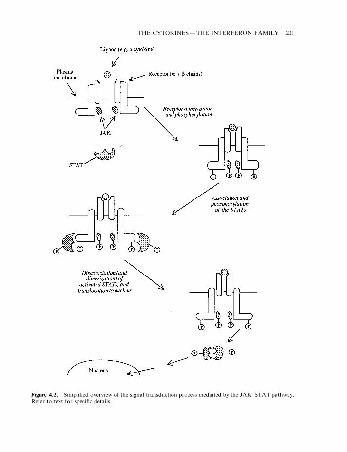
THE CYTOKINES—THE INTERFERON FAMILY 201
Figure 4.2. Simplified overview of the signal transduction process mediated by the JAK–STAT pathway.Refer to text for specific details
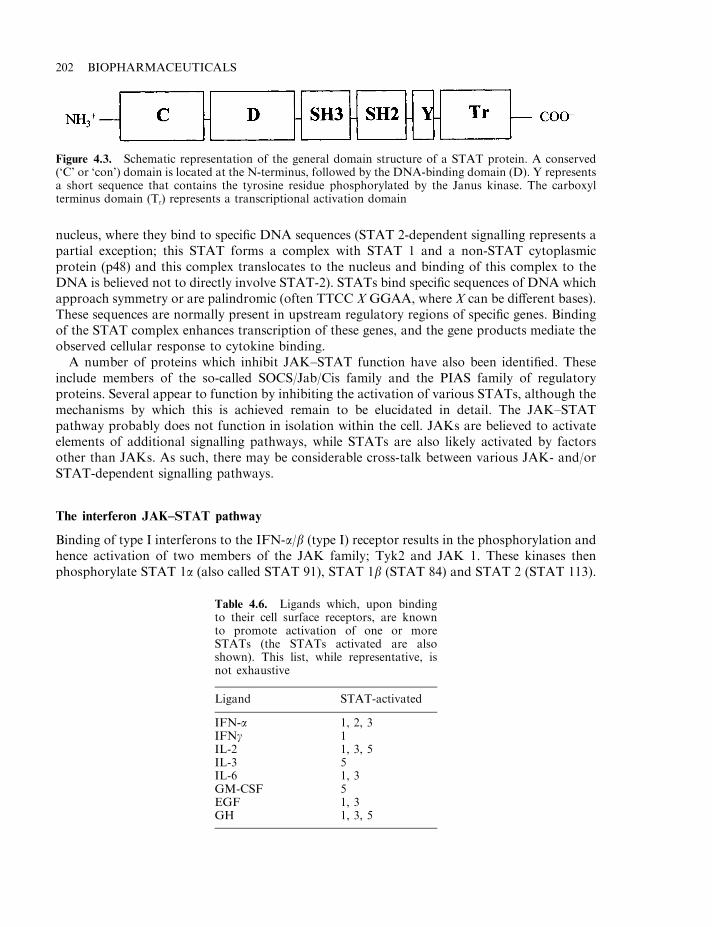
nucleus, where they bind to specific DNA sequences (STAT 2-dependent signalling represents apartial exception; this STAT forms a complex with STAT 1 and a non-STAT cytoplasmicprotein (p48) and this complex translocates to the nucleus and binding of this complex to theDNA is believed not to directly involve STAT-2). STATs bind specific sequences of DNA whichapproach symmetry or are palindromic (often TTCC X GGAA, where X can be different bases).These sequences are normally present in upstream regulatory regions of specific genes. Bindingof the STAT complex enhances transcription of these genes, and the gene products mediate theobserved cellular response to cytokine binding.
A number of proteins which inhibit JAK–STAT function have also been identified. Theseinclude members of the so-called SOCS/Jab/Cis family and the PIAS family of regulatoryproteins. Several appear to function by inhibiting the activation of various STATs, although themechanisms by which this is achieved remain to be elucidated in detail. The JAK–STATpathway probably does not function in isolation within the cell. JAKs are believed to activateelements of additional signalling pathways, while STATs are also likely activated by factorsother than JAKs. As such, there may be considerable cross-talk between various JAK- and/orSTAT-dependent signalling pathways.
The interferon JAK–STAT pathway
Binding of type I interferons to the IFN-a/b (type I) receptor results in the phosphorylation andhence activation of two members of the JAK family; Tyk2 and JAK 1. These kinases thenphosphorylate STAT 1a (also called STAT 91), STAT 1b (STAT 84) and STAT 2 (STAT 113).
202 BIOPHARMACEUTICALS
Figure 4.3. Schematic representation of the general domain structure of a STAT protein. A conserved(‘C’ or ‘con’) domain is located at the N-terminus, followed by the DNA-binding domain (D). Y representsa short sequence that contains the tyrosine residue phosphorylated by the Janus kinase. The carboxylterminus domain (Tr) represents a transcriptional activation domain
Table 4.6. Ligands which, upon bindingto their cell surface receptors, are knownto promote activation of one or moreSTATs (the STATs activated are alsoshown). This list, while representative, isnot exhaustive
Ligand STAT-activated
IFN-a 1, 2, 3IFNg 1IL-2 1, 3, 5IL-3 5IL-6 1, 3GM-CSF 5EGF 1, 3GH 1, 3, 5
The three activated STATs disengage from the receptor and bind to the cytoplasmic protein,p48. This entire complex translocates to the nucleus, where it interacts directly with upstreamregulatory regions of IFN-sensitive genes. These nucleotide sequences are termed interferon-stimulated response elements (ISREs). This induces/augments expression of specific genes, asdiscussed later.
The essential elements of the signal transduction pathway elicited by IFN-g is even morestraightforward. IFN-g binding to the type II receptor induces receptor dimerization withconsequent activation of JAK 1 and JAK 2. The JAKs phosphorylate the receptor andsubsequently the associated STAT 1a. STAT 1a is then released and forms a homodimer whichtranslocates to the nucleus. It regulates expression of IFN-g-sensitive genes by binding to aspecific upstream regulatory sequence of the gene (the IFN-g-activated sequence, GAS). ThePIAS-1 protein appears to play an inhibitory role in this pathway. By complexing with(phosphorylated) STAT 1 proteins, it inhibits DNA binding and transactivation.
The biological effects of interferons
Interferons induce a wide range of biological effects. Generally, type I IFNs induce similareffects, which are distinct from the effects induced by IFN-g. The most pronounced effect of typeI IFNs relates to their anti-viral activity, as well as their anti-proliferative effect on various celltypes, including certain tumour cell types. Anti-tumour effects are likely due not only to a directanti-proliferative effect on the tumour cells themselves but also due to the ability of type I IFNsto increase natural killer (NK) cell and T cytotoxic cell activity. These cells can recognize anddestroy cancer cells.
Not all type I IFNs induce exactly the same range of responses, and the anti-viral:anti-proliferative activity ratio differs from one type I IFN to another. As all bind the same receptor,the molecular basis by which variation in biological activities is achieved, is poorly understoodas yet.
IFN-g exhibits at best weak anti-viral and anti-proliferative activity. When co-administeredwith type I IFNs, however, it potentates these IFN-a/b activities. IFN-g is directly involved inregulating most aspects of the immune and inflammatory responses. It promotes activation,growth and differentiation of a wide variety of cell types involved in these physiologicalprocesses (Table 4.7).
IFN-g represents the main macrophage activating factor, thus enhancing macrophage-mediated effects, including:
THE CYTOKINES—THE INTERFERON FAMILY 203
Table 4.7. Cell types participating in the immune,inflammatory or other responses whose activation,growth and differentiation is promoted by IFN-g
Macrophages/monocytesPolymorphonuclear neutrophilsT lymphocytesB lymphocytesNK cellsFibroblastsEndothelial cells
. destruction of invading microorganisms;
. destruction of intracellular pathogens;
. tumour cell cytotoxicity;
. increased major histocompatibility complex (MHC) antigen expression, leading to enhancedactivation of lymphocytes via antigen presentation.
Binding of IFN-g to its surface receptor on polymorphonuclear neutrophils induces increasedexpression of the gene coding for a neutrophil cell surface protein capable of binding the Fcportion (i.e. the constant region: see also Box 10.2) of immunoglobulin (IgG). This greatlyincreases the phagocytotic and cytotoxic activities of these cells.
IFN-g also directly modulates the immune response by affecting growth, differentiation andfunction of both T and B lymphocytes. These effects are quite complex, and are often influencedby additional cytokines. IFN-g acts as a growth factor in an autocrine manner for some T cellsub-populations, while it is capable of suppressing growth of other T cell types. It appears tohave an inhibitory effect on development of immature B lymphocyte populations but maysupport mature B cell survival. It can both upregulate and downregulate antibody productionunder various circumstances.
All IFNs promote increased surface expression of class I MHC antigens. Class II MHCantigen expression is stimulated mainly by IFN-g (MHC proteins are found on the surface ofvarious cell types. They play an essential role in triggering an effective immune response, notonly against foreign antigen but also against altered host cells). While many IFNs promotesynergistic effects, some instances are known where two or more IFNs can oppose each other’sbiological activities. IFN-aJ, for example, can inhibit the IFN-aA-mediated stimulation of NKcells.
The molecular basis by which IFNs promote their characteristic effects, particularly anti-viralactivity, is understood at least in part. IFN stimulation of the JAK–STAT pathway inducessynthesis of at least 30 different gene products, many of which cooperate to inhibit viralreplication. These anti-viral gene products are generally enzymes, the most important of whichare 20-50 oligoadenylate synthetase (2, 5-An synthetase) and the eIF-2a protein kinase.
These intracellular enzymes remain in an inactive state after their initial induction. They areactivated only when the cell comes under viral attack, and their activation can inhibit viralreplication in that cell. The 20-50 An, synthetase acts in concert with two additional enzymes—an endoribonuclease and a phosphodiesterase— to promote and regulate the anti-viral state(Figure 4.4).
Several active forms of the synthetase seem to be inducable in human cells; 40 kDa and46 kDa variants have been identified which differ only in their carboxyl terminus ends. They areproduced as a result of differential splicing of mRNA transcribed from a single gene found onchromosome 11. A larger 85–100 kDa form of the enzyme has been detected, which mayrepresent a heterodimer composed of the 40 and 46 kDa variants.
The synthetase is activated by double-stranded RNA (dsRNA). Although not normallypresent in human cells, dsRNA is often associated with commencement of replication of certainviruses. The activated enzyme catalyses the synthesis of oligonucleotides of varying length inwhich the sole base is adenine (20-50An). This oligonucleotide differs from oligonucleotidespresent naturally in the cell in that the phosphodiester bonds present are 20-50 bonds (Figure4.5). The level of synthesis and average polymer length of the oligonucleotide products appearto depend upon the exact inducing IFN type, as well as the growth state of the cell.
204 BIOPHARMACEUTICALS
THE CYTOKINES—THE INTERFERON FAMILY 205
Figure 4.4. Outline of how the 20–50 synthetase system promotes its anti-viral effect. The 20–50
phosphodiesterase ‘off switch’ is omitted for clarity. Refer to text for details
The sole biochemical function of 20-50An (and hence 20-50 An synthetase) appears to be as anactivator of a dormant endo-RNase, which is expressed constitutively in the cell. This RNase,known as RNase L or RNase F, cleaves all types of single-stranded RNA (ssRNA). Thisinhibits production of both viral and cellular proteins, thus paralizing viral replication.Presumably cellular destruction of the invading ssRNA will be accompanied by destruction ofany additional viral components. Removal of dsRNA would facilitate deactivation of the endoRNase, allowing translation of cellular mRNA to resume. A 20-50-phosphodiesterase representsa third enzymatic component of this system. It functions as an off switch, as it rapidly degrades
206 BIOPHARMACEUTICALS
Figure 4.5. (a) Structural detail of the 20–50 oligonucleotides (20–50An) generated by 20–50 An synthetase.Compare the 20–50 phosphodiester linkages with the 30–50 linkages characteristic of normal cellularoligonucleotides, such as mRNA (b)
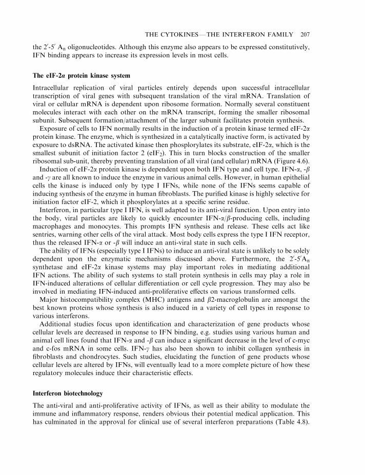
the 20-50 An oligonucleotides. Although this enzyme also appears to be expressed constitutively,IFN binding appears to increase its expression levels in most cells.
The eIF-2a protein kinase system
Intracellular replication of viral particles entirely depends upon successful intracellulartranscription of viral genes with subsequent translation of the viral mRNA. Translation ofviral or cellular mRNA is dependent upon ribosome formation. Normally several constituentmolecules interact with each other on the mRNA transcript, forming the smaller ribosomalsubunit. Subsequent formation/attachment of the larger subunit facilitates protein synthesis.
Exposure of cells to IFN normally results in the induction of a protein kinase termed eIF-2aprotein kinase. The enzyme, which is synthesized in a catalytically inactive form, is activated byexposure to dsRNA. The activated kinase then phosplorylates its substrate, eIF-2a, which is thesmallest subunit of initiation factor 2 (eIF2). This in turn blocks construction of the smallerribosomal sub-unit, thereby preventing translation of all viral (and cellular) mRNA (Figure 4.6).
Induction of eIF-2a protein kinase is dependent upon both IFN type and cell type. IFN-a, -band -g are all known to induce the enzyme in various animal cells. However, in human epithelialcells the kinase is induced only by type I IFNs, while none of the IFNs seems capable ofinducing synthesis of the enzyme in human fibroblasts. The purified kinase is highly selective forinitiation factor eIF-2, which it phosplorylates at a specific serine residue.
Interferon, in particular type I IFN, is well adapted to its anti-viral function. Upon entry intothe body, viral particles are likely to quickly encounter IFN-a/b-producing cells, includingmacrophages and monocytes. This prompts IFN synthesis and release. These cells act likesentries, warning other cells of the viral attack. Most body cells express the type I IFN receptor,thus the released IFN-a or -b will induce an anti-viral state in such cells.
The ability of IFNs (especially type I IFNs) to induce an anti-viral state is unlikely to be solelydependent upon the enzymatic mechanisms discussed above. Furthermore, the 20-50An
synthetase and eIF-2a kinase systems may play important roles in mediating additionalIFN actions. The ability of such systems to stall protein synthesis in cells may play a role inIFN-induced alterations of cellular differentiation or cell cycle progression. They may also beinvolved in mediating IFN-induced anti-proliferative effects on various transformed cells.
Major histocompatibility complex (MHC) antigens and b2-macroglobulin are amongst thebest known proteins whose synthesis is also induced in a variety of cell types in response tovarious interferons.
Additional studies focus upon identification and characterization of gene products whosecellular levels are decreased in response to IFN binding, e.g. studies using various human andanimal cell lines found that IFN-a and -b can induce a significant decrease in the level of c-mycand c-fos mRNA in some cells. IFN-g has also been shown to inhibit collagen synthesis infibroblasts and chondrocytes. Such studies, elucidating the function of gene products whosecellular levels are altered by IFNs, will eventually lead to a more complete picture of how theseregulatory molecules induce their characteristic effects.
Interferon biotechnology
The anti-viral and anti-proliferative activity of IFNs, as well as their ability to modulate theimmune and inflammatory response, renders obvious their potential medical application. Thishas culminated in the approval for clinical use of several interferon preparations (Table 4.8).
THE CYTOKINES—THE INTERFERON FAMILY 207
Ongoing clinical trials are likely to expand considerably the medical uses of these regulatorymolecules over the next few years.
While at least some of these potential therapeutic applications were appreciated as far back asthe late 1950s, initial therapeutic application was rendered impractical due to the extremely low
208 BIOPHARMACEUTICALS
Figure 4.6. Outline of how the eIF-2a protein kinase system promotes an anti-viral effect
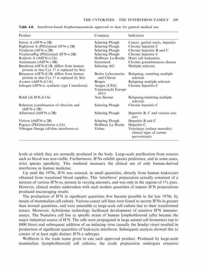
levels at which they are normally produced in the body. Large-scale purification from sourcessuch as blood was non-viable. Furthermore, IFNs exhibit species preference, and in some cases,strict species specificity. This rendered necessary the clinical use of only human-derivedinterferons in human medicine.
Up until the 1970s, IFN was sourced, in small quantities, directly from human leukocytesobtained from transfused blood supplies. This ‘interferon’ preparation actually consisted of amixture of various IFN-as, present in varying amounts, and was only in the regions of 1% pure.However, clinical studies undertaken with such modest quantities of impure IFN preparationsproduced encouraging results.
The production of IFN in significant quantities first became possible in the late 1970s, bymeans of mammalian cell culture. Various cancer cell lines were found to secrete IFNs in greaterthan normal quantities, and were amenable to large-scale cell culture due to their transformednature. Moreover, hybridoma technology facilitated development of sensitive IFN immuno-assays. The Namalwa cell line (a specific strain of human lymphoblastoid cells) became themajor industrial source of IFN. The cells were propagated in large animal cell fermenters (up to8000 litres) and subsequent addition of an inducing virus (usually the Sendai virus) resulted inproduction of significant quantities of leukocyte interferon. Subsequent analysis showed this toconsist of at least eight distinct IFN-a subtypes.
Wellferon is the trade name given to one such approved product. Produced by large-scalemammalian (lymphoblastoid) cell cultures, the crude preparation undergoes extensive
THE CYTOKINES—THE INTERFERON FAMILY 209
Table 4.8. Interferon-based biopharmaceuticals approved to date for general medical use
Product Company Indication
Intron A (rIFN-a-2B) Schering Plough Cancer, genital warts, hepatitisPegIntron A (PEGylated rIFN-a-2B) Schering Plough Chronic hepatitis CViraferon (rIFN-a-2B) Schering Plough Chronic hepatitis B and CViraferonPeg (PEGylated rIFN-a-2B) Schering Plough Chronic hepatitis CRoferon A (rhIFN-a-2A) Hoffman–La-Roche Hairy cell leukaemiaActimmune (rhIFN-g-IB) Genentech Chronic granulomatous diseaseBetaferon (rIFN-b-1B, differs from humanprotein in that Cys 17 is replaced by Ser)
Schering AG Multiple sclerosis
Betaseron (rIFN-b-1B, differs from humanprotein in that Cys 17 is replaced by Ser)
Berlex Laboratoriesand Chiron
Relapsing, remitting multiplesclerosis
Avonex (rhIFN-b-1A) Biogen Relapsing multiple sclerosisInfergen (rIFN-a, synthetic type I interferon) Amgen (USA)
Yamanouchi Europe(EU)
Chronic hepatitis C
Rebif (rh IFN-b-1A) Ares Serono Relapsing/remitting multiplesclerosis
Rebetron (combination of ribavirin andrhIFN-a-2B)
Schering Plough Chronic hepatitis C
Alfatronol (rhIFN-a-2B) Schering Plough Hepatitis B, C and various can-cers
Virtron (rhIFN-a-2B) Schering Plough Hepatitis B and CPegasys (PEGinterferon a-2A) Hoffman La Roche Hepatitis CVibragen Omega (rFeline interferon-o) Virbac Veterinary (reduce mortality/
clinical signs of canineparvovirusis)
chromatographic purification, including two immuno-affinity steps. The final product containsnine IFN-a subtypes.
Recombinant DNA technology also facilitated the production of IFNs in quantities largeenough to satisfy potential medical needs. The 1980s witnessed the cloning and expression ofmost IFN genes in a variety of expression systems. The expression of specific genes obviouslyyielded a product containing a single IFN (sub)-type.
Most IFNs have now been produced in a variety of expression systems including E. coli,fungi, yeast and also some mammalian cell lines such as Chinese hamster ovary (CHO) celllines and monkey kidney cell lines. Most IFNs currently in medical use are recombinanthuman (rh) products produced in E. coli. The inability of E. coli to carry out post-translational modifications is in most instances irrelevant, as the majority of human IFN-as, aswell as IFN-b, are not normally glycosylated. While IFN-g is glycosylated, the E. coli-derivedunglycosylated form displays a biological activity identical to the native human protein.
The production of IFN in recombinant microbial systems obviously means that any finalproduct contaminants will be microbial in nature. A high degree of purification is thus requiredto minimize the presence of such non-human substances. Most IFN final product preparationsare in the region of 99% pure. Such purity levels are achieved by extensive chromatographicpurification. While standard techniques such as gel filtration and ion-exchange are extensivelyused, reported IFN purification protocols have also entailed the use of various affinitytechniques, e.g. using anti-IFN monoclonal antibodies, reactive dyes or lectins (for glycosylatedIFNs). Hydroxyapatite, metal-affinity and hydrophobic interaction chromatography have alsobeen employed in purification protocols. Many production columns are run in fast protein orhigh-performance liquid chromotography (FPLC or HPLC) format, yielding improved andfaster resolution. Immunoassays are used to detect and quantify the IFNs during downstreamprocessing, although the product (particularly the finished product) is also usually subjected to arelevant bioassay. The production and medical uses of selected IFNs are summarized in thesections below.
Production and medical uses of IFN-a
Clinical studies undertaken in the late 1970s, with multi-component, impure IFN-apreparations, clearly illustrated the therapeutic potential of such interferons as an anti-canceragent. These studies found that IFN-a could induce regression of tumours in significantnumbers of patients suffering from breast cancer, certain lymphomas (malignant tumour of thelymph nodes) and multiple myeloma (malignant disease of the bone marrow). The IFNpreparations could also delay recurrence of tumour growth after surgery in patients sufferingfrom osteogenic sarcoma (cancer of connective tissue involved in bone formation).
The first recombinant IFN to become available for clinical studies was IFN-a2A, in 1980.Shortly afterwards the genes coding for additional IFN-as were cloned and expressed, allowingadditional clinical studies. The anti-viral, anti-tumour and immuno-modulatory properties ofthese IFNs assured their approval for a variety of medical uses. rhIFN-as manufactured/marketed by a number of companies (Table 4.8) are generally produced in E. coli.
Clinical trials have shown the recombinant IFNs to be effective in the treatment of variouscancer types, with rhIFN-a2A and -a2B both approved for treatment of hairy cell leukaemia.This is a rare B lymphocyte neoplasm for which few effective treatments were previouslyavailable. Administration of the rIFNs promotes significant regression of the cancer in up to90% of patients.
210 BIOPHARMACEUTICALS
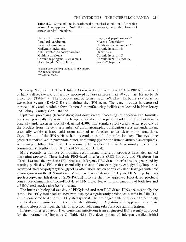
Schering Plough’s rhIFN a-2B (Intron A) was first approved in the USA in 1986 for treatmentof hairy cell leukaemia, but is now approved for use in more than 50 countries for up to 16indications (Table 4.9). The producer microorganism is E. coli, which harbours a cytoplasmicexpression vector (KMAC-43) containing the IFN gene. The gene product is expressedintracellularly and in soluble form. Intron A manufacturing facilities are located in New Jerseyand Brinny, County Cork, Ireland.
Upstream processing (fermentation) and downstream processing (purification and formula-tion) are physically separated by being undertaken in separate buildings. Fermentation isgenerally undertaken in specially designed 42 000 litre stainless steel vessels. After recovery ofthe product from the cells, a number of chromatographic purification steps are undertaken,essentially within a large cold room adapted to function under clean room conditions.Crystallization of the IFN-a-2B is then undertaken as a final purification step. The crystallineproduct is redissolved in phosphate buffer, containing glycine and human albumin as excipients.After aseptic filling, the product is normally freeze-dried. Intron A is usually sold at fivecommercial strengths (3, 5, 10, 25 and 50 million IU/vial).
More recently, a number of modified recombinant interferon products have also gainedmarketing approval. These include PEGylated interferons (PEG IntronA and Viraferon Peg(Table 4.8) and the synthetic IFN product, Infergen). PEGylated interferons are generated byreacting purified a-IFNs with a chemically activated form of polyethylene glycol (Chapter 3).Activated methoxypolyethylene glycol is often used, which forms covalent linkages with freeamino groups on the IFN molecule. Molecular mass analysis of PEGylated IFNs (e.g. by massspectroscopy, gel filtration or SDS–PAGE) indicate that the approved PEGylated productsconsist predominantly of monoPEGylated IFN molecules, with small amounts of both free anddiPEGylated species also being present.
The intrinsic biological activity of PEGylated and non-PEGylated IFNs are essentially thesame. The PEGylated product, however, displays a significantly prolonged plasma half-life (13–25 h as compared to 4 h for unPEGylated species). The prolonged half-life appears to be mainlydue to slower elimination of the molecule, although PEGylation also appears to decreasesystemic absorption from the site of injection following subcutaneous administration.
Infergen (interferon acon-1, or consensus interferon) is an engineered IFN recently approvedfor the treatment of hepatitis C (Table 4.8). The development of Infergen entailed initial
THE CYTOKINES—THE INTERFERON FAMILY 211
Table 4.9. Some of the indications (i.e. medical conditions) for whichintron A is approved. Note that the vast majority are either forms ofcancer or viral infections
Hairy cell leukaemia Laryngeal papillomatosis*Renal cell carcinoma Mycosis fungoides**Basal cell carcinoma Condyloma acuminata ***Malignant melanoma Chronic hepatitis BAIDS-related Kaposi’s sarcoma Hepatitis CMultiple myeloma Chronic hepatitis DChronic myelogenous leukaemia Chronic hepatitis, non-A,Non-Hodgkin’s lymphoma non-B/C hepatitis
*Benign growths (papillomas) in the larynx.**A fungal disease.***Genital warts.
sequence comparisons between a range of IFN-as. The product’s amino acid sequence reflectsthe most frequently occurring amino acid residue in each corresponding position of these nativeIFNs. A DNA sequence coding for the product was synthesized and inserted into E. coli. Therecombinant product accumulates intracellularly as inclusion bodies (Chapter 3).
Large-scale manufacture entails an initial fermentation step. After harvest, the E. coli cells arehomogenized and the inclusion bodies recovered via centrifugation. After solubilization and re-folding, the interferon is purified to homogeneity by a combination of chromatographic steps.The final product is formulated in the presence of a phosphate buffer and sodium chloride. It ispresented as a 30 mg/ml solution in glass vials and displays a shelf-life of 24 months when storedat 2–88C. When compared on a mass basis, the synthetic interferon displays higher antiviral,antiproliferative and cytokine-inducing activity than do native type I interferons.
Ongoing clinical trials continue to assess the efficacy of rIFN preparations in treating avariety of cancers. Some trials suggest that treatments are most effective when administered inthe early stages of cancer development. rhIFN-as have also proved effective in the treatment ofvarious viral conditions, most notably viral hepatitis. Hepatitis refers to an inflammation of theliver. It may be induced by toxic substances, immunological abnormalities or by viruses(infectious hepatitis). The main viral causative agents are:
. hepatitis A virus (hepatitis A);
. hepatitis B virus (hepatitis B, i.e. classical serum hepatitis);
. hepatitis C virus (hepatitis C, formerly known as classical non-A, non-B hepatitis);
. hepatitis D virus (hepatitis D, i.e. delta hepatitis);
. hepatitis E virus (hepatitis E, i.e. endemic non-A, non-B hepatitis);
. hepatitis GB agent.
This disease may be acute (rapid onset, often accompanied by severe symptoms but of briefduration) or chronic (very long duration).
Hepatitis A is common, particularly in areas of poor sanitation, and is transmitted by food ordrink contaminated by a sufferer/carrier. Clinical symptoms include jaundice, and are usuallymild. A full recovery is normally recorded. Hepatitis B is transmitted via infected blood.Symptoms of acute hepatitis B include fever, chill, weakness and jaundice. Most sufferersrecover from such infection, although acute liver failure and death sometimes occur; 5–10% ofsufferers go on to develop chronic hepatitis B. Acute hepatitis C is usually mild andasymptomatic. However, up to 90% of infected persons go on to develop a chronic form of thecondition. Hepatitis D is unusual in that it requires the presence of hepatitis B in order toreplicate. It thus occurs in some persons concomitantly infected with hepatitis B virus. Itsclinical symptoms are usually severe, and can occur in acute or chronic form.
Chronic forms of hepatitis (in particular B, C and D) can result in liver cirrhosis and/orhepatocellular carcinoma. This occurs in up to 20% of chronic hepatitis B sufferers, and up to30% of chronic hepatitis C sufferers. The scale of human suffering caused by hepatitis on aworldwide basis is enormous. Approximately 5% of the global population suffer from chronichepatitis B. An estimated 50 million new infections occur each year. Over 1.5 million of the 300million carriers worldwide die annually from liver cirrhosis and hepatocellular carcinoma.
IFN-a2B is now approved in the USA for the treatment of hepatitis B and C. Clinical studiesundertaken with additional IFN-a preparations indicate their effectiveness in managing suchconditions, and several such products are also likely to gain regulatory approval.
IFN-a2A, when administered three times weekly for several weeks/months, was foundeffective in treating several forms of hepatitis. Remission is observed in 30–45% of patients
212 BIOPHARMACEUTICALS
suffering from chronic hepatitis B, while a complete recovery is noted in up to 20% of cases. Thedrug induces sustained remission in up to 30% of patients suffering from chronic hepatitis C,but can ease clinical symptoms of this disease in up to 75% of such patients. Ongoing studiesalso indicate its efficacy in treating chronic hepatitis D, although relapse is frequently observedupon cessation of therapy. The drug is normally administered by the intramuscular (i.m.) orsubcutaneous (s.c.—directly beneath the skin) injection. Peak plasma concentrations of theIFN are observed more quickly upon i.m. injection (4 h versus 7.5 h). The elimination half-life ofthe drug ranges from 2.5 to 3.5 h.
IFN-a preparations have also proved efficacious in the treatment of additional viral-inducedmedical conditions. rhIFN-a2B, as well as IFN-an3 are already approved for the treatment ofsexually transmitted genital warts, caused by a human papilloma virus. While this condition isoften unresponsive to various additional therapies, direct injection of the IFN into the wartcauses its destruction in up to 70% of patients. Another member of the papilloma virus family isassociated with the development of benign growths in the larynx (laryngeal papillomatosis).This condition can be successfully treated with IFN-a preparations, as can certain papilloma-related epithelial cell cancers, such as cervical intraepithelial neoplasm (epithelial cells are thosethat cover all external surfaces of the body, and line hollow structures—with the exception ofblood and lymph vessels). IFN-a’s ability to combat a range of additional virally-induceddiseases, including AIDS, is currently being appraised in clinical trials.
Medical uses of IFN-b
RhIFN-b has found medical application in the treatment of relapsing–remitting multiplesclerosis (MS), a chronic disease of the nervous system. This disease normally presents in youngadults (more commonly women) aged 20–40. It is characterized by damage to the myelin sheath,which surrounds neurons of the central nervous system, and in this way compromises neuralfunction. Clinical presentations include shaky movement, muscle weakness, paralysis, defects inspeech, vision and other higher mental functions. The most predominant form of the conditionis characterized by recurrent relapses followed by remission. MS appears to be an autoimmunedisease, in which elements of immunity (mainly lymphocytes and macrophages) cooperate in thedestruction of the myelin. What triggers onset of the condition is unknown, although geneticand environmental factors (including viral infection) have been implicated.
IFN-b preparations approved for medical use to date include Betaferon, Betaseron, Avonexand Rebif (Table 4.8). The former two products are produced in recombinant E. coli cells,whereas the latter two are produced in CHO cell lines. Manufacture using E. coli generates anon-glycosylated product, although lack of native glycosylation does not negatively affect itstherapeutic efficacy. Typically, IFN-b-based drugs reduce the frequency of relapses by about30% in many patients. In some instances, a sustained reduction in the accumulation of MSbrain lesions (as measured by magnetic resonance imaging) is also observed. However, there islittle evidence that IFN-b significantly alters overall progression of the disease. A summaryoverview of the production of one such product (Betaferon) is presented in Figure 4.7.
The molecular mechanism by which IFN-b induces its therapeutic effect is complex and notfully understood. It is believed that the pathology of multiple sclerosis is linked to the activationand proliferation of T lymphocytes specific for epitopes found on specific myelin antigens. Uponmigration to the brain, these lymphocytes trigger an inflammatory response mediated by theproduction of pro-inflammatory cytokines, most notably IFN-g, IL-1, IL-2 and TNF-a. Theinflammatory response, in addition to other elements of immunity (e.g. antibodies and
THE CYTOKINES—THE INTERFERON FAMILY 213
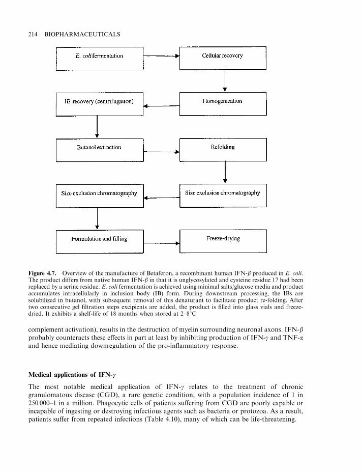
complement activation), results in the destruction of myelin surrounding neuronal axons. IFN-bprobably counteracts these effects in part at least by inhibiting production of IFN-g and TNF-aand hence mediating downregulation of the pro-inflammatory response.
Medical applications of IFN-c
The most notable medical application of IFN-g relates to the treatment of chronicgranulomatous disease (CGD), a rare genetic condition, with a population incidence of 1 in250 000–1 in a million. Phagocytic cells of patients suffering from CGD are poorly capable orincapable of ingesting or destroying infectious agents such as bacteria or protozoa. As a result,patients suffer from repeated infections (Table 4.10), many of which can be life-threatening.
214 BIOPHARMACEUTICALS
Figure 4.7. Overview of the manufacture of Betaferon, a recombinant human IFN-b produced in E. coli.The product differs from native human IFN-b in that it is unglycosylated and cysteine residue 17 had beenreplaced by a serine residue. E. coli fermentation is achieved using minimal salts/glucose media and productaccumulates intracellularly in inclusion body (IB) form. During downstream processing, the IBs aresolubilized in butanol, with subsequent removal of this denaturant to facilitate product re-folding. Aftertwo consecutive gel filtration steps excipients are added, the product is filled into glass vials and freeze-dried. It exhibits a shelf-life of 18 months when stored at 2–88C
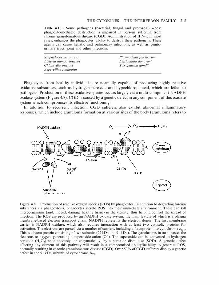
Phagocytes from healthy individuals are normally capable of producing highly reactiveoxidative substances, such as hydrogen peroxide and hypochlorous acid, which are lethal topathogens. Production of these oxidative species occurs largely via a multi-component NADPHoxidase system (Figure 4.8). CGD is caused by a genetic defect in any component of this oxidasesystem which compromises its effective functioning.
In addition to recurrent infection, CGD sufferers also exhibit abnormal inflammatoryresponses, which include granuloma formation at various sites of the body (granuloma refers to
THE CYTOKINES—THE INTERFERON FAMILY 215
Table 4.10. Some pathogens (bacterial, fungal and protozoal) whosephagocyte-mediated destruction is impaired in persons suffering fromchronic granulomatous disease (CGD). Administration of IFN-g, in mostcases, enhances the phagocytes’ ability to destroy these pathogens. Theseagents can cause hepatic and pulmonary infections, as well as genito-urinary tract, joint and other infections
Staphylococcus aureus Plasmodium falciparumListeria monocytogenes Leishmania donovaniChlamydia psittaci Toxoplasma gondiiAspergillus fumigatus
Figure 4.8. Production of reactive oxygen species (ROS) by phagocytes. In addition to degrading foreignsubstances via phagocytosis, phagocytes secrete ROS into their immediate environment. These can killmicroorganisms (and, indeed, damage healthy tissue) in the vicinity, thus helping control the spread ofinfection. The ROS are produced by an NADPH oxidase system, the main feature of which is a plasmamembrane-based electron transport chain. NADPH represents the electron donor. The first membranecarrier is NADPH oxidase, which also requires interaction with at least two cytosolic proteins foractivation. The electrons are passed via a number of carriers, including a flavoprotein, to cytochrome b558.This is a haem protein consisting of two subunits (22 kDa and 91 kDa). The cytochrome, in turn, passes theelectrons to oxygen, generating a superoxide anion (O�). The superoxide can be converted to hydrogenperoxide (H2O2) spontaneously, or enzymatically, by superoxide dismutase (SOD). A genetic defectaffecting any element of this pathway will result in a compromised ability/inability to generate ROS,normally resulting in chronic granulomatous disease (CGD). Over 50% of CGD sufferers display a geneticdefect in the 91 kDa subunit of cytochrome b558
a tissue outgrowth which is composed largely of blood vessels and connective tissue). This canlead to obstruction of various ducts, e.g. in the urinary and digestive tract.
Traditionally, treatment of CGD entailed prophylactic administration of anti-microbialagents in an attempt to prevent occurrence of severe infection. However, affected individualsstill experience life-threatening infections, requiring hospitalization and intensive medical care,as often as once a year. Attempts to control these infections rely on strong anti-microbial agentsand leukocyte transfusions.
Long-term administration of IFN-g to CGD patients has proved effective in treating/moderating the symptoms of this disease. The recombinant human IFN-g used therapeutically isproduced in E. coli, and is termed IFN-g1B. It displays identical biological activity to nativehuman IFN-g, although it lacks the carbohydrate component. The product, usually sold in liquidform, is manufactured by Genentech, who market it under the tradename Actimmune. Theproduct is administered on an ongoing basis, usually by s.c. injection three times weekly. In clinicaltrials its administration, when compared to a control group receiving a placebo, resulted in:
. a reduction in the incidence of life-threatening infections by 50% or more;
. a reduction in the incidence of total infections by 50% or more;
. a reduction in the number of days of hospitalization by three-fold and even, whenhospitalization was required, the average stay was halved.
The molecular basis by which IFN-g induces these effects is understood, at least in part. Inhealthy individuals this cytokine is a potent activator of phagocytes. It potentiates their abilityto generate toxic oxidative products (via the NADPH oxidase system), which they then use tokill infectious agents. In CGD sufferers IFN-g boosts flux through the NADPH oxidativesystem. As long as the genetic defect has not totally inactivated a component of the system, thispromotes increased synthesis of these oxidative substances. IFN-g also promotes increasedexpression of IgG Fc receptors on the surface of phagocytes. This would increase thephagocytes’ ability to destroy opsonized infectious agents via phagocytosis (Figure 4.9).
Additional molecular mechanisms must also mediate IFN-g effects, as it promotes a markedclinical improvement in some CGD patients, without enhancing phagocyte activity. IFN-g’sdemonstrated ability to stimulate aspects of cellular and humoral immunity (e.g. via T and Blymphocytes), as well as NK cell activity, is most likely responsible for these observedimprovements.
IFN-g may also prove valuable in treating a variety of other conditions, and clinical trials forvarious indications are currently under way. This cytokine shows promise in treatingleishmaniasis, a disease common in tropical and sub-tropical regions. The causative agent is aparasitic protozoan of the genus Leishmania. The disease is characterized by the presence ofthese protozoa inside certain immune cells, particularly macrophages. IFN-g appears tostimulate the infected macrophage to produce nitric oxide, which is toxic for the parasite.
Additional studies illustrate that IFN-g stimulates phagocytic activity in humans sufferingfrom various cancers, AIDS and lepromatous leprosy (leprosy is caused by the bacteriumMycobacterium leprae; lepromatous leprosy is a severe contagious form of the disease, leading todisfigurement). IFN-g may thus prove useful in treating such conditions.
Interferon toxicity
Like most drugs, administration of IFNs can elicit a number of unwanted side effects.Unfortunately, in some instances the severity of such effects can limit the maximum
216 BIOPHARMACEUTICALS
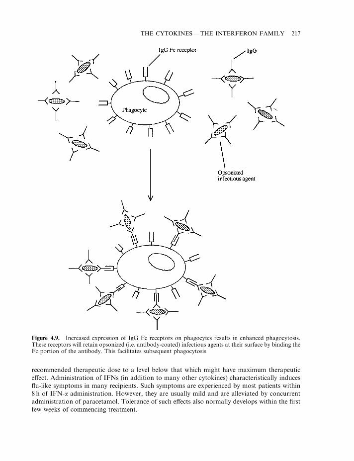
recommended therapeutic dose to a level below that which might have maximum therapeuticeffect. Administration of IFNs (in addition to many other cytokines) characteristically inducesflu-like symptoms in many recipients. Such symptoms are experienced by most patients within8 h of IFN-a administration. However, they are usually mild and are alleviated by concurrentadministration of paracetamol. Tolerance of such effects also normally develops within the firstfew weeks of commencing treatment.
THE CYTOKINES—THE INTERFERON FAMILY 217
Figure 4.9. Increased expression of IgG Fc receptors on phagocytes results in enhanced phagocytosis.These receptors will retain opsonized (i.e. antibody-coated) infectious agents at their surface by binding theFc portion of the antibody. This facilitates subsequent phagocytosis
In some instances more severe side effects are noted (Table 4.11), while in a few cases veryserious side effects, such as the induction of autoimmune reactions, central nervous system orcardiovascular disturbances, renders necessary immediate withdrawal of treatment.
Administration of IFN-b also characteristically causes flu-like symptoms. More serious sideeffects, however, are sometimes noted, including:
. hypersensitivity reactions;
. menstrual disorders;
. anxiety and emotional liability;
. depression, which in rare instances may prompt suicidal thoughts.
The only common side effect associated with IFN-g is the characteristic flu-like symptoms.However, in rare instances and at high doses, adverse clinical reactions have been noted. Thesehave included heart failure, CNS complications (confusion disorientation, parkinsonian-likesymptoms), metabolic complications (e.g. hyperglycemia) and various other symptoms.
Prediction of the range or severity of side effects noted after administration of any IFNpreparation is impossible. Careful monitoring of the patients, particularly in the earliest stagesof treatment, soon reveals the onset of any side effects which might warrant suspension oftreatment.
Additional interferons
In the last few years additional members of the IFN family has been discovered. Amino acidsequence analysis of a protein called trophoblastin—which is found in many ruminants—revealed that it was closely related to IFN-a. This result was surprising because, in sheep andseveral other ruminants, the primary function (and until recently the only known function) oftrophoblastin is to sustain the corpus luteum during the early stages of pregnancy. The 172amino acid protein is produced by the trophoblast (an outer layer of cells which surround thecells that constitute the early embryo) for several days immediately preceding implantation. Inmany ruminants, therefore, trophoblastin plays an essentially similar role to hCG in humans(Chapter 8).
If amino acid analysis hinted that trophoblastin was in fact an IFN, functional studies haveproved it. These studies show that trophoblastin:
. displays the same anti-viral activity as type I IFNs;
218 BIOPHARMACEUTICALS
Table 4.11. Side effects sometimes associated with therapeutic administrationof IFN-as. In most cases, only minor side effects are noted. However, moreserious effects, necessitating cessation of treatment, may occur in up to 17% ofpatients
Minor side effects Serious potential side effects
Range of flu-like symptoms, e.g. fever, Anorexiaheadache, chills Strong fatigue
InsomniaCardiovascular complicationsAutoimmune reactionsHepatic decompression
. displays anti-proliferative activity against certain tumour cells, in vitro at least;
. binds the type I IFN receptor.
Trophoblastin has therefore been named interferon-tau (IFN-t), and is classified as a type IIFN. There are at least three or four functional IFN-t genes in sheep and cattle. The moleculedisplays a molecular mass of 19 kDa and an isoelectric point of 5.5–5.7, in common with othertype 1 interferons. Interestingly, the molecule can also promote inhibition of reversetranscriptase activity in cells infected with the HIV virus.
IFN-t is currently generating considerable clinical interest. While it induces effects similar totype I IFN, it appears to exhibit significantly lower toxicity. Thus it may prove possible to safelyuse this IFN at dosage levels far greater than the maximum dosage levels applied to currentlyused type I IFNs; however, this can only be elucidated by future clinical trials.
Interferon-omega (IFN-o) represents an additional member of the IFN (type I) family. This170 amino acid glycoprotein exhibits 50–60% amino acid homology to IFN-as, and appearseven more closely related to IFN-t.
IFN-o genes have been found in man, pigs and a range of other mammals, but not in dogs orrodents. The IFN induces its antiviral, immunoregulatory and other effects by binding the type IIFN receptor, although the exact physiological relevance of this particular IFN remains to beelucidated. Recently, a recombinant form of feline IFN-o has been approved within the EU forveterinary use. Its approved indication is for the reduction of mortality and clinical symptoms ofparvoviral infections in young dogs. The recombinant product is manufactured using a novelexpression system, which entails direct inoculation of silkworms with an engineered silkwormnuclear polyhedrosis virus housing the feline IFN-o gene. Overviewed in Figure 3.10, theproduction process begins by (automatically) inoculating silkworms (typically 8000 worms/batch) with the recombinant virus. Infection of silkworm cells results in high-level IFN-oproduction, which is subsequently acid-extracted from silkworm body parts. After clarification,the product is purified by a two-step affinity chromatographic process (dye affinitychromatography, using blue sepharose, followed by a metal affinity step, using a copper-chelated sepharose column). A final gel filtration step is undertaken before addition of excipients(sodium chloride, D-sorbitol and gelatin). After filling, the product is freeze-dried.
CONCLUSION
Interferons represent an important family of biopharmaceutical products. They have a proventrack record in the treatment of selected medical conditions, and their range of clinicalapplications continue to grow. It is also likely that many may be used to greater efficacy in thefuture by their application in combination with additional cytokines.
While it is premature to speculate upon the likely medical applications of IFN-t, the reducedtoxicity exhibited by this molecule will encourage its immediate medical appraisal. Theclassification of t as an IFN also raises the intriguing possibility that other IFNs may yet proveuseful in the treatment of some forms of reproductive dysfunctions in veterinary and humanmedicine.
FURTHER READING
Books
Abbas, A. (2003). Cellular and Molecular Immunology. W. B. Saunders, London.Aggarwal, B. (1998). Human Cytokines. Blackwell Scientific, Oxford.
THE CYTOKINES—THE INTERFERON FAMILY 219
Estrov, Z. (1993). Interferons, Basic Principles and Clinical Applications. R. G. Landes, Florence, KY.Fitzgerald, K. (2001). The Cytokine Facts Book. Academic Press, London.Karupiah, G. (1997). Gamma Interferon in Antiviral Defence. Landes Bioscience, Lewisville, TX.Mantovani, A. (2000). Pharmacology of Cytokines. Oxford University Press, Oxford.Mire-Sluis, A. (1998). Cytokines. Academic Press, London.Reder, A. (1996). Interferon Therapy of Multiple Sclerosis. Marcel Dekker, New York.
Articles
Cytokines—general
Aringer, M. et al. (1999). Interleukin/interferon signalling—a 1999 perspective. Immunologist 7(5), 139–146.Baggiolini, M. et al. (1997). Human chemokines—an update. An. Rev. Immunol. 15, 675–705.Dardenne, M. & Savino, W. (1996). Interdependence of the endocrine and immune systems. Adv. Neuroimmunol. 6(4),
297–307.Debetes, R. & Savelkoul, H. (1996). Cytokines as cellular communicators. Mediat. Inflamm. 5(6), 417–423.Henderson, B. & Wilson, M. (1996). Cytokine induction by bacteria—beyond lipopolysaccharide. Cytokine 8(4), 269–
282.Ihle, J. (1996). STATs: signal transducers and activators of transcription. Cell 84, 331–334.Kishimoto, T. et al. (1994). Cytokine signal transduction. Cell 76, 253–262.Lau, F. & Horvath, C. (2002). Mechanisms of type I interferon cell signalling and STAT-mediated transcriptional
responses. Mt Sinai J. Med. 69(3), 156–168.Liu, L. et al. (1997). Ship, a new player in cytokine-induced signalling. Leukaemia 11(2), 181–184.Mire-Sluis, A. (1999). Cytokines: from technology to therapeutics. Trends Biotechnol. 17, 319–325.O’Shea, J.J. et al. (2002). Cytokine signalling in 2002: new surprises in the JAK–STAT pathway. Cell 109, S121–S131.Piscitelli, S. et al. (1997). Pharmacokinetic studies with recombinant cytokines— scientific issues and practical
considerations. Clin. Pharmacokinet. 32(5), 368–381.Poole, S. (1995). Cytokine therapeutics. Trends Biotechnol. 13, 81–82.Proost, P. et al. (1996). The role of chemokines in inflammation. Int. J. Clin. Lab. Res. 26(4), 211–223.Schooltink, H. & Rose, J. (2002). Cytokines as therapeutic drugs. J. Interferon Cytokine Res. 22(5), 505–516.Takatsu, K. (1997). Cytokines involved in B cell differentiation and their sites of action. Proc. Soc. Exp. Biol. Med.
215(2), 121–133.Taniguchi, T. (1995). Cytokine signalling through non-receptor protein tyrosine kinases. Science 268, 251–255.
Interferons
Alberti, A. (1999). Interferon acon-1. A novel interferon for the treatment of chronic hepatitis C. Biodrugs 12(5), 343–357.
Belardelli, F. et al. (2002). Interferon-a in tumor immunity and immunotherapy. Cytokine Growth Factor Rev. 13(2),119–134.
Boehm, U. et al. (1997). Cellular responses to interferon-g. Ann. Rev. Immunol. 15, 749–795.Brassard, D. et al. (2002). Interferon-a as an immunotherapeutic protein. J. Leukocyte Biol. 71(4), 565–581.Cencic, A. & La Bonnardiere, C. (2002). Trophoblastic interferon-g: current knowledge and possible role in early pig
pregnancy. Vet. Res. 33(2), 139–157.Chelmonska-Soyta, A. (2002). Interferon-t and its immunological role in ruminant reproduction. Arch. Immunol. Ther.
Exp. 50(1), 47–52.Colonna, M. et al. (2002). Interferon-producing cells: on the front line in immune responses against pathogens. Curr.
Opin. Immunol. 14(3), 373–379.Haria, M. & Benfield, P. (1995). Interferon-a2A. Drugs 50(5), 873–896.Jonasch, E. & Haluska, F. (2001). Interferon in oncological practice: review of interferon biology, clinical applications
and toxicities. Oncologist 6(1), 34–55.Katre, N. (1993). The conjugation of proteins with polyethylene glycol and other polymers—altering properties of
proteins to enhance their therapeutic potential. Adv. Drug Delivery Rev. 10(1), 91–114.Kirkwood, J. (2002). Cancer immunotherapy: the interferon-a experience. Semin. Oncol. 29(3), 18–26.Leaman, D. et al. (1996). Regulation of STAT-dependent pathways by growth factors and cytokines. FASEB J. 10(14),
1578–1588.Li, J. & Roberts, M. (1994). Interferon-t and interferon-a interact with the same receptors in bovine endometrium. J.
Biol. Chem. 269(18), 13544–13550.Lyseng-Williamson, K. & Plosker, G. (2002). Management of relapsing–remitting multiple sclerosis—defining the role
of subcutaneous recombinant interferon-b-1a (Rebif). Dis. Managem. Health Outcomes 10(5), 307–325.
220 BIOPHARMACEUTICALS
Meager, A. (2002). Biological assays for interferons. J. Immunol. Methods 261(1–2), 21–36.Pestka, S. & Langer, J. (1987). Interferons and their actions. Ann. Rev. Biochem. 56, 727–777.Simko, R. & Nagy, K. (1996). Interferon-a in childhood hematological malignancies. Postgrad. Med. J. 72(854), 709–
713.Soos, J. & Johnson, H. (1999). Interferon-t. Prospects for clinical use in autoimmune disorders. BioDrugs 11(2), 125–135.Todd, P. & Goa, K. (1992). Interferon-g-1b. Drugs 43(1), 111–122.Tossing, G. (2001). New developments in interferon therapy. Eur. J. Med. Res. 6(2), 47–65.Wang, Y. et al. (2002). Structural and biological characterization of PEGylated recombinant interferon-a2B and its
therapeutic implications. Adv. Drug Delivery Rev. 54(4), 547–570.Woo, M. & Burnakis, T. (1997). Interferon-a in the treatment of chronic viral hepatitis B and viral hepatitis C. Ann.
Pharmacother. 31(3), 330–337.Younes, H. & Amsden, B. (2002). Interferon-g therapy: evaluation of routes of administration and delivery systems. J.
Pharmaceut. Sci. 91(1), 2–17.Young, H. & Hardy, K. (1995). Role of interferon-g in immune cell regulation. J. Leukocyte Biol. 58, 373–379.Zavyalov, V. & Zavyalova, G. (1997). Interferons a/b and their receptors—place in the hierarchy of cytokines. APMIS
105(3), 161–186.
THE CYTOKINES—THE INTERFERON FAMILY 221

Chapter 5
Cytokines: interleukins and tumour
necrosis factor
The interleukins (ILs) represent another large family of cytokines, with at least 25 differentconstituent members (IL-1 to IL-25) having been characterized thus far. Most of thesepolypeptide regulatory factors are glycosylated (a notable exception being IL-1) and display amolecular mass in the range 15–30 kDa. A few interleukins display a higher molecular mass, e.g.the heavily glycosylated, 40 kDa, IL-9.
Most of the interleukins are produced by a number of different cell types. At least 17 differentcell types are capable of producing IL-1 (see Table 5.5), while IL-8 is produced by at least 10distinct cell types. On the other hand, IL-2, -9 and -13 are produced only by T lymphocytes.
Most cells capable of synthesizing one IL are capable of synthesizing several, and manyprominent producers of ILs are non-immune system cells (Table 5.1). Regulation of IL synthesisis exceedingly complex and only partially understood. In most instances, induction or repressionof any one IL is prompted by numerous regulators—mostly additional cytokines, e.g. IL-1promotes increased synthesis and release of IL-2 from activated T lymphocytes. It is highlyunlikely that cells capable of synthesizing multiple ILs concurrently synthesize them all at highlevels.
Nearly all ILs are soluble molecules (one form of IL-1 is cell-associated). They promote theirbiological response by binding to specific receptors on the surface of target cells. Most ILsexhibit paracrine activity (i.e. the target cells are in the immediate vicinity of the producer cells),while some display autocrine activity (e.g. IL-2 can stimulate the growth and differentiation ofthe cells that produce it). Other ILs display more systematic endocrine effects (e.g. someactivities of IL-1).
The signal transduction mechanisms by which most ILs prompt their biological response areunderstood, in outline at least. In many cases, receptor binding is associated with intracellulartyrosine phosphorylation events. In other cases, serine and threonine residues of specificintracellular substrates are also phosphorylated. For some ILs, receptor binding triggersalternative signal transduction events, including promoting an increase in intracellular calciumconcentration or inducing the hydrolysis of phosphotidylethanolamine with release of diacylglycerol.
Biopharmaceuticals: Biochemistry and Biotechnology, Second Edition. Gary WalshJohn Wiley & Sons Ltd: ISBN 0 470 84326 8 (ppc), ISBN 0 470 84327 6 (pbk)
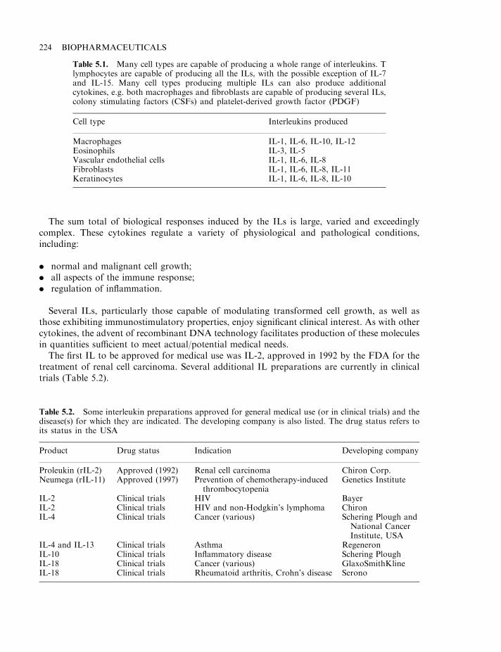
The sum total of biological responses induced by the ILs is large, varied and exceedinglycomplex. These cytokines regulate a variety of physiological and pathological conditions,including:
. normal and malignant cell growth;
. all aspects of the immune response;
. regulation of inflammation.
Several ILs, particularly those capable of modulating transformed cell growth, as well asthose exhibiting immunostimulatory properties, enjoy significant clinical interest. As with othercytokines, the advent of recombinant DNA technology facilitates production of these moleculesin quantities sufficient to meet actual/potential medical needs.
The first IL to be approved for medical use was IL-2, approved in 1992 by the FDA for thetreatment of renal cell carcinoma. Several additional IL preparations are currently in clinicaltrials (Table 5.2).
224 BIOPHARMACEUTICALS
Table 5.1. Many cell types are capable of producing a whole range of interleukins. Tlymphocytes are capable of producing all the ILs, with the possible exception of IL-7and IL-15. Many cell types producing multiple ILs can also produce additionalcytokines, e.g. both macrophages and fibroblasts are capable of producing several ILs,colony stimulating factors (CSFs) and platelet-derived growth factor (PDGF)
Cell type Interleukins produced
Macrophages IL-1, IL-6, IL-10, IL-12Eosinophils IL-3, IL-5Vascular endothelial cells IL-1, IL-6, IL-8Fibroblasts IL-1, IL-6, IL-8, IL-11Keratinocytes IL-1, IL-6, IL-8, IL-10
Table 5.2. Some interleukin preparations approved for general medical use (or in clinical trials) and thedisease(s) for which they are indicated. The developing company is also listed. The drug status refers toits status in the USA
Product Drug status Indication Developing company
Proleukin (rIL-2) Approved (1992) Renal cell carcinoma Chiron Corp.Neumega (rIL-11) Approved (1997) Prevention of chemotherapy-induced
thrombocytopeniaGenetics Institute
IL-2 Clinical trials HIV BayerIL-2 Clinical trials HIV and non-Hodgkin’s lymphoma ChironIL-4 Clinical trials Cancer (various) Schering Plough and
National CancerInstitute, USA
IL-4 and IL-13 Clinical trials Asthma RegeneronIL-10 Clinical trials Inflammatory disease Schering PloughIL-18 Clinical trials Cancer (various) GlaxoSmithKlineIL-18 Clinical trials Rheumatoid arthritis, Crohn’s disease Serono
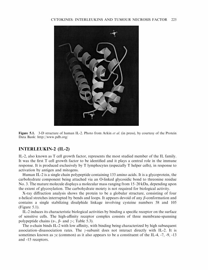
INTERLEUKIN-2 (IL-2)
IL-2, also known as T cell growth factor, represents the most studied member of the IL family.It was the first T cell growth factor to be identified and it plays a central role in the immuneresponse. It is produced exclusively by T lymphocytes (especially T helper cells), in response toactivation by antigen and mitogens.
Human IL-2 is a single chain polypeptide containing 133 amino acids. It is a glycoprotein, thecarbohydrate component being attached via an O-linked glycosidic bond to threonine residueNo. 3. The mature molecule displays a molecular mass ranging from 15–20 kDa, depending uponthe extent of glycosylation. The carbohydrate moiety is not required for biological activity.
X-ray diffraction analysis shows the protein to be a globular structure, consisting of foura-helical stretches interrupted by bends and loops. It appears devoid of any b-conformation andcontains a single stabilizing disulphide linkage involving cysteine numbers 58 and 105(Figure 5.1).
IL-2 induces its characteristic biological activities by binding a specific receptor on the surfaceof sensitive cells. The high-affinity receptor complex consists of three membrane-spanningpolypeptide chains (a-, b- and g-; Table 5.3).
The a-chain binds IL-2 with low affinity, with binding being characterized by high subsequentassociation–disassociation rates. The g-subunit does not interact directly with IL-2. It issometimes known as gc (common) as it also appears to be a constituent of the IL-4, -7, -9, -13and -15 receptors.
CYTOKINES: INTERLEUKINS AND TUMOUR NECROSIS FACTOR 225
Figure 5.1. 3-D structure of human IL-2. Photo from Arkin et al. (in press), by courtesy of the ProteinData Bank: http://www.pdb.org/
Heterodimeric complexes consisting of ag or bg can bind IL-2 with intermediate affinity. Theheterotrimeric abg complex represents the cytokine’s true high-affinity receptor (Figure 5.2). Theexact intracellular signal transduction events triggered by IL-2 are not fully elucidated. The a-receptor chain exhibits a cytoplasmic domain containing only nine amino acids, and is unlikelyto play a role in intracellular signalling. Mutational studies reveal that the 286 amino acid b-chain intracellular domain contains at least two regions (a serine-rich region and an acidicregion) essential for signal transduction. The b-chain is phosphorylated on a specific tyrosineresidue subsequent to IL-2 binding, probably via association of a protein tyrosine kinaseessential for generation of intracellular signals. The direct role played by the g-chain, althoughunclear, is likely critical. A mutation in the gene coding for this receptor constituent results insevere combined immunodeficiency (X-SCID).
Interestingly, prolonged elevated levels of IL-2 promotes the shedding of the IL-2 receptora-subunit from the cell surface. Initially, it was suspected that these circulating soluble receptorfragments, capable of binding IL-2, might play a role in inducing immunosuppression (bycompeting for IL-2 with the cell surface receptor). However, the affinity of IL-2 for the intactreceptor (abg) is far greater than for the a-subunit, rendering this theory unlikely.
The IL-2 receptor is associated with a number of cell types—mainly cells playing a centralrole in the immune response (Table 5.4). Binding of IL-2 to its receptor induces growth anddifferentiation of these cells. This cytokine, therefore, behaves as a central molecular switch,activating most aspects of the immune response.
226 BIOPHARMACEUTICALS
Figure 5.2. Schematic diagram of the high-affinity IL-2 receptor
Table 5.3. Summary of the polypeptide constituents of the high-affinityhuman IL-2 receptor
Receptor polypeptideconstituent Additional names
Molecular mass(kDa)
a P55TacCD25
55
b P75CD122
75
g P64 64
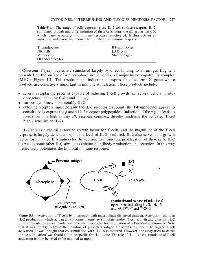
Quiescent T lymphocytes are stimulated largely by direct binding to an antigen fragmentpresented on the surface of a macrophage in the context of major histocompatibility complex(MHC) (Figure 5.3). This results in the induction of expression of at least 70 genes whoseproducts are collectively important in immune stimulation. These products include:
. several cytoplasmic proteins capable of inducing T cell growth (i.e. several cellular proto-oncogenes, including C-fos and C-myc);
. various cytokines, most notably IL-2;
. cytokine receptors, most notably the IL-2 receptor a subunit (the T-lymphocytes appear toconstitutively express the b and g IL-2 receptor polypeptides. Induction of the a gene leads toformation of a high-affinity abg receptor complex, thereby rendering the activated T cellhighly sensitive to IL-2).
IL-2 acts as a critical autocrine growth factor for T cells, and the magnitude of the T cellresponse is largely dependent upon the level of IL-2 produced. IL-2 also serves as a growthfactor for activated B lymphocytes. In addition to promoting proliferation of these cells, IL-2(as well as some other ILs) stimulates enhanced antibody production and secretion. In this wayit effectively potentates the humoral immune response.
CYTOKINES: INTERLEUKINS AND TUMOUR NECROSIS FACTOR 227
Figure 5.3. Activation of T cells by interaction with macrophage-displayed antigen. Activation results inIL-2 production, which acts in an autocrine manner to stimulate further T cell growth and division. IL-2thus represents the major regulatory molecule responsible for stimulation of cell-mediated immunity. Notethat it was initially believed that binding of presented antigen alone was insufficient to trigger T cellactivation. It was thought that co-stimulation with IL-1 was required. However, the assay used to detectthe ‘co-stimulation’ was found not to be specific for IL-1 alone. The role of IL-1 as a co-stimulator of T cellactivation is now believed to be minimal at most
Table 5.4. The range of cells expressing the IL-2 cell surface receptor. IL-2-stimulated growth and differentiation of these cells forms the molecular basis bywhich many aspects of the immune response is activated. It thus acts in anautocrine and paracrine manner to mobilize the immune response
T lymphocytes B lymphocytesNK cells LAK cellsMonocytes MacrophagesOligodendrocytes
A third biological activity of IL-2 pertinent to immunostimulation is its ability to promote thegrowth of natural killer (NK) cells. It also promotes further differentiation of NK cells, forminglymphokine-activated killer cells (LAKs), which exhibit an enhanced ability to directly killtumour cells or virally infected cells. NK cells express the b and g IL-2 receptor subunits only,thus their stimulation by IL-2 requires elevated concentrations of this cytokine. NK cells arealso activated by a variety of additional cytokines, including all IFNs as well as tumour necrosisfactor (TNF).
The immunopotentative effects of IL-2 rendered it an obvious target for clinical application.At the simplest level, it was hoped that administration of exogenous IL-2 could enhance theimmune response to a number of clinical conditions, including:
. cancers;
. T cell and other forms of immunodeficiency;
. infectious diseases.
IL-2 production
As was the case for most other cytokines, medical appraisal/use of IL-2 was initiallyimpractical due to the minute quantities in which it is normally produced. Some transformedcell lines, most notably the Jurkat leukaemia cell line, produces IL-2 in increased quantitiesand much of the IL-2 used for initial characterization studies was obtained from this source.Large-scale IL-2 production was made possible by rDNA technologies. While the IL-2 gene/cDNA has now been expressed in a wide variety of host systems, it was initially expressed inE. coli, and most products being clinically evaluated are obtained from that source. Asmentioned previously, the absence of glycosylation on the recombinant product does not alterits biological activity.
Proleukin is the trade name given to the recombinant IL-2 preparation manufactured byChiron and approved for the treatment of certain cancers. It is produced in engineered E. coliand differs from native human IL-2 in that it is non-glycosylated, lacks an N-terminal alanineresidue and cysteine 125 has been replaced by a serine residue. After extraction andchromatographic purification, the product is formulated in a phosphate buffer containingmannitol and low levels of the detergent SDS. The product displays typical IL-2 biologicalactivities, including enhancement of lymphocyte mitogenesis and cytotoxicity, induction ofLAK/NK cell activity and the induction of IFN-g production.
IL-2 and cancer treatment
The immunostimulatory activity of IL-2 has proved beneficial in the treatment of some cancertypes. An effective anti-cancer agent would prove not only medically valuable but alsocommercially very successful. In the developed world, an average of one in six deaths is causedby cancer. In the USA alone, the annual death toll from cancer stands in the region of half amillion people.
There exists direct evidence that the immune system mounts an immune response againstmost cancer types. Virtually all transformed cells: (a) express novel surface antigens notexpressed by normal cells; or (b) express, at greatly elevated levels, certain antigens presentnormally on the cell at extremely low levels. These ‘normal’ expression levels may be so low that
228 BIOPHARMACEUTICALS
they have gone unnoticed by immune surveillance (and thus have not induced immunologicaltolerance).
The appearance of any such cancer-associated antigen should thus be capable of inducing animmune response which, if successful, should eradicate the transformed cells. The exact elementsof immunity responsible for destruction of transformed cells remain to be fully characterized.Both a humoral and cell-mediated response can be induced, although the T cell responseappears to be the most significant.
Cytotoxic T cells may play a role in inducing direct destruction of cancer cells, in particularthose transformed by viral infection (and who express viral antigen on their surface). In vitrostudies have shown that cytotoxic T lymphocytes obtained from the blood of persons sufferingfrom various cancer types are capable of destroying those cancer cells.
NK cells are capable of efficiently lysing various cancer cell types and, as already discussed,IL-2 can stimulate differentiation of NK cells forming LAK cells, which exhibit enhancedtumoricidal activity. Macrophages, too, probably play a role. Activated macrophages have beenshown to lyse tumour cells in vitro, while leaving untransformed cells unaffected. Furthermore,these cells produce TNF and various other cytokines that can trigger additional immunologicalresponses. The production of antibodies against tumour antigens (and the subsequent bindingof the antibodies to those antigens) marks the transformed cells for destruction by NK/LAKcells and macrophages—all of which exhibit receptors capable of binding the Fc portion ofantibodies.
While immune surveillance is certainly responsible for the detection and eradication of sometransformed cells, the prevalence of cancer indicates that this surveillance is nowhere near100% effective. Some transformed cells obviously display characteristics that allow them toevade this immune surveillance. The exact molecular details of how such ‘tumour escape’ isachieved remains to be conformed, although several mechanisms have been implicated,including:
. Most transformed cells do not express class II MHC molecules and express lower thannormal levels of class I MHC molecules. This renders their detection by immune effector cellsmore difficult. Treatment with cytokines, such as IFN-g, can induce increased class I MHCexpression which normally promotes increased tumour cell susceptibility to immunedestruction.
. Transformed cells expressing tumour-specific surface antigens, which closely resemblenormal surface antigens, may not induce an immune response. Furthermore, some tumorantigens, while not usually expressed in adults, were expressed previously during the neonatalperiod (i.e. just after birth) and are thus believed by the immune cells to be ‘self’.
. Some tumours secrete significant quantities of cytokines and additional regulatory moleculeswhich can suppress local immunological activity, e.g. transforming growth factor-b (TGF-b;produced by many tumour types) is capable of inhibiting lymphocyte and macrophageactivity.
. Antibody binding to many tumour antigens triggers the immediate loss of the antibody–antigen complex from the transformed cell surface by either endocytosis or extracellularshedding.
. The glycocalyx can possibly shield tumour antigens from the immune system.
Whatever the exact nature of tumour escape, it has been demonstrated, both in vitro and invivo, that immunostimulation can lead to enhanced tumour detection and destruction. Several
CYTOKINES: INTERLEUKINS AND TUMOUR NECROSIS FACTOR 229
approaches to cancer immunotherapy have thus been formulated, many involving application ofIL-2 as the primary immunostimulant.
Experiments conducted in the early 1980s showed that lymphocytes incubated in vitro withIL-2 could subsequently kill a range of cultured cancer cell lines, including melanoma and coloncancer cells. These latter cancers do not respond well to conventional therapies. Subsequentinvestigations showed that cancer cell destruction was mediated by IL-2-stimulated NK cells(i.e. LAK cells). Similar responses were seen in animal models upon administration of LAK cellsactivated in vitro using IL-2.
Clinical studies have shown this approach to be effective in humans. LAK cells originallypurified from a patient’s own blood, activated in vitro using IL-2 and reintroduced into thepatient along with more IL-2, promoted complete tumour regression in 10% of patientssuffering from melanoma or renal cancer. Partial regression was observed in a further 10–25%of such patients. Administration of high doses of IL-2 alone could induce similar responses butsignificant side-effects were noted (discussed later).
IL-2-stimulated cytotoxic T cells appear even more efficacious than LAK cells in promotingtumour regression. The approach adopted here entails removal of a tumour biopsy, followed byisolation of T lymphocytes present within the tumour. These tumour-infiltrating lymphocytes(TILs) are cytotoxic T lymphocytes that apparently display a cell surface receptor thatspecifically binds the tumour antigen in question. They are thus tumour-specific cells. Furtheractivation of these TILs by in vitro culturing in the presence of IL-2, followed by reintroductioninto the patient along with IL-2, promoted partial/full tumour regression in well over 50% oftreated patients.
Further studies have shown additional cancer types, most notably ovarian and bladdercancer, non-Hodgkin’s lymphoma and acute myeloid leukaemia, to be at least partiallyresponsive to IL-2 treatment. However, a persistent feature of clinical investigations assessingIL-2 effects on various cancer types, is variability of response. Several trials have yieldedconflicting results and no reliable predictor of clinical response is available.
IL-2 and infectious diseases
Although antibiotics have rendered possible medical control of various infectious agents(mainly bacterial), numerous pathogens remain for which no effective treatment exists. Most ofthese pathogens are non-bacterial (e.g. viral, fungal and parasitic, including protozoan). Inaddition, the over-use/abuse of antibiotics has hastened the development of antibiotic-resistant‘super-bacteria’, which have become a serious medical problem.
The most difficult microbial pathogens to treat are often those that replicate within host cells(e.g. viruses and some parasites), e.g. during the complex life cycle of the malaria parasite inman, this protozoan can infect and destroy liver cells and erythrocytes. Over 2 million people dieeach year from malaria, with at least 200–300 million people being infected at any given time.Some such agents have even evolved to survive and replicate within macrophages subsequent touptake via phagocytosis. This is often achieved on the basis that the phagocytosed microbe issomehow capable of preventing fusion of the phagocytosed vesicle with lysozomes. Examples ofpathogens capable of survival within macrophages include:
. mycobacteria (e.g. M. tuberculosis, the causative agent of tuberculosis, and M. leprae, whichcauses leprosy);
230 BIOPHARMACEUTICALS
. Listeria monocytogenes, a bacterium which, when transmitted to man, causes listeriosis,which is characterized by flu-like symptoms but can cause swelling of the brain and induceabortions;
. Legionella pneumophila, the bacterium which causes Legionnaire’s disease.
The immunological response raised against intracellular pathogens is largely a T cell response.IL-2’s ability to stimulate T cells may render it useful in the treatment of a wide range of suchconditions. Clinical trials assessing its efficacy in treating a range of infectious diseases,including AIDS, continue.
A related medical application of IL-2 relates to its potential use as adjuvant material, asdiscussed in Chapter 10.
Safety issues
Like all other cytokines, administration of IL-2 can induce side effects, which can be dose-limiting. Serious side effects, including cardiovascular, hepatic or pulmonary complications,usually necessitate immediate termination of treatment. Such side effects may be induced notonly directly by IL-2 but also by a range of additional cytokines whose synthesis is augmentedby IL-2 administration. These cytokines, which can include IL-3, -4, -5 and -6, as well as TNFand IFN-g, also likely play a direct role in the overall therapeutic benefits accrued from IL-2administration.
Inhibition of IL-2 activity
A variety of medical conditions exist which are caused or exacerbated by the immune systemitself. These are usually treated by administering immunosuppressive agents. Examples include:
. autoimmune diseases in which immunological self-tolerance breaks down and the immunesystem launches an attack on self-antigens;
. tissue/organ transplantation in which the donor is not genetically identical to the recipient(i.e. in cases other than identical twins). The recipient will mount an immune response againstthe transplanted tissue, culminating in tissue rejection unless immunosuppressive agents areadministered.
Selective immunosuppression in individuals suffering from the above conditions is likely bestachieved by preventing the synthesis or functioning of IL-2. Cyclosporin A, one of the foremostimmunosuppressive agents currently in use, functions by preventing IL-2 synthesis. A number ofalternative approaches are now being considered or tested directly in clinical trials. Theseinclude:
. administration of soluble forms of the IL-2 receptor, which would complete with the native(cell surface) receptor for binding of IL-2;
. administration of monoclonal antibodies capable of binding the IL-2 receptor (it must beconfirmed that the antibody used is not itself capable of initiating signal transduction uponbinding the receptor);
. administration of IL-2 variants which retains their ability to bind the receptor but fail toinitiate signal transduction;
. administration of IL-2 coupled to bacterial or other toxins. Binding of the cytokine to itsreceptor brings the associated toxin into intimate contact with the antigen-activated T cells
CYTOKINES: INTERLEUKINS AND TUMOUR NECROSIS FACTOR 231
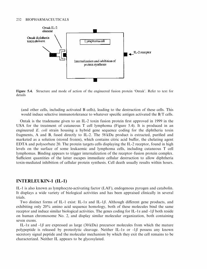
(and other cells, including activated B cells), leading to the destruction of these cells. Thiswould induce selective immunotolerance to whatever specific antigen activated the B/T cells.
Ontak is the tradename given to an IL-2 toxin fusion protein first approved in 1999 in theUSA for the treatment of cutaneous T cell lymphoma (Figure 5.4). It is produced in anengineered E. coli strain housing a hybrid gene sequence coding for the diphtheria toxinfragments, A and B, fused directly to IL-2. The 58 kDa product is extracted, purified andmarketed as a solution (stored frozen), which contains citric acid buffer, the chelating agentEDTA and polysorbate 20. The protein targets cells displaying the IL-2 receptor, found in highlevels on the surface of some leukaemic and lymphoma cells, including cutaneous T celllymphomas. Binding appears to trigger internalization of the receptor–fusion protein complex.Sufficient quantities of the latter escapes immediate cellular destruction to allow diphtheriatoxin-mediated inhibition of cellular protein synthesis. Cell death usually results within hours.
INTERLEUKIN-1 (IL-1)
IL-1 is also known as lymphocyte-activating factor (LAF), endogenous pyrogen and catabolin.It displays a wide variety of biological activities and has been appraised clinically in severaltrials.
Two distinct forms of IL-1 exist: IL-1a and IL-1b. Although different gene products, andexhibiting only 20% amino acid sequence homology, both of these molecules bind the samereceptor and induce similar biological activities. The genes coding for IL-1a and -1b both resideon human chromosome No. 2, and display similar molecular organization, both containingseven exons.
IL-1a and -1b are expressed as large (30 kDa) precursor molecules from which the maturepolypeptide is released by proteolytic cleavage. Neither IL-1a or -1b possess any knownsecretory signal peptide and the molecular mechanism by which they exit the cell remains to becharacterized. Neither IL appears to be glycosylated.
232 BIOPHARMACEUTICALS
Figure 5.4. Structure and mode of action of the engineered fusion protein ‘Ontak’. Refer to text fordetails
IL-1a is initially synthesized as a 271 amino acid precursor, with the mature form containing159 amino acids (17.5 kDa). This molecule appears to remain associated with the extracellularface of the cell membrane. IL-1b, initially synthesized as a 269 amino acid precursor, is releasedfully from the cell. The mature form released contains 153 amino acids and displays a molecularmass in the region of 17.3 kDa.
X-ray diffraction analysis reveals the 3-D structure of both IL-1 molecules to be quite similar.Both are globular proteins, composed of six strands of anti-parallel b-pleated sheet forming a‘barrel’, which is closed at one end by a further series of b-sheets.
A wide range of cells are capable of producing IL-1 (Table 5.5). Different cell types producethe different IL-1s in varying ratios. In fibroblasts and endothelial cells, both are produced inroughly similar ratios, whereas in monocytes IL-1b is produced in larger quantities than IL-1a.Activated macrophages appear to represent the major cellular source for IL-1.
The IL-1s induce their characteristic biological activities by binding to specific cell surfacereceptors present on sensitive cells. Two distinct receptors, types I and II, have been identified.Both IL-1a and IL-1b can bind both receptors. The type I receptor is an 80 kDa transmembraneglycoprotein. It is a member of the IgG superfamily. This receptor is expressed predominantlyon fibroblasts, keratinocytes, hepatocytes and endothelial cells. The type II receptor is a 60 kDatransmembrane glycoprotein, expressed mainly on B lymphocytes, bone marrow cells andpolymorphonuclear leukocytes. It displays a very short (29 amino acid) intracellular domainand some studies suggest that IL-1s can induce a biological response only upon binding to thetype I receptor.
The exact IL-1-mediated mechanism(s) of signal transduction remain to be clarified. Anumber of different signal transduction pathways have been implicated, including involvementof G proteins. IL-1 has also been implicated in activation of protein kinase C by inducing thehydrolysis of phosphotidylethanolamine.
The biological activities of IL-1
IL-1 mediates a wide variety of biological activities:
. it is a pro-inflammatory cytokine, promoting the synthesis of various substances, such aseicosanoids, as well as proteases and other enzymes involved in generating inflammatorymediators. This appears to be its major biological function;
. it plays a role in activating B lymphocytes, along with additional cytokines and may also playa role in activating T lymphocytes;
. along with IL-6, it induces synthesis of acute phase proteins in hepatocytes;
. it acts as a co-stimulator of haematopoietic cell growth/differentiation.
CYTOKINES: INTERLEUKINS AND TUMOUR NECROSIS FACTOR 233
Table 5.5. The range of cells capable of producing IL-1
T lymphocytes Vascular endothelial cellsB lymphocytes FibroblastsMonocytes/macrophages AstrocytesNK cells Dendritic cellsLarge granular lymphocytes MicrogliaKeratinocytes Glioma cellsChondrocytes
The relative prominence of these various biological activities depends largely upon thequantities of IL-1 produced in any given situation. At low concentrations, its effects are largelyparacrine, e.g. induction of local inflammation. At elevated concentrations, it acts more in anendocrine manner, inducing systematic effects such as the hepatic synthesis of acute phaseproteins, but also induction of fever (hence the name, ‘endogenous pyrogen’) and cachexia(general body wasting, such as that associated with some cancers). Many of these biologicalactivities are also promoted by TNF—another example of cytokine redundancy.
In addition to IL-1a and -1b, a third IL-1-like protein has been identified, termed IL-1receptor antagonist (IL-1Ra). As its name suggests, this molecule appears to be capable ofbinding to the IL-1 receptors without triggering an intracellular response.
IL-1 biotechnology
IL-1 continues to be a focus of clinical investigation. This stems from its observed:
. immunostimulatory effects;
. ability to protect/restore the haematopoietic process during, or subsequent to, chemotherapyor radiation therapy;
. anti-proliferative effects against various human tumour cell lines grown in vitro, or in animalmodels.
Most of these effects are most likely mediated not only directly by IL-1 but also by variousadditional cytokines (including IL-2), induced by IL-1 administration.
The observed effects prompted initiation of clinical trials assessing IL-1’s efficacy in treating:
. bone marrow suppression induced by chemo/radiotherapy;
. various cancers.
The initial findings of some such trials (involving both IL-1a and IL-1b) proved disappointing.No significant anti-tumour response was observed in many cases, although side effects werecommonly observed. Virtually all patients suffered from fevers, chills and other flu-likesymptoms. More serious side effects, including capillary leakage syndrome and hypotension,were also observed and were dose-limiting.
IL-1 thus displays toxic effects comparable to administration of TNF (see later) or high levelsof IL-2. However, several clinical studies are still under way and this cytokine may yet provetherapeutically useful, either on its own or, more likely, when administered at lower doses withadditional therapeutic agents.
Because of its role in mediating acute/chronic inflammation, (downward) modulation of IL-1levels may prove effective in ameliorating the clinical severity of these conditions. Again, severalapproaches may prove useful in this regard, including:
. administration of anti-IL-1 antibodies;
. administration of soluble forms of the IL-1 receptor;
. administration of the native IL-1 receptor antagonist.
Kineret is the trade name given to a recently approved product based on the latter strategy.Indicated in the treatment of rheumatoid arthritis, the product consists of a recombinant formof the human IL-1 receptor antagonist. The 17.3 kDa, 153 amino acid product is produced inengineered E. coli and differs from the native human molecule in that it is non-glycosylated andcontains an additional N-terminal methionine residue (a consequence of its prokaryotic
234 BIOPHARMACEUTICALS
expression system). The purified product is presented as a solution and contains sodium citrate,EDTA, sodium chloride and polysorbate 80 as excipients. A daily (s.c.) injection of 100mg isrecommended for patients with rheumatoid arthritis. This inflammatory condition is (notsurprisingly) characterized by the presence of high levels of IL-1 in the synovial fluid of affectedjoints. In addition to its pro-inflammatory properties, IL-1 also mediates additional negativeinfluences on joint/bone, including promoting cartilage degradation and stimulation of boneresorption.
An additional approach to IL-1 downregulation could entail development of inhibitors of theproteolytic enzymes that release the active IL from its inactive precursor. Moreover, suchinhibitors could probably be taken orally and, thus, would be suitable to treat chronicinflammation (the alternatives outlined above would be administered parenterally).
The enzyme releasing active IL-1b from its 31 kDa precursor has been identified and studiedin detail. Termed IL-1b converting enzyme (ICE), it is a serine protease whose only knownphysiological substrate is the inactive IL-1b precursor. ICE cleaves this precursor between Asp116 and Ala 117, releasing the active IL-1b.
ICE is an oligomeric enzyme (its active form may be a tetramer). It contains two distinctpolypeptide subunits, p20 (20 kDa) and p10 (10 kDa). These two subunit types associate veryclosely, and the protease’s active site spans residues from both. p10 and p20 are proteolytically-derived from a single 45 kDa precursor protein.
INTERLEUKIN-3: BIOCHEMISTRY AND BIOTECHNOLOGY
IL-3 is yet another cytokine whose biotechnological applications have attracted interest. Thiscytokine is produced primarily by T lymphocytes as well as mast cells and eosinophils. Themature molecule is a 133 amino acid glycoprotein of molecular mass 15–30 kDa. It induces itsbiological effects by binding a specific receptor— the IL-3 receptor (IL-3R) on sensitive cells.The IL-3R is composed of two subunits, a ligand-binding a-subunit and a b-subunit thatappears to mediate signal transduction (Figure 5.5). Binding of IL-3 to its receptor inducesphosphorylation of several (mostly unidentified) cellular proteins. These substrates arephosphorylated either on tyrosine residues or threonine and serine residues. The amino acidsequence of the intracellular part of the b-receptor subunit exhibits no homology to anyknown kinase, suggesting that phosphorylation is mediated by a distinct cytoplasmic kinasethat is activated upon ligand binding. The IL-3R b-subunit also forms part of the IL-5 andGM-CSF receptors. Not surprisingly, all of these cytokines share at least some biologicalactivities.
The IL-3 receptor is found on a wide range of haematopoietic progenitor cells (see Chapter 6).They are also present on monocytes and B lymphocytes. Its major biological activity relates tostimulation of growth of various cell types derived from bone marrow cells and which representthe immature precursors to all blood cells (Chapter 6). IL-3 thus appears to play a central role instimulating the eventual formation of various blood cell types, in particular monocytes, mastcells, neutrophils, basophils and eosinophils, from immature precursor cells in the bone marrow.Several other cytokines (including IL-2, -4, -5, -6, -7, -11, -15 and CSFs) also play important co-stimulatory roles in the maturation of the range of blood cells.
Its growth and differentiation-inducing effects on early haematopoietic progenitor cells formsthe basis of clinical interest in IL-3. Its administration to healthy patients results in increasedblood leukocyte counts, although the concentration of all white blood cell types is not equallyincreased.
CYTOKINES: INTERLEUKINS AND TUMOUR NECROSIS FACTOR 235
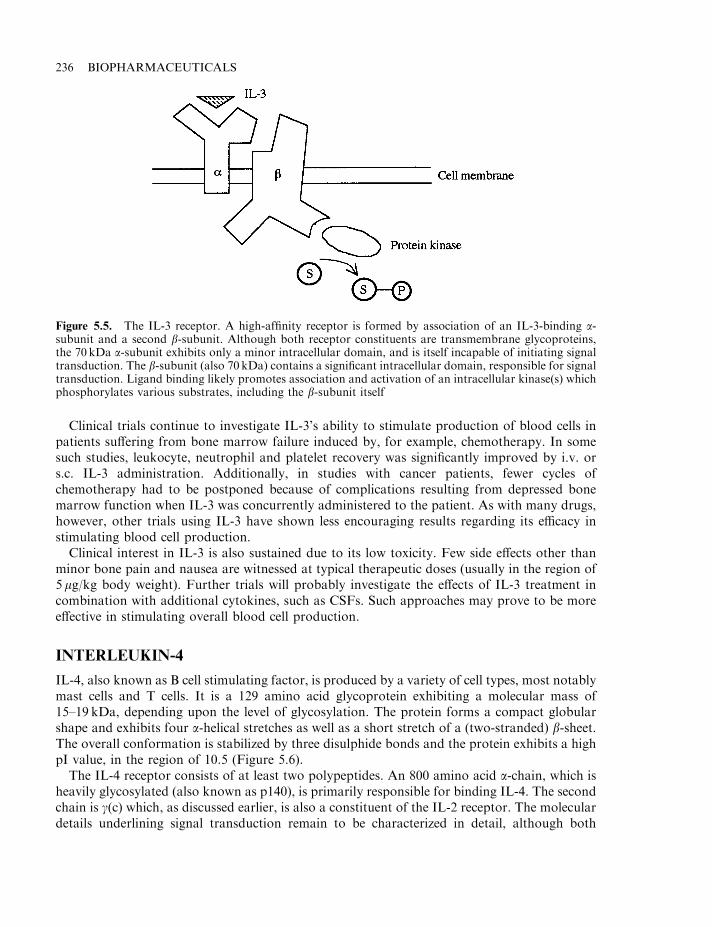
Clinical trials continue to investigate IL-3’s ability to stimulate production of blood cells inpatients suffering from bone marrow failure induced by, for example, chemotherapy. In somesuch studies, leukocyte, neutrophil and platelet recovery was significantly improved by i.v. ors.c. IL-3 administration. Additionally, in studies with cancer patients, fewer cycles ofchemotherapy had to be postponed because of complications resulting from depressed bonemarrow function when IL-3 was concurrently administered to the patient. As with many drugs,however, other trials using IL-3 have shown less encouraging results regarding its efficacy instimulating blood cell production.
Clinical interest in IL-3 is also sustained due to its low toxicity. Few side effects other thanminor bone pain and nausea are witnessed at typical therapeutic doses (usually in the region of5 mg/kg body weight). Further trials will probably investigate the effects of IL-3 treatment incombination with additional cytokines, such as CSFs. Such approaches may prove to be moreeffective in stimulating overall blood cell production.
INTERLEUKIN-4
IL-4, also known as B cell stimulating factor, is produced by a variety of cell types, most notablymast cells and T cells. It is a 129 amino acid glycoprotein exhibiting a molecular mass of15–19 kDa, depending upon the level of glycosylation. The protein forms a compact globularshape and exhibits four a-helical stretches as well as a short stretch of a (two-stranded) b-sheet.The overall conformation is stabilized by three disulphide bonds and the protein exhibits a highpI value, in the region of 10.5 (Figure 5.6).
The IL-4 receptor consists of at least two polypeptides. An 800 amino acid a-chain, which isheavily glycosylated (also known as p140), is primarily responsible for binding IL-4. The secondchain is g(c) which, as discussed earlier, is also a constituent of the IL-2 receptor. The moleculardetails underlining signal transduction remain to be characterized in detail, although both
236 BIOPHARMACEUTICALS
Figure 5.5. The IL-3 receptor. A high-affinity receptor is formed by association of an IL-3-binding a-subunit and a second b-subunit. Although both receptor constituents are transmembrane glycoproteins,the 70 kDa a-subunit exhibits only a minor intracellular domain, and is itself incapable of initiating signaltransduction. The b-subunit (also 70 kDa) contains a significant intracellular domain, responsible for signaltransduction. Ligand binding likely promotes association and activation of an intracellular kinase(s) whichphosphorylates various substrates, including the b-subunit itself
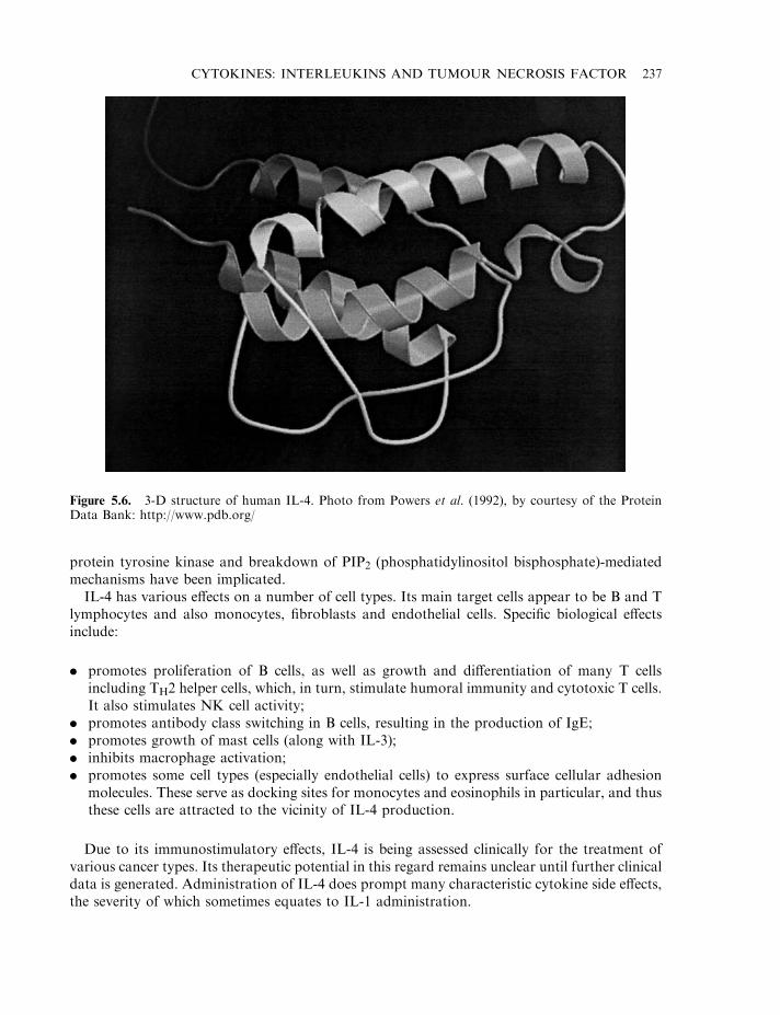
protein tyrosine kinase and breakdown of PIP2 (phosphatidylinositol bisphosphate)-mediatedmechanisms have been implicated.
IL-4 has various effects on a number of cell types. Its main target cells appear to be B and Tlymphocytes and also monocytes, fibroblasts and endothelial cells. Specific biological effectsinclude:
. promotes proliferation of B cells, as well as growth and differentiation of many T cellsincluding TH2 helper cells, which, in turn, stimulate humoral immunity and cytotoxic T cells.It also stimulates NK cell activity;
. promotes antibody class switching in B cells, resulting in the production of IgE;
. promotes growth of mast cells (along with IL-3);
. inhibits macrophage activation;
. promotes some cell types (especially endothelial cells) to express surface cellular adhesionmolecules. These serve as docking sites for monocytes and eosinophils in particular, and thusthese cells are attracted to the vicinity of IL-4 production.
Due to its immunostimulatory effects, IL-4 is being assessed clinically for the treatment ofvarious cancer types. Its therapeutic potential in this regard remains unclear until further clinicaldata is generated. Administration of IL-4 does prompt many characteristic cytokine side effects,the severity of which sometimes equates to IL-1 administration.
CYTOKINES: INTERLEUKINS AND TUMOUR NECROSIS FACTOR 237
Figure 5.6. 3-D structure of human IL-4. Photo from Powers et al. (1992), by courtesy of the ProteinData Bank: http://www.pdb.org/
In some instances, therapeutic benefit might accrue from inhibition of IL-4 activity. Thiscytokine plays a central role in the induction of immediate hypersensitivity (i.e. allergic)reactions, through processes such as:
. induction of IgE synthesis—which is a major mediator of many allergic reactions;
. its ability to promote congregation of monocytes and eosinophils, which, again, containcellular mediators of inflammation;
. its growth-promoting effect on mast cells (which harbour granules filled with inflammatory/allergic substances, such as histamine).
Blocking of IL-4 activity by strategies previously discussed in the context of other cytokines(e.g. administration of soluble receptor fragments, monoclonal antibodies, etc.) thus may have apotential role to play in treating allergies, especially acute life-threatening episodes associatedwith asthma or anaphylaxis.
INTERLEUKIN-6
IL-6 is a pleiotropic cytokine, synthesized by a variety of cell types (both leukocyte and non-leukocyte), which induces biological effects in a number of cell types—both leukocyte and non-leukocyte (Table 5.6).
The gene coding for IL-6 consists of five exons separated by four introns, and is present onchromosome 7. IL-6 is initially synthesized as a 26 kDa, 212 amino acid precursor protein.Subsequent removal of a 28 amino acid signal peptide yields the mature 184 amino acidcytokine, whose molecular mass remains at approximately 26 kDa due to glycosylation (removalof the carbohydrate component appears to have no effect on the biological activity of themolecule). Interestingly, IL-6, produced by fibroblasts and monocyte/macrophages at least, isalso phosphorylated at several residues.
The IL-6 receptor, present on IL-6-sensitive cells (Table 5.6), consists of two associatedtransmembrane polypeptides, a and b. The extracellular domains of both are glycosylated. The449 amino acid, 80 kDa a-chain (IL-6 Ra) binds IL-6 directly, while the 130 kDa b-chain (IL-6Rb, gp 130) then initiates intracellular signalling (Figure 5.7).
The exact details of signal transduction remain to be clarified. Binding of IL-6 to the a-chain,however, appears to facilitate direct interaction of the b-chain with a second b-chain. This
238 BIOPHARMACEUTICALS
Table 5.6. Cells synthesizing and targeted by IL-6. Macrophages,endothelial cells and fibroblasts are probably the major producer cells
Producer cellsIL-6 sensitive cells(i.e. express IL-6 receptor)
T lymphocytes T lymphocytesB lymphocytes Activated B lymphocytesMacrophages MonocytesFibroblasts FibroblastsBone marrow stromal cells Epithelial cellsEndothelial cells HepatocytesKeratinocytes Neural cellsAstrocytes
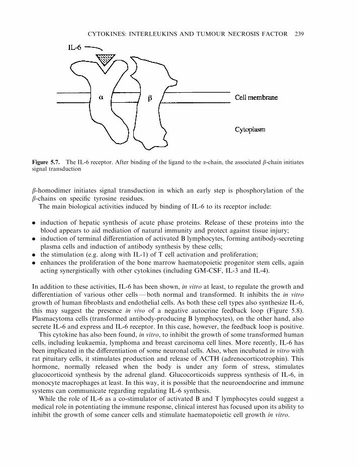
b-homodimer initiates signal transduction in which an early step is phosphorylation of theb-chains on specific tyrosine residues.
The main biological activities induced by binding of IL-6 to its receptor include:
. induction of hepatic synthesis of acute phase proteins. Release of these proteins into theblood appears to aid mediation of natural immunity and protect against tissue injury;
. induction of terminal differentiation of activated B lymphocytes, forming antibody-secretingplasma cells and induction of antibody synthesis by these cells;
. the stimulation (e.g. along with IL-1) of T cell activation and proliferation;
. enhances the proliferation of the bone marrow haematopoietic progenitor stem cells, againacting synergistically with other cytokines (including GM-CSF, IL-3 and IL-4).
In addition to these activities, IL-6 has been shown, in vitro at least, to regulate the growth anddifferentiation of various other cells—both normal and transformed. It inhibits the in vitrogrowth of human fibroblasts and endothelial cells. As both these cell types also synthesize IL-6,this may suggest the presence in vivo of a negative autocrine feedback loop (Figure 5.8).Plasmacytoma cells (transformed antibody-producing B lymphocytes), on the other hand, alsosecrete IL-6 and express and IL-6 receptor. In this case, however, the feedback loop is positive.
This cytokine has also been found, in vitro, to inhibit the growth of some transformed humancells, including leukaemia, lymphoma and breast carcinoma cell lines. More recently, IL-6 hasbeen implicated in the differentiation of some neuronal cells. Also, when incubated in vitro withrat pituitary cells, it stimulates production and release of ACTH (adrenocorticotrophin). Thishormone, normally released when the body is under any form of stress, stimulatesglucocorticoid synthesis by the adrenal gland. Glucocorticoids suppress synthesis of IL-6, inmonocyte macrophages at least. In this way, it is possible that the neuroendocrine and immunesystems can communicate regarding regulating IL-6 synthesis.
While the role of IL-6 as a co-stimulator of activated B and T lymphocytes could suggest amedical role in potentiating the immune response, clinical interest has focused upon its ability toinhibit the growth of some cancer cells and stimulate haematopoietic cell growth in vitro.
CYTOKINES: INTERLEUKINS AND TUMOUR NECROSIS FACTOR 239
Figure 5.7. The IL-6 receptor. After binding of the ligand to the a-chain, the associated b-chain initiatessignal transduction
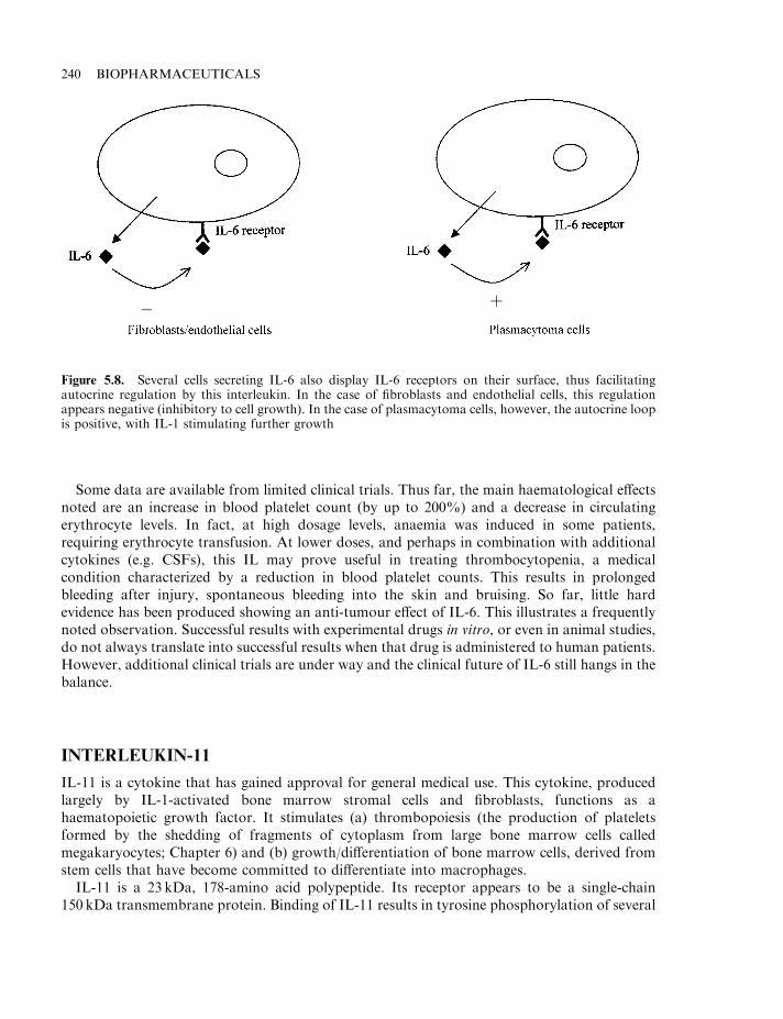
Some data are available from limited clinical trials. Thus far, the main haematological effectsnoted are an increase in blood platelet count (by up to 200%) and a decrease in circulatingerythrocyte levels. In fact, at high dosage levels, anaemia was induced in some patients,requiring erythrocyte transfusion. At lower doses, and perhaps in combination with additionalcytokines (e.g. CSFs), this IL may prove useful in treating thrombocytopenia, a medicalcondition characterized by a reduction in blood platelet counts. This results in prolongedbleeding after injury, spontaneous bleeding into the skin and bruising. So far, little hardevidence has been produced showing an anti-tumour effect of IL-6. This illustrates a frequentlynoted observation. Successful results with experimental drugs in vitro, or even in animal studies,do not always translate into successful results when that drug is administered to human patients.However, additional clinical trials are under way and the clinical future of IL-6 still hangs in thebalance.
INTERLEUKIN-11
IL-11 is a cytokine that has gained approval for general medical use. This cytokine, producedlargely by IL-1-activated bone marrow stromal cells and fibroblasts, functions as ahaematopoietic growth factor. It stimulates (a) thrombopoiesis (the production of plateletsformed by the shedding of fragments of cytoplasm from large bone marrow cells calledmegakaryocytes; Chapter 6) and (b) growth/differentiation of bone marrow cells, derived fromstem cells that have become committed to differentiate into macrophages.
IL-11 is a 23 kDa, 178-amino acid polypeptide. Its receptor appears to be a single-chain150 kDa transmembrane protein. Binding of IL-11 results in tyrosine phosphorylation of several
240 BIOPHARMACEUTICALS
Figure 5.8. Several cells secreting IL-6 also display IL-6 receptors on their surface, thus facilitatingautocrine regulation by this interleukin. In the case of fibroblasts and endothelial cells, this regulationappears negative (inhibitory to cell growth). In the case of plasmacytoma cells, however, the autocrine loopis positive, with IL-1 stimulating further growth
� þ
intracellular proteins which, in turn, somehow promote the observed biological activities ofIL-11.
The rationale for assessing IL-11 as a potential therapeutic agent relates mainly to its abilityto induce platelet synthesis. Platelet counts can be significantly lowered due to certain diseaseconditions, and in patients undergoing cancer chemotherapy. Trials in patients receivingchemotherapy illustrated that s.c. administration of IL-11, at levels above 25 mg/kg body weight/day, did stimulate platelet production, at least partially offsetting the chemotherapeutic effects.When administered at levels of 50 mg/kg/day, a significant increase in the bone marrowmegakaryocyte population was noted. At levels in excess of 75 mg/kg/day, side effects includingfatigue and oedema (tissue swelling due to accumulation of fluid) were also noted.
Neumega is the trade name given to the IL-11-based product approved for the prevention ofthrombocytopenia. The product is produced in engineered E. coli cells and is presented as apurified product in freeze-dried format. Excipients include phosphate buffer salts and glycine. Itis reconstituted (with water for injections) to a concentration of 5mg/ml before s.c.administration.
INTERLEUKIN-5
The biological activities of several other interleukins also render them likely candidates fortherapeutic application. IL-5 represents one such candidate. IL-5 is a 115 amino acidglycoprotein produced mainly by activated T lymphocytes and also by mast cells. It functions asa homodimer, exhibiting a molecular mass of 45 kDa. The individual polypeptide chains interactnon-covalently and the overall dimeric structure is stabilized by two interchain disulphidelinkages between cysteines 42 and 84 of each chain. Removal or alterations of the cytokine’scarbohydrate side-chain does not appear to affect its biological activity.
X-ray diffractional analysis of crystalline human IL-5 reveals a two-domain structure, witheach domain consisting of four a-helical stretches and one b-stretch consisting of two anti-parallel b-strands.
The human IL-5 receptor consists of two transmembrane glycoprotein subunits. Theglycosylated 60 kDa a-chain binds IL-5 but is unable to induce direct signal transduction.A b-chain, unable to bind IL-5 directly, is associated with the a-chain to form an intact receptorcomplex. This 130 kDa b-chain, which also constitutes part of the IL-3 and GM-CSF receptors,appears to initiate signal transduction. In addition to the form anchored in the plasmamembrane, cells are capable of producing a mRNA coding for a soluble form of the receptora-chain by a process of alternative splicing. The exact physiological significance of this isunclear, as expression/secretion of this alternative mRNA in humans appears very low. Whenthe corresponding cDNA is expressed in animal cell lines, and the soluble receptor purified andsubjected to ligand binding studies, it was found to compete effectively with the membranereceptor for IL-5 binding. Clearly, if synthesized and secreted by human cells, this solublereceptor protein could have an antagonistic effect on IL-5 bioactivity. Soluble receptors for anumber of additional human cytokines (some already discussed) have also been discovered(Table 5.7).
In humans, IL-5 functions as a growth and differentiation factor for eosinophils. It alsostimulates basophil growth and may influence B lymphocyte activity. Eosinophils are leukocytescontaining large cytoplasmic granules. The granules contain basic proteins capable of binding toacidic dyes such as eosin. The two main granular proteins are eosinophil cationic protein andmajor basic protein. Eosinophils play a particularly important role in mediating the destruction
CYTOKINES: INTERLEUKINS AND TUMOUR NECROSIS FACTOR 241
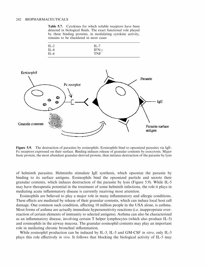
of helminth parasites. Helminths stimulate IgE synthesis, which opsonize the parasite bybinding to its surface antigens. Eosinophils bind the opsonized particle and secrete theirgranular contents, which induces destruction of the parasite by lysis (Figure 5.9). While IL-5may have therapeutic potential in the treatment of some helminth infections, the role it plays inmediating acute inflammatory disease is currently receiving most attention.
Eosinophils are believed to play a major role in many inflammatory and allergic conditions.These effects are mediated by release of their granular contents, which can induce local host celldamage. One common such condition, affecting 10 million people in the USA alone, is asthma.Most forms of asthma are actually immediate hypersensitivity reactions (i.e. inappropriate over-reaction of certain elements of immunity to selected antigens). Asthma can also be characterizedas an inflammatory disease, involving certain T helper lymphocytes (which also produce IL-5)and eosinophils in the airway mucosa. The granular eosinophil contents may play an importantrole in mediating chronic bronchial inflammation.
While eosinophil production can be induced by IL-3, IL-5 and GM-CSF in vitro, only IL-5plays this role effectively in vivo. It follows that blocking the biological activity of IL-5 may
242 BIOPHARMACEUTICALS
Figure 5.9. The destruction of parasites by eosinophils. Eosinophils bind to opsonized parasites via IgE-Fc receptors expressed on their surface. Binding induces release of granular contents by exocytosis. Majorbasic protein, the most abundant granular-derived protein, then initiates destruction of the parasite by lysis
Table 5.7. Cytokines for which soluble receptors have beendetected in biological fluids. The exact functional role playedby these binding proteins, in modulating cytokine activity,remains to be elucidated in most cases
IL-2IL-4IL-6
IL-7IFN-gTNF
provide a means of treating or alleviating chronic or inflammatory conditions associated, atleast in part, with eosinophilia (increased eosinophil counts).
A number of strategies may be adopted to inhibit IL-5 activity. Methods currently underinvestigation include:
. inhibition of IL-5 synthesis/release by T-lymphocytes;
. use of anti-IL-5 monoclonal antibodies or soluble IL-5 receptor polypeptides;
. development of a mutant IL-5 capable of binding its receptor but without triggering signaltransduction;
. identification of a low molecular mass substance which, by interacting with either IL-5 or itsreceptor, would prevent direct interaction of the cytokine and its receptor and hence preventIL-5 signalling.
A soluble receptor has been identified in vivo which, when produced by recombinant meansand administered at higher levels, does act as an effective receptor antagonist. Fab fragments ofa humanized anti-IL-5 monoclonal antibody (Chapter 10) have also been produced, which canreduce IL-5-mediated effects in animal models.
Alanine scans (use of genetic engineering to generate a series of mutant proteins in which eachresidue in the native protein is systematically changed to alanine) of IL-5 have identified severalresidues important in ligand–receptor interaction. Replacement of IL-5 residue No. 13 (usuallyGlu) with alanine, while having no effect upon receptor binding, almost totally preventssubsequent initiation of signal transduction.
The ongoing (chronic) nature of many inflammatory conditions renders most attractive thedevelopment of a drug which would:
. exhibit a long circulatory half-life;
. be relatively inexpensive;
. be capable of being administered orally.
All the protein-based potential therapeutics discussed above fail to meet any of these criteria.Low molecular mass antagonists of IL-5 or its receptor may prove of most use. A number ofresearch teams are screening for such substances. The development strategies pursued include:
. computer-aided drug design; attempts are being made to logically design molecules capableof binding to specific regions of IL-5 (or its receptor) and hence blocking their function;
. traditional screening methods; the screening of microbial, plant or other extracts forsubstances capable of interfering with IL-5 ligand–receptor interaction.
Thus far, at least one class of compound— isothiazolone derivatives—has been identified aspotent IL-5 inhibitors. These substances react with a free sulphydryl group on the surface of theIL-5 receptor a-subunit, thereby preventing ligand binding. Isothiazolones are already used asanti-microbial agents, but are quite toxic.
Overall, therefore, inhibition of IL-5-mediated biological activities may be achieved, at leastin part, by a number of means. All studies to date, however, have been undertaken in vitro.Clinical trials will be necessary to assess whether any of these strategies are effective in treatingchronic asthma or other inflammatory conditions.
CYTOKINES: INTERLEUKINS AND TUMOUR NECROSIS FACTOR 243
INTERLEUKIN-12
Interleukin 12 represents a cytokine that has just begun to generate substantial clinical interest.This is due to its ability to generate a very strong cell-mediated immune response. IL-12 is aheterodimeric structure composed of two polypeptide subunits, termed p35 and p40. p35 is a196 amino acid glycoprotein exhibiting a molecular mass in the 30–35 kDa range. p40,containing 306 amino acids, is also glycosylated and exhibits a molecular mass in the 35–45 kDarange. Neither chain on its own exhibits biological activity . Monocytes/macrophages appear torepresent the major cellular source of IL-12. Sensitive cells express a cell surface 180 kDaglycoprotein, which serves as the IL-12 receptor. The mechanism of signal transduction remainsto be characterized. Whatever its mechanism of signal transduction, IL-12 induces a number ofimportant biological effects, including:
. stimulation of IFN-g synthesis and release from T lymphocytes and NK cells;
. activation of NK cells;
. induction of the growth and differentiation of precursor T lymphocytes specificallyproducing TH1 cells.
Such biological activities suggest various clinical scenarios in which this cytokine may bepositively employed (Table 5.8).
TH1 cells (i.e. T helper type 1 lymphocytes) are the cellular population that triggers a cell-mediated immune response. This is the most effective means by which the immune system canidentify and destroy many foreign agents, particularly viruses, bacteria or parasites, that haveentered host cells.
In many instances, the unaided immune response to such infections can be inadequate,leading to prolonged and/or serious illness. Examples include the malaria and tuberculosisagents, the HIV virus (which has infected at least 14 million people worldwide), leishmaniasis(caused by the protozoan Leishmania, from which 12 million people suffer) and schistosomiasis,(caused by the Schistoma mansoni helminth, which currently infects approximately 250 millionpeople worldwide).
One underlining therapeutic hope for IL-12 is, therefore, that its administration will initiate astrong cell-mediated immune response, thus effectively destroying many infectious agents suchas those described above.
IL-12-stimulated IFN-g production and NK cell activation further potentates the overallimmune response, not only to infectious agents but also against transformed cells. Cytotoxic Tcells and NK cells represent two of the most important immune effector functions againstcancer. IL-12-mediated immunological activity also renders this cytokine a potentially
244 BIOPHARMACEUTICALS
Table 5.8. Some potential clinical applications of IL-12. While animal trials and invitro studies have yielded encouraging results, clinical trials in humans must beundertaken to assess the real therapeutic potential of this cytokine
Combating infectious diseases (viral, protozoal, fungal, bacterial, helminthic)Anti-cancer agentAlleviating allergic reactionsVaccine adjuvant
important vaccine adjuvant. Its inclusion in vaccine preparations should lead to an enhanced Tcell response against the injected antigen.
Thus far, in vitro and animal studies have confirmed IL-12’s ability to enhance manyimmunological functions. Leishmaniasis is a common protozoan disease in tropical/subtropicalregions, including Africa, India, Central and South America and Southern Europe. Animalstudies have shown that IL-12 administration initiates a strong TH1-mediated immune responsein infected mice, effective in combating the disease and conferring future immunity.
Similiar animal studies using a malaria model system have also proved successful. Using astrain of mice for which malaria infection is normally lethal, scientists have found thatadministration of low doses of IL-12 (10–30 ng/day for 5 days) to infected mice offered effectiveprotection against the disease.
IL-12 may also prove useful in the treatment of AIDS. AIDS patients exhibit reduced IL-12levels and, not surprisingly, reduced T cell-mediated immunological capability. It is hoped thatIL-12 administration to HIV-infected individuals might boost the cell-mediated immunologicalfunction, thus delaying the onset/progression of full-blown AIDS. IL-12 may also prompt amore efficient immunological response against opportunistic infectious agents to which AIDSpatients often succumb due to their immunocompromised state. IL-12 administration toimmunodeficient mice generates an effective protective response (largely by activating NK cells)to the protozoan pathogen Toxoplasma gondii, a common opportunistic infectious agent ofAIDS patients.
IL-12 therapy may yet also play a role in treating or alleviating some allergies. Most allergic/immediate hypersensitivity reactions are predominantly triggered by a TH2-mediated humoralresponse, with the production of antibodies such as IgE often triggering inflammation or hostcell damage. Administration of IL-12 may alleviate some of the effects by switching thepredominant immune response to a TH1-mediated one.
The potential of IL-12 as an effective vaccine adjuvant has also been confirmed in someanimal studies. To be effective, most vaccines must trigger a strong T cell and B cell response.This may not always occur in practice. Co-administration of IL-12 with foreign antigen wasfound to stimulate a strong TH1 response in mice, enhancing the protective effect of variousvaccines, e.g. co-administration of IL-2 with heat-killed Listeria monocytogenes protected micefrom a subsequent lethal dose challenge of the live bacteria. Immunization using heat-killedbacteria alone did not afford such complete immunological protection.
Overall, therefore, IL-12 exhibits therapeutic promise in the treatment of a broad range ofdiseases. However, as is the case with all drugs, in vitro activity/animal trials do not guaranteeefficacy in humans. The real therapeutic significance of IL-12 can only be appraised by clinicaltrials. Some studies further investigating the effect of IL-12 in malaria are under way in Rhesusmonkeys. Human clinical trials have also been initiated to assess the effects of IL-12administration in HIV, chronic viral hepatitis and renal cancer. It is interesting to note thatbiotech companies have initiated clinical trials relating to diseases prevalent in affluent FirstWorld regions. IL-12 may, however, show greatest potential in treating protozoan and otherinfectious agents widespread in poorer world regions.
Clinical trials will also yield the first reliable appraisal of any side effects associated with IL-12administration. Limited information from animal studies hints that, at high doses, IL-12 maycause toxic shock-like syndromes in recipients, and may also be linked to the development ofatherosclerotic plaques.
ILs, therefore, represent a family of proteins of considerable therapeutic promise. While it islikely that several additional IL preparations will gain regulatory approval over the next few
CYTOKINES: INTERLEUKINS AND TUMOUR NECROSIS FACTOR 245
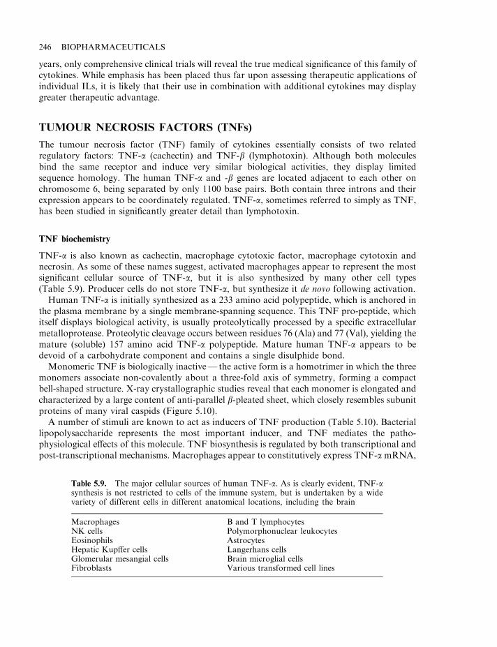
years, only comprehensive clinical trials will reveal the true medical significance of this family ofcytokines. While emphasis has been placed thus far upon assessing therapeutic applications ofindividual ILs, it is likely that their use in combination with additional cytokines may displaygreater therapeutic advantage.
TUMOUR NECROSIS FACTORS (TNFs)
The tumour necrosis factor (TNF) family of cytokines essentially consists of two relatedregulatory factors: TNF-a (cachectin) and TNF-b (lymphotoxin). Although both moleculesbind the same receptor and induce very similar biological activities, they display limitedsequence homology. The human TNF-a and -b genes are located adjacent to each other onchromosome 6, being separated by only 1100 base pairs. Both contain three introns and theirexpression appears to be coordinately regulated. TNF-a, sometimes referred to simply as TNF,has been studied in significantly greater detail than lymphotoxin.
TNF biochemistry
TNF-a is also known as cachectin, macrophage cytotoxic factor, macrophage cytotoxin andnecrosin. As some of these names suggest, activated macrophages appear to represent the mostsignificant cellular source of TNF-a, but it is also synthesized by many other cell types(Table 5.9). Producer cells do not store TNF-a, but synthesize it de novo following activation.
Human TNF-a is initially synthesized as a 233 amino acid polypeptide, which is anchored inthe plasma membrane by a single membrane-spanning sequence. This TNF pro-peptide, whichitself displays biological activity, is usually proteolytically processed by a specific extracellularmetalloprotease. Proteolytic cleavage occurs between residues 76 (Ala) and 77 (Val), yielding themature (soluble) 157 amino acid TNF-a polypeptide. Mature human TNF-a appears to bedevoid of a carbohydrate component and contains a single disulphide bond.
Monomeric TNF is biologically inactive— the active form is a homotrimer in which the threemonomers associate non-covalently about a three-fold axis of symmetry, forming a compactbell-shaped structure. X-ray crystallographic studies reveal that each monomer is elongated andcharacterized by a large content of anti-parallel b-pleated sheet, which closely resembles subunitproteins of many viral caspids (Figure 5.10).
A number of stimuli are known to act as inducers of TNF production (Table 5.10). Bacteriallipopolysaccharide represents the most important inducer, and TNF mediates the patho-physiological effects of this molecule. TNF biosynthesis is regulated by both transcriptional andpost-transcriptional mechanisms. Macrophages appear to constitutively express TNF-a mRNA,
246 BIOPHARMACEUTICALS
Table 5.9. The major cellular sources of human TNF-a. As is clearly evident, TNF-asynthesis is not restricted to cells of the immune system, but is undertaken by a widevariety of different cells in different anatomical locations, including the brain
Macrophages B and T lymphocytesNK cells Polymorphonuclear leukocytesEosinophils AstrocytesHepatic Kupffer cells Langerhans cellsGlomerular mesangial cells Brain microglial cellsFibroblasts Various transformed cell lines
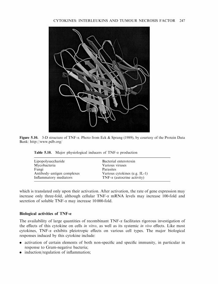
which is translated only upon their activation. After activation, the rate of gene expression mayincrease only three-fold, although cellular TNF-a mRNA levels may increase 100-fold andsecretion of soluble TNF-a may increase 10 000-fold.
Biological activities of TNF-a
The availability of large quantities of recombinant TNF-a facilitates rigorous investigation ofthe effects of this cytokine on cells in vitro, as well as its systemic in vivo effects. Like mostcytokines, TNF-a exhibits pleiotropic effects on various cell types. The major biologicalresponses induced by this cytokine include:
. activation of certain elements of both non-specific and specific immunity, in particular inresponse to Gram-negative bacteria;
. induction/regulation of inflammation;
CYTOKINES: INTERLEUKINS AND TUMOUR NECROSIS FACTOR 247
Figure 5.10. 3-D structure of TNF-a. Photo from Eck & Sprang (1989), by courtesy of the Protein DataBank: http://www.pdb.org/
Table 5.10. Major physiological inducers of TNF-a production
Lipopolysaccharide Bacterial enterotoxinMycobacteria Various virusesFungi ParasitesAntibody–antigen complexes Various cytokines (e.g. IL-1)Inflammatory mediators TNF-a (autocrine activity)

. selective cytotoxic activity against a range of tumour cells;
. mediation of various pathological conditions, including septic shock, cachexia and anorexia.
The exact range of biological effects induced by TNF-a is dependent upon a number offactors, most notably the level at which TNF-a is produced. At low concentrations, TNF-a actslocally in a paracrine and autocrine manner, predominantly influencing white blood cells andendothelial cells. Under such circumstances, TNF-a’s major activity relates to regulation ofimmunity and inflammation. In some situations, however, very large quantities of TNF-a maybe produced (e.g. during severe Gram-negative bacterial infections). In such instances, TNF-aenters the blood stream and acts in an endocrine manner. Systemic effects of TNF-a (systemicmeans relating to the whole body, not just a specific area or organ), which include severe shock,are largely detrimental. Prolonged elevated systemic levels of TNF-a induce additional effectson, for example, whole-body metabolism, as discussed later. Many of TNF-a’s biological effectsare augmented by interferon-g.
Immunity and inflammation
At low concentrations, TNF-a activates a range of leukocytes which mediate selected elementsof both specific and non-specific immunity. These TNF-a actions include:
. activation of various phagocytic cells, including macrophages, neutrophils and polymorpho-nuclear leukocytes;
. enhanced toxicity of eosinophils and macrophages towards pathogens;
. exerts anti-viral activity somewhat similar to class I interferons, and increases surfaceexpression of class I MHC molecules on sensitive cells;
. enhances proliferation of IL-2-dependent T lymphocytes.
In addition, TNF-a influences immunity indirectly by promoting synthesis and release of avariety of additional cytokines, including interferons, IL-1, IL-6, IL-8 and some CSFs.
TNF-a also plays a prominent role in mediating the inflammatory response and, indeed, thismay be its major normal physiological role. It promotes inflammation by a number of means,including:
. promoting activation of neutrophils, eosinophils and other inflammatory leukocytes;
. induction of expression of various adhesion molecules on the surface of vascular endothelialcells. These act as docking sites for neutrophils, monocytes and lymphocytes, facilitatingtheir accumulation at local sites of inflammation;
. displays chemotactic effects, especially for monocytes and polymorphonuclear leukocytes;
. enhances vascular leakness by promoting a reorganization of the cytoskeleton of endothelialcells;
. induction of synthesis of various lipid-based inflammatory mediators, including someprostaglandins and platelet-activating factor (PAF), macrophages and other cells. Many ofthese promote sustained vasodilation and increased vascular leakage;
. induction of synthesis of pro-inflammatory cytokines, such as IL-1 and IL-8.
TNF-a, therefore, promotes various aspects of immunity and inflammation. Blockage of itsactivity, e.g. by administration of anti-TNF-a antibodies, has been shown to compromise thebody’s ability to contain and destroy pathogens.
TNF-a exhibits cytotoxic effects on a wide range of transformed cells. Indeed, initial interestin this molecule (and its naming) stems from its anti-tumour activity. These investigations date
248 BIOPHARMACEUTICALS
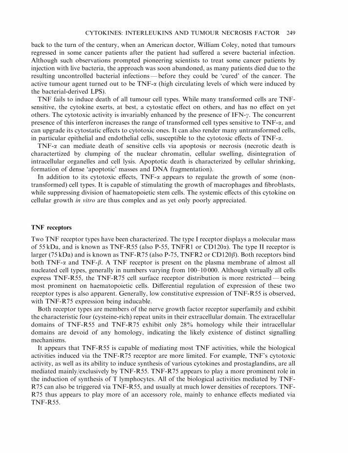
back to the turn of the century, when an American doctor, William Coley, noted that tumoursregressed in some cancer patients after the patient had suffered a severe bacterial infection.Although such observations prompted pioneering scientists to treat some cancer patients byinjection with live bacteria, the approach was soon abandoned, as many patients died due to theresulting uncontrolled bacterial infections—before they could be ‘cured’ of the cancer. Theactive tumour agent turned out to be TNF-a (high circulating levels of which were induced bythe bacterial-derived LPS).
TNF fails to induce death of all tumour cell types. While many transformed cells are TNF-sensitive, the cytokine exerts, at best, a cytostatic effect on others, and has no effect on yetothers. The cytotoxic activity is invariably enhanced by the presence of IFN-g. The concurrentpresence of this interferon increases the range of transformed cell types sensitive to TNF-a, andcan upgrade its cytostatic effects to cytotoxic ones. It can also render many untransformed cells,in particular epithelial and endothelial cells, susceptible to the cytotoxic effects of TNF-a.
TNF-a can mediate death of sensitive cells via apoptosis or necrosis (necrotic death ischaractertized by clumping of the nuclear chromatin, cellular swelling, disintegration ofintracellular organelles and cell lysis. Apoptotic death is characterized by cellular shrinking,formation of dense ‘apoptotic’ masses and DNA fragmentation).
In addition to its cytotoxic effects, TNF-a appears to regulate the growth of some (non-transformed) cell types. It is capable of stimulating the growth of macrophages and fibroblasts,while suppressing division of haematopoietic stem cells. The systemic effects of this cytokine oncellular growth in vitro are thus complex and as yet only poorly appreciated.
TNF receptors
Two TNF receptor types have been characterized. The type I receptor displays a molecular massof 55 kDa, and is known as TNF-R55 (also P-55, TNFR1 or CD120a). The type II receptor islarger (75 kDa) and is known as TNF-R75 (also P-75, TNFR2 or CD120b). Both receptors bindboth TNF-a and TNF-b. A TNF receptor is present on the plasma membrane of almost allnucleated cell types, generally in numbers varying from 100–10 000. Although virtually all cellsexpress TNF-R55, the TNF-R75 cell surface receptor distribution is more restricted—beingmost prominent on haematopoietic cells. Differential regulation of expression of these tworeceptor types is also apparent. Generally, low constitutive expression of TNF-R55 is observed,with TNF-R75 expression being inducable.
Both receptor types are members of the nerve growth factor receptor superfamily and exhibitthe characteristic four (cysteine-rich) repeat units in their extracellular domain. The extracellulardomains of TNF-R55 and TNF-R75 exhibit only 28% homology while their intracellulardomains are devoid of any homology, indicating the likely existence of distinct signallingmechanisms.
It appears that TNF-R55 is capable of mediating most TNF activities, while the biologicalactivities induced via the TNF-R75 receptor are more limited. For example, TNF’s cytotoxicactivity, as well as its ability to induce synthesis of various cytokines and prostaglandins, are allmediated mainly/exclusively by TNF-R55. TNF-R75 appears to play a more prominent role inthe induction of synthesis of T lymphocytes. All of the biological activities mediated by TNF-R75 can also be triggered via TNF-R55, and usually at much lower densities of receptors. TNF-R75 thus appears to play more of an accessory role, mainly to enhance effects mediated viaTNF-R55.
CYTOKINES: INTERLEUKINS AND TUMOUR NECROSIS FACTOR 249
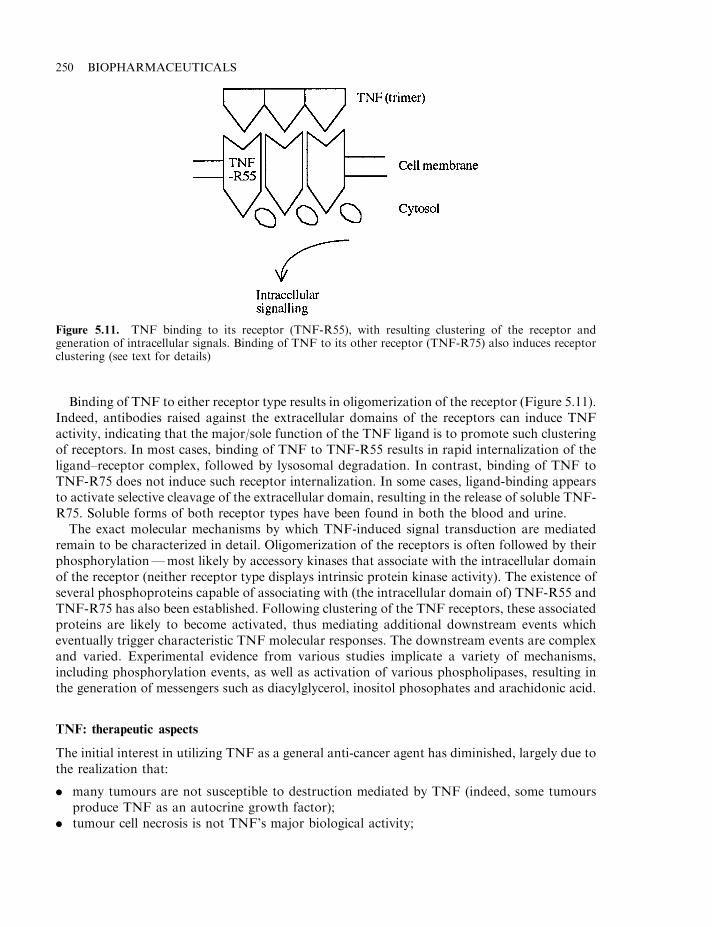
Binding of TNF to either receptor type results in oligomerization of the receptor (Figure 5.11).Indeed, antibodies raised against the extracellular domains of the receptors can induce TNFactivity, indicating that the major/sole function of the TNF ligand is to promote such clusteringof receptors. In most cases, binding of TNF to TNF-R55 results in rapid internalization of theligand–receptor complex, followed by lysosomal degradation. In contrast, binding of TNF toTNF-R75 does not induce such receptor internalization. In some cases, ligand-binding appearsto activate selective cleavage of the extracellular domain, resulting in the release of soluble TNF-R75. Soluble forms of both receptor types have been found in both the blood and urine.
The exact molecular mechanisms by which TNF-induced signal transduction are mediatedremain to be characterized in detail. Oligomerization of the receptors is often followed by theirphosphorylation—most likely by accessory kinases that associate with the intracellular domainof the receptor (neither receptor type displays intrinsic protein kinase activity). The existence ofseveral phosphoproteins capable of associating with (the intracellular domain of) TNF-R55 andTNF-R75 has also been established. Following clustering of the TNF receptors, these associatedproteins are likely to become activated, thus mediating additional downstream events whicheventually trigger characteristic TNF molecular responses. The downstream events are complexand varied. Experimental evidence from various studies implicate a variety of mechanisms,including phosphorylation events, as well as activation of various phospholipases, resulting inthe generation of messengers such as diacylglycerol, inositol phosophates and arachidonic acid.
TNF: therapeutic aspects
The initial interest in utilizing TNF as a general anti-cancer agent has diminished, largely due tothe realization that:
. many tumours are not susceptible to destruction mediated by TNF (indeed, some tumoursproduce TNF as an autocrine growth factor);
. tumour cell necrosis is not TNF’s major biological activity;
250 BIOPHARMACEUTICALS
Figure 5.11. TNF binding to its receptor (TNF-R55), with resulting clustering of the receptor andgeneration of intracellular signals. Binding of TNF to its other receptor (TNF-R75) also induces receptorclustering (see text for details)
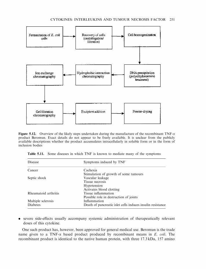
. severe side-effects usually accompany systemic administration of therapeutically relevantdoses of this cytokine.
One such product has, however, been approved for general medical use. Beromun is the tradename given to a TNF-a based product produced by recombinant means in E. coli. Therecombinant product is identical to the native human protein, with three 17.3 kDa, 157 amino
CYTOKINES: INTERLEUKINS AND TUMOUR NECROSIS FACTOR 251
Figure 5.12. Overview of the likely steps undertaken during the manufacture of the recombinant TNF-aproduct Beromun. Exact details do not appear to be freely available. It is unclear from the publiclyavailable descriptions whether the product accumulates intracellularly in soluble form or in the form ofinclusion bodies
Table 5.11. Some diseases in which TNF is known to mediate many of the symptoms
Disease Symptoms induced by TNF
Cancer CachexiaStimulation of growth of some tumours
Septic shock Vascular leakageTissue necrosisHypotensionActivates blood clotting
Rheumatoid arthritis Tissue inflammationPossible role in destruction of joints
Multiple sclerosis InflammationDiabetes Death of pancreatic islet cells induces insulin resistance
acid polypeptides forming the biologically active homotrimer. An overview of its manufacture isprovided in Figure 5.12. Beromun is indicated for the treatment of soft tissue sarcoma in thelimbs. It is administered by isolated limb perfusion (i.e. 3–4mg of product is circulated throughthe limb over 90 min via a perfusion circuit divorced from general body circulation). As theproduct does not enter systemic circulation, toxic systemic side effects are avoided. Surgicalremoval of the tumour mass generally ensues but prior treatment with Beromun prevents/delaysthe need for whole limb amputation in most patients.
Most clinical interest in TNF now centres around neutralizing its biological effects insituations where overexpression of TNF induces negative clinical effects. TNF has been firmlyimplicated in mediating many of the adverse effects associated with dozens of diseases(Table 5.11). Administration of anti-TNF monoclonal antibodies or soluble forms of the TNFreceptor should help reduce the severity of many of the symptoms of these diseases.
Enbrel is a product now approved for medical use which is based upon this strategy. Theproduct is an engineered hybrid protein consisting of the extracellular domain of the TNF p75receptor fused directly to the Fc (constant) region of human IgG (see Chapter 10, Box 10.2, for adiscussion of antibody structure). The product is expressed in a CHO cell line from which it isexcreted as a dimeric soluble protein of approximately 150 kDa. After purification and excipientaddition (mannitol, sucrose and trometamol), the product is freeze-dried. It is indicated for thetreatment of rheumatoid arthritis and is usually administered as a twice weekly s.c. injection of25mg product reconstituted in water for injections. Enbrel functions as a competitive inhibitorof TNF, a major pro-inflammatory cytokine. Binding of TNF to Enbrel prevents it frombinding to its true cell surface receptors. Although commercial product literature makes little orno reference to the function of the antibody Fc component, its presence may hasten removal anddegradation of the product–TNF complex.
More recently, an additional approach to preventing TNF toxicity has been proposed.Several metalloprotease inhibitors (most notably hydroxamic acid) prevent proteolyticprocessing (i.e. release) of TNF-a from producer cell surfaces. Such inhibitors may also proveuseful in preventing TNF-induced illness. The extent to which TNF (and inhibitors of TNF) willserve as future therapeutic agents remains to be determined by future clinical trials.
FURTHER READING
Books
Balkwill, F. (2000). The Cytokine Network. Oxford University Press, Oxford.Fitzgerald, K. (2001). Cytokine Factsbook. Academic Press, London.Mantovani, A. (2000). Pharmacology of Cytokines. Oxford University Press, Oxford.Mire-Sluis, A. (1998). Cytokines. Academic Press, London.
Articles
Interleukins
Arkin, M.A., Randal, M., Delano, W.L. et al. (in press). Binding of small molecules to a hot-spot at a protein–proteininterface. Protein Data Bank: www.rcsb.org/pdb.
Atkins, M. (2002). Interleukin 2: clinical applications. Semin. Oncol. 29(3), 12–17.Aulitzky, W. et al. (1994). Interleukins. Clinical pharmacology and therapeutic use. Drugs, 48(5), 667–677.Brown, M. & Hural, J. (1997). Function of IL-4 and control of its expression. Crit. Rev. Immunol. 17(1), 1–32.Devos, R. et al. (1995). Interleukin-5 and its receptor: a drug target for eosinophilia associated with chronic allergic
disease. J. Leukocyte Biol. 57, 813–818.
252 BIOPHARMACEUTICALS
Fehniger, T. et al. (2002). Interleukin-2 and interleukin 15: immunotherapy for cancer. Cytokine Growth Factor Rev.13(2), 169–183.
Fickenscher, H. et al. (2002). The interleukin-10 family of cytokines. Trends Immunol. 23(2), 89–96.Fry, J. & Mackall, C. (2002). Interleukin-7: from bench to clinic. Blood 99(11), 3892–3904.Jeal, W. & Goa, K. (1997). Aldesleukin (recombinant interleukin-2)—a review of its pharmacological properties, clinical
efficacy and tolerability in patients with renal cell carcinoma. Biodrugs 7(4), 285–317.Komschlies, K. et al. (1995). Diverse immunological and haematological effects of interleukin-7: implications for clinical
application. J. Leukocyte Biol. 58, 623–631.Martin, M. & Falk, W. (1997). The interleukin-1 receptor complex and interleukin 1 signal transduction. Eur. Cytokine
Network 8(1), 5–17.Noble, S. & Goa, K. (1997). Aldesleukin (recombinant interleukin-2)—a review of its pharmacological properties,
clinical efficacy and tolerability in patients with metastic melanoma. Biodrugs 7(5), 394–422.Powers, R., Garrett, D.S., March, C.J. et al. (1992). Three-dimensional solution structure of human interleukin-4 by
multidimensional heteronuclear magnetic resonance spectroscopy. Science 256, 1673.Renauld, J. et al. (1995). Interleukin-9 and its receptor: involvement in mast cell differentiation and T cell oncogenesis.
J. Leukocyte Biol. 57, 353–359.Ryan, J. (1997). Interleukin-4 and its receptor—essential mediators of the allergic response. J. Allergy Clin. Immunol.
99(1), 1–5.Scott, P. (1993). IL-12: initiation cytokine for cell mediated immunity. Science 260, 496–497.Simpson, R. et al. (1997). Interleukin-6 structure–function relationships. Protein Sci. 6(5), 929–955.Wigginton, J. & Wiltrout, R. (2002). IL-12/IL-2 combination cytokine therapy for solid tumors: translation from
benchside to bedside. Expert Opin. Biol. Therapy 2(5), 513–524.
Tumour necrosis factor
Argiles, J. & Lepezsoriano, F. (1997). Cancer cachexia—a key role for TNF. Int. J. Oncol. 10(3), 565–572.Bodmer, J. et al. (2002). The molecular architecture of the TNF superfamily. Trends Biochem. Sci. 27(1), 19–26.Bondeson, J. et al. (2000). TNF as a therapeutic agent. Immunologist 8(6), 136–140.Crowe, P. et al. (1994). A lymphotoxin-b-specific receptor. Science 264, 707–709.Darnay, B. & Aggarwal, B. (1997). Early events in TNF signalling—a story of associations and disassociations.
J. Leukocyte Biol. 61(5), 559–566.Dellinger, R. et al. (1997). From the bench to the bedside— the future of sepsis research. Chest 111(3), 744–753.Eck, M.J. & Sprang, S.R. (1989). The structure of tumor necrosis factor-a at 2.6 A resolution. Implications for receptor
binding. J. Biol. Chem. 264, 17595.Feldmann, M. et al. (1997). Anti-tumor necrosis factor-a therapy of rheumatoid arthritis. Adv. Immunol. 64, 283–350.Hohlfeld, R. (1997). Biotechnological agents for the immunotherapy of multiple sclerosis—principles, problems and
perspectives. Brain 120, 865–916.McDermot, M. (2001). TNF and TNFR biology in health and disease. Cell. Mol. Biol. 47(4), 619–635.MacEwan, D. (2002). TNF receptor subtype signalling: differences and cellular consequences. Cell. Signalling 14(6),
477–492.McGeer, E. & McGeer, P. (1997). Inflammatory cytokines in the CNS—possible role in the pathogenesis of
neurodegenerative disorders and therapeutic implications. CNS Drugs 7(3), 214–228.Maini, R. et al. (1997). TNF blockade in rheumatoid arthritis— implications for therapy and pathogenesis. APMIS
105(4), 257–263.Moreland, W. et al. (1997). Biological agents for treating rheumatoid arthritis—concepts and progress. Arthritis
Rheumatism 40(3), 397–409.Terranova, P. (1997). Potential roles of tumor necrosis factor-a in follicular development, ovulation and the life-span of
the corpus luteum. Domest. Anim. Endocrinol. 14(1), 1–15.Tracey, K. & Cerami, A. (1994). Tumor necrosis factor: a pleiotropic cytokine and therapeutic target. Ann. Rev. Med. 45,
491–503.Vandenabeele, P. et al. (1995). Two tumor necrosis factor receptors: structure and function. Trends Cell Biol. 5,
392–399.
CYTOKINES: INTERLEUKINS AND TUMOUR NECROSIS FACTOR 253

Chapter 6
Haemopoietic growth factors
Blood consists of red and white cells which, along with platelets, are all suspended in plasma. Allperipheral blood cells are derived from a single cell type: the stem cell (also known as apluripotential, pluripotent or haemopoietic stem cell). These stem cells reside in the bonemarrow, alongside additional cell types, including (marrow) stromal cells. Pluripotential stemcells have the capacity to undergo prolonged or indefinite self-renewal. They also have thepotential to differentiate, thereby yielding the range of cells normally found in blood (Table 6.1).This process, by which a fraction of stem cells are continually ‘deciding’ to differentiate (thuscontinually producing new blood cells and platelets to replace aged cells), is known ashaemopoiesis.
The study of the process of haemopoiesis is rendered difficult by the fact that it is extremelydifficult to distinguish or separate individual stem cells from their products during the earlierstages of differentiation. However, a picture of the process of differentiation is now beginning toemerge (Figure 6.1). During the haemopoietic process, the stem cells differentiate, producingcells that become progressively more restricted in their choice of developmental options.
The production of many mature blood cells begins when a fraction of the stem cellsdifferentiate, forming a specific cell type termed CFU-S (CFU refers to colony forming unit).These, in turn, differentiate yielding CFU-GEMM cells, a mixed CFU which has the potentialto differentiate into a range of mature blood cell types, including granulocytes, monocytes,erythrocytes, platelets, eosinophils and basophils. Note that lymphocytes are not derived fromthe CFU-GEMM pathway, but differentiate via an alternative pathway from stem cells(Figure 6.1).
The details of haemopoiesis presented thus far prompt two very important questions. How isthe correct balance between stem cell self-renewal and differentiation maintained? And whatforces exist that regulate the process of differentiation? The answer to both questions, inparticular the latter, is beginning to emerge in the form of a group of cytokines termed‘haemopoietic growth factors’ (Table 6.2). This group includes:
. several (of the previously described) interleukins (ILs) that primarily affect production anddifferentiation of lymphocytes;
. colony stimulating factors (CSFs), which play a major role in the differentiation of stem-derived cells into neutrophils, macrophages, megakaryocytes (from which platelets arederived), eosinophils and basophils;
Biopharmaceuticals: Biochemistry and Biotechnology, Second Edition. Gary WalshJohn Wiley & Sons Ltd: ISBN 0 470 84326 8 (ppc), ISBN 0 470 84327 6 (pbk)
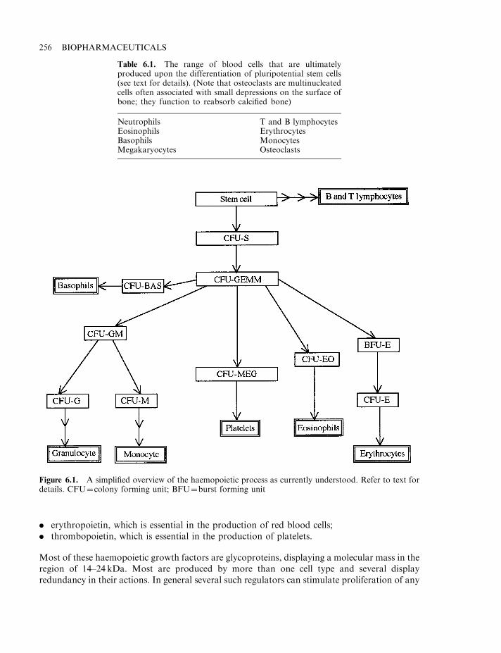
. erythropoietin, which is essential in the production of red blood cells;
. thrombopoietin, which is essential in the production of platelets.
Most of these haemopoietic growth factors are glycoproteins, displaying a molecular mass in theregion of 14–24 kDa. Most are produced by more than one cell type and several displayredundancy in their actions. In general several such regulators can stimulate proliferation of any
256 BIOPHARMACEUTICALS
Table 6.1. The range of blood cells that are ultimatelyproduced upon the differentiation of pluripotential stem cells(see text for details). (Note that osteoclasts are multinucleatedcells often associated with small depressions on the surface ofbone; they function to reabsorb calcified bone)
Neutrophils T and B lymphocytesEosinophils ErythrocytesBasophils MonocytesMegakaryocytes Osteoclasts
Figure 6.1. A simplified overview of the haemopoietic process as currently understood. Refer to text fordetails. CFU¼colony forming unit; BFU¼burst forming unit
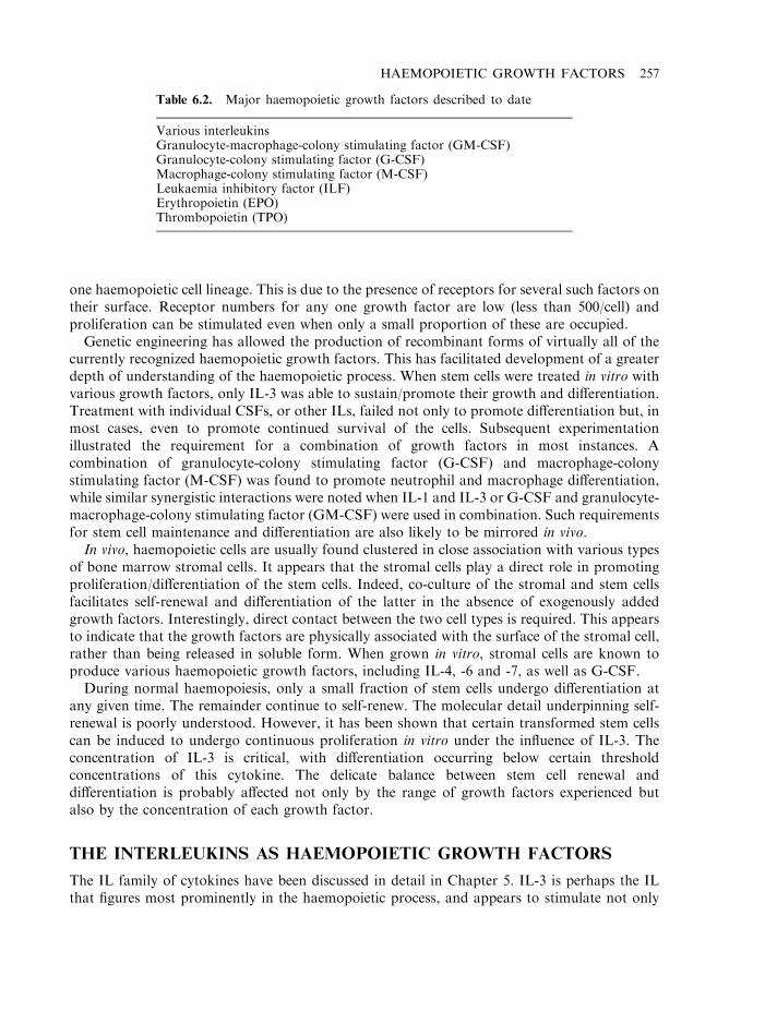
one haemopoietic cell lineage. This is due to the presence of receptors for several such factors ontheir surface. Receptor numbers for any one growth factor are low (less than 500/cell) andproliferation can be stimulated even when only a small proportion of these are occupied.
Genetic engineering has allowed the production of recombinant forms of virtually all of thecurrently recognized haemopoietic growth factors. This has facilitated development of a greaterdepth of understanding of the haemopoietic process. When stem cells were treated in vitro withvarious growth factors, only IL-3 was able to sustain/promote their growth and differentiation.Treatment with individual CSFs, or other ILs, failed not only to promote differentiation but, inmost cases, even to promote continued survival of the cells. Subsequent experimentationillustrated the requirement for a combination of growth factors in most instances. Acombination of granulocyte-colony stimulating factor (G-CSF) and macrophage-colonystimulating factor (M-CSF) was found to promote neutrophil and macrophage differentiation,while similar synergistic interactions were noted when IL-1 and IL-3 or G-CSF and granulocyte-macrophage-colony stimulating factor (GM-CSF) were used in combination. Such requirementsfor stem cell maintenance and differentiation are also likely to be mirrored in vivo.
In vivo, haemopoietic cells are usually found clustered in close association with various typesof bone marrow stromal cells. It appears that the stromal cells play a direct role in promotingproliferation/differentiation of the stem cells. Indeed, co-culture of the stromal and stem cellsfacilitates self-renewal and differentiation of the latter in the absence of exogenously addedgrowth factors. Interestingly, direct contact between the two cell types is required. This appearsto indicate that the growth factors are physically associated with the surface of the stromal cell,rather than being released in soluble form. When grown in vitro, stromal cells are known toproduce various haemopoietic growth factors, including IL-4, -6 and -7, as well as G-CSF.
During normal haemopoiesis, only a small fraction of stem cells undergo differentiation atany given time. The remainder continue to self-renew. The molecular detail underpinning self-renewal is poorly understood. However, it has been shown that certain transformed stem cellscan be induced to undergo continuous proliferation in vitro under the influence of IL-3. Theconcentration of IL-3 is critical, with differentiation occurring below certain thresholdconcentrations of this cytokine. The delicate balance between stem cell renewal anddifferentiation is probably affected not only by the range of growth factors experienced butalso by the concentration of each growth factor.
THE INTERLEUKINS AS HAEMOPOIETIC GROWTH FACTORS
The IL family of cytokines have been discussed in detail in Chapter 5. IL-3 is perhaps the ILthat figures most prominently in the haemopoietic process, and appears to stimulate not only
HAEMOPOIETIC GROWTH FACTORS 257
Table 6.2. Major haemopoietic growth factors described to date
Various interleukinsGranulocyte-macrophage-colony stimulating factor (GM-CSF)Granulocyte-colony stimulating factor (G-CSF)Macrophage-colony stimulating factor (M-CSF)Leukaemia inhibitory factor (ILF)Erythropoietin (EPO)Thrombopoietin (TPO)
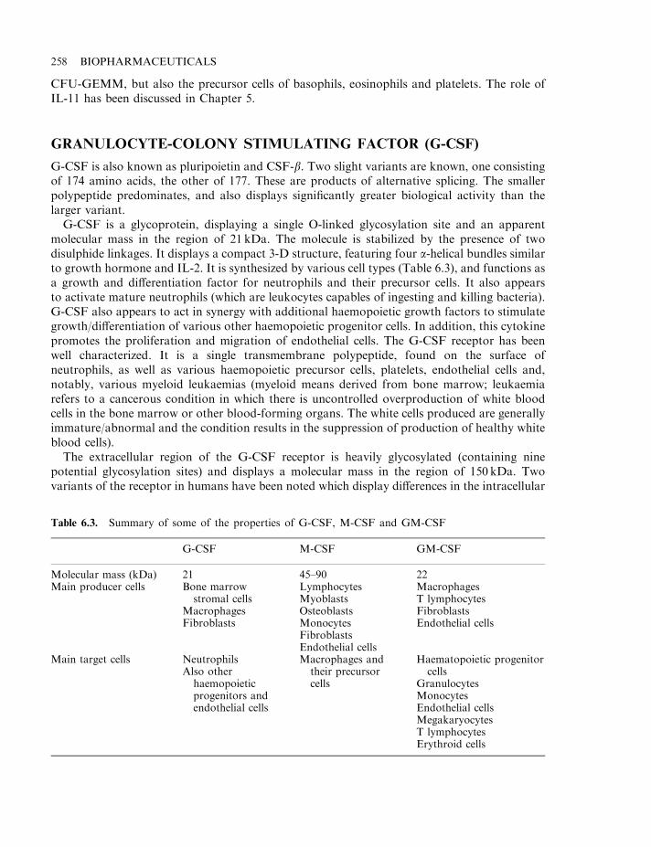
CFU-GEMM, but also the precursor cells of basophils, eosinophils and platelets. The role ofIL-11 has been discussed in Chapter 5.
GRANULOCYTE-COLONY STIMULATING FACTOR (G-CSF)
G-CSF is also known as pluripoietin and CSF-b. Two slight variants are known, one consistingof 174 amino acids, the other of 177. These are products of alternative splicing. The smallerpolypeptide predominates, and also displays significantly greater biological activity than thelarger variant.
G-CSF is a glycoprotein, displaying a single O-linked glycosylation site and an apparentmolecular mass in the region of 21 kDa. The molecule is stabilized by the presence of twodisulphide linkages. It displays a compact 3-D structure, featuring four a-helical bundles similarto growth hormone and IL-2. It is synthesized by various cell types (Table 6.3), and functions asa growth and differentiation factor for neutrophils and their precursor cells. It also appearsto activate mature neutrophils (which are leukocytes capables of ingesting and killing bacteria).G-CSF also appears to act in synergy with additional haemopoietic growth factors to stimulategrowth/differentiation of various other haemopoietic progenitor cells. In addition, this cytokinepromotes the proliferation and migration of endothelial cells. The G-CSF receptor has beenwell characterized. It is a single transmembrane polypeptide, found on the surface ofneutrophils, as well as various haemopoietic precursor cells, platelets, endothelial cells and,notably, various myeloid leukaemias (myeloid means derived from bone marrow; leukaemiarefers to a cancerous condition in which there is uncontrolled overproduction of white bloodcells in the bone marrow or other blood-forming organs. The white cells produced are generallyimmature/abnormal and the condition results in the suppression of production of healthy whiteblood cells).
The extracellular region of the G-CSF receptor is heavily glycosylated (containing ninepotential glycosylation sites) and displays a molecular mass in the region of 150 kDa. Twovariants of the receptor in humans have been noted which display differences in the intracellular
258 BIOPHARMACEUTICALS
Table 6.3. Summary of some of the properties of G-CSF, M-CSF and GM-CSF
G-CSF M-CSF GM-CSF
Molecular mass (kDa) 21 45–90 22Main producer cells Bone marrow Lymphocytes Macrophages
stromal cells Myoblasts T lymphocytesMacrophages Osteoblasts FibroblastsFibroblasts Monocytes Endothelial cells
FibroblastsEndothelial cells
Main target cells Neutrophils Macrophages and Haematopoietic progenitorAlso other their precursor cellshaemopoietic cells Granulocytesprogenitors and Monocytesendothelial cells Endothelial cells
MegakaryocytesT lymphocytesErythroid cells
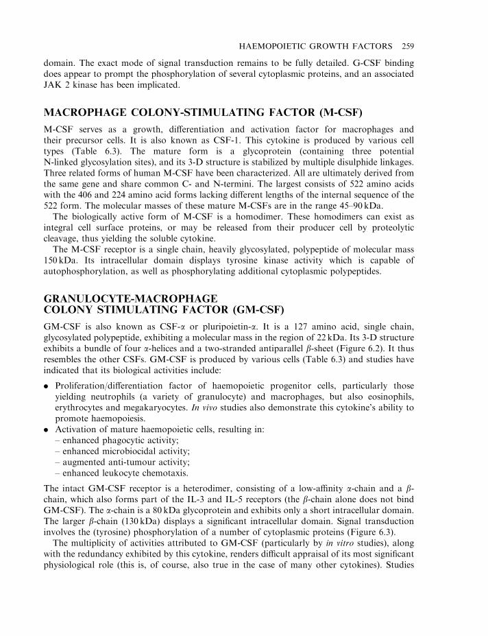
domain. The exact mode of signal transduction remains to be fully detailed. G-CSF bindingdoes appear to prompt the phosphorylation of several cytoplasmic proteins, and an associatedJAK 2 kinase has been implicated.
MACROPHAGE COLONY-STIMULATING FACTOR (M-CSF)
M-CSF serves as a growth, differentiation and activation factor for macrophages andtheir precursor cells. It is also known as CSF-1. This cytokine is produced by various celltypes (Table 6.3). The mature form is a glycoprotein (containing three potentialN-linked glycosylation sites), and its 3-D structure is stabilized by multiple disulphide linkages.Three related forms of human M-CSF have been characterized. All are ultimately derived fromthe same gene and share common C- and N-termini. The largest consists of 522 amino acidswith the 406 and 224 amino acid forms lacking different lengths of the internal sequence of the522 form. The molecular masses of these mature M-CSFs are in the range 45–90 kDa.
The biologically active form of M-CSF is a homodimer. These homodimers can exist asintegral cell surface proteins, or may be released from their producer cell by proteolyticcleavage, thus yielding the soluble cytokine.
The M-CSF receptor is a single chain, heavily glycosylated, polypeptide of molecular mass150 kDa. Its intracellular domain displays tyrosine kinase activity which is capable ofautophosphorylation, as well as phosphorylating additional cytoplasmic polypeptides.
GRANULOCYTE-MACROPHAGECOLONY STIMULATING FACTOR (GM-CSF)
GM-CSF is also known as CSF-a or pluripoietin-a. It is a 127 amino acid, single chain,glycosylated polypeptide, exhibiting a molecular mass in the region of 22 kDa. Its 3-D structureexhibits a bundle of four a-helices and a two-stranded antiparallel b-sheet (Figure 6.2). It thusresembles the other CSFs. GM-CSF is produced by various cells (Table 6.3) and studies haveindicated that its biological activities include:
. Proliferation/differentiation factor of haemopoietic progenitor cells, particularly thoseyielding neutrophils (a variety of granulocyte) and macrophages, but also eosinophils,erythrocytes and megakaryocytes. In vivo studies also demonstrate this cytokine’s ability topromote haemopoiesis.
. Activation of mature haemopoietic cells, resulting in:– enhanced phagocytic activity;– enhanced microbiocidal activity;– augmented anti-tumour activity;– enhanced leukocyte chemotaxis.
The intact GM-CSF receptor is a heterodimer, consisting of a low-affinity a-chain and a b-chain, which also forms part of the IL-3 and IL-5 receptors (the b-chain alone does not bindGM-CSF). The a-chain is a 80 kDa glycoprotein and exhibits only a short intracellular domain.The larger b-chain (130 kDa) displays a significant intracellular domain. Signal transductioninvolves the (tyrosine) phosphorylation of a number of cytoplasmic proteins (Figure 6.3).
The multiplicity of activities attributed to GM-CSF (particularly by in vitro studies), alongwith the redundancy exhibited by this cytokine, renders difficult appraisal of its most significantphysiological role (this is, of course, also true in the case of many other cytokines). Studies
HAEMOPOIETIC GROWTH FACTORS 259
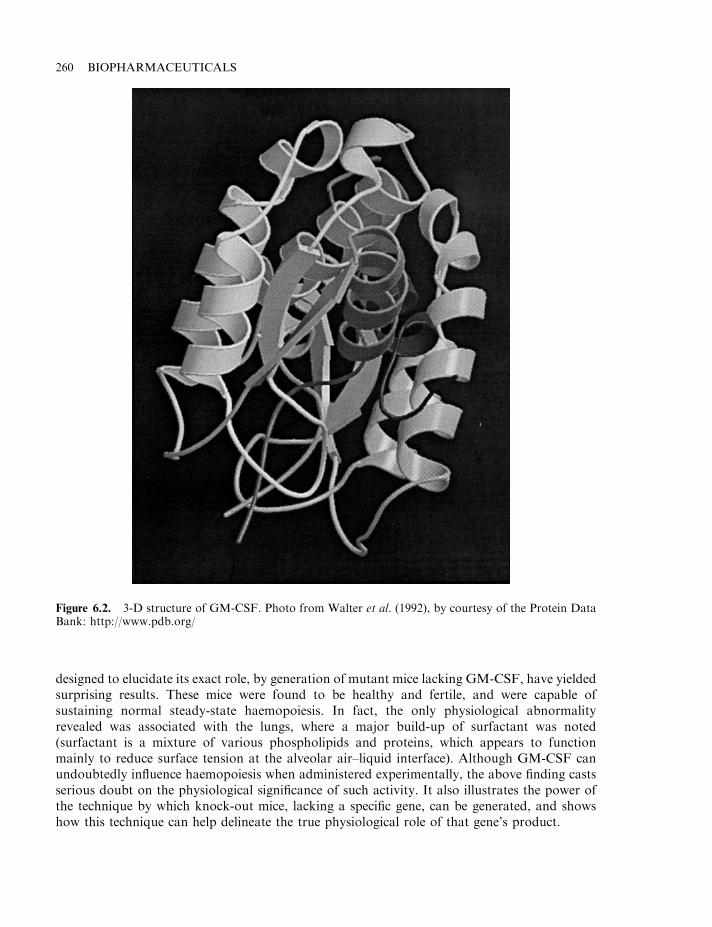
designed to elucidate its exact role, by generation of mutant mice lacking GM-CSF, have yieldedsurprising results. These mice were found to be healthy and fertile, and were capable ofsustaining normal steady-state haemopoiesis. In fact, the only physiological abnormalityrevealed was associated with the lungs, where a major build-up of surfactant was noted(surfactant is a mixture of various phospholipids and proteins, which appears to functionmainly to reduce surface tension at the alveolar air–liquid interface). Although GM-CSF canundoubtedly influence haemopoiesis when administered experimentally, the above finding castsserious doubt on the physiological significance of such activity. It also illustrates the power ofthe technique by which knock-out mice, lacking a specific gene, can be generated, and showshow this technique can help delineate the true physiological role of that gene’s product.
260 BIOPHARMACEUTICALS
Figure 6.2. 3-D structure of GM-CSF. Photo from Walter et al. (1992), by courtesy of the Protein DataBank: http://www.pdb.org/
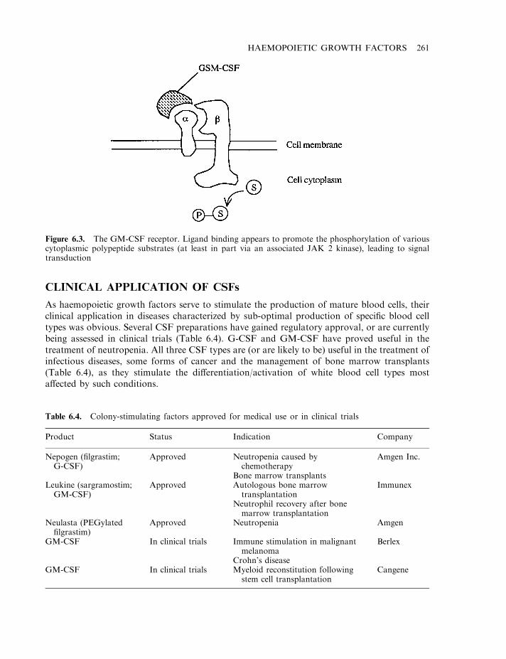
CLINICAL APPLICATION OF CSFs
As haemopoietic growth factors serve to stimulate the production of mature blood cells, theirclinical application in diseases characterized by sub-optimal production of specific blood celltypes was obvious. Several CSF preparations have gained regulatory approval, or are currentlybeing assessed in clinical trials (Table 6.4). G-CSF and GM-CSF have proved useful in thetreatment of neutropenia. All three CSF types are (or are likely to be) useful in the treatment ofinfectious diseases, some forms of cancer and the management of bone marrow transplants(Table 6.4), as they stimulate the differentiation/activation of white blood cell types mostaffected by such conditions.
HAEMOPOIETIC GROWTH FACTORS 261
Figure 6.3. The GM-CSF receptor. Ligand binding appears to promote the phosphorylation of variouscytoplasmic polypeptide substrates (at least in part via an associated JAK 2 kinase), leading to signaltransduction
Table 6.4. Colony-stimulating factors approved for medical use or in clinical trials
Product Status Indication Company
Nepogen (filgrastim;G-CSF)
Approved Neutropenia caused bychemotherapy
Amgen Inc.
Bone marrow transplantsLeukine (sargramostim;GM-CSF)
Approved Autologous bone marrowtransplantation
Immunex
Neutrophil recovery after bonemarrow transplantation
Neulasta (PEGylatedfilgrastim)
Approved Neutropenia Amgen
GM-CSF In clinical trials Immune stimulation in malignantmelanoma
Crohn’s disease
Berlex
GM-CSF In clinical trials Myeloid reconstitution followingstem cell transplantation
Cangene
Neutropenia is a condition characterized by a decrease in blood neutrophil count below1.5�109 cells/l (normal blood count 2.0–7.5�109 cells/l). Its clinical symptoms include theoccurrence of frequent and usually serious infections, often requiring hospitalization.Neutropenia may be caused by a number of factors (Table 6.5), at least some of which areresponsive to CSF treatment. Particularly noteworthy is neutropenia triggered by administra-tion of chemotherapeutic drugs to cancer patients. Chemotherapeutic agents (e.g.cyclophosphamide, doxorubicin and methotrexate), when administered at therapeuticallyeffective doses, often induce the destruction of stem cells and/or compromise stem celldifferentiation.
Filgrastim is a recombinant human G-CSF (produced in E. coli), approved forchemotherapy-induced neutropenia since 1991. Although the 18.8 kDa recombinant productis not glycosylated and contains an additional N-terminal methionine residue (due to expressionin E. coli), it displays biological activity indistinguishable from native G-CSF. The product ispresented in freeze-dried format and contains buffer elements as well as sorbitol and Tween asexcipients.
Administration of filgrastim (0.5 mg/kg/day) to healthy volunteers for six consecutive daysresulted in up to a 10-fold increase in their absolute neutrophil count. Daily administration topatients receiving chemotherapy greatly reduced the duration and severity of leucopenia(reduction in the numbers of total white blood cells). The magnitude of the specific neutrophilresponse depended upon the number of progenitor cells still surviving upon commencement oftreatment. Neutrophils from patients with neutropenia treated with filgrastim also displayenhanced phagocytosis and chemotaxis, at least transiently.
Filgrastim is used in conjunction with cancer chemotherapy in one of two ways: prophylacticadministration aims to prevent onset of neutropenia in the first place, while therapeuticadministration aims to reverse neutropenia already established. It is generally effective in bothcircumstances and in clinical trials it was found to reduce the number of (neutropenia-induced)days of patient hospitalization by up to 50%. Filgrastim administration may also permit modestincreases in the maximum dose intensity utilizable in chemotherapeutic regimes.
Filgrastim is usually administered by subcutaneous injection. Dosage levels range from5–10 mg/kg/day, with peak plasma drug concentrations being observed 2–8 hours afteradministration. The mechanism by which filgrastim is removed from circulation is unclear,but the drug’s elimination half-life ranges from less than 2 h to almost 5 h. The drug is generallywell tolerated. The most common adverse reaction is mild bone pain, which occurs in up to 20%of cancer patients receiving treatment. This is easily treated with analgesics.
262 BIOPHARMACEUTICALS
Table 6.5. Some causes of neutropenia
Genetic (particularly in black populations)Severe bacterial infectionSevere sepsisSevere viral infectionAplastic anaemia*Acute leukaemiaHodgkin’s/non-Hodgkin’s lymphomaVarious drugs, especially anti-cancer drugsAutoimmune neutropenia
*Aplastic anaemia describes bone marrow failure characterized byserious reduction in the number of stem cells present
While filgrastim has proved a useful adjuvant to chemotherapy for many cancers, itsadministration in cases of myeloid leukaemia would give cause for concern, as these cells expressreceptors for G-CSF. In such cases, G-CSF could potentially promote accelerated growth ofthese malignant cells.
Filgrastim also shows therapeutic promise in the treatment of additional conditionscharacterized by depressed blood neutrophil counts. Such conditions include severe chronicneutropenia, leukaemia and AIDS.
Neulasta is the tradename given to a PEGylated form of filgrastim approved for generalmedical use in the USA in 2002. Manufacture of this product entails covalent attachment of anactivated monomethoxypolyethylene glycol molecule to the N-terminal methionyl residue offilgrastim. The product is formulated in the presence of acetate buffer, sorbitol and polysorbateand is presented in pre-filled syringes for subcutaneous injection. As in the case of PEGylatedinterferons (IFNs) (Chapter 4), the rationale for PEGylation is to increase the drug’s plasmahalf-life, thereby reducing the frequency of injections required.
G-CSF and GM-CSF have also found application after allogenic or autologous bonemarrow transplantation, to accelerate neutrophil recovery (allogenic means that donor andrecipient are different individuals, while autologous means that donor and recipient are the sameindividual).
Bone marrow transplantation, particularly allogenic transplantation, is often a treatment ofchoice for individuals suffering from acute or chronic leukaemia, aplastic anaemia or variousstem cell-related genetic disorders (e.g. thalassaemias).
Allogenic transplantation generally entails removal of approximately 1 l of marrow from amatched donor (usually a sibling, preferably an identical twin). The recipient patient isprepared by intensive chemotherapy (or radiotherapy) to kill residual malignant marrowcells. The donated marrow cells are then administered intravenously to the patient, andthey re-populate the marrow cavity. The peripheral blood count normally rises within 2–4weeks.
Autologous bone marrow transplantation involves initial removal of some marrow from thepatient, its storage in liquid nitrogen, followed by its re-introduction into the patient subsequentto chemo- or radiotherapy. Leukine is the tradename given to a recombinant human GM-CSFpreparation produced in engineered S. cerevisiae (Table 6.4).
LEUKAEMIA INHIBITORY FACTOR (LIF)
LIF is an additional haemopoietic growth factor. It is also known as HILDA (humaninterleukin for DA cells) and hepatocyte stimulatory factor III. It is produced by a range ofcell types, including T cells, liver cells and fibroblasts. LIF is a 180 amino acid, heavilyglycosylated, 45 kDa protein. It affects both haemopoietic and non-haemopoietic tissue, oftenacting in synergy with other cytokines, particularly IL-3. It stimulates the differentiation ofmacrophages and promotes enhanced platelet formation. It also prompts synthesis of acutephase proteins by the liver and promotes increased bone resorption. The greatest concentrationsof LIF receptors are associated with monocytes, embryonic stem cells, liver cells and theplacenta. The receptor complex is composed of two transmembrane glycoproteins: the 190 kDaLIFRa-chain which displays affinity for LIF, and a b-chain (gp130), which also forms part ofthe IL-6 receptor.
HAEMOPOIETIC GROWTH FACTORS 263
ERYTHROPOIETIN (EPO)
Erythropoietin (EPO) is an additional haemopoietic growth factor. It is primarily responsiblefor stimulating and regulating erythropoiesis (i.e. erythrogenesis, the production of red bloodcells) in mammals.
The ‘erythron’ is a collective term given to mature erythrocytes, along with all stem cell-derived progeny that have committed to developing into erythrocytes. It can thus be viewed as adispersed organ, whose primary function relates to transport of oxygen and carbon dioxide(haemoglobin constitutes up to one-third of the erythrocyte cytoplasm), as well as maintainingblood pH. An average adult contains in the region of 2.3�1013 erythrocytes (weighing up to3 kg). They are synthesized at a rate of about 2.3 million cells/s, and have a circulatory life ofapproximately 120 days, during which they travel almost 200 miles.
EPO is an atypical cytokine in that it acts as a true (endocrine) hormone and is notsynthesized by any type of white blood cell. It is encoded by a single copy gene, located on(human) chromosome 7. The gene consists of four introns and five exons. The mature EPO geneproduct contains 166 amino acids and exhibits a molecular mass in the region of 36 kDa(Figure 6.4) EPO is a glycoprotein, almost 40% of which is carbohydrate. Three N-linked andone O-linked glycosylation site are evident. The O-linked carbohydrate moiety apears to play no
264 BIOPHARMACEUTICALS
Figure 6.4. 3-D structure of erythropoietin. Photo from Cheetham et al. (1998), by courtesy of theProtein Data Bank: http://www.pdb.org/
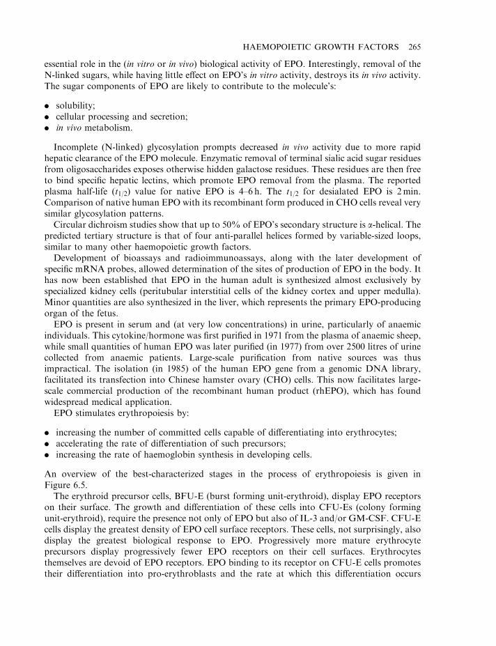
essential role in the (in vitro or in vivo) biological activity of EPO. Interestingly, removal of theN-linked sugars, while having little effect on EPO’s in vitro activity, destroys its in vivo activity.The sugar components of EPO are likely to contribute to the molecule’s:
. solubility;
. cellular processing and secretion;
. in vivo metabolism.
Incomplete (N-linked) glycosylation prompts decreased in vivo activity due to more rapidhepatic clearance of the EPO molecule. Enzymatic removal of terminal sialic acid sugar residuesfrom oligosaccharides exposes otherwise hidden galactose residues. These residues are then freeto bind specific hepatic lectins, which promote EPO removal from the plasma. The reportedplasma half-life (t1/2) value for native EPO is 4–6 h. The t1/2 for desialated EPO is 2min.Comparison of native human EPO with its recombinant form produced in CHO cells reveal verysimilar glycosylation patterns.
Circular dichroism studies show that up to 50% of EPO’s secondary structure is a-helical. Thepredicted tertiary structure is that of four anti-parallel helices formed by variable-sized loops,similar to many other haemopoietic growth factors.
Development of bioassays and radioimmunoassays, along with the later development ofspecific mRNA probes, allowed determination of the sites of production of EPO in the body. Ithas now been established that EPO in the human adult is synthesized almost exclusively byspecialized kidney cells (peritubular interstitial cells of the kidney cortex and upper medulla).Minor quantities are also synthesized in the liver, which represents the primary EPO-producingorgan of the fetus.
EPO is present in serum and (at very low concentrations) in urine, particularly of anaemicindividuals. This cytokine/hormone was first purified in 1971 from the plasma of anaemic sheep,while small quantities of human EPO was later purified (in 1977) from over 2500 litres of urinecollected from anaemic patients. Large-scale purification from native sources was thusimpractical. The isolation (in 1985) of the human EPO gene from a genomic DNA library,facilitated its transfection into Chinese hamster ovary (CHO) cells. This now facilitates large-scale commercial production of the recombinant human product (rhEPO), which has foundwidespread medical application.
EPO stimulates erythropoiesis by:
. increasing the number of committed cells capable of differentiating into erythrocytes;
. accelerating the rate of differentiation of such precursors;
. increasing the rate of haemoglobin synthesis in developing cells.
An overview of the best-characterized stages in the process of erythropoiesis is given inFigure 6.5.
The erythroid precursor cells, BFU-E (burst forming unit-erythroid), display EPO receptorson their surface. The growth and differentiation of these cells into CFU-Es (colony formingunit-erythroid), require the presence not only of EPO but also of IL-3 and/or GM-CSF. CFU-Ecells display the greatest density of EPO cell surface receptors. These cells, not surprisingly, alsodisplay the greatest biological response to EPO. Progressively more mature erythrocyteprecursors display progressively fewer EPO receptors on their cell surfaces. Erythrocytesthemselves are devoid of EPO receptors. EPO binding to its receptor on CFU-E cells promotestheir differentiation into pro-erythroblasts and the rate at which this differentiation occurs
HAEMOPOIETIC GROWTH FACTORS 265
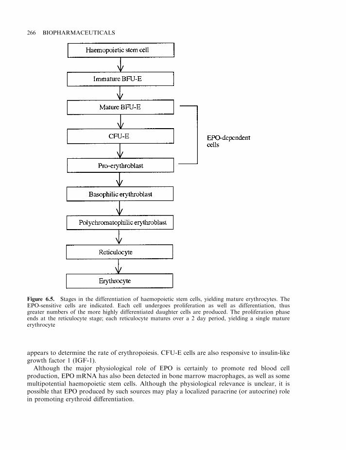
appears to determine the rate of erythropoiesis. CFU-E cells are also responsive to insulin-likegrowth factor 1 (IGF-1).
Although the major physiological role of EPO is certainly to promote red blood cellproduction, EPO mRNA has also been detected in bone marrow macrophages, as well as somemultipotential haemopoietic stem cells. Although the physiological relevance is unclear, it ispossible that EPO produced by such sources may play a localized paracrine (or autocrine) rolein promoting erythroid differentiation.
266 BIOPHARMACEUTICALS
Figure 6.5. Stages in the differentiation of haemopoietic stem cells, yielding mature erythrocytes. TheEPO-sensitive cells are indicated. Each cell undergoes proliferation as well as differentiation, thusgreater numbers of the more highly differentiated daughter cells are produced. The proliferation phaseends at the reticulocyte stage; each reticulocyte matures over a 2 day period, yielding a single matureerythrocyte
The EPO receptor and signal transduction
The availability of biologically active 125I-labelled EPO facilitated the detection and study ofcell surface receptors. In addition to erythroid precursors, various other cell lines were shownto express EPO receptors, at least when cultured in vitro. Many harboured two classes ofreceptors: a high-affinity and a low-affinity form. Most appeared to express between 1000 and3000 receptors/cell. Radiotracer experiments illustrated that the EPO receptor is rapidlyinternalized after ligand binding and the EPO–receptor complex is subsequently degradedwithin lysosomes.
The human EPO receptor is encoded by a single gene on chromosome 19. The gene houseseight exons, the first five of which appear to code for the 233 amino acid extracellular receptorportion. The sixth encodes a single 23 amino acid transmembrane domain, while the remainingtwo exons encode the 236 amino acid cytoplasmic domain. The mature receptor displays amolecular mass of 85–100 kDa. It is heavily glycosylated through multiple O-linked (and asingle N-linked) glycosylation site. High- versus low-affinity receptor variants may be generatedby self-association.
The EPO receptor is a member of the haemopoietic cytokine receptor superfamily. Itsintracellular domain displays no known catalytic activity but it appears to couple directly to theJAK 2 kinase (Chapter 4), which probably promotes the early events of EPO signaltransduction. Other studies have implicated additional possible signalling mechanisms,including the involvement of G proteins, protein kinase C and Ca2+. The exact molecularevents underlining EPO signal transduction remain to be elucidated in detail.
Binding of EPO to its receptor stimulates the proliferation of BFU-E cells and triggersCFU-Es to undergo terminal differentiation. As well as such stimulatory roles, EPO may play apermissive role, in that it also appears to inhibit apoptosis (programmed cell death) of thesecells. With this scenario, ‘normal’ serum EPO levels permit survival of a specific fraction ofBFU-E and CFU-Es, which dictates the observed rate of haemopoiesis. Increased serum EPOconcentrations permit survival of a greater fraction of these progenitor cells, thus increasing thenumber of red blood cells ultimately produced. The relative physiological importance of EPOstimulatory versus permissive activities has yet to be determined.
Regulation of EPO production
The level of EPO production in the kidneys (or liver) is primarily regulated by the oxygendemand of the producer cells, relative to their oxygen supply. Under normal conditions, whenthe producer cells are supplied with adequate oxygen via the blood, EPO (or EPO mRNA) levelsare barely detectable. However, the onset of hypoxia (a deficiency of oxygen in the tissues)results in a very rapid increase of EPO mRNA in producer cells. This is followed within 2 h byan increase in serum EPO levels. This process is prevented by inhibitors of RNA and proteinsynthesis, indicating that EPO is not stored in producer cells, but synthesized de novo whenrequired.
Interestingly, hypoxia prompts increased renal and hepatic EPO synthesis in different ways.In the kidney, the quantity of EPO produced by an individual cell remains constant, while anincrease in the number of EPO-producing cells is evident. In the liver, the quantity of EPOproduced by individual cells appears to simply increase in response to the hypoxic stimulus.
A range of conditions capable of inducing hypoxia stimulate enhanced production of EPO,thus stimulating erythropoiesis. These conditions include:
HAEMOPOIETIC GROWTH FACTORS 267
. moving to a higher altitude;
. blood loss;
. increased renal sodium transport;
. decreased renal blood flow;
. increased haemoglobin oxygen affinity;
. chronic pulmonary disease;
. some forms of heart disease.
On the other hand, hyperoxic conditions (excess tissue oxygen levels) promote a decrease inEPO production.
The exact mechanism by which hypoxia stimulates EPO production remains to be elucidated.This process has been studied in vitro using an EPO-producing cancerous liver (hepatoma) cellline as a model system. These studies suggest the existence of a haem protein (probablymembrane-bound), which effectively acts as an oxygen sensor (Figure 6.6). Adequate ambientoxygen concentration retains the haem group in an oxygenated state. Hypoxia, however,promotes a deoxy-configuration, which alters the haem conformation. The deoxy- form of haemis postulated to be capable of generating an active transcription factor which, upon migration tothe nucleus, enhances transcription of the EPO gene. Evidence cited to support such a theoryincludes the fact that cobalt promotes erythropoiesis (cobalt can substitute for the iron atom inthe haem porphyrin ring; Cobalt–haem, however, remains in the deoxy-conformation, even inthe presence of a high oxygen tension). In addition to oxygen levels, a number of otherregulatory factors can stimulate EPO synthesis, either on their own or in synergy with hypoxia(Table 6.6).
Therapeutic applications of EPO
A number of clinical circumstances have been identified which are characterized by an oftenprofoundly depressed rate of erythropoiesis (Table 6.7). Many, if not all, such conditions couldbe/are responsive to administration of exogenous EPO. The prevalence of anaemia, and themedical complications which ensue, prompts tremendous therapeutic interest in thishaemopoietic growth factor. EPO has been approved for use to treat various forms of anaemia(Table 6.8). It was the first therapeutic protein produced by genetic engineering, whose annualsales value topped $1 billion. Its current annual sales value is now close to $2 billion. EPO usedtherapeutically is produced by recombinant means in CHO cells.
Neorecormon is one such product. Produced in an engineered CHO cell line constitutivelyexpressing the EPO gene, the product displays an amino acid sequence identical to the nativehuman molecule. An overview of its manufacturing process is presented in Figure 6.7. The finalfreeze-dried product contains urea, sodium chloride, polysorbate, phosphate buffer and severalamino acids as excipients. It displays a shelf-life of 3 years when stored at 2–8 8C. A pre-filledsyrine form of the product (in solution) is also available, which is assigned a 2 year shelf-life at2–8 8C.
EPO can be administered intravenously or, more commonly, by subcutaneous (s.c.) injection.Peak serum concentrations are witnessed 8–24 h after s.c. administration. Although they arelower than the values achieved by i.v. administration, the effect is more prolonged, lasting forseveral hours. In healthy individuals, less than 10% of administered EPO is excreted intact inthe urine. This suggests that the kidneys play, at best, a minor role in the excretion of thishormone.
268 BIOPHARMACEUTICALS
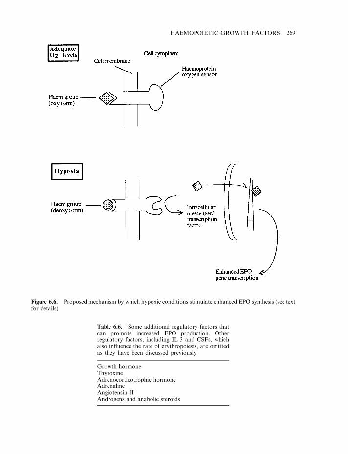
HAEMOPOIETIC GROWTH FACTORS 269
Figure 6.6. Proposed mechanism by which hypoxic conditions stimulate enhanced EPO synthesis (see textfor details)
Table 6.6. Some additional regulatory factors thatcan promote increased EPO production. Otherregulatory factors, including IL-3 and CSFs, whichalso influence the rate of erythropoiesis, are omittedas they have been discussed previously
Growth hormoneThyroxineAdrenocorticotrophic hormoneAdrenalineAngiotensin IIAndrogens and anabolic steroids
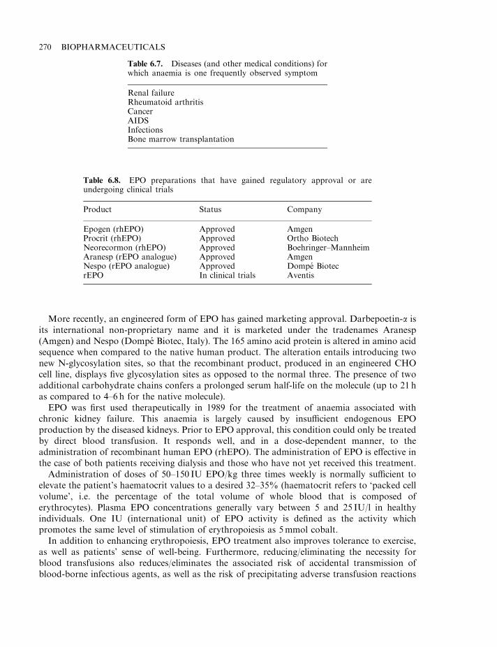
More recently, an engineered form of EPO has gained marketing approval. Darbepoetin-a isits international non-proprietary name and it is marketed under the tradenames Aranesp(Amgen) and Nespo (Dompe Biotec, Italy). The 165 amino acid protein is altered in amino acidsequence when compared to the native human product. The alteration entails introducing twonew N-glycosylation sites, so that the recombinant product, produced in an engineered CHOcell line, displays five glycosylation sites as opposed to the normal three. The presence of twoadditional carbohydrate chains confers a prolonged serum half-life on the molecule (up to 21 has compared to 4–6 h for the native molecule).
EPO was first used therapeutically in 1989 for the treatment of anaemia associated withchronic kidney failure. This anaemia is largely caused by insufficient endogenous EPOproduction by the diseased kidneys. Prior to EPO approval, this condition could only be treatedby direct blood transfusion. It responds well, and in a dose-dependent manner, to theadministration of recombinant human EPO (rhEPO). The administration of EPO is effective inthe case of both patients receiving dialysis and those who have not yet received this treatment.
Administration of doses of 50–150 IU EPO/kg three times weekly is normally sufficient toelevate the patient’s haematocrit values to a desired 32–35% (haematocrit refers to ‘packed cellvolume’, i.e. the percentage of the total volume of whole blood that is composed oferythrocytes). Plasma EPO concentrations generally vary between 5 and 25 IU/l in healthyindividuals. One IU (international unit) of EPO activity is defined as the activity whichpromotes the same level of stimulation of erythropoiesis as 5mmol cobalt.
In addition to enhancing erythropoiesis, EPO treatment also improves tolerance to exercise,as well as patients’ sense of well-being. Furthermore, reducing/eliminating the necessity forblood transfusions also reduces/eliminates the associated risk of accidental transmission ofblood-borne infectious agents, as well as the risk of precipitating adverse transfusion reactions
270 BIOPHARMACEUTICALS
Table 6.8. EPO preparations that have gained regulatory approval or areundergoing clinical trials
Product Status Company
Epogen (rhEPO) Approved AmgenProcrit (rhEPO) Approved Ortho BiotechNeorecormon (rhEPO) Approved Boehringer–MannheimAranesp (rEPO analogue) Approved AmgenNespo (rEPO analogue) Approved Dompe BiotecrEPO In clinical trials Aventis
Table 6.7. Diseases (and other medical conditions) forwhich anaemia is one frequently observed symptom
Renal failureRheumatoid arthritisCancerAIDSInfectionsBone marrow transplantation
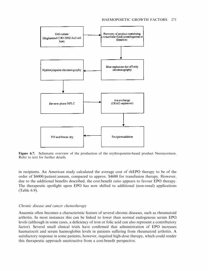
in recipients. An American study calculated the average cost of rhEPO therapy to be of theorder of $6000/patient/annum, compared to approx. $4600 for transfusion therapy. However,due to the additional benefits described, the cost:benefit ratio appears to favour EPO therapy.The therapeutic spotlight upon EPO has now shifted to additional (non-renal) applications(Table 6.9).
Chronic disease and cancer chemotherapy
Anaemia often becomes a characteristic feature of several chronic diseases, such as rheumatoidarthritis. In most instances this can be linked to lower than normal endogenous serum EPOlevels (although in some cases, a deficiency of iron or folic acid can also represent a contributoryfactor). Several small clinical trials have confirmed that administration of EPO increaseshaematocrit and serum haemoglobin levels in patients suffering from rheumatoid arthritis. Asatisfactory response in some patients, however, required high-dose therapy, which could renderthis therapeutic approach unattractive from a cost:benefit perspective.
HAEMOPOIETIC GROWTH FACTORS 271
Figure 6.7. Schematic overview of the production of the erythropoietin-based product Neorecormon.Refer to text for further details
Severe, and in particular chronic, infection can also sometimes induce anaemia—which isoften made worse by drugs used to combat the infection, e.g. anaemia is evident in 8% ofpatients with asymptomatic HIV infection. This incidence increases to 20% for those withAIDS-related complex and is greater than 60% for patients who have developed Kaposi’ssarcoma. Up to one-third of AIDS patients treated with zidovudine also develop anaemia.Again, several trials have confirmed that EPO treatment of AIDS sufferers (be they receivingzidovudine or not) can increase haematocrit values and decrease transfusion requirements.
Various malignancies can also induce an anaemic state. This is often associated withdecreased serum EPO levels, although iron deficiency, blood loss or tumour infiltration of thebone marrow can be complicating factors. In addition, chemotherapeutic agents administered tothis patient group often adversely affect stem cell populations, thus rendering the anaemia evenmore severe.
Administration of EPO to patients suffering from various cancers/receiving variouschemotherapeutic agents yielded encouraging results, with significant improvements inhaematocrit levels being recorded in approximately 50% of cases. In one large US study(2000 patients, most receiving chemotherapy) s.c. administration of an average of 150 IUEPO/kg, three times weekly, for 4 months, reduced the number of patients requiring bloodtransfusions from 22% to 10%. Improvement in the sense of well-being and overall quality oflife was also noted. The success rate of EPO in alleviating cancer-associated anaemia has variedin different trials, ranging from 32% to 85%.
The EPO receptor is expressed not only by specific erythrocyte precursor cells but also byendothelial, neural and myeloma cells. Concern has been expressed that EPO, therefore, mightactually stimulate growth of some tumour types, particularly those derived from such cells. Todate, no evidence (in vitro or in vivo) has been obtained to support this hypothesis.
Additional non-renal applications
Babies, especially babies born prematurely, often exhibit anaemia, which is characterized by asteadily decreasing serum haemoglobin level during the first 8 weeks of life. While multiplefactors contribute to development of anaemia of prematurity, a lower than normal serum EPOlevel is a characteristic trait. In vitro studies indicate that BFU-E and CFU-E cells from suchbabies are responsive to EPO, and several pilot clinical trials have been initiated. Administrationof 300–600 IU EPO/kg/week generally was found to enhance erythropoiesis and reduced thenumber of transfusions required by up to 30%.
Patients who have received an allogeneic bone marrow transplant characteristically displaydepressed serum EPO levels for up to 6 months post-transplantation. Administration of EPOthus seems a logical approach to counteract this effect. Several clinical studies have validated
272 BIOPHARMACEUTICALS
Table 6.9. Some non-renal applications of EPO (refer to text for details)
Treatment of anaemia associated with chronic diseaseTreatment of anaemia associated with cancer/chemotherapyTreatment of anaemia associated with prematurityTo facilitate autologous blood donations before surgeryTo reduce transfusion requirements after surgeryTo prevent anaemia after bone marrow transplantation
this approach, observing accelerated erythropoiesis, resulting in attainment of satisfactoryhaematocrit levels within a shorter period post-transplant.
Tolerability
In general, rhEPO is well tolerated. The most pronounced adverse effects appear to beassociated with its long-term administration to patients with end-stage renal failure. Particularlynoteworthy is an increase in blood pressure levels in some patients and the increased risk ofthromboembolic events (a thromboembolic event, i.e. a thromboembolism, describes thecircumstance where a blood clot forms at one point in the circulation but detaches, only tobecome lodged at another point; Chapter 9).
Most short-term applications of EPO are non-renal related, and generally display very fewside-effects; i.v. administration can sometimes prompt a transient flu-like syndrome, while s.c.administration can render the site of injection painful. This latter effect appears, however, to bedue to excipients present in the EOP preparations, most notably the citrate buffer. EPOadministration can also cause bone pain, although this rarely limits its clinical use.
Overall, therefore, rhEPO has proved both effective and safe in the treatment of a variety ofclinical conditions and its range of therapeutic applications is likely to increase over the comingyears.
THROMBOPOIETIN
Thrombopoietin (TPO) is the haemopoietic growth factor now shown to be the primaryphysiological regulator of platelet production. Although its existence had been inferred forseveral decades, its purification from blood proved an almost impossible task, due to its lowproduction levels and the availability of only an extremely cumbersome TPO bioassay. Itsexistence was finally proved in the mid-1990s when thrombopoietin cDNA was cloned. Thismolecule is likely to represent an important future therapeutic agent in combating depressedplasma platelet levels, although this remains to be proved by clinical trials.
Platelets (thrombocytes) carry out several functions in the body, all of which relate to thearrest of bleeding. They are disc-shaped structures 1–2 mm in diameter, and are present in theblood of healthy individuals at levels of approximately 250�109/l. They are formed by a lineage-specific stem cell differentiation process, as depicted in Figure 6.8. The terminal stages of thisprocess entails the maturation of large progenitor cells termed ‘megakaryocytes’. Plateletsrepresent small vesicles which bud off from the megakaryocyte cell surface and enter thecirculation.
A number of disorders have been identified that are primarily caused by the presence ofabnormal platelet levels in the blood. Thrombocythaemia is a disease characterized by abnormalmegakaryocyte proliferation, leading to elevated blood platelet levels. In many instances, thisresults in an elevated risk of spontaneous clot formation within blood vessels. In other instances,the platelets produced are defective, which can increase the risk of spontaneous or prolongedbleeding events.
Thrombocytopenia, on the other hand, is a condition characterized by reduced blood plateletlevels. Spontaneous bruising, bleeding into the skin (purpura) and prolonged bleeding afterinjury represent typical symptoms. Thrombocytopenia is induced by a number of clinicalconditions, including:
HAEMOPOIETIC GROWTH FACTORS 273
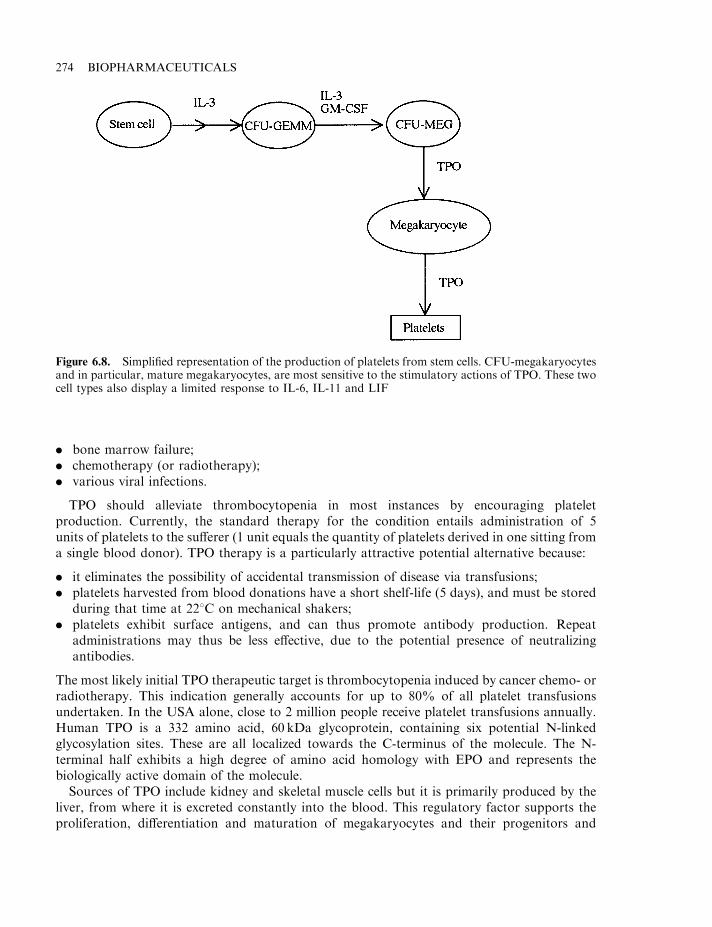
. bone marrow failure;
. chemotherapy (or radiotherapy);
. various viral infections.
TPO should alleviate thrombocytopenia in most instances by encouraging plateletproduction. Currently, the standard therapy for the condition entails administration of 5units of platelets to the sufferer (1 unit equals the quantity of platelets derived in one sitting froma single blood donor). TPO therapy is a particularly attractive potential alternative because:
. it eliminates the possibility of accidental transmission of disease via transfusions;
. platelets harvested from blood donations have a short shelf-life (5 days), and must be storedduring that time at 228C on mechanical shakers;
. platelets exhibit surface antigens, and can thus promote antibody production. Repeatadministrations may thus be less effective, due to the potential presence of neutralizingantibodies.
The most likely initial TPO therapeutic target is thrombocytopenia induced by cancer chemo- orradiotherapy. This indication generally accounts for up to 80% of all platelet transfusionsundertaken. In the USA alone, close to 2 million people receive platelet transfusions annually.Human TPO is a 332 amino acid, 60 kDa glycoprotein, containing six potential N-linkedglycosylation sites. These are all localized towards the C-terminus of the molecule. The N-terminal half exhibits a high degree of amino acid homology with EPO and represents thebiologically active domain of the molecule.
Sources of TPO include kidney and skeletal muscle cells but it is primarily produced by theliver, from where it is excreted constantly into the blood. This regulatory factor supports theproliferation, differentiation and maturation of megakaryocytes and their progenitors and
274 BIOPHARMACEUTICALS
Figure 6.8. Simplified representation of the production of platelets from stem cells. CFU-megakaryocytesand in particular, mature megakaryocytes, are most sensitive to the stimulatory actions of TPO. These twocell types also display a limited response to IL-6, IL-11 and LIF
promotes the production of platelets from megakaryocytes (Figure 6.8). It also appears tomodulate the growth/differentiation of progenitor cells, which eventually give rise to botherythrocytes and macrophages.
TPO induces its characteristic effects by binding to a specific TPO receptor present on thesurface of sensitive cells. The receptor, also known as c-mpl, is a single chain, 610 amino acidtransmembrane glycoprotein. The mechanism of signal transduction triggered upon TPO-binding remains to be elucidated.
Currently, at least two recombinant TPO-based products are being assessed in clinical trials.One product is a recombinant glycosylated form produced in a mammalian cell line, the other isa non-glycosylated variant, expressed in E. coli. The latter molecule, also known asmegakaryocyte growth and development factor (MGDF), is PEGylated subsequent to itspurification. PEGylation, as already described in the context of several other protein-basedtherapeutics, extends the molecule’s plasma half-life.
Additional cytokines (IL-3, IL-6, IL-11 and LIF) also promote a proliferative response inmegakaryocytes. Although these exhibit some ability to increase platelet levels, the physiologicalsignificance of this remains unclear. TPO, on the other hand, induces a far swifter, greater andmore specific response. In one study, its administration to mice over a period of 6 days resultedin a four-fold increase in platelet count (although some other studies reported a less pronouncedeffect). Furthermore, TPO administration to animals whose platelet production had beenseverely compromised by radio- or chemotherapy accelerated subsequent recovery of plateletnumbers to normal values. The future clinical outlook for this haemopoietic growth factor looksbright.
FURTHER READING
Books
Dallman, M. (2000). Haemapoietic and Lymphoid Cell Culture. Cambridge University Press, Cambridge.Kuter, D. et al. (Eds) (1997). Thrombopoiesis and Thrombopoietins. Humana, Totowa, NJ, USA.Medkalf, D. (1995) Haemopoietic Colony-stimulating Factors: From Biology to Clinical Applications. Cambridge
University Press, Cambridge.Morstyn, G. (1998). Filgrastim in Clinical Practice. Marcel Dekker, New York.Orlic, D. (1999). Haematopoietic Stem Cells. New York Academy of Science, New York.
Articles
Erythropoietin and thrombopoietin
Basser, R. (2002). The impact of thrombopoietin on clinical practice. Curr. Pharmaceut. Design 8(5), 369–377.Bottomley, A. et al. (2002). Human recombinant erythropoietin and quality of life: a wonder drug or something to
wonder about? Lancet Oncol. 3(3), 145–153.Buemi, M. et al. (2002). Recombinant human erythropoietin: more than just the correction of uremic anemia. J. Nephrol.
15(2), 97–103.Fried, W. (1995). Erythropoietin. Ann. Rev. Nutrit. 15, 353–377.Geddis, A. et al. (2002). Thrombopoietin: a pan-hematopoietic cytokine. Cytokine Growth Factor Rev. 13(1), 61–73.Kaushansky, K. (1995). Thrombopoietin: the primary regulator of platelet production. Blood 86(2), 419–431.Kaushansky, K. (1997). Thrombopoietin—understanding and manipulating platelet production. Annu. Rev. Med. 48,
1–11.Kaushansky, K. & Drachman, J. (2002). The molecular and cellular biology of thrombopoietin: the primary regulator of
platelet production. Oncogene 21(21), 3359–3367.Kendall, R. (2001). Erythropoietin. Clin. Lab. Haematol. 23(2), 71–80.Lok, S. et al. (1994). Cloning and expression of murine thrombopoietin cDNA and stimulation of platelet production
in vivo. Nature 369, 565–568.
HAEMOPOIETIC GROWTH FACTORS 275
Markham, A. & Bryson, H. (1995). Epoetin Alfa, a review of its pharmacodynamic and pharmacokinetic properties andtherapeutic use in non-renal applications. Drugs 49(2), 232–254.
Metcalf, D. (1994). Thrombopoietin—at last. Nature 369, 519–520.Miyazaki, H. & Kato, T. (1999). Thrombopoietin: biology and clinical potential. Int. J. Haematol. 70(4), 216–225.Ogden, J. (1994). Thrombopoietin— the erythropoietin of platelets? Trends Biotechnol. 12, 389–390.Roberts, D. & Smith, D. (1994). Erythropoietin: induction of synthesis to signal transduction. J. Mol. Endocrinol. 12,
131–148.Spivak, J. (1994). Recombinant human erythropoietin and the anaemia of cancer. Blood 84(4), 997–1004.
CSFs
Cheetham, J.C., Smith, D.M., Aoki, K.H. et al. (1998). NMR structure of human erythropoietin and a comparison withits receptor-bound conformation. Nature Struct. Biol. 5, 861.
Dranoff, G. et al. (1994). Involvement of granulocyte-macrophage colony-stimulating factor in pulmonary homeostasis.Science 264, 713–715.
Duarte, R. & Frank, D. (2002). The synergy between stem cell factor (SCF) and granulocyte colony stimulating factor(G-CSF): molecular basis and clinical relevance. Leukaemia Lymphoma 43(6), 1179–1187.
Frampton, J. et al. (1994). Filgrastim, a review of its pharmacological properties and therapeutic efficacy in neutropenia.Drugs 48(5), 731–760.
Frampton, J. et al. (1995). Lenograstim, a review of its pharmacological properties and therapeutic efficacy inneutropenia and related clinical settings. Drugs 49(5), 767–793.
Hamilton, J. (1997). CSF-1 signal transduction. J. Leukocyte Biol. 62(2), 145–155.Harousseau, J. (1997). The role of colony-stimulating factors in the treatment of acute leukaemia. Biodrugs 7(6),
448–460.Spangrude, G. (1994). Biological and clinical aspects of haematopoietic stem cells. Ann. Rev. Med. 45, 93–104.Tabbara, I. et al. (1996). The clinical applications of granulocyte-colony-stimulating factor in haematopoietic stem cell
transplantation—a review. Anticancer Res. 16(6B), 3901–3905.Walter, M.R., Cook, W.J., Ealick, S.E. et al. (1992). Three-dimensional structure of recombinant human granulocyte-
macrophage colony-stimulating factor. J. Mol. Ecol. 224, 1075.Watowich, F. et al. (1996). Cytokine receptor signal transduction and the control of haematopoietic cell development.
Ann. Rev. Cell Dev. Biol. 12, 91–128.
276 BIOPHARMACEUTICALS
Chapter 7
Growth factors
The growth of eukaryotic cells is modulated by various influences, of which growth factors areamongst the most important for many cell types. A wide range of polypeptide growth factorshave been identified (Table 7.1) and more undoubtedly remain to be characterized. Factors thatinhibit cell growth also exist, e.g. interferons (IFNs) and tumour necrosis factor (TNF) inhibitproliferation of various cell types.
Some growth factors may be classified as cytokines, e.g. ILs, transforming growth factor-b(TGF-b) and colony stimulating factors (CSFs). Others, e.g. insulin-like growth factors (IGFs)are not members of this family. Each growth factor has a mitogenic (promotes cell division)effect on a characteristic range of cells. While some such factors affect only a few cell types, moststimulate the growth of a wide range of cells. The range of growth factors considered in thischapter is limited to those that have not received attention in previous chapters.
The ability of such factors to promote accelerated cellular growth and division haspredictably attracted the attention of the pharmaceutical industry. The clinical potential of arange of such factors, e.g. to accelerate the wound-healing process, is currently being assessed invarious clinical trials (Table 7.2).
GROWTH FACTORS AND WOUND HEALING
The wound-healing process is complex and as yet not fully understood. The area of tissuedamage becomes the focus of various events, often beginning with immunological andinflammatory reactions. The various cells involved in such processes, as well as additional cellsat the site of the wound, also secrete various growth factors. These mitogens stimulate thegrowth and activation of various cell types, including fibroblasts (which produce collagen andelastin precursors, and ground substance), epithelial cells (e.g. skin cells) and vascularendothelial cells. Such cells advance healing by promoting processes such as granulation(growth of connective tissue and small blood vessels at the healing surface) and subsequentepithelialization. The growth factors that appear most significant to this process includefibroblast growth factors (FGFs), transforming growth factors (TGFs), platelet-derived growthfactor (PDGF), insulin-like growth factor 1 (IGF-1) and epidermal growth factor (EGF).
Wounds can be categorized as acute (healing quickly on their own) or chronic (healing slowly,and often requiring medication). Chronic wounds, such as ulcers (Table 7.3), occur if some
Biopharmaceuticals: Biochemistry and Biotechnology, Second Edition by Gary WalshJohn Wiley & Sons Ltd: ISBN 0 470 84326 8 (ppc), ISBN 0 470 84327 6 (pbk)
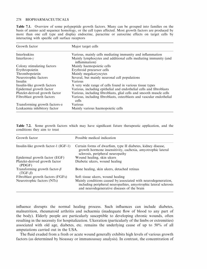
influence disrupts the normal healing process. Such influences can include diabetes,malnutrition, rheumatoid arthritis and ischaemia (inadequate flow of blood to any part ofthe body). Elderly people are particularly susceptible to developing chronic wounds, oftenresulting in the necessity for hospitalization. Ulceration (particularly of the limbs or extremities)associated with old age, diabetes, etc. remains the underlying cause of up to 50% of allamputations carried out in the USA.
The fluid exuded from a fresh or acute wound generally exhibits high levels of various growthfactors (as determined by bioassay or immunoassay analysis). In contrast, the concentration of
278 BIOPHARMACEUTICALS
Table 7.2. Some growth factors which may have significant future therapeutic application, and theconditions they aim to treat
Growth factor Possible medical indication
Insulin-like growth factor-1 (IGF-1) Certain forms of dwarfism, type II diabetes, kidney disease,growth hormone insensitivity, cachexia, amyotrophic lateralsclerosis, peripheral neuropathy
Epidermal growth factor (EGF) Wound healing, skin ulcersPlatelet-derived growth factor(PDGF)
Diabetic ulcers, wound healing
Transforming growth factor-b(TGF-b)
Bone healing, skin ulcers, detached retinas
Fibroblast growth factors (FGFs) Soft tissue ulcers, wound healingNeurotrophic factors (NTs) Mainly conditions caused by/associated with neurodegeneration,
including peripheral neuropathies, amyotrophic lateral sclerosisand neurodegenerative diseases of the brain
Table 7.1. Overview of some polypeptide growth factors. Many can be grouped into families on thebasis of amino acid sequence homology, or the cell types affected. Most growth factors are produced bymore than one cell type and display endocrine, paracrine or autocrine effects on target cells byinteracting with specific cell surface receptors
Growth factor Major target cells
Interleukins Various, mainly cells mediating immunity and inflammationInterferon-g Mainly lymphocytes and additional cells mediating immunity (and
inflammation)Colony stimulating factors Mainly haemopoietic cellsErythropoietin Erythroid precursor cellsThrombopoietin Mainly megakaryocytesNeurotrophic factors Several, but mainly neuronal cell populationsInsulin VariousInsulin-like growth factors A very wide range of cells found in various tissue typesEpidermal growth factor Various, including epithelial and endothelial cells and fibroblastsPlatelet-derived growth factor Various, including fibroblasts, glial cells and smooth muscle cellsFibroblast growth factors Various, including fibroblasts, osteoblasts and vascular endothelial
cellsTransforming growth factors-a VariousLeukaemia inhibitory factor Mainly various haemopoietic cells
such mitogens present in chronic wounds is usually several-fold lower. Direct (topical)application of exogenous growth factors results in accelerated wound healing in animals. Whilesome encouraging results have been observed in human trials, the overall results obtained thusfar have been disappointing. Having said this, one such factor (rPDGF) has been approved fortopical administration on diabetic ulcers, as described later. Future studies may well also focuson application of a cocktail of growth factors instead of a single such factor to a wound surface.
A greater understanding of wound physiology and biochemistry may also facilitate greatersuccess in future trials. It has been established, for example, that the fluid exuded from chronicwounds harbours high levels of proteolytic activity (almost 200-fold higher than associated withacute wounds). Failure of mitogens to stimulate wound healing thus may be due, in part, to theirrapid proteolytic degradation (and/or the degradation of growth factor receptors present on thesurface of susceptible cells). Identification of suitable protease inhibitors, and their applicationin conjunction with exogenous growth factor therapy, may improve clinical results recorded inthe future.
INSULIN-LIKE GROWTH FACTORS (IGFs)
The insulin-like growth factors (also termed ‘somatomedins’), constitute a family of two closelyrelated (small) polypeptides: insulin-like growth factor 1 (IGF-1) and insulin-like growth factor2 (IGF-2). As the names suggest, these growth factors bear a strong structural resemblance toinsulin (or, more accurately, proinsulin). Infusion of IGF-1 decreases circulating levels of insulinand glucagon, increases tissue glucose uptake and inhibits hepatic glucose export. IGFs displaypluripotent activities, regulating the growth, activation, differentiation (and maintenance of thedifferentiated state) of a wide variety of cell and tissue types (discussed later). The fullcomplexity and variety of their biological activities are only now beginning to be appreciated.
The liver represents the major site of synthesis of the IGFs, from where they enter the bloodstream, thereby acting in a classical endocrine fashion. A wide variety of body cells express IGFreceptors, of which there are two types. Furthermore, IGFs are also synthesized in smallerquantities at numerous sites in the body and function in an autocrine or paracrine manner atthese specific locations. IGF activity is also modulated by a family of IGF binding proteins(IGFBPs), of which there are at least six.
GROWTH FACTORS 279
Table 7.3. Various types of ulcers along with their underlying cause. An ulcer may simply be describedas a break or cut in the skin or membrane lining the digestive tract which fails to heal. The damagedarea may then become inflamed
Ulcer category Description
Decubitus ulcer(e.g. bed sores, pressure sores)
Ulcer due to continuous pressure exerted on a particular area ofskin; often associated with bed-ridden patients
Diabetic ulcers Ulcers (e.g. ‘diabetic leg’) caused by complications of diabetesVaricose ulcers Due to defective circulation, sometimes associated with varicose
veinsRodent ulcers An ulcerous cancer (basal cell carcinoma), usually affecting the
facePeptic ulcers Ulcer of the digestive tract, caused by digestion of the mucosa by
acid and pepsin; may occur in e.g. the duodenum (duodenalulcer), or the stomach (gastric ulcer)
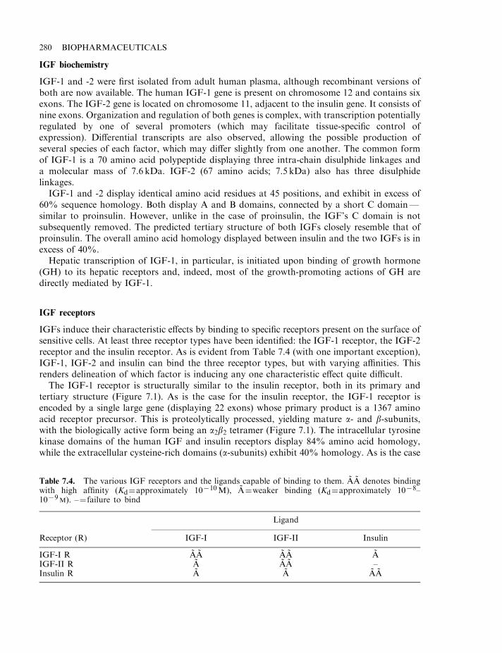
IGF biochemistry
IGF-1 and -2 were first isolated from adult human plasma, although recombinant versions ofboth are now available. The human IGF-1 gene is present on chromosome 12 and contains sixexons. The IGF-2 gene is located on chromosome 11, adjacent to the insulin gene. It consists ofnine exons. Organization and regulation of both genes is complex, with transcription potentiallyregulated by one of several promoters (which may facilitate tissue-specific control ofexpression). Differential transcripts are also observed, allowing the possible production ofseveral species of each factor, which may differ slightly from one another. The common formof IGF-1 is a 70 amino acid polypeptide displaying three intra-chain disulphide linkages anda molecular mass of 7.6 kDa. IGF-2 (67 amino acids; 7.5 kDa) also has three disulphidelinkages.
IGF-1 and -2 display identical amino acid residues at 45 positions, and exhibit in excess of60% sequence homology. Both display A and B domains, connected by a short C domain—similar to proinsulin. However, unlike in the case of proinsulin, the IGF’s C domain is notsubsequently removed. The predicted tertiary structure of both IGFs closely resemble that ofproinsulin. The overall amino acid homology displayed between insulin and the two IGFs is inexcess of 40%.
Hepatic transcription of IGF-1, in particular, is initiated upon binding of growth hormone(GH) to its hepatic receptors and, indeed, most of the growth-promoting actions of GH aredirectly mediated by IGF-1.
IGF receptors
IGFs induce their characteristic effects by binding to specific receptors present on the surface ofsensitive cells. At least three receptor types have been identified: the IGF-1 receptor, the IGF-2receptor and the insulin receptor. As is evident from Table 7.4 (with one important exception),IGF-1, IGF-2 and insulin can bind the three receptor types, but with varying affinities. Thisrenders delineation of which factor is inducing any one characteristic effect quite difficult.
The IGF-1 receptor is structurally similar to the insulin receptor, both in its primary andtertiary structure (Figure 7.1). As is the case for the insulin receptor, the IGF-1 receptor isencoded by a single large gene (displaying 22 exons) whose primary product is a 1367 aminoacid receptor precursor. This is proteolytically processed, yielding mature a- and b-subunits,with the biologically active form being an a2b2 tetramer (Figure 7.1). The intracellular tyrosinekinase domains of the human IGF and insulin receptors display 84% amino acid homology,while the extracellular cysteine-rich domains (a-subunits) exhibit 40% homology. As is the case
280 BIOPHARMACEUTICALS
Table 7.4. The various IGF receptors and the ligands capable of binding to them. AA denotes bindingwith high affinity (Kd¼approximately 10710M), A¼weaker binding (Kd¼approximately 1078–1079M). –¼ failure to bind
Ligand
Receptor (R) IGF-I IGF-II Insulin
IGF-I R AA AA AIGF-II R A AA –Insulin R A A AA
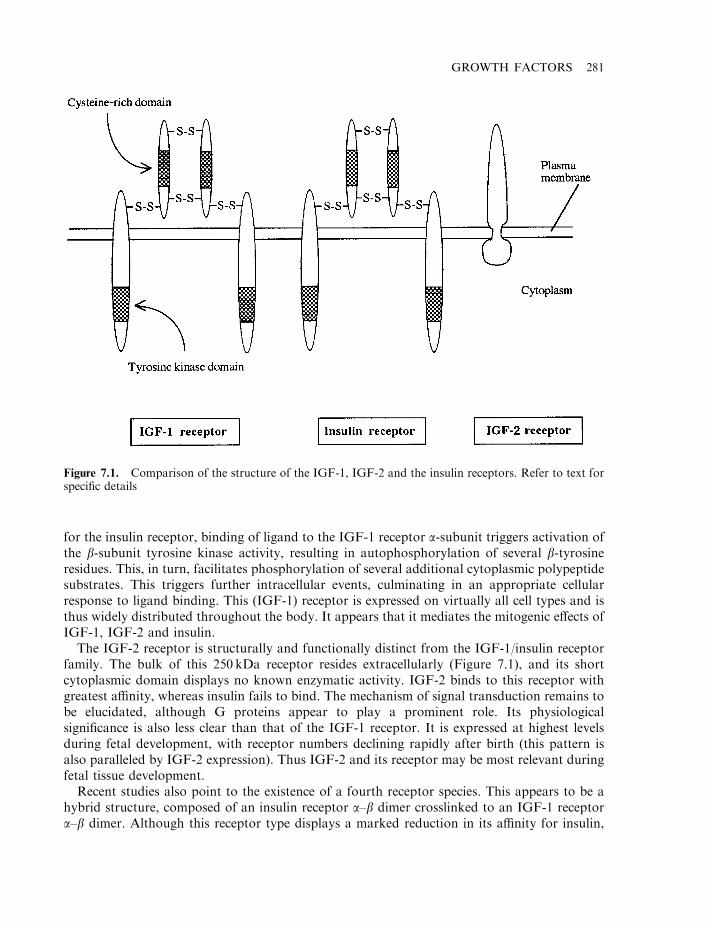
for the insulin receptor, binding of ligand to the IGF-1 receptor a-subunit triggers activation ofthe b-subunit tyrosine kinase activity, resulting in autophosphorylation of several b-tyrosineresidues. This, in turn, facilitates phosphorylation of several additional cytoplasmic polypeptidesubstrates. This triggers further intracellular events, culminating in an appropriate cellularresponse to ligand binding. This (IGF-1) receptor is expressed on virtually all cell types and isthus widely distributed throughout the body. It appears that it mediates the mitogenic effects ofIGF-1, IGF-2 and insulin.
The IGF-2 receptor is structurally and functionally distinct from the IGF-1/insulin receptorfamily. The bulk of this 250 kDa receptor resides extracellularly (Figure 7.1), and its shortcytoplasmic domain displays no known enzymatic activity. IGF-2 binds to this receptor withgreatest affinity, whereas insulin fails to bind. The mechanism of signal transduction remains tobe elucidated, although G proteins appear to play a prominent role. Its physiologicalsignificance is also less clear than that of the IGF-1 receptor. It is expressed at highest levelsduring fetal development, with receptor numbers declining rapidly after birth (this pattern isalso paralleled by IGF-2 expression). Thus IGF-2 and its receptor may be most relevant duringfetal tissue development.
Recent studies also point to the existence of a fourth receptor species. This appears to be ahybrid structure, composed of an insulin receptor a–b dimer crosslinked to an IGF-1 receptora–b dimer. Although this receptor type displays a marked reduction in its affinity for insulin,
GROWTH FACTORS 281
Figure 7.1. Comparison of the structure of the IGF-1, IGF-2 and the insulin receptors. Refer to text forspecific details
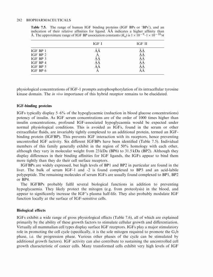
physiological concentrations of IGF-1 prompts autophosphorylation of its intracellular tyrosinekinase domain. The in vivo importance of this hybrid receptor remains to be elucidated.
IGF-binding proteins
IGFs typically display 5–6% of the hypoglycaemic (reduction in blood glucose concentrations)potency of insulin. As IGF serum concentrations are of the order of 1000 times higher thaninsulin concentrations, profound IGF-associated hypoglycaemia would be expected undernormal physiological conditions. This is avoided as IGFs, found in the serum or otherextracellular fluids, are invariably tightly complexed to an additional protein, termed an IGF-binding protein (IGFBP). This prevents IGF interaction with its receptors, hence preventinguncontrolled IGF activity. Six different IGFBPs have been identified (Table 7.5). Individualmembers of this family generally exhibit in the region of 50% homology with each other,although they vary in molecular weight from 23 kDa (BP6) to 31.5 kDa (BP2). Although theydisplay differences in their binding affinities for IGF ligands, the IGFs appear to bind themmore tightly than they do their cell surface receptors.
IGFBPs are widely expressed, but high levels of BP1 and BP2 in particular are found in theliver. The bulk of serum IGF-1 and -2 is found complexed to BP3 and an acid-labilepolypeptide. The remaining molecules of serum IGFs are usually found complexed to BP1, BP2or BP4.
The IGFBPs probably fulfil several biological functions in addition to preventinghypoglycaemia. They likely protect the mitogen (e.g. from proteolysis) in the blood, andappear to significantly increase the IGF’s plasma half-life. They also probably modulate IGFfunction locally at the surface of IGF-sensitive cells.
Biological effects
IGFs exhibit a wide range of gross physiological effects (Table 7.6), all of which are explainedprimarily by the ability of these growth factors to stimulate cellular growth and differentiation.Virtually all mammalian cell types display surface IGF receptors. IGFs play a major stimulatoryrole in promoting the cell cycle (specifically, it is the sole mitogen required to promote the G1bphase, i.e. the progression phase. Various other phases of the cycle can be stimulated byadditional growth factors). IGF activity can also contribute to sustaining the uncontrolled cellgrowth characteristic of cancer cells. Many transformed cells exhibit very high levels of IGF
282 BIOPHARMACEUTICALS
Table 7.5. The range of human IGF binding proteins (IGF BPs or ‘BPs’), and anindication of their relative affinities for ligand. AA indicates a higher affinity thanA. The approximate range of IGF BP association constants (Ka) is 161079–1610710M
IGF I IGF II
IGF BP 1 AA AAIGF BP 2 A AAIGF BP 3 AA AAIGF BP 4 AA AAIGF BP 5 AA AAIGF BP 6 A AA
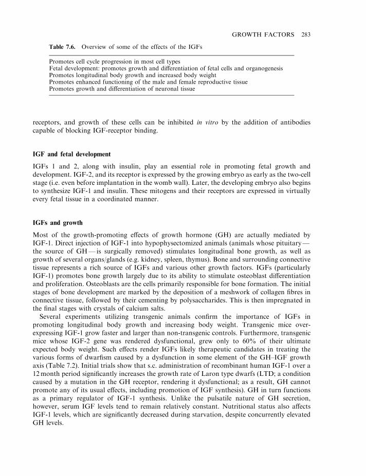
receptors, and growth of these cells can be inhibited in vitro by the addition of antibodiescapable of blocking IGF-receptor binding.
IGF and fetal development
IGFs 1 and 2, along with insulin, play an essential role in promoting fetal growth anddevelopment. IGF-2, and its receptor is expressed by the growing embryo as early as the two-cellstage (i.e. even before implantation in the womb wall). Later, the developing embryo also beginsto synthesize IGF-1 and insulin. These mitogens and their receptors are expressed in virtuallyevery fetal tissue in a coordinated manner.
IGFs and growth
Most of the growth-promoting effects of growth hormone (GH) are actually mediated byIGF-1. Direct injection of IGF-1 into hypophysectomized animals (animals whose pituitary—the source of GH— is surgically removed) stimulates longitudinal bone growth, as well asgrowth of several organs/glands (e.g. kidney, spleen, thymus). Bone and surrounding connectivetissue represents a rich source of IGFs and various other growth factors. IGFs (particularlyIGF-1) promotes bone growth largely due to its ability to stimulate osteoblast differentiationand proliferation. Osteoblasts are the cells primarily responsible for bone formation. The initialstages of bone development are marked by the deposition of a meshwork of collagen fibres inconnective tissue, followed by their cementing by polysaccharides. This is then impregnated inthe final stages with crystals of calcium salts.
Several experiments utilizing transgenic animals confirm the importance of IGFs inpromoting longitudinal body growth and increasing body weight. Transgenic mice over-expressing IGF-1 grow faster and larger than non-transgenic controls. Furthermore, transgenicmice whose IGF-2 gene was rendered dysfunctional, grew only to 60% of their ultimateexpected body weight. Such effects render IGFs likely therapeutic candidates in treating thevarious forms of dwarfism caused by a dysfunction in some element of the GH–IGF growthaxis (Table 7.2). Initial trials show that s.c. administration of recombinant human IGF-1 over a12month period significantly increases the growth rate of Laron type dwarfs (LTD; a conditioncaused by a mutation in the GH receptor, rendering it dysfunctional; as a result, GH cannotpromote any of its usual effects, including promotion of IGF synthesis). GH in turn functionsas a primary regulator of IGF-1 synthesis. Unlike the pulsatile nature of GH secretion,however, serum IGF levels tend to remain relatively constant. Nutritional status also affectsIGF-1 levels, which are significantly decreased during starvation, despite concurrently elevatedGH levels.
GROWTH FACTORS 283
Table 7.6. Overview of some of the effects of the IGFs
Promotes cell cycle progression in most cell typesFetal development: promotes growth and differentiation of fetal cells and organogenesisPromotes longitudinal body growth and increased body weightPromotes enhanced functioning of the male and female reproductive tissuePromotes growth and differentiation of neuronal tissue
In addition to promoting fetal and childhood growth, IGFs play a core role in tissue renewaland repair (e.g. wound healing) during adulthood, e.g. these growth factors play a central role inbone remodelling (i.e. reabsorption and rebuilding—which helps keep bones strong andcontributes to whole body calcium homeostasis). Reabsorption of calcified bone is undertakenby osteoclasts, cells of haemopoietic origin whose formation is stimulated by IGFs. Thesemitogens may, therefore, influence the development of osteoporosis, a prevalent condition(especially amongst the elderly), which is characterized by brittle, uncalcified bone.
IGFs also stimulate a more generalized short- and long-term whole body anabolic effect.Studies in calorie-restricted animals and humans show that IGF administration can retard orreverse catabolic events, such as tissue degradation and catabolism of body protein. Such effectshave attracted the interest of some athletes who have used it illegally as a performance-enhancing drug. Detection is rendered complex, due to the relatively wide range of IGFconcentrations classified as ‘normal’ and the fact that exercise naturally boosts endogenousIGF-1 production. IGF effects have also prompted speculation that these growth factors mightbe of clinical use in retarding/preventing/reversing cachexia, the generalized wasting of bodytissues associated with chronic disease and many cancer types. Many IGF metabolic effects(particularly IGF-1) exhibit similarities to several metabolic effects induced by insulin. Thesesimilarities (particularly IGF’s ability to enhance cellular glucose uptake), suggests a possiblerole for this growth factor in the treatment of certain forms of diabetes, most notably non-insulin dependent diabetes (type II diabetes; Table 7.2). Initial studies have shown that IGF canreduce hyperglycaemia in patients unresponsive to insulin and further more detailed studies arenow ongoing.
Renal and reproductive effects
IGFs (in particular IGF-1 and also IGFBP-1) are localized within various areas of the kidney.Direct infusion of IGF-1 influences (usually enhances) renal function by a number of means,including promoting:
. increased glomerular filtration rate;
. increased renal plasma flow;
. increased kidney size and weight.
These responses are obviously mediated by multiple effects on the growth and activity of severalrenal cell types and suggest that IGFs play a physiological role in regulating renal function. Notsurprisingly, IGF-1 is currently being assessed as a potential therapeutic agent in the treatmentof various forms of kidney disease.
GH deficiency often leads to delayed puberty. This condition often responds to exogenousGH administration. IGFs, as well as their receptors and binding proteins, are widespreadlyexpressed in the male and female reproductive tissue. Thus, IGFs are believed to affectreproductive function by both (GH-stimulated) endocrine action and via paracrine- andautocrine-based activity.
In the human female, IGF-1 is expressed by follicular theca cells, while IGF-2 is synthesizedby granulosa cells (Chapter 8). The IGF-1 and -2 receptors are widely expressed in ovariantissue, and synthesis of both growth factors and their receptors are influenced by circulatinggonadotrophin levels. IGF-1 exerts a direct mitogenic effect on human granulosa cells, andpromotes increased androgen and oestradiol synthesis by these cells. IGF-1 also promotesincreased expression of FSH and LH receptors in ovarian tissue.
284 BIOPHARMACEUTICALS
In the male, IGF-1 is synthesized by the Sertoli and Leydig cells of the testes. It alsostimulates testosterone production by the Leydig cells, and promotes growth and maintenanceof various additional testis cell types.
IGFs thus play an essential role in many facets of reproductive function. Traditionally,therapeutic intervention in reproductive disorders at a molecular level has relied almostexclusively upon administration of gonadotrophins or LHRH (luteinizing hormone releasinghormone; Chapter 8). Because of their widespread reproductive effects, IGFs may yet prove avaluable adjunct therapy in some instances.
Neuronal and other effects
IGF-1 is widely expressed in the CNS. IGF-2 is also present, being produced mainly by tissuesat vascular interfaces with the brain. Both growth factors, along with insulin, play a number ofimportant roles in the nervous system. They stimulate the growth and development of variousneuronal populations and promote neurotrophic effects (discussed later).
IGF-1 promotes differentiation of various neuronal cells, playing a central role in fosteringneurite outgrowth and synapse formation, as well as myelin formation. In addition to affectingneuronal development and maintenance, it may also stimulate regeneration of damagedperipheral neurons. This activity has prompted investigation of the therapeutic potential ofIGFs (particularly IGF-1) in the treatment of some neurodegenerative conditions most notablyamyotrophic lateral sclerosis (ALS), which is also called motor neuron disease (MND) in theUSA (strictly speaking, these two terms are not interchangeable, owing to slight differences intheir pathogenicities). ALS (and MND) are caused by a progressive degeneration of specificmotor neurons stretching out from the spinal cord. This leads to wasting of the muscle cells themotor neurons normally enervate. Remission is unknown, and death is the usual outcome.Clinical application of IGFs may be tempered by side effects. High-dose administration caninduce hypoglycaemia, hypotension and arrhythmias.
EPIDERMAL GROWTH FACTOR (EGF)
EGF was one of the first growth factors discovered. Its existence was initially noted in the 1960sas a factor present in saliva, which could promote premature tooth eruption and eyelid openingin neonatal mice. It was first purified from urine and named urogastrone, owing to its ability toinhibit the secretion of gastric acid. EGF has subsequently proved to exert a powerful mitogeniceffect on many cell types, and its receptor is expressed by most cells. Its influence on endothelialcells, epithelial cells and fibroblasts is particularly noteworthy, and the skin appears to be itsmajor physiological target. It stimulates growth of the epidermal layer. Along with several othergrowth factors, EGF plays a role in the wound-healing process. EGF is synthesized mainly bymonocytes and ectodermal cells, as well as by the kidney and duodenal glands. It is found inmost bodily fluids, especially milk.
The gene coding for human EGF is located on chromosome 4. It is an extensive structure,consisting of 24 exons and giving rise to a 110 kb primary transcript. After splicing, this yields amRNA coding for a 1208 amino acid prepro-EGF. In addition to the EGF sequence, thiscontains seven EGF-like domains, whose biological function remains unknown. Proteolyticprocessing of the larger molecule releases soluble EGF, a 6 kDa, 53 amino acid unglycosylatedpeptide. Mature EGF is very stable. Its 3-D structure, as revealed by nuclear magnetic
GROWTH FACTORS 285
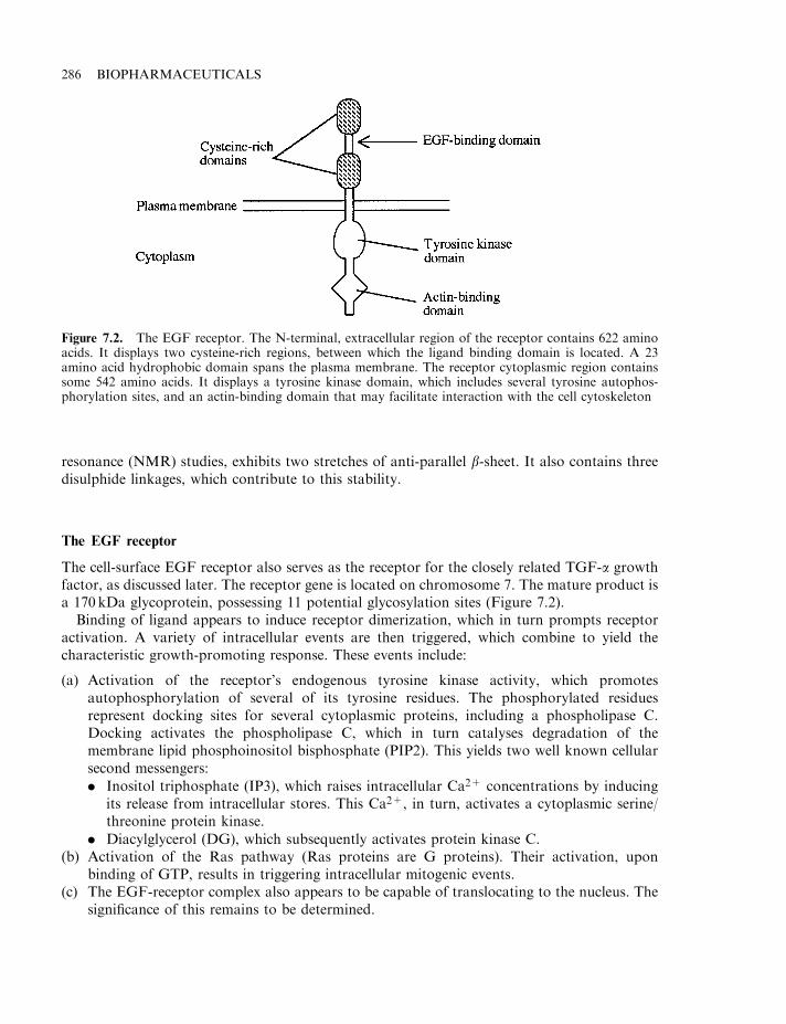
resonance (NMR) studies, exhibits two stretches of anti-parallel b-sheet. It also contains threedisulphide linkages, which contribute to this stability.
The EGF receptor
The cell-surface EGF receptor also serves as the receptor for the closely related TGF-a growthfactor, as discussed later. The receptor gene is located on chromosome 7. The mature product isa 170 kDa glycoprotein, possessing 11 potential glycosylation sites (Figure 7.2).
Binding of ligand appears to induce receptor dimerization, which in turn prompts receptoractivation. A variety of intracellular events are then triggered, which combine to yield thecharacteristic growth-promoting response. These events include:
(a) Activation of the receptor’s endogenous tyrosine kinase activity, which promotesautophosphorylation of several of its tyrosine residues. The phosphorylated residuesrepresent docking sites for several cytoplasmic proteins, including a phospholipase C.Docking activates the phospholipase C, which in turn catalyses degradation of themembrane lipid phosphoinositol bisphosphate (PIP2). This yields two well known cellularsecond messengers:
. Inositol triphosphate (IP3), which raises intracellular Ca2+ concentrations by inducingits release from intracellular stores. This Ca2+, in turn, activates a cytoplasmic serine/threonine protein kinase.
. Diacylglycerol (DG), which subsequently activates protein kinase C.(b) Activation of the Ras pathway (Ras proteins are G proteins). Their activation, upon
binding of GTP, results in triggering intracellular mitogenic events.(c) The EGF-receptor complex also appears to be capable of translocating to the nucleus. The
significance of this remains to be determined.
286 BIOPHARMACEUTICALS
Figure 7.2. The EGF receptor. The N-terminal, extracellular region of the receptor contains 622 aminoacids. It displays two cysteine-rich regions, between which the ligand binding domain is located. A 23amino acid hydrophobic domain spans the plasma membrane. The receptor cytoplasmic region containssome 542 amino acids. It displays a tyrosine kinase domain, which includes several tyrosine autophos-phorylation sites, and an actin-binding domain that may facilitate interaction with the cell cytoskeleton
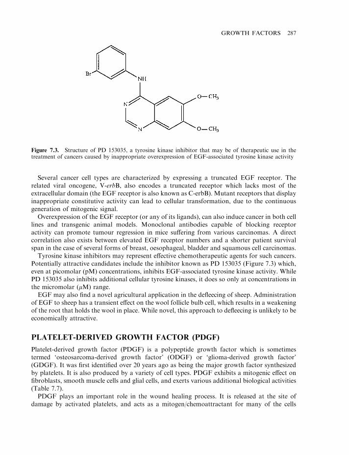
Several cancer cell types are characterized by expressing a truncated EGF receptor. Therelated viral oncogene, V-erbB, also encodes a truncated receptor which lacks most of theextracellular domain (the EGF receptor is also known as C-erbB). Mutant receptors that displayinappropriate constitutive activity can lead to cellular transformation, due to the continuousgeneration of mitogenic signal.
Overexpression of the EGF receptor (or any of its ligands), can also induce cancer in both celllines and transgenic animal models. Monoclonal antibodies capable of blocking receptoractivity can promote tumour regression in mice suffering from various carcinomas. A directcorrelation also exists between elevated EGF receptor numbers and a shorter patient survivalspan in the case of several forms of breast, oesophageal, bladder and squamous cell carcinomas.
Tyrosine kinase inhibitors may represent effective chemotherapeutic agents for such cancers.Potentially attractive candidates include the inhibitor known as PD 153035 (Figure 7.3) which,even at picomolar (pM) concentrations, inhibits EGF-associated tyrosine kinase activity. WhilePD 153035 also inhibits additional cellular tyrosine kinases, it does so only at concentrations inthe micromolar (mM) range.
EGF may also find a novel agricultural application in the defleecing of sheep. Administrationof EGF to sheep has a transient effect on the wool follicle bulb cell, which results in a weakeningof the root that holds the wool in place. While novel, this approach to defleecing is unlikely to beeconomically attractive.
PLATELET-DERIVED GROWTH FACTOR (PDGF)
Platelet-derived growth factor (PDGF) is a polypeptide growth factor which is sometimestermed ‘osteosarcoma-derived growth factor’ (ODGF) or ‘glioma-derived growth factor’(GDGF). It was first identified over 20 years ago as being the major growth factor synthesizedby platelets. It is also produced by a variety of cell types. PDGF exhibits a mitogenic effect onfibroblasts, smooth muscle cells and glial cells, and exerts various additional biological activities(Table 7.7).
PDGF plays an important role in the wound healing process. It is released at the site ofdamage by activated platelets, and acts as a mitogen/chemoattractant for many of the cells
GROWTH FACTORS 287
Figure 7.3. Structure of PD 153035, a tyrosine kinase inhibitor that may be of therapeutic use in thetreatment of cancers caused by inappropriate overexpression of EGF-associated tyrosine kinase activity
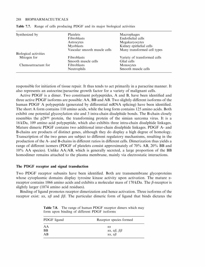
responsible for initiation of tissue repair. It thus tends to act primarily in a paracrine manner. Italso represents an autocrine/paracrine growth factor for a variety of malignant cells.
Active PDGF is a dimer. Two constituent polypeptides, A and B, have been identified andthree active PDGF isoforms are possible: AA, BB and AB. Two slightly different isoforms of thehuman PDGF A polypeptide (generated by differential mRNA splicing) have been identified.The short A form contains 110 amino acids, while the long form contains 125 amino acids. Bothexhibit one potential glycosylation site and 3 intra-chain disulphide bonds. The B-chain closelyresembles the p28sic protein, the transforming protein of the simian sarcoma virus. It is a16 kDa, 109 amino acid polypeptide, which also exhibits three intra-chain disulphide linkages.Mature dimeric PDGF contains two additional inter-chain disulphide linkages. PDGF A- andB-chains are products of distinct genes, although they do display a high degree of homology.Transcription of the two genes are subject to different regulatory mechanisms, resulting in theproduction of the A- and B-chains in different ratios in different cells. Dimerization thus yields arange of different isomers (PDGF of platelets consist approximately of 70% AB, 20% BB and10% AA species). Unlike AA/AB, which is generally secreted, a large proportion of the BBhomodimer remains attached to the plasma membrane, mainly via electrostatic interactions.
The PDGF receptor and signal transduction
Two PDGF receptor subunits have been identified. Both are transmembrane glycoproteinswhose cytoplasmic domains display tyrosine kinase activity upon activation. The mature a-receptor contains 1066 amino acids and exhibits a molecular mass of 170 kDa. The b-receptor isslightly larger (1074 amino acid residues).
Binding of ligand promotes receptor dimerization and hence activation. Three isoforms of thereceptor exist: aa, ab and bb. The particular dimeric form of ligand that binds dictates the
288 BIOPHARMACEUTICALS
Table 7.8. The range of human PDGF receptor dimers which mayform upon binding of different PDGF isoforms
PDGF ligand Receptor species formed
AA aaBB aa, ab, bbAB aa, ab
Table 7.7. Range of cells producing PDGF and its major biological activities
Synthesized by Platelets MacrophagesFibroblasts Endothelial cellsAstrocytes MegakaryocytesMyoblasts Kidney epithelial cellsVascular smooth muscle cells Many transformed cell types
Biological activitiesMitogen for Fibroblasts Variety of transformed cells
Smooth muscle cells Glial cellsChemoattractant for Fibroblasts Monocytes
Neutrophils Smooth muscle cells
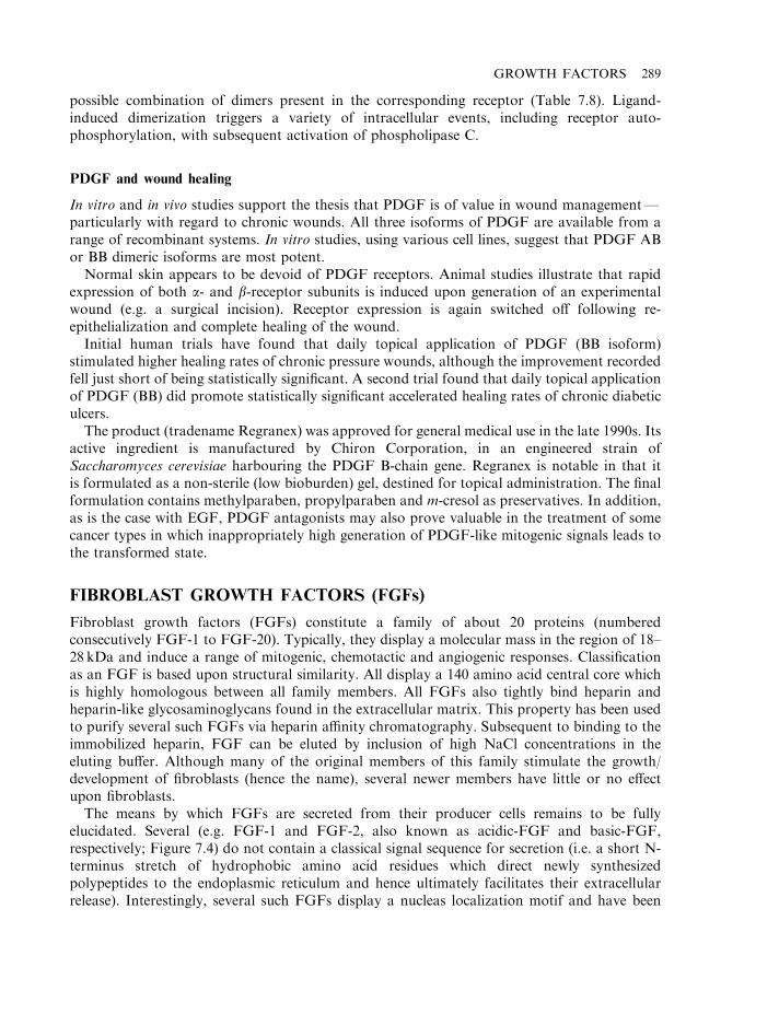
possible combination of dimers present in the corresponding receptor (Table 7.8). Ligand-induced dimerization triggers a variety of intracellular events, including receptor auto-phosphorylation, with subsequent activation of phospholipase C.
PDGF and wound healing
In vitro and in vivo studies support the thesis that PDGF is of value in wound management—particularly with regard to chronic wounds. All three isoforms of PDGF are available from arange of recombinant systems. In vitro studies, using various cell lines, suggest that PDGF ABor BB dimeric isoforms are most potent.
Normal skin appears to be devoid of PDGF receptors. Animal studies illustrate that rapidexpression of both a- and b-receptor subunits is induced upon generation of an experimentalwound (e.g. a surgical incision). Receptor expression is again switched off following re-epithelialization and complete healing of the wound.
Initial human trials have found that daily topical application of PDGF (BB isoform)stimulated higher healing rates of chronic pressure wounds, although the improvement recordedfell just short of being statistically significant. A second trial found that daily topical applicationof PDGF (BB) did promote statistically significant accelerated healing rates of chronic diabeticulcers.
The product (tradename Regranex) was approved for general medical use in the late 1990s. Itsactive ingredient is manufactured by Chiron Corporation, in an engineered strain ofSaccharomyces cerevisiae harbouring the PDGF B-chain gene. Regranex is notable in that itis formulated as a non-sterile (low bioburden) gel, destined for topical administration. The finalformulation contains methylparaben, propylparaben and m-cresol as preservatives. In addition,as is the case with EGF, PDGF antagonists may also prove valuable in the treatment of somecancer types in which inappropriately high generation of PDGF-like mitogenic signals leads tothe transformed state.
FIBROBLAST GROWTH FACTORS (FGFs)
Fibroblast growth factors (FGFs) constitute a family of about 20 proteins (numberedconsecutively FGF-1 to FGF-20). Typically, they display a molecular mass in the region of 18–28 kDa and induce a range of mitogenic, chemotactic and angiogenic responses. Classificationas an FGF is based upon structural similarity. All display a 140 amino acid central core whichis highly homologous between all family members. All FGFs also tightly bind heparin andheparin-like glycosaminoglycans found in the extracellular matrix. This property has been usedto purify several such FGFs via heparin affinity chromatography. Subsequent to binding to theimmobilized heparin, FGF can be eluted by inclusion of high NaCl concentrations in theeluting buffer. Although many of the original members of this family stimulate the growth/development of fibroblasts (hence the name), several newer members have little or no effectupon fibroblasts.
The means by which FGFs are secreted from their producer cells remains to be fullyelucidated. Several (e.g. FGF-1 and FGF-2, also known as acidic-FGF and basic-FGF,respectively; Figure 7.4) do not contain a classical signal sequence for secretion (i.e. a short N-terminus stretch of hydrophobic amino acid residues which direct newly synthesizedpolypeptides to the endoplasmic reticulum and hence ultimately facilitates their extracellularrelease). Interestingly, several such FGFs display a nucleas localization motif and have been
GROWTH FACTORS 289
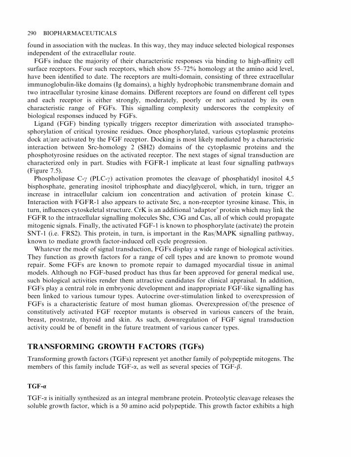
found in association with the nucleas. In this way, they may induce selected biological responsesindependent of the extracellular route.
FGFs induce the majority of their characteristic responses via binding to high-affinity cellsurface receptors. Four such receptors, which show 55–72% homology at the amino acid level,have been identified to date. The receptors are multi-domain, consisting of three extracellularimmunoglobulin-like domains (Ig domains), a highly hydrophobic transmembrane domain andtwo intracellular tyrosine kinase domains. Different receptors are found on different cell typesand each receptor is either strongly, moderately, poorly or not activated by its owncharacteristic range of FGFs. This signalling complexity underscores the complexity ofbiological responses induced by FGFs.
Ligand (FGF) binding typically triggers receptor dimerization with associated transpho-sphorylation of critical tyrosine residues. Once phosphorylated, various cytoplasmic proteinsdock at/are activated by the FGF receptor. Docking is most likely mediated by a characteristicinteraction between Src-homology 2 (SH2) domains of the cytoplasmic proteins and thephosphotyrosine residues on the activated receptor. The next stages of signal transduction arecharacterized only in part. Studies with FGFR-1 implicate at least four signalling pathways(Figure 7.5).
Phospholipase C-g (PLC-g) activation promotes the cleavage of phosphatidyl inositol 4,5bisphosphate, generating inositol triphosphate and diacylglycerol, which, in turn, trigger anincrease in intracellular calcium ion concentration and activation of protein kinase C.Interaction with FGFR-1 also appears to activate Src, a non-receptor tyrosine kinase. This, inturn, influences cytoskeletal structure. CrK is an additional ‘adaptor’ protein which may link theFGFR to the intracellular signalling molecules Shc, C3G and Cas, all of which could propagatemitogenic signals. Finally, the activated FGF-1 is known to phosphorylate (activate) the proteinSNT-1 (i.e. FRS2). This protein, in turn, is important in the Ras/MAPK signalling pathway,known to mediate growth factor-induced cell cycle progression.
Whatever the mode of signal transduction, FGFs display a wide range of biological activities.They function as growth factors for a range of cell types and are known to promote woundrepair. Some FGFs are known to promote repair to damaged myocardial tissue in animalmodels. Although no FGF-based product has thus far been approved for general medical use,such biological activities render them attractive candidates for clinical appraisal. In addition,FGFs play a central role in embryonic development and inappropriate FGF-like signalling hasbeen linked to various tumour types. Autocrine over-stimulation linked to overexpression ofFGFs is a characteristic feature of most human gliomas. Overexpression of/the presence ofconstitutively activated FGF receptor mutants is observed in various cancers of the brain,breast, prostrate, thyroid and skin. As such, downregulation of FGF signal transductionactivity could be of benefit in the future treatment of various cancer types.
TRANSFORMING GROWTH FACTORS (TGFs)
Transforming growth factors (TGFs) represent yet another family of polypeptide mitogens. Themembers of this family include TGF-a, as well as several species of TGF-b.
TGF-a
TGF-a is initially synthesized as an integral membrane protein. Proteolytic cleavage releases thesoluble growth factor, which is a 50 amino acid polypeptide. This growth factor exhibits a high
290 BIOPHARMACEUTICALS
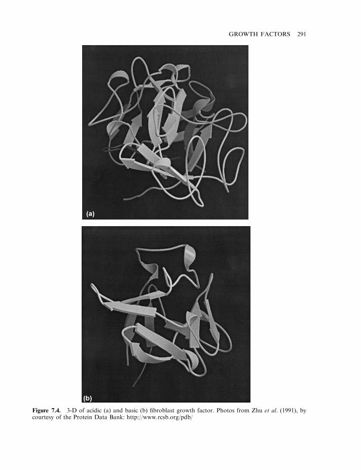
GROWTH FACTORS 291
Figure 7.4. 3-D of acidic (a) and basic (b) fibroblast growth factor. Photos from Zhu et al. (1991), bycourtesy of the Protein Data Bank: http://www.rcsb.org/pdb/
amino acid homology with EGF, and it induces its biological effects by binding to the EGFreceptor. It is synthesized by various body tissues, as well as by monocytes and keratinocytes. Itis also manufactured by many tumour cell types, for which it can act as an autocrine growthfactor.
TGF-b
TGF-b was first described as a growth factor capable of inducing transformation of severalfibroblast cell lines (hence the name, transforming growth factors). It is now recognized that
292 BIOPHARMACEUTICALS
Figure 7.5. Binding of FGF to an FGF receptor, and the possible intracellular events triggered uponbinding. Refer to the text for specific details
‘TGF-b’ actually consists of three separate growth factors: TGF-b1, -b2 and -b3. Although theproduct of distinct genes, all exhibit close homology. In the mature form, they exist ashomodimers, with each subunit containing 112 amino acid residues. Most body cells synthesizeTGF-b, singly or in combination.
TGF-bs are pleiotrophic cytokines. They are capable of inhibiting the cell cycle and hencegrowth of several cell types, most notably epithelial and haemopoietic cells. These factors,however, stimulate the growth of other cell types, most notably cells that give rise to connectivetissue, cartilage and bone. They induce the synthesis of extracellular matrix proteins, andmodulate the expression of matrix proteases. They also serve as a powerful chemoattractant formonocytes and fibroblasts. Given such activities, it is not surprising that the major physiologicalimpact of TGF-bs appear to relate to:
. tissue remodelling;
. wound repair;
. haemopoiesis.
Such activities render them potentially useful therapeutic agents and several are being assessedmedically (Table 7.2). The effect of TGF-b on haemopoietic cells has recently receivedincreasing attention. Along with TNF-a and IFN-g, TGF-b is a physiologically relevantnegative regulator of haemopoiesis. TGF-b also inhibits the growth of various humanleukaemia cell lines in vitro, rendering it of potential interest as a putative anti-cancer agent.
The TGF-bs exert their biological actions by binding to specific receptors, of which there arethree types (I, II and III). All are transmembrane glycoproteins. All three TGF-bs bind to allthree receptor types, although they bind with higher affinity to types I and II receptors (53 kDaand 65 kDa, respectively). The intracellular domains of the type I and II receptors displayendogenous serine/threonine protein kinase activity.
The larger (829 amino acid) type III receptor is a proteoglycan (a class of very highlyglycosylated acidic glycoprotein). Also known as b-glycan, it is characterized by a shortcytoplasmic domain and displays no capability of transducing signals. The type III receptor(which is less widely distributed than the others) may function to concentrate ligand and presentit to type I and II receptors. The exact molecular events underlining TGF signal transductionremain to be elucidated, although co-expression of type I and II receptors appears to be anecessary prerequisite. This has led to speculation that active signal transduction requiresligand-induced formation of a hetromeric complex containing both type I and II receptor units.A family of cytoplasmic proteins, Smad proteins, are also known to transiently associate withactivated TGF-b receptors. Interaction results in the activation via phosphorylation of theSmads, which then translocate to the nucleas. Here they function as transcriptional activators ofvarious genes.
NEUROTROPHIC FACTORS
Neurotrophic factors constitute a group of cytokines that regulate the development,maintenance and survival of neurons in both the central and peripheral nervous systems(Table 7.9). While the first member of this family (nerve growth factor, NGF) was discoveredmore than 50 years ago, it is only in the last decade that the other members have been identifiedand characterized. The major sub-family of neurotrophic factors are the neurotrophins.
The original understanding of the term ‘neurotrophic factor’ was that of a soluble agentfound in limiting quantities in the environment of sensitive neurons, and being generally
GROWTH FACTORS 293
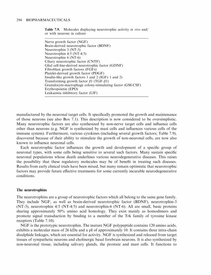
manufactured by the neuronal target cells. It specifically promoted the growth and maintenanceof those neurons (see also Box 7.1). This description is now considered to be oversimplistic.Many neurotrophic factors are also synthesized by non-nerve target cells and influence cellsother than neurons (e.g. NGF is synthesized by mast cells and influences various cells of theimmune system). Furthermore, various cytokines (including several growth factors; Table 7.9),discovered because of their ability to stimulate the growth of non-neuronal cells, are now alsoknown to influence neuronal cells.
Each neurotrophic factor influences the growth and development of a specific group ofneuronal types, with some cells being sensitive to several such factors. Many sustain specificneuronal populations whose death underlines various neurodegenerative diseases. This raisesthe possibility that these regulatory molecules may be of benefit in treating such diseases.Results from early clinical trials have been mixed, but many remain optimistic that neurotrophicfactors may provide future effective treatments for some currently incurable neurodegenerativeconditions.
The neurotrophins
The neurotrophins are a group of neurotrophic factors which all belong to the same gene family.They include NGF, as well as brain-derived neurotrophic factor (BDNF), neurotrophin-3(NT-3), neurotrophin 4/5 (NT-4/5) and neurotrophin-6 (NT-6). All are small, basic proteinssharing approximately 50% amino acid homology. They exist mainly as homodimers andpromote signal transduction by binding to a member of the Trk family of tyrosine kinasereceptors (Table 7.10).
NGF is the prototypic neurotrophin. The mature NGF polypeptide contains 120 amino acids,exhibits a molecular mass of 26 kDa and a pI of approximately 10. It contains three intra-chaindisulphide linkages, which are essential for activity. NGF is synthesized and released from targettissues of sympathetic neurons and cholinergic basal forebrain neurons. It is also synthesized bynon-neuronal tissue, including salivary glands, the prostate and mast cells. It functions to
294 BIOPHARMACEUTICALS
Table 7.9. Molecules displaying neurotrophic activity in vivo and/or with neurons in culture
Nerve growth factor (NGF)Brain-derived neurotrophic factor (BDNF)Neurotrophin 3 (NT-3)Neurotrophin 4/5 (NT-4/5)Neurotrophin 6 (NT-6)Ciliary neurotrophic factor (CNTF)Glial cell-line-derived neurotrophic factor (GDNF)Fibroblast growth factors (FGFs)Platelet-derived growth factor (PDGF)Insulin-like growth factors 1 and 2 (IGFs 1 and 2)Transforming growth factor b1 (TGF-b1)Granulocyte-macrophage colony-stimulating factor (GM-CSF)Erythropoietin (EPO)Leukaemia inhibitory factor (LIF)
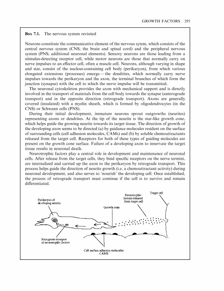
GROWTH FACTORS 295
Box 7.1. The nervous system revisited
Neurons constitute the communicative element of the nervous sytem, which consists of thecentral nervous system (CNS; the brain and spinal cord) and the peripheral nervoussystem (PNS; additional neuronal elements). Sensory neurons are those leading from astimulus-detecting receptor cell, while motor neurons are those that normally carry onnerve impulses to an effector cell, often a muscle cell. Neurons, although varying in shapeand size, consist of the nucleus-containing cell body (perikaryon), from which variouselongated extensions (processes) emerge— the dendrites, which normally carry nerveimpulses towards the perikaryon and the axon, the terminal branches of which form thejunction (synapse) with the cell to which the nerve impulse will be transmitted.
The neuronal cytoskeleton provides the axon with mechanical support and is directlyinvolved in the transport of materials from the cell body towards the synapse (anterogradetransport) and in the opposite direction (retrograde transport). Axons are generallycovered (insulated) with a myelin sheath, which is formed by oligodendrocytes (in theCNS) or Schwann cells (PNS).
During their initial development, immature neurons sprout outgrowths (neurites)representing axons or dendrites. At the tip of the neurite is the star-like growth cone,which helps guide the growing neurite towards its target tissue. The direction of growth ofthe developing axon seems to be directed (a) by guidance molecules resident on the surfaceof surrounding cells (cell adhesion molecules, CAMs) and (b) by soluble chemoattractantsreleased from the target cell. Receptors for both of these types of guiding molecules arepresent on the growth cone surface. Failure of a developing axon to innervate the targettissue results in neuronal death.
Neurotrophic factors play a central role in development and maintenance of neuronalcells. After release from the target cells, they bind specific receptors on the nerve termini,are internalized and carried up the axon to the perikaryon by retrograde transport. Thisprocess helps guide the direction of neurite growth (i.e. a chemoattractant activity) duringneuronal development, and also serves to ‘nourish’ the developing cell. Once established,the process of retrograde transport must continue if the cell is to survive and remaindifferentiated.
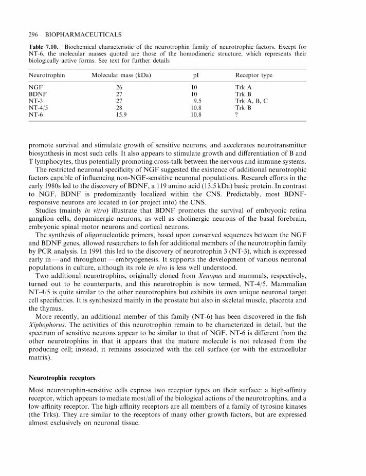
promote survival and stimulate growth of sensitive neurons, and accelerates neurotransmitterbiosynthesis in most such cells. It also appears to stimulate growth and differentiation of B andT lymphocytes, thus potentially promoting cross-talk between the nervous and immune systems.
The restricted neuronal specificity of NGF suggested the existence of additional neurotrophicfactors capable of influencing non-NGF-sensitive neuronal populations. Research efforts in theearly 1980s led to the discovery of BDNF, a 119 amino acid (13.5 kDa) basic protein. In contrastto NGF, BDNF is predominantly localized within the CNS. Predictably, most BDNF-responsive neurons are located in (or project into) the CNS.
Studies (mainly in vitro) illustrate that BDNF promotes the survival of embryonic retinaganglion cells, dopaminergic neurons, as well as cholinergic neurons of the basal forebrain,embryonic spinal motor neurons and cortical neurons.
The synthesis of oligonucleotide primers, based upon conserved sequences between the NGFand BDNF genes, allowed researchers to fish for additional members of the neurotrophin familyby PCR analysis. In 1991 this led to the discovery of neurotrophin 3 (NT-3), which is expressedearly in—and throughout—embryogenesis. It supports the development of various neuronalpopulations in culture, although its role in vivo is less well understood.
Two additional neurotrophins, originally cloned from Xenopus and mammals, respectively,turned out to be counterparts, and this neurotrophin is now termed, NT-4/5. MammalianNT-4/5 is quite similar to the other neurotrophins but exhibits its own unique neuronal targetcell specificities. It is synthesized mainly in the prostate but also in skeletal muscle, placenta andthe thymus.
More recently, an additional member of this family (NT-6) has been discovered in the fishXiphophorus. The activities of this neurotrophin remain to be characterized in detail, but thespectrum of sensitive neurons appear to be similar to that of NGF. NT-6 is different from theother neurotrophins in that it appears that the mature molecule is not released from theproducing cell; instead, it remains associated with the cell surface (or with the extracellularmatrix).
Neurotrophin receptors
Most neurotrophin-sensitive cells express two receptor types on their surface: a high-affinityreceptor, which appears to mediate most/all of the biological actions of the neurotrophins, and alow-affinity receptor. The high-affinity receptors are all members of a family of tyrosine kinases(the Trks). They are similar to the receptors of many other growth factors, but are expressedalmost exclusively on neuronal tissue.
296 BIOPHARMACEUTICALS
Table 7.10. Biochemical characteristic of the neurotrophin family of neurotrophic factors. Except forNT-6, the molecular masses quoted are those of the homodimeric structure, which represents theirbiologically active forms. See text for further details
Neurotrophin Molecular mass (kDa) pI Receptor type
NGF 26 10 Trk ABDNF 27 10 Trk BNT-3 27 9.5 Trk A, B, CNT-4/5 28 10.8 Trk BNT-6 15.9 10.8 ?
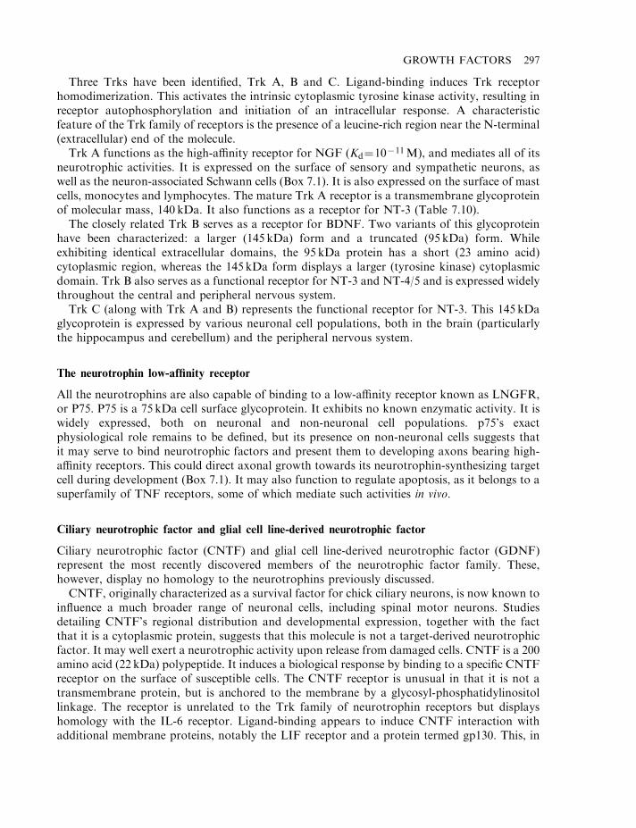
Three Trks have been identified, Trk A, B and C. Ligand-binding induces Trk receptorhomodimerization. This activates the intrinsic cytoplasmic tyrosine kinase activity, resulting inreceptor autophosphorylation and initiation of an intracellular response. A characteristicfeature of the Trk family of receptors is the presence of a leucine-rich region near the N-terminal(extracellular) end of the molecule.
Trk A functions as the high-affinity receptor for NGF (Kd¼10711M), and mediates all of itsneurotrophic activities. It is expressed on the surface of sensory and sympathetic neurons, aswell as the neuron-associated Schwann cells (Box 7.1). It is also expressed on the surface of mastcells, monocytes and lymphocytes. The mature Trk A receptor is a transmembrane glycoproteinof molecular mass, 140 kDa. It also functions as a receptor for NT-3 (Table 7.10).
The closely related Trk B serves as a receptor for BDNF. Two variants of this glycoproteinhave been characterized: a larger (145 kDa) form and a truncated (95 kDa) form. Whileexhibiting identical extracellular domains, the 95 kDa protein has a short (23 amino acid)cytoplasmic region, whereas the 145 kDa form displays a larger (tyrosine kinase) cytoplasmicdomain. Trk B also serves as a functional receptor for NT-3 and NT-4/5 and is expressed widelythroughout the central and peripheral nervous system.
Trk C (along with Trk A and B) represents the functional receptor for NT-3. This 145 kDaglycoprotein is expressed by various neuronal cell populations, both in the brain (particularlythe hippocampus and cerebellum) and the peripheral nervous system.
The neurotrophin low-affinity receptor
All the neurotrophins are also capable of binding to a low-affinity receptor known as LNGFR,or P75. P75 is a 75 kDa cell surface glycoprotein. It exhibits no known enzymatic activity. It iswidely expressed, both on neuronal and non-neuronal cell populations. p75’s exactphysiological role remains to be defined, but its presence on non-neuronal cells suggests thatit may serve to bind neurotrophic factors and present them to developing axons bearing high-affinity receptors. This could direct axonal growth towards its neurotrophin-synthesizing targetcell during development (Box 7.1). It may also function to regulate apoptosis, as it belongs to asuperfamily of TNF receptors, some of which mediate such activities in vivo.
Ciliary neurotrophic factor and glial cell line-derived neurotrophic factor
Ciliary neurotrophic factor (CNTF) and glial cell line-derived neurotrophic factor (GDNF)represent the most recently discovered members of the neurotrophic factor family. These,however, display no homology to the neurotrophins previously discussed.
CNTF, originally characterized as a survival factor for chick ciliary neurons, is now known toinfluence a much broader range of neuronal cells, including spinal motor neurons. Studiesdetailing CNTF’s regional distribution and developmental expression, together with the factthat it is a cytoplasmic protein, suggests that this molecule is not a target-derived neurotrophicfactor. It may well exert a neurotrophic activity upon release from damaged cells. CNTF is a 200amino acid (22 kDa) polypeptide. It induces a biological response by binding to a specific CNTFreceptor on the surface of susceptible cells. The CNTF receptor is unusual in that it is not atransmembrane protein, but is anchored to the membrane by a glycosyl-phosphatidylinositollinkage. The receptor is unrelated to the Trk family of neurotrophin receptors but displayshomology with the IL-6 receptor. Ligand-binding appears to induce CNTF interaction withadditional membrane proteins, notably the LIF receptor and a protein termed gp130. This, in
GROWTH FACTORS 297
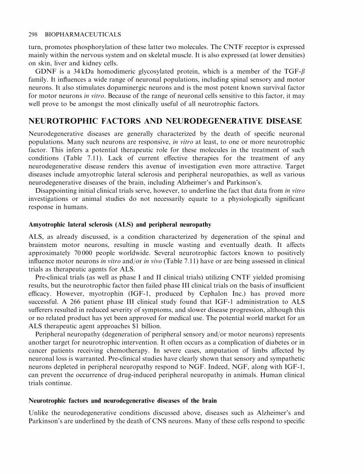
turn, promotes phosphorylation of these latter two molecules. The CNTF receptor is expressedmainly within the nervous system and on skeletal muscle. It is also expressed (at lower densities)on skin, liver and kidney cells.
GDNF is a 34 kDa homodimeric glycosylated protein, which is a member of the TGF-bfamily. It influences a wide range of neuronal populations, including spinal sensory and motorneurons. It also stimulates dopaminergic neurons and is the most potent known survival factorfor motor neurons in vitro. Because of the range of neuronal cells sensitive to this factor, it maywell prove to be amongst the most clinically useful of all neurotrophic factors.
NEUROTROPHIC FACTORS AND NEURODEGENERATIVE DISEASE
Neurodegenerative diseases are generally characterized by the death of specific neuronalpopulations. Many such neurons are responsive, in vitro at least, to one or more neurotrophicfactor. This infers a potential therapeutic role for these molecules in the treatment of suchconditions (Table 7.11). Lack of current effective therapies for the treatment of anyneurodegenerative disease renders this avenue of investigation even more attractive. Targetdiseases include amyotrophic lateral sclerosis and peripheral neuropathies, as well as variousneurodegenerative diseases of the brain, including Alzheimer’s and Parkinson’s.
Disappointing initial clinical trials serve, however, to underline the fact that data from in vitroinvestigations or animal studies do not necessarily equate to a physiologically significantresponse in humans.
Amyotrophic lateral sclerosis (ALS) and peripheral neuropathy
ALS, as already discussed, is a condition characterized by degeneration of the spinal andbrainstem motor neurons, resulting in muscle wasting and eventually death. It affectsapproximately 70 000 people worldwide. Several neurotrophic factors known to positivelyinfluence motor neurons in vitro and/or in vivo (Table 7.11) have or are being assessed in clinicaltrials as therapeutic agents for ALS.
Pre-clinical trials (as well as phase I and II clinical trials) utilizing CNTF yielded promisingresults, but the neurotrophic factor then failed phase III clinical trials on the basis of insufficientefficacy. However, myotrophin (IGF-1, produced by Cephalon Inc.) has proved moresuccessful. A 266 patient phase III clinical study found that IGF-1 administration to ALSsufferers resulted in reduced severity of symptoms, and slower disease progression, although thisor no related product has yet been approved for medical use. The potential world market for anALS therapeutic agent approaches $1 billion.
Peripheral neuropathy (degeneration of peripheral sensory and/or motor neurons) representsanother target for neurotrophic intervention. It often occurs as a complication of diabetes or incancer patients receiving chemotherapy. In severe cases, amputation of limbs affected byneuronal loss is warranted. Pre-clinical studies have clearly shown that sensory and sympatheticneurons depleted in peripheral neuropathy respond to NGF. Indeed, NGF, along with IGF-1,can prevent the occurrence of drug-induced peripheral neuropathy in animals. Human clinicaltrials continue.
Neurotrophic factors and neurodegenerative diseases of the brain
Unlike the neurodegenerative conditions discussed above, diseases such as Alzheimer’s andParkinson’s are underlined by the death of CNS neurons. Many of these cells respond to specific
298 BIOPHARMACEUTICALS
neurotrophic factors, which therefore may constitute effective therapies in the treatment of theseincurable conditions. The existence of the blood–brain barrier represents a complication, as itprecludes delivery of the drug to the brain via intravenous injection. A number of approacheshave been adopted to circumvent this problem, including:
. direct injection of neurotrophic factor into the brain;
. use of cranial infusion pumps;
. intraventricular transplantation of recombinant polymer-encapsulated cells which secreteneurotrophic factors.
While such approaches may be technically feasible, the associated risks render unlikely theirwidespread clinical application. An alternative approach that may prove more acceptableinvolves conjugating the neurotrophic factor to monoclonal antibodies raised against thetransferrin receptor. This normally transports iron across the blood–brain barrier; i.v.administration of NGF–anti-transferrin antibody conjugate results in its translocation acrossthe blood–brain barrier. Upon entry into the brain, the conjugate was found to promotesurvival of NGF-sensitive neurons. Amongst these are cholinergic striatal interneurons, whichdegenerate in Huntington’s disease.
Loss of cholinergic neurons of the basal forebrain is a characteristic feature of Alzheimer’sdisease. These neurons enervate the hippocampus (the brain’s memory centre). They respond toNGF, BDNF and NT-4/5, e.g. NGF can rescue such cells in rodents after damage induced bycutting their axons.
Degeneration of these same neurons is also associated with an age-dependent decrease inlearning and memory ability. Infusion of NGF and NT-4/5 to aged rats appears to improvetheir spatial memory ability. Further studies in rodents have shown that physical exerciseincreased BDNF expression, which may explain why exercise is often a predictor of high mentalfunction during ageing.
Dopaminergic neurons of the substantia nigra represent the major neural populationdegenerating in patients suffering from Parkinson’s disease. Several neurotrophic factors,including NT-4/5, BDNF and GDNF, sustain these cells in vitro and, hence, may be useful intreating this disease. Pre-clinical studies show that direct injection of GDNF to sites adjacent tothe substantia nigra prevents degeneration of these neurons when they are experimentallydamaged by cutting their axons.
Likewise, GDNF prevents dopaminergic loss (in animals) subsequent to the administration ofthe neurotoxin, MPTP, which induces parkinsonian symptoms in man. Even moreencouragingly, administration of GDNF subsequent to MPTP-induced dopaminergic damage
GROWTH FACTORS 299
Table 7.11. Some neurodegenerative diseases for which no current effective therapy exists, andneurotrophic factors known to stimulate growth of neuronal populations affected by these conditions
Disease Neurons mainly affected Neurotrophic factors to which cell may respond
Peripheral neuropathies Sensory/motor neurons NGF, IGF-1, NT-3, CNTFAmyotrophic lateralsclerosis
Spinal motor neurons BDNF, IGF-1, CNTF, GDNF, NT-4/5
Alzheimer’s disease Forebrain cholinergic neurons NGF, BDNF, NT-4/5Parkinson’s disease Nigral dopamine neurons GDNF, BDNF, NT-3, NT-4/5Huntington’s disease Striatal GABA neurons BDNF, NT-4/5
in mice promoted significant restoration of the nigrostriatal dopamine system. This observationleaves open the tantalizing possibility that GDNF might play a therapeutic role in halting, oreven reversing, the clinical progression of Parkinson’s. This can only be assessed by actualhuman clinical trials.
The use of a combination of neurotrophic factors in the treatment of neurodegenerativedisease may well prove an avenue worthy of consideration. For example, pre-clinical studiesreveal that administration of a combination of CNTF and BDNF to Wobbler mice (an animalmodel of motor neuron disease), prevented progression of motor neuron dysfuction, whereasadministration of either factor on its own only slowed progression of the disease.
Such an approach could be underpinned by the development of engineered proteinsdisplaying multiple neurotrophic activities. Pan-neurotrophin-1, for example, is an engineeredchimaeric neurotrophin, containing the active domains of NGF, BDNF and NT-3. This hybridmolecule can bind the neurotrophin receptors Trk A, B and C, as well as P75.
Limited clinical success keeps optimism alive that neurotrophic factors will find application inthe treatment of some neurodegenerative disease. However, no member of this family ofregulatory proteins have been approved for medical use, despite all the clinical investigationsundertaken over the last decade and a half.
FURTHER READING
Books
Evrard, P. (2002). The Newborn Brain. Cambridge University Press, Cambridge.McKay, I. (1998). Growth Factors and Receptors. Oxford University Press, Oxford.Nilsen, H. (1994). Growth Factors and Signal Transduction in Development. Wiley-Liss, New York.Thomson, A. (1998). Cytokine Handbook. Academic Press, London.
Articles
Insulin-like growth factors
Bach, L. (1999). The insulin-like growth factor system: basic and clinical aspects. Aust. N. Z. J. Med. 29(3), 355–361.Cohick, W. & Clemmons, D. (1993). The insulin-like growth factors. Ann. Rev. Physiol. 55, 131–153.Dercole, A. et al. (1996). The role of insulin-like growth factors in the central nervous system. Mol. Neurobiol. 13(3),
227–255.Furstenberger, G. & Senn, H. (2002). Insulin-like growth factors and cancer. Lancet Oncol. 3(5), 298–302.Kostecka, Z. & Blahovec, J. (2002). Animal insulin-like growth factor binding proteins and their biological functions.
Vet. Med. 47(2–3), 75–84.Leroith, D. et al. (1997). Insulin-like growth factors. N. Engl. J. Med. 336(9), 633–640.Schmid, C. (1995). Insulin-like growth factors. Cell Biol. Int. 19(5), 445–457.Zumkeller, W. (2002). The insulin-like growth factor system in hematopoietic cells. Leukaemia Lymphoma 43(3), 487–
491.
EGF and PDGF
Adjej, A. (2001). Epidermal growth factor receptor tyrosine kinase inhibitors in cancer therapy. Drugs Future 26(11),1087–1092.
Boonstra, J. et al. (1995). The epidermal growth factor. Cell Biol. Int. 19(5), 413–430.Claesson-Welsh, L. (1994). Platelet-derived growth factor receptor signals. J. Biol. Chem. 269(51), 32023–32026.Downward, J. et al. (1984). Close similarity of EGF receptor and V-erb-B oncogene protein sequences. Nature 307, 521–
527.Johnstone P. (2002). The epidermal growth factor receptor: a new target for anticancer therapy— introduction. Curr.
Probl. Cancer 26(3), 114–164.Leserer, M. et al. (2000). Epidermal growth factor receptor signal transactivation. IUBMB Life 49(5), 405–430.
300 BIOPHARMACEUTICALS
Meyer-Ingold, W. & Eichner, W. (1995). Platelet-derived growth factor. Cell Biol. Int. 19(5), 389–398.Wakeling, A. (2002). Epidermal growth factor receptor tyrosine kinase inhibitors. Curr. Opin. Pharmacol. 2(4), 382–387.
FGF/TGF
Dennler, S. et al. (2002). Transforming growth factor-b signal transduction. J. Leukocyte Biol. 71(5), 731–740.Derynck, R. (1994). TGF-b-receptor-mediated signalling. Trends Biochem. Sci. 19, 548–553.Hu, X. & Zuckerman, K. (2001). Transforming growth factor: signal transduction pathways, cell cycle mediation, and
effects on hematopoiesis. J. Hematother. Stem Cell Res. 10(1), 67–74.Powers, C. et. al. (2000). Fibroblast growth factors, their receptors and signalling. Endocr. Rel. Cancer 7(3), 165–197.Souchelnytskyi, S. (2002). Transforming growth factor-b signalling and its role in cancer. Exp. Oncol. 24(1), 3–12.Zhu, X., Komiya, A., Chirino, S. et al. (1991). Three-dimensional structures of acidic and basic fibroblast growth factors.
Science 251, 90.
Wound-healing and growth factors
Deuel, T. et al. (1991). Growth factors and wound healing: platelet-derived growth factor as a model cytokine. Ann. Rev.Med. 42, 567–584.
Martin, P. (1997). Wound healing—aiming for perfect skin regeneration. Science 276(5309), 75–81.Meyer-Ingold, W. (1993). Wound therapy: growth factors as agents to promote healing. Trends Biotechnol. 11, 387–392.Mustoe, T. et al. (1987). Accelerated healing of incisional wounds in rats induced by transforming growth factor type b.
Science 347, 1333–1336.Pierce, G. & Mustoe, T. (1995). Pharmacologic enhancement of wound healing. Ann. Rev. Med. 46, 467–481.
Neurotrophic and related factors
Butte, M. (2001). Neurotrophic factor structure reveals clues to evolution, binding, specificity and receptor activation.Cell. Mol. Life Sci. 58(8), 1003–1013.
Ebadi, M. et al. (1997). Neurotrophins and their receptors in nerve injury and repair. Neurochem. Int. 30, 347–374.Fu, S. & Gordon, T. (1997). The cellular and molecular basis of peripheral nerve regeneration. Mol. Neurobiol. 14, 67–
116.Haque, N. et al. (1997). Therapeutic strategies for Huntington’s disease, based on a molecular understanding of the
disorder. Mol. Med. Today 3(4), 175–183.Hefti, F. (1997). Pharmacology of neurotrophic factors. Ann. Rev. Pharmacol. Toxicol. 37, 239–267.J. Neurobiol. (1994) 25(11), special issue devoted exclusively to neurotrophic factors.Kwon, Y. (2002). Effect of neurotrophic factors on neuronal stem cell death. J. Biochem. Mol. Biol. 35(1), 87–93.Raffioni, S. et al. (1993). The receptor for nerve growth factor and other neurotrophins. Ann. Rev. Biochem. 62, 823–850.Sofroniew, M. et al. (2001). Nerve growth factor signalling, neuroprotection and neural repair. Ann. Rev. Neurosci. 24,
1217–1281.Streppel, M. et al. (2002). Focal application of neutralizing antibodies to soluble neurotrophic factors reduces collateral
axonal branching after peripheral nerve lesion. Eur. J. Neurosci. 15(8), 1327–1342.Tatagiba, M. et al. (1997). Regeneration of injured axons in the adult mammalian central nervous system. Neurosurgery
40(3), 541–546.Thorne, R. & Frey, W. (2001). Delivery of neurotrophic factors to the central nervous system—pharmacokinetic
considerations. Clin. Pharmacokinet. 40(12), 907–946.Walsh, G. (1995). Nervous excitement over neurotrophic factors. Bio/Technology 13, 1167–1171.Yamada, K. et al. (2002). Role for brain derived neurotrophic factor in learning and memory. Life Sci. 70(7), 735–744.Yamada, M. et al. (1997). The neurotrophic action and signalling of epidermal growth factor. Progr. Neurobiol. 51(1),
19–37.
GROWTH FACTORS 301

Chapter 8
Hormones of therapeutic interest
Hormones are amongst the most important group of regulatory molecules produced by thebody. Originally, the term ‘hormone’ was defined as a substance synthesized and released from aspecific gland in the body which, by interacting with a receptor present in or on a distantsensitive cell, brought about a change in that target cell. Hormones travel to the target cell viathe circulatory system. This describes what is now termed a ‘true endocrine hormone’.
At its loosest definition, some now consider a hormone to be any regulatory substance thatcarries a signal to generate some alteration at a cellular level. This embraces the concept ofparacrine regulators (i.e. produced in the immediate vicinity of their target cells) and autocrineregulators (i.e. producer cell is also the target cell). Under such a broad definition, all cytokines,for example, could be considered hormones. The delineation between a cytokine and a hormoneis already quite fuzzy using any definition.
True endocrine hormones, however, remain a fairly well-defined group. Virtually all of thehormones used therapeutically (discussed below) fit into this grouping. Examples includeinsulin, glucagon, growth hormone and the gonadotrophins.
INSULIN
Insulin is a polypeptide hormone produced by the b cells of the pancreatic islets of Langerhans.It plays a central role in regulating blood glucose levels, generally keeping it within narrowdefined limits (3.5–8.0 mmol/l), irrespective of the nutritional status of the animal. It also has aprofound effect on the metabolism of proteins and lipids and displays some mitogenic activity.The latter is particularly evident in in vitro studies and at high insulin concentrations. Some ofthese mitogenic effects are likely mediated via the insulin-like growth factor 1 (1GF-1) receptorand their physiological relevance is questionable.
Although many cells in the body express the insulin receptor, its most important targets areskeletal muscle fibres, hepatocytes and adipocytes, where it often antagonizes the effects ofglucagon (Table 8.1). The most potent known stimulus of pancreatic insulin release is anincrease in blood glucose levels, often occurring after meal times. Insulin orchestrates a suitablemetabolic response to the absorption of glucose and other nutrients in a number of ways:
Biopharmaceuticals: Biochemistry and Biotechnology, Second Edition by Gary WalshJohn Wiley & Sons Ltd: ISBN 0 470 84326 8 (ppc), ISBN 0 470 84327 6 (pbk)
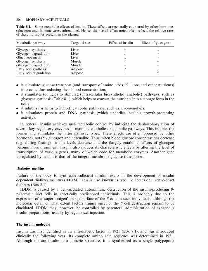
. it stimulates glucose transport (and transport of amino acids, K+ ions and other nutrients)into cells, thus reducing their blood concentration;
. it stimulates (or helps to stimulate) intracellular biosynthetic (anabolic) pathways, such asglycogen synthesis (Table 8.1), which helps to convert the nutrients into a storage form in thecells;
. it inhibits (or helps to inhibit) catabolic pathways, such as glycogenolysis;
. it stimulates protein and DNA synthesis (which underlies insulin’s growth-promotingactivity).
In general, insulin achieves such metabolic control by inducing the dephosphorylation ofseveral key regulatory enzymes in mainline catabolic or anabolic pathways. This inhibits theformer and stimulates the latter pathway types. These effects are often opposed by otherhormones, notably glucagon and adrenaline. Thus, when blood glucose concentrations decrease(e.g. during fasting), insulin levels decrease and the (largely catabolic) effects of glucagonbecome more prominent. Insulin also induces its characteristic effects by altering the level oftranscription of various genes, many of which code for metabolic enzymes. Another geneupregulated by insulin is that of the integral membrane glucose transporter.
Diabetes mellitus
Failure of the body to synthesize sufficient insulin results in the development of insulindependent diabetes mellitus (IDDM). This is also known as type 1 diabetes or juvenile-onsetdiabetes (Box 8.1).
IDDM is caused by T cell-mediated autoimmune destruction of the insulin-producing b-pancreatic islet cells in genetically predisposed individuals. This is probably due to theexpression of a ‘super antigen’ on the surface of the b cells in such individuals, although themolecular detail of what extent factors trigger onset of the b cell destruction remain to beelucidated. IDDM may, however, be controlled by parenteral administration of exogenousinsulin preparations, usually by regular s.c. injection.
The insulin molecule
Insulin was first identified as an anti-diabetic factor in 1921 (Box 8.1), and was introducedclinically the following year. Its complete amino acid sequence was determined in 1951.Although mature insulin is a dimeric structure, it is synthesized as a single polypeptide
304 BIOPHARMACEUTICALS
Table 8.1. Some metabolic effects of insulin. These effects are generally countered by other hormones(glucagon and, in some cases, adrenaline). Hence, the overall effect noted often reflects the relative ratesof these hormones present in the plasma
Metabolic pathway Target tissue Effect of insulin Effect of glucagon
Glycogen synthesis Liver " #Glycogen degradation Liver # "Gluconeogenesis Liver # "Glycogen synthesis Muscle " –Glycogen degradation Muscle – –Fatty acid synthesis Adipose " #Fatty acid degradation Adipose # "
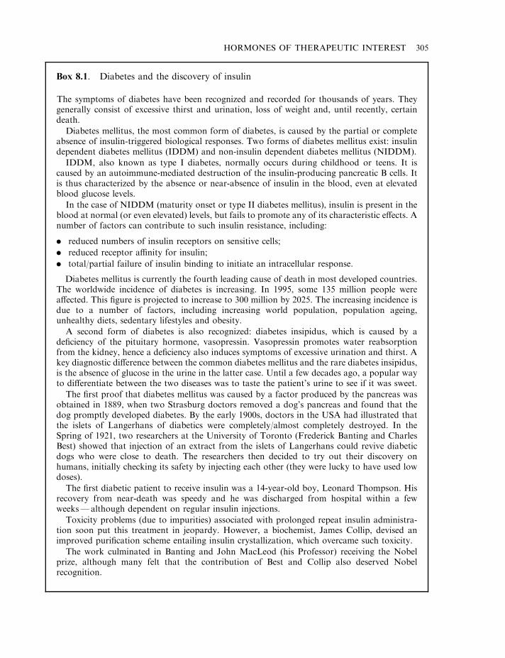
HORMONES OF THERAPEUTIC INTEREST 305
Box 8.1. Diabetes and the discovery of insulin
The symptoms of diabetes have been recognized and recorded for thousands of years. Theygenerally consist of excessive thirst and urination, loss of weight and, until recently, certaindeath.
Diabetes mellitus, the most common form of diabetes, is caused by the partial or completeabsence of insulin-triggered biological responses. Two forms of diabetes mellitus exist: insulindependent diabetes mellitus (IDDM) and non-insulin dependent diabetes mellitus (NIDDM).
IDDM, also known as type I diabetes, normally occurs during childhood or teens. It iscaused by an autoimmune-mediated destruction of the insulin-producing pancreatic B cells. Itis thus characterized by the absence or near-absence of insulin in the blood, even at elevatedblood glucose levels.
In the case of NIDDM (maturity onset or type II diabetes mellitus), insulin is present in theblood at normal (or even elevated) levels, but fails to promote any of its characteristic effects. Anumber of factors can contribute to such insulin resistance, including:
. reduced numbers of insulin receptors on sensitive cells;
. reduced receptor affinity for insulin;
. total/partial failure of insulin binding to initiate an intracellular response.
Diabetes mellitus is currently the fourth leading cause of death in most developed countries.The worldwide incidence of diabetes is increasing. In 1995, some 135 million people wereaffected. This figure is projected to increase to 300 million by 2025. The increasing incidence isdue to a number of factors, including increasing world population, population ageing,unhealthy diets, sedentary lifestyles and obesity.
A second form of diabetes is also recognized: diabetes insipidus, which is caused by adeficiency of the pituitary hormone, vasopressin. Vasopressin promotes water reabsorptionfrom the kidney, hence a deficiency also induces symptoms of excessive urination and thirst. Akey diagnostic difference between the common diabetes mellitus and the rare diabetes insipidus,is the absence of glucose in the urine in the latter case. Until a few decades ago, a popular wayto differentiate between the two diseases was to taste the patient’s urine to see if it was sweet.
The first proof that diabetes mellitus was caused by a factor produced by the pancreas wasobtained in 1889, when two Strasburg doctors removed a dog’s pancreas and found that thedog promptly developed diabetes. By the early 1900s, doctors in the USA had illustrated thatthe islets of Langerhans of diabetics were completely/almost completely destroyed. In theSpring of 1921, two researchers at the University of Toronto (Frederick Banting and CharlesBest) showed that injection of an extract from the islets of Langerhans could revive diabeticdogs who were close to death. The researchers then decided to try out their discovery onhumans, initially checking its safety by injecting each other (they were lucky to have used lowdoses).
The first diabetic patient to receive insulin was a 14-year-old boy, Leonard Thompson. Hisrecovery from near-death was speedy and he was discharged from hospital within a fewweeks—although dependent on regular insulin injections.
Toxicity problems (due to impurities) associated with prolonged repeat insulin administra-tion soon put this treatment in jeopardy. However, a biochemist, James Collip, devised animproved purification scheme entailing insulin crystallization, which overcame such toxicity.
The work culminated in Banting and John MacLeod (his Professor) receiving the Nobelprize, although many felt that the contribution of Best and Collip also deserved Nobelrecognition.
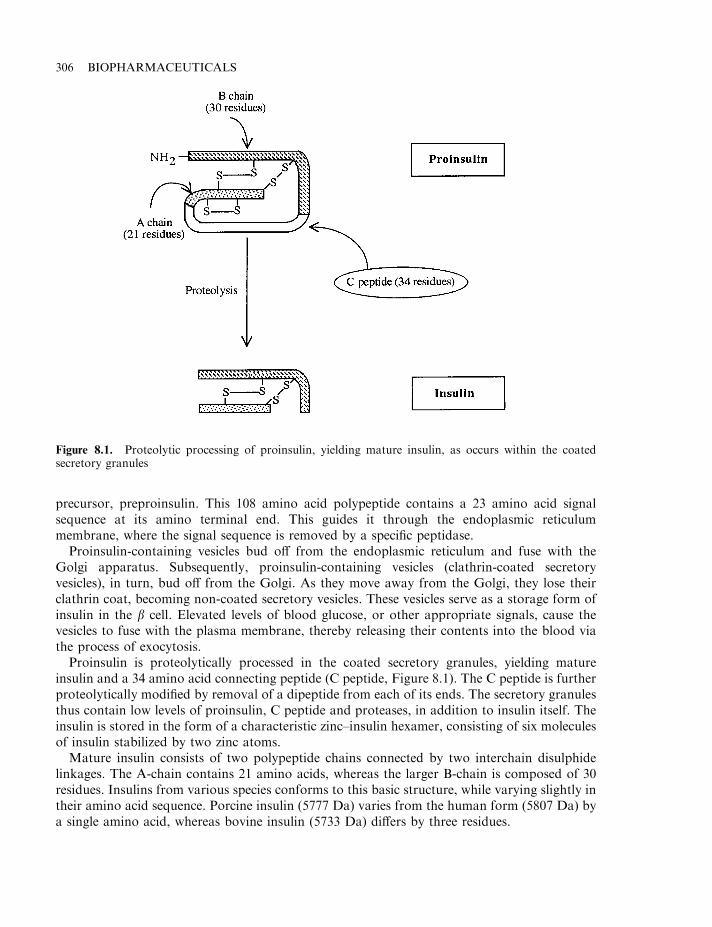
precursor, preproinsulin. This 108 amino acid polypeptide contains a 23 amino acid signalsequence at its amino terminal end. This guides it through the endoplasmic reticulummembrane, where the signal sequence is removed by a specific peptidase.
Proinsulin-containing vesicles bud off from the endoplasmic reticulum and fuse with theGolgi apparatus. Subsequently, proinsulin-containing vesicles (clathrin-coated secretoryvesicles), in turn, bud off from the Golgi. As they move away from the Golgi, they lose theirclathrin coat, becoming non-coated secretory vesicles. These vesicles serve as a storage form ofinsulin in the b cell. Elevated levels of blood glucose, or other appropriate signals, cause thevesicles to fuse with the plasma membrane, thereby releasing their contents into the blood viathe process of exocytosis.
Proinsulin is proteolytically processed in the coated secretory granules, yielding matureinsulin and a 34 amino acid connecting peptide (C peptide, Figure 8.1). The C peptide is furtherproteolytically modified by removal of a dipeptide from each of its ends. The secretory granulesthus contain low levels of proinsulin, C peptide and proteases, in addition to insulin itself. Theinsulin is stored in the form of a characteristic zinc–insulin hexamer, consisting of six moleculesof insulin stabilized by two zinc atoms.
Mature insulin consists of two polypeptide chains connected by two interchain disulphidelinkages. The A-chain contains 21 amino acids, whereas the larger B-chain is composed of 30residues. Insulins from various species conforms to this basic structure, while varying slightly intheir amino acid sequence. Porcine insulin (5777 Da) varies from the human form (5807 Da) bya single amino acid, whereas bovine insulin (5733 Da) differs by three residues.
306 BIOPHARMACEUTICALS
Figure 8.1. Proteolytic processing of proinsulin, yielding mature insulin, as occurs within the coatedsecretory granules
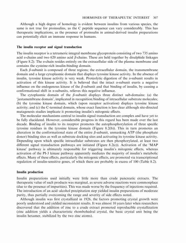
Although a high degree of homology is evident between insulins from various species, thesame is not true for proinsulins, as the C peptide sequence can vary considerably. This hastherapeutic implications, as the presence of proinsulin in animal-derived insulin preparationscan potentially elicit an immune response in humans.
The insulin receptor and signal transduction
The insulin receptor is a tetrameric integral membrane glycoprotein consisting of two 735 aminoacid a-chains and two 620 amino acid b-chains. These are held together by disulphide linkages(Figure 8.2). The a-chain resides entirely on the extracellular side of the plasma membrane andcontains the cysteine-rich insulin-binding domain.
Each b-subunit is composed of three regions; the extracellular domain, the transmembranedomain and a large cytoplasmic domain that displays tyrosine kinase activity. In the absence ofinsulin, tyrosine kinase activity is very weak. Proteolytic digestion of the a-subunit results inactivation of this kinase activity. It is believed that the intact a-subunit exerts a negativeinfluence on the endogenous kinase of the b-subunit and that binding of insulin, by causing aconformational shift in a-subunits, relieves this negative influence.
The cytoplasmic domain of the b-subunit displays three distinct sub-domains: (a) the‘juxtamembrane domain’, implicated in recognition/binding of intracellular substrate molecules;(b) the tyrosine kinase domain, which (upon receptor activation) displays tyrosine kinaseactivity; and (c) the C-terminal domain, whose exact function is less clear although site-directedmutagenesis studies implicate it promoting insulin’s mitogenic effects.
The molecular mechanisms central to insulin signal transduction are complex and have yet tobe fully elucidated. However, considerable progress in this regard has been made over the lastdecade. Binding of insulin to its receptor promotes the autophosphorylation of three specifictyrosine residues in the tyrosine kinase domain (Figure 8.2(b)). This in turn promotes analteration in the conformational state of the entire b-subunit, unmasking ATP (the phosphatedonor) binding sites as well as substrate docking sites and activating its tyrosine kinase activity.Depending upon which specific intracellular substrates are then phosphorylated, at least twodifferent signal transduction pathways are initiated (Figure 8.2(c)). Activation of the ‘MAPkinase’ pathway is ultimately responsible for triggering insulin’s mitogenic effects, whereasactivation of the PI-3 kinase pathway apparently mediates the majority of insulin’s metaboliceffects. Many of these effects, particularly the mitogenic effects, are promoted via transcriptionalregulation of insulin-sensitive genes, of which there are probably in excess of 100 (Table 8.2).
Insulin production
Insulin preparations used initially were little more than crude pancreatic extracts. Thetherapeutic value of such products was marginal, as severe adverse reactions were commonplace(due to the presence of impurities). This was made worse by the frequency of injections required.The introduction of an acid–alcohol precipitation step yielded insulin preparations of moderatepurity, thus partially overcoming the range and severity of side effects noted.
Although insulin was first crystallized in 1926, the factors promoting crystal growth werepoorly understood and yielded inconsistent results. It was almost 10 years later when researchersdiscovered that the addition of zinc to a crude extract promoted reproducible crystallization(zinc addition yields a characteristic rhombohedral crystal, the basic crystal unit being theinsulin hexamer, stabilized by the two zinc atoms).
HORMONES OF THERAPEUTIC INTEREST 307
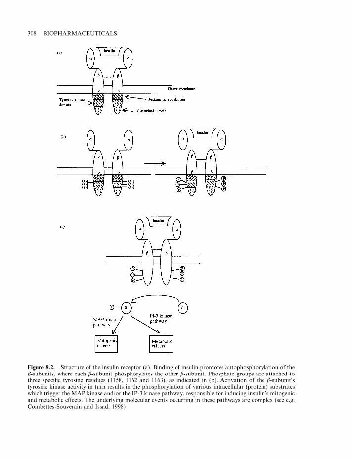
308 BIOPHARMACEUTICALS
Figure 8.2. Structure of the insulin receptor (a). Binding of insulin promotes autophosphorylation of theb-subunits, where each b-subunit phosphorylates the other b-subunit. Phosphate groups are attached tothree specific tyrosine residues (1158, 1162 and 1163), as indicated in (b). Activation of the b-subunit’styrosine kinase activity in turn results in the phosphorylation of various intracellular (protein) substrateswhich trigger the MAP kinase and/or the IP-3 kinase pathway, responsible for inducing insulin’s mitogenicand metabolic effects. The underlying molecular events occurring in these pathways are complex (see e.g.Combettes-Souverain and Issad, 1998)
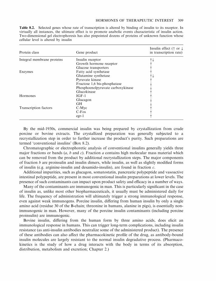
By the mid-1930s, commercial insulin was being prepared by crystallization from crudeporcine or bovine extracts. The crystallized preparation was generally subjected to arecrystallization step in order to further increase the product’s purity. Such preparations aretermed ‘conventional insulins’ (Box 8.2).
Chromatographic or electrophoretic analysis of conventional insulins generally yields threemajor fractions or bands (a, b and c). Fraction a contains high molecular mass material whichcan be removed from the product by additional recrystallization steps. The major componentsof fraction b are proinsulin and insulin dimers, while insulin, as well as slightly modified formsof insulin (e.g. arginine-insulin and desamido-insulin), are found in fraction c.
Additional impurities, such as glucagon, somatostatin, pancreatic polypeptide and vasoactiveintestinal polypeptide, are present in most conventional insulin preparations at lower levels. Thepresence of such contaminants can impact upon product safety and efficacy in a number of ways.
Many of the contaminants are immunogenic in man. This is particularly significant in the caseof insulin as, unlike most other biopharmaceuticals, it usually must be administered daily forlife. The frequency of administration will ultimately trigger a strong immunological response,even against weak immunogens. Porcine insulin, differing from human insulin by only a singleamino acid (residue 30 of the B-chain; threonine in humans, alanine in pigs), is essentially non-immunogenic in man. However, many of the porcine insulin contaminants (including porcineproinsulin) are immunogenic.
Bovine insulin, differing from the human form by three amino acids, does elicit animmunological response in humans. This can trigger long-term complications, including insulinresistance (as anti-insulin antibodies neutralize some of the administered product). The presenceof these antibodies can also affect the pharmacokinetic profile of the drug, as antibody-boundinsulin molecules are largely resistant to the normal insulin degradative process. (Pharmaco-kinetics is the study of how a drug interacts with the body in terms of its absorption,distribution, metabolism and excretion; Chapter 2.)
HORMONES OF THERAPEUTIC INTEREST 309
Table 8.2. Selected genes whose rate of transcription is altered by binding of insulin to its receptor. Invirtually all instances, the ultimate effect is to promote anabolic events characteristic of insulin action.Two-dimensional gel electrophoresis has also pinpointed dozens of proteins of unknown function whosecellular level is altered by insulin
Protein class Gene productInsulin effect (" or #in transcription rate)
Integral membrane proteins Insulin receptor "#Growth hormone receptor "Glucose transporters "
Enzymes Fatty acid synthetase "Glutamine synthetase "#Pyruvate kinase "Fructose 1,6 bis-phosphatase #Phosphoenolpyruvate carboxykinase #Glucokinase "
Hormones IGF-1 "Glucagon #GH #
Transcription factors C-Myc "C-Fos "egr-1 "
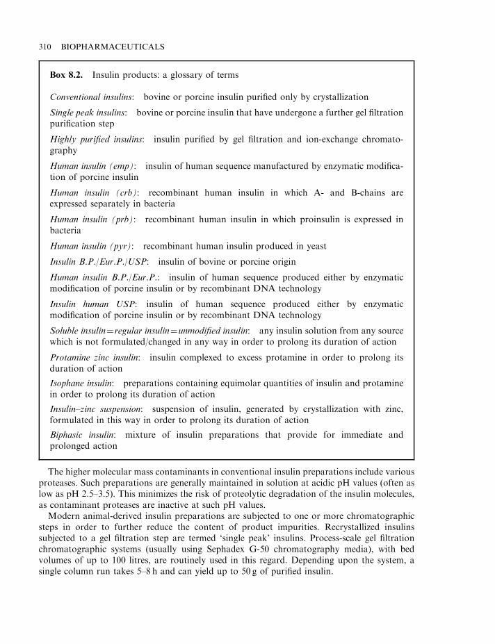
The higher molecular mass contaminants in conventional insulin preparations include variousproteases. Such preparations are generally maintained in solution at acidic pH values (often aslow as pH 2.5–3.5). This minimizes the risk of proteolytic degradation of the insulin molecules,as contaminant proteases are inactive at such pH values.
Modern animal-derived insulin preparations are subjected to one or more chromatographicsteps in order to further reduce the content of product impurities. Recrystallized insulinssubjected to a gel filtration step are termed ‘single peak’ insulins. Process-scale gel filtrationchromatographic systems (usually using Sephadex G-50 chromatography media), with bedvolumes of up to 100 litres, are routinely used in this regard. Depending upon the system, asingle column run takes 5–8 h and can yield up to 50 g of purified insulin.
310 BIOPHARMACEUTICALS
Box 8.2. Insulin products: a glossary of terms
Conventional insulins: bovine or porcine insulin purified only by crystallization
Single peak insulins: bovine or porcine insulin that have undergone a further gel filtrationpurification step
Highly purified insulins: insulin purified by gel filtration and ion-exchange chromato-graphy
Human insulin (emp): insulin of human sequence manufactured by enzymatic modifica-tion of porcine insulin
Human insulin (crb): recombinant human insulin in which A- and B-chains areexpressed separately in bacteria
Human insulin (prb): recombinant human insulin in which proinsulin is expressed inbacteria
Human insulin (pyr): recombinant human insulin produced in yeast
Insulin B.P./Eur.P./USP: insulin of bovine or porcine origin
Human insulin B.P./Eur.P.: insulin of human sequence produced either by enzymaticmodification of porcine insulin or by recombinant DNA technology
Insulin human USP: insulin of human sequence produced either by enzymaticmodification of porcine insulin or by recombinant DNA technology
Soluble insulin¼regular insulin¼unmodified insulin: any insulin solution from any sourcewhich is not formulated/changed in any way in order to prolong its duration of action
Protamine zinc insulin: insulin complexed to excess protamine in order to prolong itsduration of action
Isophane insulin: preparations containing equimolar quantities of insulin and protaminein order to prolong its duration of action
Insulin–zinc suspension: suspension of insulin, generated by crystallization with zinc,formulated in this way in order to prolong its duration of action
Biphasic insulin: mixture of insulin preparations that provide for immediate andprolonged action
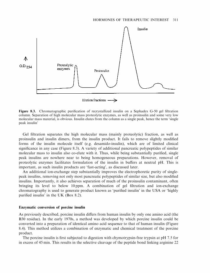
Gel filtration separates the high molecular mass (mainly proteolytic) fraction, as well asproinsulin and insulin dimers, from the insulin product. It fails to remove slightly modifiedforms of the insulin molecule itself (e.g. desamido-insulin), which are of limited clinicalsignificance in any case (Figure 8.3). A variety of additional pancreatic polypeptides of similarmolecular mass to insulin also co-elute with it. Thus, while being substantially purified, singlepeak insulins are nowhere near to being homogeneous preparations. However, removal ofproteolytic enzymes facilitates formulation of the insulin in buffers at neutral pH. This isimportant, as such insulin products are ‘fast-acting’, as discussed later.
An additional ion-exchange step substantially improves the electrophoretic purity of single-peak insulins, removing not only most pancreatic polypeptides of similar size, but also modifiedinsulins. Importantly, it also achieves separation of much of the proinsulin contaminant, oftenbringing its level to below 10 ppm. A combination of gel filtration and ion-exchangechromatography is used to generate product known as ‘purified insulin’ in the USA or ‘highlypurified insulin’ in the UK (Box 8.2).
Enzymatic conversion of porcine insulin
As previously described, porcine insulin differs from human insulin by only one amino acid (theB30 residue). In the early 1970s, a method was developed by which porcine insulin could beconverted into a preparation of identical amino acid sequence to that of human insulin (Figure8.4). This method utilizes a combination of enzymatic and chemical treatment of the porcineproduct.
The porcine insulin is first subjected to digestion with chymotrypsin-free trypsin at pH 7.5 forin excess of 45min. This results in the selective cleavage of the peptide bond linking arginine 22
HORMONES OF THERAPEUTIC INTEREST 311
Figure 8.3. Chromatographic purification of recrystallized insulin on a Sephadex G-50 gel filtrationcolumn. Separation of high molecular mass proteolytic enzymes, as well as proinsulin and some very lowmolecular mass material, is obvious. Insulin elutes from the column as a single peak, hence the term ‘singlepeak insulin’
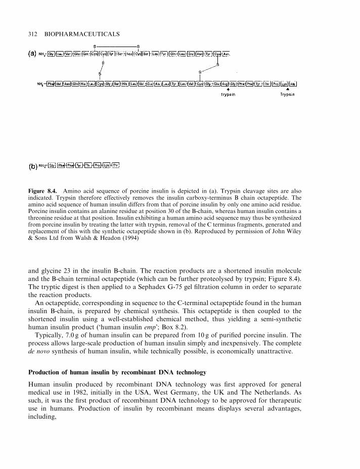
and glycine 23 in the insulin B-chain. The reaction products are a shortened insulin moleculeand the B-chain terminal octapeptide (which can be further proteolysed by trypsin; Figure 8.4).The tryptic digest is then applied to a Sephadex G-75 gel filtration column in order to separatethe reaction products.
An octapeptide, corresponding in sequence to the C-terminal octapeptide found in the humaninsulin B-chain, is prepared by chemical synthesis. This octapeptide is then coupled to theshortened insulin using a well-established chemical method, thus yielding a semi-synthetichuman insulin product (‘human insulin emp’; Box 8.2).
Typically, 7.0 g of human insulin can be prepared from 10 g of purified porcine insulin. Theprocess allows large-scale production of human insulin simply and inexpensively. The completede novo synthesis of human insulin, while technically possible, is economically unattractive.
Production of human insulin by recombinant DNA technology
Human insulin produced by recombinant DNA technology was first approved for generalmedical use in 1982, initially in the USA, West Germany, the UK and The Netherlands. Assuch, it was the first product of recombinant DNA technology to be approved for therapeuticuse in humans. Production of insulin by recombinant means displays several advantages,including,
312 BIOPHARMACEUTICALS
Figure 8.4. Amino acid sequence of porcine insulin is depicted in (a). Trypsin cleavage sites are alsoindicated. Trypsin therefore effectively removes the insulin carboxy-terminus B chain octapeptide. Theamino acid sequence of human insulin differs from that of porcine insulin by only one amino acid residue.Porcine insulin contains an alanine residue at position 30 of the B-chain, whereas human insulin contains athreonine residue at that position. Insulin exhibiting a human amino acid sequence may thus be synthesizedfrom porcine insulin by treating the latter with trypsin, removal of the C terminus fragments, generated andreplacement of this with the synthetic octapeptide shown in (b). Reproduced by permission of John Wiley& Sons Ltd from Walsh & Headon (1994)
. reliability of supply;
. elimination of the risk of accidental transmission of disease, due to the presence of pathogensin animal pancreatic tissue;
. economically attractive (once initial capital investment has been made).
The quantity of purified insulin obtained from the pancreas of one pig satisfies therequirements of one diabetic for 3 days. The supply of pancreatic tissue is dependent upon thedemand for meat, which does not necessarily correlate with the increasing worldwide incidenceof diabetes. Recombinant DNA technology provides an obvious way to ensure future adequatesupply of insulin.
The initial approach taken (by scientists at Genentech) entailed inserting the nucleotidesequence coding for the insulin A- and B-chains into two different E. coli cells (both strain K12).These cells were then cultured separately in large-scale fermentation vessels, with subsequentchromatographic purification of the insulin chains produced. The A- and B-chains are thenincubated together under appropriate oxidizing conditions in order to promote interchaindisulphide bond formation, forming human insulin crb (Box 8.2).
An alternative method (developed in the Eli Lilly research laboratories) entails inserting anucleotide sequence coding for human proinsulin into recombinant E. coli. This is followed bypurification of the expressed proinsulin and subsequent proteolytic excision of the C peptide invitro. This approach has become more popular, largely due to the requirement for a singlefermentation and subsequent purification scheme. Such preparations have been termed humaninsulin prb.
A number of studies have shown conclusively that the recombinant insulins are chemicallyand functionally identical to native human insulin. Sequencing data confirms the expectedamino acid sequence. HPLC analysis also yields identical chromatograms (Figure 8.5). Circulardichromism studies, which evaluate tertiary structure, also yield identical patterns, as doesX-ray crystallographic analysis. Radioreceptor- and radioimmuno-assays yield identical results,as does the traditional bioassay for insulin (measure of the hormone’s ability to induce ahypoglycaemic response in rabbits). Prior to its approval for medical use, human clinical trialsalso found the recombinant insulin to be as effective as previously available products in terms ofcontrolling hyperglycaemia. Recombinant human insulins are now routinely produced in 10 000gallon fermentation vessels and several such products have gained marketing approval(Table 8.3).
While the recombinant product is identical to native insulin, any impurities present will beE. coli-derived and, hence, potentially highly immunogenic. Stringent purification of therecombinant product must thus be undertaken. This entails several chromatographic steps(often gel filtration and ion-exchange, along with additional steps which exploit differences inmolecular hydrophobicity, e.g. hydrophobic interaction chromatography or reverse-phasechromatography) (Figure 8.6).
A ‘clean-up’ process-scale reverse-phase HPLC (RP-HPLC) step has been introduced intoproduction of human insulin prb. The C8 or C18 RP-HPLC column used displays an internalvolume of 80 l or more, and up to 1200 g of insulin may be loaded during a single purificationrun (Figure 8.7). Separation is achieved using an acidic (often acetic acid-based) mobile phase(i.e. set at a pH value sufficiently below the insulin pI value of 5.3 in order to keep it fully insolution). The insulin is usually loaded in the water-rich acidic mobile phase, followed bygradient elution using acetonitrile (insulin typically elutes at 15–30% acetonitrile).
HORMONES OF THERAPEUTIC INTEREST 313
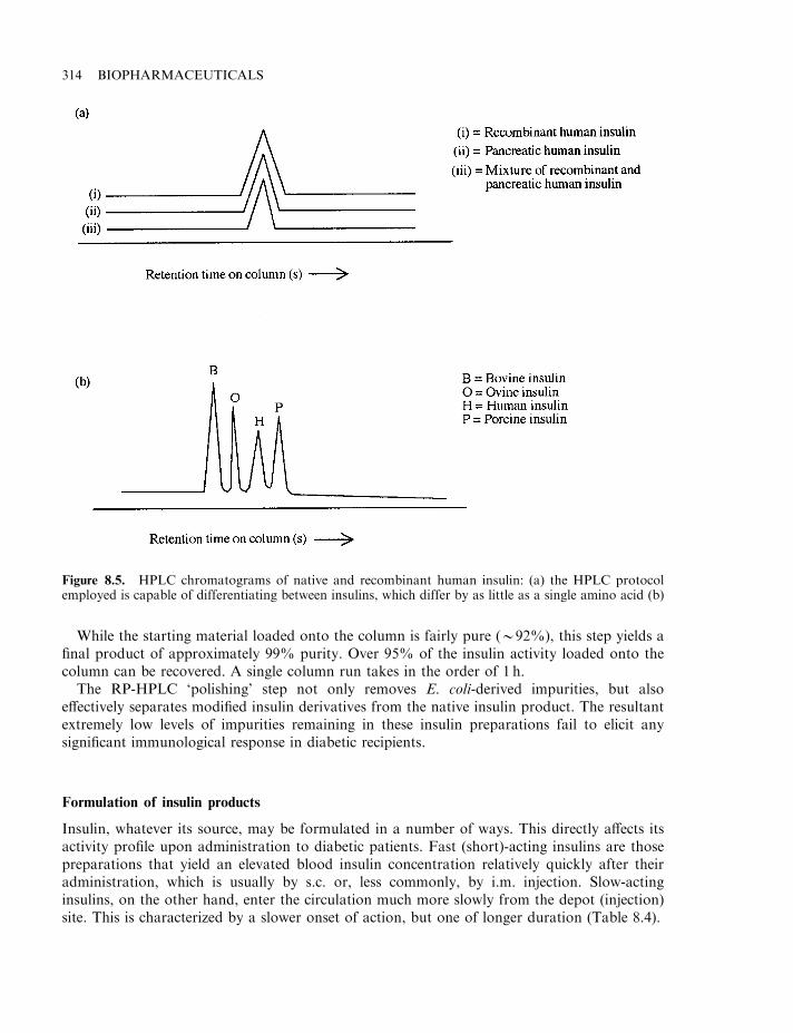
While the starting material loaded onto the column is fairly pure (*92%), this step yields afinal product of approximately 99% purity. Over 95% of the insulin activity loaded onto thecolumn can be recovered. A single column run takes in the order of 1 h.
The RP-HPLC ‘polishing’ step not only removes E. coli-derived impurities, but alsoeffectively separates modified insulin derivatives from the native insulin product. The resultantextremely low levels of impurities remaining in these insulin preparations fail to elicit anysignificant immunological response in diabetic recipients.
Formulation of insulin products
Insulin, whatever its source, may be formulated in a number of ways. This directly affects itsactivity profile upon administration to diabetic patients. Fast (short)-acting insulins are thosepreparations that yield an elevated blood insulin concentration relatively quickly after theiradministration, which is usually by s.c. or, less commonly, by i.m. injection. Slow-actinginsulins, on the other hand, enter the circulation much more slowly from the depot (injection)site. This is characterized by a slower onset of action, but one of longer duration (Table 8.4).
314 BIOPHARMACEUTICALS
Figure 8.5. HPLC chromatograms of native and recombinant human insulin: (a) the HPLC protocolemployed is capable of differentiating between insulins, which differ by as little as a single amino acid (b)
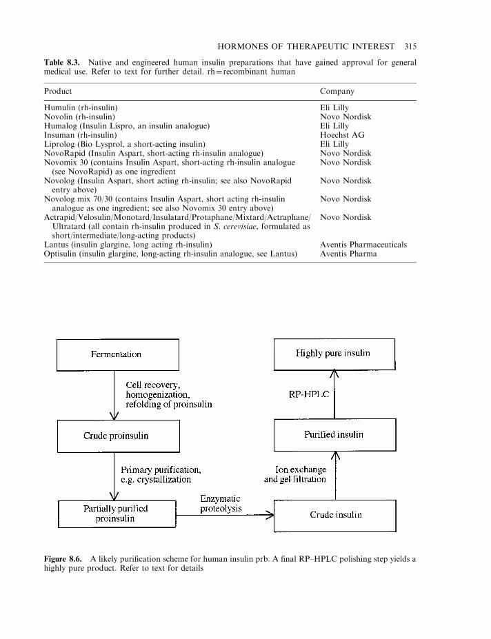
HORMONES OF THERAPEUTIC INTEREST 315
Figure 8.6. A likely purification scheme for human insulin prb. A final RP–HPLC polishing step yields ahighly pure product. Refer to text for details
Table 8.3. Native and engineered human insulin preparations that have gained approval for generalmedical use. Refer to text for further detail. rh¼recombinant human
Product Company
Humulin (rh-insulin) Eli LillyNovolin (rh-insulin) Novo NordiskHumalog (Insulin Lispro, an insulin analogue) Eli LillyInsuman (rh-insulin) Hoechst AGLiprolog (Bio Lysprol, a short-acting insulin) Eli LillyNovoRapid (Insulin Aspart, short-acting rh-insulin analogue) Novo NordiskNovomix 30 (contains Insulin Aspart, short-acting rh-insulin analogue(see NovoRapid) as one ingredient
Novo Nordisk
Novolog (Insulin Aspart, short acting rh-insulin; see also NovoRapidentry above)
Novo Nordisk
Novolog mix 70/30 (contains Insulin Aspart, short acting rh-insulinanalogue as one ingredient; see also Novomix 30 entry above)
Novo Nordisk
Actrapid/Velosulin/Monotard/Insulatard/Protaphane/Mixtard/Actraphane/Ultratard (all contain rh-insulin produced in S. cerevisiae, formulated asshort/intermediate/long-acting products)
Novo Nordisk
Lantus (insulin glargine, long acting rh-insulin) Aventis PharmaceuticalsOptisulin (insulin glargine, long-acting rh-insulin analogue, see Lantus) Aventis Pharma
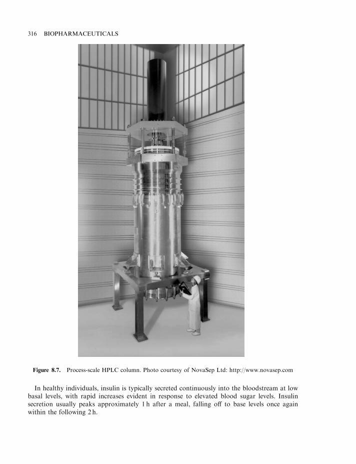
In healthy individuals, insulin is typically secreted continuously into the bloodstream at lowbasal levels, with rapid increases evident in response to elevated blood sugar levels. Insulinsecretion usually peaks approximately 1 h after a meal, falling off to base levels once againwithin the following 2 h.
316 BIOPHARMACEUTICALS
Figure 8.7. Process-scale HPLC column. Photo courtesy of NovaSep Ltd: http://www.novasep.com
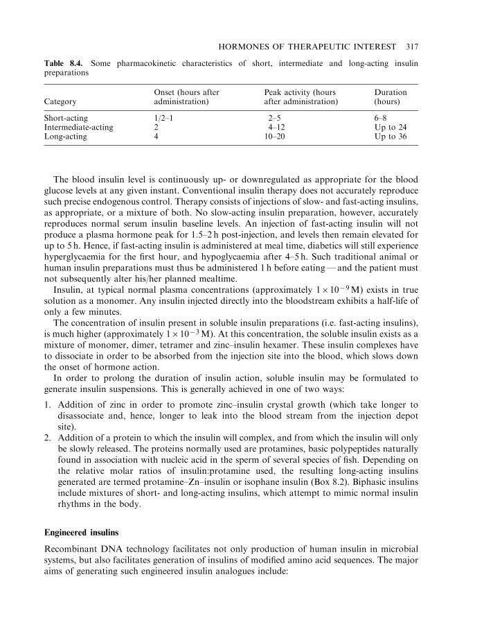
The blood insulin level is continuously up- or downregulated as appropriate for the bloodglucose levels at any given instant. Conventional insulin therapy does not accurately reproducesuch precise endogenous control. Therapy consists of injections of slow- and fast-acting insulins,as appropriate, or a mixture of both. No slow-acting insulin preparation, however, accuratelyreproduces normal serum insulin baseline levels. An injection of fast-acting insulin will notproduce a plasma hormone peak for 1.5–2 h post-injection, and levels then remain elevated forup to 5 h. Hence, if fast-acting insulin is administered at meal time, diabetics will still experiencehyperglycaemia for the first hour, and hypoglycaemia after 4–5 h. Such traditional animal orhuman insulin preparations must thus be administered 1 h before eating—and the patient mustnot subsequently alter his/her planned mealtime.
Insulin, at typical normal plasma concentrations (approximately 161079M) exists in truesolution as a monomer. Any insulin injected directly into the bloodstream exhibits a half-life ofonly a few minutes.
The concentration of insulin present in soluble insulin preparations (i.e. fast-acting insulins),is much higher (approximately 161073M). At this concentration, the soluble insulin exists as amixture of monomer, dimer, tetramer and zinc–insulin hexamer. These insulin complexes haveto dissociate in order to be absorbed from the injection site into the blood, which slows downthe onset of hormone action.
In order to prolong the duration of insulin action, soluble insulin may be formulated togenerate insulin suspensions. This is generally achieved in one of two ways:
1. Addition of zinc in order to promote zinc–insulin crystal growth (which take longer todisassociate and, hence, longer to leak into the blood stream from the injection depotsite).
2. Addition of a protein to which the insulin will complex, and from which the insulin will onlybe slowly released. The proteins normally used are protamines, basic polypeptides naturallyfound in association with nucleic acid in the sperm of several species of fish. Depending onthe relative molar ratios of insulin:protamine used, the resulting long-acting insulinsgenerated are termed protamine–Zn–insulin or isophane insulin (Box 8.2). Biphasic insulinsinclude mixtures of short- and long-acting insulins, which attempt to mimic normal insulinrhythms in the body.
Engineered insulins
Recombinant DNA technology facilitates not only production of human insulin in microbialsystems, but also facilitates generation of insulins of modified amino acid sequences. The majoraims of generating such engineered insulin analogues include:
HORMONES OF THERAPEUTIC INTEREST 317
Table 8.4. Some pharmacokinetic characteristics of short, intermediate and long-acting insulinpreparations
CategoryOnset (hours afteradministration)
Peak activity (hoursafter administration)
Duration(hours)
Short-acting 1/2–1 2–5 6–8Intermediate-acting 2 4–12 Up to 24Long-acting 4 10–20 Up to 36
. identification of insulins with altered pharmacokinetic properties, such as faster-acting orslower-acting insulins;
. identification of super-potent insulin forms (insulins with higher receptor affinities). This isdue to commercial considerations, namely the economic benefits which would accrue fromutilizing smaller quantities of insulin per therapeutic dose.
The insulin amino acid residues which interact with the insulin receptor have been identified(A1, A5, A19, A21, B10, B16, B23–25), and a number of analogues containing amino acidsubstitutions at several of these points have been manufactured, e.g. conversion of histidine toglutamate at the B10 position yields an analogue displaying five-fold higher activity in vitro.Other substitutions have generated analogues with even higher specific activities. However,increased in vitro activity does not always translate to increased in vivo activity.
Attempts to generate faster-acting insulins have centred upon developing analogues which donot dimerize or form higher polymers at therapeutic dose concentrations. The contact pointsbetween individual insulin molecules in insulin dimers/oligomers include amino acids atpositions B8, B9, B12–13, B16 and B23–28. Thus, analogues with various substitutions at thesepositions have been generated. The approach adopted generally entails insertion of charged orbulky amino acids, in order to promote charge repulsion or steric hindrance between individualinsulin monomers. Several are absorbed from the site of injection into the bloodstream far morequickly than native soluble (fast-acting) insulin. Such modified insulins could thus be injected atmealtimes, rather than 1 h before, and several such fast-acting engineered insulins have nowbeen approved for medical use (Table 8.3). Insulin Lispro was the first such engineered short-acting insulin to come to market (Box 8.3 and Figure 8.8).
Insulin Aspart is a second fast-acting engineered human insulin analogue now approved forgeneral medical use. It differs from native human insulin in that the prolineB28 residue has beenreplaced by aspartic acid. This single amino acid substitution also decreases the propensity ofindividual molecules to self-associate, ensuring that they begin to enter the bloodstream fromthe site of injection immediately upon administration.
A number of studies have also focused upon the generation of longer-acting insulinanalogues. The currently used Zn–insulin suspensions, or protamine–Zn–insulin suspensions,generally display a plasma half-life of 20–25 h. Selected amino acid substitutions have generatedinsulins which, even in soluble form, exhibit plasma half-lives of up to 35 h.
Optisulin or Lantus are the trade names given to one such analogue that gained generalmarketing approval in 2000 (Table 8.3). The international non-proprietary name (inn) for thisengineered molecule is ‘insulin glargine’. It differs from native human insulin in that the C-terminal aspargine residue of the a-chain has been replaced by a glycine residue and the b-chainhas been elongated (again from its C-terminus) by two arginine residues. The overall effect is toincrease the molecule’s isoelectric point (pI, the pH at which the molecule displays a net overallzero charge and consequently is least soluble) from 5.4 to a value approaching 7.0. Theengineered insulin is expressed in a recombinant E. coli K12 host strain and is produced via the‘proinsulin route’, as previously described. The purified product is formulated at pH 4.0, a pHvalue at which it is fully soluble.
Upon s.c. injection, the insulin experiences an increase in pH towards more neutral valuesand, consequently, appears to precipitate in the subcutaneous tissue. It resolubilizes very slowlyand hence a greatly prolonged duration of release into the bloodstream is noted. Consequently,a single daily injection supports the maintenance of acceptable basal blood insulin levels, andinsulin molecules are still detected at the site of injection more than 24 h after administration.
318 BIOPHARMACEUTICALS
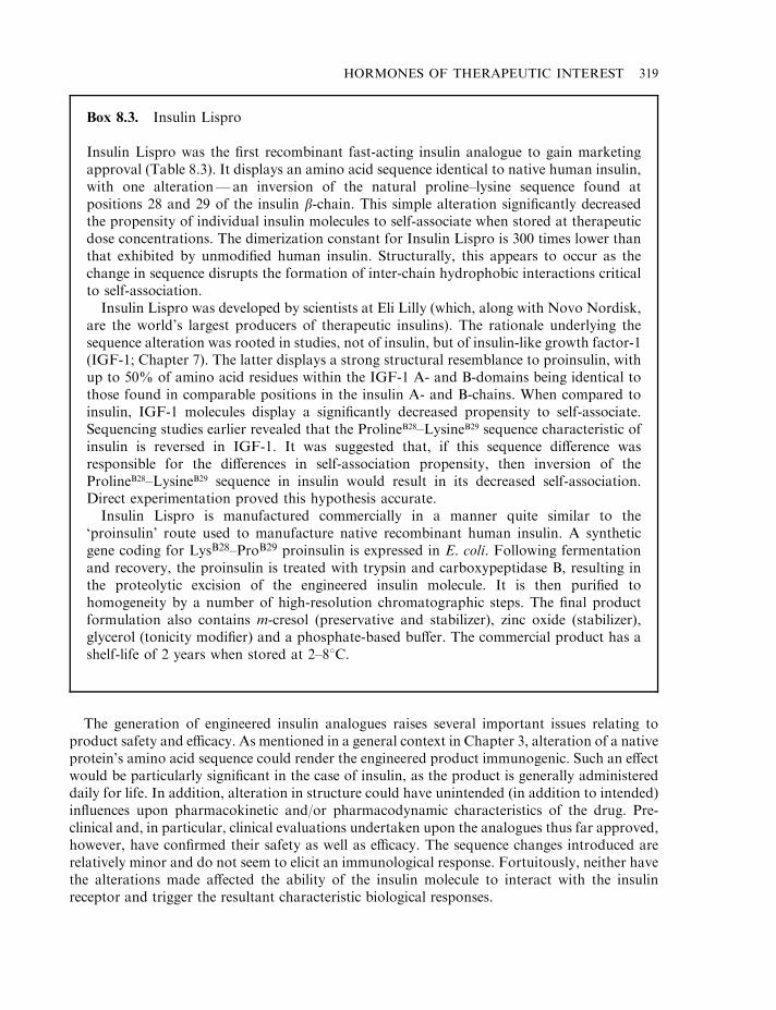
The generation of engineered insulin analogues raises several important issues relating toproduct safety and efficacy. As mentioned in a general context in Chapter 3, alteration of a nativeprotein’s amino acid sequence could render the engineered product immunogenic. Such an effectwould be particularly significant in the case of insulin, as the product is generally administereddaily for life. In addition, alteration in structure could have unintended (in addition to intended)influences upon pharmacokinetic and/or pharmacodynamic characteristics of the drug. Pre-clinical and, in particular, clinical evaluations undertaken upon the analogues thus far approved,however, have confirmed their safety as well as efficacy. The sequence changes introduced arerelatively minor and do not seem to elicit an immunological response. Fortuitously, neither havethe alterations made affected the ability of the insulin molecule to interact with the insulinreceptor and trigger the resultant characteristic biological responses.
HORMONES OF THERAPEUTIC INTEREST 319
Box 8.3. Insulin Lispro
Insulin Lispro was the first recombinant fast-acting insulin analogue to gain marketingapproval (Table 8.3). It displays an amino acid sequence identical to native human insulin,with one alteration—an inversion of the natural proline–lysine sequence found atpositions 28 and 29 of the insulin b-chain. This simple alteration significantly decreasedthe propensity of individual insulin molecules to self-associate when stored at therapeuticdose concentrations. The dimerization constant for Insulin Lispro is 300 times lower thanthat exhibited by unmodified human insulin. Structurally, this appears to occur as thechange in sequence disrupts the formation of inter-chain hydrophobic interactions criticalto self-association.
Insulin Lispro was developed by scientists at Eli Lilly (which, along with Novo Nordisk,are the world’s largest producers of therapeutic insulins). The rationale underlying thesequence alteration was rooted in studies, not of insulin, but of insulin-like growth factor-1(IGF-1; Chapter 7). The latter displays a strong structural resemblance to proinsulin, withup to 50% of amino acid residues within the IGF-1 A- and B-domains being identical tothose found in comparable positions in the insulin A- and B-chains. When compared toinsulin, IGF-1 molecules display a significantly decreased propensity to self-associate.Sequencing studies earlier revealed that the ProlineB28–LysineB29 sequence characteristic ofinsulin is reversed in IGF-1. It was suggested that, if this sequence difference wasresponsible for the differences in self-association propensity, then inversion of theProlineB28–LysineB29 sequence in insulin would result in its decreased self-association.Direct experimentation proved this hypothesis accurate.
Insulin Lispro is manufactured commercially in a manner quite similar to the‘proinsulin’ route used to manufacture native recombinant human insulin. A syntheticgene coding for LysB28–ProB29 proinsulin is expressed in E. coli. Following fermentationand recovery, the proinsulin is treated with trypsin and carboxypeptidase B, resulting inthe proteolytic excision of the engineered insulin molecule. It is then purified tohomogeneity by a number of high-resolution chromatographic steps. The final productformulation also contains m-cresol (preservative and stabilizer), zinc oxide (stabilizer),glycerol (tonicity modifier) and a phosphate-based buffer. The commercial product has ashelf-life of 2 years when stored at 2–88C.
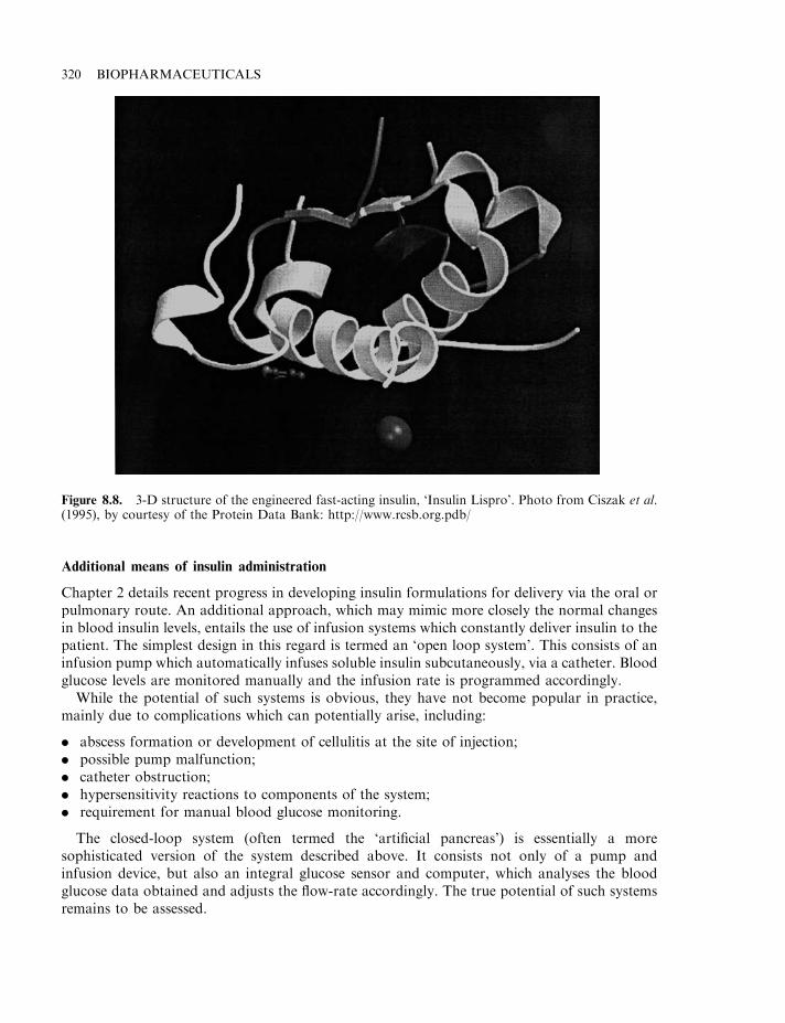
Additional means of insulin administration
Chapter 2 details recent progress in developing insulin formulations for delivery via the oral orpulmonary route. An additional approach, which may mimic more closely the normal changesin blood insulin levels, entails the use of infusion systems which constantly deliver insulin to thepatient. The simplest design in this regard is termed an ‘open loop system’. This consists of aninfusion pump which automatically infuses soluble insulin subcutaneously, via a catheter. Bloodglucose levels are monitored manually and the infusion rate is programmed accordingly.
While the potential of such systems is obvious, they have not become popular in practice,mainly due to complications which can potentially arise, including:
. abscess formation or development of cellulitis at the site of injection;
. possible pump malfunction;
. catheter obstruction;
. hypersensitivity reactions to components of the system;
. requirement for manual blood glucose monitoring.
The closed-loop system (often termed the ‘artificial pancreas’) is essentially a moresophisticated version of the system described above. It consists not only of a pump andinfusion device, but also an integral glucose sensor and computer, which analyses the bloodglucose data obtained and adjusts the flow-rate accordingly. The true potential of such systemsremains to be assessed.
320 BIOPHARMACEUTICALS
Figure 8.8. 3-D structure of the engineered fast-acting insulin, ‘Insulin Lispro’. Photo from Ciszak et al.(1995), by courtesy of the Protein Data Bank: http://www.rcsb.org.pdb/
Treating diabetics with insulin-producing cells
While infusion pumps can go some way towards mimicking normal control of blood insulinlevels, transplantation of insulin-producing pancreatic cells should effectively ‘cure’ the diabeticpatient. This approach has been adopted thus far with almost 200 patients, with encouragingresults.
Initial experiments in the 1970s using inbred strains of rats illustrated the feasibility of thisapproach. Insulin-producing pancreatic islet cells ‘donated’ by one set of rats were transplantedinto other rats of the same strain, first made diabetic by injection with drugs such asstreptozotocin, which destroy the pancreatic B cells.
Such islet grafts permanently returned blood glucose levels in the diabetic animals to normalvalues. Even more encouraging, this treatment prevented development of diabetic-associatedcomplications of kidney and eye function (which, in human diabetics, can lead to kidney failureand partial blindness).
The technique simply entails injecting the insulin-producing islets into the portal vein. Thesecells subsequently lodge in smaller vessels branching from the vein. Here, they can constantlymonitor blood glucose levels and secrete insulin accordingly. About 400 000–800 000 islet B cellsare usually transplanted. In most instances, these function for up to 3 years, although they oftenfail to control blood glucose levels fully, i.e. supplemental insulin injections are sometimesrequired.
The islet cells transplanted into humans are obtained from pancreatic tissue of deceasedhuman donors (Figure 8.9). Implantation of these cells in recipients displaying a competentimmune system would, at best, be of transient therapeutic benefit. The ensuing immune responsewould quickly destroy the foreign cells. Studies conducted thus far in humans have utilizeddiabetic patients who have received kidney transplants, as these are already subject toimmunosuppressive therapy. However, a major stumbling block to the widespread adoption ofthis therapeutic approach is, predictably, the requirement to induce concurrent immunesuppression.
GLUCAGON
Glucagon is a single-chain polypeptide of 29 amino acid residues and a molecular mass of3500Da. It is synthesized by the A cells of the Islets of Langerhans, and also by related cellsfound in the digestive tract. Like insulin, it is synthesized as a high molecular mass polypeptidefrom which the mature hormone is released by selective proteolysis.
The major biological actions of glucagon tend to oppose those of insulin, particularly withregard to regulation of metabolism. Glucagon has an overall catabolic effect, stimulating thebreakdown of glycogen, lipid and protein. A prominent metabolic effect is to increase bloodglucose levels (i.e. it is a hyperglycaemic hormone). Indeed, the major physiological function ofglucagon is to prevent hypoglycaemia. The hormone is stored in secretory vesicles, aftersynthesis in the pancreatic A cells, and released by exocytosis upon experiencing a drop in bloodglucose concentration.
Glucagon initiates its metabolic (and other) effects by binding to a specific cell surfacereceptor, thus activating a membrane-bound adenylate kinase. This in turn, promotes activationof a cAMP-dependent protein kinase (Figure 8.10). The kinase phosphorylates key regulatoryenzymes in carbohydrate metabolism, thereby modulating their activity. Hepatic glycogenphosphorylase, for example, is activated via phosphorylation, while glycogen synthetase is
HORMONES OF THERAPEUTIC INTEREST 321
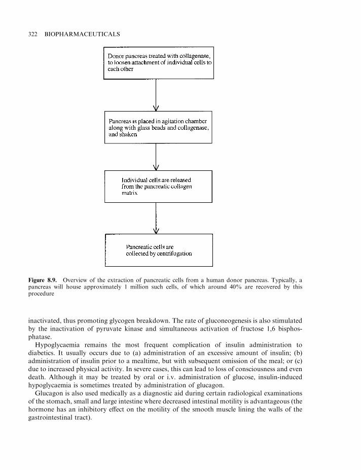
inactivated, thus promoting glycogen breakdown. The rate of gluconeogenesis is also stimulatedby the inactivation of pyruvate kinase and simultaneous activation of fructose 1,6 bisphos-phatase.
Hypoglycaemia remains the most frequent complication of insulin administration todiabetics. It usually occurs due to (a) administration of an excessive amount of insulin; (b)administration of insulin prior to a mealtime, but with subsequent omission of the meal; or (c)due to increased physical activity. In severe cases, this can lead to loss of consciousness and evendeath. Although it may be treated by oral or i.v. administration of glucose, insulin-inducedhypoglycaemia is sometimes treated by administration of glucagon.
Glucagon is also used medically as a diagnostic aid during certain radiological examinationsof the stomach, small and large intestine where decreased intestinal motility is advantageous (thehormone has an inhibitory effect on the motility of the smooth muscle lining the walls of thegastrointestinal tract).
322 BIOPHARMACEUTICALS
Figure 8.9. Overview of the extraction of pancreatic cells from a human donor pancreas. Typically, apancreas will house approximately 1 million such cells, of which around 40% are recovered by thisprocedure
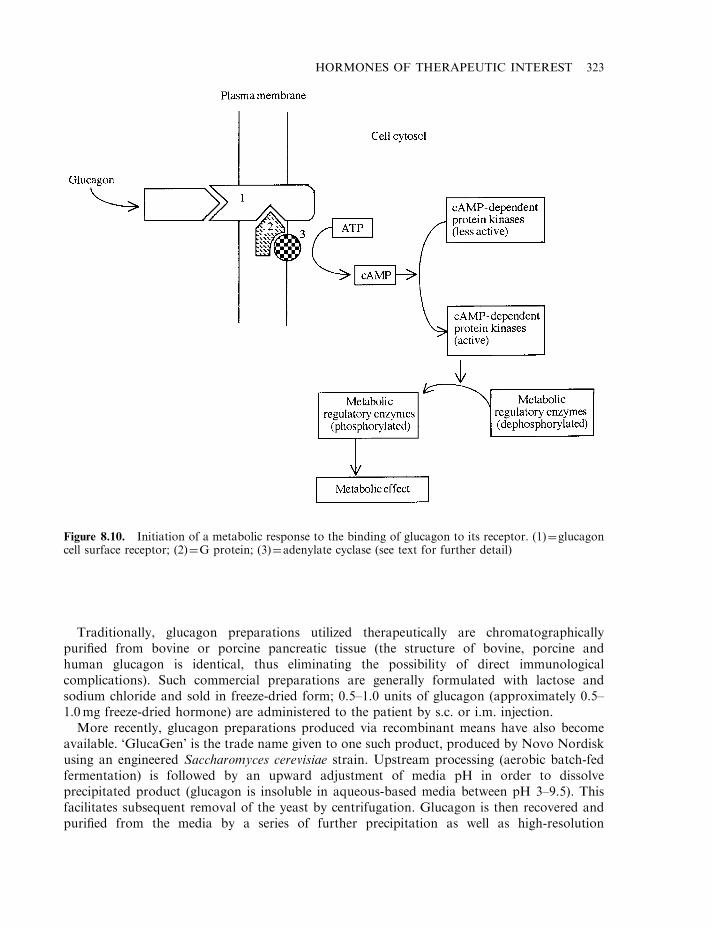
Traditionally, glucagon preparations utilized therapeutically are chromatographicallypurified from bovine or porcine pancreatic tissue (the structure of bovine, porcine andhuman glucagon is identical, thus eliminating the possibility of direct immunologicalcomplications). Such commercial preparations are generally formulated with lactose andsodium chloride and sold in freeze-dried form; 0.5–1.0 units of glucagon (approximately 0.5–1.0mg freeze-dried hormone) are administered to the patient by s.c. or i.m. injection.
More recently, glucagon preparations produced via recombinant means have also becomeavailable. ‘GlucaGen’ is the trade name given to one such product, produced by Novo Nordiskusing an engineered Saccharomyces cerevisiae strain. Upstream processing (aerobic batch-fedfermentation) is followed by an upward adjustment of media pH in order to dissolveprecipitated product (glucagon is insoluble in aqueous-based media between pH 3–9.5). Thisfacilitates subsequent removal of the yeast by centrifugation. Glucagon is then recovered andpurified from the media by a series of further precipitation as well as high-resolution
HORMONES OF THERAPEUTIC INTEREST 323
Figure 8.10. Initiation of a metabolic response to the binding of glucagon to its receptor. (1)¼glucagoncell surface receptor; (2)¼G protein; (3)¼adenylate cyclase (see text for further detail)
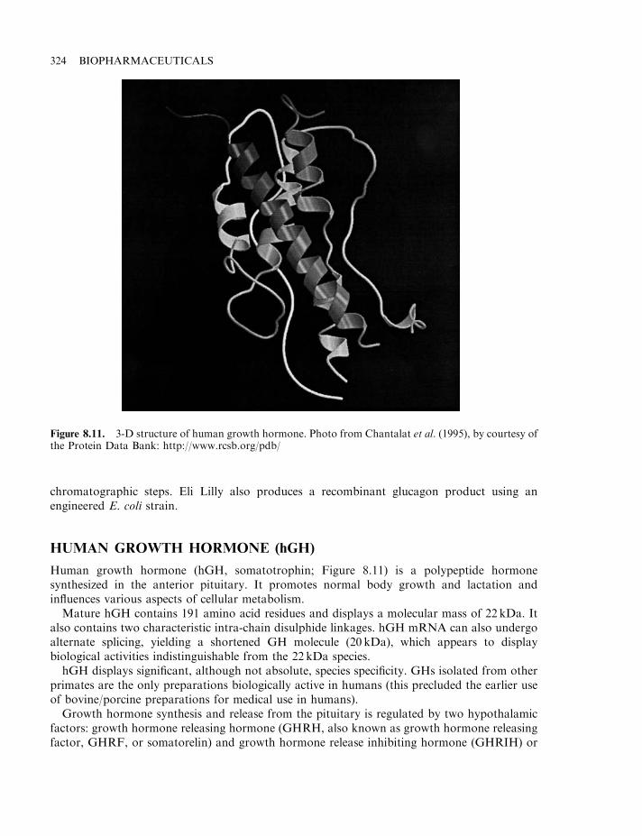
chromatographic steps. Eli Lilly also produces a recombinant glucagon product using anengineered E. coli strain.
HUMAN GROWTH HORMONE (hGH)
Human growth hormone (hGH, somatotrophin; Figure 8.11) is a polypeptide hormonesynthesized in the anterior pituitary. It promotes normal body growth and lactation andinfluences various aspects of cellular metabolism.
Mature hGH contains 191 amino acid residues and displays a molecular mass of 22 kDa. Italso contains two characteristic intra-chain disulphide linkages. hGH mRNA can also undergoalternate splicing, yielding a shortened GH molecule (20 kDa), which appears to displaybiological activities indistinguishable from the 22 kDa species.
hGH displays significant, although not absolute, species specificity. GHs isolated from otherprimates are the only preparations biologically active in humans (this precluded the earlier useof bovine/porcine preparations for medical use in humans).
Growth hormone synthesis and release from the pituitary is regulated by two hypothalamicfactors: growth hormone releasing hormone (GHRH, also known as growth hormone releasingfactor, GHRF, or somatorelin) and growth hormone release inhibiting hormone (GHRIH) or
324 BIOPHARMACEUTICALS
Figure 8.11. 3-D structure of human growth hormone. Photo from Chantalat et al. (1995), by courtesy ofthe Protein Data Bank: http://www.rcsb.org/pdb/
somatostatin (Table 8.5). Furthermore, while GH directly mediates some of its biologicalactions, its major influence on body growth is mediated indirectly via IGF-1, as discussed below.GHRH, GHRIH, GH and IGF-1 thus form a hormonal axis, as depicted in Figure 8.12.
Growth hormone releasing factor (GHRF) and inhibitory factor (GHRIF)
GHRF and GHRIF are peptides secreted by hypothalamic neurons termed ‘neuroendocrinetransducers’ (the name is apt, as these interface between the nervous and endocrine systems).The factors that regulate their secretion are poorly understood but probably involve both nerveimpulses originating from within the brain and feedback mechanisms, possibly involvingpituitary hormones.
37, 40 and 44 amino acid GHRF variants have been identified. All can promote GH releasefrom the pituitary, an activity which apparently resides in the first 29 amino acid residues ofthese molecules. Administration of GHRH to GH-deficient individuals generally promotesmodest increases in GH secretion, thereby increasing growth rate.
Hypothalamic GHRIF is a cyclic 14 amino-acid peptide, although a 28 amino acid form isalso found in some other tissues. GHRIF inhibits the release not only of GH but also oftyrotrophin and corticotrophin from the pituitary, and insulin and glucagon from the pancreas.It can also regulate the level of duodenal secretions.
The GH receptor
GH induces its characteristic biological effects by binding to a specific cell surface receptor. Thehuman receptor is a single chain 620 amino acid transmembrane polypeptide. Sequence analysisindicates it is a member of the haemopoietic receptor superfamily (which includes receptors forseveral ILs, GM-CSF and EPO). X-ray crystallographic analysis shows that GH bindssimultaneously to the extracellular domains of two receptor molecules, effectively promotingreceptor dimerization (Figure 8.13). The exact molecular detail of the subsequent signaltransduction events remain to be determined. However, ligand binding does induce receptorautophosphorylation, as well as phosphorylation of additional cellular substances and proteinkinase C activation.
HORMONES OF THERAPEUTIC INTEREST 325
Table 8.5. Some factors known to affect the rate of secretion of GH. Most of these factors influenceGH release indirectly by affecting the rate and level of secretion of GHRH and/or GHRIH
Factors promoting increased GH secretion Factors promoting decreased GH secretion
Starvation ObesitySleep Elevated blood glucoseStress b-Adrenergic antagonistsExercise b-Adrenergic agonistsLow blood glucoseSeveral amino acidsGlucagonVasopressina-Adrenergic agonistsb-Adrenergic antagonists
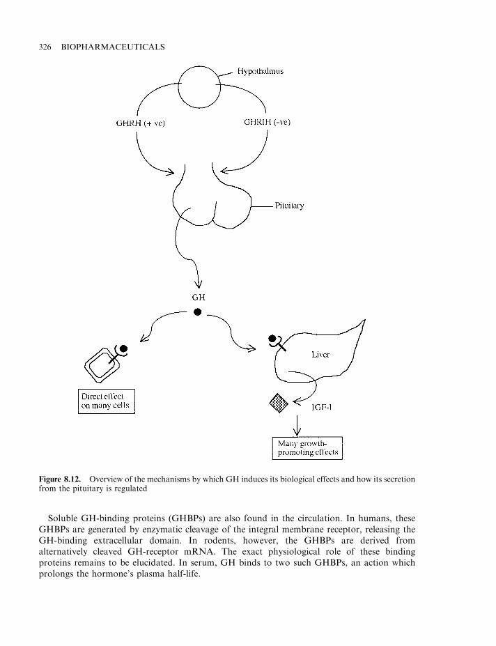
Soluble GH-binding proteins (GHBPs) are also found in the circulation. In humans, theseGHBPs are generated by enzymatic cleavage of the integral membrane receptor, releasing theGH-binding extracellular domain. In rodents, however, the GHBPs are derived fromalternatively cleaved GH-receptor mRNA. The exact physiological role of these bindingproteins remains to be elucidated. In serum, GH binds to two such GHBPs, an action whichprolongs the hormone’s plasma half-life.
326 BIOPHARMACEUTICALS
Figure 8.12. Overview of the mechanisms by which GH induces its biological effects and how its secretionfrom the pituitary is regulated
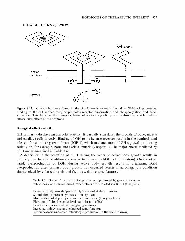
Biological effects of GH
GH primarily displays an anabolic activity. It partially stimulates the growth of bone, muscleand cartilage cells directly. Binding of GH to its hepatic receptor results in the synthesis andrelease of insulin-like growth factor (IGF-1), which mediates most of GH’s growth-promotingactivity on, for example, bone and skeletal muscle (Chapter 7). The major effects mediated byhGH are summarized in Table 8.6.
A deficiency in the secretion of hGH during the years of active body growth results inpituitary dwarfism (a condition responsive to exogenous hGH administration). On the otherhand, overproduction of hGH during active body growth results in gigantism. hGHoverproduction after primary body growth has occurred results in acromegaly, a conditioncharacterized by enlarged hands and feet, as well as coarse features.
HORMONES OF THERAPEUTIC INTEREST 327
Figure 8.13. Growth hormone found in the circulation is generally bound to GH-binding proteins.Binding to the cell surface receptor promotes receptor dimerization and phosphorylation and henceactivation. This leads to the phosphorylation of various cystolic protein substrates, which mediateintracellular effects of the hormone
Table 8.6. Some of the major biological effects promoted by growth hormone.While many of these are direct, other effects are mediated via IGF-1 (Chapter 7)
Increased body growth (particularly bone and skeletal muscle)Stimulation of protein synthesis in many tissuesMobilization of depot lipids from adipose tissue (lipolytic effect)Elevation of blood glucose levels (anti-insulin effect)Increase of muscle and cardiac glycogen storesIncreased kidney size and enhanced renal functionReticulocytosis (increased reticulocyte production in the bone marrow)
Therapeutic uses of GH
GH has a potentially wide range of therapeutic uses (Table 8.7). To date, however, its majorapplication has been for the treatment of short stature. hGH extracted from human pituitaryglands was first used to treat pituitary dwarfism (i.e. caused by sub-optimal pituitary GHsecretion), in 1958. It has subsequently proved effective in the treatment of short stature causedby a variety of other conditions, including:
. Turner’s syndrome;
. idiopathic short stature;
. chronic renal failure.
The use of hGH extracted from the pituitaries of deceased human donors came to an abrupt endin 1985, when a link between treatment and Creutzfeldt–Jakob disease (CJD, a rare, but fatal,neurological disorder) was discovered. In this year, a young man who had received hGHtherapy some 15 years previously died from CJD, which, investigators concluded, he hadcontracted from infected pituitary extract (CJD appears to be caused by a prion). At least anadditional 12 CJD cases suspected of being caused in the same way have subsequently beendocumented. Fortunately, several recombinant hGH (rhGH) preparations were coming on-stream at that time (Table 8.8), and now all hGH preparations used clinically are derived fromrecombinant sources. Currently, in excess of 20 000 people are in receipt of rhGH therapy.
rhGH was first produced in E. coli in the early 1980s. The initial recombinant preparationsdiffered from the native human hormone only in that they contained an extra methionineresidue (due to the AUG start codon inserted at the beginning of the gene). Subsequently, adifferent cloning strategy allowed production in E. coli of products devoid of this terminalmethionine.
In vitro analysis, including tryptic peptide mapping, amino acid analysis and comparativeimmunoassays, show the native and recombinant form of the molecule to be identical. Clinicaltrials in humans have also confirmed that the recombinant version promotes identical biologicalresponses to the native hormone. rhGH was first purified (on a lab scale) by Genentech scientistsusing the strategy outlined in Figure 8.14. A somewhat similar strategy is likely used in itsprocess-scale purification.
Recombinant hGH (rhGH) and pituitary dwarfism
Various studies have confirmed that rhGH promotes increased linear growth rates in childrensuffering from pituitary dwarfism (classical growth hormone deficiency). Dosages are generally
328 BIOPHARMACEUTICALS
Table 8.7. Some actual or likely therapeutic uses for hGH. Refer to text fordetails
Treatment of short stature caused by GH deficiencyTreatment of defective growth caused by various diseases/medical conditionsInduction of lactationCounteracting ageingTreatment of obesityBody buildingInduction of ovulation
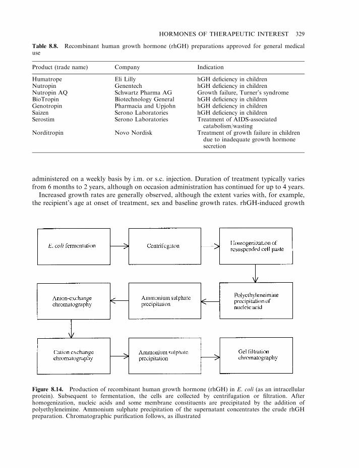
administered on a weekly basis by i.m. or s.c. injection. Duration of treatment typically variesfrom 6 months to 2 years, although on occasion administration has continued for up to 4 years.
Increased growth rates are generally observed, although the extent varies with, for example,the recipient’s age at onset of treatment, sex and baseline growth rates. rhGH-induced growth
HORMONES OF THERAPEUTIC INTEREST 329
Table 8.8. Recombinant human growth hormone (rhGH) preparations approved for general medicaluse
Product (trade name) Company Indication
Humatrope Eli Lilly hGH deficiency in childrenNutropin Genentech hGH deficiency in childrenNutropin AQ Schwartz Pharma AG Growth failure, Turner’s syndromeBioTropin Biotechnology General hGH deficiency in childrenGenotropin Pharmacia and Upjohn hGH deficiency in childrenSaizen Serono Laboratories hGH deficiency in childrenSerostim Serono Laboratories Treatment of AIDS-associated
catabolism/wastingNorditropin Novo Nordisk Treatment of growth failure in children
due to inadequate growth hormonesecretion
Figure 8.14. Production of recombinant human growth hormone (rhGH) in E. coli (as an intracellularprotein). Subsequent to fermentation, the cells are collected by centrifugation or filtration. Afterhomogenization, nucleic acids and some membrane constituents are precipitated by the addition ofpolyethyleneimine. Ammonium sulphate precipitation of the supernatant concentrates the crude rhGHpreparation. Chromatographic purification follows, as illustrated
acceleration is most notable during the initial stages of treatment, with relative effect decreasingwith time. In most cases, growth hormone treatment ensures a final body height severalcentimetres greater than would otherwise be attained in recipients.
Idiopathic short stature and Turner’s syndrome
In many cases, a root cause for slower than normal growth rate in children of short stature is notimmediately obvious (idiopathic short stature). Endogenous GH levels are often considered tobe within a normal range (although there may be changes in its pusatile secretion patterns). Ahost of clinical trials have shown, however, that rhGH administration can increase the growthrate of many children with idiopathic short stature. Several trials lasting up to 3 years showedthat, although the response was most dramatic during the first year, even during year three,mean growth rates were over 3 cm/year greater than expected.
Turner’s syndrome is a genetic defect that affects females (sufferers carry only one of the usualtwo X chromosomes). These individuals are infertile, often show developmental defects, mentalretardation and short stature. Virtually all clinical trials involving Turner’s syndrome patientsconfirm that administration of GH significantly increases growth velocity, indicating itstherapeutic usefulness in these cases.
Metabolic effects of hGH
The twin metabolic effects of hGH in promoting increased body protein synthesis and increasedlipolytic activity suggest a role for the hormone in influencing body lean mass/fat composition.Attention in this regard has focused upon treating obesity and burns, as well as counteractingsome of the effects of old age. Clinical studies in dieting obese people suggest that GH treatment(typically for 3–12 weeks) did not promote reduction of body fat levels any faster than inpersons’ dieting, but without treatment. A lipolytic effect was, however, observed in obesepeople who were not subject to caloric restriction during rhGH treatment. A future role for thishormone in treating obesity is, therefore, far from certain.
Clinical trials have also revealed a role for rhGH in the treatment of severe burns, particularlyin children. The fear and emotional trauma (as well as physical damage) associated withreceiving a severe burn triggers a neurological and immune-mediated response termed the stressresponse. This is characterized by:
. protein catabolism;
. loss of lean body mass;
. increased metabolic rate;
. futile substrate cycling and lipolysis;
. elevated body temperature.
rhGH treatment is aimed largely at slowing/preventing elevated protein catabolism. Initial trialsin burn patients showed that GH administration reduced protein loss by 50% compared to(untreated) controls. Subsequent GH studies in children with massive burning (over 50% oftotal body surface), revealed accelerated wound healing, particularly at the skin graft donor site.This, in turn, facilitated further skin grafting within shorter time periods, thus reducing the timeto close the burn wound. On the basis of such results, GH may well play a future expanding rolein burn care management.
330 BIOPHARMACEUTICALS
The production of GH is age-modulated. The highest production levels are recordedimmediately after birth, with a second increase noted at puberty. GH secretion decreasessteadily after age 40, and this decline is likely linked to age-associated decreased muscle, boneand skin mass, all of which contribute to age-associated frailty.
In recent years, several pilot clinical trials, assessing the effects of GH administration toageing adults, have been carried out. Typically, trial duration is 4–6 months. A 7% increase inlean body mass and skin thickness, along with a 14% drop in body fat, was observed in onetrial, although results recorded in other trials were less striking. More detailed clinical trials andcost:benefit analysis must be carried out in order to fully assess the potential of GH tocounteract some of the effects of ageing in the elderly population.
GH, lactation and ovulation
Recombinant bovine GH (bovine somatotrophin, BST), has been used for a number of years toboost milk yield in dairy cattle (by up to 20%). More recent studies in monkeys showed that GHadministration increased milk yields as well as promoting a slight increase in milk fat levels. Thissuggested that GH might be beneficial in the treatment of human lactation failure. Treatment ofa small group of breast-feeding women with rhGH for 1 week has been shown to increase milkproduction (although it had no effect on the milk’s nutritional composition, including fat levels).
GH also appears to impact upon ovarian physiology, mainly although not exclusivelythrough IGF-1. IGF-1 stimulates replication of cultured mammalian granulosa cells (seegonadotrophin section) and potentiates FSH action on the ovary. While GH appears not to playan essential role in ovulation or fertility, it seems to act synergistically with gonadotrophins andother reproductive hormones. One clinical trial, involving women subfertile due to the lack ofendogenous FSH secretion, showed that co-treatment with rhGH decreased the quantity ofexogenous FSH required to induce ovulation.
The range of potential applications of rhGH in clinical medicine continues to grow. The useof GH is also facilitated by the absence of any serious side effects in most instances. Although itsefficacy in promoting growth in persons of short stature is beyond doubt, more convincingclinical evidence is required before its approved clinical applications are expanded further.
THE GONADOTROPHINS
The gonadotrophins are a family of hormones for which the gonads represent their primarytarget (Table 8.9). They directly and indirectly regulate reproductive function and, in somecases, the development of secondary sexual characteristics. Insufficient endogenous productionof any member of this family will adversely affect reproductive function, which can be treated byadministration of an exogenous preparation of the hormone in question. Most gonadotrophinsare synthesized by the pituitary, although some are made by reproductive and associated tissues.
Follicle stimulating hormone (FSH), luteinizing hormone (LH) and human chorionic
gonadotrophin (hCG)
Follicle stimulating hormone (FSH) and luteinizing hormone (LH) play critical roles in thedevelopment and maintenance of male and particularly female reproductive function (Box 8.4).Human chorionic gonadotrophin (hCG), produced by pregnant women, plays a central role inmaintaining support systems for the developing embryo during early pregnancy. All three are
HORMONES OF THERAPEUTIC INTEREST 331
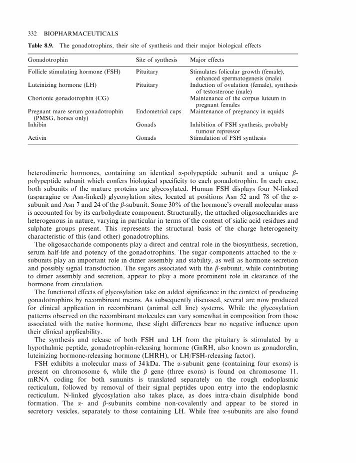
heterodimeric hormones, containing an identical a-polypeptide subunit and a unique b-polypeptide subunit which confers biological specificity to each gonadotrophin. In each case,both subunits of the mature proteins are glycosylated. Human FSH displays four N-linked(asparagine or Asn-linked) glycosylation sites, located at positions Asn 52 and 78 of the a-subunit and Asn 7 and 24 of the b-subunit. Some 30% of the hormone’s overall molecular massis accounted for by its carbohydrate component. Structurally, the attached oligosaccharides areheterogenous in nature, varying in particular in terms of the content of sialic acid residues andsulphate groups present. This represents the structural basis of the charge heterogeneitycharacteristic of this (and other) gonadotrophins.
The oligosaccharide components play a direct and central role in the biosynthesis, secretion,serum half-life and potency of the gonadotrophins. The sugar components attached to the a-subunits play an important role in dimer assembly and stability, as well as hormone secretionand possibly signal transduction. The sugars associated with the b-subunit, while contributingto dimer assembly and secretion, appear to play a more prominent role in clearance of thehormone from circulation.
The functional effects of glycosylation take on added significance in the context of producinggonadotrophins by recombinant means. As subsequently discussed, several are now producedfor clinical application in recombinant (animal cell line) systems. While the glycosylationpatterns observed on the recombinant molecules can vary somewhat in composition from thoseassociated with the native hormone, these slight differences bear no negative influence upontheir clinical applicability.
The synthesis and release of both FSH and LH from the pituitary is stimulated by ahypothalmic peptide, gonadotrophin-releasing hormone (GnRH, also known as gonadorelin,luteinizing hormone-releasing hormone (LHRH), or LH/FSH-releasing factor).
FSH exhibits a molecular mass of 34 kDa. The a-subunit gene (containing four exons) ispresent on chromosome 6, while the b gene (three exons) is found on chromosome 11.mRNA coding for both sununits is translated separately on the rough endoplasmicrecticulum, followed by removal of their signal peptides upon entry into the endoplasmicrecticulum. N-linked glycosylation also takes place, as does intra-chain disulphide bondformation. The a- and b-subunits combine non-covalently and appear to be stored insecretory vesicles, separately to those containing LH. While free a-subunits are also found
332 BIOPHARMACEUTICALS
Table 8.9. The gonadotrophins, their site of synthesis and their major biological effects
Gonadotrophin Site of synthesis Major effects
Follicle stimulating hormone (FSH) Pituitary Stimulates folicular growth (female),enhanced spermatogenesis (male)
Luteinizing hormone (LH) Pituitary Induction of ovulation (female), synthesisof testosterone (male)
Chorionic gonadotrophin (CG) Maintenance of the corpus luteum inpregnant females
Pregnant mare serum gonadotrophin(PMSG, horses only)
Endometrial cups Maintenance of pregnancy in equids
Inhibin Gonads Inhibition of FSH synthesis, probablytumour repressor
Activin Gonads Stimulation of FSH synthesis
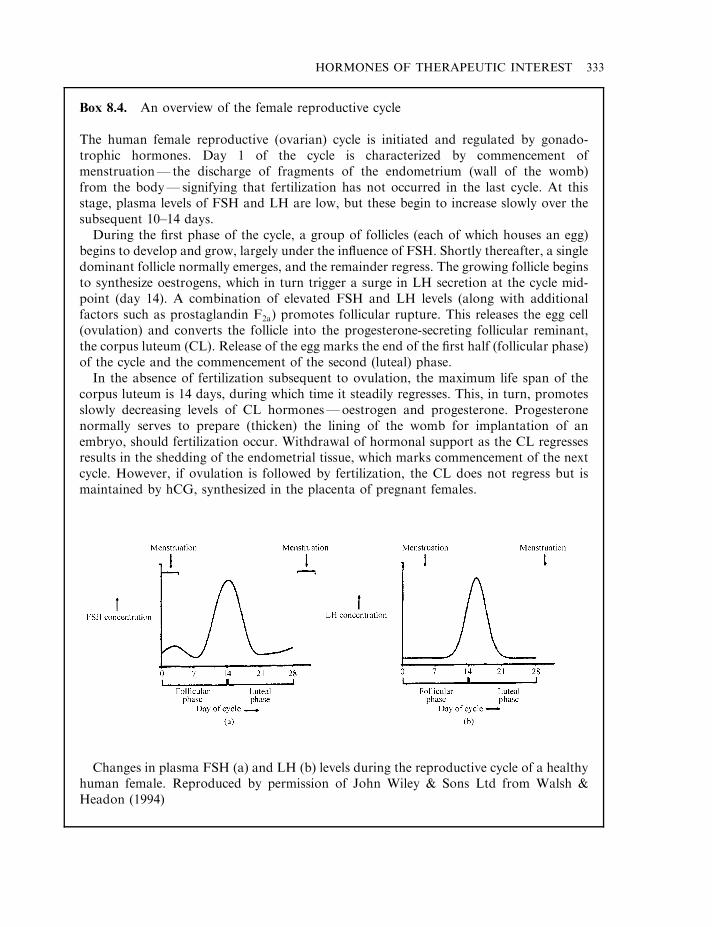
HORMONES OF THERAPEUTIC INTEREST 333
Box 8.4. An overview of the female reproductive cycle
The human female reproductive (ovarian) cycle is initiated and regulated by gonado-trophic hormones. Day 1 of the cycle is characterized by commencement ofmenstruation— the discharge of fragments of the endometrium (wall of the womb)from the body— signifying that fertilization has not occurred in the last cycle. At thisstage, plasma levels of FSH and LH are low, but these begin to increase slowly over thesubsequent 10–14 days.
During the first phase of the cycle, a group of follicles (each of which houses an egg)begins to develop and grow, largely under the influence of FSH. Shortly thereafter, a singledominant follicle normally emerges, and the remainder regress. The growing follicle beginsto synthesize oestrogens, which in turn trigger a surge in LH secretion at the cycle mid-point (day 14). A combination of elevated FSH and LH levels (along with additionalfactors such as prostaglandin F2a) promotes follicular rupture. This releases the egg cell(ovulation) and converts the follicle into the progesterone-secreting follicular reminant,the corpus luteum (CL). Release of the egg marks the end of the first half (follicular phase)of the cycle and the commencement of the second (luteal) phase.
In the absence of fertilization subsequent to ovulation, the maximum life span of thecorpus luteum is 14 days, during which time it steadily regresses. This, in turn, promotesslowly decreasing levels of CL hormones—oestrogen and progesterone. Progesteronenormally serves to prepare (thicken) the lining of the womb for implantation of anembryo, should fertilization occur. Withdrawal of hormonal support as the CL regressesresults in the shedding of the endometrial tissue, which marks commencement of the nextcycle. However, if ovulation is followed by fertilization, the CL does not regress but ismaintained by hCG, synthesized in the placenta of pregnant females.
Changes in plasma FSH (a) and LH (b) levels during the reproductive cycle of a healthyhuman female. Reproduced by permission of John Wiley & Sons Ltd from Walsh &Headon (1994)
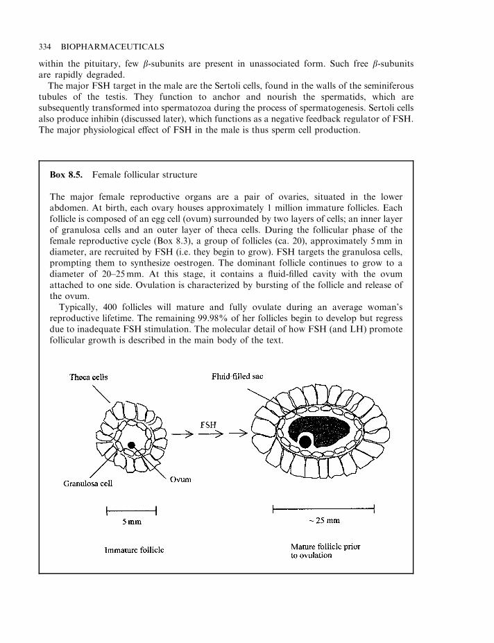
within the pituitary, few b-subunits are present in unassociated form. Such free b-subunitsare rapidly degraded.
The major FSH target in the male are the Sertoli cells, found in the walls of the seminiferoustubules of the testis. They function to anchor and nourish the spermatids, which aresubsequently transformed into spermatozoa during the process of spermatogenesis. Sertoli cellsalso produce inhibin (discussed later), which functions as a negative feedback regulator of FSH.The major physiological effect of FSH in the male is thus sperm cell production.
334 BIOPHARMACEUTICALS
Box 8.5. Female follicular structure
The major female reproductive organs are a pair of ovaries, situated in the lowerabdomen. At birth, each ovary houses approximately 1 million immature follicles. Eachfollicle is composed of an egg cell (ovum) surrounded by two layers of cells; an inner layerof granulosa cells and an outer layer of theca cells. During the follicular phase of thefemale reproductive cycle (Box 8.3), a group of follicles (ca. 20), approximately 5mm indiameter, are recruited by FSH (i.e. they begin to grow). FSH targets the granulosa cells,prompting them to synthesize oestrogen. The dominant follicle continues to grow to adiameter of 20–25mm. At this stage, it contains a fluid-filled cavity with the ovumattached to one side. Ovulation is characterized by bursting of the follicle and release ofthe ovum.
Typically, 400 follicles will mature and fully ovulate during an average woman’sreproductive lifetime. The remaining 99.98% of her follicles begin to develop but regressdue to inadequate FSH stimulation. The molecular detail of how FSH (and LH) promotefollicular growth is described in the main body of the text.
In the female, FSH mainly targets the granulosa cells of the ovarian follicle (Box 8.5). FSHexhibits a mitogenic effect upon these cells, stimulating their division and, hence, folliculargrowth and development. This activity is enhanced by the paracrine action of locally-producedgrowth factors. FSH also triggers enzymatic production of glycosaminoglycans (GAGs), as wellas expression of aromatase and other enzymes involved in oestrogen synthesis. GAGs form anessential component of the follicular fluid, while granulosa cell-derived oestrogens play multipleroles in sustaining and regulating female reproductive function.
Prior to puberty, serum FSH levels are insufficient to promote follicular recruitment anddevelopment. Subsequent to puberty, as a group of follicles begin to develop at the beginning ofa cycle, the one that is most responsive to FSH (i.e. displays the lowest FSH threshold) becomesthe first to secrete oestrogen. As one effect of oestrogen is to suppress FSH release from thepituitary, blood FSH levels then plateau or decline slightly. This slightly lower FSHconcentration is insufficient to sustain growth of follicles of higher FSH thresholds, so theydie, leaving only the single oestrogen-producing dominant follicle (Boxes 8.4 and 8.5) to matureand ovulate.
FSH exerts its molecular effects via a specific receptor on the surface of sensitive cells. Thisreceptor contains a characteristic seven transmembrane-spanning regions and is functionallycoupled (via membrane-associated G proteins) to adenylate cyclase. This generates the secondmessenger cAMP. FSH itself can promote increased expression of its own receptor in the shortterm, although longer-term exposure to elevated FSH levels downregulates receptor numbers.Cloning and analysis of gonadotrophin receptors from several species indicate a high level ofhomology between the FSH, LH and CG receptors.
LH exhibits a molecular mass of 28.5 kDa. The gene coding for the b-subunit of thisglycoprotein hormone is present on human chromosome 19. This subunit exhibits significantamino acid homology to placental CG. Both promote identical biological effects and act via thesame 93 kDa cell surface receptor. The LH receptor is present on testicular Leydig cells in malesand on female ovarian theca, granulosa, luteal and interstitial cells.
LH promotes synthesis of testosterone, the major male androgen (Box 8.6) by the testicularLeydig cells. FSH sensitizes these cells to the activities of LH, probably by increasing LHreceptor numbers on the cell surface. Leydig cells have a limited storage capacity fortestosterone (*25 mg), but secrete 5–10mg of the hormone into the bloodstream daily in younghealthy males.
The primary cellular targets of LH in the females are the follicular theca cells, whichconstitutively express the LH receptor. Under the influence of LH, these cells produceandrogens. The androgens (principally testosterone) are then taken up by granulosa cells andconverted into oestrogens (Box 8.6) by the already-mentioned aromatase complex. Thus, thefollicle represents the major female gonadal endocrine unit, in which granulosa and theca cellscooperate in the synthesis of oestrogens. Physiologically, LH in the female plays a major role inmaturation of the dominant follicle and appears central to triggering ovulation.
Pregnant mare serum gonadotrophin (PMSG)
Pregnant mare serum gonadotrophin (PMSG) is a unique member of the gonadotrophin familyof hormones. It is synthesized only by pregnant mares (i.e. is not found in other species).Furthermore, it displays both FSH-like and LH-like biological activities.
This glycoprotein hormone is a heterodimer, composed of an a- and b-subunit, andapproximately 45% of its molecular mass is carbohydrate. Reported molecular masses range
HORMONES OF THERAPEUTIC INTEREST 335
from 52 kDa to 68 kDa, a reflection of the potential variability of the hormone’s carbohydratecontent.
PMSG is secreted by cup-shaped outgrowths found in the horn of the uterus of pregnanthorses. These equine-specific endometrial cups are of fetal rather than maternal origin. Theyfirst become visible around day 40 of gestation, and reach maximum size at about day 70, afterwhich they steadily regress. They synthesize high levels of PMSG and secrete it into the blood,where it is detectable between days 40 and 130 of gestation.
336 BIOPHARMACEUTICALS
Box 8.6. The androgens and oestrogens
The androgens and oestrogens represent the major male and female sex hormones,respectively. The testicular Leydig cells represent the primary source of androgens in themale, of which testosterone is the major one. Testosterone, in turn, serves as a precursorfor two additional steroids: dihydrotestosterone and the oestrogen. These mediate many ofits biological effects.
Females, too, produce androgens, principally in the follicular theca cells. Androgens arealso produced in the adrenals in both male and females.
The biological activities of androgens (only some of which are specific to males) may besummarized as:
. promoting and regulating development of the male phenotype during embryonicdevelopment;
. promoting sperm cell synthesis;
. promoting development and maintenance of male secondary sexual characteristics atand after puberty;
. general growth-promoting effects;
. behavioural effects (e.g. male aggressiveness, etc.);
. regulation of serum gonadotrophin levels.
The follicular granulosa cells are the major site of synthesis of female steroid sexhormones: the oestrogens. b-Oestradiol represents the principal female follicularoestrogen. Oestriol is producted by the placenta of pregnant females. Oestriol as well asoestrone are also produced in small quantities as products of b-oestradiol metabolism.
Testosterone represents the immediate precursor of the oestrogens, the conversion beingcatalysed by the aromatase complex, a microsomal enzyme system. The biological actionsof oestrogens may be summarized as:
. growth and maturation of the female reproductive system;
. maintenance of reproductive capacity;
. development and maintenance of female secondary sexual characteristics;
. female behavioural effects;
. complex effects upon lipid metabolism and distribution of body fat;
. regulation of bone metabolism (oestrogen deficiency promotes bone decalcification, asseen in post-menopausal osteoporosis).
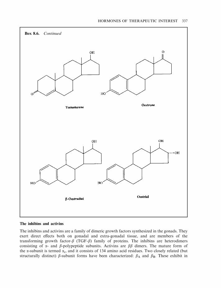
The inhibins and activins
The inhibins and activins are a family of dimeric growth factors synthesized in the gonads. Theyexert direct effects both on gonadal and extra-gonadal tissue, and are members of thetransforming growth factor-b (TGF-b) family of proteins. The inhibins are heterodimersconsisting of a- and b-polypeptide subunits. Activins are bb dimers. The mature form ofthe a-subunit is termed ac, and it consists of 134 amino acid residues. Two closely related (butstructurally distinct) b-subunit forms have been characterized: bA and bB. These exhibit in
HORMONES OF THERAPEUTIC INTEREST 337
Box 8.6. Continued
excess of 70% amino acid homology, and differ in size by only a single amino acid. The namingand polypeptide composition of the inhibin/activin family may be summarized as follows:
. Inhibin A¼acbA
. Inhibin B¼acbB
. Activin A¼bAbA
. Activin AB¼bAbB
. Activin B¼bBbB
Inhibins and activins were initially identified as gonadal-derived proteins capable of inhibiting(inhibin) or stimulating (activin) pituitary FSH production (Figure 8.15). The major gonadalsites of inhibin synthesis are the Sertoli cells (male) and granulosa cells (female). In addition totargeting the pituitary, the inhibins/activins probably play a direct (mutually antagonistic) roleas paracrine/autocrine regulators of gonadal function.
They also likely induce responses in tissues other than the pituitary and gonads, e.g. in adultsinhibin is also synthesized by the adrenal glands, spleen and nervous system. Recent studiesinvolving inhibin-deficient transgenic mice reveal a novel role for inhibin as a gonadal-specifictumour suppressor. These mice, in which the a-inhibin gene was missing, all developednormally, but all ultimately developed gonadal stromal tumours.
LHRH and regulation of gonadotrophin production
Gonadotrophin-releasing hormone (GnRH, LHRH), as previously mentioned, is a hypothalmicdecapeptide amide which stimulates synthesis and release of pituitary FSH and LH. A variety ofneurological influences alter hypothalmic secretion of LHRH, which tends to be pulsatile andhigher during sleep. Synthesis of this regulatory peptide predictably increases at puberty. Itseffects on the pituitary are mediated via a specific cell membrane receptor, which displays atypical seven transmembrane domain structure, with extracellular (ligand binding) domains andan intracellular domain that generates second messengers.
Several second messengers have been implicated in mediating GnRH’s biological effects,including: calcium mobilization, diacylglycerate production (responsible for gonadotrophinrelease) and protein kinase C activation (which promotes transcription of the LH b-subunit). Anumber of factors appear to regulate levels of pituitary GnRH receptors, modulating its activityin this way. GnRH can up- or downregulate its own receptor numbers. Oestradiol increasesreceptor numbers, while inhibin can downregulate them.
Although GnRH promotes increased FSH and LH synthesis, it can do so differentially, atleast under certain circumstances. After an injection of GnRH, the ratio of maximal to basal LHis higher than the ratio of maximal to basal FSH. This can only be partially explained on thebasis that LH is cleared more slowly from circulation than FSH. Furthermore, during thenormal female reproductive cycle, alterations in serum levels of LH and FSH do not alwaysoccur exactly in parallel. The exact molecular basis underlining the differential regulation ofsynthesis and secretion of LH and FSH is not understood at the moment. The root lies in a lackof understanding of the different intracellular pathways that trigger LH versus FSH b-subunitsynthesis.
In addition to GnRH, several other effector molecules modulate secretion of gonadotrophins.These include not only inhibins and activins, but also gonadal steroids. All of these combine viacomplex feedback loops to modulate gonadotrophin production and, hence, reproductivefunction (Figure 8.15).
338 BIOPHARMACEUTICALS
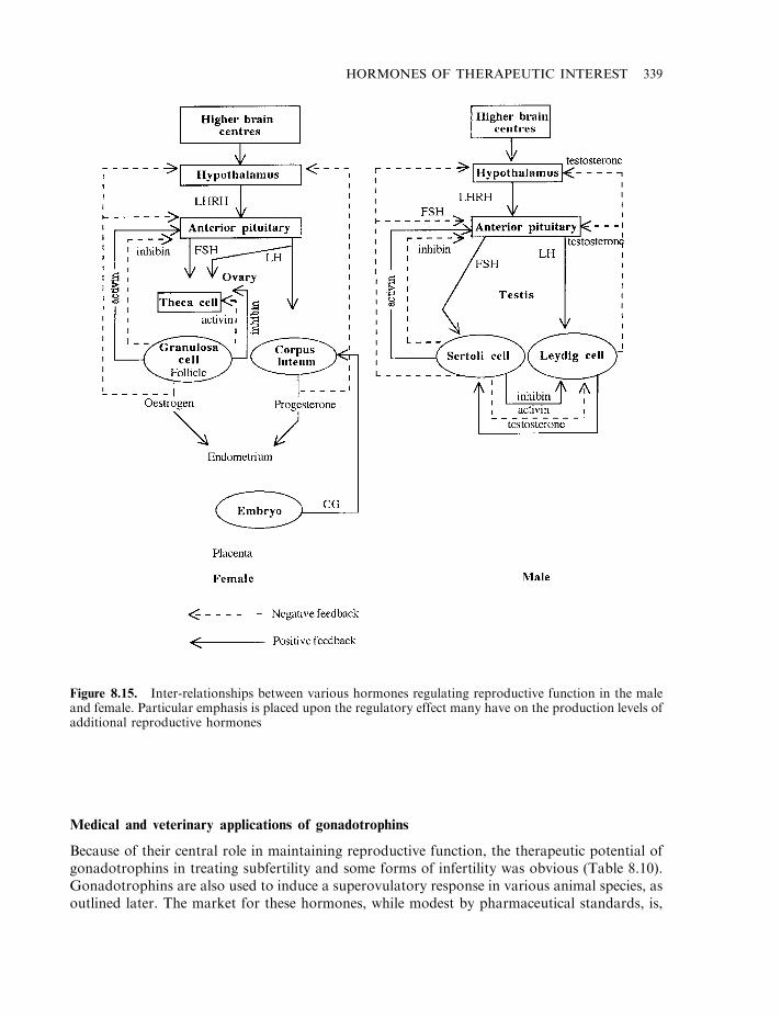
Medical and veterinary applications of gonadotrophins
Because of their central role in maintaining reproductive function, the therapeutic potential ofgonadotrophins in treating subfertility and some forms of infertility was obvious (Table 8.10).Gonadotrophins are also used to induce a superovulatory response in various animal species, asoutlined later. The market for these hormones, while modest by pharmaceutical standards, is,
HORMONES OF THERAPEUTIC INTEREST 339
Figure 8.15. Inter-relationships between various hormones regulating reproductive function in the maleand female. Particular emphasis is placed upon the regulatory effect many have on the production levels ofadditional reproductive hormones
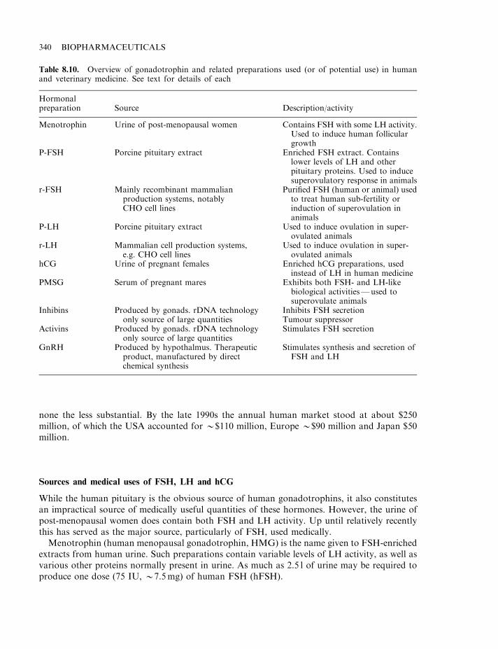
none the less substantial. By the late 1990s the annual human market stood at about $250million, of which the USA accounted for *$110 million, Europe *$90 million and Japan $50million.
Sources and medical uses of FSH, LH and hCG
While the human pituitary is the obvious source of human gonadotrophins, it also constitutesan impractical source of medically useful quantities of these hormones. However, the urine ofpost-menopausal women does contain both FSH and LH activity. Up until relatively recentlythis has served as the major source, particularly of FSH, used medically.
Menotrophin (human menopausal gonadotrophin, HMG) is the name given to FSH-enrichedextracts from human urine. Such preparations contain variable levels of LH activity, as well asvarious other proteins normally present in urine. As much as 2.5 l of urine may be required toproduce one dose (75 IU, *7.5mg) of human FSH (hFSH).
340 BIOPHARMACEUTICALS
Table 8.10. Overview of gonadotrophin and related preparations used (or of potential use) in humanand veterinary medicine. See text for details of each
Hormonalpreparation Source Description/activity
Menotrophin Urine of post-menopausal women Contains FSH with some LH activity.Used to induce human folliculargrowth
P-FSH Porcine pituitary extract Enriched FSH extract. Containslower levels of LH and otherpituitary proteins. Used to inducesuperovulatory response in animals
r-FSH Mainly recombinant mammalianproduction systems, notablyCHO cell lines
Purified FSH (human or animal) usedto treat human sub-fertility orinduction of superovulation inanimals
P-LH Porcine pituitary extract Used to induce ovulation in super-ovulated animals
r-LH Mammalian cell production systems,e.g. CHO cell lines
Used to induce ovulation in super-ovulated animals
hCG Urine of pregnant females Enriched hCG preparations, usedinstead of LH in human medicine
PMSG Serum of pregnant mares Exhibits both FSH- and LH-likebiological activities—used tosuperovulate animals
Inhibins Produced by gonads. rDNA technologyonly source of large quantities
Inhibits FSH secretionTumour suppressor
Activins Produced by gonads. rDNA technologyonly source of large quantities
Stimulates FSH secretion
GnRH Produced by hypothalmus. Therapeuticproduct, manufactured by directchemical synthesis
Stimulates synthesis and secretion ofFSH and LH
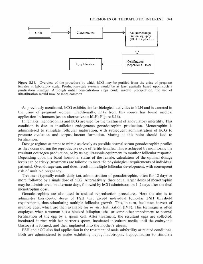
As previously mentioned, hCG exhibits similar biological activities to hLH and is excreted inthe urine of pregnant women. Traditionally, hCG from this source has found medicalapplication in humans (as an alternative to hLH; Figure 8.16).
In females, menotrophins and hCG are used for the treatment of anovulatory infertility. Thiscondition is due to insufficient endogenous gonadotrophin production. Menotrophin isadministered to stimulate follicular maturation, with subsequent administration of hCG topromote ovulation and corpus luteum formation. Mating at this point should lead tofertilization.
Dosage regimes attempt to mimic as closely as possible normal serum gonadotrophin profilesas they occur during the reproductive cycle of fertile females. This is achieved by monitoring theresultant oestrogen production, or by using ultrasonic equipment to monitor follicular response.Depending upon the basal hormonal status of the female, calculation of the optimal dosagelevels can be tricky (treatments are tailored to meet the physiological requirements of individualpatients). Over-dosage can, and does, result in multiple follicular development, with consequentrisk of multiple pregnancy.
Treatment typically entails daily i.m. administration of gonadotrophin, often for 12 days ormore, followed by a single dose of hCG. Alternatively, three equal larger doses of menotrophinmay be administered on alternate days, followed by hCG administration 1–2 days after the finalmenotrophin dose.
Gonadotrophins are also used in assisted reproduction procedures. Here the aim is toadminister therapeutic doses of FSH that exceed individual follicular FSH thresholdrequirements, thus stimulating multiple follicular growth. This, in turn, facilitates harvest ofmultiple eggs, which are then available for in vitro fertilization (IVF). This technique is oftenemployed when a woman has a blocked fallopian tube, or some other impediment to normalfertilization of the egg by a sperm cell. After treatment, the resultant eggs are collected,incubated in vitro with her partner’s sperm, incubated in culture media until the embryonicblastocyst is formed, and then implanted into the mother’s uterus.
FSH and hCG also find application in the treatment of male subfertility or related conditions.Both are administered to males exhibiting hypogonadotrophic hypogonadism to stimulate
HORMONES OF THERAPEUTIC INTEREST 341
Figure 8.16. Overview of the procedure by which hCG may be purified from the urine of pregnantfemales at laboratory scale. Production-scale systems would be at least partially based upon such apurification strategy. Although initial concentration steps could involve precipitation, the use ofultrafiltration would now be more common
sperm synthesis and normal sexual function. hCG has found limited application in the treatmentof pre-pubertal cryptorchidism (a condition characterized by failure of the testes to descend fullyinto the scrotum from the abdomen). The ability of this hormone to stimulate testosteroneproduction also caught the attention of some athletes and, as a result, the International OlympicCommittee has banned its use.
The LH/hCG cell surface receptor is found in a number of non-gonadal tissues, indicatingthat these hormones may exert physiologically relevant non-gonadal functions (Table 8.11). Inaddition, while liver, kidney and muscle cells are devoid of such a receptor, it is expressed by anumber of these tissues before birth, hinting at a potential developmental role. Receptor levelsin non-gonadal tissues are generally much lower than in gonadal tissue.
Research indicates that hCG probably has a number of pregnancy/non-pregnancy-relatednon-gonadal functions that may give rise to future additional clinical applications. It appears topromote effects such as increased uterine blood flow and immunosuppression at the maternal–fetal interface. As such, hCG may prove potentially useful in preventing some types ofpregnancy loss. Research has also indicated that infusion of hCG into the uterus can counteractprostaglandin-induced uterine contractions. As such, it may prove useful in preventing the onsetof premature labour. hCG also exhibits anti-breast cancer activity. Studies in rodents, forexample, illustrate that it prevents mammary tumour formation/growth when administeredprior or subsequent to various chemical carcinogens.
Recombinant gonadotrophins
Gonadotrophins are now also produced by recombinant DNA technology. The genes orcDNAs coding for gonadotrophins from several species have been identified and expressed invarious recombinant host systems, particularly mammalian cell lines. rhFSH produced in CHOcells has proved clinically effective. While exhibiting an amino acid sequence identical to thehuman molecule, its carbohydrate composition differs slightly. When administered to humans,the preparation is well tolerated and yields no unexpected side effects. It does not elicit animmunological response, and its plasma half-life is similar to the native hormone. rhFSH hasproved efficacious in stimulating follicular growth in females suffering from hypogonadotrophichypogonadism and is effective in the treatment of males suffering from similar conditions.rhFSH was amongst the first biopharmaceutical substances to be approved for general medical
342 BIOPHARMACEUTICALS
Table 8.11. Some notable non-gonadal tissues that expressfunctional LH/hCG receptors
Pregnancy/fertility-related tissue Other tissue
Uterus SkinCervix Blood vesselsPlacenta Adrenal cortexOviduct Brain tissueFetal membranes Prostate glandSeminal vesicles BladderSperm cells MonocytesBreast Macrophages
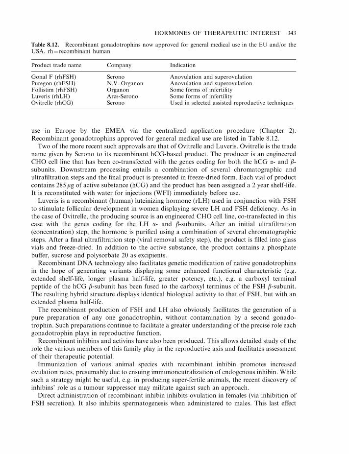
use in Europe by the EMEA via the centralized application procedure (Chapter 2).Recombinant gonadotrophins approved for general medical use are listed in Table 8.12.
Two of the more recent such approvals are that of Ovitrelle and Luveris. Ovitrelle is the tradename given by Serono to its recombinant hCG-based product. The producer is an engineeredCHO cell line that has been co-transfected with the genes coding for both the hCG a- and b-subunits. Downstream processing entails a combination of several chromatographic andultrafiltration steps and the final product is presented in freeze-dried form. Each vial of productcontains 285 mg of active substance (hCG) and the product has been assigned a 2 year shelf-life.It is reconstituted with water for injections (WFI) immediately before use.
Luveris is a recombinant (human) luteinizing hormone (rLH) used in conjunction with FSHto stimulate follicular development in women displaying severe LH and FSH deficiency. As inthe case of Ovitrelle, the producing source is an engineered CHO cell line, co-transfected in thiscase with the genes coding for the LH a- and b-subunits. After an initial ultrafiltration(concentration) step, the hormone is purified using a combination of several chromatographicsteps. After a final ultrafiltration step (viral removal safety step), the product is filled into glassvials and freeze-dried. In addition to the active substance, the product contains a phosphatebuffer, sucrose and polysorbate 20 as excipients.
Recombinant DNA technology also facilitates genetic modification of native gonadotrophinsin the hope of generating variants displaying some enhanced functional characteristic (e.g.extended shelf-life, longer plasma half-life, greater potency, etc.), e.g. a carboxyl terminalpeptide of the hCG b-subunit has been fused to the carboxyl terminus of the FSH b-subunit.The resulting hybrid structure displays identical biological activity to that of FSH, but with anextended plasma half-life.
The recombinant production of FSH and LH also obviously facilitates the generation of apure preparation of any one gonadotrophin, without contamination by a second gonado-trophin. Such preparations continue to facilitate a greater understanding of the precise role eachgonadotrophin plays in reproductive function.
Recombinant inhibins and activins have also been produced. This allows detailed study of therole the various members of this family play in the reproductive axis and facilitates assessmentof their therapeutic potential.
Immunization of various animal species with recombinant inhibin promotes increasedovulation rates, presumably due to ensuing immunoneutralization of endogenous inhibin. Whilesuch a strategy might be useful, e.g. in producing super-fertile animals, the recent discovery ofinhibins’ role as a tumour suppressor may militate against such an approach.
Direct administration of recombinant inhibin inhibits ovulation in females (via inhibition ofFSH secretion). It also inhibits spermatogenesis when administered to males. This last effect
HORMONES OF THERAPEUTIC INTEREST 343
Table 8.12. Recombinant gonadotrophins now approved for general medical use in the EU and/or theUSA. rh¼recombinant human
Product trade name Company Indication
Gonal F (rhFSH) Serono Anovulation and superovulationPuregon (rhFSH) N.V. Organon Anovulation and superovulationFollistim (rhFSH) Organon Some forms of infertilityLuveris (rhLH) Ares-Serono Some forms of infertilityOvitrelle (rhCG) Serono Used in selected assisted reproductive techniques
initially prompted interest in the use of inhibin as a potential male contraceptive, although nosignificant data on the safety or efficacy of such a treatment has yet been published.
Veterinary uses of gonadotrophins
Gonadotrophins may be utilized to treat subfertility in animals and are routinely used to inducea superovulatory response in valuable animals, most notably valuable horses and cattle.
The theory and practice of superovulation is quite similar to the use of gonadotrophins toassist in vitro fertilization procedures in humans. Exogenous FSH is administered to the animalsuch that multiple follicles develop simultaneously. After administration of LH to help promoteovulation, the animal is mated, thus fertilizing the released egg cells. Depending upon thespecific animal and the superovulatory regime employed, anything between 0 and 50 viableembryos may be produced, although more typically the number is between 4 and 10. Theembryos are then recovered from the animal (either surgically or, more usually, non-surgically)and are often maintained in cell culture for a short period of time. A single embryo is thenusually re-implanted into the donor female, while remaining embryos are implanted into otherrecipient animals, who act as surrogate mothers, carrying the offspring to term.
This technology is most often applied to valuable animals (e.g. prize-winning horses, or highmilk-yield dairy cattle) in order to boost their effective reproductive capacity several-fold. Eachof the offspring will inherit its genetic complement from the biological mother (and father),irrespective of which recipient animal carries it to term.
Gonadotrophins are usually utilized to induce a superovulatory response, and include porcineFSH (p-FSH), porcine LH (p-LH) and PMSG. P-FSH is extracted from the pituitary glands ofslaughterhouse pigs. The crude pituitary extract is usually subject to a precipitation step, usingeither ethanol or salts. The FSH-containing precipitate is normally subjected to at least onesubsequent chromatographic step. The final product often contains some LH as well as lowlevels of additional pituitary-derived proteins.
p-LH is obtained, again, by its partial purification from the pituitary glands of slaughterhousepigs. In both cases, a porcine source is utilized, as the target recipients are almost always cattleor horses. The use of a product derived from a species other than the intended recipient speciesis encouraged, as it helps minimize the danger of accidental transmission of disease via infectedsource material (many pathogens are species-specific).
Most superovulatory regimes utilizing p-FSH entails its administration to the recipientanimal twice daily for 4–5 days. Regular injections are required, due to the relatively short half-life of FSH in serum; s.c. administration helps prolong the duration of effectiveness of eachinjection. The 4 or 5 days of treatment with FSH is followed by a single dose of LH, promotingfinal follicular maturation and ovulation.
The causes of variability of superovulatory responses are complex, and not fully understood.The general health of the animal, as well as its characteristic reproductive physiology, isimportant. The exact composition of the gonadotrophin preparations administered and theexact administration protocol also influences the outcome. The variability of FSH:LH ratios inmany p-FSH preparations can affect the results obtained, with the most consistent super-ovulatory responses being observed when FSH preparations exhibiting low LH activity areused. The availability of recombinant FSH and LH will overcome these difficulties at least.
An alternative superovulatory regime entails administration of PMSG which, as describedearlier, exhibits both FSH and LH activity. The major rationale for utilizing PMSG is itsrelatively long circulatory half-life. In cattle, clearance of PMSG may take up to 5 days. The
344 BIOPHARMACEUTICALS
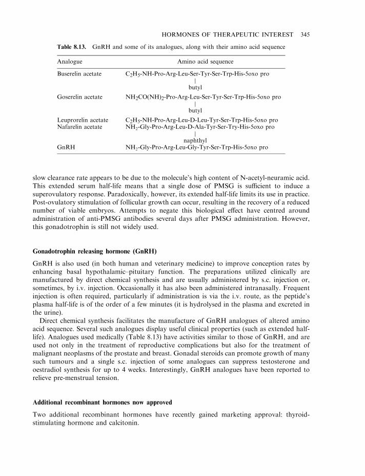
slow clearance rate appears to be due to the molecule’s high content of N-acetyl-neuramic acid.This extended serum half-life means that a single dose of PMSG is sufficient to induce asuperovulatory response. Paradoxically, however, its extended half-life limits its use in practice.Post-ovulatory stimulation of follicular growth can occur, resulting in the recovery of a reducednumber of viable embryos. Attempts to negate this biological effect have centred aroundadministration of anti-PMSG antibodies several days after PMSG administration. However,this gonadotrophin is still not widely used.
Gonadotrophin releasing hormone (GnRH)
GnRH is also used (in both human and veterinary medicine) to improve conception rates byenhancing basal hypothalamic–pituitary function. The preparations utilized clinically aremanufactured by direct chemical synthesis and are usually administered by s.c. injection or,sometimes, by i.v. injection. Occasionally it has also been administered intranasally. Frequentinjection is often required, particularly if administration is via the i.v. route, as the peptide’splasma half-life is of the order of a few minutes (it is hydrolysed in the plasma and excreted inthe urine).
Direct chemical synthesis facilitates the manufacture of GnRH analogues of altered aminoacid sequence. Several such analogues display useful clinical properties (such as extended half-life). Analogues used medically (Table 8.13) have activities similar to those of GnRH, and areused not only in the treatment of reproductive complications but also for the treatment ofmalignant neoplasms of the prostate and breast. Gonadal steroids can promote growth of manysuch tumours and a single s.c. injection of some analogues can suppress testosterone andoestradiol synthesis for up to 4 weeks. Interestingly, GnRH analogues have been reported torelieve pre-menstrual tension.
Additional recombinant hormones now approved
Two additional recombinant hormones have recently gained marketing approval: thyroid-stimulating hormone and calcitonin.
HORMONES OF THERAPEUTIC INTEREST 345
Table 8.13. GnRH and some of its analogues, along with their amino acid sequence
Analogue Amino acid sequence
Buserelin acetate C2H5-NH-Pro-Arg-Leu-Ser-Tyr-Ser-Trp-His-5oxo proj
butyl
Goserelin acetate NH2CO(NH)2-Pro-Arg-Leu-Ser-Tyr-Ser-Trp-His-5oxo proj
butyl
Leuprorelin acetate C2H5-NH-Pro-Arg-Leu-D-Leu-Tyr-Ser-Trp-His-5oxo proNafarelin acetate NH2-Gly-Pro-Arg-Leu-D-Ala-Tyr-Ser-Try-His-5oxo pro
jnaphthyl
GnRH NH2-Gly-Pro-Arg-Leu-Gly-Tyr-Ser-Trp-His-5oxo pro
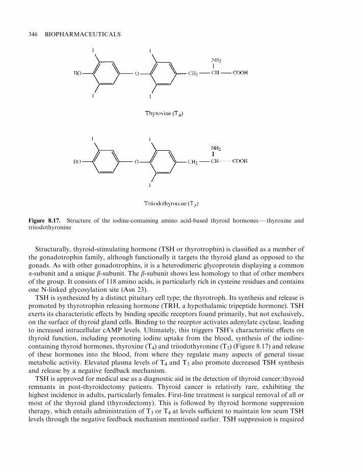
Structurally, thyroid-stimulating hormone (TSH or thyrotrophin) is classified as a member ofthe gonadotrophin family, although functionally it targets the thyroid gland as opposed to thegonads. As with other gonadotrophins, it is a heterodimeric glycoprotein displaying a commona-subunit and a unique b-subunit. The b-subunit shows less homology to that of other membersof the group. It consists of 118 amino acids, is particularly rich in cysteine residues and containsone N-linked glycosylation site (Asn 23).
TSH is synthesized by a distinct pituitary cell type; the thyrotroph. Its synthesis and release ispromoted by thyrotrophin releasing hormone (TRH, a hypothalamic tripeptide hormone). TSHexerts its characteristic effects by binding specific receptors found primarily, but not exclusively,on the surface of thyroid gland cells. Binding to the receptor activates adenylate cyclase, leadingto increased intracellular cAMP levels. Ultimately, this triggers TSH’s characteristic effects onthyroid function, including promoting iodine uptake from the blood, synthesis of the iodine-containing thyroid hormones, thyroxine (T4) and triiodothyronine (T3) (Figure 8.17) and releaseof these hormones into the blood, from where they regulate many aspects of general tissuemetabolic activity. Elevated plasma levels of T4 and T3 also promote decreased TSH synthesisand release by a negative feedback mechanism.
TSH is approved for medical use as a diagnostic aid in the detection of thyroid cancer/thyroidremnants in post-thyroidectomy patients. Thyroid cancer is relatively rare, exhibiting thehighest incidence in adults, particularly females. First-line treatment is surgical removal of all ormost of the thyroid gland (thyroidectomy). This is followed by thyroid hormone suppressiontherapy, which entails administration of T3 or T4 at levels sufficient to maintain low seum TSHlevels through the negative feedback mechanism mentioned earlier. TSH suppression is required
346 BIOPHARMACEUTICALS
Figure 8.17. Structure of the iodine-containing amino acid-based thyroid hormones— thyroxine andtriiodothyronine
in order to prevent the TSH-mediated stimulation of remnant thyroid cancer cells. Therecurrence of active thyroid cancer can be detected by administration of TSH along with radio-active iodine. TSH promotes uptake of radioactivity, which can then be detected by appropriateradio-imaging techniques.
The commercial recombinant TSH product (trade name, Thyrogen; international non-proprietary name; thyrotrophin-a) is produced in a CHO cell line co-transfected with plasmidsharbouring the DNA sequences coding for the a- and b-TSH subunits. The cells are grown inbatch harvest animal cell culture bioreactors. Following recovery and concentration(ultrafiltration), the TSH is chromatographically purified and formulated to a concentrationof 0.9mg/ml in phosphate buffer containing mannitol and sodium chloride as excipients. Aftersterile filtration and aseptic filling into glass vials, the product is freeze-dried. Finished producthas been assigned a shelf-life of 3 years when stored at 2–88C.
Calcitonin is a polypeptide hormone which (along with parathyroid hormone and the vitaminD derivative, 1,25-dihydroxycholecalciferol) plays a central role in regulating serum ionizedcalcium (Ca2+) and inorganic phosphate (Pi) levels. The adult human body contains up to 2 kgof calcium, of which 98% is present in the skeleton (i.e. bone). Up to 85% of the 1 kg ofphosphorus present in the body is also found in the skeleton (the so-called mineral fraction ofbone is largely composed of Ca3 (PO4)2 which acts as a body reservoir for both calcium andphosphorus). Calcium concentrations in human serum approximate to 0.1mg/ml and areregulated very tightly (serum phosphate levels are more variable).
Calcitonin lowers serum Ca2+ and Pi levels, primarily by inhibiting the process of boneresorption, but also by decreasing resorption of Pi and Ca2+ in the kidney. Calcitoninreceptors are predictably found primarily on bone cells (osteoclasts) and renal cells, andgeneration of cAMP via adenylate cyclase activation plays a prominent role in hormone signaltransduction.
Calcitonin is used clinically to treat hypercalcaemia associated with some forms ofmalignancy and Paget’s disease. The latter condition is a chronic disorder of the skeleton inwhich bone grows abnormally in some regions. It is characterized by substantially increasedbone turnover rates which reflects over-stimulation of both osteoclasts (promote boneresorption, i.e. degradation of old bone) and osteoblasts (promotes synthesis of new bone).
In most mammals, calcitonin is synthesized by specialized parafollicular cells in the thyroid.In sub-mammalian species, it is synthesized by specialized anatomical structures known asultimobranchial bodies.
Calcitonin produced by virtually all species is a single-chain, 32 amino acid residuepolypeptide, displaying a molecular mass in the region of 3500Da. Salmon calcitonin differs insequence from the human hormone by nine amino acid residues. It is noteworthy, however, as itis approximately 100-fold more potent than the native hormone in humans. The higher potencyappears due to both a greater affinity for the receptor and greater resistance to degradation invivo. As such, salmon, as opposed to human calcitonin, is used clinically. Traditional clinicalpreparations were manufactured by direct chemical synthesis, although a recombinant form ofthe molecule has now gained marketing approval. The recombinant calcitonin is produced in anengineered E. coli strain. Structurally, salmon calcitonin displays C-terminal amidation. A C-terminal amide group (7CONH2) replacing the usual carboxyl group is a characteristic featureof many polypeptide hormones. If present, it is usually required for full biological activity/stability. As E. coli cannot carry out post-translational modifications, the amidation of therecombinant calcitonin is carried out in vitro using an a-amidating enzyme which is itselfproduced by recombinant means in an engineered CHO cell line. The purified, amidated finished
HORMONES OF THERAPEUTIC INTEREST 347
product is formulated in an acetate buffer and filled into glass ampoules. The (liquid) productexhibits a shelf-life of 2 years when stored at 2–88C.
CONCLUSIONS
Several hormone preparations have a long history of use as therapeutic agents. In virtually allinstances they are administered simply to compensate for lower than normal endogenousproduction of the hormone in question. Since it first became medically available, insulin hassaved or prolonged the lives of millions of diabetics. Gonadotrophins have allowed tens, if nothundreds, of thousands of sub-fertile individuals to conceive. Growth hormone has improvedthe quality of life of thousands of people of short stature. Most such hormones were in medicaluse prior to the advent of genetic engineering. Recombinant hormonal preparations are nowhowever gaining greater favour, mainly on safety grounds. Hormone therapy will remain acentral therapeutic tool for clinicians for many years to come.
FURTHER READING
Books
Bercu, B. (1998). Growth Hormone Secretagogues in Clinical Practice. Marcel Dekker, New York.Fauser, B. (1997). FSH Action and Intraovarian Regulation. Parthenon, Carnforth, UK.Hakin, N. (2002). Pancreas and Islet Transplantation. Oxford University Press, Oxford.Juul, A. (2000). Growth Hormone in Adults. Cambridge University Press, Cambridge.Mac Hadley, E. (1999). Endocrinology. Prentice-Hall, Hemel Hempstead, UK.Norman, A. (1997). Hormones. Academic Press, London.O’Malley, B. (1997). Hormones and Signalling. Academic Press, London.Walsh, G. & Headon, D. (1994). Protein Biotechnology. Wiley, Chichester.
Articles
Insulin and diabetes
Atkinson, M. & McClaren, N. (1990). What causes diabetes? Sci. Am. July, 42–46.Blundell, T. et al. (1972). Insulin: the structure in the crystal and its reflection in chemistry and biology. Adv. Protein
Chem. 26, 279–402.Bristow, A. (1993). Recombinant DNA derived insulin analogues as potentially useful therapeutic agents. Trends
Biotechnol. 11, 301–305.Brunetti, P. & Bolli, G. (1997). Pharmacokinetics and pharmacodynamics of insulin relevance to the therapy of diabetes
mellitus. Diabet. Nutrit. Metab. 10(1), 24–34.Cao, Y. & Lam, L. (2002). Projections for insulin treatment for diabetics. Drugs Today 38(6), 419–427.Ciszak, E., Beals, J. M., Frank, H. et al. (1995). Role of C-terminal B-chain residues in insulin assembly: the structure of
hexameric LysB28 ProB29-human insulin. Structure 3, 615.Combettes-Souverain, M. & Issad, T. (1998). Molecular basis of insulin action. Diabet. Metab. 24, 477–489.Conrad, B. et al. (1994). Evidence for superantigen involvement in insulin-dependent diabetes mellitus aetiology. Nature
371, 351–354.Docherty, K. (1997). Gene therapy for diabetes mellitus. Clin. Sci. 92(4), 321–330.Drucker, D. (2002). Biological actions and therapeutic potential of the glucagon-like peptides. Gastroenterology 122(2),
531–544.Goa, L. et al. (1997). Lisinopril—a review of its pharmacology and use in the management of the complications of
diabetes mellitus. Drugs 53(6), 1081–1105.Greenbaum, C. (2002). Insulin resistance in type 1 diabetes. Diabet. Metab. Res. Rev. 18(3), 192–200.Hinds, K. & Kim, S. (2002). Effects of PEG conjugation on insulin properties. Adv. Drug Delivery Rev. 54(4), 505–530.Ikegami, H. & Ogihara, T. (1996). Genetics of insulin-dependent diabetes mellitus. Endocr. J. 43(6), 605–613.Johnson, I. (1983). Human insulin from recombinant DNA technology. Science 219, 632–637.Kroeff, E. et al. (1989). Production scale purification of biosynthetic human insulin by reverse phase high performance
liquid chromatography. J. Chromatogr. 461, 45–61.
348 BIOPHARMACEUTICALS
Lacy, P. (1995). Treating diabetes with transplanted cells. Sci. Am. July, 40–45.Maassen, J. & Ouwens, D. (1997). Mechanism of insulin action. Frontiers Hormone Res. 22, 201–221.Patton, J. et al. (1999). Inhaled insulin. Adv. Drug Delivery Rev. 35, 235–247.Rhodes, C. & White, M. (2002). Molecular insights into insulin action and secretion. Eur. J. Clin. Invest. 32, 3–13.Secchi, A. et al. (1997). Pancreas and Islet transplantation—current progress, problems and perspectives. Hormone
Metab. Res. 29(1), 1–8.Selam, J. (1997). Management of diabetes with glucose sensors and implantable insulin pumps— from the dream of the
60s to the realities of the 90s. ASAIO J. 43(3), 137–142.Smith, R. et al. (1997). Insulin internalization and other signalling pathways in the pleiotropic effects of insulin. Int. Rev.
Cytol. Surv. Cell Biol. 173, 243–280.Stralfors, P. (1997). Insulin 2nd messengers. Bioessays 19(4), 327–335.Vajo, Z. et al. (2001). Recombinant DNA technology in the treatment of diabetes: insulin analogues. Endocr. Rev. 22(5),
706–717.
Growth hormone
Artini, P. et al. (1996). Growth hormone co-treatment with gonadotrophins in ovulation induction. J. Endocrinol. Invest.19(11), 763–779.
Carter, C. et al. (2002). A critical analysis of the role of growth hormone and IGF-1 in aging and lifespan. Trends Genet.18(6), 295–301.
Chantalat, L., Jones, N. D., Korber, F. et al. (1995). The crystal structure of wild-type growth hormone at 2.5 Aresolution. Protein Peptide Lett. 2, 333.
De Vos, A. et al. (1992). Human growth hormone and extracellular domain of its receptor; crystal structure of thecomplex. Science 255, 306–312.
Gibson, F. & Hinds, C. (1997). Growth hormone and insulin-like growth factors in critical illness. Intens. Care Med.23(4), 369–378.
Hathaway, D. (2002). Growth hormone: challenges and opportunities for the biotechnology sector. J. Anti-aging Med.5(1), 57–62.
Neely, E. (1994). Use and abuse of human growth hormone. Ann. Review Med. 45, 407–420.Piwien-Pilipuk, G. et al. (2002). Growth hormone signal transduction. J. Pediat. Endocrinol. Metab. 15(6), 771–786.Sharara, F. & Giudice, K. (1997). Role of growth hormone in ovarian physiology and onset of puberty. J. Soc. Gynaecol.
Invest. 4(1), 2–7.Simpson, H. et al. (2002). Growth hormone replacement therapy for adults: into the new millennium. Growth Hormone
IGF Res. 12(1), 1–33.
Gonadotrophins and TSH
Barbieri, R. (1992). Clinical applications of GnRH and its analogues. Trends Endocrinol. Metab. 3(1), 30–34.Bernard, D. et al. (2002). Minireview: inhibin binding protein, b-glycan and the continuing search for the inhibin
receptor. Mol. Endocrinol. 16(2), 207–212.Cho, B. (2002). Clinical applications of TSH receptor antibodies in thyroid diseases. J. Korean Med. Sci. 17(3), 293–301.Conn, P. & Crowley, W. (1994). Gonadotropin-releasing hormone and its analogues. Ann. Rev. Med. 45, 391–405.Dardenne, M. & Savino, W. (1996). Interdependence of the endocrine and immune systems. Adv. Neuroimmunol. 6(4),
297–307.De Jong, F. (1988). Inhibin. Physiol. Rev. 68, 555–595.De Koning, W. et al. (1994). Recombinant reproduction. Bio/Technology 12, 988–992.Depaolo, L. (1997). Inhibins, activins and follistatins— the saga continues. Proc. Soc. Exp. Biol. Med. 214(4), 328–339.Fauser, B. & Vanheusden, A. (1997). Manipulation of human ovarian function—physiological concepts and clinical
consequences. Endocr. Rev. 18(1), 71–106.Gore, A. & Roberts, J. (1997). Regulation of gonadotrophin-releasing hormone gene expression in vivo and in vitro.
Frontiers Neuroendocrinol. 18(2), 209–245.Greenberg, N. et al. (1991). Expression of biologically active heterodimeric bovine follicle-stimulating hormone in milk
of transgenic mice. Proc. Natl Acad. Sci. USA 88, 8327–8331.Hayden, C. et. al. (1999). Recombinant gonadotrophins. Br. J. Obstet. Gynaecol. 106(3), 188–196.Hillier, S. (1994). Current concepts of the role of follicle stimulating hormone and luteinizing hormone in
folliculogenesis. Hum. Reprod. 9(2), 188–191.Jones, R. et al. (2002). Potential roles for endometrial inhibins, activins and follistatin during human embryo
implantation and early pregnancy. Trends Endocrinol. Metab. 13(4), 144–150.Macklon, N. & Fauser, B. (2001). Follicle stimulating hormone and advanced follicle development in the human. Arch.
Med. Res. 32(6), 595–600.
HORMONES OF THERAPEUTIC INTEREST 349
Matzuk, M. et al. (1992). a-Inhibin is a tumor-suppressor gene with gonadal specificity in mice. Nature 360, 313–319.McCann, S. et al. (2001). Control of gonadotrophin secretion by follicule stimulating hormone-releasing factor,
luteinizing hormone-releasing hormone and leptin. Arch. Med. Res. 32(6), 476–485.Rao, C. & Sanfilippo, J. (1997). New understanding in the biochemistry of implantation—potential direct roles of
luteinizing hormone and human chorionic gonadotropin. Endocrinologist 7(2), 107–111.Risbridger, G. et al. (2001). Activins and inhibins in endocrine and other tumors. Endocr. Rev. 22(6), 836–858.Schwartz, N. (1995). The 1994 Stevenson Award lecture. Follicle-stimulating hormone and luteinizing hormone: a tale of
two gonadotrophins. Can. J. Physiol. Pharmacol. 73, 675–684.Siarim, M. & Krishnamurthy, H. (2001). The role of follicle stimulating hormone in spermatogenesis: lessons from
knockout animal models. Arch. Med. Res. 32(6), 601–608.Stewart, E. (2001). Gonadotrophins and the uterus: is there a gonad-independent pathway? J. Soc. Gynaecol. Invest. 8(6),
319–326.Sturgeon, C. & McAllister, E. (1998). Analysis of hCG: clinical applications and assay requirements. Ann. Clin. Biochem.
35, 460–491.Themmen, A. et al. (1997). Gonadotropin receptor mutations. J. Endocrinol. 153(2), 179–183.Ullao-Aguirre, A. et al. (1999). Role of glycosylation in function of follicle stimulating hormone. Endocrine 11(3), 205–
215.Wonerow, P. et al. (2001). Thyrotropin receptor mutations as a tool to understand thyrotropin receptor action. J. Mol.
Med. 79(12), 707–721.
Calcitonin
Yallampalli, C. et al. (2002). Calcitonin gene regulated peptide in pregnancy and its emerging receptor heterogeneity.Trends Endocrinol. Metab. 13(6), 263–269.
350 BIOPHARMACEUTICALS
Chapter 9
Blood products and
therapeutic enzymes
Blood and blood products constitute a major group of traditional biologics. The maincomponents of blood are the red and white blood cells, along with platelets and the plasma inwhich these cellular elements are suspended. Whole blood remains in routine therapeutic use, asdo red blood cell and platelet concentrates. A variety of therapeutically important bloodproteins also continue to be purified from plasma. These include various clotting factors andimmunoglobulins (immunoglobulins will be considered in the next chapter). Such bloodproducts are summarized in Table 9.1.
DISEASE TRANSMISSION
Associated with the administration of blood or blood products is the risk of accidentaltransmission of infectious agents. Transmission of hepatitis, as well as AIDS, has generatedprominent attention in this regard, but several bacterial and parasitic diseases may also betransmitted via infected blood products (Table 9.2).
Rigorous application of good manufacturing practice (GMP) with regard to the manufactureof blood products is required to minimize the risk of pathogen transmission. Most GMP orrelated guidelines contain sections that address issues particularly relevant to the production ofblood products. The prevention of accidental pathogen transmission relies upon:
. careful screening of all blood donors/donations;
. introduction of methods of pathogen removal or inactivation during the processing steps;
. careful screening of all finished products.
The identity of each blood donor should be recorded, and all donor blood bags must belabelled carefully. Traceability of individual blood donors/donations is essential, in case thedonor or product is subsequently found to harbour blood-borne pathogens. The risk ofcontamination of blood during collection/processing is minimized by using closed systems andstrict aseptic technique.
Biopharmaceuticals: Biochemistry and Biotechnology, Second Edition by Gary WalshJohn Wiley & Sons Ltd: ISBN 0 470 84326 8 (ppc), ISBN 0 470 84327 6 (pbk)

Before any blood donation is released for issue/processing, it must be tested for the presenceof various pathogens particularly likely to be present in blood. In most countries, these testsinclude immunoassays capable of detecting:
. hepatitis B surface antigen (HBsAg);
. antibodies to HIV;
. antibodies to hepatitis C virus (HCV);
. syphilis antibodies.
However, no immunoassay is 100% accurate and all will report a low number of false negatives(and false positives). It is believed that in the order of 1 in every 42 000 blood units reported tobe HIV antibody-negative actually harbours the virus.
In general, processes capable of inactivating viral or other pathogens (e.g. heat or chemicaltreatment) may not be applied to whole blood or most blood-derived products. Thus, for wholeblood at least, effective screening of donations is exclusively relied upon to prevent pathogentransmission. Many of the processing techniques used to derive blood products from wholeblood (e.g. precipitation, but especially chromatographic purification) can be effective in
352 BIOPHARMACEUTICALS
Table 9.1. Major blood products that find therapeutic application. Whileall of these products are still sourced from human blood donations, anumber of the protein-based products are now also produced by geneticengineering. Antibody preparations are considered in the next chapter
Whole bloodRed blood cellsPlatelet concentratePlasma and plasma protein fractionAlbuminClotting factors (particularly factors VIIa, VIII, IX and XIII)Haemoglobin
Table 9.2. Some of the infectious agents that have been transmitted via administration of infectedblood or blood products. Transmission of viruses, particularly hepatitis A and B virus and HIV, aremost common
Infectious agent Ensuing medical condition
Hepatitis B virus Infectious hepatitisHepatitis C virus Infectious hepatitisHuman immunodeficiency virus (HIV) AIDSHuman T-cell lymphocytotrophic viruses I and II Possible causative agents of lymphomasCytomegalovirus (CMV) Causes cold-like symptoms in healthy individuals,
but more severe effects in immunocompromisedpersons. Causes congenital handicap in infantswhose mothers contracted CMV during pregnancy
Treponema pallidum Bacterial causative agent of syphilisProtozoa of the genus Plasmodium MalariaTrypanosoma cruzi Protozoal causative agent of Chagas’ disease
separating viral or other pathogens from the final product (see also Chapter 3). Fractionatedproducts, therefore, are less likely to harbour undetected pathogens.
WHOLE BLOOD
Whole blood is blood that has been aseptically withdrawn from humans. A suitableanticoagulant is added (often heparin or a citrate–dextrose-based substance), although nopreservative is present. The blood is usually stored at temperatures in the range 1–88C, and has ashort shelf-life (48 h after collection if heparin is used as the anticoagulant; or up to 35 days ifcitrate–phosphate–dextrose with adenine is employed). In addition to being screened for likelypathogens, the ABO blood group and the Rh group is also determined. In the USA alone, in theregion of 35 transfusion-related deaths occur annually due to errors in blood group typing orthe presence of bacteria in the product.
The blood is generally warmed to 378C immediately prior to transfusion. Whole blood isoften used to replace blood lost due to injury or surgery. The number of Units (1 Unit ^510ml)administered depends upon the health and age of the recipient, along with the therapeuticindication. Administration of whole blood may also be undertaken to supply a recipient with aparticular blood constituent (e.g. a clotting factor, immunoglobulin, platelets or red blood cells).However, this practice is minimized in favour of direct administration of the specific bloodconstituent needed.
PLATELETS AND RED BLOOD CELLS
Platelets play a central role in the blood clotting process. They are often administeredprophylactically or therapeutically in order to prevent/minimize blood loss due to haemorrhagein persons suffering from thrombocytopenia (low blood platelet levels; see Chapter 6).
Platelet concentrate consists of platelets obtained from whole blood. The platelets obtainedfrom a unit of blood are usually resuspended in 20–50ml of the original plasma and contain notless than 5.5�1010 platelets/unit (for blood preparations, a ‘unit’ refers to the amount of thatproduct purified from a single Unit of whole blood). Like whole blood, platelets have a shortshelf-life and must be used within 72 h of blood collection.
Concentrated red blood cells are prepared from the whole blood of a single donor, fromwhich the anticoagulant and some of the plasma have been removed. According to the BritishPharmacopoeia (Chapter 3), the preparation must have a packed cell volume greater than 70%(plasma-reduced blood is similar, except that its packed cell volume must be between 60–65%).
Red blood cell preparations are usually stored at 2–88C (although the United StatesPharmacopeia includes frozen storage below �658C). If stored unfrozen, its useful shelf-lifemust not exceed the shelf-life of the whole blood from which it was derived. If stored frozen, theshelf-life is extended to 3 years (although the product must subsequently be used within 24 hafter thawing).
Red blood cell fractions are administered to patients suffering from severe anaemia, includingpatients with sickle cell anaemia and new born babies suffering from haemolytic disease.
BLOOD SUBSTITUTES
Blood substitutes, as defined here, refers to any substance which can:
. replace or maintain blood volume; or
. ensure oxygen delivery to body tissues.
BLOOD PRODUCTS AND THERAPEUTIC ENZYMES 353
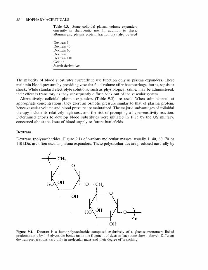
The majority of blood substitutes currently in use function only as plasma expanders. Thesemaintain blood pressure by providing vascular fluid volume after haemorrhage, burns, sepsis orshock. While standard electrolyte solutions, such as physiological saline, may be administered,their effect is transitory as they subsequently diffuse back out of the vascular system.
Alternatively, colloidal plasma expanders (Table 9.3) are used. When administered atappropriate concentrations, they exert an osmotic pressure similar to that of plasma protein,hence vascular volume and blood pressure are maintained. The major disadvantages of colloidaltherapy include its relatively high cost, and the risk of prompting a hypersensitivity reaction.Determined efforts to develop blood substitutes were initiated in 1985 by the US military,concerned about the issue of blood supply to future battlefields.
Dextrans
Dextrans (polysaccharides; Figure 9.1) of various molecular masses, usually 1, 40, 60, 70 or110 kDa, are often used as plasma expanders. These polysaccharides are produced naturally by
354 BIOPHARMACEUTICALS
Table 9.3. Some colloidal plasma volume expanderscurrently in therapeutic use. In addition to these,albumin and plasma protein fraction may also be used
Dextran 1Dextran 40Dextran 60Dextran 70Dextran 110GelatinStarch derivatives
Figure 9.1. Dextran is a homopolysaccharide composed exclusively of D-glucose monomers linkedpredominantly by 1–6 glycosidic bonds (as in the fragment of dextran backbone shown above). Differentdextran preparations vary only in molecular mass and their degree of branching
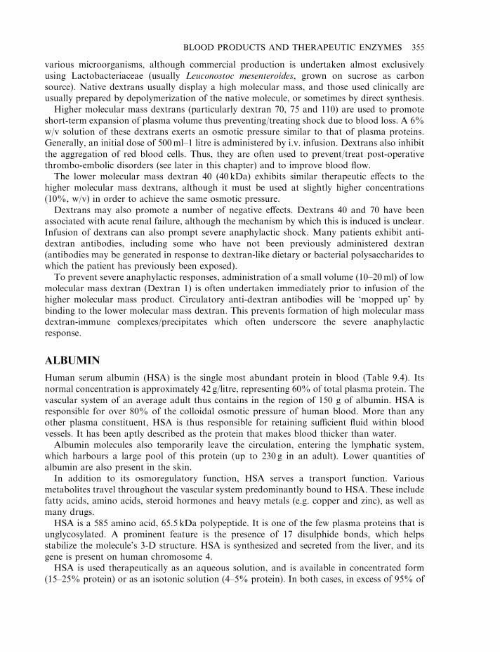
various microorganisms, although commercial production is undertaken almost exclusivelyusing Lactobacteriaceae (usually Leuconostoc mesenteroides, grown on sucrose as carbonsource). Native dextrans usually display a high molecular mass, and those used clinically areusually prepared by depolymerization of the native molecule, or sometimes by direct synthesis.
Higher molecular mass dextrans (particularly dextran 70, 75 and 110) are used to promoteshort-term expansion of plasma volume thus preventing/treating shock due to blood loss. A 6%w/v solution of these dextrans exerts an osmotic pressure similar to that of plasma proteins.Generally, an initial dose of 500ml–1 litre is administered by i.v. infusion. Dextrans also inhibitthe aggregation of red blood cells. Thus, they are often used to prevent/treat post-operativethrombo-embolic disorders (see later in this chapter) and to improve blood flow.
The lower molecular mass dextran 40 (40 kDa) exhibits similar therapeutic effects to thehigher molecular mass dextrans, although it must be used at slightly higher concentrations(10%, w/v) in order to achieve the same osmotic pressure.
Dextrans may also promote a number of negative effects. Dextrans 40 and 70 have beenassociated with acute renal failure, although the mechanism by which this is induced is unclear.Infusion of dextrans can also prompt severe anaphylactic shock. Many patients exhibit anti-dextran antibodies, including some who have not been previously administered dextran(antibodies may be generated in response to dextran-like dietary or bacterial polysaccharides towhich the patient has previously been exposed).
To prevent severe anaphylactic responses, administration of a small volume (10–20ml) of lowmolecular mass dextran (Dextran 1) is often undertaken immediately prior to infusion of thehigher molecular mass product. Circulatory anti-dextran antibodies will be ‘mopped up’ bybinding to the lower molecular mass dextran. This prevents formation of high molecular massdextran-immune complexes/precipitates which often underscore the severe anaphylacticresponse.
ALBUMIN
Human serum albumin (HSA) is the single most abundant protein in blood (Table 9.4). Itsnormal concentration is approximately 42 g/litre, representing 60% of total plasma protein. Thevascular system of an average adult thus contains in the region of 150 g of albumin. HSA isresponsible for over 80% of the colloidal osmotic pressure of human blood. More than anyother plasma constituent, HSA is thus responsible for retaining sufficient fluid within bloodvessels. It has been aptly described as the protein that makes blood thicker than water.
Albumin molecules also temporarily leave the circulation, entering the lymphatic system,which harbours a large pool of this protein (up to 230 g in an adult). Lower quantities ofalbumin are also present in the skin.
In addition to its osmoregulatory function, HSA serves a transport function. Variousmetabolites travel throughout the vascular system predominantly bound to HSA. These includefatty acids, amino acids, steroid hormones and heavy metals (e.g. copper and zinc), as well asmany drugs.
HSA is a 585 amino acid, 65.5 kDa polypeptide. It is one of the few plasma proteins that isunglycosylated. A prominent feature is the presence of 17 disulphide bonds, which helpsstabilize the molecule’s 3-D structure. HSA is synthesized and secreted from the liver, and itsgene is present on human chromosome 4.
HSA is used therapeutically as an aqueous solution, and is available in concentrated form(15–25% protein) or as an isotonic solution (4–5% protein). In both cases, in excess of 95% of
BLOOD PRODUCTS AND THERAPEUTIC ENZYMES 355
the protein present is albumin. It can be prepared by fractionation from normal plasma orserum, or purified from placentas. The source material must first be screened for the presence ofindicator pathogens. After purification, a suitable stabilizer (often sodium caprylate) is added,but no preservative. The solution is then sterilized by filtration and aseptically filled into finalsterile containers. The relative heat stability of HSA allows a measure of subsequent heattreatment, which further reduces the risk of accidental transmission of viable pathogens(particularly viruses). This treatment normally entails heating the product to 608C for 10 h. It isthen normally incubated at 30–328C for a further 14 days and subsequently examined for anysigns of microbial growth.
HSA is used as a plasma expander in the treatment of haemorrhage, shock, burns andoedema, as well as being administered to some patients after surgery. For adults, an initialinfusion containing at least 25 g of albumin is used. The annual world demand for HSA exceeds300 tonnes, representing a market value of the order of $1 billion.
Despite screening of raw material and heat treatment of final product, HSA derived fromnative blood will sometimes (although rarely) harbour pathogens. rDNA technology provides away of overcoming such concerns and the HSA gene and cDNA have been expressed in a widevariety of microbial systems, including E. coli, Bacillus subtilis, Saccharomyces cerevisiae, Pichiapastoris and Aspergillus niger (its lack of glycosylation renders possible production of nativeHSA in prokaryotic as well as eukaryotic systems). However, HSA’s relatively large size, as wellas the presence of so many disulphide bonds, can complicate recombinant production of highlevels of correctly folded products in some production systems. The main stumbling block inreplacing native HSA with a recombinant version, however, is an economic one. Unlike mostbiopharmaceuticals, HSA can be produced in large quantities and inexpensively by directextraction from its native source. Native HSA currently sells at $2–3/g. Although it can beguaranteed blood pathogen-free, recombinant HSA products will find it difficult to competewith this price.
356 BIOPHARMACEUTICALS
Table 9.4. The major plasma proteins of known function found in human blood
Protein
Normal plasmaconcentration
(g/l)
Molecularmass(kDa) Function
Albumin 35–45 66.5 Osmoregulation transportRetinol-binding protein 0.03–0.06 21 Retinol transportThyroxine binding globulin 0.01–0.02 58 Binds/transports thyroxineTranscortin 0.03–0.04 52 Cortisol and corticosterone
transportCaeruloplasmin 0.1–0.6 151 Copper transportHaptoglobinType 1-1Type 2-1Type 2-2
1.0–2.21.6–3.01.2–2.6
100200400
Binds and helps conservehaemoglobin
Transferrin 2.0–3.2 76.5 Iron transportHaemopexin 0.5–1.0 57 Binds haem destined for
disposalb2-microglobulin 0.002 11.8 Associated with HLA
histocompatibility antigeng-Globulins 7.0–15.0 150 AntibodiesTransthyretin 0.1–0.4 55 Binds thyroxine
Gelatin
Gelatin is produced by partial acid or partial alkaline hydrolysis of animal collagen. It has awide variety of therapeutic and pharmaceutical uses. It is often used in the manufacture of hardand soft capsule shells, suppositories and tablets, and is sometimes used as a ‘sponge’ duringsurgical procedures, as it can absorb many times its own weight of blood.
A 4% solution of gelatin (often modified, i.e. succinylated gelatin) is also used as a plasmaexpander. For this application, 500ml–1 l of the sterile solution is infused slowly. In rareoccurrences, infusion (particularly rapid infusion) of the gelatin solution has been known toinitiate hypersensitivity reactions. After its infusion, gelatin is excreted relatively quickly andmostly via the urine.
Oxygen-carrying blood substitutes
Thus far, the only molecules that have found even a limited clinical application as an oxygen-carrying blood substitute are the fluorocarbons and haemoglobin.
Fluosol is a fluorocarbon emulsion of perfluorodecalin and perfluorotripropylamine. Suchfluorocarbon emulsions can absorb, transport and release both oxygen and carbon dioxide.Although fluosol failed to show efficacy in the treatment of life-threatening anaemia, it is used asan effective oxygen carrier during coronary angioplasty procedures. A 20% emulsion isnormally used, and can be administered regardless of blood type. It also will be free of anyblood-borne pathogens, and is stable for years when stored at room temperature.
Haemoglobin displays the property of reversible oxygenation and, hence, is being investigatedas a blood substitute. Human haemoglobin is a tetramer of molecular mass 64 kDa. It consistsof two a-chains (141 amino acids) and two b-chains (146 amino acids), with each subunitdisplaying an associated haem prosthetic group. The interaction of the oxygen-binding haemmoiety and the globulin constituent is important, as free haem is toxic and the apoglobin isinsoluble.
Because of its structural characteristics, haemoglobin displays a non-linear oxygen saturationcurve, with maximal oxygen saturation occurring in arterial blood. At tissue sites, the oxygenpartial pressure is lower and much of the haemoglobin-bound oxygen is released. This process isaided in vivo by the effector molecule 2,3-diphosphoglycerate (2,3-DPG), which binds todeoxyhaemoglobin (in the presence of 2,3-DPG and at normal tissue oxygen partial pressures,over 20% of the bound oxygen is released from haemoglobin; in the absence of 2,3-DPG, thisvalue falls to 2–4%). Native haemoglobin, when extracted from erythrocytes, is less attractive asa tissue oxygen delivery system, as free haemoglobin no longer binds 2,3-DPG. Furthermore,outside the erythrocyte (in which haemoglobin is present at very high concentrations),haemoglobin tends to disassociate into dimers. Attempts have been made to stabilize freehaemoglobin and to modify it in order to reduce its oxygen affinity.
Pyridoxal 5’-phosphate (a derivative of pyridoxal; vitamin B6) is similar in size and charge to2,3-DPG. Covalent attachment of pyridoxal 5’-phosphate reduces the oxygen affinity of thehaemoglobin molecule. Covalent attachment of benzene isothiocyanates to the amino termini ofthe four haemoglobin polypeptide chains, also yields derivatives which display lower oxygenaffinity. These may prove worthy of clinical investigation.
In addition to altered oxygen-binding characteristics, free haemoglobin in plasmadisassociates rapidly into ab dimers, which are in turn rapidly oxidized and cleared by thekidneys. Indeed, high plasma concentrations can result in kidney toxicity. Development of a
BLOOD PRODUCTS AND THERAPEUTIC ENZYMES 357
clinically useful modified haemoglobin would have to circumvent this, probably bypolymerization or micro-encapsulation of the molecules prior to their administration.
Haemoglobin conjugated to additional high molecular mass substances, such as dextrin andpolyethylene glycol, are also generating clinical interest. When compared to nativehaemoglobin, such high molecular mass derivatives often display prolonged vascular retention(i.e. prolonged useful half-life), reduced antigenicity (if animal haemoglobin is used) and potentplasma volume expansion properties.
Polymerized haemoglobins (usually formed by crosslinking with gluteraldehyde) have alsobeen generated. The aim again is to prolong product half-life by preventing disassociation intodimers or monomers. Additionally, such products often display increased thermal stability,facilitating a subsequent viral-inactivation heat step.
In addition to chemical crosslinking, recombinant DNA technology has been used to generatestable intact haemoglobin variants. ‘Dialpha’ haemoglobin, for example, is an engineeredmolecule in which two a-chains are fused together, head to tail. Co-expression in E. coli alongwith the b-gene results in the formation of a working, stable haemoglobin analogue.Haemoglobin molecules can be obtained from whole blood/red cell concentrates. Humanhaemoglobin a and b genes have been expressed in a wide variety of prokaryotic and eukaryoticsystems, including transgenic plants and animals.
Although research continues in an effort to develop an effective haemoglobin-based red bloodcell substitute, no suitable candidate has yet been developed that has gained widespread clinicalacceptance.
HAEMOSTASIS
Blood plays various vital roles within the body and it is not surprising that a number ofprocesses have evolved capable of effectively maintaining haemostasis— the rapid arrest ofblood loss upon vascular damage, in order to maintain a relatively constant blood volume. Inhumans, three main mechanisms underline the haemostatic process:
. The congregation and clumping of blood platelets at the site of vascular injury, thuseffectively plugging the site of blood leakage.
. Localized constriction of the blood vessel, which minimizes further blood flow through thearea.
. Induction of the blood coagulation cascade. This culminates in the conversion of a solubleserum protein, fibrinogen, into insoluble fibrin. Fibrin monomers then aggregate at the site ofdamage, thus forming a clot (thrombus), which seals it off. These mechanisms are effective indealing with small vessel injuries, e.g. in capillaries and arterioles, although they areineffective when the damage relates to large veins/arteries.
The coagulation pathway
The process of blood coagulation is dependent on a large number of blood clotting factors,which act in a sequential manner. At least 12 distinct factors participate in the coagulationcascade, along with several macromolecular co-factors. The clotting factors are all designated byRoman numerals (Table 9.5) and, with the exception of factor IV, all are proteins. Most factorsare proteolytic zymogens, which become sequentially activated. An activated factor is indicatedby inclusion of a subscript ‘a’ (e.g. factor XIIa¼activated factor XII).
358 BIOPHARMACEUTICALS
Although the final steps of the blood clotting cascade are identical, the initial steps can occurvia two distinct pathways: the extrinsic and intrinsic pathways. Both pathways are initiatedwhen specific clotting proteins make contact with specific surface molecules exposed only upondamage to a blood vessel. Clotting occurs much more rapidly when initiated via the extrinsicpathway.
Two coagulation factors function uniquely in the extrinsic pathway: factor III (tissue factor)and factor VII. Tissue factor is an integral membrane protein present in a wide variety of tissuetypes (particularly lung and brain). This protein is exposed to blood constituents only uponrupture of a blood vessel, and it initiates the extrinsic coagulation cascade at the site of damageas described below.
Factor VII contains a number of g-carboxyglutamate residues (as do factors II, IX and X),which play an essential role in facilitating their binding of Ca2+ ions. The initial events initiatingthe extrinsic pathway entail the interaction of factor VII with Ca2+ and tissue factor. In thisassociated form, factor VII becomes proteolytically active. It displays both binding affinity for,and catalytic activity against, factor X. It thus activates factor X by proteolytic processing, andfactor Xa, which initiates the terminal stages of clot formation, remains attached to the tissuefactor–Ca2+ complex at the site of damage. This ensures that clot formation only occurs at thepoint where it is needed (Figure 9.2).
BLOOD PRODUCTS AND THERAPEUTIC ENZYMES 359
Table 9.5. The coagulation factors which promote the blood clotting process. Note that the factororiginally designated as VI was later shown to be factor Va
Factornumber Common name
Pathway in whichit functions Function
I Fibrinogen Both Forms structural basis of clot after itsconversion to fibrin
II Prothrombin Both Precursor of thrombin, whichactivates factors I, V, VII, VIII andXIII
III Tissue factor(thromboplastin)
Extrinsic Accessory tissue protein which initi-ates extrinsic pathway
IV Calcium ions Both Required for activation of factor XIIIand stabilizes some factors
V Proaccelerin Both Accessory protein, enhances rate ofactivation of X
VII Proconvertin Extrinsic Precursor of convertin (VIIa) whichactivates X (extrinsic system)
VIII Anti-haemophilic factor Intrinsic Accessory protein, enhancesactivation of X (intrinsic system)
IX Christmas factor Intrinsic Activated IX directly activates X(intrinsic system)
X Stuart factor Both Activated form (Xa) convertsprothrombin to thrombin
XI Plasma thromboplastinantecedent
Intrinsic Activated form (XIa) serves to acti-vate IX
XII Hageman factor Intrinsic Activated by surface contact or thekallikrenin system. XIIa helpsinitiate intrinsic system
XIII Fibrin-stabilizing factor Both Activated form cross-links fibrin,forming a hard clot
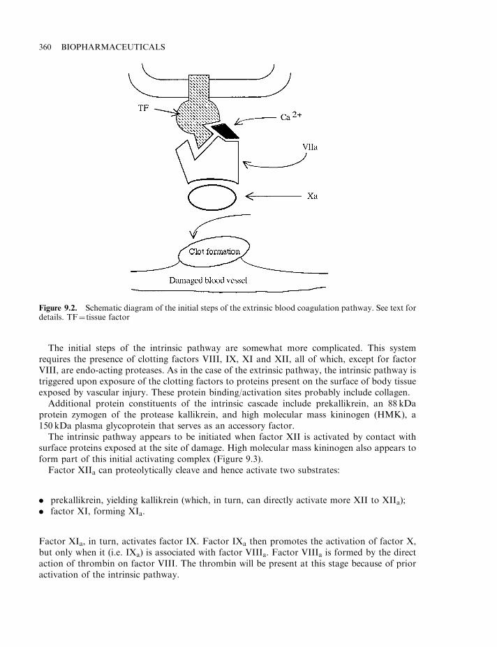
The initial steps of the intrinsic pathway are somewhat more complicated. This systemrequires the presence of clotting factors VIII, IX, XI and XII, all of which, except for factorVIII, are endo-acting proteases. As in the case of the extrinsic pathway, the intrinsic pathway istriggered upon exposure of the clotting factors to proteins present on the surface of body tissueexposed by vascular injury. These protein binding/activation sites probably include collagen.
Additional protein constituents of the intrinsic cascade include prekallikrein, an 88 kDaprotein zymogen of the protease kallikrein, and high molecular mass kininogen (HMK), a150 kDa plasma glycoprotein that serves as an accessory factor.
The intrinsic pathway appears to be initiated when factor XII is activated by contact withsurface proteins exposed at the site of damage. High molecular mass kininogen also appears toform part of this initial activating complex (Figure 9.3).
Factor XIIa can proteolytically cleave and hence activate two substrates:
. prekallikrein, yielding kallikrein (which, in turn, can directly activate more XII to XIIa);
. factor XI, forming XIa.
Factor XIa, in turn, activates factor IX. Factor IXa then promotes the activation of factor X,but only when it (i.e. IXa) is associated with factor VIIIa. Factor VIIIa is formed by the directaction of thrombin on factor VIII. The thrombin will be present at this stage because of prioractivation of the intrinsic pathway.
360 BIOPHARMACEUTICALS
Figure 9.2. Schematic diagram of the initial steps of the extrinsic blood coagulation pathway. See text fordetails. TF¼ tissue factor
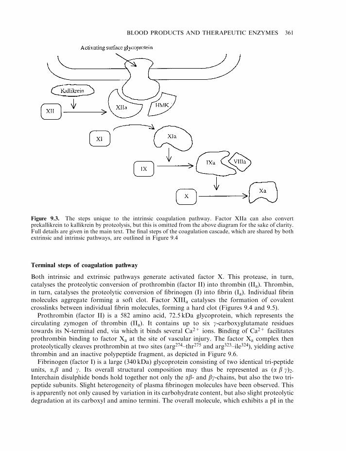
Terminal steps of coagulation pathway
Both intrinsic and extrinsic pathways generate activated factor X. This protease, in turn,catalyses the proteolytic conversion of prothrombin (factor II) into thrombin (IIa). Thrombin,in turn, catalyses the proteolytic conversion of fibrinogen (I) into fibrin (Ia). Individual fibrinmolecules aggregate forming a soft clot. Factor XIIIa catalyses the formation of covalentcrosslinks between individual fibrin molecules, forming a hard clot (Figures 9.4 and 9.5).
Prothrombin (factor II) is a 582 amino acid, 72.5 kDa glycoprotein, which represents thecirculating zymogen of thrombin (IIa). It contains up to six g-carboxyglutamate residuestowards its N-terminal end, via which it binds several Ca2+ ions. Binding of Ca2+ facilitatesprothrombin binding to factor Xa at the site of vascular injury. The factor Xa complex thenproteolytically cleaves prothrombin at two sites (arg274–thr275 and arg323–ile324), yielding activethrombin and an inactive polypeptide fragment, as depicted in Figure 9.6.
Fibrinogen (factor I) is a large (340 kDa) glycoprotein consisting of two identical tri-peptideunits, a,b and g. Its overall structural composition may thus be represented as (a b g)2.Interchain disulphide bonds hold together not only the ab- and bg-chains, but also the two tri-peptide subunits. Slight heterogeneity of plasma fibrinogen molecules have been observed. Thisis apparently not only caused by variation in its carbohydrate content, but also slight proteolyticdegradation at its carboxyl and amino termini. The overall molecule, which exhibits a pI in the
BLOOD PRODUCTS AND THERAPEUTIC ENZYMES 361
Figure 9.3. The steps unique to the intrinsic coagulation pathway. Factor XIIa can also convertprekallikrein to kallikrein by proteolysis, but this is omitted from the above diagram for the sake of clarity.Full details are given in the main text. The final steps of the coagulation cascade, which are shared by bothextrinsic and intrinsic pathways, are outlined in Figure 9.4
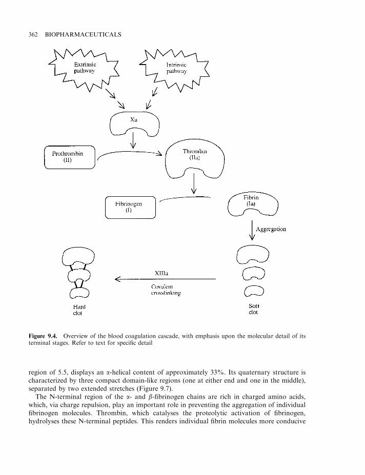
region of 5.5, displays an a-helical content of approximately 33%. Its quaternary structure ischaracterized by three compact domain-like regions (one at either end and one in the middle),separated by two extended stretches (Figure 9.7).
The N-terminal region of the a- and b-fibrinogen chains are rich in charged amino acids,which, via charge repulsion, play an important role in preventing the aggregation of individualfibrinogen molecules. Thrombin, which catalyses the proteolytic activation of fibrinogen,hydrolyses these N-terminal peptides. This renders individual fibrin molecules more conducive
362 BIOPHARMACEUTICALS
Figure 9.4. Overview of the blood coagulation cascade, with emphasis upon the molecular detail of itsterminal stages. Refer to text for specific detail
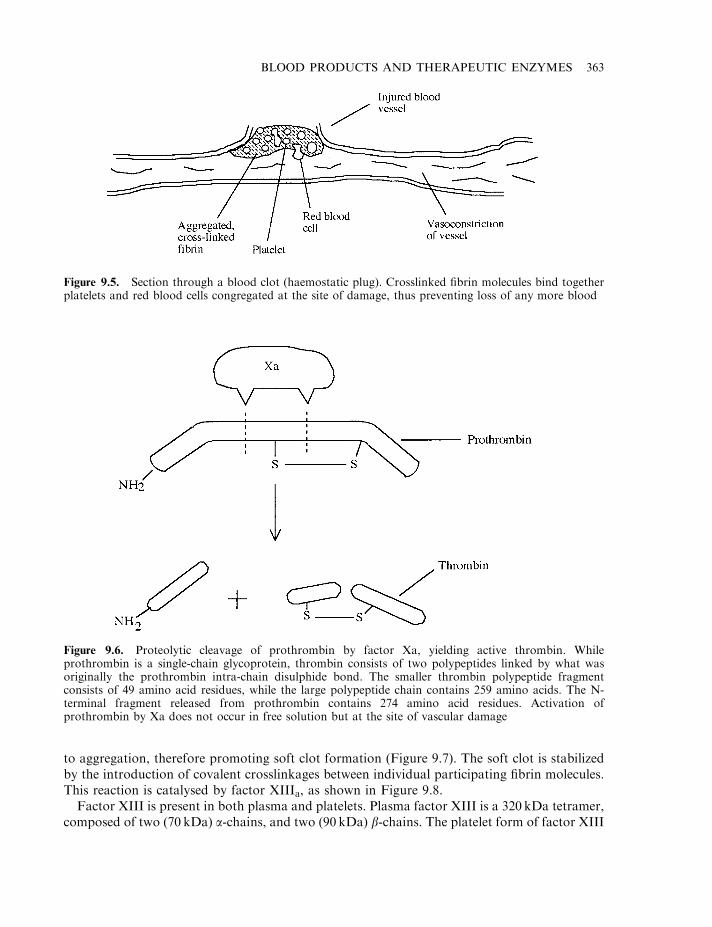
to aggregation, therefore promoting soft clot formation (Figure 9.7). The soft clot is stabilizedby the introduction of covalent crosslinkages between individual participating fibrin molecules.This reaction is catalysed by factor XIIIa, as shown in Figure 9.8.
Factor XIII is present in both plasma and platelets. Plasma factor XIII is a 320 kDa tetramer,composed of two (70 kDa) a-chains, and two (90 kDa) b-chains. The platelet form of factor XIII
BLOOD PRODUCTS AND THERAPEUTIC ENZYMES 363
Figure 9.5. Section through a blood clot (haemostatic plug). Crosslinked fibrin molecules bind togetherplatelets and red blood cells congregated at the site of damage, thus preventing loss of any more blood
Figure 9.6. Proteolytic cleavage of prothrombin by factor Xa, yielding active thrombin. Whileprothrombin is a single-chain glycoprotein, thrombin consists of two polypeptides linked by what wasoriginally the prothrombin intra-chain disulphide bond. The smaller thrombin polypeptide fragmentconsists of 49 amino acid residues, while the large polypeptide chain contains 259 amino acids. The N-terminal fragment released from prothrombin contains 274 amino acid residues. Activation ofprothrombin by Xa does not occur in free solution but at the site of vascular damage
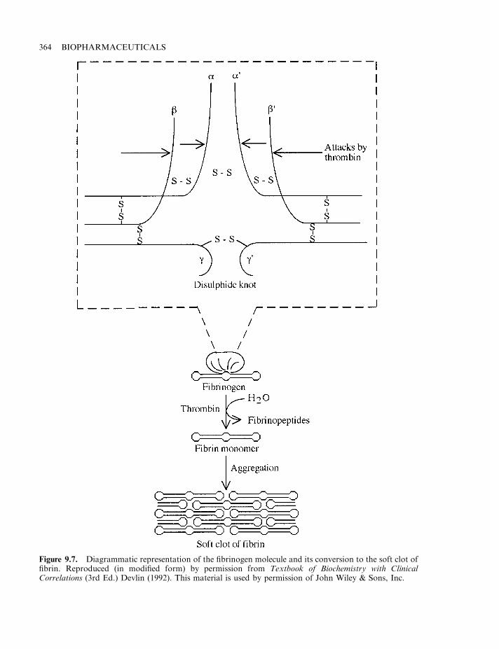
364 BIOPHARMACEUTICALS
Figure 9.7. Diagrammatic representation of the fibrinogen molecule and its conversion to the soft clot offibrin. Reproduced (in modified form) by permission from Textbook of Biochemistry with ClinicalCorrelations (3rd Ed.) Devlin (1992). This material is used by permission of John Wiley & Sons, Inc.
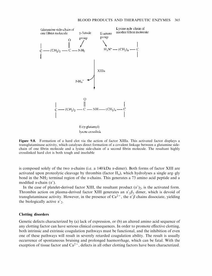
is composed solely of the two a-chains (i.e. a 140 kDa a-dimer). Both forms of factor XIII areactivated upon proteolytic cleavage by thrombin (factor IIa), which hydrolyses a single arg–glybond in the NH2 terminal region of the a-chains. This generates a 73 amino acid peptide and amodified a-chain (a 0).
In the case of platelet-derived factor XIII, the resultant product (a 0)2, is the activated form.Thrombin action on plasma-derived factor XIII generates an a02b2 dimer, which is devoid oftransglutaminase activity. However, in the presence of Ca2+, the a 0b chains dissociate, yieldingthe biologically active a 0
2.
Clotting disorders
Genetic defects characterized by (a) lack of expression, or (b) an altered amino acid sequence ofany clotting factor can have serious clinical consequences. In order to promote effective clotting,both intrinsic and extrinsic coagulation pathways must be functional, and the inhibition of evenone of these pathways will result in severely retarded coagulation ability. The result is usuallyoccurrence of spontaneous bruising and prolonged haemorrhage, which can be fatal. With theexception of tissue factor and Ca2+, defects in all other clotting factors have been characterized.
BLOOD PRODUCTS AND THERAPEUTIC ENZYMES 365
Figure 9.8. Formation of a hard clot via the action of factor XIIIa. This activated factor displays atransglutaminase activity, which catalyses direct formation of a covalent linkage between a glutamine side-chain of one fibrin molecule and a lysine side-chain of a second fibrin molecule. The resultant highlycrosslinked hard clot is both tough and insoluble
Up to 90% of these, however, relate to a deficiency in factor VIII, while much of the remainderis due to a deficiency in factor IX.
Such clotting disorders are generally treated by ongoing administration of whole blood or,more usually, concentrates of the relevant coagulation factor purified from whole blood. Thisentails significant risk of accidental transmission of blood-borne disease, particularly hepatitisand AIDS. In turn, this has hastened the development of blood coagulation factors produced bygenetic engineering, several of which are now approved for general medical use (Table 9.6).
Factor VIII and haemophilia
Haemophilia A (classical haemophilia, often simply termed haemophilia) is an X-linkedrecessive disorder which is caused by a deficiency of factor VIII. Von Willebrand’s disease is arelated disorder, also caused by a defect in the factor VIII complex, as discussed below.
Intact factor VIII, as usually purified from the blood, consists of two distinct gene products:factor VIII and (multiple copies of) von Willebrand’s factor (vWF; Figure 9.9). This complexdisplays a molecular mass of 1–2 million Da, of which up to 15% is carbohydrate. The fullyintact factor VIII complex is required to enhance the rate of activation of factor IX of theintrinsic system.
The factor VIII polypeptide portion of the factor VIII complex is coded for by an unusuallylong gene (289 kb). Transcription and processing of the mRNA generates a shorter, maturemRNA which codes for a 300 kDa protein. Upon its synthesis, this polypeptide precursor issubsequently proteolytically processed, with removal of a significant portion of its mid-region.This yields two fragments: an amino terminal 90 kDa polypeptide and an 80 kDa carboxylterminal polypeptide. These associate non-covalently (a process requiring Ca2+ ions) to producemature factor VIII (sometimes called factor VIIIc). This mature factor VIII is then released intothe plasma, where it associates with multiple copies of vWF, forming the biologically activefactor VIII complex. vWF stabilizes factor VIII in plasma (particularly against proteolyticdegradation). It also can associate with platelets at the site of vascular damage and, hence,presumably plays a role in docking the factor VIII complex in an appropriate position where itcan participate in the coagulation cascade.
Persons suffering from haemophilia A exhibit markedly reduced levels of (or the completeabsence of) factor VIII complex in their blood. This is due to the lack of production of factorVIIIc. Persons suffering from (the rarer) von Willebrand’s disease lack both components of
366 BIOPHARMACEUTICALS
Table 9.6. Recombinant blood coagulation factors which have been approved for the management ofcoagulation disorders
Product (trade name) Company Indication
Bioclate (rhFactor VIII) Centeon Haemophilia ABenefix (rhFactor IX) Genetics Institute Haemophilia BKogenate (rhFactor VIII) Bayer Haemophilia AHelixate NexGen (rhFactor VIII) Bayer Haemophilia ANovoSeven (rhFactor VIIa) Novo-Nordisk Some forms of haemophiliaRecombinate (rhFactor VIII) Baxter Healthcare/Genetics Institute Haemophilia AReFacto (B-domain deletedrhFactor VIII)
Genetics Institute Haemophilia A
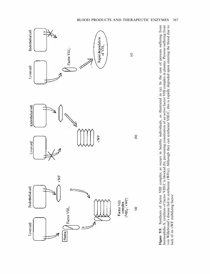
BLOOD PRODUCTS AND THERAPEUTIC ENZYMES 367
Figure
9.9.
Synthesis
offactorVIIIcomplexasoccurs
inhealthyindividuals,asillustrated
in(a).
Inthecase
ofpersonssufferingfrom
haem
ophilia
A,synthesisoffactorVIII:Cisblocked
(b),preventingconstitutionofanactivefactorVIIIcomplexin
plasm
a.Personssufferingfrom
vonWillebrand’sdisease
failto
synthesizevWF(c).Althoughthey
cansynthesizeVIII:C,thisisrapidly
degraded
uponenteringtheblooddueto
lack
ofitsvWF
stabilizingfactor
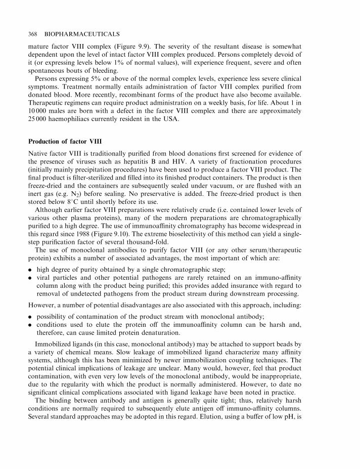
mature factor VIII complex (Figure 9.9). The severity of the resultant disease is somewhatdependent upon the level of intact factor VIII complex produced. Persons completely devoid ofit (or expressing levels below 1% of normal values), will experience frequent, severe and oftenspontaneous bouts of bleeding.
Persons expressing 5% or above of the normal complex levels, experience less severe clinicalsymptoms. Treatment normally entails administration of factor VIII complex purified fromdonated blood. More recently, recombinant forms of the product have also become available.Therapeutic regimens can require product administration on a weekly basis, for life. About 1 in10 000 males are born with a defect in the factor VIII complex and there are approximately25 000 haemophiliacs currently resident in the USA.
Production of factor VIII
Native factor VIII is traditionally purified from blood donations first screened for evidence ofthe presence of viruses such as hepatitis B and HIV. A variety of fractionation procedures(initially mainly precipitation procedures) have been used to produce a factor VIII product. Thefinal product is filter-sterilized and filled into its finished product containers. The product is thenfreeze-dried and the containers are subsequently sealed under vacuum, or are flushed with aninert gas (e.g. N2) before sealing. No preservative is added. The freeze-dried product is thenstored below 88C until shortly before its use.
Although earlier factor VIII preparations were relatively crude (i.e. contained lower levels ofvarious other plasma proteins), many of the modern preparations are chromatographicallypurified to a high degree. The use of immunoaffinity chromatography has become widespread inthis regard since 1988 (Figure 9.10). The extreme bioselectivity of this method can yield a single-step purification factor of several thousand-fold.
The use of monoclonal antibodies to purify factor VIII (or any other serum/therapeuticprotein) exhibits a number of associated advantages, the most important of which are:
. high degree of purity obtained by a single chromatographic step;
. viral particles and other potential pathogens are rarely retained on an immuno-affinitycolumn along with the product being purified; this provides added insurance with regard toremoval of undetected pathogens from the product stream during downstream processing.
However, a number of potential disadvantages are also associated with this approach, including:
. possibility of contamination of the product stream with monoclonal antibody;
. conditions used to elute the protein off the immunoaffinity column can be harsh and,therefore, can cause limited protein denaturation.
Immobilized ligands (in this case, monoclonal antibody) may be attached to support beads bya variety of chemical means. Slow leakage of immobilized ligand characterize many affinitysystems, although this has been minimized by newer immobilization coupling techniques. Thepotential clinical implications of leakage are unclear. Many would, however, feel that productcontamination, with even very low levels of the monoclonal antibody, would be inappropriate,due to the regularity with which the product is normally administered. However, to date nosignificant clinical complications associated with ligand leakage have been noted in practice.
The binding between antibody and antigen is generally quite tight; thus, relatively harshconditions are normally required to subsequently elute antigen off immuno-affinity columns.Several standard approaches may be adopted in this regard. Elution, using a buffer of low pH, is
368 BIOPHARMACEUTICALS
often attempted, followed by rapid neutralization of the eluate to pH 7–8. Elution, using buffersof high pH, can also sometimes be successfully employed. In many instances, even harsherelution conditions may be required. These can include the use of chaotropic agents ordetergents. Such conditions can obviously partially/completely denature the protein beingeluted. Although many clotting factors are very stable when exposed to various denaturinginfluences, factor VIII is quite labile. Thus, the use of a monoclonal antibody with high, but notextreme, avidity for factor VIII would likely yield the most successful results.
Although fractionation can reduce very significantly the likelihood of pathogen transmission,it cannot entirely eliminate this possibility. Blood-derived factor VIII products, including thoseprepared by immunoaffinity chromatography, generally undergo further processing stepsdesigned to remove/inactivate any virus present. The raw material is often heated for up to 10 hat 608C or treated with solvent or dilute detergent prior to chromatography. Recombinantfactor VIII is also often treated with dilute detergent in an effort to inactivate any viral particlespotentially present.
Production of recombinant factor VIII (Table 9.6) has ended dependence on blood as the onlysource of this product and eliminated the possibility of transmitting blood-borne diseasesspecifically derived from infected blood. In the past, over 60% of haemophiliacs were likely tobe accidently infected via contaminated products at some stage of their lives.
Several companies have expressed the cDNA coding for human factor VIII:C in a variety ofeukaryotic production systems (human VIII:C contains 25 potential glycosylation sites).
BLOOD PRODUCTS AND THERAPEUTIC ENZYMES 369
Figure 9.10. Purification of factor VIII complex using immunoaffinity chromatography. The immobilizedanti-factor VIII antibody is of mouse origin. Antibodies raised against specific epitopes on both the VIII:Cand vWF components have both been successfully used. Industrial-scale columns would often exhibit a bedvolume of several litres. Note that the different elements in this diagram are not drawn to the correct scalerelative to each other
Chinese hamster ovary (CHO) cells and baby hamster kidney (BHK) cell lines have been mostcommonly used, in addition to other cell lines, such as various mouse carcinoma cell lines. Therecombinant factor VIII product generally contains only VIII:C (i.e. is devoid of vWF).However, both clinical and pre-clinical studies have shown that administration of this productto patients suffering from haemophilia A is equally as effective as administering blood-derivedfactor VIII complex. The recombinant VIII:C product appears to bind plasma vWF with equalaffinity to native VIII:C, upon its injection into the patient’s circulatory system. Animal andhuman pharmacokinetic data reveal no significant difference between the properties ofrecombinant and native products.
Slight variation in the glycosylation patterns of native and recombinant product were to beexpected, due to the differing production systems. While this has turned out to be the case, thusfar it has not proved to be of any clinical significance. Significant differences could have madethe recombinant product immunogenic. Bearing in mind that most recipients would receiveseveral thousand injections of the product over their lifetime, even a mildly immunogenicproduct would promote a significant immunological response.
Some patients, particularly those suffering from severe haemophilia A (i.e. those naturallyproducing little or no VIII:C) will mount an immune response against injected factor VIII:C,whatever its source.
The production of anti-factor VIII:C antibodies renders necessary administration of highertherapeutic doses of the product. In severe cases, the product may even become ineffective.Several approaches may be adopted in order to circumvent this problem. These include:
. Exchange transfusion of whole blood. This will transiently decrease circulating anti-factorVIII:C antibodies.
. Direct administration of factor Xa, thus bypassing the non-functional step in the coagulationcascade (Figures 9.3 and 9.4).
. Administration of high levels of a mixture of clotting factors II, VII, IX and X, which workseffectively in 50% of treated patients.
. Administration of factor VIIa, as discussed subsequently.
. Administration of porcine factor VIII, which may or may not cross-react with the antibodiesraised against human factor VIIIa (however, the immune system will soon begin to produceantibodies against the porcine clotting factor).
. Administration of factor VIII, with concurrent administration of immunosuppressive agents.
Due to the frequency of product administration, the purification procedure for recombinantfactor VIII:C must be particularly stringent. Unlike the situation pertaining when the product ispurified from human blood, any contaminant present in the final product will be non-humanand hence immunogenic. Sources of such contaminants would include:
. host cell proteins;
. animal cell culture medium;
. antibody leaked from the immunoaffinity column.
Emphasis is placed not only upon ensuring the absence of contaminant proteins, but also otherpotential contaminants, particularly DNA (host cell line-derived DNA could harbouroncogenes; Chapter 3).
Recombinant factor VIII is gaining an increasing market share of the factor VIII market.Researchers are also attempting to develop modified forms of VIII:C (by site-directedmutagenesis) which display additional desirable characteristics. Particularly attractive in this
370 BIOPHARMACEUTICALS
regard would be the development of a product exhibiting an extended circulatory half-life. Thiscould reduce the frequency of injections required by haemophilia A sufferers. However, anyalteration of the primary sequence of the molecule carries with it the strong possibility ofrendering the resultant mutant immunogenic.
Factors IX, VIIa and XIII
Individuals who display a deficiency of factor IX develop haemophilia B, also known asChristmas disease. Although its clinical consequences are very similar to that of a deficiency offactor VIII, its general incidence in the population is far lower. Persons suffering fromhaemophilia B are treated by i.v. administration of a concentrate of factor IX. This wastraditionally obtained by fractionation of human blood. Recombinant factor IX is now alsoproduced in genetically engineered CHO cells.
Factor IX obtained from blood donations is normally only partially pure. In addition tofactor IX, the product contains lower levels of factors II, VII and X, and has also been used totreat bleeding disorders caused by a lack of these factors.
Factor IX may also be purified by immuno-affinity chromatography, using immobilized anti-IX murine monoclonals. Purification to homogeneity is particularly important in the case ofrecombinant products. At least one monoclonal antibody has been raised that specifically bindsonly to factor IX, which contains pre-bound Ca2+ (i.e. the Ca2+-dependent conformation offactor IX). Immobilization of this antibody allowed the development of an immuno-affinitysystem in which factor IX binds to the column in the presence of a Ca2+-containing buffer.Subsequent elution is promoted simply by inclusion of a chelating agent (e.g. EDTA) in theelution buffer.
5–25% of individuals suffering from haemophilia A develop anti-factor VIII antibodies, while3–6% of haemophilia B sufferers develop anti-factor IX antibodies. This complicates treatmentof these conditions and, as mentioned previously, one approach to their treatment is directadministration of factor VIIa. The therapeutic rationale is that factor VIIa could directlyactivate the final common steps of the coagulation cascade, independantly of either factor VIIIor IX (Figure 9.2). Factor VIIa forms a complex with tissue factor which, in the presence ofphospholipids and Ca2+, activates factor X.
A recombinant form of factor VIIa (called ‘NovoSeven’ or ‘Eptacog a-activated’) is marketedby Novo-Nordisk. The recombinant molecule is produced in a BHK cell line, and the finalproduct differs only slightly (in its glycosylation pattern only) from the native molecule.
Purification entails use of an immunoaffinity column containing immobilized murine anti-factor VII antibody. It is initially produced as an unactivated, single chain 406 amino acidpolypeptide, which is subsequently proteolytically converted into the two-chain active factorVIIa complex. After sterilization by filtration, the final product is aseptically filled into its finalproduct containers and freeze-dried. The excipients present in the product include sodiumchloride, calcium chloride, polysorbate 80, mannitol and glycylglycine. When freeze-dried in thepresence of these stabilizing substances and stored under refrigerated conditions, the productdisplays a shelf-life of at least 2 years. It has proved effective in the treatment of serious bleedingevents in patients displaying anti-factor VIII or IX antibodies.
A (very rare) genetic deficiency in the production of factor XIII also results in impairedclotting efficacy in affected persons. In this case, covalent links which normally characterizetransformation of a soft clot into a hard clot are not formed. Factor XIII preparations, partiallypurified from human blood, are used to treat individuals with this condition.
BLOOD PRODUCTS AND THERAPEUTIC ENZYMES 371
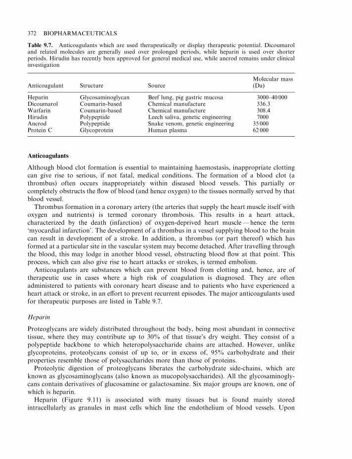
Anticoagulants
Although blood clot formation is essential to maintaining haemostasis, inappropriate clottingcan give rise to serious, if not fatal, medical conditions. The formation of a blood clot (athrombus) often occurs inappropriately within diseased blood vessels. This partially orcompletely obstructs the flow of blood (and hence oxygen) to the tissues normally served by thatblood vessel.
Thrombus formation in a coronary artery (the arteries that supply the heart muscle itself withoxygen and nutrients) is termed coronary thrombosis. This results in a heart attack,characterized by the death (infarction) of oxygen-deprived heart muscle—hence the term‘myocardial infarction’. The development of a thrombus in a vessel supplying blood to the braincan result in development of a stroke. In addition, a thrombus (or part thereof) which hasformed at a particular site in the vascular system may become detached. After travelling throughthe blood, this may lodge in another blood vessel, obstructing blood flow at that point. Thisprocess, which can also give rise to heart attacks or strokes, is termed embolism.
Anticoagulants are substances which can prevent blood from clotting and, hence, are oftherapeutic use in cases where a high risk of coagulation is diagnosed. They are oftenadministered to patients with coronary heart disease and to patients who have experienced aheart attack or stroke, in an effort to prevent recurrent episodes. The major anticoagulants usedfor therapeutic purposes are listed in Table 9.7.
Heparin
Proteoglycans are widely distributed throughout the body, being most abundant in connectivetissue, where they may contribute up to 30% of that tissue’s dry weight. They consist of apolypeptide backbone to which heteropolysaccharide chains are attached. However, unlikeglycoproteins, proteolycans consist of up to, or in excess of, 95% carbohydrate and theirproperties resemble those of polysaccharides more than those of proteins.
Proteolytic digestion of proteoglycans liberates the carbohydrate side-chains, which areknown as glycosaminoglycans (also known as mucopolysaccharides). All the glycosaminogly-cans contain derivatives of glucosamine or galactosamine. Six major groups are known, one ofwhich is heparin.
Heparin (Figure 9.11) is associated with many tissues but is found mainly storedintracellularly as granules in mast cells which line the endothelium of blood vessels. Upon
372 BIOPHARMACEUTICALS
Table 9.7. Anticoagulants which are used therapeutically or display therapeutic potential. Dicoumaroland related molecules are generally used over prolonged periods, while heparin is used over shorterperiods. Hirudin has recently been approved for general medical use, while ancrod remains under clinicalinvestigation
Anticoagulant Structure SourceMolecular mass(Da)
Heparin Glycosaminoglycan Beef lung, pig gastric mucosa 3000–40 000Dicoumarol Coumarin-based Chemical manufacture 336.3Warfarin Coumarin-based Chemical manufacture 308.4Hirudin Polypeptide Leech saliva, genetic engineering 7000Ancrod Polypeptide Snake venom, genetic engineering 35 000Protein C Glycoprotein Human plasma 62 000
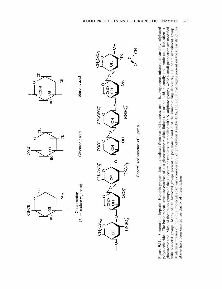
BLOOD PRODUCTS AND THERAPEUTIC ENZYMES 373
Figure
9.11.
Structure
ofheparin.Heparinpreparations,
asisolatedfrom
naturalsources,are
aheterogeneousmixture
ofvariably
sulphated
polysaccharides.Thebasicrepeatstructure
consistsofa
D-glucosamineresiduelinked
toauronic
acid,norm
ally
L-iduronic
acid,less
often
D-
glucuronicacid.Mostoftheaminogroupsoftheglucosamineresidues
are
modified
withN-sulphate
groups,whileasm
allproportionare
modified
withN-acetylgroups.
Manyofthehydroxylgroupspresentatpositions2and6oftheiduronic
ringalsocarryasulphate
substituentgroup.
Molecularmasses
ofindividualmoleculescanvary
considerably,often
between3and40kDa.Individualhydrogenspresentonthesugarstructures
abovehavebeenomittedforclarity
ofpresentation
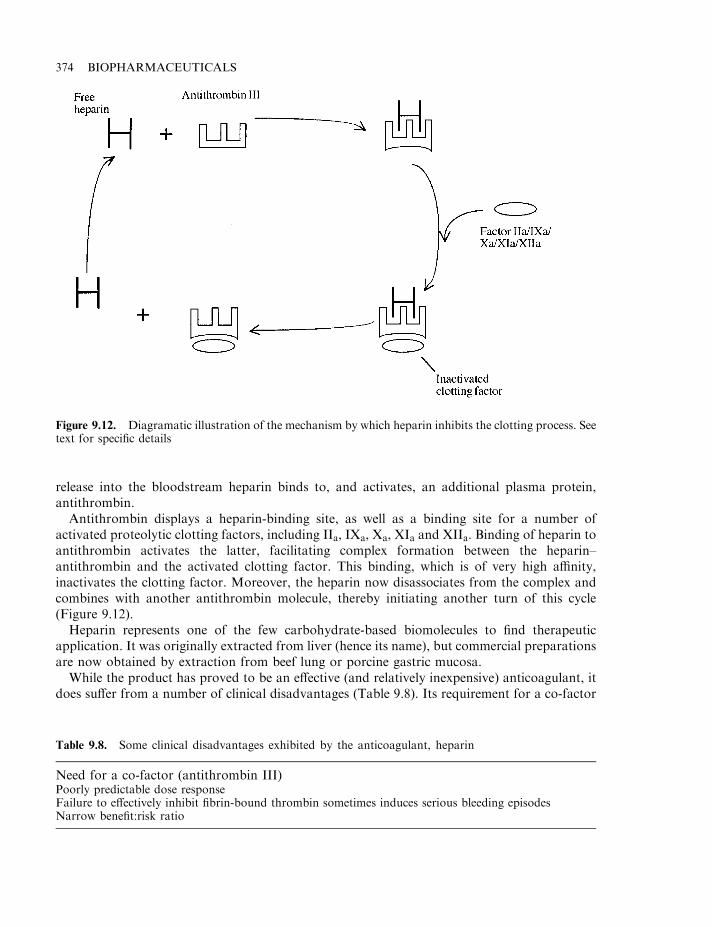
release into the bloodstream heparin binds to, and activates, an additional plasma protein,antithrombin.
Antithrombin displays a heparin-binding site, as well as a binding site for a number ofactivated proteolytic clotting factors, including IIa, IXa, Xa, XIa and XIIa. Binding of heparin toantithrombin activates the latter, facilitating complex formation between the heparin–antithrombin and the activated clotting factor. This binding, which is of very high affinity,inactivates the clotting factor. Moreover, the heparin now disassociates from the complex andcombines with another antithrombin molecule, thereby initiating another turn of this cycle(Figure 9.12).
Heparin represents one of the few carbohydrate-based biomolecules to find therapeuticapplication. It was originally extracted from liver (hence its name), but commercial preparationsare now obtained by extraction from beef lung or porcine gastric mucosa.
While the product has proved to be an effective (and relatively inexpensive) anticoagulant, itdoes suffer from a number of clinical disadvantages (Table 9.8). Its requirement for a co-factor
374 BIOPHARMACEUTICALS
Figure 9.12. Diagramatic illustration of the mechanism by which heparin inhibits the clotting process. Seetext for specific details
Table 9.8. Some clinical disadvantages exhibited by the anticoagulant, heparin
Need for a co-factor (antithrombin III)Poorly predictable dose responseFailure to effectively inhibit fibrin-bound thrombin sometimes induces serious bleeding episodesNarrow benefit:risk ratio
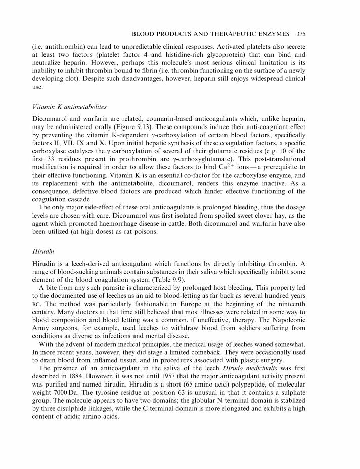
(i.e. antithrombin) can lead to unpredictable clinical responses. Activated platelets also secreteat least two factors (platelet factor 4 and histidine-rich glycoprotein) that can bind andneutralize heparin. However, perhaps this molecule’s most serious clinical limitation is itsinability to inhibit thrombin bound to fibrin (i.e. thrombin functioning on the surface of a newlydeveloping clot). Despite such disadvantages, however, heparin still enjoys widespread clinicaluse.
Vitamin K antimetabolites
Dicoumarol and warfarin are related, coumarin-based anticoagulants which, unlike heparin,may be administered orally (Figure 9.13). These compounds induce their anti-coagulant effectby preventing the vitamin K-dependent g-carboxylation of certain blood factors, specificallyfactors II, VII, IX and X. Upon initial hepatic synthesis of these coagulation factors, a specificcarboxylase catalyses the g carboxylation of several of their glutamate residues (e.g. 10 of thefirst 33 residues present in prothrombin are g-carboxyglutamate). This post-translationalmodification is required in order to allow these factors to bind Ca2+ ions—a prerequisite totheir effective functioning. Vitamin K is an essential co-factor for the carboxylase enzyme, andits replacement with the antimetabolite, dicoumarol, renders this enzyme inactive. As aconsequence, defective blood factors are produced which hinder effective functioning of thecoagulation cascade.
The only major side-effect of these oral anticoagulants is prolonged bleeding, thus the dosagelevels are chosen with care. Dicoumarol was first isolated from spoiled sweet clover hay, as theagent which promoted haemorrhage disease in cattle. Both dicoumarol and warfarin have alsobeen utilized (at high doses) as rat poisons.
Hirudin
Hirudin is a leech-derived anticoagulant which functions by directly inhibiting thrombin. Arange of blood-sucking animals contain substances in their saliva which specifically inhibit someelement of the blood coagulation system (Table 9.9).
A bite from any such parasite is characterized by prolonged host bleeding. This property ledto the documented use of leeches as an aid to blood-letting as far back as several hundred yearsBC. The method was particularly fashionable in Europe at the beginning of the ninteenthcentury. Many doctors at that time still believed that most illnesses were related in some way toblood composition and blood letting was a common, if uneffective, therapy. The NapoleonicArmy surgeons, for example, used leeches to withdraw blood from soldiers suffering fromconditions as diverse as infections and mental disease.
With the advent of modern medical principles, the medical usage of leeches waned somewhat.In more recent years, however, they did stage a limited comeback. They were occasionally usedto drain blood from inflamed tissue, and in procedures associated with plastic surgery.
The presence of an anticoagulant in the saliva of the leech Hirudo medicinalis was firstdescribed in 1884. However, it was not until 1957 that the major anticoagulant activity presentwas purified and named hirudin. Hirudin is a short (65 amino acid) polypeptide, of molecularweight 7000Da. The tyrosine residue at position 63 is unusual in that it contains a sulphategroup. The molecule appears to have two domains; the globular N-terminal domain is stablizedby three disulphide linkages, while the C-terminal domain is more elongated and exhibits a highcontent of acidic amino acids.
BLOOD PRODUCTS AND THERAPEUTIC ENZYMES 375
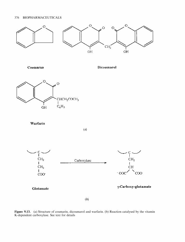
376 BIOPHARMACEUTICALS
Figure 9.13. (a) Structure of coumarin, dicoumarol and warfarin. (b) Reaction catalysed by the vitaminK-dependent carboxylase. See text for details
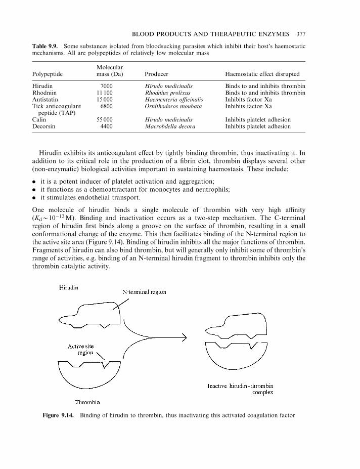
Hirudin exhibits its anticoagulant effect by tightly binding thrombin, thus inactivating it. Inaddition to its critical role in the production of a fibrin clot, thrombin displays several other(non-enzymatic) biological activities important in sustaining haemostasis. These include:
. it is a potent inducer of platelet activation and aggregation;
. it functions as a chemoattractant for monocytes and neutrophils;
. it stimulates endothelial transport.
One molecule of hirudin binds a single molecule of thrombin with very high affinity(Kd*10�12M). Binding and inactivation occurs as a two-step mechanism. The C-terminalregion of hirudin first binds along a groove on the surface of thrombin, resulting in a smallconformational change of the enzyme. This then facilitates binding of the N-terminal region tothe active site area (Figure 9.14). Binding of hirudin inhibits all the major functions of thrombin.Fragments of hirudin can also bind thrombin, but will generally only inhibit some of thrombin’srange of activities, e.g. binding of an N-terminal hirudin fragment to thrombin inhibits only thethrombin catalytic activity.
BLOOD PRODUCTS AND THERAPEUTIC ENZYMES 377
Table 9.9. Some substances isolated from bloodsucking parasites which inhibit their host’s haemostaticmechanisms. All are polypeptides of relatively low molecular mass
PolypeptideMolecularmass (Da) Producer Haemostatic effect disrupted
Hirudin 7000 Hirudo medicinalis Binds to and inhibits thrombinRhodniin 11 100 Rhodnius prolixus Binds to and inhibits thrombinAntistatin 15 000 Haementeria officinalis Inhibits factor XaTick anticoagulantpeptide (TAP)
6800 Ornithodoros moubata Inhibits factor Xa
Calin 55 000 Hirudo medicinalis Inhibits platelet adhesionDecorsin 4400 Macrobdella decora Inhibits platelet adhesion
Figure 9.14. Binding of hirudin to thrombin, thus inactivating this activated coagulation factor
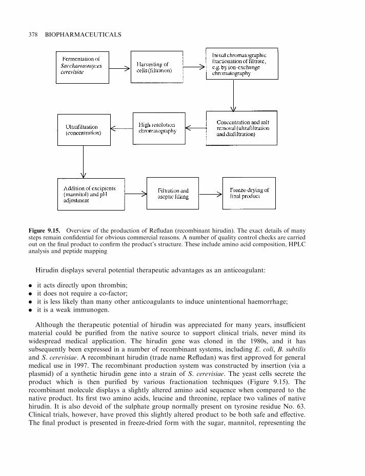
Hirudin displays several potential therapeutic advantages as an anticoagulant:
. it acts directly upon thrombin;
. it does not require a co-factor;
. it is less likely than many other anticoagulants to induce unintentional haemorrhage;
. it is a weak immunogen.
Although the therapeutic potential of hirudin was appreciated for many years, insufficientmaterial could be purified from the native source to support clinical trials, never mind itswidespread medical application. The hirudin gene was cloned in the 1980s, and it hassubsequently been expressed in a number of recombinant systems, including E. coli, B. subtilisand S. cerevisiae. A recombinant hirudin (trade name Refludan) was first approved for generalmedical use in 1997. The recombinant production system was constructed by insertion (via aplasmid) of a synthetic hirudin gene into a strain of S. cerevisiae. The yeast cells secrete theproduct which is then purified by various fractionation techniques (Figure 9.15). Therecombinant molecule displays a slightly altered amino acid sequence when compared to thenative product. Its first two amino acids, leucine and threonine, replace two valines of nativehirudin. It is also devoid of the sulphate group normally present on tyrosine residue No. 63.Clinical trials, however, have proved this slightly altered product to be both safe and effective.The final product is presented in freeze-dried form with the sugar, mannitol, representing the
378 BIOPHARMACEUTICALS
Figure 9.15. Overview of the production of Refludan (recombinant hirudin). The exact details of manysteps remain confidential for obvious commercial reasons. A number of quality control checks are carriedout on the final product to confirm the product’s structure. These include amino acid composition, HPLCanalysis and peptide mapping
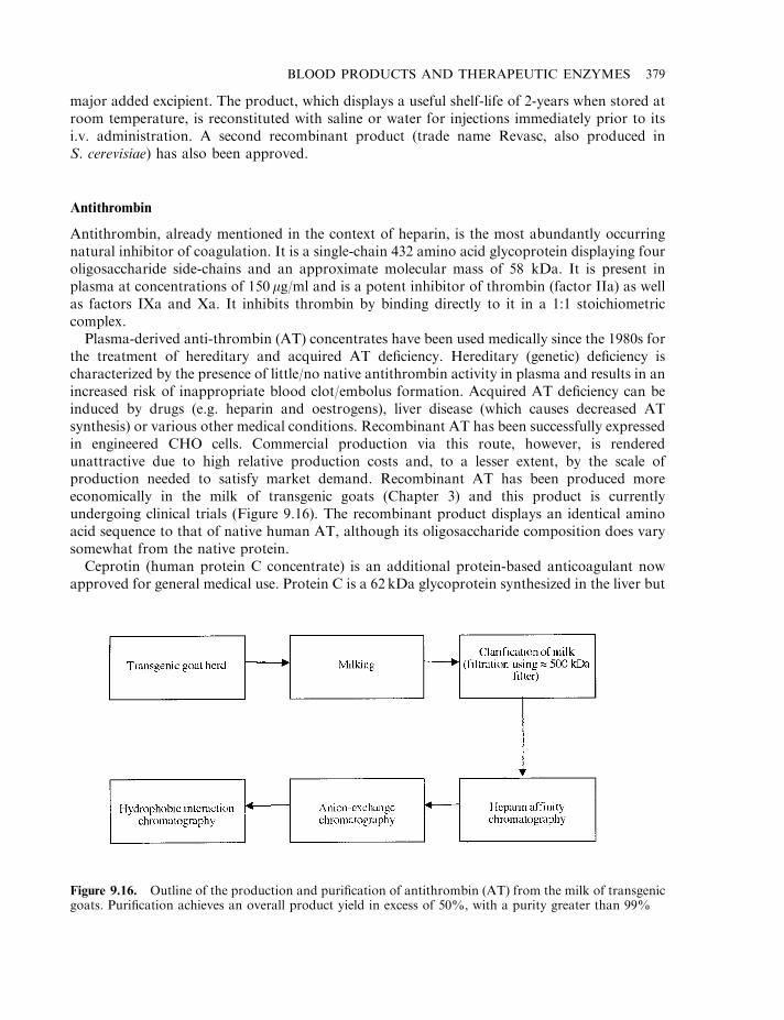
major added excipient. The product, which displays a useful shelf-life of 2-years when stored atroom temperature, is reconstituted with saline or water for injections immediately prior to itsi.v. administration. A second recombinant product (trade name Revasc, also produced inS. cerevisiae) has also been approved.
Antithrombin
Antithrombin, already mentioned in the context of heparin, is the most abundantly occurringnatural inhibitor of coagulation. It is a single-chain 432 amino acid glycoprotein displaying fouroligosaccharide side-chains and an approximate molecular mass of 58 kDa. It is present inplasma at concentrations of 150 mg/ml and is a potent inhibitor of thrombin (factor IIa) as wellas factors IXa and Xa. It inhibits thrombin by binding directly to it in a 1:1 stoichiometriccomplex.
Plasma-derived anti-thrombin (AT) concentrates have been used medically since the 1980s forthe treatment of hereditary and acquired AT deficiency. Hereditary (genetic) deficiency ischaracterized by the presence of little/no native antithrombin activity in plasma and results in anincreased risk of inappropriate blood clot/embolus formation. Acquired AT deficiency can beinduced by drugs (e.g. heparin and oestrogens), liver disease (which causes decreased ATsynthesis) or various other medical conditions. Recombinant AT has been successfully expressedin engineered CHO cells. Commercial production via this route, however, is renderedunattractive due to high relative production costs and, to a lesser extent, by the scale ofproduction needed to satisfy market demand. Recombinant AT has been produced moreeconomically in the milk of transgenic goats (Chapter 3) and this product is currentlyundergoing clinical trials (Figure 9.16). The recombinant product displays an identical aminoacid sequence to that of native human AT, although its oligosaccharide composition does varysomewhat from the native protein.
Ceprotin (human protein C concentrate) is an additional protein-based anticoagulant nowapproved for general medical use. Protein C is a 62 kDa glycoprotein synthesized in the liver but
BLOOD PRODUCTS AND THERAPEUTIC ENZYMES 379
Figure 9.16. Outline of the production and purification of antithrombin (AT) from the milk of transgenicgoats. Purification achieves an overall product yield in excess of 50%, with a purity greater than 99%
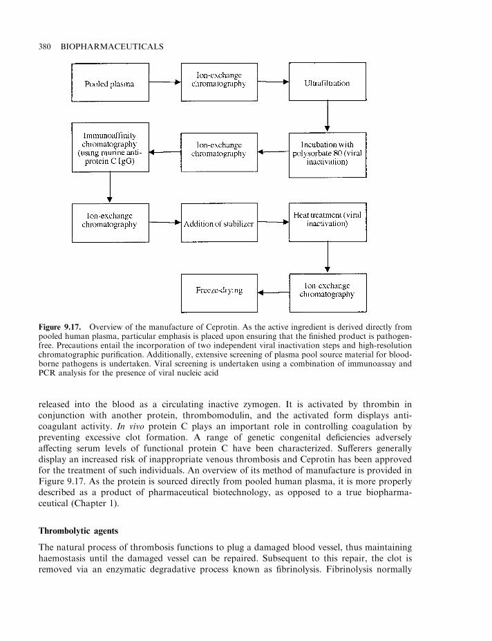
released into the blood as a circulating inactive zymogen. It is activated by thrombin inconjunction with another protein, thrombomodulin, and the activated form displays anti-coagulant activity. In vivo protein C plays an important role in controlling coagulation bypreventing excessive clot formation. A range of genetic congenital deficiencies adverselyaffecting serum levels of functional protein C have been characterized. Sufferers generallydisplay an increased risk of inappropriate venous thrombosis and Ceprotin has been approvedfor the treatment of such individuals. An overview of its method of manufacture is provided inFigure 9.17. As the protein is sourced directly from pooled human plasma, it is more properlydescribed as a product of pharmaceutical biotechnology, as opposed to a true biopharma-ceutical (Chapter 1).
Thrombolytic agents
The natural process of thrombosis functions to plug a damaged blood vessel, thus maintaininghaemostasis until the damaged vessel can be repaired. Subsequent to this repair, the clot isremoved via an enzymatic degradative process known as fibrinolysis. Fibrinolysis normally
380 BIOPHARMACEUTICALS
Figure 9.17. Overview of the manufacture of Ceprotin. As the active ingredient is derived directly frompooled human plasma, particular emphasis is placed upon ensuring that the finished product is pathogen-free. Precautions entail the incorporation of two independent viral inactivation steps and high-resolutionchromatographic purification. Additionally, extensive screening of plasma pool source material for blood-borne pathogens is undertaken. Viral screening is undertaken using a combination of immunoassay andPCR analysis for the presence of viral nucleic acid
depends upon the serine protease plasmin, which is capable of degrading the fibrin strandspresent in the clot.
In situations where inappropriate clot formation results in the blockage of a blood vessel, thetissue damage that ensues depends, to a point, upon how long the clot blocks blood flow. Rapidremoval of the clot can often minimize the severity of tissue damage. Thus, several thrombolytic(clot-degrading) agents have found medical application (Table 9.10). The market for an effectivethrombolytic agent is substantial. In the USA alone, it is estimated that 1.5 million people sufferacute myocardial infarction each year, while another 0.5 million suffer strokes.
Tissue plasminogen activator (tPA)
The natural thrombolytic process is illustrated in Figure 9.18. Plasmin is a protease whichcatalyses the proteolytic degradation of fibrin present in clots, thus effectively dissolving the clot.Plasmin is derived from plasminogen, its circulating zymogen. Plasminogen is synthesized in,and released from, the kidneys. It is a single-chain 90 kDa glycoprotein, which is stabilized byseveral disulphide linkages.
Tissue plasminogen activator (tPA, also known as fibrinokinase) represents the mostimportant physiological activator of plasminogen. tPA is a 527 amino acid serine protease. It issynthesized predominantly in vascular endothelial cells (cells lining the inside of blood vessels)and displays five structural domains, each of which has a specific function (Table 9.11). tPAdisplays four potential glycosylation sites, three of which are normally glycosylated (residues117, 184 and 448). The carbohydrate moieties play an important role in mediating hepaticuptake of tPA and hence its clearance from plasma. It is normally found in the blood in twoforms; a single-chain polypeptide (type I tPA) and a two-chain structure (type II) proteolyticallyderived from the single chain structure. The two-chain form is the one predominantly associatedwith clots undergoing lysis, but both forms display fibrinolytic activity.
Fibrin contains binding sites for both plasminogen and tPA, thus bringing these into closeproximity. This facilitates direct activation of the plasminogen at the clot surface (Figure 9.18).This activation process is potentiated by the fact that binding of tPA to fibrin (a) enhances thesubsequent binding of plasminogen and (b) increases tPA’s activity towards plasminogen by upto 600-fold.
BLOOD PRODUCTS AND THERAPEUTIC ENZYMES 381
Table 9.10. Thrombolytic agents approved for general medical use (r¼recombinant, rh¼recombinanthuman)
Product Company
Activase (rh-tPA) GenentechEcokinase (rtPA; differs from human tPA in that threeof its five domains have been deleted)
Galenus Mannheim
Retavase (rtPA; see Ecokinase) Boehringer-Mannheim/CentocorRapilysin (rtPA; see Ecokinase) Boehringer-MannheimTenecteplase (also marketed as Metalyse) (TNK-tPA,modified rtPA)
Boehringer-Ingelheim
TNKase (Tenecteplase; modified rtPA; see Tenecteplase) GenentechStreptokinase (produced by Streptokinase haemolyticus) VariousUrokinase (extracted from human urine) VariousStaphylokinase (extracted from Staphylococcus aureus andproduced in various recombinant systems)
Various
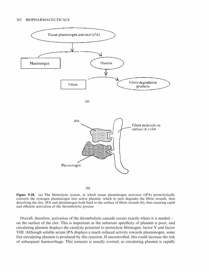
Overall, therefore, activation of the thrombolytic cascade occurs exactly where it is needed—on the surface of the clot. This is important as the substrate specificity of plasmin is poor, andcirculating plasmin displays the catalytic potential to proteolyse fibrinogen, factor V and factorVIII. Although soluble serum tPA displays a much reduced activity towards plasminogen, somefree circulating plasmin is produced by this reaction. If uncontrolled, this could increase the riskof subsequent haemorrhage. This scenario is usually averted, as circulating plasmin is rapidly
382 BIOPHARMACEUTICALS
Figure 9.18. (a) The fibrinolytic system, in which tissue plasminogen activator (tPA) proteolyticallyconverts the zymogen plasminogen into active plasmin, which in turn degrades the fibrin strands, thusdissolving the clot. tPA and plasminogen both bind to the surface of fibrin strands (b), thus ensuring rapidand efficient activation of the thrombolytic process
neutralized by another plasma protein, a2-antiplasmin (a2-antiplasmin, a 70 kDa, single-chainglycoprotein, binds plasmin very tightly in a 1:1 complex). In contrast to free plasmin, plasminpresent on a clot surface is very slowly inactivated by a1-antiplasmin. The thrombolytic systemhas thus evolved in a self-regulating fashion, which facilitates efficient clot degradation withminimal potential disruption to other elements of the haemostatic mechanism.
First-generation tPA. Although tPA was first studied in the late 1940s, its extensivecharacterization was hampered by the low levels at which it is normally synthesized. Detailedstudies were facilitated in the 1980s after the discovery that the Bowes melanoma cell lineproduces and secretes large quantities of this protein. This also facilitated its initial clinicalappraisal. The tPA gene was cloned from the melanoma cell line in 1983, and this facilitatedsubsequent large-scale production in CHO cell lines by recombinant DNA technology. The tPAcDNA contains 2530 nucleotides and encodes a mature protein of 527 amino acids. Theglycosylation pattern was similar, although not identical, to the native human molecule. Amarketing licence for the product was first issued in the USA to Genentech in 1987 (under thetrade name Activase). The therapeutic indication was for the treatment of acute myocardialinfarction. The production process entails an initial (10 000 litre) fermentation step, duringwhich the cultured CHO cells produce and secrete tPA into the fermentation medium. Afterremoval of the cells by sub-micron filtration and initial concentration, the product is purified bya combination of several chromatographic steps. The final product has been shown to be greaterthan 99% pure by several analytical techniques, including HPLC, SDS–PAGE, tryptic mappingand N-terminal sequencing.
Activase has proved effective in the early treatment of patients with acute myocardialinfarction (i.e. those treated within 12 h after the first symptoms occur). Significantly increasedrates of patient survival (as measured 1 day and 30 days after the initial event), are noted whentPA is administered in favour of streptokinase, a standard therapy (see later). tPA has thusestablished itself as a first-line option in the management of acute myocardial infarction. Atherapeutic dose of 90–100mg (often administered by infusion over 90min), results in a steady-state Activase concentration of 3–4mg/l during that period. The product is, however, clearedrapidly by the liver, displaying a serum half-life of approximately 3min. As is the case for mostthrombolytic agents, the most significant risk associated with tPA administration is the possibleinduction of severe haemorrhage.
Engineered tPA. Modified forms of tPA have also been generated in an effort to develop aproduct with an improved therapeutic profile (e.g. faster-acting or exhibiting a prolonged
BLOOD PRODUCTS AND THERAPEUTIC ENZYMES 383
Table 9.11. The five domains that constitute human tPA and the biological function of each domain
tPA domain Function
Finger domain (F domain) Promotes tPA binding to fibrin with high affinityProtease domain (P domain) Displays plasminogen-specific proteolytic activityEpidermal growth factor domain(EGF domain)
Binds hepatic receptors thereby mediating hepaticclearance of tPA from blood
Kringle-1 domain (K1 domain) Associated with binding to the hepatic receptorKringle-2 domain (K2 domain) Facilitates stimulation of tPA’s proteolytic activity by
fibrin
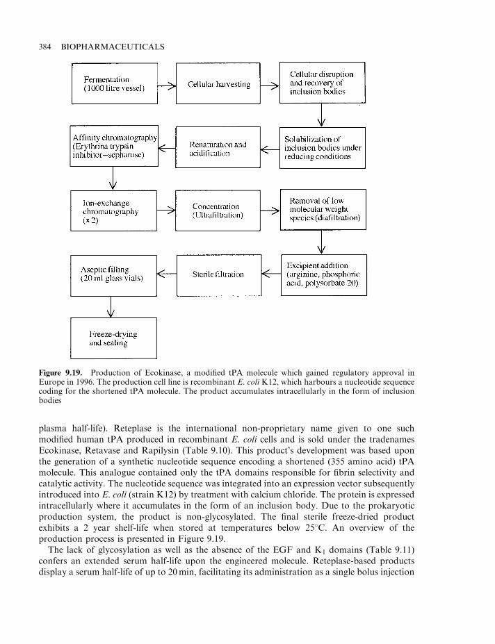
plasma half-life). Reteplase is the international non-proprietary name given to one suchmodified human tPA produced in recombinant E. coli cells and is sold under the tradenamesEcokinase, Retavase and Rapilysin (Table 9.10). This product’s development was based uponthe generation of a synthetic nucleotide sequence encoding a shortened (355 amino acid) tPAmolecule. This analogue contained only the tPA domains responsible for fibrin selectivity andcatalytic activity. The nucleotide sequence was integrated into an expression vector subsequentlyintroduced into E. coli (strain K12) by treatment with calcium chloride. The protein is expressedintracellularly where it accumulates in the form of an inclusion body. Due to the prokaryoticproduction system, the product is non-glycosylated. The final sterile freeze-dried productexhibits a 2 year shelf-life when stored at temperatures below 258C. An overview of theproduction process is presented in Figure 9.19.
The lack of glycosylation as well as the absence of the EGF and K1 domains (Table 9.11)confers an extended serum half-life upon the engineered molecule. Reteplase-based productsdisplay a serum half-life of up to 20min, facilitating its administration as a single bolus injection
384 BIOPHARMACEUTICALS
Figure 9.19. Production of Ecokinase, a modified tPA molecule which gained regulatory approval inEurope in 1996. The production cell line is recombinant E. coli K12, which harbours a nucleotide sequencecoding for the shortened tPA molecule. The product accumulates intracellularly in the form of inclusionbodies
as opposed to continuous infusion. Absence of the molecule’s F1 domain also reduces theproduct’s fibrin-binding affinity. It is theorized that this may further enhance clot degradation,as it facilitates more extensive diffusion of the thrombolytic agent into the interior of the clot.Tenecteplase (also marketed under the tradename, Metalyse) is yet an additional engineered tPAnow on the market. Produced in a CHO cell line, this glycosylated variant differs in sequence tonative tPA by six amino acids (Thr 103 converted to Asn; Asn 117 converted to Gln and theLys–His–Arg–Arg sequence at position 296–299 converted to Ala–Ala–Ala–Ala). Collectively,these modifications result in a prolonged plasma half-life (to 15–19min), as well as an increasedresistance to PAI-1 (plasminogen activator inhibitor-1, a natural tPA inhibitor).
Streptokinase
Streptokinase is an extracellular bacterial protein produced by several strains of Streptococcushaemolyticus group C. It displays a molecular mass in the region of 48 kDa and an isoelectricpoint of 4.7. Its ability to induce lysis of blood clots was first demonstrated in 1933.Early therapeutic preparations administered to patients often caused immunological andother complications, usually prompted by impurities present in these products.Chromatographic purification (particularly using gel filtration and ion-exchange columns),overcame many of these initial difficulties. Modern chromatographically pure streptokinasepreparations are usually supplied in freeze-dried form. These preparations often containalbumin as an excipient. The albumin prevents flocculation of the active ingredient upon itsreconstitution.
Streptokinase is a widespreadly employed thrombolytic agent. It is administered to treat avariety of thrombo-embolic disorders, including:
. pulmonary embolism (blockage of the pulmonary artery by an embolism), which can causeacute heart failure and sudden death (the pulmonary artery carries blood from the heart tothe lungs for oxygenation);
. deep-vein thrombosis (thrombus formation in deep veins, usually in the legs);
. arterial occlusions (obstruction of an artery);
. acute myocardial infarction.
Streptokinase induces its thrombolytic effect by binding specifically and tightly to plasminogen.This induces a conformational change in the plasminogen molecule, which renders itproteolytically active. In this way, the streptokinase–plasminogen complex catalyses theproteolytic conversion of plasminogen to active plasmin.
As a bacterial protein, streptokinase is viewed by the human immune system as an antigenicsubstance. In some cases, its administration has elicited allergic responses, ranging from mildrashes to more serious anaphylactic shock (anaphylactic shock represents an extreme andgeneralized allergic response, characterized by swelling, constriction of the bronchioles,circulatory collapse and heart failure).
Another disadvantage of streptokinase administration is the associated increased risk ofhaemorrhage. Streptokinase-activated plasminogen is capable of lysing not only clot-associatedfibrin, but also free plasma fibrinogen. This can result in low serum fibrinogen levels and hencecompromise haemostatic ability. It should not, for example, be administered to patientssuffering from coagulation disorders or bleeding conditions such as ulcers. Despite suchpotenital clinical complications, careful administration of streptokinase has saved countlessthousands of lives.
BLOOD PRODUCTS AND THERAPEUTIC ENZYMES 385
Urokinase
The ability of some components of human urine to dissolve fibrin clots was first noted in 1885,but it was not until the 1950s that the active substance was isolated and named ‘urokinase’.
Urokinase is a serine protease produced by the kidney and is found in both the plasma andurine. It is capable of proteolytically converting plasminogen into plasmin. Two variants of theenzyme have been isolated: a 54 kDa species and a lower molecular mass (33 kDa) variant. Thelower molecular mass form appears to be derived from the higher molecular mass moiety byproteolytic processing. Both forms exhibit enzymatic activity against plasminogen.
Urokinase is used clinically under the same circumstances as streptokinase and, because of itshuman origin, adverse immunological responses are less likely. Following acute medical eventssuch as pulmonary embolism, the product is normally administered to the patient at initial highdoses (by infusion) for several minutes. This is followed by hourly i.v. injections for up to 12 h.
Urokinase utilized medically is generally purified directly from human urine. It binds to arange of adsorbants, such as silica gel and, especially, kaolin (hydrated aluminium silicate),which can be used to initially concentrate and partially purify the product. It may also beconcentrated and partially purified by precipitation using sodium chloride, ammonium sulphateor ethanol as precipitants.
Various chromatographic techniques may be utilized to further purify urokinase. Commonlyemployed methods include anion (DEAE-based) exchange chromatography, gel filtration onSephadex G-100 and chromatography on hydroxyapatite columns. Urokinase is a relativelystable molecule. It remains active subsequent to incubation at 608C for several hours, or briefincubation at pHs as low as 1.0 or as high as 10.0.
After its purification, sterile filtration and aseptic filling, human urokinase is normally freeze-dried. Because of its heat stability, the final product may also be heated to 608C for up to 10 h inan effort to inactivate any undetected viral particles present. The product utilized clinicallycontains both molecular mass forms, with the higher molecular mass moiety predominating.Urokinase can also be produced by techniques of animal cell culture utilizing human kidneycells or by recombinant DNA technology.
Staphylokinase
Staphylokinase is a protein produced by a number of strains of Staphylococcus aureus, whichalso displays therapeutic potenital as a thrombolytic agent. The protein has been purified fromits natural source by a combination of ammonium sulphate precipitation and cation-exchangechromatography on CM cellulose. Affinity chromatography using plasmin or plasminogenimmobilized to sepharose beads has also been used. The pure product is a 136 amino acidpolypeptide displaying a molecular mass in the region of 16.5 kDa. Lower molecular massderivatives lacking the first six or 10 NH2-terminal amino acids have also been characterized. Allthree appear to display similar thrombolytic activity in vitro at least.
The staphylokinase gene has been cloned in E. coli, as well as various other recombinantsystems. The protein is expressed intracellularly in E. coli at high levels, representing 10–15% oftotal cellular protein. It can be purified directly from the clarified cellular homogenate by acombination of ion-exchange and hydrophobic interaction chromatography.
Although staphylokinase shows no significant homology with streptokinase, it induces athrombolytic effect by a somewhat similar mechanism— it also forms a 1:1 stoichiometriccomplex with plasminogen. The proposed mechanism by which staphylokinase induces
386 BIOPHARMACEUTICALS
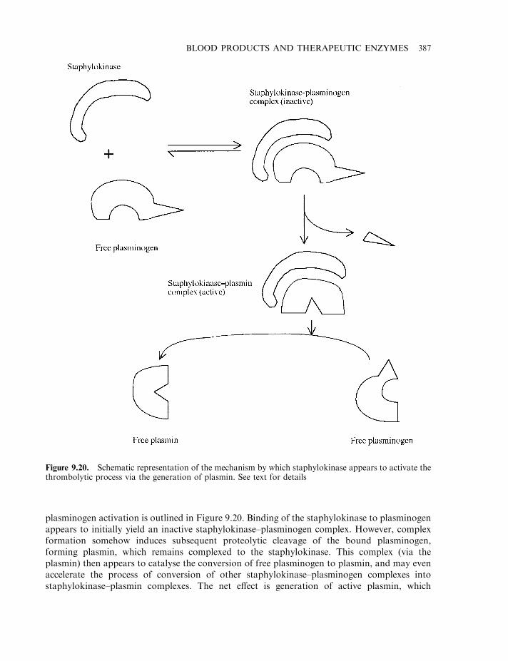
plasminogen activation is outlined in Figure 9.20. Binding of the staphylokinase to plasminogenappears to initially yield an inactive staphylokinase–plasminogen complex. However, complexformation somehow induces subsequent proteolytic cleavage of the bound plasminogen,forming plasmin, which remains complexed to the staphylokinase. This complex (via theplasmin) then appears to catalyse the conversion of free plasminogen to plasmin, and may evenaccelerate the process of conversion of other staphylokinase–plasminogen complexes intostaphylokinase–plasmin complexes. The net effect is generation of active plasmin, which
BLOOD PRODUCTS AND THERAPEUTIC ENZYMES 387
Figure 9.20. Schematic representation of the mechanism by which staphylokinase appears to activate thethrombolytic process via the generation of plasmin. See text for details
displays a direct thrombolytic effect by degrading clot-based fibrin, as described previously(Figure 9.18).
The serum protein a2-antiplasmin can inhibit the activated plasmin–staphylokinase complex.It appears that the a2-antiplasmin can interact with the active plasmin moiety of the complex,resulting in dissociation of staphylokinase, and consequent formation of an inactive plasmin–a2–antiplasmin complex.
The thrombolytic ability of (recombinant) staphylokinase has been evaluated in initial clinicaltrials, with encouraging results; 80% of patients suffering from acute myocardial infarction whoreceived staphylokinase responded positively (10mg staphylokinase was administered byinfusion over 30min). The native molecule displays a relatively short serum half-life (6.3min),although covalent attachment of polyethylene glycol (PEG) reduces the rate of serum clearance,hence effectively increasing the molecule’s half-life significantly. As with streptokinase, patientsadministered staphylokinase develop neutralizing antibodies. A number of engineered (domain-deleted) variants have been generated, which display significantly reduced immunogenicity.
a1-Antitrypsin
The respiratory tract is protected by a number of defence mechanisms which include:
. particle removal in the nostril/nasopharynx;
. particle expulsion (e.g. by coughing);
. upward removal of substances via mucociliary transport;
. presence in the lungs of immune cells, such as alveolar macrophages;
. production/presence of soluble protective factors, including a1-antitrypsin, lysozyme,lactoferrin and interferon.
Failure/ineffective functioning of one or more of these mechanisms can impair normalrespiratory function, e.g. emphysema is a condition in which the alveoli of the lungs aredamaged, which compromises the lung’s capacity to exchange gases, and breathlessness oftenresults. This condition is often promoted by smoking, respiratory infections or a deficiency inthe production of serum a1-antitrypsin.
a1-Antitrypsin is a 394 amino acid, 52 kDa serum glycoprotein. It is synthesized in the liverand secreted into the blood, where it is normally present at concentrations of 2–4 g/l. Itconstitutes in excess of 90% of the a1-globulin fraction of blood.
The a1-antitrypsin gene is located on chromosome 14. A number of a1-antitrypsin genevariants have been described. Their gene products can be distinguished by their differentialmobility upon gel electrophoresis. The normal form is termed M, while point mutations in thegene have generated two major additional forms, S and Z. These mutations results in a greatlyreduced level of synthesis and secretion into the blood of the mature a1-antitrypsin. Personsinheriting two copies of the Z gene, in particular, display greatly reduced levels of serum a1-antitrypsin activity. This is often associated with the development of emphysema (particularly insmokers). The condition may be treated by the administration of purified a1-antitrypsin. Thisprotein constitutes the major serine protease inhibitor present in blood. It is a potent inhibitor ofthe protease elastase, and serves to protect the lung from proteolytic damage by inhibitingneutrophil elastase. The product is administered on an ongoing basis to sufferers, who receive upto 200 g of the inhibitor each year. It is normally prepared by limited fractionation of wholehuman blood, although the large quantities required by patients heightens the risk of accidentaltransmission of blood-borne pathogens. The a1-antitrypsin gene has been expressed in a number
388 BIOPHARMACEUTICALS
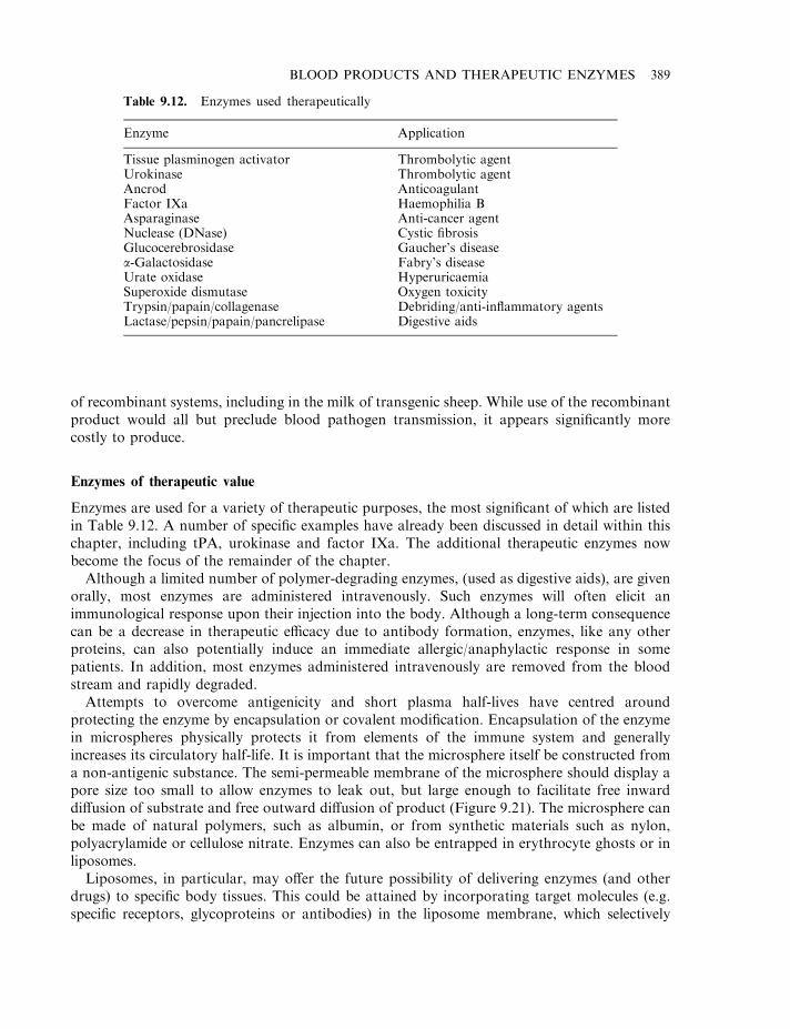
of recombinant systems, including in the milk of transgenic sheep. While use of the recombinantproduct would all but preclude blood pathogen transmission, it appears significantly morecostly to produce.
Enzymes of therapeutic value
Enzymes are used for a variety of therapeutic purposes, the most significant of which are listedin Table 9.12. A number of specific examples have already been discussed in detail within thischapter, including tPA, urokinase and factor IXa. The additional therapeutic enzymes nowbecome the focus of the remainder of the chapter.
Although a limited number of polymer-degrading enzymes, (used as digestive aids), are givenorally, most enzymes are administered intravenously. Such enzymes will often elicit animmunological response upon their injection into the body. Although a long-term consequencecan be a decrease in therapeutic efficacy due to antibody formation, enzymes, like any otherproteins, can also potentially induce an immediate allergic/anaphylactic response in somepatients. In addition, most enzymes administered intravenously are removed from the bloodstream and rapidly degraded.
Attempts to overcome antigenicity and short plasma half-lives have centred aroundprotecting the enzyme by encapsulation or covalent modification. Encapsulation of the enzymein microspheres physically protects it from elements of the immune system and generallyincreases its circulatory half-life. It is important that the microsphere itself be constructed froma non-antigenic substance. The semi-permeable membrane of the microsphere should display apore size too small to allow enzymes to leak out, but large enough to facilitate free inwarddiffusion of substrate and free outward diffusion of product (Figure 9.21). The microsphere canbe made of natural polymers, such as albumin, or from synthetic materials such as nylon,polyacrylamide or cellulose nitrate. Enzymes can also be entrapped in erythrocyte ghosts or inliposomes.
Liposomes, in particular, may offer the future possibility of delivering enzymes (and otherdrugs) to specific body tissues. This could be attained by incorporating target molecules (e.g.specific receptors, glycoproteins or antibodies) in the liposome membrane, which selectively
BLOOD PRODUCTS AND THERAPEUTIC ENZYMES 389
Table 9.12. Enzymes used therapeutically
Enzyme Application
Tissue plasminogen activator Thrombolytic agentUrokinase Thrombolytic agentAncrod AnticoagulantFactor IXa Haemophilia BAsparaginase Anti-cancer agentNuclease (DNase) Cystic fibrosisGlucocerebrosidase Gaucher’s diseasea-Galactosidase Fabry’s diseaseUrate oxidase HyperuricaemiaSuperoxide dismutase Oxygen toxicityTrypsin/papain/collagenase Debriding/anti-inflammatory agentsLactase/pepsin/papain/pancrelipase Digestive aids
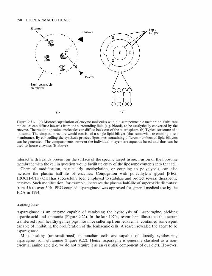
interact with ligands present on the surface of the specific target tissue. Fusion of the liposomemembrane with the cell in question would facilitate entry of the liposome contents into that cell.
Chemical modification, particularly succinylation, or coupling to polyglycols, can alsoincrease the plasma half-life of enzymes. Conjugation with polyethylene glycol [PEG;H(OCH2CH2)nOH] has successfully been employed to stabilize and protect several therapeuticenzymes. Such modification, for example, increases the plasma half-life of superoxide dismutasefrom 5h to over 30 h. PEG-coupled asparaginase was approved for general medical use by theFDA in 1994.
Asparaginase
Asparaginase is an enzyme capable of catalysing the hydrolysis of L-asparagine, yieldingaspartic acid and ammonia (Figure 9.22). In the late 1970s, researchers illustrated that serumtransferred from healthy guinea pigs into mice suffering from leukaemia, contained some agentcapable of inhibiting the proliferation of the leukaemic cells. A search revealed the agent to beasparaginase.
Most healthy (untransformed) mammalian cells are capable of directly synthesizingasparagine from glutamine (Figure 9.22). Hence, asparagine is generally classified as a non-essential amino acid (i.e. we do not require it as an essential component of our diet). However,
390 BIOPHARMACEUTICALS
Figure 9.21. (a) Microencapsulation of enzyme molecules within a semipermeable membrane. Substratemolecules can diffuse inwards from the surrounding fluid (e.g. blood), to be catalytically converted by theenzyme. The resultant product molecules can diffuse back out of the microsphere. (b) Typical structure of aliposome. The simplest structure would consist of a single lipid bilayer (thus somewhat resembling a cellmembrane). By controlling the synthesis process, liposomes containing different numbers of lipid bilayerscan be generated. The compartments between the individual bilayers are aqueous-based and thus can beused to house enzymes (E above)
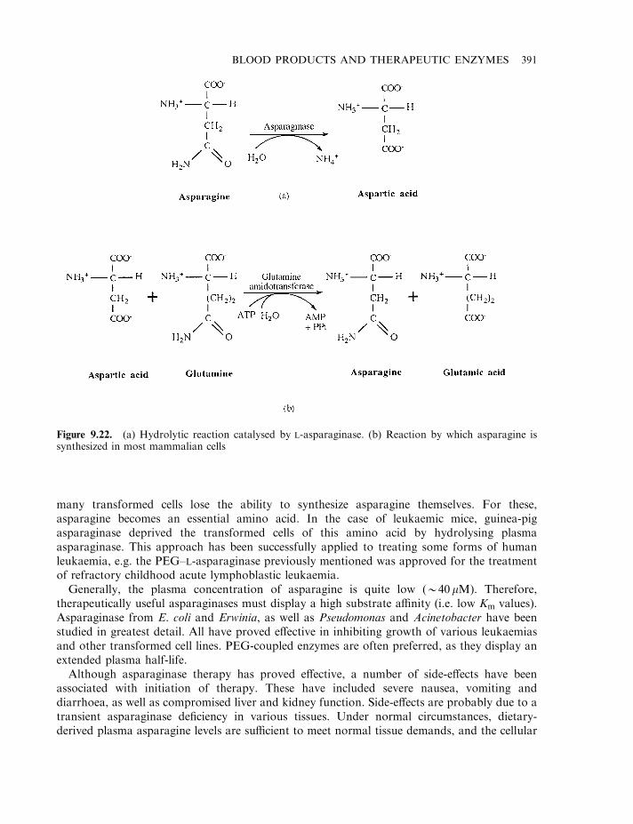
many transformed cells lose the ability to synthesize asparagine themselves. For these,asparagine becomes an essential amino acid. In the case of leukaemic mice, guinea-pigasparaginase deprived the transformed cells of this amino acid by hydrolysing plasmaasparaginase. This approach has been successfully applied to treating some forms of humanleukaemia, e.g. the PEG–L-asparaginase previously mentioned was approved for the treatmentof refractory childhood acute lymphoblastic leukaemia.
Generally, the plasma concentration of asparagine is quite low (*40 mM). Therefore,therapeutically useful asparaginases must display a high substrate affinity (i.e. low Km values).Asparaginase from E. coli and Erwinia, as well as Pseudomonas and Acinetobacter have beenstudied in greatest detail. All have proved effective in inhibiting growth of various leukaemiasand other transformed cell lines. PEG-coupled enzymes are often preferred, as they display anextended plasma half-life.
Although asparaginase therapy has proved effective, a number of side-effects have beenassociated with initiation of therapy. These have included severe nausea, vomiting anddiarrhoea, as well as compromised liver and kidney function. Side-effects are probably due to atransient asparaginase deficiency in various tissues. Under normal circumstances, dietary-derived plasma asparagine levels are sufficient to meet normal tissue demands, and the cellular
BLOOD PRODUCTS AND THERAPEUTIC ENZYMES 391
Figure 9.22. (a) Hydrolytic reaction catalysed by L-asparaginase. (b) Reaction by which asparagine issynthesized in most mammalian cells
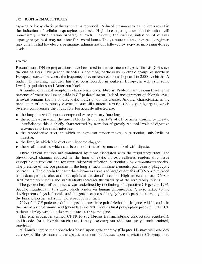
asparagine biosynthetic pathway remains repressed. Reduced plasma asparagine levels result inthe induction of cellular asparagine synthesis. High-dose asparaginase administration willimmediately reduce plasma asparagine levels. However, the ensuing initiation of cellularasparagine synthesis may not occur for several hours. Thus, a more suitable therapeutic regimenmay entail initial low-dose asparaginase administration, followed by stepwise increasing dosagelevels.
DNase
Recombinant DNase preparations have been used in the treatment of cystic fibrosis (CF) sincethe end of 1993. This genetic disorder is common, particularly in ethnic groups of northernEuropean extraction, where the frequency of occurrence can be as high as 1 in 2500 live births. Ahigher than average incidence has also been recorded in southern Europe, as well as in someJewish populations and American blacks.
A number of clinical symptoms characterize cystic fibrosis. Predominant among these is thepresence of excess sodium chloride in CF patients’ sweat. Indeed, measurement of chloride levelsin sweat remains the major diagnostic indicator of this disease. Another characteristic is theproduction of an extremely viscous, custard-like mucus in various body glands/organs, whichseverely compromise their function. Particularly affected are:
. the lungs, in which mucus compromises respiratory function;
. the pancreas, in which the mucus blocks its ducts in 85% of CF patients, causing pancreaticinsufficiency; this is chiefly characterized by secretion of greatly reduced levels of digestiveenzymes into the small intestine;
. the reproductive tract, in which changes can render males, in particular, sub-fertile orinfertile;
. the liver, in which bile ducts can become clogged;
. the small intestine, which can become obstructed by mucus mixed with digesta.
These clinical features are dominated by those associated with the respiratory tract. Thephysiological changes induced in the lung of cystic fibrosis sufferers renders this tissuesusceptible to frequent and recurrent microbial infection, particularly by Pseudomonas species.The presence of microorganisms in the lung attracts immune elements, particularly phagocyticneutrophils. These begin to ingest the microorganisms and large quantities of DNA are releasedfrom damaged microbes and neutrophils at the site of infection. High molecular mass DNA isitself extremely viscous and substantially increases the viscosity of the respiratory mucus.
The genetic basis of this disease was underlined by the finding of a putative CF gene in 1989.Specific mutations in this gene, which resides on human chromosome 7, were linked to thedevelopment of cystic fibrosis, and the gene is expressed largely by cells present in sweat glands,the lung, pancreas, intestine and reproductive tract.
70% of all CF patients exhibit a specific three-base pair deletion in the gene, which results inthe loss of a single amino acid (phenylalanine 508) from its final polypeptide product. Other CFpatients display various other mutations in the same gene.
The gene product is termed CFTR (cystic fibrosis transmembrane conductance regulator),and it codes for a chloride ion channel. It may also carry out additional (as yet undetermined)functions.
Although therapeutic approaches based upon gene therapy (Chapter 11) may well one daycure cystic fibrosis, current therapeutic intervention focuses upon alleviating CF symptoms,
392 BIOPHARMACEUTICALS
particularly those relating to respiratory function. Improved patient care has increased the lifeexpectancy of CF patients to well into their 30s. The major elements of CF management include:
. chest percussion (physically pounding on the chest) in order to help dislodge respiratory tractmucus, rendering the patient better able to expel it;
. antibiotic administration, to control respiratory and other infections;
. pancreatic enzyme replacement;
. attention to nutritional status.
A relatively recent innovation in CF therapy is the use of DNase to reduce the viscosity ofrespiratory mucus. Scientists had been aware since the 1950s that free DNA concentrations inthe lung of cystic fibrosis sufferers were extremely high (3–14mg/ml). They realized that thiscould contribute to the mucus viscosity. Pioneering experiments were undertaken, entailinginhalation of DNase-enriched extracts of bovine pancreas, but both product safety and efficacywere called into question. The observed toxicity was probably due to trypsin or othercontaminants that were damaging to the underlying lung tissue. The host immune system wasalso probably neutralizing much of the bovine DNase.
The advent of genetic engineering and improvements in chromatographic methodologyfacilitated the production of highly purified recombinant human DNase (rhDNase) prepara-tions. Initial in vitro studies proved encouraging— incubation of the enzyme with sputumderived from a CF patient resulted in a significant reduction of the sputum’s viscosity. Clinicaltrials also showed the product to be safe and effective, and Genentech received marketingauthorization for the product in December, 1993 under the trade name ‘Pulmozyme’. Theannual cost of treatment varies, but is often $10 000–$15 000.
Pulmozyme is produced in an engineered CHO cell line harbouring a nucleotide sequencecoding for native human DNase. Subsequent to upstream processing, the protein is purified bytangential flow filtration followed by a combination of chromatographic steps. The purified 260amino acid glycoprotein displays a molecular mass of 37 kDa. It is formulated as an aqueoussolution at a concentration of 1.0 mg/ml, with the addition of calcium chloride and sodiumchloride as excipients. The solution, which contains no preservative, displays a final pH of 6.3. Itis administered directly into the lungs by inhalation of an aerosol mist generated by acompressed air-based nebulizer system.
Glucocerebrosidase
Glucocerebrosidase preparations are administered to relieve the symptoms of Gaucher’s disease,which affects some 5000 people worldwide. This is a lysosomal storage disease affecting lipidmetabolism, specifically the degradation of glucocerebrosides. Glucocerebrosides are a specificclass of lipid, consisting of a molecule of sphingosine, a fatty acid and a glucose molecule(Figure 9.23). They are found in many body tissues, particularly in the brain and other neuraltissue, in which they are often associated with the myelin sheath of nerves. Glucocerebrosides,however, are not abundant structural components of membranes, but are mostly formed asintermediates in the synthesis and degradation of more complex glycosphingolipids. Theirdegradation is undertaken by specific lysosomal enzymes, particularly in cells of the reticulo-endothelial system (i.e. phagocytes, which are spread throughout the body and which functionas: (a) a defence against microbial infection and, (b), removal of worn-out blood cells from theplasma. These phagocytes are particularly prevalent in the spleen, bone marrow and liver).
BLOOD PRODUCTS AND THERAPEUTIC ENZYMES 393
Gaucher’s disease is an inborn error of metabolism characterized by lack of the enzymeglucocerebrosidase, with consequent accumulation of glucocerebrosides, particularly in tissue-based macrophages. Clinical systems include enlargement and compromised function of thesemacrophage-containing tissues, particularly the liver and spleen, as well as damage to longbones and, sometimes, mental retardation. Administration of exogenous glucocerebrosidase asenzyme replacement therapy, has been shown to reduce the main symptoms of this disease. Theenzyme is normally administered by slow intravenous infusion (over a period of 2 h) once every2 weeks.
Genzyme Corporation were granted marketing authorization in 1991 for a glucocerebrosidasepreparation to be used for the treatment of Gaucher’s disease. This Genzyme product (tradename Ceredase) was extracted from placentas (afterbirths) obtained from maternity hospitalwards. The enzyme displays a molecular mass of 65 kDa and four of its five potentialglycosylation sites are glycosylated. It had been estimated that 1 year’s supply of enzyme for anaverage patient required extraction of 27 000 placentas, which renders treatment extremelyexpensive. Genzyme then gained regulatory approval for a recombinant version of glucocer-ebrosidase produced in CHO cells. This product (trade name Cerezyme) has been on the marketsince 1994 and the total world market for glucocerebrosidase is estimated to be in the region of$200 million.
Cerezyme is produced in a CHO cell line harbouring the cDNA coding for humanb-glucocerebrosidase. The purified product is presented as a freeze-dried powder andalso contains mannitol, sodium citrate, citric acid and polysorbate 80 as excipients. Itexhibits a shelf-life of 2 years when stored at 28C–88C.
An integral part of the downstream processing process entails the modification of Cerezyme’soligosaccharide components. The native enzyme’s sugar side-chains are complex and, for themost part, are capped with a terminal sialic acid or galactose residue. Animal studies indicatethat in excess of 95% of injected glucocerebrosidase is removed from the circulation by the livervia binding to hepatocyte surface lectins. As such, the intact enzyme is not available for uptakeby the affected cell type— the tissue macrophages. These macrophages display high levels ofsurface mannose receptors. Treatment of native glucocerebrosidase with exoglycosidases, byremoving terminal sugar residues, can expose mannose residues present in their sugar side-chains, resulting in their binding to and uptake by the macrophages. In this way, the ‘mannose-engineered’ enzyme is selectively targeted to the affected cells.
394 BIOPHARMACEUTICALS
Figure 9.23. Generalized structure of a glucocerebroside
a-Galactosidase and urate oxidase
Recombinant a-galactosidase and urate oxidase represent two additional biopharmaceuticalsrecently approved for general medical use. a-Galactosidase is approved for long-term enzymereplacement therapy in patients with Fabry’s disease. Like Gaucher’s disease, Fabry’s disease isa genetic disease of lipid metabolism. Sufferers display little or no liposomal a-galactosidase Aactivity. This results in the progressive accumulation of glycosphingolipids in several body celltypes. Resultant clinical manifestations are complex, affecting the nervous system, vascularendothelial cells and major organs. Although the condition is rare (500–1000 patients within theEuropean Union), untreated sufferers usually die in their 40s or 50s.
Two recombinant a-galactosidase products are now on the market (‘Fabrazyme’, producedby Genzyme and ‘Replagal’, produced by TKT Europe). Fabrazyme is produced in anengineered CHO cell line, and downstream processing entails a combination of fivechromatographic purification steps, followed by concentration and diafiltration. Excipientsadded include mannitol and sodium phosphate buffering agents and the final product is freeze-dried after filling into glass vials. Replagal is produced in a continuous human cell line and isalso purified by a combination of five chromatographic purification steps, although it ismarketed as a liquid solution.
Human a-galactosidase is a 100 kDa homodimeric glycoprotein. Each 398 amino acidmonomer displays a molecular mass of 45.3 kDa (excluding the glycocomponent) and isglycosylated at three positions (asparagines 108, 161 and 184). After administration (usuallyevery second week by a 40min infusion), the enzyme is taken up by various body cell types anddirected to the lysosomes. This cellular uptake and delivery process appears to be mediated bymannose-6-phosphate residues present in the oligosaccharide side-chains of the enzyme.Mannose-6-phosphate receptors are found on the surface of various cell types and alsointracellularly, associated with the Golgi complex, which then directs the enzyme to thelysosomes.
The enzyme urate oxidase has also found medical application for the treatment of acutehyperuricaemia (elevated plasma uric acid levels), associated with various tumours, particularlyduring their treatment with chemotherapy.
Uric acid is the end product of purine metabolism in humans, other primates, birds andreptiles. It is produced in the liver by the oxidation of xanthine and hypoxanthine (Figure 9.24)and is excreted via the kidneys. Due to its relatively low solubility, an increase in serum uric acidlevels often triggers the formation and precipitation of uric acid crystals, typically resulting inconditions such as gout or urate stones in the urinary tract. Significantly elevated serum uricacid concentrations can also be associated with rapidly proliferating cancers or, in particular,with onset of chemotherapy. In the former instance, rapid cellular turnover results in increasedrates of nucleic acid catabolism and hence uric acid production. In the latter case,chemotherapy-induced cellular lysis results in the release of intracellular contents, includingfree purines and purine-containing nucleic acids, into the bloodstream. The increased associatedpurine metabolism then triggers hyperuricaemia. The elevated uric acid concentrations oftentrigger crystal formation in the renal tubules, and hence renal failure.
Purine metabolism in some mammals is characterized by a further oxidation of uric acid toallantoin by the enzyme, urate oxidase. Allantoin is significantly more water-soluble than uricacid and is also freely excreted via the renal route.
Administration of urate oxidase to humans suffering from hyperuricaemia results in thereduction of serum uric acid levels through its conversion to allantoin. Urate oxidase purified
BLOOD PRODUCTS AND THERAPEUTIC ENZYMES 395
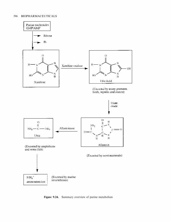
396 BIOPHARMACEUTICALS
Figure 9.24. Summary overview of purine metabolism
directly from cultures of the fungus Aspergillus flavus has been used to treat this condition for anumber of years. More recently, a recombinant form of the fungal enzyme (trade nameFasturtec), has gained regulatory approval in the European Union. Produced in an engineeredstrain of S. cerevisiae, the enzyme is a tetramer composed of four identical polypeptide subunits.Each subunit contains 301 amino acids, displays a molecular mass of 34 kDa and is N-terminalacetylated.
Superoxide dismutase
Under normal circumstances in aerobic metabolism, oxygen is reduced by four electrons,forming H2O. While this usually occurs uneventfully, incomplete reduction will result in thegeneration of oxygen radicals and other reactive species. These are: the superoxide radical, O�
2 ,hydrogen peroxide (H2O2) and the hydroxyl radical (OH�). The superoxide and hydroxylradicals are particularly reactive and can attack membrane components, nucleic acids and othercellular macromolecules, leading to their destruction or modification. O�
2 and OH� radicals, forexample, are believed to be amongst the most mutagenic substances generated by ionizingradiation.
Oxygen-utilizing organisms have generally evolved specific enzyme-mediated systems thatserve to protect the cell from such reactive species. These enzymes include superoxide dismutase(SOD) and catalase or glutathione peroxidase (GSH-px), which catalyse the following reactions:
O�.2 þO
�.2 þ 2Hþ �!SOD
H2O2 þO2
H2O2 þH2O2777777777�!Catalase or GSH�px
2H2OþO2
In general, all aerobic organisms harbour these oxygen-defence systems. At least three types ofSODs have been identified: a cytosolic eukaryotic dismutase, generally a 31 kDa dimer,containing both copper and zinc; a 75 kDa mitochondrial form and a 40 kDa bacterial form,each of which contain two manganese atoms and an iron-containing form, found in somebacteria, blue-green algae and many plants. The metal ions play a direct role in the catalyticconversion, serving as transient acceptors/donors of electrons.
In humans, increased generation of O�2 and/or reduced SOD levels have been implicated in a
wide range of pathological conditions, including ageing, asthma, accelerated tumour growth,neurodegenerative diseases and inflammatory tissue necrosis. Furthermore, administration ofSOD has been found to reduce tissue damage due to irradiation or other conditions thatgenerate O�
2 . Increased SOD production in Drosophila melanogaster leads to increased oxygentolerance and, interestingly, increased lifespan.
SOD isolated from bovine liver or erythrocytes has been used medically as an anti-inflammatory agent. Human SOD has also been expressed in several recombinant systems, andis currently being evaluated to assess its ability to prevent tissue damage induced by exposure toexcessively oxygen-rich blood.
Debriding agents
Debridement refers to the process of cleaning a wound by removal of foreign material and deadtissue. Cleansing of the wound facilitates rapid healing and minimizes the risk of infection due tothe presence of bacteria at the wound surface. The formation of a clot, followed by a scab, on a
BLOOD PRODUCTS AND THERAPEUTIC ENZYMES 397
wound surface can trap bacteria, which then multiply (usually evidenced by the production ofpus), slowing the healing process. Although debridement may be undertaken by physical means(e.g. cutting away dead tissue, washing/cleaning the wound), proteolytic enzymes are also oftenused to facilitate this process.
The value of proteases in cleansing tissue wounds have been appreciated for several hundredyears. Wounds were sometimes cleansed in the past by application of protease-containingmaggot saliva. Nowadays, this is usually more acceptably achieved by topical application of theenzyme to the wound surface. In some cases, the enzyme is formulated in an aqueous-basedcream, while in others, it is impregnated into special bandages. Trypsin, papain, collagenase andvarious microbial enzymes have been used in this regard.
Trypsin is a 24 kDa proteolytic enzyme synthesized by the mammalian pancreas in an inactivezymogen form, trypsinogen. Upon its release into the small intestine, it is proteolyticallyconverted into trypsin by an enteropeptidase. Active trypsin plays a digestive role, hydrolysingpeptide bonds in which the carboxyl group has been contributed by an arginine or lysine.Trypsin used medically is generally obtained by the enzymatic activation of trypsinogen,extracted from the pancreatic tissue of slaughterhouse animals.
Papain is a cysteine protease isolated from the latex of the immature fruit and leaves of theplant, Carica papaya. It consists of a single 23.4 kDa, 212 amino acid polypeptide and thepurified enzyme exhibits broad proteolytic activity. Although it can be used as a debridingagent, it is also used for a variety of other industrial processes, including meat tenderizing, andfor the clarification of beverages.
Collagenase is a protease that can utilize collagen as a substrate. Although it can be producedby animal cell culture, certain microorganisms also produce this enzyme, most notably certainspecies of Clostridium (the ability of these pathogens to produce collagenase facilitates theirrapid spread throughout the body). Collagenase used therapeutically is usually obtained fromcell fermentation supernatants of Clostridium histolyticum. Such preparations are appliedtopically to promote debridement of wounds, skin ulcers and burns.
Chymotrypsin has also been utilized to promote debridement, as well as the reduction of softtissue inflammation. It is also used in some ophthalmic procedures—particularly in facilitatingcataract extraction. It is prepared by activation of its zymogen, chymotrypsinogen, which isextracted from bovine pancreatic tissue.
Yet another proteolytic preparation used for debridement of wounds and skin ulcers consistsof proteolytic enzymes derived from Bacillus subtilis. The preparation displays broad proteolyticactivity and is usually applied several times daily to the wound surface.
Digestive aids
A number of enzymes may be used as digestive aids (Table 9.13). In some instances, a singleenzymatic activity is utilized, whereas other preparations contain multiple enzyme activities.These enzyme preparations may be used to supplement normal digestive activity, or to conferupon an individual a new digestive capability.
The use of enzymes as digestive aids is only applied under specific medical circumstances.Some medical conditions (e.g. cystic fibrosis) can result in compromised digestive function dueto insufficient production/secretion of endogenous digestive enzymes. Digestive enzymepreparations are often formulated in powder (particularly tablet) form, and are recommendedto be taken orally immediately prior to or during meals. As the product never enters the blood
398 BIOPHARMACEUTICALS

stream, the product purity need not be as stringent as enzymes (or other proteins) administeredintravenously. Most digestive enzymes are, at best, semi-pure preparations.
In some instances, there is a possibility that the efficacy of these preparations may becompromised by conditions associated with the digestive tract. Most function at pH valuesapproaching neutrality. They would thus display activity possibly in saliva and particularly inthe small intestine. However, the acidic conditions of the stomach (where the pH can be below1.5), may denature some of these enzymes. Furthermore, the ingested enzymes would also beexposed to endogenous proteolytic activities associated with the stomach and small intestine.Some of these difficulties, however, may be at least partially overcome by formulating theproduct as a tablet coated with an acid-resistant film to protect the enzyme as it passes throughthe stomach.
Pancreatin is a pancreatic extract usually obtained from the pancrease of slaughterhouseanimals. It contains a mixture of enzymes, principally amylase, protease and lipase and, thus,exhibits a broad digestive capability. It is administered orally, mainly for the treatment ofpancreatic insufficiency caused by cystic fibrosis or pancreatitis. As it is sensitive to stomachacid, it must be administered in high doses or, more usually, as enteric coated granules orcapsules which may be taken directly, or sprinkled upon the food prior to its ingestion.Individual digestive activities, such as papain, pepsin or bromelains (proteases), or a-amylaseare sometimes used in place of pancreatin.
Cellulase is not produced in the human digestive system. Cellulolytic enzyme preparationsobtained from Aspergillus niger or other fungal sources are available and it is thought that theiringestion may improve overall digestion, particularly in relation to high-fibre diets.
a-Galactosides are oligosaccharides present in plant matter, particularly beans. They are notnormally degraded in the human digestive tract due to the absence of an appropriateendogenous digestive enzyme (i.e. an a-galactosidase). However, upon their entry into the largeintestine, these oligosaccharides are degraded by microbial a1,6-galactosidases, thus stimulatingmicrobial fermentation. The end-products of fermentation include volatile fatty acids, carbondioxide, methane and hydrogen, which lead to flatulance. This can be avoided by minimizingdietary intake of food containing a-galactosides. Another approach entails the simultaneousingestion of tablets containing a-galactosidase activity. If these ‘flatulance factors’ are degradedbefore or upon reaching the small intestine, the monosaccharides released will be absorbed and,hence, will subsequently be unavailable to promote undesirable microbial fermentations in thelarge intestine.
BLOOD PRODUCTS AND THERAPEUTIC ENZYMES 399
Table 9.13. Enzymes that are used as digestive aids
Enzyme Application
a-Amylase Aids in digestion of starchCellulase Promotes partial digestion of cellulosea-Galactosidase Promotes degradation of flatulance factorsLactase Counteracts lactose intolerancePapainPepsin Enhanced degradation of dietary proteinBromelainsPancreatin Enhanced degradation of dietary carbohydrate, fat and protein
g
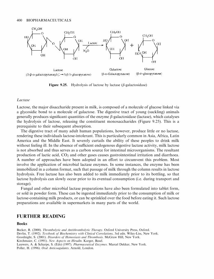
Lactase
Lactose, the major disaccharide present in milk, is composed of a molecule of glucose linked viaa glycosidic bond to a molecule of galactose. The digestive tract of young (suckling) animalsgenerally produces significant quantities of the enzyme b-galactosidase (lactase), which catalysesthe hydrolysis of lactose, releasing the constituent monosaccharides (Figure 9.25). This is aprerequisite to their subsequent absorption.
The digestive tract of many adult human populations, however, produce little or no lactase,rendering these individuals lactose-intolerant. This is particularly common in Asia, Africa, LatinAmerica and the Middle East. It severely curtails the ability of these peoples to drink milkwithout feeling ill. In the absence of sufficient endogenous digestive lactase activity, milk lactoseis not absorbed and thus serves as a carbon source for intestinal microorganisms. The resultantproduction of lactic acid, CO2 and other gases causes gastrointestinal irritation and diarrhoea.A number of approaches have been adopted in an effort to circumvent this problem. Mostinvolve the application of microbial lactase enzymes. In some instances, the enzyme has beenimmobilized in a column format, such that passage of milk through the column results in lactosehydrolysis. Free lactase has also been added to milk immediately prior to its bottling, so thatlactose hydrolysis can slowly occur prior to its eventual consumption (i.e. during transport andstorage).
Fungal and other microbial lactase preparations have also been formulated into tablet form,or sold in powder form. These can be ingested immediately prior to the consumption of milk orlactose-containing milk products, or can be sprinkled over the food before eating it. Such lactosepreparations are available in supermarkets in many parts of the world.
FURTHER READING
Books
Becker, R. (2000). Thrombolytic and Antithrombolytic Therapy. Oxford University Press, Oxford.Devlin, T. (1992). Textbook of Biochemistry with Clinical Correlations, 3rd edn. Wiley-Liss, New York.Goodnight, S. (2001). Disorders of Hemostasis and Thrombosis. McGraw Hill, New York.Kirchmaier, C. (1991). New Aspects on Hirudin. Karger, Basel.Lauwers, A. & Scharpe, S. (Eds) (1997). Pharmaceutical Enzymes. Marcel Dekker, New York.Poller, H. (1996). Oral Anticoagulants. Arnold, London.
400 BIOPHARMACEUTICALS
Figure 9.25. Hydrolysis of lactose by lactase (b-galactosidase)
Articles
Plasma proteins, including coagulation factors
Bowen, D. (2002). Haemophilia A and haemophilia B: molecular insights. J. Clin. Pathol. Mol. Pathol. 55(1), 1–18.Chuang, V. et al. (2002). Pharmaceutical strategies utilizing recombinant human serum albumin. Pharmaceut. Res. 19(5),
569–577.Crevenakova, L. et al. (2002). Factor VIII and transmissible spongiform encephalopathy: the case for safety.
Haemophilia 8(2), 63–75.Federici, A. & Mannucci, P. (2002). Advances in the genetics and treatment of von Willebrand disease. Curr. Opin.
Pediat. 14(1) 23–33.Goodey, A. (1993). The production of heterologous plasma proteins. Trends Biotechnol. 11, 430–433.Kingdon, H & Lundblad, R. (2002). An adventure in biotechnology: the development of haemophilia A therapeutics—
from whole blood transfusion to recombinant DNA technology to gene therapy. Biotechnol. Appl. Biochem. 35,141–148.
Klinge, J. et al. (2002) Hemophilia A—from basic science to clinical practice. Semin. Thrombosis Hemostasis. 28(3),309–321.
Legaz, M. et al. (1973). Isolation and characterization of human factor VIII (antihaemophilic factor). J. Biol. Chem. 248,3946–3955.
Nicholson, J. et al. (2000). The role of albumin in critical illness. Br. J. Anaesth. 85(4), 599–610.Ogden, J. (1992). Recombinant haemoglobin in the development of red blood cell substitutes. Trends in Biotech. 10,
91–95.Winslow, R. (2000). Blood substitutes: refocusing an elusive goal. Br. J. Haematol. 111(2), 387–396.Wright, G. et al. (1991). High level expression of active human a-1-antitrypsin in the milk of transgenic sheep. Bio/
Technology 9, 830–834.
Anticoagulants and related substances
Dodt, J. (1995). Anti-coagulatory substances of bloodsucking animals: from hirudin to hirudin mimetics. Angew. Chem.Int. Ed. Engl. 34, 867–880.
Eldora, A. et al. (1996). The role of the leech in medical therapeutics. Blood Rev. 10(4), 201–209.Fitzgerald, G. (1996). The human pharmacology of thrombin inhibition. Coronary Artery Dis. 7(12), 911–918.Markwardt, F. (1991). Hirudin and its derivatives as anticoagulant agents. Thromb. Haemost. 66, 141–152.Pineo, G. & Hull, R. (1997). Low molecular weight heparin—prophylaxis and treatment of venous thromboembolism.
Ann. Rev. Med. 48, 79–91.Salzet, M. (2002). Leech thrombin inhibitors. Curr. Pharmaceut. Design 8(7), 493–503.Sawyer, R. (1991). Thrombolytics and anticoagulants from leeches. Bio/Technology 9, 513–518.Schulman, S. (1996). Anticoagulation in venous thrombosis. J. R. Soc. Med. 89(11), 624–630.Sohn, J. et al. (2001). Current status of the anticoagulant hirudin: its biotechnological production and clinical practice.
Appl. Microbiol. Biotechnol. 57(5–6), 606–613.Tencate, H. et al. (1996). Developments in anti-thrombolic therapy—state of the art, anno 1996. Pharmacy World Sci.
18(6), 195–203.Walker, C & Royston, D. (2002). Thrombin generation and its inhibition: a review of the scientific basis and mechanism
of action of anticoagulant therapies. Br. J. Anaesth. 88(6), 848–863.
Thrombolytics
Al-Buhairi, A. & Jan, M. (2002). Recombinant tissue plasminogen activator for acute ischemic stroke. Saudi Med. J.23(1), 13–19.
Blasi, F. (1999). The urokinase receptor. A cell surface, regulated chemokine. APMIS 107(1), 96–101.Castillo, P. et al. (1997). Cost-effectiveness of thrombolytic therapy for acute myocardial infarction. Ann. Pharmacother.
31(5), 596–603.Collen, D. & Lijnen, H. (1994). Staphylokinase, a fibrin-specific plasminogen activator with therapeutic potential? Blood
84(3), 680–686.Collen, D. (1998). Staphylokinase: a potent, uniquely fibrin-selective thrombolytic agent. Nature Med. 4(3), 279–284.Collins, R. et al. (1997). Drug therapy—aspirin, heparin and fibrinolytic therapy in suspected acute myocardial
infarction. N. Engl. J. Med. 336(12), 847–860.Datar, R. et al. (1993). Process economics of animal cell and bacterial fermentations: a case study analysis of tissue
plasminogen activator. Bio/Technology 11, 349–357.Emeis, J. et al. (1997). Progress in clinical fibrinolysis. Fibrinolysis Proteolysis 11(2), 67–84.
BLOOD PRODUCTS AND THERAPEUTIC ENZYMES 401
Gillis, J. et al. (1995). Alteplase. A reappraisal of its pharmacological properties and therapeutic use in acute myocardialinfarction. Drugs 50(1), 102–136.
Marder, V. & Stewart, D. (2002). Towards safer thrombolytic therapy. Semin. Haematol. 39(3), 206–216.Preissner, K. et al. (2000). Urokinase receptor: a molecular organizer in cellular communication. Curr. Opin. Cell Biol.
12(5), 621–628.Rabasseda, X. (2001). Tenecteplase (TNK tissue plasminogen activator): a new fibrinolytic for the acute treatment of
myocardial infarction. Drugs Today 37(11), 749–760.Schoebel, F. et al. (1997). Antithrombotic treatment in stable coronary syndromes—long-term intermittent urokinase
therapy in end-stage coronary-artery disease and refractory angina pectoris. Heart 77(1), 13–17.Tsirka, S. (1997). Clinical implications of the involvement of tPA in neuronal cell death. J. Mol. Med. 75(5), 341–347.Verstraete, M. et al. (1995). Thrombolytic agents in development. Drugs, 50(1), 29–42.Verstraete, M. (2000). Third generation thrombolyic drugs. Am. J. Med. 109(1), 52–58.
Enzyme therapeutics
Barranger, J. et al. (1995). Molecular biology of glucocerebrosidase and the treatment of Gaucher’s disease. CytokinesMol. Ther. 1(3), 149–163.
Chakrabarti, R. & Schuster, S. (1997). L-Asparaginase—perspectives on the mechanisms of action and resistance. Int. J.Pediat. Haematol./Oncol. 4(6), 597–611.
Chapple, L. (1997). Reactive oxygen species and antioxidants in inflammatory diseases. J. Clin. Periodontol. 24(5),287–296.
Conway, S. & Littlewood, J. (1997). rhDNase in cystic fibrosis. Br. J. Hosp. Med. 57(8), 371–372.Conway, S. & Watson, A. (1997). Nebulized bronchodilators, corticosteroids and rhDNase in adult patients with cystic
fibrosis. Thorax 52(2), S64–S68.Edgington, S. (1993). Nuclease therapeutics in the clinic. Bio/Technology 11, 580–582.James, E. (1994). Superoxide dismutase. Parasitol. Today 10(12), 482–484.Mistry, P & Cox, T. (1993). The glucocerebrosidase locus in Gaucher’s disease—molecular analysis of a lysosomal
enzyme. J. Med. Genet. 30(11), 889–894.Ronghe, M. et al. (2001). Remission induction therapy for childhood acute lymphoblastic leukaemia: clinical and cellular
pharmacology of vincristine, corticosteroids, L-asparaginase and anthracyclines. Cancer Treat. Rev. 27(6), 327–337.Welsh, M. & Smith, A. (1995). Cystic fibrosis. Sci. Am. (December), 36–43.Wileman, T. (1991). Properties of asparaginase–dextran conjugates. Adv. Drug Delivery Rev. 6(2), 167–180.Zhao, H & Grabowski. G. (2002). Gaucher’s disease: perspectives on a prototype lysosomal disease. Cell. Mol. Life Sci.
59(4), 694–707.
402 BIOPHARMACEUTICALS
Chapter 10
Antibodies, vaccines and adjuvants
Few substances have had a greater positive impact upon human healthcare management thanantibodies, vaccines and adjuvants. For most of this century, these immunological agents haveenjoyed widespread medical application, predominantly for the treatment/prevention ofinfectious diseases. As a group, they are often referred to as ‘biologics’ (generally speaking,the term ‘biologic’ refers to vaccines, serum, toxins and medicinal products derived from humanblood or plasma; Chapter 1).
Polyclonal antibody preparations have been used to induce passive immunity against a rangeof foreign (harmful) agents, while vaccines are used to efficiently and safely promote activeimmunization. Adjuvants are usually co-administered with the vaccine preparation, in order toenhance the immune response against the vaccine. The development of modern biotechnologicalmethodology has had an enormous impact upon the therapeutic application of immunologicalagents, as discussed later. Monoclonal/engineered antibodies find a range of therapeutic uses,while many of the newer vaccine preparations are now produced by recombinant DNAtechnology.
POLYCLONAL ANTIBODY PREPARATIONS
Polyclonal antibody preparations have been used for several decades to induce passiveimmunization against infectious diseases and other harmful agents, particularly toxins. Theantibody preparations are usually administered by direct i.v. injection. While this affordsimmediate immunological protection, its effect is transitory, usually persisting for only 2–3 weeks(i.e. until the antibodies are excreted). Passive immunization can be used prophylactically (i.e. toprevent a future medical episode) or therapeutically (i.e. to treat a medical condition that isalready established). An example of the former would be prior administration of a specific anti-snake toxin antibody preparation to an individual before he/she travelled to a world region inwhich these snakes are commonly found. An example of the latter would be administration ofthe anti-venom antibody immediately after the individual has experienced a snake bite.
Antibody preparations used to induce passive immunity may be obtained from either animalor human sources. Preparations of animal origin are generally termed ‘antisera’, while thosesourced from humans are called ‘immunoglobulin preparations’. In both cases, the predominantantibody type present is immunoglobulin G (IgG).
Biopharmaceuticals: Biochemistry and Biotechnology, Second Edition by Gary WalshJohn Wiley & Sons Ltd: ISBN 0 470 84326 8 (ppc), ISBN 0 470 84327 6 (pbk)
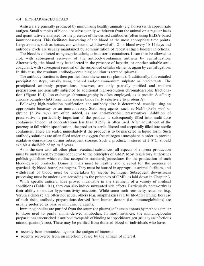
Antisera are generally produced by immunizing healthy animals (e.g. horses) with appropriateantigen. Small samples of blood are subsequently withdrawn from the animal on a regular basisand quantitatively analysed for the presence of the desired antibodies (often using ELISA-basedimmunoassays). This facilitates harvesting of the blood at the most appropriate time points.Large animals, such as horses, can withstand withdrawal of 1–2 l of blood every 10–14 days andantibody levels are usually maintained by administration of repeat antigen booster injections.
The blood is collected using aseptic technique into sterile containers. It can then be allowed toclot, with subsequent recovery of the antibody-containing antisera by centrifugation.Alternatively, the blood may be collected in the presence of heparin, or another suitable anti-coagulant, with subsequent removal of the suspended cellular elements, again by centrifugation.In this case, the resultant antibody-containing solution is termed ‘plasma’.
The antibody fraction is then purified from the serum (or plasma). Traditionally, this entailedprecipitation steps, usually using ethanol and/or ammonium sulphate as precipitants. Theprecipitated antibody preparations, however, are only partially purified and modernpreparations are generally subjected to additional high-resolution chromatographic fractiona-tion (Figure 10.1). Ion-exchange chromatography is often employed, as is protein A affinitychromatography (IgG from many species binds fairly selectively to protein A).
Following high-resolution purification, the antibody titre is determined, usually using anappropriate bioassay or an immunoassay. Stabilizing agents, such as NaCl (0.9% w/v) orglycine (2–3% w/v) are often added, as are anti-microbial preservatives. Addition ofpreservative is particularly important if the product is subsequently filled into multi-dosecontainers. Phenol, at concentrations less than 0.25%, is often used. After adjustment of thepotency to fall within specification, the product is sterile-filtered and aseptically filled into sterilecontainers. These are sealed immediately if the product is to be marketed in liquid form. Suchantibody solutions are often filled under an oxygen-free nitrogen atmosphere in order to preventoxidative degradation during subsequent storage. Such a product, if stored at 2–88C, shouldexhibit a shelf-life of up to 5 years.
As is the case with all other pharmaceutical substances, all aspects of antisera productionmust be undertaken by means conducive to the principles of GMP. Most regulatory authoritiespublish guidelines which outline acceptable standards/procedures for the production of suchblood-derived products. Donor animals must be healthy and screened for the presence of(particularly blood-borne) pathogens. They must be housed in appropriate animal facilities, andwithdrawal of blood must be undertaken by aseptic technique. Subsequent downstreamprocessing must be undertaken according to the principles of GMP, as laid down in Chapter 3.
While specific antisera have proved invaluable in the treatment of a variety of medicalconditions (Table 10.1), they can also induce unwanted side effects. Particularly noteworthy istheir ability to induce hypersensitivity reactions. While some such sensitivity reactions (e.g.‘serum sickness’) are often not acute, others (e.g. anaphylaxis) can be life-threatening. Becauseof such risks, antibody preparations derived from human donors (i.e. immunoglobulins) areusually preferred as passive immunizing agents.
Immunoglobulins are purified from the serum (or plasma) of human donors by methods similarto those used to purify animal-derived antibodies. In most instances, the immunoglobulinpreparations are enriched in antibodies capable of binding to a specific antigen (usually an infectiousmicroorganism/virus). These may be purified from donated blood of individuals who have:
. recently been immunized against the antigen of interest;
. recently recovered from an infection caused by the antigen of interest.
404 BIOPHARMACEUTICALS
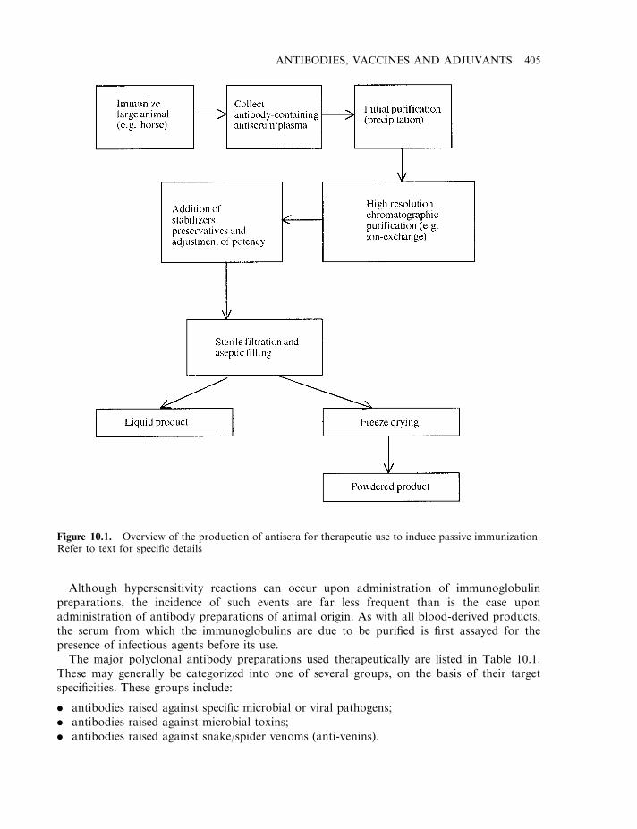
Although hypersensitivity reactions can occur upon administration of immunoglobulinpreparations, the incidence of such events are far less frequent than is the case uponadministration of antibody preparations of animal origin. As with all blood-derived products,the serum from which the immunoglobulins are due to be purified is first assayed for thepresence of infectious agents before its use.
The major polyclonal antibody preparations used therapeutically are listed in Table 10.1.These may generally be categorized into one of several groups, on the basis of their targetspecificities. These groups include:
. antibodies raised against specific microbial or viral pathogens;
. antibodies raised against microbial toxins;
. antibodies raised against snake/spider venoms (anti-venins).
ANTIBODIES, VACCINES AND ADJUVANTS 405
Figure 10.1. Overview of the production of antisera for therapeutic use to induce passive immunization.Refer to text for specific details
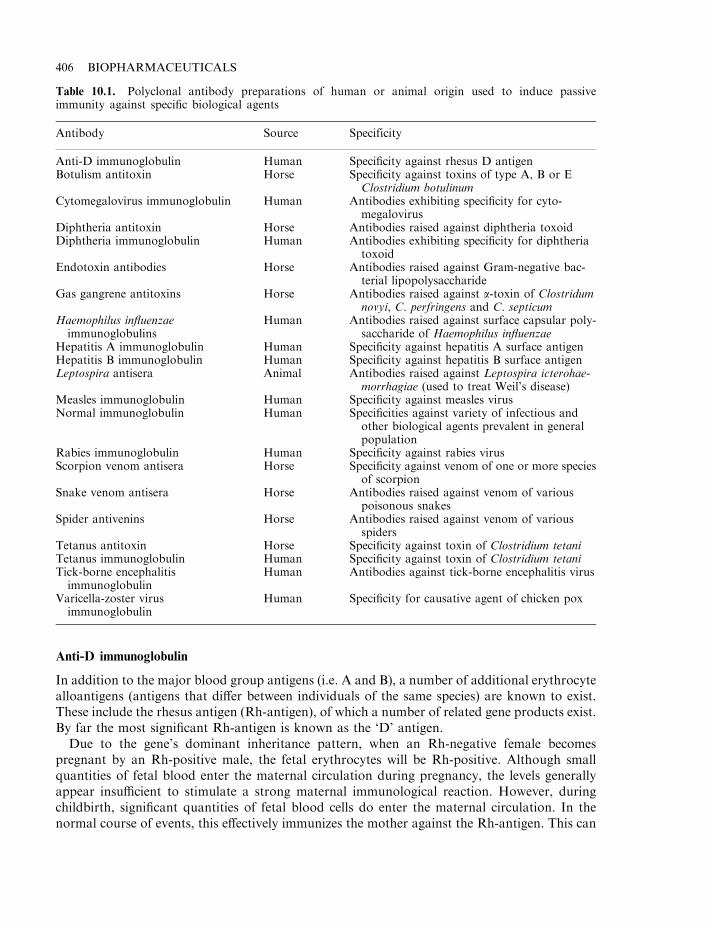
Anti-D immunoglobulin
In addition to the major blood group antigens (i.e. A and B), a number of additional erythrocytealloantigens (antigens that differ between individuals of the same species) are known to exist.These include the rhesus antigen (Rh-antigen), of which a number of related gene products exist.By far the most significant Rh-antigen is known as the ‘D’ antigen.
Due to the gene’s dominant inheritance pattern, when an Rh-negative female becomespregnant by an Rh-positive male, the fetal erythrocytes will be Rh-positive. Although smallquantities of fetal blood enter the maternal circulation during pregnancy, the levels generallyappear insufficient to stimulate a strong maternal immunological reaction. However, duringchildbirth, significant quantities of fetal blood cells do enter the maternal circulation. In thenormal course of events, this effectively immunizes the mother against the Rh-antigen. This can
406 BIOPHARMACEUTICALS
Table 10.1. Polyclonal antibody preparations of human or animal origin used to induce passiveimmunity against specific biological agents
Antibody Source Specificity
Anti-D immunoglobulin Human Specificity against rhesus D antigenBotulism antitoxin Horse Specificity against toxins of type A, B or E
Clostridium botulinumCytomegalovirus immunoglobulin Human Antibodies exhibiting specificity for cyto-
megalovirusDiphtheria antitoxin Horse Antibodies raised against diphtheria toxoidDiphtheria immunoglobulin Human Antibodies exhibiting specificity for diphtheria
toxoidEndotoxin antibodies Horse Antibodies raised against Gram-negative bac-
terial lipopolysaccharideGas gangrene antitoxins Horse Antibodies raised against a-toxin of Clostridum
novyi, C. perfringens and C. septicumHaemophilus influenzaeimmunoglobulins
Human Antibodies raised against surface capsular poly-saccharide of Haemophilus influenzae
Hepatitis A immunoglobulin Human Specificity against hepatitis A surface antigenHepatitis B immunoglobulin Human Specificity against hepatitis B surface antigenLeptospira antisera Animal Antibodies raised against Leptospira icterohae-
morrhagiae (used to treat Weil’s disease)Measles immunoglobulin Human Specificity against measles virusNormal immunoglobulin Human Specificities against variety of infectious and
other biological agents prevalent in generalpopulation
Rabies immunoglobulin Human Specificity against rabies virusScorpion venom antisera Horse Specificity against venom of one or more species
of scorpionSnake venom antisera Horse Antibodies raised against venom of various
poisonous snakesSpider antivenins Horse Antibodies raised against venom of various
spidersTetanus antitoxin Horse Specificity against toxin of Clostridium tetaniTetanus immunoglobulin Human Specificity against toxin of Clostridium tetaniTick-borne encephalitisimmunoglobulin
Human Antibodies against tick-borne encephalitis virus
Varicella-zoster virusimmunoglobulin
Human Specificity for causative agent of chicken pox
endanger fetal health during subsequent pregnancies, as maternal anti-Rh antibodies can crossthe placenta and enter the fetal circulation. Binding of anti-Rh antibodies to fetal erythrocytescan induce haemolysis. This results in medical complications, with the newborn baby oftensuffering from erythroblastosis fetalis (haemolytic disease of the newborn). Moreover, with eachsuccessive birth, the mother effectively receives booster shots of Rh-antigen. After severalpregnancies, maternal anti-Rh antibody levels can be sufficiently high to threaten fetal viability.
This scenario can be prevented by administration of anti-Rh (usually anti-D) antibodies toRh-negative mothers immediately upon the birth of a Rh-positive baby. The administeredantibodies bind the fetal Rh-positive erythrocytes, marking them for destruction before amaternal immunological reaction is triggered. Usually a dose of 200–300 mg of antibody isadministered immediately post-delivery. This would be sufficient to suppress an immuneresponse for up to 10ml of Rh-positive red blood cells.
The anti-D immunological preparations used are purified from the serum or plasma of Rh-negative individuals who have been immunized against Rh-D antigen. The purified antibodypreparations may be marketed as a liquid (shelf-life of 2 years when stored refrigerated) or as afreeze-dried preparation, which exhibits a shelf-life of up to 5 years.
Normal immunoglobulins
Normal immunoglobulins (often termed ‘immune globulins’ in the USA) represent antibodypreparations purified from the plasma, serum or placentas of normal, healthy donors. Bloodobtained from such individuals will contain a wide range of antibody specificities. The range ofspecificities were produced over many years, as the individual’s immune system came intocontact with various antigens, either naturally (infections) or artificially (by vaccination).
Normal immunoglobulin preparations are purified from pooled material obtained from 1000or more donors. They will generally contain antibodies against diphtheria, measles,poliomyelitis, hepatitis A, rubella and varicella. Normal immunoglobulin may, therefore, beused to provide passive immunization against these diseases.
Therapeutic antibody preparations generally contain in excess of 90% IgG, the concentrationof which is usually greater than 10 times the average starting concentration in the originalpooled material. The final protein concentration is generally 15–16% (w/v). Stabilizers (oftenglycine) may be added, as may preservatives—but only to product destined for sale as liquidpreparations. Aqueous preparations retain potency for 3 years when stored refrigerated.Alternatively, the product may be freeze-dried and stored in the final product container undervacuum or an inert gas (e.g. N2). Such preparations are generally assigned shelf-lives of 5 years.
Hepatitis B and tetanus immunoglobulin
Hepatitis B immunoglobulin is an example of an antibody preparation known to containantibodies displaying a specificity for a specific pathogen, in this case hepatitis B virus. HepatitisB immunoglobulins are purified from the serum or plasma of donors who exhibit high titres ofanti-hepatitis B surface antigen (HBsAg) antibodies. IgG represents in excess of 80% of the totalprotein in the product. As in the case of other immunoglobulin preparations, it may bemarketed as a liquid or freeze-dried preparation.
Hepatitis B immunoglobulins are administered to persons who have come into contact withhepatitis B, including newborn infants whose mothers have recently been infected with the virus.
ANTIBODIES, VACCINES AND ADJUVANTS 407
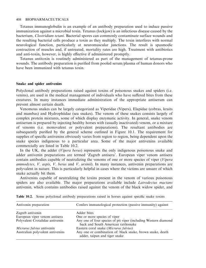
Tetanus immunoglobulin is an example of an antibody preparation used to induce passiveimmunization against a microbial toxin. Tetanus (lockjaw) is an infectious disease caused by thebacterium, Clostridium tetani. Bacterial spores can commonly contaminate surface wounds andthe resulting bacterial cells produce a toxin as they multiply. The toxin interferes with normalneurological function, particularly at neuromuscular junctions. The result is spasmodiccontraction of muscles and, if untreated, mortality rates are high. Treatment with antibioticsand anti-toxin, however, is highly effective if administered promptly.
Tetanus antitoxin is routinely administered as part of the management of tetanus-pronewounds. The antibody preparation is purified from pooled serum/plasma of human donors whohave been immunized with tetanus toxin.
Snake and spider antivenins
Polyclonal antibody preparations raised against toxins of poisonous snakes and spiders (i.e.venins), are used in the medical management of individuals who have suffered bites from thesecreatures. In many instances immediate administration of the appropriate antiserum canprevent almost certain death.
Venomous snakes can be largely categorized as Viperidae (Vipers), Elapidae (cobras, kraitsand mambas) and Hydrophiidae (sea snakes). The venom of these snakes consists largely ofcomplex protein mixtures, some of which display enzymatic activity. In general, snake venomantiserum is prepared by injecting healthy horses with (usually inactivated) venom, or a mixtureof venoms (i.e. monovalent or polyvalent preparations). The resultant antibodies aresubsequently purified by the general scheme outlined in Figure 10.1. The requirement forsupplies of specific antivenins obviously varies from region to region, being dependent upon thesnake species indigenous to a particular area. Some of the major antivenins availablecommercially are listed in Table 10.2.
In the UK, the adder (Vipera berus) represents the only indigenous poisonous snake andadder antivenin preparations are termed ‘Zagreb antisera’. European viper venom antiseracontain antibodies capable of neutralizing the venoms of one or more species of viper (Viperaammodytes, V. aspis, V. berus and V. ursinii). In many instances, antivenin preparations arepolyvalent in nature. This is particularly helpful in cases where the victims are unsure of whichsnake actually bit them.
Antivenins capable of neutralizing the toxins present in the venom of various poisonousspiders are also available. The major preparations available include Latrodectus mactansantivenin, which contains antibodies raised against the venom of the black widow spider, and
408 BIOPHARMACEUTICALS
Table 10.2. Some polyclonal antibody preparations raised in horses against specific snake toxins
Antivenin preparation Confers immunological protection (passive immunity) against
Zagreb antivenin Adder bitesEuropean viper venom antisera One or more species of viperPolyvalent Crotalidae antivenin Any one of four species of pit viper (including Western diamond
back and South American rattlesnakeMicrurus fulvius antivenin Eastern coral snake (Micrurus fulvius)Australian polyvalent antivenins Any one or combination of: black snake, brown snake, death
adder, taipan and tiger snake
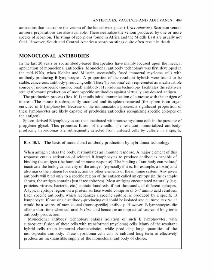
antivenins that neutralize the venom of the funnel-web spider (Atrax robustus). Scorpion venomantisera preparations are also available. These neutralize the venom produced by one or morespecies of scorpion. The stings of scorpions found in Africa and the Middle East are usually notfatal. However, South and Central American scorpion stings quite often result in death.
MONOCLONAL ANTIBODIES
In the last 20 years or so, antibody-based therapeutics have mainly focused upon the medicalapplication of monoclonal antibodies. Monoclonal antibody technology was first developed inthe mid-1970s, when Kohler and Milstein successfully fused immortal myeloma cells withantibody-producing B lymphocytes. A proportion of the resultant hybrids were found to bestable, cancerous, antibody-producing cells. These ‘hybridoma’ cells represented an inexhaustiblesource of monospecific (monoclonal) antibody. Hybridoma technology facilitates the relativelystraightforward production of monospecific antibodies against virtually any desired antigen.
The production process (Box 10.1) entails initial immunization of a mouse with the antigen ofinterest. The mouse is subsequently sacrificed and its spleen removed (the spleen is an organenriched in B lymphocytes. Because of the immunization process, a significant proportion ofthese lymphocytes are likely capable of producing antibodies recognizing specific epitopes onthe antigen).
Spleen-derived B lymphocytes are then incubated with mouse myeloma cells in the presence ofpropylene glycol. This promotes fusion of the cells. The resultant immortalized antibody-producing hybridomas are subsequently selected from unfused cells by culture in a specific
ANTIBODIES, VACCINES AND ADJUVANTS 409
Box 10.1. The basis of monoclonal antibody production by hybridoma technology
When antigen enters the body, it stimulates an immune response. A major element of thisresponse entails activation of selected B lymphocytes to produce antibodies capable ofbinding the antigen (the humoral immune response). The binding of antibody can reduce/inactivate the biological activity of the antigen (especially if it is, for example, a toxin) andalso marks the antigen for destruction by other elements of the immune system. Any givenantibody will bind only to a specific region of the antigen called an epitope (in the exampleshown, the antigen contains just three epitopes). Most antigens encountered naturally (e.g.proteins, viruses, bacteria, etc.) contain hundreds, if not thousands, of different epitopes.A typical epitope region on a protein surface would comprise of 5–7 amino acid residues.Each specific antibody, which recognizes a specific epitope, is produced by a specific Blymphocyte. If one single antibody-producing cell could be isolated and cultured in vitro, itwould be a source of monoclonal (monospecific) antibody. However, B lymphocytes dieafter a short time when cultured in vitro, and hence are an impractical source of long-termantibody production.
Monoclonal antibody technology entails isolation of such B lymphocytes, withsubsequent fusion of these cells with transformed (myeloma) cells. Many of the resultanthybrid cells retain immortal characteristics, while producing large quantities of themonospecific antibody. These hybridoma cells can be cultured long term to effectivelyproduce an inexhaustible supply of the monoclonal antibody of choice.
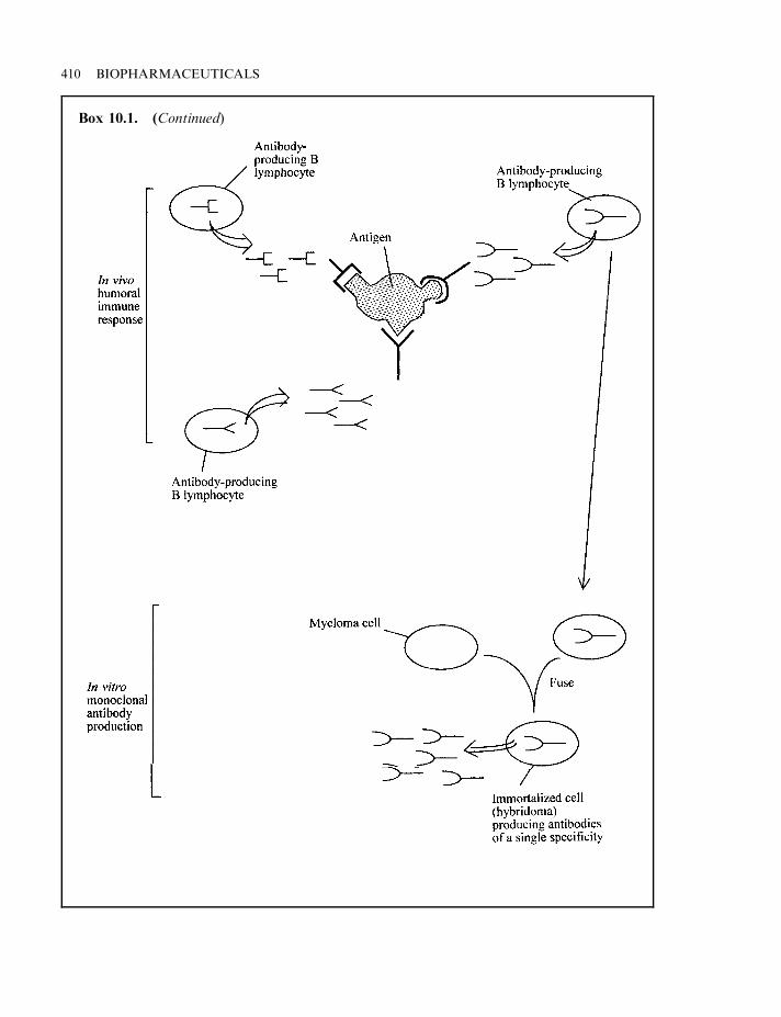
410 BIOPHARMACEUTICALS
Box 10.1. (Continued)
selection medium. Individual hybridomas can be separated from each other by simple dilutionand subsequently grown in culture, producing a clone. Individual clones can be screened toidentify which ones produce murine (monoclonal) antibody, which binds the antigen of interest.Appropriate clones are then selected and grown on a larger scale in order to producebiotechnologically useful quantities of antibody.
Production of monoclonals via hybridoma technology
The culture of hybridomas, thereby producing monoclonal antibodies, may be undertaken byascites production or by direct animal cell culture. Ascites production entails injection of thehybridoma cells into the peritoneal cavity of mice (the mice essentially serve as a livefermentation chamber). The transplanted hybridoma cells produce antibody as they grow.Ascitic fluid collects in the cavity, which contains high concentrations (up to 15mg/ml) of thedesired antibody. On average, 5ml of this fluid can be extracted per mouse. Most of the earliermonoclonal antibody preparations were produced in this manner, e.g. OKT-3, the firstmonoclonal antibody to be approved for therapeutic use by the FDA (see later), is producedusing this strategy.
Ascites production, however, suffers from a number of drawbacks. It is costly, and theproduct is contaminated by significant levels of various mouse proteins, rendering subsequentdownstream processing more complex. As a result, monoclonal antibody production bystandard animal cell culture techniques has become the method of choice for the production ofpharmaceutical-grade monoclonal antibody preparations.
Optimal fermentation parameters have been well established and air-lift, stirred tank, andhollow fibre systems have all been used. At commercial scale, fermentation volumes in excess of1000 litres can be used, which can yield 100 g or more of final product. While hybridoma growthis straightforward, production levels of antibody can be quite low compared with ascites-basedproduction systems. Typically, fermentation yields antibody concentrations of 0.1–0.5mg/ml.Removal of cells from the antibody-containing media is achieved by centrifugation or filtration.An ultrafiltration step is then normally undertaken in order to concentrate the filtrate by up to20-fold.
The concentrated extract is next subjected to 2–4 chromatographic purification steps(Figure 10.2). These serve not only to remove contaminating proteins but also to reduce orremove additional contaminants, including nucleic acids, endotoxin and adventitious agents.Affinity chromatography on protein A agarose columns is often employed as a high-resolutioninitial (IgG) purification step. The selectivity of protein A for most sub-species of IgG results ina high degree of purification, while maintaining a high product yield. This step is often followedby ion-exchange chromatography. Often both anion and cation exchange steps are undertakensequentially. In some instances, hydrophobic interaction chromatography is also employed. Gelfiltration is usually used as the final polishing step, yielding an essentially pure antibodypreparation.
Depending upon the intended application, the antibody may next be conjugated to specificmolecular tags (e.g. a radionuclide or toxin). Finally, stabilizing agents (e.g. buffer components,glycine or sometimes human serum albumin) are added to the product. This is then asepticallyfilled into the final containers after sterile filtration. The product is then usually freeze-dried andsealed under an atmosphere of an inert gas.
As with all biopharmaceuticals, strict and numerous QC checks are made upon the productduring all stages of manufacture. Because some issues of GMP are particularly relevant or
ANTIBODIES, VACCINES AND ADJUVANTS 411
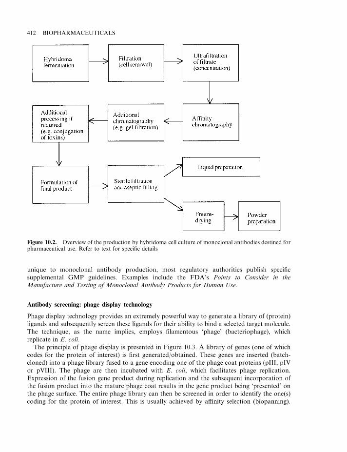
unique to monoclonal antibody production, most regulatory authorities publish specificsupplemental GMP guidelines. Examples include the FDA’s Points to Consider in theManufacture and Testing of Monoclonal Antibody Products for Human Use.
Antibody screening: phage display technology
Phage display technology provides an extremely powerful way to generate a library of (protein)ligands and subsequently screen these ligands for their ability to bind a selected target molecule.The technique, as the name implies, employs filamentous ‘phage’ (bacteriophage), whichreplicate in E. coli.
The principle of phage display is presented in Figure 10.3. A library of genes (one of whichcodes for the protein of interest) is first generated/obtained. These genes are inserted (batch-cloned) into a phage library fused to a gene encoding one of the phage coat proteins (pIII, pIVor pVIII). The phage are then incubated with E. coli, which facilitates phage replication.Expression of the fusion gene product during replication and the subsequent incorporation ofthe fusion product into the mature phage coat results in the gene product being ‘presented’ onthe phage surface. The entire phage library can then be screened in order to identify the one(s)coding for the protein of interest. This is usually achieved by affinity selection (biopanning).
412 BIOPHARMACEUTICALS
Figure 10.2. Overview of the production by hybridoma cell culture of monoclonal antibodies destined forpharmaceutical use. Refer to text for specific details
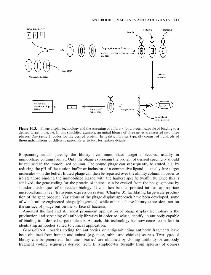
Biopanning entails passing the library over immobilized target molecules, usually inimmobilized column format. Only the phage expressing the protein of desired specificity shouldbe retained in the immobilized column. The bound phage can subsequently be eluted, e.g. byreducing the pH of the elution buffer or inclusion of a competitive ligand—usually free targetmolecules— in the buffer. Eluted phage can then be repassed over the affinity column in order toisolate those binding the immobilized ligand with the highest specificity/affinity. Once this isachieved, the gene coding for the protein of interest can be excised from the phage genome bystandard techniques of molecular biology. It can then be incorporated into an appropriatemicrobial/animal cell/transgenic expression system (Chapter 3), facilitating large-scale produc-tion of the gene product. Variations of the phage display approach have been developed, someof which utilize engineered phage (phagemids), while others achieve library expression, not onthe surface of phage but on the surface of bacteria.
Amongst the first and still most prominent application of phage display technology is theproduction and screening of antibody libraries in order to isolate/identify an antibody capableof binding to a desired target molecule. As such, this technology has now come to the fore inidentifying antibodies suited to clinical application.
Genes/cDNA libraries coding for antibodies or antigen-binding antibody fragments havebeen obtained from human and animal (e.g. mice, rabbit and chicken) sources. Two types oflibrary can be generated. ‘Immune libraries’ are obtained by cloning antibody or antibodyfragment coding sequences derived from B lymphocytes (usually from spleens) of donors
ANTIBODIES, VACCINES AND ADJUVANTS 413
Figure 10.3. Phage display technology and the screening of a library for a protein capable of binding to adesired target molecule. In this simplified example, an initial library of three genes are inserted into threephages. One (gene 2) codes for the desired protein. In reality, libraries typically consist of hundreds ofthousands/millions of different genes. Refer to text for further details
previously immunized with the target antigen. A high number of hits (positive clones) should beobtained from such libraries.
Non-immune libraries are produced in a similar fashion, but using B lymphocytes from non-immunized donors as a source of antibody genes. This approach becomes necessary if initialimmunization with the antigen of interest is not possible (due to ethical considerations, forexample). While such libraries will generate a lower number of positive clones, they can bescreened against multiple antigens. Such native non-immune libraries can also be the startingpoint for the generation of so-called ‘synthetic immune libraries’. Their generation entails initialin vitro engineering of the non-immune library of antibody genes in order to increase still furtherthe level of antibody diversity generated.
Therapeutic application of monoclonal antibodies
The unrivalled specificity of monoclonal antibodies, coupled with their relatively straightfor-ward production and their continuity of supply, renders them attractive biochemical tools.Therapeutically, they represent by far the single largest category of biopharmaceuticalsubstances under investigation. Several hundred such preparations are currently undergoingpre-clinical and clinical trials. Throughout the 1980s the focus of attention rested upon their useas either in vivo imaging (i.e. diagnostic) agents or direct therapeutic agents. Initial studiescentred mainly around cancer, but monoclonal antibody preparations are now used in a varietyof other medical circumstances (Table 10.3).
All in vivo diagnostic/therapeutic applications are dependent upon the selective interaction ofa monoclonal antibody with a specific target cell type in the body (e.g. a cancer cell). Therefore,a prerequisite to application of monoclonal antibody-based products in this way is theidentification of a cell surface antigen unique to the target cell type (Figure 10.4). Once identifiedand characterized, monoclonal antibodies may be raised against that unique surface antigen(USAg). The specificity of antibody-antigen binding should ensure that, once injected into thebody, the antibody will selectively congregate on the surface of only the target cells. Dependingupon the specific intended therapeutic/diagnostic application, the antibodies employed mayhave nothing attached to them or may be conjugated to a radioisotope, drug or toxin(Figure 10.4). With the latter approaches, the antibody is used as a ‘magic bullet’, delivering aradioactive/drug load to specific cells in the body. All of these various strategies have beenadopted in practice and specific examples will be provided in subsequent sections of this chapter.
In the context of antibody-mediated cell targeting, it is also important to appreciate thatbinding of an antibody to a USAg can, by itself, trigger a number of responses. In some instances,the antibody–antigen (Ab–USAg) complex is quickly internalized. In other instances, the Ab-USAg complex is shed from the cell surface, while in yet other cases binding induces neitherresponse. The specific response triggered is generally only determined by direct experimentation.The induction of Ab–USAg shedding renders an anti-TSA antibody clinically useless.
414 BIOPHARMACEUTICALS
Table 10.3. Range of clinical applications of monoclonal antibodies
Induction of passive immunityDiagnostic imaging (e.g. of cancer, infectious diseases, cardiovascular disease anddeep vein thrombosis)
Therapy for cancers, transplantations and cardiovascular disease
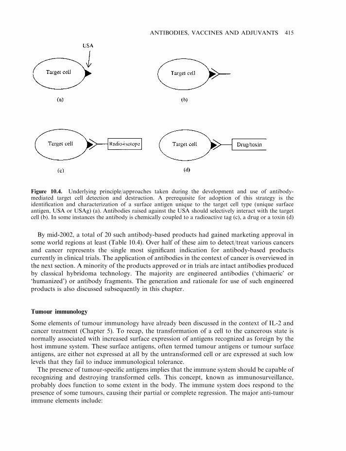
By mid-2002, a total of 20 such antibody-based products had gained marketing approval insome world regions at least (Table 10.4). Over half of these aim to detect/treat various cancersand cancer represents the single most significant indication for antibody-based productscurrently in clinical trials. The application of antibodies in the context of cancer is overviewed inthe next section. A minority of the products approved or in trials are intact antibodies producedby classical hybridoma technology. The majority are engineered antibodies (‘chimaeric’ or‘humanized’) or antibody fragments. The generation and rationale for use of such engineeredproducts is also discussed subsequently in this chapter.
Tumour immunology
Some elements of tumour immunology have already been discussed in the context of IL-2 andcancer treatment (Chapter 5). To recap, the transformation of a cell to the cancerous state isnormally associated with increased surface expression of antigens recognized as foreign by thehost immune system. These surface antigens, often termed tumour antigens or tumour surfaceantigens, are either not expressed at all by the untransformed cell or are expressed at such lowlevels that they fail to induce immunological tolerance.
The presence of tumour-specific antigens implies that the immune system should be capable ofrecognizing and destroying transformed cells. This concept, known as immunosurveillance,probably does function to some extent in the body. The immune system does respond to thepresence of some tumours, causing their partial or complete regression. The major anti-tumourimmune elements include:
ANTIBODIES, VACCINES AND ADJUVANTS 415
Figure 10.4. Underlying principle/approaches taken during the development and use of antibody-mediated target cell detection and destruction. A prerequisite for adoption of this strategy is theidentification and characterization of a surface antigen unique to the target cell type (unique surfaceantigen, USA or USAg) (a). Antibodies raised against the USA should selectively interact with the targetcell (b). In some instances the antibody is chemically coupled to a radioactive tag (c), a drug or a toxin (d)
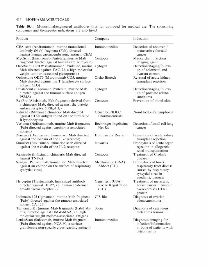
416 BIOPHARMACEUTICALS
Table 10.4. Monoclonal/engineered antibodies thus far approved for medical use. The sponsoringcompanies and therapeutic indications are also listed
Product Company Indication
CEA-scan (Arcitumomab, murine monoclonalantibody (Mab) fragment (Fab), directedagainst human carcinoembryonic antigen, CEA)
Immunomedics Detection of recurrent/metastatic colorectalcancer
MyoScint (Imiciromab-Pentetate, murine Mabfragment directed against human cardiac myosin)
Centocor Myocardial infarctionimaging agent
OncoScint CR/OV (Satumomab Pendetide, murineMab directed against TAG-72, a high molecularweight tumour-associated glycoprotein)
Cytogen Detection/staging/follow-up of colorectal andovarian cancers
Orthoclone OKT3 (Muromomab CD3, murineMab directed against the T lymphocyte surfaceantigen CD3)
Ortho Biotech Reversal of acute kidneytransplant rejection
ProstaScint (Capromab Pentetate, murine Mabdirected against the tumour surface antigenPSMA)
Cytogen Detection/staging/follow-up of prostate adeno-carcinoma
ReoPro (Abciximab, Fab fragments derived froma chimaeric Mab, directed against the plateletsurface receptor GPIIb/IIIa)
Centocor Prevention of blood clots
Rituxan (Rituximab chimaeric Mab directedagainst CD20 antigen found on the surface ofB lymphocytes)
Genentech/IDECPharmaceuticals
Non-Hodgkin’s lymphoma
Verluma (Nofetumomab, murine Mab fragments(Fab) directed against carcinoma-associatedantigen)
Boehringer Ingelheim/NeoRx
Detection of small cell lungcancer
Zenapax (Daclizumab, humanized Mab directedagainst the a-chain of the IL-2 receptor)
Hoffman La Roche Prevention of acute kidneytransplant rejection
Simulect (Basiliximab, chimaeric Mab directedagainst the a-chain of the IL-2 receptor)
Novartis Prophylaxis of acute organrejection in allogeneicrenal transplantation
Remicade (Infliximab, chimaeric Mab directedagainst TNF-a)
Centocor Treatment of Crohn’sdisease
Synagis (Palivizumab, humanized Mab directedagainst an epitope on the surface of respiratorysyncytial virus)
MedImmune (USA)Abbott (EU)
Prophylaxis of lowerrespiratory tract diseasecaused by respiratorysyncytial virus inpaediatric patients
Herceptin (Trastuzumab, humanized antibodydirected against HER2, i.e. human epidermalgrowth factor receptor 2)
Genentech (USA)Roche Registration(EU)
Treatment of metastaticbreast cancer if tumouroverexpresses HER2protein
Indimacis 125 (Igovomab, murine Mab fragment(Fab2) directed against the tumour-associatedantigen CA 125)
CIS Bio Diagnosis of ovarianadenocarcimona
Tecnemab KI (murine Mab fragments (Fab/Fab2mix) directed against HMW-MAA, i.e. highmolecular weight meloma-associated antigen)
Sorin Diagnosis of cutaneousmelanoma lesions
LeukoScan (Sulesomab, murine Mab fragment(Fab) directed against NCA 90, a surfacegranulocyte non-specific cross-reacting antigen)
Immunomedics Diagnostic imaging forinfection/inflammationin bone of patients withosteomyelitis
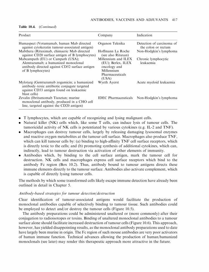
. T lymphocytes, which are capable of recognizing and lysing malignant cells.
. Natural killer (NK) cells which, like some T cells, can induce lysis of tumour cells. Thetumoricidal activity of NK cells is potentiated by various cytokines (e.g. IL-2 and TNF).
. Macrophages can destroy tumour cells, largely by releasing damaging lysosomal enzymesand reactive oxygen metabolites at the tumour cell surface. Macrophages also produce TNF,which can kill tumour cells by: (a) binding to high-affinity TNF cell surface receptors, whichis directly toxic to the cells; and (b) promoting synthesis of additional cytokines, which can,indirectly, lead to tumour destruction via activation of other elements of immunity.
. Antibodies which, by binding to the cell surface antigen, mark the tumour cell fordestruction. NK cells and macrophages express cell surface receptors which bind to theantibody Fc region (Box 10.2). Thus, antibody bound to tumour antigens directs theseimmune elements directly to the tumour surface. Antibodies also activate complement, whichis capable of directly lysing tumour cells.
The methods by which some transformed cells likely escape immune detection have already beenoutlined in detail in Chapter 5.
Antibody-based strategies for tumour detection/destruction
Clear identification of tumour-associated antigens would facilitate the production ofmonoclonal antibodies capable of selectively binding to tumour tissue. Such antibodies couldbe employed to detect and/or destroy the tumour cells (Figure 10.5).
The antibody preparations could be administered unaltered or (more commonly) after theirconjugation to radioisotopes or toxins. Binding of unaltered monoclonal antibodies to a tumoursurface alone should facilitate increased destruction of tumour cells (Figure 10.6). This approach,however, has yielded disappointing results, as the monoclonal antibody preparations used to datehave largely been murine in origin. The Fc region of such mouse antibodies are very poor activatorsof human immune function. Technical advances allowing the production of human/humanizedmonoclonals (see later) may render this therapeutic approach more attractive in the future.
ANTIBODIES, VACCINES AND ADJUVANTS 417
Table 10.4. (Continued)
Product Company Indication
Humaspect (Votumumab, human Mab directedagainst cytokeratin tumour-associated antigen)
Organon Teknika Detection of carcinoma ofthe colon or rectum
Mabthera (Rituximab, chimaeric Mab directedagainst CD20 surface antigen of B lymphocytes)
Hoffmann La Roche(see also Rituxan)
Non-Hodgkin’s lymphoma
Mabcampath (EU) or Campath (USA);Alemtuzumab; a humanized monoclonalantibody directed against CD52 surface antigenof B lymphocytes)
Millennium and ILEX(EU); Berlex, ILEXoncology andMillenniumPharmaceuticals(USA)
Chronic lymphocyticleukaemia
Mylotarg (Gemtuzumab zogamicin; a humanizedantibody–toxic antibiotic conjugate targetedagainst CD33 antigen found on leukaemicblast cells)
Wyeth Ayerst Acute myeloid leukaemia
Zevalin (Ibritumomab Tiuxetan; murinemonoclonal antibody, produced in a CHO cellline, targeted against the CD20 antigen)
IDEC Pharmaceuticals Non-Hodgkin’s lymphoma
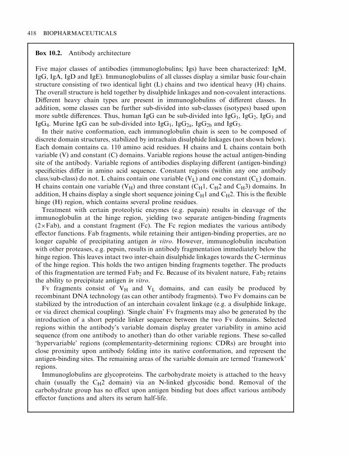
418 BIOPHARMACEUTICALS
Box 10.2. Antibody architecture
Five major classes of antibodies (immunoglobulins; Igs) have been characterized: IgM,IgG, IgA, IgD and IgE). Immunoglobulins of all classes display a similar basic four-chainstructure consisting of two identical light (L) chains and two identical heavy (H) chains.The overall structure is held together by disulphide linkages and non-covalent interactions.Different heavy chain types are present in immunoglobulins of different classes. Inaddition, some classes can be further sub-divided into sub-classes (isotypes) based uponmore subtle differences. Thus, human IgG can be sub-divided into IgG1, IgG2, IgG3 andIgG4. Murine IgG can be sub-divided into IgG1, IgG2a, IgG2b and IgG3.
In their native conformation, each immunoglobulin chain is seen to be composed ofdiscrete domain structures, stabilized by intrachain disulphide linkages (not shown below).Each domain contains ca. 110 amino acid residues. H chains and L chains contain bothvariable (V) and constant (C) domains. Variable regions house the actual antigen-bindingsite of the antibody. Variable regions of antibodies displaying different (antigen-binding)specificities differ in amino acid sequence. Constant regions (within any one antibodyclass/sub-class) do not. L chains contain one variable (VL) and one constant (CL) domain.H chains contain one variable (VH) and three constant (CH1, CH2 and CH3) domains. Inaddition, H chains display a single short sequence joining CH1 and CH2. This is the flexiblehinge (H) region, which contains several proline residues.
Treatment with certain proteolytic enzymes (e.g. papain) results in cleavage of theimmunoglobulin at the hinge region, yielding two separate antigen-binding fragments(2�Fab), and a constant fragment (Fc). The Fc region mediates the various antibodyeffector functions. Fab fragments, while retaining their antigen-binding properties, are nolonger capable of precipitating antigen in vitro. However, immunoglobulin incubationwith other proteases, e.g. pepsin, results in antibody fragmentation immediately below thehinge region. This leaves intact two inter-chain disulphide linkages towards the C-terminusof the hinge region. This holds the two antigen binding fragments together. The productsof this fragmentation are termed Fab2 and Fc. Because of its bivalent nature, Fab2 retainsthe ability to precipitate antigen in vitro.
Fv fragments consist of VH and VL domains, and can easily be produced byrecombinant DNA technology (as can other antibody fragments). Two Fv domains can bestabilized by the introduction of an interchain covalent linkage (e.g. a disulphide linkage,or via direct chemical coupling). ‘Single chain’ Fv fragments may also be generated by theintroduction of a short peptide linker sequence between the two Fv domains. Selectedregions within the antibody’s variable domain display greater variability in amino acidsequence (from one antibody to another) than do other variable regions. These so-called‘hypervariable’ regions (complementarity-determining regions: CDRs) are brought intoclose proximity upon antibody folding into its native conformation, and represent theantigen-binding sites. The remaining areas of the variable domain are termed ‘framework’regions.
Immunoglobulins are glycoproteins. The carbohydrate moiety is attached to the heavychain (usually the CH2 domain) via an N-linked glycosidic bond. Removal of thecarbohydrate group has no effect upon antigen binding but does affect various antibodyeffector functions and alters its serum half-life.
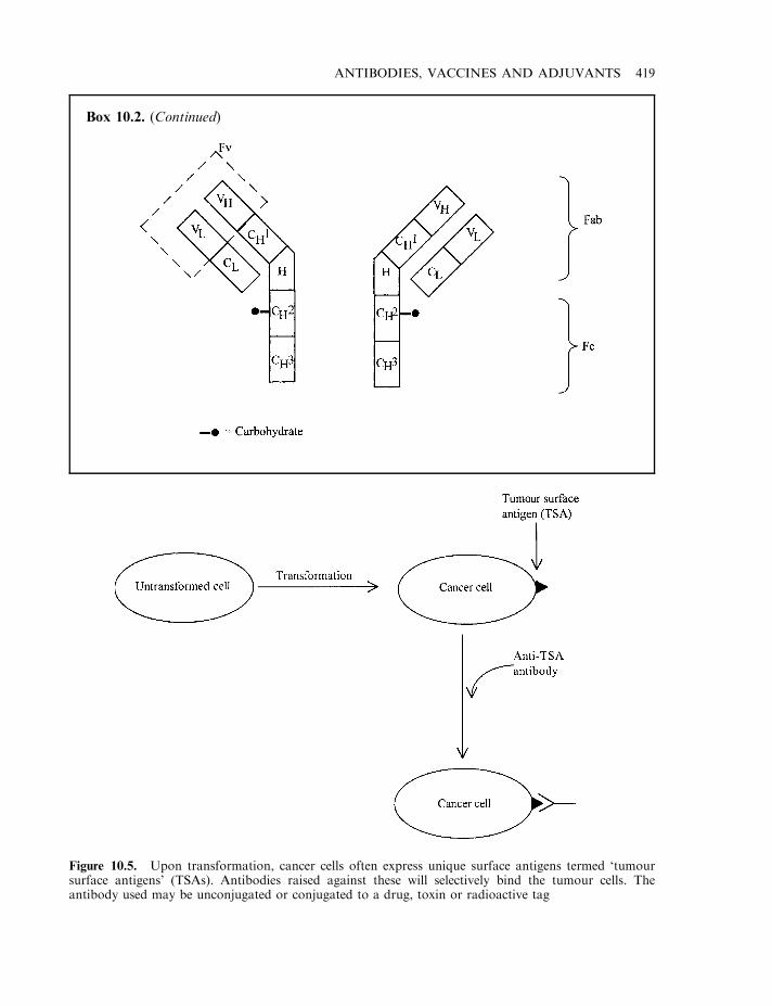
ANTIBODIES, VACCINES AND ADJUVANTS 419
Box 10.2. (Continued)
Figure 10.5. Upon transformation, cancer cells often express unique surface antigens termed ‘tumoursurface antigens’ (TSAs). Antibodies raised against these will selectively bind the tumour cells. Theantibody used may be unconjugated or conjugated to a drug, toxin or radioactive tag
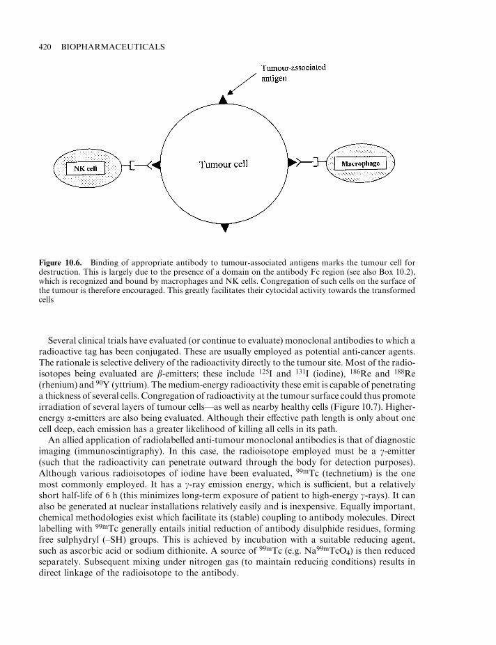
Several clinical trials have evaluated (or continue to evaluate) monoclonal antibodies to which aradioactive tag has been conjugated. These are usually employed as potential anti-cancer agents.The rationale is selective delivery of the radioactivity directly to the tumour site. Most of the radio-isotopes being evaluated are b-emitters; these include 125I and 131I (iodine), 186Re and 188Re(rhenium) and 90Y (yttrium). The medium-energy radioactivity these emit is capable of penetratinga thickness of several cells. Congregation of radioactivity at the tumour surface could thus promoteirradiation of several layers of tumour cells—as well as nearby healthy cells (Figure 10.7). Higher-energy a-emitters are also being evaluated. Although their effective path length is only about onecell deep, each emission has a greater likelihood of killing all cells in its path.
An allied application of radiolabelled anti-tumour monoclonal antibodies is that of diagnosticimaging (immunoscintigraphy). In this case, the radioisotope employed must be a g-emitter(such that the radioactivity can penetrate outward through the body for detection purposes).Although various radioisotopes of iodine have been evaluated, 99mTc (technetium) is the onemost commonly employed. It has a g-ray emission energy, which is sufficient, but a relativelyshort half-life of 6 h (this minimizes long-term exposure of patient to high-energy g-rays). It canalso be generated at nuclear installations relatively easily and is inexpensive. Equally important,chemical methodologies exist which facilitate its (stable) coupling to antibody molecules. Directlabelling with 99mTc generally entails initial reduction of antibody disulphide residues, formingfree sulphydryl (–SH) groups. This is achieved by incubation with a suitable reducing agent,such as ascorbic acid or sodium dithionite. A source of 99mTc (e.g. Na99mTcO4) is then reducedseparately. Subsequent mixing under nitrogen gas (to maintain reducing conditions) results indirect linkage of the radioisotope to the antibody.
420 BIOPHARMACEUTICALS
Figure 10.6. Binding of appropriate antibody to tumour-associated antigens marks the tumour cell fordestruction. This is largely due to the presence of a domain on the antibody Fc region (see also Box 10.2),which is recognized and bound by macrophages and NK cells. Congregation of such cells on the surface ofthe tumour is therefore encouraged. This greatly facilitates their cytocidal activity towards the transformedcells
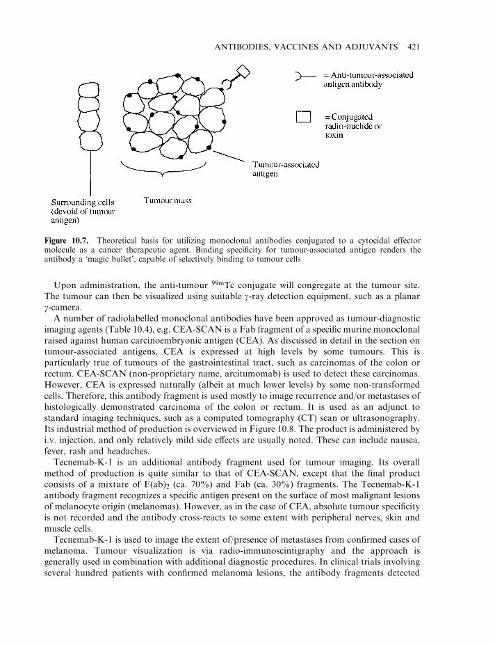
Upon administration, the anti-tumour 99mTc conjugate will congregate at the tumour site.The tumour can then be visualized using suitable g-ray detection equipment, such as a planarg-camera.
A number of radiolabelled monoclonal antibodies have been approved as tumour-diagnosticimaging agents (Table 10.4), e.g. CEA-SCAN is a Fab fragment of a specific murine monoclonalraised against human carcinoembryonic antigen (CEA). As discussed in detail in the section ontumour-associated antigens, CEA is expressed at high levels by some tumours. This isparticularly true of tumours of the gastrointestinal tract, such as carcinomas of the colon orrectum. CEA-SCAN (non-proprietary name, arcitumomab) is used to detect these carcinomas.However, CEA is expressed naturally (albeit at much lower levels) by some non-transformedcells. Therefore, this antibody fragment is used mostly to image recurrence and/or metastases ofhistologically demonstrated carcinoma of the colon or rectum. It is used as an adjunct tostandard imaging techniques, such as a computed tomography (CT) scan or ultrasonography.Its industrial method of production is overviewed in Figure 10.8. The product is administered byi.v. injection, and only relatively mild side effects are usually noted. These can include nausea,fever, rash and headaches.
Tecnemab-K-1 is an additional antibody fragment used for tumour imaging. Its overallmethod of production is quite similar to that of CEA-SCAN, except that the final productconsists of a mixture of F(ab)2 (ca. 70%) and Fab (ca. 30%) fragments. The Tecnemab-K-1antibody fragment recognizes a specific antigen present on the surface of most malignant lesionsof melanocyte origin (melanomas). However, as in the case of CEA, absolute tumour specificityis not recorded and the antibody cross-reacts to some extent with peripheral nerves, skin andmuscle cells.
Tecnemab-K-1 is used to image the extent of/presence of metastases from confirmed cases ofmelanoma. Tumour visualization is via radio-immunoscintigraphy and the approach isgenerally used in combination with additional diagnostic procedures. In clinical trials involvingseveral hundred patients with confirmed melanoma lesions, the antibody fragments detected
ANTIBODIES, VACCINES AND ADJUVANTS 421
Figure 10.7. Theoretical basis for utilizing monoclonal antibodies conjugated to a cytocidal effectormolecule as a cancer therapeutic agent. Binding specificity for tumour-associated antigen renders theantibody a ‘magic bullet’, capable of selectively binding to tumour cells
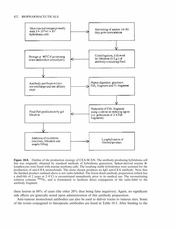
these lesions in 80% of cases (the other 20% thus being false negatives). Again, no significantside effects are generally noted upon administration of this antibody preparation.
Anti-tumour monoclonal antibodies can also be used to deliver toxins to tumour sites. Someof the toxins conjugated to therapeutic antibodies are listed in Table 10.5. After binding to the
422 BIOPHARMACEUTICALS
Figure 10.8. Outline of the production strategy of CEA-SCAN. The antibody-producing hybridoma cellline was originally obtained by standard methods of hybridoma generation. Spleen-derived murine B-lymphocytes were fused with murine myeloma calls. The resulting stable hybridomas were screened for theproduction of anti-CEA monoclonals. The clone chosen produces an IgG anti-CEA antibody. Note thatthe finished product outlined above is not radio-labelled. The freeze-dried antibody preparation (which hasa shelf-life of 2 years at 2–88C) is reconstituted immediately prior to its medical use. The reconstitutingsolution contains 99mTc, and is formulated to facilitate direct conjugation of the radio-label to theantibody fragment
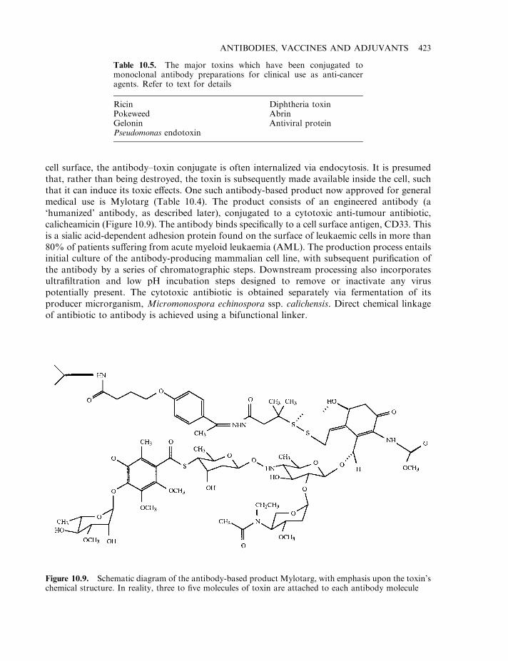
cell surface, the antibody–toxin conjugate is often internalized via endocytosis. It is presumedthat, rather than being destroyed, the toxin is subsequently made available inside the cell, suchthat it can induce its toxic effects. One such antibody-based product now approved for generalmedical use is Mylotarg (Table 10.4). The product consists of an engineered antibody (a‘humanized’ antibody, as described later), conjugated to a cytotoxic anti-tumour antibiotic,calicheamicin (Figure 10.9). The antibody binds specifically to a cell surface antigen, CD33. Thisis a sialic acid-dependent adhesion protein found on the surface of leukaemic cells in more than80% of patients suffering from acute myeloid leukaemia (AML). The production process entailsinitial culture of the antibody-producing mammalian cell line, with subsequent purification ofthe antibody by a series of chromatographic steps. Downstream processing also incorporatesultrafiltration and low pH incubation steps designed to remove or inactivate any viruspotentially present. The cytotoxic antibiotic is obtained separately via fermentation of itsproducer microrganism, Micromonospora echinospora ssp. calichensis. Direct chemical linkageof antibiotic to antibody is achieved using a bifunctional linker.
ANTIBODIES, VACCINES AND ADJUVANTS 423
Table 10.5. The major toxins which have been conjugated tomonoclonal antibody preparations for clinical use as anti-canceragents. Refer to text for details
Ricin Diphtheria toxinPokeweed AbrinGelonin Antiviral proteinPseudomonas endotoxin
Figure 10.9. Schematic diagram of the antibody-based product Mylotarg, with emphasis upon the toxin’schemical structure. In reality, three to five molecules of toxin are attached to each antibody molecule
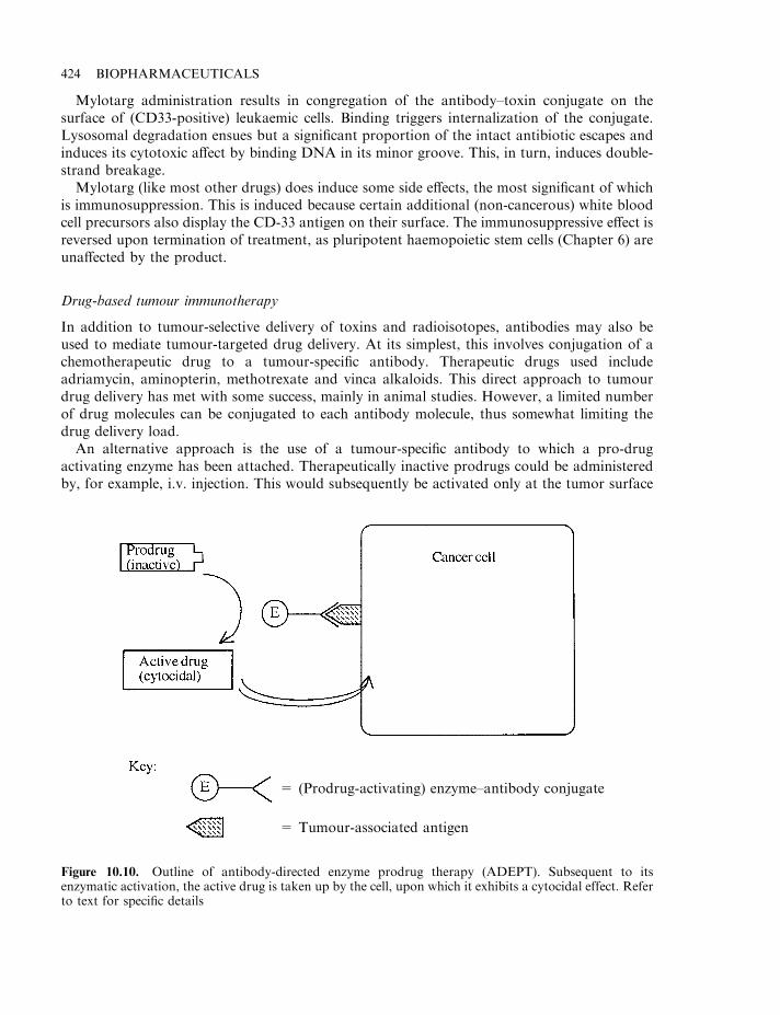
Mylotarg administration results in congregation of the antibody–toxin conjugate on thesurface of (CD33-positive) leukaemic cells. Binding triggers internalization of the conjugate.Lysosomal degradation ensues but a significant proportion of the intact antibiotic escapes andinduces its cytotoxic affect by binding DNA in its minor groove. This, in turn, induces double-strand breakage.
Mylotarg (like most other drugs) does induce some side effects, the most significant of whichis immunosuppression. This is induced because certain additional (non-cancerous) white bloodcell precursors also display the CD-33 antigen on their surface. The immunosuppressive effect isreversed upon termination of treatment, as pluripotent haemopoietic stem cells (Chapter 6) areunaffected by the product.
Drug-based tumour immunotherapy
In addition to tumour-selective delivery of toxins and radioisotopes, antibodies may also beused to mediate tumour-targeted drug delivery. At its simplest, this involves conjugation of achemotherapeutic drug to a tumour-specific antibody. Therapeutic drugs used includeadriamycin, aminopterin, methotrexate and vinca alkaloids. This direct approach to tumourdrug delivery has met with some success, mainly in animal studies. However, a limited numberof drug molecules can be conjugated to each antibody molecule, thus somewhat limiting thedrug delivery load.
An alternative approach is the use of a tumour-specific antibody to which a pro-drugactivating enzyme has been attached. Therapeutically inactive prodrugs could be administeredby, for example, i.v. injection. This would subsequently be activated only at the tumor surface
424 BIOPHARMACEUTICALS
Figure 10.10. Outline of antibody-directed enzyme prodrug therapy (ADEPT). Subsequent to itsenzymatic activation, the active drug is taken up by the cell, upon which it exhibits a cytocidal effect. Referto text for specific details
= (Prodrug-activating) enzyme–antibody conjugate
= Tumour-associated antigen
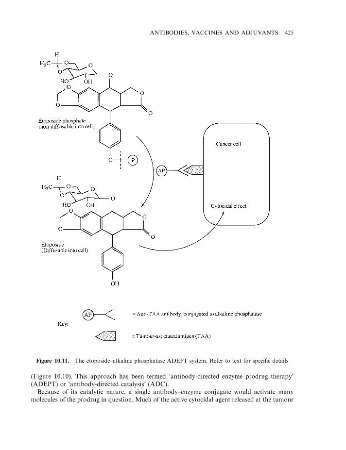
(Figure 10.10). This approach has been termed ‘antibody-directed enzyme prodrug therapy’(ADEPT) or ‘antibody-directed catalysis’ (ADC).
Because of its catalytic nature, a single antibody–enzyme conjugate would activate manymolecules of the prodrug in question. Much of the active cytocidal agent released at the tumour
ANTIBODIES, VACCINES AND ADJUVANTS 425
Figure 10.11. The etoposide–alkaline phosphatase ADEPT system. Refer to text for specific details
surface would be taken up by the tumour cells via simple diffusion or carrier-mediated activetransport. Administration of etoposide in prodrug form exemplifies this approach(Figure 10.11). Etoposide (C29H32O13; molecular mass 588.6) is a semi-synthetic derivative ofpodophyllotoxin, produced naturally by the North American plant, Podophyllum peltatum. It isused as an anti-cancer agent. Its cellular uptake is diffusion-dependent and, once inside the cell,it exerts its cytocidal effect. Phosphorylated etoposide is non-diffusable and hence represents aninactive prodrug form of etoposide (attachment of a charged group to most diffusion-dependentdrugs prevents their cellular uptake). Alkaline phosphatase, however, can cleave the phosphategroup, releasing free cytocidal agent. Administration of a tumour-detecting antibody–alkalinephosphatase conjugate thus effectively targets the enzyme to the tumour surface. Subsequentadministration of phosphorylated etoposide results in etoposide liberation at the tumoursurface, which can then enter tumour cells by diffusion (Figure 10.11). Various other prodrug–enzyme combinations have now been developed, including phenoxyacetamide derivatives ofdoxorubicin (activated by penicillin amidase) and 5-fluorocytosine (activated by cytosinedeaminase).
Prodrugs used should be inexpensive, readily available and stable to chemical/enzymaticdegradation in vivo. The enzymes used should also be stable under physiological conditions,display a reasonable turnover number in vivo and not be dependent upon a co-factor for activity.Mammalian enzymes would be likely less immunogenic than microbial enzymes. However, theuse of a prodrug capable of being activated by a mammalian enzyme can lead to complicationsif that enzyme’s (human) endogenous counterpart is capable of activating the drug at sitesdistant from the tumour.
First-generation anti-tumour antibodies: clinical disappointment
Despite the scientific elegance of the antibody-mediated approach to tumour detection/destruction, initial clinical trials proved disappointing. A number of factors contributed to theirpoor therapeutic performance, particularly against solid tumours. Most such factors relatedirectly or indirectly to the fact that the first generation of such drugs utilized whole monoclonalantibody preparations of murine origin. These factors include:
. insufficient information exists regarding tumour antigens;
. murine monoclonals prompt an immune response when administered to humans;
. poor penetration of tumour mass by antibody;
. murine monoclonals display a relatively short half-life when administered to man;
. poor recognition of murine antibody Fc domain by human effector mechanisms.
Tumour-associated antigens
In this text, the term ‘tumour-associated antigen’ represents any antigen associated with anycancer cell, no matter what factor(s) originally prompted cellular transformation (in somecircles, the term ‘tumour-associated antigen’ is often applied more specifically—to antigensassociated with virally transformed cells). Identification of tumour-associated antigens forms acore requirement for effective tumour immunotherapy. Identification of such tumour-associatedantigens remains a very active area of biomedical research. From the limited data generated todate, tumor-associated antigens can generally be categorized into one of three groups(Table 10.6).
426 BIOPHARMACEUTICALS
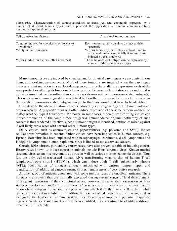
Many tumour types are induced by chemical and/or physical carcinogens we encounter in ourliving and working environments. Most of these tumours are initiated when the carcinogeninduces a point mutation in a nucleotide sequence, thus perhaps altering expression levels of thegene product or altering its functional characteristics. Because such mutations are random, it isnot surprising that each resulting tumour displays its own unique tumour-associated antigen(s).This renders an immunological approach to detection/therapy impractical in such instances, asthe specific tumour-associated antigens unique to that case would first have to be identified.
In contrast to the above situation, cancers induced by viruses generally exhibit immunologicalcross-reactivity. Any specific virus will often induce expression of the same tumour antigen, nomatter what cell type it transforms. Moreover, in some cases, different transforming viruses caninduce production of the same tumor antigen(s). Immunodetection/immunotherapy of suchcancers is thus rendered attractive. Once a tumour antigen is identified, antibodies raised againstit will likely cross-react with several other tumour types.
DNA viruses, such as adenoviruses and papovaviruses (e.g. polyoma and SV40), inducecellular transformation in rodents. Other viruses have been implicated in human cancers, e.g.Epstein–Barr virus has been implicated with nasopharyngeal carcinoma, b-cell lymphomas andHodgkin’s lymphoma; human papilloma virus is linked to most cervical cancers.
Certain RNA viruses, particularly retroviruses, have also proven capable of inducing cancer.Retroviruses known to induce cancer in animals include Rous sarcoma virus, Kirsten murinesarcoma virus, avian myelocytomatosis virus, as well as various murine leukaemia viruses. Thusfar, the only well-characterized human RNA transforming virus is that of human T celllymphocytotropic virus-1 (HTLV-1), which can induce adult T cell leukaemia/lymphoma(ATL). Identification of antigens uniquely associated with various tumour types, andidentification of additional cancer-causing viruses, remain areas of very active research.
Another group of antigens associated with some tumour types are oncofetal antigens. Theseantigens are proteins that are normally expressed during certain stages of fetal development.Subsequent repression of their structural genes, however, prevents their expression at laterstages of development and/or into adulthood. Characteristic of some cancers is the re-expressionof oncofetal antigens. Some such antigens remain attached to the cancer cell surface, whileothers are secreted in soluble form. Although these oncofetal proteins are not recognized asforeign by the host’s own immune system, they do represent important potential diagnosticmarkers. While some such markers have been identified, efforts continue to identify additionalmembers of this family.
ANTIBODIES, VACCINES AND ADJUVANTS 427
Table 10.6. Characterization of tumour-associated antigens. Antigens commonly expressed by anumber of different tumour types renders practical the application of tumour immunodetection/immunotherapy in those cases
Cell-transforming factors Associated tumour antigen
Tumours induced by chemical carcinogens orirradiation
Each tumour usually displays distinct antigenspecificity
Virally-induced tumours Various tumour types display identical tumour-associated antigens (especially if tumours areinduced by the same virus)
Various induction factors (often unknown) The same oncofetal antigen can be expressed by anumber of different tumour types
Carcinoembryonic antigen (CEA) and a-fetoprotein (AFP) represent the most extensivelycharacterized oncofetal antigens thus far. We have already encountered CEA in the guise of itsuse as a marker for cancers of the colon and rectum. CEA is a 180 kDa integral membraneglycoprotein. It also may be secreted into the blood in soluble form. It is expressed mainly in thegut, liver and pancreas during the first 6 months of fetal development. However, it is now knownto be expressed (although at greatly reduced levels) by adult colonic mucosal cells and in thelactating breast. Elevated levels of either soluble (serum) or cell-bound CEA is normallyindicative of cancers of the gastrointestinal tract.
a-Fetoprotein is a 70 kDa glycoprotein found in the circulatory system of the developingfetus. It is synthesized primarily by the yolk sac and (fetal) liver. AFP is present only invanishing low quantities in the serum of adults (where it is replaced by serum albumin). Elevatedadult serum levels of this marker are often associated with various cancers of the liver as well asgerm cell tumours. It is also sometimes expressed by gastric and pancreatic cancer cells.Although a useful tumour marker, increased serum AFP levels also often accompany cirrhosisand some other non-cancerous liver diseases.
CA125 represents an oncofetal protein which is expressed by up to 90% of ovarianadenocarcinomas. Some of the protein is released from the tumour site into the generalcirculation. Elevated serum CA125 levels, therefore, have some diagnostic value. Imaging of theactual tumour site can also be undertaken using radio-labelled antibody coupled toimmunoscintigraphy. Indimacis-125 is the trade name given to such a product approved in1996 for use in the EU. The product is an 111In-labelled F(ab)2 fragment derived from a murinehybridoma cell line (In stands for indium). Although some of the product is likely absorbed bythe circulatory form of the oncofetal protein, the product has proved effective in imagingrelapsing ovarian adenocarcinoma.
In summary, tumour surface antigen (TSA)-based complications in the context of developingantibody-based cancer therapies include:
. Limited numbers of TSAs currently characterized.
. Some cancer types, depending upon their cause, may display unique TSAs in differentpatients.
. TSAs are often expressed, albeit at lower levels, by one or more additional (non-transformed)body cell types.
. In some instances, binding of antibody results in immediate shedding of Ab-TSA from thecell surface.
. TSA expression can be transient. For many tumours, only a proportion (albeit a large one) ofthe tumour cells express TSAs at any given timepoint.
Antigenicity of murine monoclonals
Antibody immunogenicity remains one of the inherent therapeutic limitations associated withadministration of murine monoclonals to human subjects. In most instances, a single injectionof the murine monoclonal will elicit an immune response in 50–80% of patients. Human anti-mouse antibodies (HAMA) will generally be detected within 14 days of antibody administra-tion. Repeated administration of the monoclonal (usually required if the monoclonal is used fortherapeutic purposes) will increase the HAMA response significantly. It will also induce aHAMA response in the majority of individuals who display no such response after the initialinjection. The HAMA response will effectively and immediately destroy subsequent doses of
428 BIOPHARMACEUTICALS
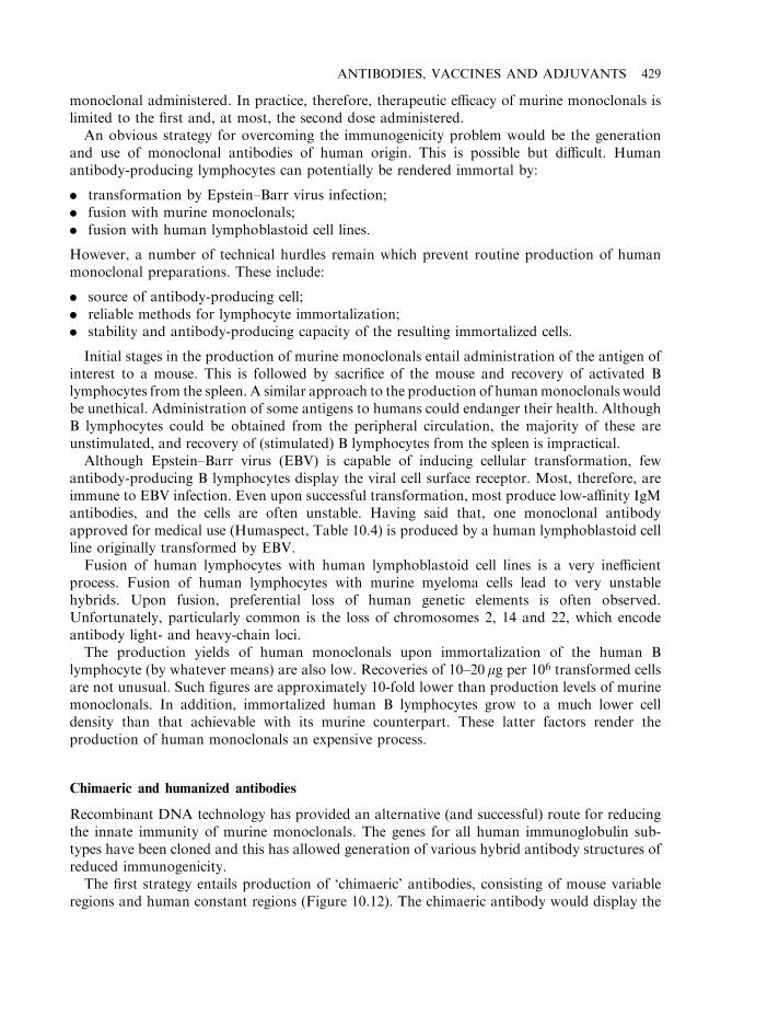
monoclonal administered. In practice, therefore, therapeutic efficacy of murine monoclonals islimited to the first and, at most, the second dose administered.
An obvious strategy for overcoming the immunogenicity problem would be the generationand use of monoclonal antibodies of human origin. This is possible but difficult. Humanantibody-producing lymphocytes can potentially be rendered immortal by:
. transformation by Epstein–Barr virus infection;
. fusion with murine monoclonals;
. fusion with human lymphoblastoid cell lines.
However, a number of technical hurdles remain which prevent routine production of humanmonoclonal preparations. These include:
. source of antibody-producing cell;
. reliable methods for lymphocyte immortalization;
. stability and antibody-producing capacity of the resulting immortalized cells.
Initial stages in the production of murine monoclonals entail administration of the antigen ofinterest to a mouse. This is followed by sacrifice of the mouse and recovery of activated Blymphocytes from the spleen. A similar approach to the production of humanmonoclonals wouldbe unethical. Administration of some antigens to humans could endanger their health. AlthoughB lymphocytes could be obtained from the peripheral circulation, the majority of these areunstimulated, and recovery of (stimulated) B lymphocytes from the spleen is impractical.
Although Epstein–Barr virus (EBV) is capable of inducing cellular transformation, fewantibody-producing B lymphocytes display the viral cell surface receptor. Most, therefore, areimmune to EBV infection. Even upon successful transformation, most produce low-affinity IgMantibodies, and the cells are often unstable. Having said that, one monoclonal antibodyapproved for medical use (Humaspect, Table 10.4) is produced by a human lymphoblastoid cellline originally transformed by EBV.
Fusion of human lymphocytes with human lymphoblastoid cell lines is a very inefficientprocess. Fusion of human lymphocytes with murine myeloma cells lead to very unstablehybrids. Upon fusion, preferential loss of human genetic elements is often observed.Unfortunately, particularly common is the loss of chromosomes 2, 14 and 22, which encodeantibody light- and heavy-chain loci.
The production yields of human monoclonals upon immortalization of the human Blymphocyte (by whatever means) are also low. Recoveries of 10–20 mg per 106 transformed cellsare not unusual. Such figures are approximately 10-fold lower than production levels of murinemonoclonals. In addition, immortalized human B lymphocytes grow to a much lower celldensity than that achievable with its murine counterpart. These latter factors render theproduction of human monoclonals an expensive process.
Chimaeric and humanized antibodies
Recombinant DNA technology has provided an alternative (and successful) route for reducingthe innate immunity of murine monoclonals. The genes for all human immunoglobulin sub-types have been cloned and this has allowed generation of various hybrid antibody structures ofreduced immunogenicity.
The first strategy entails production of ‘chimaeric’ antibodies, consisting of mouse variableregions and human constant regions (Figure 10.12). The chimaeric antibody would display the
ANTIBODIES, VACCINES AND ADJUVANTS 429
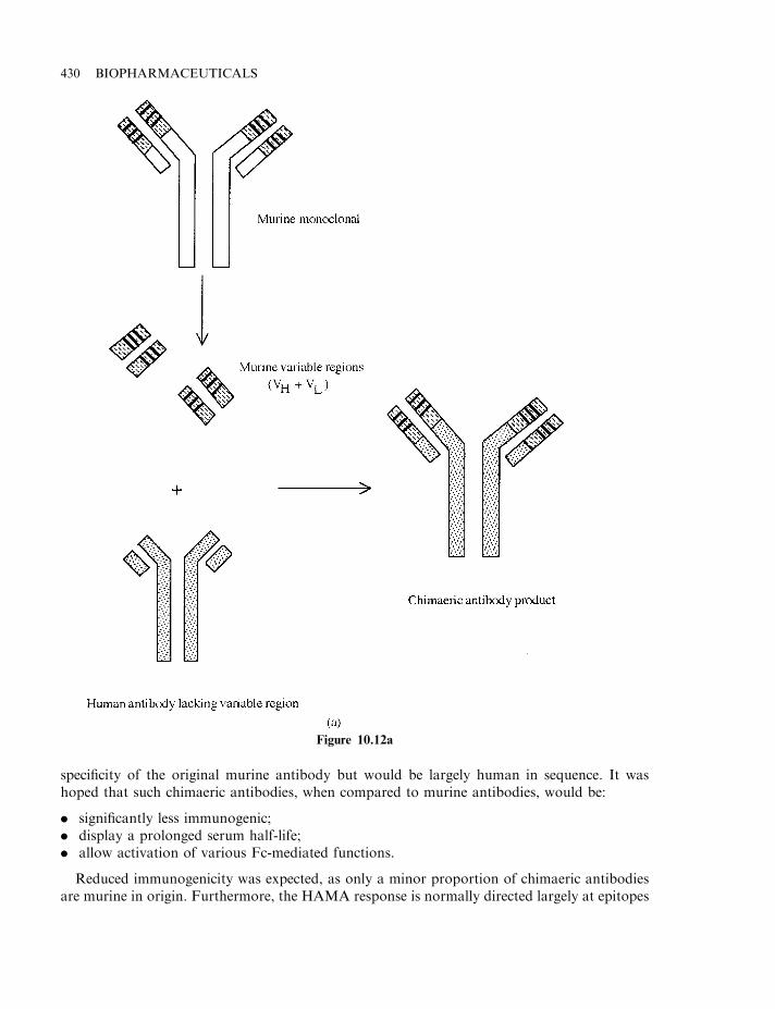
specificity of the original murine antibody but would be largely human in sequence. It washoped that such chimaeric antibodies, when compared to murine antibodies, would be:
. significantly less immunogenic;
. display a prolonged serum half-life;
. allow activation of various Fc-mediated functions.
Reduced immunogenicity was expected, as only a minor proportion of chimaeric antibodiesare murine in origin. Furthermore, the HAMA response is normally directed largely at epitopes
430 BIOPHARMACEUTICALS
Figure 10.12a
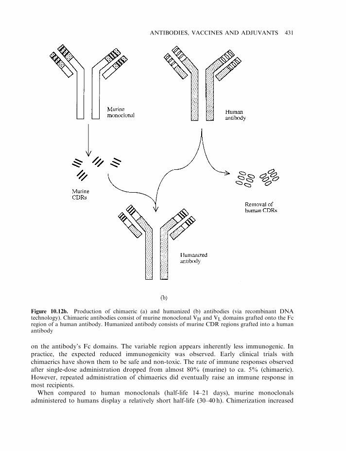
on the antibody’s Fc domains. The variable region appears inherently less immunogenic. Inpractice, the expected reduced immunogenicity was observed. Early clinical trials withchimaerics have shown them to be safe and non-toxic. The rate of immune responses observedafter single-dose administration dropped from almost 80% (murine) to ca. 5% (chimaeric).However, repeated administration of chimaerics did eventually raise an immune response inmost recipients.
When compared to human monoclonals (half-life 14–21 days), murine monoclonalsadministered to humans display a relatively short half-life (30–40 h). Chimerization increased
ANTIBODIES, VACCINES AND ADJUVANTS 431
Figure 10.12b. Production of chimaeric (a) and humanized (b) antibodies (via recombinant DNAtechnology). Chimaeric antibodies consist of murine monoclonal VH and VL domains grafted onto the Fcregion of a human antibody. Humanized antibody consists of murine CDR regions grafted into a humanantibody
serum half-life by five-fold, with typical values of 230 h being recorded (Table 10.7). A prolongedhalf-life is desirable if the antibody is to be used therapeutically, as it decreases the requiredfrequency of product administration. Chimaeric antibodies also allow activation of Fc-mediatedfunctions (e.g. activation of complement, etc.), as this domain displays human sequence.
While chimaeric antibodies contain an entire murine-derived variable region, it is only thecomplementarity-determining regions (CDRs) within this variable domain that actually dictateantigen specificity (Box 10.2). A method of reducing the antigenicity of murine antibodies stillfurther is to ‘humanize’ them. This entails transferring the nucleotide sequences coding for thesix CDR regions of the murine antibody of the desired specificity into a human antibody gene.The resulting hybrid antibody will, obviously, be entirely human in nature except for the CDRs(Figure 10.12).
Transfer of murine-derived CDR sequences into human antibody framework regions(Box 10.2) sometimes generates an antibody with greatly reduced antigen-binding affinity.Selected murine framework sequences are often also included in the humanized antibody. Thisprocess (known as ‘reshaping’ the human antibody) facilitates folding of the CDRs into theirtrue native conformations. This, in turn, normally restores antibody–antigen binding affinity.Over 95% of such hybrid antibodies are human in sequence. Clinical trials indicate that suchproteins do indeed behave similarly to native human antibodies.
Humanization has overcome many of the major factors that limited the therapeuticeffectiveness of first-generation (murine) monoclonals as therapeutic agents. Several suchhumanized products have now gained marketing authorization (Table 10.4).
Antibody fragments
One limitation to antibody-mediated treatment of solid tumours relates to their poor penetrationof tumour mass. Antibody-based tumour therapy (e.g. using radio-labelled murine monoclonals)have proved more effective in treating disseminated cancers (e.g. leukaemias and lymphomas)than solid tumours. The physical size of the intact antibody likely hinders tumour penetration. Asa result, recent interest has focused upon using antibody fragments which retain their antigen-binding capabilities. Fragments, such as F(ab), F(ab)2 and Fv (Box 10.2), can be easily generated,mainly via recombinant DNA technology. These have been labelled, e.g. with radio-active tags,with the intention of using them as diagnostic and therapeutic agents. While their lowermolecular mass may aid more effective tumour penetration, whole chimaeric/humanizedantibodies may be more effective, particularly if used for therapeutic purposes. Fragmentsgenerally display greatly reduced serum half-lives (Table 10.7) and cannot initiate effectorfunctions. Radio-labelled fragments may be better suited to diagnostic imaging purposes.
432 BIOPHARMACEUTICALS
Table 10.7. The serum half-life values of some IgG antibodypreparations when administered to humans
Antibody type Serum half-life
Intact human monoclonal 14–21 daysIntact murine monoclonal 30–40 hChimaeric antibody 200–250 hMurine F(ab)2 fragment 20 hMurine Fab fragment 2 h
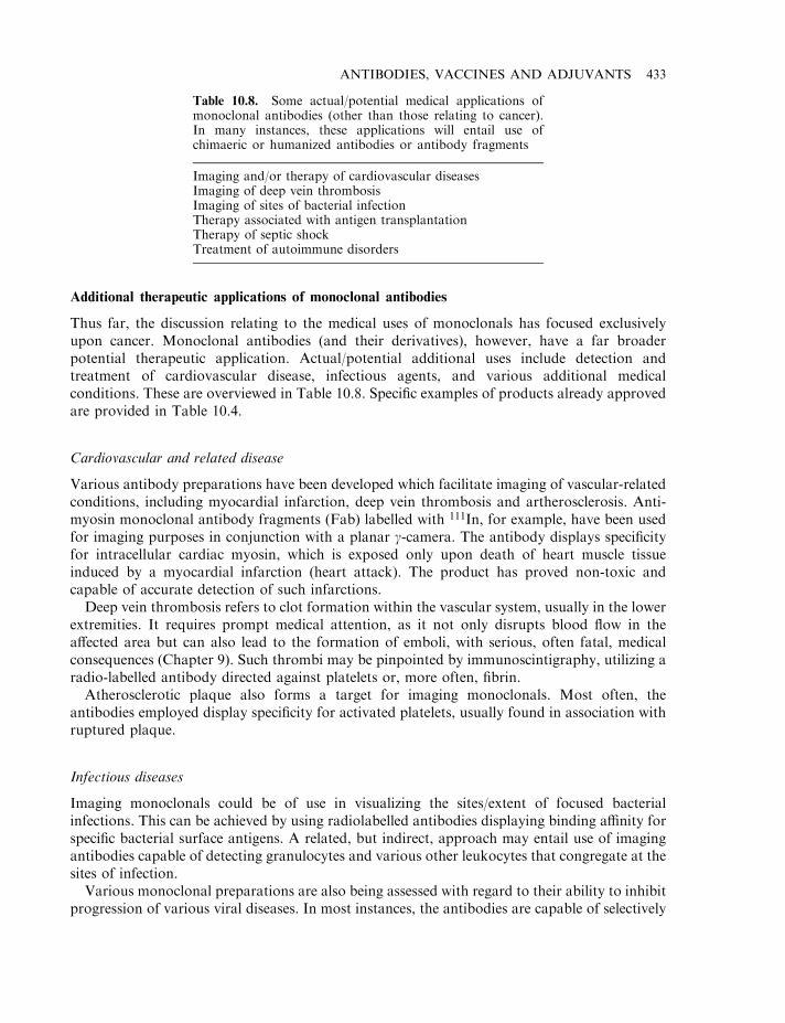
Additional therapeutic applications of monoclonal antibodies
Thus far, the discussion relating to the medical uses of monoclonals has focused exclusivelyupon cancer. Monoclonal antibodies (and their derivatives), however, have a far broaderpotential therapeutic application. Actual/potential additional uses include detection andtreatment of cardiovascular disease, infectious agents, and various additional medicalconditions. These are overviewed in Table 10.8. Specific examples of products already approvedare provided in Table 10.4.
Cardiovascular and related disease
Various antibody preparations have been developed which facilitate imaging of vascular-relatedconditions, including myocardial infarction, deep vein thrombosis and artherosclerosis. Anti-myosin monoclonal antibody fragments (Fab) labelled with 111In, for example, have been usedfor imaging purposes in conjunction with a planar g-camera. The antibody displays specificityfor intracellular cardiac myosin, which is exposed only upon death of heart muscle tissueinduced by a myocardial infarction (heart attack). The product has proved non-toxic andcapable of accurate detection of such infarctions.
Deep vein thrombosis refers to clot formation within the vascular system, usually in the lowerextremities. It requires prompt medical attention, as it not only disrupts blood flow in theaffected area but can also lead to the formation of emboli, with serious, often fatal, medicalconsequences (Chapter 9). Such thrombi may be pinpointed by immunoscintigraphy, utilizing aradio-labelled antibody directed against platelets or, more often, fibrin.
Atherosclerotic plaque also forms a target for imaging monoclonals. Most often, theantibodies employed display specificity for activated platelets, usually found in association withruptured plaque.
Infectious diseases
Imaging monoclonals could be of use in visualizing the sites/extent of focused bacterialinfections. This can be achieved by using radiolabelled antibodies displaying binding affinity forspecific bacterial surface antigens. A related, but indirect, approach may entail use of imagingantibodies capable of detecting granulocytes and various other leukocytes that congregate at thesites of infection.
Various monoclonal preparations are also being assessed with regard to their ability to inhibitprogression of various viral diseases. In most instances, the antibodies are capable of selectively
ANTIBODIES, VACCINES AND ADJUVANTS 433
Table 10.8. Some actual/potential medical applications ofmonoclonal antibodies (other than those relating to cancer).In many instances, these applications will entail use ofchimaeric or humanized antibodies or antibody fragments
Imaging and/or therapy of cardiovascular diseasesImaging of deep vein thrombosisImaging of sites of bacterial infectionTherapy associated with antigen transplantationTherapy of septic shockTreatment of autoimmune disorders
binding to a viral surface epitope. Two viral agents receiving particular attention are hepatitis Bvirus and HIV. Both murine monoclonals as well as chimaeric and humanized antibodypreparations raised against HIV surface antigens are currently being assessed with regard totheir ability to alter the clinical course of AIDS.
One of the most serious consequences of (Gram-negative) bacterial infection is the possibledevelopment of septic shock. This is caused by the release of lipopolysaccharide (LPS;endotoxin) from the bacterial cell surface. Various anti-LPS monoclonals (mainly targeted atits lipid A component; Chapter 3) have been developed. It is hoped that administration ofsuch monoclonals to affected individuals would effectively mop up free LPS, henceameliorating the severity of the condition. Most trial results to date have proved disappointingin this regard.
Autoimmune disease
Autoimmunity describes any condition in which the body’s immune system targets someelements of itself. This results from a failure of various immunological checking systems that arenormally responsible for maintaining self-tolerance. It has been estimated that 1–2% of the USpopulation suffer from autoimmune conditions, including rheumatoid arthritis, multiplesclerosis and some forms of diabetes. In many instances, an autoimmune response resultsfrom the inappropriate activation of a specific sub-set of B and/or T lymphocytes. The mostcommon immunotherapeutic approach to potentially treat such diseases is to induce depletionof the individual’s T and B cell populations. This could be achieved by administration of anantibody raised against a surface antigen present on such cells, e.g. initial trials have shown thatinjection of an (unconjugated) anti-CD 4 antibody (cell surface glycoprotein present on manyT lymphocytes) over 7 days significantly reduced the clinical symptoms of rheumatoid arthritisfor several months.
Selected adhesion molecules represent yet another antibody target. Adhesion molecules, suchas LFA-1 (leukocyte function-associated antigen-1) and ICAM-1 (intercellular adhesionmolecule 1), play central roles in promoting migration of inflammatory cells to the sites ofdamage. Such activities underlie many of the symptoms of conditions such as rheumatoidarthritis. Inhibition of adhesion molecule function by administration of antibodies raisedagainst them may, therefore, demonstrate therapeutic potential in some instances.
Transplantation
Modern surgical techniques render the transplantation of various organs relatively straight-forward. Common examples include renal (kidney), cardiac (heart) and hepatic (liver)transplants. In general, cell-mediated immunological mechanisms are responsible for mediatingrejection of transplanted organs. In many instances, transplant patients must be maintained onimmunosuppressive drugs (e.g. some steroids and, often, the fungal metabolite cyclosporine).However, complications may arise if a rejection episode is encountered which provesunresponsive to standard immunosuppressive therapy. Orthoclone OKT-3 was the firstmonoclonal antibody-based product to find application in this regard. This antibody is raisedagainst the protein-based CD3 antigen, present on the cell surface of most T lymphocytes. i.v.administration of (unconjugated) antibody appears to block normal functioning of such T cellsand promote their clearance from the blood. However, upon cessation of antibody
434 BIOPHARMACEUTICALS
administration, CD3-positive cell numbers rapidly revert to normal values. Therefore,maintenance immunosuppression (e.g. with cyclosporine) must subsequently be restored.
OKT-3 was first approved for general medical use in the USA in 1986. Its indication was thetreatment of acute kidney transplant rejection (Table 10.4). OKT-3 is produced via ascitesgrown in mice. The intact antibody is subsequently purified by a combination of ammoniumsulphate fractionation and anion exchange chromatography. Despite its therapeutic effective-ness, the product does display some limitations. Its antigenicity in humans (the HAMAresponse) is one obvious factor which limits its prolonged use.
A number of additional antibody-based products aimed at preventing transplant rejectionhave now gained general marketing authorization. Simulect (chimaeric antibody) and Zenapax(humanized antibody) were approved in the late 1990s (Table 10.4). Their engineered nature hasgreatly reduced the HAMA response upon their administration to humans. Both productstarget the 1L-2 receptor and hence bind fairly selectively to activated immune system cells,especially activated T lymphocytes, monocytes and macrophages (Chapter 5). Binding preventsfurther cellular proliferation and hence dampens the immune system’s attempts to destroy thetransplanted tissue.
VACCINE TECHNOLOGY
The application of vaccine technology forms a core element of modern medicinal endeavour. Itplays a central role, not only in human but also in veterinary medicine and represents the onlycommonly employed prophylactic (i.e. preventative) approach undertaken to control manyinfectious diseases. The current (annual) global vaccine market stands at in excess of $3 billion.Immunization programmes, particularly those undertaken on a multi-national scale, haveserved to reduce dramatically the incidence of many killer/disabling diseases, such as smallpox,polio and tuberculosis.
Continued/increased emphasis upon the implementation of such immunization programmesis likely. This is true not only of poorer world regions, but also amongst the most affluentnations. An estimated 500 000 adults die annually in the USA from conditions that could havebeen prevented by vaccination. These include pneumococcal pneumonia, influenza andhepatitis B.
Vaccination seeks to exploit the natural defence mechanisms conferred upon us by ourimmune systems. A vaccine contains a preparation of antigenic components consisting of,derived from or related to a pathogen. In most instances, upon vaccine administration,both the humoral and cell-mediated arms of the immune system are activated. The long-term immunological protection induced will normally prevent subsequent establishment ofan infection by the same or antigenically-related pathogens. While some vaccines are activewhen administered orally, more are administered parenterally. Normally, an initial doseadministration is followed by subsequent administration of one or more repeat doses overan appropriate time scale. Such booster doses serve to maximize the immunologicalresponse.
Traditional vaccine preparations have largely been targeted against viral and bacterialpathogens, as well as some bacterial toxins and, to a lesser extent, parasitic agents, such asmalaria. However, an increased understanding of the molecular mechanisms underlyingadditional human diseases suggests several novel applications of vaccines to treat or preventautoimmune conditions and cancer (Table 10.9). Such applications will be discussed later.Despite such potentially exciting future applications, recent scientific surveys indicate that the
ANTIBODIES, VACCINES AND ADJUVANTS 435
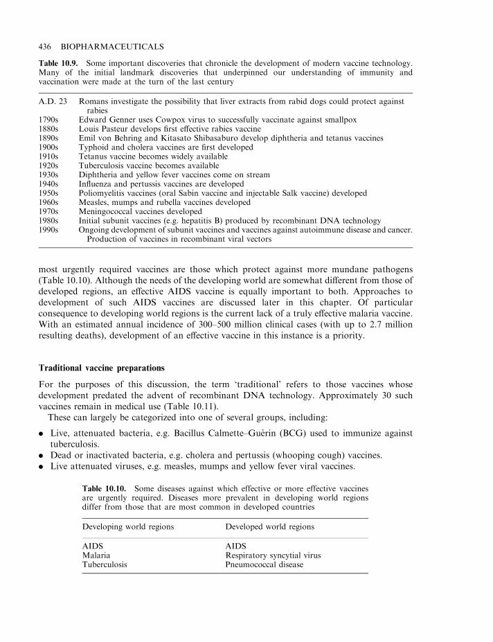
most urgently required vaccines are those which protect against more mundane pathogens(Table 10.10). Although the needs of the developing world are somewhat different from those ofdeveloped regions, an effective AIDS vaccine is equally important to both. Approaches todevelopment of such AIDS vaccines are discussed later in this chapter. Of particularconsequence to developing world regions is the current lack of a truly effective malaria vaccine.With an estimated annual incidence of 300–500 million clinical cases (with up to 2.7 millionresulting deaths), development of an effective vaccine in this instance is a priority.
Traditional vaccine preparations
For the purposes of this discussion, the term ‘traditional’ refers to those vaccines whosedevelopment predated the advent of recombinant DNA technology. Approximately 30 suchvaccines remain in medical use (Table 10.11).
These can largely be categorized into one of several groups, including:
. Live, attenuated bacteria, e.g. Bacillus Calmette–Guerin (BCG) used to immunize againsttuberculosis.
. Dead or inactivated bacteria, e.g. cholera and pertussis (whooping cough) vaccines.
. Live attenuated viruses, e.g. measles, mumps and yellow fever viral vaccines.
436 BIOPHARMACEUTICALS
Table 10.9. Some important discoveries that chronicle the development of modern vaccine technology.Many of the initial landmark discoveries that underpinned our understanding of immunity andvaccination were made at the turn of the last century
A.D. 23 Romans investigate the possibility that liver extracts from rabid dogs could protect againstrabies
1790s Edward Genner uses Cowpox virus to successfully vaccinate against smallpox1880s Louis Pasteur develops first effective rabies vaccine1890s Emil von Behring and Kitasato Shibasaburo develop diphtheria and tetanus vaccines1900s Typhoid and cholera vaccines are first developed1910s Tetanus vaccine becomes widely available1920s Tuberculosis vaccine becomes available1930s Diphtheria and yellow fever vaccines come on stream1940s Influenza and pertussis vaccines are developed1950s Poliomyelitis vaccines (oral Sabin vaccine and injectable Salk vaccine) developed1960s Measles, mumps and rubella vaccines developed1970s Meningococcal vaccines developed1980s Initial subunit vaccines (e.g. hepatitis B) produced by recombinant DNA technology1990s Ongoing development of subunit vaccines and vaccines against autoimmune disease and cancer.
Production of vaccines in recombinant viral vectors
Table 10.10. Some diseases against which effective or more effective vaccinesare urgently required. Diseases more prevalent in developing world regionsdiffer from those that are most common in developed countries
Developing world regions Developed world regions
AIDS AIDSMalaria Respiratory syncytial virusTuberculosis Pneumococcal disease
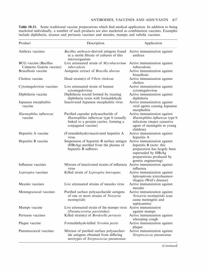
ANTIBODIES, VACCINES AND ADJUVANTS 437
Table 10.11. Some traditional vaccine preparations which find medical application. In addition to beingmarketed individually, a number of such products are also marketed as combination vaccines. Examplesinclude diphtheria, tetanus and pertussis vaccines and measles, mumps and rubella vaccines
Product Description Application
Anthrax vaccines Bacillus anthracis-derived antigens foundin a sterile filtrate of cultures of thismicroorganism
Active immunization againstanthrax
BCG vaccine (BacillusCalmette–Guerin vaccine)
Live attenuated strain of Mycobacteriumtuberculosis
Active immunization againsttuberculosis
Brucellosis vaccine Antigenic extract of Brucella abortus Active immunization againstbrucellosis
Cholera vaccine Dead strain(s) of Vibrio cholerae Active immunization againstcholera
Cytomegalovirus vaccines Live attenuated strain of humancytomegalovirus
Active immunization againstcytomegalovirus
Diphtheria vaccine Diphtheria toxoid formed by treatingdiphtheria toxin with formaldehyde
Active immunization againstdiphtheria
Japanese encephalitisvaccine
Inactivated Japanese encephalitis virus Active immunization againstviral agents causing Japaneseencephalitis
Haemophilus influenzaevaccine
Purified capsular polysaccharide ofHaemophilus influenzae type b (usuallylinked to a protein carrier, forming aconjugated vaccine)
Active immunization againstHaemophilus influenzae type binfections (major causativeagent of meningitis in youngchildren)
Hepatitis A vaccine (Formaldehyde)-inactivated hepatitis Avirus
Active immunization againsthepatitis A
Hepatitis B vaccine Suspension of hepatitis B surface antigen(HBsAg) purified from the plasma ofhepatitis B sufferers
Active immunization againsthepatitis B (note: thispreparation has largely beensuperseded by HBsAgpreparations produced bygenetic engineering)
Influenza vaccines Mixture of inactivated strains of influenzavirus
Active immunization againstinfluenza
Leptospira vaccines Killed strain of Leptospira interogans Active immunization againstleptospirosis icterohaemor-rhagica (Weil’s disease)
Measles vaccines Live attenuated strains of measles virus Active immunization againstmeasles
Meningococcal vaccines Purified surface polysaccharide antigensof one or more strains of Neisseriameningitidis
Active immunization againstNeisseria meningitidis (cancause meningitis andsepticaemia)
Mumps vaccine Live attenuated strain of the mumps virus(Paramyxovirus parotitidus)
Active immunizationagainst mumps
Pertussis vaccines Killed strain(s) of Bordetella pertussis Active immunization againstwhooping cough
Plague vaccine Formaldehyde-killed Yersinia pestis Active immunization againstplague
Pneumococcal vaccines Mixture of purified surface polysacchar-ide antigens obtained from differingserotypes of Streptococcus pneumoniae
Active immunization againstStreptococcus pneumoniae
(Continued)

. Inactivated viruses, e.g. hepatitis A and poliomyelitis (Salk) viral vaccines.
. Toxoids, e.g. diphtheria and tetanus vaccines.
. Pathogen-derived antigens, e.g. hepatitis B, meningococcal, pneumococcal and Haemophilusinfluenzae vaccines.
Attenuated, dead or inactivated bacteria
Attenuation (bacterial or viral) represents the process of elimination or greatly reducing thevirulence of a pathogen. This is traditionally achieved by, for example, chemical treatment orheat, growing under adverse conditions or propagation in an unnatural host. The attenuatedproduct should still immunologically cross-react with the wild-type pathogen. Although rarelyoccurring in practice, a theoretical danger exists in some cases that the attenuated pathogenmight revert to its pathogenic state. An attenuated bacterial vaccine is represented by BacillusCalmette–Guerin (BCG), which is a strain of tubercule bacillus (Mycobacterium bovis) that failsto cause tuberculosis but retains much of the antigenicity of the pathogen.
Killing or inactivation of pathogenic bacteria usually renders them suitable as vaccines. Thisis usually achieved by chemical or heat treatment, or both (Table 10.12). To be effective, theinactivated product must retain much of the immunological characteristics of the activepathogen. The killing or inactivation method must be consistently 100% effective in order toprevent accidental transmission of live pathogens. Cholera vaccines, for example, are sterileaqueous suspensions of killed Vibrio cholerae, selected for high antigenic efficiency. Thepreparation often consists of a mixture of smooth strains of the two main cholera serological
438 BIOPHARMACEUTICALS
Table 10.11 (Continued)
Product Description Application
Poliomyelitis vaccine(Sabin vaccine: oral)
Live attenutated strains of poliomyelitisvirus
Active immunization againstpolio
Poliomyelitis vaccine(Salk vaccine:parenteral)
Inactivated poliomyelitis virus Active immunization againstpolio
Rabies vaccines Inactivated rabies virus Active immunization againstrabies
Rotavirus vaccines Live attenuated strains of rotavirus Active immunization againstrotavirus (causes severechildhood diarrhoea)
Rubella vaccines Live attenuated strain of rubella virus Active immunization againstrubella (German measles)
Tetanus vaccines Toxoid formed by formaldehydetreatment of toxin produced byClostridium tetani
Active immunization againsttetanus
Typhoid vaccines Killed Salmonella typhi Active immunization againsttyphoid fever
Typhus vaccines Killed epidemic Rickettsia prowazekii Active immunization againstlouse-borne typhus
Varicella zoster vaccines Live attenuated strain of herpes virusvaricellae
Active immunization againstchicken pox
Yellow fever vaccines Live attenuated strain of yellow fevervirus
Active immunization againstyellow fever

types: Inaba and Ogawa. A 1.0 ml typical dose usually contains not less than 8 billion V. choleraeparticles and phenol (up to 0.5%) may be added as preservative. The vaccine can also beprepared in freeze-dried form. When stored refrigerated, the liquid vaccine displays a usualshelf-life of 18 months, while that of the dried product is 5 years.
Attenuated and inactivated viral vaccines
Viral particles destined for use as vaccines are generally propagated in a suitable animal cellculture system. While true cell culture systems are sometimes employed, many viral particles aregrown in fertilized eggs, or cultures of chick embryo tissue (Table 10.13).
Many of the more prominent vaccine preparations in current medical use consist ofattenuated viral particles (Table 10.11). Mumps vaccine consists of live attenuated strains ofParamyxovirus parotitidis. In many world regions, it is used to routinely vaccinate children,often a part of a combined measles, mumps and rubella (MMR) vaccine. Several attenuatedstrains have been developed for use in vaccine preparations. The most commonly used is theJeryl Linn strain of the mumps vaccine, which is propagated in chick embryo cell culture. Thisvaccine has been administered to well over 50 million people worldwide and, typically, results inseroconversion rates of over 97%. The Sabin (oral poliomyelitis) vaccine consists of an aqueoussuspension of poliomyelitis virus, usually grown in cultures of monkey kidney tissue. It containsapproximately 1 million particles of poliomyelitis strains 1, 2 or 3 or a combination of all threestrains.
Hepatitis A vaccine exemplifies vaccine preparations containing inactivated viral particles. Itconsists of a formaldehyde-inactivated preparation of the HM 175 strain of hepatitis A virus.Viral particles are normally propagated initially in human fibroblasts.
ANTIBODIES, VACCINES AND ADJUVANTS 439
Table 10.12. Methods usually employed to inactivate bacteria orviruses subsequently used as dead/inactivated vaccine preparations
Heat treatmentTreatment with formaldehyde or acetoneTreatment with phenol or phenol and heatTreatment with propiolactone
Table 10.13. Some cell culture systems in which viral particles destined for use as viral vaccines arepropagated
Viral particle/vaccine Typical cell culture system
Yellow fever virus Chick egg embryosMeasles virus (attenuated) Chick egg embryo cellsMumps virus (attenuated) Chick egg embryo cellsPolio virus (live, oral, i.e. Sabin and inactivatedinjectable, i.e. Salk)
Monkey kidney tissue culture
Rubella vaccine Duck embryo tissue culture, human tissue cultureHepatitis A viral vaccine Human diploid fibroblastsVaricella-zoster vaccines (chicken pox vaccine) Human diploid cells
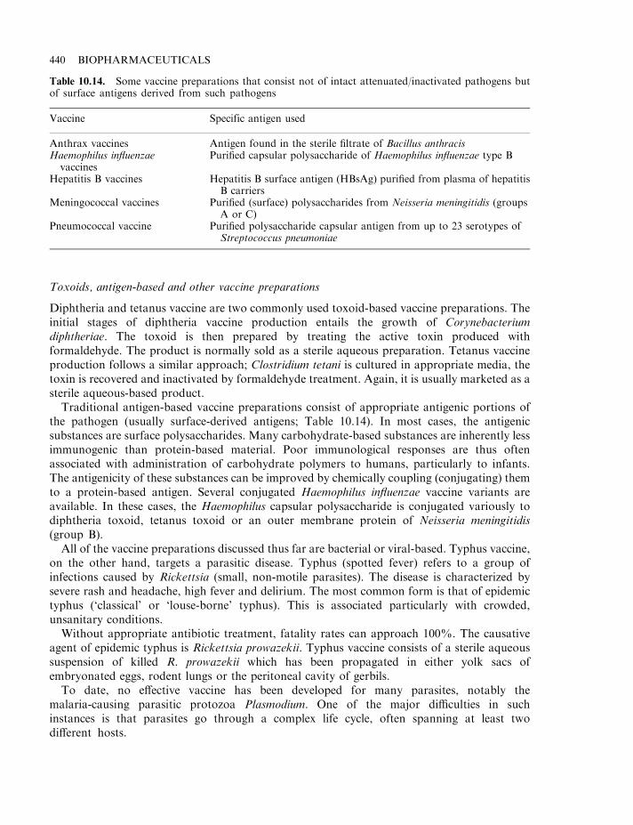
Toxoids, antigen-based and other vaccine preparations
Diphtheria and tetanus vaccine are two commonly used toxoid-based vaccine preparations. Theinitial stages of diphtheria vaccine production entails the growth of Corynebacteriumdiphtheriae. The toxoid is then prepared by treating the active toxin produced withformaldehyde. The product is normally sold as a sterile aqueous preparation. Tetanus vaccineproduction follows a similar approach; Clostridium tetani is cultured in appropriate media, thetoxin is recovered and inactivated by formaldehyde treatment. Again, it is usually marketed as asterile aqueous-based product.
Traditional antigen-based vaccine preparations consist of appropriate antigenic portions ofthe pathogen (usually surface-derived antigens; Table 10.14). In most cases, the antigenicsubstances are surface polysaccharides. Many carbohydrate-based substances are inherently lessimmunogenic than protein-based material. Poor immunological responses are thus oftenassociated with administration of carbohydrate polymers to humans, particularly to infants.The antigenicity of these substances can be improved by chemically coupling (conjugating) themto a protein-based antigen. Several conjugated Haemophilus influenzae vaccine variants areavailable. In these cases, the Haemophilus capsular polysaccharide is conjugated variously todiphtheria toxoid, tetanus toxoid or an outer membrane protein of Neisseria meningitidis(group B).
All of the vaccine preparations discussed thus far are bacterial or viral-based. Typhus vaccine,on the other hand, targets a parasitic disease. Typhus (spotted fever) refers to a group ofinfections caused by Rickettsia (small, non-motile parasites). The disease is characterized bysevere rash and headache, high fever and delirium. The most common form is that of epidemictyphus (‘classical’ or ‘louse-borne’ typhus). This is associated particularly with crowded,unsanitary conditions.
Without appropriate antibiotic treatment, fatality rates can approach 100%. The causativeagent of epidemic typhus is Rickettsia prowazekii. Typhus vaccine consists of a sterile aqueoussuspension of killed R. prowazekii which has been propagated in either yolk sacs ofembryonated eggs, rodent lungs or the peritoneal cavity of gerbils.
To date, no effective vaccine has been developed for many parasites, notably themalaria-causing parasitic protozoa Plasmodium. One of the major difficulties in suchinstances is that parasites go through a complex life cycle, often spanning at least twodifferent hosts.
440 BIOPHARMACEUTICALS
Table 10.14. Some vaccine preparations that consist not of intact attenuated/inactivated pathogens butof surface antigens derived from such pathogens
Vaccine Specific antigen used
Anthrax vaccines Antigen found in the sterile filtrate of Bacillus anthracisHaemophilus influenzaevaccines
Purified capsular polysaccharide of Haemophilus influenzae type B
Hepatitis B vaccines Hepatitis B surface antigen (HBsAg) purified from plasma of hepatitisB carriers
Meningococcal vaccines Purified (surface) polysaccharides from Neisseria meningitidis (groupsA or C)
Pneumococcal vaccine Purified polysaccharide capsular antigen from up to 23 serotypes ofStreptococcus pneumoniae
The impact of genetic engineering on vaccine technology
The advent of recombinant DNA technology has rendered possible the large-scale production ofpolypeptides normally present on the surface of virtually any pathogen. These polypeptides,when purified from the producer organism (e.g. Escherichia coli, Saccharomyces cerevisiae) canthen be used as ‘sub-unit’ vaccines. This method of vaccine production exhibits severaladvantages over conventional vaccine production methodologies. These include:
. Production of a clinically safe product; the pathogen-derived polypeptide now beingexpressed in a non-pathogenic recombinant host. This all but precludes the possibility thatthe final product could harbour undetected pathogen.
. Production of subunit vaccine in an unlimited supply. Previously, production of somevaccines was limited by supply of raw material (e.g. hepatitis B surface antigen; see below).
. Consistent production of a defined product which would thus be less likely to causeunexpected side effects.
A number of such recombinant (subunit) vaccines have now been approved for generalmedical use (Table 10.15). The first such product was that of hepatitis B surface antigen(rHBsAg), which gained marketing approval from the FDA in 1986. Prior to its approval,hepatitis B vaccines consisted of HBsAg purified directly from the blood of hepatitis B sufferers.When present in blood, HBsAg exists not in monomeric form, but in characteristic polymericstructures of 22 mm diameter. Production of hepatitis B vaccine by direct extraction from bloodsuffered from two major disadvantages:
. The supply of finished vaccine was restricted by the availability of infected human plasma.
. The starting material will likely be contaminated by intact, viable hepatitis B viral particles(and perhaps additional viruses, such as HIV). This necessitates introduction of stringentpurification procedures to ensure complete removal of any intact viral particles from theproduct stream. A final product QC test to confirm this entails a 6 month safety test onchimpanzees.
The HBsAg gene has been cloned and expressed in a variety of expression systems, includingE. coli, S. cerevisiae and a number of mammalian cell lines. The product used commercially isproduced in S. cerevisiae. The yeast cells are not only capable of expressing the gene, but alsoassembling the resultant polypeptide product into particles quite similar to those found in theblood of infected individuals. This product proved safe and effective when administered to bothanimals and humans. An overview of its manufacturing process is presented in Figure 10.13.
Various other companies have also produced recombinant HBsAg-based vaccines.SmithKline Beecham secured FDA approval for such a product (trade name, Engerix-B) in1989 (Figure 10.14). Subsequently, SmithKline Beecham have also generated variouscombination vaccines in which recombinant HBsAg is a component. ‘Twinrix’ (trade name),for example, contains a mixture of inactivated hepatitis A virus and recombinant HBsAg.Tritanrix, on the other hand, contains diphtheria and tetanus toxoids (produced by traditionalmeans), along with recombinant HBsAg.
It seems likely that many such (recombinant) subunit vaccines will gain future regulatoryapproval. One such example is that of B. pertussis subunit vaccine. B. pertussis is a Gram-negative coccobacillus, transmitted by droplet infection, and is the causative agent of the upperrespiratory tract infection commonly termed ‘whooping cough’.
ANTIBODIES, VACCINES AND ADJUVANTS 441
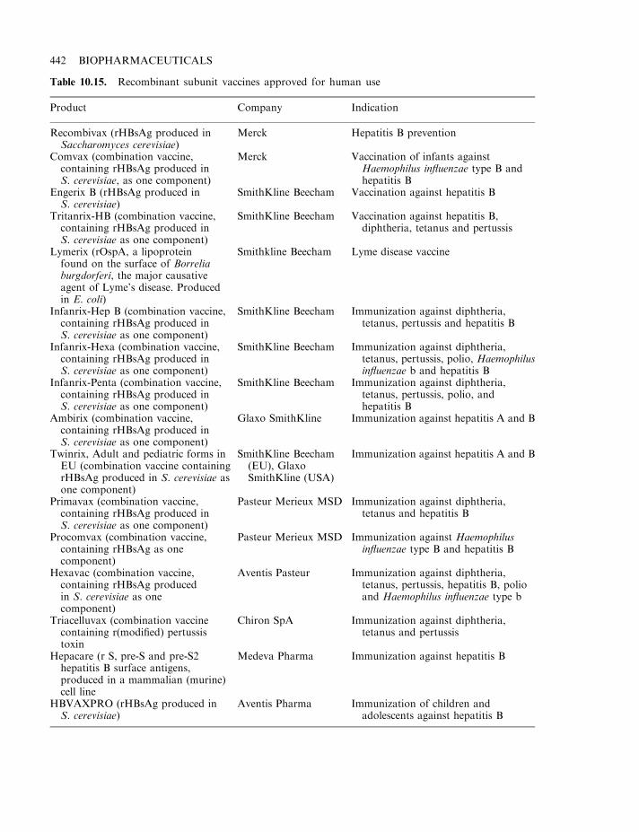
442 BIOPHARMACEUTICALS
Table 10.15. Recombinant subunit vaccines approved for human use
Product Company Indication
Recombivax (rHBsAg produced inSaccharomyces cerevisiae)
Merck Hepatitis B prevention
Comvax (combination vaccine,containing rHBsAg produced inS. cerevisiae, as one component)
Merck Vaccination of infants againstHaemophilus influenzae type B andhepatitis B
Engerix B (rHBsAg produced inS. cerevisiae)
SmithKline Beecham Vaccination against hepatitis B
Tritanrix-HB (combination vaccine,containing rHBsAg produced inS. cerevisiae as one component)
SmithKline Beecham Vaccination against hepatitis B,diphtheria, tetanus and pertussis
Lymerix (rOspA, a lipoproteinfound on the surface of Borreliaburgdorferi, the major causativeagent of Lyme’s disease. Producedin E. coli)
Smithkline Beecham Lyme disease vaccine
Infanrix-Hep B (combination vaccine,containing rHBsAg produced inS. cerevisiae as one component)
SmithKline Beecham Immunization against diphtheria,tetanus, pertussis and hepatitis B
Infanrix-Hexa (combination vaccine,containing rHBsAg produced inS. cerevisiae as one component)
SmithKline Beecham Immunization against diphtheria,tetanus, pertussis, polio, Haemophilusinfluenzae b and hepatitis B
Infanrix-Penta (combination vaccine,containing rHBsAg produced inS. cerevisiae as one component)
SmithKline Beecham Immunization against diphtheria,tetanus, pertussis, polio, andhepatitis B
Ambirix (combination vaccine,containing rHBsAg produced inS. cerevisiae as one component)
Glaxo SmithKline Immunization against hepatitis A and B
Twinrix, Adult and pediatric forms inEU (combination vaccine containingrHBsAg produced in S. cerevisiae asone component)
SmithKline Beecham(EU), GlaxoSmithKline (USA)
Immunization against hepatitis A and B
Primavax (combination vaccine,containing rHBsAg produced inS. cerevisiae as one component)
Pasteur Merieux MSD Immunization against diphtheria,tetanus and hepatitis B
Procomvax (combination vaccine,containing rHBsAg as onecomponent)
Pasteur Merieux MSD Immunization against Haemophilusinfluenzae type B and hepatitis B
Hexavac (combination vaccine,containing rHBsAg producedin S. cerevisiae as onecomponent)
Aventis Pasteur Immunization against diphtheria,tetanus, pertussis, hepatitis B, polioand Haemophilus influenzae type b
Triacelluvax (combination vaccinecontaining r(modified) pertussistoxin
Chiron SpA Immunization against diphtheria,tetanus and pertussis
Hepacare (r S, pre-S and pre-S2hepatitis B surface antigens,produced in a mammalian (murine)cell line
Medeva Pharma Immunization against hepatitis B
HBVAXPRO (rHBsAg produced inS. cerevisiae)
Aventis Pharma Immunization of children andadolescents against hepatitis B
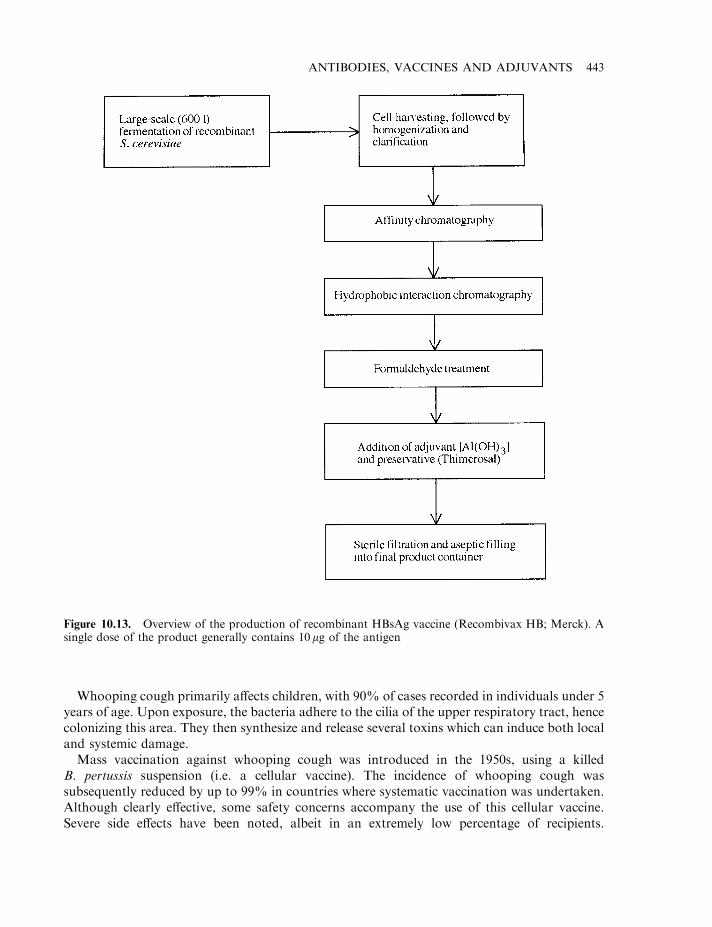
Whooping cough primarily affects children, with 90% of cases recorded in individuals under 5years of age. Upon exposure, the bacteria adhere to the cilia of the upper respiratory tract, hencecolonizing this area. They then synthesize and release several toxins which can induce both localand systemic damage.
Mass vaccination against whooping cough was introduced in the 1950s, using a killedB. pertussis suspension (i.e. a cellular vaccine). The incidence of whooping cough wassubsequently reduced by up to 99% in countries where systematic vaccination was undertaken.Although clearly effective, some safety concerns accompany the use of this cellular vaccine.Severe side effects have been noted, albeit in an extremely low percentage of recipients.
ANTIBODIES, VACCINES AND ADJUVANTS 443
Figure 10.13. Overview of the production of recombinant HBsAg vaccine (Recombivax HB; Merck). Asingle dose of the product generally contains 10mg of the antigen
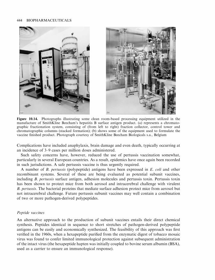
Complications have included anaphylaxis, brain damage and even death, typically occurring atan incidence of 3–9 cases per million doses administered.
Such safety concerns have, however, reduced the use of pertussis vaccination somewhat,particularly in several European countries. As a result, epidemics have once again been recordedin such jurisdictions. A safe pertussis vaccine is thus urgently required.
A number of B. pertussis (polypeptide) antigens have been expressed in E. coli and otherrecombinant systems. Several of these are being evaluated as potential subunit vaccines,including B. pertussis surface antigen, adhesion molecules and pertussis toxin. Pertussis toxinhas been shown to protect mice from both aerosol and intracerebral challenge with virulentB. pertussis. The bacterial proteins that mediate surface adhesion protect mice from aerosol butnot intracerebral challenge. Future pertussis subunit vaccines may well contain a combinationof two or more pathogen-derived polypeptides.
Peptide vaccines
An alternative approach to the production of subunit vaccines entails their direct chemicalsynthesis. Peptides identical in sequence to short stretches of pathogen-derived polypeptideantigens can be easily and economically synthesized. The feasibility of this approach was firstverified in the 1960s, when a hexapeptide purified from the enzymatic digest of tobacco mosaicvirus was found to confer limited immunological protection against subsequent administrationof the intact virus (the hexapeptide hapten was initially coupled to bovine serum albumin (BSA),used as a carrier to ensure an immunological response).
444 BIOPHARMACEUTICALS
Figure 10.14. Photographs illustrating some clean room-based processing equipment utilized in themanufacture of SmithKline Beecham’s hepatitis B surface antigen product. (a) represents a chromato-graphic fractionation system, consisting of (from left to right) fraction collector, control tower andchromatographic columns (stacked formation); (b) shows some of the equipment used to formulate thevaccine finished product. Photograph courtesy of SmithKline Beecham Biologicals s.a., Belgium
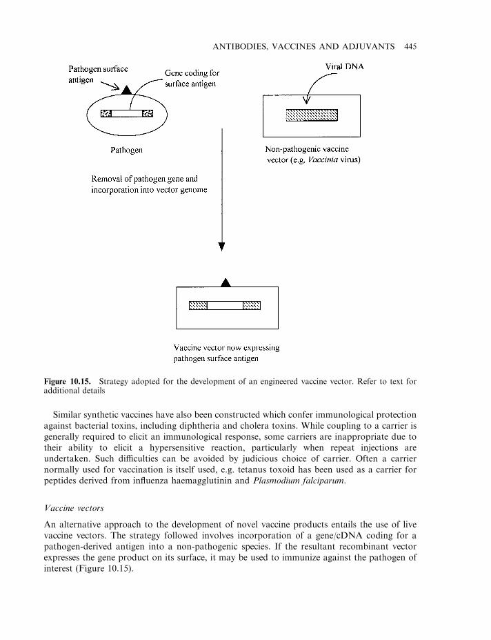
Similar synthetic vaccines have also been constructed which confer immunological protectionagainst bacterial toxins, including diphtheria and cholera toxins. While coupling to a carrier isgenerally required to elicit an immunological response, some carriers are inappropriate due totheir ability to elicit a hypersensitive reaction, particularly when repeat injections areundertaken. Such difficulties can be avoided by judicious choice of carrier. Often a carriernormally used for vaccination is itself used, e.g. tetanus toxoid has been used as a carrier forpeptides derived from influenza haemagglutinin and Plasmodium falciparum.
Vaccine vectors
An alternative approach to the development of novel vaccine products entails the use of livevaccine vectors. The strategy followed involves incorporation of a gene/cDNA coding for apathogen-derived antigen into a non-pathogenic species. If the resultant recombinant vectorexpresses the gene product on its surface, it may be used to immunize against the pathogen ofinterest (Figure 10.15).
ANTIBODIES, VACCINES AND ADJUVANTS 445
Figure 10.15. Strategy adopted for the development of an engineered vaccine vector. Refer to text foradditional details
Most vaccine vectors developed to date are viral-based, with poxviruses, picornaviruses andadenoviruses being used most. In general, such recombinant viral vectors elicit both stronghumoral and cell-mediated immunity. The immunological response (particularly the cell-mediated response) to subunit vaccines is often less pronounced.
Poxviruses and, more specifically, the vaccinia virus, remain the most thoroughlycharacterized vector systems developed. These are large, enveloped double-stranded DNAviruses. They are the only DNA-containing viruses that replicate in the cytoplasm of infectedcells. The most studied members of this family are variola and vaccinia. The former representsthe causative agent of smallpox, while the latter—being antigenically related to variola but non-pathogenic—was used to immunize against smallpox. Vaccinia-based vaccination programmesled to the global eradication of smallpox, finally achieved by the early 1980s.
Poxvirus promoters are not recognized by eukaryotic transcription machinery. Transcriptionof poxviral genes is initiated only by virally encoded RNA polymerase, normally packagedalongside the DNA in the virion particles. Purified poxvirus DNA is, therefore, non-infectious.
A number of factors render vaccinia virus a particularly attractive vector system. Theseinclude:
. capacity to successfully assimilate large quantities of DNA in its genome;
. prior history of widespread and successful use as a vaccination agent;
. ability to elicit long-lasting immunity;
. ease of production and low production costs;
. stability of freeze-dried finished vaccine product.
The ability of vaccinia (and other poxviruses) to accommodate large sequences ofheterologous DNA into its genome without adversely affecting its ability to replicate, remainsone of its most attractive features. Integration of foreign genes must occur in regions of the viralgenome not essential for viral replication. Two such sites are most often used. One is towardsthe left end of its genome, while the second is located within the thymidine kinase gene.
It is thought likely that up to 30 extra genes can be incorporated into vaccinia. The uppercapacity has not been determined, but is likely to exceed 50 kb. This facilitates the developmentof a multivalent vaccine via expression of several pathogen-derived genes in the recombinantvirus.
Early animal experiments have underlined the potential of vaccinia-based vector vaccines.Vaccinia virus-housing genes from HIV have clearly been found to elicit both humoral and cell-mediated immune responses in monkeys. Similar responses in other animals have been reportedwhen surface polypeptides from a variety of additional pathogens have been expressed inrecombinant vaccinia systems (Table 10.16). Human clinical trials are now in progress.
Adenoviruses also display potential as vaccine vectors. These double-stranded DNA virusesdisplay a genome consisting of ca. 36 000 base pairs, encoding approximately 50 viral genes.Several antigenically distinct human adenovirus serotypes have been characterized and theseviral species are endemic throughout the world. They can prompt respiratory tract infectionsand, to a lesser extent, gastrointestinal and genitourinary tract infections.
Live adenovirus strains have been isolated that cause asymptomatic infection and which haveproved to be very safe and effective adenovirus vaccines. Unlike vaccinia, few sites exist in theadenoviral genome into which foreign DNA can be integrated without comprising viralfunction. Furthermore, packing limitations curb the quantity of foreign DNA that can beaccommodated in the viral genome. However, a ca. 3000 base pair region can be removed from
446 BIOPHARMACEUTICALS
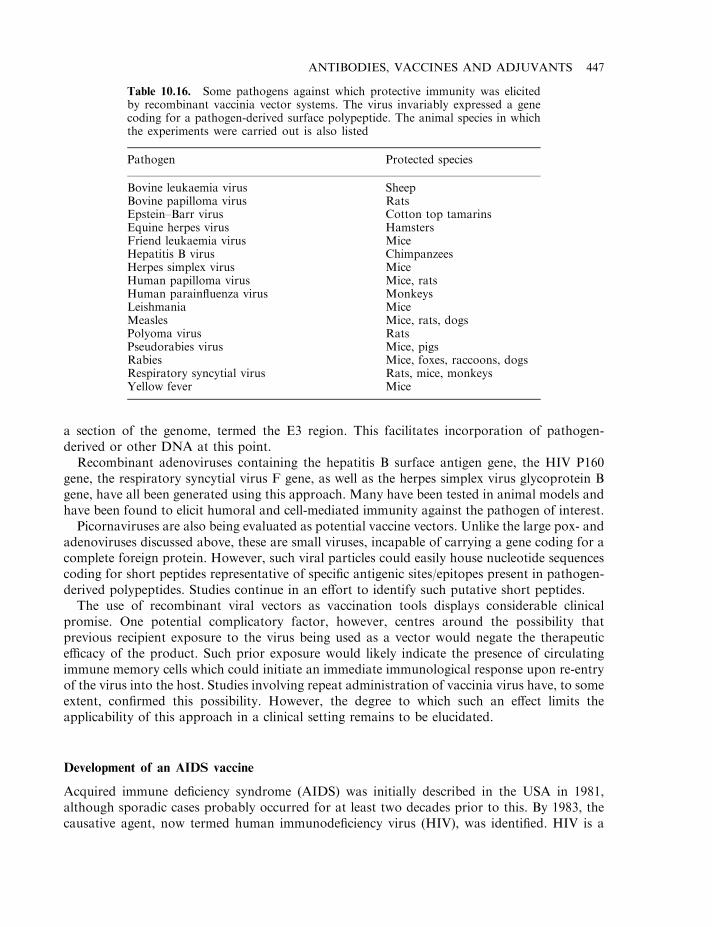
a section of the genome, termed the E3 region. This facilitates incorporation of pathogen-derived or other DNA at this point.
Recombinant adenoviruses containing the hepatitis B surface antigen gene, the HIV P160gene, the respiratory syncytial virus F gene, as well as the herpes simplex virus glycoprotein Bgene, have all been generated using this approach. Many have been tested in animal models andhave been found to elicit humoral and cell-mediated immunity against the pathogen of interest.
Picornaviruses are also being evaluated as potential vaccine vectors. Unlike the large pox- andadenoviruses discussed above, these are small viruses, incapable of carrying a gene coding for acomplete foreign protein. However, such viral particles could easily house nucleotide sequencescoding for short peptides representative of specific antigenic sites/epitopes present in pathogen-derived polypeptides. Studies continue in an effort to identify such putative short peptides.
The use of recombinant viral vectors as vaccination tools displays considerable clinicalpromise. One potential complicatory factor, however, centres around the possibility thatprevious recipient exposure to the virus being used as a vector would negate the therapeuticefficacy of the product. Such prior exposure would likely indicate the presence of circulatingimmune memory cells which could initiate an immediate immunological response upon re-entryof the virus into the host. Studies involving repeat administration of vaccinia virus have, to someextent, confirmed this possibility. However, the degree to which such an effect limits theapplicability of this approach in a clinical setting remains to be elucidated.
Development of an AIDS vaccine
Acquired immune deficiency syndrome (AIDS) was initially described in the USA in 1981,although sporadic cases probably occurred for at least two decades prior to this. By 1983, thecausative agent, now termed human immunodeficiency virus (HIV), was identified. HIV is a
ANTIBODIES, VACCINES AND ADJUVANTS 447
Table 10.16. Some pathogens against which protective immunity was elicitedby recombinant vaccinia vector systems. The virus invariably expressed a genecoding for a pathogen-derived surface polypeptide. The animal species in whichthe experiments were carried out is also listed
Pathogen Protected species
Bovine leukaemia virus SheepBovine papilloma virus RatsEpstein–Barr virus Cotton top tamarinsEquine herpes virus HamstersFriend leukaemia virus MiceHepatitis B virus ChimpanzeesHerpes simplex virus MiceHuman papilloma virus Mice, ratsHuman parainfluenza virus MonkeysLeishmania MiceMeasles Mice, rats, dogsPolyoma virus RatsPseudorabies virus Mice, pigsRabies Mice, foxes, raccoons, dogsRespiratory syncytial virus Rats, mice, monkeysYellow fever Mice
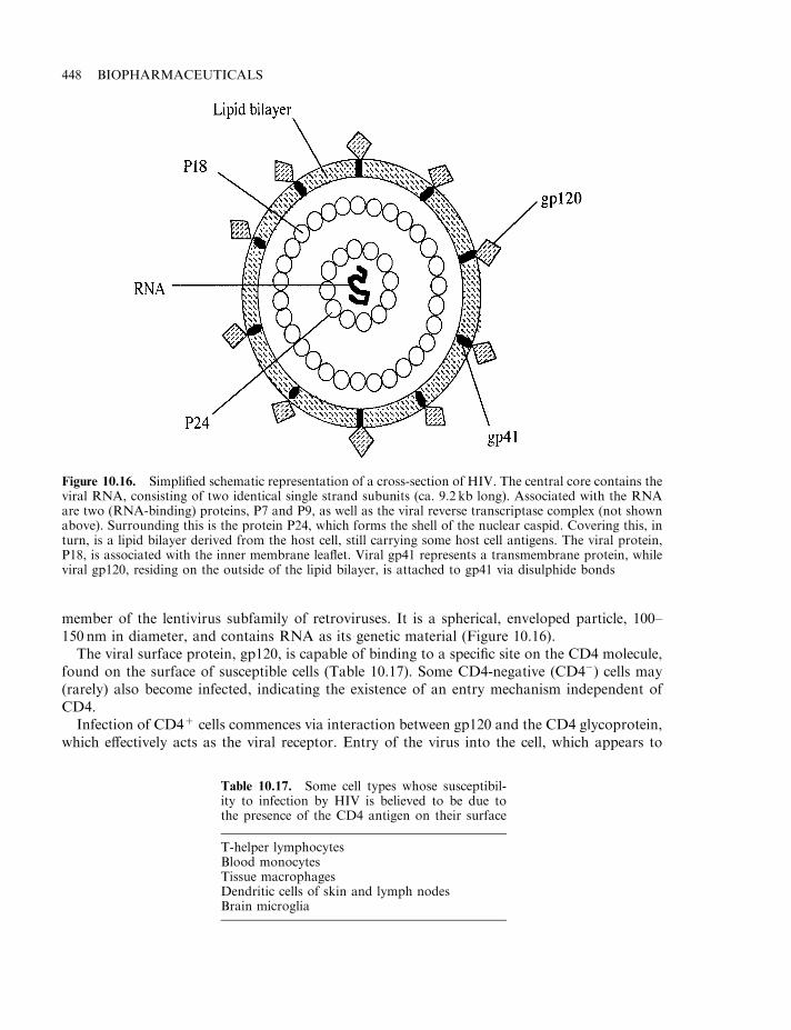
member of the lentivirus subfamily of retroviruses. It is a spherical, enveloped particle, 100–150 nm in diameter, and contains RNA as its genetic material (Figure 10.16).
The viral surface protein, gp120, is capable of binding to a specific site on the CD4 molecule,found on the surface of susceptible cells (Table 10.17). Some CD4-negative (CD4�) cells may(rarely) also become infected, indicating the existence of an entry mechanism independent ofCD4.
Infection of CD4+ cells commences via interaction between gp120 and the CD4 glycoprotein,which effectively acts as the viral receptor. Entry of the virus into the cell, which appears to
448 BIOPHARMACEUTICALS
Figure 10.16. Simplified schematic representation of a cross-section of HIV. The central core contains theviral RNA, consisting of two identical single strand subunits (ca. 9.2 kb long). Associated with the RNAare two (RNA-binding) proteins, P7 and P9, as well as the viral reverse transcriptase complex (not shownabove). Surrounding this is the protein P24, which forms the shell of the nuclear caspid. Covering this, inturn, is a lipid bilayer derived from the host cell, still carrying some host cell antigens. The viral protein,P18, is associated with the inner membrane leaflet. Viral gp41 represents a transmembrane protein, whileviral gp120, residing on the outside of the lipid bilayer, is attached to gp41 via disulphide bonds
Table 10.17. Some cell types whose susceptibil-ity to infection by HIV is believed to be due tothe presence of the CD4 antigen on their surface
T-helper lymphocytesBlood monocytesTissue macrophagesDendritic cells of skin and lymph nodesBrain microglia
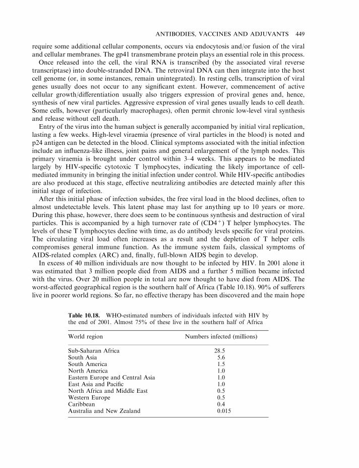
require some additional cellular components, occurs via endocytosis and/or fusion of the viraland cellular membranes. The gp41 transmembrane protein plays an essential role in this process.
Once released into the cell, the viral RNA is transcribed (by the associated viral reversetranscriptase) into double-stranded DNA. The retroviral DNA can then integrate into the hostcell genome (or, in some instances, remain unintegrated). In resting cells, transcription of viralgenes usually does not occur to any significant extent. However, commencement of activecellular growth/differentiation usually also triggers expression of proviral genes and, hence,synthesis of new viral particles. Aggressive expression of viral genes usually leads to cell death.Some cells, however (particularly macrophages), often permit chronic low-level viral synthesisand release without cell death.
Entry of the virus into the human subject is generally accompanied by initial viral replication,lasting a few weeks. High-level viraemia (presence of viral particles in the blood) is noted andp24 antigen can be detected in the blood. Clinical symptoms associated with the initial infectioninclude an influenza-like illness, joint pains and general enlargement of the lymph nodes. Thisprimary viraemia is brought under control within 3–4 weeks. This appears to be mediatedlargely by HIV-specific cytotoxic T lymphocytes, indicating the likely importance of cell-mediated immunity in bringing the initial infection under control. While HIV-specific antibodiesare also produced at this stage, effective neutralizing antibodies are detected mainly after thisinitial stage of infection.
After this initial phase of infection subsides, the free viral load in the blood declines, often toalmost undetectable levels. This latent phase may last for anything up to 10 years or more.During this phase, however, there does seem to be continuous synthesis and destruction of viralparticles. This is accompanied by a high turnover rate of (CD4+) T helper lymphocytes. Thelevels of these T lymphocytes decline with time, as do antibody levels specific for viral proteins.The circulating viral load often increases as a result and the depletion of T helper cellscompromises general immune function. As the immune system fails, classical symptoms ofAIDS-related complex (ARC) and, finally, full-blown AIDS begin to develop.
In excess of 40 million individuals are now thought to be infected by HIV. In 2001 alone itwas estimated that 3 million people died from AIDS and a further 5 million became infectedwith the virus. Over 20 million people in total are now thought to have died from AIDS. Theworst-affected geographical region is the southern half of Africa (Table 10.18). 90% of suffererslive in poorer world regions. So far, no effective therapy has been discovered and the main hope
ANTIBODIES, VACCINES AND ADJUVANTS 449
Table 10.18. WHO-estimated numbers of individuals infected with HIV bythe end of 2001. Almost 75% of these live in the southern half of Africa
World region Numbers infected (millions)
Sub-Saharan Africa 28.5South Asia 5.6South America 1.5North America 1.0Eastern Europe and Central Asia 1.0East Asia and Pacific 1.0North Africa and Middle East 0.5Western Europe 0.5Caribbean 0.4Australia and New Zealand 0.015
of eradicating this disease lies with the development of safe, effective vaccines. The first suchputative vaccine entered clinical trials in 1987 but, thus far, no truly effective vaccine has beendeveloped.
Difficulties associated with vaccine development
A number of attributes of HIV and its mode of infection conspire to render development of aneffective vaccine less than straightforward. These factors include:
. HIV displays extensive genetic variation, often even within a single individual. Such geneticvariation is particularly prominent in the viral env gene whose product, gp160, issubsequently proteolytically processed, yielding gp120 and gp41.
. HIV infects and destroys T helper lymphocytes, i.e. it directly attacks an essential componentof the immune system itself.
. Although infected individuals display a wide range of anti-viral immunological responses,these ultimately fail to destroy the virus. A greater understanding of what elements ofimmunity are most effective in combating HIV infection is required.
. After initial virulence subsides, large numbers of cells harbour unexpressed proviral DNA.The immune system has no way of identifying such cells. An effective vaccine must thusinduce the immune system to: (a) bring the viral infection under control before cellularinfection occurs; or (b) destroy cells once they begin to produce viral particles and destroy theviral particles released.
. The infection may often be spread, not via transmission of free viral particles, but via directtransmission of infected cells harbouring the proviral DNA.
AIDS vaccines in clinical trials
A number of approaches are being assessed with regard to developing an effective AIDSvaccine. No safe attenuated form of the virus has been recognized to date or is likely to bedeveloped in the foreseeable future. The high level of mutation associated with HIV would, inany case, heighten fears that spontaneous reversion of any such product to virulence would bepossible.
The potential of inactivated viral particles as effective vaccines has gained some attention butagain, fears of accidental transmission of disease if inactivation methods are not consistently100% effective have dampened enthusiasm for such an approach. In addition, the stringentcontainment conditions required to produce large quantities of the virus renders suchproduction processes expensive.
Despite such difficulties, at least one such inactivated product has reached clinical trials. Theviral particles are initially propagated in cultured human T cells. They are then treated withformaldehyde to inactivate them—a process which also removes the viral envelope. The virionparticles are then treated with g-irradiation in order to ensure inactivation of the viral genome.The final product is administered along with an adjuvant in order to maximize theimmunological response (see later).
Notwithstanding the possible value of such inactivated viral vaccines, the bulk of productsdeveloped to date are subunit vaccines. Live vector vaccines expressing HIV genes have alsobeen developed (Table 10.19).
450 BIOPHARMACEUTICALS
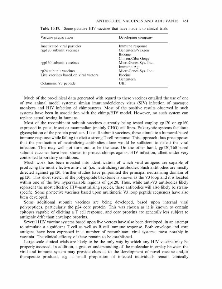
Much of the pre-clinical data generated with regard to these vaccines entailed the use of oneof two animal model systems: simian immunodeficiency virus (SIV) infection of macaquemonkeys and HIV infection of chimpanzees. Most of the positive results observed in suchsystems have been in association with the chimp/HIV model. However, no such system canreplace actual testing in humans.
Most of the recombinant subunit vaccines currently being tested employ gp120 or gp160expressed in yeast, insect or mammalian (mainly CHO) cell lines. Eukaryotic systems facilitateglycosylation of the protein products. Like all subunit vaccines, these stimulate a humoral-basedimmune response while failing to elicit a strong T cell response. This approach thus presupposesthat the production of neutralizing antibodies alone would be sufficient to defeat the viralinfection. This may well not turn out to be the case. On the other hand, gp120/160-basedsubunit vaccines have been shown to protect chimps against HIV infection, albeit under verycontrolled laboratory conditions.
Much work has been invested into identification of which viral antigens are capable ofproducing the most effective anti-viral (i.e. neutralizing) antibodies. Such antibodies are mostlydirected against gp120. Further studies have pinpointed the principal neutralizing domain ofgp120. This short stretch of the polypeptide backbone is known as the V3 loop and it is locatedwithin one of the five hypervariable regions of gp120. Thus, while anti-V3 antibodies likelyrepresent the most effective HIV-neutralizing species, these antibodies will also likely be strain-specific. Some protective vaccines based upon multimeric V3 loop peptide sequences have alsobeen developed.
Some additional subunit vaccines are being developed, based upon internal viralpolypeptides, particularly the p24 core protein. This was chosen as it is known to containepitopes capable of eliciting a T cell response, and core proteins are generally less subject toantigenic drift than envelope proteins.
Several HIV vaccine systems based upon live vectors have also been developed, in an attemptto stimulate a significant T cell as well as B cell immune response. Both envelope and coreantigens have been expressed in a number of recombinant viral systems, most notably invaccinia. The clinical efficacy of these remain to be established.
Large-scale clinical trials are likely to be the only way by which any HIV vaccine may beproperly assessed. In addition, a greater understanding of the molecular interplay between theviral and immune system may provide clues as to the development of novel vaccine and/ortherapeutic products, e.g. a small proportion of infected individuals remain clinically
ANTIBODIES, VACCINES AND ADJUVANTS 451
Table 10.19. Some putative HIV vaccines that have made it to clinical trials
Vaccine preparation Developing company
Inactivated viral particles Immune responsergp120 subunit vaccines Genentech/Vaxgen
BiocineChiron/Ciba Geigy
rgp160 subunit vaccines MicroGenes Sys. Inc.Immuno-Ag.
rp24 subunit vaccines MicroGenes Sys. Inc.Live vaccines based on viral vectors Biocine
GenentechOctameric V3 peptide UBI
asymptomatic for periods considerably greater than the average 10–15 years. An understandingof the immunological or other factors which delay onset of ARC/full-blown AIDS in theseindividuals may assist in the design of more effective vaccines. In addition, it has more recentlybeen reported that a very small proportion of individuals exposed (often repeatedly) to the virusremain uninfected. The genetic/immunological mechanisms underlining such resistance mayprovide useful insights into the elements of immunity that an effective vaccine needs to triggermost.
Although the primary objective of any vaccine is its prophylactic use (i.e. prevention of futureoccurrence of a disease), AIDS vaccines may also be of therapeutic value. This supposition isbased upon the fact that the immune system controls the viral infection for a time period.Hence, any agent capable of enhancing the anti-HIV immune response may prolong this effect.
Both industrial concerns and many government organizations continue to invest large capitalsums in AIDS vaccine research. Although much progress has been made, the complexity of thedisease has confounded the development of a truly effective vaccine thus far. By mid-2002 apreventitive AIDS vaccine ‘AIDS VAX’ (its trade name) had reached phase III clinical trials.The product, developed by a spin-off company of Genentech called Vaxgen is a recombinantgp120 glycoprotein produced in a CHO cell line.
Cancer vaccines
The identification of tumour-associated antigens could pave the way for the development of arange of cancer vaccines. A number of tumour-associated antigens have already beencharacterized, as previously described. Theoretically, administration of tumour-associatedantigens may effectively immunize an individual against any cancer type characterized byexpression of the tumour-associated antigen in question. Co-administration of a strongadjuvant (see later section) would be advantageous, as it would stimulate an enhanced immuneresponse. This is important as many tumour-associated antigens appear to be weakimmunogens. Administration of subunit-based tumor-associated antigen vaccines wouldprimarily stimulate a humoral immune response. The use of viral vectors may ultimatelyprove more effective, as a T cell response appears to be central to the immunological destructionof cancer cells.
The latter approach has been adopted in experimental studies involving malignant melanoma.These transformed cells express significantly elevated levels of a surface glycoprotein, p97. p97 isalso expressed—but at far lower levels—on the surface of many normal cell types. Initialanimal studies have indicated that administration of a recombinant vaccinia vector expressingp97 has a protective effect against challenge with melanoma cells. However, protracted safetystudies would be required in this, or similar, instances to prove that such vaccines would not, forexample, induce an autoimmune response if the antigen was not wholly tumour-specific. Thedevelopment of truly effective cancer vaccines probably requires a more comprehensiveunderstanding of the transformed phenotype and how these cells normally evade immunesurveillance in the first place. Notwithstanding this, limited clinical studies in this field havealready begun.
Recombinant veterinary vaccines
Amongst the limited number of biopharmaceuticals approved for animal use (Chapter 1),recombinant vaccines represent the single largest sub-group. Several such products target pigs,
452 BIOPHARMACEUTICALS

including Porcilis pesti and Bayovac CSF E2. Porcilis pesti, for example, contains a recombinantform of the classical swine fever virus E2 antigen, the immunodominant surface antigenassociated with this viral pathogen. It is used to immunize young pigs. An overview of itsmanufacture is presented in Figure 10.17. The process is initiated by growth of Spodopterafrugiperda cells, typically in a 500 l fermenter. The cells are then infected with the recombinantbaculovirus vector, resulting in high-level expression of the recombinant E2 antigen. Theantigen is harvested from the production medium by low-speed centrifugation and membranefiltration steps, which serve to remove intact cells/cellular debris. The antigen-containingsupernatant is then treated with b-propiolactone in order to inactivate any viral particlespresent. The antigen is not subjected to subsequent high-resolution chromatographicpurification steps, and hence is not purified to homogeneity. The product is then formulatedas an oil-in-water emulsion.
Adjuvant technology
Administration of many vaccines on their own stimulates a poor host immunological response.This is particularly true of the more recently developed subunit vaccines. An adjuvant is definedas any material that enhances the cellular and/or humoral immune response to an antigen.Adjuvants thus generally elicit an earlier, more potent and longer-lasting, immunologicalreaction against co-administered antigen. In addition, the use of adjuvants can often facilitateadministration of reduced quantities of antigen to achieve an adequate immunological response.
ANTIBODIES, VACCINES AND ADJUVANTS 453
Figure 10.17. Overview of the manufacture of the veterinary vaccine Porcilis pesti. Refer to text forspecific details
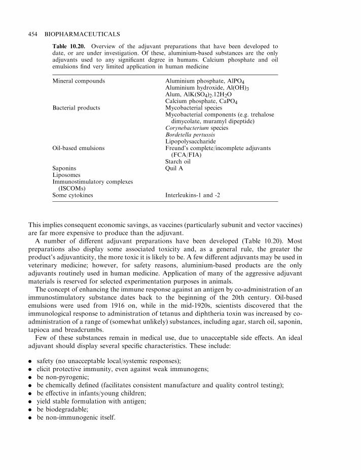
This implies consequent economic savings, as vaccines (particularly subunit and vector vaccines)are far more expensive to produce than the adjuvant.
A number of different adjuvant preparations have been developed (Table 10.20). Mostpreparations also display some associated toxicity and, as a general rule, the greater theproduct’s adjuvanticity, the more toxic it is likely to be. A few different adjuvants may be used inveterinary medicine; however, for safety reasons, aluminium-based products are the onlyadjuvants routinely used in human medicine. Application of many of the aggressive adjuvantmaterials is reserved for selected experimentation purposes in animals.
The concept of enhancing the immune response against an antigen by co-administration of animmunostimulatory substance dates back to the beginning of the 20th century. Oil-basedemulsions were used from 1916 on, while in the mid-1920s, scientists discovered that theimmunological response to administration of tetanus and diphtheria toxin was increased by co-administration of a range of (somewhat unlikely) substances, including agar, starch oil, saponin,tapioca and breadcrumbs.
Few of these substances remain in medical use, due to unacceptable side effects. An idealadjuvant should display several specific characteristics. These include:
. safety (no unacceptable local/systemic responses);
. elicit protective immunity, even against weak immunogens;
. be non-pyrogenic;
. be chemically defined (facilitates consistent manufacture and quality control testing);
. be effective in infants/young children;
. yield stable formulation with antigen;
. be biodegradable;
. be non-immunogenic itself.
454 BIOPHARMACEUTICALS
Table 10.20. Overview of the adjuvant preparations that have been developed todate, or are under investigation. Of these, aluminium-based substances are the onlyadjuvants used to any significant degree in humans. Calcium phosphate and oilemulsions find very limited application in human medicine
Mineral compounds Aluminium phosphate, AlPO4Aluminium hydroxide, Al(OH)3Alum, AlK(SO4)2.12H2OCalcium phosphate, CaPO4
Bacterial products Mycobacterial speciesMycobacterial components (e.g. trehalosedimycolate, muramyl dipeptide)
Corynebacterium speciesBordetella pertussisLipopolysaccharide
Oil-based emulsions Freund’s complete/incomplete adjuvants(FCA/FIA)
Starch oilSaponins Quil ALiposomesImmunostimulatory complexes(ISCOMs)
Some cytokines Interleukins-1 and -2
Adjuvant mode of action
Adjuvants are a heterogenous family of substances in terms of both their chemical structure andtheir mode of action. The observed adjuvanticity of any such substance may be due to one ormore of the following factors:
. depot formation of antigen; this results in the subsequent slow release of the antigen from thesite of injection which, in turn, ensures its prolonged exposure to the immune system;
. enhanced antigen presentation to the cells of the immune system;
. the direct induction of immunostimulatory substances, most notably interleukins and othercytokines.
In addition to the use of adjuvants per se, modification of the antigen may result in increasing itsinherent immunogenicity. Such modifications can include:
. polymerization of protein antigens (e.g. by reaction with gluteraldehyde or other cross-linkingagents); this approach has been successfully adopted with tetanus and diphtheria toxoids;
. conjugation of proteins to polysaccharides;
. cationization of protein antigens.
Mineral-based adjuvants
A number of mineral-based substances display an adjuvant effect. Although calcium phosphate,calcium chloride and salts of various metals (e.g. zinc sulphate and cerium nitrate) display someeffect, aluminium-based substances are by far the most potent. Most commonly employed arealuminium hydroxide and aluminium phosphate (Table 10.20). Their adjuvanticity, coupled totheir proven safety, renders them particularly valuable in the preparation of vaccines for youngchildren. They have been incorporated into millions of doses of such vaccine products so far.
The principal method by which aluminium-adjuvanted vaccines are prepared entails mixingthe antigen in solution with a pre-formed aluminium phosphate (or hydroxide) precipitate underchemically-defined conditions (e.g. of pH). Adsorption of the antigen to the aluminium-basedgel ensues, with such preparations being generally termed ‘aluminium-adsorbed vaccines’. 1mgof aluminium hydroxide will usually adsorb ca. 50–200 mg of protein.
The major mode of action of such products appears to be depot formation at the site ofinjection. The antigen is only slowly released from the gel, ensuring its sustained exposure toimmune surveillance. The aluminium compounds are also capable of activating complement.This can lead to a local inflammatory response, with consequent attraction of immunocompe-tent cells to the site of action.
Despite their popularity, aluminium-based adjuvants suffer from several drawbacks. Theytend to effectively stimulate only the humoral arm of the immune response. They cannot befrozen or lyophylized, as either process promotes destruction of their gel-based structure. Inaddition, aluminium-based products display poor or no adjuvanticity when combined withsome antigens (e.g. typhoid or Haemophilus influenzae type b capsular polysaccharides).
Oil-based emulsion adjuvants
The adjuvanticity of oil emulsions was first recognized in the early 1900s. However, the first suchproduct to gain widespread attention was Freund’s complete adjuvant (FCA), developed in1937. This product essentially contained a mixture of paraffin (i.e. mineral) oil with dead
ANTIBODIES, VACCINES AND ADJUVANTS 455
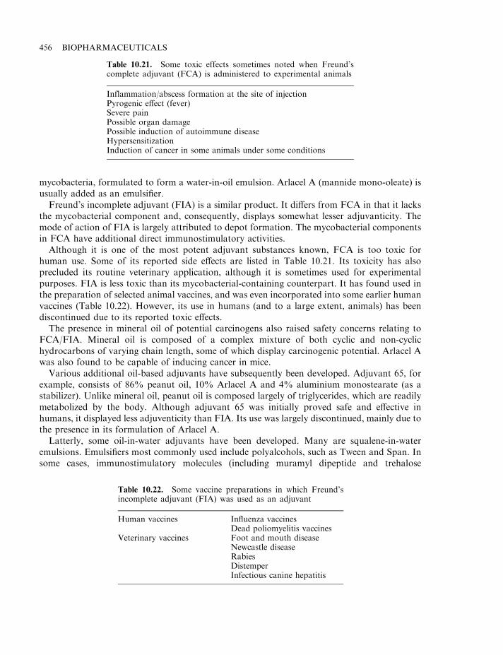
mycobacteria, formulated to form a water-in-oil emulsion. Arlacel A (mannide mono-oleate) isusually added as an emulsifier.
Freund’s incomplete adjuvant (FIA) is a similar product. It differs from FCA in that it lacksthe mycobacterial component and, consequently, displays somewhat lesser adjuvanticity. Themode of action of FIA is largely attributed to depot formation. The mycobacterial componentsin FCA have additional direct immunostimulatory activities.
Although it is one of the most potent adjuvant substances known, FCA is too toxic forhuman use. Some of its reported side effects are listed in Table 10.21. Its toxicity has alsoprecluded its routine veterinary application, although it is sometimes used for experimentalpurposes. FIA is less toxic than its mycobacterial-containing counterpart. It has found used inthe preparation of selected animal vaccines, and was even incorporated into some earlier humanvaccines (Table 10.22). However, its use in humans (and to a large extent, animals) has beendiscontinued due to its reported toxic effects.
The presence in mineral oil of potential carcinogens also raised safety concerns relating toFCA/FIA. Mineral oil is composed of a complex mixture of both cyclic and non-cyclichydrocarbons of varying chain length, some of which display carcinogenic potential. Arlacel Awas also found to be capable of inducing cancer in mice.
Various additional oil-based adjuvants have subsequently been developed. Adjuvant 65, forexample, consists of 86% peanut oil, 10% Arlacel A and 4% aluminium monostearate (as astabilizer). Unlike mineral oil, peanut oil is composed largely of triglycerides, which are readilymetabolized by the body. Although adjuvant 65 was initially proved safe and effective inhumans, it displayed less adjuventicity than FIA. Its use was largely discontinued, mainly due tothe presence in its formulation of Arlacel A.
Latterly, some oil-in-water adjuvants have been developed. Many are squalene-in-wateremulsions. Emulsifiers most commonly used include polyalcohols, such as Tween and Span. Insome cases, immunostimulatory molecules (including muramyl dipeptide and trehalose
456 BIOPHARMACEUTICALS
Table 10.21. Some toxic effects sometimes noted when Freund’scomplete adjuvant (FCA) is administered to experimental animals
Inflammation/abscess formation at the site of injectionPyrogenic effect (fever)Severe painPossible organ damagePossible induction of autoimmune diseaseHypersensitizationInduction of cancer in some animals under some conditions
Table 10.22. Some vaccine preparations in which Freund’sincomplete adjuvant (FIA) was used as an adjuvant
Human vaccines Influenza vaccinesDead poliomyelitis vaccines
Veterinary vaccines Foot and mouth diseaseNewcastle diseaseRabiesDistemperInfectious canine hepatitis
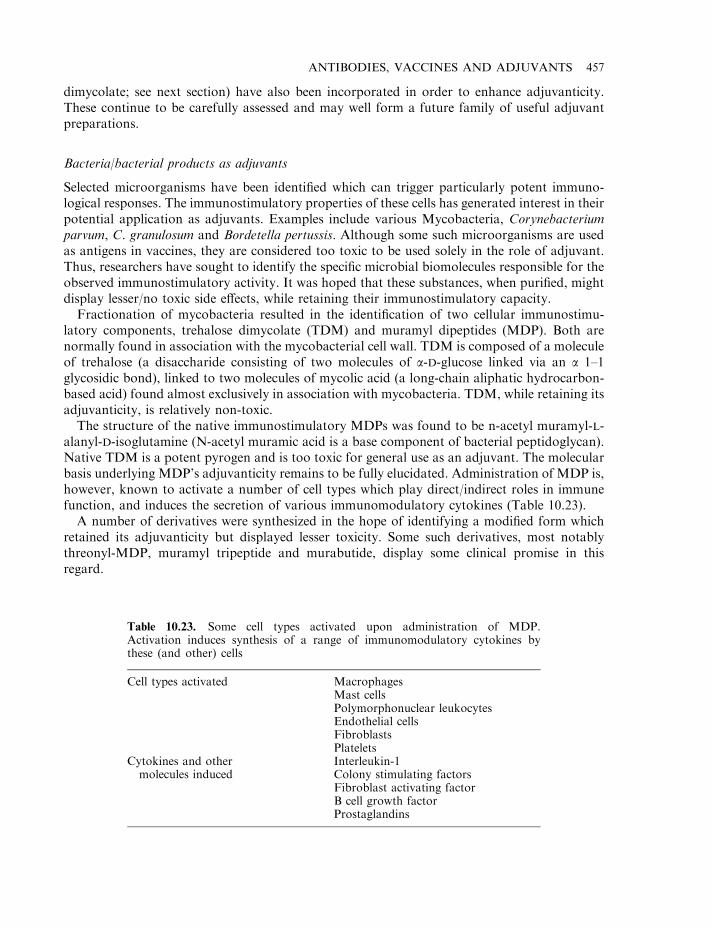
dimycolate; see next section) have also been incorporated in order to enhance adjuvanticity.These continue to be carefully assessed and may well form a future family of useful adjuvantpreparations.
Bacteria/bacterial products as adjuvants
Selected microorganisms have been identified which can trigger particularly potent immuno-logical responses. The immunostimulatory properties of these cells has generated interest in theirpotential application as adjuvants. Examples include various Mycobacteria, Corynebacteriumparvum, C. granulosum and Bordetella pertussis. Although some such microorganisms are usedas antigens in vaccines, they are considered too toxic to be used solely in the role of adjuvant.Thus, researchers have sought to identify the specific microbial biomolecules responsible for theobserved immunostimulatory activity. It was hoped that these substances, when purified, mightdisplay lesser/no toxic side effects, while retaining their immunostimulatory capacity.
Fractionation of mycobacteria resulted in the identification of two cellular immunostimu-latory components, trehalose dimycolate (TDM) and muramyl dipeptides (MDP). Both arenormally found in association with the mycobacterial cell wall. TDM is composed of a moleculeof trehalose (a disaccharide consisting of two molecules of a-D-glucose linked via an a 1–1glycosidic bond), linked to two molecules of mycolic acid (a long-chain aliphatic hydrocarbon-based acid) found almost exclusively in association with mycobacteria. TDM, while retaining itsadjuvanticity, is relatively non-toxic.
The structure of the native immunostimulatory MDPs was found to be n-acetyl muramyl-L-alanyl-D-isoglutamine (N-acetyl muramic acid is a base component of bacterial peptidoglycan).Native TDM is a potent pyrogen and is too toxic for general use as an adjuvant. The molecularbasis underlying MDP’s adjuvanticity remains to be fully elucidated. Administration of MDP is,however, known to activate a number of cell types which play direct/indirect roles in immunefunction, and induces the secretion of various immunomodulatory cytokines (Table 10.23).
A number of derivatives were synthesized in the hope of identifying a modified form whichretained its adjuvanticity but displayed lesser toxicity. Some such derivatives, most notablythreonyl-MDP, muramyl tripeptide and murabutide, display some clinical promise in thisregard.
ANTIBODIES, VACCINES AND ADJUVANTS 457
Table 10.23. Some cell types activated upon administration of MDP.Activation induces synthesis of a range of immunomodulatory cytokines bythese (and other) cells
Cell types activated MacrophagesMast cellsPolymorphonuclear leukocytesEndothelial cellsFibroblastsPlatelets
Cytokines and other Interleukin-1molecules induced Colony stimulating factors
Fibroblast activating factorB cell growth factorProstaglandins
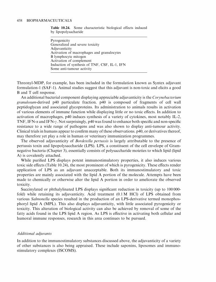
Threonyl-MDP, for example, has been included in the formulation known as Syntex adjuvantformulation-1 (SAF-1). Animal studies suggest that this adjuvant is non-toxic and elicits a goodB and T cell response.
An additional bacterial component displaying appreciable adjuvanticity is theCorynebacteriumgranulosum-derived p40 particulate fraction. p40 is composed of fragments of cell wallpeptidoglycan and associated glycoproteins. Its administration to animals results in activationof various elements of immune function while displaying little or no toxic effects. In addition toactivation of macrophages, p40 induces synthesis of a variety of cytokines, most notably IL-2,TNF, IFN-a and IFN-g. Not surprisingly, p40was found to enhance both specific and non-specificresistance to a wide range of pathogens and was also shown to display anti-tumour activity.Clinical trials in humans appear to confirmmany of these observations. p40, or derivatives thereof,may therefore yet play a role in human or veterinary immunization programmes.
The observed adjuvanticity of Bordetella pertussis is largely attributable to the presence ofpertussis toxin and lipopolysaccharide (LPS). LPS, a constituent of the cell envelope of Gram-negative bacteria (Chapter 3), essentially consists of polysaccharide moieties to which lipid (lipidA) is covalently attached.
While purified LPS displays potent immunostimulatory properties, it also induces varioustoxic side effects (Table 10.24), the most prominent of which is pyrogenicity. These effects renderapplication of LPS as an adjuvant unacceptable. Both its immunostimulatory and toxicproperties are mainly associated with the lipid A portion of the molecule. Attempts have beenmade to chemically or otherwise alter the lipid A portion in order to ameliorate the observedtoxicity.
Succinylated or phthalylinated LPS displays significant reduction in toxicity (up to 100 000-fold) while retaining its adjuvanticity. Acid treatment (0.1M HCl) of LPS obtained fromvarious Salmonella species resulted in the production of an LPS-derivative termed monophos-phoryl lipid A (MPL). This also displays adjuvanticity, with little associated pyrogenicity ortoxicity. This alteration of biological activity can also be achieved by removal of some of thefatty acids found in the LPS lipid A region. As LPS is effective in activating both cellular andhumoral immune responses, research in this area continues to be pursued.
Additional adjuvants
In addition to the immunostimulatory substances discussed above, the adjuvanticity of a varietyof other substances is also being appraised. These include saponins, liposomes and immuno-stimulatory complexes (ISCOMS).
458 BIOPHARMACEUTICALS
Table 10.24. Some characteristic biological effects inducedby lipopolysacharide
PyrogenicityGeneralized and severe toxicityAdjuvanticityActivation of macrophages and granulocytesB lymphocyte mitogenActivation of complementInduction of synthesis of TNF, CSF, IL-1, IFNSome anti-tumour activity
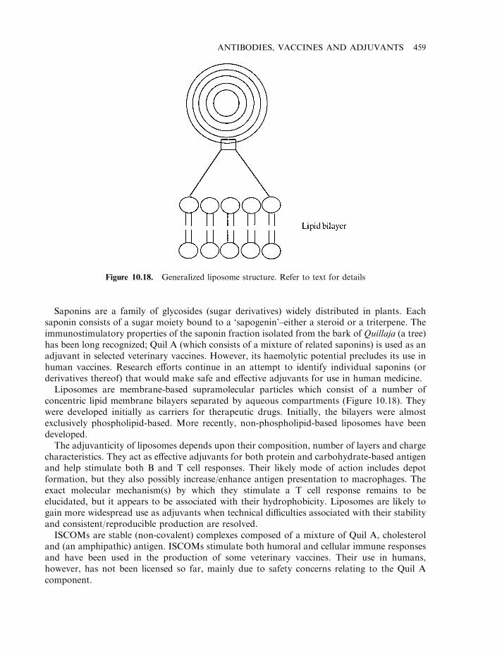
Saponins are a family of glycosides (sugar derivatives) widely distributed in plants. Eachsaponin consists of a sugar moiety bound to a ‘sapogenin’–either a steroid or a triterpene. Theimmunostimulatory properties of the saponin fraction isolated from the bark of Quillaja (a tree)has been long recognized; Quil A (which consists of a mixture of related saponins) is used as anadjuvant in selected veterinary vaccines. However, its haemolytic potential precludes its use inhuman vaccines. Research efforts continue in an attempt to identify individual saponins (orderivatives thereof) that would make safe and effective adjuvants for use in human medicine.
Liposomes are membrane-based supramolecular particles which consist of a number ofconcentric lipid membrane bilayers separated by aqueous compartments (Figure 10.18). Theywere developed initially as carriers for therapeutic drugs. Initially, the bilayers were almostexclusively phospholipid-based. More recently, non-phospholipid-based liposomes have beendeveloped.
The adjuvanticity of liposomes depends upon their composition, number of layers and chargecharacteristics. They act as effective adjuvants for both protein and carbohydrate-based antigenand help stimulate both B and T cell responses. Their likely mode of action includes depotformation, but they also possibly increase/enhance antigen presentation to macrophages. Theexact molecular mechanism(s) by which they stimulate a T cell response remains to beelucidated, but it appears to be associated with their hydrophobicity. Liposomes are likely togain more widespread use as adjuvants when technical difficulties associated with their stabilityand consistent/reproducible production are resolved.
ISCOMs are stable (non-covalent) complexes composed of a mixture of Quil A, cholesteroland (an amphipathic) antigen. ISCOMs stimulate both humoral and cellular immune responsesand have been used in the production of some veterinary vaccines. Their use in humans,however, has not been licensed so far, mainly due to safety concerns relating to the Quil Acomponent.
ANTIBODIES, VACCINES AND ADJUVANTS 459
Figure 10.18. Generalized liposome structure. Refer to text for details
In summary, therefore, a whole range of adjuvants have thus far been identified/developed.Problems of toxicity have precluded use of many of these adjuvants (particularly in humans).However, research efforts continue in an attempt to develop the next generation of safe and,hopefully, even more effective vaccine adjuvants.
FURTHER READING
Books
Amyes, S. (2002). Tumour immunology. Taylor & Francis, London.Grossbard, M. (1998). Monoclonal Antibody-based Therapy of Cancer. Marcel Dekker, New York.Harris, W. (1997). Antibody Therapeutics. CRC Press, Boca Raton, FL.Kontermann, R. (2001). Antibody Engineering. Springer-Verlag, Berlin.Liddell, J. (1995). Antibody Technology. BIOS Scientific, Oxford.Plotkin, S. (1999). Vaccines. W.B. Saunders, London.Powell, M. (1995). Vaccine Design: The Subunit and Adjuvant Approach. Plenum, New York.Stern, P. (2000). Cancer Vaccines and Immunotherapy. Cambridge University Press, Cambridge.Talwar, G. (1994). Synthetic Vaccines. Springer-Verlag, Berlin.Woodrow, G. (1997). New Generation Vaccines. Marcel Dekker, New York.
Articles
Antibody technology
Benhar, I. (2001). Biotechnological applications of phage and cell display. Biotechnol. Adv. 19, 1–33.Berger, M. et al. (2002). Therapeutic applications of monoclonal antibodies. Am. J. Med. Sci. 324(1), 14–30.Breedveld, F. (2000). Therapeutic monoclonal antibodies. Lancet 355, 735–740.Chapman, P. (2002). PEGylated antibodies and antibody fragments for improved therapy: a review. Adv. Drug Deliv.
Rev. 54(4), 531–545.Chester, K. & Hawkins, R. (1995). Clinical issues in antibody design. Trends Biotechnol. 13, 294–300.Funaro, A. et al. (2000). Monoclonal antibodies and the therapy of human cancers. Biotechnol. Adv. 18(5), 385–401.Goldenberg, D. et al. (2002). Targeted therapy of cancer with radiolabeled antibodies. J. Nuclear Med. 43(5), 693–713.Hoogenboom, H. et al. (1998). Antibody phage display technology and its applications. Immunotechnology 4, 1–20.Huennekens, F. (1994). Tumor targeting: activation of prodrugs by enzyme-monoclonal antibody conjugates. Trends
Biotechnol. 12, 234–239.Keating, G. & Perry, C. (2002). Infliximab—an updated review of its use in Crohn’s disease and rheumatoid arthritis.
Biodrugs 16(2), 111–148.Kohler, G. & Milstein, C. (1975). Continuous culture of fused cells secreting antibody of pre-defined specificity. Nature
256, 495–497.Murry, J. (2000). Monoclonal antibody treatment of solid tumors: a coming of age. Semin. Oncol. 27(6), 64–70.Senter, P. & Springer, C. (2001). Selective activation of anticancer prodrugs by monoclonal antibody–enzyme
conjugates. Adv. Drug Deliv. Rev. 53(3), 247–264.Trail, P. & Bianchi, A. (1999). Monoclonal antibody drug conjugates in the treatment of cancer. Curr. Opin. Immunol.
11(5), 584–588.Umemura, S. et al. (2002). Pathological evaluation of HER2 overexpression for the treatment of metastatic breast
cancers by humanized anti-HER2 monoclonal antibody (Trastuzumab). Acta Biochim. Cytochim. 35(2), 77–81.Wang, R. (1999). Human tumor antigens: implications for cancer vaccine development. J. Mol. Med. 77(9), 640–655.Wang, R. & Rosenberg, S. (1999). Human tumor antigens for cancer vaccine development. Immunol. Rev. 170, 85–100.Winter, G. & Milstein, C. (1991). Man-made antibodies. Nature 349, 293–299.
Vaccine technology
Andino, R. et al. (1994). Engineering poliovirus as a vaccine vector for the expression of diverse antigens. Science 265,1448–1451.
Chauhan, V. (1996). Progress towards malaria vaccine. Curr. Sci. 71(12), 967–975.Cox, J. & Coutter, A. (1997). Adjuvants—a classification and review of their modes of action. Vaccine 15(3), 248–256.Doolan, D. & Hoffman, S. (1997). Multigene vaccination against malaria—a multistage, multi-immune response
approach. Parasitol. Today 13(5), 171–178.
460 BIOPHARMACEUTICALS
Edelman, R. (2002). The development and use of vaccine adjuvants. Mol. Biotechnol. 21(2), 129–148.Esparza, J. et al. (1995). HIV-preventive vaccines, progress to date. Drugs 50(5), 792–804.Frontiers in Medicine: Vaccines (1994). Science 265 (Special Issue, September 2).Gaschen, B. et al. (2002). AIDS—diversity considerations in HIV-1 vaccine selection. Science 296(5577), 2354–2360.Gupta, R. & Siber, G. (1995). Adjuvants for human vaccines—current status, problems and future prospects. Vaccine
13(14), 1263–1274.Hilton, L. et al. (2002). The emerging role of avian cytokines as immunotherapeutics and vaccine adjuvants. Vet.
Immunol. Immunopathol. 85(3–4), 119–128.Lachman, L. et al. (1996). Cytokine-containing liposomes as vaccine adjuvants. Eur. Cytokine Network 7(4), 693–698.Lemon, S. & Thomas, D. (1997). Drug therapy—vaccines to prevent viral hepatitis. N. Engl. J. Med. 336(3), 196–204.Mason, H. et al. (2002). Edible plant vaccines: applications for prophylactic and therapeutic molecular medicine. Trends
Mol. Med. 8(7), 324–329.Ohagan, D. et al. (1997). Recent advances in vaccine adjuvants— the development of Mf59 emulsion and polymeric
microparticles. Mol. Med. Today 3(2), 69–75.Perkus, M. et al. (1995). Poxvirus-based vaccine candidates for cancer, AIDS and other infectious diseases. J. Leukocyte
Biol. 58, 1–10.Plotkin, S. (2002). Vaccines in the twenty-first century. Hybridoma Hybridom. 21(2), 135–145.Poland, G. et al. (2002). Science, medicine and the future—new vaccine development. Br. Med. J. 324(7349), 1315–1319.Russo, S. et al. (1997). What’s going on in vaccine technology? Med. Res. Rev. 17(3), 277–301.Sandhu, J. (1994). Engineered human vaccines. Crit. Rev. Biotechnol. 14(1), 1–27.Sela, M. et al. (2002). Therapeutic vaccines: realities of today and hopes for the future. Drug Discovery Today 7(12),
664–673.Singh, M. & O’Hagan, D. (2002). Recent advances in vaccine adjuvants. Pharmaceut. Res. 19(6), 715–728.Strominger, J. (1995). Peptide vaccination against cancer? Nature Med. 1(11), 1140.Wang, R. & Rosenberg, S. (1999). Human tumor antigens for cancer vaccine development. Immun. Rev. 170, 85–100.Yokoyama, N. et al. (1997). Recombinant viral vector vaccines for veterinary use. J. Vet. Med. Sci. 59(5), 311–322.
ANTIBODIES, VACCINES AND ADJUVANTS 461

Chapter 11
Nucleic acid therapeutics
Throughout the 1980s and early 1990s, the term ‘biopharmaceutical’ had become virtuallysynonymous with ‘proteins of therapeutic use’ (Chapter 1). Nucleic acid-based biopharmaceu-ticals, however, harbour great potential—a potential which is likely to become a medical realitywithin this decade. Current developments in nucleic acid based-therapeutics centre around genetherapy and antisense technology. These technologies have the potential to revolutionizemedical practice. Their full benefit, however, will accrue only after the satisfactory resolution ofseveral technical difficulties currently impeding their routine medical application.
Despite all the hype, it is important to note that, by mid-2002 at least, only a single nucleicacid-based product has been approved for medical use (an antisense-based product, discussedlater). No gene therapy-based product had been approved for general medical use by that time.
GENE THERAPY
The fundamental principle underpinning gene therapy is theoretically straightforward, butdifficult to satisfactorily achieve in practice. The principle entails the stable introduction of agene into the genetic complement of a cell, such that subsequent expression of the gene achievesa therapeutic goal. The potential of gene therapy as a curative approach for inborn errors ofmetabolism and other conditions induced by the presence of a defective copy of a specific gene(or genes) is obvious.
An increased understanding of the molecular basis of various other diseases, including cancer,some infectious diseases (e.g. AIDS) and some neurological conditions, also suggest a role forgene therapy in combating these. Indeed, well over half of all gene therapy trials conducted todate aim to treat cancer. Table 11.1 lists the major disease types for which a gene therapytreatment is currently being assessed in clinical trials. The first such trial was initiated in theUSA in 1990. Thus far, over 400 different clinical studies have been or are being undertaken,involving ca. 6000 patients worldwide. Despite initial enthusiasm, only a handful of such studieshave revealed any therapeutic benefit to the patient and, thus far, no complete, permanent cureshave been recorded.
Moreover, gene therapy— like all other medical interventions— is not without associatedrisk. A US patient died in 1999 as a result of participating in a gene therapy-based trial. Evenmore disturbingly, the ensuing FDA investigation unearthed allegations that at least six other
Biopharmaceuticals: Biochemistry and Biotechnology, Second Edition by Gary WalshJohn Wiley & Sons Ltd: ISBN 0 470 84326 8 (ppc), ISBN 0 470 84327 6 (pbk)
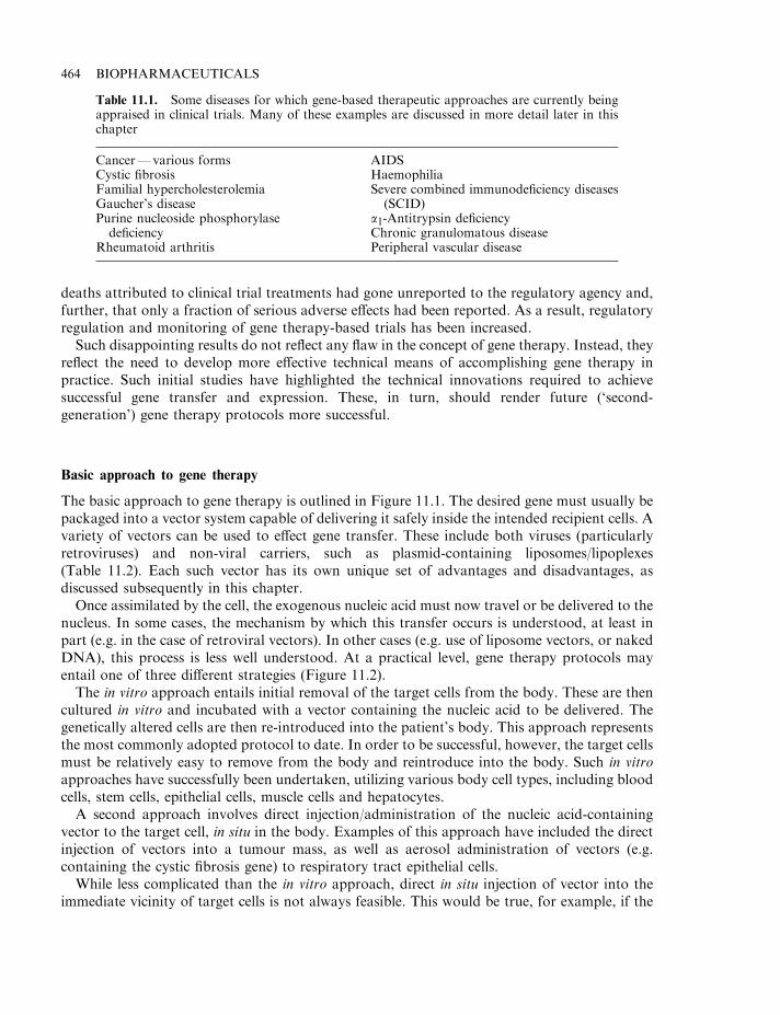
deaths attributed to clinical trial treatments had gone unreported to the regulatory agency and,further, that only a fraction of serious adverse effects had been reported. As a result, regulatoryregulation and monitoring of gene therapy-based trials has been increased.
Such disappointing results do not reflect any flaw in the concept of gene therapy. Instead, theyreflect the need to develop more effective technical means of accomplishing gene therapy inpractice. Such initial studies have highlighted the technical innovations required to achievesuccessful gene transfer and expression. These, in turn, should render future (‘second-generation’) gene therapy protocols more successful.
Basic approach to gene therapy
The basic approach to gene therapy is outlined in Figure 11.1. The desired gene must usually bepackaged into a vector system capable of delivering it safely inside the intended recipient cells. Avariety of vectors can be used to effect gene transfer. These include both viruses (particularlyretroviruses) and non-viral carriers, such as plasmid-containing liposomes/lipoplexes(Table 11.2). Each such vector has its own unique set of advantages and disadvantages, asdiscussed subsequently in this chapter.
Once assimilated by the cell, the exogenous nucleic acid must now travel or be delivered to thenucleus. In some cases, the mechanism by which this transfer occurs is understood, at least inpart (e.g. in the case of retroviral vectors). In other cases (e.g. use of liposome vectors, or nakedDNA), this process is less well understood. At a practical level, gene therapy protocols mayentail one of three different strategies (Figure 11.2).
The in vitro approach entails initial removal of the target cells from the body. These are thencultured in vitro and incubated with a vector containing the nucleic acid to be delivered. Thegenetically altered cells are then re-introduced into the patient’s body. This approach representsthe most commonly adopted protocol to date. In order to be successful, however, the target cellsmust be relatively easy to remove from the body and reintroduce into the body. Such in vitroapproaches have successfully been undertaken, utilizing various body cell types, including bloodcells, stem cells, epithelial cells, muscle cells and hepatocytes.
A second approach involves direct injection/administration of the nucleic acid-containingvector to the target cell, in situ in the body. Examples of this approach have included the directinjection of vectors into a tumour mass, as well as aerosol administration of vectors (e.g.containing the cystic fibrosis gene) to respiratory tract epithelial cells.
While less complicated than the in vitro approach, direct in situ injection of vector into theimmediate vicinity of target cells is not always feasible. This would be true, for example, if the
464 BIOPHARMACEUTICALS
Table 11.1. Some diseases for which gene-based therapeutic approaches are currently beingappraised in clinical trials. Many of these examples are discussed in more detail later in thischapter
Cancer—various forms AIDSCystic fibrosis HaemophiliaFamilial hypercholesterolemiaGaucher’s disease
Severe combined immunodeficiency diseases(SCID)
Purine nucleoside phosphorylase a1-Antitrypsin deficiencydeficiency Chronic granulomatous disease
Rheumatoid arthritis Peripheral vascular disease
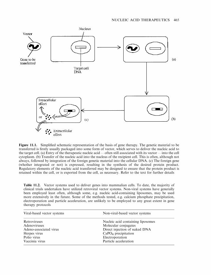
NUCLEIC ACID THERAPEUTICS 465
Figure 11.1. Simplified schematic representation of the basis of gene therapy. The genetic material to betransferred is firstly usually packaged into some form of vector, which serves to deliver the nucleic acid tothe target cell. (a) Entry of the therapeutic nucleic acid—often still associated with its vector— into the cellcytoplasm. (b) Transfer of the nucleic acid into the nucleus of the recipient cell. This is often, although notalways, followed by integration of the foreign genetic material into the cellular DNA. (c) The foreign gene(whether integrated or not) is expressed, resulting in the synthesis of the desired protein product.Regulatory elements of the nucleic acid transferred may be designed to ensure that the protein product isretained within the cell, or is exported from the cell, as necessary. Refer to the text for further details
Table 11.2. Vector systems used to deliver genes into mammalian cells. To date, the majority ofclinical trials undertaken have utilized retroviral vector systems. Non-viral systems have generallybeen employed least often, although some, e.g. nucleic acid-containing liposomes, may be usedmore extensively in the future. Some of the methods tested, e.g. calcium phosphate precipitation,electroporation and particle acceleration, are unlikely to be employed to any great extent in genetherapy protocols
Viral-based vector systems Non-viral-based vector systems
Retroviruses Nucleic acid containing liposomesAdenoviruses Molecular conjugatesAdeno-associated virus Direct injection of naked DNAHerpes virus CaPO4 precipitationPolio virus ElectroporationVaccinia virus Particle acceleration
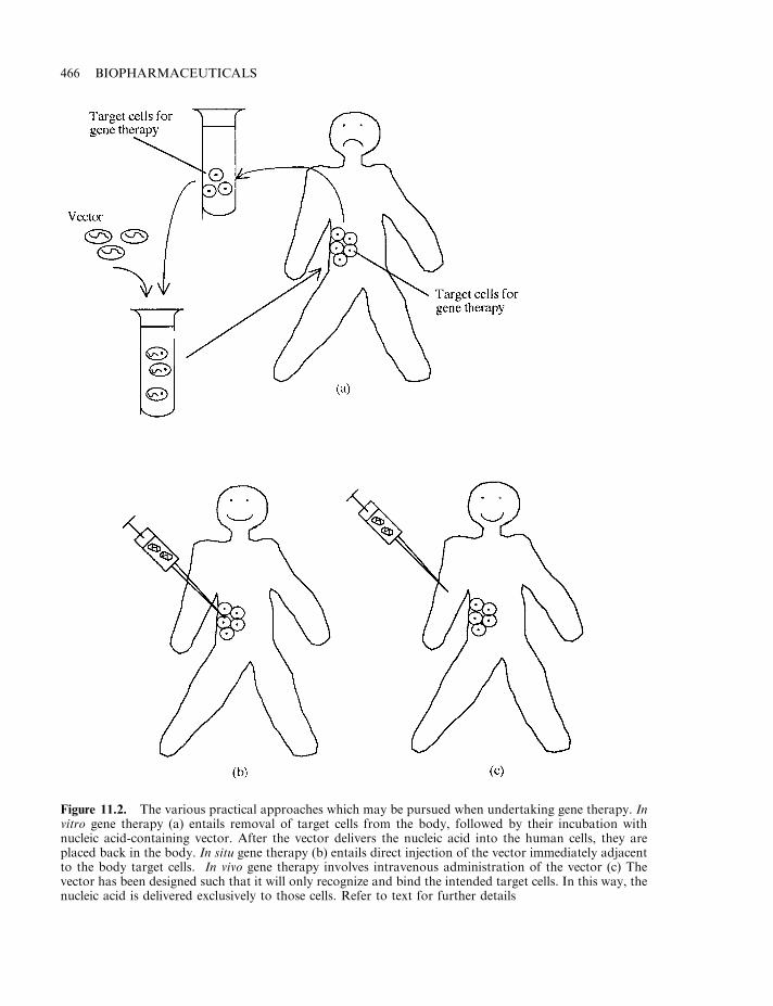
466 BIOPHARMACEUTICALS
Figure 11.2. The various practical approaches which may be pursued when undertaking gene therapy. Invitro gene therapy (a) entails removal of target cells from the body, followed by their incubation withnucleic acid-containing vector. After the vector delivers the nucleic acid into the human cells, they areplaced back in the body. In situ gene therapy (b) entails direct injection of the vector immediately adjacentto the body target cells. In vivo gene therapy involves intravenous administration of the vector (c) Thevector has been designed such that it will only recognize and bind the intended target cells. In this way, thenucleic acid is delivered exclusively to those cells. Refer to text for further details
target cells are not localized to one specific area of the body (e.g. blood cells). An alternative (invivo) approach entails the development of vectors capable of recognizing and binding only tospecific, pre-defined cell types. Such vectors could then be administered easily, e.g. byintravenous injection. Through appropriate bio-specific interactions, they would only delivertheir nucleic acid payload to the specified target cells. The simplicity and specificity of thisapproach renders it the method of choice. However, thus far, no such vector systems have beendeveloped for routine therapeutic use. Intensive efforts to develop these are under way and anumber of different strategies are being pursued. For example, the inclusion of an antibody onthe vector surface, which specifically binds a surface antigen uniquely associated with the targetcell, would allow selective delivery. Another approach entails engineering the vector to display aspecific hormone which would bind only to cells displaying the hormone receptor. Thefeasibility of this approach has been demonstrated using retroviral vectors engineered to displayerythropoietin on their surface.
Some additional questions
The choice of vector, target cell and protocol used will depend upon a number of considerations.The major consideration is obviously what the ultimate goal of the gene therapy treatment is inany given case, e.g. in some instances it may be to correct an inherited genetic defect, whereas inother instances it may be to confer a novel function upon the recipient cell. An example of theformer would be the introduction of the cystic fibrosis transmembrane conductance regulator(CFTR) gene (the cystic fibrosis gene) into the airway epithelial cells of CF sufferers. Anexample of the latter would be the introduction of a novel gene into white blood cells whoseprotein product is capable of in some way interfering with HIV replication; such an approachmight prove an effective therapeutic strategy for the treatment of AIDS.
An additional consideration that may influence the protocol used is the desired duration ofsubsequent expression of the gene product. In most cases of genetic disease, long-termexpression of the inserted gene would be required. In other instances (e.g. some forms of cancertherapy, or the use of gene therapy to deliver a DNA-based vaccine), short-term expression ofthe gene introduced would be sufficient/desirable.
For most applications of gene therapy, straightforward expression of the gene product itselfwill suffice. However, in some instances, regulation of expression of the transferred gene wouldbe required, e.g. if gene therapy combating insulin-dependent diabetes mellitus was to beconsidered. Achieving such expressional control over transferred genes is a pursuit that is onlyin the early stages of development.
The choice of target cells is another point worthy of discussion. In some instances, this choiceis pre-determined, e.g. treatment of the genetic condition familial hypercholesterolemia wouldrequire insertion of the gene coding for the low-density lipoprotein receptor specifically inhepatocytes.
In other cases, however, some scope may be available to choose a target cell population. Evenin the case of redressing some genetic diseases, it may not be necessary to genetically correct theexact population of cells affected, e.g. a hallmark of several of the best-characterized geneticdiseases is the exceedingly low production of a circulatory gene product; examples includeclotting factors VIII and IX, a lack of which leads to haemophilia. It may be possible to correctsuch defects by introducing the appropriate gene into any recipient cell capable of exporting thegene product into the blood. In such cases, choosing a target cell could be made upon practical
NUCLEIC ACID THERAPEUTICS 467
considerations, such as their ease of isolation and culture, their capacity to express (and excrete)the protein product, and their half-lives in vivo.
Several cell types, including keratinocytes, myoblasts and fibroblasts, have been studied inthis regard. It has been shown, for example, that myoblasts into which the factor IX gene andthe growth hormone gene have been introduced could express their protein products and secretethem into the circulation.
Vectors used in gene therapy
A list of the various vectors capable of introducing genes into recipient cells has been providedin Table 11.2. These vectors are conveniently categorized as being viral-based or non-viral-basedsystems. The main vector systems developed thus far are discussed in somewhat more detailbelow.
Retroviral vectors
In the region of 80% of gene therapy clinical trials undertaken to date have employed retroviralvectors as gene delivery systems. Retroviruses are enveloped viruses. Their genome consists ofsingle-stranded RNA (ssRNA) of approximately 5–8 kb. Upon entry into sensitive cells, theviral RNA is reverse-transcribed and eventually yields double-stranded DNA. This subse-quently integrates into the host cell genome (Box 11.1). The basic retroviral genome contains aminimum of three structural genes; gag (codes for core viral protein), pol (codes for reversetranscriptase) and env (codes for the viral envelope proteins). At either end of the viral genomeare the long terminal repeats (LTRs), which harbour powerful promoter and enhancer regionsand sequences required to promote integration into the host DNA. Also present, immediatelyadjacent to the 5’ LTR, is the packing sequence (c). This is required to promote viral RNApackaging.
The ability of such retroviruses to (a) effectively enter various cell types and (b) integrate theirgenome into the host cell genome in a stable, long-term fashion, made them obvious potentialvectors for gene therapy.
The construction of retroviruses to function as gene vectors entails replacing the endogenousviral genes, required for normal viral replication, with the exogenous gene of interest(Figure 11.3a). Removal of the viral structural genes means that the resulting vector cannotitself replicate. In order to generate mature virion particles harbouring the vector nucleic acid(Figure 11.3b), this genetic material must be introduced into a ‘packing cell’. These arerecombinant cells that have previously been engineered to contain the gag, pol and env structuralgenes (Figure 11.4). In this way, packing cells are capable of producing mature but replication-deficient viral particles, harbouring the gene to be transferred (see later section on Manufactureof viral vectors). These viral particles function as so-called ‘one-time, single-hit’ gene transfersystems.
More recently, various modifications have been introduced to this basic retroviral system. Theinclusion of the 5’ end of the gag gene is shown to enhance levels of vector production by up to200-fold. Additionally, specific promoters have been introduced in order to attempt to controlexpression of the inserted gene. Most work has focused upon the use of tissue-specific promotersin an effort to limit expression of the desired gene to a specific tissue type. The most commonlyemployed (recombinant-deficient) retrovirus used in this regard has been derived from theMaloney murine leukaemia virus (MoMuLV).
468 BIOPHARMACEUTICALS
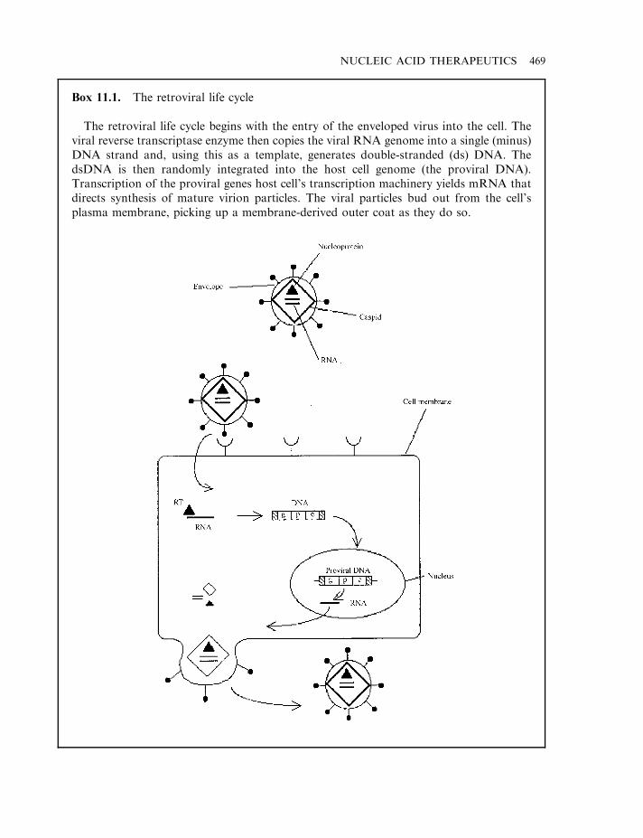
NUCLEIC ACID THERAPEUTICS 469
Box 11.1. The retroviral life cycle
The retroviral life cycle begins with the entry of the enveloped virus into the cell. Theviral reverse transcriptase enzyme then copies the viral RNA genome into a single (minus)DNA strand and, using this as a template, generates double-stranded (ds) DNA. ThedsDNA is then randomly integrated into the host cell genome (the proviral DNA).Transcription of the proviral genes host cell’s transcription machinery yields mRNA thatdirects synthesis of mature virion particles. The viral particles bud out from the cell’splasma membrane, picking up a membrane-derived outer coat as they do so.
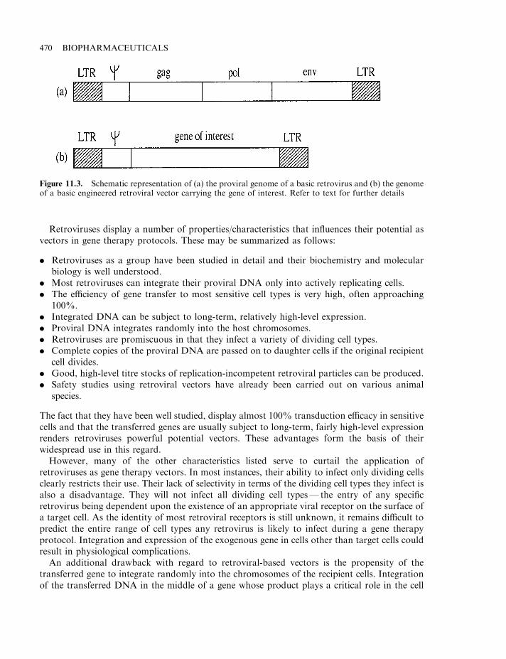
Retroviruses display a number of properties/characteristics that influences their potential asvectors in gene therapy protocols. These may be summarized as follows:
. Retroviruses as a group have been studied in detail and their biochemistry and molecularbiology is well understood.
. Most retroviruses can integrate their proviral DNA only into actively replicating cells.
. The efficiency of gene transfer to most sensitive cell types is very high, often approaching100%.
. Integrated DNA can be subject to long-term, relatively high-level expression.
. Proviral DNA integrates randomly into the host chromosomes.
. Retroviruses are promiscuous in that they infect a variety of dividing cell types.
. Complete copies of the proviral DNA are passed on to daughter cells if the original recipientcell divides.
. Good, high-level titre stocks of replication-incompetent retroviral particles can be produced.
. Safety studies using retroviral vectors have already been carried out on various animalspecies.
The fact that they have been well studied, display almost 100% transduction efficacy in sensitivecells and that the transferred genes are usually subject to long-term, fairly high-level expressionrenders retroviruses powerful potential vectors. These advantages form the basis of theirwidespread use in this regard.
However, many of the other characteristics listed serve to curtail the application ofretroviruses as gene therapy vectors. In most instances, their ability to infect only dividing cellsclearly restricts their use. Their lack of selectivity in terms of the dividing cell types they infect isalso a disadvantage. They will not infect all dividing cell types— the entry of any specificretrovirus being dependent upon the existence of an appropriate viral receptor on the surface ofa target cell. As the identity of most retroviral receptors is still unknown, it remains difficult topredict the entire range of cell types any retrovirus is likely to infect during a gene therapyprotocol. Integration and expression of the exogenous gene in cells other than target cells couldresult in physiological complications.
An additional drawback with regard to retroviral-based vectors is the propensity of thetransferred gene to integrate randomly into the chromosomes of the recipient cells. Integrationof the transferred DNA in the middle of a gene whose product plays a critical role in the cell
470 BIOPHARMACEUTICALS
Figure 11.3. Schematic representation of (a) the proviral genome of a basic retrovirus and (b) the genomeof a basic engineered retroviral vector carrying the gene of interest. Refer to text for further details
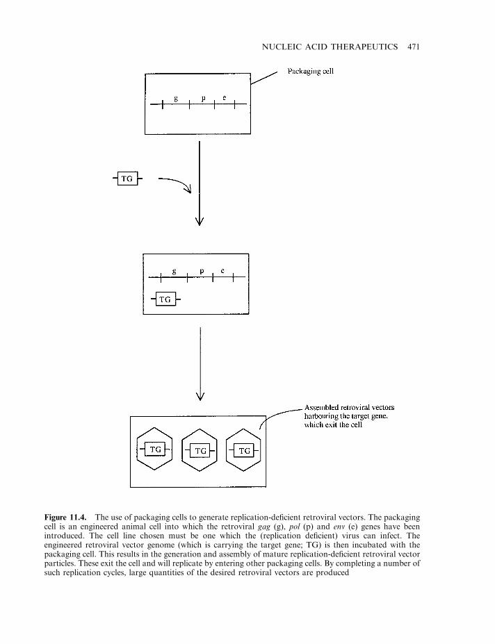
NUCLEIC ACID THERAPEUTICS 471
Figure 11.4. The use of packaging cells to generate replication-deficient retroviral vectors. The packagingcell is an engineered animal cell into which the retroviral gag (g), pol (p) and env (e) genes have beenintroduced. The cell line chosen must be one which the (replication deficient) virus can infect. Theengineered retroviral vector genome (which is carrying the target gene; TG) is then incubated with thepackaging cell. This results in the generation and assembly of mature replication-deficient retroviral vectorparticles. These exit the cell and will replicate by entering other packaging cells. By completing a number ofsuch replication cycles, large quantities of the desired retroviral vectors are produced
could irrevocably damage cellular function, e.g. disruption of a central metabolic enzyme couldcause cell death, while disruption of a tumour suppresser gene could give rise to cellulartransformation; in addition, integration of the proviral nucleic acid to sites adjacent to quiescentcellular proto-oncogenes could result in their activation. Another impediment to routine use ofretroviral vectors is the relatively labile nature of these particles. Thus, while retroviruses arerelatively easy to propagate, they are often damaged by subsequent purification andconcentration— steps essential for their clinical use.
Additional viral-based vectors
A number of additional viral types may also prove useful as vectors in the practice of genetherapy. Chief amongst these are the adenoviruses. Adeno-associated virus, the herpes virus anda number of other viruses are also being considered (Table 11.2).
Adenoviruses are relatively large, non-enveloped structures, housing double-stranded DNAas their genetic material. Their genome is much larger (approximately 35 kb), and more complexthan those of retroviruses. In most instances, only a small fraction of this genome is removedwhen constructing an adenovirus-based vector. Upon cellular infection, adenoviral DNAbecomes localized in the nucleus, but does not integrate into the host cell DNA. Usually,infection by wild-type adenoviruses is associated with, at most, mild clinical symptoms inhumans.
As potential vectors for gene therapy, adenoviruses display a number of both advantages anddisadvantages (Table 11.3). Their major advantage relates to their ability to efficiently infectnon-dividing cells and the usually observed expression of large quantities of the desired geneproducts. However, the failure of the adenoviral-based DNA to integrate into the host cellgenerally means that its survival, and hence the duration of gene expression, is limited.Adenovirus-based vectors carrying various marker genes (i.e. a gene whose expression productis easily detected) have been administered to animals. Marker gene expression has beensubsequently noted in various tissues, including heart, liver, muscle, bone marrow, CNS andendothelial cells. Duration of marker gene expression ranged from 2–3 weeks to several months.
While short-term, high-level gene expression may be appropriate for some gene therapyapplications, it would be of less use for the treatment of, for example, genetic diseases, wherelong-term gene expression would be required. This could be achieved in theory by repeatadministration of the adenoviral vector. However, adenoviruses prompt a strong immuneresponse, which limits the efficacy of repeat administration.
472 BIOPHARMACEUTICALS
Table 11.3. Some characteristic advantages and disadvantages of adenoviruses as potential vectors forgene therapy. Refer to text for further details
Advantages Disadvantages
Adenoviruses are capable of gene transfer tonon-dividing cells
Adenoviruses are highly immunogenic in man
They are easy to propagate in large quantitiesHigh levels of gene expression are usually
The duration of expression of transferred genescan vary, and is usually transient
recordedInfection of permissive cells with wild-typeadenovirus usually results in cell lysis
They are relatively stable viruses Adenoviruses display a broad selectivity in the celltypes they can infect
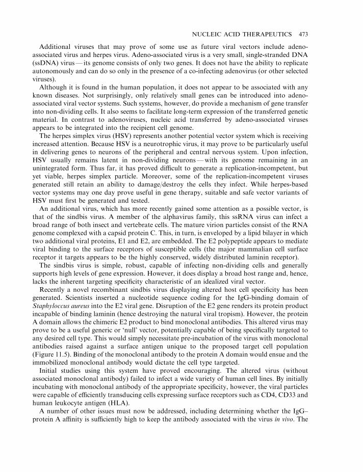
Additional viruses that may prove of some use as future viral vectors include adeno-associated virus and herpes virus. Adeno-associated virus is a very small, single-stranded DNA(ssDNA) virus— its genome consists of only two genes. It does not have the ability to replicateautonomously and can do so only in the presence of a co-infecting adenovirus (or other selectedviruses).
Although it is found in the human population, it does not appear to be associated with anyknown diseases. Not surprisingly, only relatively small genes can be introduced into adeno-associated viral vector systems. Such systems, however, do provide a mechanism of gene transferinto non-dividing cells. It also seems to facilitate long-term expression of the transferred geneticmaterial. In contrast to adenoviruses, nucleic acid transferred by adeno-associated virusesappears to be integrated into the recipient cell genome.
The herpes simplex virus (HSV) represents another potential vector system which is receivingincreased attention. Because HSV is a neurotrophic virus, it may prove to be particularly usefulin delivering genes to neurons of the peripheral and central nervous system. Upon infection,HSV usually remains latent in non-dividing neurons—with its genome remaining in anunintegrated form. Thus far, it has proved difficult to generate a replication-incompetent, butyet viable, herpes simplex particle. Moreover, some of the replication-incompetent virusesgenerated still retain an ability to damage/destroy the cells they infect. While herpes-basedvector systems may one day prove useful in gene therapy, suitable and safe vector variants ofHSV must first be generated and tested.
An additional virus, which has more recently gained some attention as a possible vector, isthat of the sindbis virus. A member of the alphavirus family, this ssRNA virus can infect abroad range of both insect and vertebrate cells. The mature virion particles consist of the RNAgenome complexed with a capsid protein C. This, in turn, is enveloped by a lipid bilayer in whichtwo additional viral proteins, E1 and E2, are embedded. The E2 polypeptide appears to mediateviral binding to the surface receptors of susceptible cells (the major mammalian cell surfacereceptor it targets appears to be the highly conserved, widely distributed laminin receptor).
The sindbis virus is simple, robust, capable of infecting non-dividing cells and generallysupports high levels of gene expression. However, it does display a broad host range and, hence,lacks the inherent targeting specificity characteristic of an idealized viral vector.
Recently a novel recombinant sindbis virus displaying altered host cell specificity has beengenerated. Scientists inserted a nucleotide sequence coding for the IgG-binding domain ofStaphyloccus aureus into the E2 viral gene. Disruption of the E2 gene renders its protein productincapable of binding laminin (hence destroying the natural viral tropism). However, the proteinA domain allows the chimeric E2 product to bind monoclonal antibodies. This altered virus mayprove to be a useful generic or ‘null’ vector, potentially capable of being specifically targeted toany desired cell type. This would simply necessitate pre-incubation of the virus with monoclonalantibodies raised against a surface antigen unique to the proposed target cell population(Figure 11.5). Binding of the monoclonal antibody to the protein A domain would ensue and theimmobilized monoclonal antibody would dictate the cell type targeted.
Initial studies using this system have proved encouraging. The altered virus (withoutassociated monoclonal antibody) failed to infect a wide variety of human cell lines. By initiallyincubating with monoclonal antibody of the appropriate specificity, however, the viral particleswere capable of efficiently transducing cells expressing surface receptors such as CD4, CD33 andhuman leukocyte antigen (HLA).
A number of other issues must now be addressed, including determining whether the IgG–protein A affinity is sufficiently high to keep the antibody associated with the virus in vivo. The
NUCLEIC ACID THERAPEUTICS 473
full potential of this approach will also require more detailed characterization of surfacemarkers uniquely associated with different cell types. However, the approach exemplifies thetypes of technical innovations now being introduced, which will make second-generation vectorsmore suited to their role in gene therapy.
Manufacture of viral vectors
Literature reports describing the large-scale manufacture of viral vectors for gene therapyapplication are few and far between. Development of manufacturing protocols has largely beenundertaken by companies engaged in gene therapy product development and, consequently,protocol details remain confidential. The overall approach likely taken is not too dissimilar tothat of therapeutic protein manufacture (Chapter 3). It involves synthesis in cells, recovery,concentration, purification and formulation steps. A likely generalized manufacturing scenariofor retroviral-based vectors is outlined in Figure 11.6. The manufacture of alternative viral
474 BIOPHARMACEUTICALS
Figure 11.5. Generation of engineered sindbis virus capable of being targeted to bind specific cell types.(a) Simplified depiction of the virus, displaying the surface E2 protein. Genetic engineering facilitatesdisruption of the E2 gene by incorporation of the IgG-binding domain of protein A (b). Incubation of suchengineered viral particles with most monoclonal antibody types results in effective immobilization of theantibody on the viral surface (c). Thus, the engineered viral vector should be targetable to any specific celltype, simply by its pre-incubation with monoclonal antibodies, which selectively bind a surface antigenuniquely associated with the target cell
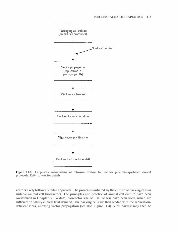
vectors likely follow a similar approach. The process is initiated by the culture of packing cells insuitable animal cell bioreactors. The principles and practice of animal cell culture have beenoverviewed in Chapter 3. To date, bioreactor size of 100 l or less have been used, which aresufficient to satisfy clinical trial demand. The packing cells are then seeded with the replication-deficient virus, allowing vector propagation (see also Figure 11.4). Viral harvest may then be
NUCLEIC ACID THERAPEUTICS 475
Figure 11.6. Large-scale manufacture of retroviral vectors for use for gene therapy-based clinicalprotocols. Refer to text for details
undertaken by methods of microfiltration, which separates intact packing cells/cell debris fromthe vector-containing product stream. Viral vector concentration can then be undertaken byultrafiltration (Chapter 3). Subsequent vector purification strategies employed includechromatographic approaches similar to those used to purify proteins. Ion exchange andvarious forms of affinity chromatography have received most attention. A detailed under-standing of the interactions between chromatographic media and viral surface molecules iscurrently lacking and hence is the subject of ongoing research. A comprehensive understandingof such interactions would be required to optimize chromatographic purification protocols.After purification, final product analysis and formulation is undertaken. Again, few details ofthese steps have been openly published.
Non-viral vectors
Although viral-mediated gene delivery systems currently predominate, some 20–25% of currentclinical trials use non-viral-based methods of gene delivery. General advantages quoted withrespect to non-viral delivery systems include:
. their low/non-immunogenicity;
. non-occurrence of integration of the therapeutic gene into the host chromosome (thiseliminates the potential to disrupt essential host genes or to activate host oncogenes.
The initial approach adopted entailed administration of ‘naked’ plasmid DNA housing the geneof interest. This avenue of research was first opened in 1990, when it was shown that nakedplasmid DNA was expressed in mice muscle cells subsequent to its i.m. injection. The plasmidDNA concerned housed the b-galactosidase gene as a reporter. Subsequent expression ofb-galactosidase activity could persist for anything from a few months to the remainder of theanimal’s life. The transfection rate recorded was low (1–2% of muscle fibres assimilated theDNA) and the DNA was not integrated into the host cell’s chromosomes.
Up until this point, it was assumed that naked DNA injected into animals would not bespontaneously taken up and expressed in host cells. This finding vindicated the cautiousapproach taken by the FDA and other regulatory authorities with regard to the presence of freeDNA in biopharmaceutical products (Chapter 3).
Scientists have also since demonstrated that DNA (coated on microscopic gold beads)propelled into the epidermis of test animals with a ‘gene gun’ is expressed in the animal’s skincells. Furthermore, the introduction in this fashion of DNA coding for human influenza viralantigens resulted in effective immunization of the animal against influenza. Similar results, usingother pathogen models, have also now been generated. It is assumed that expressed antigen issecreted by the cell and, in this way, is exposed to immune surveillance. Further research hasillustrated that systematic administration via i.v. injection rarely achieves meaningful celltransfection. This is most likely due to the high nuclease levels present in serum. In contrast, freenuclease activity in muscle tissue is extremely low.
Modern non-viral-based systems generally entail complexing/packaging the gene of interest(present, along with appropriate promoters, etc., in a circular plasmid) with additionalmolecules, particularly various lipids or some polypeptides. These generally display a positivecharge and hence interact with the negatively-charged DNA molecules. The function of suchcarrier molecules is to stabilize the DNA, protect it, e.g. from serum nucleases, and ideally tomodulate interaction with the biological system, e.g. help target the DNA to particular celltypes—or away from other cell types.
476 BIOPHARMACEUTICALS
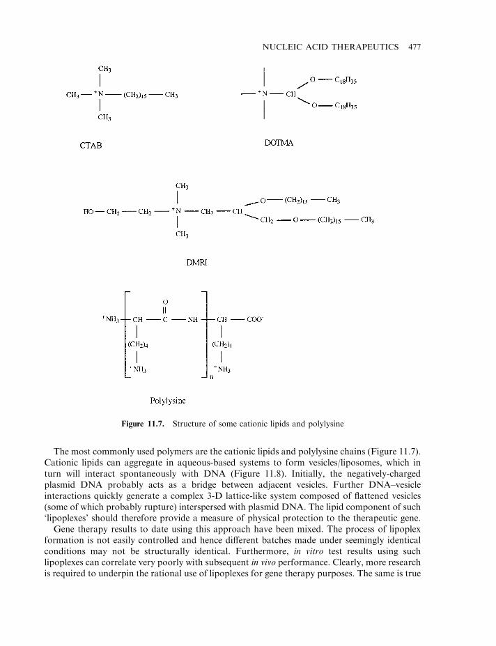
The most commonly used polymers are the cationic lipids and polylysine chains (Figure 11.7).Cationic lipids can aggregate in aqueous-based systems to form vesicles/liposomes, which inturn will interact spontaneously with DNA (Figure 11.8). Initially, the negatively-chargedplasmid DNA probably acts as a bridge between adjacent vesicles. Further DNA–vesicleinteractions quickly generate a complex 3-D lattice-like system composed of flattened vesicles(some of which probably rupture) interspersed with plasmid DNA. The lipid component of such‘lipoplexes’ should therefore provide a measure of physical protection to the therapeutic gene.
Gene therapy results to date using this approach have been mixed. The process of lipoplexformation is not easily controlled and hence different batches made under seemingly identicalconditions may not be structurally identical. Furthermore, in vitro test results using suchlipoplexes can correlate very poorly with subsequent in vivo performance. Clearly, more researchis required to underpin the rational use of lipoplexes for gene therapy purposes. The same is true
NUCLEIC ACID THERAPEUTICS 477
Figure 11.7. Structure of some cationic lipids and polylysine
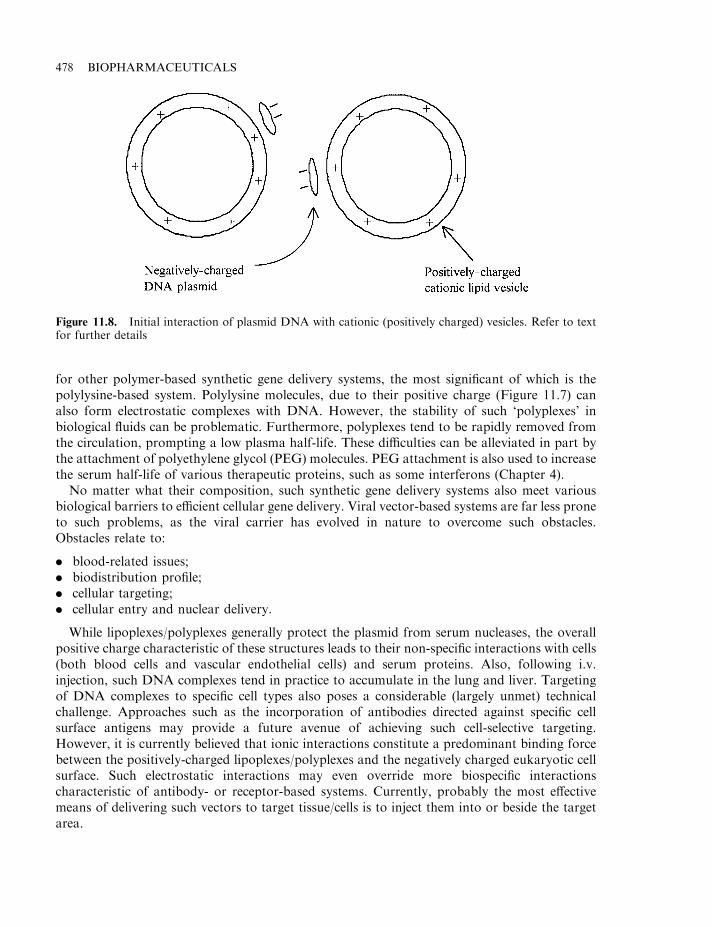
for other polymer-based synthetic gene delivery systems, the most significant of which is thepolylysine-based system. Polylysine molecules, due to their positive charge (Figure 11.7) canalso form electrostatic complexes with DNA. However, the stability of such ‘polyplexes’ inbiological fluids can be problematic. Furthermore, polyplexes tend to be rapidly removed fromthe circulation, prompting a low plasma half-life. These difficulties can be alleviated in part bythe attachment of polyethylene glycol (PEG) molecules. PEG attachment is also used to increasethe serum half-life of various therapeutic proteins, such as some interferons (Chapter 4).
No matter what their composition, such synthetic gene delivery systems also meet variousbiological barriers to efficient cellular gene delivery. Viral vector-based systems are far less proneto such problems, as the viral carrier has evolved in nature to overcome such obstacles.Obstacles relate to:
. blood-related issues;
. biodistribution profile;
. cellular targeting;
. cellular entry and nuclear delivery.
While lipoplexes/polyplexes generally protect the plasmid from serum nucleases, the overallpositive charge characteristic of these structures leads to their non-specific interactions with cells(both blood cells and vascular endothelial cells) and serum proteins. Also, following i.v.injection, such DNA complexes tend in practice to accumulate in the lung and liver. Targetingof DNA complexes to specific cell types also poses a considerable (largely unmet) technicalchallenge. Approaches such as the incorporation of antibodies directed against specific cellsurface antigens may provide a future avenue of achieving such cell-selective targeting.However, it is currently believed that ionic interactions constitute a predominant binding forcebetween the positively-charged lipoplexes/polyplexes and the negatively charged eukaryotic cellsurface. Such electrostatic interactions may even override more biospecific interactionscharacteristic of antibody- or receptor-based systems. Currently, probably the most effectivemeans of delivering such vectors to target tissue/cells is to inject them into or beside the targetarea.
478 BIOPHARMACEUTICALS
Figure 11.8. Initial interaction of plasmid DNA with cationic (positively charged) vesicles. Refer to textfor further details
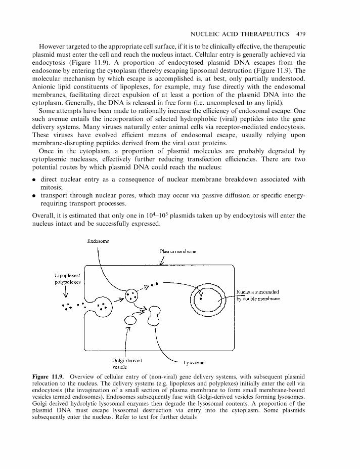
However targeted to the appropriate cell surface, if it is to be clinically effective, the therapeuticplasmid must enter the cell and reach the nucleus intact. Cellular entry is generally achieved viaendocytosis (Figure 11.9). A proportion of endocytosed plasmid DNA escapes from theendosome by entering the cytoplasm (thereby escaping liposomal destruction (Figure 11.9). Themolecular mechanism by which escape is accomplished is, at best, only partially understood.Anionic lipid constituents of lipoplexes, for example, may fuse directly with the endosomalmembranes, facilitating direct expulsion of at least a portion of the plasmid DNA into thecytoplasm. Generally, the DNA is released in free form (i.e. uncomplexed to any lipid).
Some attempts have been made to rationally increase the efficiency of endosomal escape. Onesuch avenue entails the incorporation of selected hydrophobic (viral) peptides into the genedelivery systems. Many viruses naturally enter animal cells via receptor-mediated endocytosis.These viruses have evolved efficient means of endosomal escape, usually relying uponmembrane-disrupting peptides derived from the viral coat proteins.
Once in the cytoplasm, a proportion of plasmid molecules are probably degraded bycytoplasmic nucleases, effectively further reducing transfection efficiencies. There are twopotential routes by which plasmid DNA could reach the nucleus:
. direct nuclear entry as a consequence of nuclear membrane breakdown associated withmitosis;
. transport through nuclear pores, which may occur via passive diffusion or specific energy-requiring transport processes.
Overall, it is estimated that only one in 104–105 plasmids taken up by endocytosis will enter thenucleus intact and be successfully expressed.
NUCLEIC ACID THERAPEUTICS 479
Figure 11.9. Overview of cellular entry of (non-viral) gene delivery systems, with subsequent plasmidrelocation to the nucleus. The delivery systems (e.g. lipoplexes and polyplexes) initially enter the cell viaendocytosis (the invagination of a small section of plasma membrane to form small membrane-boundvesicles termed endosomes). Endosomes subsequently fuse with Golgi-derived vesicles forming lysosomes.Golgi derived hydrolytic lysosomal enzymes then degrade the lysosomal contents. A proportion of theplasmid DNA must escape lysosomal destruction via entry into the cytoplasm. Some plasmidssubsequently enter the nucleus. Refer to text for further details
Manufacture of plasmid DNA
Plasmid DNA is routinely extracted and purified from various microbial cells at the researchlevel. However, industrial-scale manufacture to the exacting standards of purity demanded ofpharmaceutical products is a pursuit still in its infancy, and little has been published on thesubject. The overall generalized approach used to produce plasmid DNA for the purposes ofgene therapy trials is presented in Figure 11.10. Prior to its manufacture, researchers would haveconstructed an appropriate vector housing the therapeutic gene and introduced it into aproducer microorganism, such as E. coli. Routine large-scale plasmid manufacture then entailsculture of a batch of producer microorganisms by fermentation, followed by plasmid extractionand purification. In this regard, the overall approach used resembles the approaches taken in thelarge-scale manufacture of recombinant therapeutic proteins, as described in Chapter 3.
Industrial-scale microbial fermentation (upstream processing) has also been described inChapter 3, to which the reader is referred. Fermentation promotes microbial cell replication andthus the biosynthesis of large quantities of plasmid. Subsequent to fermentation, the microbialcells are harvested (collected) by either centrifugation or microfiltration. Following resuspensionin a low volume of buffer, the cells must be disrupted in order to release the plasmids therein.This appears to be most commonly achieved by the addition of a lysis reagent, consisting ofNaOH and SDS (sodium dodecyl sulphate). The combination of high pH and detergent actiondisrupts the microbial cell wall and membranes, with consequent release of the intracellularcontents. In addition to the desired plasmid DNA, this crude mixture will also contain variousimpurities that must be removed by subsequent downstream processing steps. Notableimpurities include:
. cell wall debris and some intact cells;
. proteins;
. genomic DNA;
. RNA;
. low molecular mass metabolites;
. endotoxin.
After lysis is complete, the next step can entail the addition of a high-salt neutralizationsolution, such as potassium acetate. This promotes formation of aggregates of genomic DNA(gDNA) and SDS–protein complexes, which can subsequently be removed by centrifugation orfiltration. The plasmids can then be themselves precipitated from the resultant solution by theaddition of appropriate solvent (usually either isoproponal or ethanol). Upon resuspension, theplasmid preparation can then be subjected to chromatographic purification. The majorcontaminants likely still present include RNA, gDNA fragments, nicked or other plasmidvariants and endotoxins. Gel filtration chromatography can effectively remove contaminantsthat differ substantially in shape/size from the desired plasmid. These can include most gDNAfragments, RNA and (most) endotoxins. It can also achieve partial removal of plasmidvariants, such as open circular plasmids, from the main (supercoiled) plasmid preparation.Ion exchange can remove many protein contaminants, as well as RNA. However, gDNAand endotoxins generally co-purify with the plasmid DNA. Additional chromatographicapproaches based upon reverse-phase and affinity systems have been developed at laboratoryscale at least.
A significant feature of plasmid purification employing capture chromatography (i.e. involvesplasmids binding to the chromatographic beads) is the low plasmid-binding capacities observed.
480 BIOPHARMACEUTICALS
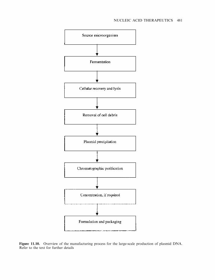
NUCLEIC ACID THERAPEUTICS 481
Figure 11.10. Overview of the manufacturing process for the large-scale production of plasmid DNA.Refer to the text for further details
The pore size of commercially-available capture chromatographic media is insufficiently large toallow entry of plasmids, restricting binding to the bead surface. Binding capacities can,therefore, be 100-fold or more lower than those observed when the same medium is used topurify (much smaller) therapeutic proteins (Chapter 3).
Purified plasmids may then be analysed using various analytical techniques. Freedom fromcontaminating nucleic acid and/or proteins can be assessed electrophoretically. Endotoxin andsterility tests would also be routinely undertaken. The purified plasmid DNA must next beformulated to yield the final non-viral delivery system. Formulation studies relating to suchsystems remain an area requiring further investigation. Most work reported to date relates toformulating and stabilizing lipoplex-based gene delivery systems. Aqueous suspensions of these(and other) non-viral based systems tend to quickly aggregate (in a matter of minutes to hours).In order to circumvent this problem, the final delivery systems were often actually formulated atthe patient’s bedside in earlier clinical trials.
Research aimed at identifying appropriate stabilizing excipients and formulation formats isongoing. Simple freezing is an option, particularly as frozen formulations would be immune toagitation-induced aggregation. However, the process of freezing, particularly slow freezing, initself induces aggregation. This can be minimized by flash freezing (e.g. by immersion in liquidnitrogen), although this approach may not prove practicable at an industrial scale. The additionof cryoprotectants may help minimize this problem and initial studies indicate that varioussugars (e.g. glucose, sucrose and trehalose) show some potential in this regard. Another avenueunder investigation relates to the generation of a final freeze-dried product. Again issues, such asthe (relatively) slow freezing process characteristic of industrial-scale freeze-driers, complicateattaining this goal in practice.
Gene therapy and genetic disease
Well over 4000 genetic diseases have been characterized to date. Many of these are caused bylack of production of a single gene product or are due to the production of a mutated geneproduct incapable of carrying out its natural function. Gene therapy represents a seeminglystraightforward therapeutic option which could correct such genetic-based diseases. This wouldbe achieved simply by facilitating insertion of a ‘healthy’ copy of the gene in question intoappropriate cells of the sufferer.
Although simple in concept, the application of gene therapy to treat or cure genetic diseaseshas, thus far, made little impact in practice. The slow progress in this regard is likely due to anumber of factors. These include:
. The number of genetic diseases for which the actual gene responsible has been identified andstudied is relatively modest, although completion of the human genome project shouldrapidly accelerate identification of such genes.
. As discussed previously, none of the first-generation gene-delivering vectors have provedfully satisfactory.
. Some genetic diseases are quite complex, with several organs/cell types being affected. Inmost instances, it has proved difficult in practice to introduce the required gene into all theaffected cell types.
. Regulation of expression levels of the genes transferred has proved problematic.
. Drug companies often display greater interest in applying gene therapy to more prevalentdiseases, such as cancer. The patient population suffering from many genetic diseases is
482 BIOPHARMACEUTICALS

relatively modest. In some instances, a limited patient population may not be sufficient toallow the developing company to recoup the cost of drug development.
Some of the genetic conditions for which the defective gene has been pinpointed aresummarized in Table 11.4. Many of the initial attempts to utilize gene therapy in practicefocused upon haemoglobinopathies (e.g. sickle cell anaemia and thalassemias). These conditionswere amongst the first genetic disorders to be characterized at a molecular level, with the defectcentring around the haemoglobin a- or b-chain genes. Furthermore, the target cells in the bonemarrow could be removed and subsequently replaced with relative ease. However, theseconditions proved to be a difficult initial choice for the gene therapist. The production of theappropriate quantities of functional haemoglobin is dependent not only upon the presence of a-and b-globin genes of the correct sequence, but also upon detailed regulation of gene expression.Such tight regulation of expression of transferred genes is beyond the capability of gene therapytechnology as it currently stands.
Another early genetic disease for correction by gene therapy was severe combinedimmunodeficiency (SCID). This disease is caused by a lack of adenosine deaminase (ADA)activity. ADA is an enzyme that plays a central role in the degradation of purine nucleosides (itcatalyzes the removal of ammonia from adenosine, forming inosine which, in turn, is usuallyeventually converted to uric acid). This leads to T and B lymphocyte dysfunction. Lack of aneffective immune system means that SCID sufferers must be kept in an essentially sterileenvironment.
NUCLEIC ACID THERAPEUTICS 483
Table 11.4. Some examples of genetic diseases for which the defective gene responsible hasbeen identified
Disease Defective genes protein product
Haemophilia A Factor VIIIHaemophilia B Factor IXThalassemia b-GlobinSickle cell anaemia b-GlobinFamilial hypercholesterolemia Low-density protein receptorSevere combined immunodeficiency Adenosine deaminase, purine nucleoside
phosphorylaseNiemann–Pick disease SphingomylinaseGaucher’s disease GlucocerebrosidaseCystic fibrosis Cystic fibrosis transmembrane regulatorEmphysema a1-AntitrypsinLeukocyte adhesion deficiency CD18Hyperammonemia Ornithine transcarbamylaseCitrullinemia Arginosuccinate synthetasePhenylketonuria Phenylalanine hydroxylaseMaple syrup disease Branched chain a-ketoacid dehydrogenaseTyrosinemia type 1 Fumarylacetoacetate hydrolaseGlycogen storage deficiency type 1A Glucose-6-phosphataseFucosidosis a-L-FucosidaseMucopolysaccharidosis type VII b-GlucuronidaseMucopolysaccharidosis type I a-L-IduronidaseGalactosemia Galactose-1-phosphate uridyl transferase
When compared to treating diseases such as thalassemia, regulation of the level of expressionof a corrected ADA gene was believed to be less important for a successful therapeutic outcome(in most—although not all—metabolic diseases caused by an enzyme deficiency, it appearsthat expression of even a fraction of normal enzyme levels is sufficient to ameliorate the diseasesymptoms).
Gene therapy trials aimed at counteracting ADA deficiency were initiated in 1990. The firstrecipient was a 4 year-old SCID sufferer. The protocol used entailed the isolation of the child’speripheral lymphocytes, followed by the in vitro introduction of the human ADA gene into thesecells, using a retroviral vector. After a period of expansion (by culture in vitro), these treatedcells were re-injected into the patient. As the lymphocytes (and by extension, the corrective gene)had a finite life span, the therapy was repeated every 6–8 weeks. This approach appearedsuccessful in that it has resulted in a marked and sustained improvement in the recipient’simmune function. Critically, however, interpetration of this outcome was made more difficult,owing to the later revelation that the patient also initiated more conventional SCID therapy justprior to the gene therapy treatment.
Stem cells are attractive potential recipients cells, as they are immortal. Successfulintroduction of the target gene into these cells should facilitate ongoing production of thegene product in mature blood cells, which are continually derived from the stem cell population.This would likely remove the requirement for repeat gene transfers to the affected individual.
The routine transduction of stem cells has, thus far, proved technically difficult. They arefound only in low quantities in the bone marrow, and there is a lack of a suitable assay for stemcells. However, recent progress has been made in this regard and routine transduction of suchcells will likely be achievable within the next few years.
Additional genetic diseases for which a gene therapy approach is currently being evaluatedinclude familial hypercholesterolaemia and cystic fibrosis. Familial hypercholesterolaemia iscaused by the absence (or presence of a defective form of) low-density lipoprotein receptors onthe surface of liver cells. This results in highly elevated serum cholesterol levels, normallyaccompanied by early onset of serious vascular disease. Gene therapy approaches that havebeen attempted thus far to counteract this condition have entailed the initial removal of arelatively large portion of the liver. Hepatocytes derived from the liver are then cultured in vitro,with gene transfer being undertaken using retroviral vectors. The corrected hepatocytes are thenusually infused back into the liver via a catheter. Although studies in animals have been partiallysuccessful, transduction of only a small proportion of the hepatocytes is normally observed.Subsequent expression of the corrective gene can also be variable. In vivo approaches to hepaticgene correction, using both viral and non-viral approaches, are also currently being assessed.
The cystic fibrosis (cf ) gene was first identified in 1989. It codes for a 170 kDa protein, thecystic fibrosis transmembrane conductance regulator (CFTR), which serves as a chloridechannel in epithelial cells. Inheritance of a mutant cftr gene from both parents results in the CFphenotype. While various organs are affected, the most severely affected are the respiratoryepithelial cells, which have, unsurprisingly, become the focus of attempts at corrective genetherapy.
Several vectors have been used in an attempt to deliver the cf gene to the airway epithelial cellsof sufferers. The most notable systems include adenoviruses and cationic liposomes. Vectordelivery to the target cells can be achieved directly by aerosol technology. Delivery of cftr cDNAto airway epithelial cells (and subsequent gene expression) has been demonstrated with the useof both vector types. However, in order to be of therapeutic benefit, it is essential that 5–10% ofthe target cell population receive and express the cftr gene. This level of integration has not been
484 BIOPHARMACEUTICALS
achieved so far and, furthermore, gene expression has often been transient. However, it isconsidered likely that ongoing developments in this field will render gene therapy a usefultreatment for CF within the earlier part of the twenty-first century.
Gene therapy and cancer
To date, the majority of gene therapy trials undertaken aim to cure not inherited genetic defects,but cancer. The average annual incidence of cancer reported in the USA alone stands at ca. 1.4million cases. Survival rates attained by pursuit of conventional therapeutic strategies (surgery,chemo-therapy, radio-therapy) stands at about 50%. Gene therapy will likely provide themedical community with an additional therapeutic tool with which to combat cancer within thenext 10–15 years.
Initial gene therapy trials aimed at treating/curing cancer began in 1991. Various strategicapproaches have since been developed in this regard (Table 11.5). Numerous trials aimed atassessing the application of gene therapy for the treatment of a wide variety of cancer types arenow under way (Table 11.6).
While many of the results generated to date provide hope for the future, thus far gene therapyhas failed to provide a definitive cure for any cancer type. The lack of success is likely due to anumber of factors, including:
. a requirement for improved, more target-specific vector systems;
. a requirement for a better understanding of how cancer cells evade the normal immuneresponse;
. for ethical reasons, most patients treated to date were suffering from advanced andwidespread terminal cancer (i.e. little/no hope of survival if treated using conventional
NUCLEIC ACID THERAPEUTICS 485
Table 11.5. Some therapeutic strategies being pursued in an attempt to treat cancerusing a gene therapy approach. Refer to text for details
Modifying lymphocytes in order to enhance their anti-tumour activityModifying tumour cells to enhance their immunogenicityInserting tumour suppressor genes into tumour cellsInserting toxin genes in tumour cells in order to promote tumour cell destructionInserting suicide genes into tumour cellsInserting genes, such as a multiple drug resistance (mrd) gene, into stem cells to protectthem from chemotherapy-induced damaged
Counteracting the expression of oncogenes in tumour cells by inserting an appropriateantisense gene
Table 11.6. Some specific cancer types for which human gene therapy trialshave been initiated. Although several of the strategies listed in Table 11.5 arebeing employed in these trials, many focus upon the introduction of variouscytokines into the tumour cells themselves in order to attract and enhance atumour-specific immune response
Breast cancer Colorectal cancerMalignant melanoma Tumours of the CNSOvarian cancer Renal cell carcinomaSmall-cell lung cancer Non-small-cell lung cancer
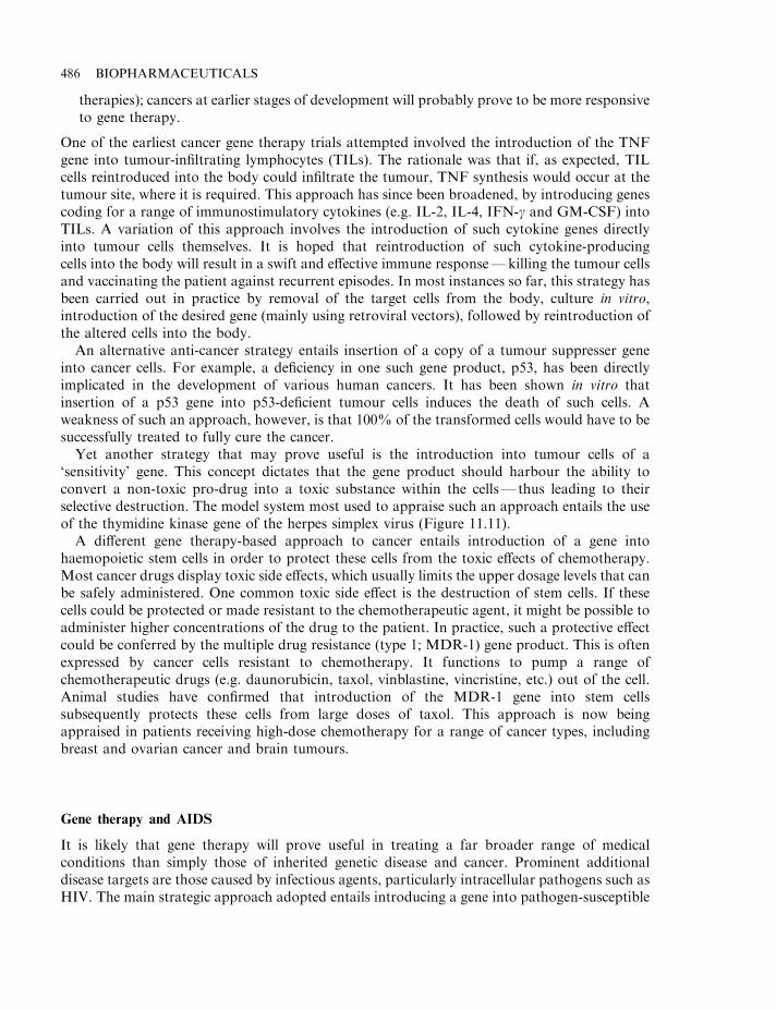
therapies); cancers at earlier stages of development will probably prove to be more responsiveto gene therapy.
One of the earliest cancer gene therapy trials attempted involved the introduction of the TNFgene into tumour-infiltrating lymphocytes (TILs). The rationale was that if, as expected, TILcells reintroduced into the body could infiltrate the tumour, TNF synthesis would occur at thetumour site, where it is required. This approach has since been broadened, by introducing genescoding for a range of immunostimulatory cytokines (e.g. IL-2, IL-4, IFN-g and GM-CSF) intoTILs. A variation of this approach involves the introduction of such cytokine genes directlyinto tumour cells themselves. It is hoped that reintroduction of such cytokine-producingcells into the body will result in a swift and effective immune response—killing the tumour cellsand vaccinating the patient against recurrent episodes. In most instances so far, this strategy hasbeen carried out in practice by removal of the target cells from the body, culture in vitro,introduction of the desired gene (mainly using retroviral vectors), followed by reintroduction ofthe altered cells into the body.
An alternative anti-cancer strategy entails insertion of a copy of a tumour suppresser geneinto cancer cells. For example, a deficiency in one such gene product, p53, has been directlyimplicated in the development of various human cancers. It has been shown in vitro thatinsertion of a p53 gene into p53-deficient tumour cells induces the death of such cells. Aweakness of such an approach, however, is that 100% of the transformed cells would have to besuccessfully treated to fully cure the cancer.
Yet another strategy that may prove useful is the introduction into tumour cells of a‘sensitivity’ gene. This concept dictates that the gene product should harbour the ability toconvert a non-toxic pro-drug into a toxic substance within the cells— thus leading to theirselective destruction. The model system most used to appraise such an approach entails the useof the thymidine kinase gene of the herpes simplex virus (Figure 11.11).
A different gene therapy-based approach to cancer entails introduction of a gene intohaemopoietic stem cells in order to protect these cells from the toxic effects of chemotherapy.Most cancer drugs display toxic side effects, which usually limits the upper dosage levels that canbe safely administered. One common toxic side effect is the destruction of stem cells. If thesecells could be protected or made resistant to the chemotherapeutic agent, it might be possible toadminister higher concentrations of the drug to the patient. In practice, such a protective effectcould be conferred by the multiple drug resistance (type 1; MDR-1) gene product. This is oftenexpressed by cancer cells resistant to chemotherapy. It functions to pump a range ofchemotherapeutic drugs (e.g. daunorubicin, taxol, vinblastine, vincristine, etc.) out of the cell.Animal studies have confirmed that introduction of the MDR-1 gene into stem cellssubsequently protects these cells from large doses of taxol. This approach is now beingappraised in patients receiving high-dose chemotherapy for a range of cancer types, includingbreast and ovarian cancer and brain tumours.
Gene therapy and AIDS
It is likely that gene therapy will prove useful in treating a far broader range of medicalconditions than simply those of inherited genetic disease and cancer. Prominent additionaldisease targets are those caused by infectious agents, particularly intracellular pathogens such asHIV. The main strategic approach adopted entails introducing a gene into pathogen-susceptible
486 BIOPHARMACEUTICALS
cells whose product will interfere with pathogen survival/replication within that cell. Such astrategy is sometimes termed ‘intracellular immunization’.
One such anti-AIDS strategy being pursued is the introduction into virus-sensitive cells of agene coding for an altered (dysfunctional) HIV protein, such as gag, tat or env. The presence ofsuch mutant forms of gag, in particular, was shown to be capable of inhibiting viral replication.This is probably due to interference by the mutated gag product with correct assembly of theviral core. An additional approach entails the transfer to sensitive cells of a gene coding forantibody fragments capable of binding to the HIV envelope proteins. This may also interferewith viral assembly in infected cells.
Scientists have also generated recombinant cells capable of synthesizing and secreting solubleforms of the HIV cell surface receptor, the CD4 antigen. It was suggested that release of suchsoluble viral receptors into the blood would bind circulating virions, hence blocking their abilityto ‘dock’ at sensitive cells. Although this proved to be the case in vitro, early in vivo studies havenot proved as encouraging.
NUCLEIC ACID THERAPEUTICS 487
Figure 11.11. Schematic representation of the therapeutic rationale underpinning the introduction of a‘sensitivity’ gene into tumour cells in order to promote their selective destruction. As depicted in (a), the geneproduct should be capable of converting an inactive pro-drug into a toxic drug, capable of killing the cell. Aspecific example of this approach is presented in (b): introduction of the herpes simplex thymidine kinase(HSVtk) gene confers sensitivity to the anti-herpes drug, Ganciclovir (GCV) on the cell. GCV is convertedby HSVtk into a monophosphorylated form (GCV-MP). This, in turn, is phosphorylated by endogenouskinases, yielding ganciclovir triphosphate (GCV-TP). GCV-TP induces cell death by inhibiting DNApolymerase. A potential advantage of this system is that some adjacent tumour cells (which themselves lackthe HSVtk gene) are also destroyed. This is most likely due to diffusion of the GCV-MP or GCV-TP(perhaps via gap junctions) into such adjacent cells. This so-called ‘bystander effect’ means that all thetransformed cells in a tumour would not necessarily need to be transduced for the therapy to be successful
Yet additional therapeutic approaches to AIDS, based upon antisense technology, will bediscussed later in this chapter. The use of gene therapy to combat this disease has now becomefirmly established as an approach worthy of significant future research.
Gene-based vaccines
Conventional vaccine technology, including the generation of modern recombinant ‘subunit’vaccines, has been discussed in Chapter 10. An additional gene therapy-based approach tovaccination is also now under investigation. The approach entails the administration of a DNAvector housing the gene coding for a surface antigen protein from the target pathogen. In thisway, the body itself would produce the pathogen-associated protein. Theoretically, virtually anybody cell could be targeted, the only requirement being that target cells export the resultantantigenic protein such that it is encountered by the immune system. Additionally, geneexpression need only be transient; sufficiently long to facilitate the induction of an immuneresponse. Target conditions for gene-based vaccines that have entered clinical trials thus farinclude malaria, hepatitis B and AIDS.
Gene therapy: some additional considerations
The importance of gene therapy to the future practice of medicine is no longer being seriouslyquestioned. However, in addition to technical difficulties, a number of non-technical issues mustbe satisfactorily addressed before its practice becomes widespread. Chief amongst these issuesare the questions of public perception, ethics and costs.
Gene therapy is not, and will not be, an inexpensive therapeutic tool. The cost of suchtreatments will likely be broadly similar to the cost of present-day biopharmaceuticals.However, if proved successful in treating many currently incurable conditions, the cost:benefitratio will greatly favour its medical use.
Public perception and ethical considerations are, in many ways, inter-linked. The ability to soreadily modify our genetic complement holds great therapeutic promise. However, strictregulations overseeing the use of this technology are required (and are being enforced). Withoutproper controls, the danger exists that gene therapy could eventually be used to ‘improve’human characteristics. The technical know-how to underpin a new era of eugenics is now almosta reality. The most important safeguard aimed at preventing eugenic-type developments isalready in place. Currently, gene therapy is restricted to somatic cells— the geneticmanipulation of human germ cells is banned. Any genetic alterations achieved thus will notbe transmitted to future generations. Like nuclear technology, there is no ‘going back’ inrelation to gene therapy. The challenge is to ensure that human genetic manipulation is usedonly for purposes that clearly represent the ‘common good’.
ANTI-SENSE TECHNOLOGY
Various disease states are associated with the inappropriate production or overproduction ofgene products. Examples include:
. the expression of oncogenes, leading to the transformed state;
. the overexpression of cytokines during some disease states, with associated worsening ofdisease symptoms;
. the overproduction of angiotensinogen, which ultimately results in hypertension.
488 BIOPHARMACEUTICALS
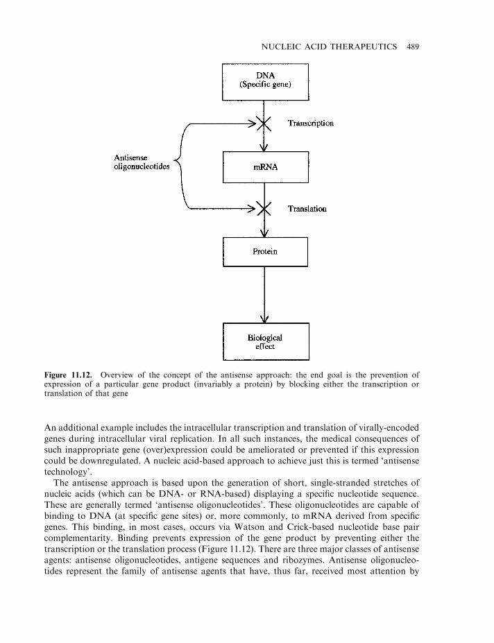
An additional example includes the intracellular transcription and translation of virally-encodedgenes during intracellular viral replication. In all such instances, the medical consequences ofsuch inappropriate gene (over)expression could be ameliorated or prevented if this expressioncould be downregulated. A nucleic acid-based approach to achieve just this is termed ‘antisensetechnology’.
The antisense approach is based upon the generation of short, single-stranded stretches ofnucleic acids (which can be DNA- or RNA-based) displaying a specific nucleotide sequence.These are generally termed ‘antisense oligonucleotides’. These oligonucleotides are capable ofbinding to DNA (at specific gene sites) or, more commonly, to mRNA derived from specificgenes. This binding, in most cases, occurs via Watson and Crick-based nucleotide base paircomplementarity. Binding prevents expression of the gene product by preventing either thetranscription or the translation process (Figure 11.12). There are three major classes of antisenseagents: antisense oligonucleotides, antigene sequences and ribozymes. Antisense oligonucleo-tides represent the family of antisense agents that have, thus far, received most attention by
NUCLEIC ACID THERAPEUTICS 489
Figure 11.12. Overview of the concept of the antisense approach: the end goal is the prevention ofexpression of a particular gene product (invariably a protein) by blocking either the transcription ortranslation of that gene
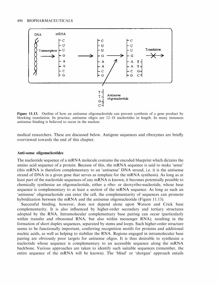
medical researchers. These are discussed below. Antigene sequences and ribozymes are brieflyoverviewed towards the end of this chapter.
Anti-sense oligonucleotides
The nucleotide sequence of a mRNAmolecule contains the encoded blueprint which dictates theamino acid sequence of a protein. Because of this, the mRNA sequence is said to make ‘sense’(this mRNA is therefore complementary to an ‘antisense’ DNA strand, i.e. it is the antisensestrand of DNA in a given gene that serves as template for the mRNA synthesis). As long as atleast part of the nucleotide sequences of any mRNA is known, it becomes potentially possible tochemically synthesize an oligonucleotide, either a ribo- or deoxyribo-nucleotide, whose basesequence is complementary to at least a section of the mRNA sequence. As long as such an‘antisense’ oligonucleotide can enter the cell, the complementarity of sequences can promotehybridization between the mRNA and the antisense oligonucleotide (Figure 11.13).
Successful binding, however, does not depend alone upon Watson and Crick basecomplementarity. It is also influenced by higher-order secondary and tertiary structuresadopted by the RNA. Intramolecular complementary base pairing can occur (particularlywithin transfer and ribosomal RNA, but also within messenger RNA), resulting in theformation of short duplex sequences, separated by stems and loops. Such higher-order structureseems to be functionally important, conferring recognition motifs for proteins and additionalnucleic acids, as well as helping to stabilize the RNA. Regions engaged in intramolecular basepairing are obviously poor targets for antisense oligos. It is thus desirable to synthesize anucleotide whose sequence is complementary to an accessible sequence along the mRNAbackbone. Various approaches are taken to identify such suitable sequences (remember, theentire sequence of the mRNA will be known). The ‘blind’ or ‘shotgun’ approach entails
490 BIOPHARMACEUTICALS
Figure 11.13. Outline of how an antisense oligonucleotide can prevent synthesis of a gene product byblocking translation. In practice, antisense oligos are 12–18 nucleotides in length. In many instancesantisense binding is believed to occur in the nucleus
synthesizing large numbers of oligos targeted to various (often overlapping) regions of themRNA. The ability of each oligo to block translation of the mRNA is then directly assessed inan in vitro assay system using cell-free extracts. The second design approach entails the use ofvarious computer programmes to interrogate the mRNA sequence in an attempt to predict itshigher-order structure (and hence identify accessible sequences). This approach remains to beoptimized. The translation initiation site of mRNAs are often popular targets because they areessential to translation and they are generally free from secondary structure. However, sequencehomologies can exist within these sequences in unrelated genes. This reduces the specificity ofthe blocking effect and could lead to clinically significant side effects.
Binding results in the blocking of translation of the mRNA, and hence prevents synthesis of,the mature gene’s protein product. Indeed, certain microorganisms appear to synthesizeantisense molecules naturally as a means of regulating gene expression.
The prevention of mRNA translation by duplex formation with antisense oligonucleotidesappears to be underpinned by twin mechanisms. First, the oligonucleotides act as stericblockers, i.e. prevent proteins involved in translation, or other aspects of mRNA processing,from binding to appropriate sequences in the mRNA. The generation of duplexes also likelyallows targeting by intracellular RNases such as RNase H. This enzyme is capable of binding toRNA–DNA duplexes and degrading the RNA portion of the duplex (most synthetic antisenseoligonucleotides are DNA-based).
Uses, advantages and disadvantages of ‘oligos’
Antisense oligonucleotides (‘oligos’) are being assessed in pre-clinical and clinical studies astherapeutic agents in the treatment of cancer, as well as a variety of viral diseases (e.g. HIV,hepatitis B, herpes virus and papillomavirus infections). They also have potential application intreating other disease states for which blocking of gene expression would likely have a beneficialeffect. Such medical conditions include restenosis, rheumatoid arthritis and allergic disorders.As potential drugs, antisense oligos display a number of desirable characteristics, the mostsignificant of which is extreme specificity. Statistical analysis reveals that any specific basesequence of 17 or more bases is extremely unlikely to occur more than once in a human cell’snucleic acid complement. It thus follows that an oligonucleotide of 17 or more nucleotide unitsin length, which is designed to successfully duplex with a specific mRNA species, is unlikely toform a duplex with any other (unintended) mRNA species. Most synthetic oligos are thereforein the region of 17 nucleotides units long. These will display virtually an absolute specificity forthe target sequence. Additional advantages of the oligonucleotide antisense approach include:
. minimal toxicity: thus far, most trials report few or no side effects. This is likely due to thehighly specific nature of oligo duplexing, and the fact that they are ‘natural’ biomolecules;
. the requirement for only low levels of the oligo to be present inside the cell, as target mRNAis, itself, usually present only in nanomolar (nM) concentrations;
. the ability to manufacture oligos of specified nucleotide sequence is relatively straightforwardusing automated synthesizers.
However, native antisense oligonucleotides also suffer from a number of disadvantages, whichinclude:
. sensitivity to nucleases;
. very low serum half-lives;
NUCLEIC ACID THERAPEUTICS 491
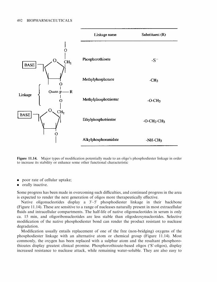
. poor rate of cellular uptake;
. orally inactive.
Some progress has been made in overcoming such difficulties, and continued progress in the areais expected to render the next generation of oligos more therapeutically effective.
Native oligonucleotides display a 3’–5’ phosphodiester linkage in their backbone(Figure 11.14). These are sensitive to a range of nucleases naturally present in most extracellularfluids and intracellular compartments. The half-life of native oligonucleotides in serum is onlyca. 15 min, and oligoribonucleotides are less stable than oligodeoxynucleotides. Selectivemodification of the native phosphodiester bond can render the product resistant to nucleasedegradation.
Modification usually entails replacement of one of the free (non-bridging) oxygens of thephosphodiester linkage with an alternative atom or chemical group (Figure 11.14). Mostcommonly, the oxygen has been replaced with a sulphur atom and the resultant phosphoro-thioates display greatest clinical promise. Phosphorothioate-based oligos (‘S’-oligos), displayincreased resistance to nuclease attack, while remaining water-soluble. They are also easy to
492 BIOPHARMACEUTICALS
Figure 11.14. Major types of modification potentially made to an oligo’s phosphodiester linkage in orderto increase its stability or enhance some other functional characteristic
-S�
synthesize chemically and display a biological half-life of up to 24 h. Most antisense oligoscurrently being assessed in clinical trials are S-oligos.
Delivery and cellular uptake of oligonucleotides
Oligo administration during many clinical trials entails direct i.v. infusion, often over a course ofseveral hours. Although relatively stable in serum, the commonly employed phosphorothioateoligos (and indeed most other oligo types) encounter several barriers to reaching their finaldestinations. They bind various serum proteins, including serum albumin, as well as a range ofheparin-binding and other proteins, which commonly occur on many cell surfaces. Targeting ofnaked oligos to specific cell types is therefore not possible. Following administration, theseoligos tend to be distributed to many tissues, with the highest proportion accumulating in theliver and kidney.
The precise mechanism(s) by which oligos enter cells are not fully understood. Most arecharged molecules, sometimes displaying a molecular mass of up to 10–12 kDa. Receptor-mediated endocytosis appears to be the most common mechanism by which charged oligos, suchas phosphorothioates, enter most cells. One putative phosphorothioate receptor appears toconsist of an 80 kDa surface protein, associated with a smaller 34 kDa membrane protein.However, this in itself seems to be an inefficient process, with only a small proportion of theadministered drug eventually being transferred across the plasma membrane.
Uncharged oligos appear to enter the cell by passive diffusion, as well as possibly byendocytosis. However, elimination of the charges renders the resultant oligos relativelyhydrophobic, thus generating additional difficulties with their synthesis and delivery.
Attempts to increase delivery of oligos into the cell centre mainly on the use of suitable carriersystems. Liposomes, as well as polymeric carriers (e.g. polylysine-based carriers), are gainingmost attention in this regard. Details of such carriers have already been discussed earlier in thischapter.
An alternative system, which effectively results in the introduction of antisense oligonucleo-tides into the cell, entails the application of gene therapy. In this case, a gene which, whentranscribed, yields (antisense) mRNA of appropriate nucleotide sequence, is introduced into thecell by a retroviral or other appropriate vector. This approach, as applied to the treatment ofcancer and AIDS, is being appraised in a number of trials.
Oligos, including modified oligos, appear to be ultimately metabolized within the cell by theaction of nucleases, particularly 3’-exonucleases. Breakdown metabolic products are thenmainly excreted via the urinary route.
Manufacture of oligonucleotides
In contrast to the biopharmaceuticals thus far discussed (recombinant proteins and genetherapy plasmids), antisense oligonucleotides are manufactured by direct chemical synthesis.Organic synthetic pathways have been developed, optimized and commercialized for some time,as oligonucleotides are widely used reagents in molecular biology. They are required as primers,probes and for the purposes of site-directed mutagenesis. The basic synthetic strategy is verysimilar in concept to the means by which peptides are synthesized via the Merrifield method, asdescribed in Chapter 2 (Box 2.1). The nucleotides required (themselves either modified orunmodified, as desired) are first reacted with a protecting chemical group. Each protectednucleotide is then coupled in turn to the growing end of the nucleotide chain, itself attached to a
NUCLEIC ACID THERAPEUTICS 493
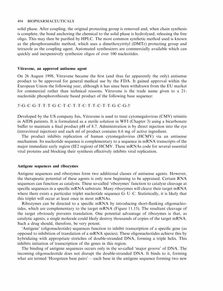
solid phase. After coupling, the original protecting group is removed and, when chain synthesisis complete, the bond anchoring the chemical to the solid phase is hydrolysed, releasing the freeoligo. This may then be purified by HPLC. The most common synthetic method used is knownas the phosphoramidite method, which uses a dimethoxytrityl (DMTr) protecting group andtetrazole as the coupling agent. Automated synthesizers are commercially available which canquickly and inexpensively synthesize oligos of over 100 nucleotides.
Vitravene, an approved antisense agent
On 26 August 1998, Vitravene became the first (and thus far apparently the only) antisenseproduct to be approved for general medical use by the FDA. It gained approval within theEuropean Union the following year, although it has since been withdrawn from the EU marketfor commercial rather than technical reasons. Vitravene is the trade name given to a 21-nucleotide phosphorothioate based product of the following base sequence:
5’-G–C–G–T–T–T–G–C–T–C–T–T–C–T–T–C–T–T–G–C–G-3’
Developed by the US company Isis, Vitravene is used to treat cytomegalovirus (CMV) retinitisin AIDS patients. It is formulated as a sterile solution in WFI (Chapter 3) using a bicarbonatebuffer to maintain a final product pH of 8.7. Administration is by direct injection into the eye(intravitreal injection) and each ml of product contains 6.6 mg of active ingredient.
The product inhibits replication of human cytomegalovirus (HCMV) via an antisensemechanism. Its nucleotide sequence is complementary to a sequence in mRNA transcripts of themajor immediate early region (IE2 region) of HCMV. These mRNAs code for several essentialviral proteins and blocking their synthesis effectively inhibits viral replication.
Antigene sequences and ribozymes
Antigene sequences and ribozymes form two additional classes of antisense agents. However,the therapeutic potential of these agents is only now beginning to be appraised. Certain RNAsequences can function as catalysts. These so-called ‘ribozymes’ function to catalyse cleavage atspecific sequences in a specific mRNA substrate. Many ribozymes will cleave their target mRNAwhere there exists a particular triplet nucleotide sequence G–U–C. Statistically, it is likely thatthis triplet will occur at least once in most mRNAs.
Ribozymes can be directed to a specific mRNA by introducing short-flanking oligonucleo-tides, which are complementary to the target mRNA (Figure 11.15). The resultant cleavage ofthe target obviously prevents translation. One potential advantage of ribozymes is that, ascatalytic agents, a single molecule could likely destroy thousands of copies of the target mRNA.Such a drug should, therefore, be very potent.
‘Antigene’ (oligonucleotide) sequences function to inhibit transcription of a specific gene (asopposed to inhibition of translation of a mRNA species). These oligonucleotides achieve this byhybridizing with appropriate stretches of double-stranded DNA, forming a triple helix. Thisinhibits initiation of transcription of the genes in this region.
The binding of antigene sequences occurs only in the so-called ‘major groove’ of DNA. Theincoming oligonucleotide does not disrupt the double-stranded DNA. It binds to it, formingwhat are termed ‘Hoogsteen base pairs’— each base in the antigene sequence forming two new
494 BIOPHARMACEUTICALS
hydrogen bonds with a purine base in the targeted region of the double helix. Much research,however, must be undertaken before it will become clear whether such antigene sequences willbe of therapeutic use.
CONCLUSION
Every few decades, a medical innovation is perfected that profoundly influences the practice ofmedicine. Widespread vaccination against common infectious agents and the discovery ofantibiotics serve as two such examples. Many scientists now believe that the potential of genetherapy and antisense technology rivals even the most significant medical advances achieved todate.
It is now just over a decade since the first nucleic acid-based drugs began initial tests.Several such drugs will likely be in routine medical use in less than one decade more. Theapplication of gene technology could also change utterly the profile of biopharmaceuticaldrugs currently on the market. Virtually all such products are proteins, currently administeredto patients for short or prolonged periods, as appropriate. Gene therapy offers the possibilityof equipping the patient’s own body with the ability to synthesize these drugs itself, and overwhatever time scale is appropriate. Taken to its logical conclusion, gene therapy thus offersthe potential to render obsolete most of the biopharmaceutical products currently on themarket. Of all the biopharmaceuticals discussed throughout this text, nucleic acid-based drugsmay well turn out to have the most profound influence on the future practice of molecularmedicine.
NUCLEIC ACID THERAPEUTICS 495
Figure 11.15. Outline of how ribozyme technology could prevent translation of specific mRNA, thuspreventing synthesis of a specific target protein
Flanking sequenceswhich ‘dock’ ribozymeat the appropriatesequence of theappropriate mRNA viacomplementary basepairing
Flanking sequenceswhich ‘dock’ ribozymeat the appropriatesequence of theappropriate mRNA viacomplementary basepairing
FURTHER READING
Books
Blankenstein, T. (Ed.) (1999). Gene Therapy: Principles and Applications. Birkhauser-Verlag.Crooke, S. (Ed.) (2001). Antisense Drug Technology. Marcel Dekker, New York.Kresina, T. (Ed.) (2001). An Introduction to Molecular Medicine and Gene Therapy, Parts I and II. Wiley-Liss, New York.Lowrie, D. (1999). DNA Vaccines. Humana, New York.Phillips, M. (2000). Antisense Technology (Methods in Enzymology, Vol. 313). Academic Press, New York.Stein, C. & Krieg, A. (1998). Applied Antisense Oligonucleotide Technology. Wiley, Chichester.
Articles
Gene therapy
Buchschacher, G. & Wong-Staal, F. (2001). Approaches to gene therapy for human immunodeficiency virus infection.Human Gene Ther. 12(9), 1013–1019.
Davies, J. et al. (2001). Gene therapy for cystic fibrosis. J. Gene Med. 3(5), 409–417.Demeterco, C. & Levine, F. (2001). Gene therapy for diabetes. Frontiers Biosci. 6, D175–D191.Demoly, P. et al. (1997). Gene therapy strategies for asthma. Gene Therapy 4(6), 507–516.Docherty, K. (1997). Gene therapy for diabetes mellitus. Clin. Sci. 92(4), 321–330.Donnelly, J. (1997). DNA vaccines. Ann. Rev. Immunol. 15, 617–648.Felgner, P. (1997). Nonviral strategies for gene therapy. Sci. Am. June, 86–90.Ferreira, G. et al. (2000). Downstream processing of plasmid DNA for gene therapy and DNA vaccine applications.
Trends Biotechnol. 18(9), 380–388.Lewin, A. & Hauswirth, W. (2001). Ribozyme gene therapy: applications for molecular medicine. Trends Mol. Med. 7(5),
221–228.Liras, A. (2001). Gene therapy for haemophilia: the end of a ‘royal pathology’ in the third millennium?Haemophilia 7(5),
441–445.Mhashilkar, A. et al. (2001). Gene therapy— therapeutic approaches and implications. Biotechnol. Adv. 19(4), 279–297.Moller, P. & Schadendorf, D. (1997). Somatic gene therapy and its implications in melanoma treatment. Arch. Dermatol.
Res. 289(2), 71–77.Mulligan, R. (1993). The basic science of gene therapy. Science 260, 926–931.Pfeifer, A. & Verma, I. (2001). Gene therapy: promises and problems. Ann. Rev. Genom. Hum. Genet. 2, 177–211.Phillips, A. (2001). The challenge of gene therapy and DNA delivery. J. Pharm. Pharmacol. 53(9), 1169–1174.Robertson, J. & Griffiths, E. (2001). Assuring the quality, safety and efficacy of DNA vaccines. Mol. Biotechnol. 17(2),
143–149.Rosenberg, S. (1997). Cancer vaccines based on the identification of genes encoding cancer regression antigens. Immunol.
Today 18(4), 175–182.Schatzlein, A. (2001). Non-viral vectors in cancer gene therapy: principles and progress. Anti-cancer Drugs 12(4),
275–304.Scott-Taylor, T. & Dalgeish, A. (2000). DNA vaccines. Expert Opin. Invest. Drugs 9(3), 471–480.Smith, A. (1995). Viral vectors in gene therapy. Ann. Rev. Microbiol. 49, 807–838.Smith, H. & Klinman, D. (2001). The regulation of DNA vaccines. Curr. Opin. Biotechnol. 12(3), 299–303.Wu, N. & Ataai, M. (2000). Production of viral vectors for gene therapy applications. Curr. Opin. Biotechnol. 11(2),
205–208.
Antisense technology
Adah, S. et al. (2001). Chemistry and biochemistry of 2’-5’ oligoadenylate-based antisense strategy. Cur. Med. Chem.8(10), 1189–1212.
Akhtar, S. et al. (2000). The delivery of antisense therapeutics. Adv. Drug Delivery Rev. 44(1), 3–21.Askari, F. (1996). Molecular medicine: antisense-oligonucleotide therapy. N. Engl. J. Med. 334(5), 316–318.Galderisi, U. et al. (2001). Antisense oligonucleotides as drugs for HIV treatment. Expert Opin. Therapeut. Patents
11(10), 1605–1611.Hughes, M. et al. (2001). The cellular delivery of antisense oligonucleotides and ribozymes. Drug Discovery Today 6(6),
303–315.Lebedeva, I. & Stein, C. (2001). Antisense oligonucleotides: promise and reality. Ann. Rev. Pharmacol. Toxicol. 41,
403–419.Pawlak, W. et al. (2000). Antisense therapy in cancer. Cancer Treatment Rev. 26(5), 333–350.
496 BIOPHARMACEUTICALS

Reddy, D. (1996). Antisense oligonucleotides: a new class of potential anti-AIDS and anti-cancer drugs. Drugs Today32(2), 113–137.
Taylor, M. (2001). Emerging antisense technologies for gene functionalization and drug discovery. Drug DiscoveryToday 6(15), S97–S101.
Wagner, R. & Flanagan, W. (1997). Antisense technology and prospects for therapy of viral infections and cancer. Mol.Med. Today 3(1), 31–38.
Wickstrom, E. (1992). Strategies for administering targeted therapeutic oligodeoxynucleotides. Trends Biotechnol. 10,281–286.
NUCLEIC ACID THERAPEUTICS 497

Appendix 1
Biopharmaceuticals thus far approved
in the USA or European Union
Notes: (a) Several products have been approved for multiple indications. Only the firstindication for which each was approved is listed. (b) ‘Vet’ listing in therapeutic indicationcolumn indicates an animal application. All other products are used in human medicine.
Abbreviations: r¼recombinant, rh¼recombinant human, CHO¼Chinese hamster ovary,BHK¼baby hamster kidney, Mab¼monoclonal antibody, tPA¼ tissue plasminogen activator,hGH¼human growth hormone, FSH¼ follicle stimulating hormone, TSH¼ thyroid stimulatinghormone, EPO¼erythropoietin, GM-CSF¼granulocyte-macrophage colony stimulatingfactor, IFN¼ interferon, IL¼ interleukin, HBsAg¼hepatitis B surface antigen, PDGF¼platelet-derived growth factor, TNFR¼ tumour necrosis factor receptor, E. coli¼Escherichiacoli, S. cerevisiae¼Saccharomyces cerevisiae.
Product Company Therapeutic indication Date approved
Recombinant blood factors
Bioclate (rhFactor VIII producedin CHO cells)
Centeon Haemophilia A 1993 (USA)
Benefix (rhFactor IX producedin CHO cells)
Genetics Institute Haemophilia B 1997 (USA, EU)
Kogenate (rhFactor VIII producedin BHK cells. Also sold asHelixate by Centeon via a licenseagreement)
Bayer Haemophilia A 1993 (USA), 2000(EU)
Helixate NexGen (octocog-a;rhFactor VIII produced inBHK cells)
Bayer Haemophilia A 2000 (EU)
NovoSeven (rhFactor VIIaproduced in BHK cells)
Novo Nordisk Some forms ofhaemophilia
1995 (EU), 1999(USA)
(Continued)
Biopharmaceuticals: Biochemistry and Biotechnology, Second Edition by Gary WalshJohn Wiley & Sons Ltd: ISBN 0 470 84326 8 (ppc), ISBN 0 470 84327 6 (pbk)
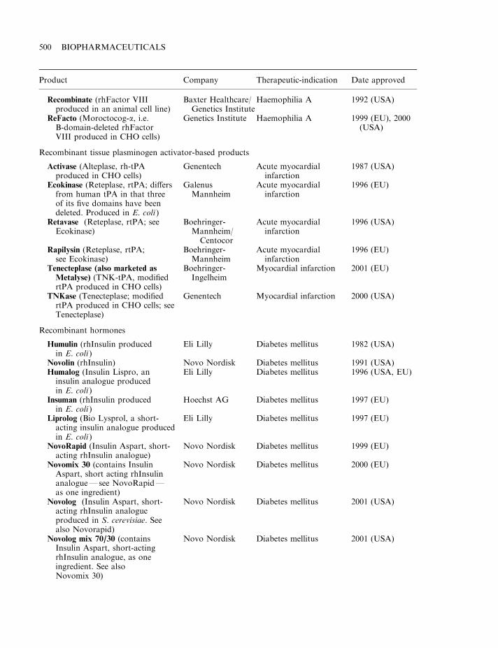
Product Company Therapeutic-indication Date approved
Recombinate (rhFactor VIIIproduced in an animal cell line)
Baxter Healthcare/Genetics Institute
Haemophilia A 1992 (USA)
ReFacto (Moroctocog-a, i.e.B-domain-deleted rhFactorVIII produced in CHO cells)
Genetics Institute Haemophilia A 1999 (EU), 2000(USA)
Recombinant tissue plasminogen activator-based products
Activase (Alteplase, rh-tPAproduced in CHO cells)
Genentech Acute myocardialinfarction
1987 (USA)
Ecokinase (Reteplase, rtPA; differsfrom human tPA in that threeof its five domains have beendeleted. Produced in E. coli )
GalenusMannheim
Acute myocardialinfarction
1996 (EU)
Retavase (Reteplase, rtPA; seeEcokinase)
Boehringer-Mannheim/
Centocor
Acute myocardialinfarction
1996 (USA)
Rapilysin (Reteplase, rtPA;see Ecokinase)
Boehringer-Mannheim
Acute myocardialinfarction
1996 (EU)
Tenecteplase (also marketed as
Metalyse) (TNK-tPA, modifiedrtPA produced in CHO cells)
Boehringer-Ingelheim
Myocardial infarction 2001 (EU)
TNKase (Tenecteplase; modifiedrtPA produced in CHO cells; seeTenecteplase)
Genentech Myocardial infarction 2000 (USA)
Recombinant hormones
Humulin (rhInsulin producedin E. coli )
Eli Lilly Diabetes mellitus 1982 (USA)
Novolin (rhInsulin) Novo Nordisk Diabetes mellitus 1991 (USA)Humalog (Insulin Lispro, aninsulin analogue producedin E. coli )
Eli Lilly Diabetes mellitus 1996 (USA, EU)
Insuman (rhInsulin producedin E. coli )
Hoechst AG Diabetes mellitus 1997 (EU)
Liprolog (Bio Lysprol, a short-acting insulin analogue producedin E. coli )
Eli Lilly Diabetes mellitus 1997 (EU)
NovoRapid (Insulin Aspart, short-acting rhInsulin analogue)
Novo Nordisk Diabetes mellitus 1999 (EU)
Novomix 30 (contains InsulinAspart, short acting rhInsulinanalogue—see NovoRapid—as one ingredient)
Novo Nordisk Diabetes mellitus 2000 (EU)
Novolog (Insulin Aspart, short-acting rhInsulin analogueproduced in S. cerevisiae. Seealso Novorapid)
Novo Nordisk Diabetes mellitus 2001 (USA)
Novolog mix 70/30 (containsInsulin Aspart, short-actingrhInsulin analogue, as oneingredient. See alsoNovomix 30)
Novo Nordisk Diabetes mellitus 2001 (USA)
500 BIOPHARMACEUTICALS
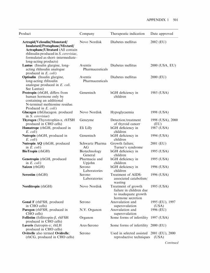
Product Company Therapeutic indication Date approved
Actrapid/Velosulin/Monotard/
Insulatard/Protaphane/Mixtard/Actraphane/Ultratard (All containrhInsulin produced in S. cerevisiae,formulated as short–intermediate–long-acting products)
Novo Nordisk Diabetes mellitus 2002 (EU)
Lantus (Insulin glargine, long-acting rhInsulin analogueproduced in E. coli )
AventisPharmaceuticals
Diabetes mellitus 2000 (USA, EU)
Optisulin (Insulin glargine,long-acting rhInsulinanalogue produced in E. coli.See Lantus)
AventisPharmaceuticals
Diabetes mellitus 2000 (EU)
Protropin (rhGH, differs fromhuman hormone only bycontaining an additionalN-terminal methionine residue.Produced in E. coli )
Genentech hGH deficiency inchildren
1985 (USA)
Glucagen (rhGlucagon. producedin S. cerevisiae)
Novo Nordisk Hypoglycaemia 1998 (USA)
Thyrogen (Thyrotrophin-a, rhTSHproduced in CHO cells)
Genzyme Detection/treatmentof thyroid cancer
1998 (USA), 2000(EU)
Humatrope (rhGH, produced inE. coli )
Eli Lilly hGH deficiency inchildren
1987 (USA)
Nutropin (rhGH, produced inE. coli )
Genentech hGH deficiency inchildren
1994 (USA)
Nutropin AQ (rhGH, producedin E. coli )
Schwartz PharmaAG
Growth failure,Turner’s syndrome
2001 (EU)
BioTropin (rhGH) BiotechnologyGeneral
hGH deficiency inchildren
1995 (USA)
Genotropin (rhGH, producedin E. coli )
Pharmacia andUpjohn
hGH deficiency inchildren
1995 (USA)
Saizen (rhGH) SeronoLaboratories
hGH deficiency inchildren
1996 (USA)
Serostim (rhGH) SeronoLaboratories
Treatment of AIDS-associated catabolism/wasting
1996 (USA)
Norditropin (rhGH) Novo Nordisk Treatment of growthfailure in children dueto inadequate growthhormone secretion
1995 (USA)
Gonal F (rhFSH, producedin CHO cells)
Serono Anovulation andsuperovulation
1995 (EU), 1997(USA)
Puregon (rhFSH, produced inCHO cells)
N.V. Organon Anovulation andsuperovulation
1996 (EU)
Follistim (follitropin-b, rhFSHproduced in CHO cells)
Organon Some forms of infertility 1997 (USA)
Luveris (lutropin-a ; rhLHproduced in CHO cells)
Ares-Serono Some forms of infertility 2000 (EU)
Ovitrelle also termed Ovidrelle;(rhCG, produced in CHO cells)
Serono Used in selected assistedreproductive techniques
2001 (EU), 2000(USA)
Continued
APPENDIX 1 501
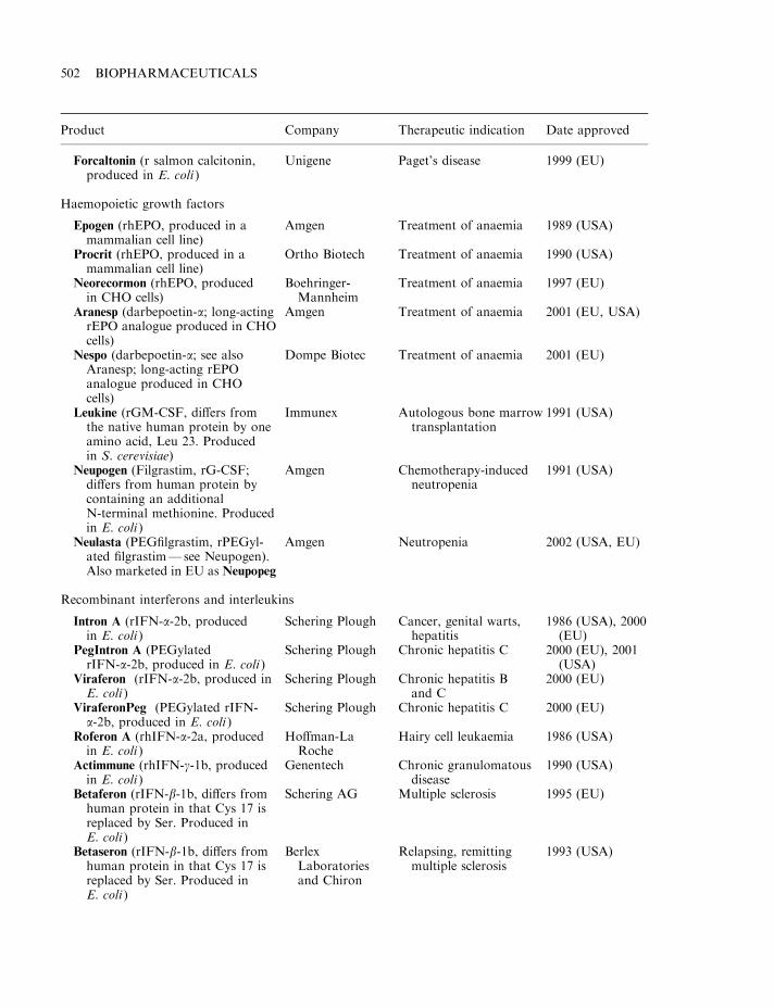
Product Company Therapeutic indication Date approved
Forcaltonin (r salmon calcitonin,produced in E. coli )
Unigene Paget’s disease 1999 (EU)
Haemopoietic growth factors
Epogen (rhEPO, produced in amammalian cell line)
Amgen Treatment of anaemia 1989 (USA)
Procrit (rhEPO, produced in amammalian cell line)
Ortho Biotech Treatment of anaemia 1990 (USA)
Neorecormon (rhEPO, producedin CHO cells)
Boehringer-Mannheim
Treatment of anaemia 1997 (EU)
Aranesp (darbepoetin-a; long-actingrEPO analogue produced in CHOcells)
Amgen Treatment of anaemia 2001 (EU, USA)
Nespo (darbepoetin-a; see alsoAranesp; long-acting rEPOanalogue produced in CHOcells)
Dompe Biotec Treatment of anaemia 2001 (EU)
Leukine (rGM-CSF, differs fromthe native human protein by oneamino acid, Leu 23. Producedin S. cerevisiae)
Immunex Autologous bone marrowtransplantation
1991 (USA)
Neupogen (Filgrastim, rG-CSF;differs from human protein bycontaining an additionalN-terminal methionine. Producedin E. coli )
Amgen Chemotherapy-inducedneutropenia
1991 (USA)
Neulasta (PEGfilgrastim, rPEGyl-ated filgrastim—see Neupogen).Also marketed in EU as Neupopeg
Amgen Neutropenia 2002 (USA, EU)
Recombinant interferons and interleukins
Intron A (rIFN-a-2b, producedin E. coli )
Schering Plough Cancer, genital warts,hepatitis
1986 (USA), 2000(EU)
PegIntron A (PEGylatedrIFN-a-2b, produced in E. coli )
Schering Plough Chronic hepatitis C 2000 (EU), 2001(USA)
Viraferon (rIFN-a-2b, produced inE. coli )
Schering Plough Chronic hepatitis Band C
2000 (EU)
ViraferonPeg (PEGylated rIFN-a-2b, produced in E. coli )
Schering Plough Chronic hepatitis C 2000 (EU)
Roferon A (rhIFN-a-2a, producedin E. coli )
Hoffman-LaRoche
Hairy cell leukaemia 1986 (USA)
Actimmune (rhIFN-g-1b, producedin E. coli )
Genentech Chronic granulomatousdisease
1990 (USA)
Betaferon (rIFN-b-1b, differs fromhuman protein in that Cys 17 isreplaced by Ser. Produced inE. coli )
Schering AG Multiple sclerosis 1995 (EU)
Betaseron (rIFN-b-1b, differs fromhuman protein in that Cys 17 isreplaced by Ser. Produced inE. coli )
BerlexLaboratoriesand Chiron
Relapsing, remittingmultiple sclerosis
1993 (USA)
502 BIOPHARMACEUTICALS
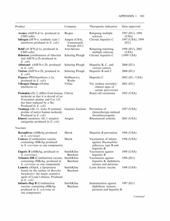
Product Company Therapeutic indication Date approved
Avonex (rhIFN-b-1a, produced inCHO cells)
Biogen Relapsing multiplesclerosis
1997 (EU), 1996(USA)
Infergen (rIFN-a, synthetic type 1interferon produced in E. coli )
Amgen (USA),YamanouchiEurope (EU)
Chronic hepatitis C 1997 (USA), 1999(EU)
Rebif (rh IFN-b-1a, produced inCHO cells)
Ares-Serono Relapsing/remittingmultiple sclerosis
1998 (EU), 2002(USA)
Rebetron (combination of ribavirinand rhIFN-a-2b produced inE. coli )
Schering Plough Chronic hepatitis C (1999 USA)
Alfatronol (rhIFN-a-2b, producedin E. coli )
Schering Plough Hepatitis B, C, andvarious cancers
2000 (EU)
Virtron (rhIFN-a-2b, produced inE. coli )
Schering Plough Hepatitis B and C 2000 (EU)
Pegasys (PEGinterferon a-2a,produced in E. coli )
Hoffman-LaRoche
Hepatitis C 2002 (EU, USA)
Vibragen Omega (rFelineinterferon o)
Virbac Vet. (reduce mortality/clinical signs ofcanine parvovirus)
2001 (EU)
Proleukin (rIL-2, differs from humanmolecule in that it is devoid of anN-terminal alanine and Cys 125has been replaced by a Ser.Produced in E. coli )
Chiron Renal cell carcinoma 1992 (USA)
Neumega (rIL-11, lacks N-terminalproline of native human molecule.Produced in E. coli )
Genetics Institute Prevention ofchemotherapy-inducedthrombocytopenia
1997 (USA)
Kineret (anakinra; rIL-1 receptorantagonist produced in E. coli )
Amgen Rheumatoid arthritis 2001 (USA)
Vaccines
Recombivax (rHBsAg producedin S. cerevisiae)
Merck Hepatitis B prevention 1986 (USA)
Comvax (Combination vaccine,containing rHBsAg producedin S. cerevisiae as one component)
Merck Vaccination of infantsagainst Haemophilusinfluenzae type B andhepatitis B
1996 (USA)
Engerix B (rHBsAg, produced inS. cerevisiae)
SmithKlineBeecham
Vaccination againsthepatitis B
1998 (USA)
Tritanrix-HB (Combination vaccine,containing rHBsAg, produced inS. cerevisiae as one component)
SmithKlineBeecham
Vaccination againsthepatitis B, diphtheria,tetanus and pertussis
1996 (EU)
Lymerix (rOspA, a lipoproteinfound on the surface of Borreliaburgdorferi, the major causativeagent of Lyme’s disease. Producedin E. coli )
SmithKlineBeecham
Lyme disease vaccine 1998 (USA)
Infanrix-Hep B (Combinationvaccine, containing rHBsAgproduced in S. cerevisiae asone component)
SmithKlineBeecham
Immunization againstdiphtheria, tetanus,pertussis and hepatitis B
1997 (EU)
Continued
APPENDIX 1 503
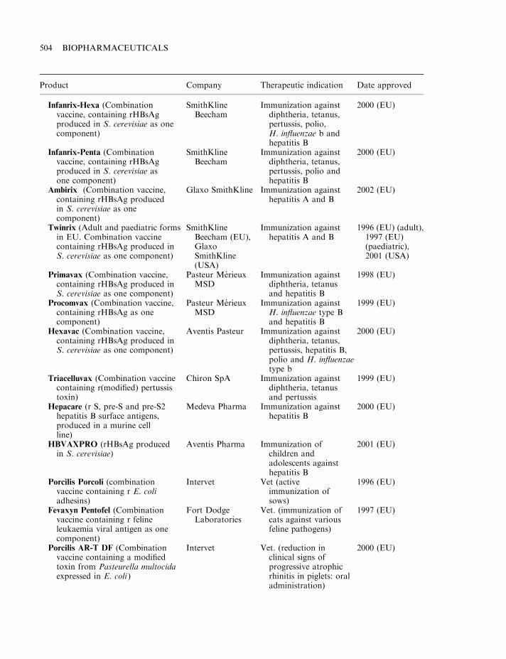
Product Company Therapeutic indication Date approved
Infanrix-Hexa (Combinationvaccine, containing rHBsAgproduced in S. cerevisiae as onecomponent)
SmithKlineBeecham
Immunization againstdiphtheria, tetanus,pertussis, polio,H. influenzae b andhepatitis B
2000 (EU)
Infanrix-Penta (Combinationvaccine, containing rHBsAgproduced in S. cerevisiae asone component)
SmithKlineBeecham
Immunization againstdiphtheria, tetanus,pertussis, polio andhepatitis B
2000 (EU)
Ambirix (Combination vaccine,containing rHBsAg producedin S. cerevisiae as onecomponent)
Glaxo SmithKline Immunization againsthepatitis A and B
2002 (EU)
Twinrix (Adult and paediatric formsin EU. Combination vaccinecontaining rHBsAg produced inS. cerevisiae as one component)
SmithKlineBeecham (EU),GlaxoSmithKline(USA)
Immunization againsthepatitis A and B
1996 (EU) (adult),1997 (EU)(paediatric),2001 (USA)
Primavax (Combination vaccine,containing rHBsAg produced inS. cerevisiae as one component)
Pasteur MerieuxMSD
Immunization againstdiphtheria, tetanusand hepatitis B
1998 (EU)
Procomvax (Combination vaccine,containing rHBsAg as onecomponent)
Pasteur MerieuxMSD
Immunization againstH. influenzae type Band hepatitis B
1999 (EU)
Hexavac (Combination vaccine,containing rHBsAg produced inS. cerevisiae as one component)
Aventis Pasteur Immunization againstdiphtheria, tetanus,pertussis, hepatitis B,polio and H. influenzaetype b
2000 (EU)
Triacelluvax (Combination vaccinecontaining r(modified) pertussistoxin)
Chiron SpA Immunization againstdiphtheria, tetanusand pertussis
1999 (EU)
Hepacare (r S, pre-S and pre-S2hepatitis B surface antigens,produced in a murine cellline)
Medeva Pharma Immunization againsthepatitis B
2000 (EU)
HBVAXPRO (rHBsAg producedin S. cerevisiae)
Aventis Pharma Immunization ofchildren andadolescents againsthepatitis B
2001 (EU)
Porcilis Porcoli (combinationvaccine containing r E. coliadhesins)
Intervet Vet (activeimmunization ofsows)
1996 (EU)
Fevaxyn Pentofel (Combinationvaccine containing r felineleukaemia viral antigen as onecomponent)
Fort DodgeLaboratories
Vet. (immunization ofcats against variousfeline pathogens)
1997 (EU)
Porcilis AR-T DF (Combinationvaccine containing a modifiedtoxin from Pasteurella multocidaexpressed in E. coli )
Intervet Vet. (reduction inclinical signs ofprogressive atrophicrhinitis in piglets: oraladministration)
2000 (EU)
504 BIOPHARMACEUTICALS
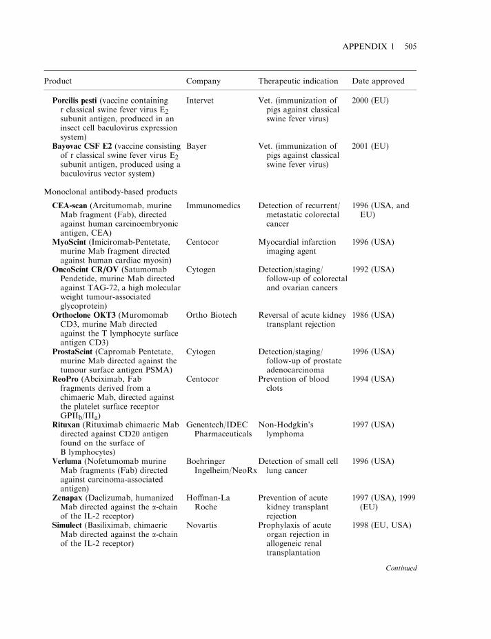
Product Company Therapeutic indication Date approved
Porcilis pesti (vaccine containingr classical swine fever virus E2subunit antigen, produced in aninsect cell baculovirus expressionsystem)
Intervet Vet. (immunization ofpigs against classicalswine fever virus)
2000 (EU)
Bayovac CSF E2 (vaccine consistingof r classical swine fever virus E2subunit antigen, produced using abaculovirus vector system)
Bayer Vet. (immunization ofpigs against classicalswine fever virus)
2001 (EU)
Monoclonal antibody-based products
CEA-scan (Arcitumomab, murineMab fragment (Fab), directedagainst human carcinoembryonicantigen, CEA)
Immunomedics Detection of recurrent/metastatic colorectalcancer
1996 (USA, andEU)
MyoScint (Imiciromab-Pentetate,murine Mab fragment directedagainst human cardiac myosin)
Centocor Myocardial infarctionimaging agent
1996 (USA)
OncoScint CR/OV (SatumomabPendetide, murine Mab directedagainst TAG-72, a high molecularweight tumour-associatedglycoprotein)
Cytogen Detection/staging/follow-up of colorectaland ovarian cancers
1992 (USA)
Orthoclone OKT3 (MuromomabCD3, murine Mab directedagainst the T lymphocyte surfaceantigen CD3)
Ortho Biotech Reversal of acute kidneytransplant rejection
1986 (USA)
ProstaScint (Capromab Pentetate,murine Mab directed against thetumour surface antigen PSMA)
Cytogen Detection/staging/follow-up of prostateadenocarcinoma
1996 (USA)
ReoPro (Abciximab, Fabfragments derived from achimaeric Mab, directed againstthe platelet surface receptorGPIIb/IIIa)
Centocor Prevention of bloodclots
1994 (USA)
Rituxan (Rituximab chimaeric Mabdirected against CD20 antigenfound on the surface ofB lymphocytes)
Genentech/IDECPharmaceuticals
Non-Hodgkin’slymphoma
1997 (USA)
Verluma (Nofetumomab murineMab fragments (Fab) directedagainst carcinoma-associatedantigen)
BoehringerIngelheim/NeoRx
Detection of small celllung cancer
1996 (USA)
Zenapax (Daclizumab, humanizedMab directed against the a-chainof the IL-2 receptor)
Hoffman-LaRoche
Prevention of acutekidney transplantrejection
1997 (USA), 1999(EU)
Simulect (Basiliximab, chimaericMab directed against the a-chainof the IL-2 receptor)
Novartis Prophylaxis of acuteorgan rejection inallogeneic renaltransplantation
1998 (EU, USA)
Continued
APPENDIX 1 505
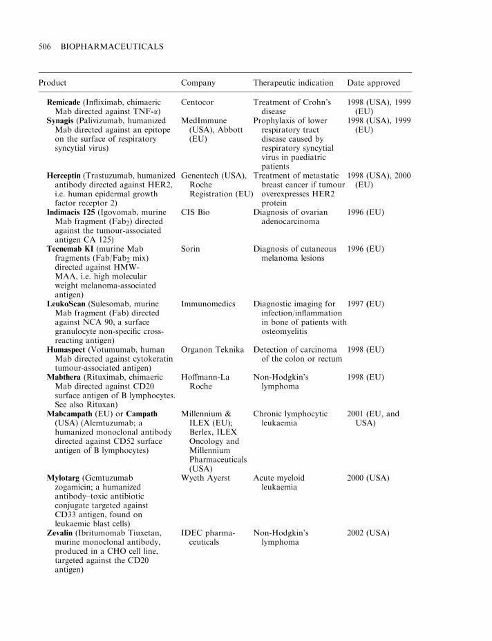
Product Company Therapeutic indication Date approved
Remicade (Infliximab, chimaericMab directed against TNF-a)
Centocor Treatment of Crohn’sdisease
1998 (USA), 1999(EU)
Synagis (Palivizumab, humanizedMab directed against an epitopeon the surface of respiratorysyncytial virus)
MedImmune(USA), Abbott(EU)
Prophylaxis of lowerrespiratory tractdisease caused byrespiratory syncytialvirus in paediatricpatients
1998 (USA), 1999(EU)
Herceptin (Trastuzumab, humanizedantibody directed against HER2,i.e. human epidermal growthfactor receptor 2)
Genentech (USA),RocheRegistration (EU)
Treatment of metastaticbreast cancer if tumouroverexpresses HER2protein
1998 (USA), 2000(EU)
Indimacis 125 (Igovomab, murineMab fragment (Fab2) directedagainst the tumour-associatedantigen CA 125)
CIS Bio Diagnosis of ovarianadenocarcinoma
1996 (EU)
Tecnemab KI (murine Mabfragments (Fab/Fab2 mix)directed against HMW-MAA, i.e. high molecularweight melanoma-associatedantigen)
Sorin Diagnosis of cutaneousmelanoma lesions
1996 (EU)
LeukoScan (Sulesomab, murineMab fragment (Fab) directedagainst NCA 90, a surfacegranulocyte non-specific cross-reacting antigen)
Immunomedics Diagnostic imaging forinfection/inflammationin bone of patients withosteomyelitis
1997 (EU)
Humaspect (Votumumab, humanMab directed against cytokeratintumour-associated antigen)
Organon Teknika Detection of carcinomaof the colon or rectum
1998 (EU)
Mabthera (Rituximab, chimaericMab directed against CD20surface antigen of B lymphocytes.See also Rituxan)
Hoffmann-LaRoche
Non-Hodgkin’slymphoma
1998 (EU)
Mabcampath (EU) or Campath
(USA) (Alemtuzumab; ahumanized monoclonal antibodydirected against CD52 surfaceantigen of B lymphocytes)
Millennium &ILEX (EU);Berlex, ILEXOncology andMillenniumPharmaceuticals(USA)
Chronic lymphocyticleukaemia
2001 (EU, andUSA)
Mylotarg (Gemtuzumabzogamicin; a humanizedantibody–toxic antibioticconjugate targeted againstCD33 antigen, found onleukaemic blast cells)
Wyeth Ayerst Acute myeloidleukaemia
2000 (USA)
Zevalin (Ibritumomab Tiuxetan,murine monoclonal antibody,produced in a CHO cell line,targeted against the CD20antigen)
IDEC pharma-ceuticals
Non-Hodgkin’slymphoma
2002 (USA)
506 BIOPHARMACEUTICALS
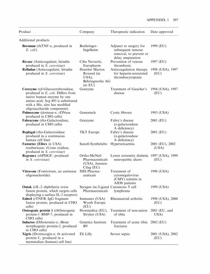
Product Company Therapeutic indication Date approved
Additional products
Beromun (rhTNF-a, produced inE. coli )
Boehringer-Ingelheim
Adjunct to surgery forsubsequent tumourremoval, to prevent ordelay amputation
1999 (EU)
Revasc (Anticoagulant; hirudinproduced in S. cerevisiae)
Ciba Novartis,Europharm
Prevention of venousthrombosis
1997 (EU)
Refludan (Anticoagulant; hirudinproduced in S. cerevisiae)
Hoechst MarionRoussel (inUSA),Behringwerke AG(in EU)
Anticoagulation therapyfor heparin-associatedthrombocytopenia
1998 (USA), 1997(EU)
Cerezyme (rb-Glucocerebrosidase,produced in E. coli. Differs fromnative human enzyme by oneamino acid; Arg 495 is substitutedwith a His, also has modifiedoligosaccharide component)
Genzyme Treatment of Gaucher’sdisease
1994 (USA), 1997(EU)
Pulmozyme (dornase-a, rDNaseproduced in CHO cells)
Genentech Cystic fibrosis 1993 (USA)
Fabrazyme (rha-Galactosidase,produced in CHO cells)
Genzyme Fabry’s disease(a-galactosidaseA deficiency)
2001 (EU)
Replagal (rha-Galactosidaseproduced in a continuoushuman cell line)
TKT Europe Fabry’s disease(a-galactosidaseA deficiency)
2001 (EU)
Fasturtec (Elitex in USA)(rasburicase; rUrate oxidase,produced in S. cerevisiae)
Sanofi-Synthelabo Hyperuricaemia 2001 (EU), 2002(USA)
Regranex (rhPDGF, producedin S. cerevisiae)
Ortho-McNeilPharmaceuticals(USA), Janssen-Cilag (EU)
Lower extremity diabeticneuropathic ulcers
1997 (USA), 1999(EU)
Vitravene (Fomivirsen, an antisenseoligonucleotide)
ISIS Pharma-ceuticals
Treatment ofcytomegalovirus(CMV) retinitis inAIDS patients
1998 (USA)
Ontak (rIL-2–diphtheria toxinfusion protein, which targets cellsdisplaying a surface IL-2 receptor)
Seragen inc/LigandPharmaceuticals
Cutaneous T celllymphoma
1999 (USA)
Enbrel (rTNFR–IgG fragmentfusion protein, produced in CHOcells)
Immunex (USA)Wyeth Europa(EU)
Rheumatoid arthritis 1998 (USA), 2000(EU)
Osteogenic protein 1 (rhOsteogenicprotein-1–BMP-7, produced inCHO cells)
Howmedica (EU),Stryker (USA)
Treatment of non-unionof tibia
2001 (EU, andUSA)
Inductos (Dibotermin-a; rBonemorphogenic protein-2, producedin CHO cells)
Genetics InstituteBV
Treatment of acute tibiafractures
2002 (EU)
Xigris (Drotrecogin-a; rh activatedprotein C, produced in amammalian (human) cell line)
Eli Lilly Severe sepsis 2001 (USA), 2002(EU)
APPENDIX 1 507

Appendix 2
Some Internet addresses relevant
to the biopharmaceutical sector
Note: most home pages listed themselves contain relevant and extensive Internet site links
SOME BIOTECHNOLOGY/PHARMACEUTICAL/MEDICAL ORGANIZATIONS
BIO home page
Site address: http://www.bio.orgHome page of the biotechnology industry organization. Also contains many excellent links.
Pharmaceutical researchers and manufacturers of America
Site address: http//phrma.orgExcellent site, providing information on a wide range of pharmaceutical issues, includingreports such as the annual ‘Biotechnology Medicines in Development’ series.
Drug Information Association (DIA)
Site address: http://www.diahome.orgHome page of the DIA, contains information on various facets of the pharmaceutical industry,including pharmaceutical biotechnology.
European Association of Pharma Biotechnology (EAPB)
Site address: http://www.eapb.orgHome page of the EAPB, containing selected pharmaceutical biotechnology information.
Biopharmaceuticals: Biochemistry and Biotechnology, Second Edition by Gary WalshJohn Wiley & Sons Ltd: ISBN 0 470 84326 8 (ppc), ISBN 0 470 84327 6 (pbk)
European Federation of Biotechnology (EFB)
Site address: http://www.efbweb.orgHome page of the EFB, containing information on various facets of biotechnology, includingpharmaceutical biotechnology.
World Health Organization (WHO)
Site address: http//www.who.int/en/Excellent although general site. Contains information regarding e.g. global disease incidence,vaccination/immunization, etc.
REGULATORY AND ASSOCIATED SITES
Food and Drug Administration (FDA) home page
Site address: http://www.fda.govFDA home page. A key reference for regulatory issues (United States) for (bio)pharmaceuticaldevelopment and production. Also contains information on approved products.
European Medicines Evaluation Agency (EMEA) home page
Site address: http://www.emea.eu.intEMEA home page. A key reference for regulatory issues (European) for biopharmaceuticaldevelopment and production. Also contains information on approved products.
International Conference on Harmonization (ICH)
Site address: http://www.ich.orgICH home page. The International Conference on Harmonization of Technical Requirementsfor Registration of Pharmaceuticals for Human Use (ICH), a unique project that bringstogether the regulatory authorities of Europe, Japan and the USA, and experts from thepharmaceutical industry.
Pharmacos (European Commission)
Site address: http://pharmacos.eudra.orgPharmaceuticals home page of the EC. Contains documents relating to various aspects of theEuropean pharmaceutical industry, including the text of The Rules Governing MedicinalProducts in the EU and a register of all approved pharmaceutical products.
US Patent Office
Site address: http://www.uspto.govThis site from the US government contains a wealth of information on patenting of materialsand has a searchable database of patents.
510 BIOPHARMACEUTICALS
European Directorate for the Quality of Medicines
Site address: http//www.pheur.orgHouses information relating to various quality aspects of pharmaceuticals, including details ofthe European Pharmacopoeia.
United States Pharmacopoeia (USP)
Site address: http//www.usp.orgHouses information detailing the USP.
SOME BIOPHARMACEUTICAL COMPANIES
Amgen
Site address: http//www.amgen.com
Genentech
Site address: http//www.genentech.com
Biogen
Site address: http://www.biogen.com
Genzyme
Site address: http://www.genzyme.com
Wyeth
Site address: http://wyeth.com
Eli Lilly
Site address: http://www.lilly.com
Novo
Site address: http://www.novonordisk.com
Schering Plough
Site address: http//www.sch-plough.com/
APPENDIX 2 511
PROTEINS AND GENES
The genome database
Site address: http//gdbwww.gdb.orgA focal database for human gene mapping that attempts to integrate physical and genetic maps.
The Institute for Genomics Research
Site address: http//www.tigr.org/tdb/Excellent source of information regarding various completed/ongoing genome sequencingprojects.
Databases at the European Bioinformatics Institute (EBI)
Site address: http//www.ebi.ac.uk/dbases/topdata.htmlDatabases at the EBI for nucleotide/protein searches (data largely overlap that at NationalCenter for Biotechnology Information (NCBI)).
Protein Databank (PDB)
Site address: http//www.rcsb.org/pdbSearchable repository of 3-D protein structure.
ExPASy
Site address: http//www.expasy.org/Proteomics server of the Swiss institute of bioinformatics. Dedicated to analysis of proteinsequences and structure, as well as 2-D SDS–PAGE.
PredictProtein
Site address: http//www.embl-heidelberg.de/Services/sander/predictprotein/Submit a protein sequence and you will receive secondary structure prediction via e-mail.
Principles of Protein Structure
Site address: http//www.cryst.bbk.ac.uk/pps2Provides information relating to protein structure, including some basic course material.
3D searching with receptor-based queries
Site address: http//www.ch.ic.ac.uk:80/ectoc/papers/guner/This is an interesting site presenting information on the ability to search for molecules withsimilar chemical structures.
512 BIOPHARMACEUTICALS
The Immunology Link
Site address: http://www.immunologylink.comSite provides links to additional immunology-related sites.
American Society for Gene Therapy
Site address: http//www.asgt.org
European Society for Gene Therapy
Site address: http://www.esgt.org
APPENDIX 2 513

Appendix 3
Two selected monographs reproduced
from the European Pharmacopoeia,with permission from the
European Commission*
I. RECOMBINANT DNA TECHNOLOGY, PRODUCTS OF
Producta ab ADN recombinante
This monograph provides general requirements for the development and manufacture of products ofrecombinant DNA technology. These requirements are not necessarily comprehensive in a givencase and requirements complementary or additional to those prescribed in this monograph may beimposed in an individual monograph or by the competent authority.
The monograph is not applicable to modified live organisms that are intended to be used directlyin man and animals, for example as live vaccines.
Definition
Products of rDNA technology are produced by genetic modification in which DNA coding forthe required product is introduced, usually by means of a plasmid or a viral vector, into asuitable microorganism or cell line, in which that DNA is expressed and translated into protein.The desired product is then recovered by extraction and purification.
The cell or microorganism before harbouring the vector is referred to as the host cell, and thestable association of the two used in the manufacturing process is referred to as the host–vectorsystem.
Biopharmaceuticals: Biochemistry and Biotechnology, Second Edition by Gary WalshJohn Wiley & Sons Ltd: ISBN 0 470 84326 8 (ppc), ISBN 0 470 84327 6 (pbk)
*Copyright holder: European Directorate for the Quality of Medicines
Production
Production is based on a validated seed-lot system using a host–vector combination that hasbeen shown to be suitable to the satisfaction of the competent authority. The seed-lot systemuses a master cell bank and a working cell bank derived from the master seed lot of the host–vector combination. A detailed description of cultivation, extraction and purification steps anda definition of the production batch shall be established.
The determination of the suitability of the host–vector combination and the validation of theseed-lot system include the following elements.
Cloning and expression
The suitability of the host–vector system, particularly as regards microbiological purity, isdemonstrated by:
. Characterization of the host cell, including source, phenotype and genotype, and of the cell-culture media. Documentation of the strategy for the cloning of the gene and characterization ofthe recombinant vector, including:i. the origin and characterization of the gene;ii. nucleotide-sequence analysis of the cloned gene and the flanking control regions of the
expression vector. The cloned sequences are kept to a minimum and all relevantexpressed sequences are clearly identified and confirmed at the RNA level. The DNAsequence of the cloned gene is normally confirmed at the seed-lot stage, up to and beyondthe normal level of population doubling for full-scale fermentation. In certain systems,for example, where multiple copies of the gene are inserted into the genome of acontinuous cell line, it may be inappropriate to sequence the cloned gene at theproduction level. Under these circumstances, Southern blot analysis of total cellularDNA or sequence analysis of the messenger RNA (mRNA) may be helpful, particularattention being paid to characterization of the expressed protein;
iii. the construction, genetics and structure of the complete expression vector.. Characterization of the host–vector system, including:
i. mechanism of transfer of the vector into the host cells;ii. copy number, physical state and stability of the vector inside the host cell;iii. measures used to promote and control the expression.
Cell banking system
The master cell bank is a homogeneous suspension of the original cells already transformed bythe expression vector containing the desired gene, distributed in equal volumes into individualcontainers for storage (e.g. in liquid nitrogen). In some cases it may be necessary to establishseparate master cell banks for the expression vector and the host cells.
The working cell bank is a homogeneous suspension of the cell material derived from themaster cell bank(s) at a finite passage level, distributed in equal volumes into individualcontainers for storage (e.g. in liquid nitrogen).
In both cell banks, all containers are treated identically during storage and, once removedfrom storage, the containers are not returned to the cell stock.
The cell bank may be used for the production at a finite passage level or for continuous-culture production.
516 BIOPHARMACEUTICALS
Production at a finite passage level
This cultivation method is defined by a limited number of passages or population doublingswhich must not be exceeded during production. The maximum number of cell doublings, orpassage levels, during which the manufacturing process routinely meets the criteria describedbelow, must be stated.
Continuous-culture production
By this cultivation method the number of passages or population doublings is not restrictedfrom the beginning of production. Criteria for the harvesting as well as for the termination ofproduction have to be defined by the manufacturer. Monitoring is necessary throughout the lifeof the culture: the required frequency and type of monitoring will depend on the nature of theproduction system and the product.
Information is required on the molecular integrity of the gene being expressed and on thephenotypic and genotypic characteristics of the host cell after long-term cultivation. Theacceptance of harvests for further processing must be clearly linked to the schedule ofmonitoring applied and a clear definition of a ‘batch’ of product for further processing isrequired.
Validation of the cell banks
Validation of the cell banks includes:i. stability, by measuring viability and the retention of the vector;ii. identity of the cells by phenotypic features;iii. where appropriate, evidence that the cell banks are free from potentially oncogenic or
infective adventitious agents (viral, bacterial, fungal or mycoplasmal). Special attentionhas to be given to viruses that can commonly contaminate the species from which the cellline has been derived. Certain cell lines contain endogenous viruses, e.g. retroviruses,which may not readily be eliminated. The expression of these organisms, under a varietyof condition known to cause their induction, shall be tested:
iv. for mammalian cells, details of the tumorigenic potential of the cell bank shall beobtained.
Control of the cells
The origin, form, storage, use and stability at the anticipated rate of use must be documented infull for all cell banks under conditions of storage and recovery. New cell banks must be fullyvalidated.
Validation of the production process
Extraction and purification
The capacity of each step of the extraction and purification procedure to remove and/orinactivate contaminating substances derived from the host cell or culture medium, including, inparticular, virus particles, proteins, nucleic acids and added substances, must be validated.
APPENDIX 3 517
Validation studies are carried out to demonstrate that the production process routinely meetsthe following criteria:
. Exclusion of extraneous agents from the product. Studies including, for example, viruses withrelevant physico-chemical features are undertaken, and a reduction capacity for suchcontaminants at each relevant stage of purification is established.
. Adequate removal of vector, host cell culture medium and reagent-derived contaminantsfrom the product. The reduction capacity for DNA is established by spiking. The reductionof proteins of animal origin can be determined by immunochemical methods.
. Maintenance within stated limits of the yield of product from the culture.
. Adequate stability of any intermediate of production and/or manufacturing when it isintended to use intermediate storage during the process.
Characterization of the substance
The identity, purity, potency and stability of the final bulk product are established initially bycarrying out a wide range of chemical, physical, immunochemical and biological tests. Prior torelease, each batch of the product is tested by the by the manufacturer for identity and purityand an appropriate assay is carried out.
Production consistency
Suitable tests for demonstrating the consistency of the production and purification areperformed. The tests include, especially, characterization tests, in-process controls and final-product tests, for example.
Amino acid composition
Partial amino acid sequence analysis. The sequence data permit confirmation of the correct N-terminal processing and detection of the loss of the C-terminal amino acids.
Peptide mapping. Peptide mapping using chemical and/or enzymatic cleavage of the proteinproduct and analysis by a suitable method such as two-dimensional gel electrophoresis, capillaryelectrophoresis or liquid chromatography must show no significant difference between the testprotein and the reference preparation. Peptide mapping can also be used to demonstrate correctdisulphide bonding.
Determination of molecular mass
Cloned-gene retention. The minimum amount in percentage of the cells containing the vectoror the cloned gene after cultivation is approved by the relevant authority.
Total protein. The yield of protein is determined.
Chemical purity. The purity of the protein product is analysed in comparison with a referencepreparation by a suitable method, such as liquid chromatography, capillary electrophoresis orsodium dodecyl sulphate polyacrylamide gel electrophoresis (SDS–PAGE).
518 BIOPHARMACEUTICALS
Host-cell-derived proteins. Host-cell-derived proteins are detected by immunochemicalmethods, using, for example, polyclonal antisera raised against protein components of thehost–vector system used to manufacture the product, unless otherwise prescribed. The followingtypes of procedure may be used: liquid-phase displacement assays (e.g. radio-immunoassay),liquid-phase direct-binding assays and direct-binding assays using antigens immobilized onnitrocellulose (or similar) membranes (e.g. dot-immunoblot assays, Western blots). Generalrequirements for the validation of immunoassay procedures are given under 2.7.1,lmmunochemical Methods. In addition, immunoassay methods for host–cell contaminantsmeet the following criteria:
. Antigen preparations. Antisera are raised against a preparation of antigens derived from thehost organism, into which has been inserted the vector used in the manufacturing processthat lacks the specific gene coding for the product. This host cell is cultured, and proteins areextracted, using conditions identical to those used for culture and extraction in themanufacturing process. Partly purified preparations of antigens, using some of thepurification steps in the manufacturing process, may also be used for the preparation ofantisera.
. Calibration and standardization. Quantitative data are obtained by comparison with dose–response curves obtained using standard preparations of host-derived protein antigens. Sincethese preparations are mixtures of poorly defined proteins, a standard preparation isprepared and calibrated by a suitable protein determination method. This preparation isstored in a stable state suitable for use over an extended period of time.
. Antisera. Antisera contain high-avidity antibodies recognizing as many different proteins inthe antigen mixture as possible, and do not cross react with the product.
Host-cell and vector-derived DNA. Residual DNA is detected by hybridization analysis, usingsuitably sensitive sequence-independent analytical techniques or other suitably sensitiveanalytical techniques.
Hybridization analysis
DNA in the test sample is denatured to give single-stranded DNA, immobilized on anitrocellulose or other suitable filter and hybridized with labelled DNA prepared from the host–vector manufacturing system (DNA probes). Although a wide variety of experimentalapproaches is available, hybridization methods for measurement of host–vector DNA meetthe following criteria:
. DNA probes. Purified DNA is obtained from the host–vector system grown under the sameconditions as those used in the manufacturing process. Host chromosomal DNA and vectorDNA may be separately prepared and used as probes.
. Calibration and standardization. Quantitative data are obtained by comparison withresponses obtained using standard preparations. Chromosomal DNA probes and vectorDNA probes are used with chromosomal DNA and vector DNA standards, respectively.Standard preparations are calibrated by spectroscopic measurements and stored in a statesuitable for use over an extended period of time.
APPENDIX 3 519
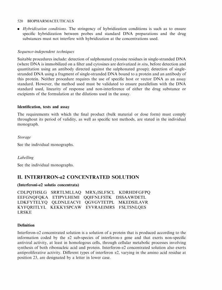
. Hybridization conditions. The stringency of hybridization conditions is such as to ensurespecific hybridization between probes and standard DNA preparations and the drugsubstances must not interfere with hybridization at the concentrations used.
Sequence-independent techniques
Suitable procedures include: detection of sulphonated cytosine residues in single-stranded DNA(where DNA is immobilized on a filter and cytosines are derivatized in situ, before detection andquantitation using an antibody directed against the sulphonated group); detection of single-stranded DNA using a fragment of single-stranded DNA bound to a protein and an antibody ofthis protein. Neither procedure requires the use of specific host or vector DNA as an assaystandard. However, the method used must be validated to ensure parallelism with the DNAstandard used, linearity of response and non-interference of either the drug substance orexcipients of the formulation at the dilutions used in the assay.
Identification, tests and assay
The requirements with which the final product (bulk material or dose form) must complythroughout its period of validity, as well as specific test methods, are stated in the individualmonograph.
Storage
See the individual monographs.
Labelling
See the individual monographs.
II. INTERFERON-a2 CONCENTRATED SOLUTION
(Interferoni-a2 solutio concentrata)
CDLPQTHSLG SRRTLMLLAQ MRX1ISLFSCL KDRHDFGFPQEEFGNQFQKA ETIPVLHEMI QQIFNLFSTK DSSAAWDETLLDKFYTELYQ QLDNLEACVI QGVGVTETPL MKEDSILAVRKYFQRITLYL KEKKYSPCAW EVVRAEIMRS FSLTSNLQESLRSKE
Definition
Interferon-a2 concentrated solution is a solution of a protein that is produced according to theinformation coded by the a2 sub-species of interferon-a gene and that exerts non-specificantiviral activity, at least in homologous cells, through cellular metabolic processes involvingsynthesis of both ribonucleic acid and protein. Interferon-a2 concentrated solution also exertsantiproliferative activity. Different types of interferon a2, varying in the amino acid residue atposition 23, are designated by a letter in lower case.
520 BIOPHARMACEUTICALS
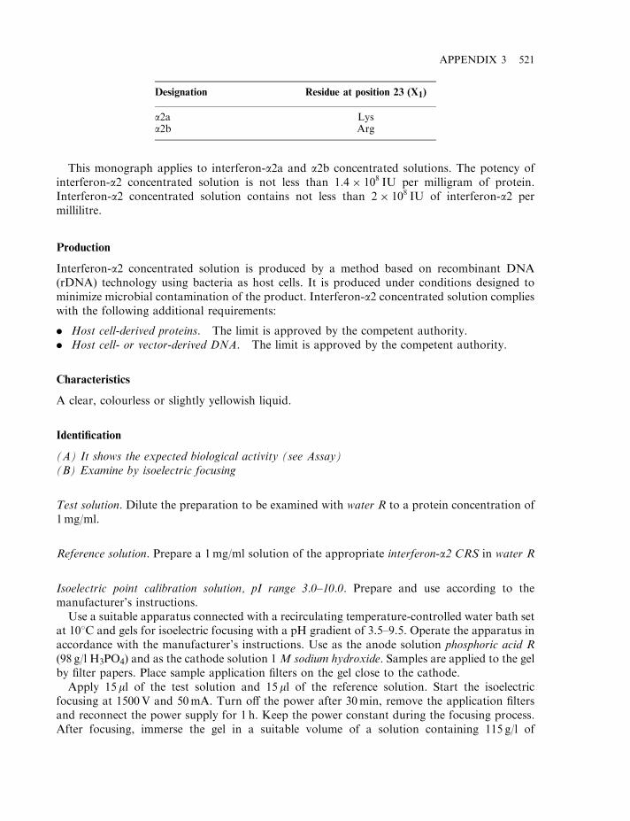
This monograph applies to interferon-a2a and a2b concentrated solutions. The potency ofinterferon-a2 concentrated solution is not less than 1:4� 108 IU per milligram of protein.Interferon-a2 concentrated solution contains not less than 2� 108 IU of interferon-a2 permillilitre.
Production
Interferon-a2 concentrated solution is produced by a method based on recombinant DNA(rDNA) technology using bacteria as host cells. It is produced under conditions designed tominimize microbial contamination of the product. Interferon-a2 concentrated solution complieswith the following additional requirements:
. Host cell-derived proteins. The limit is approved by the competent authority.
. Host cell- or vector-derived DNA. The limit is approved by the competent authority.
Characteristics
A clear, colourless or slightly yellowish liquid.
Identification
(A) It shows the expected biological activity (see Assay)(B) Examine by isoelectric focusing
Test solution. Dilute the preparation to be examined with water R to a protein concentration of1mg/ml.
Reference solution. Prepare a 1mg/ml solution of the appropriate interferon-a2 CRS in water R
Isoelectric point calibration solution, pI range 3.0–10.0. Prepare and use according to themanufacturer’s instructions.Use a suitable apparatus connected with a recirculating temperature-controlled water bath set
at 108C and gels for isoelectric focusing with a pH gradient of 3.5–9.5. Operate the apparatus inaccordance with the manufacturer’s instructions. Use as the anode solution phosphoric acid R(98 g/l H3PO4) and as the cathode solution 1 M sodium hydroxide. Samples are applied to the gelby filter papers. Place sample application filters on the gel close to the cathode.
Apply 15 ml of the test solution and 15 ml of the reference solution. Start the isoelectricfocusing at 1500V and 50mA. Turn off the power after 30min, remove the application filtersand reconnect the power supply for 1 h. Keep the power constant during the focusing process.After focusing, immerse the gel in a suitable volume of a solution containing 115 g/l of
APPENDIX 3 521
Designation Residue at position 23 (X1)
a2a Lysa2b Arg
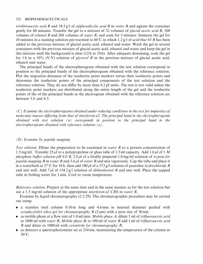
trichloroacetic acid R and 34.5 g/l of sulphosalicylic acid R in water R and agitate the containergently for 60 minutes. Transfer the gel to a mixture of 32 volumes of glacial acetic acid R, 100volumes of ethanol R and 268 volumes of water R, and soak for 5 minutes. Immerse the gel for10 minutes in a staining solution pre-warmed to 608C in which 1.2 g/l of acid blue 83 R has beenadded to the previous mixture of glacial acetic acid, ethanol and water. Wash the gel in severalcontainers with the previous mixture of glacial acetic acid, ethanol and water and keep the gel inthis mixture until the background is clear (12 h to 24 h). After adequate destaining, soak the gelfor 1 h in a 10% (V/V) solution of glycerol R in the previous mixture of glacial acetic acid,ethanol and water.
The principal bands of the electropherogram obtained with the test solution correspond inposition to the principal bands of the electropherogram obtained with the reference solution.Plot the migration distances of the isoelectric point markers versus their isoelectric points anddetermine the isoelectric points of the principal components of the test solution and thereference solution. They do not differ by more than 0.2 pI units. The test is not valid unless theisoelectric point markers are distributed along the entire length of the gel and the isoelectricpoints of the of the principal bands in the electrogram obtained with the reference solution arebetween 5.8 and 6.3.
(C) Examine the electropherograms obtained under reducing conditions in the test for impurities ofmolecular masses differing from that of interferon-a2. The principal band in the electropherogramobtained with test solution (a) corresponds in position to the principal band in theelectropherogram obtained with reference solution (a).
(D) Examine by peptide mapping
Test solution. Dilute the preparation to be examined in water R to a protein concentration of1.5mg/ml. Transfer 25 ml to a polypropylene or glass tube of 1.5ml capacity. Add 1.6 ml of 1 Mphosphate buffer solution pH 8.0 R, 2.8 ml of a freshly prepared 1.0mg/ml solution of trypsin forpeptide mapping R in water R and 3.6 ml of water R and mix vigorously. Cap the tube and place itin a waterbath at 378C for 18 h, then add 100 ml of a 573 g/l solution of guanidine hydrochloride Rand mix well. Add 7 ml of 154.2 g/l solution of dithiothreitol R and mix well. Place the cappedtube in boiling water for 1min. Cool to room temperature.
Reference solution. Prepare at the same time and in the same manner as for the test solution butuse a 1.5 mg/ml solution of the appropriate interferon-a2 CRS in water R.
Examine by liquid chromatography (2.2.29). The chromatographic procedure may be carriedout using:
. a stainless steel column 0.10m long and 4.6mm in internal diameter packed withoctadecylsilyl silica gel for chromatography R (5 mm) with a pore size of 30 nm;
. as mobile phase at a flow rate of 1.0ml/min.Mobile phase A; dilute 1 ml of trifluoroacetic acidto 1000 ml with water R; Mobile phase B; to 100ml of water R add 1ml of trifluoroacetic acidR and dilute to 1000ml with cetonitrile for chromatography R;
. as detector a spectrophotometer set at 214 nm, maintaining the temperature of the column at308C.
522 BIOPHARMACEUTICALS
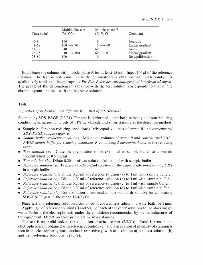
Equilibrate the column with mobile phase A for at least 15 min. Inject 100 ml of the referencesolution. The test is not valid unless the chromatogram obtained with each solution isqualitatively similar to the appropriate Ph. Eur. Reference chromatogram of interferon-a2 digest.The profile of the chromatogram obtained with the test solution corresponds to that of thechromatogram obtained with the reference solution.
Tests
Impurities of molecular mass differing from that of interferon-a2
Examine by SDS–PAGE (2.2.31). The test is performed under both reducing and non-reducingconditions, using resolving gels of 14% acrylamide and silver staining as the detection method.
. Sample buffer (non-reducing conditions). Mix equal volumes of water R and concentratedSDS–PAGE sample buffer R.
. Sample buffer (reducing conditions). Mix equal volumes of water R and concentrated SDS–PAGE sample buffer for reducing conditions R containing 2-mercaptoethanol as the reducingagent.
. Test solution (a). Dilute the preparation to be examined in sample buffer to a proteinconcentration of 0.5mg/ml.
. Test solution (b). Dilute 0.20ml of test solution (a) to 1ml with sample buffer.
. Reference solution (a). Prepare a 0.625mg/ml solution of the appropriate interferon-a2 CRSin sample buffer.
. Reference solution (b). Dilute 0.20ml of reference solution (a) to 1ml with sample buffer.
. Reference solution (c). Dilute 0.20ml of reference solution (b) to 1ml with sample buffer.
. Reference solution (d). Dilute 0.20ml of reference solution (c) to 1ml with sample buffer.
. Reference solution (e). Dilute 0.20ml of reference solution (d) to 1ml with sample buffer.
. Reference solution (f). Use a solution of molecular mass standards suitable for calibratingSDS–PAGE gels in the range 15–67 kDa.
Place test and reference solutions, contained in covered test-tubes, in a waterbath for 2min.Apply 10 ml of reference solution (f) and 50 ml of each of the other solutions to the stacking gel
wells. Perform the electrophoresis under the conditions recommended by the manufacturer ofthe equipment. Detect proteins in the gel by silver staining.
The test is not valid unless: the validation criteria are met (2.2.31); a band is seen in theelectropherogram obtained with reference solution (e); and a gradation of intensity of staining isseen in the electropherograms obtained, respectively, with test solution (a) and test solution (b)and with reference solutions (a) to (e).
APPENDIX 3 523
Time (min)Mobile phase A(% V/V)
Mobile phase B(% V/V) Comment
0–8 100 0 Isocratic8–68 100 �! 40 0 �! 60 Linear gradient68–72 40 60 Isocratic72–75 40 �! 100 60 �! 0 Linear gradient75–80 100 0 Re-equilibration
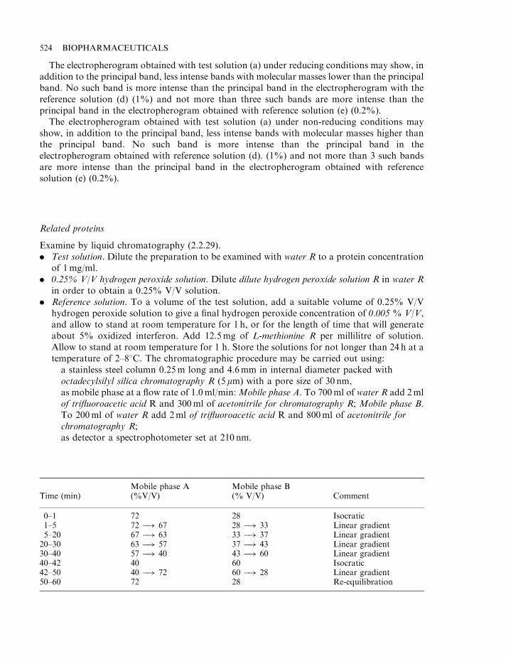
The electropherogram obtained with test solution (a) under reducing conditions may show, inaddition to the principal band, less intense bands with molecular masses lower than the principalband. No such band is more intense than the principal band in the electropherogram with thereference solution (d) (1%) and not more than three such bands are more intense than theprincipal band in the electropherogram obtained with reference solution (e) (0.2%).
The electropherogram obtained with test solution (a) under non-reducing conditions mayshow, in addition to the principal band, less intense bands with molecular masses higher thanthe principal band. No such band is more intense than the principal band in theelectropherogram obtained with reference solution (d). (1%) and not more than 3 such bandsare more intense than the principal band in the electropherogram obtained with referencesolution (e) (0.2%).
Related proteins
Examine by liquid chromatography (2.2.29).. Test solution. Dilute the preparation to be examined with water R to a protein concentration
of 1mg/ml.. 0.25% V/V hydrogen peroxide solution. Dilute dilute hydrogen peroxide solution R in water R
in order to obtain a 0.25% V/V solution.. Reference solution. To a volume of the test solution, add a suitable volume of 0.25% V/V
hydrogen peroxide solution to give a final hydrogen peroxide concentration of 0.005 % V/V,and allow to stand at room temperature for 1 h, or for the length of time that will generateabout 5% oxidized interferon. Add 12.5mg of L-methionine R per millilitre of solution.Allow to stand at room temperature for 1 h. Store the solutions for not longer than 24 h at atemperature of 2–88C. The chromatographic procedure may be carried out using:
a stainless steel column 0.25m long and 4.6mm in internal diameter packed withoctadecylsilyl silica chromatography R (5 mm) with a pore size of 30 nm,as mobile phase at a flow rate of 1.0ml/min:Mobile phase A. To 700ml of water R add 2mlof trifluoroacetic acid R and 300ml of acetonitrile for chromatography R; Mobile phase B.To 200ml of water R add 2ml of trifluoroacetic acid R and 800ml of acetonitrile forchromatography R;as detector a spectrophotometer set at 210 nm.
524 BIOPHARMACEUTICALS
Time (min)Mobile phase A(%V/V)
Mobile phase B(% V/V) Comment
0–1 72 28 Isocratic1–5 72 �! 67 28 �! 33 Linear gradient5–20 67 �! 63 33 �! 37 Linear gradient20–30 63 �! 57 37 �! 43 Linear gradient30–40 57 �! 40 43 �! 60 Linear gradient40–42 40 60 Isocratic42–50 40 �! 72 60 �! 28 Linear gradient50–60 72 28 Re-equilibration
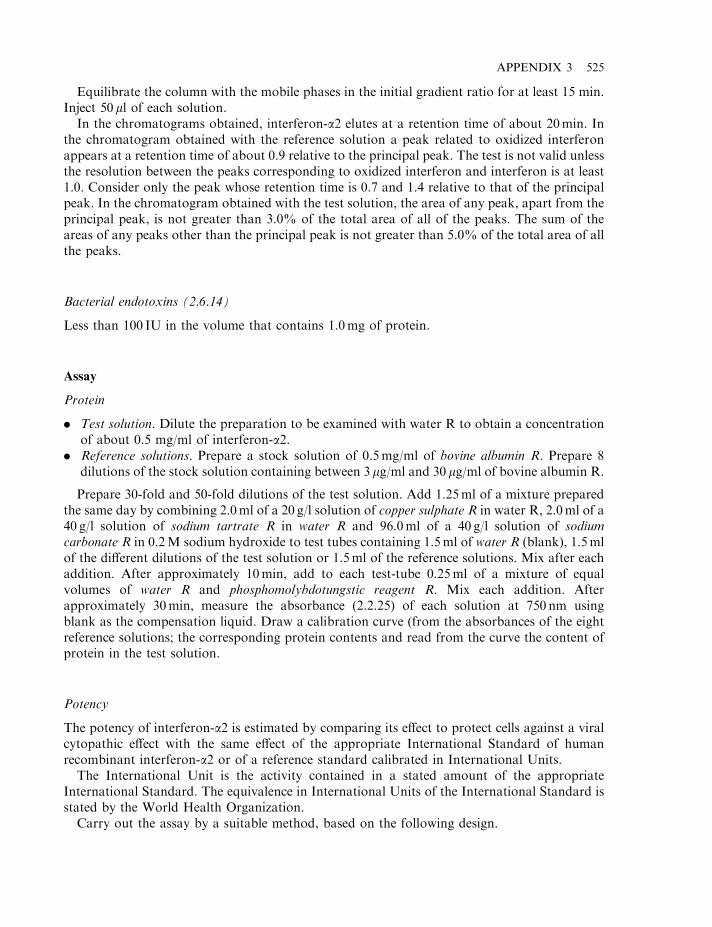
Equilibrate the column with the mobile phases in the initial gradient ratio for at least 15 min.Inject 50 ml of each solution.
In the chromatograms obtained, interferon-a2 elutes at a retention time of about 20min. Inthe chromatogram obtained with the reference solution a peak related to oxidized interferonappears at a retention time of about 0.9 relative to the principal peak. The test is not valid unlessthe resolution between the peaks corresponding to oxidized interferon and interferon is at least1.0. Consider only the peak whose retention time is 0.7 and 1.4 relative to that of the principalpeak. In the chromatogram obtained with the test solution, the area of any peak, apart from theprincipal peak, is not greater than 3.0% of the total area of all of the peaks. The sum of theareas of any peaks other than the principal peak is not greater than 5.0% of the total area of allthe peaks.
Bacterial endotoxins (2.6.14)
Less than 100 IU in the volume that contains 1.0mg of protein.
Assay
Protein
. Test solution. Dilute the preparation to be examined with water R to obtain a concentrationof about 0.5 mg/ml of interferon-a2.
. Reference solutions. Prepare a stock solution of 0.5mg/ml of bovine albumin R. Prepare 8dilutions of the stock solution containing between 3 mg/ml and 30 mg/ml of bovine albumin R.
Prepare 30-fold and 50-fold dilutions of the test solution. Add 1.25ml of a mixture preparedthe same day by combining 2.0ml of a 20 g/l solution of copper sulphate R in water R, 2.0ml of a40 g/l solution of sodium tartrate R in water R and 96.0ml of a 40 g/l solution of sodiumcarbonate R in 0.2M sodium hydroxide to test tubes containing 1.5ml of water R (blank), 1.5mlof the different dilutions of the test solution or 1.5ml of the reference solutions. Mix after eachaddition. After approximately 10min, add to each test-tube 0.25ml of a mixture of equalvolumes of water R and phosphomolybdotungstic reagent R. Mix each addition. Afterapproximately 30min, measure the absorbance (2.2.25) of each solution at 750 nm usingblank as the compensation liquid. Draw a calibration curve (from the absorbances of the eightreference solutions; the corresponding protein contents and read from the curve the content ofprotein in the test solution.
Potency
The potency of interferon-a2 is estimated by comparing its effect to protect cells against a viralcytopathic effect with the same effect of the appropriate International Standard of humanrecombinant interferon-a2 or of a reference standard calibrated in International Units.
The International Unit is the activity contained in a stated amount of the appropriateInternational Standard. The equivalence in International Units of the International Standard isstated by the World Health Organization.
Carry out the assay by a suitable method, based on the following design.
APPENDIX 3 525
Use, in standard culture conditions, an established cell line sensitive to the cytopathic effect ofa suitable virus (a human diploid fibroblast cell line, free of microbial contamination, responsiveto interferon and sensitive to encephalomyocarditis virus, is suitable).
The following cell cultures and virus have shown to be suitable: MDBK cells (ATCC No.CCL22), or Mouse L cells (NCTC clone 929; ATCC No. CCL I) as the cell culture and vesicularstomatitis virus VSV:Indiana strain (ATCC No. VR-158) as the infective agent; or humandiploid fibroblast FS-71 cells responsive to interferon as the cell culture, and encephalomyo-carditis virus (ATCC No. VR-129B) as the infective agent.
Incubate at least four series, cells with three or more different concentrations of thepreparation to be examined and the reference preparation in a microtitre plate and include ineach series appropriate controls of untreated cells. Choose the concentrations of thepreparations such that the lowest concentration produces some protection and the largestconcentration produces less than maximal protection against the viral cytopathic effect. At asuitable time add the cytopathic virus to the wells with the exception of a sufficient number ofwells in all series, which are left with uninfected control cells. Determine the cytopathic effect ofvirus quantitatively with a suitable method. Calculate the potency of the preparation to beexamined by the usual statistical methods for a parallel line assay.
The estimated potency is not less than 80% and not more than 125% of the stated potency.The fiducial limits of error of the estimated potency ( p ¼ 0.95) are not less than 64% and notmore than 156% of the stated potency.
Storage
Store in an airtight container, protected from light, at or below �208C.
Labelling
The label states:
. the type of interferon (a2a or a2b);
. the type of production.
526 BIOPHARMACEUTICALS
Appendix 4
Annex 2 ‘Manufacture of biological
medicinal products for human use’1
SCOPE
The methods employed in the manufacture of biological medicinal products are a critical factorin shaping the appropriate regulatory control. Biological medicinal products can be definedtherefore largely by reference to their method of manufacture. Biological medicinal productsprepared by the following methods of manufacture will fall under the scope of this annex2:
(a) Microbial cultures, excluding those resulting from rDNA techniques;(b) Microbial and cell cultures, including those resulting from recombinant DNA or hybridoma
techniques;(c) Extraction from biological tissues;(d) Propagation of live agents in embryos or animals.
Not all of the aspects of this annex may necessarily apply to products in category (a).
Note: In drawing up this guidance, due consideration has been given to the generalrequirements for manufacturing establishments and control laboratories proposed by the WHO.
The present guidance does not lay down detailed requirements for specific classes of biologicalproducts, and attention is therefore directed to other guidelines issued by the Committee forProprietary Medicinal Products (CPMP), e.g. the note for guidance on monoclonal antibodiesand the note for guidance on products of recombinant DNA technology (The Rules GoverningMedicinal Products in the European Community, Volume III).
Biopharmaceuticals: Biochemistry and Biotechnology, Second Edition by Gary WalshJohn Wiley & Sons Ltd: ISBN 0 470 84326 8 (ppc), ISBN 0 470 84327 6 (pbk)
1From The Rules Governing Medicinal Products in the European Community. Vol. 4, Good ManufacturingPractice for Medicinal Products. Reproduced by permission of the European Union Publications Office,Luxembourg2Biological medicinal products manufactured by these methods include: vaccines, immunosera, antigens,hormones, cytokines, enzymes and other products of fermentation (including monoclonal antibodies andproducts derived from rDNA).
PRINCIPLE
The manufacture of biological medicinal products involves certain specific considerations arisingfrom the nature of the products and the processes. The way in which biological medicinal productsare produced, controlled and administered make some particular precautions necessary.
Unlike conventional medicinal products, which are produced using chemical and physicaltechniques capable of a high degree of consistency, the production of biological medicinalproducts involves biological processes and materials, such as cultivation of cells or extraction ofmaterial from living organisms. These biological processes may display inherent variability, sothat the range and nature of by-products are variable. Moreover, the materials used in thesecultivation processes provide good substrates for growth of microbial contaminants.
Control of biological medicinal products usually involves biological analytical techniqueswhich have a greater variability than physico-chemical determinations. In-process controls,therefore, take on a great importance in the manufacture of biological medicinal products.
PERSONNEL
1. All personnel (including those concerned with cleaning, maintenance or quality control)employed in areas where biological medicinal products are manufactured should receiveadditional training specific to the products manufactured and to their work. Personnelshould be given relevant information and training in hygiene and microbiology.
2. Persons responsible for production and quality control should have an adequatebackground in a relevant scientific discipline, such as bacteriology, biology, biometry,chemistry, medicine, pharmacy, pharmacology, virology, immunology and veterinarymedicine, together with sufficient practical experience to enable them to exercise theirmanagement function for the process concerned.
3. The immunological status of personnel may have to be taken into consideration for productsafety. All personnel engaged in production, maintenance, testing and animal care (andinspectors) should be vaccinated, where necessary, with appropriate specific vaccines andhave regular health checks. Apart from the obvious problem of exposure of staff toinfectious agents, potent toxins or allergens, it is necessary to avoid the risk ofcontamination of a production batch with infectious agents. Visitors should generally beexcluded from production areas.
4. Any changes in the immunological status of personnel, which could adversely affect thequality of the product, should preclude work in the production area. Production of BCGvaccine and tuberculin products should be restricted to staff who are carefully monitored byregular checks or immunological status or chest X-ray.
5. In the course of a working day, personnel should not pass from areas where exposure to liveorganisms or animals is possible to areas where other products or different organisms arehandled. If such passage is unavoidable, clearly defined decontamination measures,including change of clothing and shoes and, where necessary, showering, should be followedby staff involved in any such production.
PREMISES AND EQUIPMENT
6. The degree of environmental control of particulate and microbial contamination of theproduction premises should be adapted to the product and the production step, bearing inmind the level of contamination of the starting materials and the risk to the finished product.
528 BIOPHARMACEUTICALS
7. The risk of cross-contamination between biological medicinal products, especially duringthose stages of the manufacturing process in which live organisms are used, may requireadditional precautions with respect to facilities and equipment, such as the use of dedicatedfacilities and equipment, production on a campaign basis and the use of closed systems. Thenature of the product, as well as the equipment used, will determine the level of segregationneeded to avoid cross-contamination.
8. In principle, dedicated facilities should be used for the production of BCG vaccine and forthe handling of live organisms used in production of tuberculin products.
9. Dedicated facilities should be used for the handling of Bacillus anthracis, of Clostridiumbotulinum and of Clostridium tetani until the inactivation process is accomplished.
10. Production on a campaign basis may be acceptable for other spore-forming organismsprovided that the facilities are dedicated to this group of products and not more than oneproduct is processed at any one time.
11. Simultaneous production in the same area, using closed systems of biofermenters, may beacceptable for products such as monoclonal antibodies and products prepared by rDNAtechniques.
12. Processing steps after harvesting may be carried out simultaneously in the same productionarea provided that adequate precautions are taken to prevent cross-contamination. Forkilled vaccines and toxoids, such parallel processing should only be performed afterinactivation of the culture or after detoxification.
13. Positive-pressure areas should be used to process sterile products but negative pressure inspecific areas at point of exposure of pathogens is acceptable for containment reasons.
Where negative-pressure areas or safety cabinets are used for aseptic processing ofpathogens, they should be surrounded by a positive-pressure sterile zone.
14. Air filtration units should be specific to the processing area concerned and recirculation ofair should not occur from areas handling live pathogenic organisms.
15. The layout and design of production areas and equipment should permit effective cleaningand decontamination (e.g. by fumigation). The adequacy of cleaning and decontaminationprocedures should be validated.
16. Equipment used during handling of live organisms should be designed to maintain culturesin a pure state and uncontaminated by external sources during processing.
17. Pipework systems, valves and vent filters should be properly designed to facilitate cleaningand sterilization. The use of ‘clean in place’ and ‘sterilize in place’ systems should beencouraged. Valves on fermentation vessels should be completely steam sterilizable. Airvent filters should be hydrophobic and validated for their scheduled lifespan.
18. Primary containment should be designed and tested to demonstrate freedom from leakagerisk.
19. Effluents which may contain pathogenic microorganisms should be effectively decontami-nated.
20. Due to the variability of biological products or processes, some additives or ingredientshave to be measured or weighed during the production process (e.g. buffers). In these cases,small stocks of these substances may be kept in the production area.
ANIMAL QUARTERS AND CARE
21. Animals are used for the manufacture of a number of biological products, e.g. polio vaccine(monkeys), snake antivenoms (horses and goats), rabies vaccine (rabbits, mice and
APPENDIX 4 529

hamsters) and serum gonadotropin (horses). In addition, animals may also be used in thequality control of most sera and vaccines, e.g. pertussis vaccine (mice), pyrogenicity(rabbits), BCG vaccine (guinea-pigs).
22. General requirements for animal quarters, care and quarantine are laid down in Directive86/609/EEC. Quarters for animals used in production and control of biological productsshould be separated from production and control areas. The health status of animals fromwhich some starting materials are derived, and of those used for quality control and safetytesting, should be monitored and recorded. Staff employed in such areas must be providedwith special clothing and changing facilities. Where monkeys are used for the production orquality control of biological medicinal products, special consideration is required as laiddown in the current WHO Requirements for Biological Substances No. 7.
DOCUMENTATION
23. Specifications for biological starting materials may need additional documentation on thesource, origin, method of manufacture and controls applied, particularly microbiologicalcontrols.
24. Specifications are routinely required for intermediate and bulk biological medicinalproducts.
PRODUCTION
Starting materials
25. The source, origin and suitability of starting materials should be clearly defined. Where thenecessary tests take a long time, it may be permissible to process starting materials beforethe results of the tests are available. In such cases, release of a finished product isconditional on satisfactory results of these tests.
26. Where sterilization of starting materials is required, it should be carried out, where possible,by heat. Where necessary, other appropriate methods may also be used for inactivation ofbiological materials (e.g. irradiation).
Seed lot and cell bank system
27. In order to prevent the unwanted drift of properties which might ensue from repeatedsubcultures or multiple generations, the production of biological medicinal productsobtained by microbial culture, cell culture or propagation in embryos and animals should bebased on a system of master and working seed lots and/or cell banks.
28. The number of generations (doublings, passages) between the seed lot or cell bank and thefinished product should be consistent with the marketing authorization dossier. Scaling upof the process should not change this fundamental relationship.
29. Seed lots and cell banks should be adequately characterized and tested for contaminants.Their suitability for use should be further demonstrated by the consistency of thecharacteristics and quality of the successive batches of product. Seed lots and cell banksshould be established, stored and used in such a way as to minimize the risks ofcontamination or alteration.
530 BIOPHARMACEUTICALS
30. Establishment of the seed lot and cell bank should be performed in a suitably controlledenvironment to protect the seed lot and the cell bank and, if applicable, the personnelhandling it. During the establishment of the seed lot and cell bank, no other living orinfectious material (e.g. virus, cell lines or cell strains) should be handled simultaneously inthe same area or by the same persons.
31. Evidence of the stability and recovery of the seeds and banks should be documented.Storage containers should be hermetically sealed, clearly labelled and kept at an appropriatetemperature. An inventory should be meticulously kept. Storage temperature should berecorded continuously for freezers and properly monitored for liquid nitrogen. Anydeviation from set limits and any corrective action taken should be recorded.
32. Only authorized personnel should be allowed to handle the material and this handlingshould be done under the supervision of a responsible person. Access to stored materialshould be controlled. Different seed lots or cell banks should be stored in such a way toavoid confusion or cross-contamination. It is desirable to split the seed lots and cell banksand to store the parts at different locations so as to minimize the risks of total loss.
33. All containers of master or working cell banks and seed lots should be treated identicallyduring storage. Once removed from storage, the containers should not be returned to thestock.
Operating principles
34. The growth-promoting properties of culture media should be demonstrated.35. Addition of materials or cultures to fermenters and other vessels, and the taking of samples,
should be carried out under carefully controlled conditions to ensure that absence ofcontamination is maintained. Care should be taken to ensure that vessels are correctlyconnected when addition or sampling take place.
36. Centrifugation and blending of products can lead to aerosol formation and containment ofsuch activities to prevent transfer of live microorganisms is necessary.
37. If possible, media should be sterilized in situ. In-line sterilizing filters for routine addition ofgases, media, acids or alkalis, defoaming agents, etc., to fermenters should be used wherepossible.
38. Careful consideration should be given to the validation of any necessary virus removal orinactivation undertaken (see CPMP notes for guidelines).
39. In cases where a virus inactivation or removal process is performed during manufacture,measures should be taken to avoid the risk of recontamination of treated products by non-treated products.
40. A wide variety of equipment is used for chromatography and, in general, such equipmentshould be dedicated to the purification of one product and should be sterilized or sanitizedbetween batches. The use of the same equipment at different stages of processing should bediscouraged. Acceptance criteria, lifespan and sanitation or sterilization method of columnsshould be defined.
QUALITY CONTROL
41. In-process controls play a specially important role in ensuring the consistency of the qualityof biological medicinal products. Those controls which are crucial for quality (e.g. virus
APPENDIX 4 531
removal) but which cannot be carried out on the finished product, should be performed atan appropriate stage of production.
42. It may be necessary to retain samples of intermediate products in sufficient quantities, andunder appropriate storage conditions, to allow the repetition or confirmation of a batchcontrol.
43. Continuous monitoring of certain production processes is necessary, e.g. fermentation.Such data should form part of the batch record.
44. Where continuous culture is used, special consideration should be given to the qualitycontrol requirements arising from this type of production method.
532 BIOPHARMACEUTICALS
Index
Page numbers in italic indicate figures or tables.
2, 5-An synthetase 204, 205, 206Abbott 416, 506abnormal toxicity tests 183acetaminophen 2acetylsalicylate 34acromegaly 327ACTH 239Actimmune 209, 216, 502Activase 381, 500activin 332, 337–8, 340recombinant 343
Actraphane 315, 501Actrapid 315, 501acute myeloid leukaemia 230, 423acyclovir 2adders 408adeno-associated virus 473adenosine deaminase (ADA) 483adenovirusescancer 427vectors 446–7, 472
adhesion molecules 434adjuvants 245, 453–60ADME 70adrenal cortex 19adrenal insufficiency 20adrenaline 13, 21–2, 24adrenocorticotrophin 239adriamycin 424adult T cell leukaemia/lymphoma 427AF-1 199affinity chromatography 140, 142ageing 331Agrobacterium rhizogenes 122Agrobacterium tumefaciens 122AIDSanaemia 272filgrastim 263gene therapy 486–8IL-12 245monoclonal antibodies 434vaccine 447–52
AIDS-related complex 272AIDS VAX 452alanine scans 243albumin 14, 355–6aldosterone 21Alexander, Albert 34Alfatronol 209, 503alkaloids 2, 28–30allantoin 395allergy 238, 245Alteplase 383aluminium-based adjuvants 455Alzheimer’s disease 298, 299Ambirix 442, 504American Society for Gene Therapy 513Ames test 72Amgen 10, 11, 209, 261, 270, 502, 503, 511amino acids
analysis 169–70microbial fermentation 132as stabilizing agents 151–2
aminoglycosides 38aminophylline 32aminopterin 424ampicillin 36a-amylase 399amyotrophic lateral sclerosis 285, 298, 299anaemia 268, 270, 271–2, 353anaphylactic shock 385anaphylaxis 23, 238, 404anchorage dependence 134ancrod 5, 372androgens 14–15, 16, 336angina pectoris 30animals
cell culture systems 72, 116, 411patenting 65as pharmaceutical sources 13–27pre-clinical trials 70, 71transgenic 118–22
ansamycins 38anthrax vaccine 437, 440
Biopharmaceuticals: Biochemistry and Biotechnology, Second Edition by Gary WalshJohn Wiley & Sons Ltd: ISBN 0 470 84326 8 (ppc), ISBN 0 470 84327 6 (pbk)
antibiotics 2, 4, 33–9, 132antibodies 2architecture 418–19fragments 122, 432polyclonal 13, 403–9see also monoclonal antibodies
antibody–antigen binding 414antibody-directed catalysis (ADC) 425antibody-directed enzyme prodrug therapy
(ADEPT) 425anticoagulants 372–9anti-D immunoglobulin 406–7antigene sequences 494–5antigenstumour-associated 426–8vaccines 440
anti-haemophilic factor 359anti-inflammatories 20a2-antiplasmin 383, 388anti-sense technology 4, 11–12, 488–95antisera 403, 404, 405antistatin 377antithrombin 374, 379–80a1-antitrypsin 388–9apoptosis 249arachidonic acid 23, 25Aranesp 270, 502arcitumomab 421Ares-Serono 209, 501, 503Arlacel A 456armyworm cells 123aromatase 335arterial occlusion 385ascites 411aseptic filling, validation 184asparaginase 390–2aspartic acid 145aspartimides 145aspirin 4, 27, 28, 33assisted reproduction 341asthma 30, 238, 242atherosclerosis 433athletes 284Atrax robustus 409atropine 28–9Australian polyvalent antivenins 408autoclaving 178validation 183
Autographa californica nuclear polyhedrovirus123
autoimmune diseaseIL-2 231monoclonal antibodies 434
Aventis Pasteur 442, 504Aventis Pharmaceuticals 68, 270, 315, 442, 501, 504Avonex 11, 209, 213, 503
axillarin 31axons 295
B cell stimulating factor 236B lymphocytes 191immortalized 429
baby hamster kidney cells 112, 116Bacillus Calmette–Guerin (BCG) vaccine 437, 438Bacillus subtilis 398bacitracin 39bacterial adjuvants 457–8baculovirus 123–4baffles 129Banting, Frederick 305basophils 192Baxter Healthcare 366, 500Bayer 4, 33, 366, 499, 505Bayovac CSF E2 12, 124, 453, 505BCG vaccine 437, 438BCX-34 56beans 399bed sores 279Behringwerke AG 507Benefix 366, 499benzamidine 144Berlex Laboratories 209, 261, 417, 502, 506Beromun 251–2, 507Best, Charles 305Betaferon 209, 213, 214, 502betamethasone 23, 24Betaseron 209, 213, 502Betasteron 11betulinic acid 33BFU-E 265bicinchonic acid method 163, 164BIO home page 509Bio Lysprol 315bioassays 161–2bioavailability 70Biocine 451Bioclate 366, 499bioequivalence 70Biogen 10, 11, 209, 503, 511BioGenerix 10biologic 1–2, 3, 80biological sources 12–39biologics licence application (BLA) 80biopanning 412–13biopharmaceuticals 2, 3, 5–12approved products 11, 499–507delivery systems 66–9sources 112–24
bioreactors 129Biotechnology General 329, 501biotechnology medicine 3BioTropin 329, 501
534 INDEX
biuret method 163black widow spider 408bladder cancer 230blank run approach 169blind approach 490–1blinding 76blood, transgenic animals 121–2blood-borne pathogens 351–3blood–brain barrier 299blood coagulation factors 13, 366blood donations 351, 404blood factors 11, 499–500blood letting 375blood products 2, 351–89blood substitutes 353–5blood-sucking parasites 375, 377blow–fill–seal 153, 155Boehringer-Ingelheim 381, 416, 500, 505, 507Boehringer-Mannheim 270, 381, 500, 502Bombyx mori nuclear polyhedrovirus 123bone marrowfailure 236transplantation 263, 272–3
bone remodelling 284Bordatella pertussis 441adjuvanticity 458
botulism antitoxin 406bovine growth hormone 9bovine insulin 306, 309Bowes melanoma cell line 383Bradford method 163brainneurodegenerative disease 298–300tumours 486
brain-derived neurotrophic factor (BDNF) 294,296, 299, 300
breast cancer 14, 15, 16, 17, 342, 345, 486bromelains 399bronchial spasms 30brucellosis vaccine 437buccal delivery 69burns 330buserelin acetate 345
C-erbB 287c-mpl 275C peptide 306C-terminus 173C3G 290CA125 428cachectin 246cachexia 284caeruloplasmin 356caffeine 28, 31, 32calcitonin 345, 347–8calcium ions 290, 359
calcium mobilization 338calin 377cAMP 335, 347Campath 506camphor 32cancer
anaemia 272epidermal growth factor 287fibroblast growth factor 290gene therapy 485–6IL-2 228–30platelet-derived growth factor antagonists 289radiolabelled monoclonal antibodies 420tumour immunology 415, 417–28tumour necrosis factor 251uric acid 395vaccines 452virally induced 427see also chemotherapy; specific cancers
Cangene 261canola 122capillary electrophoresis 166–7carbohydrates, stabilizing agents 151, 152carboxypeptidase C 173carcinoembryonic antigen (CEA) 428carcinogenicity 72cardiac glycosides 33cardiopulmonary resuscitation 23cardiovascular disease 433Carica papya 398Cas 290catabolin 232catecholamines 21–3, 24caterpillars 124cationic lipids 477CD4 191CD8 191CD33 423CD120a 249CD120b 249CEA-SCAN 416, 421, 422, 505cell adhesion molecules (CAMs) 295cell banking systems 127–8cell-based agents 4cell culture
animal cells 72, 116, 411mammalian cells 133–4plant cells 32–3
cell cycle 282cellulase 399Centeon 366, 499Center for Biologics Evaluation and Research
(CBER) 79, 80, 83, 97Center for Devices and Radiological Health 83Center for Drug Evaluation and Research
(CDER) 79, 80
INDEX 535
Centocor 381, 416, 500, 505, 506Cephaelis spp. 3Cephalon Inc 298cephalosporins 35, 37Ceprotin 379–80Ceredase 394Cerezyme 11, 394, 507Certificate of Analysis 158–9cervical cancer 427cervical intraepithelial neoplasms 213CFTR 392, 484CFU-Es 265–6CFU-GEMM 255CFU-S 255chaperones 114chemical synthesis plants 1chemokines 192chemotherapyanaemia 272bone marrow failure 236hyperuricaemia 395neutropenia 262peripheral neuropathy 298platelet count 241stem cell protection 486thrombocytopenia 274
chimaeric antibodies 429–32Chinese hamster ovary cells 112, 116chips 48Chiron 11, 209, 228, 442, 451, 502, 503, 504chloramphenicol 39chlormadinone 19chlortetracycline 37cholera vaccine 437, 438–9Christmas disease 371Christmas factor 359chromatography systemsbreakdown products 182–3cleaning 102–4downstream processing 140, 142
chronic granulomatous disease 214–16chrysosplenol B 31chymotrypsin 398Ciba Geigy 5, 451Ciba Novartis 507ciliary neurotrophic factor (CNTF) 297–8, 300cimetidine 2Cinchona spp. 3, 30CIS Bio 416, 506citrate–dextrose-based substance 353citrullinemia 483clean rooms 98–101cleaning, decontamination and sanitation (CDS)
101–4validation 185
cleaning in place (CIP) 102
clinical trials 73–8protocol 77, 78clomiphene 16, 18closed-loop system 320Clostridium histolyticum 398Clostridium tetani 408clotting disorders 365–6clotting factors 358, 359coagulation pathway 358–65coca leaves 29cocaine 28, 29–30codeine 28, 29collagenase 398Collip, James 305colloidal plasma expanders 354colon cancer 230, 421colony stimulating factors 258–63, 278combinatorial peptide libraries 56–7Committee for Orphan Medicinal Products
(COMP) 86Committee for Proprietary Medicinal Products
(CPMP) 85Committee for Veterinary Medicinal Products
(CVMP) 86computer modelling 54computerized records 111Comvax 442, 503concurrent validation 184Congene 10constant fragment 418constant regions 418containers 182contaminant-clearance validation 184–5contraceptives 16, 19, 344control groups 75Coomassie blue G-250 163, 164coronary angioplasty 357coronary heart disease 372coronary thrombosis 372corpus luteum 333corticosteroids 13, 19–20, 21, 22, 23, 24corticotrophin 19cortisol 21, 23cortisone 21costs, gene therapy 488cough suppressant 30coumaran 376coumarins 33Cowpox virus 196Creutzfeldt–Jakob disease (CJD) 328CrK 290cross-over design 77Crotalidae antivenin 408cryoprotectant 151, 156cryptorchidism 342CSF-1 259
536 INDEX
CSF-a 259CSF-b 258cutaneous T cell lymphoma 232cyanogen bromide 170cyclosporin A 231cysteine knot cytokines 192cysteine oxidation 145, 146cystic fibrosisdigestive aids 398, 399DNase 392–3gene therapy 483, 484–5
cystic fibrosis transmembrane conductanceregulator (CFTR) 392, 484
Cytogen 416, 505cytokines 189–96
see also specific cytokinescytomegalovirusblood transmission 352immunoglobulin 406retinitis 494vaccine 437
cytopathic effect inhibition assay 162
10-DAB 32dacarbazine 33danazol 15, 16debriding agents 397–8decorsin 377decubitus ulcers 279deep vein thrombosis 385, 433defleecing 287deionization 106delivery systems 66–9delmadinone acetate 20dendrites 295deoxycorticosterone 21deoxycortone acetate 23, 24design qualification 184dexamethasone 2, 23, 24dextrans 354–5diabeteshypoglycaemia 322IGF-1 284islet cell transplants 321peripheral neuropathy 298tumour necrosis factor 251type I 304, 305type II 284, 305ulcers 278, 279, 289
diabetes insipidus 305diabetes mellitus 304, 305diacylglycerate production 338diacylglycerol (DG) 286, 290Dialpha 358diazoxide 2dicoumarol 28, 33, 372, 375, 376
20-30-didoxynosine 54diethylstilboestrol 15, 18digestive aids 398–9digestive proteases 66digitalis 3, 33digitoxin 28, 33digoxin 28, 33dihydrotestosterone 3362,3-diphosphoglycerate (2,3-DPG) 357diphtheria
antitoxin/immunoglobulin 406vaccine 437, 440
directive 84disease transmission 351–3disinfectants 102distillation 107disulphide exchange 147DNA
arrays 47, 48clearance studies 185contaminants 179patenting 65viruses 427
DNase 392–3documentation 109–11Dolly the sheep 121Dompe Biotec 270, 502donated blood 351, 404dot-blot assay 179double-stranded RNA 204downstream processing 124–5, 134–40doxorubicin, phenoxyacetamide derivatives 426Drug Information Association (DIA) 509drugs
design 54–6development 43–91discovery 44–57manufacturing 93–187, 527–32
dry heat 178dwarfism 283, 327, 328–30dye affinity chromatography 142dye-binding 164dyes, artificial 4dysentery 3
E1/E2 473Ecokinase 113, 381, 384, 500ecosanoids 23edible vaccines 123Edman degradation 171, 172EDTA 144, 179egg cell 334egg white 122eIF-2a protein kinase 204, 207, 208elderly, wounds 278electronic records 111
INDEX 537
Eli Lilly 5, 8, 313, 315, 319, 324, 329, 500, 501,507, 511
Elitex 507elution 368–9embolism 372Embrel 11emphysema 388, 483enabling publication 62Enbrel 252, 507encapsulation 67, 389, 390endocytosis 479endogenous pyrogens 174, 232endometrial cancer 17endometriosis 17endo-proteases 144endo-RNase 206endosomal escape 479endotoxin 174, 176antibodies 406
Engerix-B 441, 442, 503enteric coat 67env 468, 487enzymes 2, 132, 389–400eosinophils 192, 224, 241–2ephedrine 29, 30epidermal growth factor (EGF) 192, 278, 285–7,
383epinephrine 21–2, 24epithelial cells 213epitope 409Epogen 11, 270, 502Epstein–Barr virus 196, 427, 429Eptacog a-activated 371equipmentcleaning, decontamination and sanitation 102–4validation 183
ergometrine 30ergot alkaloids 30erythroblastosis fetalis 407erythrocytes 264erythromycin 38erythron 264erythropoietin (EPO) 122, 264–73, 278bioassays 162
Eschericia coli 112–16ethicsgene therapy 488randomized control studies 76
ethinyloestradiol 15, 17, 18ethnobotany 53ethynodiol diacetate 19, 20etoposide 426EU Guide to Good Manufacturing Practice for
Medicinal Products 95, 96European Association of Pharma Biotechnology
(EAPB) 509
European Bioinformatics Institute (EBI) 512European Commission 84European Directorate for the Quality of
Medicines 511European Federation of Biotechnology 510European Medicines Evaluation Agency (EMEA)
85–6, 510European patent directive 65European Patent Organization 63European pharmacopoeia, monographs 515–26European regulations 84–8European Society for Gene Therapy 513European viper venom antisera 408Europharm 507excipients 72, 140, 143, 150–2, 156exo-proteases 144ExPASy 512Exubera 68
F(ab) 432F(ab)2 432Fab fragments 418, 432Fab2 418Fabrazyme 395, 507Fabry’s disease 395factor I 359, 361–2factor Ia 361factor II 359, 361factor IIa 361factor III 359factor IV 359factor V 359factor VII 359factor VIIa 371factor VIII 359 360, 366–71factor IX 359, 360, 371factor X 359, 360, 361factor XI 359, 360factor XII 359, 360factor XIIa 360factor XIII 359, 363, 365, 371factorial design 77far UV method, protein assay 163, 173Fasturtec 397, 507Fc 418feed batch approach 133female reproductive cycle 333fetal development, insulin-like growth factors 283a-fetoprotein 428Fevaxyl Pentafel 12Fevaxyn Pentofel 504fibrin 361, 381fibrin-stabilizing factor 359fibrinogen 359, 361–2fibrinolysis 380–1fibroblast growth factors (FGF) 278, 289–90, 291
538 INDEX
fibroblasts 224fibrokinase 381filgrastim 261, 262–3filtration 107final productanalysis 159–85fill 153–5formulation 140, 142production 124–59
finger domain 383finished product pharmaceutical facilities 1first-pass metabolism 66first to invent 62flatulence 399flavonoids 30–3Fleming, Sir Alexander 34fludrocortisone acetate 23, 24fluorocarbons 3575-fluorocytosine 426fluosol 357‘fogging’ 102follicle stimulating hormone (FSH) 13, 331–5,
340–2recombinant 342–3
follicles 333, 334Follistim 343, 501Food and Drug Administration (FDA) 78–84, 510Forcaltonin 502Fort Dodge Laboratories 12, 504framework regions 418framycetin 38freeze-drying 151, 155–8Freund’s complete adjuvant (FCA) 455–6Freund’s incomplete adjuvant (FIA) 456FRS2 290fruit, vaccines in 123fucosidosis 483functional genomics 46–7fungal production systems 117–18funnel-web spider 409Furegon 343Fv 418, 432
gag 468, 487galactosemia 483a-galactosidase 395, 399b-galactosidase 400Galenus Mannheim 381, 500gas gangrene antitoxins 406gastric acid secretion 27Gaucher’s disease 393, 394, 483gel-filtration chromatography 142protein-based contaminant removal 160
gelatin 357gelonin 423gene chips 47–8
gene gun 476gene neighbourhood method 47gene patenting 65gene therapy 4, 10–11, 463–88
AIDS 486–8anti-sense oligonucleotides 493cancer 485–6genetic disease 482–5vaccines 488vector systems 464, 465, 467, 468–79
Genemedix 10Genentech 8, 10, 209, 216, 313, 328, 329, 381, 393,
416, 451, 452, 500, 501, 502, 505, 506, 507, 511general safety tests 183generic biopharmaceuticals 9–10genetic disease, gene therapy 482–5genetic engineering 5, 8
vaccines 441–7Genetics Institute 366, 499, 500, 503, 507genital warts 213genome database 512genomics 45–52Genotropin 329, 501gentamicin 38Genzyme 11, 394, 395, 501, 507, 511GH-binding proteins (GHBPs) 326gigantism 327glass transition temperature 158glass vials 182Glaxo 5Glaxo SmithKline 442, 504glial derived neurotrophic factor (GDNF) 297,
298, 299–300glioma-derived growth factor 287gliomas 290g-globulins 356Glucagen 323, 501glucagon 13, 321–4glucocerebrosidase 13, 122, 393–4glucocorticoids 19, 20, 22glucosamine 373glucuronic acid 373glutamic acid 145glutarimides 145b-glycan 293glycerol 152glycine 151glycogen storage deficiency type 1A 483glycoprotein glycosylation patterns 147–50glycosaminoglycans 335, 372glycosylation 114goats’ milk 118, 379gonadorelin 13, 332gonadotrophin releasing hormone (GnRH) 332,
338, 340, 345analogues 345
INDEX 539
gonadotrophins 14, 331–48recombinant 342–4veterinary use 339, 344–5
Gonal F 343, 501good manufacturing practice (GMP)blood products 351depyrogenation 174, 178guides to 94–7monoclonal antibodies 411–12
goserelin acetate 345gout 395gp41 449gp120 448, 451gp130 297gp160 451gramicidin 39granulocyte-colony stimulating factor (G-CSF)
258–9, 261–3granulocyte-macrophage colony stimulating factor
(GM-CSF) 122, 258, 259–63granulocytes 192granuloma 215–16granulosa cells 334, 335, 336, 338growth, insulin-like growth factors 283–4growth cone 295growth factors 277–301growth hormone releasing factor (GHRF) 324,
325growth hormone releasing hormone (GHRH) 324growth hormone releasing inhibiting hormone
(GHRIH) 324, 325GTC Biotherapeutics 121gynaecology 27
haematocrit 270haematopoietins 192haemoglobin 357–8haemoglobinopathies 483haemolytic disease of the newborn 353, 407haemopexin 356haemophilia A 366–71, 483haemophilia B 366, 371, 483Haemophilus influenzaeimmunoglobulin 406vaccine 437, 440
haemopoiesis 255haemopoietic growth factors 11, 255–76, 502haemopoietic stem cells 255haemostasis 358–89Hageman factor 359hairy cell leukaemia 210hamster antibody production test 182hapatocyte stimulatory factor III 263haptoglobin 356Harvard mouse 65hayfever 30
HBVAXPRO 442, 504heart attack see myocardial infarctionheart conditions 3heart failure 33heat treatmentdepyrogenation 178viral inactivation 181
heavy chains 418Helixate 499Helixate NexGen 366, 499helminths 242HEPA filters 98–9validation 183
Hepacare 442, 504heparin 353, 372–5hepatitis, IFN-a 212–13hepatitis Aimmunoglobulin 406vaccine 437, 439
hepatitis B 352IFN-a 212–13immunoglobulin 406, 407monoclonal antibodies 434surface antigen (HBsAG) 13, 122, 441, 444vaccine 437, 440, 441
hepatitis C 212, 352hepatitis D 212Herceptin 416, 506herpes simplex virus 437heterologous protein production 112Hexavac 442, 504high-efficiency particulate air (HEPA) filters 98–9validation 183
high molecular mass kininogen (HMK) 360high pressure liquid chromatography (HPLC)
140, 167–8HILDA 263hinge region 418hirudin 122, 372, 375, 377–9histiocytes 191historical control trials 76HIVanaemia 272blood transmission 352monoclonal antibodies 434see also AIDS
Hodgkin’s disease 30Hoechst AG 4, 315, 500Hoechst Marion Roussel 507Hoffman-La Roche 209, 416, 417, 502, 503, 505,
506homogeneity determination 165homogenization 136–7Hoogsteen base pairs 494hormone replacement therapy 17hormones 11, 13, 303–50, 500–2
540 INDEX
Howmedica 507Humalog 315, 500human antimouse antibodies (HAMA) 428–9human chorionic gonadotrophin (hCG) 13, 331–5,
340–2recombinant 343
human genome project 45–6human growth hormone 13, 147, 324–31human insulin crb 310, 313human insulin emp 310human insulin prb 310, 313human menopausal gonadotrophin (HMG) 340human papilloma virus 213, 427human serum albumin (HSA) 13, 122, 150, 151,
355–6human T-cell lymphotrophic viruses I and II 352,
427humanized antibodies 432Humaspect 417, 429, 506Humatrope 329, 501Humulin 8, 113, 315, 500Huntington’s disease 299hybrid trial design 77hybridization 48hybridoma technology 5, 409–12hydrocortisone 21, 23hydrogen peroxide 215, 397hydrolysis 143hydrophobic interaction chromatography 142hydroxamic acid 252hydroxyapatite chromatography 142hydroxyl radical 397hyperammonemia 483hypercalcaemia 347hypercholesterolaemia 483, 484hypersensitivity 238, 245, 404, 405hyperuricaemia 395hypervariable regions 418hypochlorous acid 215hypoglycaemia 322hypogonadotrophic hypogonadism 342hypotension 23hypoxia 267–8
ICAM-1 434ICE 235IDEC Pharmaceuticals 416, 417, 505, 506iduronic acid 373IGF binding proteins (IGFBPs) 279, 282ILEX 417, 506imide formation 145immortalized B lymphocytes 429immune globulins 407immune interferon 198immune libraries 413–14Immunex 10, 11, 261, 502, 507
immunity, TNF-a 248–9Immuno-Ag 451immunoaffinity chromatography 142immunoassays
contaminant detection 168–9potency determination 162–3viral assays 182
immunogenicity 160immunoglobulin G (IgG) 403immunoglobulins 403, 404–8, 418Immunology Link 513Immunomedics 416, 505, 506immunoscintigraphy 420, 433immunostimulatory complexes (ISCOMS) 459immunosuppression 20, 231, 424immunosurveillance 415immunotoxicity 72impeller 129in vitro fertilization (IVF) 341Inaba 439inclusion bodies 114indicator molecule 72Indimacis-125 416, 428, 506Inductos 507Infanrix-Hep B 442, 503Infanrix-Hexa 442, 504Infanrix-Penta 442, 504infectious disease
anaemia 272IL-2 230–1monoclonal antibodies 433–4
Infergen 209, 211–12, 503infertility 339, 341inflammation, TNF-a 248–9influenza vaccine 437information technology 111infusion systems 320inhibins 332, 334, 337–8, 340
recombinant 343–4inositol 152inositol triphosphate (IP3) 286, 290insect cell-based systems 123–4installation qualification 184Institute for Genomics Research 512Insulatard 315, 501insulin 2, 4, 13, 278, 303–21
biphasic 310, 317conventional 309, 310delivery systems 320engineered 317–19molecule 304, 306–7- producing cell transplants 321product formulation 314, 316–17production 307, 309–11products, glossary 310purified/highly purified 310, 311
INDEX 541
Insulin (continued)receptor 280, 307, 308recombinant 113, 312–14signal transduction 307single peak 310
Insulin Aspart 315, 318insulin-dependent diabetes mellitus 304, 305insulin glargine 315, 318insulin-like growth factors (IGF-1 and IGF-2)
278, 279–85, 298, 325, 327, 331Insulin Lispro 315, 318, 319Insuman 315, 500intercellular adhesion molecule 1 (ICAM-1) 434interferon-stimulated response elements (ISREs)
198, 203interferons (IFNs) 11, 196–219approved biopharmaceuticals 502–3bioassays 162biological effects 203–7biotechnology 207–10IFN-a 122, 196, 197–8, 210–13, 520–6IFN-b 122, 196, 197, 198, 213–14IFN-g 196, 197, 198, 214–16, 278IFN-g 1B 216IFN-o 219IFN-t 219JAK–STAT pathway 202–3PEGylated 211receptors 199signal transduction 198–9toxicity 216–18type I/type II 197veterinary use 9, 12, 219
interleukin-1 receptor antagonist (IL-1Ra) 234interleukin-1bconverting enzyme (ICE) 235interleukins (ILs) 223–46, 278approved biopharmaceuticals 11, 502–3haemopoietic growth factors 257–8IL-1 232–5IL-1a 232–3IL-1Ra 234IL-1b 232–3IL-2 224, 225–32IL-3 235–6, 257–8IL-4 224, 236–8IL-5 241–3IL-6 238–40IL-10 224IL-11 240–1IL-12 244–6IL-13 224IL-18 224
International Conference on Harmonization(ICH) 89, 510
International Patent Cooperation Treaty 63Internet addresses 509–13
Intervet 12, 504, 505intracellular immunization 487Intron A 11, 209, 211, 502investigational new drug application (IND
application) 80–2ion exchange chromatography 142HPLC format 168
ipecac 28ischaemia 278ISIS Pharmaceuticals 494, 507islet cell transplant 321isoelectric focusing 165–6isophane insulin 310, 317isoprene 31isothiazolones 243Ivax 10
JAK–STAT pathway 199–203Janssen-Cilag 507Janus kinases (JAKs) 200Japan, drug registration 88Japanese encephalitis vaccine 437Jeryl Linn strain 439Johnson & Johnson 11Jurkat leukaemia cell line 228juvenile-onset diabetes 304, 305
kallikrein 360kanamycin 38kaolin 386Kaposi’s sarcoma 272keratinocytes 224ketamine 2kidney disease 284kidney failure 270–1Kineret 234–5, 503knock-out animal studies 47Kogenate 366, 499kringle-1/2 domain 383
labelling 110–11, 158–9labour, premature 342b-lactams 35lactase 400lactation 331lactose intolerance 400Lactrodectus mactans 408LAL-based assay 176–8Lantus 315, 318, 501large, simple clinical trial 77large granular lymphocytes 191Laron type dwarfs 283laryngeal papillomatosis 213LD50 71lead compounds 53lectin affinity chromatography 142
542 INDEX
leeches 375Legionella pneumophilia 231legionnaires’ disease 231leishmaniasis 216, 245leprosy 216, 230Leptospiraantisera 406vaccine 437
lethal dose 71Leuconostoc mesenteroides 355leukaemia 30, 263
see also specific leukaemiasleukaemia inhibitory factor (LIF) 263, 278, 297Leukine 261, 263, 502leukocyte adhesion deficiency 483leukocyte function-associatedantigen-1 (LFA-1) 434leukocytes 191–2LeukoScan 416, 506leuprorelin acetate 345levamisole 2Leydig cells 335, 336LFA-1 434LH/FSH-releasing factor 332libraries 56, 413–14LIF receptor 297ligand leakage 368Ligand Pharmaceuticals 507light chains 418limonene 32Limulus amoebocyte lysate (LAL) test 176–8lipid A moiety 176lipoplexes 477lipopolysaccharide (LPS)adjuvanticity 458monoclonal antibodies 434pyrogenicity 114, 116, 174–6
liposomes 389–90, 459Liprolog 315, 500liquid format 142Listeria monocytogenes 231listeriosis 231LNGFR 297local anaesthetic additives 23local toxicity tests 72lockjaw 408long terminal repeats 468loop system 107, 109Lowry method 163LSD 30lung surfactant 67, 260luteinizing hormone (LH) 14, 331–5, 340–2recombinant 343
luteinizing hormone-releasing hormone (LHRH)332, 338
Luveris 343, 501Lymerix 442, 503
lymphocyte-activating factor (LAF) 232lymphocytes 191lymphokine-activated killer cells (LAKs) 228, 230lymphoma 232lymphotoxin 246lyophilization 155D-lysergic diethylamide 30
Mabcampath 417, 506Mabthera 417, 506MacLeod, John 305macrolides 38macrophage colony-stimulating factor (M-CSF)
258, 259, 261–3macrophage cytotoxic factor 246macrophage cytotoxin 246macrophages 191, 224, 229, 417Madagascar periwinkle 30magic bullet 414, 421malaria
IL-2 230IL-12 245quinine 3, 30vaccine 436, 440
Malaysian pit viper 5malnutrition 278Maloney murine leukaemia virus (MoMuLV) 468mammalian cell culture systems 133–4mammary-specific expression 118–21mannitol 152mannose-6-phosphate receptors 395manufacturing 93–187, 527–32manufacturing facility 97–111manufacturing formulae 110–11manufacturing records 111MAP kinase pathway 307Maple syrup disease 483Martindale, The Extra Pharmacopoeia 94, 95mass spectrometry 168master cell bank 127measles
immunoglobulin 406vaccine 437, 439
Medeva Pharma 442, 504medical devices 83MedImmune 416, 506medroxyprogesterone acetate 19, 20megakaryocyte growth and development factor
(MGDF) 275megakaryocytes 240, 273megestrol acetate 19, 20melanoma
dacarbazine 33IL-2 230Tecnemab-K-1 421–2vaccine 452
INDEX 543
meningococcal vaccine 437, 440menopausal symptoms 16menotrophin 340, 341menstruation 333disorders 17
menthol 32Merck 442, 503mercury 3Merrifield solid phase synthesis method 58mestranol 17, 18metal chelate chromatography 142metalloprotease inhibitors 252Metalyse 381, 385, 500methicillin 36methionine oxidation 145, 146, 147methotrexate 424methylprednisolone 23, 24methyltestosterone 15, 16microbescell fermentation 129, 131–3contaminants 180–1as pharmaceutical source 33–9, 53–4
Microbix 10microcarriers 134Microgenes Sys. Inc. 451b2-microglobulin 356microheterogeneity 150Micromonospora echinospora spp. calichensis
423microspheres 67, 389, 390Micrurus fulvius antivenin 408milk, transgenic animals 118–21, 379Millennium Pharmaceuticals 417, 506mineral adjuvants 455mineral oil 456mineralocorticoids 19–20miroprostol 2Mixtard 315, 501MMR vaccine 439molecular mass 178–9monoclonal antibodies 4, 8, 409–35approved biopharmaceuticals 11, 416, 505–6hybridoma technology 5, 409–12radiolabelled 420–1screening 412–14
mononuclear phagocytes 191monophosphoryl lipid A (MPL) 458Monotard 315, 501morphine 28, 29–30motion sickness 29motor neuron disease 285motor neurons 295mouse antibody production tests 182mucoadhesive delivery systems 67mucopolysaccharides 372mucopolysaccharidosis type I/type VII 483
multiple drug resistance (MDR-1) gene product486
multiple sclerosisIFN-b 213–14tumour necrosis factor 251
mumps vaccine 437, 439murabutide 457muramyl dipeptide 456–7muramyl tripeptide 457murine monoclonals, antigenicity 428–9mutagenicity 72mutual recognition 88mycobacteria 230Mycobacteria leprae 230Mycobacteria tuberculosis 230myelin sheath 295myeloid leukaemia 258, 263Mylotarg 417, 423–4, 506myocardial infarction (heart attack) 381Alteplase 383anticoagulants 372monoclonal antibodies 433plant alkaloids 30streptokinase 385
MyoScint 416, 505myotrophin 298
N-terminal sequencing 171–3NADPH oxidase system 215nafarelin acetate 345naked plasmid DNA 476Namalwa cell line 209nasal decongestant 30nasal delivery 68–9natural killer (NK) cells 191, 228, 229, 417necrosin 246necrosis 249neomycin 38Neorecormon 268, 270, 502NeoRx 416, 505Nepogen 261nerve growth factor 293, 294, 296, 299nervous system 295Nespo 270, 502Neulasta 261, 263, 502Neumega 224, 241, 503Neupogen 11, 502neurites 295neurodegenerative disease 285, 298–300neuroendocrine transducers 325neurons 295insulin-like growth factors 285
neurotrophic factors 278, 293–300neurotrophin-3 (NT-3) 294, 296neurotrophin-4/5 (NT-4/5) 294, 296, 299neurotrophin-6 (NT-6) 294, 296
544 INDEX
neurotrophins 294, 296neutral red 72neutropenia 262, 263neutrophils 192new chemical entities 86new drug application (NDA) 80, 82–4Niemann–Pick disease 483non-Hodgkin’s lymphoma 230non-immune libraries 414non-obviousness 62non-randomized concurrent clinical trials 76noradrenaline 21–2, 23, 24Norditropin 329, 501norepinephrine 21–2, 23, 24norethinodrone 20norethisterone 19normal immunoglobulin 406, 407Novartis 416, 505novelty 62Novo Nordisk 315, 319, 323, 329, 366, 371, 499,
500, 501, 511Novolin 315, 500Novolog 315, 500Novolog mix 70/30 315, 500Novomix 30 500, 315NovoRapid 315, 500NovoSeven 366, 371, 499nuclear magnetic resonance 50nuclear transfer 121nucleic acid therapeutics 463–97null cells 191Nutropin 329, 501Nutropin AQ 329, 501N. V. Organon 501
obesity 330obstetrics 27b-oestradiol 15, 17, 18, 336, 337, 338oestriol 15, 336, 337oestrogens 15–16, 17, 333, 336oestrone 15, 17, 18, 336, 337Ogawa 439oil-based emulsion adjuvants 455–7oil-in-water adjuvant 456old age 33120-50 oligoadenylate synthetase 204, 205, 206oligodendrocytes 295oligonucleotide arrays 48oligonucleotides, anti-sense 489–94oncofetal antigens 427–8oncogenes 179OncoScint CR/OV 416, 505one-time, single-hit gene transfer 468Ontak 232, 507OP-1 implant 83–4open loop systems 320
operational qualification 184opium 29Optisulin 315, 318, 501oral contraceptives 16, 19oral delivery 66–7
vaccines 123Organon Teknika 417, 501, 506orphan medicines 86Ortho Biotech 270, 416, 502, 505Ortho-McNeil Pharmaceuticals 507Orthoclone OKT3 416, 434–5, 505osmotic pressure 107osteoblasts 283osteoclasts 284Osteogenic protein 1 507osteoporosis 16, 284osteosarcoma-derived growth factor 287ovarian adenocarcinoma 428ovarian cancer 230, 486ovarian cycle 333ovaries 334Ovidrelle 501Ovitrelle 343, 501ovulation 331, 333ovum 334oxidation 145–7oxygen-carrying blood substitutes 357–8oxymetholone 15, 16oxytocin 13ozone 109
P-55 249P-75 249p24 451p40 458p53 486P75 297p97 452PABA 6, 7packaging instructions 110–11packing 158–9packing cell 468, 471packing sequence (c) 468Paget’s disease 347pan-neurotrophin-1 300pancreas, artificial 320pancreatin 399pancreatitis 399pancrelipase 13papain 398, 399papaverine 30papilloma virus family 213papovaviruses 427paracellular transport 68paracetamol 2parenteral administration 66
INDEX 545

Parkinson’s disease 298, 299–300paromycin 38parvoviral infection 219passivation 109passive immunization 403Pasteur Merieux MSD 442, 504patenting 9, 57, 61–5, 510PD 153035 287peanut oil 456pecacuanha 3Pegasys 209, 503PEGIntronA 209, 211, 502PEGylated filgrastim 261, 263PEGylated interferons 211pencillins 4, 34–7pepsin 399pepstatin A 144peptic ulcers 279peptides 4antibiotics 39chemical synthesis 58hormones 13libraries 56mapping 170, 171vaccines 444–5
performance-enhancing drugs 284performance qualification 184perikaryon 295peripheral neuropathy 298, 299pertussis vaccine 437Peterson method 163Pfizer 68phage display technology 412–14phagemids 413phagocytes 215Pharmaceutical Affairs Bureau (PAB) 88pharmaceutical companies 9, 511
see also specific companiespharmaceutical industry, history 3–5pharmaceutical products 1Pharmaceutical researchers and manufacturers of
America 509Pharmacia 329, 501pharmacodynamics 69–70pharmacogenetics 51–2pharmacogenomics 52pharmacokinetics 69–70pharmacopoeia 93–4, 515–26Pharmacos 510phase I–IV trials 73–4phenylketonuria 483phosphatidyl inositol 4,5 biphosphate 29020-50-phosphodiesterase 206–7phosphoinositol biphosphate (PIP2) 286phospholipase C 286phospholipase C-g 290
phosphoramidite method 494phosphorothioate-based oligos 492–3phylogenetic profiling 47PI-3 kinase pathway 307PIAS-1 203PIAS family 202picornaviruses 447pipework 109pit vipers 5, 408pituitary dwarfism 327, 328–30plague vaccine 437plantscell culture 32–3as pharmaceutical source 4, 27–33, 52–3transgenic 122–3vaccines in 123
plasma 404plasma expanders 354, 357plasma thromboplastin antecedent 359plasmacytoma cells 239plasmid DNAmanufacture 480–2naked 476
plasmin 381, 383plasminogen 381Plasmodium spp. 352plastic pipework 109platelet-derived growth factor (PDGF) 278, 287–9platelets 273, 353aggregation inhibition 27counts 241
pluripoietin 258pluripoietin-a 259pluripotential stem cells 255PMSF 144pneumococcal vaccine 437, 440Podophyllum peltatum 426‘Points to Consider’ 96, 97pokeweed 423pol 468poliomyelitis vaccine 438, 439polyclonal antibodies 13, 403–9polyethylene glycol (PEG) 152, 390, 478polyglycol coupling 390polylysine chains 477, 478polymer vectors 477–8polymorphonuclear leukocytes 192polymyxins 39polyol stabilizing agents 151, 152polyoma 427polyplexes 478polysorbate 152Porcilis AR-T DF 12, 504Porcilis pesti 12, 124, 453, 505Porcilis porcoli 12, 504porcine FSH (p-FSH) 344
546 INDEX
porcine insulin 306, 309enzymatic conversion 311–12
porcine LH (p-LH) 344post-marketing surveillance 43, 74post-menopausal symptoms 16postnatal toxicity 71potable water 105, 106potato 122potency 161–3powdered format 142poxviruses 446PPL Therapeutics 121pre-anaesthetic 29pre-clinical trials 69–73PredictProtein 512pregnant mare serum gonadotrophin (PMSG)
332, 335–6, 340, 344–5prekallikrein 360prematurity 272pre-menstrual tension 345preproinsulin 306pressure sores 279Primavax 442, 504Principles of Protein Structure 512prior art 62proaccelerin 359procedure validation 183–4processpatent 63validation 183–4
processing instructions 110–11Procomvax 442, 504proconvertin 359Procrit 11, 270, 502prodrugs 424–6productcharacterization 57patent 62
products of biotechnology 1, 3progesterone 17, 19, 20, 333progestogens 17, 19, 20proinsulin 306, 307Proleukin 224, 228, 503Prontosil rubrum 4, 6prospective validation 184prostaglandins 13, 23, 25–7ProstaScint 416, 505prostate cancer 14, 16, 345protamine zinc insulin 310, 317, 318Protaphane 315, 501protease domain 383protease inhibitors 144proteases, wound debridement 398protein 4biological activity 142–50catabolism 330
concentration determination 163–4deamination 144–5
protein A chromatography 142protein-based contaminants 159–60
detection 164–6protein C 372, 379–80Protein Databank (PDB) 512protein kinase C 290, 338proteolytic degradation 143–4proteomics 49–50, 51prothrombin 359, 361proton-NMR 173Protropin 501pseudoephedrine 28Pseudomonas endotoxin 423psilocybin 30puberty, delayed 284public perceptions, gene therapy 488pulmonary delivery 67–8pulmonary embolism 385, 386Pulmozyme 393, 507pupil dilation 29Puregon 501purification 57purified water 105–7, 109purine metabolism 395, 396purine nucleoside phosphorylase 54pyridoxal 50-phosphate 357pyrogens 173–9
qualification 184quality control identity tests 57Quil A 459quinestrol 17, 18quinine 3, 28, 29, 30
rabbitantibody production tests 182pyrogen test 176
rabiesimmunoglobulin 406vaccine 438
radiolabellingantibody fragments 432monoclonal antibodies 420–1
radiotherapy 274randomized control studies 76Rapilysin 381, 384, 500rapporteurs 86Ras/MAPK signalling 290Ras pathway 286rat poisons 375reactive oxygen species 215reactor design 134Rebetron 209, 503
INDEX 547
Rebif 209, 213, 503recombinant DNA technology 5, 7, 515–20recombinant protein 4Recombinate 366, 500Recombivax 442, 503records 111rectal cancer 421rectal delivery 69red blood cells 353ReFacto 366, 500Refludan 378, 507refractile bodies 114Regranex 289, 507regulation 84regulatory authorities 43, 78–89, 510–11regulatory inspectors 43Remicade 416, 506renal cancer 230renal effects, insulin-like growth factors 284ReoPro 416, 505Replagal 395, 507replication-defective retroviral vectors 121reproductive effectsinsulin-like growth factors 284–5pre-clinical trials 71
reserpine 28reshaping 432respiratory tract, protection 388Retavase 381, 384, 500reteplase 384–5retinol-binding protein 356retrospective validation 184retrovirusescancer induction 427vectors 468–72
Revasc 379, 507reverse osmosis 107reverse-phase HPLC 167–8rhesus antigen 406–7rheumatoid arthritisanaemia 271cytokines 196Enbrel treatment 252IL-1 treatment 234–5monoclonal antibodies 434tumour necrosis factor 251wound healing 278
rhodniin 377ribozymes 494rice 122ricin 423Rickettsia prowazekii 440rifamycins 38ring-main 107cleaning 104
Rituxan 416, 505
RNA 47–8double-stranded 204viruses, cancer induction 427
RNase F 206RNase H 491RNase L 206Roche 5, 416, 506rodent ulcers 279Roferon A 209, 502roller bottles 134Romanowsky stain 191Rosetta Stone approach 47rotavirus vaccine 438RP-HPLC 167–8rubella vaccine 438, 439Rules Governing Medicinal Products in the European
Union 85
S-oligos 492–3Sabin vaccine 438, 439Saccharomyces cerevisiae 116, 117safety 5Saizen 329, 501salicin 33, 34salicylic acid 33, 34Salk vaccine 438, 439salmon calcitonin 347Sanofi-Synthelabo 507saponins 459sargramostim 261Schering AG 209, 502Schering Plough 11, 209, 502, 503, 511Schwann cells 295Schwartz Pharma AG 329, 501scopolamine 28–9scorpion venom antisera 406, 409screening 53SDS–PAGE 164, 165SE-HPLC 168secondary structure analysis 173seminal fluid, transgenic animals 122sensitivity gene 486, 487sensory neurons 295septic shockcytokines 196monoclonal antibodies 434tumour necrosis factor 251
sequence homology 47Seragen inc 507Serono Laboratories 329, 501Serostim 329, 501Sertoli cells 334, 338serum albumin 150–1serum sickness 404severe combined immunodeficiency (SCID) 226,
483–4
548 INDEX
sex hormones 13, 14–19Shc 290sheepdefleecing 287milk 118
Shope fibroma virus 196short stature 283, 327, 328–30shotgun approach 490–1sickle cell anaemia 353, 483Sicor 10silkworms 123, 124, 219silver-binding method 163, 164Simulect 416, 435, 505sindbis virus 473single nucleotide polymorphisms (SNPs) 52single peak insulins 310sissomicin 38site-directed mutagenesis 5, 7size exclusion HPLC 168Smad proteins 293smallpox 446SmithKline Beecham 441, 442, 503, 504snake venom 5antisera 406, 408–9
SNT-1 290SOCS/Jab/Cis family 202sodium caprylate 356sodium dodecyl sulphate polyacrylamide gel elec-
trophoresis (SDS–PAGE) 164, 165sodium hydroxide 104soft tissue sarcoma 252somatomedins 279somatorelin 324somatostatin 325somatotrophin 9, 324sorbitol 152Sorin 416, 506Span 456specifications 110sperm cell production 334spider antivenins 406, 408–9split synthesis 56–7, 60Spodoptera frugiperda 123Src 290stabilizing agents 143, 150–2standard operating procedures (SOPs) 110stanozolol 15, 16staphylokinase 381, 386–8starvation 283STATs 200steam 178stem cells 255, 484, 486sterility testing 180steroid (sex) hormones 13, 14–19stilboesterol 15, 17, 18stomach acid 66
stomach ulcers 29streptokinase 381, 385streptomycin 35, 38Streptozon 7stress response 330stroke 372, 381structural genomics 50structure-based drug design 54Stryker 507Stuart factor 359study population 75subfertility 339, 341–2, 344subunit vaccines 441–4, 451succinylation 390sufficiency of disclosure 62sulpha drugs 4, 6, 7sulphanilamide 6, 7superovulation 339, 344–5superoxide dismutase 397superoxide radical 397surfactant
lungs 67, 260stabilizers 152
SV40 427swine fever vaccine 124Synagis 416, 506synapse 295Syntex adjuvant formulation-1 (SAF-1) 458synthetic immune libraries 414syphilis 3
T-bag synthesis 56T cell growth factor 225T cytotoxic cells 191T helper cells 191T helper type 1 lymphocytes 244T lymphocytes 191, 417T suppressor cells 191tamoxifen 16, 18targeted screening 53tat 487taxol 4, 28, 31, 32–399mTC (technetium) 420–1Tecnemab-K-1 421–2Tecnemab KI 416, 506Tenecteplase 381, 385, 500teratogenicity 71terpenoids 30–3tertiary structure analysis 173testosterone 14, 15, 16, 335, 336, 337testosterone–oestradiol binding globulin (TEBG)
14tetanus
antitoxin 406immunoglobulin 406, 408vaccine 438, 440
INDEX 549
tetracyclines 37tetrahydrofolic acid 7thalassemias 483theca cells 334, 335, 336theophylline 28, 31, 32thiamphenicol 39thioredoxin 114Thompson, Leonard 3053D searching with receptor-based queries 512threonyl-MDP 457, 458thrombin 361, 362, 365thrombocytes 273thrombocythaemia 273thrombocytopenia 240, 273–4thromboembolic event 273thrombolytic agents 11, 380–9thrombomodulin 380thromboplastin 359thrombopoietin 273–5, 278thrombus 372Thyrogen 347, 501thyroid cancer 346thyroid-stimulating hormone (TSH) 345, 346–7thyrotrophin 346thyrotrophin-a 347thyrotrophin releasing hormone (TRH) 346thyroxine 346thyroxine binding globulin 356tick anticoagulant peptide 377tick-borne encephalitis immunoglobulin 406tissue-based agents 4tissue factor 359tissue plasminogen activator (tPA) 113, 118, 136,
381–5, 500TKT Europe 395, 507TNF-R55 249TNF-R75 249TNFR1 249TNFR2 249TNKase 381, 500tobacco 122tobramycin 38toxicity studies 71toxins, delivery to tumour sites 422–4toxoids 440traditional pharmaceuticals 12–39transcortin 356transcytosis 68transdermal delivery 68–9transferrin 356transforming growth factors (TGF-aand TGF-b)
278, 290, 292–3transgenic animals 118–22patenting 65
transgenic plants 122–3transmucosal delivery 68–9
transplantation 231, 263, 272–3, 434–5transthyretin 356b-trefoil cytokines 192trehalose dimycolate 456–7Treponema pallidum 352Triacelluvax 442, 504triidothyronine 346Tritanrix-HB 441, 442, 503Trks (Trk A, B, C) 296–7trophoblastin 218–19true endocrine hormone 303Trypanosoma cruzi 352trypsin 13, 170, 398tuberculosis 230tumourescape 229immunology 415, 417–28see also cancer
tumour-infiltrating lymphocytes (TILs) 230,486
tumour necrosis factors (TNF-a and TNF-b) 192,246–52
Turner’s syndrome 330Tween 456Twinrix 441, 442, 5042-D electrophoresis 164–5typhoid vaccine 438typhus vaccine 438, 440tyrosinemia type 1 483
UBI 451ulcers 277–8, 279, 289ultimobranchial bodies 347ultrafiltration 140, 141Ultratard 315, 501Unigene 502unique surface antigen 414United States Pharmacopeia (USP) 511units of activity 161Upjohn 329, 501upstream processing 124, 128–9urate oxidase 395, 397urate stones 395uric acid 395urine 5transgenic animals 122
urogastrone 285urokinase 13, 381, 386US Patent Office 510use patent 63uterine contractions 30utility 62UV methodsprotein assay 163, 173viral decontamination 181water purification 107
550 INDEX
V-erbB 287V3 loop 451V8 protease 170vaccines 2, 435–60adjuvants 245, 453–60approved biopharmaceuticals 11, 503–5gene-based 488genetic engineering 441–7peptides 444–5in plants and fruit 123traditional 436–40vectors 445–7veterinary 9, 12, 124, 452–3
vaccinia virus 446vaginal delivery 69validation master plan 184validation protocol 184validation studies 178, 183–5vancomycin 39variable regions 418varicella zosterimmunoglobulin 406vaccine 438, 439
varicose ulcers 279variola virus 446vascular endothelial cells 224vasodilation 27vasopressin 13, 305Vaxgen 451, 452vectorsgene therapy 464, 465, 467, 468–79vaccines 445–7
Velosulin 315, 501Verluma 416, 505veterinary applications 9, 12gonadotrophins 339, 344–5interferons 9, 12, 219vaccines 9, 12, 124, 452–3
Vibragen Omega 12, 124, 126, 209, 503vinblastine 28, 30vinca alkaloids 30, 424vincristine 28, 30Vipera spp. 408Viraferon 209, 502ViraferonPeg 209, 211, 502
viral assays 181–2viral clearance studies 185viral contaminants 180–1viral disease, monoclonal antibodies 433–4viral vectors, gene therapy 468–76virally-induced cancer 427Virbac 12, 209, 503Virtron 209, 503vitamin B12 64vitamin K anti-metabolites 375Vitravene 8, 494, 507von Willebrand’s disease 366, 368von Willebrand’s factor 366
warfarin 33, 372, 375, 376water for biopharmaceutical processing 104–5water for injections (WFI) 105–9Wellcome 5Wellferon 209–10Western blot 164whole blood 353whooping cough 441, 443–4Wobbler mice 300working cell bank 127World Health Organization (WHO) 510wound
debridement 397–8healing 277–9, 284, 289
Wyeth 511Wyeth Ayerst 417, 506Wyeth Europa 507
X-ray crystallography 50, 54xanthines 30–3Xigris 507
Yamanouchi Europe 209, 503yeasts 116–17yellow fever vaccine 438, 439
Zagreb antisera 408Zenapax 416, 435, 505Zevalin 417, 506zidovudine 2zinc–insulin suspension 310, 318
INDEX 551