47
Review Article
www.cmj.ac.kr
https://doi.org/10.4068/cmj.2017.53.1.47Ⓒ Chonnam Medical Journal, 2017 Chonnam Med J 2017;53:47-55
The Clinical Importance of Perforator Preservation in Intracranial Aneurysm Surgery: An Overview with a Review of the LiteratureSung-Pil Joo and Tae-Sun Kim*
Department of Neurosurgery, Chonnam National University Hospital, Chonnam National University Medical School, Gwangju, Korea
Clipping for intracranial aneurysms is done to achieve complete occlusion of the aneur-ysm without a remnant sac. Despite modern advancements of neurosurgical techni-ques, morbidity related to the clipping of intracranial aneurysms still exists. Clip occlu-sion of a parent artery or small hidden perforators commonly leads to permanent neuro-logical deficits, and is a serious and unwanted complication. Thus, preserving blood flow in the branches and perforators of a parent artery is very important for successful surgery without postoperative morbidity and mortality. The aim of this review article is to discuss the consequences of perforator injury and how to avoid this phenomenon in aneurysm surgeries using intraoperative monitoring devices.
Key Words: Arteries; Intracranial Aneurysm; Monitoring, Intraoperative;Neurosurgical Procedures; Surgical Instruments
This is an Open Access article distributed under the terms of the Creative Commons Attribution Non-Commercial License (http://creativecommons.org/licenses/by-nc/4.0) which permits unrestricted non-commercial use, distribution, and reproduction in any medium, provided the original work is properly cited.
Article History:Received June 27, 2016Revised August 16, 2016Accepted August 16, 2016
Corresponding Author:Tae-Sun KimDepartment of Neurosurgery, ChonnamNational University Hospital, 42 Jebong-ro, Dong-gu, Gwangju 61469, KoreaTel: +82-62-220-6607Fax: +82-62-224-9865E-mail: [email protected]
INTRODUCTION
The success of aneurysm surgeries lie in the complete clipping of the aneurysm neck and in the preservation of branching and perforating arteries. Injury to perforating arteries has always been one of the major causes of post-operative morbidity in aneurysm clipping. This is of serious concern especially in specific locations such as the basilar bifurcation and anterior choroidal artery region. If possi-ble, no neurosurgeon would like to include a perforator in-side the clip to achieve satisfactory clipping even when only a small area of infarction can be seen on a postoperative fol-low-up computed tomography (CT) scan.
To avoid these complications, a variety of intraoperative technical advancements have been employed to immedi-ately readjust an inappropriate and imperfect aneurysm clipping intraoperatively; intraoperative digital subtraction angiography (DSA),1-3 microvascular Doppler ultrasono-graphy (MDU),4 electrophysiological monitoring,4-6 intra-operative indocyanine green angiography (ICGA) and neu-roendoscopic techniques,7-11 have been used alone or in combination to provide sufficient intraoperative neuro-monitoring during cerebrovascular surgery.
In the past, intraoperative DSA was considered the gold standard for detecting aneurysm neck remnants and unin-
tended branch occlusions. However, this method is time consuming, expensive, difficult to access, and associated with not a low complication rate.1-3,12 MDU is a safe, un-complicated, and repeatable procedure with a low cost and low rate of morbidity. Even in deep surgical fields, MDU evaluation of vessels is possible and is not greatly influ-enced by other adverse occurrences such as mi-cro-bleedings. However, MDU may be limited in the assess-ment of small perforating arteries and neck remnants.4 Intraoperative monitoring, including motor-evoked poten-tial (MEP) and somatosensory-evoked potential (SSEP) monitoring, is an indirect method for the verification of safe clip placement. However, those two methods have some limitations in the identification of cerebral ischemia, espe-cially outside the pyramidal tract.13 Gruber et al.14 reported a comprehensive prospective comparison and analysis of intraoperative monitoring and imaging modalities performed. They were able to accurately identify the specif-ic advantages and limitations of various modalities used in neurovascular surgery. Although it is difficult, incon-venient, and time-consuming to use all these modalities on a routine basis, a neurovascular surgeon should be familiar with how to use them and be comfortable with the multi-modal assessment of the intraoperative evaluation of aneurysm clipping outcomes. Moreover, they must be pre-
48
Avoiding Perforator Injury
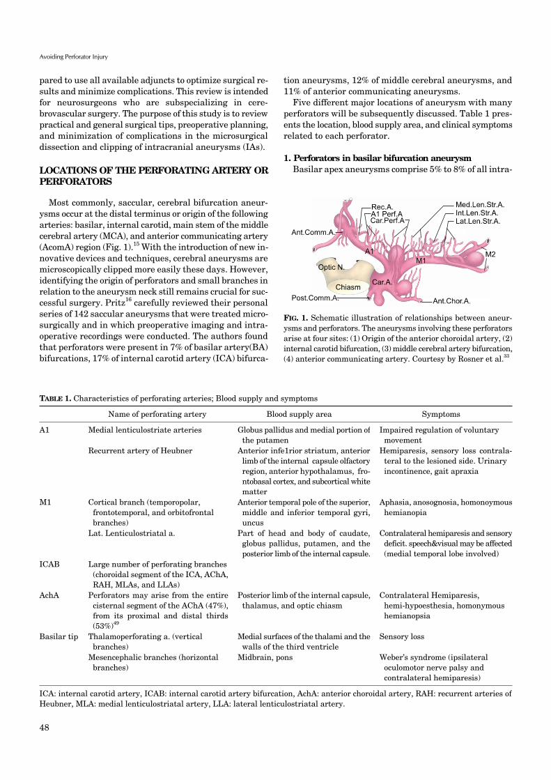
TABLE 1. Characteristics of perforating arteries; Blood supply and symptoms
Name of perforating artery Blood supply area Symptoms
A1 Medial lenticulostriate arteries Globus pallidus and medial portion ofthe putamen
Impaired regulation of voluntary movement
Recurrent artery of Heubner Anterior infe1rior striatum, anterior limb of the internal capsule olfactory region, anterior hypothalamus, fro-ntobasal cortex, and subcortical whitematter
Hemiparesis, sensory loss contrala-teral to the lesioned side. Urinary incontinence, gait apraxia
M1 Cortical branch (temporopolar, frontotemporal, and orbitofrontal branches)
Anterior temporal pole of the superior,middle and inferior temporal gyri, uncus
Aphasia, anosognosia, homonoymoushemianopia
Lat. Lenticulostriatal a. Part of head and body of caudate, globus pallidus, putamen, and the posterior limb of the internal capsule.
Contralateral hemiparesis and sensorydeficit. speech&visual may be affected(medial temporal lobe involved)
ICAB Large number of perforating branches(choroidal segment of the ICA, AChA,RAH, MLAs, and LLAs)
AchA Perforators may arise from the entire cisternal segment of the AChA (47%), from its proximal and distal thirds (53%)49
Posterior limb of the internal capsule,thalamus, and optic chiasm
Contralateral Hemiparesis, hemi-hypoesthesia, homonymous hemianopsia
Basilar tip Thalamoperforating a. (vertical branches)
Medial surfaces of the thalami and thewalls of the third ventricle
Sensory loss
Mesencephalic branches (horizontal branches)
Midbrain, pons Weber’s syndrome (ipsilateral oculomotor nerve palsy and contralateral hemiparesis)
ICA: internal carotid artery, ICAB: internal carotid artery bifurcation, AchA: anterior choroidal artery, RAH: recurrent arteries of Heubner, MLA: medial lenticulostriatal artery, LLA: lateral lenticulostriatal artery.
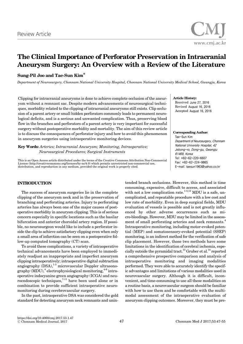
FIG. 1. Schematic illustration of relationships between aneur-ysms and perforators. The aneurysms involving these perforatorsarise at four sites: (1) Origin of the anterior choroidal artery, (2)internal carotid bifurcation, (3) middle cerebral artery bifurcation,(4) anterior communicating artery. Courtesy by Rosner et al.33
pared to use all available adjuncts to optimize surgical re-sults and minimize complications. This review is intended for neurosurgeons who are subspecializing in cere-brovascular surgery. The purpose of this study is to review practical and general surgical tips, preoperative planning, and minimization of complications in the microsurgical dissection and clipping of intracranial aneurysms (IAs).
LOCATIONS OF THE PERFORATING ARTERY OR PERFORATORS
Most commonly, saccular, cerebral bifurcation aneur-ysms occur at the distal terminus or origin of the following arteries: basilar, internal carotid, main stem of the middle cerebral artery (MCA), and anterior communicating artery (AcomA) region (Fig. 1).15 With the introduction of new in-novative devices and techniques, cerebral aneurysms are microscopically clipped more easily these days. However, identifying the origin of perforators and small branches in relation to the aneurysm neck still remains crucial for suc-cessful surgery. Pritz16 carefully reviewed their personal series of 142 saccular aneurysms that were treated micro-surgically and in which preoperative imaging and intra-operative recordings were conducted. The authors found that perforators were present in 7% of basilar artery(BA) bifurcations, 17% of internal carotid artery (ICA) bifurca-
tion aneurysms, 12% of middle cerebral aneurysms, and 11% of anterior communicating aneurysms.
Five different major locations of aneurysm with many perforators will be subsequently discussed. Table 1 pres-ents the location, blood supply area, and clinical symptoms related to each perforator.
1. Perforators in basilar bifurcation aneurysm Basilar apex aneurysms comprise 5% to 8% of all intra-
49
Sung-Pil Joo and Tae-Sun Kim
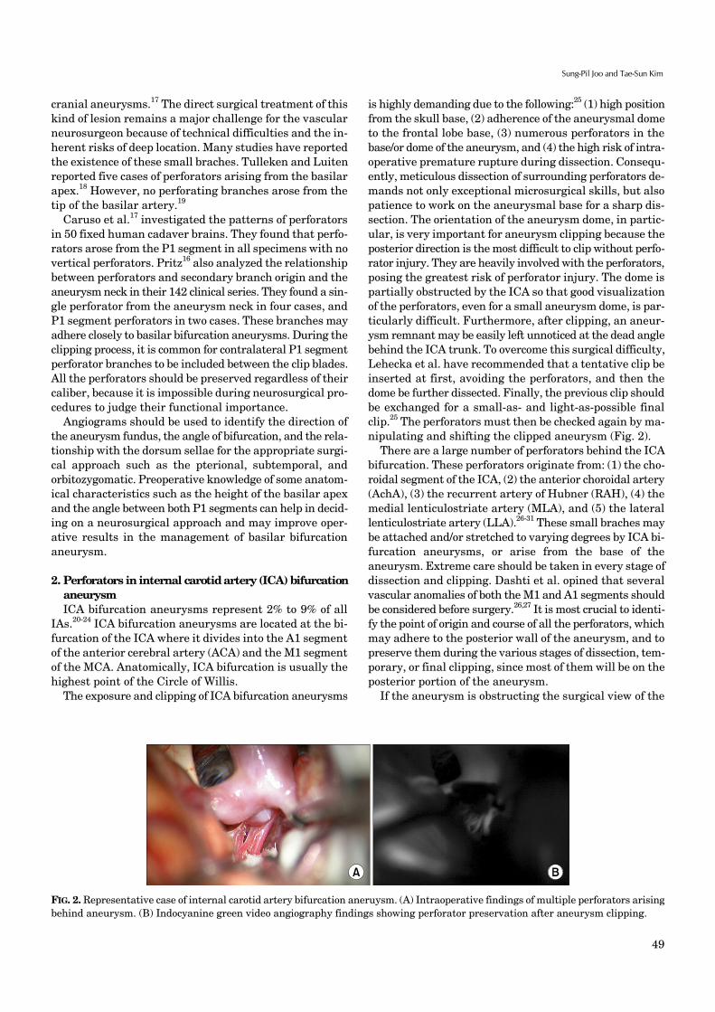
FIG. 2. Representative case of internal carotid artery bifurcation aneruysm. (A) Intraoperative findings of multiple perforators arisingbehind aneurysm. (B) Indocyanine green video angiography findings showing perforator preservation after aneurysm clipping.
cranial aneurysms.17 The direct surgical treatment of this kind of lesion remains a major challenge for the vascular neurosurgeon because of technical difficulties and the in-herent risks of deep location. Many studies have reported the existence of these small braches. Tulleken and Luiten reported five cases of perforators arising from the basilar apex.18 However, no perforating branches arose from the tip of the basilar artery.19
Caruso et al.17 investigated the patterns of perforators in 50 fixed human cadaver brains. They found that perfo-rators arose from the P1 segment in all specimens with no vertical perforators. Pritz16 also analyzed the relationship between perforators and secondary branch origin and the aneurysm neck in their 142 clinical series. They found a sin-gle perforator from the aneurysm neck in four cases, and P1 segment perforators in two cases. These branches may adhere closely to basilar bifurcation aneurysms. During the clipping process, it is common for contralateral P1 segment perforator branches to be included between the clip blades. All the perforators should be preserved regardless of their caliber, because it is impossible during neurosurgical pro-cedures to judge their functional importance.
Angiograms should be used to identify the direction of the aneurysm fundus, the angle of bifurcation, and the rela-tionship with the dorsum sellae for the appropriate surgi-cal approach such as the pterional, subtemporal, and orbitozygomatic. Preoperative knowledge of some anatom-ical characteristics such as the height of the basilar apex and the angle between both P1 segments can help in decid-ing on a neurosurgical approach and may improve oper-ative results in the management of basilar bifurcation aneurysm.
2. Perforators in internal carotid artery (ICA) bifurcation aneurysmICA bifurcation aneurysms represent 2% to 9% of all
IAs.20-24 ICA bifurcation aneurysms are located at the bi-furcation of the ICA where it divides into the A1 segment of the anterior cerebral artery (ACA) and the M1 segment of the MCA. Anatomically, ICA bifurcation is usually the highest point of the Circle of Willis.
The exposure and clipping of ICA bifurcation aneurysms
is highly demanding due to the following:25 (1) high position from the skull base, (2) adherence of the aneurysmal dome to the frontal lobe base, (3) numerous perforators in the base/or dome of the aneurysm, and (4) the high risk of intra-operative premature rupture during dissection. Consequ-ently, meticulous dissection of surrounding perforators de-mands not only exceptional microsurgical skills, but also patience to work on the aneurysmal base for a sharp dis-section. The orientation of the aneurysm dome, in partic-ular, is very important for aneurysm clipping because the posterior direction is the most difficult to clip without perfo-rator injury. They are heavily involved with the perforators, posing the greatest risk of perforator injury. The dome is partially obstructed by the ICA so that good visualization of the perforators, even for a small aneurysm dome, is par-ticularly difficult. Furthermore, after clipping, an aneur-ysm remnant may be easily left unnoticed at the dead angle behind the ICA trunk. To overcome this surgical difficulty, Lehecka et al. have recommended that a tentative clip be inserted at first, avoiding the perforators, and then the dome be further dissected. Finally, the previous clip should be exchanged for a small-as- and light-as-possible final clip.25 The perforators must then be checked again by ma-nipulating and shifting the clipped aneurysm (Fig. 2).
There are a large number of perforators behind the ICA bifurcation. These perforators originate from: (1) the cho-roidal segment of the ICA, (2) the anterior choroidal artery (AchA), (3) the recurrent artery of Hubner (RAH), (4) the medial lenticulostriate artery (MLA), and (5) the lateral lenticulostriate artery (LLA).26-31 These small braches may be attached and/or stretched to varying degrees by ICA bi-furcation aneurysms, or arise from the base of the aneurysm. Extreme care should be taken in every stage of dissection and clipping. Dashti et al. opined that several vascular anomalies of both the M1 and A1 segments should be considered before surgery.26,27 It is most crucial to identi-fy the point of origin and course of all the perforators, which may adhere to the posterior wall of the aneurysm, and to preserve them during the various stages of dissection, tem-porary, or final clipping, since most of them will be on the posterior portion of the aneurysm.
If the aneurysm is obstructing the surgical view of the
50
Avoiding Perforator Injury
perforators hidden behind the dome, it may be necessary to place the tentative clip at the aneurysm base at first to secure the space and then continue dissection for the per-forators. A small cottonoid or cellulose material may be use-ful to protect and mobilize perforators during the final dis-section at this stage. Occasionally, operators encounter sit-uations where the aneurysm dome is partially or fully bur-ied in the frontal base. The pia mater can then be opened around the aneurysm dome, and the subpial tissue can be resected to isolate the aneurysm.
3. Perforators in proximal middle cerebral artery (MCA) aneurysmProximal M1 aneurysms are located in the main trunk
of the MCA, between the bifurcation of the ICA and the main bifurcation of M1.27 Proximal MCA aneurysms are microsurgically and hemodynamically challenging. Proximal MCA aneurysms are located deep and proximally in the syl-vian cistern in a complex cisternal and vascular anatomy, where anatomical variations may affect the surgical outcome. These aneurysms are closely connected to one or more branching arteries of M1 and are often small and thin-wal-led, which make their precise clipping difficult. Proximal MCA aneurysms are uncommon. Rinne et al.32 reported that proximal M1 aneurysms represented 6% of all aneur-ysms and 16% of MCA aneurysms. Proximal MCA aneur-ysms are, in particular, often associated with other aneur-ysms in 72% in their series.32
Preservation of M1 branches is pertinent in the treat-ment for M1 aneurysms. Türe et al.31 classified M1 branch-es into: (1) the cortical branches (often named as tempor-opolar, frontotemporal, and orbitofrontal branches), and (2) the lateral lenticulostriate artery (LLA) branches. LLAs enter the brain via the central and lateral parts of the anterior perforating substance and supply the substantia innominata, putamen, globus pallidus, head and body of the caudate nucleus, internal capsule and adjacent corona radiata, and the central portion of the anterior commis-sure.31
During dissection and clipping of M1 aneurysms, the portion and pattern of exit of LLAs are of special concern because proximal M1 aneurysms may involve these perfo-rators at their branching sites. In these cases, the LLAs may be stretched, distorted, and compressed by M1 aneur-ysmal clipping.
Proximal MCA aneurysms are often wide-necked and oc-casionally, are closely connected to a M1 branch at its origin on M1; these characteristics favor open microsurgery rath-er than endovascular surgery. The direction of the parent and branching arteries and the orientation of the aneur-ysm fundus are the most important factors affecting the ef-ficacy and safety of clipping. Therefore, neurosurgeons should have a clear understanding of these three-dimen-sional (3D) relationships before surgery.
4. Perforators in proximal anterior cerebral artery (ACA) aneurysmProximal A1 aneurysms are located in the proximal seg-
ment (A1) of the ACA, between the bifurcation of the ICA and the junction of the A1 and A2 segments at the AcomA complex. The ICA bifurcates just below the anterior perfo-rating substance into 2 terminal branches, the M1 and the A1. The A1 arises from the ICA in the carotid cistern and, with a medial and somewhat anterior course, enters the cis-tern of lamina terminalis. A group of thick arachnoid bands extending from the olfactory triangle to the lateral side of the optic nerve encase the A1 segment at this point. This is important to note when dissection and mobilization of the A1 segment is required.26 The course of the A1 segment varies considerably according to its length and dominance under the frontal lobe.33 Vascular anomalies are frequently seen with proximal A1 aneurysms. Anomalies of the A1 segment include hypoplasia, aplasia, duplication, fenes-tration, and infraoptic course.34
Proximal A1 aneurysms are rare, forming less than 1% of all IAs.35-39 These aneurysms usually arise at the origin of perforators on the A1 segment and may be adherent to the perforators.35-39 Microsurgical clipping is preferable to endovascular treatment due to its small size and involve-ment of perforating arteries. Sometimes, the Proximal A1 aneurysm is of a fusiform or dissecting nature, which makes aneurysmal clipping challenging or even impossible.40-44 Many studies suggest that proximal A1 aneurysms, generally rupture at smaller sizes than other IAs in general.35-39 Consequently, unruptured A1 segment aneurysms may re-quire therapy even though they are relatively small.
The perforators of the A1 segment can be divided into two parts: (1) the MLA, and (2) RAH. The number of MLAs var-ies highly, with a mean of 8 (range, 2-15).34,45 Most MLAs arise from the proximal half of the A1 trunk. MLAs are dif-ferent from the LLAs that arise from the M1 segment.26,27 MLAs supply the septum pellucidum, the medial part of the anterior commissure and pallidum, the pillars of the fornix, the paraolfactory area, the anterior limb of the internal capsule, the anterior-inferior part of the striatum, and the anterior hypothalamus.34,45,46 Knowing the spectrum of neurologic symptoms that may occur if these perforators are injured is important.
In general, the RAH arises from the first few millimeters of the A2 segment or the A1-A2 junction, and in only about 10% of cases, it originates from the distal A1 segment.34,45,47,48 However, the point of origin of the RAH and its course in relation to the A1 trunk varies considerably. Therefore, an understanding of this variability is of primary importance when dissecting and placing temporary clips on the A1 seg-ment during dissection. The RAH supplies the anterior in-ferior striatum, anterior limb of the internal capsule, olfac-tory region, anterior hypothalamus, frontobasal cortex, and subcortical white matter.34,45-47 The point of origin and the course of the RAH should be identified to prevent dam-age during further dissection toward AcomA complex and temporary clipping on the A1 segment.
51
Sung-Pil Joo and Tae-Sun Kim
Preservation of all perforators might be impossible in fu-siform and dissecting aneurysms because a long segment of the A1 segment is involved with several perforators. In these cases, an encircling Sundt clip is used to clip the af-fected segment although there still is the probable risk of perforator injuries. Simple trapping involving segments without revascularization may cause cerebral infarction if the contralateral A1 is hypoplastic or aplastic.
5. Perforators in anterior choroidal artery (AchA) aneurysmAchA aneurysms represent 2%-5% of all intracranial
aneurysms, arising from the posterolateral wall of the su-praclinoid ICA at the origin of the AchA.49-51 The point of origint of the AchA is frequently incorporated in the base of the aneurysm and may be distorted or hidden by the aneurysm dome.50,51 Occlusion of the AchA causes severe ischemia in the posterior limb of the internal capsule, resul-ting in clinical conditions such as hemiparesis, hemisensory loss, and homonymous hemianopsia.
The AchA is divided into two segments: (1) the cisternal segment, starting from the origin of the AchA and extending into the choroidal fissure, and (2) the choroidal segment, formed by several branches passing through the choroidal fissure to the choroid plexus of the temporal horn.52,53
After its origin, the AchA has an initial lateral course in-side the carotid cistern and then follows a posteromedial course to enter the crural cistern. Passing through the wing of the ambient cistern, it travels the lateral and inferior to the optic tract, crosses the optic tract first from the 0lateral to medial, and then from the medial to lateral to enter the choroidal fissure.52-54
Branches of the AchA supply the uncus, the poster-omedial amygdala, the anterior hippocampus and the den-tate gyrus, the tail of caudate nucleus, the inferior aspect of the optic chiasm, posterior two-thirds of the optic tract, the optic radiation, medial segments of the globus pallidus, the genu and posterior limb of the internal capsule, the mid-dle one-third of the cerebral peduncle, the substantia ni-gra, the upper part of the red nucleus, a portion of the sub-thalamus, the lateral thalamic nuclei, the lateral geniculate body, and the choroid plexus of the lateral ventricle.53,55,56 The vascular territory of the AchA varies reciprocally with the supply from the posterior communicating artery, the ICA, and the MCA.33,53 To identify and preserve the AchA during different stages of dissection or temporary clipping, the neurosurgeon should be aware of these possible ana-tomic variations.
THE ROLE OF ADJUVANT MONITORING DEVICES
In recent years, MDU, intraoperative DSA, electrophy-siological monitoring, neuroendoscope, and ICGA have been used alone or in combination to provide sufficient in-traoperative neuromonitoring during cerebrovascular surgery.14
The ‘ultimate, all-in-one’ diagnostic tool has not yet been designed, but it should be simple, rapid, reliable, inex-
pensive, radiation free, noninvasive, integrated into the surgical work flow, repeatedly applicable, yet sensitive and specific for detecting aneurysm remnants on the hidden side of the vessel, even in calcified aneurysms, as well as diagnosing hemodynamically relevant major vessel steno-sis or perforating artery occlusions. Due to the various sit-uations described in many studies where one tool failed but another tool was able to diagnose a problem after clipping, it is more likely that a combination of tools will be more use-ful than even the best single tool. Thus far, there is no tool superior to intraoperative DSA for identifying a neck rem-nant in an incompletely clipped aneurysm. No tool is faster than MDU for checking whether a major arterial branch is occluded after a clip application. To make sure that the aneurysm sac is excluded from the circulation, nothing is more reliable than puncturing the sac. Inadvertent occlu-sion or kinking of perforating arteries may be best vi-sualized using ICGA loop analysis, keeping in mind that the moment of ICGA inflow is the most diagnostically effec-tive and that backflow may dye a vessel and prevent normal filling even after 1 to 2 seconds. Hidden proximal deep per-forating arteries such as branches of the AchA or the poste-rior communicating artery are possibly best diagnosed by SSEP or MEP monitoring. Yet in some cases, neuroendos-copy can visualize compromised perforating or branching arteries or, as shown by the authors, an incompletely clip-ped aneurysm. However, for a correct interpretation of postclipping ICGA, it may be required to have information available about the normal flow delay of branching arteries caused by their different lengths until they come into the visual inspection field.
It is reasonable to assess the correct clip position by se-quentially using the aforementioned methods. After clip-ping and surgical inspection, parent artery patency is rap-idly confirmed sonographically and major cerebral perfu-sion impairment is ruled out. Aneurysm occlusion, perfo-rator patency, and correct parent artery reconstruction are instantly evaluated by ICGA and intraoperative DSA, However, none of the monitoring tools used was shown to be absolutely reliable, and each device had distinct short-comings when used as a stand-alone method without intra-operative correlation of the information generated.
In conclusion, we suggest that no one intraoperative monitoring or vascular imaging technique currently has the potential to replace all others, but the methods de-scribed are complementary in nature. Moreover, a high lev-el of intraoperative safety can be provided when these tech-niques are used together.
1. Neuroendoscope The neuroendoscope has been widely used in the field of
neurosurgery, for both cerebrovascular surgery and tumor resection with various advantages. Particularly, it offers higher magnification, better observation, and additional illumination and, thus, provides information that may not be available in microscopic aneurysmal surgery. In general, the microscope only gives us a straight line of sight and re-
52
Avoiding Perforator Injury
sults in inadequate observation of structures that may lie behind other structures.57 To overcome the above limita-tions, many neurosurgeons have recommended neuro-endoscopy for aneurysm surgeries. Through this proce-dure, surgeons can detect hidden structures, dissect perfo-rators at the back of aneurysm and check for complete clip-ping of aneurysms. Since Apuzzo et al. adopted the side-an-gled rigid endoscope for surgery,58 its usefulness has been reported in compensating for the shortcomings of the direct microscope.7-9,59-64 In summary, the advantages of the neu-roendoscope are as follows: (1) increased light intensity while approaching an object, (2) clear depiction of details in close-up positions, and (3) extended viewing angles.65,66 The position of the clip blades in relation to perforating branches in the vicinity of the aneurysm neck can be as-sessed endoscopically in selected cases.
The efficacy of endoscope-assisted aneurysmal clipping depends on the location of the aneurysm. The neuroen-doscope is especially helpful in the identification of deep-seated aneurysms. However, several shortcomings of the neuroendoscope have been reported as follows:7 (1) the endoscope can cause aneurysmal rupture during entry or inspection, especially in cases of large or giant aneurysms, (2) the endoscope is helpless when there are blood clots in the operating field and clots have to be irrigated and re-moved, and (3) there is still a lack of sufficient equipment specially designed for endoscopic surgery.
2. Indocyanine green video angiography (ICGA)Intraoperative ICG-VA has been widely used in aneur-
ysm surgery to confirm the complete occlusion of the aneur-ysm and for the precise assessment of small perforating ar-teries in the vicinity of the aneurysm neck.4,5,67-71 This method offers a real time assessment of the blood flow of vasculatures and reduces operative morbidity related to vascular occlusion after aneurysmal clipping, thus, in-creasing the number of positive outcomes for aneurysmal surgery.57 However, the major limitation of ICGA is that the microscope must have a direct line-of-sight to the region of interest, which might hamper the identification of pin-ched hidden arteries as well as remnants at the averted aneurysm neck.
Mielke et al.13 in their clinical series, applied a prototype endoscope for the visualization of ICG fluorescence in hid-den regions of the microsurgical field and evaluated its po-tential usefulness compared with microscopic ICGA. They suggested that endoscopic ICGA was capable of emerging as a useful adjunct to aneurysm surgery and had the poten-tial to further improve operative results. Yoshioka and Kinouchi57 found that endoscopic ICGA was especially use-ful in ICA-PcomA aneurysm cases for the observation of the blood flow at the origins of the perforators behind the pa-rent arteries and aneurysms. Thus, combination endoscopy and microscopic guidance may reveal important informa-tion hidden from the microscope, and may be used to verify the status of the operative process under direct visualiza-tion.13,57
3. Microvascular Doppler ultrasonography (MDU)MDU extended the information derived from surgical
exploration after aneurysm clipping and provided repeat-able information about parent artery patency. Despite ex-tensive effort to preserve patency at the time of the clip ap-plication, intraoperative 3D visual inspection of the entire vascular complex may not reveal arterial compromise or occlusion. An intraoperative MDU has been used for this purpose for the past few years.72-74
The usefulness of MDU is for detecting the intravascular flow and patency, as well as the decrease or increase in CBF through the parent arteries for vascular compromise. For example, after clipping of the aneurysm with an athero-sclerotic neck, the vessel contour itself looks normal and shows no compromise, microscopic ICGA can show the in-travascular flow, and MEP/SSEP cannot detect a decrease in CBF. However, the patient can manifest ischemic symptoms. So, quantitative monitoring of flow patency is also impor-tant, and MDU can be useful in evaluating quantitative as well as qualitative intravascular flow. Intraoperative MDU has an advantage as it assists surgeons in making difficult intraoperative decisions, especially, when a compromise is necessary between complete obliteration of the aneurysm neck and maintenance of adequate parent and branching vessel patency. Therefore, MDS is a safe, feasible, and very reliable technique and should be used routinely in intra-cranial aneurysm surgery. The major limitation of this method is as follows: (1) inability to detect the persistence of a minimal blood flow after clipping, (2) aneurysm rem-nants or a residual neck may go unnoticed, and (3) Events after completing the IMD evaluation may potentially lead to a vessel stenosis and an ischemia.75
4. Electrophysiological monitoringThe many advances in motor system assessment ach-
ieved in the last two decades have undoubtedly improved monitoring efficacy without unduly compromising safety. Although the aforementioned intraoperative vascular im-aging techniques are used to directly visualize the cerebral arteries of interest, electrophysiological neuromonitoring can be considered a method for permanent background surveillance. SSEP and MEP monitoring have provided functional information affecting the surgical strategy. This information is crucial and may change the surgical strategies employed. However, we still need to explore the usefulness and problems of intraoperative monitoring such as MEP and SSEP to evaluate brain dysfunction caused by an insufficiency of cerebral blood flow. Since it is difficult to predict the postoperative ischemic complica-tions despite evaluating the motor function via MEP mon-itoring in some cases.76 Future studies and experience will likely clarify existing controversies and bring further advances.
CONCLUSIONS
To prevent kinking or occlusion of adjacent and/or perfo-
53
Sung-Pil Joo and Tae-Sun Kim
rating branches, the smallest possible while still sufficient final clip should be applied to prevent perforator injury. A proper selection of clips with different shapes and lengths of blades, and applicators, suiting each aneurysmal config-uration, should be ready for use. Moreover, adjuvant mon-itoring devices and various types of dissectors for perfo-rator preservation should be used appropriately.
ACKNOWLEDGEMENTS
This study was supported by a grant (CRI 13902-21) Chonnam National University Hospital Biomedical Rese-arch Institute.
CONFLICT OF INTEREST STATEMENT
None declared.
REFERENCES
1. Barrow DL, Boyer KL, Joseph GJ. Intraoperative angiography in the management of neurovascular disorders. Neurosurgery 1992;30:153-9.
2. Siasios I, Kapsalaki EZ, Fountas KN. The role of intraoperative micro-Doppler ultrasound in verifying proper clip placement in intracranial aneurysm surgery. Neuroradiology 2012;54:1109-18.
3. Washington CW, Zipfel GJ, Chicoine MR, Derdeyn CP, Rich KM, Moran CJ, et al. Comparing indocyanine green videoangiography to the gold standard of intraoperative digital subtraction angiog-raphy used in aneurysm surgery. J Neurosurg 2013;118:420-7.
4. de Oliveira JG, Beck J, Seifert V, Teixeira MJ, Raabe A. Asse-ssment of flow in perforating arteries during intracranial aneur-ysm surgery using intraoperative near-infrared indocyanine green videoangiography. Neurosurgery 2007;61(3 Suppl):63-72; discussion 72-3.
5. Raabe A, Beck J, Gerlach R, Zimmermann M, Seifert V. Near-in-frared indocyanine green video angiography: a new method for in-traoperative assessment of vascular flow. Neurosurgery 2003; 52:132-9; discussion 139.
6. Fischer G, Stadie A, Oertel JM. Near-infrared indocyanine green videoangiography versus microvascular Doppler sonography in aneurysm surgery. Acta Neurochir (Wien) 2010;152:1519-25.
7. Kalavakonda C, Sekhar LN, Ramachandran P, Hechl P. Endo-scope-assisted microsurgery for intracranial aneurysms. Neuro-surgery 2002;51:1119-26; discussion 1126-7.
8. Kinouchi H, Yanagisawa T, Suzuki A, Ohta T, Hirano Y, Sugawara T, et al. Simultaneous microscopic and endoscopic monitoring during surgery for internal carotid artery aneurysms. J Neurosurg 2004;101:989-95.
9. Taniguchi M, Takimoto H, Yoshimine T, Shimada N, Miyao Y, Hirata M, et al. Application of a rigid endoscope to the micro-surgical management of 54 cerebral aneurysms: results in 48 patients. J Neurosurg 1999;91:231-7.
10. Kato Y, Sano H, Nagahisa S, Iwata S, Yoshida K, Yamamoto K, et al. Endoscope-assisted microsurgery for cerebral aneurysms. Minim Invasive Neurosurg 2000;43:91-7.
11. van Lindert E, Perneczky A, Fries G, Pierangeli E. The supra-
orbital keyhole approach to supratentorial aneurysms: concept and technique. Surg Neurol 1998;49:481-9; discussion 489-90.
12. Martin NA, Bentson J, Viñuela F, Hieshima G, Reicher M, Black K, et al. Intraoperative digital subtraction angiography and the surgical treatment of intracranial aneurysms and vascular malformations. J Neurosurg 1990;73:526-33.
13. Mielke D, Malinova V, Rohde V. Comparison of intraoperative mi-croscopic and endoscopic ICG angiography in aneurysm surgery. Neurosurgery 2014;10 Suppl 3:418-25; discussion 425.
14. Gruber A, Dorfer C, Standhardt H, Bavinzski G, Knosp E. Prospective comparison of intraoperative vascular monitoring technologies during cerebral aneurysm surgery. Neurosurgery 2011;68:657-73; discussion 673.
15. Pritz MB. Cerebral aneurysm classification based on angioarchi-tecture. J Stroke Cerebrovasc Dis 2011;20:162-7.
16. Pritz MB. Perforator and secondary branch origin in relation to the neck of saccular, cerebral bifurcation aneurysms. World Neurosurg 2014;82:726-32.
17. Caruso G, Vincentelli F, Giudicelli G, Grisoli F, Xu T, Gouaze A. Perforating branches of the basilar bifurcation. J Neurosurg 1990;73:259-65.
18. Tulleken CA, Luiten ML. The basilar artery bifurcation: micro-scopical anatomy. Acta Neurochir (Wien) 1987;85:50-5.
19. Pedroza A, Dujovny M, Ausman JI, Diaz FG, Cabezudo Artero J, et al. Microvascular anatomy of the interpeduncular fossa. J Neurosurg 1986;64:484-93.
20. Da Pian R, Pasqualin A, Scienza R. Direct microsurgical approach to aneurysms of the internal carotid bifurcation. Surg Neurol 1980;13:27-37.
21. Kyoshima K, Kobayashi S, Nitta J, Osawa M, Shigeta H, Nakagawa F. Clinical analysis of internal carotid artery aneur-ysms with reference to classification and clipping techniques. Acta Neurochir (Wien) 1998;140:933-42.
22. Laranjeira M, Sadasivan B, Ausman JI. Direct surgery for carotid bifurcation artery aneurysms. Surg Neurol 1990;34:250-4.
23. Miyazawa N, Nukui H, Horikoshi T, Yagishita T, Sugita M, Kanemaru K. Surgical management of aneurysms of the bifurca-tion of the internal carotid artery. Clin Neurol Neurosurg 2002;104:103-14.
24. van Rooij WJ, Sluzewski M, Beute GN. Internal carotid bifurca-tion aneurysms: frequency, angiographic anatomy and results of coiling in 50 aneurysms. Neuroradiology 2008;50:583-7.
25. Lehecka M, Dashti R, Romani R, Celik O, Navratil O, Kivipelto L, et al. Microneurosurgical management of internal carotid ar-tery bifurcation aneurysms. Surg Neurol 2009;71:649-67.
26. Dashti R, Hernesniemi J, Lehto H, Niemelä M, Lehecka M, Rinne J, et al. Microneurosurgical management of proximal anterior cerebral artery aneurysms. Surg Neurol 2007;68:366-77.
27. Dashti R, Rinne J, Hernesniemi J, Niemelä M, Kivipelto L, Lehecka M, et al. Microneurosurgical management of proximal middle cerebral artery aneurysms. Surg Neurol 2007;67:6-14.
28. Gibo H, Lenkey C, Rhoton AL Jr. Microsurgical anatomy of the supraclinoid portion of the internal carotid artery. J Neurosurg 1981;55:560-74.
29. Marinković S, Gibo H, Milisavljević M. The surgical anatomy of the relationships between the perforating and the leptomeningeal arteries. Neurosurgery 1996;39:72-83.
54
Avoiding Perforator Injury
30. Marinković SV, Milisavljević MM, Marinković ZD. The perforat-ing branches of the internal carotid artery: the microsurgical anatomy of their extracerebral segments. Neurosurgery 1990; 26:472-8; discussion 478-9.
31. Türe U, Yaşargil MG, Al-Mefty O, Yaşargil DC. Arteries of the insula. J Neurosurg 2000;92:676-87.
32. Rinne J, Hernesniemi J, Niskanen M, Vapalahti M. Analysis of 561 patients with 690 middle cerebral artery aneurysms: anatom-ic and clinical features as correlated to management outcome. Neurosurgery 1996;38:2-11.
33. Rosner SS, Rhoton AL Jr, Ono M, Barry M. Microsurgical anatomy of the anterior perforating arteries. J Neurosurg 1984;61:468-85.
34. Perlmutter D, Rhoton AL Jr. Microsurgical anatomy of anterior cerebral anterior communicating recurrent artery complex. Surg Forum 1976;27:464-5.
35. Handa J, Nakasu Y, Matsuda M, Kyoshima K. Aneurysms of the proximal anterior cerebral artery. Surg Neurol 1984;22:486-90.
36. Hino A, Fujimoto M, Iwamoto Y, Oka H, Echigo T. Surgery of prox-imal anterior cerebral artery aneurysms. Acta Neurochir (Wien) 2002;144:1291-6; discussion 1296.
37. Suzuki M, Onuma T, Sakurai Y, Mizoi K, Ogawa A, Yoshimoto T. Aneurysms arising from the proximal (A1) segment of the ante-rior cerebral artery. A study of 38 cases. J Neurosurg 1992;76:455-8.
38. Wakabayashi T, Tamaki N, Yamashita H, Saya H, Suyama T, Matsumoto S. Angiographic classification of aneurysms of the horizontal segment of the anterior cerebral artery. Surg Neurol 1985;24:31-4.
39. Wanibuchi M, Kurokawa Y, Ishiguro M, Fujishige M, Inaba K. Characteristics of aneurysms arising from the horizontal portion of the anterior cerebral artery. Surg Neurol 2001;55:148-54; dis-cussion 154-5.
40. Lee JM, Joo SP, Kim TS, Go EJ, Choi HY, Seo BR. Surgical man-agement of anterior cerebral artery aneurysms of the proximal (A1) segment. World Neurosurg 2010;74:478-82.
41. Evans AL, Corkill RA, Wenderoth JD. Ruptured fusiform aneur-ysm of fenestrated A1 segment of the anterior cerebral artery. Case report and review of the literature. Neuroradiology 2006; 48:196-9.
42. Hirao J, Okamoto H, Watanabe T, Asano S, Teraoka A. Dissecting aneurysms at the A1 segment of the anterior cerebral artery--two case reports. Neurol Med Chir (Tokyo) 2001;41:271-8.
43. Kashimura H, Mase T, Ogasawara K, Ogawa A, Endo H. Trapping and vascular reconstruction for ruptured fusiform aneurysm in the proximal A1 segment of the anterior cerebral artery. Neurol Med Chir (Tokyo) 2006;46:340-3.
44. Nomura M, Kida S, Kita D, Higashi R, Hasegawa M, Matsui O, et al. Fusiform aneurysm of the proximal anterior cerebral artery (A1). Acta Neurochir (Wien) 2000;142:1163-4.
45. Dunker RO, Harris AB. Surgical anatomy of the proximal ante-rior cerebral artery. J Neurosurg 1976;44:359-67.
46. Ostrowski AZ, Webster JE, Gurdjian ES. The proximal anterior cerebral artery: an anatomic study. Arch Neurol 1960;3:661-4.
47. Gomes F, Dujovny M, Umansky F, Ausman JI, Diaz FG, Ray WJ, et al. Microsurgical anatomy of the recurrent artery of Heubner. J Neurosurg 1984;60:130-9.
48. Tulleken CA. A study of the anatomy of the anterior communicat-ing artery with the aid of the operating microscope. Clin Neurol
Neurosurg 1978;80:169-73.49. Drake CG, Vanderlinden RG, Amacher AL. Carotid-choroidal
aneurysms. J Neurosurg 1968;29:32-6.50. Friedman JA, Pichelmann MA, Piepgras DG, Atkinson JL, Maher
CO, Meyer FB, et al. Ischemic complications of surgery for ante-rior choroidal artery aneurysms. J Neurosurg 2001;94:565-72.
51. Yasargil MG, Yonas H, Gasser JC. Anterior choroidal artery aneurysms: their anatomy and surgical significance. Surg Neurol 1978;9:129-38.
52. Erdem A, Yaşargil G, Roth P. Microsurgical anatomy of the hippo-campal arteries. J Neurosurg 1993;79:256-65.
53. Rhoton AL Jr, Fujii K, Fradd B. Microsurgical anatomy of the an-terior choroidal artery. Surg Neurol 1979;12:171-87.
54. Fujii K, Lenkey C, Rhoton AL Jr. Microsurgical anatomy of the choroidal arteries. Fourth ventricle and cerebellopontine angles. J Neurosurg 1980;52:504-24.
55. Marinković S, Gibo H, Brigante L, Nikodijević I, Petrović P. The surgical anatomy of the perforating branches of the anterior cho-roidal artery. Surg Neurol 1999;52:30-6.
56. Saeki N, Rhoton AL Jr. Microsurgical anatomy of the upper basi-lar artery and the posterior circle of Willis. J Neurosurg 1977;46:563-78.
57. Yoshioka H, Kinouchi H. The roles of endoscope in aneurysmal surgery. Neurol Med Chir (Tokyo) 2015;55:469-78.
58. Apuzzo ML, Heifetz MD, Weiss MH, Kurze T. Neurosurgical en-doscopy using the side-viewing telescope. J Neurosurg 1977;46: 398-400.
59. Cohen AR, Perneczky A, Rodziewicz GS, Gingold SI. Endos-cope-assisted craniotomy: approach to the rostral brain stem. Neurosurgery 1995;36:1128-9; discussion 1129-30.
60. Fischer G, Oertel J, Perneczky A. Endoscopy in aneurysm surgery. Neurosurgery 2012;70(2 Suppl Operative):184-90; dis-cussion 190-1.
61. Fischer J, Mustafa H. Endoscopic-guided clipping of cerebral aneurysms. Br J Neurosurg 1994;8:559-65.
62. Galzio RJ, Di Cola F, Raysi Dehcordi S, Ricci A, De Paulis D. En-doscope-assisted microneurosurgery for intracranial aneurysms. Front Neurol 2013;4:201.
63. Profeta G, De Falco R, Ambrosio G, Profeta L. Endoscope-assisted microneurosurgery for anterior circulation aneurysms using the angle-type rigid endoscope over a 3-year period. Childs Nerv Syst 2004;20:811-5.
64. Wang E, Yong NP, Ng I. Endoscopic assisted microneurosurgery for cerebral aneurysms. J Clin Neurosci 2003;10:174-6.
65. Fries G, Perneczky A. Endoscope-assisted brain surgery: part 2--analysis of 380 procedures. Neurosurgery 1998;42:226-31; dis-cussion 231-2.
66. Perneczky A, Fries G. Endoscope-assisted brain surgery: part 1--evolution, basic concept, and current technique. Neurosurgery 1998;42:219-24; discussion 224-5.
67. Kuroda K, Kinouchi H, Kanemaru K, Nishiyama Y, Ogiwara M, Yoshioka H, et al. Intra-arterial injection fluorescein video-angiography in aneurysm surgery. Neurosurgery 2013;72(2 Suppl Operative):ons141-50; discussion ons150.
68. Raabe A, Nakaji P, Beck J, Kim LJ, Hsu FP, Kamerman JD, et al. Prospective evaluation of surgical microscope-integrated in-traoperative near-infrared indocyanine green videoangiography

55
Sung-Pil Joo and Tae-Sun Kim
during aneurysm surgery. J Neurosurg 2005;103:982-9.69. Yoshioka H, Kinouchi H, Nishiyama Y, Kanemaru K, Yagi T,
Hanihara M, et al. Advantage of microscope integrated for both indocyanine green and fluorescein videoangiography on aneur-ysmal surgery: case report. Neurol Med Chir (Tokyo) 2014;54:192-5.
70. Wrobel CJ, Meltzer H, Lamond R, Alksne JF. Intraoperative as-sessment of aneurysm clip placement by intravenous fluorescein angiography. Neurosurgery 1994;35:970-3; discussion 973.
71. Suzuki K, Kodama N, Sasaki T, Matsumoto M, Ichikawa T, Munakata R, et al. Confirmation of blood flow in perforating ar-teries using fluorescein cerebral angiography during aneurysm surgery. J Neurosurg 2007;107:68-73.
72. Gilsbach JM, Harders AG. Microvascular and transcranial Doppler sonographic evaluation of cerebral aneurysm flow
pattern. Neurol Res 1989;11:41-8.73. Gilsbach JM, Hassler WE. Intraoperative Doppler and real time
sonography in neurosurgery. Neurosurg Rev 1984;7:199-208.74. Marchese E, Albanese A, Denaro L, Vignati A, Fernandez E,
Maira G. Intraoperative microvascular Doppler in intracranial aneurysm surgery. Surg Neurol 2005;63:336-42; discussion 342.
75. Cui H, Wang Y, Yin Y, Wan J, Fei Z, Gao W, et al. Role of intraop-erative microvascular Doppler in the microsurgical management of intracranial aneurysms. J Clin Ultrasound 2011;39:27-31.
76. Ishizaki T, Endo O, Fujii K, Matsudaira T, Okada T, Kobayashi N, et al. Usefulness and problems of intraoperative monitoring for unruptured aneurysm surgery with the motor evoked potential. No Shinkei Geka 2016;44:283-93.
Recommended