Bulgarian Journal of Veterinary Medicine, 2014, 17, No 1, 5060 ISSN 1311-1477; online at http://tru.uni-sz.bg/bjvm/bjvm.htm
Original article
STABILITY OF INTRAOSSEOUS DENTAL IMPLANTS WITH GUIDED BONE REGENERATION
(IN VIVO EXPERIMENT IN DOGS)
J. INDJOVA1, KH. FAKIH1, D. SIVREV2, D. YOVCHEV3
& TS. CHAPRAZOV4
1Department of Oral and Maxillofacial Surgery, 3Department of Imaging and Oral Diagnostics, Faculty of Dental Medicine, Medical University, Sofia,
Bulgaria; 2Department of Anatomy, Medical Faculty, Trakia University, Stara Zagora; 4Department of Veterinary Surgery, Faculty of Veterinary Medicine,
Trakia University, Stara Zagora; Bulgaria
Summary
Indjova, J., Kh. Fakih, D. Sivrev, D. Yovchev & Ts. Chaprazov, 2014. Stability of intraosse-ous dental implants with guided bone regeneration (in vivo experiment in dogs). Bulg. J. Vet. Med., 17, No 1, 5060. Three different protocols for dental implantation: immediate (ImI), delayed (DI) and conventional (CI) were used. The primary stability is important for the secondary stability of implants. The purpose of the experiments was to monitor the stability of ImI, DI and CI following guided bone regeneration (GBR). A total of 18 implants were placed in the alveoli of 9 extracted premolars of the lower jaw of three mixed breed dogs. GBR was performed with a combination of Bio Oss® and Emdogain® and the Bio Gide® membrane. Control GBR was performed with coagulum and the same membrane. The stability of the implants was measured with Osstell® ISQ. The primary stability of the three types of implants was high, and differed statistically significantly (P<0.05). By the end of the first month after the placement, the stability of ImI and DI was reduced. The secondary stability of ImI and DI increased substantially (P<0.05) by the end of the third month both with regard to primary stability and stability by the end of the first month. The primary stability of intraosseous implants was a prerequisite for a high secondary stability. The decline in stability by the end of the first month after implantation was not an obstacle to achieve a high secondary stability.
Key words: guided bone regeneration, implants, stability, xenografts
INTRODUCTION
Stability is the most concise but accurate enough term to describe a successful treat-ment using intraosseous implants. The biological substrate is of primary signifi-cance for implant stability. This however,
does not underestimate implant design and coating or operative precision. The latter are not part of this research's incentives.
The stability of an implant is primary and secondary. The primary, initial stabi-

J. Indjova, Kh. Fakih, D. Sivrev, D. Yovchev & Ts. Chaprazov
BJVM, 17, No 1 51
lity (at the moment of implant placement) is of mechanical nature. It is due to the incorporation of the implant in the jaw bone and largely depends on the cortical bone. Secondary stability is a function of repair process beginning after implant placement. It is biologically determined and associated mainly with cancellous bone and cancellous bone events (Atsumi et al., 2007). After the implant is loaded, the bone regenerates and undergoes remo-delling with regard to implant surface connection and proper osteointegration (Atsumi et al., 2007). Primary or mecha-nical stability is the necessary condition for occurrence and development of secon-dary or biological stability (Sennerby & Roos, 1998). The mechanical stability and osteointegration (being a sequel to prima-ry and measure of secondary implant stability) are mainly a function of several quantitative and quality features of jaw bone, such as bone volume and bone structure – bone density, cortical thickness (Roze et al., 2009).
A number of methods for preservation and/or augmentation of jaw bone for intraosseous implant placement are avai-lable. Some examples are the atraumatic tooth extraction and GBR with osseous coagulum and barrier membrane, or a combination of bone graft and membrane. A number of bone grafts have been used. Bio Oss is one of the most extensively explored xenografts with acknowledged osteoconduction properties (Baldini et al., 2011). A number of attempts were ac-complished for improvement of its mani-pulation and biological properties through combinations with fibrin sealant (Carmag-nola et al., 2000), autogenous bone (Hall-man et al., 2002). We have previously reported that the GBR with a mixture of Bio Oss and Emdogain resulted in a more significant lamellar bone accumulation
compared to the independent use of Bio Oss + Emdogain (Indjova et al., 2013). No data are however available whether this combination applied via GBR, influ-ences the stability of implants depending on the implantation protocol.
Three methods for dental implantation were used – immediate, delayed/immedia-tely delayed and conventional.
The immediate placement of imp-lant(s) was performed immediately after tooth extraction. It shortened the treatment by 3 to 6 months after extraction and was reported to restrict alveolar bone resorp-tion (Bhola et al., 2008).
The contact between implant surface and extraction socket ensures a better osteointegration (Lundgren et al., 1992). Clinical practice has evidenced that in immediate implantation, the mismatch of the shape of the extraction socket and implant shape often resulted in a slit around different parts of the implant – platform, neck, body. Depending on its vertical and horizontal dimensions, dela-yed or absent bone regeneration, epitheli-um migrate into the slit, difficulties in obtaining reliable primary stability are present and thus, osteointegration is post-poned, does not occur or is faulty (Sca-rano et al., 2006). In such instances, GBR with graft and/or membrane confines the growth of epithelial and connective tissue structures in the slit (Lang et al., 1994).
Mainly marginal slits with horizontal dimensions of 1–4 mm have been studied (Wilson et al., 1998). Opinions are conf-licting – some assume that spontaneous repair is possible for defects with horizon-tal dimensions of up to 2.5 mm (Botticelli et al., 2004) while others affirm that GBR should be applied (Wilson et al., 1998). Thus, the problem for treatment of defects with horizontal dimensions over 3 mm remains still open.
Stability of intraosseous dental implants with guided bone regeneration (in vivo experiment in dogs)
BJVM, 17, No 1 52
The investigations on the marginal slit and the lack of regular contact between implant surface and alveolar wall were mainly aimed at possibilities for spontane-ous or guided regeneration (West et al. 2007). Apart the regenerative resp. histo-morphological aspect of the problem, the monitoring and the evaluation of stability of implants in extraction sockets following GBR are also essential for the clinical practice.
For the other two approaches for imp-lantation – immediately delayed and con-ventional which takes place 4 to 8 weeks and at least 3 months after tooth extrac-tion, respectively, the problems in achie-ving primary stability are related to resul-ting reduction of alveolar bone volume. For keeping of volume, either atraumatic extraction and/or GBR with membrane or membrane + graft are used (Atnoun et al. 2007).
Along with the necessity for a biolo-gical substrate of adequate amount and quality, intraosseous implants also require objective measurement of their stability. Techniques for implant stability measure-ment are a valuable reference point of occurring peri-implant bone repair events before the placement of the abutment and the prosthesis. Various tests and methods for implant stability assessment are ap-plied. Some of them, i.e. the percussion test, are subjective, others do not allow stability monitoring (torque test, cutting torque resistance test) and a third group including the reverse torque test, are destructive and inapplicable in a clinical setting (Meredith, 1998;Atsumi et al., 2007; Sennerby & Meredith, 2008).
For the needs of clinical practice the applied method should be objective, quantitative, rapid, and suitable for use in clinical conditions, non-invasive, non-destructive and atraumatic with regard to
the bone-implant interface. A method responding to all these requirements is the resonance frequency analysis (RFA) (Al-Nawas et al., 2007). The force applied on implants has precisely defined parameters, equal for each measurement. The subjec-tive element is completely absent. The sensitivity of the last generation Osstell®
ISQ, which uses magnetic pulses is eva-luated as very high (Sennerby & Mere-dith, 2008). It allows measuring the stabi-lity of implants.
The information about objective com-parative monitoring of implant stability placed in tooth sockets with different substrates for GBR through one of afore-mentioned methods is scarce.
The purpose of the present experiment was to follow up the time course of stability after placement of immediate, delayed and conventional implants in a dog model.
MATERIALS AND METHODS
The experiments were conducted in the Veterinary dental cabinet, Department of Veterinary Surgery, Faculty of Veterinary Medicine at the Trakia University – Stara Zagora.
All surgical interventions were per-formed in strict compliance with the Law on Veterinary Medical Activities, Ordi-nance 25/10.06.2005, and Directive 2010/ 63/EU on the protection of animals used for scientific purposes. Three mixed-breed dogs, aged 1 to 3 years were included. The initial health status of dogs was evaluated as good, and the dentition – intact. The experiments, part of Grant No. 17/2013, Medical University – Sofia were approved as compliant to national and international ethical standards regarding animal experimentation (decision of the Research Ethics Commission to the

J. Indjova, Kh. Fakih, D. Sivrev, D. Yovchev & Ts. Chaprazov
BJVM, 17, No 1 53
Medical University – Sofia, protocol 5/17.04.2013).
Preoperative preparation
Twenty-four hours before the surgery, each dog received an intramuscular injection of amoxicillin and clavulanic acid (Synulox, Pfizer Animal Health, UK) at a dose of 8.75 mg/kg. Premedication was done with 0.02 mg/kg atropine sulfate (Atropin, Sopharma Ltd, Sofia, Bulgaria). The induction of anaesthesia was done 15 min later i. v. with 0.5 mg/kg diazepam (Diazepam, Sopharma Ltd, Sofia, Bulga-ria) and 10 mg/kg ketamine hydrochloride (Anaket 10%, Richter Pharma AG, Aust-ria). Maintenance of inhalation anaesthe-sia was performed with 1.2–3 vol% iso-flurane (AErrane Inhalation vapour, li-quid, Baxter d.o.o. Ljubljana, Slovenia).
Intraoral preoperative preparation
Immediately before the operation, the site was aseptically prepared by thorough cleaning of dental surfaces with 3% hyd-rogen peroxide and profuse washing with water, as well as scrubbing the mucosa and dental surfaces with povidone iodine (Iodseptadon 10%).
Surgical interventions
All operative interventions were perfor-med aseptically by the same surgeon. In each dog, atraumatic extraction of the 3rd and 4th premolars on one side and the 4th premolar on the contralateral side of the mandible was done after mucoperiosteal flap preparation. After teeth extraction, three pairs of sockets were obtained in each dog. For the first socket, regenera-tion of defects occurred with osseous coagulum and for the other – with xeno-grafts (Table 1). In each dog, two im-mediate (ImI – Fig. 1), two delayed (DI – Fig. 2) and two conventional (CI – Fig. 3) implants were placed at the proper time. All implants were of conical shape and equal dimensions: 4.2 mm diameter and 8 mm length (Alfa Gate Bioactive SCI 842). The total number of implants was 18 (6 per dog). Despite the purposefully selec-ted conical shape of implants similar to that of tooth roots, a marginal slit up to 1 mm wide and up to 1.5 mm deep occurred between immediate implants and buccal and lingual alveolar wall, respectively. With a bone cutter, a part of the lingual wall of sockets for immediate implants and GBR with xenografts and membrane
Table 1. Distribution of implants and measurements depending on the implantation approach and guided bone regeneration
Implantation approach
Immediate (immediate implants)
Depayed-immediate (delayed implants)
Conventional (conventional implants)
Guided bone regeneration
Bio Oss + Emdogain+
Bio Gide
Coagulum + Bio Gide
Bio Oss + Emdogain+
Bio Gide
Coagulum + Bio Gide
Bio Oss + Emdogain+
Bio Gide
Coagulum + Bio Gide
Number of implants
3 3 3 3 3 3
Number of measurements
9 9 9 9 9 9

Stability of intraosseous dental implants with guided bone regeneration (in vivo experiment in dogs)
BJVM, 17, No 1 54
was removed (Table 1). Thus, bone de-fects with horizontal size of 3–4 mm and depth 4 mm (measured with graduated
Fig. 1. Immediate implants.
Fig. 2. Delayed implants.
Fig. 3. Conventional implants.
probe, compass and ruler) were formed. The artificially enlarged slits were filled with a mixture of two xenografts – Bio Oss® (Geistlich Pharma AG) and Emdo-gain® (Straumann Emdogain®, Institut Staumann AG, Bazel/Switzerland).
The placed ImI together with the artificial bone defect were covered with resorbable membrane Bio Gide® (Geist-lich Pharma AG). In one socket of the other two pairs of extraction sockets, a mixture of xenografts was placed, and the other remained filled with coagulum. All sockets were covered with resorbable membrane as per GBR principles. One month after the extraction, delayed imp-lants were placed (two for each dog – one in an alveolus using GBR with coagulum; the other in a socket using GBR with aforementioned xenografts. Three months after extraction of premolars, the last two (conventional) implants were placed. For all three implantation protocols, each of the six implants was placed approximately in the middle of the respective socket at the level of vestibular cortical bone. Co-ver screws were placed on all implants. The flaps were replaced and sutured with interrupted non-absorbable sutures 4/0 Medicon ex to ensure a coated healing of implants and extraction sockets with GBR.
Postoperative care
Over 10 days after each surgery, each dog was i.m. treated with antibiotic Synulox (Pfizer Animal Health, UK) at a dose of 8.75 mg/kg. During the first 2 post ope-rative days, dogs drank only water, and then were fed soft food for another 10 days. Oral hygiene was maintained until sutures removal by the 14th post operative day.
Implant stability measurements
The stability of dental implants was eva-luated by the newest version of the reso-

J. Indjova, Kh. Fakih, D. Sivrev, D. Yovchev & Ts. Chaprazov
BJVM, 17, No 1 55
nance frequency analyzator Osstell® ISQ (Osstell MentorTM Integration diagnos-tiсs; Osstell AB, Göteborg, Sweden). In this unit, the pulses are generated by a probe with built-in magnetic elements and are detected by a metal rod (SmartPeg), which is screwed on the implant and receives emitted signals (Fig. 4). Magnetic pulses are of 1 ms duration. They cause the SmartPeg to resonate. The effect is identical to horizontal loads borne by the functioning implant. The waves reflected by the resonating metal rod are detected by the measuring probe and displayed in numeric values by the instrument. The values reflect implant stability quotient
Fig. 4. Osstell ISQ probe and a SmartPeg mounted on the implant.
Fig. 5. Osstell ISQ instrument with a measured ISQ value on its display.
(ISQ) values (Sennerby & Meredith, 2008), which are standardised to provide a numerical expression of implant stability within a scale from 0 to 100 (Fig. 5). The higher the value, the higher the stability.
Three measurements in buccolingual direction were made on each implant, as per manufacturers’ directions (Table 1).
Statistical analysis
Data were processed using descriptive statistics tools and submitted to either ANOVA or Kruskal-Wallis analysis at a level of significance P<0.05.
RESULTS
The measured primary stability values were high for all implants regardless of the used implantation protocol (immedi-ate, delayed or conventional). The highest primary stability was exhibited by CI. It
Stability of intraosseous dental implants with guided bone regeneration (in vivo experiment in dogs)
BJVM, 17, No 1 56
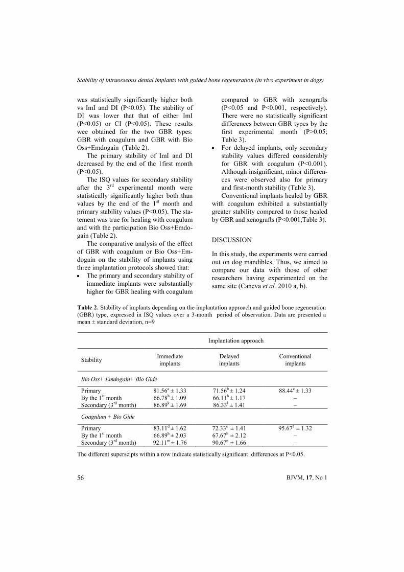
was statistically significantly higher both vs ImI and DI (Р<0.05). The stability of DI was lower that that of either ImI (Р<0.05) or CI (Р<0.05). These results wee obtained for the two GBR types: GBR with coagulum and GBR with Bio Oss+Emdogain (Table 2).
The primary stability of ImI and DI decreased by the end of the 1first month (Р<0.05).
The ISQ values for secondary stability after the 3rd experimental month were statistically significantly higher both than values by the end of the 1st month and primary stability values (Р<0.05). The sta-tement was true for healing with coagulum and with the participation Bio Oss+Emdo-gain (Table 2).
The comparative analysis of the effect of GBR with coagulum or Bio Oss+Em-dogain on the stability of implants using three implantation protocols showed that: The primary and secondary stability of
immediate implants were substantially higher for GBR healing with coagulum
compared to GBR with xenografts (Р<0.05 and Р<0.001, respectively). There were no statistically significant differences between GBR types by the first experimental month (Р>0.05; Table 3).
For delayed implants, only secondary stability values differed considerably for GBR with coagulum (Р<0.001). Although insignificant, minor differen-ces were observed also for primary and first-month stability (Table 3). Conventional implants healed by GBR
with coagulum exhibited a substantially greater stability compared to those healed by GBR and xenografts (P<0.001;Table 3).
DISCUSSION
In this study, the experiments were carried out on dog mandibles. Thus, we aimed to compare our data with those of other researchers having experimented on the same site (Caneva et al. 2010 a, b).
Table 2. Stability of implants depending on the implantation approach and guided bone regeneration (GBR) type, expressed in ISQ values over a 3-month period of observation. Data are presented a mean ± standard deviation, n=9
Implantation approach
Stability Immediate implants
Delayed implants
Conventional implants
Bio Oss+ Emdogain+ Bio Gide
Primary 81.56a ± 1.33 71.56b ± 1.24 88.44c ± 1.33 By the 1st month 66.78h ± 1.09 66.11h ± 1.17 – Secondary (3rd month) 86.89k ± 1.69 86.33l ± 1.41 –
Coagulum + Bio Gide
Primary 83.11d ± 1.62 72.33e ± 1.41 95.67f ± 1.32 By the 1st month 66.89h ± 2.03 67.67h ± 2.12 – Secondary (3rd month) 92.11m ± 1.76 90.67n ± 1.66 –
The different superscipts within a row indicate statistically significant differences at P<0.05.
J. Indjova, Kh. Fakih, D. Sivrev, D. Yovchev & Ts. Chaprazov
BJVM, 17, No 1 57
The high primary stability of all imp-lants was anticipated. Similar data were already reported (Sennerby et al., 2005) and findings were attributed to the relati-vely high density of the mandibular alve-olar bone in dogs (Caneva et al. 2010 a, b).
Relatively lower primary stability quotients of delayed implants vs those of immediate and conventional types could be attributed to the cumulative effects of two traumas: tooth extraction and pla-cement of xenografts, without leaving enough time to the organism to cope with it. The trauma from the preparation of the implantation bed in an environment with already reduced biological potential should be also considered. The local reac-tions after immediate and conventional implantations were different. After ImI placement, the traumatic effect of tooth extraction was not manifested. After CI implantation, not only had the tissue
reactions of extraction and xenografts abated, but regeneration was already far advanced (Piatelli et al., 1999).
The highest primary stability quotient of conventional implants, which was statistically significantly higher than those of ImI and DI, could be attributed to the close contact of implant and cortical bone achieved with this approach. After pre-paration of gingivoperiosteal flap, cortical bone was clinically completely restored.
The reduced stability of ImI and DI by the end of the first post implantation month corresponded at a high extent to other reports (Rabel et al., 2007). One month is a rather short period after imp-lantation, when the traumatic consequen-ces were not overcome, the biopotential of bone was not restored and its remodelling has not yet begun (Sennerby & Meredith, 2008).
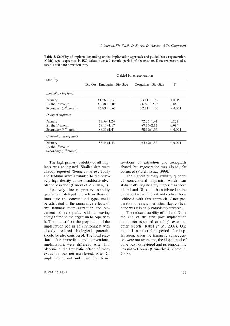
Table 3. Stability of implants depending on the implantation approach and guided bone regeneration (GBR) type, expressed in ISQ values over a 3-month period of observation. Data are presented a mean ± standard deviation, n=9
Guided bone regeneration
Stability
Bio Oss+ Emdogain+ Bio Gide Coagulum+ Bio Gide P
Immediate implants
Primary 81.56 ± 1.33 83.11 ± 1.62 < 0.05 By the 1st month 66.78 ± 1.09 66.89 ± 2.03 0.863 Secondary (3rd month) 86.89 ± 1.69 92.11 ± 1.76 < 0.001
Delayed implants
Primary 71.56±1.24 72.33±1.41 0.232 By the 1st month 66.11±1.17 67.67±2.12 0.094 Secondary (3rd month) 86.33±1.41 90.67±1.66 < 0.001
Conventional implants
Primary 88.44±1.33 95.67±1.32 < 0.001 By the 1st month – – Secondary (3rd month) – –

Stability of intraosseous dental implants with guided bone regeneration (in vivo experiment in dogs)
BJVM, 17, No 1 58
The reduced stability of immediate and delayed implants by the end of the first month was not an obstacle for the subsequent increase in their stability. Af-ter the third post implantation month, the measured ISQ was considerably higher both vs primary stability and one-month stability values. This is associated to al-veolar bone remodelling events. Osteo-integration between the implant and the bone socket, and secondary stability has occurred (Akca et al., 2006). In ImI and DI, the primary stability resulted from the mechanical bond between implant and cancellous bone, i.e. mechanical cancel-lous bone stability was observed. Over the 3-month period, the peri-implant cortical bone was restored (Carmagnola et al., 2003). It is the primary source of stability (Sennerby & Meredith, 2008). Meanwhi-le, cancellous bone stability was transfor-med into biological cancellous bone stability by virtue of new bone formation and remodelling, and appearing osteoin-tegration. Osteointegration is present over the entire length of the implant (Rocci et al., 2003). This way, a cortical stability was added biological cancellous bone sta-bility which has replaced the mechanical type of stability.
In this experiment on three dogs and 18 implants, their stability quotients were substantially higher in an environment of GBR with osseous coagulum than in cases of GBR with the xenograft combination Bio Oss+Emdogain.
Interventions for maxillary sinus floor augmentation with guided bone regenera-tion with Bio Oss have shown that even after 18 months or more, xenograft partic-les were not fully resorbed (Piattelli et al., 1999). New bone, inlcuding mature mine-ralised lamellar bone with osteons and Haversian canals has formed, similar to what was observed in GBR with coa-
gulum. The presence of non-resorbed bone graft however, reduced the relative amount of newly formed bone (Hammerle et al. 1998). We suggested that the bone formed by its natural constituents and non-resorbed Bio Oss particles could be of inferior mechanical quality compared to newly formed bone from coagulum.
The comparison of implant stability quotients in the light of the limited num-ber of experimental subjects (3 dogs, 18 implants of three types: immediate, dela-yed and conventional) should be done carefully without definite conclusions. The observed tendencies were in agree-ment were already reported data (Botti-celli et al., 2003, Caneva et al., 2010a, b).
CONCLUSION
The present in vivo experiment in dogs demonstrated high implant stability quo-tients for the 18 implants. This primary stability decreased by the end of the first month. This did not impede the occur-rence of a high secondary stability by the end of the third month. The implant sta-bility for guided bone regeneration with coagulum was superior to that of implants healed by GBR with Bio Oss+Emdogain xenografts.
ACKNOWLEDGMENTS
The experiment have been financially suppor-ted by Grant No. 17/2013, Medical University – Sofia, Council of Medical Science.
REFERENCES
Akca, K., T. Chang, L. Tekdemir & M. Fanus-cu, 2006. Biomechanical aspects of initial intraosseous stability and implant design: a quantitative micromorphometric analysis.
J. Indjova, Kh. Fakih, D. Sivrev, D. Yovchev & Ts. Chaprazov
BJVM, 17, No 1 59
Clinical Oral Implant Research, 17, 465–472.
Al-Nawas, B., K. Groetz, H. Goetz, H. Dus-chner & W. Wagner, 2007. Comparative histomorphometry and resonance frequen-cy analysis of implants with moderately rough surfaces in a loaded animal model. Clinical Oral Implant Research, 18, 1–8.
Atnoun, H., C. Chemaly & P. Missika, 2007. Bone substitutes. In: Bone Augmentation in Oral Implantology, Quintessence Publ, рр. 341–372.
Atsumi, M., S. Park & H. Wang, 2007. Methods used to assess implant stability: current status. International Journal of Oral and Maxillofacial Implants, 22, 743–754.
Baldini, N., M. De Sanctis & M. Ferrari, 2011. Deproteinized bovine bone in periodontal and implant surgery. Dental Materials, 27, 61–70.
Bhola, M., A. Neely & Sh. Kolhatkar, 2008. Immediate implant placement: Clinical decisions, advantages, and disadvantages. Journal of Prosthodontics, 17, 576–581.
Botticelli, D., T. Berglundh, D. Buser & J. Lindhe, 2003. Appositional bone forma-tion in marginal defects at implants. An experimental study in the dog. Clinical Oral Implant Research, 14, 1–9.
Botticelli, D, T. Berglundh & J. Lindhe, 2004. Hard-tissue alterations following immedia-te implant placement in extraction sites. Journal of Clinical Periodontology, 31, 820–828.
Caneva, M., L. Salata, S. Souza, E. Bressan, D. Botticelli & N. Lang, 2010a. Hard tis-sue formation adjacent to implants of various size and configuration immediately placed into extraction sockets: An experi-mental study in dogs. Clinical Oral Imp-lant Research, 21, 885–890.
Caneva, M., D. Botticelli, L. Salata, S. Souza, L. Cardoso & N. Lang, 2010b. Collagen membranes at immediate implants: A histomorphometric study in dogs. Clinical Oral Implant Research,, 21, 891–897.
Carmagnola, D., Т. Berglundh, M. Araujo, T. Albrektsson & J. Lindhe, 2000. Bone hea-ling around implants placed in a jaw defect augmented with Bio-OssA. An experi-mental study in dogs. Journal of Clinical Periodontology, 27, 799–805.
Carmagnola, D., P. Adriaens & T. Berglundh, 2003. Healing of human extraction sockets filled with Bio-Oss. Clin. Oral Impl. Res, 14, 137–143.
Indjova, J., D. Sivrev, H. Fakih, M. Paskalev & Ts. Chaprazov, 2013. Guided bone regeneration with xenografts and aloplasts (in vivo experiments in rabbits). Interna-tional Scientific Conference "90 Years Faculty of Veterinary Medicine in Bul-garia, 30–31 May 2013, Stara Zagora, Abstract Book, p. 57.
Hallman, M., L. Sennerby & S. Lundgren, 2002. A clinical and histologic evaluation of implant integration in the posterior maxilla after sinus floor augmentation with autogenous bone, bovine hydroxyapatite, or a 20:80 mixture. International Journal of Oral and Maxillifacial Implants, 17, 635–643.
Hammerle, C., G. Chiantell, J. Karring & T. Lang, 1998. The effect of deproteinized bovine bone mineral on bone regeneration aroud titanium implants. Clinical Oral Implant Research,, 9, 151–162.
Lang, N., U. Bragger, C. Hämmerle & F. Sut-ter, 1994. Immediate transmucosal imp-lants using the principle of guided tissue regeneration. I. Rationale, clinical proce-dures and 30-month results. Clinical Oral Implant Research, 5, 154–163.
Lundgren, D., H. Rylander & M. Andersson, 1992. Healing-in of root analogue titanium implants placed in extraction sockets. An experimental study in the beagle dog Clinical Oral Implant Research, 3, 136–143.
Meredith, N., 1998. Assessment of implant stability as a prognostic determinant. The International Journal of Prosthodontics, 11, 491–501.

Stability of intraosseous dental implants with guided bone regeneration (in vivo experiment in dogs)
BJVM, 17, No 1 60
Piattelli, M., G. Favero, A. Scarano, G. Orsini & A. Piattelli, 1999. Bone reactions to anorganic bovine bone (Bio-Oss) used in sinus augmentation procedures: A histo-logic long-term report of 20 cases in humans. International Journal of Oral and Maxillifacial Implants, 14, 835–840.
Rabel, A., S. Köhler & A. Schmidt-Westhau-sen, 2007. Clinical study on the primary stability of two dental implant systems with resonance frequency analysis. Cli-nical Oral Investigations, 11, 257–265.
Rocci, A., M. Martignoni, P. Burgos, J. Got-tlow & L. Sennerby, 2003. Histology of retrieved immediately and early loaded oxidized implants: Light microscopic oser-vations after 5 to 9 months of loading in the posterior mandible. Clinical Implant Dentistry and Related Research, 5, Suppl, 88–98.
Roze, J., S. Babu, A. Saffarzadeh, M. Dela-croix, A. Hoornaert & P. Layrolle, 2009. Correlating implant stability to bone structure. Clinical Oral Implant Research, 20, 1140–1145.
Scarano, A., M. Degidi, G. Iezzi, G. Petrone & A. Piattelli, 2006. Correlation between implant stability quotient and bone-implant contact: A retrospective histolo-gical and histomorphometrical study of seven titanium implants retrieved from humans. Clinical Implant Dentistry & Re-lated Research, 8, 218–222.
Sennerby, L. & J. Roos, 1998. Surgical deter-minants of clinical success of osseointeg-rated oral implants. Review. The Interna-tional Journal of Prosthodontics, 11, 408–420.
Sennerby, L., L. Persson, T. Berglundh, A. Wennerberg & J. Lindhe, 2005. Implant stability during initiation and resolution of experimental periimplantitis: An experi-mental study in the dog. Clinical Implant Dentistry and Related Research, 7, 136–140.
Sennerby, L. & N. Meredith, 2008. Implant stability measurements using resonance frequency analysis: Biological and biome-chanical aspects and clinical implications. Periodontology 2000, 47, 51–66.
West, J. D. & T. Oates, 2007. Identification of stability changes for immediately placed dental implants. International Journal of Oral and Maxillifacial Implants, 22, 623–630.
Wilson, T. G. Jr, R. Schenk & D. Buser, 1998. Implants placed in immediate extraction sites: A report of histologic and histo-metric analyses of human biopsies. Inter-national Journal of Oral and Maxillifacial Implants, 13, 333–341.
Paper received 25.06.2013; accepted for publication 11.09.2013
Correspondence: Dr. Jermen Indjova Department of Oral and Maxillofacial Surgery, Faculty of Dental Medicine, Medical University – Sofia, Bulgaria, e-mail: [email protected]
Recommended