Embed Size (px)
Citation preview

1
IntraOsseous Devices – a Randomized Controlled Trial comparing three
intraosseous devices
Klaas A. Hartholt1, MD, Esther M.M. van Lieshout1, PhD, Wim C. Thies1, Peter Patka, MD
PhD1, Inger B. Schipper, MD PhD1,2
1 Erasmus MC, Department of Surgery-Traumatology, Rotterdam, Netherlands
2 Current address: LUMC, Department of Surgery, Leiden, Netherlands
Corresponding author and reprints:
E.M.M. van Lieshout, PhD
Erasmus MC, University Medical Center Rotterdam
Department of Surgery-Traumatology
P.O. Box 2040
3000 CA Rotterdam
The Netherlands
Tel. +31 10-7031050
Fax +31 10-7032396
Mail [email protected]
Keywords: Intraosseous infusion, FAST1, Bone injection-gun, Jamshidi, RCT
2
Abstract
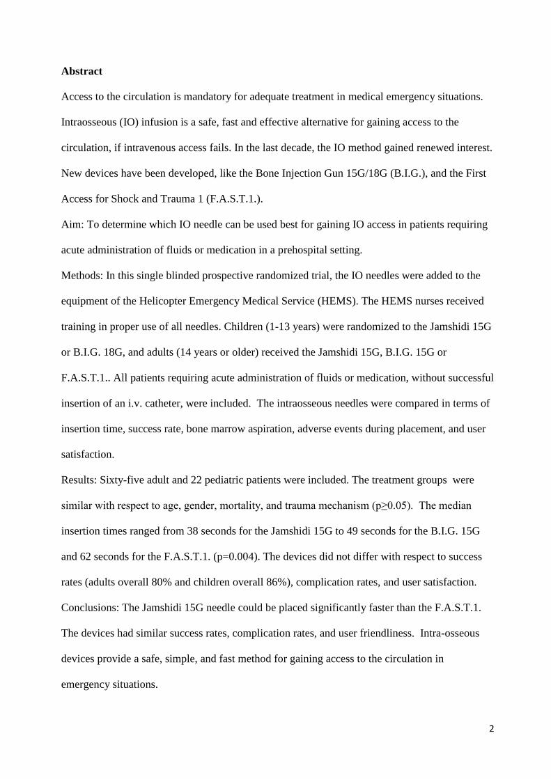
Access to the circulation is mandatory for adequate treatment in medical emergency situations.
Intraosseous (IO) infusion is a safe, fast and effective alternative for gaining access to the
circulation, if intravenous access fails. In the last decade, the IO method gained renewed interest.
New devices have been developed, like the Bone Injection Gun 15G/18G (B.I.G.), and the First
Access for Shock and Trauma 1 (F.A.S.T.1.).
Aim: To determine which IO needle can be used best for gaining IO access in patients requiring
acute administration of fluids or medication in a prehospital setting.
Methods: In this single blinded prospective randomized trial, the IO needles were added to the
equipment of the Helicopter Emergency Medical Service (HEMS). The HEMS nurses received
training in proper use of all needles. Children (1-13 years) were randomized to the Jamshidi 15G
or B.I.G. 18G, and adults (14 years or older) received the Jamshidi 15G, B.I.G. 15G or
F.A.S.T.1.. All patients requiring acute administration of fluids or medication, without successful
insertion of an i.v. catheter, were included. The intraosseous needles were compared in terms of
insertion time, success rate, bone marrow aspiration, adverse events during placement, and user
satisfaction.
Results: Sixty-five adult and 22 pediatric patients were included. The treatment groups were
similar with respect to age, gender, mortality, and trauma mechanism (p≥0.05). The median
insertion times ranged from 38 seconds for the Jamshidi 15G to 49 seconds for the B.I.G. 15G
and 62 seconds for the F.A.S.T.1. (p=0.004). The devices did not differ with respect to success
rates (adults overall 80% and children overall 86%), complication rates, and user satisfaction.
Conclusions: The Jamshidi 15G needle could be placed significantly faster than the F.A.S.T.1.
The devices had similar success rates, complication rates, and user friendliness. Intra-osseous
devices provide a safe, simple, and fast method for gaining access to the circulation in
emergency situations.

3
Introduction
Access to the circulation is required for optimal treatment in emergency situations. The gold
standard for vascular access is by use of an intravenous (i.v.) catheter. In certain (prehospital)
emergency situations placement of an i.v. catheter is not feasible. It may for instance, be a
challenge to establish i.v. access in patients with severe burn wounds, status epilepticus, major
trauma, severe sepsis, or in hemodynamically unstable patients and in small children.1-7
In
addition, environmental factors may limit the success rate of gaining i.v. access.
Multiple animal and clinical studies have shown that intraosseous (IO) access is a safe,
simple and effective technique for gaining vascular access in adults and children.6, 8-16
An IO
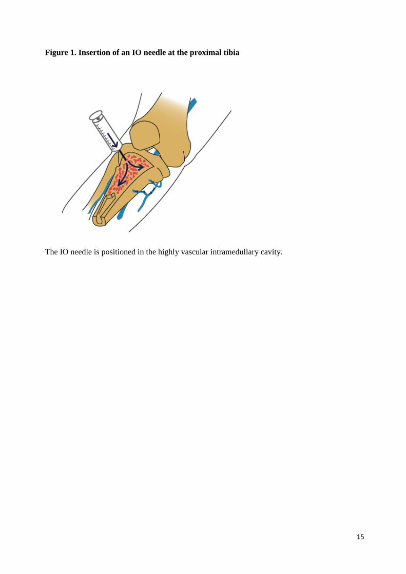
needle is a small hollow metal tube that can be inserted at different bone sites such as the distal
and proximal tibia (Figure 1), femur, sternum, humerus, radius and clavicula.17, 18
Even bones
without medullary cavity such as the calcaneus may serve as insertion place.13, 19-21
Once the IO needle is properly inserted into the bone marrow, an infusion system can be
connected to it. The IO route can be used for administering fluids, medication, crystalloids,
colloids and blood products. Due to the unique and highly vascular trabecular network in the
bone marrow, it is continuously being perfused, even during shock and hypotension. The
administered compounds quickly enter the circulation from the intramedullary cavity.
Medication administered intraosseously can be detected in the circulation almost as quickly as
medication given intravenously.13, 22-24
Bone marrow taken from an IO insertion location can be
used to determine hemoglobin, sodium, potassium, magnesium, lactate, and calcium levels,
blood group, and acid-base balance even during CPR.13, 25-31
IO infusion is an ancient technique and was widely used around 1940, but lost interest
after the Second Wold War. In the last decade IO access gained renewed interest for use in
emergency situations. This is reflected in the production of new intraosseous devices like the
First Access for Shock and Trauma 1 (F.A.S.T.1.TM
)32
, Bone Injection Gun (B.I.G.) and, more
recently, the EZ-IO (Vidacare, San Antonio, USA).

4
Moreover, gaining IO access is also included in several guidelines for clinical practice.
The European Resuscitation Council (ERC) prescribes that intraosseous vascular access should
be established in both pediatric and adult emergency patients if it is difficult or impossible to
establish peripheral venous access for cardiopulmonary resuscitation.33, 34
It is also included in
the curricula for Advanced Trauma Life Support (ATLS)35
and Advanced Paediatric Life
Support (APLS).36
In the Netherlands, IO devices are frequently used by Helicopter Emergency
Medical Services (HEMS) and Emergency Medical Services (EMS), as well as at Emergency
Wards.
The aim of this prospective randomized controlled trial was to determine which IO needle can be
used best for gaining acute IO access in patients requiring acute administration of fluids or
medication in the prehospital setting. One manual system (Jamshidi 15G) and two semi-
automatic IO systems (B.I.G. 15G/18G and F.A.S.T.1.) were compared in this study.
5
Materials and Methods
Study design
The study was designed as a single-centre, and single-blinded, prospective randomized clinical
trial. Patients were randomized between Jamshidi 15G, B.I.G. 15G/18G and F.A.S.T.1. (see
Table 1 for specifications of these devices). The study was performed at a level I trauma center,
serving over 4 million inhabitants (Erasmus MC, Rotterdam, the Netherlands) with a physician
staffed HEMS. The HEMS team consists of an anaesthesiologist or a trauma surgeon with a
HEMS nurse and a pilot. The local ethics committee approved the study protocol. The study
started on June 21, 2006 and ended on March 5, 2009.
The power analysis performed preliminary to this study, was based on a study of Calkins et al32
and data provided of the manufacturers, to show a difference in insertion time of 30 seconds
between the different IO needles to detect significant results with 80% power.
Patients and material
All patients in the prehospital setting in which the HEMS provided additional medical support,
requiring immediate fluid resuscitation or drugs, were considered eligible for inclusion after the
HEMS or EMS nurse failed to successfully insert an i.v. catheter on two consecutive attempts, or
when cardiopulmonary resuscitation (CPR) was needed. The HEMS physician decided whether
or not an IO device was needed, based on these inclusion criteria. Since F.A.S.T.1. placement
required an intact sternum, patients with a sternal anomaly or (suspected) sternal fracture were
excluded. Patients below 1 year of age were also excluded.
Pediatric patients, aged 1-13 years, were randomized between the Jamshidi 15G or B.I.G. 18G.
Adult patients, aged 14 years and older, were randomized between the Jamshidi 15G, B.I.G. 15G
or F.A.S.T.1..
All HEMS nurses received training in proper use and placement of the three intraosseous

6
devices, prior to the start of the study. Special instruction and training sets were placed at the
helicopter station. The training was repeated after one year.
All devices were ready for direct use, and were applied following the manufacturer’s
instructions. The IO needles should not be placed in fractured bones. Each device was packed
separately in a blinded plastic container that was sealed with an adhesive label in random order.
Boxes for adult and pediatric patients differed in colour. Each box contained an IO device, two
10ml syringes, 10ml saline 0.9%, 10ml Lidocaine 1%, a stopwatch, an information brochure for
in-hospital physicians, and a data entry form to be completed by the HEMS nurse that inserted
the IO needle. An adult- as well as a pediatric randomization box was added to the regular
HEMS equipment. Upon decision of the attending HEMS physician, the HEMS nurse opened
the container containing the IO device if the patient met the inclusion criteria, thereby being
unblinded for the IO device to be positioned. Time measurement started directly after opening of
the sealed container and stopped after administration of 10ml of saline when bone marrow
aspiration was attempted. Aspiration of bone marrow is a strong indicator of correct placement
of an IO needle. Therefore, correct placement of all needles was verified by aspiration of bone
marrow and flushing with saline. After placement the nurse completed the data form. The
insertion time, success, aspiration of bone marrow, side effects, medication given, trauma
mechanism and user satisfaction were recorded. The user satisfaction was scored on a visual
analogue scale (VAS), in which 0 implied the device was not user-friendly at all, and 10 implied
the highest user-friendliness possible.
All study data were entered into an electronic database (Microsoft®
Excel 2000). The IO devices
were compared in terms of insertion times, success rates, adverse events during placement and
user satisfaction. Also sex, age and trauma mechanism were recorded. Statistical analysis was
performed by using the SPSS (Statistical Package for the Social Sciences) 16.0.1 statistical

7
software. Differences between groups were tested in terms of gender, mortality rate, trauma
mechanism, success rate, bone marrow aspiration, and adverse events were analysed using the
Chi square test. The Mann-Whitney U-test was used to assess differences between groups
regarding user satisfaction, time for placement and age. For the adult patient group, Mann-
Whitney U-test was performed for post-hoc pairwise comparisons upon a Kruskal-Wallis
ANOVA. Correction for multiple comparison was performed where needed. A p-value <0.05
was considered statistically significant.
8
Results
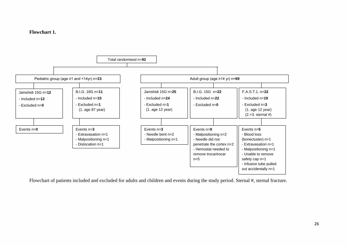
During the study period 87 patients, 65 adult and 22 pediatric patients were included (Flowchart
1). In addition, five randomization boxes were opened by mistake for patients who did not meet
the inclusion criteria; two patients with a clinical sternum fracture (F.A.S.T.1.), two adult
randomization boxes for pediatric patients (Jamshidi and F.A.S.T.1.), one pediatric
randomization box for an adult patient (B.I.G.). Tables 2 and 3 display patient demographic data.
In both groups two-third of the patients was male. The adult patients had an average age of 43
years (P25-P75 25-59), the pediatric patients 7.5 years (P25-P75 2-11). Treatment groups showed
no differences regarding these demographic characteristics. A high mortality rate of
approximately 71% and 59% was seen in the adult and pediatric groups, respectively, during the
resuscitation period prehospital or at the Emergency Department (ED). The main trauma
mechanism was High Energetic Trauma (HET, all blunt trauma, were due to motor vehicle
collisions 54.1%, fall from height 29.7%, person vs vehicle accidents 16.2% and non-specified
8.1%) (46% in adult patients versus 32% in pediatric patients) followed by Cardio Pulmonary
Resuscitation (CPR, including medical and traumatic CPR) (20% versus 36%) and drowning
(9% versus 9%). Convulsions (non-traumatic) and suicide attempts (including hanging,
toxification and bloodletting) were infrequently seen. Also, isolated cases of severe burns,
accidental strangulation, hypoglycaemia, carbon monoxide intoxication, stab injuries and
electrocution were reported on a few occasions during this study.
All HEMS nurses participated and placed the randomly assigned IO needles. Each nurse
performed 1-5 IO needle introduction procedures in the pediatric group and 3-19 in the adult
group (p=0.4). The times needed for insertion are shown in Figure 2. The overall median
insertion time was 50 (P25-P75 34-62) seconds. In the adult group, the Jamshidi 15G was placed
fastest (median insertion time 37 seconds; P25-P75 30-49). This was significantly faster than
placement of F.A.S.T.1. (median 62 seconds; P25-P75 50-131) (p=0.002). Time needed to insert
the B.I.G. 15G (median 49 seconds; P25-P75 33-60) did not differ statistically significantly from

9
the other devices. In the pediatric group the median insertion time of the Jamshidi 15G was 43
(P25-P75 33-79) seconds versus 48 (P25-P75 28-65) seconds for the B.I.G. 18G (p=0.74).
Table 4 and 5 display the IO needle insertion characteristics such as success rate, bone
marrow aspiration, adverse events and user satisfaction for the adult and pediatric group,
respectively. No statistically significant difference was noted with respect to the rate of
successful placement between the different types of IO needles in adults and in children.
Successful placement of the IO needle was confirmed by bone marrow aspiration in over 80%
both in the adult and the pediatric patient group. The overall score of user satisfaction was 9.8
(P25-P75 9.2-9.9) in both the adults and pediatric group. This was similar in all groups (p>0.05).
The number of needles inserted during the study by each HEMS nurse did not correlate
significantly with either the success rate or the rating of user satisfaction (data not shown).
Twenty-one adverse events occurred (i.e., 18 in the adult patients, 3 in pediatric patients),
however at similar rates in each treatment group (p>0.05). In the adult group; two Jamshidi 15G
needles bent during insertion, in one case the needle was malpositioned. In 5 cases, a hemostat
was needed to remove the trocar of the B.I.G. 15G. In two cases the B.I.G. 15G needle did not
penetrate the cortex and in two cases the B.I.G. 15G was wrongly positioned next to the bone.
After insertion of the F.A.S.T.1. the infusion tube was pulled out during withdrawal of the
introducer in one case. In one case there was reasonable blood loss at the location of the ‘bone-
cluster’. Also, the needle was malpositioned in one case, and in one case it was not possible to
remove the safety cap from the infusion tube. One procedural error was reported in case of a
F.A.S.T.1.; the removal tool got lost during transport to the Intensive Care Unit (ICU) and the
HEMS nurses had to bring a new removal tool. In the pedriatric group, three adverse events
occurred with the B.I.G. 18G. In one case extravasation was reported and in one case the needle
was wrongly positioned next to the bone. In one case the nurse could not remove the trocar.

10
Discussion
The aim of this study was to investigate which IO needle could be used best for gaining acute IO
access in patients requiring immediate fluids or drug therapy, in cases where gaining i.v. access
failed. On average, the median insertion times showed that the Jamshidi needles were placed
faster as compared to the F.A.S.T.1.. The devices (adult and pediatric) did not differ statistically
significantly with respect to success rate, complication rates and user satisfaction.
Although there were no significant differences in complication rates, all needles showed
different types of complications possibly coherent to the insertion method. Insufficient
perforation of the cortex or misplacement may be caused by the mechanical mechanism in both
devices, which replaces the manual pressure that is needed for placement of the Jamshidi needle.
Tactile references may be important to assure correct positioning.
All three IO devices tested were considered user friendly. Medical personnel are able to
use the different types of IO devices after appropriate training.37
Depending on circumstances a
rational decision should be made in favor of a particular IO device. For example, military medics
have to carry all their medical equipment. In this situation it can be better to use a light and small
IO needle like the Jamshidi or B.I.G., but the sternum is a preferred insertion location since it is
well protected in a bullet-proof vest and the sternum is easy reachable in a helicopter or
ambulance (F.A.S.T.1.).
Clinical and practical disadvantages of the F.A.S.T.1. include the complexity of the
device and the number of different parts. All parts are supplied in one pack, which is quite bulky.
Care should be taken to prevent separate parts from getting lost. This may be a potential problem
when using the device at the accident scene, particularly in windy and dark situations, but also
when the patient is transported to a different department or ward within the hospital. During the
current study, there was one case in which the infusion tube was removed without using the
removal tool and the metal tip was left behind in the sternum. This did not have consequences
for this patient. After this incident the nurses were instructed to tape the removal tool to the
11
patient. In this study, we used a F.A.S.T.1., which required the use of a specialized removal tool.
Pyng has adapted their model of the F.A.S.T.1., and in the latest released version a removal tool
is no longer needed. Although a F.A.S.T.1. for use in pediatric patients is under development, the
current device is indicated for use in adult patients only.
Apart from differences in complexity there are also considerable differences in prices of
the different IO needles. The newer, more sophisticated, IO devices are much more expensive
than the simple and easy to use manual IO needles.
All IO needles tested should be applied in less than 60 seconds according to the different
manufacturers. This was observed in our study only on several occasions, more often longer time
was needed for identification of the correct site, insertion, aspiration, and flushing.
A limitation of the current study is the lack of patient follow-up, therefore the number of
complications encountered might be slightly underestimated. Not much is known about
complication rates after IO infusion. Only a few complications of IO infusion have been
reported, but some can have devastating results. Complications reported include myonecrosis,
osteomyelitis, epiphysiolysis, fat/air embolism and fractures do occur.38-43
No cases of
osteomyelitis or myonecrosis was brought to our attention during the course of the current study.
However, it should be noted the in the current study patients were transported to 16 different
hospitals spread over the Soutwestern part of the Netherlands, and accurate collection of follow-
up data concerning complications was not successful. Due to the high injury severity of the study
population, a high mortality rate at the ED was observed. An accurate assessment of
complication rates requires additional research in a larger study population, with a longer follow
up period
A post hoc power analysis showed that groups of at least 300 IO needles would have
been needed to detect significant results with a power of 90% between all IO needles. During the
study period IO access was used on 92 occasions. We expected to place 60 IO needles a year,
based on the registration of IO infusion during the year prior to the study. During the study

12
period a new IO needle, the EZ-IO, a battery-powered electric drill, was introduced in the
Netherlands. The EZ-IO was not included in this study, as it was not approved for use on the
Dutch market at the time the trial started. Many EMS started using the EZ-IO and often an IO
needle was inserted before arrival of the HEMS. The EZ-IO is at this moment becoming more
and more popular in North America and Europe, and should be compared to other IO devices in
further research.

13
Conclusion
Creating vascular access is crucial, during the initial treatment of patients in life threatening
situations. I.v. access remains the gold standard and should not be replaced, but the IO technique
is a good alternative if i.v. catheter placement is not possible. The Jamshidi needle was placed
significantly faster, compared with the F.A.S.T.1. in the adult group and had a success
percentage of 91%. The devices, Jamshidi, B.I.G. and F.A.S.T.1. did not differ statistically
significant with respect to success rate, complication rate and user friendliness in both the adult
and the pediatric group. However, the Jamshidi did not statistically differs from the B.I.G., there
seems to be a trend in favor towardsthe use of the Jamshidi needle, the least costly of the three
devices tested, in terms of placement time, success rate, and adverse events may be noted. These
differences may become statistically significant in larger patients group.

14
Acknowledgements
We like to thank the personnel of the HEMS, Lifeliner 2, for their participation and support for
this study. We also like to thank manufacturers, Waismed for donating the Bone Injection Guns
and Pyng Medical Corporation for donating F.A.S.T.1. devices. The Dutch liability insurance
company for medical personnel Medirisk financially supported this study.
15
Figure 1. Insertion of an IO needle at the proximal tibia
The IO needle is positioned in the highly vascular intramedullary cavity.
16
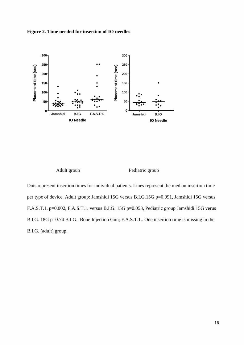
Figure 2. Time needed for insertion of IO needles
Adult group Pediatric group
Dots represent insertion times for individual patients. Lines represent the median insertion time
per type of device. Adult group: Jamshidi 15G versus B.I.G.15G p=0.091, Jamshidi 15G versus
F.A.S.T.1. p=0.002, F.A.S.T.1. versus B.I.G. 15G p=0.053, Pediatric group Jamshidi 15G verus
B.I.G. 18G p=0.74 B.I.G., Bone Injection Gun; F.A.S.T.1.. One insertion time is missing in the
B.I.G. (adult) group.
Jamshidi B.I.G. F.A.S.T.1.0
50
100
150
200
250
300
IO Needle
Pla
cem
en
t ti
me (
sec)
Jamshidi B.I.G. x0
50
100
150
200
250
300
IO Needle
Pla
ce
men
t ti
me (
se
c)

17
Table 1. Characteristics of IO devices
Jamshidi 15G B.I.G. 15G and 18G F.A.S.T.1.
Manufacturer Cardinal Health, Ohio, USA WaisMed Ltd, New York, USA PYNG Medical Corporation,
British Columbia, Canada
Insertion method Manual rotation and pressure Preloaded spring Manual pressure
Insertion location Long bones Long bones Sternum only
Insertion depth adjustable Yes Yes No
Removal tool needed No No Yes
Weight in grams 18 99 160
Package dimensions l x w x h cm 22.8 x 8.7 x 1.2 16.5 x 7.5 x 3.0 20.5 x 20.0 x 4.1
Package Soft Hard Soft
Price (Euros)
€ 26.90 € 58.20 € 140.00
Reusable No No No

18
Table 2. Patient characteristics for the adult group
Overall Jamshidi 15G B.I.G. 15G F.A.S.T.1. p-value
N 65 24 22 19
Males1
42 (64.6%) 11 (45.8%) 15 (68.2%) 16 (84.2%) 0.030+
Age2
43 (25-59) 45 (38-62) 40 (28-67) 26 (20-49) N.S.*
Mortality1
46 (70.8%) 16 (66.7%) 16 (72.7%) 14 (73.7%) N.S.+
Trauma mechanism1
- HET
- CPR
- Drowning
- Epilepsy
- Attempted Suicide
- Other
30 (46.2%)
13 (20.0%)
6 (9.2%)
2 (3.1%)
3 (4.6%)
11 (16.9%)
9 (37.5%)
7 (29.2%)
2 (8.3%)
1 (4.2%)
2 (8.3%)
3 (12.5%)
9 (40.9%)
3 (13.6%)
1 (4.5%)
1 (4.5%)
1 (4.5%)
7 (31.8%)
12 (63.2%)
3 (15.8%)
3 (15.8%)
0 (0.0%)
0 (0.0%)
1 (5.2%)
N.S.+
19
Data are shown as 1numbers with percentage within brackets or as
2 median with P25-P75 within brackets. ‘Other’ includes for the Jamshidi
electrocution, hypoglycaemia, severe burns, for the B.I.G. septic shock, explosion, severe burns, stab injuries, CVA, status epilepticus and a
plasma deficiency, for the F.A.S.T.1. a collaps. +Chi square test,
* Kruskal Wallis Anova. P-values <0.05 were considered statistically significant.
B.I.G., Bone Injection Gun; F.A.S.T.1., First Access for Shock and Trauma; N.S., Not Significant; HET, High Energy Trauma; CPR,
Cardiopulmonary Resuscitation.

20
Table 3. Patient characteristics for the pediatric group
Overall Jamshidi 15G B.I.G. 18G p-value
N 22 12 10
Males1
15 (68.2%) 10 (83.3%) 4 (44.4%) N.S. +
Age2
7.5 (2.0-11.0) 9.5 (2.5-12.5) 6.5 (2.0-8.7) N.S.*
Mortality1
13 (59.1%) 7 (58.3%) 6 (60.0%) N.S.+
Trauma mechanism1
- HET
- CPR
- Drowning
- Epilepsy
- Attempted Suicide
- Other
7 (31.8%)
8 (36.4%)
2 (9.1%)
1 (4.5%)
-
4 (18.2%)
4 (33.3%)
5 (41.7%)
1 (8.3%)
0 (0.0%)
-
1 (16.7%)
3 (30.0%)
3 (30.0%)
1 (10.0%)
1 (10.0%)
-
2 (20.0%)
N.S.+

21
Data are shown as 1numbers with percentage within brackets or as
2 median with P25-P75 within brackets. ‘Other’ includes for the Jamshidi CO-
intoxication and electrocution, for the B.I.G. severe burns and strangulation. +Chi square test,
* Kruskal Wallis Anova. P-values <0.05 were
considered statistically significant. B.I.G., Bone Injection Gun; N.S., Not Significant; HET, High Energy Trauma; CPR, Cardiopulmonary
Resuscitation.

22
Table 4. IO needle placement characteristics in the adult group
Device Overall Jamshidi 15G B.I.G. 15G F.A.S.T.1. p-value
N 65 24 22 19
Success rate1
52 (80.0%) 22 (91.7%) 13 (59.1%) 17 (89.5%) 0.010+
Bone Marrow aspiration1
43 (66.2%) 21 (87.5%) 11 (50.0%) 11 (57.9%) 0.018+
Insertion location
- Proximal tibia
45 (69.2%)
23 (95.8%)
22 (100%)
0 (0.0%)
- Iliac crest 1 (1.5%) 1 (4.2%) 0 (0.0%) 0 (0.0%)
- Sternum 19 (30.2%) 0 (0.0%) 0 (0.0%) 19 (100%)
Adverse events1
18 (26.1%) 3 (12.5%) 9 (40.9%) 5 (26.3%) N.S.+
VAS user satisfaction2
9.8 (9.2-9.9) 9.8 (8.8-9.8) 9.8 (9.3-9.9) 9.7 (9.3-9.9) N.S.*

23
Data are shown as 1numbers with percentage within brackets or as
2 median with P25-P75 within brackets.
+Chi square test,
* Kruskal Wallis
Anova. P-values <0.05 were considered statistically significant. B.I.G., Bone Injection Gun; F.A.S.T.1., First Access for Shock and Trauma;
N.S., Not Significant; VAS, Visual Analogue Scale
24
Table 5. IO needle placement characteristics in the pediatric group
Device Overall Jamshidi 15G B.I.G. 18G p-value
N 22 12 10
Success rate1
19 (86.4%) 12 (100%) 7 (70.0%) N.S. +
Bone Marrow aspiration1
17 (77.3%) 10 (83.3%) 7 (70.0%) N.S. +
Adverse events1
3 (13.6%) 0 (0.0%) 3 (30.0%) N.S. +
VAS user satisfaction2
9.8 (9.3-9.9) 9.8 (9.3-9.9) 9.8 (5.8-9.9) N.S.*
25
Data are shown as 1numbers with percentage within brackets or as
2 median with P25-P75 within brackets. VAS, Visual Analogue Scale;
+ Chi
square test, * Mann-Whitney U-test. P-values <0.05 were considered statistically significant; B.I.G., Bone Injection Gun; N.S., Not Significant.
26
Flowchart 1.
Flowchart of patients included and excluded for adults and children and events during the study period. Sternal #, sternal fracture.
Total randomised n=92
Jamshidi 15G n=25
- Included n=24
- Excluded n=1
(1. age 12 year)
F.A.S.T.1. n=22
- Included n=19
- Excluded n=3
(1. age 12 year)
(2.+3. sternal #)
Jamshidi 15G n=12
- Included n=12
- Excluded n=0
Pediatric group (age ≥1 and <14yr) n=23
B.I.G. 18G n=11
- Included n=10
- Excluded n=1
(1. age 87 year)
B.I.G. 15G n=22
- Included n=22
- Excluded n=0
Adult group (age ≥14 yr) n=69
Events n=5
- Blood loss
(bonecluster) n=1
- Extravasation n=1
- Malpositioning n=1
- Unable to remove
safety cap n=1
- Infusion tube pulled
out accidentally n=1
Events n=9
- Malpositioning n=2
- Needle did not
penetrate the cortex n=2
- Hemostat needed to
remove trocartrocar
n=5
Events n=3
- Needle bent n=2
- Malpositioning n=1
Events n=3
- Extravasation n=1
- Malpositioning n=1
- Dislocation n=1
Events n=0
27
References
1. Fiser DH. Intraosseous infusion. N Engl J Med 1990;322(22):1579-81.
2. Frascone R, Kaye K, Dries D, Solem L. Successful placement of an adult sternal intraosseous line through
burned skin. J Burn Care Rehabil 2003;24(5):306-8.
3. Glaeser PW, Losek JD, Nelson DB, et al. Pediatric intraosseous infusions: impact on vascular access time.
The American journal of emergency medicine 1988;6(4):330-2.
4. Helm M, Gries A, Fischer S, Hauke J, Lampl L. [Invasive techniques in emergency medicine. III.
Intraosseous punction--an alternative vascular access in paediatric emergencies]. Anaesthesist 2005;54(1):49-
56.
5. Hurren JS, Dunn KW. Intraosseous infusion for burns resuscitation. Burns 1995;21(4):285-7.
6. Schwartz D, Amir L, Dichter R, Figenberg Z. The use of a powered device for intraosseous drug and fluid
administration in a national EMS: a 4-year experience. The Journal of trauma 2008;64(3):650-4; discussion
4-5.
7. David JS, Dubien PY, Capel O, Peguet O, Gueugniaud PY. Intraosseous infusion using the bone injection
gun in the prehospital setting. Resuscitation 2009;80(3):384-5.
8. Brenner T, Bernhard M, Helm M, et al. Comparison of two intraosseous infusion systems for adult
emergency medical use. Resuscitation 2008;78(3):314-9.
9. Brickman KR, Rega P, Koltz M, Guinness M. Analysis of growth plate abnormalities following intraosseous
infusion through the proximal tibial epiphysis in pigs. Ann Emerg Med 1988;17(2):121-3.
10. Glaeser PW, Hellmich TR, Szewczuga D, Losek JD, Smith DS. Five-year experience in prehospital
intraosseous infusions in children and adults. Ann Emerg Med 1993;22(7):1119-24.
11. Halm B, Yamamoto LG. Comparing ease of intraosseous needle placement: Jamshidi versus cook. The
American journal of emergency medicine 1998;16(4):420-1.
12. Helm M, Hauke J, Bippus N, Lampl L. [Intraosseous puncture in preclinical emergency medicine: Ten years
experience in air rescue service.]. Anaesthesist 2007;56(1):18-24.
13. Iserson KV. Intraosseous infusions in adults. J Emerg Med 1989;7(6):587-91.
14. Kerz T, Dick W. [Routes for drug administration during cardiopulmonary resuscitation]. Anaesthesist
1996;45(6):550-65.
15. Runyon DE, Bruttig SP, Dubick MA, Clifford CB, Kramer GC. Resuscitation from hypovolemia in swine
with intraosseous infusion of a saturated salt-dextran solution. The Journal of trauma 1994;36(1):11-9.

28
16. Waisman M, Waisman D. Bone marrow infusion in adults. The Journal of trauma 1997;42(2):288-93.
17. Boon JM, Gorry DL, Meiring JH. Finding an ideal site for intraosseous infusion of the tibia: an anatomical
study. Clin Anat 2003;16(1):15-8.
18. Ong ME, Chan YH, Oh JJ, Ngo AS. An observational, prospective study comparing tibial and humeral
intraosseous access using the EZ-IO. The American journal of emergency medicine 2009;27(1):8-15.
19. McCarthy G, Buss P. The calcaneum as a site for intraosseous infusion. J Accid Emerg Med 1998;15(6):421.
20. McCarthy G, O'Donnell C, O'Brien M. Successful intraosseous infusion in the critically ill patient does not
require a medullary cavity. Resuscitation 2003;56(2):183-6.
21. Clem M, Tierney P. Intraosseous infusions via the calcaneus. Resuscitation 2004;62(1):107-12.
22. Cameron JL, Fontanarosa PB, Passalaqua AM. A comparative study of peripheral to central circulation
delivery times between intraosseous and intravenous injection using a radionuclide technique in
normovolemic and hypovolemic canines. J Emerg Med 1989;7(2):123-7.
23. Von Hoff DD, Kuhn JG, Burris HA, 3rd, Miller LJ. Does intraosseous equal intravenous? A pharmacokinetic
study. The American journal of emergency medicine 2008;26(1):31-8.
24. Brickman KR, Rega P, Guinness M. A comparative study of intraosseous versus peripheral intravenous
infusion of diazepam and phenobarbital in dogs. Ann Emerg Med 1987;16(10):1141-4.
25. Kissoon N, Idris A, Wenzel V, Murphy S, Rush W. Intraosseous and central venous blood acid-base
relationship during cardiopulmonary resuscitation. Pediatr Emerg Care 1997;13(4):250-3.
26. Kissoon N, Rosenberg H, Gloor J, Vidal R. Comparison of the acid-base status of blood obtained from
intraosseous and central venous sites during steady- and low-flow states. Crit Care Med 1993;21(11):1765-9.
27. Kissoon N, Peterson R, Murphy S, Gayle M, Ceithaml E, Harwood-Nuss A. Comparison of pH and carbon
dioxide tension values of central venous and intraosseous blood during changes in cardiac output. Crit Care
Med 1994;22(6):1010-5.
28. Abdelmoneim T, Kissoon N, Johnson L, Fiallos M, Murphy S. Acid-base status of blood from intraosseous
and mixed venous sites during prolonged cardiopulmonary resuscitation and drug infusions. Crit Care Med
1999;27(9):1923-8.
29. Johnson L, Kissoon N, Fiallos M, Abdelmoneim T, Murphy S. Use of intraosseous blood to assess blood
chemistries and hemoglobin during cardiopulmonary resuscitation with drug infusions. Crit Care Med
1999;27(6):1147-52.

29
30. Hurren JS. Can blood taken from intraosseous cannulations be used for blood analysis? Burns
2000;26(8):727-30.
31. Brickman KR, Krupp K, Rega P, Alexander J, Guinness M. Typing and screening of blood from intraosseous
access. Ann Emerg Med 1992;21(4):414-7.
32. Macnab A, Christenson J, Findlay J, et al. A new system for sternal intraosseous infusion in adults. Prehosp
Emerg Care 2000;4(2):173-7.
33. Biarent D, Bingham R, Richmond S, et al. European Resuscitation Council guidelines for resuscitation 2005.
Section 6. Paediatric life support. Resuscitation 2005;67 Suppl 1:S97-133.
34. Nolan JP, Deakin CD, Soar J, Bottiger BW, Smith G. European Resuscitation Council guidelines for
resuscitation 2005. Section 4. Adult advanced life support. Resuscitation 2005;67 Suppl 1:S39-86.
35. Surgeons ACo. Advanced Trauma Life Support for Doctors. 7th ed. Chigaco, USA: First Impression; 2004.
36. Pediatrics ACoEPaAAo. APLS: The Pediatric Emergency Medicine Resource Fourth Edition. 4th ed: Jones
and Bartlett Publishers; 2006.
37. Calkins MD, Fitzgerald G, Bentley TB, Burris D. Intraosseous infusion devices: a comparison for potential
use in special operations. The Journal of trauma 2000;48(6):1068-74.
38. Stoll E, Golej J, Burda G, Hermon M, Boigner H, Trittenwein G. Osteomyelitis at the injection site of
adrenalin through an intraosseous needle in a 3-month-old infant. Resuscitation 2002;53(3):315-8.
39. Launay F, Paut O, Katchburian M, Bourelle S, Jouve JL, Bollini G. Leg amputation after intraosseous
infusion in a 7-month-old infant: a case report. The Journal of trauma 2003;55(4):788-90.
40. van Rijn RR, Knoester H, Maes A, van der Wal AC, Kubat B. Cerebral arterial air embolism in a child after
intraosseous infusion. Emergency radiology 2008;15(4):259-62.
41. Alam HB, Punzalan CM, Koustova E, Bowyer MW, Rhee P. Hypertonic saline: intraosseous infusion causes
myonecrosis in a dehydrated swine model of uncontrolled hemorrhagic shock. The Journal of trauma
2002;52(1):18-25.
42. Hasan MY, Kissoon N, Khan TM, Saldajeno V, Goldstein J, Murphy SP. Intraosseous infusion and
pulmonary fat embolism. Pediatr Crit Care Med 2001;2(2):133-8.
43. Bowley DM, Loveland J, Pitcher GJ. Tibial fracture as a complication of intraosseous infusion during
pediatric resuscitation. The Journal of trauma 2003;55(4):786-7.