Th
e Scien
ce Jou
rnal o
f the L
and
er Co
llege o
f Arts an
d S
ciences
Vo
lum
e VI |
Nu
mb
er 2 | S
prin
g 2
01
3

The
SCIENCE JOURNAL of the Lander College of
Arts and Sciences-Flatbush a division of Touro College
Volume VI | Number 2 | Spring 2013

Throughout its 36-year history, Touro’s Lander College of Arts and Sciences in Flatbush
(with separate men’s and women’s schools) has provided cohorts of aspiring high school
graduates from well-regarded yeshivas and seminaries with a foundation of academic excel-
lence for professional career growth, in an environment that is supportive of the religious
values of its students. Graduates have assumed leadership roles and continue to strengthen
Jewish communities throughout the world.
Lander College of Arts and Sciences–Flatbush offers more than 25 majors and preprofes-
sional options, and three joint undergraduate/graduate degree programs in occupational
therapy, physical therapy and physician assistant studies with the School of Health Sci-
ences. Honors tracks in biology, the health sciences, political science and psychology are
currently offered.
Students are also required to complete a carefully designed core curriculum that empha-
sizes the development of communications skills, critical thinking and analytical competen-
cies, computer literacy and quantitative reasoning. Enrollment in science courses, notably
and health science students.
Faculty members continue to earn recognition for outstanding achievements, including
Joshua November, Assistant Professor of Languages and Literature, who was selected as
-
Notable alumni distinctions of Touro’s Lander College of Arts and Sciences in Flatbush
-
reviewed journals.
!e Lander College of Arts and Sciences at Touro in Flatbush

CAN MARIJUANA BE HARMFUL WHEN USED PRENATALLY OR DURING ADOLESCENCE? 1 Penninah Dean THE CARCINOGENIC EFFECTS OF ASPARTAME 12 Devora Sara Gelbfish COMPLEX REGIONAL PAIN SYNDROME: A REVIEW OF CURRENT TREATMENTS 22 Yosef Lewis ALOPECIA AREATA: AN OVERVIEW 41 Chaya Gestetner THE SCIENTIFIC EVIDENCE VALIDATING THE USE OF HONEY AS A MEDICINAL AGENT 55 Raitzel Chemda Bernstein CAN HEALTHY TRANSPLANTED TISSUE BE USED TO RESTORE MOTORFUNCTION IN PATIENTS WITH PARKINSON’S DISEASE? 71 Aliza Erlbaum CANCER IMMUNOTHERAPY TREATMENTS 84 Shifra Sadowsky THE PATHOGENESIS AND TREATMENT OF GOUT 99 Daniel Silberstein Cover Pictures Top: Sadowsky Middle: Bernstein Bottom: Gestetner
The SCIENCE
JOURNAL of the Lander College of
Arts and Sciences-Flatbush a division of Touro College

Executive Editors
Ralph Nussbaum
Griendy Indig-Weingarten
Associate Editors
Pnina Dean
Shaina Drizin
Mordechai Fonfeder
Joseph Gerstel
Yisroel Gross
Jonathan Kahanovitch
Benjamin Kalimi
Esther Michelson
Shifra Sadowsky
Emeritus Editors
Rivka H. Borger
Michell Gordon-Grunin
Faculty Reviewers
Robert S. Bressler Ph.D
Aliza Holtz Ph.D
Alan B. Levine D.C.
Faculty Advisor
Robert S. Bressler Ph.D., Chairman of Department of Biology
The SCIENCE JOURNAL of the Lander College of Arts and Sciences-Flatbush a division of Touro College

Penninah Dean graduated in June of 2013 with a B.S. in Biology. She is currently a Nephrology Research Assistant at Evanston Medical Center.
1
CAN MARIJUANA BE HARMFUL WHEN USED PRENATALLY OR DURING
ADOLESCENCE?
Penninah Dean Abstract Marijuana is a popular recreational drug with a strong following campaigning to legalize it for both medicinal and recreational use. This paper serves to illustrate the harmful effects of marijuana use as it pertains to prenatal, adolescent and adult use. By understanding the methods of absorption and mechanism of interaction in the body, we can see a correlation between the effects of marijuana and its toxicity. Through extensive research of case studies on marijuana use we were able to determine marijuana’s harmful effects physically, developmentally and cognitively. Through these methods of research, it can be concluded that marijuana has detrimental effects on the developing body in utero ,as well as, during adolescence. Furthermore, marijuana has consistently been found to cause long term damage such as short stature, attention span, and verbal retention (Solowij, et. al. 2011). In adults, smoke inhalation of the substance has been found to be more detrimental than the smoke inhalation of tobacco. While marijuana touts a variety of medicinal benefits in its application as a form of palliative care, its toxicity and the prolonged adverse effects of the substance are too strong to ignore. Introduction: Cannabis is one of the first plants to have been used medically, recreationally, and spiritually dating back 5000 years, with the first documented medical use in Central Asia and later in China and India (Pertwee, 2006). Cannabis is the most widely used illicit recreational drug after the three most popular substances, tobacco, alcohol, and caffeine (Green, 1998). Since its discovery, cannabis has been used by millions to both induce pleasure and alleviate pain. Physicians have prescribed it for a plethora of ailments until the government classified it as a Schedule I substance, rendering it illegal, and without medical value. There is a lot of effort being done by the public to try to legalize marijuana with claims that there is no basis for the fear and anxiety the public is placing on the drug, and it is in fact a benign substance. (NORML, 2013). There are surprisingly limited resources for research done on marijuana, largely due to the fact that it is difficult to find subjects willing to cooperate with a study concerning their illegal behavior. With over 300 million users worldwide, 28 million of which live in the United States, it is important to educate the public about the substance, how to use it safely, and if it exhibits adverse effects (Diaz, 1997). The purpose of the research done in this paper is to ascertain the safety or dangers of marijuana, focusing on a few aspects to determine if it is in fact harmless. It concentrates on the repercussions of prenatal use and its effect on the fetus,its effect on adolescents and determining if there is any observable long term damage. Method Used:
The author’s research was done using Touro College’s search engines such as ProQuest,

CAN MARIJUANA BE HARMFUL?
2
MEDLINE, and EBSCO, as well as research articles found through PubMed and Google Scholar. The method of research included reviewing studies and published articles that have been peer reviewed. In certain cases the author questioned the validity and accuracy of the methods used to attain the data presented and documented their uncertainty of the method of research. In other cases the author presented conflicting arguments to refute some peer reviewed studies to present that not all studies can be accepted at face value. Marijuana Intake and Potency:
Cannabis, colloquially known as marijuana, is a recreational drug whose leaves, flowers, and stems are all utilized in its use. The chemical compounds found within the Marijuana plant identify it as a member of the cannabinoid class. The cannabinoid plant, whose scientific name is Cannabis sativa, has a distinctive smell that is similar to that of skunk musk. The described effects of marijuana are relaxing, calming, mellowing, and sometimes anxiety and paranoia provoking. Collectively, these effects are referred to as a ‘high.’ (Sharman, et. al. 2013). The predominant psychoactive component in marijuana that determines its potency is delta-9-tetrahydrocannabinol, or THC, and was only isolated in 1964. This molecule is the chemical stimulant in the Cannabis plant that produces the altered states of consciousness in the user. THC actually exists as Tetrahydrocannabinolic acid, THCA, in the Cannabis plant and is the biosynthetic precursor of THC. Conversion of THCA to THC occurs through burning of the plant. Combustion causes decarboxylation to occur on the THCA converting it to the more psychoactive THC molecule (Hazekamp, et. al. 2005). The depth or strength of the psychoactive component of the cannabis is highly dependent upon the growing conditions and the genetic strain of the plants (Copeland, et. al. 2006). There are a variety of common methods for marijuana intake. These include but are not limited to smoking the dried leaf of the plant in a the form of a rolled cigarette or “joint”, using a water pipe or “bong” to inhale the fumes, consumption in food, inhalation of vapors through a vaporizer, and ingestion of the plants oils. Smoking is the most common and preferred route of intake but it is dulled by the fact that only 5-14% of the smoke is actually THC, and 30-80% of the smoke in the “joint” is lost to escaped smoke (Copeland, et. al. 2006). Smoking in itself is a dangerous method of intake as it is harmful to the lungs and respiratory system. Marijuana smokers are subject to the same dangers and health risks as tobacco smokers with similar negative results such as respiratory distress, asthma, cardiovascular disease, lung and esophageal cancers (Ellenhorn, Barceloux, 1988). The second method of choice involves using a water pipe commonly known as a “bong”. The bong minimizes the THC lost in the smoke because it is all contained within the bowl and then effectively inhaled. This method can be dangerous due to the larger amounts of carbon monoxide and tar inhaled. Smoking hashish, which is the resin from the plant smoked in a pipe, is less common but is done by adding a few drops of oil to tobacco or cannabis leaves and smoking it in a joint. Another alternative is heating the oils and inhaling the vapors. The oil can also be incorporated in food and consumed, but produces less of an intense high and causes a delayed onset of effects (Copeland, et. al.

Penninah Dean
3
2006). Newer methods, such as the use of vaporizers, have been utilized and have less harmful effects. These machines heat the cannabis and trap the tar and toxins in a special chamber allowing only the THC to be inhaled without the added harmful smoke. This is a useful method for patients who are using marijuana to aid in palliative care and treat illnesses. Through this method they are able to maximize the benefits of marijuana use without risking further damage to their health. Inhalers are also available for oral doses of THC, once again created for the purpose of medical palliative care (Martin, Wiley, 2004). Chemical Pathways of THC: THC is an extremely potent chemical and takes only a matter of seconds to enter your bloodstream and reach your brain. When smoked, it takes effect almost immediately and can last anywhere from 1-3 hours. When consumed in food there is a delayed onset of the desired effect, but the THC stays in your system for a longer period of time. Though the full mechanisms of THC still remain unknown, neuroscientists have some information about its effects on the brain (Diaz, 1997). To understand how THC is interacts with the brain’s cells, we must first understand the mechanisms that the brain uses to communicate. Neurons are the cells of the brain that transmit information. Neurons interact with each other through a chemical messenger system known as neurotransmitters. Neurotransmitters attach to protein structures imbedded in the membrane of the receiving neuron known as receptors. The attachment of neurotransmitters to these receptors facilitates the transmission of important information from one cell to the other. Each neuron has thousands of receptors and each receptor is specific to a certain neurotransmitter (Diaz, 1997). THC is a cannabinoid and is therefore able to mimic endogenous cannabinoid neurotransmitters, such as N-arachidonoylethanolamine (anandamide) or 2-arachidonoyl glycerol. The discovery of these endocannabinoids in 1992 by Israeli scientist Raphael Mechoulam emerged from a study in which he was trying to determine the purpose of cannabinoid receptors in the body (Devane, et. al. 1992). It was discovered that these endocannabinoid neurotransmitters are released by the body into the brain when the body senses an elevation in intracellular calcium. The THC binds to the cannabinoid receptors in place of the anandamide and therefore activates the appropriate neurons that would alternately be activated by anandamide (Sharman, et. al. 2013). THC exerts a majority of its influence through the midbrain reward center, triggering dopamine release in the prefrontal cortex which causes marijuana to have an addictive quality (Kogan, Mechoulam, 2007). The presence of THC in the brain interferes with the neurons’ normal function by artificially stimulating the cannabinoid receptors. Certain portions of the brain have concentrated cannabinoid receptors while others contain only a small number. These receptors can be found in areas of the brain including; the cerebellum, hippocampus and basal ganglia, areas that influence pleasure, memory, concentration, sensory and time perception, as well as coordinated movement. Therefore, THC can affect the sensations associated with thefunctions of these regions of the brain in which the cannabinoid receptors are found, resulting in the sensation of being ‘high’ (Devane, et. al. 1992). The largest portion of the cannabinoid receptors are found in the hippocampus which is located in the medial temporal lobe, beneath the cortical surface of the brain and is associated with short term

CAN MARIJUANA BE HARMFUL?
4
memory. THC therefore, has the greatest effect on that portion of the brain, explaining why users typically report having trouble with short term memory. The cerebellum and basal ganglia have many cannabinoid receptors as well and therefore those under the influence of THC also report problems with coordination and muscle movements (Sharman, et. al. 2013). There are two types of cannabinoid receptors identified as CB1 and CB2, in order of their discovery. These receptors act through inhibiting adenylate cyclase. The CB1 receptors are primarily found in the central nervous system, brain and nerve tissue, specifically the basal ganglia, hippocampus, cerebellum, and cerebral cortex, as well as, on the peripheral neurons. Their main function is to mediate inhibition of on-going release of certain excitatory and inhibitory neurotransmitters. CB2 is found in non neuronal cells in immune system tissues such as leukocytes, the spleen, and bone marrow, and was first discovered in human leukemia (Green, 1998). Marijuana Toxicity: An important factor to consider is the toxicity level of marijuana. In comparison with regular tobacco smokers, marijuana smoke creates a greater cardiovascular burden due to the high levels of carbon monoxide and tar found in cannabis resulting in a heavy respiratory burden on the smoker. Marijuana smokers are also known to take larger, deeper puffs and hold the smoke in their lungs for a longer period of time. Because of this practice, the retention of tar in the respiratory tract is one third greater than the amount of tar built up from tobacco smoke. Additionally, smoking marijuana results in much higher level of carboxyhemoglobin than its counterpart, tobacco. Regardless of the THC content, the smoking of cannabis in itself yields a higher carbon monoxide and tar weight on the respiratory tract (Wu, et. al. 1988). Marijuana has also been found to exacerbate psychotic illnesses in susceptible users, particularly schizophrenia. After testing the correlation between THC and psychosis, marijuana was found to cause consequent anxiety and neuropsychological impairment in users. THC can induce a transient, acute psychotic reactions in psychiatrically well individuals (Rais, et. al. 2008; Barch, Smith, 2008).
Minutes after a dose of THC is delivered in an individual there are notable deficits in working memory and executive functions with a trend towards an impaired episodic memory. This is significant as it is well established that schizophrenia is associated with deficits in those functions (Rais, et. al. 2008; Barch, Smith, 2008). Although the data is telling, the properties of THC are highly dose dependent with a possibility for bidirectional effects. There is also not much explanation of why some individuals are more susceptible to psychotic symptoms than others. Marijuana is absorbed in the bloodstream from the lungs within minutes of inhalation generating an extremely immediate reaction in the body with the swift onset of a ‘high.’ The degree of intensity of the high depends on the quality of the cannabis, the method of use, and the experience of the user. Familiarity is a factor because a more experienced user will know how to maximize the inhalation, but also may be immune and therefore unaffected by some of the THC absorbed (Copeland, et. al. 2006). Immunity occurs when the body is chemically altered and builds a certain level of tolerance to the presence of marijuana thus requiring a higher dose to attain identical results from the previous use. The effects of tolerance can be dangerous when the subjects gradually increase their dose to achieve a certain

Penninah Dean
5
degree of high, while compromising his body and health. While the THC carries out it psychoactive effects, it is simultaneously harming the body’s cardiovascular system by lowering blood pressure and increasing heart rate: a potential danger for chronic marijuana users (Gorelick, et. al. 2013). Marijuana and Fetal Development: To further understand the toxicity level and dangers of the substance, we must observe its effects on a developing fetus. Studies have been conducted that test the neurodegenerative effect of cannabis exposure on a developing rodent’s brain. Tests like these help scientists build a parallel analysis on the effects of cannabis in human neonatal development. Because of the differences in human and rodent development, analyses were done on a seven day old rodent which is most similar to a third trimester fetus. Perhaps the most obvious limitations to this study is that testing was done exclusively on rats rather than relying on information gathered from actual human case studies and assessing the available neurodegenerative data. Furthermore, the fact that the rodent was not in utero during testing raises questions as to the environmental differences in the conditions of the third trimester of a human fetus. One might argue that there is a level of neonatal protection when a child is in the womb, and that could protect it from foreign toxins as opposed to a rodent pup that has to fend for itself. There can also be claims that a child in utero may be exposed to more toxins due to the direct stream of oxygen and nutrition passed from the mother, therefore exposing the fetus to greater risk when its body is still vulnerable and reliant on maternal nutrients rather than depending on its own immune response. There is evidence, however, supporting the research done on rodents by studying the effects of marijuana on a fetus during the second trimester. Smoking of marijuana was found to have a significant effect on the stature of the unborn child. There is an additional increased risk of premature birth, stunted growth, and morbidity if the offspring is that of an adolescent even if their levels of drug use are lower than those of adult pregnant women (Cornelius, et. al. 2002). Further evidence can be found in preschool children who were assessed for sustained attention after fetal exposure to marijuana. In these studies, children were found to have various levels of decreased sustained attention. Although this implies that marijuana can have an adverse direct effect on the fetus, the fact that these mothers were users of other drugs including alcohol and tobacco, complicates analysis. Therefore, although there is conclusive data linking marijuana to these results, it is difficult to isolate which substance was the precise cause of the inattentiveness (Fried, et. al. 1992). With an increase of admitted dose of marijuana use, however, there was a correlated increase in the failure of the exposed children to maintain vigilance and sustain information appropriate to their grade level. There is also a greater likelihood of omission errors, indicating a lack of attention and a described impulsivity and hyperactivity that grew with increasing prenatal dose exposure (Noland, et. al. 2005). These effects were predominantly exhibited in preschool aged children as altered inattentive behavior and if exposed to these drugs at a young age, they also exhibit greater trouble with behavior and focusing, Double blinded studies such as these are well assessed and dependable due to the fact that the testers are not aware of the substance exposure status of the children and therefore minimizing biased

CAN MARIJUANA BE HARMFUL?
6
answers or observations. There are limitations as noted previously as many of the mothers of the children tested were exposed to various drugs as well. This limits the scope of observation and obscures our view as to which of the substances were the cause of the inattentiveness. (Richardson, et. al. 2002). Experiments on pregnant mammals have shown adverse effects and though the results have been quite supportive of the data, it remains difficult to predict how similar levels of THC would affect pregnant humans. One aspect that has been neglected by these studies is the adverse effect that smoke inhalation may have on the child. Although THC in itself is proven to be detrimental to the fetus, there is an added risk when marijuana is smoked, which is usually the case since that is the most common form of intake. Although there are some human studies revealing the effects on a fetus, there is limited data available due to the shortage of people willing to be included in a study (Jutras-Aswad, et. al. 2009).
Clearly, marijuana use and exposure during pregnancy is extremely harmful to the unborn child. THC is especially dangerous due to the ease in which it is able to cross the placental barrier, therefore entering the fetus’s blood supply where it could cause adverse effects. The THC builds up in the fat and liver tissue of the mother and is then passed through the placental barrier. The levels of THC present can be easily measured in the amniotic fluid, with stronger concentrations yielding more harmful results. The speed of transfer is essential because it enables the drug to achieve its pharmacological effects once it comes in contact with the fetus. Consequently, injection of THC during early pregnancy in rodents produced a seventy percent feticide (Harbison, Mantilla-Plata, 1972).
Additionally, negative effects of the marijuana are also observed if the fetus survives. Once the child develops, they can exhibit; an altered response to visual stimuli, increased tremulousness, problems with sustained attention and memory, and poor problem-solving skills (Diaz, 1997). Adolescent Use: The number of teenagers informed of the harmful effects of marijuana is decreasing, and consequently there is an increase in adolescent daily marijuana smokers. Marijuana can have an effect on the brain for users who began to smoke during adolescence, as opposed to adulthood. This creates a noticeable decline in IQ from the point of adolescence to adulthood. Through standardized IQ testing it was determined that there was an average of an eight point decline of IQ by mid age. There is a significant impairment of cognitive function, specifically related to attention and memory, and there is an increasing vulnerability to psychosis. There is no proof that stopping use of marijuana will improve cognitive function, and the effects of persistent cannabis remain, causing a neuropsychological decline. Many teenagers and even clinicians are not aware of the high probability of intellectual or psychopathological impairment due to the neurotoxic effects of THC (Meier, et. al. 2012). Long term cannabis use is specifically detrimental to the white matter of the brain in adolescence and early adulthood. Magnetic Resonance Imaging devices make it easy to determine the portion of an individual’s white matter that has been affected. Heavy cannabis use affects axonal connectivity and impairs fimbria of the hippocampus. This is due to the many cannabinoid receptors present in the developing white matter of the brain in fibre pathways (Zalesky, et. al. 2012). The age of commencement of use of cannabis is crucial in determining its effect on white matter, the earlier the onset of use, the more detrimental its effects. This is also in line with extensive research that establishes

Penninah Dean
7
a link between long term marijuana use and the onset of schizophrenia as discussed previously (Rais, et. al. 2008). Overall, cannabis use is more detrimental to the cognitive effects of a growing adolescent than in adults. Unfortunately, marijuana has always been linked to younger users, where it has the greater effect on the subject’s cognitive function. Even more so, smaller doses of marijuana pose a greater risk to the developing brain than larger doses will have on a fully developed adult brain. (Solowij, et. al. 2011) Tested at differing intervals of exposure; before use, during, and after, cannabis users are found to be more anxious, more susceptible to depression, and have lower cognitive abilities than their counterparts. Those that used marijuana consistently have lower verbal learning and memory scores than even alcohol users and control groups alike. There is also impaired retention, storage, and retrieval in cannabis users. Cannabis at low doses in adolescents is still proven to be destructive. The earlier the use of cannabis, and the more frequent, the greater the damage associated with the brain even once cannabis use has ceased (Solowij, et. al. 2011). A convincing amount of data builds a strong correlation between the use of marijuana and impaired cognition. This demonstrates that even in low doses, cannabis can impair the memory of young adults (Reynolds, Parfit, 1993). Debate: In a 1992 study, information was published concerning marijuana safety. The scientists, Nahas and Latour, (1992) concluded that extended marijuana use caused prolonged impairment of psychomotor performance; impairment of memory in adolescents; cancer of mouth and jaw; fetotoxicity; an increase in the incidence of schizophrenia; and leukemia in children of marijuana smoking mothers. Soon after reviewing the information presented, further research was conducted to determine its accuracy. Regrettably, the additional investigation into the study confirmed that eighty percent of the citations were inaccurate and numerous others were misrepresented or biasedly reported. Hence, it is certainly necessary to inquire further whenever new research material is presented, and to be aware of possible discrepancies in any form of research (Macdonald, Gregory, 1994). This of course does not discredit all the research presented, but advises the reader to always verify sources and inquire further. Another instance of contradictory studies is a 2003 analysis stating that cannabis use was found to cause impairment in both cognitive function and mood (Klugman, Gruzelier, 2003). A later review in 2006, however, noted that workers reported that they performed equally well in controls, working memory, and selective attention tasks as their counterparts (Wadsworth, et. al. 2006). In addition to possible discrepancies with research studies, there are other possible perspectives on marijuana. While the effects of smoking marijuana itself can be harmful to the health of an individual, and by all accounts it is extremely toxic in young adults as well as fetuses, it is not considered a highly toxic substance. Marijuana is unique in that it has an extremely high lethal dose. Meaning, an individual would have to consume 40,000 times the usual dose to trigger a lethal response. Equal amounts of caffeine would lead to death quicker than marijuana. As of yet there are no documented cases implicating marijuana as the cause of death (Annas, 1997).
Recent interest has fueled progress in development of medicinal drugs. One such drug can be used topically to introduce the lipophilic substance into the body by using micro-emulsions and cyclodextrins to create greater solubility in aqueous solutions. This new form of application could result

CAN MARIJUANA BE HARMFUL?
8
in a less harmful method to utilize the beneficial medicinal properties of marijuana. (Green, 1998). In addition, new forms of use can enable patients suffering from life threatening illnesses with symptoms that compromise their health and quality of life a way to control the pain. The discovery of the endocannabinoid system has led to an interest in the production of cannabinoid medications for treatment of symptoms such as nausea, vomiting, weight loss, and pain relief. Some of these synthetically produced cannabinoid medications have already been FDA approved which could prove to significantly enhance the quality of life for a patient suffering from an illness (Martin, Wiley, 2004). While marijuana may have therapeutic benefits, a majority of the research conducted is on real users of the substance. This is a drawback because the dosage of THC in their systems are too high to properly assess what the outcome would be if the doses were administered and regulated. Even though there is promise in the study for the drug to be used medically, not enough case studies have been performed as of yet to examine all the parameters of the drug (Zuurman, et. al. 2009). Conclusion: Educated by the media and influenced by current social cultures, the author initially began this research project with the impression that marijuana was a benign and harmless substance. After doing extensive research on the subject, and reading a wealth of information, the author’s views have been dramatically transformed.
The data concerning prenatal use as well as adolescent abuse of marijuana have proved to be quite conclusive with evidence demonstrating the harmful effects of the substance. There is a considerable amount of information available detailing a plethora of study methods and techniques which all yield similar adverse results. Although there seems to be promising research regarding marijuana as a medicinal therapeutic drug, as of now it is a very new and undeveloped method of treatment with lack of adequate information to ensure its long term safety. While there is still a lot more research to be done, the information gathered concerning marijuana’s adverse effects are too strong to ignore. References:
Annas, G. (1997). Reefer madness--the federal response to California's medical-marijuana law. The New England Journal Of Medicine, 337, 435-439. Barch, D.M., Smith, E. (2008). The cognitive neuroscience of working memory: relevance to CNTRICS and schizophrenia. Biological Psychiatry 64, 11–17. Copeland, J., Gerber, S., Swift, W. (2006). “Evidence-based answers to cannabis questions,” Australian National Council on Drugs, Canberra, Australia: New Millennium Print. 2-5. Cornelius, M., Goldschmidt, L., Day, N., & Larkby, C. (2002). Alcohol, tobacco and marijuana use among pregnant teenagers: 6-year follow-up of offspring growth effects. Neurotoxicology And Teratology, 24, 703-710. Devane, W., Hanus, L., Breuer, A., Pertwee, R., Stevenson, L., Griffin, G., Gibson, D., Mandelbaum, A., Etinger, A., Mechoulam, R. (1992). Isolation and structure of a brain

Penninah Dean
9
constituent that binds to the cannabinoid receptor. Science (New York, N.Y.), 258, 1946-1949. Diaz, J. (1997) How Drugs Influence Behavior. A Neuro-Behavioral Approach. Upper Saddle River, New Jersey: Prentice Hall. Ellenhorn, M.J., and Barceloux, D.G. (1988). Medical Toxicology - Diagnosis and Treatment of Human Poisoning. New York, NY: Elsevier Science Publishing Co., Inc., p. 680. Fletcher, P. C., Honey, G. D. (2006). Schizophrenia, ketamine and cannabis: evidence of overlapping memory deficits. Trends In Cognitive Sciences, 10, 167-174. Fried, P., Watkinson, B., & Gray, R. (1992). A follow-up study of attentional behavior in 6 year old children exposed prenatally to marihuana, cigarettes, and alcohol. Neurotoxicology And Teratology, 14, 299-311. Gorelick, D., Goodwin, R., Schwilke, E., Schwope, D., Darwin, W., Kelly, D., McMahon, R., Liu, F., Ortemann-Renon, C., Bonnet, D., Huestis, M. (2013). Tolerance to effects of high-dose oral δ9-tetrahydrocannabinol and plasma cannabinoid concentrations in male daily cannabis smokers. Journal Of Analytical Toxicology, 37, 11-16. Green, K. (1998). Marijuana smoking vs cannabinoids for glaucoma therapy. Archives Of Ophthalmology, 116, 1433-1437. Harbison, R., and Mantilla-Plata, B. (1972). Prenatal toxicity, maternal distribution and placental transfer of tetrahydrocannabinol. The Journal Of Pharmacology And Experimental Therapeutics, 180, 446-453. Hazekamp, A., Ruhaak, R., Zuurman, L., van Gerven, J., and Verpoorte, R. (2006). Evaluation of a vaporizing device (Volcano®) for the pulmonary administration of tetrahydrocannabinol. Journal Of Pharmaceutical Sciences, 95, 1308-1317. Hermanns-Clausen, M., Kneisel, S., Szabo, B. and Auwärter, V. (2012). Acute toxicity due to the confirmed consumption of synthetic cannabinoids: clinical and laboratory findings. Addiction. doi: 10.1111/j.1360-0443.2012.04078.x. Jutras-Aswad, D., DiNieri, J.A., Harkany, T., and Hurd, Y.L. (2009). Neurobiological consequences of maternal cannabis on human fetal development and its neuropsychiatric outcome. Eur Arch Psychiatry Clin Neurosci. 259:395–412. Klugman, A., and Gruzelier, J. (2003). Chronic cognitive impairment in users of 'ecstasy' and cannabis. World Psychiatry: Official Journal Of The World Psychiatric Association (WPA), 2, 184-190. Kogan, N., & Mechoulam, R. (2007). Cannabinoids in health and disease. Dialogues In Clinical Neuroscience, 9, 413-430. Macdonald, C., Gregory, C. (1994). Drug and Alcohol Review. Informa Healthcare Volume 13, Number 2, pp. 209-216. Martin, B., & Wiley, J. (2004). Mechanism of action of cannabinoids: how it may lead to treatment of cachexia, emesis, and pain. Journal Of Supportive Oncology, 2, 305-314.

CAN MARIJUANA BE HARMFUL?
10
Mechoulam, R., Deutsch, D. (2005). Toward an anandamide transporter. Proceedings Of The National Academy Of Sciences Of The United States Of America, 102, 17541-17542. Meier, M., Caspi, A., Ambler, A., Harrington, H., Houts, R., Keefe, R., McDonald, K., Ward, A., Poulton, R., Moffitt, T. (2012). Persistent cannabis users show neuropsychological decline from childhood to midlife. Proceedings Of The National Academy Of Sciences Of The United States Of America, 109, E2657-E2664. Nahas, G., & Latour, C. (1992). The human toxicity of marijuana. The Medical Journal Of Australia, 156, 495-497. Noland, J.S., Singer, L.T., Short, E.J., Minnes, S., Arendt, R.E., Kirchner, H.L., Bearer, C. (2005). Neurotoxicol Teratol. May-Jun; 27:429-38. NORML Foundation (2013). NORML.org - Working to Reform Marijuana Laws. Retrieved January 5, 2013, from http://norml.org Pertwee, R. G. (2006). Cannabinoid pharmacology: the first 66 years. British Journal Of Pharmacology, 147S163-S171. Rachelefsky, G., Opelz, G., Mickey, M., Lessin, P., Kiuchi, M., Silverstein, M., & Stiehm, E. (1976). Intact humoral and cell-mediated immunity in chronic marijuana smoking. The Journal Of Allergy And Clinical Immunology, 58, 483-490. Rais, M., Cahn, W., Van Haren, N., Schnack, H., Caspers, E., Hulshoff Pol, H., Kahn, R. (2005). Excessive brain volume loss over time in cannabis-using first-episode schizophrenia patients. American Journal Of Psychiatry, 165, 490-496. Reynolds, J. E. F., & Parfitt, K. K. (1993). Martindale: The Extra Pharmacopoeia. 30th ed. London; United Kingdom: The Pharmaceutical Press. ISBN 0-85369-300-5. Richardson, G., Ryan, C., Willford, J., Day, N., & Goldschmidt, L. (2002). Prenatal alcohol and marijuana exposure: effects on neuropsychological outcomes at 10 years. Neurotoxicology And Teratology, 24, 309-320. Sharman, J.L., Benson, H.E., Pawson, A.J., Lukito, V., Mpamhanga, C.P., Bombail, V., Davenport, A.P., Peters, J.A., Spedding, M., Harmar, A.J., (2013). IUPHAR-DB: updated database content and new features. Nucleic Acids Research, 41(Database issue), D1083-D1088. doi:10.1093/nar/gks960. Silva, L., Zhao, N., Popp, S., & Dow-Edwards, D. (2012). Prenatal tetrahydrocannabinol (THC) alters cognitive function and amphetamine response from weaning to adulthood in the rat. Neurotoxicology & Teratology, 34, 63-71. Solowij, N., Jones, K., Rozman, M., Davis, S., Ciarrochi, J., Heaven, P., Lubman, D., Yücel, M. (2011). Verbal learning and memory in adolescent cannabis users, alcohol users and non-users. Psychopharmacology, 216, 131-144. Wadsworth, E. K., Moss, S. C., Simpson, S. A., & Smith, A. P. (2006). Cannabis use, cognitive performance and mood in a sample of workers.Journal Of Psychopharmacology, 20, 14-23. Wu, T., Tashkin, D., Djahed, B., & Rose, J. (1988). Pulmonary hazards of smoking marijuana as compared with tobacco. The New England Journal Of Medicine, 318, 347-351.

Penninah Dean
11
Zalesky, A., Solowij, N., Yucel, M., Lubman, D., Takagi, M., Harding, I., Lorenzetti, V., Wang, R., Searle, K., Pantelis, C., Seal, M. (2012). Effect of Long-Term Cannabis Use on Axonal Fibre Connectivity. Brain, 135, 2245-2255. Zuurman, L., Ippel, A. E., Moin, E., & van Gerven, J. A. (2009). Biomarkers for the effects of cannabis and THC in healthy volunteers. British Journal Of Clinical Pharmacology, 67, 5-21.

Devora Sara Gelbfish graduated in June of 2013 with a B.S. in Biology. She will be attending the Physician Assistant program at Pace University-‐ Lenox Hill Hospital.
12
THE CARCINOGENIC EFFECTS OF ASPARTAME Devora Sara Gelbfish
Abstract Aspartame, one of the most common artificial sweeteners, is used as a food additive worldwide. Because of early experimentation with rats linking aspartame to higher risk of cancer, there is much concern regarding the safety of aspartame. However, analytical review and numerous subsequent studies have disproven previous experimentation and reaffirmed that aspartame consumption in humans does not increase the risk of cancers. At the current time there is no credible evidence to support the idea that aspartame is carcinogenic. The evidence confirms that at current levels of consumption aspartame is a safe alternative to sucrose. Introduction
In recent years artificial sweeteners have become more and more popular as consumers continue to seek alternatives to regular table sugar that offer sweetness without calories. Because artificial sweeteners contain virtually no calories, they can be very effective in aiding weight control. Additionally, artificial sweeteners like aspartame are useful to diabetics because they are not carbohydrates, and therefore, do not raise blood sugar. Instead, aspartame is broken down into its constituent amino acids and incorporated into normal metabolism without impacting blood sugar levels (Renwick 1986).
However, the safety of artificial sweetener consumption has been debated for years due to early studies linking them to incidents of cancer. Despite the fact that these studies were later overturned, concern about the long-term deleterious effects of artificial sweeteners remains strong.
On the one hand, excess sugar consumption is unhealthy. The prevalence of obesity and diabetes is rising at alarming rates, leading to numerous health problems. On the other, with the increase in consumption of artificial sweeteners, are we putting ourselves at risk for cancer? Unfortunately, so many studies have been conducted only later to be overturned, leading to much confusion in the area of artificial sweeteners. Many consumers avoid artificial sweeteners because they believe that the “chemicals” are hazardous to their health or because they have read headlines linking aspartame to cancer. However, are those claims backed by scientific data? Is it all just publicity and hysteria, or is aspartame truly carcinogenic? History of Aspartame
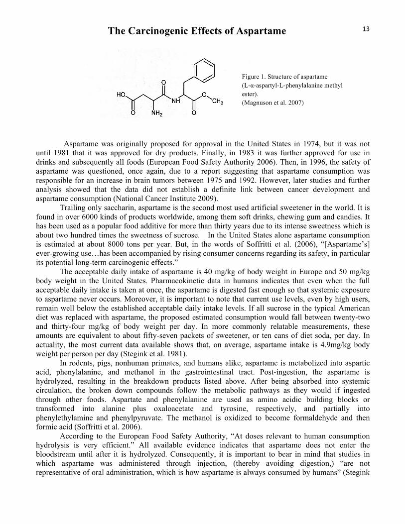
Aspartame is formally known as L-α-aspartyl-L-phenylalanine methyl ester. (Figure 1) Commonly known by the brand names Equal and NutraSweet, aspartame was accidentally discovered in 1965 by a scientist who was working on the synthesis of a gastrointestinal secretory hormone inhibitor. While working in the lab, some solution was accidentally spilled and splashed on his hand. Soon afterwards, against all good safety practices, he licked his finger to pick up a piece of paper and was shocked by the intense sweetness of the aspartame that had been splattered there (Magnuson et al. 2007).
The Carcinogenic Effects of Aspartame
13
Aspartame was originally proposed for approval in the United States in 1974, but it was not until 1981 that it was approved for dry products. Finally, in 1983 it was further approved for use in drinks and subsequently all foods (European Food Safety Authority 2006). Then, in 1996, the safety of aspartame was questioned, once again, due to a report suggesting that aspartame consumption was responsible for an increase in brain tumors between 1975 and 1992. However, later studies and further analysis showed that the data did not establish a definite link between cancer development and aspartame consumption (National Cancer Institute 2009).
Trailing only saccharin, aspartame is the second most used artificial sweetener in the world. It is found in over 6000 kinds of products worldwide, among them soft drinks, chewing gum and candies. It has been used as a popular food additive for more than thirty years due to its intense sweetness which is about two hundred times the sweetness of sucrose. In the United States alone aspartame consumption is estimated at about 8000 tons per year. But, in the words of Soffritti et al. (2006), “[Aspartame’s] ever-growing use…has been accompanied by rising consumer concerns regarding its safety, in particular its potential long-term carcinogenic effects.”
The acceptable daily intake of aspartame is 40 mg/kg of body weight in Europe and 50 mg/kg body weight in the United States. Pharmacokinetic data in humans indicates that even when the full acceptable daily intake is taken at once, the aspartame is digested fast enough so that systemic exposure to aspartame never occurs. Moreover, it is important to note that current use levels, even by high users, remain well below the established acceptable daily intake levels. If all sucrose in the typical American diet was replaced with aspartame, the proposed estimated consumption would fall between twenty-two and thirty-four mg/kg of body weight per day. In more commonly relatable measurements, these amounts are equivalent to about fifty-seven packets of sweetener, or ten cans of diet soda, per day. In actuality, the most current data available shows that, on average, aspartame intake is 4.9mg/kg body weight per person per day (Stegink et al. 1981). In rodents, pigs, nonhuman primates, and humans alike, aspartame is metabolized into aspartic acid, phenylalanine, and methanol in the gastrointestinal tract. Post-ingestion, the aspartame is hydrolyzed, resulting in the breakdown products listed above. After being absorbed into systemic circulation, the broken down compounds follow the metabolic pathways as they would if ingested through other foods. Aspartate and phenylalanine are used as amino acidic building blocks or transformed into alanine plus oxaloacetate and tyrosine, respectively, and partially into phenylethylamine and phenylpyruvate. The methanol is oxidized to become formaldehyde and then formic acid (Soffritti et al. 2006). According to the European Food Safety Authority, “At doses relevant to human consumption hydrolysis is very efficient.” All available evidence indicates that aspartame does not enter the bloodstream until after it is hydrolyzed. Consequently, it is important to bear in mind that studies in which aspartame was administered through injection, (thereby avoiding digestion,) “are not representative of oral administration, which is how aspartame is always consumed by humans” (Stegink
Figure 1. Structure of aspartame (L-α-aspartyl-L-phenylalanine methyl ester). (Magnuson et al. 2007)

Devora Sara Gelbfish 14
1987). Any cancers that resulted from systemic exposure to aspartame are not relevant to humans, since when ingested orally, aspartame is always broken down before entering systemic circulation. Overview and Analysis of Original Studies Performed From the time of its introduction by Searle Laboratories, aspartame has been surrounded by controversy. With the assistance of various scientists, Searle conducted hundreds of tests, summarized in Table 1 below, to ascertain the safety of aspartame as a food additive. Among the experimental works were studies conducted on rats, mice, hamsters, dogs, and monkeys. At the time, many felt that some important data was not reported to the Food and Drug Administration when Searle applied for the approval of aspartame. Ultimately, however, further analysis proved that none of the studies found evidence linking aspartame consumption to cancer.
In a 1996 report by Olney et al., the authors suggested that aspartame might be a cause of the increase in brain cancer in humans. From a descriptive analysis of national cancer data, they noted that the introduction of aspartame in food in the early 1980’s corresponded to the rise in brain cancer in the United States. They recommended that the safety of aspartame as a sugar substitute be reevaluated. Consequently, Gurney et al. performed a case-control study to assess the relationship of aspartame consumption and the risk of childhood brain tumors. They held in-person interviews with the biological mothers of their fifty-six case patients and ninety-four control subjects, collecting data on aspartame consumption prior to the date of diagnosis or reference date. The children were all born during or after 1981, corresponding to the Food and Drug Administration’s approval of aspartame. In addition, for forty-nine case patients and ninety control subjects, the authors evaluated the risk of brain tumor as a result of the mother’s aspartame consumption during pregnancy and breast feeding. As a result of their studies the authors found that “case children were no more likely than control children to consume foods containing aspartame,” and that “there was no suggestion of a dose-response relation based on age at first consumption, number of years of consumption, or frequency of consumption.” Additionally, they found no correlation between maternal consumption of aspartame during pregnancy or breast feeding and increased risk of brain tumors (Gurney et al. 1997). This study is informative; however, due to a number of weaknesses, it is not a strong enough proof to rule out the possibility that aspartame is linked to elevated risk of brain tumors. Firstly, much of the data was amassed through in-person interviews with the mothers of the case patients and control subjects, leaving lots of room for error as a result of biases. Aside from the fact that no one has perfect recall, people also tend to lie or exaggerate information. Also, it is very possible that the interviewers asked leading questions and that the interviewees skewed the information in an attempt to provide what they thought the interviewers wanted to hear. Furthermore, the study sample was very small and may not have been an accurate representation of the full population. Finally, studies of children are naturally limited because one cannot study the effect of the possibly carcinogenic agent over time. Therefore, even if one were to accept the results of the study, the possibility that the children who were exposed to aspartame consumption would have increased brain tumor risk as adults cannot be ruled out.

The Carcinogenic Effects of Aspartame
15
‘

Devora Sara Gelbfish 16
Table 1: Chronic Oral Toxicity Studies with aspartame and diketopiperazine (Magnuson et. al 2007) Aspartame Consumption in Relation to Childhood Brain Tumor Risk: Results from a Case-Control Study Nevertheless, there is one last important point that the authors did make. Given the fact that the peak rise in brain tumors and the introduction of aspartame occurred almost simultaneously, without the expected

The Carcinogenic Effects of Aspartame
17
period of latency, “it appears unlikely that any carcinogenic effect of aspartame ingestion could have accounted for the recent brain tumor trends as Olney et al. contend” (Gurney et al. 1997) Aspartame Induces Lymphomas and Leukemias in Rats In 2005 and 2006 Soffritti et al. of the European Ramazzini Foundation published a set of alarming study results. In fact, much of the concern regarding the safety of aspartame was generated by the initial findings of their research. In their study, the authors administered aspartame to male and female Sprague-Dawley rats with their feed. The rats, which were eight weeks old at the start of the experiment, were treated with aspartame containing feed until spontaneous death. The groups of 100-150/sex were given concentrations of 100,000; 50,000; 10,000; 2,000; 400; 80; and 0 ppm to simulate assumed daily intake by humans of 5,000; 2,500; 500; 100; 20; 4; and 0 mg/kg of body weight. The study continued for 151 weeks until the death of the last rat at 159 weeks. Upon their deaths, the animals underwent complete necropsy and examination. The authors reported the following differences observed between the treated groups and the untreated control.
1. “An increase in malignant tumor-bearing animals with a significant positive trend in males…and in female…and a statistically significant difference in females treated at 50,000 ppm…,compared to controls;
2. An increased incidence of hyperplasia of the olfactory epithelium with a significant positive trend in males and females…;
3. An increase in the incidence of…carcinomas of the renal pelvis and ureter were observed in females…;
4. A dose-related increased incidence in malignant schwannomas of peripheral nerves was observed, with a significant positive trend in males…, while in females, nine malignancies were observed among treated animals of the different dosage groups and none among controls…;
5. A dose-related increased incidence in lymphomas-leukemias was observed, with a significant positive trend in males…and females. When compared to controls, a statistically significant difference was observed in females treated at doses of [400 ppm and above]…”
Finally, they also reported sparse brain malignancies observed in the treated groups, (among males and females,) whereas none were found in the control groups.
They concluded that, for the first time, they had demonstrated a dose-related, statistically significant increase in lymphomas and leukemias in females as a result of aspartame intake. Furthermore, they felt that these results were noted at levels close to those to which humans can be exposed. “Since the results of carcinogenicity bioassays in rodents, mainly rats and mice, have been shown to be a consistent predictor of human cancer risk,” they closed their work by calling for an “urgent re-examination of permissible exposure levels of aspartame in both food and beverages” (Soffritti et al. 2006). Evaluation of the European Ramazzini Foundation Study
Immediately following the publication of the disquieting carcinogenicity study carried out by the European Ramazzini Foundation, many scientists and researchers began to assess their reported results. As stated previously, the European Ramazzini Foundation “considered that the results of their study indicate that aspartame is a ‘multipotential carcinogenic agent,’” leading to much concern. As a result, many specialists set out to attempt to either verify or discount their findings.
For example, after extensive evaluation, the European Food Safety Authority Panel concluded that the studies by the European Ramazzini Foundation contained “too many methodological flaws to be

Devora Sara Gelbfish 18
taken into consideration when determining the carcinogenic potential of aspartame” and that the Panel had “no reason to revise the previously established acceptable daily intake for aspartame (European Food Safety Authority 2006). Below are a number of flaws which invalidate the findings of the European Ramazzini Foundation study.
Firstly, there was a high background incidence of chronic inflammatory disease among the colony of rats used (European Food Safety Authority 2006). This condition was not mentioned in the study. However, the fact that the colony was already suffering from chronic respiratory disease is a very plausible explanation of the lymphomas and leukemias that developed. This information, along with the lack of a positive dose-response relationship, makes it unlikely that the increased lymphomas and leukemias were related to aspartame.
Additionally, concerning the lesions of the renal pelvis, ureter, and bladder, although they were likely treatment related, the same results cannot be expected in humans due to differences between rat and human metabolism. Due to differences in urinary protein levels, rats are much more susceptible to these tumors than humans are when exposed to high doses of chemical irritants (Cohen 1995). According to the European Food Safety Authority panel, “It is widely accepted that the effect is a high dose effect of irritant chemicals or chemicals producing renal pelvic calcification as a result of imbalances in calcium metabolism, specific to the rat.” The Panel did not consider these effects to be relevant to humans in any way.
Furthermore, the authors reported statistics for “total malignant tumors.” However, aggregating all of the incidences of malignant tumors for statistical purposes was not justified given that the lymphomas, leukemias, and renal tumors should have been excluded, as explained above.
With regard to the malignant schwannomas, the number of tumors were low. Also, despite the fact that the dose-response relationship showed a positive trend in males, it was very flat over a wide range. The European Food Safety Authority panel felt that there was also general uncertainty as to the diagnoses of these tumors and that further histopathological peer-review of the relevant nervous system slides was necessary for complete evaluation.
Finally, actual human consumption of aspartame is far less than the concentrations at which the treated rats exhibited differences from the control group. Some might be tempted to say that carcinogenicity at high doses shows that there is carcinogenicity at low doses as well, but that it is at a lower rate, referred to as dose extrapolation. However, it is incorrect to automatically assume that this is so (Cohen 1995).
As a result of the numerous flaws present in multiple areas of the study findings, the results of the European Ramazzini Foundation study are considered invalid. Further studies were necessary to ascertain the safety of aspartame. Consumption of Aspartame-Containing Beverages and Incidence of Hematopoietic and Brain Malignancies
In 2006, following multiple animal experiments which attempted to link aspartame to hematopoietic (pertaining to blood cell formation) and brain cancers, most importantly the seemingly positive linkage found in the European Ramazzini Foundation studies, a group of scientists set out to investigate the risks in humans. They “investigated the association between self-reported consumption of aspartame-containing beverages and incident hematopoietic and brain cancers.” The authors mailed out 3.5 million questionnaires to AARP members who were between the ages of fifty and seventy-one years old. Information about daily aspartame intake was obtained from these self-administered food frequency questionnaires. Of the 617,119 that were returned, 567,169 were satisfactorily completed. Excluded from the study were 52,887 persons with history of cancer, one
The Carcinogenic Effects of Aspartame
19
withdrawal, a number of duplicates, and people who had died. As a result, 473,984 questionnaires were considered, (285,079 men and 188,905 women). During a follow up of more than five years, 1,888 hematopoietic cancers and 315 malignant gliomas (brain cancer) were discovered. These findings were largely comparable to overall rates of hematopoietic cancers and gliomas within the age range, for both the male and female subgroups. Moreover, the findings were not in any way linked to aspartame intake, nor was there any correlation between higher levels of aspartame intake and increased cancer risk.
As a result of their study, the authors concluded that their findings do not support the hypothesis that aspartame increases the risk of hematopoietic or brain cancer, and that it was in direct contradiction with the study conducted by the European Ramazzini Foundation. Furthermore, the authors noted the fact that the European Food Safety Authority dismissed the findings of the European Ramazzini Foundation due to the high background of chronic inflammatory conditions in the rat colony used, the lack of a dose-response, and other issues, as mentioned previously (Lim et al. 2006).
The study of Lim et al. has a number of strengths. First and foremost, the large sample size provided for more accurate results. Additionally, the fact that the dietary and lifestyle data was collected before the patients were diagnosed with cancer minimized the biases normally found due to differential reporting between cases and controls. (This is unlike the study by Gurney et al., for example, in which the data was collected from the mothers after their children had been diagnosed.) Furthermore, the food frequency questionnaire used was developed by the National Institute of Health in conjunction with the AARP through extensive cognitive testing.
(All the same, it is important to bear in mind that any information obtained through self-reporting by study members is bound to involve at least a small amount of bias.) Conclusion After thorough review of all of the scientific data available with regard to aspartame, there is no evidence that aspartame, as consumed by humans, is carcinogenic. Nevertheless, despite the lack of scientific evidence, many people feel that aspartame is “dangerous” and “causes cancer.” Popular media often refers to aspartame with phrases like “deadly sweet” or “the sweet poison.” Headlines like “Aspartame linked to cancer” are quickly believed and difficult to overturn in the minds of the public.
For example, following the European Ramazzini Foundation study in 2005, a branch of Harvard hospital promoted research which analyzed hospital records of tens of thousands of men and women. A report publicized by the hospital concluded that “those who drink a daily diet soda sweetened with aspartame could have an increased risk of leukemia, lymphoma, or non-Hodgkin’s lymphoma.” However, in actuality, the risk was prevalent among drinkers of mostly sugared soda, as well. The lead author of the study was asked whether the research proved that aspartame is dangerous, and she emphatically replied, “No, it does not” (Bazell 2012).
Subsequently, the hospital apologized and admitted that the science it promoted was weak. However, the damage was already done. In the words of Dr. Steven Nissen, chair of the Cleveland Clinic’s cardiovascular medicine department, “Promoting a study that its own authors agree is not definite, not conclusive, and not useful for the public is not in the best interests of public health.” Unfortunately, much of the commonly believed information about aspartame is exactly that, inconclusive data that was only intended to lead to further studying (Bazell 2012).
Despite the lack of evidence proving the carcinogenicity of aspartame, wouldn’t it be better to stick to sucrose, which is considered “natural,” and avoid the chemically produced aspartame? No. With the ever-growing obesity and obesity-related conditions, sucrose itself is like a toxin. Obesity related conditions are now the number one leading cause of preventable death in the United States. More than

Devora Sara Gelbfish 20
one third of adults in the United States are obese, resulting in overwhelming numbers of conditions like heart disease, stroke, type 2 diabetes, and even obesity-related cancers (statistics based on review by the Centers for Disease Control and Prevention 2011).
Whereas the carcinogenic effects of aspartame in humans are doubtful, the deleterious effects of obesity are unambiguous and very alarming. So, where it’s a question of the diet soda or a glass of water, no one would recommend the soda. However, when it’s between a cup of juice and an aspartame-sweetened beverage, it is not so clear-cut. In essence it comes down to the question of which “poison” is worse.
References Bazell, Robert. November 5, 2012. Harvard Hospital Admits it Promoted Weak Science on Aspartame.
NBC News. http://vitals.nbcnews.com/_news/2012/10/24/14674053-harvard-hospital-admits-it-promoted-weak-science-on-aspartame.
Centers for Disease Control and Prevention. 2012. Adult Obesity Facts. http://www.cdc.gov/obesity/data/adult.html.
Cohen SM. Human Relevance of Animal Carcinogenicity Studies. Regulatory Toxicology and Pharmacology. 1995(21):75-80.
European Food Safety Authority. 2006. Evaluation of the New Study of Aspartame Carried Out by the European Ramazzini Foundation. http://www.efsa.europa.eu/.
Gurney JG, Pogoda JM, Holly EA, Hecht SS, Preston-Martin S. 1997. Aspartame Consumption in Relation to Childhood Brain Tumor Risk: Results From a Case-Control Study. Journal of the National Cancer Institute. 84(14):1072-1074.
Lim U, Subar AF, Mouw T, Hartge P, Morton LM, Stolzenberg-Solomon R, Campbell D, Hollenbeck AR, Schatzkin A. 2006. Consumption of Aspartame-Containing Beverages and Incidence of Hematopoietic and Brain Malignancies. Cancer Epidemiology, Biomarkers and Prevention. 15(9):1654-1659.
Magnuson BA, Williams GM, Burdock GA, Doull J, Kroes RM, Marsh GM, Pariza MW, Spencer PS, Waddell WJ, Walker R. 2007. Aspartame: A Safety Evaluation Based on Current use Levels, Regulations, and Toxicological and Epidemiological Studies. Critical Reviews in Toxicology. 37(8):629-727.
Mayo Clinic Staff. 2010. Artificial Sweeteners: Understanding These and Other Sugar Substitutes. http://www.mayoclinic.com/.
National Cancer Institute. Artificial Sweeteners and Cancer. 2009. http://www.cancer.gov/cancertopics/factsheet/Risk/artificial-sweeteners.
Olney JW, Farber NB, Spitznagel E, Robins LN. Increasing Brain Tumor Rates: Is There a Link to Aspartame? Journal of Neuropathology and Experimental Neurology. 1996(55):1115-1123.
Renwick AG. The Metabolism of Intense Sweeteners. Xenobiotica. 1986(16):1057-1071. Soffritti M, Fiorella B, Espositi DD, Lambertini L. 2005. Aspartame Induces Lymphomas and
Leukemias in Rats. European Journal of Oncology. 10(2):107-116. Soffritti M, Fiorella B, Espositi DD, Lambertini L. 2006. Results of Long-Term Carcinogenicity
Bioassay on Sprague-Dawley Rats Exposed to Aspartame Administered in Feed. Annals of the New York Academy of Sciences. 1076(1):559-577.
Stegink LD. The Aspartame Story: A Model for the Clinical Testing of a Food Additive. American Journal of Clinical Nutrition. 1987(46):204-215.

The Carcinogenic Effects of Aspartame
21
Stegink LD, Filer LJ Jr., Baker GL. 1981. Plasma and Erythrocyte Concentrations of Free Amino Acids in Adult Humans Administered Abuse Doses of Aspartame. Journal of Toxicology and Environmental Health. (7):291-305.

Yosef Lewis is currently completing his second year at the Physician Assistant at Touro College.
22
COMPLEX REGIONAL PAIN SYNDROME: A REVIEW OF CURRENT TREATMENTS Yosef Lewis
Abstract
Complex Regional Pain Syndrome (CRPS) is a syndrome that develops infrequently in patients that experience a minor or severe trauma to a bodily extremity. CRPS has two subtypes; Type-I and II, both are clinically characterized by hyperalgesia. During its acute stage, CRPS hyperalgesia is clinically characterized by edema in the subcutaneous tissues of the epidermis, allodynia, and localized bone resorption. In the later chronic stage, hyperalgesia is aroused by the disregulation of blood flow to the extremity and permanent dystonic and trophic changes to the skin. Because the epidemiology and central causation of CRPS remains unknown until today, health professionals are challenged to diagnose and treat its unique and changing presentations as they appear. This approach, has led to a plethora of tests and treatments that address the syndrome as it presents its clinical features. This paper is an in-depth review of available treatment modalities and surmise the efficacy of the treatments based the quality of the available research. By reviewing the effectiveness of some of the currently available treatment modalities we may gain some understanding of this enigmatic syndrome.
Introduction and History
CRPS type-I has previously been known as Sudeck’s Atrophy and Reflex Sympathetic Dystrophy. Sundeck’s Atrophy was the syndrome’s original name. It was named after Paul Sudeck who proposed in 1902 that this syndrome was an exaggerated response to nerve damage, or soft tissue injury, (Janig, 2003). In the 1950’s, John Bonica, the founder of the International Association for the Study of Pain, proposed the name Reflex Sympathetic Dystrophy; “sympathetic” dystrophy because it aptly described the pathology of the syndrome that was discovered to be maintained by the Sympathetic Nervous System. This SNS trend was discovered after patients treated with a temporary blockade of the sympathetic nervous system experienced relief from the syndrome’s symptoms, (Bonica, 1990).
CRPS Type-II was previously known as Causalgia, a derivative of the Greek terms, “Caus”, and “Algia”, which mean, heat and pain, respectively. The name, Causalgia, was coined by Dr. Silas Weir Mitchell, a nerve pain pioneer during the American Civil War. He noted in his observations the exaggerated nature of the presentation of pain in relation to the injury, a problem that was frequently found in veterans of the Civil War who were exposed to low velocity, high mass missiles used by the confederates, which overtime caused an extreme inflammatory response, followed by trophic changes at the site of injury. He also recorded, that the ensuing level of pain following injury was dependent on the extent of peripheral nerve damage, (Lau and Chung, 2004).
It became evident in the 1990’s that the dissonance created by the syndromes varying names was affecting the ability of doctors to accurately diagnose this syndrome. With this in mind a consensus workshop was held in Orlando, Florida in 1993 to develop singular terminology for the syndromes multiple etiologies and manifestations. The term Complex Regional Pain Syndrome Type-I and II was determined to be a more accurate and descriptive of the syndrome. At the same conference consensus diagnostic criteria were also laid out for two CRPS types, (Stanton-Hicks, et al., 1995).

COMPLEX REGIONAL PAIN SYNDROME
23
The defined IASP diagnostic criterion for CRPS type-I is: The presence of an initiating noxious event or a cause of immobilization. Continuing pain, allodynia or hyperalgesia that is disproportionate to the inciting event. Evidence, at some time, of edema, changes in skin blood flow, or abnormal sudomotor activity in the area of pain. The diagnosis is excluded by the existence of any condition that would otherwise account for the degree of pain and dysfunction.
The defined International Association for the Study of Pain diagnostic criterion for CRPS type-II is: The presence of continuing pain, allodynia, or hyperalgesia after a nerve injury, not necessarily limited to the distribution of the injured nerve. Evidence, at some time, of edema, changes in skin blood flow, or abnormal sudomotor activity in the region of pain. The diagnosis is excluded by the existence of any condition that would otherwise account for the degree of pain and dysfunction, (1995).
A majority of CRPS patients can identify an initial noxious event that preceded the clinical features of CRPS. In the case of CRPS type-I, which presents without a nerve lesion, the initial event is usually a minor trauma, such as an ankle sprain. In the case of CRPS type-II, which presents with a nerve lesion, a severe trauma is the usual culprit. Both CRPS Type-I and II usually present in a unilateral fashion with only one limb being affected. Both CRPS Type-I and II are marked by the dysfunction of the sympathetic nervous system (SNS), which leads to many forms of pain. About 81% of patients complain of spontaneous burning and stinging pain. Discoloration and vasomotor changes occur in 86.9% and 78.7% of patients, respectively. Allodynia, found in 69% of patients, is an abnormal sensitivity to normal mechanical or temprature stimuli. The sensitivity is so great with regards to mechanical and temperature stimuli, that clothing resting on a limb, or a breeze passing over the limb, instigates hyperalgesia. Additionally, CRPS type-II patients have symptoms that are common to neuropathy, i.e. electrical sensations, shooting pain, (Birklein, 2005).
CRPS type-I and II are marked by three distinct stages. The “Acute Stage”, during which patients are found to be undergoing an extreme inflammatory response and which is characterized by reddening and edema at the distal end of the affected limb. During the second “Chronic Stage” the affected limb begins turning bluish and cold and is said to be undergoing sudomotor dysfunction. During the final “Trophic Stage”, permanent changes to the underlying tissues occur leading to extreme weakness and fixed dystonia in the affected limb, (Johan, et al,. 2011).
Incidence rates of CRPS are unclear; two population-based studies arrived at very different data sets. A Netherlands based study found 26.2 cases per 100,000 person-years, and a USA based study found 5.5 cases per 100,000 person-years. Based on these varying data sets it can be surmised that there may be between 20,000-80,000 new cases per year in the USA. CRPS, once considered a rare syndrome, has lately become a more prevalent diagnosis, (Johan, et. al,. 2011). Some medical professionals believe its discovery by personal injury lawyers has greatly increased the reported incidents of CRPS, (Harden, 2011).

Yosef Lewis 24
1. CRPS Susceptibility via Genetic, Physiological, Psychological Predispositions and Onset Prevention
A predisposition to Complex Regional Pain Syndrome (CRPS) would go far in helping to explain why only some patients with CRPS inducing injuries go on to develop the full-blown syndrome and why others do not.
A. Genetic Predisposition Two studies have been done focusing on genetics being a predisposition variable in the onset of
Complex Regional Pain Syndrome. A study by Dutch researchers found evidence that CRPS, “runs in the family”. In this study, families with multiple familial CRPS occurrences were recruited through the Dutch Association of CRPS Patients and through referral by clinicians. The number of affected members per family and the phenotypic expression and inheritance were assessed. Demographic and clinical characteristics of Familial CRPS (fCRPS) patients were compared with those of sporadic CRPS (sCRPS) patients from a Dutch population-based study. Thirty-one CRPS families with two or more affected relatives were identified, including two families with five, four with four, eight with three and 17 with two affected relatives. In comparison with sCRPS patients, fCRPS patients had a younger age at onset and more often had multiple affected extremities and dystonia. The study concluded that CRPS could occur in a familial form, but no with clear inheritance pattern, even in the absence of a trauma, for siblings of young on-set patients. The sibling recurrence risk ratio provides the ratio of risk of disease for a person given that a sibling is affected, compared with the risk to develop the disease in the general population. The study outcome found the sibling with a familial occurrence of CRPS under the age of 50 had recurrence risk ratio of 3.4 to 5.6, values higher than 1 are indicative of familial aggregation, (de Rooij et al, 2008).
Another CRPS genetic study, used gene technology to type the Class-1 and Class-2 Major histocompatibility complexes of CRPS patients. The study found that it’s fifty-two CRPS patients CRPS were found to have a higher frequency of the HLA-DQ1 antigen then the general population (M.A. Kemler et al, 1999).
Taken as whole, these results indicate that there may be a genetic component to CRPS, and that young patients with a history of familial CRPS (fCRPS) should be aware that they carry a greater risk of developing this CRPS even in the absence of a severe trauma.
B. Physiological Predisposition
Early studies have found that the immobilization that follows an injury, especially in the case of fractures, due to casting, leads to physiological susceptibility and can cause the onset of CRPS (Johan et al, 2011). Supporting this theory, a recent study found that mechanosensitivity and thermosensitivity, characteristic features of CRPS, can be artificially induced, in healthy individuals, by immobilizing an extremity for four weeks, (Terkelsen et al, 2008).
C. Psychological Predisposition
In the past, there was a school of thought that promoted the concept of a “Sudeck personality” that predisposes one to CRPS. This “personality” construct was premised on the idea that individuals

COMPLEX REGIONAL PAIN SYNDROME
25
with a specific psychiatric pathology are prone to develop CRPS. This psychiatric perspective of CRPS has persisted even in the absence of evidence to support such a conclusion (Feliu and Edwards, 2009). In fact, a recent multi-center cohort study of 600 bone fracture patients, clearly disputes the psychiatric perspective (Johan et al, 2011). In the study, 600 patients were made to undergo the “Symptom Checklist-90”, which is an instrument that helps evaluate a broad range of psychological problems and symptoms of psychopathology. The checklist results found that none of the psychological factors included in the list predicted the onset of CRPS (Beerthuzin et al, 2011). This isn’t to say that physio-phsyco co-morbidity does not exist in patients with CRPS. It has been definitively proven that CRPS patients have higher incidences of depression and anxiety then the general populace, (Bruhel, 1992). Whether patients with CRPS have a higher rate of depression and anxiety then patients with other chronic pain syndromes is as of yet undetermined, (ibid).
Regardless of if depression is a factor in the onset of CRPS, treatment for depression with tricyclic anti-depressants has tri-fold benefits for CRPS patients. A majority of CRPS patients are depressed because of the pain and immobility that CRPS causes. Tricyclic anti-depressants relieve the depression. Pain is reduced due to the inhibitory affects that anti-depressant agents have on the re-uptake of norepinephrine and sertonin, known analgesics. Patients with CRPS are known to suffer from insomnia due the incessant pain they experience. Some anti-depressant medications have sedation type affects that bring a welcome respite from the incessant pain, (Rho et al, 2002)
D. CRPS Onset Prevention - Vitamin C An interesting, randomized, double blind trial, studied 123 patients with casted wrist fractures to see
if vitamin C could help prevent the onset of CRPS. The experimental group was treated with 500 mg of Vitamin-C per day for 50 days; the control group was treated with a placebo. Seven percent of patients in the group taking vitamin C developed CRPS-I, against 22% of patients in the control group, (Paul Zollinger et al, 1999).
There are many pharmacotherapy and interventional therapeutic techniques that are available in the treatment of CRPS. A line can be drawn between those with a discernible mechanism and those without. In addition, some treatments are directed at a specific presentation of the syndrome, while others are non-specific to the chronology and treat the general pain that is present in CRPS.
I. Pharmacotherapy & Interventional Therapeutic Techniques that are Stage Specific with Known Mechanisms.
Acute Stage – Inflammation, Testing, Pain, and Treatment
The origin of the inflammation during the acute stage of CRPS has pharmacotherapy implications, but its presence alone plays an important role in the diagnosis of CRPS, (Getson, 2006). Infrared Thermography is used to test for inflammation. The test is conducted on several symmetrical points on the affected and contra-lateral extremity, a temperature difference of 0.1 Celsius between contra-lateral limbs is considered telling and unusual, (Rho et al, 2002). Magnetic Resonance Imaging (MRI) machines are also used in diagnosing the inflammatory component of CRPS because of their

Yosef Lewis 26
ability to show activity deep in the muscle and connective tissue. The unusual MRI abnormalities found in the acute phase of CRPS are consistent with muscular edema, interstitial edema, and vascular hyper-permeability. Such MRI findings usually suggest the presence of hemodynamic abnormalities caused by sympathetic changes which may lead to ischemia of affected muscles. Chronic phase abnormalities indicated the presence of muscle atrophy and fibrosis or fatty infiltration of the affected muscle (Nishida, et al,. 2009). Three types of inflammation are possibly at play during the acute stage of CRPS, each with its own mechanism and treatment options:
A. The Classic Inflammatory Response B. Neurogenic Inflammation C. Autoimmune Inflammation
A1) Classic Inflammatory Response - Mechanism
Cornelius Celsus, a Roman encyclopediast, was the first to observe and transcribe the “Classic Inflammatory Response” as the presence of calor, dolor, tumor and rubor, which translate to warmth, pain, swelling, redness (Celsus, 47 BC). In the case of CRPS patients, an acute Classic Inflammatory Response is undoubtedly present. Today, the cause of Celsus’s observations are known to be the result of localized macromolecule extravasation, tissue acidosis, and reduced oxygen extraction, which leads to severe pain via the formation of free oxygen radicals that damage and thicken the basement membrane, (Goris, 1991).
A2) Classic Inflammatory Response - Treatment
The theorized role that free oxygen radicals play in the pain and inflammation associated with CRPS has been shown to be efficacious by the successful use of treatment modalities that scavenge and remove free oxygen radicals. In a comprehensive randomized double blind study two free radical scavenger medications were used. Topical 50% Dimethylsulfoxide (DMSO) was applied five times a day, or N-Acetylcysteine (NAC) tablets were ingested three times a day. Both treatments were found to be equally effective in providing temporary relief from CRPS’s inflammatory response, (Perez et al, 2002).
B1) Neurogenic Inflammation - Mechanism
A second possible culprit in CRPS’s inflammatory response may be caused by abnormal cytokine activity at the trauma site, which leads to “Neurogenic Inflammation”. Cytokines are heavily involved in the cascading reaction that activates the natural nociceptor response by the retrograde depolarization of small-diameter primary afferent nerves. This leads to the release of numerous neuropeptides, including, Substance P, Calcitonin Gene Related Peptide (CGRP), and Somatostatin, (Birklein F. , 2001). These neuropeptides in turn evoke a vasodilation and a protein extravasation response in the epidermis, leading to the signature reddening, warming, and edema, of CRPS, (Weber et al, 2000).
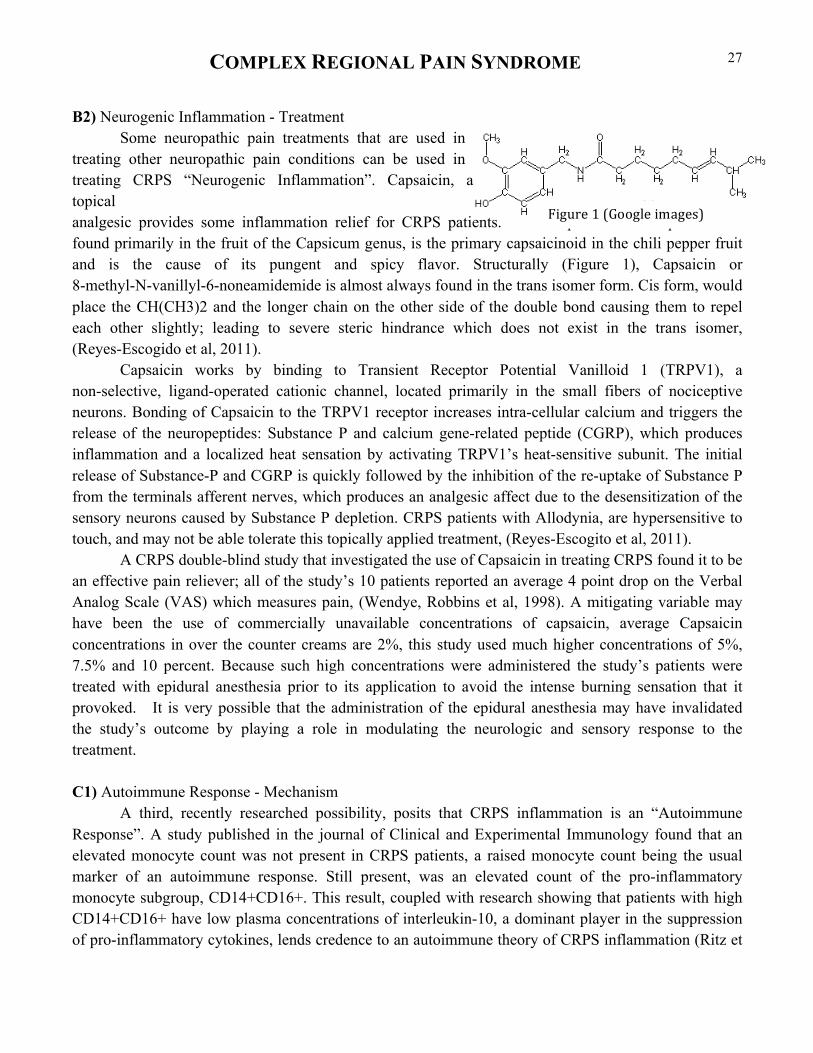
COMPLEX REGIONAL PAIN SYNDROME
27
B2) Neurogenic Inflammation - Treatment Some neuropathic pain treatments that are used in
treating other neuropathic pain conditions can be used in treating CRPS “Neurogenic Inflammation”. Capsaicin, a topical analgesic provides some inflammation relief for CRPS patients. Capsaicin is a unique alkaloid found primarily in the fruit of the Capsicum genus, is the primary capsaicinoid in the chili pepper fruit and is the cause of its pungent and spicy flavor. Structurally (Figure 1), Capsaicin or 8-methyl-N-vanillyl-6-noneamidemide is almost always found in the trans isomer form. Cis form, would place the CH(CH3)2 and the longer chain on the other side of the double bond causing them to repel each other slightly; leading to severe steric hindrance which does not exist in the trans isomer, (Reyes-Escogido et al, 2011).
Capsaicin works by binding to Transient Receptor Potential Vanilloid 1 (TRPV1), a non-selective, ligand-operated cationic channel, located primarily in the small fibers of nociceptive neurons. Bonding of Capsaicin to the TRPV1 receptor increases intra-cellular calcium and triggers the release of the neuropeptides: Substance P and calcium gene-related peptide (CGRP), which produces inflammation and a localized heat sensation by activating TRPV1’s heat-sensitive subunit. The initial release of Substance-P and CGRP is quickly followed by the inhibition of the re-uptake of Substance P from the terminals afferent nerves, which produces an analgesic affect due to the desensitization of the sensory neurons caused by Substance P depletion. CRPS patients with Allodynia, are hypersensitive to touch, and may not be able tolerate this topically applied treatment, (Reyes-Escogito et al, 2011).
A CRPS double-blind study that investigated the use of Capsaicin in treating CRPS found it to be an effective pain reliever; all of the study’s 10 patients reported an average 4 point drop on the Verbal Analog Scale (VAS) which measures pain, (Wendye, Robbins et al, 1998). A mitigating variable may have been the use of commercially unavailable concentrations of capsaicin, average Capsaicin concentrations in over the counter creams are 2%, this study used much higher concentrations of 5%, 7.5% and 10 percent. Because such high concentrations were administered the study’s patients were treated with epidural anesthesia prior to its application to avoid the intense burning sensation that it provoked. It is very possible that the administration of the epidural anesthesia may have invalidated the study’s outcome by playing a role in modulating the neurologic and sensory response to the treatment.
C1) Autoimmune Response - Mechanism
A third, recently researched possibility, posits that CRPS inflammation is an “Autoimmune Response”. A study published in the journal of Clinical and Experimental Immunology found that an elevated monocyte count was not present in CRPS patients, a raised monocyte count being the usual marker of an autoimmune response. Still present, was an elevated count of the pro-inflammatory monocyte subgroup, CD14+CD16+. This result, coupled with research showing that patients with high CD14+CD16+ have low plasma concentrations of interleukin-10, a dominant player in the suppression of pro-inflammatory cytokines, lends credence to an autoimmune theory of CRPS inflammation (Ritz et
Figure 1 (Google images)

Yosef Lewis 28
al, 2011). A small trial at University College London gave further support to an autoimmune inflammation theory. Participants in the trial were prescribed Intravenous Immunoglobulin (IVIG) antibodies, an autoimmune treatment modality, to treat CRPS inflammation. The study found that patients experienced 50% more pain relief with Intravenous Immunoglobulin over placebo, (Andreas Goebel et al, 2010). Limitations of the trial were its small size and that the effective dosage was not ascertained. C2) Autoimmune Response - Treatment
Ketamine, a veterinary and human anesthetic has been found to be efficacious in treating the CRPS “Autoimmune Response” inflammation. Recent research found that ketamine suppresses lipopolysaccharide-induced tumor necrosis factor-A, interleukin (IL)-6, and IL-8 production, and recombinant human tumor necrosis factor-induced IL-6 and IL-8 production in human whole blood, all of which play a role in the proliferation of pro-inflammatory cytokines, (Kawasaki et al, 1999). In a recent case study in Germany, a single CRPS patient was treated with Ketamine after other CRPS treatment modalities failed to relieve the symptoms. Under standard ICU conditions the patient was given bolus injections of Ketamine while sedated with Midazolam. Upon waking the patient was relieved of all CRPS pain. After discharge from the hospital, the patient’s chief complaint was regarding the psycomimetic side effects of Ketamine. Steady treatment with Midazolam helped negate this side effect, which abated after a month. In a subsequent German study, a larger grouping of patients was used. The study concluded that Ketamine is only effective during the acute stage of CRPS, when pain and swelling is localized to the distal end of a limb. It was ineffective in treating chronic CRPS patients whose symptoms were refractory and spreading, (Kiefer et al, 2007). A recent double-blind study arrived at similar findings with Ketamine delivering significant pain relief when administered on an outpatient basis and in low doses, (Schwartzman et al, 2009). The aforementioned study did not find any evidence that Ketamine was ineffective based on the duration of the syndrome; the case study outcome can therefore be called into question. Corticosteroids – A non-mechanism specific inflammation treatment
Corticosteroids, known commonly as steroids are part of a number of options that health professionals have when treating inflammation. Steroids are one of the most oft prescribed anti-inflammatory drugs and are an accepted treatment for the acute inflammatory stage of CRPS. The predominant effect of corticosteroids is to switch off multiple inflammatory genes that encode for, cytokines, chemokines, adhesion molecules, inflammatory enzymes, receptors and proteins that are activated during a chronic inflammatory process, (Fischera et al, 2010).
A recent study evaluated the use Prednisolone, a steroidal anti-inflammatory, versus Piroxicam, a non-steroidal anti-inflammatory. The study found Piroxicam to be an effective inhibitor of arachidonic acid metabolism, an inflammation enhancing intermediate. It was also found to inhibit the production of the pain neurotransmitters, substance P and Calcitonin Gene Related Peptide, which play a part in the production of inflammation enhancing leukotrienes. The study concluded that Piroxicam seems to have

COMPLEX REGIONAL PAIN SYNDROME
29
definitive chemical benefits, but little if any benefit in actually reducing sensory and motor pain, which continued unabated, (Kalita et al, 2005).
These findings suggest a dominant neural mechanism in the pathophysiology in CRPS. Confounding this possibility was the study’s discovery that Prednisolone, the steroidal anti-inflammatory, was very effective in relieving actual pain in 83% of patients, (ibid). A drawback to this study was the failure to address this discrepancy. Some of the recorded side effects of long-term steroid use are hematologic, hepatic, musculoskeletal, neurologic and psychiatric damage and in some cases a severe weakening of the heart, (Parker and Thompson, 2010).
IV. Acute Stage - CRPS Bone Resorption, Testing, Pain and Treatment Bone Resorption Testing Bone resorption in CRPS can be diagnosed by simple radiography and Three Phase Bone
Scintigraphy tests. Both tests register the presence of bone resorption and osteoporosis. A normal bone scan finding, without radiographic osteoporosis, precludes a diagnosis of adult CRPS. Some disagreement has been registered as to how early-on radiography can be used. A study by P.H.J.M Veldman (1993) I in the Netherlands, found spotty osteoporotic changes 4-8 weeks after a CRPS diagnosis. Other research has pointed to there being spotty changes earlier at the two-week marker, (Rho and Brewer, 2002).
Three Phase Bone Scintigraphy coupled with the tracer, Technetium Tc-99 Bisphosphonates, is highly sensitive test that can detect osseous changes earlier then radiography. A positive result will register a marked increase in tracer uptake during bone scintigraphy’s, blood pool, blood, and mineralization phases. Increased tracer uptake is an indication of increased bone metabolism and breakdown. Some variability in the results of Bone Scintigraphy has been found based on the duration of CRPS , (Rho and Brewer, 2002).
Bone Resorption Pain
Immunohistochemical studies of the bone have shown there to be an extensive network of peptidergic and sympathetic sensory fibers that are present throughout the bone marrow and periosteum, Included in this network are two acid sensors, Acid-Sensing Ion Channels (ASICs) and Transient Receptor Potential Vanilloid Subtype 1 (TRPV1). In CRPS patients these pain-inducing receptors are activated by the presence of an acidic microenvironment in the bone. CRPS’s acidic bone environment is generated by osteoclasts, a membrane bound proton pump that releases protons (acid) through Vacuolar H+-ATPase. Osteoclasts are the primary culprit in bone resorption, (Yanow et al, 2008).
Also active and present during CRPS bone inflammation are: mast cells, macrophages, endothelial cells, osteocytes, osteoblasts, and bone marrow stromal cells, which are involved in the production and storage of Nerve Growth Factor (NGF) protein. When present, NGF initiates the transcription for genes that encode the TRPV1 pain receptor, causing a further increase in the already painful CRPS nociceptive response. Cytokines and Prostaglandins are also found in the CRPS bone micro-environment and act synergistically to activate the network of nociceptors innervating the bone and bone marrow (Leon and Buriani, 1994).

Yosef Lewis 30
Bone Resorption Treatment
An accepted treatment for CRPS bone related pain are, nitrogen and non-nitrogen bisphosphonates, which have anti-nociceptive properties that ameliorate bone pain; bisphosphonates work on osteoclasts and inhibit their activity leading to a decrease in the proton concentration and Nerve Growth Factor expression in the bone micro-environment. The non-nitrogen bisphosphonates, Clodronate ® and Etidronate ®, work by causing the buildup of metabolites in the cell which inhibit osteoclasts from functioning, causing cell death. Nitrogen-containing bisphosphonates, such as Alendronate®, Risedronate® and Zoledronate® interfere with the mevalonate biosynthetic pathway and protein lipidation, and in the signaling functions of key regulatory proteins, also leading to cell death. Bisphosphonates are a well-vetted and efficacious treatment, which has been well documented in four studies, (Manicourt et al, 2004; Robinson and Sandom, 2004; Varenna and Zucchi, 2000; Admai and Fossaluzza, 1997).
The randomized, double-blind study done by Varenna and Zucchi on the use of Clodronate®,
found that 72% of patients showed significant pain relief 40 days into the trial. The numbers continued to improve with the passage of time, 75% at 90 days and 93.2% at 180 days, all experienced significant pain relief. Taken as a whole, these studies point to bisphosphonates being very useful in the treatment of CRPS bone related pain. The aforementioned studies did not find that bisphosphonates relieved any of CRPS other symptoms and the pain associated with them. Since their use is limited to bone pain, bisphosphonates need to be incorporated into a complete pharmacotherapy solution in order for CRPS patients to gain effective relief from the syndrome.
V. Non-Stage Specific Pharmacotherapy & Interventional Therapeutic, with known mechanisms.
Neurogenic Pain relief via Regional Sympathetic Blockade
Intravenous regional anesthesia was first proposed by Dr. August Bier in 1908 and till today is known as the “Bier Block”. Technically the treatment is simple. A tourniquet is applied to the ailing extremity creating a complete venous blockade followed by the injection of an analgesic. This treatment modality has since been used in the treatment of many localized pain diseases. In 1974 John Hannington-Kiff proposed a CRPS treatment with a derivative of the Bier Block, called Intravenous Regional Sympathetic Blockade (IRSB), (Hanington-kiff, 1974). The favored analgesic for this procedure was Guanethidine, because it is sympatholytic that selectively prevents peripheral sympathetic nerve transmission with the following mechanism: at first, when injected, Guanethidine causes the release of norepinephrine from the synaptic cleft, followed by the movement of Guanethidine into the synaptic cleft via uptake 1; it is then transferred into the presynaptic norepinephrine vesicle via norepinephrine transporter, also known as NET. Because norepinephrine is prevented from returning to the vesicle there is an initial spike in the sympathetic tone of the limb. Once the released norepinephrine

COMPLEX REGIONAL PAIN SYNDROME
31
is metabolized, the Guanethidine populating the norepinephrine vesicle prevents the further production and release of norepinephrine, leading to a state of analgesia, , (Tollison and Satterthwaite, 2002).
The few double-blind studies that have reviewed the use of Guanethidine have shown it to be ineffective against a saline placebo in both short and long-term observations, (Kingery, 1997). A number of other sympatholytics such as Reserpine®, and Clonidine® have been found to be ineffective in relieving CRPS pain, aside from Bretylium®, which showed positive effects when used in the IRSB treatment, (ibid).
Local Anesthetic Sympathetic Blockade (LASB) is another Bier Block type treatment modality that is precluded from needing a tourniquet because the anesthetic solution is injected directly into the spine’s sympathetic structures. In this procedure, one of two spinal structures are targeted, either the stellate ganglion, or the lumber sympathetic chain. The injection takes place under fluoroscopic or computed tomographic guidance to avoid damaging the spinal column. Once injected, the anesthetic solution induces a complete signal blockade. All sympathetic signals to and from the affected limb are effectively muted by the anesthetized spinal structure.
In the past, the “gold standard” for the treatment of CRPS was the local anesthetic sympathetic blockade (LASB). The LASB treatment played a unique role in diagnosing and treating CRPS. It was unique in this dual application. The 1993 Orlando Conference changed this role when CRPS’s diagnostic criterion was modified to exclude an analgesic response to a sympathetic nerve block. Thereafter, LASB was no longer considered an essential piece of confirmatory diagnostic information because the syndrome’s classification was broadened to include patients without Sympathetically Maintained Pain (SMP). Accordingly, a response to a sympathetic block is not required to diagnose CRPS because the syndrome’s criterion now includes patients without sympathetic dysfunction. Identifying patients who have Sympathetically Maintained Pain is still very important because of the short term relief gained from a sympathetic blockade; this effective short term relief has made the LASB the go to adjunctive therapy for patients undergoing physiotherapy, (Sharma and Williams, 2006).
But this positive view of LASB has been tempered by a comprehensive review on behalf of the Chocrane Collaboration where little evidence was found to support the positive view of LASB . The Chocrane Collaborations criteria for inclusion were that the trials be randomized and double blind. Excluded from the review were studies evaluating somatic nerve blocks and studies evaluating the effects of orally, intravenously and epidurally administered anesthetic or sympatholytic drugs. Two unpublished studies cited in this review, fit this criterion. One, a double-blind study by Dr. Donald D. Price, found that six out of seven patients experienced at least 50% short-term pain relief when treated with the anesthetic Lidocaine® over normal saline. The duration of relief was 3 days for local Lidocaine® versus 19.9 hours for the saline placebo. A second study by R.J. Verdugo found that 12 of 16 patients had at least 50% short-term pain relief while receiving Bupivacaine® versus the saline placebo. The combined Relative Risk Ratio of these two trials was 1.17. A Relative Risk Ratio of 1.17 is slightly greater then a Risk Ratio of 1, which implies little significant difference was found between experimental and control groups in the study. These findings clearly show that LASB treatment needs further research to reclaim its status as a “gold standard” in treating CRPS. The studies long-term outcomes were precluded from being combined in a Risk Ratio because the both studies gauged

Yosef Lewis 32
different outcomes. Dr. Price recorded the duration of pain relief, while Dr. Verdugo evaluated number of subjects who had at least 50% of pain relief (Cepeda et al, 2010). Neurogenic Pain Treatment: Surgical and Chemical Sympathetic Denervation
CRPS patients with Sympathetically Maintained Pain (SMP) suffer from a dysfunctioning Sympathetic Nervous System. Permanently denervating the system inhibits signaling and can be achieved via a surgical or chemical sympathectomy. Surgical denervation
Surgical denervation is accomplished via Open Lumbar or endosopic sympathectomy surgery, (Bandyk et al, 2002). In these procedures the sympathetic ganglion are destroyed via its open removal or by electro-coagulation. A well recognized side effect to a surgical sympathectomy is hyperhidrosis, this side effect was recognized as early as 1933, when a Dr. J. Paterson Ross lectured at the Royal College of Surgeons in London and recounted that, “some of our patients have stated emphatically that the secretion of sweat has been considerably more profuse in areas not affected by the operation . . . the remark has been so frequently made that the possibility of compensatory hypersecretion cannot be excluded”, (Ross, 1933). Current research wholly concurs with his observation. In one study, patients experiencing palmer hyperhidrosis that underwent a sympathectomy almost all experienced some form of compensatory sweating, usually in the trunk area, (Gossot et al, 2003; Ojimba & Cameron, 2004). Another study of 73 patients found that 8 out of the 83 surgical sympothectamy procedures done in the study, 10 of the studies 73 patients needed the procedure to be repeated, resulted in Hyperhidrosis, (Bandyk et al, 2002). Chemical Sympathectomy
Chemical Sympathectomy is achieved by injecting 50% to 100% ethanol or 7% to 10% Phenol into sympathetic ganglia, (Dunn, 2000). Phenol, a strong anti-septic solution causes protein coagulation and necrosis when directly applied to nerves (Copping et al, 1969). Ethanolcauses alcohol neurolysis, which involves the extraction of phospholipids and cerebrosides from the neural membranes and by precipitation of mucoproteins. Both are effective in causing nerve / ganglion death which delivers anesthesia by the inhibition of nociceptive signaling. The only available double blind of chemical sympathectomy contrasted the use of chemical sympathectomy to radio frequency sympathectomy. Both treatments were found to equally effective in delivering 50% pain relief in a grouping of 20 patients, (Straube et al, 2010).
VI. Pharamcotherapy & Interventional Therapeutic Techniques with Unknown Mechanisms
Bone Resorption Treatment - Calcitonin
In 1961, Dr. D.H. Copp discovered a calcium-regulating neuropeptide hormone. At the time of his discovery he was studying the control of calcium secretion by parathyroid hormone (PTH) when he found that a neuropeptide was released in the presence of hypercalcemia that lowered plasma calcium by

COMPLEX REGIONAL PAIN SYNDROME
33
inhibiting osteolysis. The newly discovered neuropeptide was named calcitonin (Copp, 1994). Subsequently, experiments were performed to see if in vivo use of calcitonin would be useful in the treatment of osteoporosis / bone resorption, (Özoran and Seçkin, 2005), studies have concluded that Calcitonin is an effective in the treatment of osteoporosis, (Karsdal et al, 2010).The use of intramuscular or subcutaneously injected calcitonin is not without side effects. Its most common side effects being gastrointestinal: anorexia, nausea, vomiting, a metallic taste and diarrhea are the prevalent presentations of its gastrointestinal effects. Vascular phenomena, such as flushing or shivering, are also observed in some patients. Generally these side effects are dose related and inconvenient rather than serious, but they can occur in up to 80% of patients on high doses, (Siminoski et al, 1996).
In addition to inhibiting osteoporotic type bone resorption, calcitonin was found have analgesic effects on bone related pain, making it a perfect candidate for the treatment of CRPS patients suffering from bone resorption and bone pain, (Blau, 2003). A study of CRPS patients by Fushun Sahin and Figen Yilmaz assessed the efficacy of salmon calcitonin in a randomized, single-blind study. The control group received Paracetamol®, an over the counter analgesic 1500 m/day, while the other experimental group received salmon calcitonin 200 IU/day, for 2 months. Both the control and experimental groups showed remarkable recovery on either paracetamol or calcitonin when given the treatment in conjunction with physical therapy, but little marked difference was found between the two groups. The study concluded that physical therapy combined with a simple analgesic is an efficient means of therapy and that calcitonin makes little or no contribution in the treatment of patients with acute CRPS, (Sahin et al, 2006).
Another study utilized nasal calcitonin given the disadvantage in the administration of regular calcitonin, i.e. it must be administered parenterally. This prospective randomized double-blind study; using sensitive methods of measuring the response to treatment, did not demonstrable any effect on the clinical or skeletal progression of CRPS bone resorption,. The authors hypothesized, “not finding a difference between the treated and placebo wing is that the bioavailability of the nasal formulation was too low. Although there was a small decrease in serum calcium during the treatment period, the response was not associated with changes in the indices of bone turnover. In particular, the fasting urine excretion of calcium did not change. This suggests that the decrease in serum calcium was due to a decrease in renal tubular reabsorption of calcium, a known action of calcitonin”, (Bickerstaff and Kanis, 1992)
A blinded meta-analysis of 21 CRPS treatment clinical trials concluded that using Calcitonin in the treatment of CRPS has a positive effect on pain on average and encouraged its use, (Perez et al, 2001). Perez based his assertion on a study that found calcitonin to have the conclusively beneficial effect of significantly less pain, (Gobelet et al, 1992). Calcitonin’s mechanism and effectiveness as a treatment option, remains as of yet undetermined. The antinociceptive properties of calcitonin have been attributed to serotoninergic and cathecholaminergic mechanisms, Ca 2 fluxes, protein phosphorylation, endorphin production, cyclooxygenase inhibition and histamine inference. None of these mechanisms have been conclusively proven.

Yosef Lewis 34
Gabapentin Gabapentin was marketed for the first time in Febuary 1994 as an anti-convulsant drug. Today it
is widely viewed as an affective adjunctive therapy for seizures. Structurally, it is an analogue of y-aminobutyric acid (GABA). It’s synthesis as a GABA mimetic drug was so that it could cross the blood-brain barrier. Pharmacologicaly it is different from other substances that interact with GABA synapses for the reason that it binds only in the outer layers of the neocortex and hippocampus and does not bind with GABA receptors at all.
The first gabapentin study on RSD patients had a remarkably successful outcome with all six patients, with one fully recovering from RSD. The authors hypothesized that a gabapentin induced an increase in 5HT, (which are serotonin receptors) and serotonergic-like activity at a novel receptor site in the CNS, and also via serotoninergic fibers in the raphe magnus which descend into the posterior portion of the lateral funiculus of the spinal cord.
These descending fibers they posited blocked the transmission of nociceptive information through the dorsal horn and root ganglion by reciprocally blocking pain-inducing catecholamines, resulting in a gradual reduction of the nor-adrenergic induced hyperalgesia, , (Mellick and Mellick, 1995). A problem with their hypothesis is that it was vicariously deduced from the increased presence of serotonin in patients treated with gabapentin. Therefore few definitive conclusions can be drawn from it. Even so, the efficacy of the use gabapentin to treat CRPS “pain” is well founded. A double-blind and randomized placebo-controlled 8-week study of 304 CRPS patients found that measured pain scores decreased by 21% in patients receiving gabapentin and by 14% in placebo treated patients, (Serpell, 2002).
Spinal Cord Stimulation
Spinal cord stimulation is a relatively new therapeutic modality to gain acceptance in the treatment of CRPS. In 1965, Melzack and Wall published a revolutionary paper that theorized that a “gate” existed in the dorsal horn of the spinal cord which controlled the transmission of neural activity that signaled pain. This “gate” was said to be open when there was an excess of small over large afferent fiber activity in the peripheral nervous system, and the gate was closed when there was an excess of large-diameter afferent fiber activity. On the basis of this theory, scientists have tried to selectively activate large diameter afferent fibers through electrical stimulation, thereby closing the “gate” and reducing or eliminating painful inputs to the spinal cord and brain, (Melzack and Wall, 1965). Utilizing this theory, medical professionals have been able to induce this effect using a spinal cord stimulator which is surgically placed along the spine. It is theorized that its analgesic effect may come from neuromodulation that restores normal gamma-aminobutyric acid (GABA) levels in the dorsal horn and affects the release of adenosine, which is an anti-inflammatory, thereby reducing neuropathic pain (Oakley and Prager, 2002).
A systematic review of the literature on spinal cord stimulation reported a meaningful 2-point mean reduction in the Visual Analogue Pain Scale ratings in patients with CRPS type-I, (Taylor et al, 2006). This was based on results derived from a randomized controlled study (Kemler et al, 2000), and 25 other case series. The controlled study wasn’t blinded for the obvious reason that it is not advisable to

COMPLEX REGIONAL PAIN SYNDROME
35
try to mimic parasthesia-type effects on the spinal cord of patients in the placebo group. The controlled study showed -2.7 drop on the Visual Analogue Pain Scale scale in the spinal cord stimulated group at 12 months, while the control group had a +0.4 increase on the Visual Analogue Pain Scale pain scale. The systematic review data showed that almost two-thirds of CRPS type-I and type-II patients, reported at least 50% improvement in their pain scores over a median follow-up period of 33 months. It can be concluded that spinal cord stimulation is an efficacious treatment for CRPS patients with sympathetically maintained pain.
In addition to the study’s pain analysis, Taylor also reviewed treatment cost to ascertain long
term cost of spinal cord stimulation vis-à-vis physical therapy. He found that the control group which received physical therapy cost $6000 over 12 months. The spinal cord stimulation treatment group had a significantly higher balance of $10,200. But in an interesting turn around, the cost numbers reversed over time and the spinal cord stimulation treatment produced a lifetime cost saving of approximately $60,000. Opioids
Opioids are one of the oldest classes of pain relievers, (Ballantyne and Mao, 2003). Victorian women were said to use a morphine containing tincture called, laudanum, which is a 40% ethanol solution of dissolved opium and herbs. This was done to treat the travails and boredom of Victorian life, accomplishing what is known in the vernacular as getting high, (Berridge and Edwards, 1987). Opioids are still used in the treatment of chronic nociceptive and neuropathic pain, (Harke et al, 2001). Research has shown that opioids are ineffective in delivering analgesia in low dosage; concurrent research has found that the use of opioids in large dosage can itself cause hyperalgesia, a key symptom of CRPS. Other known opioid side effects are hypogonadism, long-term cognitive impairment, personality changes, tolerance, long-term toxicity and drug dependence (Harden, 2007). The use of opioids in treating neuropathic pain has been frowned upon for some time, not because of its cornucopia of side effects, but also because research found it be an ineffective treatment modality. A study found infusions of morphine to be completely ineffective in relieving neuropathic pain, (Arnér and Meyerson, 1989).
Current research is slowly shifting this negative opioid perspective. A randomized control study used Methadone to treat 20 patients and found statistically significant pain relief P 0.013-0.020 on the Visual Analogue Pain Scale scale, (Morley et al, 2003). Dose titration must be done cautiously, since large differences in Methadone dose tolerance have been found, (Cruciani, 2007). In too large dosage Methadone causes drug toxicity and affects the heart by causing an elongated QTc interval, (Kornick et al, 2003). Still, its prolonged half-life, potency, and low cost make it a preferable candidate amongst other opioids. Tramadol, another opioid, has also been vindicated as an effective reliever of allodynia and other pain in recent randomized double-blind study, (Sindrup et al , 1999). Intrathecal analgesia can be achieved with an injection of Baclofen for patients that cannot tolerate orally administered opioids because of their side effects, (Cohen 2007).

Yosef Lewis 36
Physical Therapy Physical Therapy is broadly mentioned in the literature as an important aspect in recovering from CRPS, (Harden R. 2011; Kemler et al, 2000; Rho et al, 2002). Unfortunately there is little hard evidence to support this treatment modality. An oft mentioned proof of Physical Therapy’s effectiveness, is randomized double-blind study of patients with CRPS Type-I of less than one year of duration, which indicated that physical therapy was superior to occupational therapy, and that both physical therapy and occupational therapy were more effective then social work therapy, (Oerlemans et al, 2000). Unfortunately, the study’s outcome was tempered by the fact no significant differences were found in the long-term for active range of motion of the shoulder, elbow, and forearm utilizing the three treatment modalities. The immediate improvement seen in patients that were treated with physical therapy weren’t any different then the control group a year after inclusion in the study. Conclusion
If left untreated, CRPS can result in permanent deformities and chronic pain requiring a range of long-term pharmacologic and non-pharmacologic treatments. If CRPS is caught early, sympathetic nerve blocks may be used to stop or cure the progression of the disease. Other therapies used to treat patients with CRPS such as, spinal cord stimulation, opioids, anti-inflammatory medications can all play a role in modulating and sometimes curing CRPS pain. The absence of well-defined criteria for the diagnosis of this syndrome has resulted in a lack of Randomized Controlled Trials for the treatment of CRPS. Some of the medications that have been tested to treat the CRPS population include certain antidepressants, anticonvulsants, anesthetics, anti-inflammatories, opioids, calcitonin, bisphosphonates, and neuropathic coanalgesics. There are no medications that are FDA approved for the treatment of CRPS.
References
Admai, S., Fossaluzza. V. (1997) Bisphosphonate therapy of reflex sympathetic dystrophy. Annals of the Rheumatic Diseases, 56: 201-204.
Arnér, S., Meyerson, B. (1989). Lack of analgesic effect of opioids on neuropathic and idiopathic forms of pain. Pain , 11–23.
Assessments, P. (n.d.). http://psychcorp.pearsonassessments.com/HAIWEB/Cultures/en-us/Productdetail.htm?Pid=PAg514. Retrieved from Pearson Assessments.
Ballantyne, J. C., Mao,A. J. (2003). Opioid Therapy for Chronic Pain. New England Journal Medicine , 349:1943-1953.
Bandyk, D. F., Johnson, B. L. (2002). Surgical Sympathectomy for RSD Syndromes. Journal of Vascular Surgery , 269-277.
Beerthuizen, A, AVan ‘t Spijker, F. J.P.M. Huygen, J Klein, R De Wit. "Is There an Association between Psychological Factors and the Complex Regional Pain Syndrome Type 1 (CRPS1) in Adults? A Systematic Review." Pain 145.1-2 (2009): 52-59. Print.

COMPLEX REGIONAL PAIN SYNDROME
37
Berridge, V., Edwards, G. (1987 ). Opium and the People: Opiate Use in Nineteenth-Century England. New Haven: Yale University Press.
Bickerstaff, D., Kanis, J. A. (1992). The Use of Intranasal Calcitonin in the Treatment of Algodystrophy. Rheumatology , 8: 568-569.
Birklein, F. (2005). Complex Regional Pain Syndrome. Journal of Neurology , 131-138. Blau, L. A. (2003). Analgesic Efficacy of Calcitonin for Vertebral Fracture Pain. Analgesic
Efficacy of Calcitonin for Vertebral Fracture Pain , 4: 564-570. Bonica, J. (1990). Causalgia and other reflex symapathetic dystrophies. Philadelphia: Lea &
Febiger. Bruhel, S. (1992). Predisposing Psychological Factors in the Development of Reflex
Sympathetic Dystrophy: The Clinical Journal of Pain , 287-299. Celsus, A. C. (47 BC). De Medicina. Florence. Cepeda, M., Carr, D., Lau, J. (2010). Local Anasthetic sympathetic blockade for CRPS . The
Cochrane Library , 1-11. Cohen, S. P., Dragovich, A. (2007). Intrathecal Analgesia. Medical Clinics of North America ,
863–882. Copp, D. (1994 ). Calcitonin: discovery, development, and clinical application. Clinical And
Investigative Medicine 77-268. Copping, J., Willix, R., Kraft, R. (1969). PAllitive Chemical Splanchnicectomy. Archives of
Surgery , 98-418. Cruciani, R. A. (2007). The Use of Opioids in CRPS - related Pain. RSDSA Review. De Rooij, A., M. Demos, M. Sturkenboom, J. Marinus, A. Vandenmaagdenberg J. Vanhilten.
"Familial Occurrence of Complex Regional Pain Syndrome."European Journal of Pain 13.2 (2009): 171-77. Print.
Dunn. (2000). CRPS type-I: Part II. AORN Journal , 643-653. Feliu, M. H., C. L. Edwards. "Psychologic Factors in the Development of Complex Regional
Pain Syndrome: History, Myth, and Evidence." The Clinical Journal of Pain 26.3 (2010): 258-63. Print.
Fischera, S. G., Zuurmonda, W. W., Birkleinc, F., Loera, S. A., Pereza, R. S. (2010). Anti-inflammatory treatment of Complex Regional Pain Syndrome. Pain , 251–256.
Getson, P. (2006). The Use of Thermography in the Diagnosis of CRPS. The Journal of the American Academy of Pain Managment .
Gobelet, C., Waldburger, M., Meir, J. (1992). The effect of adding calcitonin to physical treatment of reflex sympathetic dystrophy. Pain , 48: 171-175.
Goebel, A, A. Baranowski, K Maurer, A.Ghiai, C McCabe, P. (2010). Intravenous Immunoglobulin Treatment of the Complex Regional. Annals of Internal Medicine , 152:152-158.Goris, R. (1991). Are toxic oxygen radicals invloved in the pathogenesis of Reflex Sympathetic Dystrophy. Free Radical Research , 8-13.
Gossot, D., Galetta, D., Pascal, A. (2003). Long-Term Results of Endoscopic Thoracic Sympathectomy for Upper Limb Hyperhidrosis. Annals of Thoracic Surgery , 75: 1075-9.
Hanington-kiff, J. (1974). Intravenous regional sympathetic block with guanthidine. Lancet , 1019-1020.
Harden, R. (2011). Complex Regional Pain Syndrome. British Journal of Anasthesia , 99-106. Harden, R. N. (2007). Opioids for CRPS? Think again. RSDSA Review .

Yosef Lewis 38
Harke, H., Gretenkort, P., Ladleif, H. U. (2001 ). The Response of Neuropathic Pain and Pain in Complex Regional Pain Syndrome. Anesthesia and Analgesia , 488-495.
Janig, W. (2003). Complex Regional Pain Syndrome: Mystery Explained? The Lancet - Neurology , 687-688.
Johan, M., Moseley, L., Birklein, F. (2011). Clinical features and pathphysiology of CRPS. The Lancet - Neurology
Kalita, J., Vajpayee, A., Misra, U. (2005). Comparison of prednisolone with piroxicamin complex regional pain syndrome following stroke: a randomized controlled trial. Q J Med - Oxford University Press , 99: 89–95.
Karsdal, M., Byrjalsen, I., Henriksen, K. (2010). The effect of oral salmon calcitonin delivered with 5-CNAC on bone and cartilage degradation in osteoarthritic patients: a 14-day randomized study. Journal of Osteoarthritis and Cartilage .
Kawasaki, T., Ogata, M., Kawasaki, C., Ogata, J.-i. (1999). Ketamine Suppresses Proinflammatory Cytokine Production in Human Whole Blood In Vitro. Anesthesia-Analgesia , 89:665–9.
Kemler, M. A., Barnedse, G. A., Kleef, M. V. (2000). Spinal Cord Stimulation In Patients With Chronic Reflex . The New England Journal of Medicine , 618-624.
Kiefer, R.-T., Rohr, P., Ploppa, A., Altemeyer, K.-H., Schwartzman, R. J. (2007). Complete Recovery From Intractable Complex Regional Pain Syndrome CRPS-Type I Following Anesthetic Ketamine and Midazolam. Pain Practice , Volume 7, Issue 2, 2007 147–150.
Kornick, C., Kilborn, M., Santiago-Palma, J. (2003). QTc interval prolongation associated with intravenous methadone. Pain , 3:499-506.
Lau, F. H., Chung, K. C. (2004). Silar Weir Mitchell, MD: The physicain who discovered Causalgia. Journal of Hand Surgery , 181-185.
Leon, A., Buriani, A. (1994 ). Mast cells synthesize, store, and release nerve growth factor. PNAS - Neurobiology 9: 3739-3743. M.A. Kemler, M. A.-L. (1999). HLA-DQ1 associated with reflex sympathetic dystrophy. Neurology 53(6):1350-1351
Mama, K. (2000). http://www.ivis.org/advances/Steffey_Anesthesia/mama_horse/ivis.pdf. Retrieved from IVIS - International Veterinary Information Service .
Manicourt, D., Brasseur, J., Boutsan, Y., al, e. (2004). Role of alendronate in therapy for posttraumatic CRPS type I of the lower extremity. Arthritis & Rheumatism , 50: 3690-3697.
Mellick, G. A., Mellick, L. B. (1995). Reflex sympathetic dystrophy treated with gabapentin. Archives of Physical Medicine and Rehabilitation , 98–105.
Melzack, R., Wall, R. (1965). Pain Mechanisms: A new theory . Science , 971-979. Morley, J., Bridson, J., Nash, T. (2003 ). Low-dose methadone has an analgesic effect in
neuropathic pain: a double-blind randomized controlled crossover trial. Journal of Palliative Medicine , 7:576-587.
Nishida Y, S. Y. (2009). Skeletal muscle MRI in complex regional pain syndrome. Journal of Internal Medicine , 209-212.
Oakley, J. C., Prager, J. P. (2002). Spinal Cord Stimulation. SPINE , 2574–2583. Oerlemans, H. M., Oostendop, R. A., Boo, T. D. (2000). Adjuvant Physical Therapy Versus Occupational Therapy in Patients With Reflex Sympathetic Dystrophy/Complex Regional Pain Syndrome type I. Archives of Physical and Medicine Rehabilitation , 49-56.

COMPLEX REGIONAL PAIN SYNDROME
39
Ojimba, T., Cameron, A. (2004). Drawbacks of endoscopic thoracic sympathectomy. British Journal of Surgery, 91: 264–269.
Özoran, K., Seçkin, Ü. (2005). Calcitonin therapy in postmenopausal osteoporosis: effects on serum levels of interleukin-1, interleukin-6 and tumor necrosis factor-α. APLAR Journal of Rheumatology, 39-42.
Parker, M. W., Thompson, P. D. (2010). Anabolic-Androgenic Steroids Worse for the Heart Than We Knew? Circulation: Heart Failure, 3: 470-471.
Paul Zollinger, W. T. (1999). Effect of vitamin C on frequency of reflex sympathetic dystrophy. The Lancet, 2025-2028.
Perez, R., Kwakkel, G., (2001). Treatment of Reflex Sympathetic Dystrophy (CRPS Type 1): A Research Synthesis of 21 Randomized Clinical Trial. Journal of Pain and Symptom Management , 511-526.
Perez, R., Zuurmond, W. (2002). The treatment of complex regional pain syndrome type I with free radical. International Association for the Study of Pain , 297–307.
Reyes-Escogido, M. D., Gonzalez-Mondragon, E. G., Vazquez-Tzompantzi, E. (2011). Chemical and Pharmacological Aspects of Capsaicin. Molecules , 1253-1270.
Rho, R. H., Brewer, R. P. (2002). Complex Regional Pain Syndrome. Mayo Clinic Proceedings , 174-180.
Ritz, B. W., Alexander, G. M., Nogusa, S., Perreault, M. J., Peterlin, B. L., Grothusen, J. R., et al. (2011). Elevated blood levels of inflammatory monocytes (CD14+CD16+) in patients with complex regional pain syndrome. Clinical & Experimental Immunology , 108–117.
Robinson, J., Sandom, J. (2004). Efficacy of pamidronate in CRPS type-I. Pain Medicine , 5: 276-280.
Ross, J. P. (1933). Sympathectomy as an experiment in human physiology. British Journal of Surgery , 5-19.
Sahin, F., Yilmaz, F. Y., et. al. (2006). Efficacy of salmon calcitonin in complex regional pain syndrome (type 1) in addition to physical therapy. Clinical Rheumotology , 143-148.
Schwartzman, R. J., Alexander, G. M., Grothusen, J. R., Paylor, T., Reichenberger, E., Perreault, M. (2009). Outpatient intravenous ketamine for the treatment of complex regional pain. Pain , 107–115.
Serpell, M. (2002). Gabapentin in neuropathic pain syndromes:a randomised, double-blind, placebo-controlled trial. Pain , 557–566.
Sharma, A., Williams, K. (2006). Advances in the treatment of CRPS: recent insights on a preplexing disease. Current Opinion in Anaesthesiology , 19: 566-572.
Siminoski, K., Josse, R. G. (1996). Calcitonin in the treatment of osteoporosis. Canadian Medical Association Journal , 962-965.
Sindrup, S., Andersen, G., Madsen, C. (1999). Tramadol relieves pain and allodynia in polyneuropathy: a randomised, double-blind, controlled trial. Pain , 1: 85-90. Stanton-Hicks M, J. W. (1995). Reflex sympathetic dystrophy: changing concepts and taxonomy. Journal of Pain , 127-133.
Straube, S,, S.Derry, A. Moore. "Result Filters." National Center for Biotechnology Information. U.S. National Library of Medicine, n.d. Web. 07 May 2013.
Taylor, R. S., Buyten, J. P. (2006). SCS for CRPS: A systemic review of the clinical and cost effectiveness literature and assesment of prognostic factors. European Journal of Pain , 91-101.

Yosef Lewis 40
Terkelsen, A. J., Bach, F. W., Jensen, T. S. (2008 ). Experimental Forearm Immobilization in Humans Induces Cold and Mechanical Hyperalgesia. Anesthesiology , 297-307.
Tollison, C. D., Satterthwaite, J. (2002). Practical Pain Management. Philadelphia: Lippincot Williams and Wilkins.
Varenna, M., Zucchi, F. (2000). Intravenous Clodronate in the treatment of RSD: a randomized, doub;e-blind, placebo controlled study. Journal of Rheumatology , 27: 1477-1483.
Veldman, P. "Signs and Symptoms of Reflex Sympathetic Dystrophy: Prospective Study of 829 Patients." The Lancet 342.8878 (1993): 1012-016. Print.
Weber, M., Birkleina, F., Neundörfera, B., Schmelz, M. (2000). Facilitated neurogenic inflammation in complex regional pain syndrome. Journal of Pain. 91: 251-285
Yanow, J., Pappagallo, M., Pillai, L. (2008). Complex Regional Pain Syndrome (CRPS/RSD) and Neuropathic Pain: Role of Intravenous Bisphosphonates as Analgesics. The Scientific World , 8:229–236.

Chaya Gestetner graduated in June of 2013 with a B.S. in Biology. She will be attending LIU college of Pharmacy.
41
ALOPECIA AREATA: AN OVERVIEW Chaya Gestetner
Abstract This review seeks to find the most efficient treatment for alopecia areata. Alopecia is not very well understood as demonstrated by the unsatisfactory treatment options. The author reviewed many studies with different treatment options and concluded that treatment with Diphenylcyclopropenone (DPCP), a topical sensitizer, has the best results and that extensive research into the pathogenesis of alopecia areata is still necessary and may result in better treatment options for those afflicted with the disease. Alopecia Areata: an Overview
Hair is an important part of the human presence; it is the crowning glory that sits upon one’s temples. Therefore, hair loss of any sort is extremely painful and embarrassing. Most people automatically correlate hair loss with cancer treatments or old age. However, there is another less known mechanism of hair loss; alopecia areata, an autoimmune disease, causes partial or complete hair loss to occur. Alopecia areata affects approximately four and a half million people in the United States (Etzioni et.al. 2012). The prevalence of this disease raises many questions, namely: Why does this spontaneous hair loss occur? Does alopecia areata occur in response to stress, or is it due to genetic factors? How can it be treated? The Hair Cycle In order to understand the nuances of the pathogenesis of alopecia areata, it is necessary to be familiar with the normal hair cycle. Normal hair follicles go through several stages; the anagen or growth phase, the catagen or regression phase, and the telogen or rest phase. Then the follicle goes through a fourth, shedding phase, called the exogen phase. The dermal papilla of the hair follicle consists of an oval cluster of mesenchymal cells. A substance that is full of acid mucopolysaccharides surrounds the mesenchymal cells. The whole structure is at the very bottom of the follicle, and is surrounded by the matrix cells. The dermal papilla is responsible for initiating and directing hair growth (Alaiti, 2011). Figure 1 depicts the location of the dermal papilla on a healthy hair follicle.
Figure 1- structure of hair follicle (Promocell).

ALOPECIA AREATA: AN OVERVIEW 42
During the anagen phase, the dermal papilla grows (Botchkarev, Cotsarelis 2012). The hair matrix, of the follicle, consists of many epidermal cells that then proliferate rapidly into six different types of cells. These daughter cells move upward and become part of the Inner Root Sheath (IRS) and the Hair Shaft (HS). The HS cells shed their organelles and become stratified with bundles of cysteine-rich hair keratin filaments. The IRS also keratinizes by forming trichohyalin granules. Soon the matrix cells cannot proliferate any further, and the follicle enters the catagen stage (Alonso, Fuchs 2006). In Catagen, the epithelial cells in the outermost epithelial layer undergo apoptosis (Botchkarev et.al. 1997). The Hair bulb becomes keratinized forming a club hair. This cuts the follicle off from its blood supply. The club hair then travels upwards, pushed forward by a column of epithelial cells. At first the column of cells is thick but soon begins to shrink upward becoming secondary follicular germ cells (Alonso, Fuchs 2006) (Alaiti, Samer 2011). After the club hair is formed, the telogen phase begins. The follicle prepares to shed the hair from the scalp (Botchkarev, Cotsarelis 2012). The dermal papilla and secondary germ cells form the telogen germinal unit. Exogen is the shedding phase. In this phase, the old hair is shed after the new anagen phase begins. Figure 2 illustrates the four stages of the normal hair cycle.
Figure 2 - The Stages of the Normal Hair Cycle (Australian Skin Clinics).
In alopecia areata, the general hair cycle is disrupted and disfigured. The follicles show signs of dystrophy in the anagen phase. This is caused by an inflammatory infiltrate that surrounds the hair bulb, the base of the hair follicle. The infiltrate is mainly composed of T-cells, but it also includes eosinophils, mast cells, plasma cells and Langerhans cells. There is also some penetration of lymphocytes to intrafollicular areas (McElwee, Wang 2011). Most cells enter and attack the dermal

Chaya Gestetner
43
papilla and hair matrix, causing apoptosis and necrosis of the follicle’s epithelium. The inflammation cripples the hair shaft, eventually forming the exclamation-point hairs that are associated with alopecia areata (Dy, Whiting 2011). Although old hair is only shed after the new growth begins in healthy hair follicles, in follicles with alopecia areata, exogen commonly occurs before anagen begins. This results in a state where no visible hair fiber is left in the follicle. This state is called kenogen (McElwee, Wang 2011).
In the most severe form of alopecia areata, all body hair is lost. This condition is referred to as alopecia universalis. This condition is rare. Diagnosis Of Alopecia Areata The first step in treating any disease is to diagnose it. Alopecia areata is identified through a physical examination. In its general form, alopecia areata is recognized by its typical hair loss pattern- causing round or oval, smooth patches of hair loss that are most noticeable on the scalp and eyebrows (Etzioni et.al. 2012). Figures 3a and 3b depict the pattern of hair loss triggered by alopecia areata.
Figure 3a- alopecia areata symptoms http://www.curespotbaldness.com Figure 3b- alopecia areata symptoms http://www.curespotbaldness.com
First, the dermatologist will perform the “hair pull” test. The doctor will use his/her thumb and index fingers to gently tug at hairs in various areas of the scalp. On a healthy scalp, between 2 and 6 hairs will detach. If more hairs fall out, that indicates excessive shedding. The dermatologist will then proceed to study the hairs underneath a microscope. This is done to determine the growth phase of the hair. Normally, the hairs that fall out easily are in the telogen phase. Anagen roots are stronger, younger and still growing. If the pulled roots are in the anagen phase, this is indicative that there is a serious health problem (Tosti, Gray 2007)
A newer diagnostic tool is known as videodermoscopy; it involves a high-definition video camera allowing for a direct, magnified examination of the scalp (Ross et.al. 2006). Videodermoscopy is a helpful diagnostic tool when trying to differentiate between similar diseases. In the case of alopecia, videodermoscopy can be used to differentiate alopecia areata from other causes of patchy alopecia. Use of the technology shows that in cases of alopecia areata, numerous yellow dots, known as degenerate follicular keratinocytes, are found on the scalp surface (Tosti, Gray 2007). Figure 4 shows the exclamation mark hairs and yellow dots found on patients with alopecia areata.
a
b

ALOPECIA AREATA: AN OVERVIEW 44
Figure 4- Alopecia areata- standard exclamation mark hairs (Tosti, Gray .2007)
The dermatologist will also examine the patient’s nails, as alopecia areata is thought to be associated with nail dystrophy (Bergfeld, 2009). At times, nail distortion is one of the first signs of alopecia areata. Symptoms include; tiny dents on nails, white spots, lines, roughness, loss of natural shine, and splitting. Causes
The etiology of alopecia areata is complex: determinants of the disease include genetic factors, psychological factors, autoimmune processes, and infectious factors. Despite active research on alopecia areata, investigators still have a limited understanding of its pathogenesis and etiology. Genetics A genetic basis for alopecia areata was indicated by statistical studies which showed that relatives of a patient diagnosed were more likely to have the condition themselves. For example, in one study of 206 patients with alopecia areata, 5.5% of their immediate family members (parents, siblings, children) were also stricken with alopecia. Of the 206 patients, forty-five had at least one immediate family member with the disease (Blaumeiser et.al. 2006).
After early statistical studies of alopecia areata showed high family-linked prevalence for the disease, researchers began searching for genes linked to its outbreak. In 2011, researchers performed the first genome-wide association study of alopecia areata. This identified 139 significant single nucleotide polymorphisms (SNPs) in eight sections throughout the genome, which implicated genes of the immune system and the hair follicle. For example, one important find was the involvement of chromosome 2q33.2 (CTLA4). CTLA4 becomes displayed following T cell activation (Petukhova et.al 2011). It is assumed that CTLA4 plays a critical role in the onset and maintenance of alopecia areata. CTLA4 binds CD80 and CD86 on antigen-presenting cells. One study injected monoclonal antibodies (mAb’s) against antigen presenting cell (APC) surface markers CD80 and CD86 as well as a monoclonal anti-CTLA4 antibody into mice that were predisposed to develop alopecia areata in an attempt to interfere with the interactions between T cells and antigen presenting cells that involve CTLA4. The treatment prevented the onset of the disease, proving that CTLA4 plays a big role in the pathogenesis of the disease (Sundberg et al. 2011). Psychological Factors
In a group of 45 patients with alopecia areata, 31 identified stressful events, including death of a parent, family disputes, and exams. Most events occurred less than three months before the onset of the disease (Manolache, Benea 2007). Another study investigated the effects of a heat treatment, a stressful

Chaya Gestetner
45
event, on mice that were predisposed to develop alopecia areata. Petroleum jelly was applied to the mural skin to improve conductance. Then, heat was applied with a copper cylinder connected to a precision water bath (119.3°F) for twenty minutes daily for 12 consecutive days. Researchers then used an icepack to cool the area (Wikramanayake et.al. 2012). While the mice generally begin to show signs of alopecia at 18 months, mice that underwent the treatment developed signs of the disease at least ten months earlier than normal. This study suggests that stress does not cause alopecia areata to occur, but it may help promote onset of the disease (Wikramanayake et.al. 2010).
One question arises from this study: Perhaps the heat itself destroys the hair follicle, and that’s what caused the hair loss. The study used a control; forty mice were treated with room temperature water. Of these mice, only 7% developed the disease. Was the higher hair loss rate in heat-treated mice due to the stress or to the heat itself? While it is possible that some of the hair loss was due to the heat itself, the presence of alopecia areata was confirmed when histological studies showed that there were leukocyte infiltrates in anagen hair follicles (Wikramanayake et.al. 2010). Autoimmunity Research results overwhelmingly point to the conclusion that alopecia areata is an autoimmune disease. There are many indications supporting this belief:
Firstly, as mentioned previously, a thick cluster of lymphocytic cells surrounds alopecia areata- affected hair follicles. These include antigen presenting cells (APCs) such as macrophages and Langerhans cells, which are sometimes even found within the hair follicle.
Another reason is that alopecia areata is associated with other autoimmune diseases such as thyroid disorders and vitiligo (Thomas et.al. 2008). Alopecia areata also responds to immunomodulatory treatments such as corticosteroids, light phototherapy, and non-specific irritants such as anthralin and inducers of contact dermatitis (Lu et.al. 2006).
One proposed hypothesis is that the follicles’ immune-privileged state is compromised in alopecia areata. Thus, normally innocuous hair-follicle-specific proteins are exposed to activated antigen-presenting cells. The antigen-presenting cells capture and process these proteins, which are then expressed on major histocompatibility complexes class II and I as antigens (Lu et.al. 2006). These antigens originate in the body and are known as autoantigens (Hordinsky, Ericson 2004). The exposed antigens activate T cells; they proliferate and travel to the skin forming the infiltrate around anagen-stage hair follicles, disrupting hair growth and the hair follicle dystrophy. As a result, the anagen phase is cut short and the follicles enter an early telogen phase. This cycle continues and hair loss occurs (Lu et.al. 2006).
A few questions arise from the previous paragraph. The first question is: what stimulates the breakdown of immune privilege in the hair follicle? The second question is: What causes the dysfunction of immunoregulation that is supposed to delete autoreactive T cells? A third question is: which proteins function as the autoantigens in alopecia areata?
These questions are important because their answers can give insight on how to treat the disease. For example, if the disease initiation were caused by loss of immune privilege in the follicle, a possible therapeutic mechanism would be to replace or reinforce immune privilege. This can be done through

ALOPECIA AREATA: AN OVERVIEW 46
promoting immune-protective cytokines and cell ligands, which reduce the exposure of follicular antigens (Lu et.al. 2006).
The first two questions can be explained by genetic studies, as discussed previously. The third question is the subject of a lot of current research on alopecia areata. If the autoantigens involved in alopecia areata were identified, new treatment strategies can be developed. It could be possible to modify or inhibit autoantigen presentation. Another method mentioned by Lu and his co-authors would be to expose the immune system to such large amounts of antigen polypeptides that would then overload the receptors of pathogenic, autoreactive lymphocytes and could theoretically cause anergy or death of these cells (Lu et.al. 2006).
However, Hordinsky and Ericson (2004), mention that an autoantibody has yet to be implicated in alopecia areata. Gilhar and Kalish (2006), came to the logical conclusion that the autoantigen involved should be melanocyte derived. This is because generally alopecia areata affects only pigmented hair.
In a separate experiment, researchers used immunoperoxidase stains to test scalp samples for melanocyte density. They found that scalp samples from alopecia areata patients had a decreased number of follicular melanocytes. The researchers did not know if the decrease in melanocyte density is caused by the immune attack or by the rapid hair cycles that characterize the disease (Trautman et.al. 2009). Although these results are not conclusive, they do back the theory that autoantigens in alopecia areata may be derived from melanocytes.
Another recent study isolated a structural protein, trichohyalin, as a potential autoantigen involved in alopecia areata, as well as Keratin 16. Researchers used mass spectrometry on ten samples; these showed strong reactivity to the protein trichohyalin in all alopecia areata sera (Leung et.al. 2010). Trichohyalin is a 200-kDa protein of the IRS. It is a doublet in some animals but is a single polypeptide in humans (O’Keefe et.al. 1993). Moreover, immunofluorescense studies with alopecia areata sera and a monoclonal antibody to trichohyalin supported the theory as the immunoreactivity in the alopecia areata sera was in the same location as the trichohyalin in the inner root sheath of the hair follicle (Leung et.al. 2010). IgG tightly binds the inner root sheath and reacts with a 200/220kDa doublet by immunoblotting in all species studied, including canines affected by alopecia areata (Tobin et.al. 2003).
With further research, one of the above possibilities may be implicated as the definitive autoantigen in alopecia areata. This would promote new treatments directed toward the autoantigen itself or toward the lymphocytic cells that respond to it.
Hair-follicle-specific autoantibodies are found in the peripheral blood of individuals with the disease. Although some are found in normal individuals, autoantibodies such as hair-follicle specific IgGs are found in higher concentrations in alopecia areata patients than in non-affected individuals (Lu et.al. 2006). These appear to be targeting intracellular antigens. Abnormal deposits of complement and immunoglobulin IgG and IgM were also found in 92% of 12 patients with alopecia areata (Bystryn et.al. 1979). These findings indicate that autoimmune processes are responsible for the hair loss associated with alopecia areata.
One study questions this theory. In this study forty nude mice were grafted with scalp skin transplants from patients with alopecia areata. One group of mice was given intravenous injections of

Chaya Gestetner
47
serum from patients, and the other group was given normal serum. Although deposition of immunoreactants such as immunoglobulins and complement was noted in hair follicles of mice who received patients’ serum, hair growth was observed in most cases. The researchers concluded that immunoreactants do not inhibit hair growth in alopecia areata (Gilhar, Pillar 1992).
Although hair follicle-specific antibodies may not be responsible for the initiation of alopecia areata, their production and presence in the hair follicle may stimulate extra damage or even maintain the disease. Tobin and his colleagues performed a study to further understand the pathogenic potential of these antibodies. They conducted the passive transfer of serum from horses affected by alopecia areata into the anagen skin of C57BL/ 10 mice. Although normal hair regrowth was observed in mice injected with the normal serum, hair did not regrow in the area around the injection site of the alopecia areata serum even 13 weeks after the injection (Tobin et.al. 1998). The evidence shown here supports the idea that anti-hair follicle autoantibodies promote the pathogenesis of alopecia areata. Infectious Factors The theory that alopecia areata was caused by an infectious disease was the leading theory until recently. In 1899, sixty-three out of sixty-nine teenage girls in a homeless shelter found bald patches on their heads (Bowen 1899). In 1971, there were reports of a widespread breakout of alopecia areata (Messenger, McDonagh 1997). Scientists of the time believed that alopecia areata was a contagious disease.
After one study where scientists tested ten scalp biopsies for the presence of cytomegalovirus DNA, alopecia areata was thought to be associated with the viral infection. Out of ten samples, nine tested positively- leading to the belief that CMV infection leads to alopecia areata. However, in 1996, when the above study could not be replicated, it was determined that the first samples were contaminated, and that CMV has no connection to alopecia areata (Tosti et.al. 1996). The results of a study in year 2000 support the idea that CMV is neither a triggering factor for the immune response in alopecia areata nor an activator of the autoimmunity (Offidani et.al. 2000). Perhaps the teenage girls and other hair loss patients had another non-related disease whose side effects include hair loss.
The etiology of alopecia areata is complex and involves many factors. It appears to have both a genetic and autoimmune basis. While scientists once believed that alopecia areata was itself, or was associated with, an infectious disease, that belief is insignificant today. Evidence of strong family history trends indicates that alopecia areata is, indeed, genetically based. Additionally, there are many studies that support the hypothesis that alopecia areata is an autoimmune disease and lead to the conclusion is that alopecia areata is mediated by autoimmune processes caused by chromosomal mutations in the genome. Current Treatment Methods Available treatments for alopecia areata include: corticosteroids, topical anthralin, topical minoxidil solution and contact sensitizers. Most of these therapies “bandage” the disease, stimulating hair growth instead of solving the immune problem. Corticosteroids Corticosteroids have an anti-inflammatory and immunomodulatory effect. Immunomodulators lessen the immune system’s ability to produce antibodies that recognize and react with the antigen that

ALOPECIA AREATA: AN OVERVIEW 48
stimulated their production. Corticosteroids weaken the T-cell mediated immune attack on the hair follicle (Kumaresan M. 2010). Based on the theory that alopecia areata is carried out through attack of follicular autoantigens, treatment with corticosteroids should be successful at lessening the severity of the disease.
Corticosteroids are heavily used in the treatment of alopecia areata. However, their efficacy remains uncertain.
In one case study, eighteen patients with severe alopecia areata were given an oral dose of prednisone daily. Terminal hair growth (satisfactory growth- over 50% of the scalp) occurred in seven patients. Upon discontinuation of the drug, all the patients had a relapse. The researchers concluded that although initial growth was observed, this therapy does not produce lasting effects. Moreover, it was determined that the only way growth is retained after tapering the dosage or discontinuing the treatment is if spontaneous remission occurs (Alabdulkareem et.al. 1998). Oral corticosteroid therapy is also not recommended because of the adverse side effects (Nakajima et.al. 2007). Based on these results, one can state that corticosteroid treatment mitigates disease symptoms while undergoing treatment but does not cure the disease in one case.
The use of topical corticosteroid treatments yield slightly better results. In one study, five out of twenty-eight patients (17.8%) had almost complete hair renewal after being treated with 0.05% Clobetasol ointment. Eleven participants experienced painful folliculitis on the treated scalp (Tosti et.al. 2003). Although these results aren’t excellent, the researchers noted that all participants had already undergone and failed to respond to topical immunotherapy. Therefore, they conclude that clobetasol propionate under occlusion should be listed as an effective treatment (Tosti et.al. 2003). Patients apply the ointment and then cover the area with plastic wrap, securing it to the skin with tape. Occlusion holds perspiration close to the skin, hydrating the top layer of the epidermis. Topical medications are absorbed into moist skin much more efficiently than with dry skin (Brannon, 2010).
Intralesional corticosteroids have an advantage over the oral and topical treatment options; this treatment maximizes the effect of the corticosteroids by penetrating the skin and injecting its contents straight into the affected tissue (Gregoriou et.al. 2011).
Doctors prescribe steroids with low solubility, this allows maximum action at the injection site for they absorb very slowly (Kumaresan, M. 2010). The most common drugs used are triamcinolone acetonide and betamethasone (Gregoriou et.al. 2011).
This method of treatment works best in groups of patients who have experienced less than 75% scalp hair loss, who have had a shorter duration of hair loss, and in children (Kumaresan M. 2010). However, a study using ten participants with over 50% loss showed that the treatment does work well for patients with extensive loss. Six of the ten participants responded to treatment of intralesional triamcinolone acetonide. The researchers concluded that the reason intralesional corticosteroids are not generally used on patients with extensive loss is because it is painful and time consuming for the patient This study also mentions that the average length of the episode of alopecia areata was the same in responders and non-responders (Chang et.al. 2009). From this it can be determined that the treatment is not curative, but can interfere with the pathogenesis of the disease while in use.
Another faster, less painful option is now being used to treat extensive alopecia areata. Instead of

Chaya Gestetner
49
using conventional needles to apply the steroids, doctors use a multi-injection plate. There are several advantages to this method. Firstly, it allows for the simultaneous injection in five to seven different points at a fixed distance. This leads to uniformity in application, which may prevent skin atrophy. Treatments are followed by a gentle massage, also to help spread the steroids evenly. Another advantage is that the needle is long and the tip can reach the desired depth of the hair bulb where the drug is delivered. The disadvantage to this treatment is that the needles are large, and if not handled gently, can cause pain (Ferrando, Moreno-Arias 2000). Although the aforementioned studies indicate that the intralesional treatment of alopecia areata is beneficial to those with excessive hair loss, this method is still considered the best option for patients with less than 50% loss (Kumaresan, M. 2010). Minoxidil
Minoxidil is an antihypertensive vasodilator known to slow the onslaught of alopecia areata and promote hair regrowth. Minoxidil’s exact mechanism is not known. However, tissue studies showed that treatment with minoxidil causes an increase of follicles in anagen and a reduction of follicles in telogen. This is indicated by the fact that after treatment, increased hair length is found on the forehead and other areas of the body that do not usually grow long hairs. The abnormal growth, referred to as hypertrichosis, suggests that the anagen phase of these follicles is of a longer duration (Messenger, Rundegren 2003). Cellular uptake studies in murine follicles showed that minoxidil and minoxidil sulphate converged in the melanocytes and pigmented epithelial cells of the hair follicle. This is interesting because previously mentioned studies implicate melanocytes as a possible source of autoantigens in alopecia areata. If minoxidil binds to melanocytes, and then is observed to cause an increase in hair growth, this observation can then serve as proof as to the role of melanocytes in the pathogenesis of alopecia areata.
However, in this case, the researchers believed the reaction of minoxidil and melanin had no bearing on the growth that resulted, as there was no evidence of minoxidil binding to non-pigmented follicles although there was noted growth in both pigmented and non-pigmented follicles (Messenger, Rundegren 2003). This was a significant realization, as alopecia areata does not affect non-pigmented hair follicles. Perhaps the mechanism that minoxidil uses does not include or affect the melanocytes but causes growth in a way that both interferes with the pathogenesis of the disease and enhances growth of healthy follicles as well. Or, maybe the minoxidil and melanin connection does exist and does reverse the pathogenesis of the disease, as well as it enhances healthy, non-pigmented follicular growth by other mechanisms. This would explain why there was excessive growth noted in non-pigmented hair follicles as well. The concentration of the active ingredient in minoxidil treatment can be varied. However, study results imply that the treatment is more effective when higher concentrations of minoxidil are used. In one experiment, forty-seven patients were treated with topical 1% minoxidil and forty-six patients were treated with topical 5% minoxidil. Patients with extensive hair loss showed a response rate of 38% with 1% minoxidil, while those who received 5% minoxidil showed an 81% response rate (Fiedler-Weiss,

ALOPECIA AREATA: AN OVERVIEW 50
1987). In one double blind, placebo controlled study, researchers divided patients into two groups; eleven
patients received 3% topical minoxidil while fourteen other patients received a placebo. Hair growth was observed in seven of the eleven patients (63.6%) in the minoxidil group and in five of the placebo group (35%). Furthermore, 27% of the minoxidil group showed cosmetically acceptable hair growth compared with 7.1% in the placebo group (Price, 1987). This study showed that treatment with 3% minoxidil results in hair growth, even though the dosage is not as strong as 5% minoxidil drugs.
Although the topical 5% minoxidil treatment plan yields better results than the versions with lower concentrations, it is also known to cause hypertrichosis on the face and neck, especially in children (Wang et.al. 2012). Several patients also reported contact dermatitis as another adverse side effect. This may be the reason that most treatment plans for alopecia areata include a low concentration of topical minoxidil along with other drugs such as corticosteroids. Studies also suggest that patients with minimal hair loss have a better chance of hair renewal with this treatment than those patients with more hair loss. An early double-blind study observed that patients with severe hair loss responded more weakly to treatment of topical 1% minoxidil than those with moderate hair loss. Those with localized alopecia areata gained cosmetically acceptable hair regrowth (Gregoriou et.al. 2011). Anthralin Treatment with anthralin is another possible option for achieving hair regrowth. In one study, patients with extensive alopecia areata were treated with 0.5%- 1.0% anthralin cream. As a result of the treatment, 25% had cosmetically acceptable hair renewal. All patients suffered from the same side effects: intense itchiness, scaling, and erythema on the treated area (Fiedler-Weiss, Buys 1987). However, in another study, only five of fifty-one patients treated with a mixture of 5% minoxidil and 0.5% anthralin had cosmetically acceptable hair growth (Alsantali, 2011). While it is possible that the co-treatment of minoxidil and anthralin has undesirable effects, it is more likely that there are other factors involved. More research must be done on treatment of alopecia areata with anthralin in order to render this treatment worthy. Topical Immunotherapy
Diphenylcyclopropenone (DPCP) is a topical sensitizer used in the treatment of alopecia areata. DPCP is a novel therapy because of its high success rate. It has a response rate of 60% in severe alopecia areata and about 88 to 100% in patients with patchy alopecia areata (Singh, Lavanya 2010).
Perhaps its success rate is due to the fact that contact sensitization agents may modify antigen presenting cells. The antigen presenting cells then have trouble recognizing autoantigens. This stops the alopecia areata cycle (Hordinsky, Ericson 2004).
Generally, treatment is performed on the scalp. A cotton-tipped applicator soaked with 2% acetone solution of DPCP is applied to an area on the scalp. Two weeks later, a 0.001% solution of DPCP is applied. The solution should be retained on the scalp for forty-eight hours. The application is repeated weekly, increasing the concentration each time until a mild dermatitis is observed (Singh, Lavanya 2010). If the patient does not respond after six months, the patient should terminate DPCP treatment.

Chaya Gestetner
51
Out of fifty-four Greek alopecia areata patients that underwent immunotherapy, 83% responded. All research participants had extensive alopecia, or long lasting alopecia. Twenty patients experienced re-growth of terminal hair on the whole scalp; fifteen achieved re-growth of most terminal hair with some remaining patches of alopecia; nine had sparse regrowth of pigmented, terminal hair; and one patient observed regrowth of vellus hair. Terminal hair is dark, thick and long, while vellus hair is described as short, fine and barely noticeable hair. Overall, the treatment was very successful. Thirty-one patients had a relapse after conclusion of the treatment and underwent immunotherapy again (Avgerinou et.al. 2007). The relapse rate is high, but according to the researchers, this may be due to the failure of patients to undergo maintenance therapy once they experience cosmetically adequate regrowth (Gregoriou et.al. 2011).
Topical immunotherapy can cause side effects such as persistent dermatitis, swollen lymph nodes and contact leukoderma (Singh, Lavanya 2010). Conclusions Although some patients with alopecia areata have positive hair growth as a result of treatment, most are disappointed. Many patients who respond to treatment initially experience a relapse as soon as they taper the dosage or stop treatment. This is because most of the treatments that are currently available to patients do not rectify the problems that cause alopecia areata. Rather, they are general hair growth treatments that are nonspecific to alopecia areata. The best treatment option on the market today is immunotherapy with DPCP. This is because the mechanism that DPCP uses actually alters the cells that promote pathogenesis of alopecia areata. If more research is done into the etiology of alopecia areata, newer treatments can be developed that target the disease’s pathogenic development. References Alabdulkareem, A., Abahussein, A., & Okoro, A. (1998). Severe alopecia areata treated with systemic
corticosteroids. International Journal of Dermatology, 622-624. Retrieved from EBSCO database.
Alaiti, S. (2011, August 5). Hair anatomy. Retrieved December 18, 2012, from MedscapeReference website: http://emedicine.medscape.com/article/835470-overview#aw2aab6b4
Alonso, L., & Fuchs, E. (2006). The hair cycle. Journal of Cell Science, 391-393. Alsantali, A. (2011). Alopecia areata: A new treatment plan. Clinical, Cosmetic and Investigational
Dermatology, (4), 107-115. doi:10.2147/CCID.S22767 Avgerinou, G., Gregoriou, S., Rigopoulos, D., Stratigos, A., Kalogeromitros, D., & Katsambas, A.
(2007). Alopecia areata: Topical immunotherapy treatment with diphencyprone [PDF]. Journal of the European Academy of Dermatology and Venereology, 22(3), 320-323. doi:10.1111/j.1468-3083.2007.02411.x
Bergfeld, W. F. (2009, January). Hair disorders. Retrieved December 10, 2012, from The Cleveland Clinic Foundation website: http://www.clevelandclinicmeded.com/medicalpubs/
diseasemanagement/dermatology/hair-disorders/

ALOPECIA AREATA: AN OVERVIEW 52
Blaumeiser, B., Van Der Goot, I., Fimmers, R., Hanneken, S., Ritzman, S., Seymons, K., . . . Nothen, M. M. (2006). Familial aggregation of alopecia areata. American Academy of Dermatology, 54(4), 627-632. Retrieved from ScienceDirect database.
Botchkarev, V., & Cotsarelis, G. (2012). Biology of hair follicles. In L. Goldsmith, P. Gilchrest, & D. Leffell (Authors), Fitzpatrick's dermatology in general medicine. 8th ed. New York: McGraw-Hill.
Botchkarev, V. A., Botchkareva, N. V., Lindner, G., Ling, G., Paus, R., & Van Der Veen, C. (1997). Analysis of apoptosis during hair follicle regression (Catagen). The American Journal of Pathology, 151(6), 1601-1617. Retrieved from PMC database.
Bowen, J. T. (1899.). Archives a Century Ago: Two epidemics of alopecia areata in an asylum for girls. Journal of Cutaneous and Genito-Urinary Diseases, 135(9), 1038.
Brannon, H. (2010, March 1). How to use topical steroids. Retrieved December 31, 2012, from About.com Dermotology website: http://dermatology.about.com/cs/medications/a/howtosteroid.htm
Bystryn, J.-C., Orentreich, N., & Stengel, F. (1979). Direct immunofluorescence studies in alopecia areata and male pattern alopecia. Journal of Investigative Dermatology, 73(5), 317-320. Retrieved from http://www.nature.com/jid/journal/v73/n5p1/full/5616150a.html
Chang, K. H., Goldberg, L. J., & Rojhirunsakool, S. (2009). Treatment of severe alopecia areata with intralesional steroid injections. Journal of Drugs in Dermatology, 8(10), 909. Retrieved from Academic OneFile database.
Dy, L. C., & Whiting, D. A. (2011). Histopathology of alopecia areata, acute and chronic: Why is it important to the clinician? Dermatologic Therapy, 24(3), 369-374. Retrieved from Academic Search Complete database.
Etzioni, A., Gilhar, A., & Paus, R. (2012). Medical progress: Alopecia areata. The New England Journal of Medicine, 366(16), 1515-1525. Retrieved from Proquest Central database.
Ferrando, J., & Moreno-Arias, G. A. (2000). Multi?Injection plate for intralesional corticosteroid treatment of patchy alopecia areata. Dermatologic Surgery, 26(7), 690-691. Retrieved from Academic Search Complete database.
Fiedler-Weiss, V. C. (1987). Topical minoxidil solution ( 1% and 5%) in the treatment of alopecia areata. Journal of the American Academy of Dermatology, 16(3), 745-748. Retrieved from ScienceDirect database.
Fiedler-Weiss, V. C., & BuysPu, C. M. (1987). Evaluation of anthralin in the treatment of alopecia areata. Archives of Dermatology, 123(11), 1491-1493. Abstract retrieved from PubMed database.
Gilhar, A., & Kalish, R. S. (2006). Alopecia areata: A tissue specific autoimmune disease of the har follicle. Autoimmunity Reviews, 5(1), 64-69. Retrieved from ScienceDirect database.
Gilhar, A., Pillar, T., Assay, B., & David, M. (1992). Failure of passive transfer of serum from patients with alopecia areata and alopecia universalis to inhibit hair growth in transplants of human scalp skin grafted onto nude mice. British Journal of Dermatology, 126(2), 166-171.

Chaya Gestetner
53
Gregoriou, S., Kazakos, C., & Rigopoulos, D. (2011). Treatment options for alopecia areata. Expert Review of Dermatology, 6(5), 537-548. Retrieved from ProQuest Science Journals database.
Hordinsky, M., & Ericson, M. (2004). Autoimmunity: Alopecia areata. The Journal of Investigative Dermatology, 9(1), 73-78. Retrieved from ProQuest Science Journals database.
Kumaresan, M. (2010). Intralesional steroids for alopecia areata. International Journal of Trichology, 2(1), 63-65. Retrieved from ProQuest Science Journals database.
Leung, M. C., Sutton, C. W., Fenton, D. A., & Tobin, D. J. (2010). Trichohyalin is a potential major autoantigen in human alopecia areata. Journal of Proteome Research. Abstract retrieved from http://www.bionity.com/en/publications/36882/trichohyalin-is-a-potential-major-autoantigen-in-human-alopecia-areata.html
Lu, W., Shapiro, J., Barekatain, A., Lo, B., Finner, A., & McElwee, K. (2006). Alopecia areata: Pathogenesis and potential for therapy. Expert Reviews in Molecular Medicine, 8(14), 1. doi:10.1017/S146239940601101X
Manolache, L., & Benea, V. (2007). Stress in patients with alopecia areata and vitiligo. Journal of the European Academy of Dermatology & Venereology, 21(7), 921-928. Retrieved from Academic Search Complete database.
McElwee, K. J., & Wang, E. (2011). Etiopathogenesis of alopecia areata: Why do our patients get it? Dermatologic Therapy, 24(3), 337-347. Retrieved from Academic Search Complete database.
Messenger, A. G., & McDonagh, A. J.G. (1997). Alopecia areata: Aetiology and pathogenesis [PDF]. Korean Journal of Investigative Dermatology, 4(2), 109-118.
Messenger, A. G., & Rundegren, J. (2003). Minoxidil: Mechanisms of action on hair growth [PDF]. British Journal of Dermatology, 150(2), 186-194.
Nakajima, T., Inui, S., & Itami, S. (2007). Pulse corticosteroid therapy for alopecia areata: Study of 139 patients. Dermatology, 215(4), 320-324. Retrieved from ProQuest Health and Medical Complete database.
Promocell [Normal Hair Follicle Structure]. (n.d.). Retrieved from http://www.promocell.com/ fileadmin/promocell/Kapitelbilder/Follicle_Dermal_Papilla_Cells_2.jpg
Offidani, A., Amerio, P., Bernardini, M. L., Feliciani, C., & Bossi, G. (2000). Role of cytomegalovirus replication in alopecia areata pathogenesis. Journal of Cutaneous Medicine and Surgery, 4(2), 63-65. Retrieved from PubMed database.
O'Keefe, E. J., Hamilton, E. H., Lee, S.-C., & Steinert, P. (1993). Trichohyalin: A structural protein of hair, tongue, nail, and epidermis. Journal of Investigative
Petukhova, L., Cabral, R. M., Mackay-Wiggan, J., Clynes, R., & Christiano, A. M. (2011). The genetics of alopecia areata: What's new and how will it help our patients. Dermatologic Therapy, 24(3), 326-336. Retrieved from EbscoHost database.
Price, V. H. (1987). Double-Blind, placebo-controlled evaluation of topical minoxidil in extensive alopecia areata. Journal of the American Academy of Dermatology, 16(3), 730-736. Retrieved from ScienceDirect database.

ALOPECIA AREATA: AN OVERVIEW 54
Ross, E. K., Vincenzi, C., & Tosti, A. (2006). Videodermoscopy in the evaluation of hair and scalp disorders. Journal of the American Academy of Dermatology, 55(5), 799-806. Retrieved from ScienceDirect database.
Singh, G., & Lavanya, M. (2010). Topical immunotherapy in alopecia areata. International Journal of Trichology, 2(1), 36-39. Retrieved from ProQuest Science Journals database.
[The Stages of the Normal Hair Cycle]. (n.d.). Retrieved from http://ozskin.wordpress.com/ 2012/06/01/the-science-of-laser-hair-removal/
Sundberg, J. P., Mcelwee, K. J., Carroll, J. M., & King, L. E., Jr. (2011). Hypothesis testing: CTLA4 co-stimulatory pathways critical in the pathogenesis of human and mouse alopecia areata. Journal of Investigative Dermatology, 131(11), 2323-2324. Retrieved from ProQuest Science Journals database.
Thomas, E. A., & Kadyan, R. S. (2008). Alopecia areata and autoimmunity: A clinical study. Indian Journal of Dermatology, 53(2), 70-74. Retrieved from PMC database.
Tobin, D. J., Alhaidari, Z., & Olivry, T. (1998). Equine alopecia areata autoantibodies target multiple hair follicle antigens and may alter hair growth. A preliminary study. Experimental Dermatology, 7(5), 289-297. Retrieved from PubMed database.
Tobin, D. J., Gardner, S. H., Luther, P. B., Dunston, S. M., Lindsey, N. J., & Olivry, T. (2003). A natural canine homologue of alopecia areata in humans. British Journal of Dermatology, 149(5), 938-950.
Tosti, A., & Gray, J. (2007). Assessment of hair and scalp disorders. Journal of Investigative Dermatology Symposium Proceedings, 12(2), 23-27. Retrieved from ProQuest Science Journals database.
Tosti, A., La Placa, M., Placucci, F., Gentilomi, G., Venturoli, S., Zerbini, M., & Musiani, M. (1996). [Letter to the editor]. Journal of Investigative Dermatology, 443. Retrieved from http://www.nature.com/jid/journal/v107/n3/pdf/5610419a.pdf
Tosti, A., Piraccini, B. M., Pazzaglia, M., & Vincenzi, C. (2003). Clobetasol propionate 0.05% under occlusion in the treatment of alopecia totalis/universalis. Journal of the American Academy of Dermatology, 49(1), 96-98. Retrieved from ScienceDirect database.
Trautman, S., Thompson, M., Roberts, J., & Thompson, C. T. (2009). Melanocytes: A possible autoimmune target in alopecia areata. Journal of the American Academy of Dermatology, 61(3), 529-530. Retrieved from ScienceDirect database.
Wang, E., Lee, J., & Tang, M. (2012). Current treatment strategies in pediatric alopecia areata. Indian Journal of Dermatology, 57(6), 459-465.
Wikramanayake, T. C., Alvarez-Connelly, E., Simon, J., Mauro, L. M., Guzman, J., Elgart, G., . . . Jimenez, J. J. (2010). Heat treatment increases the incidence of alopecia areata in the c3h/hej mouse model. Cell Stress & Chaperones, 15(9), 985-991. Retrieved from PMC database.
Wikramanayake, T. C., Villasante, A. C., Mauro, L. M., Perez, C. I., Schachner, L. A., & Jimenez, J. J. (2012). Prevention and treatment of alopecia areata with quercetin in the c3h/hej mouse model. Cell Stress Chaperones, 17(2), 267-274. doi:10.1007/s12192-011-0305-3
Raitzel Chemda Bernstein graduated in June of 2013 with a B.S. in Biology
55
THE SCIENTIFIC EVIDENCE VALIDATING THE USE OF HONEY
AS A MEDICINAL AGENT
Raitzel Chemda Bernstein
Abstract:
Honey has been used to treat wounds, infections, and other diseases since ancient times. Due to the widespread bacterial resistance to antibiotics, scientists have investigated the healing power of honey. Numerous studies have documented the broad-spectrum antibacterial effect of honey and its success in inhibiting MRSA. The use of honey as a wound dressing has proven to successfully heal chronic wounds in short amounts of time by inhibiting pathogens, reducing inflammation, rebuilding damaged tissue, and minimizing scarring. The mechanisms of action are directly related to the high hydrogen peroxide and rich polyphenol content present in honey. These two components account for much of the antibacterial, anti-inflammatory, and antioxidant activity of honey. Other significant components include acidic molecules and water and sugar content. Current research is exploring the antiproliferative effect of honey on cancer cells, and the results are positive. Many companies have patented medical-grade honey, and the scientific proof regarding the medicinal properties of honey demonstrates that honey should be considered as a treatment option for various diseases.
Introduction
Humans have known the medicinal properties of honey since the origin of mankind. The use of honey to treat wounds, bacterial infections, and other ailments has been popular in the field of alternative medicine. Recently, honey has been experiencing a revival in modern medicine. The development of antimicrobial agents has always been a priority in reducing the casualties of infectious diseases. As a result of the overuse of antibiotics, pathogens are becoming resistant and no longer respond to traditional treatment. This emerging threat has led scientists to reevaluate the effectiveness of ancient remedies, including honey, and much research has been done proving the medicinal and antimicrobial properties of honey. (Mandal M, Mandal S, 2011)
Honey is composed of numerous unique qualities that allow it to ideally improve wound healing. Honey provides a moist healing environment due to its low water content. The high viscosity of honey serves as a protective barrier that prevents infection and cross contamination. Honey has a pH of 3.6-3.7, and most bacteria are unable to grow in that acidic environment. In addition, honey stimulates the production of lymphocytes, which aid in the body’s immune response. One of the key antibacterial components present in honey is hydrogen peroxide; however, some types of honey are effectively inhibit bacteria despite their low levels of hydrogen peroxide. (Lotfi, 2008)
Although many of the mechanisms behind the success of honey are a mystery and are currently being delved into, scientists have proven that using honey as a wound dressing has multiple benefits.

Raitzel Chemda Bernstein
56
Rapid healing, especially in burns, was noted, and ulcers that were present for long periods of time were healed due to the ability of honey to stimulate the healing process. Unlike antibiotics, honey has no side effects, yet it is an effective antimicrobial agent for even resistant bacteria such as, methicillin-resistant Staphylococcus aureus (MRSA). Honey has powerful cleansing properties and is unique in the manner in which it rebuilds damaged tissue while minimizing scarring. In addition, honey has anti-inflammatory and antioxidant properties. (Molan, 1999)
Recently, the theory of using honey as a medicine has resurfaced and gained popularity. Multiple studies have proven that the once ancient remedy has considerable scientific evidence supporting the miraculous role of honey in the treatment of wounds and other diseases.
The Origin and Composition of Honey
Honeybees create honey to serve as a food source during the winter. A bee will fly 55,000 miles and collect nectar from 2 million flowers in order to produce one pound of honey. The worker bees will regurgitate the nectar so that it is partly digested before storing it in the honeycomb. There, the bees will fan the honeycomb with their wings in order to evaporate the nectar and to avoid the fermentation of the honey (Jaganathan, Mandal, 2009). Bees incorporate the antibacterial characteristics of honey during the gathering of pollen and the ripening process (Garcia, et al. 2001). Hundreds of variations of honey are documented, and the differences lie in their botanical origin.
In order to study the medicinal benefits of honey and its mechanisms of action, it is imperative to examine the composition of the substance. Although the nutrition found in honey is small compared to the recommended daily intake, its significance lies in its diverse physiological effects (Bogdanov, et al. 2008). Honey is primarily composed of carbohydrates that take up 95% of the dry weight. In addition, honey contains other compounds such as organic acids, proteins, amino acids, minerals, polyphenols, vitamins, and aroma compounds.
Figure 1 indicates the breakdown of the components present in honey. The primary sugars include the monosaccharides fructose and glucose. In addition, 25 oligosaccharides have been identified, significantly among them are the disaccharides sucrose, maltose, trehalose, turanose
and other nutritionally essential ones. During digestion, the carbohydrates fructose and glucose are absorbed into the blood and can be used as an energy source by the human body. Honey is comprised of 0.5% proteins that are primarily enzymes and free amino acids. Three important enzymes are diastase that breaks down starch or glycogen; invertase that decomposes sucrose into fructose and glucose; and glucose oxidase that produces hydrogen peroxide and gluconic acid from glucose. Another functional ingredient present in honey is polyphenols. The main polyphenols in honey are flavonoids and phenolic acids, which are responsible for the antioxidant properties of honey. (Bogdanov, et al. 2008)

THE SCIENTIFIC EVIDENCE VALIDATING THE USE OF HONEY
AS A MEDICINAL AGENT
57
The Antimicrobial Activity of Honey
Honey has been used to treat microbial infections and wounds since ancient times. In 1882, a Dutch scientist by the name of Van Ketel first documented the antibacterial properties of honey. Sacket followed in 1919 with the observation that the antibacterial potency of honey was increased by limited dilution. In 1937, Dold coined the term inhibine to describe the antibiotic feature present in honey (Molan, 1992). As explained by Bogdanov (1997), scientists debate the definition of inhibine. Some believe that the hydrogen peroxide produced by honey glucose oxidase is the main antibacterial agent. Others argue that the amount of peroxide present is not enough to inhibit bacteria, and that the non-peroxide activity plays a greater role. Honey is primarily composed of sugars, which kill bacteria by their osmotic effect. Research has also proven that the acids, pH levels, and flavonoids present in honey contribute to the antibiotic effect. Therefore, it can be concluded that a combination of these factors contributes to the antimicrobial effect of honey.
Figure 1. Honey Composition (Data in g/100 g (Bogdanov, et al. 2008)

Raitzel Chemda Bernstein
58
There are two basic microbiological techniques, outlined by Molan (1992), that are used to measure the antibacterial activity of honey. This first is the agar diffusion assay technique. A small amount of honey or solution of honey is placed on a nutrient agar plate inoculated with a microbial culture. During incubation, the honey spreads out into the agar from its point of application. A clear zone is observed where the concentration of honey is high enough to inhibit growth. The size of the clear zone indicates the measure of potency of the honey. Because the honey gets diluted as it diffuses across the agar, the actual effective concentration is lower than the concentration of honey applied. The second type of assay involves incorporating the honey into the nutrient agar or nutrient broth in which the culture is grown. Using different concentrations will help identify the minimum inhibitory concentration (MIC) for each kind of honey. A honey that can retain its antibacterial activity while undergoing dilutions will be most effective.
The frequency of bacterial resistance to antibiotics and even last resort drugs is increasing at an alarming rate to the extent that drug companies have slowed research in the field of antibiotic drug discovery. Honey is emerging as a popular topical antimicrobial agent due to its effectiveness, and especially because bacteria resistance to honey has not yet been recognized (Kwakman, et al. 2008). Laboratory studies demonstrate that manuka honey is effective against several human pathogens, including Escherichia coli, Enterobacter aerogenes, Salmonella typhimurium, and S. aureus. Other experiments show that honey kills methicillin-resistant S. aureus (MRSA), β-haemolytic streptococci and vancomycin-resistant Enterococci (VRE). Figure 2 illustrates some bacteria that cause life-threatening diseases that are susceptible to honey(Mandal M, Mandal S, 2011). Willix, et al. (1992) studied seven major wound-infecting species of bacteria and compared their sensitivity to manuka honey and another type of honey. The two honeys differed in their known mechanism of action. In general, the high sugar content of honey controls infection by the osmotic effect; however, honey also inhibits bacteria by its hydrogen peroxide activity (standard honey) or by an unidentified floral source (manuka honey). At a concentration of 1.8% (v/v), the non-peroxide activity of manuka honey completely inhibited Staphylococcus aureus after an incubation of only 8 hours. All seven species of bacteria were completely inhibited by both manuka and standard honey at a concentration below 11% (v/v) (Willix, et al. 1992). In conclusion, the overall antibacterial activity of both peroxide and non-peroxide honeys successfully inhibit bacteria in only 8 hours. These studies bring undeniable proof that honey should be seriously considered as an antimicrobial agent.
In order to document the efficacy of therapeutic honeys, scientists from all around the world compare the effectiveness of commercial, medical-grade honey to honey that is locally produced. One study analyzed the pollen source and antibacterial activity of Spanish honeys. Twenty-five samples of honey from various botanical origins were tested using Staphylococcus aureus as the resistant microorganism. Honey originating from the labiate and rosemary families exhibited the greatest zone of inhibition against S. aureus, while heather honey proved to be ineffective (Garcia, 2001). Another study, conducted in Australia, compared the effectiveness of Medihoney® (medical grade manuka honey), manuka honey, and honey obtained from local beekeepers (Lusby, et al. 2005). Results indicated that

THE SCIENTIFIC EVIDENCE VALIDATING THE USE OF HONEY
AS A MEDICINAL AGENT
59
Figure 2. Antibacterial activity of honey against bacteria causing life-threatening infection to humans. (Mandal M, Mandal S, 2011)
twelve out of thirteen bacteria were inhibited by all honeys, with the exception of Serratia marcescens and the yeast Candida albicans. Evidently, local, unprocessed honey and medical-grade honey display antibacterial properties. However, three species of bacteria were more sensitive to Medihoney® proving that medical-grade honey possesses stronger antibacterial agents. In contrast to this, a study done in India comparing the antibacterial activity of manuka honey (Australia), heather honey (UK), and khadikraft honey (India) against 152 strains of resistant Pseudomonas aeruginosa isolated from chronic wound infections, proved that local khadikraft honey displayed the highest antibiotic power (Mullai,

Raitzel Chemda Bernstein
60
Menon, 2007). Despite the fact that some local honeys seemed more powerful than some medical grade honeys, it would be wise to use medical grade honey due to the consistency of its components.
The large variations in honey found in the natural environment can lead to inconsistent experimental results. An experiment conducted in Amsterdam used Revamil medical grade honey, which is produced by bees in closed greenhouses. The bactericidal spectrum of the honey was tested in vitro, and its usefulness in eliminating microbial skin colonization in healthy humans was measured by topical application. The activity of 11 batches of Revamil medical-grade honey was compared in a microdilution assay with B. subtilis as the target organism. At a concentration of 40% (v/v), the honey completely killed the inocula of resistant strains of S. aureus, S. epidermis, and E. faecium (gram-positive bacteria). At a lower concentration of 20% and 10% (v/v), most of the bacteria were killed as well. The honey was equally effective against gram-negative bacteria, including ESBL-producing strains that were killed after 24 hours of incubation in 20% (v/v) honey. The effectiveness of honey was tested in vivo by applying it to the forearm skin of healthy volunteers. After only 48 hours, the median level of skin colonization was reduced 100 fold (Kwakman, et al. 2008). It can be concluded that Revamil, which is produced under controlled conditions, is a reliable broad-spectrum antibacterial agent.
The Antibacterial Activity of Honey against MRSA
One of the most acclaimed benefits of using honey as an antimicrobial agent is its success in inhibiting methicillin-resistant Staphylococcus aureus (MRSA). Cooper, et al. (2002) tested 18 strains of MRSA isolated from infected wounds against manuka honey, pasture honey and artificial honey. The minimum inhibitory concentration (MIC) values for all the strains were between 2.7-4% (v/v) for the manuka and pasture honey, yet none of the bacteria were inhibited by the artificial honey even at concentrations of >30% (v/v) (Cooper, et al. 2002).
A study investigated the effect of manuka honey on the cell cycle of MRSA (Jenkins, et al. 2011). In staphylococci, cell division is preceded by the formation of a septum at the cell equator, followed by cleavage, which separates the two daughter cells. Murein hydrolases, also called autolysins, are the enzymes responsible for hydrolyzing structural components in the cell wall, such as peptidoglycan. A decrease in murein hydrolases will prevent cell cleavage from occurring. These enzymes are encoded by the atl gene, and sensitivity to manuka honey in S. aureus atl mutants cause an accumulation of cells containing septa. After treating MRSA cultures with various concentrations of manuka honey, results showed significantly higher proportions of cells with partial and complete septa (64-67%). Furthermore, murein hydrolase activity was not detected in MRSA treated with manuka honey (Jenkins, et al. 2011). Figure 3 indicates that enlarged cells containing septa were observed in MRSA that was treated with honey. In addition, treating MRSA with manuka honey reversed oxacillin resistance and down-regulated the mecR1 pathway responsible for oxacillin resistance (Jenkins, Cooper, 2012). Evidently, treating MRSA with honey and antibiotic combinations may restore the bacteria’s susceptibility to a particular drug.

THE SCIENTIFIC EVIDENCE VALIDATING THE USE OF HONEY
AS A MEDICINAL AGENT
61
Honey in Wound Care
In order to convince skeptical clinicians that honey is more than a therapeutic healing agent, Molan (2006a) collected all the experimental evidence proving the effectiveness of using honey as a wound dressing. In total, positive findings on honey in wound care have been documented from 17 randomized controlled trials involving 1965 participants, and 5 clinical trials involving 97 participants treated with honey. Similar results were also seen in over 533 wounds on experimental animals. Wounds included burns of all degrees, chronic ulcers, surgical wounds, incisions plus others (Molan, 2006a). In a study surveying these patients, most reported complete healing of 99% within 2-9 weeks. However, in controlled clinical trials, the healing effect of honey was only 56% in 4-12 weeks (Medhi, et al. 2008).
Figure 3. Effect of honey on the structure of MRSA cells. Transmission electron micrographs show MRSA at x32000 magnification following incubation for 120 min in (a) nutrient broth (NB), (b) NB+10% (w/v) artificial honey, (c) NB+2.5% (w/v) manuka honey (MH), (d) NB+5% (w/v) MH, (e) NB+10% (w/v) MH, and (f) NB+20% (w/v). The percentage of cells with septa was significantly increased following treatment with concentrations of MH >2.5% (w/v), and cells were also significantly larger than control cells. (Jenkins, et al. 2011)

Raitzel Chemda Bernstein
62
The role of honey in pediatric wound management was evaluated by Bittmann, et al. (2010). Despite the fact that there is little research in this area, 15 pediatric oncology patients with infected wounds were successfully treated with Medihoney (Bittmann, et al. 2010). Therefore, it is clear that using honey as a wound dressing shows positive results; however, further research is needed to establish its true efficacy.
Part of the success in using honey as a wound dressing lies in its antibacterial properties. A wound will not heal if it is heavily saturated with bacteria. Bacteria produce toxins that inhibit growth of repair tissue; proteases produced by bacteria will digest connective tissue; and bacterial endotoxins are known to stimulate inflammatory responses that restrict blood flow to the wound site (Molan, 2006b). The low water content component draws water away from the wound by osmosis, thereby inhibiting bacterial growth. Yet the watery layer present in honey will not adhere to the newly formed skin and will provide for painless dressing changes (Molan, 2006b). The glucose oxidase present in honey produces gluconic acid and hydrogen peroxide, which kills bacteria without harmful side effects (Bittmann, et al. 2010). These qualities depict honey as an ideal wound dressing. The antibacterial activity of honey has been widely established, and further research has been done concerning wounds that are specifically caused by resistant bacteria.
The effectiveness of honey against antibiotic-resistant strains of coagulase-negative staphylococci was determined in a study by done Cooper, et al. (1999). Fifty-eight strains of coagulase-negative staphylococci, isolated from wounds, were inhibited by manuka and pasture honeys at concentrations of 2-4% (v/v), with manuka honey being more powerful. A similar study was done by French, et al. (2005), testing honey against coagulase-negative staphylococci present in biofilms on the surface of medical equipment. These pathogens are included in the top five causative agents of hospital-acquired infection caused by the insertion of temporary and permanent invasive medical devices. Despite the fact that these devices are initially sterilized, skin organisms contribute to contamination during implantation and following use. Both manuka and pasteurized honeys inhibited 18 isolates of coagulase-negative staphylococcus at a concentration of 2.7-5% (v/v) (French, et al. 2005), proving that honey has great potential as an antimicrobial agent to prevent infection. Additionally, there are great advantages in applying honey to the damaged tissue around medical devices. Honey’s anti-inflammatory properties prevent serous exudates, which often supplies a medium for bacteria to grow; honey provides a moist environment to stimulate the growth of tissues; and honey has no harmful side effects on the tissue (Molan, 1999).
Numerous studies conducted on animals prove the healing power of honey (Lotfi, 2008). Of ten dogs suffering burns to part of their skin, those that received honey dressings showed 98% recovery in 21 days. Their wounds displayed less bacterial growth in comparison with the group that received saline solution as a wound dressing (Jalali, et al. 2007). In an experiment done on 24 mice with skin excisions, the ones that received honey as a wound dressing showed more extensive epithelization and a greater thickness of granulation tissue in the center of the wounds compared to the control group (Bergman, et al. 1983). Gupta, et al. (1992) studied the effect of topical honey on the healing of 90 infected skin wounds in buffalo calves. Surprisingly, the authors claim that honey was more effective than ampicillin in speeding up the healing process. This statement is hard to believe, but it does illustrate

THE SCIENTIFIC EVIDENCE VALIDATING THE USE OF HONEY
AS A MEDICINAL AGENT
63
that honey is a powerful healing agent. The honey treated wounds displayed less neutrophilic infiltration and greater formation of angioblasts and fibroblasts (Gupta, et al. 1992). Studies that compared the use of honey and silver sulfadiazine as a burn dressing for pigs and dogs reported more rapid results when using honey (Lotfi, 2008). These studies provide evidence for the success of using honey as a wound dressing over other conventional medications.
The efficacy of using honey in wound dressings has also been documented in humans. One hospital in the UK has adopted the use of honey-impregnated dressings for over a year (Visavadia, et al. 2008). An 80-year-old man with a split skin graft from his upper arm suffered from a MRSA infected wounds for 6 months. Figure 4 illustrates that the wound healed after only 2 weeks of applying a manuka honey dressing. A woman with a 3-year-old recalcitrant wound had tried conventional medicine and four surgeries to treat her wound with no success. After using a manuka honey dressing for one week, the wound became smaller and less
inflamed, the scarred area became more pliable, and bacteria ceased to grow. The infection stopped, and the wound completely healed within 4 months (Cooper, et al. 2001).
Another study was done on 8 patients with leg ulcerations. Over a 4-week period, the wounds decreased by an average of 50% with the use of manuka honey dressings. Malodor, which is common in chronic wounds, was absent after one week. However, the 2 patients with arterial wounds did not report as successful results, leading to the conclusion that manuka honey may not be the best course of treatment for arterial wounds (Gethin, Cowman, 2005). Another study involves a 47 year old woman with a MRSA colonized, hydroxyurea-induced leg ulcer (Natarajan, et al. 2001). After 3 months of using topical medications with no results, gamma-irradiated manuka honey was applied to the wound. The wound completely healed within 3 weeks, and the MRSA was not present, despite the patient
Figure 4. (A) Infected split skin donor site in the upper arm. (B) Upper arm healed after 2 weeks. (Visavadia, et al. 2008)

Raitzel Chemda Bernstein
64
continuing hydroxyurea therapy, the direct cause of the ulcer. Figure 5 illustrates the progress over a 3-week period. In addition to wound healing, using honey as a wound dressing promotes patient comfort, safety and quality of life as reported by 3 individuals undergoing treatment using Medihoney along with other antibiotics (Sare, 2008). Although a physician should always treat serious wounds, these studies all indicate that the use of honey as a wound dressing should be seriously considered among the treatment options for chronic wound infections.
Honey in Oral Health
effects of honey is thought to counteract its cariogenic effects. In an experiment
Several studies have explored the efficacy of using honey to limit oral pathogens. Streptococcus mutans, the main bacteria contributing to dental caries, along with other bacteria, forms a microbial community on the tooth surface called dental biofilm (plaque). The bacteria present produce lactic acid, which demineralizes the tooth (Nassar, et al. 2011). Despite the fact that honey contains 70% sugar
Figure 5. (a) Hydroxyurea-induced ulcer over the right lateral malleolus, (b) Ulcer 1 week following commencement of honey treatment, (c) Healed ulcer 3 weeks following commencement of honey treatment. (Natarajan, et. al. 2001)

THE SCIENTIFIC EVIDENCE VALIDATING THE USE OF HONEY
AS A MEDICINAL AGENT
65
and that high sugar consumption leads to tooth decay, the antibacterial testing two kinds of manuka honey against oral bacteria in their planktonic and biofilm states, the honey inhibited the bacteria at a high concentration (Badet, Quero, 2011). Another study measured the antimicrobial activity of natural honey (NH) versus artificial honey (AH) against the growth and biofilm formation of S. mutans. The NH wells indicated less growth and a greater zone of inhibition than the AH wells (Nassar, et al. 2011). Scientists have also evaluated the effectiveness of honey as a root canal medication against endodontic bacteria in comparison to standard drugs (Mittal, et al. 2012). At a concentration of 100%, honey displayed impressive results against S. aureus, P. aeriginosa, and other bacteria present in an infected root canal. Despite the fact that the antibiotics Ampicillin and Gentamycin revealed the maximum mean bacterial inhibition, honey did inhibit common endodontic microflora at a level deserving consideration for use as an intracanal medication. Further studies need to determine whether oral bacteria can be inhibited at levels lower than 100%, since such a high concentration of honey may potentially erode the tooth.
Mechanisms of Action
The efficacy of honey has much to do with its medicinal properties such as its antibacterial activity, anti-inflammatory action, and antioxidant activity. In addition, honey boosts the immune system and stimulates cell growth (Molan, 2001). Some of the mechanisms contributing to the healing power of honey have been briefly mentioned, and in this section they will be explored in greater depth.
Scientists have argued over the component present in honey that contributes to its antibacterial effect. The main theories are the osmotic effect, low acidity, and hydrogen peroxide activity. Honey is a saturated or supersaturated solution mainly comprised of sugar and partially made up of water. The sugar and water molecules interact strongly leaving very few water molecules available for other microorganisms. The free water is measured as the water activity (aw), and its values are as low as 0.562-0.62 in honey. This environment inhibits bacteria; however, results of studies comparing the effectiveness of natural honey with artificial honey containing identical sugar and water concentrations, prove that honey contains additional factors responsible for inhibiting bacteria (Molan, 1992). The low acidity of honey (pH3.2-4.5) is caused by the presence of gluconolactone/gluconic acid, which is produced as a result of the enzymatic action in the ripening nectar. The low pH of honey would inhibit most organisms that grow under an optimum pH of 7.2-7.4; however, under experimental conditions, the growth medium can create a neutralizing effect on the honey, preventing inhibition. This is less likely to occur in a wound dressing where acidity plays a more significant role in inhibiting bacteria (Bogdanov, 1997, Molan, 1992).
White, et al. (1963) first identified hydrogen peroxide as the primary antibacterial agent present in honey. Hydrogen peroxide is produced by the enzyme glucose oxidase (found in the hypopharyngeal glands in honey bees), and upon dilution, its activity increases by a factor of 2,500-50,000 (Bang, et al.

Raitzel Chemda Bernstein
66
2003, Molan, 1992). Catalase, an enzyme present in honey that originates from pollen, effectively destroys hydrogen peroxide. Therefore, the levels of hydrogen peroxide present in honey will be determined by the amount of glucose oxidase and catalase (Weston, 2000). At a very high concentration, hydrogen peroxide can cause cellular and protein damage in tissues by producing oxygen radicals (Bang, et al. 2003). Experiments done by Taormina, et al. (2001) and Bang, et al. (2003) have validated that upon dilution, hydrogen peroxide levels in honey are high enough to inhibit foodborne and wound pathogens without causing damage. However, certain dark honeys successfully inhibited bacteria despite the addition of catalase proving that other non-peroxide factors were in effect (Taormina, et al. 2001).
Manuka honey exhibits non-peroxide antibacterial activity, and the primary factor that destroys the bacteria is yet to be determined. However, this component is believed to derive from the unique floral sources that the honey originated from. In an experiment done by Westen, et al. (1999), active phenolic compounds such as methyl syringate, phenyllactic acid, and flavonoid components were extracted. These products were determined to have antibiotic properties, but are only partly responsible for the non-peroxide antibacterial effects of manuka honey. Honeys are labeled ‘non-peroxide’ when they exhibit antibacterial activity despite being exposed to catalase, which destroys hydrogen peroxide. Weston (2000) hypothesized that the catalase added was insufficient to effectively destroy the hydrogen peroxide present in manuka honey, and that the honey contained abnormally high amounts of hydrogen peroxide. An experiment done by Snow and Manley-Harris (2004) compared the effect of a 10-fold excess of catalase and the normal amount of catalase used to destroy the hydrogen peroxide. No statistical difference was observed between the two, indicating that the non-peroxide antibacterial activity was not due to residual hydrogen peroxide present in manuka honey. Therefore, the healing mechanisms of manuka honey are still unknown, but are most probably derived from the honey’s large range of phytochemicals.
It has been recently discovered that the activity of hydrogen peroxide alone does not cause DNA strand breaks, but rather, it is the coupling chemistry between the hydrogen peroxide and phenolic components present in honey. Hydroxyl radicals (OH�) are created as a result of the coupling chemistry between hydrogen peroxide and metal ions (Fe(II) or Cu(II)) via the Fenton reaction. When adding metal ions to honey, the hydroxyl radical content increases by 30-fold, and the resistant bacteria is inhibited by only 0.78% v/v, a much lower concentration than normally required (Brudzynski, Lanigan, 2012a). Further experimentation determined that the removal of hydrogen peroxide by catalase prevented DNA degradation in bacteria, but the polyphenols extracted from honey degraded plasmid DNA in the presence of hydrogen peroxide and Cu(II) via the Fenton reaction. At low content, honey polyphenols exhibited pro-oxidant activity damaging to DNA (Brudzynski, et al. 2012b). Therefore, phenolic/hydrogen peroxide-induced-oxidative stress explains the mechanism of honey antibacterial and DNA damaging activities. This recent study proves that many unknown mechanisms regarding the activity of honey will be unraveled with systematic and advanced levels of experimentation.
Honey inflammation is the immunological and pathological response of tissues, and it is triggered by infectious organisms, cancer, autoimmune diseases, toxic chemical substances, or physical injury (Kassim, et al. 2010). Typical wound healing is a complex process in which damaged tissue is

THE SCIENTIFIC EVIDENCE VALIDATING THE USE OF HONEY
AS A MEDICINAL AGENT
67
removed and slowly replaced by restorative tissue. Inflammation is an important step in wound healing, and honey has been shown to stimulate monocytes in cells to release cytokines TNF-a, 1, and IL-6, which are the cell messengers that mediate the immune response (Tonks, et al. 2003). In addition, honey stimulates the production of B-lymphocytes, T-lymphocytes, and neutrophils, which aid in immunity (Abuharfeil, 1999). In the case of a chronic wound, prolonged inflammation can prevent healing and cause damage. During excessive inflammation, leukocytes release prostaglandins which cause pain; other chemical messengers stimulate swelling, which restricts the flow of blood through the capillaries and starves the wounded tissues of much needed oxygen and nutrients; reactive oxygen species are produced, which can potentially erode body tissues; and excessive fibroblast activity leads to fibrosis and scarring (Molan, 2001). Honey demonstrates strong anti-inflammatory action, and when honey is applied to wounds, leukocyte action is reduced, minimizing pain, wound exudate, and scarring. The anti-inflammatory activity of honey is attributed to its phenolic content, and the results of an experiment done on the inflammation of rats’ paws showed that the methanol and ethyl acetate extracts of honey reduced inflammatory signs and markers, observed through the inhibition of swelling, decrease in pain, and reduction of the mediators of inflammation tested (NO and PGE2) (Kassim, 2012). Extensive research is yet to be done regarding the anti-inflammatory effects of honey on a cellular level, however, the present evidence provides proof that the honey does work as an anti-inflammatory agent.
The presence of flavonoids and polyphenols in honey contribute to its antioxidant activity, which is defined as the ability of honey to scavenge free radicals. Free radicals are dangerous to the human body, because they attack DNA and proteins, leading to cell injury (Fujita, 2002). The antioxidant effect of honey is not only due to its ability to scavenge free radicals, but is also due to its initial inhibition of the formation of free radicals. Inflammation, the body’s natural response to infection or injury, creates superoxide that is then converted to hydrogen peroxide, which generates the extremely reactive peroxide radical. The peroxide radical is generated by the Fenton reaction and is catalyzed by metal ions such as iron and copper. The flavonoids and polyphenols present in honey sequester these metal ions in complexes with organic molecules, and through this mechanism, honey is a powerful antioxidant. (Molan, 2001)
Apitherapy is the use of honey to treat wounds, burns, skin ulcers, dyspepsia, and peptic ulcers, specifically utilizing honey’s antioxidant activity, caused by its phenolic compounds. Polyphenols prevent serious chronic disease such as cancer, cardiovascular diseases, and diabetes, which are caused by oxidative stress. Oxidative stress is defined as the imbalance between free radical production and the antioxidant defense system. Specifically, the polyphenols in honey have proven to suppress oxidative degradative reactions. In a study done by Inoue, et al. (2005), the antioxidant activities of various honeys were evaluated with 1,1-diphenyl-2-picrylhydroazyl (DPPH) radical and free radical (methyl (CH3�), hydroxyl (OH�), and superoxide anions (O2�-)) scavenging systems. Buckwheat honey displayed the highest scavenging activity for DPPH and hydroxyl radicals, and manuka honey specifically scavenged superoxide anion radicals due to its high content of methyl syringate (Inoue, et al. 2005). Evidently, the
Raitzel Chemda Bernstein
68
polyphenols present in honey have much to do with the antibacterial, anti-inflammatory, and antioxidant activity of honey.
Conclusion
Historically, honey has been utilized as a vital medicinal agent since ancient times. With the discovery of antibiotics, drug companies have shifted their focus to the development of expensive and potentially harmful antibiotics. Recent antibacterial resistance has led scientists to seriously consider the validity of ancient remedies. Upon experimentation, honey has proven itself to be a powerful broad-spectrum antimicrobial agent, even against resistant bacteria that is no longer responding to conventional medicine (Mandal M, Mandal S, 2011). Honey has revolutionized wound care, and upon using honey dressings, chronic wounds have been cured in relatively short periods of time without adverse side effects (Molan, 2006a). Some hospitals have incorporated honey wound dressings in their treatment plans (Visavadia, et al. 2008), and many companies have patented medical grade honey (Kwakman, et al. 2008, Lusby, et al. 2005). Scientists are currently delving into the mechanisms of action of honey, and many have successfully verified the biochemical explanation of these effects. The hydrogen peroxide content coupled with the phenolic content of honey successfully degrades bacterial DNA (Brudzynski, et al. 2012b). Other components present in honey contribute to its antibacterial, anti-inflammatory, and antioxidant activity. Scientists have begun to prove that the rich phenolic content present in honey inhibits cancer cell proliferation and provides anti-tumor activity (Jaganathan, et al. 2010). The effectiveness of honey as a medicinal agent has been unequivocally demonstrated, and the once ancient remedy has gained widespread acceptance as a proven cure.
References Abuharfeil N, Al-Oran R, Aboshehada M. 1999. The effect of bee honey on the proliferative activity of human B-
and T-lymphocytes and the activity of phagocytes. Food Agric Immunol 11:169-177 Badet C, Quero F. 2011. The in vitro effect of manuka honeys on growth and adherence of oral bacteria.
Anaerobe 17(1):19-22 Bang LM, Buntting C, Molan P. 2003. The effect of dilution on the rate of hydrogen peroxide production in
honey and its implications for wound healing. J Altern Complement Med 9(2):267-273 Bergman A, Yanai J, Weiss J, Bell D, David MP. 1983. Acceleration of wound healing by topical application of
honey: An animal model. The American Journal of Surgery 145(3):374-376 Bittmann S, Luchter E, Thiel M, Kameda G, Hanano R, Langler A. 2010. Does honey have a role in paediatric
wound management? Br J Nurs 19(15)S19-S20, S22, S24 Bogdanov S. 1997. Nature and origin of the antibacterial substances in honey. LWT-Food Sci Technol
30(7):748-753 Bogdanov S, Jurendic T, Sieber R, Gallmann P. 2008. Honey for nutrition and health: A review. J Am Coll Nutr
27(6):677-689 Brudzynski K, Lannigan R. 2012a. Mechanism of honey bacteriostatic action against MRSA and VRE involves
hydroxyl radicals generated from honey’s hydrogen peroxide. Front Microbiol 3(36):1-8

THE SCIENTIFIC EVIDENCE VALIDATING THE USE OF HONEY
AS A MEDICINAL AGENT
69
Brudzynski K, Abubaker K, Miotto D. 2012b. Unraveling a mechanism of honey antibacterial action: Polyphenol/H2O2-induced oxidative effect on bacterial cell growth and on DNA degradation. Food Chem 133:329-336
Cooper RA, Molan PC, Harding KG. 1999. Antibacterial activity of honey against strains of Staphylococcus aureus from infected wounds. J R Soc Med 92:283-285
Cooper RA, Molan PC, Krishnamoorthy L, Harding KG. 2001. Manuka honey used to heal a recalcitrant surgical wound. Eur J Clin Microbiol Infect Dis 20(10):758-759
Cooper RA, Molan PC, Harding KG. 2002. The sensitivity to honey of Gram-positive cocci of clinical significance isolated from wounds. J Appl Microbiol 93:857-863
French VM, Cooper RA, Molan PC. 2005. The antibacterial activity of honey against coagulase-negative staphylococci. J Antimicrob Chemother 56(1):228-231
Fujita N. 2002. Formation and removal of reactive oxygen species, lipid peroxides and free radicals, and their biological effects. J Pharm Soc Japan 122:203-218
Garcia M, Perez-Arquillue C, Juan T, Juan MI, Herrera A. 2001. Pollen analysis and antibacterial activity of Spanish honeys. Food Sci Technol Int 7(2):155-158
Gethin G, Cowman S. 2005. Case series of use of Manuka honey in leg ulceration. Int Wound J 2(1):10-15 Gupta SK, Singh H, Varshney AC, Prakash P. 1992. Therapeutic efficacy of honey in infected wounds in
buffaloes. Indian J Anim Sci 62(6):521-523 Inoue K, Murayama S, Seshimo F, Takeba K, Yoshimura Y, Nakzazwa H. 2005. Identification of phenolic
compound in manuka honey as specific superoxide anion radical scavenger using electron spin resonance (ESR) and liquid chromatography with coulometric array detection. J Sci Food Agric 85:872-878
Jaganathan SK, Mandal M. 2009. Antiproliferative effects of honey and of its polyphenols: A review. J Biomed Biotechnol Jan:1-13
Jaganathan SK, Mondhe D, Wani ZA, Pal HC, Mandal M. 2010. Effect of honey on eugenol and Ehrlich ascites and solid carcinoma. J Biomed Biotechnol Mar:1-5
Jalali JSS, Trajik H, Saifzadeh S, Fartash B. 2007. Topical application of natural Urmia honey on experimental burn wounds in the dog: Clinical and microbiological studies. Asian J Anim Vet Adv 2(3)133-139
Jenkins R, Burton N, Cooper R. 2011. Manuka honey inhibits cell division in methicillin-resistant Staphylococcus aureus. J Antimicrob Chemother 66:2536-2542
Jenkins RE, Cooper R. 2012. Synergy between oxacillin and manuka honey sensitizes methicillin-resistant Staphylococcus aureus to oxacillin. J Antimicrob Chemother 67:1405-1407
Kassim M, Achoui M, Mansor M, Yusoff KM. 2010. The inhibitory effects of Gelam honey and its extracts on nitric oxide and prostaglandin E2 in inflammatory tissues. Fitoterapia 81:1196-1201
Kwakman PHS, Van den Akker JPC, Güçlü A, Aslami H, Binnekade JM, De Boer L, Boszhard L, Paulus F, Middelhoek P, Te Velde A, Vandenbroucke-Grauls CMJE, Schultz MJ, Zaat SAJ. 2008. Medical-grade honey kills antibiotic-resistant bacteria in vitro and eradicates skin colonization. Clin Infect Dis 46(11): 1677-1682
Lotfi A. 2008. Use of honey as a medicinal product in wound dressing (human and animal studies): A review. Res J Biol Sci 3(1):136-140
Lusby PE, Coombes AL, Wilkinson JM. 2005. Bactericidal activity of different honeys against pathogenic bacteria. Arch Med Res 36(5):464-467
Raitzel Chemda Bernstein
70
Mandal MD, Mandal S. 2011. Honey: Its medicinal property and antibacterial activity. Asian Pac J Trop Biomed 1(2):154-160
Medhi B, Puri A, Upadhyay S, Kaman L. 2008. Topical Application of honey in the treatment of wound healing: A metaanalysis. JK Sci 10(4)166-169
Mittal L, Kakkar P, Verma A, Dixit KK, Mehrotra MM. 2012. Anti microbial activity of honey against various endodontic micro organisms: An in vitro study. J Int Dent Med Res 5(1):9-13
Molan PC. 1992. The antibacterial activity of honey: 1. The nature of the antibacterial activity. Bee World 73(1)5-28
Molan PC. 1999. Why honey is effective as a medicine. 1. Its use in modern medicine. Bee World 80(2):80-92 Molan PC. 2001. Why honey is effective as a medicine. 2. The scientific explanation of its effects. Bee World
82(1):22-40 Molan PC. 2006a. The evidence supporting the use of honey as a wound dressing. Low Extrem Wounds
5(1):40-54 Molan PC. 2006b. Using honey in wound care. Int J Clin Aromather 3(2):21-24 Mullai V, Menon T. 2007. Bactericidal activity of different types of honey against clinical and environmental
isolates of Pseudomonas aeruginosa. J Altern Complement Med 13(4):439-441 Natarajan S, Williamson D, Grey J, Harding KG, Cooper RA. 2001. Healing of an MRSA-colonized,
hydroxyurea-induced leg ulcer with honey. J Dermatolog Treat 12:33-36 Nassar HM, Li M, Gregory RL. 2011. Effect of honey on Streptococcus mutans growth and biofilm formation.
Appl Environ Microbio 78(2):536-540 Sare JL. 2008. Leg ulcer management with topical medical honey. Br J Community Nurs 13(9):S22-S32 Snow MJ, Manley-Harris M. 2004. On the nature of non-peroxide antibacterial activity in New Zealand manuka
honey. Food Chem 84:145-147 Taormina PJ, Neimira BA, Beuchat LR. 2001. Inhibitory activity of honey against foodborne pathogens as
influenced by the presence of hydrogen peroxide and level of antioxidant power. Int J Food Microbiol 69:217-225
Tonks AJ, Cooper RA, Jones KP, Blair S, Parton J, Tonks A. 2003. Honey stimulates inflammatory cytokine production from monocytes. Cytokine 21:242-247
Visavadia BG, Honeysett J, Danford MH. 2008. Manuka honey dressing: An effective treatment for chronic wound infections. Br J Oral Maxillofac Surg 46:55-56
Weston RJ, Mitchell KR, Allen KL. 1999. Antibacterial phenolic components of New Zealand manuka honey. Food Chem 64:295-301
Weston RJ. 2000. The contribution of catalase and other natural products to the antibacterial activity of honey: a review. Food Chem 71:235-239
White JW, Subers MH, Schepartz AI. 1963. The identification of inhibine, the antibacterial factor in honey, as hydrogen peroxide and its origin in a honey glucose-oxidase system. Biochim Biophys Acta 73:57-70
Willix DJ, Molan PC, Harfoot CG. 1992. A comparison of the sensitivity of wound-infecting species of bacteria to the antibacterial activity of Manuka honey and other honey. J Appl Microbiol 73:388-394

Aliza Erlbaum graduated in June of 2013 with a B.S. in Biology. She will be attending LIU College of Pharmacy.
71
CAN HEALTHY TRANSPLANTED TISSUE BE USED TO RESTORE MOTOR
FUNCTION IN PATIENTS WITH PARKINSON’S DISEASE? Aliza Erlbaum
Abstract: Parkinson’s Disease is a condition that disrupts the lives the many people. The disease is characterized by a loss of dopamine producing neurons in the pars compacta of the substantia nigra of the ventral midbrain, and symptoms include a lack of motor control and rigidity in motion. Currently, there are many treatments available to treat patients with Parkinson’s disease. However, each treatment involves many adverse side effects that most wish to avoid. Science is discovering possible innovative, alternative options to treat Parkinson’s disease such as the transplantation of healthy dopaminergic neurons directly into the striatum of the patient. Methods include using stem cells from original fetal sources, embryonic stem cells, induced pluripotent cells, or directly converting somatic cells into dopaminergic neurons. This study explores each possible treatment method along with the risks and advantages associated with each one, citing original experimental data and significant review articles. The results of this study do suggest potential in this new area of treatment for Parkinson’s disease, yet much perfection of techniques and additional research must be completed before this idea can be used as a standardized treatment plan.
Introduction: Parkinson’s disease (PD) is a neurodegenerative disorder characterized by a loss of dopamine
producing neurons, primarily in the pars compacta of the substantia nigra in the ventral midbrain (Aguila et.al 2012). The substantia nigra is a part of a cohesive group of nuclei in the brain commonly referred to as the basal ganglia. Other nuclei of the basal ganglia include the caudate, putamen, globus pallidus, and the nucleus accumbens (Knierim 2012). In addition to the death of neurons in the substantia nigra, resulting in a loss of dopamine and melanin, this damage caused by PD can spread to other parts of the brain as well and affect other neurotransmitters and instigate other symptoms. Parkinson’s disease can also be characterized by a collection of protein consisting bodies found in the brainstem of the patient commonly referred to as Lewy bodies. Although the presence of Lewy bodies is now considered an acceptable neurological disorder of PD, there is still no identifiable direct relationship between their existence and the physical symptoms of Parkinson’s disease (Aguila et. al 2012). The nigostriatal pathway, the pathway which transports dopamine from the substantia nigra to the striatum, is largely associated with motor control. Therefore, some primary features of PD are rigidness, trouble with simple motor movement such as standing, and tremor. Over the years various treatments have been used to treat Parkinson’s disease. Most of these treatments and medications involve either replacement of dopamine in the substantia nigra, or a chemical that can imitate the action of dopamine in the ventral midbrain. However, although most of these methods do temporarily relieve some symptoms of PD, the side effects of the medication, and the inability of the therapy to last permanently and alleviate all symptoms begs scientists to pursue other alternative treatment plans. One popular idea is to transplant dopaminergic

TRANSPLANTED TISSUE USED TO RESTORE MOTOR FUNCTION
72
neurons directly into the striatum of a patient with PD. Different options include using healthy tissue from fetal sources, embryonic stem cells (ESCs), or induced pluripotent stem cells (Hedlund 2009). Science is also discovering the possibility of deriving dopaminergic neurons from other somatic cells such as fibroblasts (Cummins and Barker 2012). Much research and experimentation has been done to determine whether such an idea as transplantation of healthy nervous tissue into the damaged area of the brain is valid and whether it can ever be a used as a widespread treatment for PD. Methods: This study was conducted through obtaining original experimental data on the subject matter. Acquiring this information was possible through the cross referencing of various articles, the matching of sources, and the reccomendation of articles by people knowledgable in this area of interest. Additionally, review articles by noteworthy professionals were also obtained and evaluated. The various pieces of data from different sources was then organized by category and verified for its authenticity and reliability. All the works cited in this study proved to be dependable and accurate sources. Current Treatment:
Currently, there are many treatments used to relieve symptoms of Parkinson’s disease. The most common and effective method is the administration of levodopa (L-Dopa), a chemical which can convert to dopamine once injected into the brain. For patients of PD, L-Dopa is useful as it alleviates rigidity and slowness of movement often associated with the disease. However, only 1-5% of levodopa actually enters the dopamine neurons, and the rest is broken down into dopamine elsewhere in the brain causing a number of adverse side effects. The main side effect, as observed in more than 50% of PD patients, is a loss of muscle control or dyskinesia (Hedlund and Perlmann 2009). It has also been found that the effects of levodopa only last for approximately ten years. Specifically in one study, 50% of patients exhibited signs of decreased motor ability after being on levodopa for five years and 80% of patients displayed similar symptoms after ten years. Also, levodopa only targets the deficiency of dopamine neurons whereas to completely relieve PD patients of symptoms the restoration of other neurotransmitters may also be important (Hickey and Stacey 2009). Other side effects include low blood pressure, nausea, gastrointestinal bleeding, and disturbances in breathing. Sometimes levodopa may be combined with other drugs, such as carbidopa, in order to decrease side effects. Carbidopa decreases some side effects of levodopa by preventing the conversion of L-dopa to dopamine before reaching the brain (Rao et al.2006). However, the adverse side effects can never be completely avoided and therefore the use of levodopa is generally avoided if possible.
Often to temporarily defer treatment of levodopa, a dopamine agonist can be used alternatively. A dopamine agonist is a chemical that imitates the actions of dopamine. Unlike levodopa, a dopamine agonist does not convert to dopamine itself, but rather behaves like the neurotransmitter and stimulates the dopaminergic receptors of the brain. Yet, although the risk of developing dyskinesia is less while using dopamine agonists, this method is altogether less effective and can cause other issues such as hallucination, addiction, and drowsiness (Hudlund and Perlmann 2009).
Aliza Erlbaum
73
Another treatment for PD is the use of monoamine oxidase-B inhibitors. Monoamine oxidase is an enzyme responsible for the breakdown of dopamine in the basal ganglia. By inhibiting this enzyme using drugs such as selegiline and rasagiline, symptoms of PD can be mildly relieved (Rao et al. 2006).
If a patient does not significantly respond to any medication, or if the side effects are too severe, deep brain stimulation therapy may also be used. In brief, deep brain stimulation is a surgical procedure which involves implanting a device to transmit high frequency electrical currents and block abnormal impulses of the brain. Deep brain stimulation is effective in treating symptoms of PD involving motor dysfunction. However, like any other form of brain surgery, patients who undergo deep brain stimulation therapy run the risk of internal bleeding and infection (University of Maryland 2009). Transplantation Methodology:
This involves transplanting healthy dopamine neurons directly into the striatum to replace the dysfunctional tissue and restore motor function in PD patients. However, although this suggestion does look promising, there are several potential complications that must be taken into consideration. First of all, it is crucial that the cells survive the transplantation process itself (Hedlund and Perlmann 2009). Some measures taken to increase survival of the cells have been to incubate the cells together with growth factors and substances that reduce the risk of cell death. One common substance used is glial cell line derived neurotrophic factor (GDNF). Another idea is to first increase the number of dopamine cells by growing them in vitro prior to grafting them into the patient. This growth process not only proves beneficial by increasing the number of dopamine producing cells, but it also allows for increased cell differentiation of the graft as this way more dopamine precursor cells are enabled to reach maturity. In fact, in a study which used dopamine neurons derived in vitro, the chances of the graft survival were increased based on the cultured growth of the cells before transplantation. In this particular study, the growth factors used were brain-derived neurotrophic factor (BDNF) together with ascorbic acid and later GDNF was added amongst other substances such as FGF2, Wnt5a, and FGF20. Results of the experiment revealed a direct correlation between the growth of cells with growth factors in vitro the graft survival in vivo (Sanchez-Pernaute et al. 2008). In addition, during the actual transplantation process, care must be taken to inject the dopamine neurons into strategic multiple locations in the striatum. This is to ensure that all necessary parts of the brain are innervated and that there is maximal axonal coverage (Hedlund and Perlmann 2009).
There are many studies being done to determine the optimum cellular composition and proportional makeup of the graft. Typically, grafted tissue contains a mix of cells including dopamine cells, a majority of glial cells, GABA and serotonin. Many experimental studies have proven that high concentrations of dopamine producing cells within the graft are most beneficial for the patient’s recovery. One particular experiment published in The Journal of Neuroscience yielded results which support this hypothesis. In animals injected with 6-hydroxydopamine (6-OHDA), an organic synthetic compound used by researchers to target and destroy dopaminergic neurons in the brain, the ones which received a graft with the highest amount of dopamine neurons had the highest rate of recovery with the least side effects. This was in contrast to the animals that were transplanted with tissues of higher concentrations of serotonin. This group exhibited the most side effects, the main one being graft induced

TRANSPLANTED TISSUE USED TO RESTORE MOTOR FUNCTION
74
dyskinesia. It should be noted that the experiment used control groups to be sure that the success rates of dopamine were independent of the concentrations of serotonin and vice versa (Carlsonn et. al 2007). Furthermore, contained within dopaminergic neurons are two subtypes of cells, the A9 neurons, primarily found in the substantia nigra, and the A10 neurons of the ventral tegmental area, (located on the floor of the midbrain). Experimental data tends to favor the significance and functionality of the A9 neurons as pertains to PD over the A10 neurons. Yet, there are no techniques that specialize in purposefully differentiating between the A9 and A10 neurons and therefore their proportional composition within a graft will vary, resulting in an increased variability of experimental results (Lindvall and Bjorklund 2012).
It should also be noted that formulation of the cell graft as it is injected into the patient is significant. Different studies have proven that the method of transplantation, (i.e. suspension of cells, graft of solid tissue, or pieces), can greatly impact the results of the experiment. In one specific study, 90% of the patients who received grafts through suspension of the cells did not exhibit nearly as many side effects as those who received transplanted tissue through other methods (Mendez et.al 2005). Sources of Cells Fetal Tissue:
The dopamine producing cells grafted into the patient used can be derived from several sources each with their own advantages and setbacks. Most logically, healthy mesencephalic tissue can be obtained from aborted fetal embryos. Although this tissue is ideal for its authenticity, the experimental results from using fetal tissue are extremely varied. One experiment was performed using aborted fetal embryos taken 6-9 weeks after conception. Tissue from 3-5 donors was transplanted in each patient into each putamen. According to the UPDRS (Unified Parkinson’s Disease Rating Scale) scores, symptoms of PD were reduced in these trials by 30-40% and the postoperative need of L-dopa was decreased by 16-45% (Lindvall and Bjorkland 2012). In another trial, similar positive results were also produced. Embryos of seven weeks old were aborted and the tissue was cultured in preparation for the graft. In this experiment, however, the patients were divided into two groups. In the first group, tissue was transplanted unilaterally into both the putamen and caudate, (most of the time the tissue is transplanted into the putamen since that is the area of the brain sustaining the greatest loss of Dopamine neurons in PD). In the second experimental group, tissue was transplanted bilaterally into just the putamen. Although in the end both groups exhibited improvement in motor function, the relief of the groups receiving the grafts bilaterally was slower to come. In the four to six weeks following the surgery, the symptoms of these patients actually worsened. The reason for this phenomenon is unclear. To test the results of the study, positron emission topography was used (PET scan). The PET scan indicated an increased uptake of fluorodopa. Fluorodopa is an organic compound that is often used as a tool to test dopamine function and activity. Patients with PD have a low fluorodopa uptake and in this particular study, all the patients displayed an increase in fluorodopa uptake (Freed et al.2011). This experiment is especially valid as it tested the effects of transplantation on seven humans each with different health backgrounds, and symptoms of PD were relieved in each individual case.
Yet, a separate experiment displayed an increased variability of results. In this study, four patients received unilateral transplants into the caudate nucleus of the brain. After six months following

Aliza Erlbaum
75
the surgery, only three of the patients exhibited signs of improvement while the symptoms of one patient worsened. Also, any improvement was to a small extent as no drastic recovery was recorded in any of the four patients tested. Interestingly enough, in this experiment there was a lack of tissue rejection after the transplant. It could be that using tissue from fetal sources provides the advantage that there is a decreased risk of immune system rejecting the graft. In this case, it is possible that the mild improvement can be related to the fact that all the patients were in an advanced state of PD or that not enough tissue was transplanted due to the lack of availability of fetal sources (Spencer et al. 1992). In general though, this fluctuation of results caused by using fetal cell sources can be attributed to the de-standardization of cells in fetal samples. Additionally, tissue from fetal sources is not available in large enough quantities to be used as a standard procedure as tissue from more than one donor is needed to treat each patient (Lindvall and Bjorklund 2012). Also, aside from the technical difficulties with using fetal tissue sources, there are also many ethical issues involved with obtaining the fetal embryonic tissue (Cummins and Barker 2012). Many believe that the life of a fetal embryo should be considered as much as the life of an unborn fully developed baby. Embryonic Stem Cells: An alternative to the use of fetal tissue, the use of embryonic stem cells (ESCs) seems a more promising prospect. ESCs are a valuable source since they can be culturally harvested in large quantities in an undifferentiated state, providing unlimited access to many cell types (Kim et. al 2002). Nevertheless, this process of inducing ESCs into dopaminergic neurons can be complicated. Many groups of neurons in the human brain are marked by Tyrosine Hydroxylase. Tyrosine Hydroxylase is a rate-limiting enzyme involved in the production of dopamine which in turn gives rise to other catecholamines. Many tyrosine hydroxylase neurons can be derived from ESCs and some even produce dopamine. However, it is unclear whether those dopaminergic neurons are compatible with the ones of the substantia nigra lost in PD, and as to whether they can serve as an effective replacement (Sanchez-Pernaute et al. 2008). Yet, one study did prove successful in improving symptoms of PD through the process of transplanting ESCs into rats subjected to PD symptoms and conditions. In this experiment, ESCs were obtained through the process of parthenogenesis, the development of an embryo without fertilization. This was to avoid many ethical problems typically associated with the use of embryonic stem cells. The ESCs were first culturally harvested using a variety of growth factors including brain-derived neurotrophic factor and ascorbic acid. Later on the cells were differentiated by removing certain proteins involved in organogenesis and stem cell division, such as sonic hedgehog hemelog, and by adding other specific growth factors and proteins, such as glial cell-derived neurotrophic factor and dibutyryl cAMP. Finally, the cells were suspended and prepared for the actual transplantation procedure. The rats were immunosuppressed using cyclosporine A and the grafts were injected into the right striatum at two locations. To assess the effectiveness of the graft, throughout the transplantation process the rats were subjected to different behavioral tests specialized to check motor asymmetry and coordination. Before being grafted with the embryonic stem cells, rats exhibited severe motor deficits, and after the ESC transplant rats displayed an increased motor ability. To contrast, the experimental control group did not exhibit any significant change in behavior over time (Sanchez-Pernaute et. al 2008). This experiment provides hope that the use of ESCs can eventually

TRANSPLANTED TISSUE USED TO RESTORE MOTOR FUNCTION
76
become an acceptable treatment for PD. However, this study cannot determine what the effects of such a type graft would have on a human. Additionally, it is possible that the experimental evidence would have differed with a more diverse group of rats each lesioned with 6-OHDA at a different time. (There is experimental evidence that the effectiveness of the treatment is related to the amount of time the patient had been suffering from the disease).
Besides the benefits though, there are also a number of potential problems posed through the use of embryonic stem cells. There is a risk of the graft containing residual undifferentiated embryonic stem cells which can lead to unwanted growths and tumors. Specifically, this problem was obvious in one experiment where the amount of dopaminergic neurons in vivo seemed to decrease. This could possibly be due to the proliferation of residual undifferentiated neurons, as graft overgrowth was observed in the sample. Further information derived from the study proved that this overgrowth of cells did not result from pluripotent cells within the body, rather from induced pluripotent stem cells cells that had failed to differentiate in cultures before the transplant itself. It should be noted that this particular experiment used the same growth factors and induction strategies as the studies that proved successful, (such as GDNF, SHH, FGF8 etc.) and no major differences in methodology between other experiments and this one were apparent. The results of this study encourage extra precautionary measures to be taken while dealing with the transplantation of ES cells. Safer methods of isolating and restricting composition of the graft to nerve cell progenitors or mature dopaminergic cells are still being developed. Additionally, scientists should also be concerned that the new implanted differentiated cells maintain their appropriate phenotype and survive once inside the CNS (Roy et al. 2006). Sometimes, to prevent or detect this potential danger, a chemical such as BrdU is used to detect cell division. BrdU is also helpful as it distinguishes the cells derived in vitro from the cells in the transplant inside the brain (Sanchez-Pernaute et al. 2008). Induced Pluripotent Cells: Another possible source for obtaining dopamine neurons is through the use of pluripotent stem cells, or stem cells that have the potential to differentiate into any cell type. Induced pluripotent stem cells (IPs) can be cultured in large quantities and can be accessed easily from patient donor tissue, characteristics that make IPs cells seem ideal for use. IPs cells are also useful since they avoid any ethical issues that can potentially arise through the use of embryonic stem cells or primary fetal tissue. An additional advantage is that since IPs cell lines are obtained from the patient donor all the genetic information is matched and no immunosuppressants are needed after transplantation (Hargus et al 2010). However, similar to the ES cells, induced pluripotent cells require special attention to ensure that they mature into their desired cell form. Because of this, researchers are developing specific markers and intrinsic and extrinsic factors in order to identify and induce precursor cells into their appropriate cell type (Aguila et al.2012). Some examples of proteins used in experimental studies to guide these undifferentiated stem cells include sonic hedgehog hemelog, and fibroblast growth factor 8. Sonic hedgehog hemelog is more commonly used by the body to regulate human organogenesis and fibroblast growth factor 8 also typically plays a key role in regulating biological processes and embryonic development (Aguila et al. 2012).

Aliza Erlbaum
77
One specific experimental study using IPs cell lines yielded satisfactory results. First, IP cell lines were obtained from a PD patient. Interestingly enough, the IP cells of a PD patient did not significantly differ from cells of a healthy person. Then, to test the ability of these stem cells to survive in vivo, differentiated IP cells were grafted into healthy rats, unlesioned by OHDA-6. All IP cell lines survived and integrated into the striatum with no evidence of tumor formation or graft overgrowth even after twelve weeks after transplantation. Finally, grafts derived from IP cell lines were transplanted into the striatum of a lesioned Parkisonian rat. The grafts contained a large number of dopaminergic neurons distributed evenly throughout. After the procedure, significant outgrowth and branching of the transplanted dompaminergic neurons was recorded. The grafted dopamine neurons were tested for markers such as Girk2 and calbindin and came out positive for most of the neurons. Additionally, no tumor formation or graft overgrowth was observed in the transplanted cells (Hargus et al.2010). Although this experiment does seem to promise a future for the use of IP cell lines in treating PD, the information obtained must be verified by repeated experimentation with identical or similar results. It is possible that if left for more time the grafts may exhibit signs of overgrowth or tumor formation. Also, because IP cell lines were originally derived from the patients themselves, there is a risk that the dopaminergic neurons may lose some function or may display symptoms of PD, (such as the appearance of Lewy bodies within the graft), given a significant number of years after the initial procedure.
One experiment, in fact, came out with the idea that dopaminergic neurons derived from IP cells of PD patients are inferior to Da neurons derived from IP cells of a healthy person. Dopaminergic neurons were taken from IP cells of both a healthy person and a person with PD. The two sets of Da neurons were then cultured under identical conditions and were subjected to careful analytical watch. In the end, after a lengthy period of time, the dopaminergic neurons from the PD patient exhibited signs of neurodegeneration, an increase in apoptosis (cell death), and a decrease in neural branching and integration. This is an important discovery as it indicates that symptoms of PD are encoded in the genetic makeup of all the cells of the patient and the disease is not a result of environmental factors (Sanchez-Danes et al. 2012). Therefore, some researchers suggest that the use of IP cells be restricted to studying the pathology of PD through cellular modeling and to experimentation in laboratories (Cummins and Barker 2012). Reprogramming Fibroblasts: Recently, science has discovered that it could be possible for one somatic cell to transform into a completely different type of cell with a different function. Specifically, experimentation is being performed using fibroblasts (Cummins and Barker 2012). Fibroblasts are a type of cell in the body that produces the structural matrix outside the cells, supporting them and holding the cells in place. Fibroblasts are in charge of the production of collagen and are found in the largest quantity in most of the connective tissue of the body. If a method could be developed for transforming fibroblasts into dopaminergic neurons it could mean a whole new avenue of treatment for PD patients. An additional advantage is that if this procedure were possible, the cells could avoid a pluripotent state, eliminating the danger of graft overgrowth and the risk of tumors. Much experimentation has been done in this area and many have even proven successful. It should be noted that extra care must be granted to ensure that within the originating fibroblast material only fibroblast cells are present. If within the sample other cells

TRANSPLANTED TISSUE USED TO RESTORE MOTOR FUNCTION
78
such as neural glia or neural crest cells are existent, they could expand once in the culture and serve as contaminating material for the remainder of the transplantation process (Pfisterer et al.2011). In one specific study, specific antibodies targeting neural progenitors were employed to rid the fibroblasts of any unwanted material. Then the fibroblasts were induced into neural cells through certain transcription factors, (Ascl1, Brn2, and Myt1l). Afterwards though, further specification using viruses was required to further differentiate the induced neuronal cells into dopaminergic neurons. For this to occur, each cell had to be exposed to six viruses, including A, B, M, Fuw, Lmx1a, and FoxA2. The percentage of induced neuronal cells converted into Da neurons turned out to be approximately 10%. This is a satisfactory achievement although with further study a greater turnout rate can be anticipated. Even more interesting, though, is the fact that within the original induced neuronal material, no expressions of tyrosine hydroxylase were found, yet dopaminergic neurons were still able to be induced from these cells (Pfisterer et al. 2011). It is important to note that a separate experiment performed by Vierbuchen et al., (2012), used the same four initial transcription factors to induce a pluripotent state from the fibroblasts and this also proved to be a successful procedure as induced neurons were produced. In both experiments these induced neuronal cells were deemed functional as they were able to conduct action potentials and form a performing synapse (Vierbuchen et al. 2012, Pfisterer et al.2011). Also, similar to IP cells, the use of fibroblast material dodges any ethical concerns typically raised in relation to stem cell discussions. Using induced neuronal cells for transplantation additionally provides the benefit that it poses no problems of rejection by the surrounding body tissue. One obstacle, however, with using this method of induced neuronal cells is that the number of derived Da neurons is solely dependent on the amount of original fibroblast cells in the starting material. So far, a limited number of Da neurons have been able to be successfully converted from the fibroblast cells. Further experimentation, though, should eventually determine precisely the ideal amount of fibroblast cell starting material (Pfisterer et al.2011). Risks and Side Effects of Transplantation Graft Induced Dyskinesia:
Although the idea of transplanting healthy nervous tissue into the striatum of PD patients does sound like a tempting alternative to the current available treatments for Parkinson’s disease, there are still a number of remaining challenges and risk factors involved with this method that need to be resolved before the use of transplantation can become widespread (Hedlund and Perlmann 2009). The main concern associated with grafting of dopaminergic neurons is graft-induced dyskinesias (GID). It is suspected that a main cause for this condition is the presence of too much serotonin contained within the graft. The reason for this hypothesis is that in a healthy functional human brain, dopamine levels are regulated by the Da transporter and the D2 auto receptor feedback control mechanism. Serotonin is a particularly significant neurotransmitter since it has the power to convert L-dopa to dopamine, (Levodopa is always continuously administered to the patient during transplantation with the hopes of the dosage to be eventually being reduced after the patients show signs of improvement after surgery), store dopamine in vesicles for later use, and then release the neurotransmitter when seems necessary. However, when the striatum is damaged and there is a lack of regulatory feedback control, dopamine

Aliza Erlbaum
79
can be released from the serotonin terminals in excessive quantities, possibly causing Graft Induced dyskinesia (GID) (Carlsson et al.2007).
This hypothesis was proven by Carlsonn et al.(2007) in an experimental study using rats. The animals were first injected unilaterally with 6-OHDA, transforming them into a model of a human PD patient. After the lesion the rats were treated with levodopa every day to model dyskinesias in a PD patient. Twelve weeks later, the rats were divided into four groups. The first group was transplanted with cells from the anterior portion of the ventral midbrain containing high concentrations of dopaminergic neuroblasts and low concentrations of serotonergic neuroblasts. The second group received a graft containing a wider portion of Ventral Midbrain tissue and a larger number of serotonergic neurons. The third group was grafted with tissue from the dorsal pontine raphe region of the lower pons. This type of tissue contains high concentrations of serotonergic neurons and very few, if any at all dopaminergic neurons. Finally, the fourth group was set aside as a control group. In addition, the experiment included another control group of rats who were lesioned with 6-OHDA but who received neither the grafts, nor administrations of levodopa. After twenty eight weeks, the rats were euthanized and their brains were examined. One test detected the presence of TH positivity, a marker for the outgrowth of dopamine neurons, in each of the samples. The results of this test are displayed in the picture below. The brains which received the grafts from the ventral midbrain exhibited significant neuronal outgrowth and integration. Interestingly enough, the graft with a narrower portion from the VM displayed a greater density of neuronal outgrowth than the sample that contained a wider portion of the ventral mesencephalon. As predicted, the graft containing tissue from the dorsal pontine raphe region displayed significant serotonergic neuronal outgrowth but very little TH positivity was detected. The control groups displayed neither dopaminergic outgrowth nor a presence of serotonin (Carlsonn et al. 2007). After eight weeks, the rats were subjected to behavioral testing to determine whether the transplant led to any functional and observable improvement. Significant progress was noticed in the rats who received transplants rich in dopaminergic neurons from the VM whereas little improvement was observed in the rats who were grafted with tissue from the lower pons. In addition, while on a continued dosage of L-dopa, the rats who received grafts from the VM exhibited signs of reduced dyskinesias, whereas the rats who were transplanted with serotonergic tissue displayed worse symptoms of dyskinesias than prior to the surgery (Carlsson et al.2007).
The results of this experiment clearly indicate the risks of the graft containing serotoninergic neurons in large amounts. However, it is possible that if the serotonergic neurons were combined in the graft with high concentrations of dopaminergic neurons then the adverse effects would not prove to be as severe. This study may have been more productive had they included another experimental group of rats receiving grafts that were both dopaminergic and serotonergic rich. However, the results of this study can be relied upon since the results are consistent with numerous other experiments done to evaluate similar concerns (Carta et al.2010). If methods of transplantation should ever become a widespread treatment for PD, care must be taken to ensure that the graft contain only the type of cells that will benefit the patient and not cause graft induced dyskinesia (Hedlund and Perlmann 2009).

TRANSPLANTED TISSUE USED TO RESTORE MOTOR FUNCTION
80
Suggested methods for differentiating between desired and undesired cell types include flourescence activated cell sorting and Pitx-3 enhanced green flourescent protein (Hedlund et al). Graft Overgrowth, Tetratoma Formation: Stem cells (ESCs or IPs) differentiated in vitro with the purpose of being transplanted into a PD patient at a later time can present many risk factors. The primary concern regarding these cells are the possibility for residual undifferentiated cells to be grafted into the brain of the patient and then undergo rapid proliferation (Hedlund and Perlmann 2009). A plausible solution to this potential problem is to use imaging and filtering techniques to efficiently differentiate and removed the unwanted cells from the mature desired cells prior to transplantation (Hedlund and Perlmann 2009). One particular study used biotechniques such Pitx-3 enhanced green flourescent protein and fluorescence-activated cell sorting to distinguish between cells and isolate mature dopaminergic neurons from less desirable cell types. In this experiment, it was observed that this method of cell differentiatin is possible and proved benefical in rat models of PD. Grafts composed of this enhanced cell culture survived longer in vivo than transplants containing a larger variety of cells with different pluripotencies. In addition, rats who received these grafts that underwent screening before transplantation experienced greater symptomatic relief (Hedlund et al.2009). If this filtering method could be employed as successfully as it was in the experiment by Hedlund et al., then immature cells can be separated from preferred cell types thus decreasing the risk of tumor formation and graft overgrowth. Of course, however, further repeated testing is required before such a treatment plan can become accepted. Another possible way to avoid this problem of tetratoma formation is to use chemicals before transplantation that inhibit the cells’ ability to replicate (Hedlund and Perlmann 2009). Specifically in one study, the use of mitomycin C, (a chemical commonly used in chemotherapy), before culturing the cells in vitro eliminated the chances of these cells proliferating unnecassarily in vivo (Sanchez-Pernaute et al.2008). Even if a chemical is not used to isolate the desired cells, certain poisons can also rid the culture of any harmful cells before being transplanted into the PD patient (Hedlund and Perlmann 2009). Degeneration of Transplanted Tissue: A major problem often encountered with transplanting healthy dopaminergic tissue into the striatum of a PD patient is the tendency for the graft to revert back to the previous PD state. This is often manifested in the observation of Lewy bodies, (a diagnostic characteristic of PD), within the graft. In most cases the presence of these bodies did not begin to appear until ten years after the transplantation took place (Hedlund and Perlmann 2009). According to Li et al. (2008), this difficulty poses the possibility that the disease can be transferred from the cells of the patient into the newly grafted tissue. One study compared the brain of a PD patient who died fourteen years after receiving a graft, with the brains of two PD patients who died four years after undergoing the transplantation process. In the brain of the fourteen year old graft there existed diagnostic features of Parkinson’s Disease such as Lewy bodies and abnormal protein clusters. In the brains taken four years after the transplant, diagnostic features of PD did exist yet not at the same level of progression as the fourteen year old grafted brain. The results of this experiment suggest that the mechanism for the progression of PD is an ongoing process and can continue to affect even newly grafted tissue in a patient (Kordower et al.2008). However, it should be noted that in this particular study, only primary fetal tissue was used in the

Aliza Erlbaum
81
patients’ grafts and it is possible that the experiment would have yielded different results if stem cell tissue would have been used. Additionally, it is unknown whether the results of this study are universal. Different results could have been possible if a larger experimental sample was used (Kordower et al.2008). Interestingly enough, in a separate experiment preformed by Mendez et al. (2005), three subjects were grafted with healthy dopaminergic tissue using very specific methods and techniques, (graft composition etc.), and the grafts survived without any pathological features for fourteen years. Another option is that the inclusion of Lewy bodies, and other pathological features of PD such as neuroinflammation, in grafted tissue can be a result of cellular stress from the surgical grafting procedure itself (Hedlund and Perlmann 2009). It has been tested and revealed that solid grafts containing blood vessels are more likely to cause cellular stress and induce immunoreactivity in the brain than grafts that do not contain blood vessels (Hedlund and Perlmann 2009). It is noteworthy that the experiment by Mendez et al.2005,in which the grafts did not exhibit signs of degeneration, the transplanted tissue did not contain any blood vessels. However, this idea can only explain the presence of Lewy bodies in relatively young brains and does not give reason for the degeneration of tissue in older samples. Discussion: Parkinson’s disease is a condition characterized by a loss of dopaminergic neurons in the pars compacta of the substancia nigra in the human midbrain. Symptoms of PD include rigidity, and a loss of motor ability and coordination. Currently, there are many treatments available for patients of Parkinson’s disease. However, due to the many side effects and imperfections associated with the existing treatments, science is now researching alternative options. Primarily, many studies have been done on the subject of transplanting healthy dopaminergic tissue directly into the striatum of a PD patient. Various options include the use of primary fetal tissue, embryonic stem cells, or induced pluripotent cells. Additionally, science has recently proposed the idea of reprogramming fibroblasts directly into nervous tissue. However, the results of each of these methods are highly varied and the methodology must be perfected before the use of this treatment can become widespread. Also, there are many risks and potential problems that must be resolved before the use of transplantation can be available as a standard treatment for PD. Such side effects include graft induced diskinesia, graft overgrowth, and the degeneration of the grafted tissue. Although there is still much to be improved and perfected in this area of treatment for PD, hopefully, with the appropriate dedication to the field and further testing and experimentation, patients with Parkinson’s disease can fully recover from their symptoms and experience risk-free relief. References "Available and emerging treatments for Parkinson’s disease: a review." Review, by Patrick Hickey and Mark Stacey. Dove Press Journal (2009): n. pag. PMC. Web. 31 Dec. 2012. <http://pubmedcentral.com>. Carlsson, Thomas, et al. "Serotonin Neuron Transplants Exacerbate L-DOPAInduced Dyskinesias in a
Rat Model of Parkinson’s Disease." The Journal of Neuroscience (2007): n. pag. Web. 31 Dec. 2012.

TRANSPLANTED TISSUE USED TO RESTORE MOTOR FUNCTION
82
Carta, Manolo, et al. "Role of serotonin neurons in the induction of levodopa- and graft-induced dyskinesias in Parkinson's disease." Movement Disorders (2010): n. pag. Web. 31 Dec. 2012.
"Cellular Programming and Reprogramming: Sculpting`." Review, by Julio C. Aguila, Eva Hedlund, and Rosario Sanchez-Pernaute. Stem Cells International (2012): n. pag. Hindawi Publishing Corporation. Web. 31 Dec. 2012. <http://hindawi.com>.
Cummins, Gemma, and Roger Barker. "What is the most promising treatment for Parkinson's disease: Genes, cells, growth factors or none of the above?" Regenerative Medicine 7.5 (2012): 617-21. Future Medicine. Web. 31 Dec. 2012. <http://futuremedicine.com>.
"Dopamine Cell Transplantation for Parkinson's Disease: The Importance of Controlled Clinical Trials." Review, by Curt R. Freed, Wenbo Zhou, and Robert E. Breeze. Neurotherapeutics (2011): n. pag. Web. 31 Dec. 2012.
Hargus, Gunnar, et al. "Differentiated Parkinson patient-derived induced pluripotent stem cells grow in the adult rodent brain and reduce motor asymmetry in Parkinsonian rats." PNAS (2010): 15921-26. Web. 31 Dec. 2012.
Hedlund, Eva, et al. "Embryonic Stem Cell-Derived Pitx3-Enhanced Green Fluorescent Protein Midbrain Dopamine Neurons Survive Enrichment by Fluorescence-Activated Cell Sorting and Function in an Animal Model of Parkinson’s Disease." NIH Public Access (2009): n. pag. Web. 31 Dec. 2012.
Hudlund, E., and T. Perlmann. "Neuronal Cell Replacement in Parkinson's Disease." Journal of Internal Medicine (2009): 358-71. Web. 31 Dec. 2012.
Kim, Jong Hoon, et al. "Dopamine neurons derived from embryonic stem cells function in an animal model of Parkinson’s disease." Nature Publishing Group 418 (2002): n. pag. Nature Publishing Group. Web. 25 Dec. 2012. <http://nature.com/nature>.
Knierim, James, Ph.D. "The Basal Ganglia." Neuroscience Electronic Textbook. N.p., n.d. Web. 31 Dec. 2012.
Kordower, Jeffrey, et al. "Lewy body–like pathology in long-term embryonic nigral transplants in Parkinson’s disease." Nature Medicine (2008): n. pag. Web. 25 Dec. 2012.
Kordower, Jeffrey H., et al. "Neuropathological evidence of graft survival and striatal reinnervation after the transplantation of fetal mesencephalic tissue in a patient with Parkinson's disease." The New England Journal of Medicine 332.17 (1995): n. pag. Web. 31 Dec. 2012.
Li, Jia-Yi, et al. "Lewy bodies in grafted neurons in subjects with Parkinson’s disease suggest host-to-graft disease propagation." Nature Medicine (2008): n. pag. Nature Publishing Group. Web. 25 Dec. 2012. <http://nature.com/naturemedicine>.
Lindvall, Olle, and Anders Bjorklund. "Cell Therapeutics in Parkinson's Disease." Neurotherapeutics 8.4: 539-48. Web. 31 Dec. 2012.
Mendez, Ivar, et al. "Cell type analysis of functional fetal dopamine cell suspension transplants in the striatum and substantia nigra of patients with Parkinson’s disease." Oxford Journals (2005): 1498-510. Web. 31 Dec. 2012.

Aliza Erlbaum
83
- - -. "Dopamine neurons implanted into people with Parkinson’s disease survive without pathology for 14 years." National Institute of Health (2008): n. pag. PMC. Web. 31 Dec. 2012. <http://Pubmedcentral.com>.
Pfisterer, Ulrich, et al. "Direct conversion of human fibroblasts to dopaminergic neurons." PNAS (2011): n. pag. Web. 31 Dec. 2012.
Rao, Shobha, Laura Hofmann, and Amer Shakil. "Parkinson’s Disease: Diagnosis and Treatment." American Family Physician (2006): n. pag. American Family Physician. Web. 31 Dec. 2012. <http://aafp.org>.
Roy, Neeta S., et al. "Functional Engraftment of human E-S cell derived dopaminergic neurons enriched by coculture with telomerase- immortalized midbrain astrocytes." Nature Publishing Group (2006): n. pag. Nature Publishing Group. Web. 24 Dec. 2012. <http://nature.com/naturemedicine>.
Sa´nchez-Dane´s, Adriana, et al. "Disease-specific phenotypes in dopamine neurons from human iPS-based models of genetic and sporadic Parkinson’s disease." EMBO Molecular Medicine (2012): n. pag. Web. 31 Dec. 2012.
Sanchez-Pernaute, Rosario, Hyojin Lee, and Michaela Patterson. "Parthenogenetic dopamine neurons from primate embryonic stem cells restore function in experimental Parkinson's disease." Oxford Journals (2008): 2127-39. Web. 31 Dec. 2012.
Spencer, Dennis D., et al. "Unilateral Transplantation of Human Fetal Mesencephalic Tissue into the Caudate Nucleus of Patients with Parkinson's Disease." New England Journal of Medicine (1992): n. pag. Print.
University of Maryland Medical Center Departments of Neurology and Neurosurgery “Deep Brain Stimulation” (2009) www.umm.edu
Vierbuchen, Thomas, et al. "Direct conversion of fibroblasts to functional neurons by defined factors." Pubmed Central (2012): n. pag. Web. 31 Dec. 2012.

Shifra Sadowsky graduated in June of 2013 with a B.S. in Biology.
84
CANCER IMMUNOTHERAPY TREATMENTS: CURRENT RESEARCH ON THE USAGE OF COINHIBITION BLOCKADE AND
COMBINATORIAL APPROACHES TO TREAT CANCER Shifra Sadowsky
Abstract Cancer is the second leading cause of death in American, with over half a million deaths from cancer reported in 2009. Cancer chemotherapy treatments were developed in the nineteen hundreds and remain the backbone of current treatments; however, they have some limitations. New immunotherapy cancer treatments, where biologic agents are given to patients to influence the body’s natural immune response, are being researched. Among these immunotherapy treatments are co-inhibition blockade of T cells, and combination blockade treatments together with chemotherapy treatment. This review will discuss T cell activation and the role of T cell coinhibitors such as CTLA-4 and PD-1 in immune system function. It will go through some immune system dysfunctions seen in breast cancer patients. The review will focus on the usage of anti-CTLA-4 and anti-PD-1 antibodies in coinhibition blockade treatments, as well as combination immunotherapy approaches in clinical trials. The mechanism involved in the blockade of T cell coinhibition is important for understanding why this form of immunotherapy is successful. Anti-CTLA-4 and PD-1 antibodies have resulted in objective responses in a good percentage of cancer patients. New combination immunotherapy approaches, as well as immunotherapy treatments in addition to chemotherapy, has been shown to be more effective. Also, the blockade of multiple T cell receptors combined with vaccination in mice has yielded a high survival rate. Most of the material for this paper was located from journals, and extracted via The Touro College Library search engine—primarily through Pubmed. Introduction
Cancer is the second leading cause of death in America. The Centers for Disease Control and Prevention reported 567,628 deaths from cancer in 2009. One in six people in the U.S. and Europe will die of cancer (Paul, 1991). Cancer treatment evolution began with ancient physicians using surgery. Little progress was made until the early nineteen hundreds, when radiation therapy and chemotherapy were developed. Until the late 1990s nearly all drugs used in cancer treatment worked by killing cells that were in the process of mitosis. These chemotherapy drugs also killed some normal cells but had a greater effect on cancer cells. Better understanding of the biology of cancer cells has led to the development of a new type of cancer treatment called immunotherapy, where biologic agents like interferons, interleukins, and other cytokines are given to patients to imitate or influence the natural immune response. They function either by directly altering the cancer cell growth, or by acting indirectly to help healthy cells control the cancer (The American Cancer Society, 2012).
In adoptive immunotherapy, or cell-transfer therapy, cells involved in immune defense are removed from a cancer patient and “educated” to react against the cancer, or else to enhance the patient’s native ability to kill cancer cells. The cells are then returned to the bloodstream. Molecules that are important in the immune response are administered in combination with the transfer of

Shifra Sadowsky
85
immune-system cells, or alone. Attempts to stimulate anticancer activity directly in the body’s immune system cells are made with these molecules. Immunotherapy is a particularly appealing addition to all of the existing treatments because it can be delivered systemically to combat metastases (like chemotherapy). However, since the immune system is selective—it attacks only diseased cells ignoring the healthy ones—immunotherapies might be devised that are more cancer-specific than chemotherapies (Paul, 1991).
Cancer chemotherapies remain the backbone of current treatment but they are limited by a narrow therapeutic index, significant toxicities and frequently acquired resistance. A lot of hematologic malignancies and metastatic solid tumors cannot be wiped out by the available anti-cancer therapeutic agents. Improved understanding of cancer pathogenesis, such as how immunosuppresion regulates anti-tumor immune responses, has given rise to a new treatment option—cancer immunotherapy. New approaches for cancer immunotherapy are being developed and tested by researchers around the world. One developing approach focuses on activating lymphocytes by cytokines in order to maximize their therapeutic potential. Another approach being investigated is the method of sensitizing patient lymphocytes through vaccinations (Weiss, et. al., 2003). New immunotherapy treatments alone, or used in combination with chemotherapy, may be able to improve the prognosis of cancer patients as well as the long-term outcomes of cancer survivors. In this review we will discuss T cell co-inhibition, co-inhibition blockade of T cells as an immunotherapy treatment, and combination immunotherapy approaches; we will also determine if combination immunotherapy, as well as combining immunotherapy with chemotherapy proves to be more effective in cancer treatments as compared to monotherapy treatment. Methods
The author utilized Google and Google Scholar to find general information as well as peer reviewed articles on the topic. The majority of the peer reviewed articles were found via the Touro College library search engine, specifically through Pubmed, Medline and Ebsco, to retrieve journal articles. Background knowledge on this topic came from Touro College’s on-campus library, from books including, “Immunology Recognition and Response.” Discussion
The immune system plays an active role in finding and eliminating newly developing cancer cells. Since immunotherapy influences the natural immune response, it is important to have a general understanding of the immune system and tumor immunology. The immune system has two broader branches: the innate immune system and the adaptive immune system. The innate immune system is the first line of defense, and it responds generally to threats. The adaptive immune system is responsible for precise, antigen-specific, targeted immune attacks. The principle effector cells of the adaptive immune system are lymphocytes that have antigen-specific receptors on their cell surface. The three major types of lymphocytes include T cells, B cells and Natural Killer cells. T cells and their coreceptors, such as T helper cells (CD4+) and cytotoxic T cells (CD8+), play a key role in immunity

CANCER IMMUNOTHERAPY TREATMENTS
86
and tumor immunology. The mere presence of T cells has no effect; they need to be activated in order to contribute to the body’s immune response.
The first part of T-cell activation is the T cell maturation/selection process. In the maturation process a T cell is committed to one of two lines: the CD4+ T helper cell line or the CD8+ cytotoxic T cell line. Once they have matured in the thymus, CD4 T cells and CD8 T cells are released into the bloodstream in search of an antigen. These newly matured T cells are naïve in the sense that they have never encountered the antigen that their T cell receptors were built to recognize. There are two determinants that decide what a naïve T cell will mature into: the type of coreceptor it expresses (CD4 or CD8) and the nature of its first contact with its antigen (Inman, et al., 2007). The relationship between the T cell and its antigen presenting cell (APC) is critical to understanding how cancers can trick the immune system into a state of unresponsiveness. In other words, it helps explain how cancers can suppress the body’s immune system, so that the body cannot eliminate the cancer. Also, the role of the antigen presenting cell is important in many immunotherapies. If antigens from tumors can be injected into naïve T cells, for example in the form of a vaccine, they have the potential to help the body become immune to that tumor’s antigen. Of course, the cell needs to undergo the proper activation signal in order to promote cancer immunity.
For naïve CD4 T cells, activation requires the presence of a mature dendritic cell with antigen loaded with Major Histocompatibility Complex (MHC) II. The activation of a CD8 T cells requires a target cell, and antigen presenting cell (APC) with antigen loaded MHC I and an antigen-specific effector CD4 T cell for cytokine support. Activation of any T cell requires a team effort. The many steps involved show that T cell activation is not a chance occurrence. For the T cell to be activated by an MHC bound target antigen, several things must happen. First, cell adhesion molecules must be present on both the APC and the T cell. The cell adhesion molecules serve two main purposes. One is to adhere the T cell to the APC and help these two cells remain in contact long enough to allow as many T cell receptors on the T cell as possible to become activated. The adhesion molecules also help the formation of an immunologic synapse, which is necessary for T cell activation. After binding the right MHC antigen combination, the T cell receptor is phosphorylated and a suitable signal is directed into the T cell for processing. If enough T cell receptors bind to the MHC antigen complexes, the T cell has the opportunity to test itself at signal II (Inman, et al., 2007).
After processing the first signal, the T cell must receive a second confirmatory signal in order to avoid apoptosis, or programmed cell death. The molecules that give this second signal are called costimulatory molecules. Certain molecules give the T cell the activation signal (costimulation) it is programmed for, while others do the opposite (coinhibition). Some costimulatory molecules can be either stimulatory or inhibitory such as B7-1, B7-2 and B7-H1 (Subudhi, et al., 2005). The basic costimulatory molecule is CD28, a receptor that is expressed on the surface of nearly all CD4 T cells, and most CD8 T cells. When the T cell receptor and CD28 bind to their ligands at the same time, their respective intracellular signals act together to stimulate the cell’s replicative mechanism and secretory apparatus. A coinhibitor receptor called CTLA-4 travels to the T cell plasma membrane upon activation by the second signal. CTLA-4 expression is rapidly upregulated following T cell activation. A number of autoimmune diseases including insulin-dependent diabetes mellitus, rheumatoid arthritis and multiple

Shifra Sadowsky
87
sclerosis, have shown genetic linkage to the CTLA-4 locus (Greenwald, et al., 2005). This means that the gene encoding for CTLA-4 may be involved in causing different immune diseases. CTLA-4 is significant in current research on T cell immunotherapy and will be discussed in detail as an example of coinhibition blockade.
Ultimately, there are a lot ways a T cell can mature, which will lead to different T cell functions. Some of these will be helpful in fighting the cancer and some hold back the immune system from ridding the body of the cancer. If a tumor-antigen is presented to a T cell, and it receives the proper activation signal, the T cell will cause anti-cancer responses in the body. However, if the T cell does not receive the right stimulus it can result in the body tolerating the cancerous cells. If a T cell receives a signal from coinhibitory receptors like CTLA-4 this can suppress the T cell response. Interaction of a T cell with CTLA-4 might promote T regulatory formation. T regulatory cells are important because they suppress immune responses in the body which can help prevent autoimmune diseases. Unfortunately, they are not helpful when they prevent the body’s immune system from destroying cancerous cells.
Several new costimulatory molecules have been discovered more recently like the PD-1 (programmed death 1) receptor, which is found on T cells and numerous other cell types. This is another coinhibitor receptor currently being researched. Evidence shows that PD-1 signals inhibit T cell activation and proliferation (Dong, et al., 1999). PD-1 has two known ligands, PD-L1 and PD-L2. PD-L2 appears to have a stronger attraction for the PD-1 receptor than PD-L1 and it is expressed on dendritic cells and macrophages. Contrarily, PD-L1 is expressed on T cells, B cells and macrophages in response to inflammatory cytokines. When PD-1 interacts with PD-L1/PD-L2 it can suppress T cell responses. It might also promote T regulatory formation. If the PD-1/PD-L1 pathway can be blockaded, T cell activation activity may be enhanced. This explains why the blockade of the PD-1/ PD-L1 pathway is being promoted as a potential cancer treatment.
CTLA-4 and PD-1 are similar in that they are coinhibitor receptors on T cells that block T cell activation and proliferation. From the other end, the antigen presenting cells (APC) which are necessary in T cell activation have ligands that play a role in coinhibition as well. The role of a ligand called B7-H3 is not completely understood; conflicting studies found evidence suggesting that B7-H3 is a positive costimulator while others found the exact opposite. In mice, the B7-H3 molecule is found to have the same effect as CTLA-4 and PD-1; it blocks T cell activation and proliferation (Prasad, et al., 2004).
In summary, the immune system is an important defense mechanism against cancer and is therefore a worthy target for cancer therapy. The adaptive branch of the immune system is responsible for antigen-specific immune attacks. The principle effector cell of the adaptive branch, the T cell, plays a major role in cancer immunotherapy. During T cell activation the T cell matures and expresses CD4 or CD8 coreceptors. In another step to T cell activation the T cell receives a signal from costimulatory or coinhibitory molecules. Some of these coinhibitory molecules, such as CTLA-4 and PD-1, inhibit T cell activation and proliferation. The role of these coinhibitor T cells is explored as a new immunotherapy approach in cancer treatment, specifically in coinhibition blockade. If coinhibitory receptors can be blockaded, there is a chance that immune function may be enhanced instead of suppressed.

CANCER IMMUNOTHERAPY TREATMENTS
88
As previously noted, costimulation and coinhibition through the B7 and CD28 family plays a
role in regulating T lymphocyte activation. This can result either in cancer tolerance, or anti-cancer reactions by the immune system. Up-regulation of coinhibitory B7/ CD28 and PD-L1 members on tumor cells can create tumor evasion pathways. Chemotherapy can affect the expression of these molecules, for example, by increasing expression of PD-L1 on tumor cells (Janakiram, et al., 2012). This dampens the immune response against cancer. This is just one of many ways a tumor cell can suppress the immune system. While immunotherapy approaches offer the hope of activating a patient’s immune system (even if the cancer is already far-reaching), this hope is not reflected by the current reality. The slow pace of progress is likely due to incomplete understanding of immune regulation in the context of cancer, and the fact that the tumor microenvironment is inherently immunosuppressive. However as researchers expand their understanding in this regard, new ideas are advancing and being translated into improved anti-tumor immunotherapy. Immunotherapy targeting T cell coinhibition as monotherapy or combined with standard therapies, such as chemotherapy, are in early stages of clinical development but hold great potential for treatment of human cancer.
Studies show that breast tumors, among other tumors, have the ability to suppress the immune system, specifically by suppressing the T cell response through T cell coinhibition. As previously discussed, activation of a T cell requires two signals: the presentation of a specific antigen on the MHC of an antigen presenting cell, together with a costimulatory signal delivered by the antigen presenting cell to the T cell. CTLA-4 is a coinhibitory member of B7/CD28 group and it is expressed in breast tumors. It negatively regulates the proliferation and the effector functions of T cells. In breast cancer patients, CTLA-4 is up-regulated in activated T cells and binds to B7-1 or B7-2. CTLA-4 reduces T cell activation in two ways. The first is through direct inhibitory signals, and the second is through opposition of CD28 binding (Krummel, Allison, 1995). CTLA-4 is strongly expressed in breast cancer patients, in contrast to normal breast tissue where it is weakly expressed. This abnormal expression helps explain the evasion of anti-tumor immune responses in breast cancer patients. Some costimulators are limited in where they might be expressed in the body. Costimulators B7-1 and B7-2 are limited to lymphoid organs. PD-L1 and B7-H3 are not limited; they can be expressed in non-lymphoid organs and on tumor cells in cancers. Since abnormal expression of costimulators partially explains tumor evasion pathways, the balance of costimulation and coinhibition can be used as an important checkpoint in T cell function. CTLA-4 in particular is important in the development of breast cancer.

Shifra Sadowsky
89
Figure 1: The Co-Signal Network Model (Chen, 2004)
This figure shows the co-inhibitory molecules of the B7-CD28 family in the control of T cell immunity (Chen, 2004). Costimulators and coinhibitors function in controlling the many stages of T cell activation. These may include priming, differentiation, maturation and memory responses. Naïve T cells in lymphoid organs constitutively express CD28. After ligation by the T cell ligands (CD80 or CD86) from an antigen presenting cell, the priming stage occurs. Then a series of costimulator and coinhibitor interactions occur that lead to the differentiation and maturation of primed T cells into effector T cells. Interactions between costimulatory molecules like CD137 and OX40 with their respective ligand receptors promote T cell maturation. Interactions between coinhibitors such as CD80/CD86 with the CTLA-4 receptor can negatively affect the maturation of a primed T cell into an effector T cell. It is important that strongly self-reactive T cells are destroyed in the lymphoid organs (like the thymus) during the maturation process. If they were not inhibited they might cause damage to the body. After maturation the effector T cells travel into peripheral tissues, where they are regulated by co-signals from their target cells. At this stage, the negative regulation of coinhibitors again is needed to prevent potential damage of tissues. One coinhibitory pathway is B7-H1-:D1 (a.k.a. programmed cell death 1) with receptor B7-H1-B7-H4. In breast cancer, the costimulatory signal balance is skewed towards coinhibition due to dysregulation of the expression of some B7 and CD28 family members. Studies indicate at least three types of immune dysregulation in breast cancer patients. First, there is some dysregulation in breast cancer patients involving CTLA-4, not only in the tumor microenvironment but also possibly extending

CANCER IMMUNOTHERAPY TREATMENTS
90
to the systemic immune system (Zhang, et al., 2011). Another dysregulation recorded in breast cancer patients is that PD-L1 is highly expressed in breast cancer tissue samples, while PD-L1 is not expressed in normal breast tissue. In fact, PD-L1 expression is higher specifically in tumors that have a higher proliferation index (Ghebeh, et al., 2007). This suggests that the PD-L1/ PD-1 pathway is more important in certain, more serious and dangerous, breast cancer subtypes; this includes breast cancer subtypes that have a higher proliferation rate, and high lymphocytic response. A third dysregulation prominent in breast cancer is that B7-H3 is expressed in breast cancer tissue samples but not in normal breast tissue (Arigami, et al., 2010). Expression of B7-H3 is associated with an increase in tumor size and overall lymph node metastasis, which also indicates a more dangerous subtype of breast cancer. All of these dysregulations show that cancer affects the balance of costimulation and coinhibition through the expression of T cells. Researchers wish to reverse these dysregulations, and that is why it is imperative that they understand the mechanism behind the co-signal network, and T cell expression. Developing T cell-based immunotherapy strategies focus on coinhibition blockade of T cells as a cancer treatment, to rectify the skewed balance of coinhibition in cancer patients. After decades of minimal results, researchers have finally managed to achieve some therapeutic success with the blockade of T cell coinhibitory molecules in the past few years. The development of the antibodies of coinhibitory molecules in the form of drugs may be the most important development in cancer immunotherapy history yet. Clinical studies in metastatic melanoma have shown tumor regression and an improvement in overall survival of patients after treatment with the anti-CTLA-4 antibody. Both CTLA-4 and PD-1 antibodies have produced cases of long-lasting remission in patients treated with these antibodies. However, these drugs can also result in adverse autoimmune side effects including pneumonitis, colitis, hepatitis and diabetes mellitus. The most common drug-related negative effects include fatigue, rash, nausea, pruritis and headache. Majority of the drug-related adverse events in trials were low grade, and were handled with interruption or discontinuation of treatment. The side effects in general do not seem to be as harsh as those caused by cancer chemotherapy treatments. Many chemotherapy side effects occur because the treatment affects non-cancerous parts of the body in addition to destroying cancer cells. Since this does not happen in immunotherapy treatments, there is a smaller risk of painful side effects. The mechanism behind this treatment is coinhibition blockade, in order to reverse the dysregulation of T cell coinhibitory molecule expression. Therefore, the development of antibody based immunotherapy not only gives cancer patients the hope of survival, but also the possibility of a gentler treatment option. The overall results of coinhibition blockade therapy prove that stimulating the immune system in this way can effectively induce long standing anti-tumor immunity even in advanced cancers. The creation of CTLA-4 antibody based therapy in breast cancer initially began with clinical trials from mouse models. In the trials, the mice were treated with a vaccine followed by anti-CTLA-4 antibody. The result of this treatment was that the mice rejected submucosa1 cell line-induced mammary carcinoma. In addition, they were immune to re-challenge of the same cancerous cell line afterward. Following this success in mice, a human anti-CTLA-4 antibody called Tremelimumab was developed. Tremelimumab worked by blocking the binding of CTLA-4 to B7-1 and B7-2 on the antigen presenting cell. It was tested in combination with Exemestane, an inhibitor of the production of estrogen

Shifra Sadowsky
91
(Vonderheid et al., 2012). In a phase 1 study, 26 patients with advanced hormone receptor positive breast cancer were treated with Tremelimumab every 28 days or every 90 days, along with Exemestane orally daily. Due to an increase in diarrhea, the 28 day dosing schedule was discontinued later in the trial. The discontinuation of treatments that cause overly harsh side effects shows the caution that researchers are displaying in their methods. In order to ensure the safety of the patients involved in the trial the maximum tolerated dose is not reached. The overall response rate was rewardingly positive—stable disease in 11 out of 26 patients (42 percent). The treatment was associated with an increase in the ratio of CD4 and CD8 T cells in comparison with T regulatory cells. This suggests enhanced cellular immune function due to this treatment because it led to proliferation of effector T cells. As previously mentioned, while T regulatory cells are important in immune regulation, they are not so helpful in eliminating cancer cells. Since treatment with the anti-CTLA-4 antibody increased effector T cell activation, and reduced Treg cells comparatively, it resulted in enhanced cellular immune function overall. The results of this trial were extremely encouraging, motivating the creation of Phase II and III trials in a similar vein, many of which are currently in progress. Based on the results of anti-CTLA-4 therapy in mouse tumor models, two human anti-CTLA-4 antibodies, Ipilimumab and Ticilimumab, were developed and have entered into multiple clinical trials. In addition to prostate, ovarian, breast, and colon carcinoma, these antibodies have been tested in metastatic melanoma and in renal cell cancer. As previously mentioned, there are a lot metastatic tumors that cannot be wiped out by the available anti-cancer therapies, so any progress that can be made in this area (i.e. metastatic melanoma) is vital. Encouraging observations can be drawn from these phase I/II clinical trials. Anti-CTLA-4 monotherapy is capable of inducing objective tumor responses (meaning partial or even complete response) in patients with melanoma, renal cell, and non-Hodgkin’s lymphoma. Objective tumor responses were observed to be as high as 20 percent in heavily pretreated melanoma patients. This shows that anti-CTLA-4 can enhance the immune system and be used as a successful treatment even after many other treatments have failed to cure the patient. Another positive result was that CTLA-4 blockade does not appear to inhibit the function of T regulatory cells, which are necessary to prevent autoimmune diseases. The most common adverse reactions to these trials involved the skin (rash and pruritus) and the gastrointestinal tract (diarrhea and colitis). In the event of severe reactions the therapy was either discontinued, or the reaction was reversed with the treatment of steroids (Zang, Allison, 2007).
The most successful anti-CTLA-4 drug, Ipilimumab, has already entered phase III trials in late-stage metastatic melanoma (Hodi, et al., 2010). This immunotherapy treatment can be used either as an initial treatment or after relapse. What sets Ipilimumab apart from previous cancer treatments is its ability to increase the chance of survival in cancer patients with no other therapeutic options. In March 2011 the FDA granted broad approval for its usage in cancer patients suffering from metastatic melanoma. One of the initial goals for developing cancer immunotherapy approaches was the hope of being able to activate a patient’s immune system, even in the event of late-stage, advanced cancer. Impilimumab may have the ability to finally bring this goal to fruition. Many human cancers have been reported to express the ligand PD-L1, including melanoma and cancers of the lung, ovary and breast. There as inverse correlation between PD-L1 expression in tumor

CANCER IMMUNOTHERAPY TREATMENTS
92
cells and poor prognosis of patients (Konishi, et al., 2004). This means that an increase in PD-L1 expression is linked to poor prognoses of cancer patients. Patients with high expression levels of PD-L1 displayed aggressive tumors and were at a noticeably increased risk of death from renal cell carcinoma. The trend appears to be that the more PD-L1 expressed by the patient the worse off they are. Studies also showed that intratumor expression of PD-L1 had a significant correlation with clinical outcome in breast cancer, ovarian cancer and pancreatic cancer. For example, 34 percent of breast cancer patients had intratumor expression of PD-L1. This is a decidedly significant percentage, and worthy of noting. Due to the correlation between PD-L1 expression and poor prognoses in cancer patients, researchers are looking to reverse this effect through immunotherapy strategies. One possibility of an immunotherapy approach may be to blockade the PD-1/PD-L1 pathway.
Clinical trials testing the treatment of anti PD-1 antibody in cancer are in progress. In a phase 1 study, the anti-PD-1 antibody was evaluated in 296 patients with advanced refractory solid tumors. It was studied at different doses of 1.0, 3.0 or 10.0 mg per kilogram of body weight (Topalian et al., 2012). Researchers need to experiment with different doses in order to determine the most effective one. As is the case in most immunotherapy trials, the maximum tolerated dose was not reached and the common side effects were skin rash, diarrhea and pruritus. The overall response rate was 18 percent in patients with non-small cell lung cancer, 28 percent in melanoma, and 27 percent in patients with renal cell cancer. 36 percent of patients whose tumors expressed PD-L1 demonstrated an objective response. An objective response means that the patients responded partially or even completely. The patients whose tumors did not express PD-L1 did not demonstrate a response. This is only logical because the PD-1 receptor binds to the PD-L1 ligand. If the ligand is not expressed by the tumor, it is likely that the PD-1 is not causing the adverse effect of coinhibition in these cancer patients. Therefore, if the tumor is not being assisted via PD-1 expression, then it cannot be cured through anti-PD-1 antibody treatment. Due to the effectiveness in this treatment in patients whose tumors expressed PD-L1 additional studies of different anti-PD-L1 antibodies are being evaluated. However, since the effectiveness of the anti-PD-1 antibody is limited to patients whose tumors express PD-L1, it seems a less important development than the anti-CTLA-4 antibody. Both CTLA-4 blockade and PD-1 blockade have specific mechanisms that are used in coinhibition therapy. The basis of this therapy lies in the T cell maturation process. The cross-priming process is critical for the initiation of T cell responses to tumors (Huaung, et al. 1994). During this activation process antigen presenting cells (APCs) can pick up antigens released from tumor cells, and present them to naïve T cells in the context of B7-1 and B7-2 costimulation (see figure 2). Upon T cell activation, CTLA-4 is expressed and it starts to carry out its inhibitory function. Therefore, the mechanism behind anti-CTLA-4 antibody treatment lies in the specific blockade of CTLA-4 signals. The result of the blockade is intact T cell receptor and CD28 signals, and enhanced effector T cell function. The molecular mechanisms regulating PD-L1 expression are not as clear as CTLA-4 expression. Two striking trends have been recorded regarding PD-L1 expression. Apparently, inflammatory mediators are responsible for up-regulation of PD-L1 expression on the surface of several tumor cell lines. Additionally, PD-L1 expression was higher in freshly isolated tumor tissue specimens than in

Shifra Sadowsky
93
cultured tumor cell lines (Dong, et al., 2002). The expression of PD-L1 in newly isolated tumor tissue suggests that cytokines in the tumor microenvironment induce the expression of PD-L1 on tumor cells. It follows that these cytokines are not present in cultured tumor cell lines, which would explain the lower level of PD-L1 expression in these samples. Understanding the regulation of PD-L1 expression in cancer better will help clarify the links between oncogenesis and cancer immune evasion. This will in turn help to refine immunotherapy approaches. Although the extent to which PD-L1 protein expression directly affects tumor progression remains to be determined, it is generally accepted that expression of PD-L1 on tumor cells impairs antitumor immunity in the body. Therefore the blockade of the PD-L1/ PD-1 pathway is another possibility for tumor immunotherapy.
Figure 2: Blockade of T-cell Coinhibition (Barach, et al. 2012)
This figure illustrates the blockade of T cell coinhibition as an immunotherapy approach for cancer, specifically in prostate cancer (Barach, et al., 2012). It shows the relationship between the tumor cell, antigen presenting cell, T cell and T regulatory cell. The blockade of CTLA-4 serves a dual purpose: first, it enhances antitumor immunity by leaving T cell receptors and CD28 signaling intact, and second, the blockade of CTLA-4 on T-regulatory cells reduces T-regulatory immunosuppression. The figure also shows that immune cells infiltrating prostate cancer have increased expression of PD-L1 and PD-1. In clinical trials of PD-1 antibodies two metastatic tumor models have already proven to be sensitive to PD-1 blockade (Thompson, et al, 2004) (Konishi, et al., 2004). Administration of PD-1

CANCER IMMUNOTHERAPY TREATMENTS
94
blocking antibodies markedly inhibited colon carcinoma metastasis to the lung and melanoma metastasis to the liver. Both antigen presenting cells and T cells express PD-L1; therefore, enhanced antitumor immunity via blockade of the PD-L1 pathway is likely the result of inhibition of interaction between PD-L1 on tumor cells and PD-1 on T cells. The anti-PD-1 antibody trials must be monitored carefully because PD-L1 is expressed widely, not only in tumors, but also in immune cells and other tissue cells. Thus, there is a possibility of a scenario where anti-PD-1 blockades PD-L1 expression in parts of the body where there are no cancer cells. This would not be ideal because one of the aspirations of immunotherapy approaches is to target cancer cells without negatively effecting normal cells (which chemotherapy does). Potential autoimmune diseases may be induced by the blockade of PD-L1/PD-1, thus necessitating meticulous attention and caution in clinical trials. Combination immunotherapy approaches involving chemotherapy have been studied extensively in animal models, setting the stage for clinical trials. Chemotherapy may boost the immune response to cancer; however, the potential immunosuppressive effects of chemotherapy render issues of dosing and timing critical. An exciting combinatorial approach is the co-administration of multiple immunological treatments. For example, the combined blockade of PD1 and CTLA-4 has shown to enhance antitumor immune responses compared with either agent alone. Combining immunotherapies either with each other or with other modalities of cancer treatment, such as chemotherapy, could lead to enhanced effectiveness with diminished toxic effect. Ipilimumab, a fully human monoclonal antibody against CTLA-4, has been clinically evaluated in combination with Dacarbazine, a chemotherapy agent. In a recent phase III trial, patients with metastatic melanoma receiving Ipilimumab in combination with Dacarbazine had significantly improved overall survival (11.2 months) compared with patients receiving Dacarbazine alone (9.1 months) (Thomas, et al., 2011). Additionally, an important phase II trial in patients with stage IIIb/1V non-small-cell lung cancer or extensive-disease small-cell lung cancer investigated whether it would be optimal to initiate Ipilimumab at the same time as chemotherapy, or after two cycles of treatment. The goal was to determine the timing of treatments that would yield the best prognoses for the patients. The results from this trial showed that a ‘phased regimen’ in which immunotherapy began after chemotherapy resulted in substantially improved progression-free survival compared with chemotherapy alone. The data clearly shows that the clinical effects of administering immunotherapy in combination with chemotherapy are strongly dependent on the sequencing of treatment. The study did not actively investigate dosing effects, so additional studies are necessary to gather more information in that regard. Since chemotherapy treatment was used it is likely that the typical side effects were experienced. Since the general adverse effects of immunotherapy are not very harsh, the addition of Ipilimumab as a treatment after chemotherapy appears to be a valuable option, especially considering the clear increase in survival rate it causes. Immunotherapeutic agents, with differing mechanisms of action, could be combined as a means of further enhancing immune responses against tumors. In this regard, recent data suggests that antitumor T cells may express multiple inhibitory receptors. In order to effectively blockade the antitumor T cell entirely, it is likely that more than one antibody must be employed. Single blockade of either CTLA-4 or PD-1 have been shown to enhance the infiltration of activated T cells into tumors, but

Shifra Sadowsky
95
the T cells accumulated high levels of unblocked negative coreceptors that eventually limited their expansion. Hence, singular blockade with merely one antibody can have serious limitations. Blocking CTLA-4, PD1 and PD-L1 simultaneously allowed T cells to continue to survive, and resulted in enhanced infiltration, activation and cytokine production (Curran MA, 2010). This resulted in decreasing tumor-induced immune suppression, which ultimately promoted tumor rejection.
Figure 3: Combination Blockade of PD-1, CTLA-4, PD-L1 with FVAX (Drake, 2012)
Combination blockade of the PD1, CTLA-4 and PD-L1 coinhibitory molecules coupled with Fvax vaccination increased survival of mice challenged with antigen-presenting melanoma cells (Drake, 2012). Untreated mice had survival rate of 0 percent after 27 days. Mice treated with different combinations such as Fvax plus anti-CTLA-4, Fvax plus anti PD-1, and Fvax plus anti-PD-L1 showed a survival rate of up to 25 percent after 90 days. The mice that were treated with Fvax, anti-CTLA-4, anti-PD1-1 and anti-PD-L1 had the highest survival rate, a rate of up to 75 percent survival after 90 days. This study clearly shows the advantages of blockading multiple coinhibitors at once. In this case, combinatorial blockade treatment was experimented with vaccination as a treatment as well, and it resulted in an increased survival rate in those mice.
Similar results were obtained in a mouse model of metastatic colon carcinoma, evaluating the combination of IL-15 with antibodies against CTLA-4 and PD-L1. IL-15 is another promising approach in cancer immunotherapy. In this study, although IL-15 significantly prolonged survival in mice with metastatic tumors, it also increased the expression of PD1 and the secretion of the immunosuppressive cytokine, IL-10. These unexpected side effects could potentially have a negative impact on the immune system. However, the mice were also given anti-CTLA-4 and anti-PD-1 antibodies, which blockade the expression of coinhibitors. In this situation, they could be employed to reverse the effect that the IL-15 had in increasing coinhibitorial expression. Combining the immune stimulatory properties of IL-15 with the simultaneous removal of two critical immune system inhibitors significantly increased antitumor

CANCER IMMUNOTHERAPY TREATMENTS
96
activity compared with IL-15 alone or combined with either anti-PD-L1 or anti-CTLA-4 (Steel, et al., 2010). This data supports the idea that the synergistic blockade of multiple checkpoints can enhance immune responses. No part of the treatment combated the secretion of additional immunosuppressive IL-10, so that is an aspect that might need to be addressed. However, the results of combining IL-15 and antibodies of CTLA-4 and PD-L1 thus far are encouraging. They prove that immunotherapy approaches that work to stimulate the natural immune system are effective, in that they enhance anti-tumor activity and increase the overall chance of survival for cancer patients. Conclusion Immunotherapy, a relatively new form of cancer treatment, functions through influencing the immune system’s natural response. The approach offers the hope of activating a patient’s immune system even if the cancer is already far-reaching. However, progress has been slow in this field in the past due to incomplete understanding of immune regulation in the context of cancer. New understanding is being translated into improved anti-tumor immunotherapy. T cells in particular play an important role in many new developing clinical trials. Coinhibitor T cells such as CTLA-4 and PD-1 block T cell activation and proliferation. Their coinhibitory functions are explored in an immunotherapy approach called coinhibition blockade. Researchers have achieved some therapeutic success with the blockade of T cell coinhibitory molecules in the past few years. CTLA-4 and PD-1 antibodies have produced cases of durable remission in patients treated with these antibodies. Two human anti-CTLA-4 antibodies—Ipilimumab and Ticilimumab—have been developed and entered into clinical trials. Ipilimumab in particular appears to be a valuable new option in cancer immunotherapy. The rationale behind Ipilimumab monotherapy is that anti-tumor T cells exist in the patient before the therapy, and these cells will exert anti-tumor activity if CTLA-4 is blockaded. PD-1 antibody therapies are also an option to be explored in new cancer treatments. There is an inverse correlation between PD-L1 expression in tumor cells and poor prognosis of patients. Administration of PD-1 blocking antibodies has markedly inhibited colon carcinoma metastasis to the lung and melanoma metastasis to the liver. Even more effective than coinhibition blockade immunotherapy, are new combination immunotherapy approaches, as well as immunotherapy plus chemotherapy combination treatments. A study on Ipilimumab, an antibody against CTLA-4, combined with a chemotherapy drug called Dacarbazine showed significantly improved survival compared with treatment of Dacarbazine alone. Another study in combination immunotherapy showed that the blockade of multiple T cell receptors (CTLA-4, PD-1 and PD-L1) combined with Fvax vaccine yielded the highest survival rate in mice. Defining the optimum dose and schedule of combination therapies remains a major challenge, and clinical investigations to optimize dose and schedule in patients are required. Combining immunotherapies, particularly agents that target different T cell coinhibitors, is a promising cancer treatment approach, with the potential for an increase in overall survival. References

Shifra Sadowsky
97
Arigami, T., Narita, N., Mizuno, R., Nguyen, L., Ye, X., Chung, A., . . . Hoon, D. S. (2010). B7-H3 ligand expression by primary breast cancer and associated with regional nodal metastasis. Ann Surg, 252(6), 1044-1051.
Barach, Y. S., Lee, S. J., & Zang, X. (2010). T Cell Coinhibition in Prostate Cancer: New Immune Evasion Pathways and Emerging Therapeutics. Trends in Molecular Medicine, 17(1), 47-55.
Chen, L. (2004). Co-inhibitory Molecules of the B7-CD28 Family in the Control of T-cell Immunity. Nature Reviews Immunology, 4, 336-347.
Curran, M. A., Montalvo, W., Yagita, H., & Allison, J. P. (2010). PD-1 and CTLA-4 Combination Blockade Expands Infiltrating T Cells and Reduces Regulatory T and Myeloid Cells Within B16 Melanoma Tumors. Proceedings of the National Academy of Sciences of the United States of America, 107(9), 4275-4280.
Dong, H., Strome, S. E., & Salomao, D. R. (2002). Tumor-associated B7-H1 promotes T-cell apoptosis: a potential mechanism of immune evasion. Nat Med, 8, 793-800.
Drake, C. G. (2012). Combination Immunotherapy Approaches. Annals of Oncology, 23(8), viii41-viii46.
Ghebeh, H., Tulbah, A., Mohammed, S., Elkum, N., Bin-Amer, S. M., Al-Tweigeri, T., & Dermime, S. (2007). Expression of B7-H1 in breast cancer patients is strongly associated with high proliferative Ki-67-expressing tumor cells. Int J Cancer, 121(4), 751-758.
Greenwald, R. J., Freeman, G. J., & Sharpe, A. H. (2005). The B7 Family Revisited. Annual Review of Immunology, 23(1), 515-548.
Haidong, D., Gefeng, Z., Koji, T., & Lieping, C. (1999). A third member of the B7 family, co-stimulates T-cell proliferation and interleukin-10 secretion. Nature Medicine, 5, 1365-1369.
The History of Cancer. (2012). Retrieved from The American Cancer Society website: http://www.cancer.org/
Huang, A. Y., Golumbek, P., Ahmadzadeh, M., Jaffee, E., Pardoll, D., & Levitsky, H. (1994). Role of bone marrow-derived cells in presenting MHC class I-restricted tumor antigens. Science, 264, 961-965.
Inman, B. A., Frigola, X., Dong, H., & Kwong, E. D. (2007). Costimulation, Coinhibition and Cancer. Current Cancer Drug Targets, 7, 15-30.
Janakiram, M. (2012). , T Cell Coinhibition and Immunotherapy in Breast Cancer. Discovery Medicine, 14(77), 229-236.
Konishi, J., Yamazaki, K., Azuma, M., Kinoshita, I., Dosaka-Akita, H., & Nishimura, M. (2004). B7-1 expression on non-small cell lung cancer cells and its relationship with tumor-infiltrating lymphocytes and their PD-1 expression. Clin Cancer Res, 10, 5094-5100.
Krummel, M. F., & Allison, J. P. (1995). . CD28 and CTLA-4 have opposing effects on the response of T cells to stimulation. J Mol Med, 182(2), 459-465.
Leading Causes of Death. (2012). Retrieved from Centers for Disease Control and Prevention website: http://www.cdc.gov/
Paul, W. E. (Ed.). (1991). Immunology: Recognition and Response: readings from Scientific American magazine. New York, NY: W.H. Freeman and Company.

CANCER IMMUNOTHERAPY TREATMENTS
98
Prassad, D. V., Nguyen, T., Li, Z., Yang, Y., Duong, J., Wang, Y., & Dong, C. (2004). Murine B7-H3 is a negative regulator of T cells. The Journal of Immunology, 173, 2500-2506.
Subhudi, S. K., Allegre, M. L., & Fu, Y. X. (2005). The Balance of Immune Responses: Costimulation verses Coinhibition. J Mol Med, 83, 193-202.
Thompson, R. H., Gillett, M. D., & Cheville, J. C. (2004). Costimulatory B7-1 in renal cell carcinoma patients: indicator of tumor aggressiveness and potential therapeutic target. Proc Natl Acad Sci U S A, 101, 17174-17179.
Weiss, L., Morecki, S., Gurevitch, O., Zeira, M., Elkin, G., Prighozina, T., . . . Zilberstein, T. (2003). Bone Marrow Transplantation and Cancer Immunotherapy Cell Therapy and Transplantation Immunobiology Research Center Israel’s Bone Marrow Transplantation Center, Medical Research and Development. Research at Hadassah, 101-113.
Yu, P., Steel, J. C., Zang, M., Morris, J. C., & Waldmann, T. A. (2010). Simultaneous Blockade of Multiple Immune System Inhibitory Checkpoints Enhances Antitumor Activity Mediated by Interleukin-15 in a Murine Metastatic Colon Carcinoma Model. Clinical Cancer Research.
Zang, X., & Allison, J. P. (2007). The B7 Family and Cancer Therapy: Costimulation and Coinhibition. Cinical Cancer Research, 13, 5271-5279.
Zhang, B., Beeghly-Fadiel, A., Long, J., & Zheng, W. (2011). Genetic variants associated with breast-cancer risk: comprehensive research synopsis, meta-analysis, and epidemiological evidence. Lancet Oncol, 12(5), 477-488.

Daniel Silberstein graduated in June of 2013 with a B.S. in Biology
99
THE PATHOGENESIS AND TREATMENT OF GOUT
Daniel Silberstein
ABSTRACT In the past, the etiology of gout was simplistically believed to be based in the generous
indulgence of rich foods and alcohol. However, research has revealed that gout has complex environmental and genetic origins. Specifically, researchers have begun to focus attention on the molecular basis of gout and its related features. These features include hyperuricemia, the stages of gout, and the decreased solubility of uric acid. Furthermore, with epidemiologic evidence indicating that the prevalence of gout is consistently rising, it is imperative that medical providers understand the research-based guidelines for treatment. This includes what medications to administer, monitoring for drug-induced adverse effects, and modifying the treatment plan in elderly or unresponsive patients. Medical providers must also be aware of the importance of diet as a contributing factor to gout and which foods increase or decrease the risk of gout. This review will, therefore, attempt to present the current understanding of the pathophysiology of gout and guidelines for treatment and dietary modifications.
Because gout is a disease related to metabolic dysfunction and produces arthritic symptoms, the information presented in this review was extracted from textbooks and journals chiefly relating to biochemistry, rheumatology, and pharmacology. The results of the research conducted revealed that there are three features that are genetically induced that independently contribute to the onset of gout: phosphoribosyl pyrophosphate (PRPP) synthetase hyperactivity, partial deficiency of hypoxanthine-guanine phosphoribosyltransferase (HGPT), and hyperactivity of the uric acid transporter in the renal tubule. In addition, diets rich in meat and seafood and devoid of dairy products substantially increase the risk of developing gout. Finally, research has indicated that the preferred treatment plan for gout includes using NSAIDs to alleviate the pain and inflammation of an acute gout attack, using colchicine for prophylactic therapy, and using either uricosurics or xanthine oxidase inhibitors for the long-term management of uric acid levels. Based on the results presented, medical providers will be better informed of methods to treat gout by knowing how to skillfully manage drug therapy, thereby reducing dangerous adverse effects and improving patient adherence to the drug regimen. In addition, by understanding the role of diet in the onset of gout, providers will better be able to advise patients on what foods to include or limit in their diet. From a research perspective, the elucidation of the pathophysiology of gout can lead to the development of even more effective therapeutic options. INTRODUCTION
The number of patients who have developed gout has increased to approximately three to five million people in the United States (Smith, 2009), making gout a serious health concern. In the past, gout was portrayed as a demonic affliction, a punishment for immorality and excessive indulgence in food and alcohol; gout was, therefore, known as “the disease of kings (Smith, 2009).” Researchers, however, have established that the development of gout depends on the complex interplay of genetic and environmental influences. Gout is a syndrome chiefly defined by hyperuricemia, a term that describes levels of uric acid that exceed the solubility limit of the blood. Men and postmenopausal women are considered hyperuricemic if the serum uric acid level surpasses 7.0 mg/dl. Premenopausal women are considered hyperuricemic is the serum uric acid level surpasses 6.0 mg/dl. This higher threshold for premenopausal women is due to the increased clearance of uric acid by estrogen (Helms, et. al. 2006). After the concentration of uric acid surpasses its saturation point, crystallization of uric acid occurs in

Daniel Silberstein
100
the joints and tissues, triggering debilitating attacks involving pain and inflammation. Furthermore, gout is classified into two forms. Primary gout is caused by the inheritance of genetic defects that result in either the overproduction or the underexcretion of uric acid. In contrast, secondary gout is caused by other syndromes that cause secondary hyperuricemia (Wyngaarden, et. al. 1992). Regardless of the classification, in treating gout, medical providers seek to alleviate the inflammation and lower serum uric acid levels by pharmacologic intervention and nutritional adjustments. This paper will, therefore, explore the pathogenesis and treatment of gout by investigating the underlying biochemical dysfunction, by analyzing the effects of hyperuricemia, and by advancing pharmacologic and nutritional treatment options. METHODS
Since gout is a disease that can be caused by a genetically induced change in metabolism, the author selected and extracted information from medical biochemistry texts and journals to elucidate the metabolic pathways related to the onset of gout and to clarify mechanisms of pathogenesis. Moreover, articles from journals relating to rheumatology were consulted when addressing the symptomatology of gout and the detrimental effects that gout causes to the integrity of the joints. Finally, pharmacology texts and journals were accessed to synthesize a general treatment approach for patients with gout. DISCUSSION
Hyperuricemia is intimately related to the dysregulation of the purine metabolic cycle. As Figure 1 illustrates, the purine metabolic cycle can be divided into three distinct metabolic pathways: purine biosynthesis, purine catabolism, and the purine salvage pathway.
Figure 1: A depiction of the three components of the purine metabolic cycle (Prescott, et. al. 2011a).
Figure 2: A survey of the purine biosynthetic pathway and its regulation by feedback inhibition (Nelson, Cox, 2005).

THE PATHOGENESIS AND TREATMENT OF GOUT
101
Although purine biosynthesis is a complex series of reactions, Figure 2 depicts the two key steps
that determine the operation of the pathway. The first step is the catalytic conversion of ribose-5-phosphate and ATP into phosphoribosyl pyrophosphate (PRPP) by phosphoribosyl pyrophosphate synthetase (PRPP synthetase). In the second step, glutamine-PRPP amidotransferase, the rate limiting step of the pathway, then adds glutamine to the PRPP to produce 5-phosphoribosylamine. Moreover, PRPP is not only a substrate but is also the positive allosteric modulator of glutamine-PRPP amidotransferase. The subsequent steps involve further chemical modifications to produce the characteristic purine bicyclic ring structure, and these steps culminate in the production of the purines, adenylate monophosphate (AMP) and guanylate monophosphate (GMP) (Nelson, Cox, 2005).
In addition, Figure 2 displays the two prominent feedback regulation mechanisms to ensure excess production of AMP and GMP does not occur. Firstly, the AMP and GMP end products are phosphorylated to ADP and GDP, which act as negative allosteric modulators, reducing the flux through the pathway when end products accumulate by reducing the catalytic activity of PRPP Synthetase. Secondly, AMP and GMP allosterically inhibit glutamine-PRPP amidotransferase (Nelson, Cox, 2005).
Purine catabolism also plays an important role in regulating the concentration of purines. Cells normally maintain a steady concentration of the purines, adenine and guanine, by ensuring that the biosynthesis of purines is balanced by purine catabolism. As Figure 3 shows, purine catabolism is a series of metabolic reactions that aid in degrading AMP and GMP into uric acid when cellular requirements for these purines are met. Through a series of enzymatic conversions, GMP is chemically modified to xanthine by xanthine oxidase. AMP is also converted to xanthine, but through the intermediate hypoxanthine. Xanthine oxidase then oxidizes the xanthine from both paths into uric acid (Sheriff, 2004).
The Purine Salvage Pathway ensures the recycling of excess guanine and hypoxanthine significantly contributing to AMP and GMP biosynthesis. This reaction is catalyzed by hypoxanthine-guanine phosphoribosyltransferase (HPRT). This enzyme attaches the hypoxanthine to PRPP to synthesize inosine monophosphate (IMP), which is further converted to AMP or GMP. The enzyme also attaches guanine to PRPP to synthesize GMP (Horton, et. al. 2002).
The source of purine biosynthesis dysregulation is a genetically acquired defect that results in structural variations of PRPP synthetase that endow this enzyme with unusual catalytic or regulatory properties. Point mutations in the X linked gene known as PRPS1 lead to the translation of a PRPP synthetase that exhibits hyperactivity (Becker, et. al. 1996). The mutation can either adversely alter the structural integrity of the catalytic site of PRPP synthetase, resulting in an abnormally accelerated rate of catalysis, or target the regulatory site of PRPP synthetase, desensitizing the enzyme to the allosteric inhibitors GDP and ADP (Ronco, Rodeghiero, 2005). Enzymatic resistance to inhibition has been
Figure 3: The steps of the purine catabolic pathway (Nelson, Cox, 2005).

Daniel Silberstein
102
demonstrated experimentally by comparing the catalytic activity of the mutant PRPP synthetase to the catalytic activity of the normal enzyme.
As table 1 illustrates, the PRPP synthetase of Propositus (O.G.), who has inherited the mutant, is weakly responsive to inhibition by GDP and ADP in both the low and high protein media, displaying more than a two-fold increase in activity as compared to the enzyme of the control subjects (Zoref, et. al. 1975). In addition, overactive PRPP synthetase is hypersensitive to its allosteric activator, inorganic phosphate (Pi), even at low concentrations (ibid). PRPP synthetase superactivity results in the overproduction of PRPP, the positive allosteric modulator and substrate for glutamine-PRPP amidotransferase, which promotes purine biosynthesis.
Despite the loss of PRPP synthetase regulation, why does the feedback mechanism fail to inhibit glutamine-PRPP amidotransferase if this enzyme has structural and catalytic integrity? Kinetically, it has been demonstrated that glutamine-PRPP amidotransferase activity is sigmoidal in the presence of its inhibitors (Zoref, et. al. 1975). Enzymes that possess a sigmoidal character exhibit large changes in catalytic activity in response to small changes in substrate concentration (Nelson, Cox, 2005). Therefore, an overproduction of PRPP can result in a substantial increase in catalysis even in the presence of enzymatic inhibitors. The consequence is excessive purine production beyond cellular needs with the unneeded purines sent to be degraded to uric acid, resulting in hyperuricemia.
The source of purine salvage pathway dysregulation is a genetically induced partial deficiency of HPRT that results in three prominent changes in metabolism that promote hyperuricemia. Firstly, since the concentration of HPRT has been reduced, the need for PRPP will decline and as a result, PRPP will accumulate. Increased levels of PRPP will allosterically activate purine biosynthesis by stimulating glutamine-PRPP amidotransferase. Secondly, the inability to recycle the guanine and hypoxanthine released during metabolic turnover will drive significant quantities of purines into catabolic pathways that will produce large quantities of uric acid. Thirdly, since salvage pathways generate AMP and GMP, the reduced flux through the salvage pathway will result in decreased concentrations of AMP and GMP. The lower levels of AMP and GMP will no longer be able to allosterically reduce the activity of glutamine-PRPP amidotransferase, further promoting excessive purine biosynthesis (Puri, 2011).
In contrast to purine biosynthesis and purine salvage metabolism, dysregulation of purine catabolism has not been implicated as a contributor to hyperuricemia. The purine catabolic pathway simply directs excess purines into uric acid. Although examination of the xanthine oxidase from the liver of patients with gout revealed an increased capacity to process purines, this observation seems to be a response to the elevated purine concentration rather than an independent inherited enzymatic defect (Wyngaarden, et. al. 1992).
Hyperuricemia is also related to renal underexcretion of uric acid. The regulation of the serum uric acid levels by the kidneys depends chiefly upon glomerular filtration and tubular reabsorption.
Table 1: The degree of PRPP synthetase responsiveness to inhibition by ADP and GDP in control subjects as compared to the Propositus (Zoref, et. al. 1975).

THE PATHOGENESIS AND TREATMENT OF GOUT
103
Secretion, however, plays a minor role in uric acid homeostasis (Schrier, 2007). Filtration is the process by which substances under high pressure in the glomerular capillaries are released through the capillary fenestrations and are captured by the glomerular capsule. Reabsorption then occurs as the filtrate flows from the capsule into the proximal convoluted tubule where substances are transported from the filtrate into the blood. (Tortora, Derrickson, 2006). The urate transporter 1(URAT-1) has been recently identified as the organic anion protein transporter that is responsible for orchestrating the reabsorption of uric acid. The URAT-1 is located in the apical membrane of the cells that line the proximal convoluted tubule and is coded by a gene known as SLC22A12 (Hediger, et. al. 2005). The role that URAT-1plays was confirmed by analyzing patients that possessed mutations in SLC22A12gene that rendered the expressed URAT-1 nonfunctional. The kidneys of these patients were unable to reabsorb urate from the filtrate, resulting in hypouricemia and hyperuricosuria. This patient analysis illustrates that URAT-1 is the principal mediator of urate reabsorption, and in its absence, the filtered urate is excreted in the urine (Klippel, 2008). Furthermore, URAT-1 has been recognized by researchers as playing a significant role in the development of hyperuricemia. Research conducted on the German Caucasian population strongly suggests that underexcretion of uric acid is associated with genetic variations of the N terminus of the URAT-1gene (Graessler, et. al. 2006).The genetic polymorphisms induce URAT-1 hyperactivity, promoting excessive uric acid reabsorption with a subsequent elevation of serum uric acid (Prescott, et. al. 2011a).
In contrast to primary gout, the classification of secondary gout is warranted when an independent syndrome results in secondary hyperuricemia. Tumor lysis syndrome (TLS) is an example of this phenomenon. Patients that are diagnosed with acute leukemia, a malignancy characterized by a heightened sensitivity to chemotherapy and a high rate of cellular division, have greater risk for developing TLS. Two to three days after the administration of chemotherapy treatment, large numbers of leukemic cells may burst, releasing enormous quantities of DNA and RNA into circulation. The free nucleotides derived from the degradation of the DNA and RNA are then directed into the purine catabolic pathway, significantly elevating the serum uric acid level. The patient, therefore, will experience the clinical features of gout (Del Toro, et. al. 2005).
All the signs and symptoms of gout can be traced back to the decreased solubility and subsequent crystallization of uric acid (Bhagavan, 2002). Therefore, although hyperuricemia is a predisposing factor for uric acid crystallization, factors that affect uric acid solubility play significant roles in determining the onset, location and severity of uric acid deposits. The elements that influence uric acid solubility include: the local biochemical environment, temperature, and pH.
The local biochemical environment strongly determines the solubility of uric acid. A reduced concentration of albumin, which binds to uric acid, and the occurrence of trauma may stimulate crystal formation (McCance, Huether, 1998). Furthermore, an elevated local ion concentration, the presence of a large proportion of compounds that promote crystal growth, and a relatively small concentration of compounds that hinder crystallization elevate the risk of developing gout. The variable presence of these factors in the human population helps to explain why many patients with hyperuricemia are asymptomatic. A similar explanation can be offered to explain the symptomatology of gout in patients with a serum uric acid level less than 7.0 mg/dl (Oloff, 1994).
The solubility of uric acid is a function of temperature, with the saturating concentration of uric acid rising with an increasing temperature. For example, at 37°C with a pH of 7 the solubility of uric acid is 6.8 mg/dl. Under identical conditions, but at a lower temperature of 30°C, only 4.5 mg/dl of uric acid is soluble. The dependency of solubility on temperature explains the tendency of gout to manifest itself chiefly in the extremities like the knee or ankle which have temperatures of 32°C and 29°C respectively (Wyngaarden, et. al. 1992).

Daniel Silberstein
104
Figure 4: A linear portrayal of how neutrophils trigger an inflammatory response that results in painful acute gout attacks (McCance, Huether, 1998).
The solubility of uric acid is a function of pH. Uric acid is a weak organic acid with a physiologically relevant pKa1 of 5.5. The pKa1 value describes the tendency for uric acid to release a proton. A medium that has a pH above the pKa1 of uric acid will promote ionization of uric acid and cause an increase in the urate anion concentration. Since urate is a charged species, it has an increased solubility in water. In a medium with a pH below 5.5, a large percentage of uric acid remains protonated and exists as a neutral species, thereby displaying poor solubility in water. For example, after adding uric acid to a medium with a pH of 7.0, the urate ion was the predominate species and displayed a solubility of 200 mg/dl. In contrast, after adding uric acid to a medium with a pH of 5.0, uric acid was the predominate species with a measured solubility of only 15 mg/dl. This has significant implications in patients who have hyperuricemia. Hyperuricemic patients excrete higher concentrations of uric acid and if the patients’ urine is acidic, the uric acid may crystallize forming renal calculi (Dipiro, et. al. 1997).
In the absence of medical intervention, a hyperuricemic patient can gradually develop increasingly severe symptoms that culminate in joint deterioration and potential immobility. The course of gout can be segmented into four distinct stages. The first stage is the asymptomatic stage. The patient presents with elevated serum uric acid but does not experience any symptoms of gout. Approximately twenty-percent of the patients in the asymptomatic stage will progress to the next stage known as the acute gouty arthritis stage where the manifestation of gout takes the form of painful and debilitating arthritis. The extremities are chiefly affected during this stage with, for example, 90% of patients eventually experiencing an arthritic attack involving the metatarsophalangeal joint (Oloff, 1994). Depending on the severity of the attack, the duration of the symptoms can range from several hours to several weeks. The characteristic symptoms of swelling, redness, and pain are a result of the leukocyte mediated inflammatory reaction in response to uric acid crystallization in the joints. The cellular sequence that initiates the inflammatory response is described in Figure 4. Figure 4 depicts a neutrophil engulfing uric acid crystals by enclosing the crystals in a membrane vesicle. The vesicle then fuses with a lysosome, forming a phagolysosome. The crystals, however, puncture the phagolysosome and cause the release of hydrolytic enzymes into the cytoplasm. The release of enzymes that promote cellular degradation causes a loss of membrane integrity, which results in the discharge of the cytoplasmic contents of the neutrophil into the synovial fluid of the joint. The discharge attracts mast cells, macrophages, and lymphocytes that gather at the site releasing pro-inflammatory chemicals, such as leukotriene B4 and interleukin1. Moreover, the antibody Immunoglobulin G binds to the uric acid crystals inducing increased neutrophil phagocytic activity (McCance, Huether, 1998).
With the termination of the symptoms, the patient enters the intercritical stage. This stage is defined as the time interval between acute gouty arthritic attacks. Although the patient experiences
symptomatic relief of the arthritic symptoms, an analysis of the synovial fluid would reveal uric acid crystals. Moreover, the concentration of uric acid continues to rise during this stage, potentially promoting a regression to the second stage. For example, it has been found that approximately 62% of patients revert to the second stage and suffer another arthritic attack; with each recurrence, the severity and the duration of the attacks escalate. Approximately five to twenty- five percent of patients with

THE PATHOGENESIS AND TREATMENT OF GOUT
105
gout will advance to the fourth stage known as the chronic tophaceous gout stage. During this stage, uric acid exerts deleterious effects on the structural integrity of the cartilage and bone, resulting in joint degeneration and hampered patient mobility. In addition, uric acid also accumulates subcutaneously, forming localized areas of uric acid deposition known as tophi (Oloff, 1994).
Because untreated gout can progress and cause irreversible joint damage and painful arthritis, it is essential that a treatment plan be formulated. However, since research has shown that many patients with asymptomatic hyperuricemia never develop the more severe stages of gout (Koda-Kimble, et. al. 2005), medical providers initiate treatment when the patient experiences recurrent gouty arthritic attacks or renal complications. Table 2 lists the optimum dosage, dose schedule, and potential adverse side effects for a number of pharmaceutical agents that are used in the course of treating a patient who is diagnosed with gout. Medical providers utilize pharmacological intervention to address three principal aims. Firstly, if the patient is currently experiencing an acute gout attack, it is imperative that the inflammation and pain is controlled. Secondly, it is vital that future attacks are limited or prevented. Thirdly, the patient’s serum uric acid must be reduced and maintained at a level less than 6.0mg/dl (Helms, et. al. 2006).
Nonsteroidal anti-inflammatory drugs (NSAIDs) and colchicine are used to alleviate and control
the intense inflammation and incapacitating pain that is characteristic of acute gout flares. Upon the
Table 2: A survey of the pharmaceutical agents that are used to treat gout, with the optimum dosage, dose schedule, and potential adverse effects provided (Prescott, et. al. 2011b).

Daniel Silberstein
106
initiation of an acute gout flare, colchicine is administered in doses of 0.6 mg every one to two hours for a maximum of ten doses in a twelve-hour time period or until the patient experiences relief. Furthermore, the therapeutic effects of colchicine are only achieved if it is administered less than 48 hours after the start of symptoms. The adverse effects of colchicine, which include vomiting and diarrhea, limit its usage. Medical providers are, therefore, prescribing NSAIDs more frequently because of limited side effects and equivalent effectiveness as compared to colchicine (Fiebach, et. al. 2007). The most popular NSAID prescribed is indomethacin. For optimal effectiveness, Indomethacin should be initially administered within one to two days of the start of the gout flare at 75 mg. After the administration of the initial dose, the dosage is adjusted to 50 mg every six hours for the following two days, and is then modified to 50 mg every eight hours for the fourth day of treatment (Dipiro, et. al. 1997).
Though immediate treatment with anti-inflammatory drugs offers considerable relief to patients who are afflicted with acute gout flares, there are certain characteristics that promote susceptibility to future attacks. There are three features that aid in the identification of a patient that requires prophylaxis against a potentially impending attack: a serum uric acid level that surpasses 10.0 mg/dl, a history of renal calculi, and persistent gout flares. Prophylactic therapy is terminated when the serum uric acid levels decline to below 7.0 mg/dl and when the patient has not experienced recurrent flares for a period of a year (Dipiro, 1997). Though NSAIDs are the optimum choice for combatting acute gout attacks, chronic use of NSAIDs during prophylactic therapy is associated with more severe adverse effects as compared to colchicine. Therefore, medical providers prefer a low dosage of 0.6 mg of colchicine administered twice a day for the long term prevention of recurrent gout attacks (Fiebach, et. al. 2007).
Long term management of serum uric acid levels is indispensable for precluding future acute gout attacks and detrimental effects to the skeletal system. Serum uric acid levels that are less than 6.0 mg/dl result in substantial improvements in patients with recurrent gout flares (Helms, et. al. 2006). There are two classes of drugs that are commonly used to reduce serum uric acid levels: Uricosuric drugs, which enhance renal excretion of uric acid, and xanthine oxidase inhibitors, which decrease the production of uric acid.
Probenecid and sulfinpyrazone are the two uricosuric agents that are frequently prescribed to enhance uric acid excretion. These drugs, therefore, should be preferentially prescribed to patients that have demonstrated uric acid underexcretion in a twenty-four hour urine collection test; if the urine contains less than 800 mg of uric acid when the patient is on a regular western diet, it strongly suggests that the patient is an underexcreter (Prescott, et. al. 2011a). Although sulfinpyrazone is more effective as a uricosuric agent as compared to probenecid, sulfinpyrazone possesses additional antiplatelet biological activity. Uricosuric drugs promote renal excretion of uric acid by interfering with the proximal tubular reabsorption of uric acid (West, 2002). These drugs are weak organic acids that competitively inhibit URAT-1, the protein transporter responsible for mediating the selective passage of uric acid from the filtrate into the blood. Inhibition of this transporter prevents excessive tubular reabsorption of urate from the filtrate, resulting in a reduction of serum uric acid with a simultaneous elevation of uric acid in the urine (Klippel, 2008; West, 2002). These changes have been observed in patients given doses of 1-2 mg of Probenecid; the urinary uric acid excretion level in these patients increased between four to six fold (Helms, et. al. 2006).
The dosage of probenecid and sulfinpyrazone is low upon initiation of drug therapy to prevent an abrupt elevation in the quantity of uric acid excreted. For example, the dosage of probenecid is 250 mg twice a day for the first week and is subsequently increased to 500 mg twice a day (Koda-Kimble, et. al. 2005). Similarly, the dosage of sulfinpyrazone is started at 50 mg twice a day for four days, with the dose subsequently increased to 100 mg twice a day. Thereafter, the dose is increased by 100 mg every

THE PATHOGENESIS AND TREATMENT OF GOUT
107
week until the maximum dose of 800 mg is reached. This precautionary measure is necessary to lessen the possibility of developing uric acid nephrolithiasis or kidney stones in patients that have a low urinary pH; a low pH promotes protonation of urate, raising the concentration of poorly soluble uric acid in the urine (Dipiro, 1997).
There are other methods of further reducing the risk of nephrolithiasis. Patients are advised to increase their fluid intake by drinking 2.0 L per day to ensure that their urine becomes more dilute, reducing the concentration of uric acid. In addition, sodium bicarbonate dosed at one gram three times a day can be used to alkalinize their urine, effecting the ionization of uric acid to the more soluble urate anion. Furthermore, since a diminished renal clearance results in a decrease in the volume of the filtrate and a consequent increase in uric acid concentration, patients with a creatinine clearance lower than 50 ml/min should avoid using uricosurics (Helms, et. al. 2006). Therefore, patients with a history of uric acid nephrolithiasis, or patients who have renal insufficiency characterized by a glomerular filtration rate that is less than 60ml/dl, or patients above 60 years old who have experienced the inevitable renal impairment that accompanies aging should not be administered uricosuric drugs (Panda, 2002). The adverse side effects of uricosurics include gastrointestinal upset, rash, headaches, and allergic reactions (West, 2002).
The xanthine oxidase inhibitors, allopurinol and the newly designed febuxostat, on the other hand, are commonly prescribed to reduce uric acid biosynthesis. These drugs, therefore, should be preferentially prescribed to patients who have demonstrated uric acid overproduction by excreting more than 800mg of uric acid in a twenty-four hour urine collection test (Koda-Kimble, et. al. 2005). Therefore, xanthine oxidase inhibitors are warranted in a situation where uric acid overproduction is a result of enzymatic deficiency or hyperactivity (Panda, 2002). However, xanthine oxidase inhibitors would also be effective in underexcreters, reducing the load of uric acid the kidney has to process (Koda-Kimble, et. al. 2005). To reduce the possibility of triggering an acute attack by dramatically altering the serum uric acid concentration, allopurinol is dosed gradually; a daily dose of 100 mg is administered and the dose is steadily raised during a three-week period until the dose reaches 300 mg daily (Seth, Seth, 2009). In addition, the magnitude and frequency of the dose must reflect the creatinine clearance, an indicator of renal function. A patient with a reduced creatinine clearance will be administered a decreased dose of allopurinol (Seyffart, 1991). In contrast, the dosage of febuxostat remains unaltered regardless of renal efficiency, with doses of either 80 mg daily or 120 mg daily available depending on the severity of the gout symptoms (Seth, Seth, 2009).
The effectiveness of Allopurinol as compared to Febuxostat was tested in the Febuxostat Allopurinol Controlled trial (FACT). This randomized, double blind trial attempted to evaluate what percentage of patients taking either allopurinol or febuxostat would achieve a uric acid level below 6.0mg/dl, called the endpoint. When patients were administered 80 mg and 120 mg of febuxostat, 53 percent and 62 percent of patients achieved the endpoint, respectively. In contrast, only 21 percent of the patients achieved the endpoint when administered allopurinol. This result suggests that febuxostat is more effective than Allopurinol. Moreover, both drugs produced approximately the same number of adverse effects (Becker, et. al. 2005). The adverse effects of allopurinol include the development of rashes and allopurinol hypersensitivity syndrome, a potentially fatal syndrome that is characterized by fever, cutaneous rash, and multi-organ injury (Lee, et. al. 2008). The side effects of febuxostat include liver and gastric complications as well as headaches (Bridgeforth, cherf, 2011).
Allopurinol inhibits the inordinate production of uric acid by interfering with both purine biosynthesis and catabolism. Since allopurinol is structurally similar to the purine hypoxanthine, hypoxanthine-guanine phosphoribosyl transferase (HGPT) catalyzes the attachment of PRPP to allopurinol to produce allopurinol ribonucleotide. The impact of this reaction is twofold. Firstly, the

Daniel Silberstein
108
production of allopurinol ribonucleotide consumes PRPP, the allosteric stimulator and substrate of the rate limiting step of purine biosynthesis. Secondly, the allopurinol ribonucleotide operates as a negative allosteric effector to glutamine-PRPP amidotransferase, further strengthening the inhibition of purine synthesis (Bhagavan, 2002). In addition, allopurinol functions as a xanthine oxidase inhibitor, inhibiting the final two steps that are responsible for synthesizing uric acid. Allopurinol is converted into alloxanthine in the active site of xanthine oxidase, and it acts as a competitive inhibitor, resulting in an increase in the more soluble endogenous substrates xanthine and hypoxanthine (Finkel, et. al. 2009). The consequence of allopurinol’s effects is to decrease excessive purine biosynthesis and reduce the production of uric acid. Febuxostat administration achieves its hypouricemic effect by exclusively targeting xanthine oxidase for inhibition and, in contrast to allopurinol, does not interfere in purine biosynthesis. The selectivity of febuxostat can be attributed to its non-purine structure, preventing catalysis by HGPT (Seth, Seth, 2009).
Table 3: A listing of different food categories patients with gout should consume in limited quantities because of their elevated purine content (Prescott, et. al. 2011b).
Although drug therapy is effective in the management of gout, the patient needs to dramatically modify dietary habits to ensure a low and stable uric acid level. Table 3 lists six categories of foods that have been shown to have elevated purine content. An increase in ingested purines will increase the production of uric acid. Therefore, it is essential for the patient to recognize foods that are high in purine content and those foods that promote a decrease in serum uric acid. A study carried out by the Third National Health and Nutrition Examination Survey, which was conducted on 14, 363 subjects, sought to evaluate how different food categories influence the serum uric acid level. The study found a link between the daily consumption of milk and reduced serum uric acid levels. The importance of

THE PATHOGENESIS AND TREATMENT OF GOUT
109
expanding one’s diet to include the daily consumption of dairy products was further strengthened by the discovery of an association between a diet devoid of dairy products and an elevated serum uric acid level (Choi, et. al. 2005). Furthermore, Choi et al organized a study to assess the relationship between a person’s dietary practices and the onset of gout. It was found that compared to the participants who consumed 0.5 daily servings of meat, participants who consumed 2.5 daily serving had a 41 percent increased risk of developing gout. Seafood consumption was also shown to significantly increase the risk of developing gout. As compared to the participants who consumed 0.04 daily servings of seafood, participants who consumed 0.8 daily servings had a 51 percent elevated risk of developing gout (Choi, et. al. 2004). The results of the above-mentioned studies emphasize how diet can be a significant contributing factor to the onset of gout. Therefore, patients need to modify their dietary practices in order to ensure maximum reduction and stabilization of serum uric acid levels. CONCLUSION
Though it can be a potentially debilitating and painful metabolic disease, gout is not as disabling as it was in the past. With a deeper understanding of the purine metabolic cycle and the identification of the URAT-1 transporter in the renal tubules, researchers have made headways into clarifying the pathogenesis of gout. These advances will likely lead to even more effective therapeutic solutions. However, the current treatment regimen generally includes the use of NSAIDs to control the acute gout attacks, the use of colchicine for prophylactic therapy, and the use of either uricosurics or xanthine oxidase inhibitors for the long-term management of uric acid levels. The patient must also make lifestyle changes by limiting the intake of purine-rich foods. If the patient is committed to a new dietary regimen and is compliant with drug therapy, gout will be a chronic but manageable disease. REFERENCES Becker MA, Schumacher HR Jr, Wortmann RL.(2005) Febuxostat compared with allopurinol in patients
with hyperuricemia and gout. N Engl J Med.; 353(23):2450-61. doi: 10.1056/NEJMoa050373. Becker, MA, Taylor W, Smith PR, Ahmed M.(1996) Overexpression of the Normal
Phosphoribosylpyrophosphate Synthetase 1 Isoform Underlies Catalytic Superactivity of Human Phosphoribosylpyrophosphate Synthetase. J. Biol. Chem.; 271: 19894-19899. doi:10.1074/jbc.271.33.19894.
Bhagavan N. (2002) Medical Biochemistry. San Diego, CA: Harcourt Academic Press. Bridgeforth GM, Cherf J. (2011) Lippincott’s Primary Care Musculoskeletal Radiology. Philadelphia,
PA: Lippincott Williams & Wilkins. Choi HK, Atkinson K, Karlson EW, Willett W, Curhan G. (2004) Purine-rich foods, dairy and protein
intake, and the risk of gout in men. N Engl J Med.;350(11):1093-1103. Choi HK, Liu S, Curhan G. (2005) Intake of purine-rich foods, protein, and dairy products and
relationship to serum levels of uric acid: The Third National Health and Nutrition Examination Survey. Arthritis Rheum. (1):283-9. doi: 10.1002/art.20761.
Del Toro G, Morris E, Cairo MS. (2005) Tumor lysis syndrome: pathophysiology, definition, and alternative treatment approaches. Clin Adv Hematol Oncol. Jan;3(1):54-61. Available at: http://www.clinicaladvances.com/article_pdfs/ho-article-200501-deltoro.pdf. Accessed May 14, 2012.
Dipiro JT, Talbert RL, Yee GC, Matzke GR, Wells BG, Posey LM, eds. (1997) Pharmacotherapy: A Pathophysiologic Approach. New York: McGraw-Hill.

Daniel Silberstein
110
Fiebach NH, Kern DE, Thomas PA, Ziegelstein RC, eds. (2007) Principles of Ambulatory Medicine. Philadelphia, PA: Lippincott Williams & Wilkins.
Finkel R, Cubeddu LX, Clark MA. (2009)Pharmacology. Philadelphia, PA: Lippincott Williams & Wilkins.
Graessler J, Graessler A, Unger S, et. al. (2006) Association of the human urate transporter 1 with reduced renal uric acid excretion and hyperuricemia in a German Caucasian population. Arthritis Rheum. Jan; 54(1):292-300. http://dx.doi.org/10.1002/art.21499 .
Hediger MA, Johnson RJ, Miyazaki H, Endou H. (2005) Molecular physiology of urate transport. Physiology. April 20:(2) 125-133; doi:10.1152/physiol.00039.2004.
Helms RA, Quan DJ, Herfindal ET, Gourley DR, eds.(2006) Textbook of Therapeutics: Drug and Disease Management. Philadelphia, PA: Lippincott Williams & Wilkins.
Horton HR, Moran LA, Ochs RS, Rawn JD, Scrimgeour KG.(2002) Principles of Biochemistry. NJ: Prentice-Hall, Inc.
Klippel J. Primer on the Rheumatic Diseases. (2008) New York, NY: Springer Science and Business Media.
Koda-Kimble MA, Young LY, Kradjan WA, Guglielmo BJ, Alldredge BK, Corelli RL. (2005) Applied Therapeutics: The Clinical Use of Drugs. Philadelphia, PA: Lippincott Williams & Wilkins.
Lee HY, Ariyasinghe JT, Thirumoorthy T. (2008) Allopurinol hypersensitivity syndrome: a preventable severe cutaneous adverse reaction? Singapore Med J. May; 49(5):384-7. Available at: http://smj.sma.org.sg/4905/4905a3.pdf. Accessed April 30, 2012.
McCance KL, Heuther SE. (1998) Pathophysiology: The Biologic Basis for Disease in Adults and Children. ST. Lois, MO: Mosby.
Nelson DL, Cox MM. Lehninger Principles of Biochemistry. (2005) New York, NY: Freeman and Company.
Oloff LM. (1994) Musculoskeletal Disorders of the Lower Extremities. Philadelphia, PA: W. B. Saunders Company.
Panda U, eds. (2002) Current Medical Diagnosis and Treatment. New Delhi, India: Jaypee. Prescott J, Page M, Ayyala S. (2011) The Contemporary Management of Gout: Identification and
Diagnosis of Gout (Case 1). Pharmacy Times. April. Available at: https://secure.pharmacytimes.com/lessons/201104-02.asp. Accessed April 30, 2012 (a).
Prescott J, Page M, Ayyala S. (2011) The Contemporary Management of Gout: Active Treatment of Gout (Case 2). Pharmacy Times. April. Available at: https://secure.pharmacytimes.com/lessons/201104-02.asp. Accessed April 30, 2012 (b).
Puri D. (2011) Textbook of Medical Biochemistry. India: Elsevier. Ronco C, Rodeghiero, eds. (2005) Hyperuricemic Syndromes: Pathophysiology and Therapy. Basel
(Switzerland): Karger. Schrier R. (2007) Diseases of the Kidney and Urinary Tract. Philadelphia, PA: Lippincott Williams
&Wilkins. Seth SD, Seth V. (2009) Textbook of Pharmacology. India: Elsevier. Seyffart G. (1991) Drug Dosage in Renal Insufficiency. AA Dordrecht, Netherlands: Kluwer Academic
Publishers. Sheriff DS. 2004Medical Biochemistry. New Delhi, India: Jaypee Brothers medical Publishers. Smith RG. The Diagnosis and Treatment of Gout. US Pharm. 2009; 34 (5): 40-47. Available at:
http://www.uspharmacist.com/content/c/13430/?t=men's_health,pain_management. May 19, 2009. Accessed April 30, 2012.

THE PATHOGENESIS AND TREATMENT OF GOUT
111
Tortora GJ, Derrickson B. (2006) Principles of Anatomy and Physiology. Hoboken, NJ: John Wiley and Sons, Inc.
West S. Rheumatology Secrets. 2002Philadelphia, PA: Hanley and Belfus. Wyngaarden JB, Smith LH, Bennett JC, eds. (1992) Cecil Textbook of Medicine. Philadelphia, PA: W.
B. Saunders Company. Zoref E, De Vries A, Sperling O. (1975) Mutant feedback-resistant phosphoribosylpyrophosphate
synthetase associated with purine overproduction and gout. Phosphoribosylpyrophosphate and purine metabolism in cultured fibroblasts. J Clin Invest. November; 56(5): 1093–1099. doi: 10.1172/JCI108183.

Notes

Lander College of Arts and Sciences
Flatbush Campus1602 Avenue J
Brooklyn, NY 11230718.252.7800
www.touro.edu
a division of Touro College
Recommended