DETERMINATION OF ISOELECTRIC FOCUSING METHOD
FOR PROTEOMIC ANALYSIS OF
PURPLE VARIETY OF Orthosiphon stamineus
ZETTY AMIRAH BINTI ZULKIFLI
A dissertation submitted in partial fulfilment of the
requirements for the award of the degree of
Master of Science (Biotechnology)
Faculty of Biosciences and Medical Engineering
Universiti Teknologi Malaysia
JULY 2017
iii
ACKNOWLEDGEMENT Though only my name appears on the cover of this dissertation, great many
peoples have contributed to its production. I owe my gratitude to all those people who
have made this dissertation possible.
My deepest appreciation to Dr Zaidah bt Rahmat, Faculty of Biosciences and
Medical Engineering who has been my supervisor for dissertation project. She
provided me with many helpful suggestions, important advice and constant
encouragement throughout of my work. One simply could not wish for a better
supervisor.
Sincere gratitude to my fellow seniors, lab-mates, and staff in Faculty of
Biosciences and Medical Engineering, who made valuable discussions, essential
guidance and endless support during the course of this work. All of the hardwork we
shared will never be forgotten.
Special attribution goes to my beloved parent, Zulkifli bin Ali and Latifah Abd
Jalil. They gave me my name, they lead me my amibition, and everything else in
between. They are the reason I did this. Their pride is my main goal in life as the only
thing I aspire for is when they say, "I am proud of my only daughter."
Thanks to my family and friends for coming into my life and giving me joy,
thank you for having me around. Thank you for the memories I will cherish forever.
Finally, bottomless gratitude to my husband and daughter, Mohd Saifudin bin
Othman and Laiqa Maira bt Mohd Saifudin for endless supports, countless helps and
precious time, the most thoughtful gift of all. Thank you for the good times, the days
you filled with pleasure. Thank you for those fond memories and for feelings I’ll
always treasure.
iv
ABSTRACT An ancient medicinal plant known as Orthosiphon stamineus purple variety
exhibiting multiple remedial effects by the bioactive compound had been reported.
However, no study on its protein separation work was documented. Indeed, two-
dimensional (2D) electrophoresis systems have few major issues such as complex
time-consuming procedure and poor reproducibility result. Therefore, this proteomic
research can be utilized as preliminary platform in identification of protein responsible
for the production of bioactive compound and its application as remedies. Different
sample require different voltage of isoelectric focusing (IEF) to separate the protein
due to its isoelectric point (pI) hence this study was conducted in order to determine
the best IEF method producing reproducible protein spots and to determine the protein
spot distribution and pattern from 2D proteomics of O. stamineus. Initially, protein
extraction utilizing unmodified and modified (1 mL of 80% methanol in 0.1M
ammonium acetate, 0.6 mL of each phenol and SDS buffer in 5 % ß-mercaptoethanol,
and 0.15mL lysis buffer) phenol extraction with three preliminary washes were
performed. The extracted protein was then quantified by Bradford Assay whereas its
quality was checked via Sodium Dodecyl Sulfate- Polyacrylamide Gel Electrophoresis
(SDS-PAGE). The modified extraction method resulted in higher spots distribution
compared to the unmodified method. In this study, three different IEF method of
method 1 (2000V), method 2 (4000V), and method 3 (8000V) were applied before the
protein spots pattern and distribution were analysed further. It was found that method
2 was the best IEF method for this plant as 2-fold higher reproducible protein spots
were produced. In fact, 15 proteins were expected to be associated with the protein
spots obtained that may have high relation with photosynthesis and antioxidant
activity. The findings suggested that numerous valuable proteins are expressed in O.
stamineus. Hence, the proteins mechanism and properties should be revealed to serve
the medical sector in the future.
v
ABSTRAK
Pokok perubatan purba iaitu Orthosiphon stamineus berwarna ungu
mempunyai pelbagai kesan pemulihan kesan daripada sebatian bioaktif telah
dilaporkan sebelum ini. Tetapi, tiada sebarang kajian tentang pemisahan protein pokok
ini didokumenkan. Sistem elektroforesis dua dimensi (2D) mempunyai beberapa
kelemahan iaitu prosedur yang rumit dan kadar kebolehulangan yang rendah. Oleh itu,
kajian proteomik terhadap pokok ini dapat digunakan sebagai platform kerja awal
dalam mengenal pasti protein yang bertanggungjawab menghasilkan sebatian bioaktif
dan aplikasinya sebagai penawar. Voltan isoelectric focusing” (IEF) yang diperlukan
untuk memisahkan protein berbeza bagi setiap sampel kerana “isoelectric point” (pI).
Kajian ini dijalankan bagi mengenal pasti kaedah IEF terbaik yang menghasilkan titik
protein berulang dan mengenal pasti corak dan taburan titik protein bagi pokok
ini.menggunakan kaedah proteomik 2D. Pada mulanya, protein diekstrak dengan
kaedah pengekstrakan menggunakan fenol yang asal dan yang diubah suai (1 mL 80%
methanol dalam 0.1M ammonium acetate, 0.6 mL phenol dan SDS buffer dalam 5 %
ß-mercaptoethanol, dan 0.15mL lysis buffer) sebelum kuantiti dan kualiti protein
dikenal pasti menggunakan Bradford assay dan sodium dodecyl sulfate-poliakrilamid
gel elektroforesis (SDS-PAGE). Kaedah pengekstrakan menggunakan fenol yang telah
diubah suai menghasilkan lebih banyak bilangan titik protein berbanding kaedah yang
asal. Selain itu, tiga kaedah IEF yang digunakan dalam kajian ini ialah kaedah
1(2000V), kaedah 2(4000V) dan kaedah 3(8000V). Kaedah 2 menghasilkan corak titik
protein dua kali ganda lebih tinggi berbanding kaedah IEF lain. Seperkara lagi, 15
protein yang dijangka mempunyai hubungkait dengan titik protein dari elektroforesis
2D telah dikenal pasti dan mungkin mempunyai kaitan dengan proses fotosintesis dan
aktiviti antioksidan pokok ini. Kesimpulannya, terdapat pelbagai protein berharga
dihasilkan oleh pokok ini. Oleh itu, sifat dan mekanisma protein ini harus dikaji agar
dapat dimanfaatkan dalam bidang perubatan di masa hadapan.

vi
TABLE OF CONTENTS
CHAPTER TITLE PAGE
DECLARATION ii
ACKNOWLEDGEMENTS iii
ABSTRACT iv
ABSTRAK v
TABLE OF CONTENTS vi
LIST OF TABLES ix
LIST OF FIGURES x
LIST OF SYMBOLS xi
LIST OF APPENDICES xii
LIST OF ABBREVIATIONS xiii
1 INTRODUCTION
1.1 Background of Study 1
1.2 Problem Statement 2
1.3 Objectives 3
1.4 Significance of Work 3
1.5 Scope of Work 3
vii
2 LITERATURE REVIEW
2.1 Orthosiphon stamineus 5
2.2 Morphology of Orthosiphon stamineus 7
2.3 Pharmacological Properties of Orthosiphon stamineus 8
2.3.1 Diuretic and Antidiabetic Properties
of Orthosiphon stamineus
9
2.3.2 Orthosiphon stamineus Versus Bacteria, Fungi,
and Cancer Cell
10
2.4 Plant Proteomics 11
3 MATERIALS AND METHODS
3.1 Experimental Design 13
3.2 Plant Material 14
3.3 Protein extraction 15
3.3.1 Phenol/SDS buffer with 3 preliminary washes
15
3.3.2 Modified Phenol/SDS buffer with
3 preliminary washes
16
3.4 Protein Quantification 16
3.5 Protein Separation 18
3.5.1 One-dimensional SDS-PAGE 18
3.5.2 Two-dimensional Gel Electrophoresis 19
4 RESULTS AND DISCUSSION
4.1 Determination of the best IEF method in 2D
proteomics of purple O. stamineus
21
4.2 Determination of the protein spot distribution and
pattern from 2D proteomics of purple Orthosiphon
stamineus.
29
4.3 Expected protein from 2D protein spots
30

viii
5 CONCLUSION
5.1 Conclusion 34
5.2 Future Works 35
REFERENCES 36
APPENDIX A 43
APPENDIX B 44
APPENDIX C 45
ix
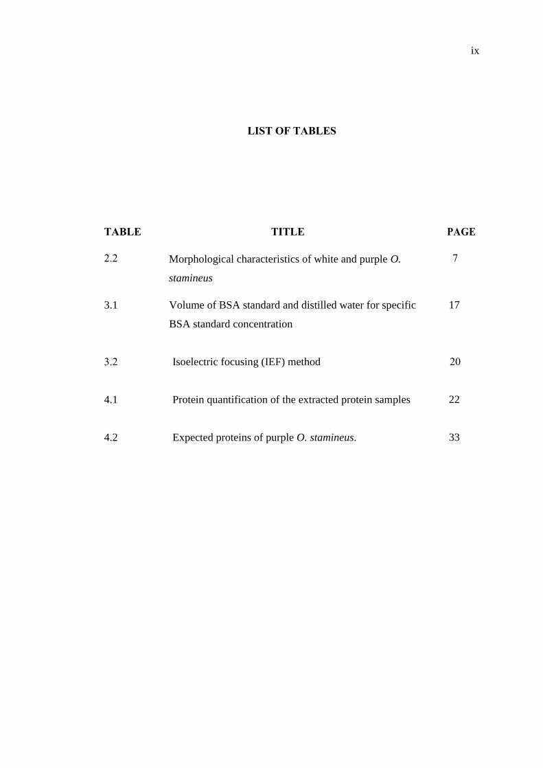
LIST OF TABLES
TABLE TITLE PAGE
2.2 Morphological characteristics of white and purple O.
stamineus
7
3.1 Volume of BSA standard and distilled water for specific
BSA standard concentration
17
3.2 Isoelectric focusing (IEF) method
20
4.1 Protein quantification of the extracted protein samples
22
4.2 Expected proteins of purple O. stamineus. 33
x
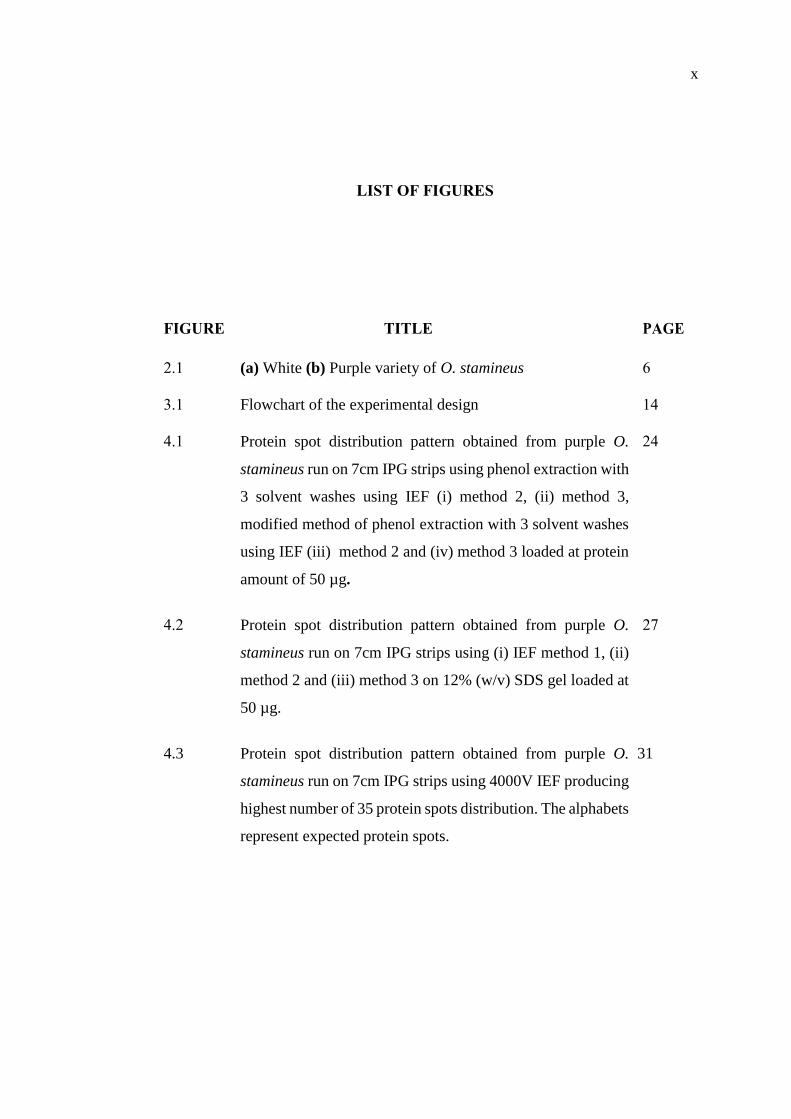
LIST OF FIGURES
FIGURE TITLE PAGE
2.1 (a) White (b) Purple variety of O. stamineus 6
3.1 Flowchart of the experimental design 14
4.1
Protein spot distribution pattern obtained from purple O.
stamineus run on 7cm IPG strips using phenol extraction with
3 solvent washes using IEF (i) method 2, (ii) method 3,
modified method of phenol extraction with 3 solvent washes
using IEF (iii) method 2 and (iv) method 3 loaded at protein
amount of 50 µg.
24
4.2
Protein spot distribution pattern obtained from purple O.
stamineus run on 7cm IPG strips using (i) IEF method 1, (ii)
method 2 and (iii) method 3 on 12% (w/v) SDS gel loaded at
50 µg.
27
4.3
Protein spot distribution pattern obtained from purple O.
stamineus run on 7cm IPG strips using 4000V IEF producing
highest number of 35 protein spots distribution. The alphabets
represent expected protein spots.
31
xi
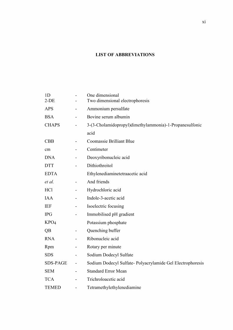
LIST OF ABBREVIATIONS
1D - One dimensional
2-DE - Two dimensional electrophoresis
APS - Ammonium persulfate
BSA - Bovine serum albumin
CHAPS - 3-(3-Cholamidopropyl)dimethylammonia)-1-Propanesulfonic
acid
CBB - Coomassie Brilliant Blue
cm - Centimeter
DNA - Deoxyribonucleic acid
DTT - Dithiothreitol
EDTA Ethylenediaminetetraacetic acid
et al. - And friends
HCl - Hydrochloric acid
IAA - Indole-3-acetic acid
IEF - Isoelectric focusing
IPG - Immobilised pH gradient
KPO4 Potassium phosphate
QB - Quenching buffer
RNA - Ribonucleic acid
Rpm - Rotary per minute
SDS - Sodium Dodecyl Sulfate
SDS-PAGE - Sodium Dodecyl Sulfate- Polyacrylamide Gel Electrophoresis
SEM - Standard Error Mean
TCA - Trichroloacetic acid
TEMED - Tetramethylethylenediamine
xii
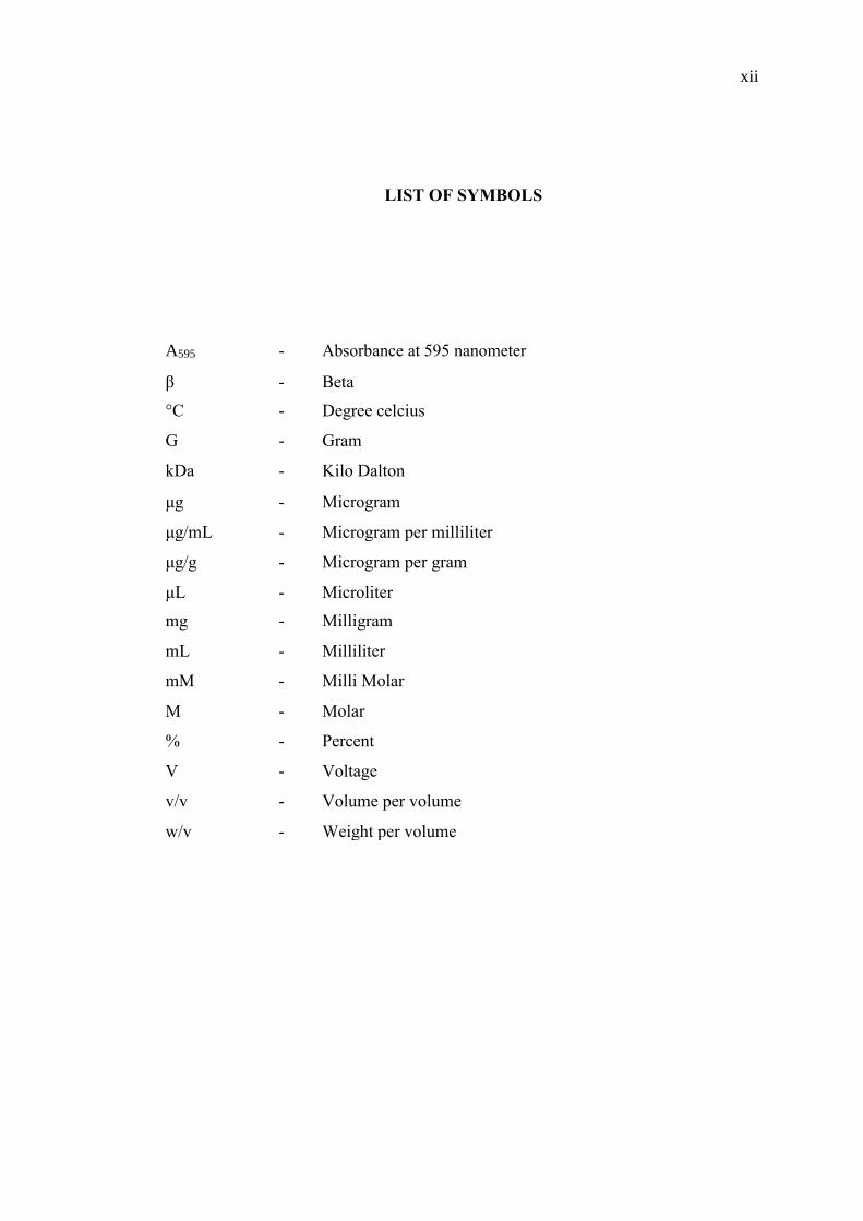
LIST OF SYMBOLS
A595 - Absorbance at 595 nanometer
β - Beta
°C - Degree celcius
G - Gram
kDa - Kilo Dalton
μg - Microgram
μg/mL - Microgram per milliliter
μg/g - Microgram per gram
μL - Microliter
mg - Milligram
mL - Milliliter
mM - Milli Molar
M - Molar
% - Percent
V - Voltage
v/v - Volume per volume
w/v - Weight per volume
xiii
LIST OF APPENDICES
APPENDIX TITLE PAGE
A Standard curve of BSA standard concentration against
absorbance (A595).
43
B O.stamineus protein electrophoretic pattern on 12 %
(w/v) 1D SDS-PAGE gel (M = marker, S1 = Sample
1, S2 = Sample 2, S3 = Sample 3, S4 = Sample 4, S5
= Sample 5, S6 = Sample 6).
44
C O.stamineus protein electrophoretic pattern on 12 %
(w/v) 1D SDS-PAGE gel.
45
1
CHAPTER 1
INTRODUCTION
1.1 Background of study
Orthosiphon stamineus Benth or Cat’s Whiskers or “misai kucing” is from the
family of Lamiaceae. O. stamineus is an ancient medicinal herb that had been widely
used since many centuries before (Wiart, 2000). This herbal tea is a well-known herbal
drink consumed to cure critical human disease (Basheer and Majid, 2010). It is
commonly found in South East Asia used for treatment of various disorders and
ailments including urinary lithiasis, edema, eruptive fever, influenza, rheumatism,
hepatitis, jaundice and biliary lithiasis (Tran, 1990).
Extracts of O. stamineus leaves contain wide variety of compounds, including
flavonoids, terpenoids or organic acids such as rosmarinic or caffeic acid (Tezuka et
al., 2000). The polyphenols for instance are able to provide enzyme inhibition and
antioxidant activity which lead to therapeutic effect to its consumer (Hossain and
Rahmanjung, 2011). The various remedial effects of this herb has attracted
researcher’s interest to further investigate its valuable components and mechanism of
action. Proteomic analysis approach is a great option to complement previous studies
as it enables the study of protein properties extracted from the valuable herbs. Indeed,
the study of the plant proteomes wholly contributes to its medicinal value mainly for
human sake.
2
Proteomics was traditionally used for protein separation utilizing the
phenomenal two-dimensional gel electrophoresis technology (Celis and Bravo, 1984).
This enables quantitative analysis of protein amounts available in the complex extracts
according to the protein’s isoelectric point and molecular weightet al, 1989). The
fundamentals and principles of two dimensional (2D) proteomics have been
established and practiced in numerous successful studies (Doleckova et al., 2012;
Barrabes et al., 2010; Hartinger et al., 1996; Tang et al., 1997). Hence, its advantages
and limitations are widely found in reviews and articles (Rogowska et al., 2013).
1.2 Problem statement
O. stamineus is well known for its medicinal values contributed by the
bioactive compound which attracts researchers’ interest. Still, no study on its protein
separation work was reported due to the complexity of the protein and the proteomic
protocol itself. In fact, this plant specifically require more starting material to extract
its protein sample compared to other plant since it contain high impurities. The most
commonly used protein separation alternative is the 2D proteomics methods;
combination of IEF protocol and molecular weight (Rabilloud et al., 2010). However,
2D electrophoresis systems still have few major issues that need to be addressed, such
as complex time-consuming experimental procedure and poor reproducibility of the
result (Barrabes et al., 2010). In this study, the experiment was conducted in order to
determine the best IEF protocol producing reproducible protein spot of Orthosiphon
stamineus thus saving time. The findings of the study can be further utilised for
identification of protein responsible in producing valuable bioactive compound and
application of the protein in medical area.
3
1.3 Objectives
The objectives of this study are as follows:
i. To determine the best IEF method producing reproducible protein spots
from purple variety of Orthosiphon stamineus.
ii. To determine the protein spot distribution and pattern from 2D
proteomics of purple Orthosiphon stamineus.
1.4 Significance of work
Various medicinal value that O. stamineus offers since decades ago has
attracted the researchers’ interest (Tran, 1990). Numerous study were done on its
pharmacological and phytochemical properties. On the other hand, fewer study related
to the herb’s protein content was established. The findings of this project are beneficial
as the best IEF method producing reproducible spots of extracted protein from O.
stamineus was determined thus saving time consumed in performing IEF. The 2D
proteomic analysis of this herbal plant can be utilized as preliminary work platform
for further protein identification especially related to its medicinal potential. Hence,
those valuables protein will be able to serve the mankind in the pharmaceutical
industry and the medical sector according to its medicinal properties.
1.5 Scope of work
The purple variety of O. stamineus was chosen in this study since it is
pigmented hence it was expected to contain higher protein content compared to the
white variety. In order to achieve the objectives of the study, there are 3 main
procedures involved. These procedures are, protein extraction from the O. stamineus
leaves using phenol extraction with 3 solvent wash, protein quantification utilizing

4
Bradford assay, and protein separation technique which focuses on the 2D gel
electrophoresis. The 2D gel electrophoresis involves the isoelectric focusing (IEF) and
SDS-PAGE principles which separate extracted protein according to its pI and
molecular weight respectively. In fact, different protein sample require different
voltage of IEF method in order to separate and focus the protein spots according to its
pI. In this study, three IEF methods of method 1 (2000V), method 2 (4000V), and
method 3 (8000V) were applied to compare its protein spot distribution and pattern.
36
REFERENCES
Adam, Y., Somchit, N. N., Sulaiman, M.R., Nasaruddin, A. A., Zuraini, A.,
Bustamam, A. A., and Zakaria, Z. A. (2009). Diuretic properties of
Orthosiphon stamineus Benth.. Journal of Ethnopharmacology. 124, 154–158.
Almatar, M., Rahmat, Z., and Salleh, F. M. (2013). Preliminary morphological and
anatomical study of O. stamineus. Indian Journal of Pharmaceutical and
Biological.Research. 1(4), 1-6.
Alves, C. T., Ferreira, I. C. F. R., Barros, L., Silva, S., Azeredo, J., and Henriques, M.
(2014). Antifungal activity of phenolic compounds identified in flowers from
North Eastern Portugal against Candida species. Future Microbiology. 9(2),
139–146.
Ameer, O. Z., Salman, I. M., Asmawi, M. Z., Ibraheem, Z. O., and Yam, M. F. (2012).
Orthosiphon stamineus: traditional uses, phytochemistry, pharmacology, and
toxicology. Journal of Medicinal Food. 15, 678–690.
Amzad, H. M., Ismail, Z., Rahman, A., and Kang, S. C. (2008). Chemical composition
and anti-fungal properties of the essential oils and crude extracts of
Orthosiphon stamineus Benth industrial crops and products. 27, 328–334.
Ansari, P., Stoppacher, N., and Baumgartner, S. (2012). Marker peptide selection for
the determination of hazelnut by LC–MS/MS and occurrence in other nuts.
Analytical and Bioanalytical Chemistry. 402(8), 2607–2615.
Arafat, O. M., Tham, S. Y., Sadikun, A., Zhari, I., Haughton, (2008). Effects of
Orthosiphon stamineus methanol extracts in rats. Journal of
Ethnopharmacology. 13, 118(3), 354-60.
Awale, S., Tezuka, Y., Banskota, A. H., Adnyana, I. K., and Kadota, S. (2003). Highly
oxygenated isopimarane-type diterpenes from Orthosiphon stamineus of
37
Indonesia and their nitric oxide inhibitory activity. Chemical and
Pharmaceutical Bulletin. 51, 268–275.
Barrabes, S., Sarrats, A., Fort, E., De Llorens, R., Rudd, P. M., and Peracaula, R.
(2010). Effect of sialic acid content on glycoprotein pI analyzed by two-
dimensional electrophoresis. Electrophoresis. 31, 2903–2912.
Basheer, A. M. and Majid, A. A. (2010). Medicinal Potentials of Orthosiphon
Stamineus Benth. WebmedCentral CANCER. 1(12), WMC001361.
Bauw, G., Van Damme, J., Puype M., Vandekerckhove, J., Gesser, B., Ratz, G. P., et
al. (1989). Protein-electroblotting and microsequencing strategies in
generating protein data bases from two-dimensional gels. Proceedings of the
National Academy of Sciences USA. 86, 7701–7705.
Beaux, D., Fleurentin, J., and Mortier, F. (1999). Effect of extracts of Orthosiphon
stamineus Benth, Hieracium pilosella L., Sambucus nigra L. and
Arctostaphylos uva-ursi (L.) Spreng. In Rats. Phytotherapy Research. 13,
222–225.
Bjellqvist, B. K., Ek, P.G., Righetti, E., Gianazza, A., Gorg, R., Westermeier, and
Postel, W. (1982). Isoelectric focusing in immobilized pH gradients:
principle, methodology and some applications. Journal of Biochemical and.
Biophysical Methods. 6, 317-339.
Bojórquez-Velázquez, E., Lino-López, G. J., Huerta-Ocampo, J. A., Barrera-Pacheco,
A., de la Rosa, A. P., Moreno, A., Mancilla-Margalli, N. A., and Osuna-Castro,
J. A., (2016). Purification and biochemical characterization of 11S globulin
from chan (Hyptissuaveolens L. Poit) seeds. Food chemistry. 1(192), 203-211.
Carter, B. L. and Basile, J., (2005). Development of Diabetes with Thiazide Diuretics:
The Potassium Issue. The Journal of Clinical Hypertension. 7 (11), 638-640.
Caverzan, A., Passaia, G., Rosa, S. B., Ribeiro, C. W., Lazzarotto, F., and Margis-
Pinheiro, M. (2012). Plant responses to stresses: Role of ascorbate peroxidase
in the antioxidant protection. Genetics and Molecular Biology. 35(4), 1011–
1019.
Chan, L. K., and Loo, P. S., (2006). Morphological Similarities and Differences
between the Two Varieties of Cat`s Whiskers (Orthosiphon stamineus
Benth.) grown in Malaysia. International Journal of Botany. 2, 1-6.
Celis, J. E. and Bravo. R. (1984). Two-dimensional Gel Electrophoresis of
Proteins: Methods and Applications. Academic Press New York.
38
Celis, J. E., Carter, N., Hunter, T., Shotton, D., Simons, K., and Small, J. V., (1997).
Cell Biology: A laboratory Handbook. New York: Academic Press. 4(2).
Chin, J. H., Abas, H. H., and Sabariah, I., (2008). Toxicity study of Orthosiphon
stamineus Benth (Misai Kucing) on Sprague Dawley rats. Tropical
Biomedicine. 25, 9–16.
Chun-Hoong, H., Ismail, N., Shaida-Fariza, S., and Ahmad, R. (2010). In vitro
antibacterial and antioxidant activities of Orthosiphon stamineus Benth
extracts against food-borne bacteria. Food Chemistry. 122, 1168–1172.
Costa, T.R., Fernandes, O.L.F., Santos, S.C., Oliveria, C.M.A., Liao, L.M., Ferri, P.H.,
Paulo, J.R., Ferreira, H.D., Sales, B.H.N., and Silva, M.R.R., (2000).
Antifungal activity of volatile constituents of Eugenia dysenterica leaf oil.
Journal of Ethnopharmacology. 72 (12), 111-117.
Doleckova, I., Rarova, L., Gruz, J., Vondrusova, M., Strnad, M., and Krystof, V.
(2012). Antiproliferative and antiangiogenic effects of flavone eupatorin, an
active constituent of chloroform extract of Orthosiphon stamineus leaves.
Fitoterapia. 83, 1000–1007.
Eisai, P. T. (1995). Indonesia Medicinal Herb Index in Indonesia. Dian Rakyat
Publication,Jakarta, Indonesia, 263.
Ernst, O., and Zor, T. (2010). Linearization of the Bradford Protein Assay. Journal of
Visualized Experiments : JoVE, 38, 1918.
Faizul, F. M., Aminudin, N., Kadir, H. A., and Tayyab, S. (2009). Bilirubin lowering
potential of Orthosiphon stamineus in temporarily jaundiced adult rats. African
Journal of Pharmacy and Pharmacology. 3(7), 359-361.
Faurobert, M., Pelpoir, E., and Chaïb, J. (2007). Phenol extraction of proteins for
proteomic studies of recalcitrant plant tissues. Methods Molecular Biology.
355, 9–14.
Gorg, A., Weiss, W., and Dunn, M. J. (2004). Current two-dimensional electrophoresis
technology for proteomics. Proteomics. 4, 3665–3685.
Gorg, A. and Weiss, W. (2000). Two-dimensional electrophoresis with immobilized
pH gradients. Rabilloud, T. (Ed.). In Proteome Research: Two-dimensional
Gel Electrophoresis and Identification Methods. (57-106). German: Springer.
Haleem, J. I. and Timothy, D. V. (2008). Two-dimensional polyacrylamide gel
electrophoresis (2D-PAGE): advances and perspectives. BioTechniques. 44,
697-700.
39
Haloui, M., Louedec, L., Michel, J. B., and Lyoussi, B., (2000). Experimental diuretic
effects of Rosmarinus officinalis and Centaurium erythraea. Journal of
Ethnopharmacology. 71, 465–472.
Hartinger, J., Stenius, K., Hogemann, D., and Jahn, R., (1996). 16-BAC/SDS-PAGE:
a two-dimensional gel electrophoresis system suitable for the separation of
integral membrane proteins. Analytical Biochemistry. 240, 126–133.
Hew, C. S., and Gam, L. H. (2011). Proteome analysis of abundant proteins extracted
from the leaf of Gynura procumbens (Lour.) Merr. Applied Biochemistry and
Biotechnology. 165, 1577–1586.
Hossain, M. A., and Rahman, S. M. (2011). Isolation and characterisation of
flavonoids from the leaves of medicinal plant Orthosiphon stamineus. Arabian
Journal of Chemistry. Article in press.
Hsuan, K. (1986). Order dan Famili Tumbuhan Berbiji di Tanah Melayu. Dewan
Bahasa dan Pustaka, Kuala Lumpur, Malyasia. 393–397.
Janine, K., Margarete, B., Frank, H., Uwe, K., Gary, H., Peter, S., and Karl-Josef, D.
(2002). The plant-specific function of 2-Cys peroxiredoxin mediated
detoxification of peroxides in the redox-hierarchy of photosynthetic electron
flux. Proceedings of the National Academy of Sciences. 99(8), 5738–5743.
Jiaman, S., Junfan, F., and Rujun, Z. (2014). Proteomic analysis of differentially
expressed proteins induced by salicylic acid in suspension-cultured ginseng
cells. Journal of Biological Sciences. 21(2), 185–190.
Jung, M., Park, M., Lee, H. C., Kang, Y. H., Kang, E. S., and Kim, S. K. (2006).
Antidiabetic agents from medicinal plants. Current Medicinal Chemistry. 13,
1203-1218.
Jungblut, P., Otto, A., Zeindl-Eberhart, E., Plessner, K. P., Knecht, M.,
RegitzZagrosek, V., Fleck, E., Wittmann-Liebold, B., (1994). Protein
composition of the human heart: the construction of a myocardial two-
dimensional electrophoresis database. Electrophoresis. 15, 685-707.
Klose, J. and Kobalz, U. (1995). Two-dimensional electrophoresis of proteins: An
updated protocol and implications for a functional analysis of the genome.
Electrophoresis. 16, 1034-1059.
Kolin, A. (1954). Separation and concentration of proteins in a pH field combined with
an electric field. Journal of Chemical Physics. 22, 1628-1629.
40
Kreydiyyeh, S. I., and Usta, J., (2002). Diuretic effect and mechanism of action of
parsley. Journal of Ethnopharmacology. 79, 353–357.
Ku, H. K., Lim, H. M., Oh, K. H., Yang, H. J., Jeong, J. S., and Kim, S. K. (2013).
Interpretation of protein quantitation using the Bradford assay: comparison
with two calculation models. Analytical Biochemistry. 434, 178-180.
L´opez, J. L. (2007). Two-dimensional electrophoresis in proteome expression
analysis. Journal of Chromatography B. 849, 190–202.
Laemmli, U. K. (1970). Cleavage of structural proteins during the assembly of the
head of bacteriophage T4. Nature. 227, 680-685.
Lee, K. S. (2015). Gel-Based Proteome Analysis of Ocimum sanctum. Bachelor of
Science, Universiti Teknologi Malaysia, Skudai.
Lee, W. L. (2014). Micropropagation and cell culture of misai kucing (Orthosiphon
stamineus Benth) and detection of rosmarinic acid in the in vitro cultures.
Master of Science, Universiti Sains Malaysia, Penang, Malaysia.
Li, Q., Li, Y., Wang, Y., Cui, Z., Gong, L., Qu, Z., Zhong, Y., Zhou, J., Zhou, Y., Gao,
Y., and Li, Y. (2016). Quantitative proteomic study of human prostate cancer
cells with different metastatic potentials. International journal of oncology.
48(4), 1437-1446.
Lu, Y. X., Zhang, Q., Li, J., Sun, Y. X., Wang, L.Y., Cheng, W. M., et al.(2010).
Antidiabetic effects of total flavonoids from Litsea coreana leave on fat-fed,
streptozotocin-induced type 2 diabetic rats. The American Journal of Chinese
Medicine. 38, 713-725.
Mariam, A., Asmawi, M. Z., and Sadikun, A. (1996). Hypoglycaemic activity of the
aqueous extract of Orthosiphon stamineus. Fitoterapia. 67, 465-468.
Mohamed, E. A. H., Yam, M. F., Ang, L. F., Mohamed, A. J., Mohamed, A. J., and
Asmawi, M. Z. (2013). Antidiabetic Properties and Mechanism of Action of
Orthosiphon stamineus Benth Bioactive Sub-fraction in Streptozotocin-
induced Diabetic Rats. Journal of Acupuncture and Meridian Studies. 6(1), 31-
40.
Nail, A. M. and Zin, N. H. M. (2015). Protein profiling of different plant tissues from
herb Phyllanthus niruri. Jurnal Teknologi. 77(24), 95-99.
Natarajan, S., Xu, C., Caperna, T. J., and Garrett, W. M. (2005). Comparison of protein
solubilisation method suitable for proteomic analysis of soybean seed proteins.
Analytical Biochemistry. 342, 214-220.
41
Ng, M. L. (2016). Gel-based Proteome Analysis of Orthosiphon stamineus. Bachelor
of Science, Universiti Teknologi Malaysia, Skudai.
O’Farrell, P. H. J. (1975). High resolution two-dimensional electrophoresis of
proteins. Journal of Biological Chemistry. 250 (10), 4007-4021.
Ong, B. H. (2017). Heavy Metal Content and Protein Expression Profile of Melastoma
malabathricum from Ex-mining Site. Master of Science, Universiti Teknologi
Malaysia, Skudai
Perry, L. M. (1980). Medicinal plants of east and south East Asia; attributed properties
and uses. The Massachusetts Institute of Technology, Cambridge, MA, 190–
191.
Rabilloud, T., Chevallet, M., Luche, S., and Lelong, C. (2010). Two-dimensional gel
electrophoresis in proteomics: past, present, and future. Journal of Proteomics.
73, 2064–2077.
Rabilloud, T. and Charmont, S. (2000). Detection of proteins on two-dimensional gels.
Rabilloud, T. (Ed.). Proteome Research:Two-dimensional Gel Electrophoresis
and Identification Methods. (107–126). France: Springer
Ramly, N. A. (2016). Evaluation of 1D and 2D gel-based proteome from fresh leaf of
Moringa oleifera. Master of Science, Universiti Teknologi Malaysia, Skudai.
Raunkjær, K., Hvitved-Jacobsen, T., and Nielsen, P. H. (1994). Measurement of Pools
of Protein: Carbohydrate and Lipid in Domestic Wastewater. Water Research.
28(2), 251-262.
Rogowska-Wrzesinskaa, A., Bihana, M. C. L., Morten, T. A. and Roepstorff P. (2013).
2D gels still have a niche in proteomics. Journal of proteomics. 88, 4-13.
Saidan, N. H., Hamil, M. S. R., Memon, A. H., Abdelbari, M. M., Hamdan, M. R.,
Mohd, K. S., Ismail, Z. (2015). Selected metabolites profiling of Orthosiphon
stamineus Benth leaves extracts combined with chemometrics analysis and
correlation with biological activities. BMC Complementary and Alternative
Medicine. 15, 350.
Saravanan, R. S., and Rose, J. K. (2004). A critical evaluation of sample extraction
techniques for enhanced proteomic analysis of recalcitrant plant tissues.
Proteomics. 4, 2522–2532.
Shim, S. Y. and Gam, L. H. (2012). Analysis of Piper sarmentosum proteome using
two dimensional gel electrophoresis and mass spectrometry. Asia-Pacific
Journal of Molecular Biology and Biotechnology. 20(4), 124-139.
42
Shaw, M. M. and Riederer, B. M. (2003). Sample preparation for two-dimensional gel
Electrophoresis. Proteomics. 3, 1408–1417.
Tang, W., Harrata, A. K., and Lee, C. S., (1997). Two-dimensional analysis of
recombinant E. coli proteins using capillary isoelectric focusing electrospray
ionization mass spectrometry. Analytical Chemistry. 69, 3177-3182.
Tezuka, Y., Stampoulis, P., Banskota, A. H., Awale, S., Tran, K. Q., Saiki, I., et al.
(2000). Constituents of the Vietnamese medicinal plant Orthosiphon
stamineus. Chemical Pharmaceutical Bulletin (Tokyo). 48, 1711–1719.
Tran, K. (1990), Medicinal Plants in Viet Nam. WHO Regional Office for the Western
Pacific Manila and Institute of Material Medica Hanoi, Science and
Technology Publishing House, Hanoi, Vietnam, 271.
Usman, K. (2014). Protein Profiling of stamineus. Master of Science, Universiti
Teknologi Malaysia, Skudai.
Wang, W., Bignani, R., Scali, M., and Cresti, M. (2006). A Universal and Rapid
Protocol and Protein Extraction from Recalcitrant Plant Tissues for Proteomic
Analysis. Electrophoresis. 27(0). 2782-2786.
Wang, Y., Shan, Q., Hou, G., Zhang, J., Bai, J., Lv, X., Xie, Y., Zhu, H., Su, S., Li,
Y., and Zi, J. (2016). Discovery of potential colorectal cancer serum
biomarkers through quantitative proteomics on the colonic tissue interstitial
fluids from the AOM–DSS mouse model. Journal of Proteomics. 132, 31-
40.
Wiart, C., (2000). Medicinal plant of Southeast Asia. Pelanduk Publication Sdn. Bhd,
Selangor, 37–45.
Wilkins, M. R., Williams, K. L., Appel, R. D., and Hochstrasser, D. F. (1997).
Proteomic Research: New Frontiers in Functional Genomics (Principles and
Practice). Berlin: Springer Verlag; 1997
Recommended


![[Group 5] electrochemistry, electrophoresis, isoelectric focusing](https://img.dokumen.tips/doc/110x75/55c5bdefbb61eb5a3b8b458a/group-5-electrochemistry-electrophoresis-isoelectric-focusing.jpg)