Louisiana State UniversityLSU Digital Commons
LSU Master's Theses Graduate School
2016
Convergence of Excitatory and InhibitoryProjections in the Mouse Medial Geniculate BodyBlaise Andre ClarkeLouisiana State University and Agricultural and Mechanical College
Follow this and additional works at: https://digitalcommons.lsu.edu/gradschool_theses
Part of the Medicine and Health Sciences Commons
This Thesis is brought to you for free and open access by the Graduate School at LSU Digital Commons. It has been accepted for inclusion in LSUMaster's Theses by an authorized graduate school editor of LSU Digital Commons. For more information, please contact [email protected].
Recommended CitationClarke, Blaise Andre, "Convergence of Excitatory and Inhibitory Projections in the Mouse Medial Geniculate Body" (2016). LSUMaster's Theses. 4591.https://digitalcommons.lsu.edu/gradschool_theses/4591

CONVERGENCE OF EXCITATORY AND INHIBITORY PROJECTIONS IN THE MOUSE
MEDIAL GENICULATE BODY
A Thesis
Submitted to the Graduate Faculty of the Louisiana State University and
Agricultural and Mechanical College in partial fulfillment of the
requirements for the degree of Master of Science in Biomedical and Veterinary Medical Sciences
in
The Department of Comparative Biomedical Sciences
by
Blaise A. Clarke B.S., Louisiana State University, 2012
May 2017
ii
ACKNOWLEDGEMENTS
Most importantly, I would like to express my sincere thanks to my advisor, Dr. Charles C.
Lee, for his unwavering support of my Master’s studies, for his training, understanding, and
motivation to succeed. His mentoring has aided me immensely in terms of research and preparing
this thesis. I certainly would not have come close to completing this undertaking without his
support. I would also like to thank the rest of my committee, Dr. George M. Strain and Dr. Jason
W. Middleton, for their additional guidance and for agreeing to be a part of my committee.
Additional thanks also goes out to my fellow labmates, Dr. Olalekan Ogundele, Razia
Sultana, and Tanya Gandhi, for making laboratory life and conferences even more enjoyable than
they already are. I’d particularly like to thank Dr. Ogundele for his fantastic training in techniques
and methods throughout my time in the program.
I have endless gratitude for my close friends here at Louisiana State University: Samia
O’Bryan, Brandon Lewis, Yi-Fan Chen, Harriet Hammond, and Rebecca Hill, for making all of
the stressful nights before deadlines and exams manageable, and for all of the non-school-related
fun we’ve had throughout my time here.
Lastly, I would like to thank my parents, Yvette Marsh, Stephan Clarke, Andre Marsh, and
Colleen Clarke, for supporting me throughout my entire life, and for showing me that it was
possible to achieve anything in life.
iii
TABLE OF CONTENTS ACKNOWLEDGEMENTS ...................................................................................................ii
ABSTRACT ...........................................................................................................................iv
CHAPTER 1. INTRODUCTION .........................................................................................1
CHAPTER 2. LITERATURE REVIEW ...............................................................................13
CHAPTER 3. INVESTIGATING CONVERGENT PROJECTIONS TO THE MGB .........23 3.1 Introduction ..........................................................................................................23
3.2 Injection of Cholera Toxin Beta subunit (CTβ) Conjugate .................................24 3.3 Post-Surgical Processing of VGAT-Venus Mouse Brain ....................................24 3.4 Fluorescence Imaging of Brain Tissue Samples ..................................................25
CHAPTER 4. ANALYSIS OF CONVERGENT PROJECTIONS TO THE MGB ..............27 4.1 Convergent Excitatory and Inhibitory Projections to the MGB ..........................27 4.2 Quantitative Analysis of GABAergic and Excitatory Projections .......................32
CHAPTER 5. DISCUSSION .................................................................................................35
CHAPTER 6. CONCLUSION ..............................................................................................38
REFERENCES ......................................................................................................................39
VITA ......................................................................................................................................46

iv
ABSTRACT
The medial geniculate body (MGB) is the target of excitatory and inhibitory inputs from
several neural sources. Among these, the inferior colliculus (IC) is an important nucleus in the
midbrain that acts as a nexus for many auditory pathways and projections, ascending and
descending, throughout the rest of the central auditory system and provides both excitatory and
inhibitory projections to the MGB. In addition, the thalamic reticular nucleus (TRN) is a major
source of inhibition to the MGB, particularly in rodents. Finally, the auditory cortex (AC) is a
major source of descending input to the MGB, providing direct excitation and indirect inhibition
via the TRN.
In our study, we assessed the relative contribution from these excitatory and inhibitory
projection sources to the MGB of the auditory system in mice. Using retrograde tract tracing with
CTβ -Alexa Fluor 594 injected into the MGB of the mouse, we quantitatively mapped the
projections from both the ipsilateral and contralateral IC, the TRN, and the AC to the ipsilateral
MGB. Our results indicate significant GABAergic projections from the IC and TRN to the MGB
and excitation from the AC that play an overlooked role in shaping auditory processing. These
results complement prior studies in other species, which suggests that these pathways are important
factors in the regulation of neuronal activity in the auditory forebrain.
1
CHAPTER 1 INTRODUCTION
The primary function of the auditory system is to develop the sense of hearing. To do this,
the system is divided into two subsystems – the peripheral auditory system and central auditory
system. Being a very intricate process, involving many regions and mechanisms, audition has a
lengthy process in the goal to translate sound waves into neural electrical signals. This process
begins from sound waves entering the ear, and eventually culminates in the cochlea, where the
neural signals are created (Hudspeth, 1997). These signals then proceed as cranial nerve VIII to
the cochlear nuclei, where one will also see nerve afferents crossing to the superior olivary
complex. To help prevent loss of hearing due to unilateral damage, most inputs are distributed
bilaterally throughout the brain. From the lower brainstem nuclei, all ascending inputs make their
way to the inferior colliculi (IC) and medial geniculate body (MGB) in the thalamus, reaching the
cortex as the last stop. The MGB is the target of many excitatory and inhibitory projections, has a
wide integration of auditory connections throughout the central auditory system, and receives
inputs from all lower brainstem and midbrain nuclei (Buentello et al., 2015; Ito and Oliver, 2010).
As an overview, sound processing in the mammalian auditory pathway proceeds as
follows: sound enters the outer ear as time-varying air-pressure waves, which is then eventually
transduced into neural signals via the cochlea (Hudspeth, 1997). The frequency of those sounds
are analyzed, along with the location and timing of the sounds, by the lower brainstem nuclei,
specifically the cochlear nuclei (CN), lateral leminscus nuclei (NLL), and the superior olivary
complex (SOC) (Blosa et al., 2013; Cant, 1992; Hudspeth, 1997; Oliver, 2000; Schwartz, 1992).
These nuclei are also involved in intensity and interval analysis for localization of sound.
The dorsal region of the NLL is a major source of GABAergic input to the inferior colliculus, as
well as all other lower brainstem nuclei, all projecting excitatory and inhibitory inputs (Buentello

2
et al., 2015; Ito and Oliver, 2010). IC nuclei, like those in the brainstem, are located in both
hemispheres of the brain, and along with the superior olivary complex (SOC), they are the first
region where sound location is fully integrated, fusing vertically and horizontally oriented data
(Oliver, 1984; Reetz and Ehret, 1999; Winer and Schreiner, 2005). Furthermore, like most brain
areas, excitatory and inhibitory inputs from other auditory regions is integrated in the IC; excitatory
input is integrated from the brainstem and auditory cortex, and inhibitory input is integrated from
nearly every peripheral brainstem nucleus found in the auditory system (Winer and Schreiner,
2005). Once this integration is completed, it is then sent to an ascending auditory center, namely
the MGB, which then projects to the primary auditory cortex (Hackett, 2011; Kaas and Hackett,
2000; Lee and Winer, 2011). Once here, the primary AC integrates with the secondary AC, and
interconnects with the frontal lobes for further processing, such as speech perception (Hackett,
2011; Kaas and Hackett, 2000; Lee and Winer, 2011).
The higher auditory regions in the brain include the inferior colliculus (IC), medial
geniculate body (MGB), thalamic reticular nucleus (TRN), and the auditory cortex (AC), structures
that all play an important role in higher-order auditory processing (Wenstrup, 2005; Winer, 1992).
Among these, the auditory thalamic nucleus, i.e. the MGB, receives convergent information from
several structures, including the IC, the TRN, and AC (Lee, 2013; Lee, 2015; Winer, 1984). All
auditory information processed through lower brainstem and midbrain structures eventually
reaches the MGB, where that information is then sent to the auditory cortex (Lee, 2013).
Information from each of these sources to the MGB may have an excitatory, inhibitory, or mixed
influence of the activity of neurons in the MGB (Winer, 1992). Consequently, understanding the
neural interactions between these structures and the MGB is crucial to understanding how auditory
information is processed in the brain and how it is altered during diseases of the auditory system.

3
Of the upstream inputs to the MGB, the inferior colliculus (IC) is the main auditory
structure found in the midbrain, and is a major hub for ascending and descending pathways in the
central auditory system (Buentello et al., 2015; Ito and Oliver, 2010; Oliver, 1984; Wenstrup,
2005). The IC is mostly involved in auditory orientation and perception, and acts as a nexus for
many auditory pathways, receiving and sending projections from other nuclei and regions (Winer
and Schreiner, 2005). The inferior colliculus is composed of three main subregions - the central
nucleus (CN), dorsal cortices (DC), and lateral cortices (LC). These three subdivisions are
generally similar in cellular makeup, but have stark differences in their projections (Saldaña and
Marchán, 2005; Wenstrup, 2005). The central nucleus of the IC (ICC) projects its axons mainly to
the ventral division of the medial geniculate body (MGBv). The dorsal and lateral cortices project
their axons to the dorsal and medial MGB subdivisions (Hackett, 2011; Wenstrup, 2005). All
components of the IC mainly send their projections to the ipsilateral MGB, with very few going to
the contralateral MGB (Mellott et al., 2014a).
Glutamate is the main excitatory neurotransmitter of the central auditory system, with
gamma-aminobutyric acid (GABA) and glycine being the main inhibitory neurotransmitters. The
IC contains both types of neurotransmitters, with varying degrees of concentration; approximately
21% of IC neurons are GABAergic, with the remaining 75% of neurons being almost solely
glutamatergic. The glutamatergic neuron percentage is based on the findings of those neurons
expressing vesicle glutamate transporters (VGLUT) (Altschuler et al., 2008; Caspary et al., 2008).
As the IC has both GABAergic and glutamatergic neurotransmitters, the IC would also have both
excitatory and inhibitory effects in the auditory system (Mellott et al., 2014a; Peruzzi et al., 1997;
Winer et al., 1996). The ascending inhibitory component of the IC projection to the MGB is a
novel discovery within the IC (Mellott et al., 2014a; Peruzzi et al., 1997; Winer et al., 1996). The
4
IC integrates nearly all lower brainstem auditory information before sending its output to the
medial geniculate body (MGB), but said output is particularly intriguing, as it contains both
GABAergic and glutamatergic projection neurons, unlike many other areas of the brain. These
cells are involved in what is known as the tectothalamic pathway, the largest output pathway of
the IC, mainly sending inhibitory projections to the MGB (Wenstrup, 2005). The rest of the
tectothalamic pathway is involved in excitation, sending ascending glutamatergic projections to
the MGB. Interestingly, it has been shown that the inferior colliculus displays signs of reduced
GABA release as it ages (Caspary et al., 2008).
Decreasing GABA levels imply a decreased level of neural inhibition, thus understanding
the cellular makeup and layout of the IC and its projections to other structures is invaluable
information in truly appreciating and comprehending its role in auditory processing, particularly
as it pertains to age-related hearing loss. Furthermore, the studies described in this thesis are
concentrated on the distribution of the IC’s tectothalamic pathway as it projects to the MGB, but,
as noted, there are many more structures involved in the MGB’s auditory processing function.
The medial geniculate body (MGB) is the main auditory center of the thalamus. It is the
target of excitatory and inhibitory inputs from many structures of the central auditory system
(Hackett, 2011; Llano and Sherman, 2008; Villa et al., 1991; Wenstrup, 2005). A knee-shaped
structure, the MGB receives inhibitory input from the TRN, excitation and inhibition from the IC,
and direct excitation from the AC (Winer, 1984). It acts as a thalamic way station, receiving input
from the IC, and sending to, as well as receiving projections from the auditory cortex (Lee and
Winer, 2008; Lee, 2013). Most of its output is directed towards to the auditory cortex, but
projections are sent to other regions as well, such as the TRN, amygdala, and striatum (Lee, 2015).
5
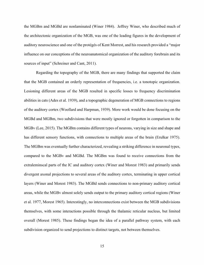
The medial geniculate body can be separated into three subdivisions – ventral (MGBv),
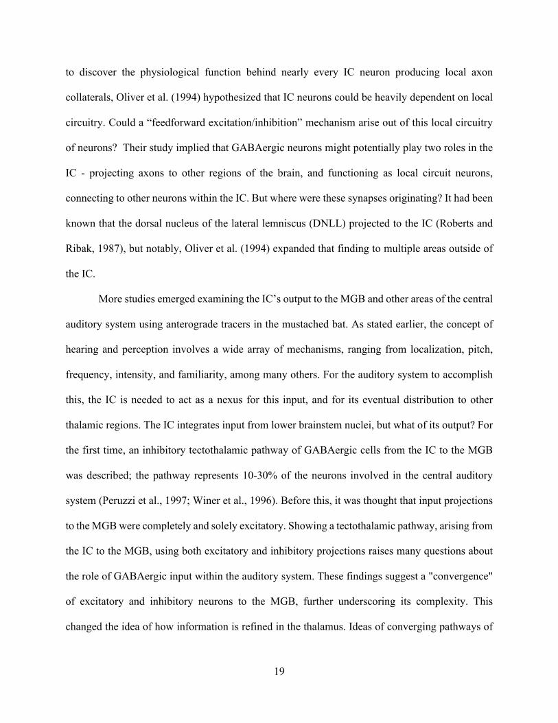
dorsal (MGBd), and medial (MGBm) (Figure 1) (Winer, 1992). The ventral subdivision (MGBv)
is the main nucleus of the MGB, projecting axons via the thalamocortical pathway to layers III and
IV of the auditory cortex (Huang and Winer, 2000; Smith, 2012). The MGBv also receives input
mainly from the ICC. The dorsal MGB (MGBd) is very similar to the MGBv, projecting to the
same layers of the AC. The difference is that MGBd’s axons terminate in what is known as the
extralemniscal auditory cortex (Hackett, 2011; Kaas and Hackett, 2000). The medial MGB
(MGBm) is unique, having an axonal output terminating in many cortical layers, particularly layers
I and VI of the auditory cortex. MGBv and MGBd’s projections to the AC are excitatory, being
shown to be non-GABAergic (Lee, 2015).
Hearing loss is one of the most prevalent conditions in the world today (Dalton et al., 2003;
Salomon, 1986; Yamasoba et al., 2013; Yueh et al., 2013). It affects many members of both older
and younger generations, being the third most seen chronic condition in the elderly (Salomon,
1986; Yamasoba et al., 2013; Yueh et al., 2013). It affects approximately one in three people over
the age of 65, and one in two people over 85 (Yamasoba et al., 2013; Yueh et al., 2013). Modern
technology has equipped us with the means to treat some of these hearing-related problems through
the use of hearing aids (Dalton et al., 2003; Yueh et al., 2013). However, these devices primarily
manage hearing loss and are rarely fully restorative (Bielefeld et al., 2010). It thus remains an
arduous issue for society to tackle. Even with hearing-aids, many elderly people continue to
struggle with engaging in daily conversation, and the essential skill of blocking out unwanted
auditory information while focusing on a particular auditory source (Bielefeld et al., 2010; Dalton
et al., 2003; Humes et al., 2006). The world is full of a wide array of unique, special sounds, which
vary in terms of their location, intensity, frequency, or pitch (Hudspeth, 1997). The brain, over

6
time, loses efficiency in processing these sounds, and because of this, people eventually exhibit
difficulty comprehending the incredible amount of auditory stimulation experienced on a daily
basis (Dalton et al., 2003; Yamasoba et al., 2013).
On a physiological level, one of the current theories to explaining age-related hearing loss
(presbycusis) is the gradual inability of the auditory system’s inhibitory circuitry to “filter out”
unwanted information (Allen and Eddins, 2010; Caspary et al., 2008; Llano et al., 2012; Stebbings
et al., 2016). This filtering ability becomes less potent as our brains age (Caspary et al., 2008;
Llano et al., 2012; Wehr and Zador, 2003), and can occur centrally or peripherally. Peripheral
issues stem from the destruction of inner ear hair cells, usually due to physical trauma or loud
noises, and are generally well researched. Central issues, on the other hand, can range from several
physiological changes in the central auditory system, and is far less understood. These issues are
shown by molecular changes in inhibitory processes in many auditory regions at differing levels
of the auditory system. Lower levels of the auditory system are specialized for the initial detection
and localization of sounds, while higher levels of the auditory system specialize in the integration
of these sounds and stimuli with other sensory modalities to form holistic auditory percepts
(Geissler and Ehret, 2004; Harkrider et al., 2005; Hipp et al., 2011; Hudspeth, 1997).
Sounds don’t exist in isolation - there is a massive amount of stimuli in our environment
surrounding a wanted auditory target, and our brain faces the daunting task of integrating and
pruning those stimuli into behaviorally relevant meaning. The difficulty of this task is only
increased when introducing localization of sound, temporal scales, and filtering out unwanted
information.

7
One way that the auditory brain accomplishes this task is through the descending
projections of the auditory cortex in modulating lower auditory structures, such as the IC, TRN,
and MGB (Lee, 2015; Llano and Sherman, 2008).
Figure 1. Coronal section of the medial geniculate body depicting the distinct cytoarchitectonic subdivisions; MGd, the dorsal nucleus; MGv, the ventral nucleus; MGm, the medial nucleus. The MGv is characterized by neurons that are oriented in sheets parallel to the tonotopic axis. The MGd has larger unoriented cells, while the MGm contains many large neurons that project broadly across the auditory cortex. Coronal section from a fox. Scale bar: 2 mm. (Nadjzion et al., 2011). Used under the terms of John Wiley and Sons, License #4081481200894.
The auditory cortex (AC) sends descending projections from its fifth and sixth layers to the
MGB via the corticothalamic projection system. The corticothalamic projection of the AC is one
of the greatest found throughout the entire brain, in terms of relative contribution to several
systems. Other corticothalamic projections, such as the ones found in the visual and somatosensory
systems, seem to perform similarly (Briggs and Usrey, 2008), allowing the cortex to communicate

8
with the thalamus continuously. Most of those projections are glutamatergic, resulting in an
excitatory response, mainly ending in the ventral region of the MGB (MGBv), for the layer VI
projection (Llano and Sherman, 2008; Winer et al., 2001). One function of the corticothalamic
system may be rooted in modulation, involving different terminals depending on cortical layer of
origin (Llano and Sherman, 2008).
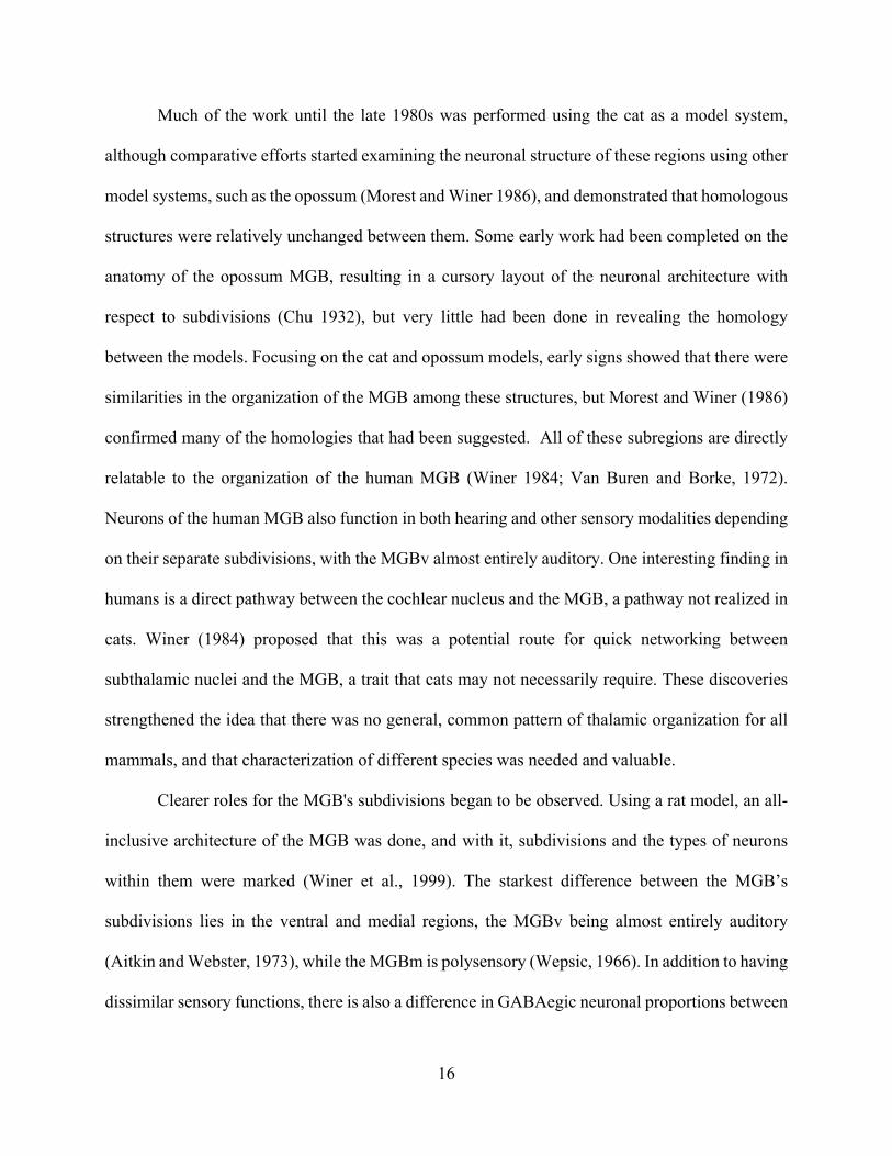
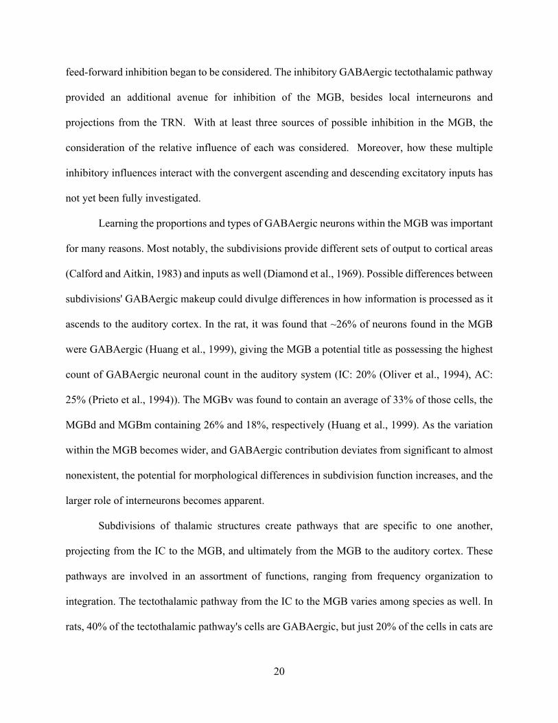
The thalamic reticular nucleus is a structure in the ventral thalamus, and is one of the only
thalamic nuclei not to project axons to the cerebral cortex (Figure 2) (Crabtree, 1998). However,
as stated, the AC provides indirect inhibition via the TRN; it sends projections to the auditory
region of the TRN, which then provides inhibitory output to the MGB.
Figure 2. Schematic diagram of circuitry between the thalamus and cortex. Thalamocortical neurons (blue) receive peripheral inputs and project axons to layer 4 of primary sensory cortex. Corticothalamic neurons (red), local thalamic interneurons (black). receive local input from thalamocortical recipient layer 4 and provide output to layer 4 and to the thalamus (Briggs and Usrey, 2008). Used under the terms of the Creative Commons Attribution License (CC BY).

9
The TRN is the main source of inhibition to many thalamic structures, including the MGB
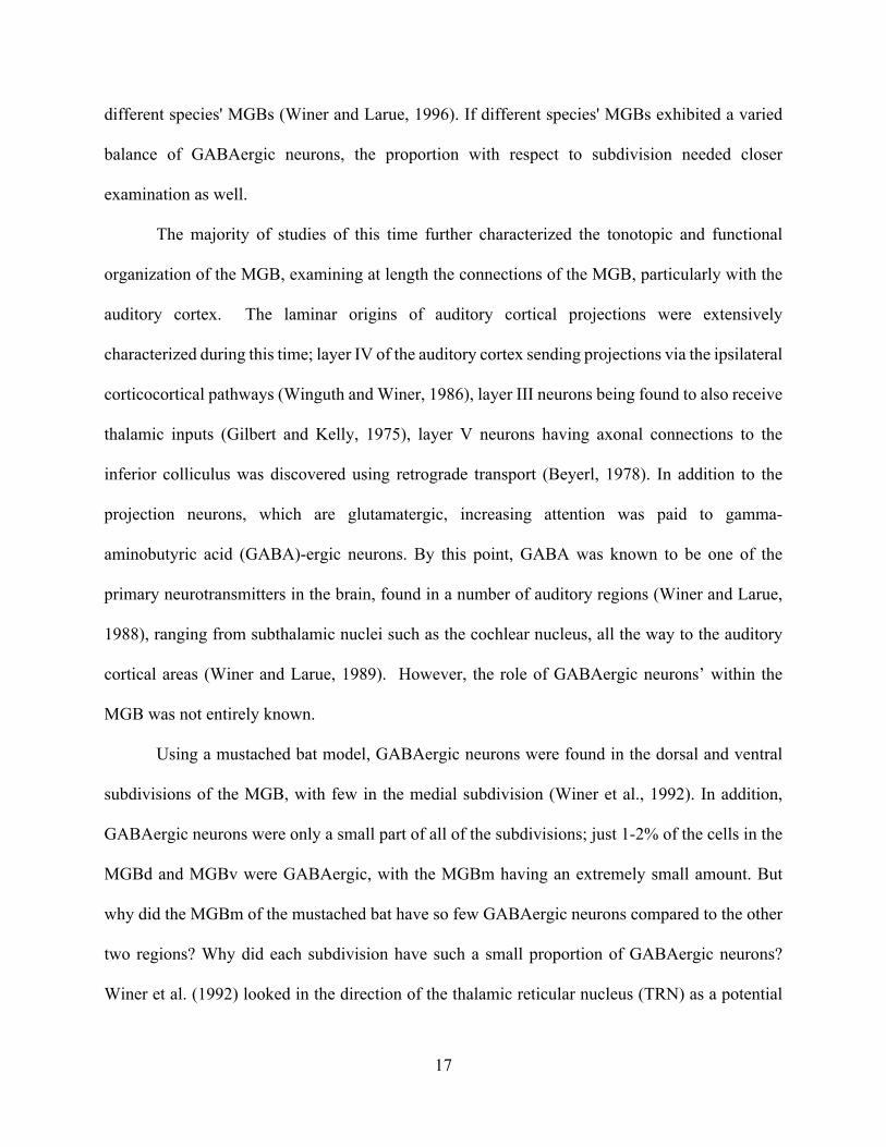
(Lam and Sherman, 2005; Sherman and Guillery, 2006). The corticocollicular system projections
arise from the AC ending in the IC, originating mainly from layers V and VI (Figure 3) (Llano and
Sherman, 2009; Llano et al., 2014).
Figure 3. Schematic diagram showing the major anatomical subdivisions of the IC, MGB and AC that illustrates the pathways from the midbrain up to the cortex and back. A1, primary auditory cortex; AC, auditory cortex; CNIC, central nucleus of the inferior colliculus; DCIC, dorsal cortex of the inferior colliculus; LCIC, RCIC; lateral and rostral cortex of the inferior colliculus; MGD; dorsal division of the medial geniculate body; MGM; medial division of the medial geniculate body; MGV; ventral division of the medial geniculate body. (Malmierca et al., 2015). Used under the terms of the Creative Commons Attribution License (CC BY).
Almost all regions of the AC project their axons to the ipsilateral IC, mainly the dorsal and
lateral nuclei (Winer et al., 1998). Layer V and VI neurons have been described as mostly
pyramidal (Llano and Sherman, 2009). The majority of layer V neurons are large pyramidal
neurons that project to the ipsilateral IC, while layer VI neurons are smaller and tend to terminate
10
their projections in the central nucleus of the IC (Winer et al., 1998). These projections are
glutamatergic as well, implying a role of direct excitatory modulation to the IC (Llano et al., 2014).
Corticocollicular projections can induce depression of sounds and activity, suggesting
some type of an inhibitory relationship in this circuitry, which would conflict with the direct
glutamatergic projections from the AC to the IC, but may be a function of indirect inhibition via
local IC interneurons (Saldaña and Marchán, 2005). These findings have been shown in a variety
of species, from cat (Winer and Larue, 1996) to gerbil (Cant and Benson, 2006) to bat (Winer et
al., 1992). One theory claimed that the target cells of the AC axons may be inhibitory, but a
previous study ruled that just 4% of these target neurons were GABAergic.
To truly understand the general integration of these auditory inputs and outputs,
characterizations of each of these projection systems are required. Previous studies have attempted
to characterize the inhibitory and excitatory projections from the IC to the MGB in a fairly wide
range of species (Mellott et al., 2014a; Peruzzi et al., 1997; Winer et al., 1996). GABAergic
tectothalamic neurons can be detected throughout the IC, and it was shown that IC GABAergic
cells contribute approximately 40% of the tectothalamic pathway in the rat, but just 20% of the
tectothalamic pathway in cats (Peruzzi et al., 1997; Winer et al., 1996). The stark difference is
particularly unusual, given that 20-25% of both species’ IC cells are GABAergic. This could be
due to the differences in frequency ranges between the cat and rat (Cat: 45 Hz to 64 kHz; Rat: 200
Hz to 76 kHz), but nonetheless stimulates the idea that rats have a disproportionately high amount
of GABAergic cells involved in the tectothalamic pathway compared to cats (Winer and Larue,
1996). Rats may have a higher concentration of GABAergic cells compared to cats due to their
almost complete lack of MGB interneurons (<1%) (Winer, 1992). Cats have been shown to display

11
25% of total MGB neurons as interneurons, which may explain the lower percentage of
tectothalamic GABAergic neurons (22%) (Winer and Larue, 1996).
There may also be a difference in how the excitatory and inhibitory inputs are integrated
within the MGB, depending on MGB subdivision. Focusing on the IC’s output to the MGB, in
guinea pigs, non-GABAergic cells from the IC were the most numerous cells involved in the
pathway; GABAergic cells supplied various levels of intensity, specifically 22% of the projections,
ending in distinct subdivisions of the MGB (Mellott et al., 2014b). These findings also hamper
previous studies’ hypotheses about the basis behind interneuron count. The guinea pig has 22% of
its tectothalamic cells as GABAergic, similar to the cat’s, yet a very low interneuron count.
(Mellott et al., 2014b). This substantiates the idea that a high GABAergic cell count is not simply
making up for a low interneuron count. If this were not so, then the guinea pig should have a far
higher GABAergic tectothalamic count than shown. The variability of the amount of GABAergic
cells in these three species makes it difficult to translate and generalize to overall auditory
processing (Mellott et al., 2014b; Peruzzi et al., 1997; Winer et al., 1996).
Studies of the contralateral IC projections to the MGB also receive far less attention than
ipsilateral studies (Mellott et al., 2014a; Wenstrup, 2005). Much work has been covered on the
ipsilateral IC and MGB’s projections, while the contralateral structures only contribute a small
amount in comparison (Mellott et al., 2014a). A thorough characterization of excitatory and
inhibitory projections to MGB from bilateral structures would be informative and beneficial. As
stated earlier, there has been a large number of studies completed in other species, focusing on the
corticothalamic and corticocollicular pathways of the auditory system (Llano and Sherman, 2008;
Winer et al., 1998; Winer, 2006). Differences have been shown in these studies, sometimes to a
great degree, and because of this, an inclusion of other species is worthy of study. There also exists

12
a substantial lack of characterization of the TRN’s involvement in auditory thalamic inhibition,
particularly as it pertains to excitation and inhibition via the AC (Crabtree, 1998; Crabtree et al.,
1998). These projections have been examined individually in various species, but there has not
been an overarching amount of research done to quantify these convergent projections to the MGB.
In this thesis, I used quantitative analyses of convergent projections using retrograde
tracing and transgenic immunofluorescence of inhibitory neurons. I examined the relative
contribution of excitatory and inhibitory projections from the IC, TRN and AC to the MGB via
mapping of their respective convergent projections. These analyses, in addition to past studies of
these regions, proposes the idea that there lies a meaningful and compelling role for these structures
in the regulation and modulation of auditory processing.
Audition is an extremely complicated and intricate process, involving many brain
structures, regions, mechanisms, and pathways, ultimately resulting in what is perceived as a
simple and quick deciphering of the auditory environment. The medial geniculate body in
particular is a substantial contributor to this process - a thalamic station that acts as a key mediator
in the integration of these auditory signals. Many axonal projections converge in the MGB, which
integrates these excitatory and inhibitory inputs. The neuroanatomical mapping of the GABAergic
and glutamatergic projections throughout this area will only serve to advance an understanding of
these pathways as it relates to each structure. This would prove invaluable in the eventual goal to
reduce and perhaps ultimately reverse age-related hearing loss in the future.

13
CHAPTER 2 LITERATURE REVIEW
The layout, divisions, and connections between the medial geniculate body (MGB) and
other thalamic structures have been investigated for decades, if not centuries (Winer, 1992). Focus
on the MGB alone began in simpler terms - as far back as the early 1900s, where confirmed
sections or subdivisions of the structure were denoted. The number of defined subdivisions in the
MGB has evolved over time, with the earliest noting between two (Rioch 1929), a lateral pars
principalis and medial pars magnocellularis, and three (Cajal 1911), with many nuclei to be
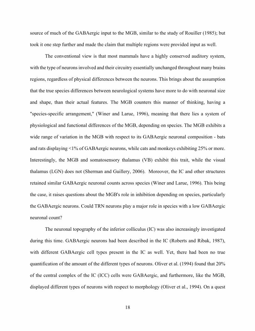
described later (Morest 1964). D. Kent Morest was and is still considered the “father of modern
neuroanatomy of the auditory system” (Winer, 1992). His work focused on the neuroanatomy of
the thalamic areas and the then novel usage of the Golgi histological stain, to identify the
morphology of the cells in the MGB (Figure 4).
Figure 4. Typical distribution of principal neuronal morphologies at the junction of the anterior and middle thirds of the medial geniculate body. Transverse section, Golgi–Cox. 15-day-old cat. (Morest, 1964). Used under the terms of John Wiley and Sons, License # 4081711367257.
14
The subdivisions identified by these morphological criteria were furthered by studies of
the connections of subthalamic nuclei to the MGB ending in different regions (Morest 1964), each
correlating with various functions in the auditory pathway (Morest 1965). These early studies were
the first to suggest that auditory, somatosensory, and visual systems all had distinct processing
roles within different subdivision of the MGB, further detailing its uniqueness and internal
variability (Morest, 1965). Due to these studies, the MGB had been well described in terms of cell
morphology and basic connectivity with subcortical structures, but the complexity of the MGB,
particularly its diverse functions and connections with other auditory structures of the brain, were
yet to be unveiled.
Broadly, one can consider the MGB as being composed of three main subdivisions, the
ventral (MGBv), dorsal (MGBd), and the medial subdivision (MGBm), although further
parcellation of these regions exist in some species (Winer, 1992). The dorsal subdivision of the
MGB (MGBd) is mostly auditory with higher order auditory duties, while the ventral subdivision
of the MGB (MGBv) is entirely an auditory nucleus. The medial subdivision (MGBm)
organization is multimodal (Cajal 1911). Within the MGBv, neurons are oriented to form a
laminar structure (Morest 1965) - that is, neurons form different layers or sheets within the
structure, much like the layering of an onion – which was first hinted by Cajal (1955). Lamination
had been shown in other thalamic structures involved in audition before; the inferior colliculus
(IC) (Katsuki et al. 1958, Morest 1964b) displayed signs of lamination, and in other auditory
structures as well (Wenstrup, 2005), strengthening the claim that laminated regions were a staple
of the auditory system. The 1965 study by Morest went further, implying that the laminar structure
of the MGBv was almost certainly due to a topographic auditory organization, perhaps “frequency
discrimination.” Among the nuclei, only the MGBv exhibits such a laminated organization, while
15
the MGBm and MGBd are nonlaminated (Winer 1984). Jeffrey Winer, who described much of
the architectonic organization of the MGB, was one of the leading figures in the development of
auditory neuroscience and one of the protégés of Kent Morrest, and his research provided a “major
influence on our conceptions of the neuroanatomical organization of the auditory forebrain and its
sources of input” (Schreiner and Cant, 2011).
Regarding the topography of the MGB, there are many findings that supported the claim
that the MGB contained an orderly representation of frequencies, i.e. a tonotopic organization.
Lesioning different areas of the MGB resulted in specific losses to frequency discrimination
abilities in cats (Ades et al. 1939), and a topographic degeneration of MGB connections to regions
of the auditory cortex (Woollard and Harpman, 1939). More work would be done focusing on the
MGBd and MGBm, two subdivisions that were mostly ignored or forgotten in comparison to the
MGBv (Lee, 2015). The MGBm contains different types of neurons, varying in size and shape and
has different sensory functions, with connections to multiple areas of the brain (Erulkar 1975).
The MGBm was eventually further characterized, revealing a striking difference in neuronal types,
compared to the MGBv and MGBd. The MGBm was found to receive connections from the
extraleminscal parts of the IC and auditory cortex (Winer and Morest 1983) and primarily sends
divergent axonal projections to several areas of the auditory cortex, terminating in upper cortical
layers (Winer and Morest 1983). The MGBd sends connections to non-primary auditory cortical
areas, while the MGBv almost solely sends output to the primary auditory cortical regions (Winer
et al. 1977, Morest 1965). Interestingly, no interconnections exist between the MGB subdivisions
themselves, with some interactions possible through the thalamic reticular nucleus, but limited
overall (Morest 1985). These findings began the idea of a parallel pathway system, with each
subdivision organized to send projections to distinct targets, not between themselves.
16
Much of the work until the late 1980s was performed using the cat as a model system,
although comparative efforts started examining the neuronal structure of these regions using other
model systems, such as the opossum (Morest and Winer 1986), and demonstrated that homologous
structures were relatively unchanged between them. Some early work had been completed on the
anatomy of the opossum MGB, resulting in a cursory layout of the neuronal architecture with
respect to subdivisions (Chu 1932), but very little had been done in revealing the homology
between the models. Focusing on the cat and opossum models, early signs showed that there were
similarities in the organization of the MGB among these structures, but Morest and Winer (1986)
confirmed many of the homologies that had been suggested. All of these subregions are directly
relatable to the organization of the human MGB (Winer 1984; Van Buren and Borke, 1972).
Neurons of the human MGB also function in both hearing and other sensory modalities depending
on their separate subdivisions, with the MGBv almost entirely auditory. One interesting finding in
humans is a direct pathway between the cochlear nucleus and the MGB, a pathway not realized in
cats. Winer (1984) proposed that this was a potential route for quick networking between
subthalamic nuclei and the MGB, a trait that cats may not necessarily require. These discoveries
strengthened the idea that there was no general, common pattern of thalamic organization for all
mammals, and that characterization of different species was needed and valuable.
Clearer roles for the MGB's subdivisions began to be observed. Using a rat model, an all-
inclusive architecture of the MGB was done, and with it, subdivisions and the types of neurons
within them were marked (Winer et al., 1999). The starkest difference between the MGB’s
subdivisions lies in the ventral and medial regions, the MGBv being almost entirely auditory
(Aitkin and Webster, 1973), while the MGBm is polysensory (Wepsic, 1966). In addition to having
dissimilar sensory functions, there is also a difference in GABAegic neuronal proportions between
17
different species' MGBs (Winer and Larue, 1996). If different species' MGBs exhibited a varied
balance of GABAergic neurons, the proportion with respect to subdivision needed closer
examination as well.
The majority of studies of this time further characterized the tonotopic and functional
organization of the MGB, examining at length the connections of the MGB, particularly with the
auditory cortex. The laminar origins of auditory cortical projections were extensively
characterized during this time; layer IV of the auditory cortex sending projections via the ipsilateral
corticocortical pathways (Winguth and Winer, 1986), layer III neurons being found to also receive
thalamic inputs (Gilbert and Kelly, 1975), layer V neurons having axonal connections to the
inferior colliculus was discovered using retrograde transport (Beyerl, 1978). In addition to the
projection neurons, which are glutamatergic, increasing attention was paid to gamma-
aminobutyric acid (GABA)-ergic neurons. By this point, GABA was known to be one of the
primary neurotransmitters in the brain, found in a number of auditory regions (Winer and Larue,
1988), ranging from subthalamic nuclei such as the cochlear nucleus, all the way to the auditory
cortical areas (Winer and Larue, 1989). However, the role of GABAergic neurons’ within the
MGB was not entirely known.
Using a mustached bat model, GABAergic neurons were found in the dorsal and ventral
subdivisions of the MGB, with few in the medial subdivision (Winer et al., 1992). In addition,
GABAergic neurons were only a small part of all of the subdivisions; just 1-2% of the cells in the
MGBd and MGBv were GABAergic, with the MGBm having an extremely small amount. But
why did the MGBm of the mustached bat have so few GABAergic neurons compared to the other
two regions? Why did each subdivision have such a small proportion of GABAergic neurons?
Winer et al. (1992) looked in the direction of the thalamic reticular nucleus (TRN) as a potential
18
source of much of the GABAergic input to the MGB, similar to the study of Rouiller (1985); but
took it one step further and made the claim that multiple regions were provided input as well.
The conventional view is that most mammals have a highly conserved auditory system,
with the type of neurons involved and their circuitry essentially unchanged throughout many brains
regions, regardless of physical differences between the neurons. This brings about the assumption
that the true species differences between neurological systems have more to do with neuronal size
and shape, than their actual features. The MGB counters this manner of thinking, having a
"species-specific arrangement," (Winer and Larue, 1996), meaning that there lies a system of
physiological and functional differences of the MGB, depending on species. The MGB exhibits a
wide range of variation in the MGB with respect to its GABAergic neuronal composition - bats
and rats displaying <1% of GABAergic neurons, while cats and monkeys exhibiting 25% or more.
Interestingly, the MGB and somatosensory thalamus (VB) exhibit this trait, while the visual
thalamus (LGN) does not (Sherman and Guillery, 2006). Moreover, the IC and other structures
retained similar GABAergic neuronal counts across species (Winer and Larue, 1996). This being
the case, it raises questions about the MGB's role in inhibition depending on species, particularly
the GABAergic neurons. Could TRN neurons play a major role in species with a low GABAergic
neuronal count?
The neuronal topography of the inferior colliculus (IC) was also increasingly investigated
during this time. GABAergic neurons had been described in the IC (Roberts and Ribak, 1987),
with different GABAergic cell types present in the IC as well. Yet, there had been no true
quantification of the amount of the different types of neurons. Oliver et al. (1994) found that 20%
of the central complex of the IC (ICC) cells were GABAergic, and furthermore, like the MGB,
displayed different types of neurons with respect to morphology (Oliver et al., 1994). On a quest
19
to discover the physiological function behind nearly every IC neuron producing local axon
collaterals, Oliver et al. (1994) hypothesized that IC neurons could be heavily dependent on local
circuitry. Could a “feedforward excitation/inhibition” mechanism arise out of this local circuitry
of neurons? Their study implied that GABAergic neurons might potentially play two roles in the
IC - projecting axons to other regions of the brain, and functioning as local circuit neurons,
connecting to other neurons within the IC. But where were these synapses originating? It had been
known that the dorsal nucleus of the lateral lemniscus (DNLL) projected to the IC (Roberts and
Ribak, 1987), but notably, Oliver et al. (1994) expanded that finding to multiple areas outside of
the IC.
More studies emerged examining the IC’s output to the MGB and other areas of the central
auditory system using anterograde tracers in the mustached bat. As stated earlier, the concept of
hearing and perception involves a wide array of mechanisms, ranging from localization, pitch,
frequency, intensity, and familiarity, among many others. For the auditory system to accomplish
this, the IC is needed to act as a nexus for this input, and for its eventual distribution to other
thalamic regions. The IC integrates input from lower brainstem nuclei, but what of its output? For
the first time, an inhibitory tectothalamic pathway of GABAergic cells from the IC to the MGB
was described; the pathway represents 10-30% of the neurons involved in the central auditory
system (Peruzzi et al., 1997; Winer et al., 1996). Before this, it was thought that input projections
to the MGB were completely and solely excitatory. Showing a tectothalamic pathway, arising from
the IC to the MGB, using both excitatory and inhibitory projections raises many questions about
the role of GABAergic input within the auditory system. These findings suggest a "convergence"
of excitatory and inhibitory neurons to the MGB, further underscoring its complexity. This
changed the idea of how information is refined in the thalamus. Ideas of converging pathways of
20
feed-forward inhibition began to be considered. The inhibitory GABAergic tectothalamic pathway
provided an additional avenue for inhibition of the MGB, besides local interneurons and
projections from the TRN. With at least three sources of possible inhibition in the MGB, the
consideration of the relative influence of each was considered. Moreover, how these multiple
inhibitory influences interact with the convergent ascending and descending excitatory inputs has
not yet been fully investigated.
Learning the proportions and types of GABAergic neurons within the MGB was important
for many reasons. Most notably, the subdivisions provide different sets of output to cortical areas
(Calford and Aitkin, 1983) and inputs as well (Diamond et al., 1969). Possible differences between
subdivisions' GABAergic makeup could divulge differences in how information is processed as it
ascends to the auditory cortex. In the rat, it was found that ~26% of neurons found in the MGB
were GABAergic (Huang et al., 1999), giving the MGB a potential title as possessing the highest
count of GABAergic neuronal count in the auditory system (IC: 20% (Oliver et al., 1994), AC:
25% (Prieto et al., 1994)). The MGBv was found to contain an average of 33% of those cells, the
MGBd and MGBm containing 26% and 18%, respectively (Huang et al., 1999). As the variation
within the MGB becomes wider, and GABAergic contribution deviates from significant to almost
nonexistent, the potential for morphological differences in subdivision function increases, and the
larger role of interneurons becomes apparent.
Subdivisions of thalamic structures create pathways that are specific to one another,
projecting from the IC to the MGB, and ultimately from the MGB to the auditory cortex. These
pathways are involved in an assortment of functions, ranging from frequency organization to
integration. The tectothalamic pathway from the IC to the MGB varies among species as well. In
rats, 40% of the tectothalamic pathway's cells are GABAergic, but just 20% of the cells in cats are

21
(Peruzzi et al., 1997). The deviation between these two species becomes more compelling, given
that both species' IC total GABAergic count is roughly the same, 20-25%.
Why does the rat's tectothalamic pathway involve far more GABAergic neurons in
proportion to the IC's total count, than the cat's? Interneurons may be the key; rats feature a higher
percentage of its GABAergic neurons due to their lack of interneurons in the IC, at <1% (Winer
and Larue, 1988). Cats, on the other hand, have a far higher interneuron count at 25% (Huang et
al., 1999). One theory that could account for this is that interneurons were an integral component
of the tectothalamic pathway, and species lacking interneurons compensate by simply increasing
the involvement of GABAergic neurons form other sources. To strengthen this theory, other
species have been examined (Mellott et al., 2014). Characterizing the guinea pig, it was discovered
that 22% of the ipsilateral IC's cells involved in the tectothalamic pathway were GABAergic
(Mellott et al., 2014). Contrary to the interneuron theory, guinea pigs were more similar to cats
than rats; the guinea pig MGB contains very few GABAergic cells, but does not have an inflated
amount of GABAergic cells involved in that pathway. High GABAergic count was found to not
be merely compensating for low interneuron count.
The concept of these thalamic structures participating in an array of grouped connections
began to surface. Perceiving, detecting, and ultimately integrating sound requires a joint effort
from multiple regions and structures. The thalamic reticular nucleus (TRN) is one of those
structures, designed to act as a relay station between the auditory cortex (AC) and other thalamic
regions, such as the MGB (Lam and Sherman, 2005). It is a relatively small region of the thalamus,
and is involved in a number of different sensory characteristics, not committed solely to hearing.
(Sherman and Guillery, 2006). The AC, MGB and TRN are all intertwined, as thalamocortical and
corticothalamic neurons branch to innervate TRN targets. The inferior colliculus’ (IC) connection
22
with the MGB is an evident component of this converging and diverging projection system; the
IC’s inputs to the MGB are organized by subdivision and terminate in different areas of the MGB.
The MGB also sends projections to the AC, terminating in a number of different layers of the
cortex. The first projection mostly terminates in layer IV of the AC, while the second does so in
layers II and III (Huang and Winer, 2000). In terms of output, the MGB receives axonal projections
from layers V and VI of the AC (Winer et al., 1999). All of these pathways result in a notable
contribution to the auditory system’s overall function of integrating and extracting meaning out of
sound. The challenge remaining is to integrate all of these projection systems into a coherent
framework that describes auditory neural operations. This thesis extends these past studies by
examining the convergence of excitatory and inhibitory inputs to the MGB from three sources in
the mouse: the inferior colliculus, the thalamic reticular nucleus and the auditory cortex.
23
CHAPTER 3 INVESTIGATING CONVERGENT PROJECTIONS TO THE MGB
3.1 Introduction
The medial geniculate body (MGB) is a prominent region of the central auditory system,
acting as a critical neural hub for ascending and descending axonal projections (Winer and Larue,
1996; Winer et al., 1996). It is part of the thalamus, and as such, is a gateway for flowing most
sensory input on its way to the cerebral cortex (Winer et al., 1996). The MGB contains three
subdivisions (dorsal, ventral, and medial), composed of different neuronal cell types, including
excitatory and inhibitory neurons in most species (Winer and Wenstrup, 1994). The MGB has been
studied extensively over a century (Cajal 1911) in terms of its anatomy (Morest 1964), tonotopic
organization (Winer 1984), neuronal makeup, and connections to other auditory regions of the
midbrain (Winer and Larue, 1996). However, a unified framework of the converging projections
to the MGB from midbrain, thalamic and cortical regions, particularly in the mouse model, have
not been extensively examined (Winer 2005, Smith et al. 2012).
The goal of this project is to examine the contribution of excitatory and inhibitory
projections to the MGB, with a goal of establishing a comprehensive neuroanatomical map
marking these projections from its main input sources, i.e. the ipsilateral/contralateral inferior
colliculus (IC), thalamic reticular nucleus (TRN), and the auditory cortex (AC). Here we used a
VGAT-Venus transgenic mouse model which enabled the rapid and unambiguous identification
of inhibitory neurons in these structures (Wang et al., 2009). We employed retrograde tract-tracing
to identify the various connections from the aforementioned input regions that ultimately terminate
in the MGB. An additional objective for this project is to assess each projection’s proportional
input to the MGB, quantitatively marking the intensity of each projection based on retrograde-

24
labeled neuronal count, and to also confirm the type of projection, excitatory and inhibitory,
involved in each pathway.
3.2 Injection of Cholera Toxin Beta subunit (CTb) Conjugate
The following procedures were approved by the Institutional Animal Care and Use
Committee (IACUC) of the Louisiana State University School of Veterinary Medicine. Vesicular
GABA transporter (VGAT)–Venus transgenic mice were anesthetized with a ketamine/xylazine
mixture (0.1 ml / 20 g). The reasons for choosing this strain of mouse are strong; the VGAT-Venus
mouse natively contains the vesicular GABA transporter (VGAT), which expresses the Venus
protein, an autofluorescent protein. Being naturally fluorescent, it eliminates the need for less
efficient or effective antibody staining and allows for high detection of inhibitory neuronal
populations. The head was shaved and cleaned with hydrogen peroxide prior to injection, then
placed in a stereotaxic head-holder frame. Body temperature was maintained with a heating pad.
Sterile instruments and aseptic techniques were used throughout the surgery.
An incision in the scalp and a drilled opening in the skull were made above the stereotactic
target coordinates dorsal to the medial geniculate body (MGB). Once the opening to the brain was
made, a Hamilton microsyringe with a sterile needle was lowered to the medial geniculate body
(MGB) and used to pressure inject Cholera Toxin Subunit B (recombinant) Alexa Fluor® 594
Conjugate (Thermofisher Scientific, Waltham, MA), a fluorescent tracer dye, according to the
manufacturer’s recommendations for retrograde tracing volume. After the tracer dye was deposited
and equilibrated, the animal’s scalp was then sutured, and the head removed from the stereotaxic
framework. The animal was observed and monitored post-surgery until fully recovered.
25
3.3 Post-Surgical Processing of VGAT-Venus Mouse Brain
The animals were then housed for three to five days for the tracer to transport throughout
the brain optimally, and were monitored for health during this period. Following the transport time,
the animal was then deeply anesthetized with isoflurane and sacrificed via transcardial perfusion,
using 5 mL of 10 mM phosphate-buffered saline (PBS) solution, followed by 6 ml of 4%
paraformaldehyde (PFA) in 10 mM PBS fixative. The brain was then removed and postfixed in
4% PFA / in 10 mM PBS at 4° C for 24 hours, after which it was stored in a 4% PFA with 30%
sucrose solution in 10 mM PBS for 24 hours for cryoprotection at 4° C. Subsequently, the brain
was then prepared for cryosectioning; the brain was blocked with a 35-degree cut on the dorsal
surface and sliced to preserve the IC, MGB and AC (Lee and Sherman, 2009). Using a cryostat,
each brain was frozen and sectioned at 50 µM, then sections were collected in 48-well plates
containing 10 mM PBS.
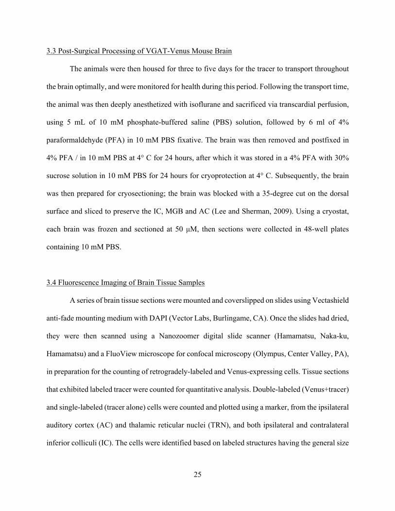
3.4 Fluorescence Imaging of Brain Tissue Samples
A series of brain tissue sections were mounted and coverslipped on slides using Vectashield
anti-fade mounting medium with DAPI (Vector Labs, Burlingame, CA). Once the slides had dried,
they were then scanned using a Nanozoomer digital slide scanner (Hamamatsu, Naka-ku,
Hamamatsu) and a FluoView microscope for confocal microscopy (Olympus, Center Valley, PA),
in preparation for the counting of retrogradely-labeled and Venus-expressing cells. Tissue sections
that exhibited labeled tracer were counted for quantitative analysis. Double-labeled (Venus+tracer)
and single-labeled (tracer alone) cells were counted and plotted using a marker, from the ipsilateral
auditory cortex (AC) and thalamic reticular nuclei (TRN), and both ipsilateral and contralateral
inferior colliculi (IC). The cells were identified based on labeled structures having the general size

26
and characteristics of cells in those areas. The cells were counted manually from processed images
produced by the FluoView confocal microscope and a Nanozoomer digital slide scanner. An
automatic counter analysis was also done via ImageJ for further confirmation of the results.
Completing this process, the results of this analysis was used for an overall summary of the tracer’s
distribution, labeling, and plotting of cells. Figures showing the distribution of the tracer
throughout the aforementioned auditory regions were taken using NDP.view2 (Hamamatsu).

27
CHAPTER 4 ANALYSIS OF CONVERGENT PROJECTIONS TO THE MGB
Retrograde tract tracing of convergent inputs to the MGB with CTb Alexa Fluor 594
showed a significant number of single and double-labeled retrograde cells within the regions being
observed. Single-labeled cells were identified as VGAT (vesicular GABA transporter)-negative
retrograde cells (i.e. red only), ones that did not express the Venus protein. Double-labeled cells
were identified as VGAT-positive, while also being identified as retrograde cells via the tracer dye
(i.e. red and green). The VGAT-Venus transgenic mice are very beneficial for identifying and
tracing GABAergic circuitry; cells expressing the Venus protein with the vesicular GABA
transporter, and also labeled with the tracer dye, are confirmed to be GABAergic (Lee at al., 2015).
The single-labeled cells are VGAT-negative, and as such, are not GABAergic, but presumed
excitatory. The labeling process does not provide absolute proof of glutamatergic populations, and
can only contrast GABAergic from non-GABAergic populations.
4.1 Convergent Excitatory and Inhibitory Projections to the MGB
The injection of the tracer dye was made in the medial geniculate body, to ensure correct
and accurate retrograde tracing (Figure 5). The mouse received an injection at a volume of 0.2-
0.5µl in the area. The injection site displayed deep diffusion of the tracer dye as expected, across
much of the lateral sector of the MGB, and labeled many cells locally.
Retrograde labeling in both IC, the AC, and TRN were found as well; to compound on the
evidence of single and double-labeled retrograde cells in these areas, high-magnification images
of the regions were also produced, the IC being one example (Figure 6).
28
Figure 5. Confocal microcopy imagery of the injection site for the CTb-Alexa 594 tracer dye (red), located in the medial geniculate body of the VGAT-Venus transgenic mouse.
The ipsilateral inferior colliculus (IC) contained a large amount of single- and double-
labeled retrograde cells projecting to the medial geniculate body (MGB); 30.4% of the IC's
retrograde cells were double-labeled, proving them to be GABAergic, while the rest were VGAT-
negative, resulting in them being defined as excitatory. Being a nexus for ascending and
descending projections in the central auditory system, these results confirmed previous studies
(Saldaña and Marchán, 2005; Wenstrup, 2005; Buentello et al., 2015; Ito and Oliver, 2010; Oliver,
1984). The ipsilateral IC (Figure 6) displayed less GABAergic (14, 30.4%) and non-GABAergic
(32, 69.6%) neurons labeled by the tracer dye, in comparison to the contralateral IC (12, 36.3%;
21, 63.6% respectively). While these results are close enough to make it likely of an
inconsequential difference, they further confirm past studies in other species, such as the guinea

29
pig and the mustached bat (Mellott et al., 1994; Winer, 1992). The distribution of single- and
double-labeled retrograde cells in both IC were similar; they displayed a larger count of retrograde
cells near the dorsal edges of the region. A large majority of the two types of retrograde cells were
also overlapping, as many single-labeled cells were clustered adjacent to the double-labeled cells.
Figure 6. Confocal microscopy imagery of ipsilateral IC. VGAT-negative retrograde cells are labeled red, VGAT-positive retrograde cells are labeled red and green.

30
The thalamic reticular nucleus (TRN) also exhibited GABAergic neuronal projections to
the MGB, as seen by double-labeled retrograde cells; 77.8% of retrograded cells found in the TRN
were GABAergic. (Figure 7). As the TRN provides a major source of inhibition to the MGB, these
findings demonstrate a noteworthy source of inhibition to the MGB from the TRN. The distribution
of the TRN's retrograded cells, single- and double-labeled, were found in the region of the TRN
closest to the hippocampus.
Figure 7. Confocal microscopy imagery of the ipsilateral thalamic reticular nucleus. VGAT-negative retrograde cells are labeled red, VGAT-positive retrograde cells are labeled red and green.

31
The ipsilateral auditory cortex was also scanned and shown to display projections to the
MGB via retrograde cell tracing (Figure 8).
Figure 8. Confocal microscopy imagery of the ipsilateral auditory cortex. VGAT-negative retrograde cells are labeled red, VGAT-positive retrograde cells are labeled red and green.
All of the retrograded cells found in the AC were single-labeled; most of the distribution
of these cells was found in the lower layers of the AC, which could be described as layers V and
32
VI. These findings strengthen the established idea of the AC providing a substantial excitatory
contribution to the MGB.
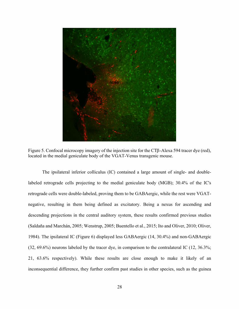
4.2 Quantitative Analysis of GABAergic and Excitatory Projections
The distribution of single- and double-labeled retrograde cells were counted, from the
confocal and Nanozoomer digital microscopy images (Figure 9).
Figure 9. High-magnification confocal microscopy imagery of an example of single- and double-labeled cells in the medial geniculate body. VGAT-negative retrograde cells are labeled red, VGAT-positive retrograde cells are labeled red and green.
33
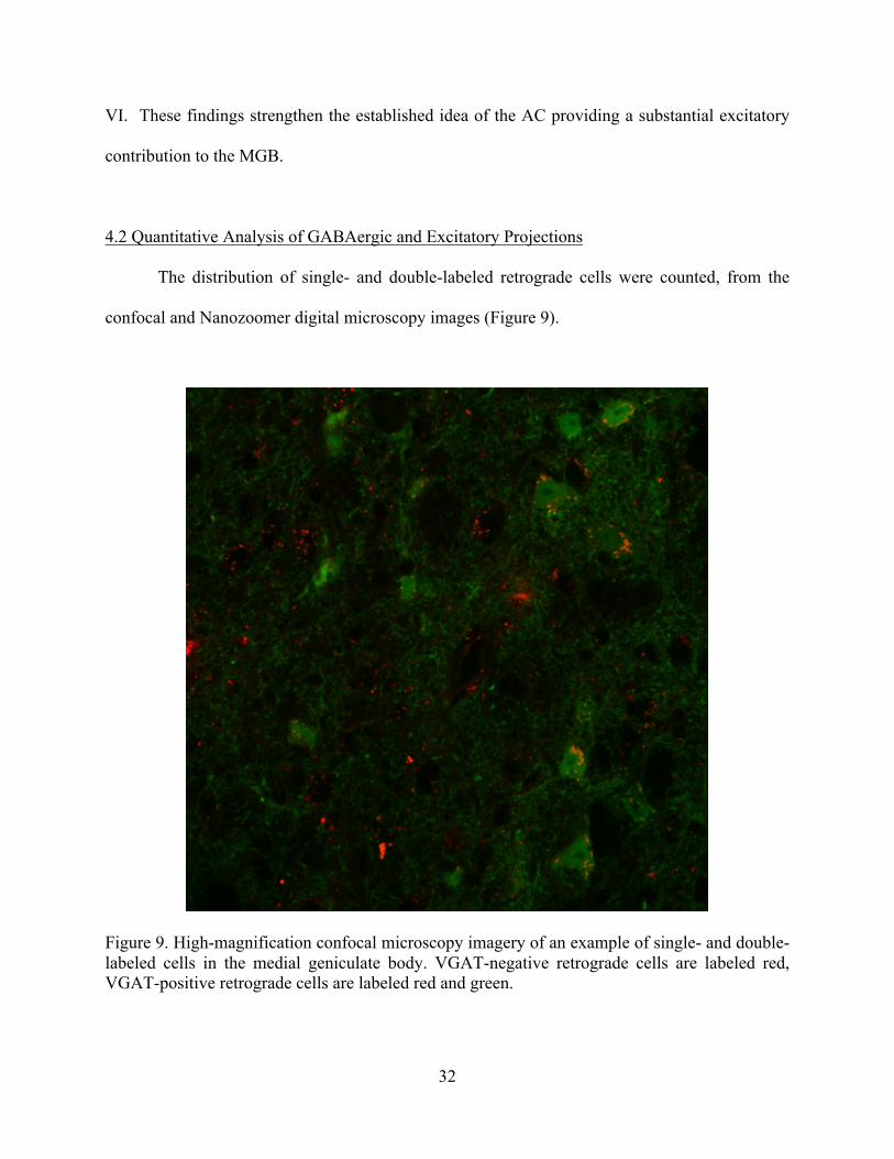
Cells were counted several times to ensure precise findings; the cell counts further
displayed and characterized what was found via imaging, in that a strong GABAergic projection
from the three of the neuronal regions observed (IC, contralateral IC, TRN) to the MGB was found.
Manual cell counting was completed using plotter and marker. The TRN was shown to provide
just 25% of total inhibitory neuronal count (Table 1).
Table 1. Quantitative analysis of proportional convergence of retrogradely labeled GABAergic and non-GABAergic projections to the MGB in the VGAT-Venus mouse. VGAT+ and VGAT- retrograde cells values are described as percentages of total excitatory and/or inhibitory neurons relative to overall labeling. Inferior colliculus
(ipsi) Inferior colliculus (con)
Thalamic reticular nucleus
Auditory cortex
Mean VGAT+ retrograde cells (%)
41.8 ± 0.9 32.9 ± 24.1 25.3 ± 33.6 0 ± 0
Mean VGAT- retrograde cells (%)
32.2 ± 0.1 21.2 ± 0.1 2.3 ± 0.2 44.3 ± 1.0
Mean total retrograde cells (%)
34.5 ± 0.01 24 ± 1.33 7.7 ± 1.7 33.8 ± 0
Type of projection
Excitatory/Inhibitory Excitatory/Inhibitory Inhibitory Excitatory
We found that most of the presumed excitatory inputs arose from the auditory cortex (Table
2, 44% of total excitatory retrograde cells), with the ipsilateral and contralateral IC showing lesser
of an excitatory count (32% and 21%, respectively). The number of VGAT-negative retrograde
cells exceeded the VGAT-positive retrograde cells in every region besides the TRN, indicating a
larger non-GABAergic projection from the majority of the structures than GABAergic. Focusing
on GABAergic projections, the region shown to provide the most GABAergic output to the MGB

34
was the ipsilateral IC, with the contralateral IC as the next highest. The AC, being involved in
direct excitation of the MGB, had a large amount of VGAT-negative retrograde cells, indicating a
more robust non-GABAergic, excitatory projection to the MGB. Most of the inhibitory inputs
labeled were sourced from the inferior colliculi (Table 2, 42% and 33%, respectively).
Table 2. Quantitative analysis of retrograded GABAergic and non-GABAergic projections to the MGB in the VGAT-Venus mouse. VGAT+ and VGAT- retrograde cells values are described as percentages of total excitatory and/or inhibitory neurons in that region. Inferior colliculus
(ipsi) Inferior colliculus (con)
Thalamic reticular nucleus
Auditory cortex
Mean VGAT+ retrograde cells (%)
30.4 ± 2.0 36.4 ± 5.0 77.8 ± 1.0 0 ± 0
Mean VGAT- retrograde cells (%)
69.6 ± 2.0 63.6 ± 5.0 22.2 ± 1.0 100.0 ± 0
Type of projection
Excitatory/Inhibitory Excitatory/Inhibitory Inhibitory Excitatory
Table 2 also shows once more, the type of output that the structures project to the MGB.
Table 1 compounds on Table 2’s finding, showing each region’s percentage of labeling in
comparison to overall labeling done in all structures. What was counted in the structures matched
with the type of projection the regions were previously known to have.

35
CHAPTER 5 DISCUSSION
In this study, we used retrograde tract tracing with the CTb Alexa Fluor 594 tracer dye to
quantitatively characterize the neuronal pathways arising from the thalamic reticular nucleus
(TRN), auditory cortex (AC), and both inferior colliculi (IC), that project to the medial geniculate
body (MGB). The AC exhibited a substantial presumed glutamatergic projection to the MGB,
while both IC and TRN provided inhibitory GABAergic and/or excitatory glutamatergic
projections. Our data, using the VGAT-Venus mouse, reveals a clear convergence of excitatory
and inhibitory projections from multiple IC, TRN and cortical regions to the MGB. This tract-
tracing strengthens past findings of homologous regions in various species, such as the cat, rat,
and gerbil (Winer and Larue, 1996; Mellott et al., 2014b; Cant and Benson, 2006). Our use of the
VGAT-Venus transgenic mouse strain is advantageous compared to prior studies, since the native
expression of the Venus-fluorescent protein enabled the ready and relatively unambiguous
identification of inhibitory neuronal cells, where prior studies were reliant on
immunohistochemical detection of these cells.
Prior studies have typically focused on examining projections from one particular region
to the MGB, but assessing the convergence of multiple projections quantitatively to the MGB has
not been accomplished in the mouse. In addition to the topographical location of these projections,
the relative contribution of presumed excitatory and inhibitory inputs was also assessed, based on
retrograde cell counting from each region. The intensity of these projections varied from structure
to structure. The auditory cortex displayed a high count of VGAT-negative retrograde cells,
indicating a powerful and robust glutamatergic projection to the MGB. Next in intensity was the
ipsilateral IC, then the contralateral IC, and ending with the TRN. These results underscore the

36
complex interactions between these structures, and further refine the neuroanatomical organization
established by previous studies. (Figure 10)
Figure 10. Schematic diagram of excitatory (red) and inhibitory (blue) projections of the auditory cortex (AC), thalamic reticular nucleus (TRN) , medial geniculate body (MGB), and inferior colliculus (IC and IC (con)). Thickness of arrows represents the intensity of the projection.
The analysis of the MGB’s importance dates back nearly 60 years, continuing with further
discoveries of its anatomical subdivisions (Morest 1964, Aitkin and Webster 1972, Winer and
Morest 1983), tonotopic organization (Aitkin 1973, Winer 1984, Winer 1999), function (Morest
and Winer 1986), and connections with other thalamic regions (Rouiller et al. 1985, Winer and
Larue 1996). Further dissection has been carried out on the auditory cortex’s important role in
integrating and refining ascending input from the MGB (Winer 2005, Smith et al. 2012) via
corticothalamic projections, sending excitatory input to the TRN and MGB (Wenstrup, 2005). The

37
AC is also charged with the task of indirectly inhibiting the MGB via the TRN. The IC is
particularly important in this framework, acting as a nexus for all lower brainstem nuclei, such as
the superior olivary complex and cochlear nuclei, and is also the first region where inputs begin to
integrate in the central auditory system (Winer and Schreiner, 2005). The connectivity between
the IC and MGB cannot be understated; the tectothalamic pathway is the largest output pathway
of the IC, and is responsible for sending a substantial amount of GABAergic and glutamatergic
inputs to the MGB. Our results augment these prior findings by grouping and quantifying all of
these connections in one overall schema, in a species not extensively characterized.
The results found and described in this thesis correlate previous studies in this area of study.
As mentioned before, retrograde tracing of central auditory regions has been accomplished in
many other models, such as the mustached bat (Winer et al., 1992), gerbil (Cant and Benson,
2006), guinea pig (Mellott et al., 2014), and mouse. These studies have characterized thalamic and
cortical regions extensively, and provided a substantial foundation for quantitative analysis of the
neuronal architecture within these areas.
A major finding of this thesis consists of a framework of excitatory and inhibitory
projections from multiple auditory regions, converging in the MGB, providing a singular look at
a unified neuroanatomical map. Of note, the projections from the contralateral IC have been
especially interesting, as there has not been a substantial amount of bilateral IC studies involving
those structures’ connections to the MGB (Mellott et al., 2014a.). Our study shows that the
contralateral IC provides a less intense, but apparently inhibitory projection to the contralateral
MGB, which may enable further ascending inhibitory control of auditory processing. Overall,
extending the mapping of the many pathways and projections of the auditory system should
provide a broader framework for interpreting and stimulating further research on this topic.

38
CHAPTER 6 CONCLUSION
Real-world application of the findings from this thesis may emerge and have the potential
to affect the treatment of disorders of the auditory system. The neural processing of sound is an
extremely complicated task, and the labyrinthian aspects of that task have taken many years to
uncover, all concluding in what is perceived as a quite simple and speedy process (Lee and Winer,
2008). Our auditory system uses excitation and inhibition to stimulate, prune, filter, decipher, and
integrate the massive amount of stimuli obtained almost constantly throughout the usual day (Lee
and Winer, 2011). This system is further complicated by the daunting task of understanding this
integration; in order to effectively interpret the integration of ascending and descending inputs,
and excitatory and inhibitory projections to the MGB, a thorough characterization of these
projection systems is needed (Mellott et al., 2014a; Peruzzi et al., 1997; Winer et al., 1996).
More than that, an overall characterization of every main structure involved in the
contribution of those projections to the MGB is necessary for an increased comprehension of these
pathways. Obtaining this knowledge only furthers our insight into the excitation/inhibition balance
of hearing; one of many theories states that loss of GABAergic activity may play a role in the onset
and persistence of presbycusis (Tang et al., 2014; Gao et al., 2015; Winer, 1992). Further studies
of how these neuroanatomical connections change temporally would be very beneficial in testing
its role in this disorder and will almost certainly prove to be a welcome addition to the future goal
of reducing and alleviating age-related hearing loss.

39
REFERENCES
Ades, H.W., Mettler, F.A., Culler, E.A., 1939. Effect of lesions in the medial geniculate bodies upon hearing in the cat. Amer. J. Physiol. 125, 15.
Aitkin, L. M., & Webster, W. R., 1972. Medial geniculate body of the cat: organization and
responses to tonal stimuli of neurons in ventral division. Journal of Neurophysiology, 35(3), 365–380.
Allen, P.D., Eddins, D.A., 2010. Presbycusis phenotypes form a heterogeneous continuum when
ordered by degree and configuration of hearing loss. Hear Res. 264, 10-20. Altschuler, R.A., Tong, L., Holt, A.G., Oliver, D.L., 2008. Immunolocalization of vesicular
glutamate transporters 1 and 2 in the rat inferior colliculus. Neuroscience. 154, 226-232. Beyerl, B. D., 1978. Afferent projections to the central nucleus of the inferior colliculus in the rat.
Brain Research, 145(2), 209–223. Bielefeld, E.C., Tanaka, C., Chen, G.D., Henderson, D., 2010. Age-related hearing loss: is it a
preventable condition? Hear Res. 264, 98-107. Blosa, M., Sonntag, M., Brückner, G., Jäger, C., Seeger, G., Matthews, R.T., Rübsamen, R.,
Arendt, T., Morawski, M., 2013. Unique features of extracellular matrix in the mouse medial nucleus of trapezoid body-implications for physiological functions. Neuroscience. 228, 215-234.
Briggs, F., & Usrey, W. M. (2008). Emerging views of corticothalamic function. Current Opinion
in Neurobiology, 18(4), 403–407. Buentello, D.C., Bishop, D.C., Oliver, D.L., 2015. Differential distribution of GABA and glycine
terminals in inferior colliculus of rat and mouse. J Comp Neurol. in press. Calford M. B., Aitkin L. M., 1983. Ascending projections to the medial geniculate body of the cat:
evidence for multiple, parallel auditory pathways through thalamus. J. Neurosci. 3, 2365–2380.
Cant, N.B., 1992. The cochlear nucleus: neuronal types and their synaptic organization. In: The
Mammalian Auditory Pathway: Neuroanatomy. Vol., D.B. Webster, A.N. Popper, R.R. Fay, ed.^eds. Springer, New York, pp. 66-116.
Cant, N. B., & Benson, C. G. (2006). ORGANIZATION OF THE INFERIOR COLLICULUS OF
THE GERBIL (MERIONES UNGUICULATUS): DIFFERENCES IN DISTRIBUTION OF PROJECTIONS FROM THE COCHLEAR NUCLEI AND THE SUPERIOR OLIVARY COMPLEX. The Journal of Comparative Neurology, 495(5), 511–528.
40
Caspary, D.M., Ling, L., Turner, J.G., Hughes, L.F., 2008. Inhibitory neurotransmission, plasticity and aging in the mammalian central auditory system. J Exp Biol. 211, 1781-1791.
Chu, H.N., 1932. The fiber connections of the diencephalon of the opossum, Didelphis virginia.
Monogr Nat Res Inst Psychol (Nanking) 3, 1-34. Crabtree, J.W., 1998. Organization in the auditory sector of the cat's thalamic reticular nucleus. J
Comp Neurol. 390, 167-182. Crabtree, J.W., Collingridge, G.L., Isaac, J.T., 1998. A new intrathalamic pathway linking
modality-related nuclei in the dorsal thalamus. Nat Neurosci. 1, 389-394. Dalton, D.S., Cruickshanks, K.J., Klein, B.E., Klein, R., Wiley, T.L., Nondahl, D.M., 2003. The
impact of hearing loss on quality of life in older adults. Gerontologist. 43, 661-668. Erulkar, S. D. (1975) Physiological studies of the inferior colliculus and medial geniculate
complex. In Handbook of Sensory Physiology. Vol. 5, Pt. 2: Auditory System. Physiology (CNS), Behavioral Studies, Psychoacoustics, 145-198.
Gao F, Wang G, Ma W, et al. Decreased auditory GABA+ concentrations in presbycusis
demonstrated by edited magnetic resonance spectroscopy. NeuroImage. 2015;106:311-316.
Geissler, D.B.D., Ehret, G.G., 2004. Auditory perception vs. recognition: representation of
complex communication sounds in the mouse auditory cortical fields. Eur J Neurosci. 19, 1027-1040.
Gilbert C.D., Kelly J.P., 1975. The projections of cells in different layers of the cat's visual cortex.
Journal of Comparative Neurology. 1975;163:81–105. Hackett, T.A., 2011. Information flow in the auditory cortical network. Hear Res. 271, 133-146. Harkrider, A.W., Plyler, P.N., Hedrick, M.S., 2005. Effects of age and spectral shaping on
perception and neural representation of stop consonant stimuli. Clin Neurophysiol. 116, 2153-2164.
Hipp, J.F., Engel, A.K., Siegel, M., 2011. Oscillatory synchronization in large-scale cortical
networks predicts perception. Neuron. 69, 387-396. Hudspeth, A.J., 1997. How hearing happens. Neuron. 19, 947-950. Huang, C. L., Larue, D. T. and Winer, J. A., 1999, GABAergic organization of the cat medial
geniculate body. J. Comp. Neurol., 415: 368–392. Huang, C.L., and Winer, J.A., 2000. Auditory thalamocortical projections in the cat: laminar and
areal patterns of input. Journal of Comparative Neurology, 427, 402-431.

41
Humes, L.E., Joellenbeck, L.M., Durch, J.S. Eds.), 2006. Noise and Military Service: Implications
for Hearing Loss and Tinnitus. The National Academies Press, Washington, D.C. Ito, T., Oliver, D.L., 2010. Origins of glutamatergic terminals in the inferior colliculus identified
by retrograde transport and expresión of VGLUT1and VGLUT2 genes. Front Neuroanat. 4, 135.
Kaas, J.H., Hackett, T.A., 2000. Subdivisions of auditory cortex and processing streams in
primates. Proc Natl Acad Sci U S A. 97, 11793-11799. Lam, Y.W., Sherman, S.M., 2005. Mapping by laser photostimulation of connections between the
thalamic reticular and ventral posterior lateral nuclei in the rat. J Neurophysiol. 94, 2472-2483.
Lee, C.C., Winer, J.A., 2008. Connections of cat auditory cortex: I. Thalamocortical system. J
Comp Neurol. 507, 1879-1900. Lee, C.C., Winer, J.A., 2011. Convergence of thalamic and cortical pathways in cat auditory
cortex. Hear Res. 274, 85-94. Lee, C.C., 2013. Thalamic and cortical pathways supporting auditory processing. Brain Lang. 126,
22-28. Lee, C.C., 2015. Exploring functions for the non-lemniscal auditory thalamus. Front Neural
Circuits. 9, 69. Llano, D.A., Sherman, S.M., 2008. Evidence for non-reciprocal organization of the mouse auditory
thalamocortical-corticothalamic projections systems. J Comp Neurol. 507, 1209-1227. Llano, D.A., Sherman, S.M., 2009. Differences in intrinsic properties and local network
connectivity of identified layer 5 and layer 6 adult mouse auditory corticothalamic neurons support a dual corticothalamic projection hypothesis. Cereb Cortex. 2009, 2810-2826.
Llano, D.A., Turner, J., Caspary, D.M., 2012. Diminished cortical inhibition in an aging mouse
model of chronic tinnitus. J Neurosci. 32, 16141-16148. Llano, D.A., Slater, B.J., Lesicko, A.M., Stebbings, K.A., 2014. An auditory
colliculothalamocortical brain slice preparation in mouse. J Neurophysiol. 111, 197-207. Malmierca, M. S., Anderson, L. A., & Antunes, F. M. (2015). The cortical modulation of stimulus-
specific adaptation in the auditory midbrain and thalamus: a potential neuronal correlate for predictive coding. Frontiers in Systems Neuroscience, 9, 19.

42
Mellott, J.G., Foster, N.L., Nakamoto, K.T., Motts, S.D., Schofield, B.R., 2014a. Distribution of GABAergic cells in the inferior colliculus that project to the thalamus. Front Neuroanat. 8, 17.
Mellott, J.G., Foster, N.L., Ohl, A.P., Schofield, B.R., 2014b. Excitatory and inhibitory projections
in parallel pathways from the inferior colliculus to the auditory thalamus. Front Neuroanat. 8, 124.
Morest, D. K., 1964. The neuronal architecture of the medial geniculate body of the cat. Journal
of Anatomy, 98(Pt 4), 611–630.1. Morest D.K., 1964b. The laminar structure of the inferior colliculus of the cat. Anat Rec 148:314 Morest, D. K., 1965. The lateral tegmental system of the midbrain and the medial geniculate body.
Journal of Anatomy, 99(Pt 3), 611–634. Morest, D.K., and Winer, J.A. The comparative anatomy of neurons: homologous neurons in the
medial geniculate body of the opossum and the cat. Advances in Anatomy, Embryology and Cell Biology, 1986, 97, 1996.
Najdzion, J., Wasilewska, B., Rowniak, M. Bogus-Nowakowska, K., Szteyn, S., Robak, A. 2011.
A Morphometric Comparative Study of the Medial Geniculate Body of the Rabbit and the Fox. Anantomia Histologia Embryologia 40:326-334.
Oliver, D.L., 1984. Dorsal cochlear nucleus projections to the inferior colliculus in the cat: a light
and electron microscopic study. J Comp Neurol. 224, 155-172. Oliver, D. L., Winer, J. A., Beckius, G. E., & Saint Marie, R. L., 1994. Morphology of GABAergic
neurons in the inferior colliculus of the cat. The Journal of Comparative Neurology, 340(1), 27–42.
Oliver, D.L., 2000. Ascending efferent projections of the superior olivary complex. Microsc Res
Tech. 15, 355-363. Peruzzi, D., Bartlett, E., Smith, P.H., Oliver, D.L., 1997. A monosynaptic GABAergic input from
the inferior colliculus to the medial geniculate body in rat. J Neurosci. 17, 3766-3777. Prieto, J. J., Peterson, B. A. and Winer, J. A., 1994. Morphology and spatial distribution of
GABAergic neurons in cat primary auditory cortex (AI). J. Comp. Neurol., 344: 349–382. Reetz, G., Ehret, G., 1999. Inputs from three brainstem sources to identified neurons of the mouse
inferior colliculus slice. Brain Res. 816, 527-543. Ramon Y Cajal, S., 1911. Histologie du système nerveux de l'homme et des vertébrés.
43
Ramon Y Cajal, S., 1955. Histologie du Systeme Nerveux de l'Homme et des Vertebris, t. ii. Madrid: Instituto Ranl6n y Cajal.
Rioch, DMK. 1929. Studies on the diencephalon of the carnivora. Part I. The nuclear configuration
of the thalamus, epithalamus, and hypothalamus of the dog and cat. J Comp Neurol49:1–119.
Rouiller, E. M., Colomb, E., Capt, M., & De Ribaupierre, F., 1985. Projections of the reticular
complex of the thalamus onto physiologically characterized regions of the medial geniculate body. Neuroscience Letters, 53(2), 227–232.
Roberts, R. C., & Ribak, C. E., 1987. An electron microscopic study of GABAergic neurons and terminals in the central nucleus of the inferior colliculus of the rat. Journal of Neurocytology, 16(3), 333–345.
Saldaña, E., Marchán, M.A., 2005. Intrinsic and commissural connections of the inferior
colliculus. In: The inferior colliculus. Vol., J.A. Winer, C.E. Schreiner, ed.^eds. Springer, New York, pp. 155-182.
Salomon, G., 1986. Hearing problems and the elderly. Dan Med Bull. 33, 1-22. Schreiner, C. E., & Cant, N. B., 2011. A Tribute to Jeffery A. Winer. Hearing Research, 274(1-2),
1-2. Schwartz, I.R., 1992. The superior olivary complex and lateral lemniscal nuclei. In: The
mammalian auditory pathway: neuroanatomy. Vol., ed.^eds. Springer, New York, pp. 117-167.
Sherman, S.M., Guillery, R.W., 2006. Exploring the thalamus and its role in cortical function,
Vol., MIT Press, London. Smith, P.H., Uhlrich, D.J., Manning, K.A., Banks, M.I., 2012. Thalamocortical projections to rat
auditory cortex from the ventral and dorsal divisions of the medial geniculate nucleus. J Comp Neurol. 520, 34-51.
Stebbings, K.A., Choi, H.W., Ravindra, A., Caspary, D.M., Turner, J.G., Llano, D.A., 2016.
Ageing-related changes in GABAergic inhibition in mouse auditory cortex, measured using in vitro flavoprotein autofluorescence imaging. J Physiol. 594, 207-221.
Tang X, Zhu X, Ding B, Walton JP, Frisina RD, Su J. Age-related Hearing Loss: GABA, Nicotinic
Acetylcholine and NMDA Receptor Expression Changes in Spiral Ganglion Neurons of the Mouse. Neuroscience. 2014; 259:184-193.
Van Buren, J.M., Borke, R.C., 1972. Variations and Connections of the Human Thalamus, Vol. 1.
44
Villa, A.E., Rouiller, E.M., Simm, G.M., Zurita, P., de Ribaupierre, Y., de Ribaupierre, F., 1991. Corticofugal modulation of the information processing in the auditory thalamus of the cat. Exp Brain Res. 86, 506-517.
Wang, Y., Kakizaki, T., Sakagami, H., Saito, K., Ebihara, S., Kato, M., . . . Yanagawa, Y. (2009).
Fluorescent labeling of both GABAergic and glycinergic neurons in vesicular GABA transporter (VGAT)–Venus transgenic mouse. Neuroscience, 164(3), 1031-1043.
Wehr, M., Zador, A.M., 2003. Balanced inhibition underlies tuning and sharpens spike timing in auditory cortex. Nature 426.
Wenstrup, J.J., 2005. The tectothalamic system. In: The Inferior Colliculus. Vol., J.A. Winer, C.E.
Schreiner, ed.^eds. Springer, New York, pp. 200-230. Wepsic, J. G., 1966. Multimodal sensory activation of cells in the magnocellular medial geniculate
nucleus. Experimental Neurology,15(3), 299–318. Winer, J.A., Diamond, I.T., and Raczkowski, D. Subdivisions of the auditory cortex of the cat:
the retrograde transport of horseradish peroxidase to the medial geniculate body and posterior thalamic nuclei. Journal of Comparative Neurology, 1977, 176, 387-418.
Winer, J. A., Morest, D. K., 1983. The medial division of the medial geniculate body of the cat:
implications for thalamic organization. Journal of Neuroscience,3(12), 2629–2651. Winer, J.A., 1984. The human medial geniculate body. Hear Res. 15, 225-247. Winer, J.A., and Larue, D.T. Anatomy of glutamic acid decarboxylase immunoreactive neurons
and axons in the rat medialgeniculate body. Journal of Comparative Neurology, 1988, 278, 47-68.
Winer, J.A., 1992. The functional architecture of the medial geniculate body and the primary
auditory cortex. In: Springer Handbook of Auditory Research, volume 1, The Mammalian Auditory Pathways: Neuroanatomy., Vol., D.B. Webster, A.N. Popper, R.R. Fay, ed.^eds. Springer-Verlag, New York, pp. 222-409.
Winer, J.A., Wenstrup, J.J., and Larue, D.T. Patterns of GABAergic immunoreactivity define
subdivisions of the mustached bat's medial geniculate body. Journal of Comparative Neurology, 1992, 319, 172-190.
Winer, J.A., Larue, D.T., 1996. Evolution of GABAergic circuitry in the mammalian medial
geniculate body. Proc Natl Acad Sci U S A. 93, 3083-3087. Winer, J.A., Saint Marie, R.L., Larue, D.T., Oliver, D.L., 1996. GABAergic feedforward
projections from the inferior colliculus to the medial geniculate body. Proc Natl Acad Sci U S A. 93, 8005-8010.

45
Winer, J.A., Larue, D.T., Diehl, J.J., Hefti, B.J., 1998. Auditory cortical projections to the cat inferior colliculus. J Comp Neurol. 400, 147-174.
Winer, J.A., Kelly, J.B., and Larue, D.T. Neural architecture of the rat medial geniculate body.
Hearing Research, 1999, 130, 19-41. Winer, J.A., Diehl, J.J., Larue, D.T., 2001. Projections of auditory cortex to the medial geniculate
body of the cat. J Comp Neurol. 430, 27-55. Winer, J.A., Schreiner, C.E. Eds., 2005. The Inferior Colliculus. Springer, New York. Winer, J.A., 2006. Decoding the auditory corticofugal systems. Hear Res. 212, 1-8. Winguth, S.D., and Winer, J.A. Corticocortical connections of cat primary auditory cortex (AI):
laminar organization and identification of supragranular neurons projecting to area AII. Journal of Comparative Neurology, 1986, 248, 36-56.
Woollard, H.H., Harpman, A., 1939. The cortical projection of the medial geniculate body. J
Neurol Psychiatry, 2:35-44 Yamasoba, T., Lin, F.R., Someya, S., Kashio, A., Sakamoto, T., Kondo, K., 2013. Current
concepts in age-related hearing loss: epidemiology and mechanistic pathways. Hear Res. 303, 30-38.
Yueh, B., Shapiro, N., MacLean, C.H., Shekelle, P.G., 2013. Screening and management of adult
hearing loss in primary care. J Am Med Assoc. 289, 1976-1985.
46
VITA
Blaise Clarke, a native of Baton Rouge, La., received his bachelor’s degree in Psychology
at Louisiana State University in December 2012. Afterwards, he took additional science-related
courses and performed research in various laboratories. His passion for neuroscience never
wavering, he decided to enter graduate school at Louisiana State University in August 2014. He is
a candidate to receive his master’s degree in May 2017, and plans to enter a doctoral program the
following fall semester.
Recommended