Division of Comparative Physiology and Biochemistry, Society for Integrative andComparative Biology
The Physiological Adaptations of the Lahontan Cutthroat Trout (Oncorhynchus clarkihenshawi) following Transfer from Well Water to the Highly Alkaline Waters of PyramidLake, Nevada (pH 9.4)Author(s): Michael P. Wilkie, Patricia A. Wright, George K. Iwama and Chris M. WoodSource: Physiological Zoology, Vol. 67, No. 2 (Mar. - Apr., 1994), pp. 355-380Published by: The University of Chicago Press. Sponsored by the Division of ComparativePhysiology and Biochemistry, Society for Integrative and Comparative BiologyStable URL: http://www.jstor.org/stable/30163853Accessed: 16-09-2016 03:23 UTC
JSTOR is a not-for-profit service that helps scholars, researchers, and students discover, use, and build upon a wide range of content in a trusted
digital archive. We use information technology and tools to increase productivity and facilitate new forms of scholarship. For more information about
JSTOR, please contact [email protected].
Your use of the JSTOR archive indicates your acceptance of the Terms & Conditions of Use, available at
http://about.jstor.org/terms
The University of Chicago Press, Division of Comparative Physiology andBiochemistry, Society for Integrative and Comparative Biology are collaborating with JSTORto digitize, preserve and extend access to Physiological Zoology
This content downloaded from 130.113.111.210 on Fri, 16 Sep 2016 03:23:05 UTCAll use subject to http://about.jstor.org/terms

355
The Physiological Adaptations of the Lahontan Cutthroat Trout (Oncorhynchus clarki henshawi) following Transfer from Well Water to the Highly
Alkaline Waters of Pyramid Lake, Nevada (pH 9.4)
Michael P. Wilkie*
Patricia A. Wrightt
George K. Iwamat
Chris M. Wood*
Pyramid Lake Fisheries, Star Route, Sutcliffe, Nevada 89510
Accepted 10/1/93
Abstract
Salmonids experience severe disturbances in the excretion and internal regula- tion of ammonia, acid-base balance, and ionoregulation when challenged with alkaline pH. We followed the responses of a high-pH-tolerant salmonid, the La- hontan cutthroat trout (Oncorhynchus clarki henshawi)for 72 h after transfer from pH 8. 4 well water into the alkaline water (pH 9. 4) of Pyramid Lake, Ne- vada. Fish that had been living in Pyramid Lake for 3 wk, 5 wk, and 2 yr were also examined. A combined metabolic and respiratory alkalosis (negative meta-
bolic acid load [AH+] and decreased arterial CO2 tension [Paco2], respectively) occurred initially. The metabolic component was corrected within 24 h, but the respiratory component persisted for up to 5 wk. Transfers also resulted in an im-
mediate 70% reduction in the ammonia excretion rate (J,,m) and a 30% in- crease in total plasma ammonia (Tm,,). The Tm,,,m was corrected within 3 d, but
Jamm remained depressed, which indicates reduced ammonia production rates. Because the urea excretion rate (J,,rea) did not change, the contribution of Jrea to total N excretion increased from 10% in well water to 25% in fish acutely and chronically exposed to lake water. Liver enzyme activities indicated that the pathway for urea production was uricolysis, not the ornithine-urea cycle. Bran- chial chloride cell fractional surface area increased in lake water, and this may have counteracted the base load by promoting base equivalent excretion via
Cl-/HCOj exchange. Plasma Na+ and Cl- levels were slightly higher in Pyramid Lake water. We conclude that the Lahontan cutthroat trout are able to survive in
Pyramid Lake's alkaline environment because of their ability to reduce ammo-
* Present address: Department of Biology, McMaster University, Hamilton, Ontario L8S 4K1, Canada.
t Present address: Department of Pathology, University of Guelph, Guelph, Ontario N1G 2W1, Canada.
t Present address: Department of Animal Science, University of British Columbia, Vancouver, British Columbia
V6T 2A2, Canada.
Physiological Zoology 67(2):355-380. 1994. c 1994 by The University of Chicago. All rights reserved. 0031-935X/94/6702-9328$02.00
This content downloaded from 130.113.111.210 on Fri, 16 Sep 2016 03:23:05 UTCAll use subject to http://about.jstor.org/terms
356 M. P. Wilkie, P. A. Wright, G. K. Iwama, and C. M. Wood
nia production, thereby avoiding chronic elevation ofplasma Tamm, and their ability to control blood acid-base and ionic status under alkaline conditions.
Introduction
Wilkie and Wood (1991) recently described the physiological responses of
rainbow trout (Oncorhynchus mykiss) following transfer from circumneutral
(pH 8.1) to alkaline (pH 9.5) water . Substantial disturbances occurred in nitrogenous waste (N waste) excretion, blood acid-base balance, and ionic status, none of which were fully corrected within 72 h. Other studies on rainbow trout have documented similar effects (Wright and Wood 1985; Heming and Blumhagen 1988; Lin and Randall 1990; Yesaki and Iwama 1992). Natural acute exposure to such a high pH is, however, a very unusual
circumstance for this species.
We examined the physiological responses of a related salmonid, the La- hontan cutthroat trout (LCT; Oncorhynchus clarki henshawi) following transfer to pH 9.4. Acute alkaline exposure is part of the LCT's "natural" life cycle. The first year of life is spent in well water (pH 8.4), following which the fish are moved abruptly into the highly alkaline (pH 9.4) and moderately saline waters (4.4 %o) of Pyramid Lake, Nevada (table 1). Atten- tion has recently shifted to this species (Wilkie et al. 1993; Wright, Iwama, and Wood 1993) because it exhibits unusual tolerance to high pH and thrives
in highly alkaline lakes throughout the northwestern United States (Trotter
1991). Attempts to stock other salmonids, such as coho salmon (Oncorhyn- chus kisutch), kokanee (Oncorhynchus nerka), brown trout (Salmo trutta), and rainbow trout into these lakes have failed (Galat et al. 1985; Kucera,
Koch, and Marco 1985; Coleman and Johnson 1988). The LCT is now designated as "threatened" because of a paucity of suc-
cessfully reproducing populations (Williams et al. 1989). Historically, the Pyramid Lake LCT spawned and passed through the juvenile life stages in the freshwater environment of the Truckee River, which feeds into Pyramid
Lake. Prolonged drought and water diversion have made this river inacces-
sible to spawning-condition LCT for many years, however (Galat et al. 1981, 1985; Kucera et al. 1985; Coleman and Johnson 1988). Indeed, by 1944 the
original Pyramid Lake LCT was declared extinct. A vigorous stocking pro- gram, in which juvenile LCT are reared for 1 yr in well water prior to intro-
duction into Pyramid Lake's alkaline waters, has revived the lake's cutthroat
trout fishery (Coleman and Johnson 1988 ).
This content downloaded from 130.113.111.210 on Fri, 16 Sep 2016 03:23:05 UTCAll use subject to http://about.jstor.org/terms

Lahontan Cutthroat Trout in Alkaline Water 357
TABLE 1
Typical chemical composition of well water (pH 8. 4) and Pyramid Lake
water (pH 9. 4)
Pyramid Lake Well Water Water
pHa 8.35 9.36
[H+] (pmol L-') 4.40 X 10⁻³ .43 X 10-3 [OH-] (gmol L-1) .66 6.70 Pco2 (Torr) .78 .26 [HCO3] (mmol L-') 4.35 13.80 [COg] (mmol L-1) .04 4.97 Titration alkalinityb (mmol - L-') . .. 4.45 23.08 [Na+] (mmol L-') 7.30 58.20 [CI-] (mmol L-1) 4.15 59.70 Total salinity (g - L-') .59 4.43
a Measured at 10.40C.
b Titration alkalinity to pH = 4.0.
The minimal mortality experienced by LCT following transfer into Pyramid
Lake (Coleman and Johnson 1988; D. Mosely and P. Wagner, personal com-
munication) suggests that these fish are able to rapidly correct, or resist, the
physiological disturbances observed in other salmonids at high pH. The purpose of the present investigation was to determine the physiological characteristics that allow LCT to adapt to alkaline Pyramid Lake water (pH 9.4) and to establish the time course of these adaptations. We followed the
responses of naive LCT, which had never been exposed to high alkalinity, through a 72-h acute exposure to Pyramid Lake water. Analyses focused on N waste excretion, acid-base balance, and ionoregulation. Apart from the fact that this study was performed in the field using the well water and
alkaline lake water available on site, methods closely duplicated those of our earlier laboratory study on 0. mykiss (Wilkie and Wood 1991).
In addition, we investigated long-term adaptations by studying fish that
had been in lake water for 3 wk, 5 wk, and 2 yr (returning spawners). In
light of emerging evidence on the possible importance of urea production
as an adaptation to high pH (Wood 1993), we also measured hepatic activities
of uricolytic and ornithine-urea cycle (OUC) enzymes. Similarly, in view of recent findings on alterations in branchial chloride cell (CC) surface area
This content downloaded from 130.113.111.210 on Fri, 16 Sep 2016 03:23:05 UTCAll use subject to http://about.jstor.org/terms
358 M. P. Wilkie, P. A. Wright, G. K. Iwama, and C. M. Wood
in response to acid-base challenge (Goss et al. 1992 b), we looked for changes
in the surface morphometry of these cells following transfer to alkaline lake
water. Galat et al. (1985) reported apparent CC hyperplasia in LCT living in Pyramid Lake.
Material and Methods
All experiments were performed at the lakeside laboratory of Pyramid Lake
Fisheries during May and June. We followed responses for 72 h following
exposure to Pyramid Lake water in 1-yr-old LCT (Oncorhynchus clarki hen-
shawi; n = 8) of both sexes. The mean weight of these fish was 243.5 + 16.6 g (standard error of the mean [SEM]). Fish were kept indoors for 3 wk prior to the experiments in 500-L fiberglass holding tanks served with
flowing hatchery well water. The chemistry of this well water (table 1; mod-
erately high HCO0 and pH) reflects its origin from deep desert wells. The fish had been hatchery reared in the well water for the first year of their
lives and were exposed to highly alkaline, and moderately saline, Pyramid Lake water for the first time during our experiments (table 1).
Separate groups of 1-yr-old, well-water-reared LCT were sampled 3 wk
(202.3 + 17.5 g [n = 9]) and 5 wk (242.4 + 9.2 g [n = 13]) after exposure to lake water. Data are also reported for 3-4-yr-old cutthroat trout (500.0
+ 16.3 g [n = 6]) that had been free-living in Pyramid Lake for approximately 2 yr. These latter trout were netted as they migrated up an artificial spawning
channel. All fish from these three groups were held in 500-L tanks provided
with flowing lake water for at least 1 wk prior to experimentation. All fish were starved a minimum of 1 wk to minimize variation in N metabolism
(Fromm 1963).
Two days prior to sampling, fish were fitted with chronic indwelling dorsal
aortic catheters (Soivio, Westman, and Nyholm 1972) under MS 222 anes-
thesia (1:10,000 dilution; Sigma) and immediately placed in darkened, well-
aerated (gas partial pressure of 02 [P02] = 125-130 Torr), acrylic flux boxes (McDonald 1983). Water ammonia levels were less than 5 jmol N * L-1 when the boxes were operated as open systems. Mean water temperature
during the study of the acute responses was 10.40 + 0.4"C. The mean well
water pH for this study was 8.35 + 0.010, and the mean lake water pH was 9.36 + 0.006. For the longer-term comparisons, the water temperature, at
the time of sampling, was 7.50 C (pH = 9.41 + 0.005) for the fish sampled after 3 wk of exposure to lake water (3-wk fish), 9.50C (pH = 9.39 + 0.003) for those sampled after 5 wk (5-wk fish), and 10.00C (pH = 9.38 + 0.008) for those sampled after 2 yr (2-yr fish).
This content downloaded from 130.113.111.210 on Fri, 16 Sep 2016 03:23:05 UTCAll use subject to http://about.jstor.org/terms
Lahontan Cutthroat Trout in Alkaline Water 359
Well-aerated water was distributed to each flux box at 0.5 L - min-1 via a
flow-splitter. Two 500-L central reservoirs continuously served the flux boxes
with an excess of either well water (pH 8.4) or lake water (pH 9.4). Water
pH was monitored with a Radiometer GK 2401C combination electrode and
PHM 72 pH meter.
Experimental Protocol for Acute Response Experiments
Experimental methods followed those of Wilkie and Wood (1991). Water samples (15 mL) were taken at 0 h, 1 h, and 3 h of each period. The ammonia
excretion rate (JAmm) and urea excretion rate (Jurea) were determined first
in well water, then at 0-3 h, 8-11 h, 24-27 h, 48-51 h, and 72-75 h of
exposure to Pyramid Lake water. With the exception of the 0-3-h period of
lake water exposure, blood samples were taken 30 min prior to box closure.
This was done to minimize disturbance to the fish and to ensure that truly representative JAmm and Jurea were measured (see Wilkie and Wood 1991;
Wilkie et al. 1993). Blood samples (1 mL) were drawn into two heparinized
500-jtL gastight Hamilton syringes via the arterial catheter. Arterial pH (pHa) and 02 tension (Pao2) were measured immediately; blood used for the latter (approximately 200 gL) was reinfused into the fish. Cortland's saline was then infused to replace blood lost on account of sampling and to main-
tain the internal ionic and osmotic status of the fish (Wolf 1963). Aliquots
of whole blood were saved for later analysis of hemoglobin (20 gtL) and lactate content (100 gtL). The remainder was centrifuged, and a small amount of plasma (50 pL) decanted for immediate determination of plasma total
CO2 and protein concentration. The remaining plasma (400-500 LL) was frozen for later analysis of total ammonia (TAmm), urea, Na+ and Cl-, glucose, and cortisol. Water Po2, pH, and total CO2 concentration in each box were also measured at the time of blood sampling. To establish how ammonia excretion was achieved in Pyramid Lake's highly alkaline environment, the blood-to-water partial pressure gradients for NH3 (APNH3) and concentration
gradients for NH' (A[NH-]) were estimated from the measured pH values and Tmm values in plasma and water (Wright and Wood 1985; Wilkie and
Wood 1991). In a few fish, the transepithelial potential (TEP) across the gills between the arterial blood and the environment was also measured
with methods identical to those described by Perry and Wood (1985). De- termination of TEP allowed us to estimate the transbranchial electrochemical
gradients for OH- (H+), HCOQ, and C0 . Fish were sacrificed after 72 h with an overdose of MS 222 (1.5 g * L-l),
and the second left gill arch excised for morphometric analysis of branchial
CC surface area. In addition, the livers were quickly extracted, freeze-
This content downloaded from 130.113.111.210 on Fri, 16 Sep 2016 03:23:05 UTCAll use subject to http://about.jstor.org/terms

360 M. P. Wilkie, P. A. Wright, G. K. Iwama, and C. M. Wood
clamped, and stored in liquid N2 for later determination of ureagenic enzyme
activity. A control group, kept in well water, was similarly sacrificed and sampled.
Our analytical techniques and the calculations used to estimate N waste
excretion rates, water and blood chemical parameters, and electrochemical gradients are described by Wilkie and Wood (1991), Wilkie et al. (1993), and Wright et al. (1993).
Experimental Protocol for Long-Term Comparisons
Water and blood samples were taken essentially as described above. Only single samples were taken from the 3-wk fish and 2-yr fish. These fish were
then sacrificed for gill and liver excision. Tissue samples were not extracted from the 5-wk fish.
Gill Sampling Techniques and Analysis
The methods used in this study are based on those of Laurent and Perry (1990) and Goss, Laurent, and Perry (1992a). The gill filaments were trimmed from the excised second left gill arch in small pieces (approxi-
mately 10 filaments per piece), rinsed in ice-cold, 0.15 mol * L-1 Na+ cac-
odylate buffer, and then fixed in 5% glutaraldehyde for 60-70 min. After fixation, pairs of filaments, joined at the septum, were dissected away from
one another and washed three times with ice-cold buffer and refrigerated
at 40C for several hours. The paired filaments were then taken through a partial ethanol dehydration series (30%, 50%, and 70% ethanol) and shipped back to McMaster University in 70% ethanol. Subsequently, the gills were completely dehydrated in 95% ethanol and then absolute ethanol, and then taken through two successive baths (2 min each) of 1,1,1,3,3,3-hexamethyl-
disilazane (Aldrich) and air-dried. The paired filaments were then mounted on aluminum stubs, sputter-coated, and viewed on an ISI-DS130 dual-stage
scanning electron microscope at 2,000 times magnification. At least eight
noncontiguous fields (approximately 2,500 Ltm2 per field), along the trailing edge of a filament, were randomly photographed for each fish. The individual
surface areas of CC in each field were subsequently determined with a Graphic Master digitizing tablet (Numonics) and an accompanying software program (Sigma Scan; Jandel Scientific). Chloride cell fractional surface area (CC FSA) and CC density were calculated from the estimates of indi- vidual CC surface areas and the total filamental surface areas measured per
fish. The filamental epithelium was used for morphometry, rather than the
lamellar epithelium, because the paired filaments could be mounted parallel
This content downloaded from 130.113.111.210 on Fri, 16 Sep 2016 03:23:05 UTCAll use subject to http://about.jstor.org/terms

Lahontan Cutthroat Trout in Alkaline Water 361
to the face of the aluminum stub. This made the flat, relatively uniform
surface of the trailing filamental epithelium accessible for examination with
the scanning electron microscope. Furthermore, the gill lamellae are less
appropriate for such analysis because the undulating nature of their topog-
raphy makes viewing more difficult and measurements prone to error (Goss et al. 1992a).
Statistics
All data are expressed as means -t 1 SEM (n). For the 72-h acute lake water
exposure experiment, each animal served as its own control, and paired, two-tailed t-tests were used to determine statistical significance (P< 0.05).
For long-term exposures, data for fish held in lake water were compared with data generated for fish held in well water. Therefore, an F ratio was
calculated to test for homogeneity of variance, followed by an unpaired, two-tailed t-test to determine significant differences (P < 0.05). Ureagenic enzymes and gill morphometric data were evaluated by ANOVA, and sub- sequent paired contrasts (P< 0.05) were made by a Tukey-Kramer Honestly Significant Difference test using a commercially available statistics package (SAS JMP; SAS Institute 1989).
Results
Nitrogenous Waste Excretion
Lahontan cutthroat trout in well water exhibited a JAmm of approximately
330 tmol N - kg-' - h-'. This was reduced nearly 70% during the first hour of exposure to alkaline lake water; depression of JAmm persisted throughout
the 3-d exposure (fig. 1A). The associated initial 30% rise in plasma TAmm, to about 300 jtmol N - L-1, seen at 8-24 h (fig. 1B) was no longer evident
by 72 h (fig. 1B). Urea excretion rates remained stable around 40 .tmol N - kg-' - h-' for the duration of the 3-d exposure (fig. 1A). As a result,
the percentage contribution of Jrea to total N excretion (JTotal N) increased from 10% in well water to approximately 25% in lake water. Plasma urea
levels were between 5,500 and 6,500 tmol N - L-1 for 24 h following ex- posure to lake water but then declined to approximately 4,000 iimol N L-' after 48 h.
In well water, both the APNH3 and A[NHil] gradients were outwardly di-
rected (approximately 25 btTorr and 165 Ctmol N * L-', respectively; fig. 1C and D) and were in accordance with the increase in plasma TAmm (fig. 1B).
After 8 h of exposure to lake water both APNH3 and A[NH0-] increased sig-
This content downloaded from 130.113.111.210 on Fri, 16 Sep 2016 03:23:05 UTCAll use subject to http://about.jstor.org/terms

362 M. P. Wilkie, P. A. Wright, G. K. Iwama, and C. M. Wood
Well Water -- Pyramid Lake Water---- 400 A TT 300 CD JN-waste
200
E
E 100 -
0 1 IB Plasma TAmm
400 - I
o 200 I E
50
S400 - A[NH 6 200
0 -
C 8 24 48 72
Time (h)
Fig. 1. Changes in (A) JAmm (solid line) and JUrea (dashed line); (B) TAmm;
(C) the calculated APNH3; and (D) the calculated A[NH ] of Lahontan cutthroat trout, following transfer into alkaline Pyramid Lake water (pH 9.4) from well water (pH 8. 4). Values are means + 1 SEM; n = 7. Aster-
isks indicate significant differences from well water values (P < 0. 05).
nificantly to approximately 100 gLTorr and 290 pmol N * L-1, respectively (fig. 1C and D). The greater relative increase in APNH3 reflected a marked
elevation in blood pH. Thereafter, APNH3 and A[NH0-] gradually declined in parallel with the fall in plasma TAmm, and, by 72 h, neither was significantly different from control values.
Values of APNH3 in fish sampled at 3 and 5 wk were not significantly
different from well water values. Similarly, A[NHf], approximately 180 IImol N - L-1 in these fish, was also similar to that seen in well water. Fish sampled
after 2 yr in lake water had significantly elevated APNH3 and A[NHT] (ap-
proximately 70 pTorr and 280 jlmol N * L-', respectively). The reduction in JAmm seen upon initial exposure to lake water persisted
for an extended period. Fish sampled after 3- and 5-wk exposure to lake
This content downloaded from 130.113.111.210 on Fri, 16 Sep 2016 03:23:05 UTCAll use subject to http://about.jstor.org/terms

Lahontan Cutthroat Trout in Alkaline Water 363
water exhibited JAmm's of only about 100 pmol N f kg-1 - h-1. These values are the same as those measured over the first hour at pH 9.4. The JAmm of
the 2-yr trout was not significantly different from the rates observed in well
water fish, however (fig. 2A).
Despite the persistent depression ofJmm in lake-water-adapted fish, plasma TAmm was not persistently elevated. Rather, TAmm remained relatively stable,
from 72 h onward in all groups. The 2-yr fish, however, showed elevated
plasma TAmm (fig. 2B). The partial pressure of NH3 (PNH3) in plasma was
Well Water 4- Pyramid Lake Water 'A1
S400 - A T 300 - JAmm z T
200 - o E Z 100 -
IB
400 - Plasma TAmm
S 200 3L 100
150 - Plasma PNH, ID 0 100
S80- Urea
0" 60-
z40-
E 20-
0-
1 year 3 days 3 weeks 5 weeks 2 years
Time
Fig. 2. The influence of long-term exposure to alkaline Pyramid Lake wa-
ter (pH 9. 4) upon (A) Jamm, (B) TAmm, (C) PNH3 in arterial blood, and (D) JUrea of Lahontan cutthroat troutpreviously reared in well water (pH
8.4). Values are means + 1 SEM; n = 7 in well water and after 3 d atpH 9. 4; n 8 after 3 wk; n = 13 after 5 wk; and n >6 6after 2 yr of exposure to lake water. Asterisks indicate significant differences from well water values (P < 0. 05).
This content downloaded from 130.113.111.210 on Fri, 16 Sep 2016 03:23:05 UTCAll use subject to http://about.jstor.org/terms
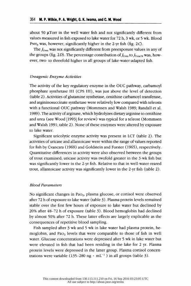
364 M. P. Wilkie, P. A. Wright, G. K. Iwama, and C. M. Wood
about 50 gTorr in the well water fish and not significantly different from
values measured in fish exposed to lake water for 72 h, 3 wk, or 5 wk. Blood
PNH3 was, however, significantly higher in the 2-yr fish (fig. 2C).
The Jurea was not significantly different from preexposure values in any of
the groups (fig. 2D). The percentage contribution ofJurea tOJTotalN was, how- ever, two- to threefold higher in all groups of lake-water-adapted fish.
Ureagenic Enzyme Activities
The activity of the key regulatory enzyme in the OUC pathway, carbamoyl phosphate synthetase III (CPS III), was just above the level of detection (table 2). Activities of glutamine synthetase, ornithine carbamoyl transferase,
and argininosuccinate synthetase were relatively low compared with teleosts
with a functional OUC pathway (Mommsen and Walsh 1989; Randall et al. 1989). The activity of arginase, which hydrolyzes dietary arginine to ornithine
and urea (see Wood [1993] for review) was typical for a teleost (Mommsen and Walsh 1991; table 2). None of these enzymes were altered by exposure to lake water.
Significant uricolytic enzyme activity was present in LCT (table 2). The activities of uricase and allantoicase were within the range of values reported
for fish by Cvancara (1969) and Goldstein and Forster (1965), respectively. Quantitative differences in activity were also observed between the groups of trout examined; uricase activity was twofold greater in the 3-wk fish but was significantly lower in the 2-yr fish. Relative to that in well-water-reared
trout, allantoicase activity was significantly lower in the 2-yr fish (table 2).
Blood Parameters
No significant changes in Pao2, plasma glucose, or cortisol were observed after 72 h of exposure to lake water (table 3). Plasma protein levels remained
stable over the first few hours of exposure to lake water but declined by
20% after 48-72 h of exposure (table 3). Blood hemoglobin had declined by almost 50% after 72 h. These latter effects are largely explicable as the
consequences of repetitive blood sampling. Fish sampled after 3 wk and 5 wk in lake water had plasma protein, he-
moglobin, and Pao2 levels that were comparable to those of fish in well water. Glucose concentrations were depressed after 5 wk in lake water but were elevated in fish that had been residing in the lake for 2 yr. Plasma
protein levels were depressed in the latter group. Plasma cortisol concen-
trations were variable (135-280 ng f mL-' ) in all groups (table 3).
This content downloaded from 130.113.111.210 on Fri, 16 Sep 2016 03:23:05 UTCAll use subject to http://about.jstor.org/terms

Lahontan Cutthroat Trout in Alkaline Water 365
TABLE 2
Activities of OUC enzymes and uricolytic enzymes in Labontan cutthroat
trout living in well water or Pyramid Lake water
Lake Water
Well Water 3 wk 2 yr
OUC enzymes:a Glutamine
synthetase ....... .43 + .08 .35 + .07 .57 + .08 (6) (8) (6)
Carbamoyl
phosphate synthetase III .... .01 + .01 .02 + .01 .03 + .01
(4) (4) (6)
Ornithine carbamoyl transferase ...... .03 + .01 .03 + .00 .03 + .00
(6) (8) (6) Argininosuccinate synthetase ....... .05 + .01 .04 + .00 .05 + .01
(5) (5) (5)
Arginase .......... 38.51 + 2.99 40.96 + 3.92 40.76 + 6.32 (6) (8) (6)
Uricolytic enzymes: Uricase ........... .92 + .19 1.83 + .23b .51 + .11c
(6) (7) (6) Allantoinase ....... 1.70 + .27 1.61 + .22 .78 + .27
(6) (8) (6) Allantoicase ....... .66 + .09 .48 + .08 .28 + .10b
(6) (8) (6)
Note. Data are means + 1 SEM, with n in parentheses below. a Activities are expressed as jimol - g-1 wet liver tissue - min-1, except CPS III, which is expressed as Jimol - g-1 mitochondria h-'. b Significantly different from fish held in well water (P < 0.05). c Significantly different from fish held in lake water for 3 wk (P < 0.05).
Transepithelial Potential
Transepithelial potentials were -6.2 + 1.7 mV in well water fish and significantly
lower than values measured in trout exposed to lake water for 5 wk (-3.2 mV;
This content downloaded from 130.113.111.210 on Fri, 16 Sep 2016 03:23:05 UTCAll use subject to http://about.jstor.org/terms
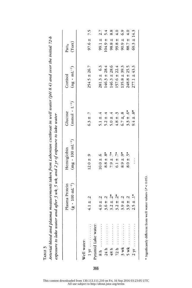
TABLE 3 Arterial blood and plasma measurements taken from Lahontan cutthroat in well water (pH 8. 4) and over the initial 72-h exposure to lake water and after 3 wk, 5 wk, and 2 yr of exposure to lake water
Plasma Protein Hemoglobin Glucose Cortisol Pao2 (g f 100 mL-1) (mg f 100 mL-1) (mmol - L-1) (ng * mL-1) (Torr)
Well water:
1 yr ........... 4.1 + .2 12.0 + .9 6.3 + .7 254.5 + 26.7 97.6 + 7.5 Pyramid Lake water:
8 h ........... 4.0 .2 10.0 .6 5.1 + .4 281.3 + 6.3 99.1 2.7 24 h .......... 3.6 + .2 8.8 + .8* 5.2 + .4 146.3 + 28.4 104.9 + 5.4 48 h .......... 3.2 + .2* 7.9 + .7* 5.0 + .4 146.3 + 28.6 98.8 + 8.8 72 h .......... 3.2 + .2* 6.1 + .7* 4.8 + .3 157.6 + 22.4 99.8 + 4.0 3 wk .......... 3.9 + .2 8.9 + .9* 6.7 ++.8 135.8 + 39.3 90.9 + 6.9 5 wk .......... 3.9 + .2 8.0 + .5* 3.3 + .6* 248.8 + 25.5 88.7 + 4.0 2 yr ............ 2.5 + .1* ... 9.4 + .8* 277.1 + 43.3 69.3 + 14.3 * Significantly different from well water values (P < 0.05).
CA3
This content downloaded from 130.113.111.210 on Fri, 16 Sep 2016 03:23:05 UTCAll use subject to http://about.jstor.org/terms
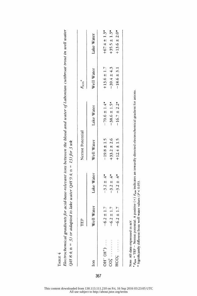
TABLE 4 Electrochemical gradients for acid-base-relevant ions between the blood and water of Lahontan cutthroat trout in well water (pH 8. 4; n = 3) or adapted to lake water (pH 9. 4; n = 13) for 3 wk
TEP Nernst Potential FIONa
Ion Well Water Lake Water Well Water Lake Water Well Water Lake Water OH- (H+) -6.2 + 1.7 -3.2 +.4* -19.8 + 1.5 -70.6 + 1.4* +13.6 + 1.7 +67.4 + 1.3* CO3 -6.2 + 1.7 -3.2 + .4* +33.2 + 2.6 -38.6 + 1.5* -39.4 + 4.3 +35.5 + 1.3" HCO3 -6.2 + 1.7 -3.2 + .4* +12.4 + 1.5 -16.7 + 2.2* -18.6 + 3.1 +13.6 + 2.0* Note. All data expressed in mV. 8 FION = TEP - Nernst potential. A positive (+) FIo, indicates an inwardly directed electrochemical gradient for anions. * Significantly different from well water values (P < 0.05).
Cr3
This content downloaded from 130.113.111.210 on Fri, 16 Sep 2016 03:23:05 UTCAll use subject to http://about.jstor.org/terms

368 M. P. Wilkie, P. A. Wright, G. K. Iwama, and C. M. Wood
table 4). Estimates of the electrochemical gradients for OH- indicated that the
inwardly directed gradient for OH- (= outwardly directed electrochemical
gradient for H+) increased fivefold in lake water (table 4). Furthermore, the outwardly directed electrochemical gradients for HCO0 and CO= in well water
were reversed following transfer into lake water and resulted in large inwardly
directed gradients for these basic anions (table 4).
Acid-Base Balance and Ionoregulation
The LCT in well water had a pHa of 7.9 (fig. 3A), an arterial plasma CO2 tension (Paco2) of 1.8 Torr (fig. 3B), and an arterial plasma concentration
of HCOS ([HCOK]a) of approximately 6.7 mmol - L-1. By 8 h, following
Well Water Pyramid Lake Water IA
8.20 I
S8.00 - M pHa
7.80I 2.0 -
Paco. 1.5
F- 1.0 -
0.5
2.0 I Blood Lactate
0I
o -2.0 E E
-4.0
-6.0 I C 8 24 48 72
Time (h)
Fig. 3. Changes in (A) pHa, (B) Paco2, and (C) blood lactate and AH+ of Lahontan cutthroat trout following transfer into alkaline Pyramid Lake wa-
ter (pH 9.4) from well water (pH 8. 4). Values are means + 1 SEM; n = 7. Asterisks indicate significant differences from well water values (P < 0. 05).
This content downloaded from 130.113.111.210 on Fri, 16 Sep 2016 03:23:05 UTCAll use subject to http://about.jstor.org/terms

Lahontan Cutthroat Trout in Alkaline Water 369
transfer into lake water, the fish rapidly underwent a combined respiratory
and metabolic alkalosis, which was characterized by a 0.2-unit increase
in pHa, a 30% reduction in Paco2, no change in [HCO-]a, and a metabolic
acid load (AHm) of approximately -4 mmol ' L-1 (i.e., a metabolic base load; fig. 3A, B, and C). The partial correction and subsequent stabilization
of pHa, at approximately pH 8.05, after 24 h, was the result of a stabiliza-
tion of Paco2 and the elimination of the metabolic base load (fig. 3A, B, and C). These alterations in blood acid-base status occurred without al-
terations in blood lactate concentration (fig. 3C). In the long-term ex- posure groups, blood acid-base status remained very similar to that at- tained by 24-72 h and was characterized by a chronic respiratory alkalosis (fig. 4).
A progressive 6%-8% increase in plasma Na+ and Cl- was observed during the first 72 h of exposure to lake water (fig. 5A). This trend toward
greater plasma Na+ and C1- was still seen at 3 wk. At 5 wk and 2 yr, levels had decreased slightly, but, even at the latter time, the elevation in Cl- above the levels in well-water-adapted fish remained significant (fig. 5B).
Gill Structure and CC Morphometry
The filamental surface of the LCT gill was predominantly composed of microridged pavement (or respiratory) cells and contained varying numbers
of villous CCs, which appeared in openings of the respiratory epithelium
(fig. 6A, B, and C). Mucous cells were rarely seen. Qualitatively, the relative
absence and small size of filamental epithelial CCs in well water fish is apparent in figure 6A. Following transfer into lake water, the exposure of CCs on the filamental surface became much more pronounced through changes in both individual cell surface areas and cell densities (fig. 6B and C). Similarly, the lamellar gill surface of well-water-adapted fish had very few CCs. Exposure to lake water led to changes in lamellar CC exposure and density that paralleled those observed on the filamental epithelium (fig. 6D, E, and F).
The CC FSA was fourfold greater after 3 d of exposure to lake water and 10-fold and 20-fold higher after 3 wk and 2 yr in lake water, re- spectively (fig. 7A). Differences in CC FSA between the naive trout and
those exposed to lake water for 3 d were due to the twofold greater individual CC surface area, combined with a twofold greater CC density (fig. 7B and C). The higher CC FSAs observed in fish exposed to lake water for 3 wk and 2 yr were solely due to greater individual CC surface areas (fig. 7B).
This content downloaded from 130.113.111.210 on Fri, 16 Sep 2016 03:23:05 UTCAll use subject to http://about.jstor.org/terms
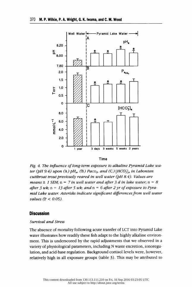
370 M. P. Wilkie, P. A. Wright, G. K. Iwama, and C. M. Wood
Well Water ~- Pyramid Lake Water- IA I pH
8.20 - o I
0.5 -T
8.00
0 IC
2.0 - I 6. 1.0- 0
0.5
8.0 T T
E 4.0 K
2.0 -
1 year 3 days 3 weeks 5 weeks 2 years
Time
Fig. 4. The influence of long-term exposure to alkaline Pyramid Lake wa-
ter (pH 9. 4) upon (A) pHa, (B) Paco2, and (C) [HCO5]a in Lahontan cutthroat trout previously reared in well water (pH 8.4). Values are means + 1 SEM; n = 7 in well water and after 3 d in lake water; n = 8 after 3 wk; n = 13 after 5 wk; and n = 6 after 2 yr of exposure to Pyra-
mid Lake water. Asterisks indicate significant diferences from well water values (P < 0. 05).
Discussion
Survival and Stress
The absence of mortality following acute transfer of LCT into Pyramid Lake
water illustrates how readily these fish adapt to the highly alkaline environ-
ment. This is underscored by the rapid adjustments that we observed in a
variety of physiological parameters, including N waste excretion, ionoregu- lation, and acid-base regulation. Background cortisol levels were, however,
relatively high in all exposure groups (table 3). This may be attributed to
This content downloaded from 130.113.111.210 on Fri, 16 Sep 2016 03:23:05 UTCAll use subject to http://about.jstor.org/terms

Lahontan Cutthroat Trout in Alkaline Water 371
Well Waterl4 - Pyramid Lake Water--- 160 - IA
I [Na]
150 - I
__11 0 140 I
130 -
120 I [C-] 120
C 8 24 48 72 Time (h)
160 - I IB [Z [Na+]
150 I *[CI] T T
.-J
0140 - E I " * E
130
120
110 1 year 3 days 3 weeks 5 weeks 2 years
Time
Fig 5. (A) Changes in plasma Na+ and CI- concentrations in Lahontan cutthroat trout over the first 72 hfollowing exposure to alkaline Pyramid Lake water (pH 9. 4) and (B) after 3 d, 3 wk, 5 wk, and 2 yr in lake
water. Values are means + 1 SEM; n = 7 in well water (pH 8.4) and af- ter 3 d in lake water; n = 8 after 3 wk; n = 13 after 5 wk; and n = 6
after 2 yr of exposure. Asterisks indicate significant differences from well
water values (P < O. 05)for Na+ and CI-.
the handling, confinement, and catheterization of a wild trout (Woodward
and Strange 1987; McDonald and Milligan 1992). Accordingly, we believe that transfer into lake water was not unduly stressful. The absence of any increase in plasma glucose, following acute lake water exposure (table 3), supports our conclusion.
Nitrogenous Waste Excretion
The initial reduction of JAmm, immediately observed following transfer into
lake water, parallels that seen in Oncorhynchus mykiss exposed to pH 9.5
This content downloaded from 130.113.111.210 on Fri, 16 Sep 2016 03:23:05 UTCAll use subject to http://about.jstor.org/terms

372 M. P. Wilkie, P. A. Wright, G. K. Iwama, and C. M. Wood
Fig. 6. Representative scanning electron micrographs ofthefilamental epithelium (A, B, C) and lamellar epithelium (D, E, F) of Lahontan cutthroat trout reared in well water (pH 8.4; A, D) or alkaline Pyramid Lake water (pH 9.4) for 3 d (B, E) or 3 wk (C, F). Note the increased density of CC (indicated by arrows) on the filamental and lamellar epi-
thelium offish exposed to lake water for 3 d and the larger CCs after 3
wk vs. 3 d of exposure to lake water. pvc, Gill pavement cell; bar = 20 pCm.
water (Wilkie and Wood 1991). Unlike the rainbow trout, however, there
was no tendency for JAmm to return to preexposure levels in the LCT (figs.
1A, 2A). We estimate that approximately 16,000 pmol N - kg-1 of waste ammonia was unaccounted for during the first 72 h at pH 9.4. Plasma TAmm levels (fig. 1B) suggest that virtually none (<1%) of the "missing" ammonia
was stored in the extracellular space. The fact thatJAmm remained depressed
This content downloaded from 130.113.111.210 on Fri, 16 Sep 2016 03:23:05 UTCAll use subject to http://about.jstor.org/terms

Lahontan Cutthroat Trout in Alkaline Water 373
Well Waterq- Pyramid Lake Water - IA
350 CC FSA * t o
O 300
x 150 -
E 100 - E I a T E 50
B CC Surface * t 125 - Area
E 100 -I 75-
50-
25-
CC Density
3000 -I +
E E 2000 -
T I
0
1000
1 year 3 days 3 weeks 2 years
Time
Fig. 7 Diferences in branchial (A) CC FSA, (B) mean individual CC sur- face area, and (C) CC density of Lahontan cutthroat trout exposed to well water (pH 8.4) or Pyramid Lake water (pH 9. 4) for 3 d, 3 wk, or 2
yr. Asterisks indicate significant diferences from well water values; stars indicate significant diferences from 3 d of exposure to Pyramid Lake wa- ter; daggers indicate significant differences from 3 wk of exposure to lake water (P < 0.05).
indefinitely while plasma TAmm eventually returned to, and remained at,
preexposure levels (figs. 1B, 2B) suggests that LCT were either excreting
another N waste product(s) and/or amino acid deamination rates were per- manently reduced.
The possibility that LCT were excreting another N waste product is in-
triguing and deserves further investigation. Absolute rates of Jurea did not
This content downloaded from 130.113.111.210 on Fri, 16 Sep 2016 03:23:05 UTCAll use subject to http://about.jstor.org/terms
374 M. P. Wilkie, P. A. Wright, G. K. Iwama, and C. M. Wood
change following transfer to lake water (figs. 1A, 2D), so urea did not fulfill
this role. Potential alternate waste products include trimethylamine oxide
(TMAO), glutamine, creatine, creatinine, or purines such as uric acid (see
Forster and Goldstein 1969; Mommsen and Walsh 1992). In a related study (Wright et al. 1993), however, we were unable to detect any uric acid ex-
cretion in LCT that had been exposed to lake water for about 4 wk and that
exhibited JAmm and Jurea very similar to those of the present study.
The alternate possibility, that of reduced use of amino acids as an energy
source and reduced endogenous ammonia production, also deserves further
investigation (Walton and Cowey 1982; Van Waarde 1983). The persistent respiratory alkalosis (figs. 3, 4) may have altered metabolic enzyme activities
and led to a greater reliance on other fuels such as glycogen and/or fatty acids.
Pyramid Lake water's greater salinity (approximately 60 mmol t L-1 NaCl; table 1) may also have accounted for the some of the persistent reduction
in JAmm. Ammonia production may have declined in accord with an overall
reduction in metabolic rate as a result of lower ionoregulatory and osmo-
regulatory costs in the more isotonic environment (Rao 1968). Brett (1979)
noted that growth rates generally increase with salinity in euryhaline fish,
such as the cutthroat trout. A greater proportion of amino acids may have
been incorporated and retained in structural protein, rather than deaminated.
Indeed, the rapid growth of the LCT in Pyramid Lake's alkaline/saline waters
is legendary (Coleman and Johnson 1988). The initial inhibition of JAmm upon exposure of LCT to lake water was
similar to that observed in rainbow trout exposed to comparable pH, and it is likely that similar explanations apply (Wright and Wood 1985; Lin and Randall 1990; Wilkie and Wood 1991; Yesaki and Iwama 1992). These in- clude a decrease in the APNH3 between the blood and gill boundary layers
that drives the diffusive efflux of NH3, and/or an inhibition of Na+/NH exchange. Although the blood-bulk water APNH3 increased rather than de-
creased upon exposure to alkaline pH (fig. 1C), this value will be differ- entially affected by the presence or absence of boundary layer acidification
and associated diffusion trapping of NH3 as NH- in the gill water (Randall and Wright 1989). Acidification of gill water by CO2 and possibly H+ efflux
across the gills is well established in rainbow trout in fresh water (Playle and Wood 1989; Lin and Randall 1990), and this phenomenon clearly aug-
ments JAmm (Wright, Randall, and Perry 1989). It is likely that the same phenomenon occurs in the LCT in well water. Blockade of CO2 hydration and boundary layer acidification in gill water by acetazolamide clearly re-
duces JAmm in rainbow trout (Wright et al. 1989). In other words, boundary layer acidification increases the blood-boundary layer APNH3 above the
This content downloaded from 130.113.111.210 on Fri, 16 Sep 2016 03:23:05 UTCAll use subject to http://about.jstor.org/terms
Lahontan Cutthroat Trout in Alkaline Water 375
blood-bulk water APNH3 and effectively augmentsJAmm. In the highly buffered
waters of Pyramid Lake, however, boundary acidification is probably reduced
or nonexistent. As a result, gill boundary layer pH will approach the pH of
the bulk water, and therefore approximately 50% of the excreted ammonia
will exist as NH3 in the gill water. This would effectively reduce the blood-
boundary layer APNH3 below the blood-bulk water APNH3 and result in re-
duced JAmm. Indeed, Wright et al. (1993) found no effect of acetazolamide
treatment on JAmm in LCT adapted to Pyramid Lake water. They also found
that Na+-free water and amiloride treatment did not effect JAmm, suggesting
that Na /NHt exchange does not occur once the animals are adapted to Pyramid Lake water. In contrast, amiloride treatment consistently reduces JAmm in rainbow trout in fresh water (Kirschner, Greenwald, and Kerstetter
1973; Wright and Wood 1985).
Wright et al. (1993) did not examine the LCT in well water, but they concluded that maintenance of a large blood-to-water APNH3, via chronically
elevated pHa and plasma TAmm, was probably the most important factor sus-
taining the (limited) .Amm in lake water. The fact that the arterial blood PNH3 in LCT in well water was similar to values observed after 3 wk and 5 wk in
lake water (approximately 56 gLTorr: fig. 2C) suggests that this species was "preadapted" to living in a high-pH lake. Such a preadaptation would likely
be selected for, as would an ability to chronically reduce ammonia produc-
tion rates, in a salmonid that has a long evolutionary history in alkaline lakes (Trotter 1991).
Ureagenic Enzyme Activities
Urea excretion did not account for the missing waste N. Its persistence at
high pH and its greater percentage contribution to N excretion appears to maintain minimal rates of Jwase N. The presence of significant activities of
enzymes involved in uricolysis in both well water fish and those adapted to
Pyramid Lake for 3 wk or 2 yr suggests that the majority of urea production
resulted from uricolysis (table 2). Danulat and Kempe (1992) observed very high rates of urea excretion in the cyprinid Chalcalburnus tarichi, endemic
to highly alkaline Lake Van, Turkey (pH 9.8), and suggested that much of
this excretion was due to hydrolysis of arginine, catalyzed by hepatic argi-
nase. We found hepatic arginase activity was about 16-fold higher than that
reported by Danulat and Kempe (1992) but equal to values reported by Chiu, Austic, and Rumsey (1986) for fingerling rainbow trout. Arginine is an essential amino acid for teleosts (Forster and Goldstein 1969), and, since
the fish in the present study had been starved prior to experimentation, it
This content downloaded from 130.113.111.210 on Fri, 16 Sep 2016 03:23:05 UTCAll use subject to http://about.jstor.org/terms

376 M. P. Wilkie, P. A. Wright, G. K. Iwama, and C. M. Wood
seems unlikely that there would have been sufficient arginine flux through
arginase to sustain urea production.
The OUC pathway contribution to urea synthesis was insignificant in cutthroat trout, as CPS III and other enzyme activities in the cycle were
negligible or very low. This observation is consistent with work performed
on other salmonids (Huggins, Skutch, and Baldwin 1969; Chiu et al. 1986)
and the high-pH-tolerant cyprinid Chalcalburnus tarichi (Danulat and Kempe 1992).
Acid-Base Balance and lonoregulation
The respiratory alkalosis accompanying high pH exposure persisted indef- initely (figs. 3B, 4B), but the metabolic alkalosis was corrected within 24 h (figs. 3C, 4C). The long-term control of metabolic acid-base status is inter-
esting in view of the fact that large electrochemical gradients, favoring losses
of metabolic acid (protons) or gains of metabolic base, developed following
transfer to high pH (table 4). The presence of significant inwardly directed
electrochemical gradients for HCO- and CO: can be attributed to the un-
usually high concentrations of these ions in Pyramid Lake water (Galat et
al. 1985; table 1). Despite these exogenous factors, the similar levels of plasma HC03 in well water LCT and those residing in Pyramid Lake for up to 2 yr suggests that these fish are in a steady state with respect to long-term
acid-base status. One possible mechanism of acid-base control is the in- creased production of metabolic acid, via increased lactic acid production (Eichenholz et al. 1962). Such a response has been observed in rainbow trout at pH 9.5 and in LCT in pH 10 water (Wilkie and Wood 1991; Wilkie et al. 1993). We observed no such response in this study, however. Another
possibility is that the persistent external base load was counteracted by a stimulation of gill Cl-/HCO3 exchange. Increased Cl-/HCO3 exchange might have been augmented by branchial CC proliferation, as has been demonstrated in other teleosts subjected to alkalotic disturbances in systemic acid-base status (see Goss et al. 1992a, 1992b).
We suggest that the greater CC FSA found in lake-water-adapted LCT (fig.
7) is linked to long-term acid-base regulation in an environment that exerts a continual base load on the fish. The rapidity (24 h) of the correction of
the metabolic alkalosis, following transfer into Pyramid Lake water, does
not argue against a branchial mechanism of metabolic acid-base control. Indeed, Goss et al. (1992a) demonstrated that changes in branchial mor- phology occur 6 h after the initiation of acid-base disturbances. Therefore,
it is quite possible that increased CC FSA, and associated Cl-/HCO- ex- change, accounted for the correction of the alkalosis after only 24 h of lake
This content downloaded from 130.113.111.210 on Fri, 16 Sep 2016 03:23:05 UTCAll use subject to http://about.jstor.org/terms

Lahontan Cutthroat Trout in Alkaline Water 377
water exposure. Further evidence in support of branchial acid-base regu-
lation by the LCT was the presence of a significant correlation between pH,
and CC FSA (CC FSA = [1.15 X 10⁶][pHa] - [9.16 X 10⁶]; r = 0.585, P < 0.05). Galat et al. (1985) reported that LCT, living in a variety of alkaline lakes, had considerable CC hyperplasia and suggested that it was correlated to the sum of external HCO3, CO=, and Cl-. Our results corroborate these
findings.
Ionoregulatory failure, characterized by 15%-20% decreases in plasma Na+ and/or Cl-, has been cited as a potential contributing factor in the deaths of rainbow trout exposed to high pH (Heming and Blumhagen 1988; Wilkie and Wood 1991; Yesaki and Iwama 1992). In contrast, the LCT ex-
hibited slight increases in plasma Na+ and Cl- following transfer to lake water (fig. 5). The high salinity of Pyramid Lake water may account for this
difference. The increase in branchial CC FSA accompanying lake water ad-
aptation (figs. 6, 7), however, may have also have prevented ionoregulatory disturbance in these fish (Laurent, Hobe, and Dunel-Erb 1985; Laurent and
Perry 1990; Perry, Goss, and Laurent 1992).
Conclusions
Unlike other salmonids, LCT readily adapt to the extreme pH of Pyramid
Lake by making a number of unique physiological adjustments. The apparent
persistence of reduced JAmm in lake-water-adapted fish suggests that this
species adapts to alkaline lake water by decreasing endogenous ammonia production. This allows the fish to rapidly correct internal ammonia levels
and prevents NH3 from reaching toxic levels. Furthermore, the LCT also rapidly corrects, and continues to regulate, its metabolic acid-base status,
despite the presence of large inwardly directed electrochemical gradients for basic equivalents. Increases in branchial CC FSA may actually augment this acid-base regulation, and also prevent plasma ion dilution, through the
modulation of Na+ and C1- uptake at the gill.
Acknowledgments
We thank P. Wagner, L. Carlson, N. Vucinich, and D. Mosely for their in- valuable assistance and support during our stay at Pyramid Lake. Dr. G. Wedemeyer, of the U.S. Fish and Wildlife Service, was instrumental in help-
ing to launch this study. C. Mazur and J. McGeer, from the University of British Columbia, provided excellent technical help and advice. At McMaster
University we thank R. Ellis, S. Munger, K. Schultes, and D. Flannigan. We
This content downloaded from 130.113.111.210 on Fri, 16 Sep 2016 03:23:05 UTCAll use subject to http://about.jstor.org/terms

378 M. P. Wilkie, P. A. Wright, G. K. Iwama, and C. M. Wood
are grateful to Drs. R. Wilson, D. G. McDonald, and G. G. Goss and to R. Lauff for useful discussions and comments. This study was supported by
Natural Sciences and Engineering Council (NSERC) of Canada operating grants to C.M.W., G.K.I., and P.A.W.; an NSERC International Collaborative
Grant to G.K.I. and C.M.W.; and an Ontario Graduate Scholarship to M.P.W.
Literature Cited
BRETT, J. R. 1979. Environmental factors and growth. Pages 599-675 in W. S. HOAR, D. J. RANDALL, and J. R. BRETT, eds. Fish physiology. Vol. 8. Academic Press, New York.
CHIU, Y. N., R. E. AusTIC, and G. L. RUMSEY. 1986. Urea cycle activity and arginine formation in rainbow trout (Salmo gairdneri). J. Nutr. 116:1640-1650.
COLEMAN, M. E., and V. K. JOHNSON. 1988. Summary of management at Pyramid Lake,
Nevada, with emphasis on Lahontan cutthroat trout, 1954-1987. Am. Fisheries Soc. Symp. 4:107-115.
CVANCARA, V. A. 1969. Comparative study of liver uricase activity in freshwater teleosts.
Comp. Biochem. Physiol. 28:725-732. DANULAT, E., and S. KEMPE. 1992. Nitrogenous waste excretion and accumulation of
urea and ammonia in Chalcalburnus tarichi (Cyprinidae), endemic to the ex- tremely alkaline Lake Van (eastern Turkey). Fish Physiol. Biochem. 9:377-386.
EICHENHOLZ, A., R. O. MULHAUSEN, W. E. ANDERSON, and F. M. MACDONALD. 1962.
Primary hypocapnia: a cause of metabolic acidosis. J. Appl. Physiol. 17:283-288. FORSTER, R. P., and L. GOLDSTEIN. 1969. Formation of excretory products. Pages 313-
350 in W. S. HOAR and D. J. RANDALL, eds. Fish physiology. Vol. 1. Academic Press, New York.
FROMM, P. 0. 1963. Studies on renal and extra-renal excretion in a freshwater teleost,
Salmo gairdneri. Comp. Biochem. Physiol. 10:121-128. GALAT, D. L., E. L. LIDER, S. VIGG, and S. R. ROBERTSON. 1981. Limnology of a large
deep North American terminal lake, Pyramid Lake, Nevada, U.S.A. Hydrobiologia 82:281-317.
GALAT, D. L., G. PosT, T. J. KEEFE, and G. R. BoucKs. 1985. Histological changes in the gill, kidney and liver of Lahontan cutthroat trout, Salmo clarki henshawi, living in lakes of different salinity-alkalinity. J. Fish Biol. 27:533-552.
GOLDSTEIN, L., and R. P. FORSTER. 1965. The role of uricolysis in the production of
urea by fishes and other aquatic vertebrates. Comp. Biochem. Physiol. 14:567- 576.
Goss, G. G., P. LAURENT, and S. F. PERRY. 1992a. Evidence for a morphological com-
ponent in acid-base regulation during environmental hypercapnia in the brown bullhead (Ictalurus nebulosus). Cell Tissue Res. 268:539-552.
Goss, G. G., S. F. PERRY, C. M. WOOD, and P. LAURENT. 1992b. Mechanisms of ion and acid-base regulation at the gills of freshwater fish. J. Exp. Zool. 263:143-159.
HEMING, T. A., and K. A. BLUMHAGEN. 1988. Plasma acid-base and electrolyte status of rainbow trout exposed to alum (aluminum sulphate) in acidic and alkaline environments. Aquatic Toxicol. 12:125-140.
This content downloaded from 130.113.111.210 on Fri, 16 Sep 2016 03:23:05 UTCAll use subject to http://about.jstor.org/terms
Lahontan Cutthroat Trout in Alkaline Water 379
HUGGINS, A. K., G. SKUTCH, and E. BALDWIN. 1969. Ornithine-urea cycle enzymes in
teleostean fish. Comp. Biochem. Physiol. 28:587-602. KIRSCHNER, L. B., L. GREENWALD, and T. H. KERSTETTER. 1973. Effect of amiloride on
sodium transport across body surfaces of freshwater animals. Am. J. Physiol. 224: 832-837.
KUCERA, P. A., D. L. KOCH, and G. F. MARCO. 1985. Introductions of Lahontan cutthroat
trout in Omak Lake, Washington. North Am. J. Fisheries Manage. 5:296-301. LAURENT, P., H. HOBE, and S. DUNEL-ERB. 1985. The role of environmental sodium
chloride relative to calcium in gill morphology of freshwater salmonid fish. Cell Tissue Res. 240:675-692.
LAURENT, P., and S. F. PERRY. 1990. Effects of on gill chloride cell morphology and ionic uptake in freshwater trout, Salmo gairdneri. Cell Tissue Res. 259:429-442.
LIN, H. L., and D. J. RANDALL. 1990. The effect of varying water pH on the acidification
of expired water in rainbow trout. J. Exp. Biol. 149:149-160. MCDONALD, D. G., and C. L. MILLIGAN. 1992. Chemical properties of the blood in
fish. Pages 55-133 in W. S. HOAR and D. J. RANDALL, eds. Fish physiology. Vol. 12. Academic Press, New York.
MCDONALD, D. G., and C. L. MILLIGAN. 1992. Chemical properties of the blood in Fish. Pages 55-133 in W. S. HOAR and D. J. RANDALL, eds. Fish physiology. Vol. 12. Academic Press, New York.
MOMMSEN, T. P., and P. J. WALSH. 1989. Evolution of urea synthesis in vertebrates: the piscine connection. Science 243:72-75.
. 1991. Urea synthesis in fishes: evolutionary and biochemical perspectives. Pages 137-163 in P. HOCHACHKA and T. P. MOMMSEN, eds. The biochemistry and molecular biology of fishes. Elsevier, New York.
. 1992. Biochemical and environmental perspectives on nitrogen metabolism in fishes. Experientia 48:583-593.
PERRY, S. F., G. G. Goss, and P. LAURENT. 1992. The interrelationships between gill chloride cell morphology and ionic uptake in four freshwater teleosts. Can. J. Zool. 70:1775-1786.
PERRY, S. F., and C. M. WOOD. 1985. Kinetics of branchial calcium uptake in the rainbow trout: effects of acclimation to various external calcium levels. J. Exp. Biol. 116:411-433.
PLAYLE, R. C., and C. M. WOOD. 1989. Water chemistry changes in the gill microen- vironment of rainbow trout: experimental observations and theory. J. Comp. Phys- iol. 159B:527-537.
RANDALL, D. J., C. M. WOOD, S. F. PERRY, H. BERGMAN, G. M. O. MALOIY, T. P. MOMMSEN,
and P. A. WRIGHT. 1989. Urea excretion as a strategy for survival in a fish living in a very alkaline environment. Nature 337:165-166.
RANDALL, D. J., and P. A. WRIGHT. 1989. The interaction between carbon dioxide and
ammonia excretion and water pH in fish. Can. J. Zool. 67:2936-2942.
RAo, G. M. M. 1968. Oxygen consumption of rainbow trout (Salmo gairdneri) in relation to activity and salinity. Can. J. Zool. 46:781-786.
SAS Institute. 1989. SAS JMP user's guide. SAS Institute, Cary, N.C. Soivio, A. K., K. WESTMAN, and K. NYHOLM. 1972. Improved method of dorsal aortal
catheterization: haematological effects followed for three weeks in rainbow trout. Finn. Fisheries Res. 1:11-21.
This content downloaded from 130.113.111.210 on Fri, 16 Sep 2016 03:23:05 UTCAll use subject to http://about.jstor.org/terms

380 M. P. Wilkie, P. A. Wright, G. K. Iwama, and C. M. Wood
TROTTER, P. 1991. Cutthroat trout. Pages 236-265 in J. STOLZ and J. SCHNELL, eds. Trout. Stackpole, Harrisburg, Pa.
VAN WAARDE, A. 1983. Aerobic and anaerobic ammonia production by fish. Comp. Biochem. Physiol. 74B:675-684.
WALTON, M.J., and C. B. COWEY. 1982. Aspects of intermediary metabolism in salmonid
fish. Comp. Biochem. Physiol. 73B:59-79. WILKIE, M. P., and C. M. WOOD. 1991. Nitrogenous waste excretion, acid-base reg-
ulation, and ionoregulation in rainbow trout (Oncorhynchus mykiss) exposed to extremely alkaline water. Physiol. Zool. 64:1069-1086.
WILKIE, M. P., P. A. WRIGHT, G. K. IWAMA, and C. M. WOOD. 1993. The physiological responses of the Lahontan cutthroat trout (Oncorhynchus clarki henshawi), a resident of highly alkaline Pyramid Lake (pH 9. 4), to challenge at pH 10. J. Exp. Biol. 175:173-194.
WILLIAMS, J. E., J. E. JOHNSON, D. A. HENDRICKSON, S. CONTRERAS-BALDERAS, J. D. WIL-
LIAMS, J. NAVARRO-MENDOZA, D. E. MCALLISTER, and J. E. DEACON. 1989. Fishes of
North America endangered, threatened, or of special concern: 1989. Fisheries 14: 2-20.
WOLF, K. 1963. Physiological salines for freshwater teleosts. Progressive Fish Cul- turalist 25:615-625.
WOOD, C. M. 1993. Ammonia and urea metabolism and excretion. Pages 379-425 in D. H. EVANS, ed. The physiology of fishes. CRC, Boca Raton, Fla.
WOODWARD, J. J., and R. J. STRANGE. 1987. Physiological stress responses in wild and
hatchery-reared rainbow trout. Trans. Am. Fisheries Soc. 116:574-579. WRIGHT, P. A., G. K. IWAMA, and C. M. WOOD. 1993. Ammonia and urea excretion in
Lahontan cutthroat trout (Oncorhynchus clarki henshawi) adapted to highly al- kaline Pyramid Lake (pH 9.4). J. Exp. Biol. 175:153-172.
WRIGHT, P. A., D. J. RANDALL, and S. F. PERRY. 1989. Fish gill boundary layer: a site of linkage between carbon dioxide and ammonia excretion. J. Comp. Physiol. 158B:627-635.
WRIGHT, P. A., and C. M. WOOD. 1985. An analysis of branchial ammonia excretion in the freshwater rainbow trout: effects of environmental pH change and sodium uptake blockade. J. Exp. Biol. 114:329-353.
YESAKI, T. Y., and G. K. IWAMA. 1992. Some effects of water hardness on survival, acid-base regulation, ion regulation and ammonia excretion in rainbow trout in highly alkaline water. Physiol. Zool. 65:763-787.
This content downloaded from 130.113.111.210 on Fri, 16 Sep 2016 03:23:05 UTCAll use subject to http://about.jstor.org/terms
Recommended