Embed Size (px)
Citation preview
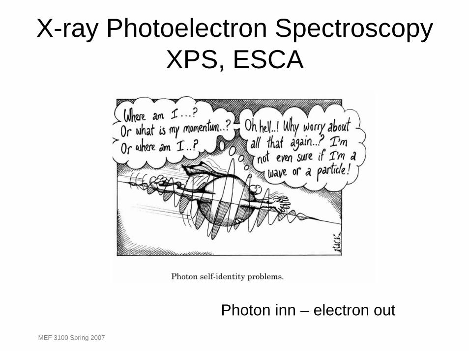
X-ray Photoelectron Spectroscopy XPS, ESCA
Photon inn – electron outMEF 3100 Spring 2007
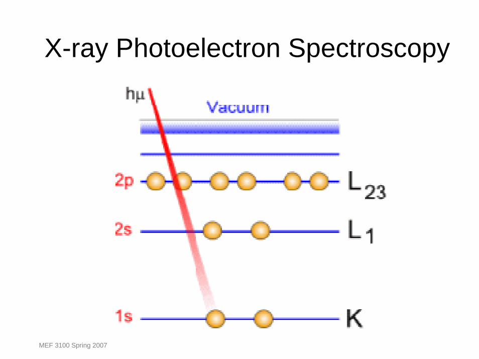
X-ray Photoelectron Spectroscopy
MEF 3100 Spring 2007
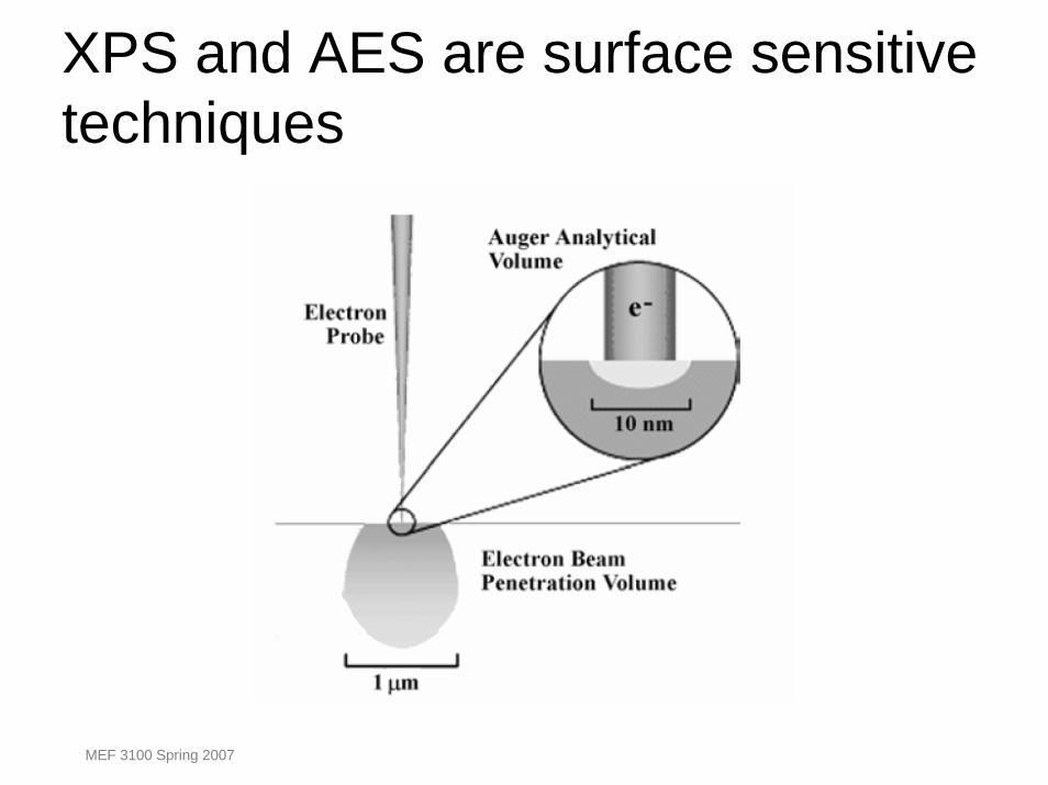
XPS and AES are surface sensitive techniques
MEF 3100 Spring 2007
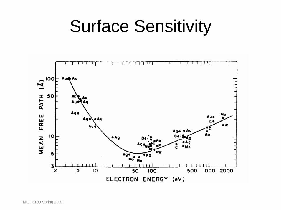
Surface Sensitivity
MEF 3100 Spring 2007
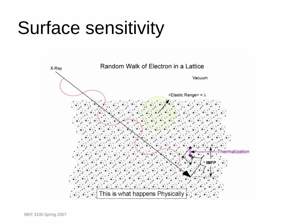
Surface sensitivity
MEF 3100 Spring 2007

Photo emission process is often envisaged as three steps
•Absorption and ionisation - (initial state effect)
•Response from atom and creation of photoelectron -(final state effect)
•Transport of electron to surface and escape - (extrinsic loss)
MEF 3100 Spring 2007
XPS – ESCA
MEF 3100 Spring 2007

IntroductionX-ray Photoelectron Spectroscopy (XPS)X-ray photoelectron spectroscopy works by irradiating a sample material with mono energetic soft x-rays causing electrons to be ejected.
Identification of the elements in the sample can be made directly from the kinetic energies of these ejected photoelectrons.
The relative concentrations of elements can be determined from the photoelectron intensities.
MEF 3100 Spring 2007
XPS - ESCA
• Surface sensitive analysis technique based on photoelectric effect. Depth of analysis ~4-40 nm.
• All elements except Hydrogen.• Wide range of materials: Polymers, Ceramics,
metals …. (vacuum compatible)• Applications: corrosion, catalysis, thin films,
surface coatings, segregation …• Gives information on chemical composition and
chemical state.
MEF 3100 Spring 2007
History
• 1887 Heinrich Hertz /1888 Wilhelm Hallwachesilluminating metal surfaces resulted in electronic emission
• 1900 Max Planck -black body radiation• 1905 Einstein - light is quantized
E=hν• 1950 Kai Siegbahn 1981 : Nobel Prizestudied photoejection of electrons with x-rays
• 1950: Instrumentation 1960: Chemical Applications
MEF 3100 Spring 2007

Absorbing X-rays – emitting Photoelectrons
MEF 3100 Spring 2007
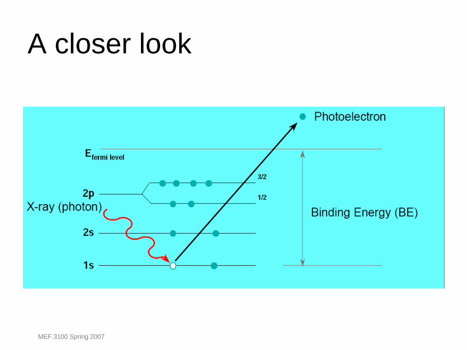
A closer look
MEF 3100 Spring 2007
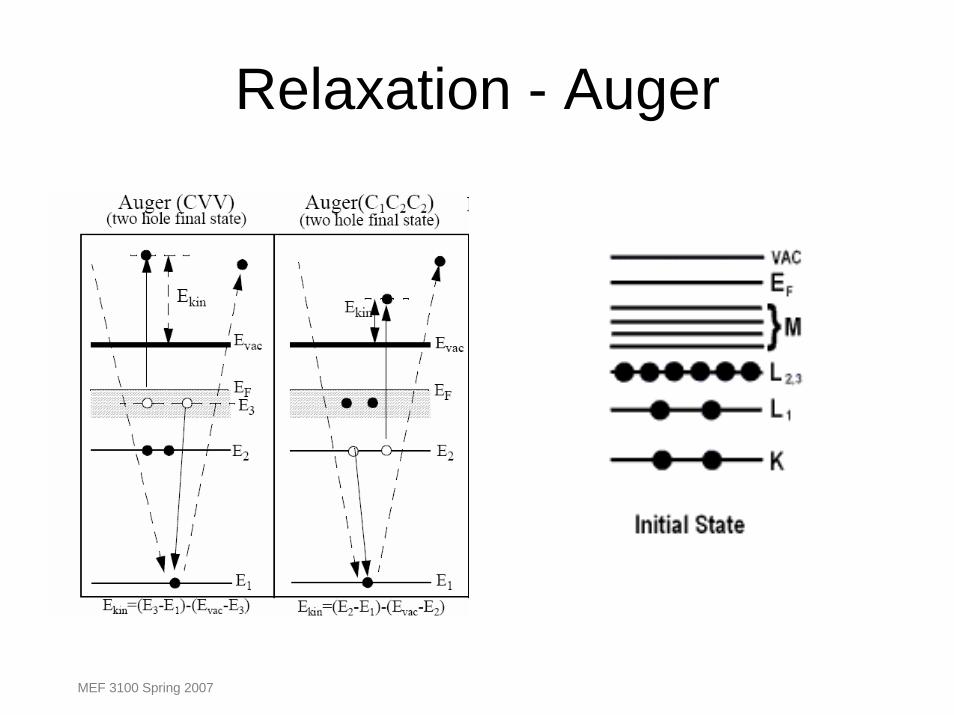
Relaxation - Auger
MEF 3100 Spring 2007
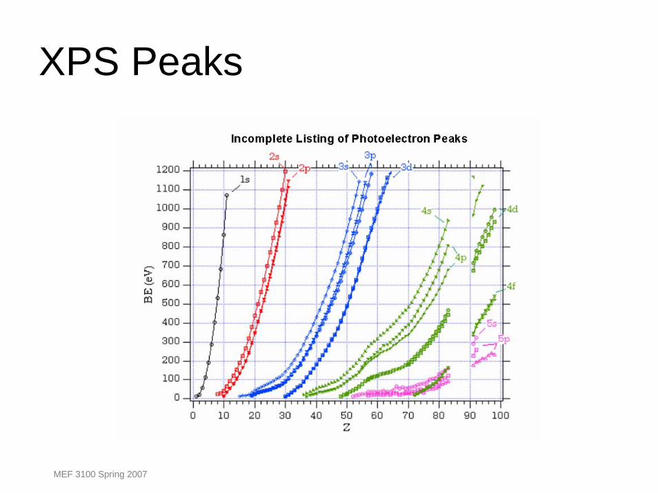
XPS Peaks
MEF 3100 Spring 2007
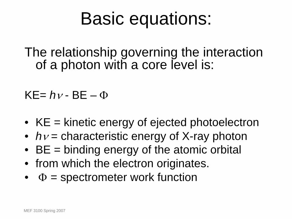
Basic equations:
The relationship governing the interaction of a photon with a core level is:
KE= hν - BE – Φ
• KE = kinetic energy of ejected photoelectron• hν = characteristic energy of X-ray photon• BE = binding energy of the atomic orbital• from which the electron originates. • Φ = spectrometer work function
MEF 3100 Spring 2007
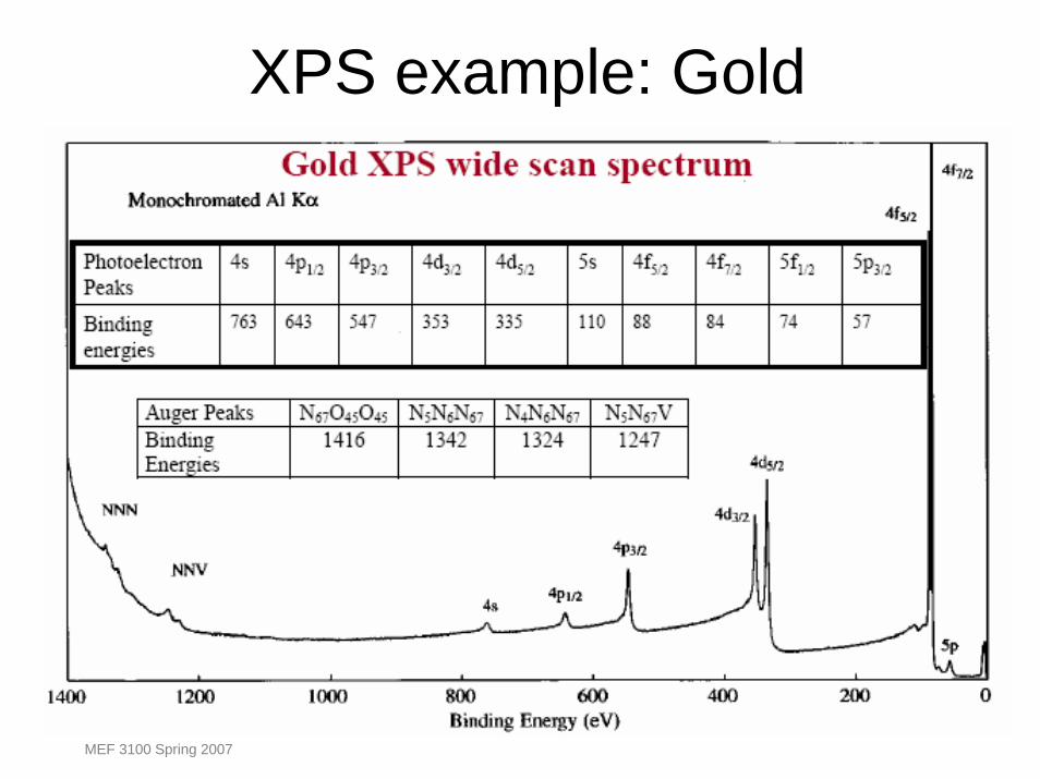
XPS example: Gold
MEF 3100 Spring 2007

Electronic structure and spectroscopy
MEF 3100 Spring 2007
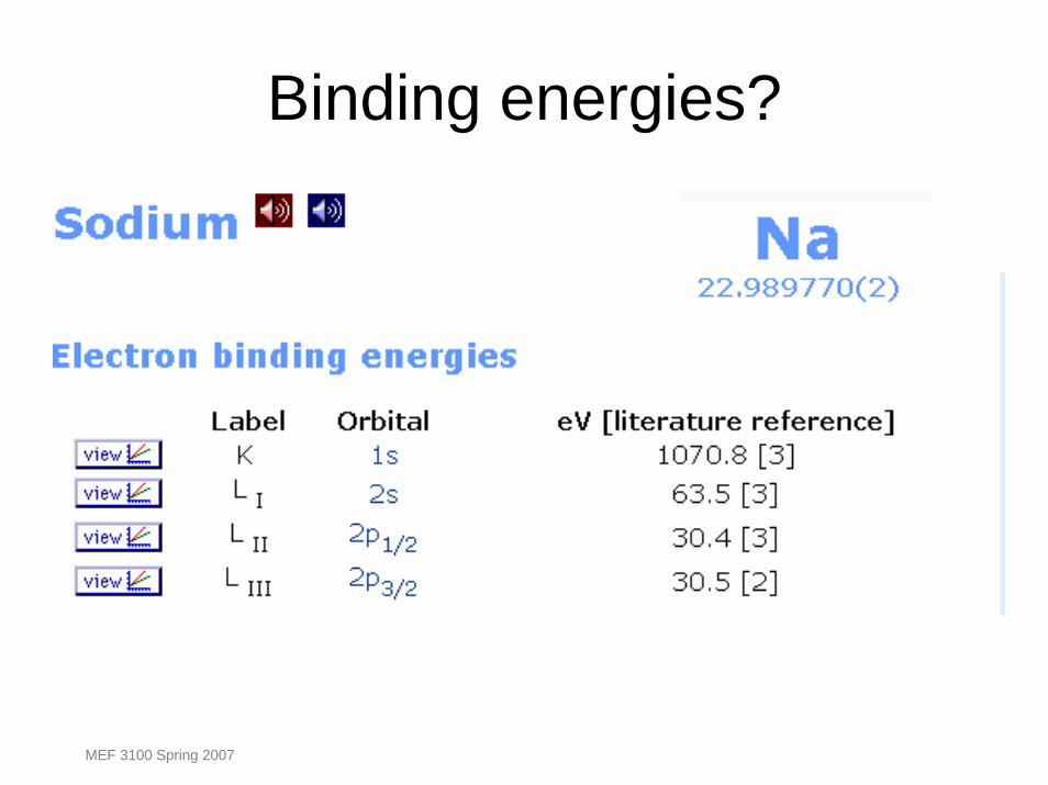
Binding energies?
MEF 3100 Spring 2007

MEF 3100 Spring 2007
MEF 3100 Spring 2007
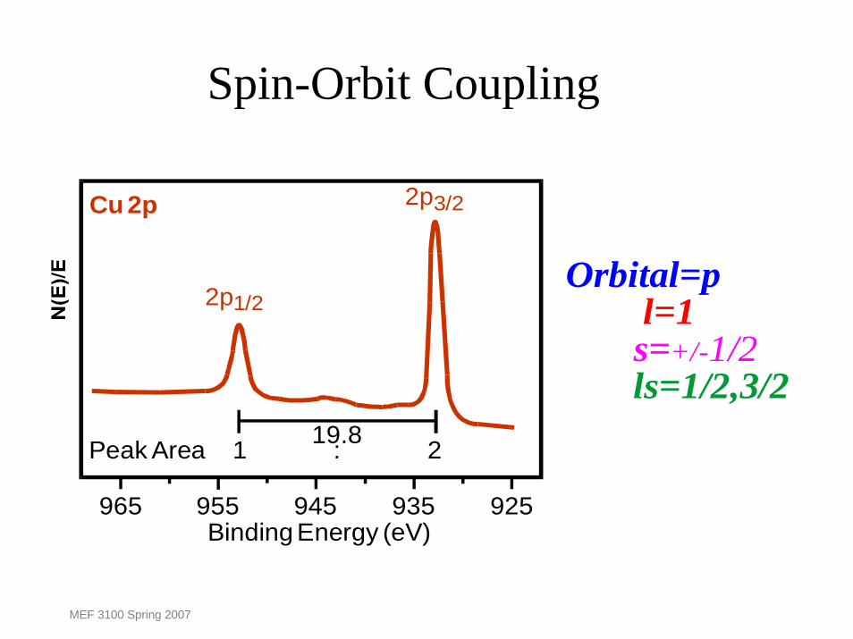
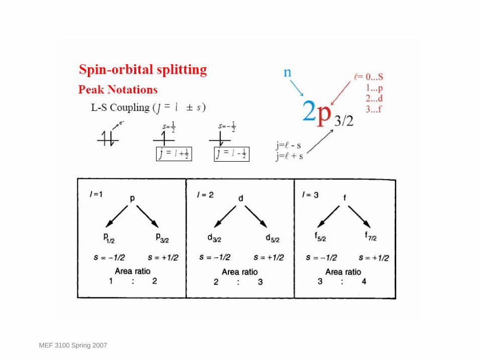
Spin-Orbit Coupling
965 955 945 935 925
19.8
Binding Energy (eV)
Cu 2p
2p1/2
2p3/2
Peak Area 1 : 2
Orbital=p
ls=1/2,3/2
l=1s=+/-1/2
MEF 3100 Spring 2007
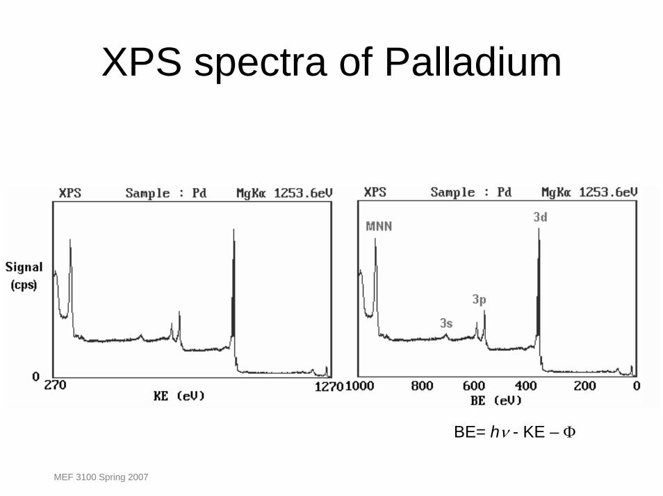
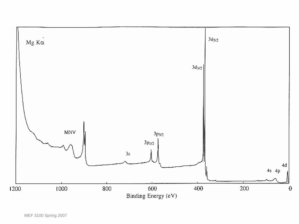
XPS spectra of Palladium
BE= hν - KE – Φ
MEF 3100 Spring 2007
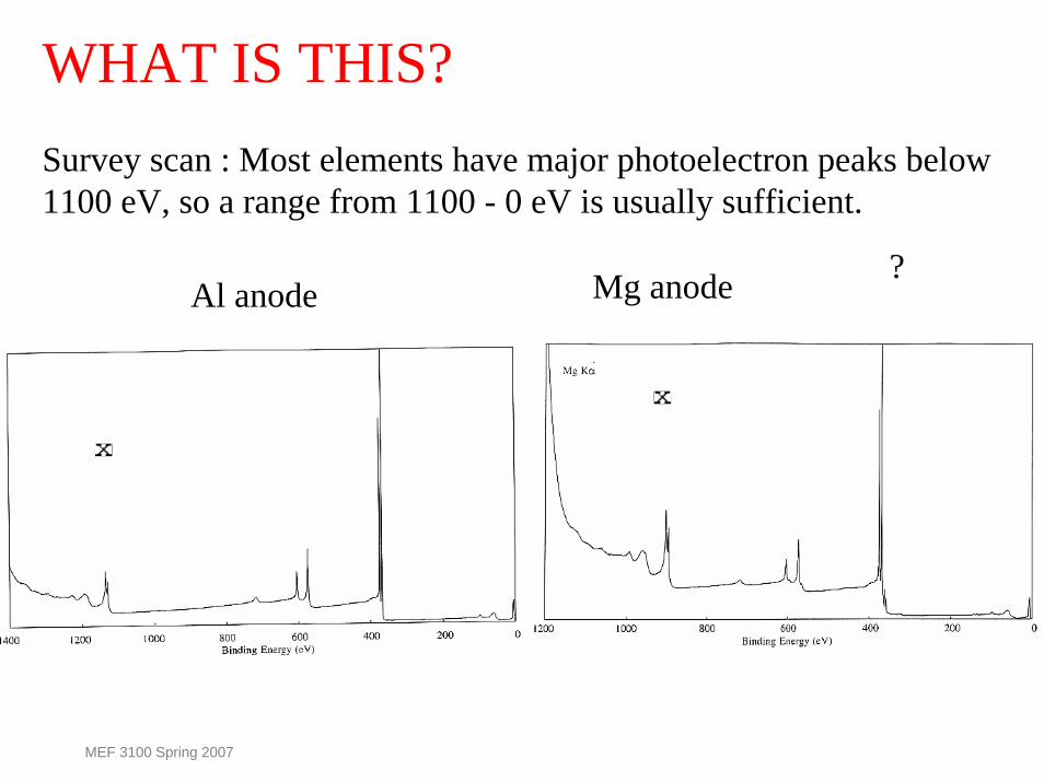
WHAT IS THIS?Survey scan : Most elements have major photoelectron peaks below1100 eV, so a range from 1100 - 0 eV is usually sufficient.
?Mg anodeAl anode
MEF 3100 Spring 2007
MEF 3100 Spring 2007

MEF 3100 Spring 2007
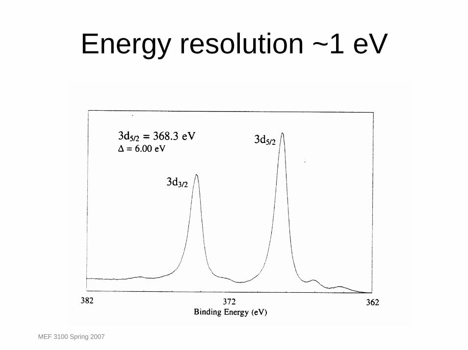
Energy resolution ~1 eV
MEF 3100 Spring 2007
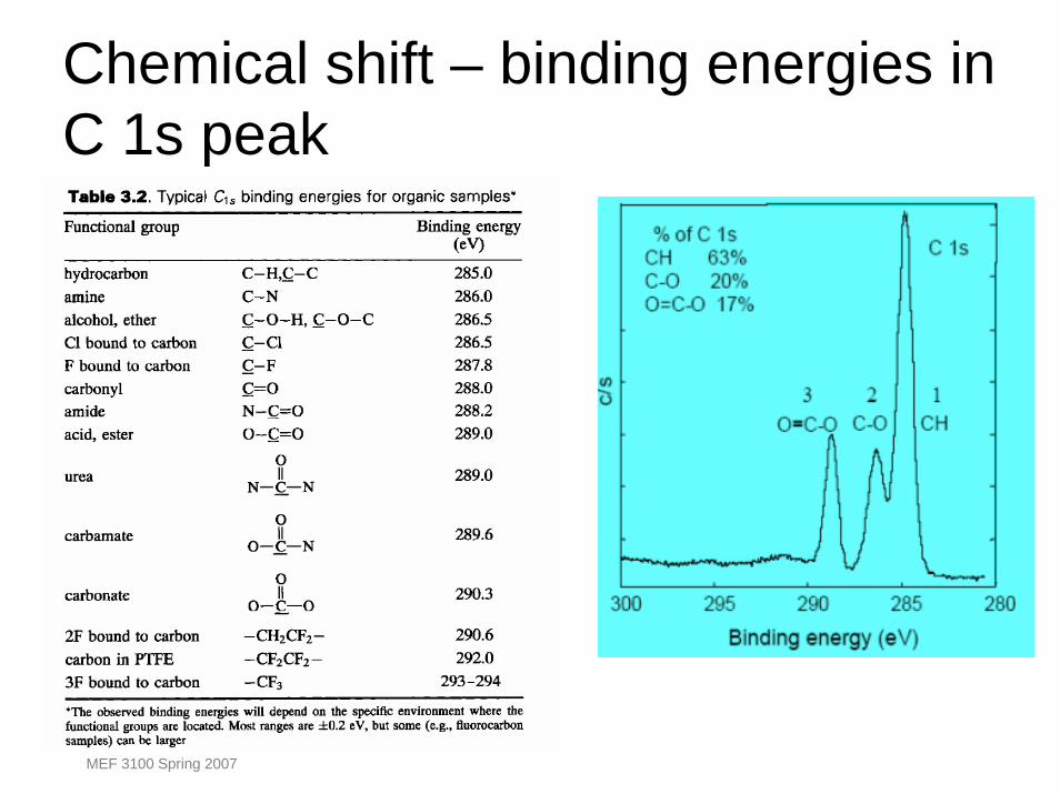
Chemical shift – binding energies in C 1s peak
MEF 3100 Spring 2007
Chemical shiftFunctional
GroupBinding Energy
(eV)hydrocarbon C-H, C -C 285.0
amine C-N 286.0
alcohol, ether C-O-H, C -O-C 286.5
Cl bound to C C-Cl 286.5
F bound to C C-F 287.8
carbonyl C=O 288.0
MEF 3100 Spring 2007
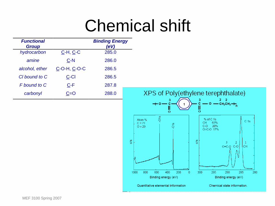
Chemical shift
MEF 3100 Spring 2007
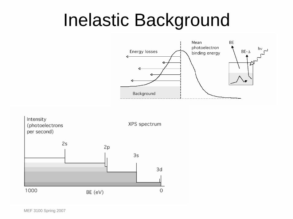
Inelastic Background
MEF 3100 Spring 2007
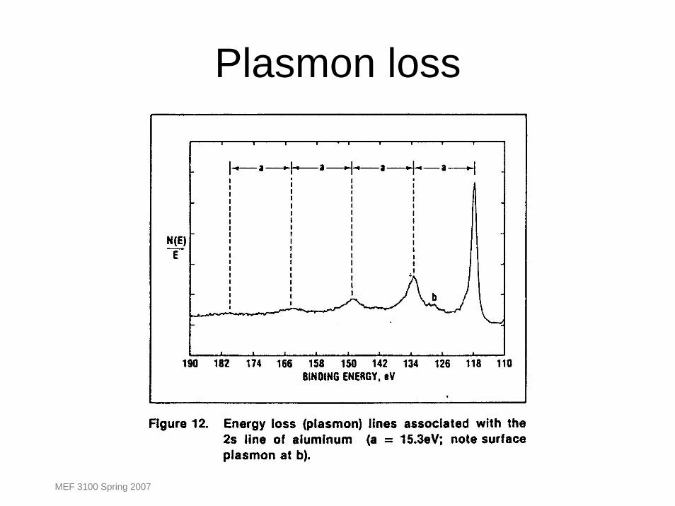
Plasmon loss
MEF 3100 Spring 2007
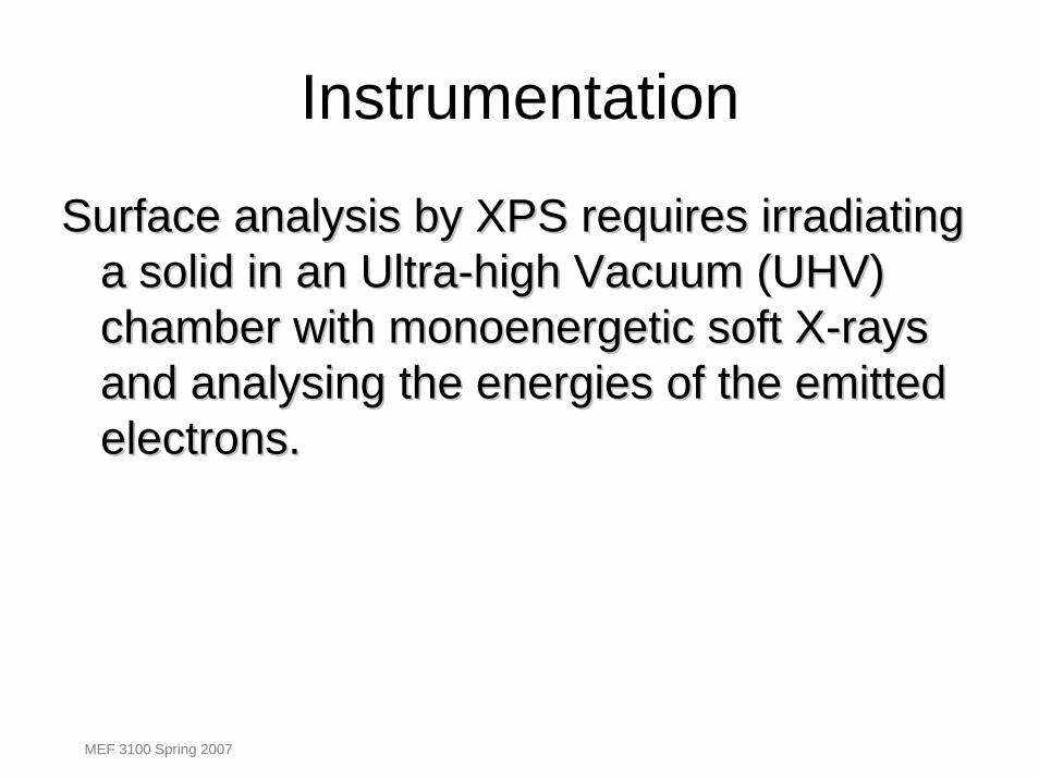
Instrumentation
Surface analysis by XPS requires irradiating Surface analysis by XPS requires irradiating a solid in an Ultraa solid in an Ultra--high Vacuum (UHV) high Vacuum (UHV) chamber with monoenergetic soft Xchamber with monoenergetic soft X--rays rays and analysing the energies of the emitted and analysing the energies of the emitted electrons.electrons.
MEF 3100 Spring 2007
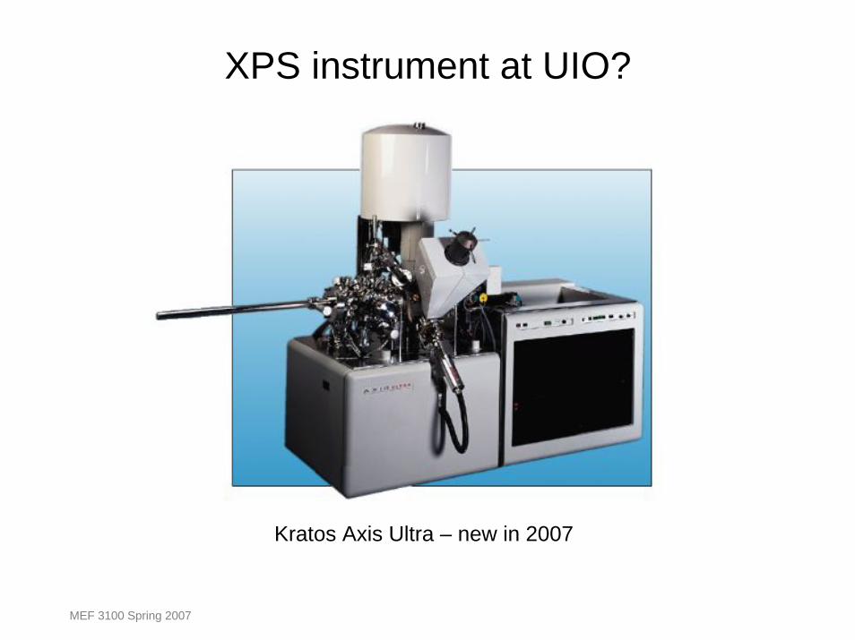
XPS instrument at UIO?
Kratos Axis Ultra – new in 2007
MEF 3100 Spring 2007
MEF 3100 Spring 2007
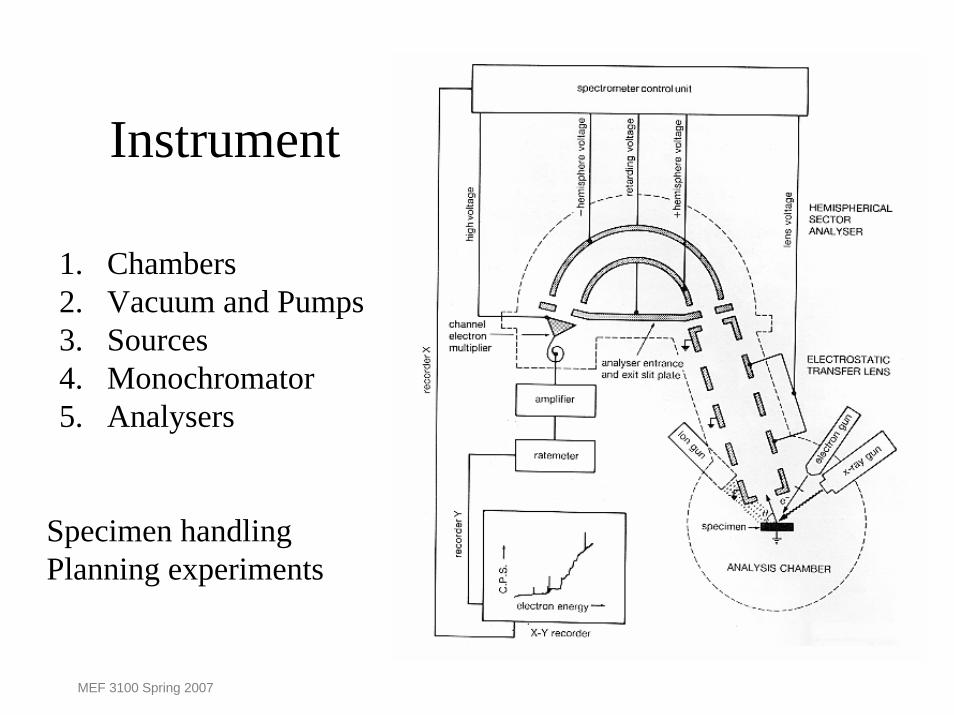
Instrument
1. Chambers 2. Vacuum and Pumps 3. Sources 4. Monochromator5. Analysers
Specimen handlingPlanning experiments
MEF 3100 Spring 2007
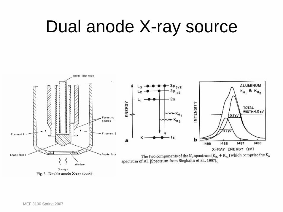
Dual anode X-ray source
MEF 3100 Spring 2007
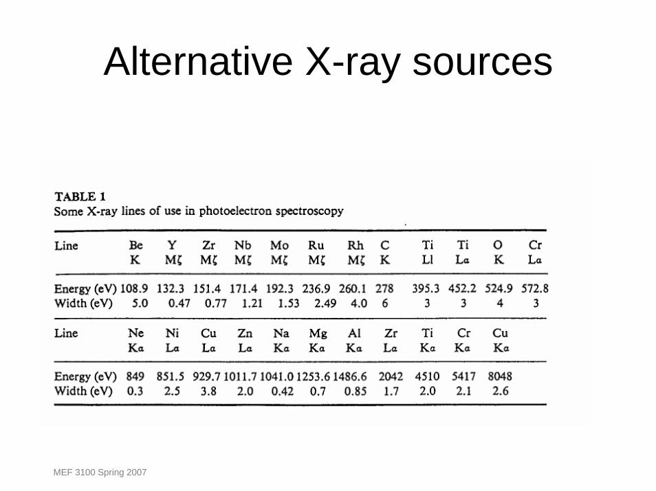
Alternative X-ray sources
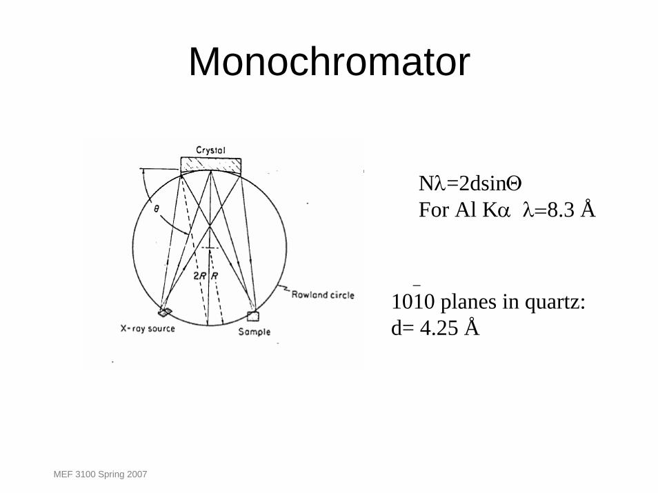
Monochromator
Nλ=2dsinΘFor Al Kα λ=8.3 Å
_
1010 planes in quartz:d= 4.25 Å
MEF 3100 Spring 2007
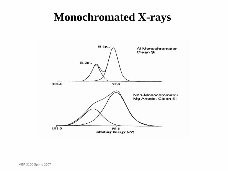
Monochromated X-rays
MEF 3100 Spring 2007
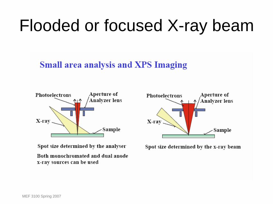
Flooded or focused X-ray beam
MEF 3100 Spring 2007

CHA with standard input lens
MEF 3100 Spring 2007
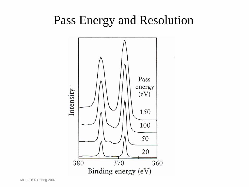
Pass Energy and Resolution
MEF 3100 Spring 2007
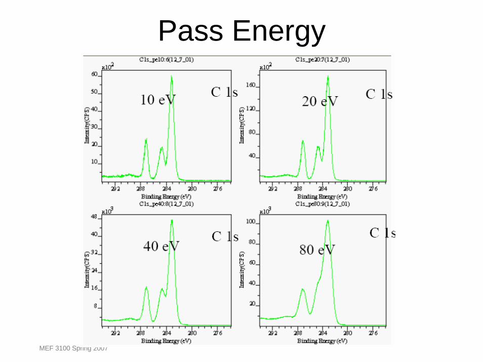
Pass Energy
MEF 3100 Spring 2007
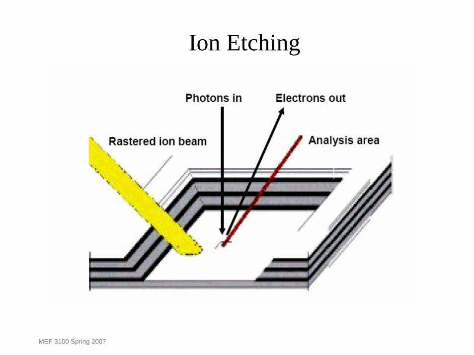
Ion Etching
MEF 3100 Spring 2007
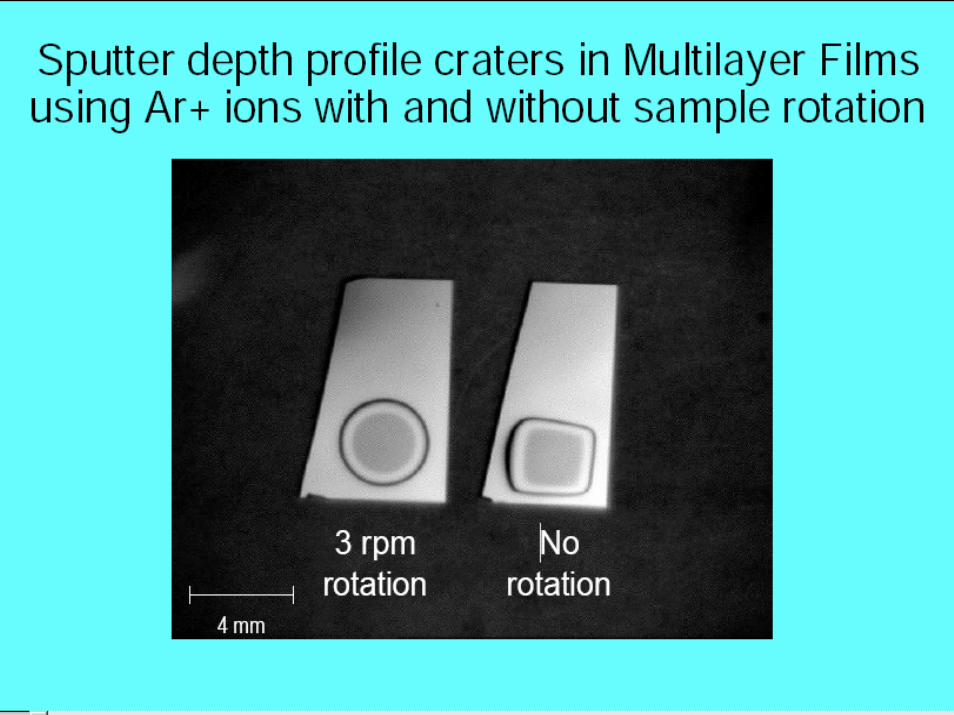
MEF 3100 Spring 2007

MEF 3100 Spring 2007

Survey scan : Most elements have major photoelectron peaks below1100 eV, so a range from 1100 - 0 eV is usually sufficient.
Mg anodeAl anode
?
MEF 3100 Spring 2007
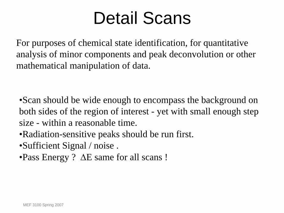
Detail ScansFor purposes of chemical state identification, for quantitative analysis of minor components and peak deconvolution or othermathematical manipulation of data.
•Scan should be wide enough to encompass the background on both sides of the region of interest - yet with small enough step size - within a reasonable time.•Radiation-sensitive peaks should be run first.•Sufficient Signal / noise .•Pass Energy ? ∆E same for all scans !
MEF 3100 Spring 2007
MEF 3100 Spring 2007
MEF 3100 Spring 2007
MEF 3100 Spring 2007
MEF 3100 Spring 2007
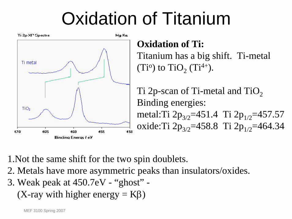
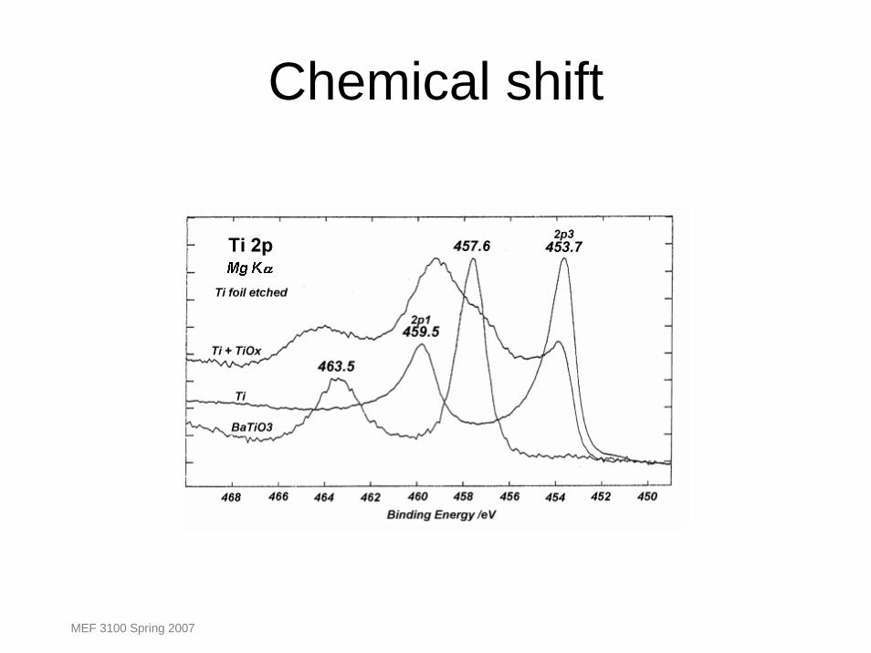
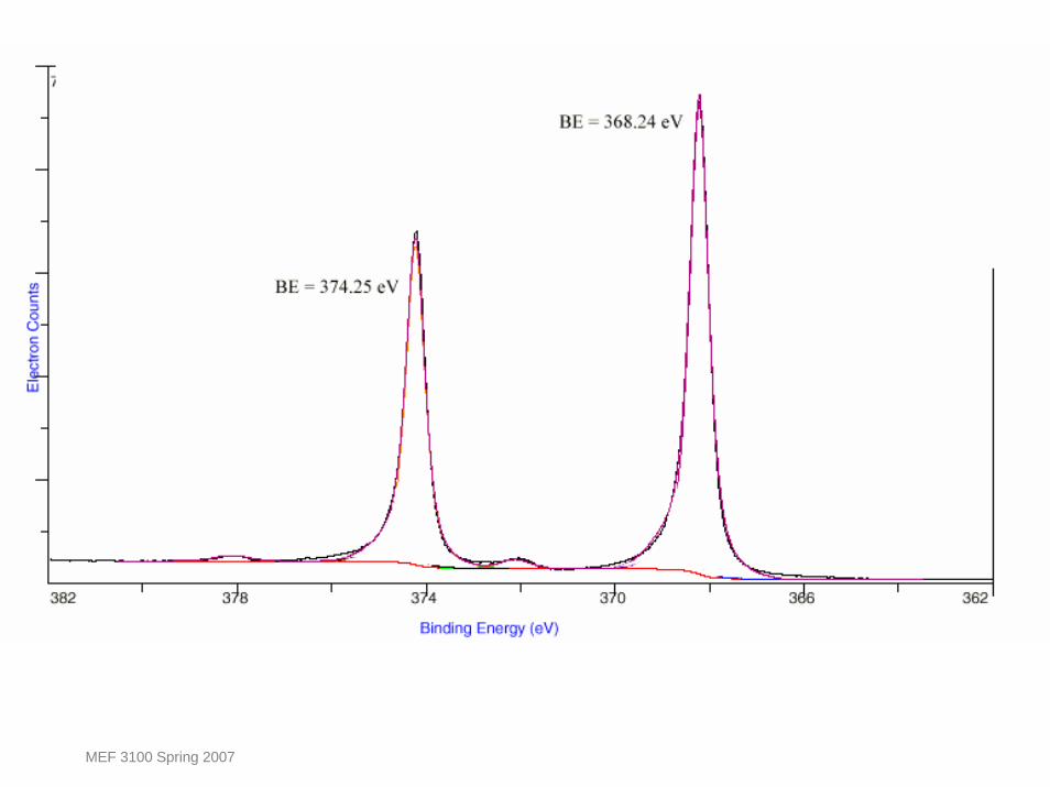
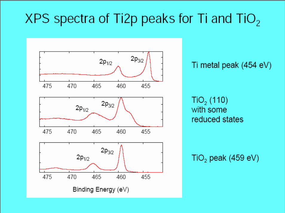
Oxidation of TitaniumOxidation of Ti:Titanium has a big shift. Ti-metal(Tio) to TiO2 (Ti4+).
Ti 2p-scan of Ti-metal and TiO2Binding energies:metal:Ti 2p3/2=451.4 Ti 2p1/2=457.57oxide:Ti 2p3/2=458.8 Ti 2p1/2=464.34
1.Not the same shift for the two spin doublets.2. Metals have more asymmetric peaks than insulators/oxides.3. Weak peak at 450.7eV - “ghost” -
(X-ray with higher energy = Kβ)MEF 3100 Spring 2007

Density of state DOS
DOS Ti-metal
Degree of asymmetry proportional to DOS at EFMEF 3100 Spring 2007
MEF 3100 Spring 2007
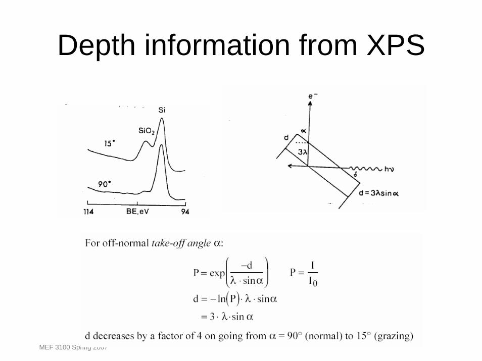
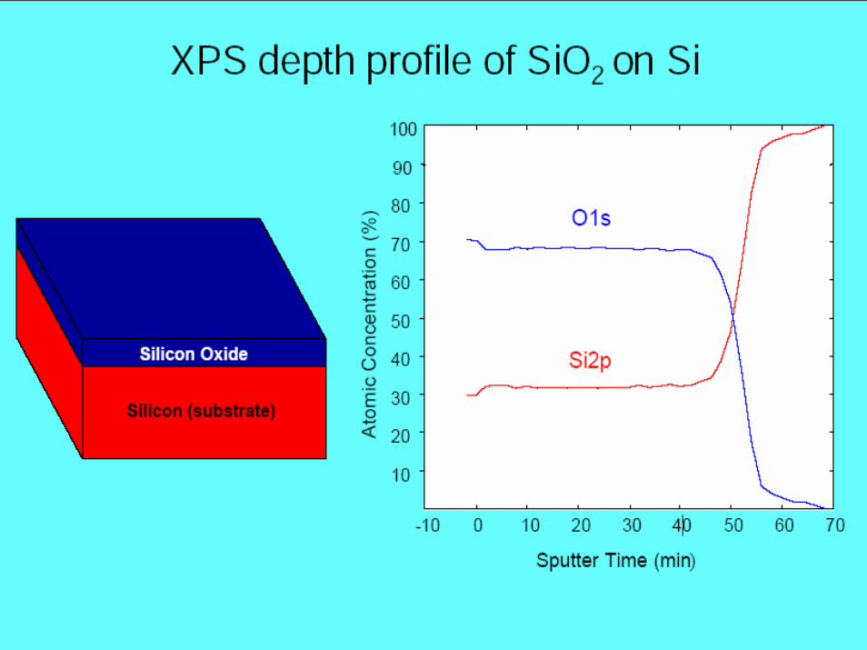
Depth information from XPS
MEF 3100 Spring 2007
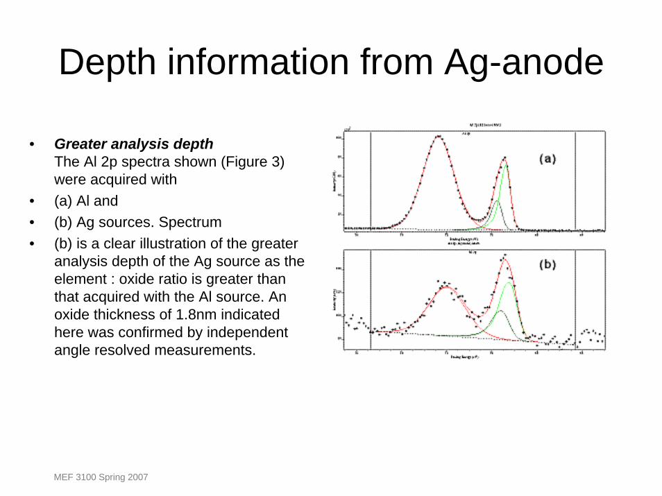
Depth information from Ag-anode
• Greater analysis depthThe Al 2p spectra shown (Figure 3) were acquired with
• (a) Al and • (b) Ag sources. Spectrum • (b) is a clear illustration of the greater
analysis depth of the Ag source as the element : oxide ratio is greater than that acquired with the Al source. An oxide thickness of 1.8nm indicated here was confirmed by independent angle resolved measurements.
MEF 3100 Spring 2007
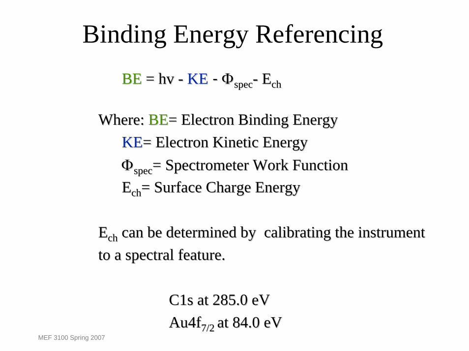
Binding Energy Referencing
MEF 3100 Spring 2007
BEBE = hv = hv -- KEKE -- ΦΦspecspec-- EEchch
Where: Where: BEBE= Electron Binding Energy= Electron Binding EnergyKEKE= Electron Kinetic Energy= Electron Kinetic EnergyΦΦspecspec= Spectrometer Work Function= Spectrometer Work FunctionEEchch= Surface Charge Energy= Surface Charge Energy
EEchch can be determined by calibrating the instrument can be determined by calibrating the instrument to a spectral feature.to a spectral feature.
C1s at 285.0 eVC1s at 285.0 eVAu4fAu4f7/2 7/2 at 84.0 eVat 84.0 eV

Binding Energy Referencing
When analysing insulating samples more care is requiredbecause of sample charging and the uncertainty in the location of the Fermi Level within the band gap.
The term Binding energy is often used without specifyingthe reference level.
Take care when evaluating spectra !
MEF 3100 Spring 2007
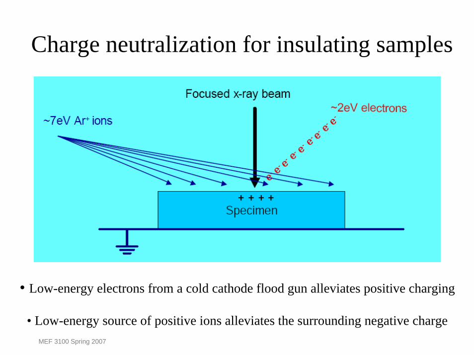
Charge neutralization for insulating samples
• Low-energy electrons from a cold cathode flood gun alleviates positive charging
• Low-energy source of positive ions alleviates the surrounding negative chargeMEF 3100 Spring 2007
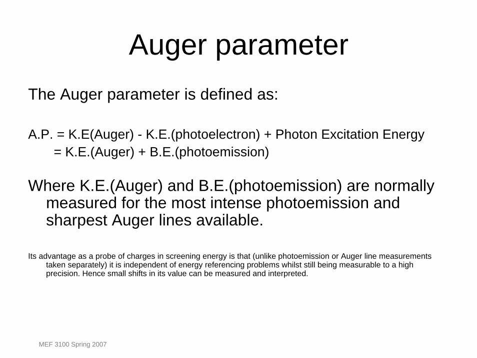
Auger parameterThe Auger parameter is defined as:
A.P. = K.E(Auger) - K.E.(photoelectron) + Photon Excitation Energy = K.E.(Auger) + B.E.(photoemission)
Where K.E.(Auger) and B.E.(photoemission) are normally measured for the most intense photoemission and sharpest Auger lines available.
Its advantage as a probe of charges in screening energy is that (unlike photoemission or Auger line measurements taken separately) it is independent of energy referencing problems whilst still being measurable to a high precision. Hence small shifts in its value can be measured and interpreted.
MEF 3100 Spring 2007
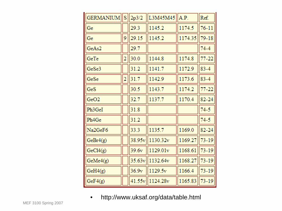
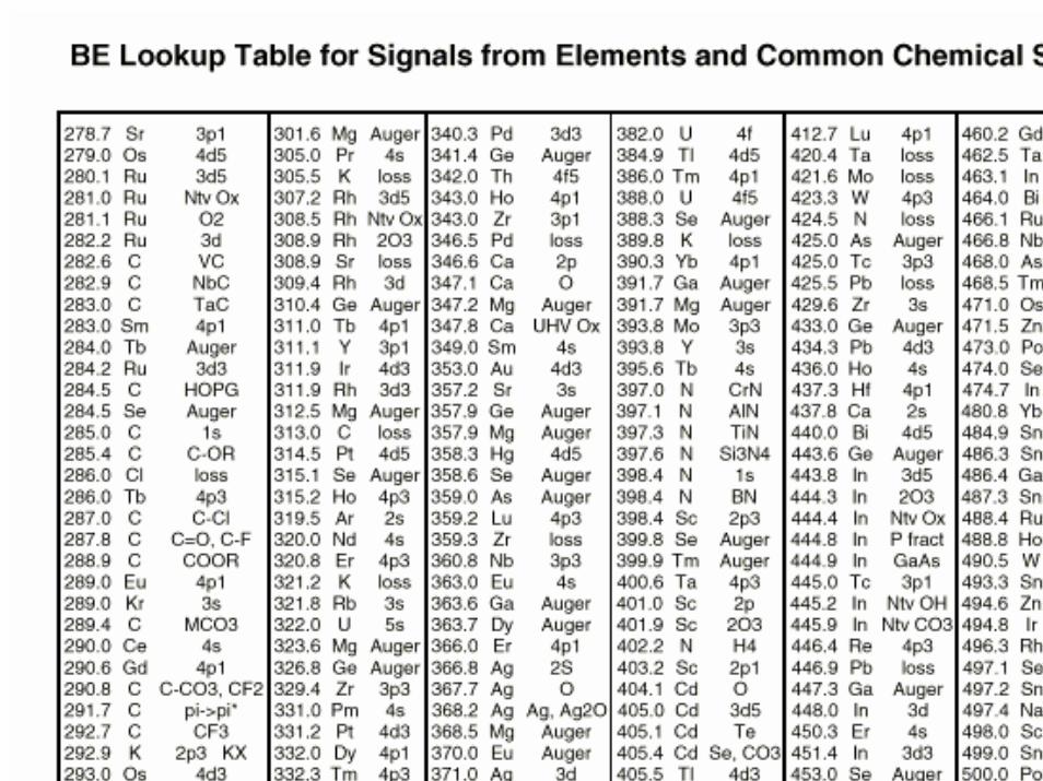
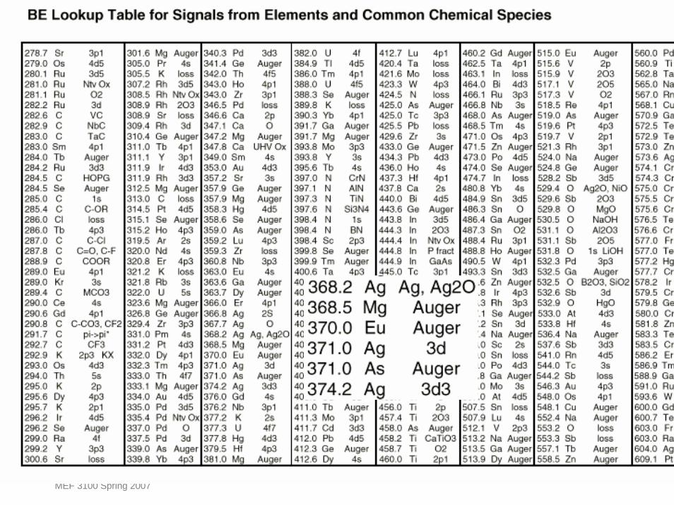
• http://www.uksaf.org/data/table.htmlMEF 3100 Spring 2007
MEF 3100 Spring 2007
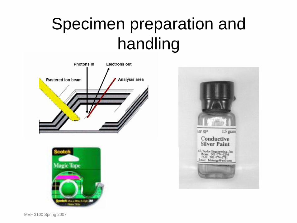
Specimen preparation and handling
MEF 3100 Spring 2007

Analysis of Multi-Layer Paint Cross Section
MEF 3100 Spring 2007
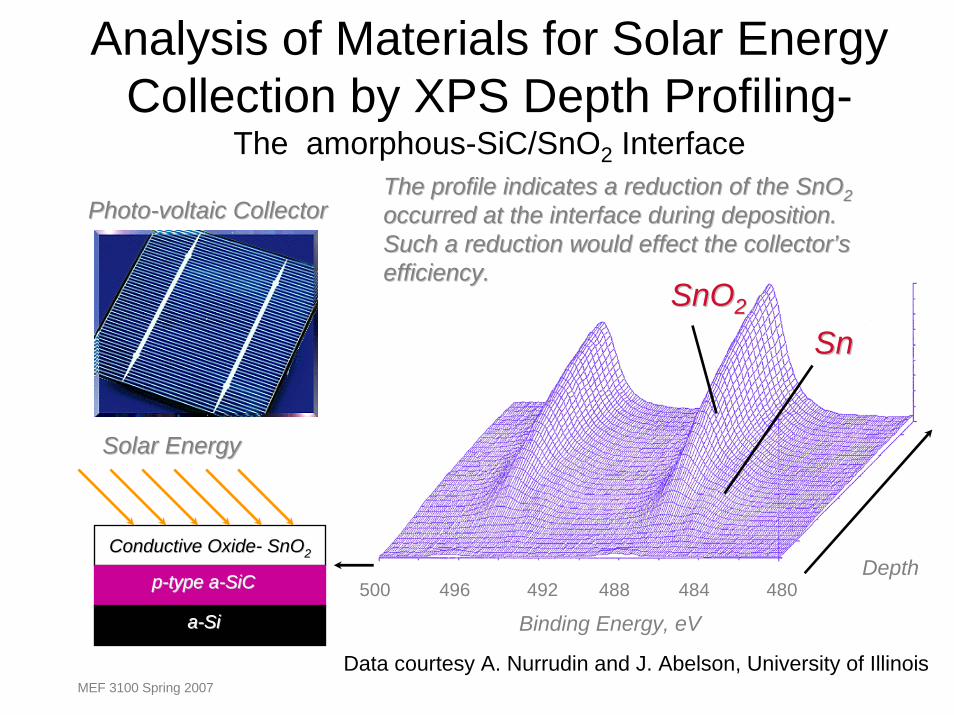
Analysis of Materials for Solar Energy Collection by XPS Depth Profiling-
The amorphous-SiC/SnO2 InterfaceThe profile indicates a reduction of the SnOThe profile indicates a reduction of the SnO22occurred at the interface during deposition. occurred at the interface during deposition. Such a reduction would effect the collector’s Such a reduction would effect the collector’s efficiency.efficiency.
PhotoPhoto--voltaic Collectorvoltaic Collector
Conductive OxideConductive Oxide-- SnOSnO22
pp--type atype a--SiCSiC
aa--SiSi
Solar EnergySolar Energy
SnOSnO22
SnSn
Depth500 496 492 488 484 480
Binding Energy, eV
Data courtesy A. Nurrudin and J. Abelson, University of IllinoisMEF 3100 Spring 2007

Probing Glass Coatings
Windows are coated with complexMulti-layer thin films to meet demands:1) Energy conservation 2) Appearance3) Durability
MEF 3100 Spring 2007
MEF 3100 Spring 2007
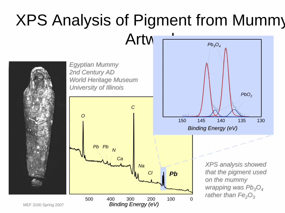
XPS Analysis of Pigment from MummyArtwork
150 145 140 135 130
Binding Energy (Binding Energy (eVeV))
PbO2
Pb3O4
500 400 300 200 100 0Binding Energy (Binding Energy (eVeV))
O
Pb Pb
Pb
N
Ca
C
NaCl
XPS analysis showed XPS analysis showed that the pigment used that the pigment used on the mummy on the mummy wrapping was Pbwrapping was Pb33OO44rather than Ferather than Fe22OO33
Egyptian Mummy Egyptian Mummy 2nd Century AD2nd Century ADWorld Heritage MuseumWorld Heritage MuseumUniversity of IllinoisUniversity of Illinois

Databases
MEF 3100 Spring 2007