Embed Size (px)
Citation preview

RESEARCH ARTICLE
What do rates of deposition of dental
cementum tell us? Functional and
evolutionary hypotheses in red deer
F. J. Perez-BarberıaID1,2*, F. E. Guinness3, M. Lopez-QuintanillaID
1, A. J. Garcıa1,
L. Gallego1, J. Cappelli1, M. P. Serrano1, T. Landete-Castillejos1
1 Game and Livestock Resources Unit, University of Castilla-La Mancha, IDR, IREC, Albacete, Spain,
2 Wildlife Research Unit UIRCP, Universidad de Cordoba, Cordoba, Spain, 3 Department of Zoology, Large
Animal Research Group, University of Cambridge, Cambridge, United Kingdom
Abstract
Cementum is a bone connective tissue that provides a flexible attachment for the tooth to
the alveolar bone in many mammalian species. It does not undergo continuous remodelling,
unlike non-dental bone, which combined with its growth pattern of seasonal layering makes
this tissue uniquely suitable as a proxy for tracking changes in body repair investment
throughout an animal´s life. We tested functional and sexual selection hypotheses on the
rate of cementum deposition related to the highly polygynous mating strategy of red deer.
We used a sample of 156 first lower molars from wild Scottish red deer of known age
between 1 and 17 years old, approximately balanced by sex and age class. Cementum
deposition on the inter-radicular pad increased with age at a constant average rate of 0.26
mm per year, with no significant differences between sexes. Cementum deposition was
independent of (i) tooth wear, other than that associated with age, and (ii) enamel and den-
tine micro-hardness. The results partially supported the hypothesis that the main function of
cementum is the repositioning of the tooth to maintain opposing teeth in occlusion. However,
teeth that had more wear or males´ teeth that had faster rates of tooth wear than those of
females did not present the expected higher rates of cementum deposition.
Introduction
Cementum is a dynamic connective dental bone tissue that provides a flexible attachment
structure via the periodontal ligament in mammals and crocodilians. Recent studies indicate
that cementum and periodontal ligament are plesiomorphic traits in Amniota [1]. Cementum
is mainly deposited on the radicular dentine of the root apex and on the furcations of multi-
rooted teeth, forming an inter-radicular pad [2], although the distribution varies with species,
and many mammals (e.g. ungulates, elephants, rodents, odontocete whales) have extensive
coronal cement coatings [3]. Mammalian cementum is unique in that it is avascular [although
it can be vascular in some reptilians [4]] receiving its nutrition through embedded cells
(cementocytes) that feed from the vascular periodontal ligament. Cementum does not undergo
PLOS ONE
PLOS ONE | https://doi.org/10.1371/journal.pone.0231957 April 28, 2020 1 / 15
a1111111111
a1111111111
a1111111111
a1111111111
a1111111111
OPEN ACCESS
Citation: Perez-Barberıa FJ, Guinness FE, Lopez-
Quintanilla M, Garcıa AJ, Gallego L, Cappelli J, et al.
(2020) What do rates of deposition of dental
cementum tell us? Functional and evolutionary
hypotheses in red deer. PLoS ONE 15(4):
e0231957. https://doi.org/10.1371/journal.
pone.0231957
Editor: Gianpaolo Papaccio, Università degli Studi
della Campania, ITALY
Received: September 11, 2019
Accepted: April 4, 2020
Published: April 28, 2020
Copyright: © 2020 Perez-Barberıa et al. This is an
open access article distributed under the terms of
the Creative Commons Attribution License, which
permits unrestricted use, distribution, and
reproduction in any medium, provided the original
author and source are credited.
Data Availability Statement: Data is fully available
through the provided figures and Zenodo
Repository: http://doi.org/10.5281/zenodo.
3749244.
Funding: The study was supported by projects
Ministerio de Economıa, Industria y
Competitividad, Gobierno de España RTC-2016-
5327-2 (Dr. T Landete-Castillejos); Junta de
Comunidades de Castilla-La Mancha SBPLY/19/
180501/000115 (Dr. MP Serrano). The red deer
continuous remodelling under normal circumstances, unlike non-dental bone, but continues
to grow in thickness throughout life [3]. Its growth pattern of seasonal layering, resulting from
variations in microstructure [5], has been extensively used in archaeology, life history studies
in population ecology [6–10] and as a useful technique to estimate age [11–13]. However,
there is a lack of information on the rate of cementum deposition over an animal´s life and its
functional, ecological and evolutionary significance in ungulates. This is due to the fact that
the functional mechanisms that drive the activity of the cementoblasts remain obscure.
Cementum is composed of equal parts per volume of water, organic matrix and mineral
[2,3]. About 50% of the dry mass is an organic matrix containing mainly collagen fibres
embedded in an interfibrillar ground substance of glycoproteins. About 90% of collagen is
type I and 5% is type III, with the remaining 5% being glycosaminoglycans, chondroitin 4-sul-
phate, dermatan sulphate, and non-collagenous proteins such as alkaline phosphatase. The
other 50% of the dry mass is inorganic, mainly calcium and phosphate in the form of hydroxy-
apatite crystals, and traces of the elements copper, fluorine, iron, lead, potassium, silicon,
sodium and zinc [2,3].
Primary cementum is laid by cementoblasts situated on the surface of the dentine, where
they make a layer of acellular cementum around the cervical part of the root before the tooth
reaches the occlusal plane. Acellular cementum is mainly formed by Sharpey´s fibres (extrinsic
fibres), which come from the periodontal ligament. They are inserted perpendicular to the
root surface, where primary cementum is mineralised with thin flakes of hydroxyapatite at
such a fast rate of deposition that the incremental lines are wide apart.
The secondary cementum develops mainly on the apical portion of the root in mammals,
when the tooth reaches the occlusal plane. It contains cementocytes that are trapped in indi-
vidual lacunae and is less mineralised than the acellular cementum, although the hydroxyapa-
tite crystals are larger and globular. There are less Sharpey´s fibres and other fibres derived
from the cementoblasts (intrinsic fibres) run parallel to the root surface. There is also an area
of mixed fibre cementum where intrinsic and periodontal ligament fibres meet.
Changes in cementum microstructure are responsible for its layering structure, both
around the root apex and root furcation in multi-rooted teeth. There are two main mecha-
nisms that affect the microstructure of cementum, (i) changes in the rate of tissue growth
together with differences in composition and degree of mineralization [3,14], and (ii) variation
in the orientation of the fibres [5]. Slow deposition of mixed fibre cementum, poor in intrinsic
fibres and cells yields thin layers. Thicker and more irregular cementum layers are produced at
faster rates of deposition, are richer in intrinsic fibres and entrap more extrinsic fibres and
cells. Tooth occlusal surfaces are repositioned by resorption of the extrinsic fibres in the peri-
odontal ligament, and new fibres are entrapped by growing pre-cementum [3]. The pattern of
tooth reposition can be tracked by following changes in the orientation of fibres [15].
The cause of the seasonal pattern in cementum deposition is not yet clear. It has been sug-
gested that it might be related to seasonal changes in diet composition or other aspects of phys-
iology. Saxon and Higham [16] suggested that a decrease of fodder availability during winter
might be responsible for less tooth wear and so less compensatory deposition of cementum.
This would not apply equally to wild and domestic species, as the latter are less subject to die-
tary changes. The cementum layering pattern has been used as a convenient trait to estimate
the age in many population studies [8–11,13], but it also inspires research on the biological,
ecological and evolutionary mechanisms that might drive different rates of cementum deposi-
tion, which is the aim of this study.
The disposable soma theory of senescence postulates that the durability of somatic struc-
tures should correlate positively with reproductive lifespan [17]. In highly polygynous species,
in which females have very limited number of offspring per birth, it has been demonstrated
PLOS ONE Hypotheses on rates of deposition of dental cementum
PLOS ONE | https://doi.org/10.1371/journal.pone.0231957 April 28, 2020 2 / 15
study on the Isle of Rum National Nature Reserve
takes place with the support of the UK Natural
Environment Research Council and Scottish
Natural Heritage. The European Union Lifelong
learning programme (Leonardo da Vinci) for
supporting the postgraduate students who
collaborated in this study.
Competing interests: The authors have declared
that no competing interests exist.

that females live longer than males to maximise their reproductive output. Males instead invest
in reproduction during their prime when success for mating opportunities via male-male com-
petition is at its highest [18]. In these species there is evidence that males wear their teeth faster
than their conspecific females [9,19–22] and that males´ tooth size is smaller than in females,
in agreement with the disposable soma theory [23]. If cementum is a flexible mechanism of
tooth attachment to the alveolus and a structure to enable repositioning of the occlusal plane,
then, the rate at which cementum is deposited could be used as an evolutionary life history
trait, something that has never been investigated.
In this paper we test the following hypotheses on the rate of cementum deposition on the
root furcation pad of the first lower molar of Scottish red deer (Table 1): H1. If the main func-
tion of cementum is the repositioning of the tooth to maintain opposing teeth in occlusion
and adjacent teeth in mesial-distal contact, then, the cementum root pad should get thicker as
the animal gets older; H2. In addition, for animals of the same age and size those that wear
their teeth faster should present thicker cementum pads than animals with lower rates of tooth
wear; H3. There is evidence that red deer males wear their teeth faster than their conspecific
females (see above), consequently, in agreement with H1 males should develop thicker cemen-
tum pads than females after controlling for tooth size and age; H4. It has been hypothesised
that males of polygynous species reduce their investment in somatic maintenance after they
pass their reproductive prime, while females of the same species sustain a constant investment
in maintenance to maximise their reproductive fitness across their life. Consequently, it should
be expected that the rate of cementum deposition slows down in males after their prime in
comparison with those in females; H5. Red deer stags reduce their feeding activity during the
rutting season and it has been suggested that this decreases the rate of cementum deposition
[12]. As a consequence the radicular pad thickness should increase at a slower rate in males
after they become reproductively active than in females of similar age; H6. Harder teeth wear
more slowly than softer teeth under similar conditions of mastication effort and diet, therefore
and in agreement with H2, harder teeth should present thinner inter-radicular cementum
pads than softer teeth; H7. The prediction in H6 would not hold if hardness of dental tissue
changes across the life of an animal, as happens with dentine, which hardens as the animal gets
older by a process of mineralisation [24,25], while enamel hardness mineralisation is not so
age-dependent [26]; H8. If cementum deposition functions as a repair mechanism then higher
deposition rates should be expected during times when teeth suffer the most stress. That is at
periods of maximum intake rates, during growth and prime condition, but also at old age
when tooth wear by mesial attrition creates diastemata between adjacent teeth, leaving teeth
less supported and so exposed to higher labial-buccal and mesial-distal movement stress.
Table 1. Hypotheses and predictions on cementum deposition rate across life in red deer. Type of hypothesis: F,
functional; S, sexual selection.
Hypothesis Type Prediction
H1. Maintenance of tooth in occlusion F Cementum deposition increases with age
H2. Maintenance of tooth in occlusion F Cementum deposition increases with tooth wear
H3. Sexual selection in tooth wear S Higher rate of cementum deposition in males than in females
H4. Sexual selection in somatic repair S Cementum deposition decreases in males after prime age
H5. Effect of rut in mastication activity S Cementum deposition decreases in males during rut
H6. Effect of tooth hardness F Cementum deposition rate is lower in harder teeth
H7. Tooth hardness is affected by tooth
mineralisation with age
F H6 prediction does not hold when controlling for age
H8. Repair mechanism F Cementum deposition rate higher at the periods of maximum
mastication activity and dental stress
https://doi.org/10.1371/journal.pone.0231957.t001
PLOS ONE Hypotheses on rates of deposition of dental cementum
PLOS ONE | https://doi.org/10.1371/journal.pone.0231957 April 28, 2020 3 / 15

Materials and methods
Tooth sampling
This study used the red deer teeth sample from Perez-Barberıa et al [11] and Perez-Barberıa
[27], which is summarised by cohort, sex and age in Tables 2 and 3. It is comprised of 156 first
lower molars (M1) of wild Scottish red deer of known age, between 1 and 17 years old, born
between 1980 and 2006 on the island of Rum (Scotland). The teeth came from animals that
were individually identified using ear tags, their age was accurate to within a month. The sam-
ple was approximately balanced by sex and age class (Table 3).
Tooth measures
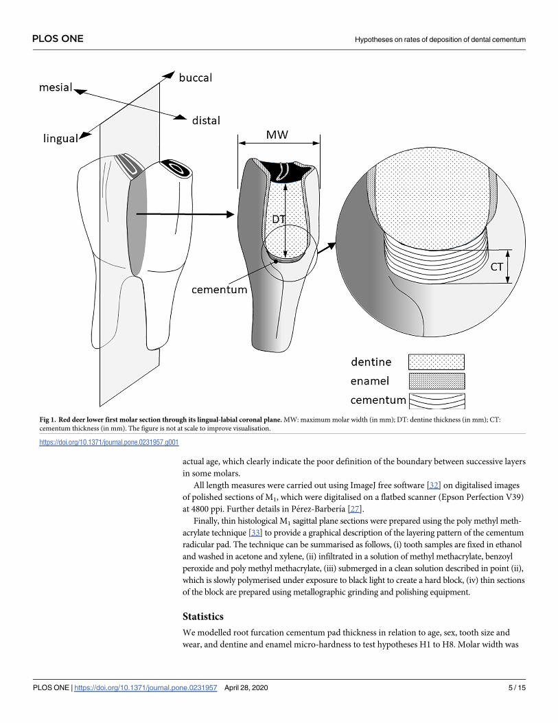
Five traits were measured from each molar, (1) cementum thickness (in mm), as the depth of
the cementum along the vertical axis of a buccal-lingual section of M1 (Fig 1); (2) dentine
thickness (in mm), as a proxy of tooth wear, measured as the depth of the dentine along the
vertical axis from the dentine-cementum junction of the radicular pad to the middle point of
the occlusal plane on a buccal-lingual sectioned crown of M1 [8–10,27]; (3) molar width (in
mm) as the maximum buccal-lingual width of M1, this was used in the analysis as a covariate
to account for the size of the molar and size of the animal (see Statistics); and enamel and den-
tine micro-hardness in mega Pascal (MPa), measured on the polished surface of the buccal-lin-
gual section of M1 by micro-indentation using a Vickers hardness tester (IndentaMet series
11100, Buehler), as described in Perez-Barberıa [27]. Although in red deer enamel is about
four times harder than dentine [27], both traits were considered as both constitute the occlusal
surface of the tooth and consequently tooth crown depletion will be affected by the combined
hardness of enamel and dentine [3,27,28]. Special care was taken to measure tooth hardness
on the same anatomical part of the enamel and normodentine [ie. primary dentine, the most
homogeneous and mineralized type of dentine of the tooth [29]] to minimise variation
between the five micro-hardness tests carried out within each tooth and across different ani-
mals. The mean of these five hardness tests was used in the statistical modelling. Micro-inden-
tation measures the resistance of a material to plastic deformation [30], and although
deformation is not the only process that operates during abrasion and attrition which affects
wear [28,31], it has been effectively used as a proxy of tooth resilience to wear [27].
The reason why cementum thickness was measured as the thickness of all layers of cemen-
tum, rather than measuring the thickness of each annual layer, was the difficulty in identifying
individual layers. Perez-Barberıa et al [13] found, in the same molar sample used in this study,
that 50% of the estimates of age by counting cementum layers were under an error of +/- 1
year, and in 2% of the molars there was a bias up to 5 years younger and 4 years older than the
Table 2. Number of red deer of known age by sex and cohort. Cohort is coded by the last two digits of the year, between 1980 and 2006.
Cohort 80 83 84 85 86 87 88 89 90 91 92 93 94 95 96 97 98 99 00 01 02 03 05 06
female 1 1 4 3 3 2 2 4 2 2 3 6 9 7 4 7 9 6 4 1 0 3 2 1
male 0 0 1 3 4 2 1 1 5 2 1 3 6 12 7 1 4 3 2 2 3 7 0 0
https://doi.org/10.1371/journal.pone.0231957.t002
Table 3. Number of red deer teeth by age and sex.
Age (year) 2 3 4 5 6 7 8 9 10 11 12 13 14 15 16 17
female 7 6 6 7 6 4 7 5 5 6 3 6 5 6 6 1
male 10 5 4 6 5 5 5 4 5 6 2 5 6 2 0 0
https://doi.org/10.1371/journal.pone.0231957.t003
PLOS ONE Hypotheses on rates of deposition of dental cementum
PLOS ONE | https://doi.org/10.1371/journal.pone.0231957 April 28, 2020 4 / 15
actual age, which clearly indicate the poor definition of the boundary between successive layers
in some molars.
All length measures were carried out using ImageJ free software [32] on digitalised images
of polished sections of M1, which were digitalised on a flatbed scanner (Epson Perfection V39)
at 4800 ppi. Further details in Perez-Barberıa [27].
Finally, thin histological M1 sagittal plane sections were prepared using the poly methyl meth-
acrylate technique [33] to provide a graphical description of the layering pattern of the cementum
radicular pad. The technique can be summarised as follows, (i) tooth samples are fixed in ethanol
and washed in acetone and xylene, (ii) infiltrated in a solution of methyl methacrylate, benzoyl
peroxide and poly methyl methacrylate, (iii) submerged in a clean solution described in point (ii),
which is slowly polymerised under exposure to black light to create a hard block, (iv) thin sections
of the block are prepared using metallographic grinding and polishing equipment.
Statistics
We modelled root furcation cementum pad thickness in relation to age, sex, tooth size and
wear, and dentine and enamel micro-hardness to test hypotheses H1 to H8. Molar width was
Fig 1. Red deer lower first molar section through its lingual-labial coronal plane. MW: maximum molar width (in mm); DT: dentine thickness (in mm); CT:
cementum thickness (in mm). The figure is not at scale to improve visualisation.
https://doi.org/10.1371/journal.pone.0231957.g001
PLOS ONE Hypotheses on rates of deposition of dental cementum
PLOS ONE | https://doi.org/10.1371/journal.pone.0231957 April 28, 2020 5 / 15
included in the model (i) as a covariate that enables using dentine thickness as an inverse mea-
sure of the dentine that has been depleted over time and therefore a measure of tooth wear
[22,27]; (ii) larger-bodied adults are normally larger at birth and their rates of growth are also
faster than smaller conspecifics, especially before weaning [34,35]. Consequently, molar width
can be used as a proxy of body size when it is fitted in the model together with age, and is nor-
mally not affected by tooth wear when it is measured at its maximum crown width [22].
Enamel and dentine micro-hardness were fitted in two separate models. The period between
an animal´s death and the micro-hardness test (between 8 to 24 years) might affect tooth-hard-
ness because of enamel or dentine degradation. However, using the same sample of teeth as in
the present study, Perez-Barberıa [27] demonstrated that this potential effect was negligible
and therefore it was not included in our models. In addition, we fitted cohort as a random
effect in the model to account for any environmental or population conditions associated with
the year of birth that could directly affect cementum formation, dentinogenesis or amelogen-
esis in the offspring or via maternal effects [27].
In an early part of the analysis we used generalised additive models GAM [36] in the R soft-
ware package mgcv [37] to check for a potential non-linear response between dependent and
explanatory variables. This was relevant because some hypotheses are based on changes in the
rate of cementum deposition through life. Although GAM models are additive by definition,
we fitted some pertinent interactions between explanatory variables, using the ti function
within GAM model formulae, that produce a tensor product interaction which is appropriate
when the main effects and any lower interactions are also present [38,39]. A thin plate regres-
sion spline with a penalized smoothing basis was used for fitting fixed-effects continuous vari-
ables, and random effects were treated as smooths (using the “re” basis in model formulae).
The terms produce a parametric interaction of the predictors and penalize the corresponding
coefficients with a multiple of the identity matrix, corresponding to an assumption of normal-
ity [39]. We tried a linear versus basis of modest size (k = 3 and k = 4) in the main effects and
interactions. Model selection was performed using Akaike (AIC) weights aided by the normal-
ised probability of the Kullback–Leibler discrepancy ratio, in which model A is to be preferred
over competing model B [40]. The most parsimonious models were those that fitted linear
responses, consequently, we used a linear mixed model approach implemented by the R soft-
ware package lme4 [41] and lmerTest [42]. lmerTest provides p-values for models fitted using
lme4 with Satterthwaite’s degrees of freedom approximation, as in linear mixed-effects models
determining the “correct” value of degrees of freedom in the estimate of the coefficients is
meaningless [43,44]. The coefficients of the linear mixed model were calculated using re-
stricted maximum likelihood REML, as the estimates are more accurate than using maximum
likelihood [44]. The variance explained by the linear mixed model was represented as R2 mar-
ginal (variance accounted for by the fixed effects (R2LMM(m)) and R2 conditional (variance
accounted for by random and fixed effects; R2LMM(c)), following a method developed for linear
mixed-effects models [44] and used in different studies [10,22,27]. Graphics were created
using the ggplot2 R package based on The Grammar of Graphics [45].
Results
There was visual evidence that the cementum deposition rate was not constant across different
parts of the molar (Fig 2A). The thickest layers of cementum were deposited between the roots
and on their apexes, and the thinnest by the cementum-enamel junction. The cementum depo-
sition front showed clear macroscopic indentations, mainly around the root apex, facilitating
the anchorage of the tooth into the alveolar bone via the periodontal ligament. Fine undula-
tions on the cementum deposition front were observed at greater magnifications, these
PLOS ONE Hypotheses on rates of deposition of dental cementum
PLOS ONE | https://doi.org/10.1371/journal.pone.0231957 April 28, 2020 6 / 15

undulations did not necessary match in shape and thickness across successive layers of cemen-
tum (Fig 2C and 2D). The pattern of deposition of cementum was highly variable. Although a
layering pattern was predominant there were places where different fronts of deposition
seemed to converge, some other places developed very sinuous layers and even concentric
cementum nodules (Fig 2C). The canaliculi of the lacunae had more development in the direc-
tion of the cementum deposition (Fig 2D and 2E). The lacunae did not have a uniform spatial
Fig 2. a. Sagital section of a first lower molar of a red deer hind (thickness: 350 μm). Occlusal surface (OC); dentine (D); pulp (P); cementum (C); dentine-cementum
junction (CD); cementum radicular pad (RP); root apex (RA); double-headed arrows show variation in cementum thickness across different parts; cementum and
dentine have become separated along CD at the top of RP. b. sagittal section of radicular cementum showing the direction of proliferation of cementum layers (white
double-headed arrow); most recent cementum layers (CL); change of direction of cementum deposition (ChD); aggregation of cementocites (CC). c. as inset b showing
multiple changes in direction (ChD) and different shapes of cementum deposition layers (CT); undulated cementum deposition front (CF). d. arrows show two
cementocite lacunae; note that the direction of lacuna canaliculi proliferates in the same direction that cementum deposition does. e. detail of a cementocyte lacuna with
its canaliculi expanding in the same direction of cementum deposition. Thickness of sections b-e was 70 μm. Section c was Von Kossa stained, all other sections were
unstained.
https://doi.org/10.1371/journal.pone.0231957.g002
PLOS ONE Hypotheses on rates of deposition of dental cementum
PLOS ONE | https://doi.org/10.1371/journal.pone.0231957 April 28, 2020 7 / 15

distribution, in some places they were aligned along cementum layers, in other places formed
dense clusters that seems to span many layers of cementum (Fig 2B).
After controlling for tooth size, cementum thickness increased with age at an annual rate of
0.26 mm (se = 0.024, p< 0.001), with no significant differences between sexes (Table 4, Fig 3).
Dentine thickness had no effect on cementum thickness, which clearly indicates that cemen-
tum deposition is independent of tooth wear, other than that associated with age (estimate =
-0.019 mm, se = 0.032, p = 0.553, Table 3). Neither enamel hardness (estimate = -3.16E-05,
se = 3.08E-04, p = 0.918, Table 4) nor cohort (p< 0.001, Table 4) affected cementum thickness.
The marginal and conditional R2 explained 83% of the variance of the data. Similar results
were obtained from a model that replaced enamel hardness with dentine hardness (S1 Table).
An alternative model for cementum thickness that fitted only dentine hardness, sex and its
interaction as fixed effects suggested that cementum thickness increased with dentine hardness
(estimate = 0.004 mm, se = 0.002, p = 0.042, Table 5, Fig 4). However, this effect was subro-
gated by age when it was included in the model (Table 4). The model in Table 4 explained 2%
of the marginal variance of the system and 58% of the conditional variance. A similar model to
that in Table 5, in which dentine hardness was replaced with enamel hardness, showed no sig-
nificant fixed effects (Supplementary material, Table 3).
Discussion
The results clearly indicate that cementum deposition takes place at a constant rate across the
life of the animals of our sample, supporting hypothesis H1 that cementum is a tissue whose
main function is maintaining opposing teeth in occlusion [3]. However, contrary to H2, teeth
that presented higher rates of tooth wear, and consequently needed more cementum to main-
tain the tooth in occlusion, did not have thicker cementum inter-radicular pads in comparison
to less worn teeth. Despite this study having used the same sample of teeth in Perez-Barberıa
[27], this study found faster rates of tooth wear in males than in females after controlling for
tooth size and micro-hardness [Fig 6 in Perez-Barberıa [27]]. However, the rates of cementum
deposition did not differ between sexes, which is contrary to prediction H3 based on the dis-
posable soma theory of sexual selection [17]. Also related to the disposable soma theory is
Table 4. Coefficients of a linear mixed model on the inter-radicular cementum thickness pad (in mm), controlling for sex, molar size (molar width, MW, in mm),
age (in years), dentine thickness (DT, in mm), enamel micro-hardness (EH, in MPa) and the pertinent interactions as fixed effects, and cohort as random effect. p
(> Chi2): probability of tests of random-effect terms in the model, each term is removed and REML-likelihood ratio tests computed. R2LMM(m) marginal variance
accounted for the fixed effects; R2LMM(c) conditional variance accounted for random and fixed effects.
Random effects variance sdev p (> Chi2)
cohort (n = 24) 0 0 1.0
residual (n = 151) 0.292 0.540
Fixed effects estimate se df t value p
(Intercept) 1.389 1.246 143 1.115 0.267
MW -0.142 0.068 143 -2.093 0.038
age 0.261 0.024 143 10.847 < 0.001
sex (male) -0.044 1.554 143 -0.028 0.977
DT -0.019 0.032 143 -0.594 0.553
EH -3.16E-05 3.08E-04 143 -0.103 0.918
age × sex (male) -0.007 0.022 143 -0.313 0.755
EH × sex (male) 1.06E-04 4.92E-04 143 0.216 0.830
R2LMM(m) 0.827
R2LMM(c) 0.827
https://doi.org/10.1371/journal.pone.0231957.t004
PLOS ONE Hypotheses on rates of deposition of dental cementum
PLOS ONE | https://doi.org/10.1371/journal.pone.0231957 April 28, 2020 8 / 15

hypothesis H4, which predicts that males should decrease somatic investment after their
prime, time at which their reproductive success decreases. The results did not support predic-
tion H4, as the rate of cementum deposition remained constant with age.
The fact that the thickest deposition of cementum takes place on the radicular pad and root
apex in a perpendicular direction to the occlusal plane (Fig 2A) suggest that cementum deposi-
tion is a mechanism to bring a worn tooth in occlusion with its opposing tooth. The finding
Fig 3. Prediction of inter-radicular cementum thickness pad (in mm) against age (in years) using the model in
Table 4. Circle and purple thin line: female; triangle and black thick line: male. No significant differences in slope and
intercept between sexes. The marginal distribution of x and y variables are displayed as grey ticks along the axes.
https://doi.org/10.1371/journal.pone.0231957.g003
Table 5. Coefficients of a linear mixed model on the inter-radicular cementum thickness pad (in mm), controlling for sex, dentine micro-hardness (DH, in MPa)
and the interaction as fixed effects, and cohort as random effect. Details in Table 4.
Random effects variance sdev p (> Chi2)
cohort (n = 24) 0.998 0.999 < 0.001
residual (n = 153) 0.755 0.869
Fixed effects estimate se df t value p
(Intercept) -1.176 1.582 140.7 -0.744 0.458
DH 0.004 0.002 137.7 2.051 0.042
sex (male) 2.236 2.215 133.5 1.009 0.315
DH × sex (male) -0.003 0.003 133.3 -1.051 0.295
R2LMM(m) 0.017
R2LMM(c) 0.576
https://doi.org/10.1371/journal.pone.0231957.t005
PLOS ONE Hypotheses on rates of deposition of dental cementum
PLOS ONE | https://doi.org/10.1371/journal.pone.0231957 April 28, 2020 9 / 15

that the rate of deposition across an animal´s life was constant and the pattern of cementum
layering deposition was quite irregular across years (as indicated by visual observation Fig 2
and the regression residuals Fig 3), suggest that the rate of cementum deposition can be tuned
to particular events. These events might be related to periods of greater tooth wear (i.e. changes
in diet and food intake) or for improving tooth anchorage in the alveolar bone. Furthermore,
our rate of cementum apposition with age was calculated as the thickness of the accumulated
layers of cementum across the animal´s life, as it was not possible to measure each layer of
cementum because not all were well defined. This implies that no information was available
on actual annual cementum apposition. This might explain why only prediction H1 of the
“maintenance of tooth in occlusion” hypothesis was supported by our statistical analyses but
not the associated H2 prediction. However, it is still surprising that our proxy of tooth wear
(i.e. dentine thickness controlling for molar size) did not have any significant effect or at least
a positive trend on cementum apposition as predicted. We based the H2 prediction on the
functional concept described by Hillson [3] that tooth occlusal surfaces can be repositioned by
remodelling the periodontal ligament fibres and creation of new fibres that are eventually
entrapped by the growing cementum. Experimental support seems inconclusive, in rats, in
which an opposing molar was extracted, the growth of cementum of these molars was less than
that of the control contralateral molar that their occlusion was not modified [46]. In a similar
experiment on rats carried out by Tsuchiya et al [47] M1 was extracted, as a consequence
the adjacent M2 and M3 showed a significant distal drift and the proliferation of acellular
Fig 4. Prediction of inter-radicular cementum thickness pad (in mm) against dentine micro-hardness (in mega
Pascal) using the model in Table 5. Circle and purple thin line: female; triangle and black thick line: male. No
significant differences in slope and intercept between sexes. Details in Table 2.
https://doi.org/10.1371/journal.pone.0231957.g004
PLOS ONE Hypotheses on rates of deposition of dental cementum
PLOS ONE | https://doi.org/10.1371/journal.pone.0231957 April 28, 2020 10 / 15
cementum on the distal side of the root of the molar but not on its mesial side. It is difficult to
compare the results of both experiments, because the directions of the forces exerted on the
teeth of both experiments are different. Despite this, Tsuchiya et al [47] experiment provides
some support to prediction H2 that cementum is implicated in the mechanism of tooth
repositioning.
A non-linear rate of cementum deposition was expected, with a decrease after their prime
at around 10 years of age, when males´ reproductive success declines in this population [18]. It
is well known that males of deer species that are highly dimorphic in body size reduce their
feeding activity during rut [48,49], which results in lower deposition rates and thinner layers
of cementum [12,16,29]. The result of this process across a male´s reproductive life should be
reflected in reduced cementum thickness, in comparison with that of females of the same age,
but this hypothesis (H5) was not supported by the results. It is plausible that (i) compensatory
mechanisms might be involved eg., extra feeding activity after rut as an attempt to recover
body condition [50], which might accelerate tooth wear and so cementum deposition; (ii) as
not all males of a population are equally involved in rut through their lives [51–53], it could be
possible that this effect was negligible in many males and so difficult to detect across the popu-
lation. Harder teeth should have a lower rate of tooth wear and in agreement with H2 these
teeth should have lower rates of cementum deposition (H6). Contrary to the prediction of H6,
a simple model that fitted dentine hardness, sex and its interaction revealed that cementum
deposition increased with dentine hardness, but when age was included in the model the effect
disappeared, as predicted in H7. This could be explained by the positive effect that age has on
dentine and enamel mineralisation [26]. Perez-Barberıa [27] found that dentine and enamel
micro-hardness increased with age up to 10 in red deer, probably due to the mineralisation
process during maturation of these two tissues. It could be hypothesised that this age effect
supersedes the expected negative effect of tooth hardness on cementum deposition. It is, how-
ever, unclear why the effect was detected on dentine hardness but not enamel hardness. It is
plausible that this is because the coefficient of variation (CV) of enamel micro-hardness is
more than twice that of dentine (enamel CV = 5.3%, dentine CV = 2.2%) [27], which makes it
more difficult to detect small responses to variation in enamel hardness. The fact that the rate
of deposition of cementum was constant through life does not support prediction H8, as our
GAM explorative models did not detect any significant change in the rate of deposition at the
age when males are expected to become competitive in reproduction; neither at the oldest ages
when highly worn teeth might need extra support from the periodontal ligament. In relation
to the latter, it is possible that the small sample size at the oldest ages made it more difficult to
detect any change in the rate of deposition.
It could be hypothesised that rates of cementum deposition are faster in adult males than in
females, as a mechanism of improving the attachment of the tooth to the alveolar bone during
the processes of bone resorption that takes place during the phase of antler growth. During
this period it has been claimed that a process of bone resorption (osteoporosis) takes place as
insufficient mineral uptake can occur with a natural diet [54–57]. However, it seems unlikely
that bone resorption has been under positive selection on the mandibular bone, which would
put at risk the attachment of teeth, crucial structures for food intake and digestion. Neverthe-
less, our results do not support this hypothesis, as significant differences were not found
between the sexes in the intercepts or in the slopes of the regression lines of cementum thick-
ness versus age after controlling for molar size.
We remark that our hypotheses and predictions are based on the biology and life history
traits of deer species, concretely red deer, and therefore generalisations of our results on the
cementum apposition patterns of other mammalian or vertebrate species should be considered
with caution. To the best of our knowledge most of the studies carried out on cementum
PLOS ONE Hypotheses on rates of deposition of dental cementum
PLOS ONE | https://doi.org/10.1371/journal.pone.0231957 April 28, 2020 11 / 15

deposition have focused on temporal apposition patterns to detect chronological and physio-
logical events. More work has been carried out on dentine. In belugas (Delphinapterus leucas)
it was hypothesised that rates in dentine deposition changed at sexual maturity, but the results
were inconclusive [58]. In humans, circadian rhythms of dentine apposition have been identi-
fied using fluorochrome markers, but no information on deposition rate changes were pro-
vided [59]. A follow up study by Dean and Scandrett [60], also in humans, found that rates of
dentine formation varied in different parts of the tooth across a period of 40 months. In some
parts the rate was constant while in other parts accelerated or slowed, apparently in order to
match the rates of growth of the different parts of the tooth. The use of bone-labeling tech-
nique is a useful tool to track dynamics of bone formation and remodelling under experimen-
tal conditions and when animals can be handled [61]. However, the technique is logistically
intractable to be applied to wild ungulates on longitudinal population studies to assess hypoth-
eses on life history traits. As a compromise, and to the best of our knowledge, our study is the
first to use population cross-sectional cementum thickness data and statistical modelling
approach to assess functional and evolutionary hypotheses on cementum apposition rates in a
mammalian species.
In conclusion, cementum deposition in red deer takes place at a constant rate across an ani-
mal’s life with no differences between the sexes, even after controlling for tooth size. The rates
of cementum deposition do not respond as expected to any of the sexual selection hypotheses
stated in this study, which are related to the highly polygynous mating strategy of this species.
Our results suggest that cementum´s main function is to maintain precise positioning of the
tooth occlusal surface as a response to changes in the occlusal surface and alveolus associated
with age. We, however, could not support the prediction that once the contribution of this
effect was accounted for cementum apposition responded positively to tooth wear. Lack of
detailed information on cementum apposition rates across a variety of mammalian and reptil-
ian species make it difficult to identify a single evolutionary mechanism that might have driven
the patterns of cementum deposition.
Supporting information
S1 Table.
(DOCX)
S2 Table.
(DOCX)
Acknowledgments
Josephine Pemberton for facilitating FJPB access to the red deer project facilities at the Isle of
Rum and samples. Martyn Baker provided field support during the collection of the samples.
Aaron LeBlanc, Andrew McWilliam, David Walker and an anonymous referee reviewed an
early version of this paper.
Author Contributions
Conceptualization: F. J. Perez-Barberıa.
Data curation: F. J. Perez-Barberıa, F. E. Guinness, M. Lopez-Quintanilla.
Formal analysis: F. J. Perez-Barberıa.
Methodology: F. J. Perez-Barberıa.
PLOS ONE Hypotheses on rates of deposition of dental cementum
PLOS ONE | https://doi.org/10.1371/journal.pone.0231957 April 28, 2020 12 / 15

Resources: F. E. Guinness, A. J. Garcıa, L. Gallego, J. Cappelli, T. Landete-Castillejos.
Writing – original draft: F. J. Perez-Barberıa.
Writing – review & editing: M. P. Serrano, T. Landete-Castillejos.
References1. LeBlanc A.R.H. & Reisz R.R. (2013). Periodontal Ligament, Cementum, and Alveolar Bone in the Old-
est Herbivorous Tetrapods, and Their Evolutionary Significance. PLoS ONE 8.
2. Bosshardt D.D. & Selvig K.A. (1997). Dental cementum: the dynamic tissue covering of the root. Period-
ontol. 2000 13, 41–75.
3. Hillson S. (1986). Teeth., Cambridge Manuals in Archaeology. Cambridge: Cambridge University
Press.
4. Caldwell M.W., Budney L.A. & Lamoureux D.O. (2003). Histology of tooth attachment tissues in the
Late Cretaceous mosasaurid Platecarpus. J. Vertebr. Paleontol. 23, 622–630.
5. Lieberman D.E. (1993). Life-History Variables Preserved in Dental Cementum Microstructure. Science
261, 1162–1164. https://doi.org/10.1126/science.8356448 PMID: 8356448
6. Lieberman D.E. & Meadow R.H. (1992). The Biology of Cementum Increments (with An Archaeological
Application). Mammal Rev. 22, 57–77.
7. Lieberman D.E. (1994). The Biological Basis for Seasonal Increments in Dental Cementum and Their
Application to Archaeological Research. J. Archaeol. Sci. 21, 525–539.
8. Perez-Barberıa F.J., Ramsay S. l., Hooper R. j., Perez-Fernandez E., Robertson A. h. j., Aldezabal A.,
et al. (2014b). The influence of habitat on body size and tooth wear in Scottish red deer (Cervus ela-
phus). Can. J. Zool. 93, 61–70.
9. Perez-Barberıa F.J., Ramsay S. l., Hooper R. j., Perez-Fernandez E., Robertson A. h. j., Aldezabal A.,
et al. (2015b). Corrigendum: The influence of habitat on body size and tooth wear in Scottish red deer
(Cervus elaphus). Can. J. Zool. 93, 249–249.
10. Perez-Barberıa F.J., Carranza J. & Sanchez-Prieto C. (2015a). Wear Fast, Die Young: More Worn
Teeth and Shorter Lives in Iberian Compared to Scottish Red Deer. Plos One 10, e0134788.
11. Mitchell B. (1963). Determination of age in scottish red deer from growth layers in dental cement. Nature
198, 350–351.
12. Mitchell B. (1967). Growth layers in dental cement for determining the age of red deer (_Cervus ela-
phus_ L.). J.Anim.Ecol. 36, 279–293.
13. Perez-Barberıa F.J., Duff E.I., Brewer M.J. & Guinness F.E. (2014a). Evaluation of methods to age
Scottish red deer: the balance between accuracy and practicality. J. Zool. 294, 180–189.
14. Osborn, J.W. (1999). Companion to Dental Studies: Dental Anatomy and Embryology (Volume 1/2)
(ed): Wiley-Blackwell [WWW Document]. URL https://www.abebooks.com/9780632007998/
Companion-Dental-Studies-Anatomy-Embryology-0632007990/plp
15. Sims M.R. (1980). Angular changes in collagen cemental attachment during tooth movement. J. Peri-
odontal Res. 15, 638–645. https://doi.org/10.1111/j.1600-0765.1980.tb00323.x PMID: 6461746
16. Saxon A. & Higham C.F.W. (1968). Identification and Interpretation of Growth Rings in the Secondary
Dental Cementum of Ovis aries L. Nature 219, 634. https://doi.org/10.1038/219634a0 PMID: 5665721
17. Kirkwood T.B.L. (1977). Evolution of ageing. Nature 270, 301–304. https://doi.org/10.1038/270301a0
PMID: 593350
18. Nussey D.H., Kruuk L.E., Morris A., Clements M.N., Pemberton J.M. & Clutton-Brock T.H. (2009). Inter-
and Intrasexual Variation in Aging Patterns across Reproductive Traits in a Wild Red Deer Population.
Am. Nat. 174, 342–357. https://doi.org/10.1086/603615 PMID: 19653847
19. Kubo M.O., Minami M., Higuchi N., Ohnishi N., Okada A., Kaji K., et al. (2013). Female sika deer have
evolved larger incisors than males under relaxed selection against rapid tooth wear. Biol. J. Linn. Soc.
110, 384–397.
20. Loe L.E., Mysterud A., Langvatn R. & Stenseth N.C. (2003). Decelerating and sex-dependent tooth
wear in Norwegian red deer. Oecologia 135, 346–353. https://doi.org/10.1007/s00442-003-1192-9
PMID: 12721823
21. Loison A., Cuyler L.C., Linnell J.D.C. & Landa A. (2001). Sex, age, condition and tooth wear of har-
vested caribou Rangifer tarandus groenlandicus in west Greenland, 1995–1998. Wildl. Biol. 7, 263–
273.
PLOS ONE Hypotheses on rates of deposition of dental cementum
PLOS ONE | https://doi.org/10.1371/journal.pone.0231957 April 28, 2020 13 / 15

22. Lieberman D.E. & Meadow R.H. (1992). The Biology of Cementum Increments (with An Archaeological
Application). Mammal Rev. 22, 57–77.
23. Carranza J. & Perez-Barberıa F.J. (2007). Sexual selection and senescence: Male size-dimorphic
ungulates evolved relatively smaller molars than females. Am. Nat. 170, 370–380. https://doi.org/10.
1086/519852 PMID: 17879188
24. Caruso S., Bernardi S., Pasini M., Giuca M.R., Docimo R., Continenza M.A., et al. (2016). The process
of mineralisation in the development of human tooth. Eur. J. Paediatr. Dent. Off. J. Eur. Acad. Paediatr.
Dent. 17, 322–326.
25. Berkovitz B.K.B., Holland G.R. & Moxham B.J. (2017). Oral Anatomy, Histology and Embryology E-
Book. Elsevier Health Sciences.
26. Margolis H.C., Kwak S.-Y. & Yamazaki H. (2014). Role of mineralization inhibitors in the regulation of
hard tissue biomineralization: relevance to initial enamel formation and maturation. Front. Physiol. 5,
339. https://doi.org/10.3389/fphys.2014.00339 PMID: 25309443
27. Perez-Barberıa F.J. (2019). Tooth wear as a practical indicator of sexual differences in senescence and
mastication investment in ecology studies. Ecol. Indic. 103, 735–744.
28. Lucas P.W. (2004). Dental Functional Morphology: how teeth work. Cambridge: Cambridge University
Press.
29. Baker G., Jones L.H.P. & Wardrop L.D. (1959). Cause of wear in sheep’s teeth. Nature 184, 1583–
1584. https://doi.org/10.1038/1841583b0 PMID: 13795990
30. Smith R.L. & Sandly G.E. (1922). An Accurate Method of Determining the Hardness of Metals, with Par-
ticular Reference to Those of a High Degree of Hardness. Proc. Inst. Mech. Eng. 102, 623–641.
31. Perez-Barberıa F.J. & Gordon I.J. (1998). Factors affecting food comminution during chewing in rumi-
nants: a review. Biol. J. Linn. Soc. 63, 233–256.
32. Rueden C.T., Schindelin J., Hiner M.C., DeZonia B.E., Walter A.E., Arena E.T., et al. (2017). ImageJ2:
ImageJ for the next generation of scientific image data. BMC Bioinformatics 18, 529. https://doi.org/10.
1186/s12859-017-1934-z PMID: 29187165
33. Arzac A., Lopez-Cepero J.M., Babushkina E.A. & Gomez S. (2018). Applying methods of hard tissues
preparation for wood anatomy: Imaging polished samples embedded in polymethylmethacrylate. Den-
drochronologia 51, 76–81.
34. McEwan E.H. (1968). Growth and development of the barren-ground caribou. II. Postnatal growth
rates. Can. J. Zool. 46, 1023–1029. https://doi.org/10.1139/z68-143 PMID: 5749642
35. Parker K. (1989). Growth rates and morphological measurements of Porcupine caribou calves. Rangifer
9, 9–13.
36. Hastie T. & Tibshirani R. (1986). Generalized Additive Models. Stat. Sci. 1, 297–310.
37. Wood S.N. (2011). Fast stable restricted maximum likelihood and marginal likelihood estimation of
semiparametric generalized linear models. J. R. Stat. Soc. Ser. B Stat. Methodol. 73, 3–36.
38. Wood S.N. (2006). Low-rank scale-invariant tensor product smooths for generalized additive mixed
models. Biometrics 62, 1025–1036. https://doi.org/10.1111/j.1541-0420.2006.00574.x PMID:
17156276
39. Wood S.N. (2017). Generalized Additive Models: An Introduction with R, Second Edition. Edicion: 2.
Boca Raton: Chapman and Hall/CRC.
40. Wagenmakers E.-J. & Farrell S. (2004). AIC model selection using Akaike weights. Psychon. Bull. Rev.
11, 192–196. https://doi.org/10.3758/bf03206482 PMID: 15117008
41. Bates D., Maechler M., Bolker B. & Walker S. (2015). Fitting Linear Mixed-Effects Models Using lme4.
J. Stat. Softw. 67, 1–48.
42. Kuznetsova, A., Brockhoff, B. & Christensen, R.H.B. (2015). lmerTest: Tests in Linear Mixed Effects
Models. R package version 2.0–29.
43. Baayen R.H., Davidson D.J. & Bates D.M. (2008). Mixed-effects modeling with crossed random effects
for subjects and items. J. Mem. Lang. 59, 390–412.
44. Nakagawa S. & Schielzeth H. (2013). A general and simple method for obtaining R^2 from generalized
linear mixed-effects models. Methods Ecol. Evol. 4, 133–142.
45. Wickham, H. (2009). Elegant graphics for data analysis. New York.
46. Kato K., Nakagaki H., Okumura H., Li J., Weatherell J.A. & Robinson C. (1992). Influence of occlusion
on the fluoride distribution in rat molar cementum. Caries Res. 26, 418–422. https://doi.org/10.1159/
000261480 PMID: 1294300
PLOS ONE Hypotheses on rates of deposition of dental cementum
PLOS ONE | https://doi.org/10.1371/journal.pone.0231957 April 28, 2020 14 / 15

47. Tsuchiya S., Tsuchiya M., Nishioka T., Suzuki O., Sasano Y. & Igarashi K. (2013). Physiological distal
drift in rat molars contributes to acellular cementum formation. Anat. Rec. Hoboken NJ 2007 296,
1255–1263.
48. Mitchell B., Staines B. & Welch D. (1977). Ecology of red deer. A research review relevant to their man-
agement in Scotland. Cambridge, United Kindom: Institute of Terrestrial Ecology.
49. Nowak R.M. (1999). Walker’s mammals of the world. Baltimore: The Johns Hopkins University Press.
50. Gaspar-Lopez E., Landete-Castillejos T., Estevez J.A., Ceacero F., Gallego L. & Garcıa A.J. (2010).
Biometrics, testosterone, cortisol and antler growth cycle in Iberian red deer stags (Cervus elaphus his-
panicus). Reprod. Domest. Anim. Zuchthyg. 45, 243–249.
51. Bowyer R.T. (1981). Activity, movement, and distribution of roosevelt elk during rut. J. Mammal. 62,
574–582.
52. Hirotani A. (1989). Social relationships of reindeer rangifer-tarandus during rut—implications for female
choice. Appl. Anim. Behav. Sci. 24, 183–202.
53. Wolff J.O. & Van Horn T. (2003). Vigilance and foraging patterns of American elk during the rut in habi-
tats with and without predators. Can. J. Zool.-Rev. Can. Zool. 81, 266–271.
54. Banks W.J., Epling G.P., Kainer R.A. & Davis R.W. (1968). Antler growth and osteoporosis. I. Morpho-
logical and morphometric changes in the costal compacta during the antler growth cycle. Anat. Rec.
162, 387–398. https://doi.org/10.1002/ar.1091620401 PMID: 5701619
55. Hillman J.R., Davis R.W. & Abdelbaki Y.Z. (1973). Cyclic bone remodeling in deer. Calcif. Tissue Res.
12, 323–330. https://doi.org/10.1007/bf02013745 PMID: 4746698
56. Baxter B.J., Andrews R.N. & Barrell G.K. (1999). Bone turnover associated with antler growth in red
deer (Cervus elaphus). Anat. Rec. 256, 14–19. https://doi.org/10.1002/(SICI)1097-0185(19990901)
256:1<14::AID-AR3>3.0.CO;2-A PMID: 10456981
57. Gomez S., Garcia A.J., Luna S., Kierdorf U., Kierdorf H., Gallego L, et al. (2013). Labeling studies on
cortical bone formation in the antlers of red deer (Cervus elaphus). Bone 52, 506–515. https://doi.org/
10.1016/j.bone.2012.09.015 PMID: 23000508
58. Luque S.P., Higdon J.W. & Ferguson S.H. (2007). Dentine Deposition Rates in Belugas (Delphinap-
terus leucas): An Analysis of the Evidence. Aquat. Mamm. 33, 241–245.
59. Kawasaki K., Tanaka S. & Ishikawa T. (1979). On the daily incremental lines in human dentine. Arch.
Oral Biol. 24, 939–943. https://doi.org/10.1016/0003-9969(79)90221-8 PMID: 232978
60. Dean M.C. & Scandrett A.E. (1995). Rates of dentine mineralization in permanent human teeth. Int. J.
Osteoarchaeol. 5, 349–358.
61. Erben R.G. (2003). Bone-Labeling Techniques. In Handbook of Histology Methods for Bone and Carti-
lage: 99–117. An Y.H. & Martin K.L. (Eds.). Totowa, NJ: Humana Press.
PLOS ONE Hypotheses on rates of deposition of dental cementum
PLOS ONE | https://doi.org/10.1371/journal.pone.0231957 April 28, 2020 15 / 15

![Microanalysis of Root Cementum in Patients with Rapidly ......at the exposed cementum [4]. Chemical analysis of the exposed cementum has shown an increase in calcium, magnesium, and](https://img.dokumen.tips/doc/110x75/5f237b2b5d795a336e24c740/microanalysis-of-root-cementum-in-patients-with-rapidly-at-the-exposed-cementum.jpg)