Embed Size (px)
Citation preview

International Journal of Dental Sciences and Research, 2016, Vol. 4, No. 5, 90-94 Available online at http://pubs.sciepub.com/ijdsr/4/5/3 ©Science and Education Publishing DOI:10.12691/ijdsr-4-5-3
Microscopic Differences in Cementum Structure and Mineral Composition of Teeth Extracted from Patients with Gingivitis, Chronic Periodontitis and Aggressive
Periodontitis. A Preliminary Comparative Study
Zahraa Mohamed Nasreldin1, Elhadi Mohieldin Awooda1,*, Nada Tawfig Hashim2
1Department of Restorative Dentistry, Faculty of Dentistry, University of Medical Sciences and Technology, Sudan 2Department of Periodontics: Faculty of Dentistry, University of Medical Sciences and Technology & Department of Periodontology:
Faculty of Dentistry, University of Khartoum, Sudan *Corresponding author: [email protected]
Abstract Background: During the progression of periodontal diseases, cementum undergoes alterations in its structure and mineral composition. Aim: To detect different alterations in structure and mineral composition of cementum according to different periodontal statuses. Subjects and Methods: A true experimental study carried out among six extracted human teeth (One gingivitis of badly decayed tooth (used as control), two chronic periodontitis and three aggressive periodontitis (teeth were extracted due to severe bone loss and mobility). Specimens were taken from different parts of the cementum of the extracted teeth. Each specimen was put in one millilitre of sodium hypochlorite 10 % concentration for 24 hours, then dehydrated by 100% alcohol for half an hour and cementum structures was examined under Scanning Electron Microscope. Student t-test was used to determine the difference between the groups with the level of significance set at P value ≤0.05. Results: There is difference in structure and minerals components of cementum between the specimens of gingivitis, chronic and aggressive periodontitis. Chronic periodontitis has higher amount of minerals with cracks on cementum. Aggressive periodontitis has hypoplasia in cementum, cracks and there is unequal distribution of phosphate and calcium on cemental surface. Conclusion: Different diseases in the periodontium can cause different changes in cementum structure and minerals composition.
Keywords: gingivitis, chronic periodontitis, aggressive periodontitis, SEM, root cementum
Cite This Article: Zahraa Mohamed Nasreldin, Elhadi Mohieldin Awooda, and Nada Tawfig Hashim, “Microscopic Differences in Cementum Structure and Mineral Composition of Teeth Extracted from Patients with Gingivitis, Chronic Periodontitis and Aggressive Periodontitis. A Preliminary Comparative Study.” International Journal of Dental Sciences and Research, vol. 4, no. 5 (2016): 90-94. doi: 10.12691/ijdsr-4-5-3.
1. Introduction Cementum is an avascular mineralized tissue covering
the entire root surface. Owing to its intermediary position, forming the interface between root dentin and periodontal ligament, cementum is a component of the tooth itself, but belongs functionally to the dental attachment apparatus, that is, the periodontium [1,2]. Cementum is light yellow in colour and semi permeable to variety of materials and this permeability diminishes with age. It begins at cervical portion of the teeth at the cemento-enamel junction and continues to the apex.
One of the main functions of cementum is to anchor the principal collagen fibres of the periodontal ligament to the root surface, but it also has important adaptive and reparative functions, playing a crucial role to maintain occlusal relationship and to protect the integrity Of the root surface [1,3,4].
Cementum has two types a cellular (primary cementum) and cellular type (secondary cementum); about 50% of its dry mass as inorganic matter which consists of hydroxyapatite crystals, while the remaining organic matrix is formed largely of collagens and to a lesser degree glycoproteins and proteoglycans [5]. The organic matrix of cementum consists primarily of collagens, similar to ones found in bone and periodontal ligament [6].
Periodontal diseases are highly prevalent and can affect up to 90% of the world population. Gingivitis, the mildest form of periodontal disease, is caused by the bacterial biofilm (dental plaque) that accumulates on teeth adjacent to the gingiva [7]. Periodontitis is defined as “an inflammatory disease of the supporting tissues of the teeth caused by specific microorganisms or groups of specific microorganisms, resulting in progressive destruction of the periodontal ligament and alveolar bone with pocket formation, recession, or both [8].
Children and adolescents can have any of the several forms of periodontitis as described in 1999 International Workshop for a Classification of Periodontal Diseases and
International Journal of Dental Sciences and Research 91
Conditions (aggressive periodontitis, chronic periodontitis, and periodontitis as a manifestation of systemic diseases). However, chronic periodontitis is more common in adults, while aggressive periodontitis may be more commonin children and adolescents [9]. Aggressive periodontitis that occurs in young patients, has a rapid rate of progression, and causes vertical bone loss, the amount of plaque and calculus is not commensurate with amount of destruction. It is different from chronic periodontitis which occurs in older people, and has a slow rate of progression with a horizontal pattern of bone loss; also amount of plaque and calculus formation is commensurate with amount of destruction [10].
Cementum deposition is necessary for maintenance of periodontal health, and defective cementum formation may lead to the development of periodontal pockets. Thus, cementum structure may be the major element of susceptibility to microbial invasion and formation of periodontal pockets. Root surfaces of teeth extracted for aggressive periodontitis have been found to have hypoplastic or aplastic cementum [11].
Two factors have been suggested to be responsible for attachment loss in aggressive periodontitis, which are bacteria and cementopathia [12]. Aggressive periodontitis was described by Gottleib as a deep cementopathia, referred to the fact that the disease might arise due to defect in cementum [13]. This means that the cementum of those individuals may be considered as a risk factor for developing aggressive periodontitis.
In view of this suggestion it is noted worthy to examine the cementum of aggressive periodontitis subjects and to answer the question whether the abnormal structure of cementum is responsible for the abnormal pattern and extremely rapid bone destruction in aggressive periodontitis.
Few inclusive studies have evaluated the cementum of aggressive periodontitis patients. Thus, the present study was conducted to detect differences in cementum structure of extracted teeth from patients with gingivitis, aggressive periodontitis, chronic periodontitis and to detect differences between mineral compositionsin the three groups.
2. Materials and Methods A true experimental study, carried out among extracted
human teeth, which were collected from patients, attended Khartoum Dental Hospital during the time period January 2016. The total number of teeth was six; they were indicated for extraction according to the treatment plan made by one of the consultants in the Periodontology Department. Extraction was done by one of the registrars in the Department of Oral and Maxillofacial Surgery.
Patients were excluded from the study if they met any of the following exclusion criteria: systemic diseases that might have affected the thickness of cementum, para-functional habits, localized periapical pathology, re-implanted teeth, any kind of pulpal conditions affecting root surfaces, radiographic evidence of hypercementosis or root resorption, trauma from occlusion, teeth without antagonists, unilateral chewing habit, open bite and periodontal treatment (mechanical or chemical) within the past one year.
The distribution of sample was as follows; one tooth from a patient with gingivitis, two from chronic
periodontitis and three from aggressive periodontitis. Each extracted tooth was immersed in 2millilitre of 10% sodium hypochlorite solution for 24 hours in order to make the root surface inorganic, after 24 hours teeth were dehydrated by using 1 millilitre of alcohol (100%) for 30 minutes and heated for 30 minutes for evaporation of alcohol to render it dry because electron microscope is very sensitive to water. Finally the teeth were coated with gold and palladium under coating device to provide high contrast for the Scanning electron microscope (SEM) magnification, and Energy Dispersing X-ray (EDX)) which uses INCA software and was attached to the SEM to measure the mineral composition [14].
The comparison between gingivitis, chronic periodontitis and aggressive periodontitis in the structures or the shape were detected by picture shown in SEM which scans every area in the root surface and detects if there are any changes or areas of fracture or lack of layer of cementum. Comparison in mineral composition was detected by selection of five areas in the root surface of each tooth; the changes in cementum were assessed and measured by EDX. Student t-test was `used to determine the significance difference between groups with the level of significance set at P value ≤0.05.
The study was approved by the research ethics committee of University of Medical Sciences and Technology. Before extraction the eligible patients were requested to leave their teeth voluntary after explanation of the purpose of the study and informing them that scanned photos of their teeth will be shown in future publication. Those who agreed signed an informed written consent. Their names were anonymous and any personal related data were kept confidential.
3. Results
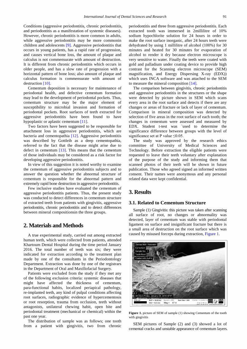
3.1. Related to Cementum Structure Sample (1) Gingivitis: this picture was taken after scanning
all surface of root, no changes or abnormality was detected, layer of cementum was stable with periodontal ligament on surface and insignificant fracture but there is a small area of destruction on the root surface which was caused by misused forceps during extraction, Figure 1.
Figure 1. picture of SEM of sample (1) showing Cementum of the tooth with gingivitis
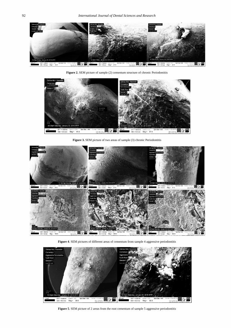
SEM pictures of Sample (2) and (3) showed a lot of cemental cracks and unstable appearance of cementum layers.
92 International Journal of Dental Sciences and Research
Figure 2. SEM picture of sample (2) cementum structure of chronic Periodontitis
Figure 3. SEM picture of two areas of sample (3) chronic Periodontitis
Figure 4. SEM pictures of different areas of cementum from sample 4 aggressive periodontitis
Figure 5. SEM picture of 2 areas from the root cementum of sample 5 aggressive periodontitis

International Journal of Dental Sciences and Research 93
Figure 6. SEM picture of two areas from root cementum of sample (6) aggressive periodontitis
Sample (4), (5), and (6) had similar results microscopically which revealed that cementum was dissimilar from gingivitis and chronic periodontitis in its shape. Also the root surface exhibited variable shapes in different sites within the same root. These areas were further magnified and some areas demonstrated gaps between cementum that indicated cementum hypoplasia. This was further confirmed by the measurements of mineral composition in this area.
3.2. Regarding Mineral Composition Five areas or more were selected in different sites of
root surface that showed different appearance under SEM. It was measured by an external device (EDX) attached to SEM, and the comparison between different groups was displayed in Table 1- Table 3.
Table 1. Comparison between Gingivitis (Group 1) and Chronic Periodontitis (Group 2) Group N Mean Std. Deviation Std. Error Mean p. value
Ca group 1 5 15.1160 1.66103 .74284
Group 2 11 20.1418 7.57857 2.28503 0.004
P group 1 5 7.9360 1.01441 .45366 0.010
Group 2 10 11.0170 3.88460 1.22842
P value below 0.05, there is significant difference between groups in Calcium and Phosphate with group 2 showing higher minerals composition than group 1, this
indicates that Chronic periodontitis has higher minerals composition than gingivitis (Table 1).
Table 2. Comparison between Group 1 (gingivitis), group 3 (aggressive periodontitis) Group N Mean Std. Deviation Std. Error Mean p. value
Ca group 1 5 15.1160 1.66103 .74284
Group 3 18 23.5289 22.68754 5.34750 0.157
P group 1 5 7.9360 1.01441 .45366 0.003
Group 3 18 8.7361 5.35156 1.22183
Comparison between group 1 and 2, denoting insignificant difference in calcium concentration between two groups
(P = 0.157). However, there is significant difference in phosphate composition (P value 0.003) (Table 2).
Table 3. Comparison between Group 2(Chronic periodontitis), group 3 (Aggressive periodontitis) Group N Mean Std. Deviation Std. Error Mean p. value
Ca 2 11 20.1418 7.57857 2.28503 0.227
3 18 23.5289 22.68754 5.34750
P 2 10 11.0170 3.88460 1.22842 0.115
3 18 8.7361 5.35156 1.22183
There is no statistically significant difference in mean value of calcium and phosphate between chronic and aggressive periodontitis P>0.05 (Table 3).
4. Discussion This study was considered as a preliminary or pilot
study because the topic in its objectives of comparison between the three clinical diseases was not investigated before. Additionally, it was not easy to find extracted teeth that fulfilled the criteria because of ethical issues and
difficulties in diagnosis and planning for extraction of the diseased teeth, beside the high cost of investigations when using the SEM. So the sample size was too small to draw a conclusion about the comparison between the three diseases and hence limiting generalization of the results. In spite of that, the study was strengthened by appropriate diagnosis of the diseases prior to extraction and the specimens were prepared after 24 hours from the extraction then examined by an expert lab technician specialized in scanning electron microscope who exerted an effort to show the differences between different groups according to different areas of the teeth. Results showed

94 International Journal of Dental Sciences and Research
that chronic periodontitis group has higher amount of mineral composition when compared with gingivitis, a conceivable clarification for this may be because of its root surface exposure to oral environment which allows saliva minerals to be deposited there. Similar results were obtained by James et al. Their study compared normal and diseased cementum using a scanning electron microscopy [15]. When cementum surfaces affected by periodontal disease; the inorganic contents were altered and the hyper mineralized layer in diseased cementum may play a role in preventing total diseased cementum removal by root planning [15].
Cracks in cementum of chronic and aggressive periodontitis that have been showed in the ESM pictures can be due to chemical changes from inflammation and bacterial toxin as stated in some previous studies [11,15]. Cracks can cause spaces between cementum layer which will lead to the exposure of the dentinal tubules to the environment which consequently causing patients to complain from sensitivity at the beginning of disease, but sensitivity will be alleviated; because of the protective measure in deposition of minerals that close dentinal tubules. This provides a clarification as to why diseased cementum has high mineral composition [15,16].
Gottlieb and (Lindskog&Blomlof) suggested cementum hypoplasia as a manifestation of aggressive periodontitis [13,16]. In this study, similar results showing that cementum hypoplasia was found in teeth affected by aggressive periodontitis can be noted; this can explain the early attachment loss and mobility found in aggressive periodontitis, since the areas that lack cementum have absolutely no periodontal ligament attachment. A previous hypothesis suggested that cytomegalovirus infection during root formation may be involved in the hypoplastic changes that affect the cementum leading to development of localized aggressive periodontitis [10]. The present study showed that the shape and structure of cementum of aggressive periodontitis was completely not alike to the cementum of gingivitis and chronic periodontitis as there is fractured, gaps and area of deficient formation. These areas had lack of minerals composition that indicates there is hypoplasia in cementum of aggressive periodontitis.
Upon the comparison of minerals composition between chronic periodontitis and aggressive periodontitis the results showed that, there is a difference in phosphate and calcium level and this is similar to what has been found by Atilla et al., [17], however, the difference was statistically insignificant and this could be justified by the small size of the present study.
Hypermineralization, which is defined as the presence of a highly mineralized surface layer in the cementum following exposure to the external environment, has frequently been detected by microradiography, chemical analysis, Electron microprobe analysis and nuclear resonance reaction analysis [18,19]. The development of a hyper mineralized zone apparently depends on the ionic concentration of inorganic elements in the local environment. Thus, this zone may be more or less generally present on the exposed root surface or may be completely absent [20].
5. Conclusion The differences between the cementum structures and
mineral composition in gingivitis, chronic periodontitis and aggressive periodontitis can be clearly detected under ESM. A large sample size should be included to determine the hypoplastic changes in cementum and to detect if the cytomegalovirus (CMV) has a role in these changes.
References [1] Gonçalves PF, Sallum EA, Casati MZ, Toledo SD, Junior FH,
Dental cementum reviewed: development, structure, composition, regeneration and potential functions. Braz J Oral Sci.2005; 4: 651-658.
[2] Illustrated Dental Embryology, Histology, and Anatomy, Bath-Balogh and Fehrenbach, Elsevier, 2011, page 170.
[3] Becker J, Schuppan D, Rabanus JR Rauch R, Niechoy U, Gelderblom HR. Immunoelectron microscopic localiz- ation of collagens Type I, V, VI and of procollagen me 111 in human periodontal ligament and cementum. J Histo- chemCytochem 1991: 39: 103-110.
[4] Bosshardt DD, Selvig KA. Dental cementum: the dynamic tissue covering of the root. Periodontol 2000 1997; 13: 41-75.
[5] Saygin NE, Giannobile WV, Somerman MJ. Molecular and cell biology of cementum. Periodontol 2000 2000; 24: 73-98.
[6] Hammarström L. Enamel matrix, cementum development and regeneration. Journal of clinical periodontology. 1997 Sep 1; 24(9): 658-68.
[7] Pihlstrom BL, Michalowicz BS, Johnson NW. Periodontal diseases. The Lancet. 2005 Nov 25;366(9499): 1809-20.
[8] Gandhi M, Kothiwale S. Association of Periodontal Diseases with Genetic Polymorphisms. International Journal of Genetic Engineering 2012; 2: 19-27.
[9] Armitage G. Development of a classification system for periodontal diseases and conditions. Ann Periodontol 1999;4:1-6.
[10] Armitage GC, Cullinan MP. Comparison of the clinical features of chronic and aggressive periodontitis. Periodontology 2000. 2010 Jun 1; 53(1): 12-27.
[11] Lindskog S and Blomlof L. Cementum hypoplasia in teeth affected by juvenile periodontitis. Journal of Clinical Periodontology 1983: 10: 443-451.
[12] Paknejad M, Khorsand A, Yaghobee S, Motahhari P, Etebarian A, Bayani M, Mehrfard A. Cementogenesis in Patients with Localized Aggressive Periodontitis. Journal of Dentistry, Tehran University of Medical Sciences 2015; 12:347-351.
[13] Gottlieb B. The formation of the pocket: Diffuse atrophy of alveolar bone. J Am Dent Assoc 1928; 15: 462-76.
[14] Andresson, T.F. Techniques for the preservation of three dimensional structure on preparing specimens for the electron microscope. transactions of the New York academy of science (1951); 13: 130-134.
[15] James SS, Gary SY, Leif BK. Comparison of cellular cementum in normal and diseased teeth-a scanning electron microscopic study. Journal of Endodontics 1981; 7: 370-37.
[16] Leknes KN1, Lie T, Selvig KA.Cemental tear: a risk factor in periodontal attachment loss. J Periodontol 1996; 67:583-8.
[17] Atilla G, Baylas H. Electron probe analysis of cementum surfaces. Journal of Marmara University Dental Faculty 1996; 2:510-4.
[18] Christoffersen J, Landis WJ. A contribution with review to the description of mineralization of bone and other calcified tissues in vitro. Anat Rec 1991; 230: 435-450.
[19] Butler WT. The nature and significance of osteopontin. Connect Tissue Res 1989; 23: 123-126.
[20] Cohen M, Garnick JJ, Ringle RD, Hanes PJ, Thompson WO. Calcium and phosphorus content of roots exposed to the oral environment. J ClinPeriodontol 1992; 19: 268-273.


![Cementum in Disease[Nalini]](https://img.dokumen.tips/doc/110x75/55cf9d52550346d033ad2077/cementum-in-diseasenalini.jpg)


![Soal Uts Anestesi 2015 [Cementum 2013]](https://img.dokumen.tips/doc/110x75/577c7dbe1a28abe0549fb9bf/soal-uts-anestesi-2015-cementum-2013.jpg)


![Adv in Cementum Devt[1]](https://img.dokumen.tips/doc/110x75/55cf99ce550346d0339f453c/adv-in-cementum-devt1.jpg)


