Embed Size (px)
Citation preview

What causes Foot Pad Dermatitis in Growing
Turkeys?
Ros Mayne, Rod Else and Paul HockingRosin Institute and R(D)SVS, University of
Edinburgh

Outline• Field data
– How important is FPD?• Survey
– When does it start?• Experimental “model”
– Moisture vs excreta?– Allergy or inflammation?
• Is supplementary biotin a cure?• What next

Field Data
• 110,000 stags average 146 d– 90 % affected
– 60 % mild, 40 % score 2+
• 55,500 hens average 118 d– 80 % affected
– 60 % mild, 40 % score 2+
• Season not very important

Field study• Affected/unaffected (N=40)
• Age 1,2,3,4,5,6,7,8, 10 and 21 weeks
• Lesions from week 1
• Fully developed lesion at 3 weeks
• All lesions full scale at 6 weeks
• Increase in size after 6 weeks

External Scoring System• Score 0 = No signs of FPD, skin soft• Score 1 = Slight swelling &/or redness• Score 2 = Skin harder &/or swollen with redness, with compressed
reticulate scales• Score 3 = Skin swollen, red, hard, reticulate scales enlarged &
separated. Small black necrotic areas• Score 4 = Reticulate scales black, <1/8 total foot pad area • Score 5 = Necrosis extends to a ¼ of foot pad• Score 6 = Up to ½ pad necrotic• Score 7 = Over ½ of foot pad covered in necrotic scales

External Scores at 6 Weeks
External score 0 External score 6
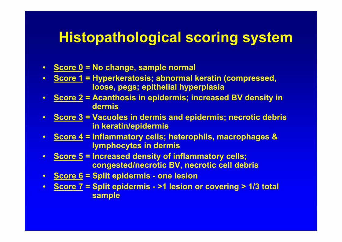
Histopathological scoring system
• Score 0 = No change, sample normal• Score 1 = Hyperkeratosis; abnormal keratin (compressed,
loose, pegs; epithelial hyperplasia• Score 2 = Acanthosis in epidermis; increased BV density in
dermis• Score 3 = Vacuoles in dermis and epidermis; necrotic debris
in keratin/epidermis• Score 4 = Inflammatory cells; heterophils, macrophages &
lymphocytes in dermis• Score 5 = Increased density of inflammatory cells;
congested/necrotic BV, necrotic cell debris • Score 6 = Split epidermis - one lesion• Score 7 = Split epidermis - >1 lesion or covering > 1/3 total
sample

Microscopic view of lesion• Early FPD• 1 week old• Histopath score 3
• Advanced FPD• 6 week old • Histopath score 7

Dry Clean Wood Shavings

Litter wetness trial results (8 d)
4.01.3No litter
HistopathExternal
0.92
3.02.30.7
Mean Scores at 8 Days
1.76SED
3.3Wet dirty5.7Wet clean1.7Dry clean
Litter
Maximum scores 6 days after wetting

Litter Wetness: Large Pens (n=5)
2.2***
74
13
Water %
HistopathExternal
0.53***
6.3
0.7
Mean Scores 6 d
0.41***SED
6.5Wet clean
2.5Dry clean
Litter

Wet or Dry Litter for 48 h
Wet Litter
Dry Litter

Large pens: 6 pens, n=6
0.90.015
6.70.90*
0.38SED
2.90.4124.90.396.50.366.70.53
WetDryTreatmentDay
Histopathology scores at 15 days:
Dry - 1.2Wet - 3.7
* After 48 hours on dry or wet litter

Litter Type and Wetness (6 d)
0.954.8
5.35.85.5Wet
1.0
4.81.85.0Dry
External
6.33.0Shavings
WetDry
1.10
4.83.35.8
Histopath
SED
6.3Straw6.8Paper6.3Cardboard
Litter

Immune Responses (with IAH)
• Aim– Identify cells and cytokines in FPD– Immune response or allergy (irritant)?
• Immune response in tissue– Cells
• CD4+ T, CD8+ T, macrophages, B cells stained– Cytokines
• mRNA levels of IFN-γ, IL-1ß, IL-6, IL-8, IL-10, IL-13

Immune Experiment
• 6 pens dry, 6 pens wet for 48 h.• 6 turkeys/pen, 3 for immune tests• At 48 h massive response
– Unwilling to place foot on litter– Suggests painful

Immunology: Cytokine responses
**0.268.57.4IL13
ns0.782.54.0IL10
***1.0121.010.1IL8
*1.0610.57.5IL6
***0.8513.03.6IL1ß
***0.566.51.4IFN-γ
SignificanceSEDWetDryCytokine

• Wet pens had more– Macrophages, CD4+ and CD8+ cells
• Higher inflammatory cytokines
• Non-specific inflammatory response– Time too short for allergic response– Cytokines consistent with inflammation
Immunology: Immune responses

Biotin Trial (with DSM)
• Dietary biotin – 0, 200, 800,1600 µg/kg– Day old to 14 weeks
• 4 replicates of 38 birds
• Every 2 weeks– Body weights– Foot score– Histology (2/pen)– Plasma biotin (2/pen)
• At 4, 8 and 14 weeks– Liver sample– Litter moisture
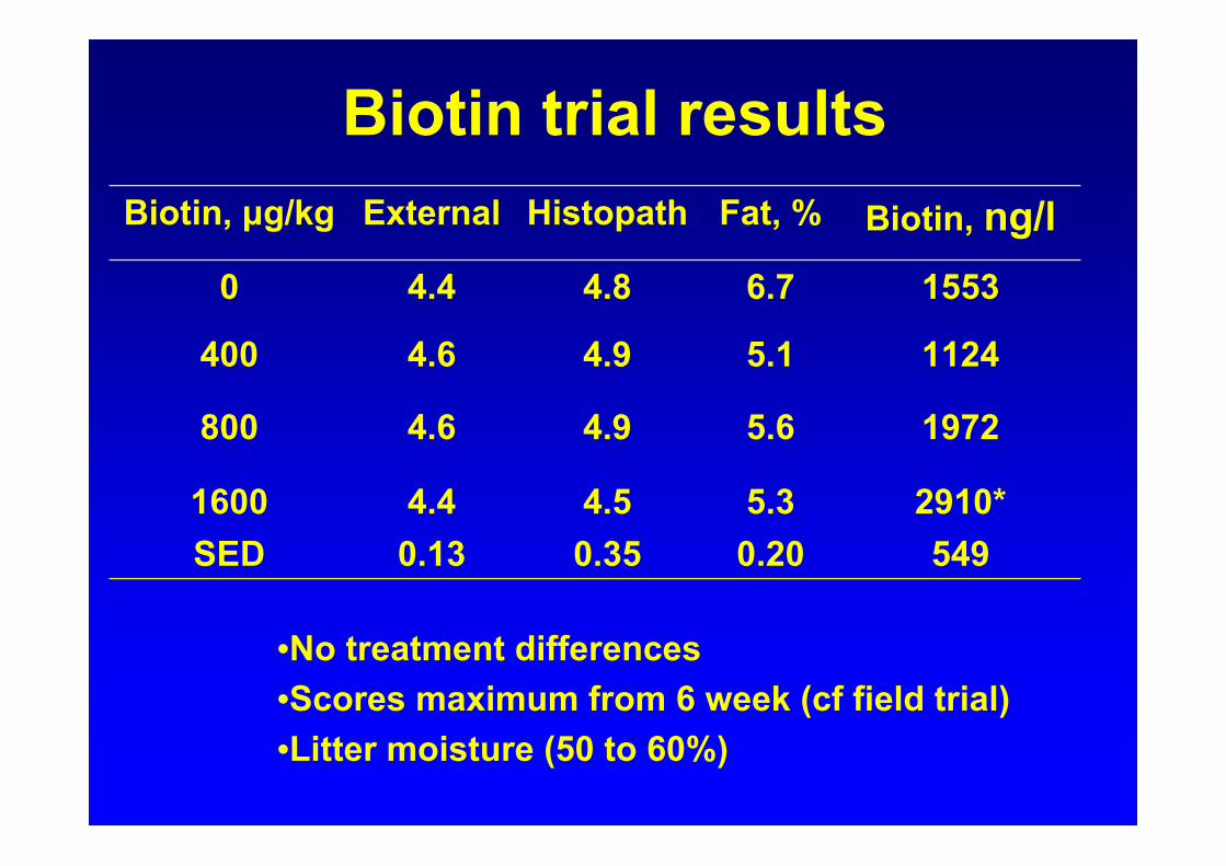
Biotin trial results
2910*5.34.54.416000.20
5.6
5.1
6.7
Fat, %
5490.350.13SED
19724.94.6800
11244.94.6400
15534.84.40
Biotin, ng/lHistopathExternalBiotin, μg/kg
•No treatment differences•Scores maximum from 6 week (cf field trial)•Litter moisture (50 to 60%)

Histopath Scores and Weight, r=0.65
3.13.23.33.43.53.63.73.83.9
44.1
Weight, kg
01234567
External scores and weight, r=0.54
*
*
Histopathology score

External and Histopathology Scores are Poorly Related (r=0.54)
External scoreHistopathology score
Num
ber o
f bird
s

General Conclusions 1
• Primary cause of FPD is wet litter– Not nutrition or gut infection
• Absorbency of litter may affect FPD– Straw and cardboard are poor
• FPD develops at a young age (1-3 weeks)• FPD develops rapidly (2 d) on wet litter
– Maximum scores at 6 weeks– Increase in affected area thereafter

General Conclusions 2• Lesions heal after 15 days on dry litter
– Confirmation of water as cause of FPD
• No effect of high dietary biotin– Usual concentrations adequate
• Inflammatory response not allergy– Physical injury?– Immature epidermis?
• Management solutions– Drinker design– Ventilation and humidity control

General Conclusions 3
• FPD is not caused by high stocking rates
• FPD is caused by high litter moisture
• External score is not a good indicator of histopathology
• FPD might be negatively associated with weight
• FPD may be painful

EU Project (research for SMEs)Three experiments• Minimum litter moisture that causes PFD
• Involvement of acidity (formic acid)
• Genetic differences (2 small, 2 large)
– Pathological differences
– Behavioural changes
Subsequently• Immune response mechanism
• Physical differences (EM, collagen)
• Pain model?
• Is commercial FPD painful?

EU Project (research for SMEs)
Participants• European turkey group
– BUT and producers
• UK, Germany, France, Italy, Poland, Sweden
• Research
– University of Edinburgh (Hocking)
– University of Oxford (Dawkins)
– University of Hannover (Kamphues )
– University of Warmia (Jankowski )
– Schothorst Feed Research (Veldkamp)

EU Project Research• University of Edinburgh (Hocking)
– Welfare assessment of FPD
– Immune mechanisms
• University of Oxford (Dawkins)
– Stocking rate and management
• University of Hannover (Kamphues)
– Nutrition and the GIT
• University of Warmia (Jankowski)
– Feed ingredients; amino acid digestibility
• Schothorst Feed Research (Veldkamp)
– Interaction of nutrition-gut-microbes