Embed Size (px)
Citation preview


We are here
Nervous SystemCentral
Nervous System
Brain
Brain Imaging
Peripheral Nervous System
Building Blocks
Genetics
EvolutionaryEndocrine
System
Neurotransmitters
SomaticAutonomic
Sympathetic Parasympathetic
Biological Psychology
Spinal Cord
Neurons
SensoryMotor
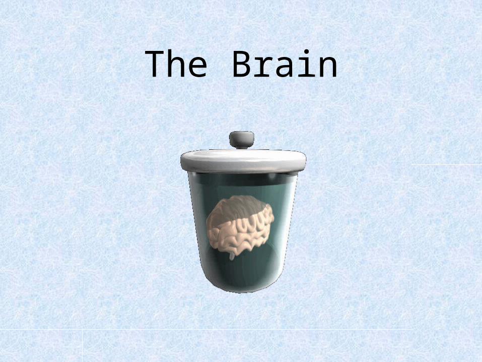
The Brain


Facts about the brain
• Weight= 2.87-3.1 pounds• Men’s brains are slightly larger than
women’s brains (HOWEVER size does Not equal intelligence level! )

Ways we Study the Brain
• Accidents/Injuries• Lesions/
Stimulation• CAT Scan• PET Scan• MRI• Functional MRI

Accidents/Injuries
Phineas Gage • Personality
changed after the accident.
What this this tell us?• That different parts
of the brain control different aspects of who we are.

http://www.youtube.com/watch?v=oPAqTP7058Q

Lesions• Removal or destruction
of some part of the brain.
• Destroyed part of the temporal lobe in Rhesus monkeys, and they became less aggressive and less fearful. (Destroyed the area that controlled aggression.

Stimulation
• Electrodes may be used to set off the firing of neurons
• EX: If you apply a current to the temporal lobe of the brain during surgery, you might hear a familiar song so clearly you think the song is playing in the operating room! – Also used to relieve intolerable pain of cancer
patients and to control violent behavior in otherwise uncontrollable patients.

https://www.youtube.com/watch?v=M_fjiEOb40M

Electroencephalogram
• EEG• Detects brain
waves through the electricity of neural communication.
• Used frequently in sleep research.


Computerized Axial Tomography
• CAT Scan• 3D X-Ray of the
brain.• Good for tumor
locating, or finding brain deterioration but tells us nothing about function.


Magnetic Resonance Imaging
• MRI• More detailed
picture of brain using magnetic field to knock electrons off axis.
• Takes many still pictures and turns images into a movie like production.


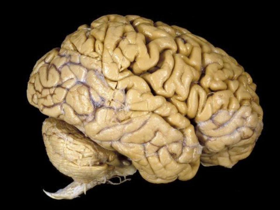
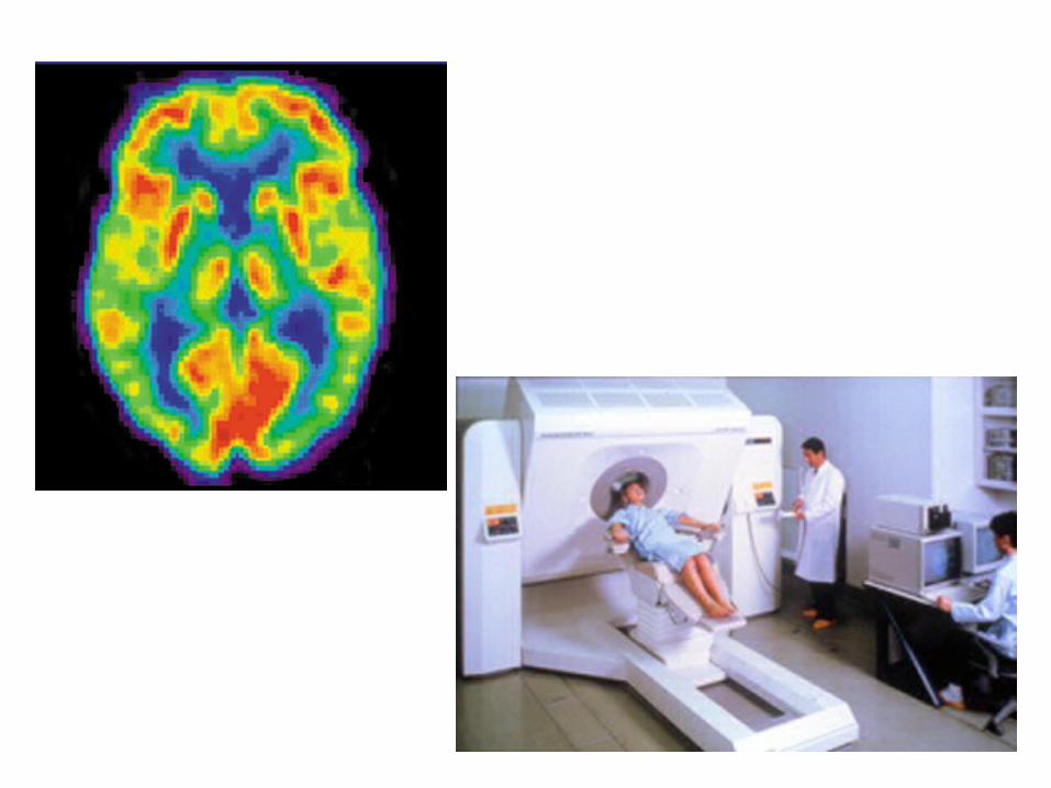
Positron Emission tomography (PET Scan)
• Used to see which brain areas are being activated while performing tasks – The scan changes when one is talking v. when
one is looking at a piece of art.
• Involves injecting a slightly radioactive solution into the blood and then measuring the amount of radiation absorbed by blood cells.– Active neurons absorb more radioactive
solution than non-active ones.


Functional MRI• Combination of PET and MRI

We are here
Nervous SystemCentral
Nervous System
Brain
Brain Imaging
Peripheral Nervous System
Building Blocks
Genetics
EvolutionaryEndocrine
System
Neurotransmitters
SomaticAutonomic
Sympathetic Parasympathetic
Biological Psychology
Spinal Cord
Neurons
SensoryMotor

The Braini. Brain Stem
Medulla, Pons, Reticular Formation, Cerebellum, and the Thalamus
ii. Limbic SystemHypothalamus, Amygdala, and the Hippocampus
iii. Cerebral Cortex (Left and Right Hemispheres and the corpus callosum) Occipital Lobe, Parietal Lobe, Temporal Lobe, and the Frontal Lobe, Primary Motor Cortex and Primary Sensory Cortex, Wernicke's Area and Broca's Area

26
“Older” Brain StructuresThe Brainstem is the oldest part of the brain, beginning where the spinal cord
swells and enters the skull. It is responsible for automatic survival functions.

27
Brain Stem

The Brain Stem/Hindbrain (Automatic Functions)
Brain Structure Primary Function Secondary Function
Medulla Respiration, blood pressure, heart rate
Vomiting
Pons Puts you to sleep
Reticular Formation Attention, regulates awareness
Cerebellum Balance&coordination; implicit memory
Thalamus Directs sensory information to the rest of the brain (except smell)

Hindbrain: Cerebellum
Means “little brain”

30
The Limbic System is a doughnut-shaped
system of neural structures at the
border of the brainstem and
cerebrum, associated with emotions such as fear, aggression and drives for food and sex. It includes the
hippocampus, amygdala, and hypothalamus.
The Limbic System

Limbic System (Emotion Center)Brain Structure Primary Function
Hypothalamus Food, fight/flight, Fahrenheit, sex
Amygdala Fear & Agression
Hippocampus STM to LTM

32
Rats cross an electrified grid for self-stimulation when electrodes are
placed in the reward (hypothalamus) center (top picture).
Reward CenterS
anjiv Talwar, S
UN
Y D
ownstate

Cerebral Cortex/Forebrain
The intricate fabric of interconnected neural cells that covers the cerebral hemispheres. It is the body’s ultimate control and information
processing center.

The Cerebral Cortex• Made up of densely
packed neurons we call “gray matter”
• Glial Cells: support brain cells.
• Wrinkles are called fissures.
• If you lay brain out it would be as big as 2 large Pizzas.
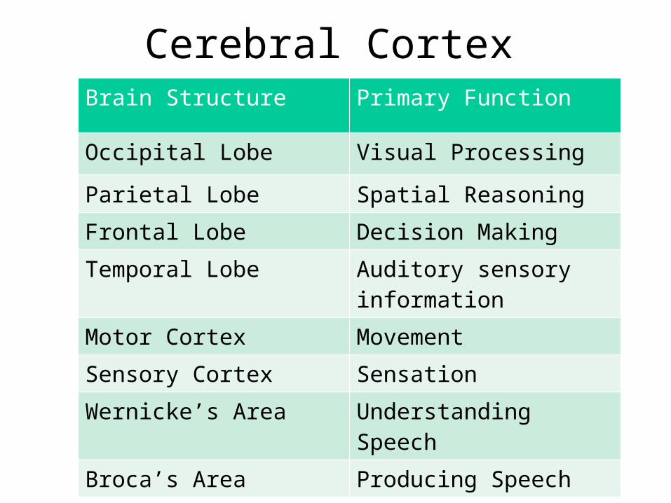
Cerebral Cortex Brain Structure Primary Function
Occipital Lobe Visual Processing
Parietal Lobe Spatial Reasoning
Frontal Lobe Decision Making
Temporal Lobe Auditory sensory information
Motor Cortex Movement
Sensory Cortex Sensation
Wernicke’s Area Understanding Speech
Broca’s Area Producing Speech

Structure of the Cortex
Each brain hemisphere is
divided into four lobes that are separated by
prominent fissures. These lobes are the
frontal lobe (forehead), parietal
lobe (top to rear head), occipital lobe
(back head) and temporal lobe (side
of head).

Frontal Lobes• Abstract thought
(planning) and emotional control (think Gage).
• Contains Motor Cortex: sends signals to our body controlling muscle movements.
• Contains Broca’s Area: responsible for controlling muscles that produce speech.
• Damage to Broca’s Area is called Broca’s Aphasia: unable to make movements to talk.

Parietal Lobes• Contain Sensory
Cortex: receives incoming touch sensations from rest of the body.
• Most of the Parietal Lobes are made up of Association Areas.
Where would this girl feel the most pain from her sunburn?
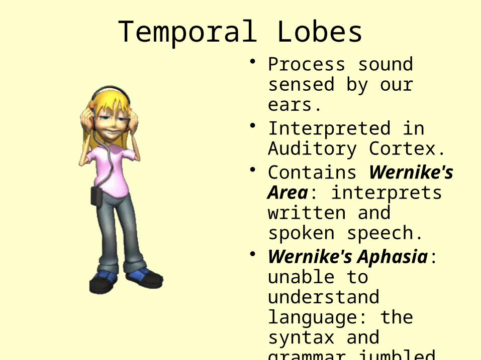
Temporal Lobes• Process sound
sensed by our ears.• Interpreted in
Auditory Cortex.• Contains Wernike's
Area: interprets written and spoken speech.
• Wernike's Aphasia: unable to understand language: the syntax and grammar jumbled.

Occipital Lobes
• Deals with vision.• Contains Visual
Cortex: interprets messages from our eyes into images we can understand.

41
Functions of the CortexThe Motor Cortex is the area at the rear of the frontal lobes that control voluntary movements. The Sensory Cortex (parietal cortex) receives
information from skin surface and sense organs.

Visual/Auditory Function
The functional MRI scan shows the visual cortex is
active as the subject looks at faces.
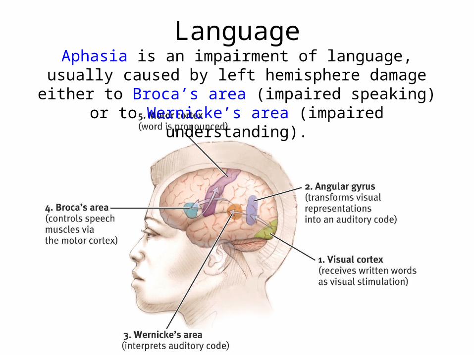
LanguageAphasia is an impairment of language, usually
caused by left hemisphere damage either to Broca’s area (impaired speaking) or to Wernicke’s area
(impaired understanding).

Association Areas• Any area not associated with receiving sensory information
or coordinating muscle movements. More intelligent animals have increased “uncommitted” or association areas of the cortex.


Brain Activity when Hearing, Seeing, and Speaking Words


Decreasing Left-handers

Brain Plasticity• The idea that
the brain, when damaged, will attempt to find news ways to reroute messages.
• Children’s brains are more plastic than adults.