Embed Size (px)
Citation preview
RESEARCH Open Access
Two siblings with immunodeficiency, facialabnormalities and chromosomal instabilitywithout mutation in DNMT3B gene but liabilitytowards malignancy; a new chromatin disorderdelineation?Anna Polityko1*, Olga Khurs1, Natalia Rumyantseva1, Irina Naumchik1, Nadezda Kosyakova2, Holger Tönnies3,4,Karl Sperling3, Heidemarie Neitzel3, Anja Weise2, Thomas Liehr2
Abstract
Background: ICF syndrome (standing for Immunodeficiency, Centromere instability and Facial anomaliessyndrome) is a very rare autosomal recessive immune disorder caused by mutations of the gene de novo DNA-methyltransferase 3B (DNMT3B). However, in the literature similar clinical cases without such mutations arereported, as well.
Results: We report on a family in which the unrelated spouses had two female siblings sharing similar phenotypicfeatures resembling ICF-syndrome, i.e. congenital abnormalities, immunodeficiency, developmental delay and highlevel of chromosomal instability, including high frequency of centromeric/pericentromeric rearrangements andbreaks, chromosomal fragments despiralization or pulverization. However, mutations in DNMT3B could not bedetected.
Conclusion: The discovery of a new so-called ‘chromatin disorder’ is suggested. Clinical, molecular genetic andcytogenetic characteristics are reported and compared to other ‘chromatin disorders’.
BackgroundSeveral human diseases which are characterized byalterations or modification of chromatin structurecaused by changes of methylation pattern are consideredas so-called ‘chromatin disorders’ [1]. Their delineationand the encoding of mechanisms underlying thesegenetic syndromes is one of the ways to understandingthe global principles of functioning of genomic DNAand chromatin in human. ICF syndrome (for immuno-deficiency, centromere instability and facial anomalies;OMIM #242860) belongs to the aforementioned disor-ders and is a rare recessive disease caused by mutationsof the gene DNMT3B that encodes the ‘de novo DNA-methyltransferase 3B’ [2-4]. Patients with ICF syndrome
demonstrate immunodeficiency, facial anomalies, mentalretardation and developmental delay. The main clinicalfeature is reduced serum immunoglobulin levels thatlead to death due to severe recurrent infections, oftenbefore adulthood. The typical cytogenetic markers ofICF syndrome are distinctive ‘undercondensation’ ofheterochromatic segments of chromosomes 1, 9, and 16and multibranched configurations of those. The chro-mosomal instability observable in PHA-stimulated lym-phocytes correlates with a severe hypomethylation ofthe classical satellites 2 and other genomic sequencessuch as alpha satellites, the centromeric component ofconstitutive heterochromatin 3, Alu sequences D4Z4and NBL2 repeats and certain imprinted genes [5].Recent data showed that clinically defined ICF patients
divided into two subgroups and only about 60% of themhad the DNMT3B mutations and normal methylation of
* Correspondence: [email protected] Medical Center “Mother and Child”, Orlovska Street 66, 220053Minsk, Republic of Belarus
Polityko et al. Molecular Cytogenetics 2010, 3:5http://www.molecularcytogenetics.org/content/3/1/5
© 2010 Polityko et al; licensee BioMed Central Ltd. This is an Open Access article distributed under the terms of the Creative CommonsAttribution License (http://creativecommons.org/licenses/by/2.0), which permits unrestricted use, distribution, and reproduction inany medium, provided the original work is properly cited.
the alpha satellites. Those ICF patients without the typi-cal mutation showed hypomethylation of the alpha satel-lites [5-7]. For both groups is in common, that theyhave characteristic heterochromatin abnormalities andundermethylation of classical satellites 2 and 3.Here we report on a family with ICF-like symptoms
but without DNMT3B mutation and with a rather unty-pical pattern of heterochromatin abnormalities.
ResultsCase reportMother and father were not related and physically andintellectually healthy. The first gestation of this partner-ship ended with a spontaneous abortion (no furtherinformation available). After uncomplicated second andthird full-term pregnancies two girls were born, desig-nated as P1 and P2 in the following. Additionally, thereis a half-sister of P1 and P2, a child of father’s first mar-riage, who was healthy.P1 was born when father and mother were 33 and 34
years old, respectively, with a birth weight of 2200 g(<3rd centile), length of 45 cm (<3rd centile), and anOFC of 31.5 cm (<3rd centile). P2 was born 4 yearslater and had a birth weight of 1900 g (<3rd centile), alength of 43 cm (<3rd centile), and an OFC of 31 cm(<3rd centile). P1 and P2 showed severe pre- and post-natal growth delay which persisted in the following. Atthe age of 6 years P1 had a weight of 13 kg (<3rd cen-tile), a height of 110 cm (= 10 centile) and an OFC of46.5 cm (<3rd centile). Similarly P2 had at 1 year aweight of 6 kg (<3rd centile), a height of 62 cm (<3rdcentile) and an OFC of 42 cm (<3rd centile). Both sis-ters suffered from recurrent respiratory infections,immunodeficiency, anemia and mild mental retardation.They had concordant phenotypic features: microcephaly,craniofacial features included “bird-like” appearance,hypertelorism, low-set ears, epicanthal folds, archedpalate, microretrognathia, eye abnormalities (P1 - stra-bismus, myopia; P2 - hypermetropic astigmatism), chestdeformations, arachnodactyly and plano-valgus feet.Renal and brain defects were not found by ultrasoundexamination. Biochemical screening for metabolic dis-eases were normal in both of cases. Finally, P1 had car-diomyopathia starting at the age of 2 years and athrombocytopenia at age of 4 years. With the age of 6years she developed an unspecified type leukemia, fromwhich she died at age of 8 years and 2 months; thebone marrow cells karyotype was complex and includedabnormal clones harboring monosomy 7 and other con-stitutional aberrations like deletion in chromosomes 1,5, 6 and 13: The leukemia karyotype was as follows:mos 45, XX,-7/45, idem, del(13)(q11q22)/46, XX, del(1)(p35)/46, XX, del(5)(q12q33), del(6)(q13q25). Cytoge-netics of peripheral blood of P1 and P2 as well of the
parents was done at this time and the ‘chromatin disor-der’ detected (see below).Furthermore, the DNMT3B DNA methyltransferase
gene mutated in the ICF immunodeficiency syndrome[5-7] was sequenced in P1 and P2 and no hint on amutation was detected, thus, excluding a common ICFsyndrome. In detail the following polymorphisms weredetected leading to no effect on the aminoacid sequence:in exon 1: NT_028392.4 position 1534716; in exon 15:NT_028392.4 position 1553115 and NT_028392.4 posi-tion 1553217; in intron IVS18-75: NT_028392.4 position1555777, in intron IVS17-5: NT_028392.4 position1555404, in intron IVS16-5. NT_028392.4 position1554722; in intron IVS09-113: NT_028392.4 position1547997; in untranslated RNA region 5’mrnautr:NT_028392.4 position 1562920. Moreover, in exon 9 atENST00000328111 position 30355 the sequence wasaltered in a way that a leucin instead of an isoleucin isinserted in the DNMT3B gene product. However, alsothis alteration is classified as a harmless polymorphismin ‘Human Mutation Database’ http://www.hgmd.cf.ac.uk and ‘Mutation Taster’ http://neurocore.charite.de/MutationTaster/comparison.html.Unfortunately, the family was lost during further fol-
low-up. Nonetheless, Epstein Barr virus based immorta-lization of the parental blood and blood of P1 and P2was done [8]. Thus, cell lines are available for furtherresearch.
Cytogenetic and molecular cytogenetic studiesKaryotypes (GTG-banding) of the siblings P1 and P2and their parents were normal. Chromosomal stabilityof P1 and P2 and their parents were tested by analysisof GTG-banding or standard solid-stained chromosomesusing 48, 72 and 96 h blood lymphocyte cultures as pre-sented in Table 1.The chromosomal stability in parents of P1 and P2
was considered as normal, besides a previously unrecog-nized hint of a specific low level of mosaic aneuploidyof X-chromosome in the father (5 of 100 cells with 47,XXY). This was studied by two-color interphase FISHusing centromeric probes for the X- and the Y-chromo-somes (Abbott/Vysis) in 670 cells: 4 cells had abnormalsex chromosome constitution (1 times 45, X; 3 times 47,XXY). Thus, the presence of a minimal mosaic 47,XXY/46, XY in the father could not be excluded.The cytogenetic findings in somatic cells of both the
affected girls (P1 and P2) were similar; i.e. high level ofunspecific spontaneous chromosomal aberrations werepresent: chromatide and chromosome type deletions,dicentric and ring chromosomes, reciprocal and nonre-ciprocal translocations, (small) marker chromosomes,end-to-end chromosomal junctions, aneuploid, poliploidcells and other were found (Figs. 1 and 2). Specific for
Polityko et al. Molecular Cytogenetics 2010, 3:5http://www.molecularcytogenetics.org/content/3/1/5
Page 2 of 6
our patients were especially centromeric rearrange-ments: high frequency of breaks in centromeric region,isochromosomes, frequently metaphases contained iso-chromosome and the rest of another arm, additionalwhole-size chromosomal arms in metaphases, inter-changes between chromosomes in pericentromericregions, fragmentation and/or despiralization of the
chromatin. Multiplex-fluorescence in situ hybridization(M-FISH) analysis [9] was applied for further investiga-tion of chromosomal specificity of cytogenetic instabilityin these family members (Table 2). Some aberrations asdetectable by M-FISH analysis are presented in Fig. 3.Using M-FISH approach we obtained data of chromo-
somal origin in cases of undercondensation, fragmenta-tion or pulverization of abnormal chromatin. M-FISHwas helpful in analysis of near-triploid and near-tetra-ploid metaphases bearing multiple constitutional aberra-tions. That made it possible to analyze the involvementof all chromosomes into the aberrations in somatic cellsin both of affected children (P1 and P2; see Fig. 4).
DiscussionThe clinical, cytogenetic and molecular genetic status ofa family with two siblings having concordant phenotypicabnormalities, developmental delay, immunodeficiencyand highly expressed chromosomal breakage arereported. 72 h and 96 h cell cultures were applied foraberration frequency evaluation in spite of the fact thatthere is the elimination of unstable chromosomal aber-ration in it. The first cytogenetic examination of P1 dis-covered 15.6% of aberrations in 72 h culture. A secondtest 8 months later showed 24.2% in 48 h culture usingthe same standard staining. Banding technique using thesame 48 h culture showed 32.8% of aberrations in sec-ond test at age 6 years 2 months of P1, and 1 year 8months later the level was 22.2%. Patient P2 was exam-ined twice using 96 h and 72 h cultures and GTG-banding method. She showed 40% of aberrations at age1 year 8 months, and 2 years 4 months later 9.5%.
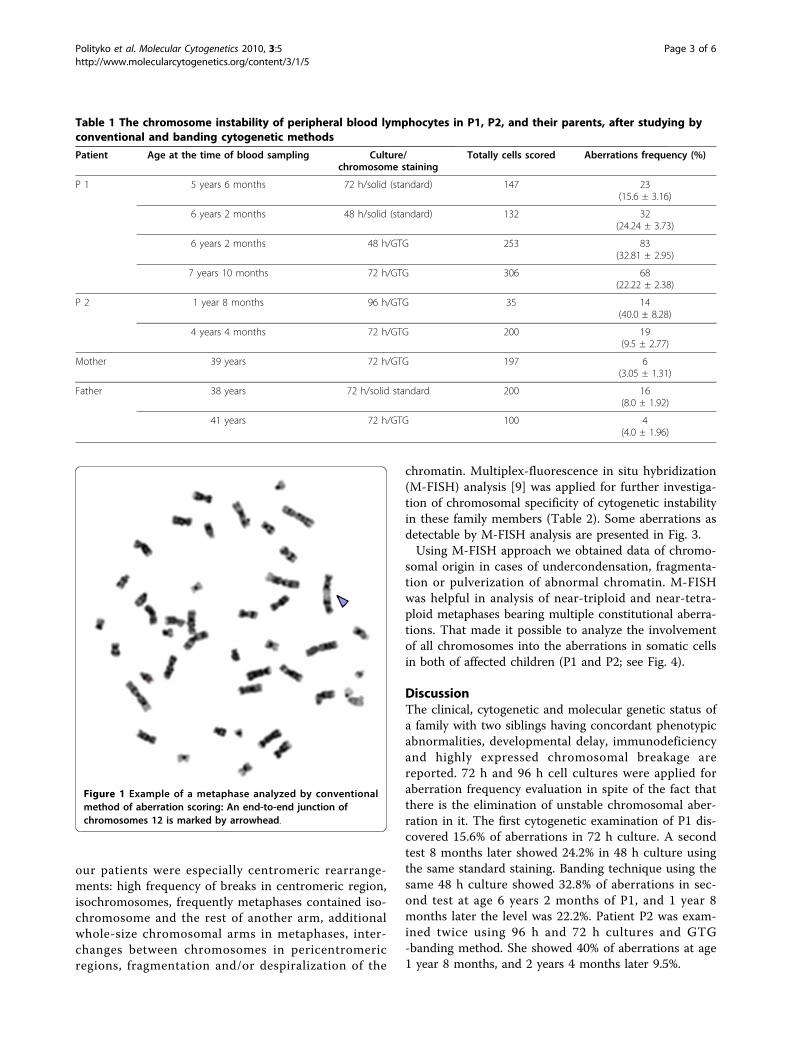
Table 1 The chromosome instability of peripheral blood lymphocytes in P1, P2, and their parents, after studying byconventional and banding cytogenetic methods
Patient Age at the time of blood sampling Culture/chromosome staining
Totally cells scored Aberrations frequency (%)
P 1 5 years 6 months 72 h/solid (standard) 147 23(15.6 ± 3.16)
6 years 2 months 48 h/solid (standard) 132 32(24.24 ± 3.73)
6 years 2 months 48 h/GTG 253 83(32.81 ± 2.95)
7 years 10 months 72 h/GTG 306 68(22.22 ± 2.38)
P 2 1 year 8 months 96 h/GTG 35 14(40.0 ± 8.28)
4 years 4 months 72 h/GTG 200 19(9.5 ± 2.77)
Mother 39 years 72 h/GTG 197 6(3.05 ± 1.31)
Father 38 years 72 h/solid standard 200 16(8.0 ± 1.92)
41 years 72 h/GTG 100 4(4.0 ± 1.96)
Figure 1 Example of a metaphase analyzed by conventionalmethod of aberration scoring: An end-to-end junction ofchromosomes 12 is marked by arrowhead.
Polityko et al. Molecular Cytogenetics 2010, 3:5http://www.molecularcytogenetics.org/content/3/1/5
Page 3 of 6
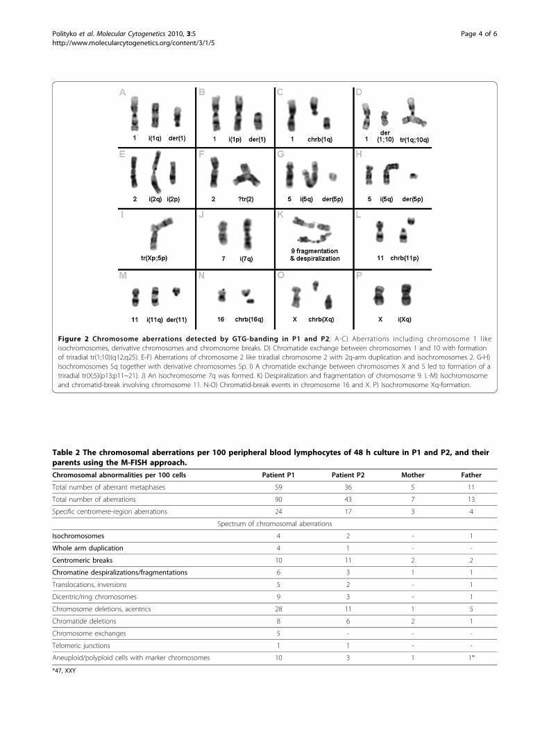
Figure 2 Chromosome aberrations detected by GTG-banding in P1 and P2: A-C) Aberrations including chromosome 1 likeisochromosomes, derivative chromosomes and chromosome breaks. D) Chromatide exchange between chromosomes 1 and 10 with formationof triradial tr(1;10)(q12;q25). E-F) Aberrations of chromosome 2 like triradial chromosome 2 with 2q-arm duplication and isochromosomes 2. G-H)Isochromosomes 5q together with derivative chromosomes 5p. I) A chromatide exchange between chromosomes X and 5 led to formation of atriradial tr(X;5)(p13;p11~21). J) An isochromosome 7q was formed. K) Despiralization and fragmentation of chromosome 9. L-M) Isochromosomeand chromatid-break involving chromosome 11. N-O) Chromatid-break events in chromosome 16 and X. P) Isochromosome Xq-formation.
Table 2 The chromosomal aberrations per 100 peripheral blood lymphocytes of 48 h culture in P1 and P2, and theirparents using the M-FISH approach.
Chromosomal abnormalities per 100 cells Patient P1 Patient P2 Mother Father
Total number of aberrant metaphases 59 36 5 11
Total number of aberrations 90 43 7 13
Specific centromere-region aberrations 24 17 3 4
Spectrum of chromosomal aberrations
Isochromosomes 4 2 - 1
Whole arm duplication 4 1 - -
Centromeric breaks 10 11 2 2
Chromatine despiralizations/fragmentations 6 3 1 1
Translocations, inversions 5 2 - 1
Dicentric/ring chromosomes 9 3 - 1
Chromosome deletions, acentrics 28 11 1 5
Chromatide deletions 8 6 2 1
Chromosome exchanges 5 - - -
Telomeric junctions 1 1 - -
Aneuploid/polyploid cells with marker chromosomes 10 3 1 1*
*47, XXY
Polityko et al. Molecular Cytogenetics 2010, 3:5http://www.molecularcytogenetics.org/content/3/1/5
Page 4 of 6
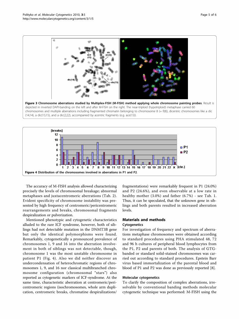
The accuracy of M-FISH analysis allowed characterizingprecisely the levels of chromosomal breakage; abnormalmetaphases and juxtacentromeric aberrations (Tab. 2).Evident specificity of chromosome instability was pre-sented by high frequency of centromeric/pericentromericrearrangements and breaks, chromosomal fragmentsdespiralization or pulverization.Mentioned phenotypic and cytogenetic characteristics
alluded to the rare ICF syndrome, however, both of sib-lings had not detectable mutation in the DNMT3B genebut only the identical polymorphisms were found.Remarkably, cytogenetically a pronounced prevalence ofchromosomes 1, 9 and 16 into the aberration involve-ment in both of siblings was not detectable, though,chromosome 1 was the most unstable chromosome inpatient P1 (Fig. 4). Also we did neither discover anundercondensation of heterochromatic regions of chro-mosomes 1, 9, and 16 nor classical multibranched chro-mosome configuration (chromosomal “stars”) alsoreported as cytogenetic markers of ICF-syndrome. At thesame time, characteristic aberration at centromeric/peri-centromeric regions (isochromosomes, whole arm dupli-cation, centromeric breaks, chromatine despiralizations/
fragmentations) were remarkably frequent in P1 (24.0%)and P2 (24.6%), and even observable at a low rate inhealthy mother (3.0%) and father (6.7%) - see Tab. 1.Thus, it can be speculated, that the unknown gene in sib-lings and both parents resulted in increased aberrationlevels.
Materials and methodsCytogeneticsFor investigation of frequency and spectrum of aberra-tions metaphase chromosomes were obtained accordingto standard procedures using PHA stimulated 48, 72and 96 h cultures of peripheral blood lymphocytes fromthe P1, P2 and parents of both. The analysis of GTG-banded or standard solid-stained chromosomes was car-ried out according to standard procedures. Epstein Barrvirus based immortalization of the parental blood andblood of P1 and P2 was done as previously reported [8].
Molecular cytogeneticsTo clarify the composition of complex aberrations, irre-solvable by conventional banding methods molecularcytogenetic technique was performed: M-FISH using the
Figure 3 Chromosome aberrations studied by Multiplex-FISH (M-FISH) method applying whole chromosome painting probes. Result isdepicted in inverted DAPI-banding on the left and after M-FISH on the right. The near-triploid (hypotriploid) metaphase carried 60chromosomes and multiple aberrations including fragmented chromatin belonging to chromosome 8 (= f(8)), dicentric chromosomes like a dic(14;14), a dic(15;15), and a dic(2;22), accompanied by acentric fragments (e.g. ace(15)).
Figure 4 Distribution of the chromosomes involved in aberrations in P1 and P2.
Polityko et al. Molecular Cytogenetics 2010, 3:5http://www.molecularcytogenetics.org/content/3/1/5
Page 5 of 6
24 human whole chromosome painting probes was car-ried out for the analysis of chromosomal regionsand chromosomes most frequently involved in aberra-tions. Slide preparation, in situ hybridization and cytoge-netic analysis were performed as previously described[9]. 100 metaphases were analyzed per individual.Two-color FISH using centromeric probes for X- andY-chromosomes (Abbott/Vysis) was done according tostandard procedures for 2-color-FISH experiments.
AcknowledgementsSupported by the DFG 436 WER 17/1/04.
Author details1National Medical Center “Mother and Child”, Orlovska Street 66, 220053Minsk, Republic of Belarus. 2Jena University Hospital, Institute of HumanGenetics and Anthropology, Kollegiengasse 10, D-07743 Jena, Germany.3Institute of Human Genetics, Charité, Humboldt-University, AugustenburgerPlatz 1, 13353 Berlin, Germany. 4Robert Koch Institute, Nordufer 20, 13353Berlin, Germany.
Authors’ contributionsAP, OK, NR, IN and NK performed the cytogenetic studies in the presentcases and collected the data relative to this report. AP and TL did themolecular cytogenetic analysis and interpretations. HT, KS and HN wereinvolved in the EBV-immortalization procedures and sequencing. KS and AWdid analysis and interpretation of the DNMT3B sequence. AP and TL draftedthe paper and all authors contributed to the finalizing of the paper and allauthors read and approved the final manuscript.
Competing interestsThe authors declare that they have no competing interests.
Received: 27 January 2010 Accepted: 8 March 2010Published: 8 March 2010
References1. Hendrich B, Bickmore W: Human diseases with underlying defects in
chromatin structure and modification. Hum Mol Genet 2001,10(20):2233-2242.
2. Hulten M: Selective somatic pairing and fragility at 1q12 in a boy withcommon variable immunodeficiency. Clin Genet 1978, 14(2):294.
3. Hansen RS, Stöger R, Wijmenga C, Stanek AM, Canfield TK, Luo P,Matarazzo MR, D’Esposito M, Feil R, Gimelli G, Weemaes CM, Laird CD,Gartler SM: Escape from gene silencing in ICF syndrome: evidence foradvanced replication time as a major determinant. Hum Mol Genet 2000,9(18):2575-2587.
4. Xu GL, Bestor TH, Bourc’his D, Hsieh CL, Tommerup N, Bugge M, Hulten M,Qu X, Russo JJ, Viegas-Péquignot E: Chromosome instability andimmunodeficiency syndrome caused by mutations in a DNAmethyltransferase gene. Nature 1999, 402(6758):187-191.
5. Jiang YL, Rigolet M, Bourchis D, Nigon F, Bokesoy I, Fryns JP, Hultén M,Jonveaux P, Maraschio P, Mégarbané A, Moncla A, Viegas-Péquignot E:DNMT3B mutations and DNA methylation defect define two types ofICF syndrome. Hum Mutat 2005, 25(1):56-63.
6. Kubota T, Furuumi H, Kamoda T, Iwasaki N, Tobita N, Fujiwara N, Goto Y,Matsui A, Sasaki H, Kajii T: ICF syndrome in a girl with DNAhypomethylation but without detectable DNMT3B mutation. Am J MedGenet A 2004, 129A(3):290-293.
7. Kloeckener-Gruissem B, Betts DR, Zankl A, Berger W, Güngör T: A new anda reclassified ICF patient without mutations in DNMT3B and itsinteracting proteins SUMO-1 and UBC9. Am J Med Genet A 2005,136(1):31-37.
8. Neitzel H: A routine method for the establishment of permanentgrowing lymphoblastoid cell lines. Hum Genet 1986, 73(4):320-326.
9. Kuechler A, Neubauer S, Grabenbauer GG, Claussen U, Liehr T, Sauer R,Wendt TG: Is 24-color FISH detection of in-vitro radiation-induced
chromosomal aberrations suited to determine individual intrinsicradiosensitivity? Strahlenther Onkol 2002, 178(4):209-215.
doi:10.1186/1755-8166-3-5Cite this article as: Polityko et al.: Two siblings with immunodeficiency,facial abnormalities and chromosomal instability without mutation inDNMT3B gene but liability towards malignancy; a new chromatindisorder delineation? Molecular Cytogenetics 2010 3:5.
Submit your next manuscript to BioMed Centraland take full advantage of:
• Convenient online submission
• Thorough peer review
• No space constraints or color figure charges
• Immediate publication on acceptance
• Inclusion in PubMed, CAS, Scopus and Google Scholar
• Research which is freely available for redistribution
Submit your manuscript at www.biomedcentral.com/submit
Polityko et al. Molecular Cytogenetics 2010, 3:5http://www.molecularcytogenetics.org/content/3/1/5
Page 6 of 6