Embed Size (px)
Citation preview

ISS
N 0
249-
6399
ISR
N IN
RIA
/RR
--52
02--
FR
+E
NG
ap por t de r ech er ch e
Thème BIO
INSTITUT NATIONAL DE RECHERCHE EN INFORMATIQUE ET EN AUTOMATIQUE
Variational, Geometric and Statistical Methods forModeling Brain Anatomy and Function
Olivier Faugeras, Geoffray Adde, Guillaume Charpiat, Christophe Chefd’Hotel,Maureen Clerc, Thomas Deneux, Rachid Deriche, Gerardo Hermosillo,
Renaud Keriven, Pierre Kornprobst, Jan Kybic, Christophe Lenglet,Lucero Lopez-Perez, Théo Papadopoulo, Jean-Philippe Pons, Florent Segonne,
Bertrand Thirion, David Tschumperlé, Thierry Viéville, Nicolas Wotawa
N° 5202
May 17, 2004


Unité de recherche INRIA Sophia Antipolis2004, route des Lucioles, BP 93, 06902 Sophia Antipolis Cedex (France)
Téléphone : +33 4 92 38 77 77 — Télécopie : +33 4 92 38 77 65
Variational, Geometric and Statistical Methods for ModelingBrain Anatomy and Function
Olivier Faugeras, Geoffray Adde, Guillaume Charpiat, Christophe Chefd’Hotel,Maureen Clerc, Thomas Deneux, Rachid Deriche, Gerardo Hermosillo,
Renaud Keriven, Pierre Kornprobst, Jan Kybic, Christophe Lenglet,Lucero Lopez-Perez, Théo Papadopoulo, Jean-Philippe Pons, Florent Segonne,
Bertrand Thirion, David Tschumperlé, Thierry Viéville, Nicolas Wotawa
Thème BIO — Systèmes biologiquesProjet Odyssée
Rapport de recherche n° 5202 — May 17, 2004 — 22 pages
Abstract: We survey the recent activities of the Odyssée Laboratory in the area of the applicationof mathematics to the design of models for studying brain anatomy and function. We start with theproblem of reconstructing sources in MEG and EEG and discuss the variational approach we havedeveloped for solving these inverse problems. This motivates the need for geometric models of thehead. We present a method for automatically and accurately extracting surface meshes of severaltissues of the head from anatomical MR images. Anatomical connectivity can be extracted fromDiffusion Tensor Magnetic Resonance Images but, in the current state of the technology, it must bepreceded by a robust estimation and regularization stage. We discuss our work based on variationalprinciples and show how the results can be used to track fibers in the white matter as geodesics insome Riemannian space. We then go to the statistical modeling of fMRI signals from the viewpointof their decomposition in a pseudo-deterministic and stochastic part which we then use to performclustering of voxels in a way that is inspired by the theory of Support Vector Machines and in away that is grounded in information theory. Multimodal image matching is discussed next in theframework of image statistics and Partial Differential Equations with an eye on registering fMRI tothe anatomy. The paper ends with a discussion of a new theory of random shapes that may proveuseful in building anatomical and functional atlases.
Key-words: MEG, EEG, fMRI, DT-MRI, Maxwell equations, inverse problems, segmentation,level sets, Riemannian spaces, Partial Differential Equations, Brownian motion, diffusion tensor,
This work has been accepted for publication in Neuroimage.

2 O. Faugeras et al.
Lie groups, Eikonal equation, tractography, mutual information, information theory, Kernel PCA, In-formation Bottleneck, retinotopy, Laplace-Beltrami operator, shape topologies, Hausdorff distance,mean shape, covariance of shapes.
INRIA

Méthodes Variationnelles, Géométriques et Statistiques pour laModélisation de l’Anatomie et de l’Activité Cérébrale
Résumé : Nous présentons les récentes activités du Projet Odyssée dans le domaine de la modéli-sation mathématique de l’anatomie et de l’activité cérébrales. Nous commençons par le problèmede localisation de source en MEG, EEG et discutons l’approche variationnelle que nous avons pro-posée pour résoudre ces deux problèmes. Ces derniers ont motivé le développement de modèlesgéométriques de la tête et nous présentons donc une méthode d’extraction précise et automatique demaillages surfaciques pour les divers tissus visibles en IRM anatomique. La connectivité anatomiqueentre aires corticales peut être estimée à partir des images de tenseur de diffusion. L’estimation ro-buste et la régularisation des images de tenseur sont des étapes fondamentales avant l’application detout algorithme de tractographie. Nous exposons nos travaux à base de méthodes variationnelles etmontrons comment ils peuvent être mis en oeuvre afin de retrouver les fibres axonales considéréescomme les géodésiques d’un espace Riemannien. Nous passons ensuite à la modélisation statistiquedes signaux d’IRM fonctionnelle à travers leur decomposition en une partie pseudo-deterministe etune partie stochastique. Sur la base de cette analyse, nous proposons un algorithme de classifications’inspirant des techniques à base de SVM et de la théorie de l’information. La mise en corres-pondance d’images issues de modalités d’acquisition différentes est ensuite formulée dans un cadrestatistique à base d’équations aux dérivées partielles. Nous illustrons cette technique par le recalaged’IRM fonctionnelles sur des images anatomiques. Nous concluons par une discussion sur une nou-velle theorie des formes aléatoires qui pourrait se révéler très utile dans la cadre de la constructiond’atlas anatomiques et fonctionnels.
Mots-clés : MEG, EEG, IRMf, IRMd, équations de Maxwell, problèmes inverses, segmentation,ensembles de niveaux, espaces Riemanniens, Équations aux Dérivées Partielles, mouvement Brow-nien, tenseur de diffusion, groupes de Lie, équation eikonale, tractographie, information mutuelle,théorie de l’information, ACP à noyau, "Information Bottleneck", rétinotopie, operateur de Laplace-Beltrami, topologies de forme, distance de Hausdorff, forme moyenne, covariance de formes.

4 O. Faugeras et al.
1 Introduction
The Odyssée laboratory is interested in developing a detailed understanding of the neural compu-tations underlying human visual perception. This interest arises from several motivations. One isthe desire to participate in the increase of basic knowledge regarding one of the most sophisticatedsensory modality that supports action and reasoning, another one is the hope that this quest willeventually lead to breakthroughs in the way we interact with computers.
In order to model human visual perception it is necessary to observe humans while they performthe act of seeing. This can be achieved by using such imaging/measurement techniques as MRI,MEG, EEG in order to make qualitative and quantitative measurements of the changes in the stateparameters of some volunteers’ brains. These measurements can then be used to support the designand the test of neural mathematical and computational models of human visual perception. In thisarticle we focus only on the part of the problem that consists in processing the data that are producedby some of the previous imaging modalities.
The theory of Partial Differential Equations (PDE) is central to the source reconstruction problemin MEG and EEG, and to our work with Diffusion Tensor Magnetic Resonance Imaging (DT-MRI);it is also the core of our curve and surface evolution work as applied to segmentation and warping.Differential geometry plays an important role because we are dealing with spaces with a naturalRiemannian structure, e.g. the white matter through the diffusion tensor or the cortical surface.Many of these PDEs derive from energy functionals through the calculus of variation as in our workon multimodal image matching. Last but not least, statistics are crucial to correctly take into accountthe immense variability of the signals and the shapes that arise when one attempts to "look" at thebrain.
2 Source Reconstruction in MEEG
MEG and EEG (commonly called MEEG) record noninvasively the electromagnetic field resultingfrom electrical activity inside the brain. Their high time resolution (of the order of 1 ms) make thesetwo modalities very valuable for the functional analysis of the human brain. Their drawbacks are 1)a relatively poor spatial resolution (compared to fMRI) and 2) the need for solving a delicate inverseproblem for localizing the electrical activity inside the brain.
This inverse problem is driven by a forward model, which computes the electromagnetic fieldoutside the head from a known electrical activity profile inside the brain. The quality of the recon-struction greatly depends on the forward problem, whose accuracy must be controlled with greatcare. This problems obeys the rules of electromagnetic propagation under the quasi-static approxi-mation (because at frequencies of interest and at spatial scales smaller than the head, inductive andcapacitive effects can be ignored). In this case, the Maxwell equations relate the potential
�to the
sources ��� for tissues of conductivity � through����� � ��� ����� � ��� (1)
with a vanishing Neumann boundary condition on the scalp. The magnetic field can be computedfrom the potential, e.g. through the Biot-Savart equation.
INRIA
Mathematics for the Brain 5
Of course, a good geometrical and physical model of the head is crucial to solve properly thisequation. Our group relies on two different – mesh based – strategies: the boundary element method(BEM) which describes the head as a set of nested surfaces delimiting domains each with a uniform,isotropic conductivity; the finite (volume) element method (FEM) which can assign a different con-ductivity to each tessel. The meshes describing the head are naturally subject dependent and mustbe computed beforehand from physiological data, via anatomic MRI segmentation. As the headis a very complex object, an accurate geometrical description signifies handling a huge number ofgeometrical elements, on which the solution is discretized, and the resulting computations can onlybe solved via iterative methods (rather than a direct matrix inversion) using leading-edge numericalmethods.
In our work, we use a distributed source model for the inverse problem. This is notoriously ill-posed, due to the existence of “silent sources”. Consequently, some constraints on the solution mustbe added (minimum norm or minimum gradient solutions are often chosen). The inverse problem isthus solved by minimizing an energy term which is the sum of a data term and a regularizing termtaking the constraints into account. The sources are iteratively updated by computing the gradient ofthe energy. The computationally efficient way to compute the energy gradient is to use the adjointproblem [12]. Note that, since the relationship (1) between the source term � � and the potential�
is linear, the matrix which represents the adjoint problem is simply the transpose of the matrixrepresenting the forward problem.
The BEM method takes advantage of the harmonicity of a potential�
satisfying (1) inside eachcompartment of homogeneous conductivity. It uses the representation theorem to reformulate theproblem in terms of single-layer and double-layer potentials defined on the interfaces between con-ductivities. It requires only surface meshes, which are much easier to obtain than volume meshes. Adrawback in terms of numerical complexity is that in the BEM the matrix representing the forwardproblem is dense and requires a lot of memory to be computed and stored. To alleviate this prob-lem, we have introduced the Fast Multipole Method (FMM), an algorithm originally developpedfor N-body gravitational field computations [8]. The FMM is a multi-resolution approach whichapproximates the electromagnetic interaction between surface elements by performing multipoleexpansions at coarse resolutions. It avoids matrix storage, and significantly reduces the computa-tional burden of the matrix-vector products: a matrix-vector product of dimension � is performedin � � ��������� instead of � � � . The computational savings allow finer discretizations to be used,leading to more accurate forward and inverse problem solutions.
Accuracy of the forward problem is also a major concern in BEM, because the electrical sourcesare close to the discontinuities in conductivity, causing the potential to have very sharp variations,which are difficult to discretize. All the BEM methods used so far in MEEG have used the sameintegral formulation [15]. However, this integral formulation, based on a double-layer potentialapproach, is by no means the only one available. A thorough analysis of integral formulationsderiving from the representation theorem has enabled us to propose a new formulation, combiningsingle and double-layer potentials [22]. This approach has three main advantages compared to theprevious BEM: it leads to symmetric matrices, it only couples elements from adjacent surfaces, andits accuracy outperforms all other surface methods [1], especially when the ratio of conductivitiesbetween neighboring layers is high, as occurs inside the head.
RR n° 5202

6 O. Faugeras et al.
For the BEM inverse problem, the electrical sources are constrained to be orthogonal to a knownsurface inside the cortex. In this case, the problem is no longer ill-posed and the intensity field ofthe sources can be recovered, up to a constant, from boundary measurements [2]. The field to bereconstructed is then simply the signed intensity of the source.
Figure 1 shows the reconstruction of electrical sources, using the BEM model, from measure-ments of a somatosensory evoked magnetic field (experimental protocol presented in [27] and dataalso used in [10]).
Figure 1: BEM reconstruction of somatosensory sources from MEG data (CTF Omega 151-channel, Hopitalde la Salpetriere, Paris).
The FEM method represents all the head related quantities (conductivities, potentials, sources)as piecewise linear functions on the elements of the mesh describing the head. Obviously, this re-strictive model has a strong impact on the accuracy of the results compared to the BEM method (inwhich no restriction is made on the solution in the domains delimited by the surfaces). This has,however, several advantages: anisotropy of conductivities can easily be modeled for each volumeelement, the accuracy seems somewhat less sensitive to the closeness of the sources to the con-ductivity discontinuity interfaces, the matrices generated by the method are quite sparse leading tocomputation times that are much smaller than those of the BEM method. It remains that constructingaccurate 3D meshes to model the head is somewhat difficult mainly because the cortex is very thinand thus a lot of triangles are needed to represent it correctly. The sources are restricted to belongto the volume of the gray matter (instead of a surface for the BEM method), but they can again beconstrained in direction by forcing them to be aligned with the (common) normal to the interfacesof the grey matter.
3 Geometric Modeling
3.1 Anatomical MRI segmentation
We have designed a method to automatically and accurately extract surface meshes of several headtissues from anatomical MRI images. The input of our algorithm is a ��� -weighted MRI image andthe approximate intensities of the main head tissues: air, skin, cerebrospinal fluid (CSF), gray matter(GM) and white matter (WM). It robustly generates triangle meshes of the outer skin interface, ofthe brain contour and of the inner and outer interfaces of the cortex. In the future, we plan to extendit to the skull and to some subcortical structures of interest.
INRIA

Mathematics for the Brain 7
Our method guarantees some topological properties of the meshes, such as spherical topology,absence of self- or mutual intersections. These properties are crucial in some applications such ascortex unfolding or source reconstruction in MEG and EEG with the BEM or the FEM.
Our method is a successful combination of hidden Markov random field (HMRF) classification[50] and of active contour segmentation with the topology preserving level set method [17].
The former is a statistical approach to classify voxels into a small number of tissue classeschosen a priori. The tissue distribution is modeled by a Markov random field (MRF) encouragingneighboring voxels to have the same class labels, while the observed intensity of each tissue classis modeled by a Gaussian distribution. The labels of the voxels are estimated with a maximum aposteriori (MAP) criterion. The problem translates into the minimization of an energy function butan exact minimization is computationnally unfeasible due to the huge number of unknowns. Asa consequence, a greedy strategy yielding a suboptimal solution, such as the iterated conditionalmodes (ICM) algorithm, is preferred. The parameters of the model are the mean and the variance ofeach tissue class and a bias field accounting for the inhomogeneties in the RF field. In our method,this bias is taken as affine against intensities and smooth and non-parametric over space. An initialestimate of the tissue parameters is provided by the user. Iteratively, class labels are estimated byMAP, then the tissue parameters and the bias field are updated with the expectation-maximization(EM) algorithm. The output is a classification of image voxels, the mean/variance of each tissueclass and a bias-corrected image.
The ability to automatically handle topology changes is a long claimed advantage of the levelset method over explicit deformable models, but is often not desirable in biomedical image segmen-tation, where the topology of the target shape is prescribed by anatomical knowledge. A topologypreserving variant of the level set method has been proposed in [17] to overcome this problem: thelevel set function is evolved with a modified update procedure based on some concepts from dig-ital topology, then the final mesh is obtained with a modified topology-consistent marching cubesalgorithm. This method ensures that the resulting mesh has the same topology than the user-definedinitial level set. We have extended this method in order to evolve several nested level sets whilepreventing mutual intersections. Contrarily to some methods for explicit deformable models basedon repulsion forces, our method guarantees the absence of intersections and is computationnallycheaper than checking mesh-to-mesh intersection.
We apply successively the HMRF classification and active contour segmentation with the topol-ogy preserving level set method to benefit from the advantages of both methods while discardingtheir respective drawbacks. We first run the HMRF classification. This statistical approach is power-ful as regards automatic parameter estimation but it is not sub-voxel accurate and disregards topol-ogy. Hence we feed the resulting output into an active contour segmentation task. We first fit aset of nested topology preserving level sets to the labeled image, in order to alleviate the sensitivityof active contour segmentation to the position of the initial contour. Then we drop the labels andwe evolve the level sets according to the intensities of the bias-corrected image. Since the imageinhomogeneities have been removed, the interfaces between the different tissues can be found easilyand robustly by driving each level set with an adequate intensity threshold. Moreover, the thresholdsthat best separate the intensity distributions of the tissues can be computed from the tissue parameterestimated during the HMRF classification.
RR n° 5202

8 O. Faugeras et al.
Our method is used routinely to obtain the surfaces that are needed in the BEM or FEM sourcelocalization techniques from MEG and EEG. The cortical surface in Figure 1 was extracted with thissegmentation tool. As an illustration of the technique, see Fig. 2.
Figure 2: The skin/air, CSF/GM and GM/WM interfaces of one of the authors’ head.
3.2 Diffusion Tensor Estimation
The basic principles of Diffusion MRI and the formalism of the Diffusion Tensor have been intro-duced in [24, 23, 5]. The key concept is that the random motion of water molecules, referred toas diffusion, reflects the structure of the underlying biological tissues at a microscopic scale, wellbeyond the usual image resolution. This opens the possibility of recovering a detailed geometricdescription of the anatomical connectivity between brain areas. We are attempting to get closer tothis challenging goal.
The estimation of a ����� symmetric positive definite tensor at each voxel from diffusion weighteddata uses the Stejskal-Tanner equation [37]. Many approaches have been derived to estimate the ten-sor. Minimal approaches, [47], are very sensitive to noise and outliers. Outlier-related artifacts canbe combatted by using more measurements and robust estimators as in [26]. However the resultingtensors may not be positive definite. We have proposed in [44] to incorporate such priors as ten-sor positivity and spatial regularity into a variational formulation of the estimation problem on themanifold of positive definite tensors.
3.3 Diffusion Tensor Regularization
We have studied the regularization of noisy diffusion tensor data. There exist two main classesof techniques. Non-spectral methods are based on a direct anisotropic smoothing of the diffusionweighted data [45] or consider each tensor as � scalar coordinates. This method suffers from the factthat the eigenvalues tend to diffuse faster than eigenvectors.Spectral methods process the eigen-elements of the tensors. Eigenvalue smoothing is typically per-formed by a vector-valued anisotropic PDE ([43] and references therein) satisfying the maximumprinciple in order to preserve positiveness. However, these approaches are plagued by the fact thatall vectors are defined up to a change of direction, resulting in a computational explosion [9].
We recently proposed an alternative to the previous spectral techniques, called the fast isospec-tral method [7]. It builds flows acting on a given sub-manifold of the linear space of matrix-valued
INRIA

Mathematics for the Brain 9
functions and preserving some constraints. The constraints (orthogonality, invariance of eigenval-ues. . . ) can be expressed in terms of Lie groups and homogeneous spaces. Results of non-spectralsmoothing and isospectral flow on diffusion tensors estimated in the genu of the corpus callosum areshown in Fig. 3.
Figure 3: Left to right: (a) Raw tensors in the genu of the corpus callosum and regularized fields by (b) anon-spectral method, (c) our isospectral flow. Data courtesy of CEA-SHFJ/Orsay. We thank J.F. Mangin andJ.B. Poline for providing us with the data.
3.4 Fiber Tractography
The main idea most classical algorithms for brain connectivity mapping rely on ([29, 30, 34] andreferences therein) is that water diffusion in many regions of the white matter is highly anisotropicand thus the orientation of the principal eigenvector is that of the predominant axonal direction. Allthese line propagation techniques however fail whenever they enter a region of low anisotropy, theestimate of the curve tangent becoming highly unreliable.
To overcome this problem we have introduced a physically motivated distance function in thewhite matter through stochastic processes and differential geometry. In our approach, the whitematter is seen as a 3-manifold � and fibers become geodesics [25] of this manifold. There is afascinating connection between the diffusion tensor, Brownian motion and the Eikonal equation. If�
is the concentration of water molecules, it can be shown that it satisfies the equation� ���� ����� ��� � � � �
(2)
When�
is the identity,
is the Laplace operator, the diffusion is isotropic. A Brownian motion inEuclidean space (e.g. � � ) is entirely defined by its initial distribution and the conditional probability� of finding a molecule at � at time
�given that it was at ��� at time 0. For an unbounded anisotropic
homogeneous medium, � is the minimal fundamental solution of (2).These notions have their counterparts when moving from the Euclidean space to a Rieman-
nian space � . In particular an isotropic diffusion on � is governed by a differential operator, theLaplace-Beltrami operator, which defines the geometry of � . In effect it is possible to consider ananisotropic diffusion in the Euclidean space (governed by the diffusion tensor
�) as an isotropic dif-
fusion in ��� seen as a Riemannian space with metric tensor � ���������� equal to��� � . It is therefore
natural to consider the intrinsic distance � in the space of the brain white matter to any voxel ��� as
RR n° 5202

10 O. Faugeras et al.
defined by this Riemannian metric. It verifies the intrinsic Eikonal equation
� � � � �� �� � �
� �� � �� ��� ��� � (3)
with� � ��� � � � � . In [25], we propose a level-set formulation and the associated numerical
scheme to solve (3). Computing neural fibers as geodesics in the region of the splenium of thecorpus callosum yields the results presented in Fig. 4. The main advantage of this method over linepropagation techniques is that it is not influenced by locally isotropic areas.
Figure 4: Inferred geodesics in the splenium of the corpus callosum (red: low anisotropy - blue: highanisotropy)
4 Statistical Modeling
4.1 Superresolution in fMRI
In functional Magnetic Resonance Imaging (fMRI) a major goal is to maximize the image spatialresolution. The decrease in SNR induced by the decrease of voxel size can be obviated by the useof higher magnetic fields implying much higher equipment costs, an increased inhomogeneity andhence larger distortion artifacts in the images. To overcome these problems, one possible solutionis to use superresolution techniques which allow us to generate a high resolution volume from aset of low resolution ones. These techniques have already been used in different image processingapplications. For anatomical MR images, superresolution can be used in 2D FT MR imaging inthe image space, i.e. in the slice direction, see [16, 33] and [35] who proposed a superresolutionapproach for 3D volumes.
Our aim is to investigate how these techniques can be applied to process long fMRI sequenceand improve the activation maps [32, 21]. The approach is based on two steps. The first is theacquisition protocol. Images are acquired at a low resolution using alternate shifts of the imageslice stack over half-a-slice thickness, generating two separate slice-shifted overlapping volumes.The second is a variational superresolution reconstruction technique which combines recent workon edge preserving PDEs (see [3] for a review) and convergence rate studies [31]. Experiments
INRIA
Mathematics for the Brain 11
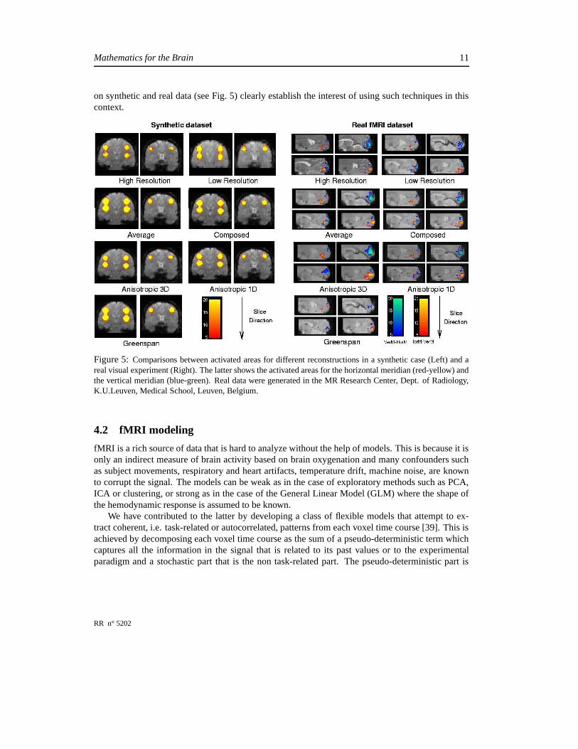
on synthetic and real data (see Fig. 5) clearly establish the interest of using such techniques in thiscontext.
Figure 5: Comparisons between activated areas for different reconstructions in a synthetic case (Left) and areal visual experiment (Right). The latter shows the activated areas for the horizontal meridian (red-yellow) andthe vertical meridian (blue-green). Real data were generated in the MR Research Center, Dept. of Radiology,K.U.Leuven, Medical School, Leuven, Belgium.
4.2 fMRI modeling
fMRI is a rich source of data that is hard to analyze without the help of models. This is because it isonly an indirect measure of brain activity based on brain oxygenation and many confounders suchas subject movements, respiratory and heart artifacts, temperature drift, machine noise, are knownto corrupt the signal. The models can be weak as in the case of exploratory methods such as PCA,ICA or clustering, or strong as in the case of the General Linear Model (GLM) where the shape ofthe hemodynamic response is assumed to be known.
We have contributed to the latter by developing a class of flexible models that attempt to ex-tract coherent, i.e. task-related or autocorrelated, patterns from each voxel time course [39]. This isachieved by decomposing each voxel time course as the sum of a pseudo-deterministic term whichcaptures all the information in the signal that is related to its past values or to the experimentalparadigm and a stochastic part that is the non task-related part. The pseudo-deterministic part is
RR n° 5202
12 O. Faugeras et al.
obtained by applying the Minimum Description Length (MDL) method and can be very efficientlyestimated. If we note ��� � � the fMRI time course at voxel � , this analysis results in the decompo-sition � � � � ��� � � � ��� � � � �� � being the pseudo-deterministic part,
� � the stochastic part. This univariate analysis is followedby a multivariate one that overcomes some of the difficulties encountered in PCA (resp. ICA) (theassumption that structures of interest in the data are uncorrelated in the temporal and (resp. or) spa-tial domain, the fact that the experimental paradigm is not taken into account). This is achieved bydefining a generalized covariance matrix whose eigenvectors represent the spatial modes of coher-ent dynamic activity in the data. is built from a kernel � � that depends upon the scale parameter� . Two voxels � �
and � �are represented by the signals
� �and
� �. The correlation ��� ��� � � � � can be
thought of in terms of shared information between the time courses. � penalizes the correlations thevalues of ��� which are far from 1. To that effect we multiply the usual covariance matrix ��� ��� � � � � by the function ����� � ��� ���
�� � � �� ��
. As described in [39], the number of eigenvectors kept in the
final description can be determined by a variant of the MDL criterion.We have also explored the use of clustering in the analysis of fMRI data. The problem of choos-
ing the number of clusters can be addressed by the Information Bottleneck (IB) approach developedfor vector quantization [41] which deals explicitely with a tradeoff between quantization and datafidelity through an information theoretic formulation. Our approach assumes that ��� � � has beenprojected in the space spanned by � regressors
���:
� � � � ���� � �"! �
� � � � � ��#� � � � �for example by the GLM. The vectors $ � are modelled as Gaussian random variables with knownmean and covariance. The IB method can be formulated as follows. Given the set of voxels � ,the set of interest % (the set of possible values for $ ) and the normal densities � � % � � � � , findthe fuzzy clusters & that maximize compression while retaining most of the information on � � % � � .This leads to the minimization with respect to & of the function ' � � � & (�*) ' � & � % , where ' � � � & is the mutual information between the dataset � and its compressed representation, ' � & � % is themutual information between the compressed representation and the variable of interest, and
)a
positive scalar. The IB method finds its roots in statistical physics and)
plays the role of an inversetemperature: a high value freezes the system into a hard clustering while a small one heats thesystem and ultimately fuses all clusters into a single one. The minimization can be done by an EMalgorithm. The results are sensitive to the choice of
)but this choice can be made in a principled
manner as explained in [40]. We illustrate the method with a synthetic example. We simulate oneslice of fMRI data with + � �-,
� � voxels and 3 foci of 21 pixels. Independent Gaussian noise isadded to all voxels so that the SNR is 0.5 in the activated areas. The simulated paradigm has twoconditions, the simulated time courses and the spatial maps are shown in Fig. 6(a), (b). By keepingonly the effects of interest, we obtain a 2-dimensional feature-space. The estimated feature valuesat each voxel are represented in Fig. 6(c). The IB method yields four clusters. The correspondingpdfs � � !
� & are shown in Fig. 6(d) For comparison, we have applied a C-Means and a fuzzy-C-
INRIA

Mathematics for the Brain 13
50 100 150 200−0.2
0
0.2
0.4
0.6
0.8
1
(a) (b)(c) (d)
Figure 6: a): Simulated time courses for the three foci shown in b). c): Feature values showing onebig cluster and two big ones. d) Pdfs produced by the IB method.
Means algorithm with an initial number of clusters equal to 4 and never obtained the results shownin Fig. 6(d). More statistical approaches for modeling fMRI data can be found in the thesis [38]which is written in English.
4.3 Cortical filtering and applications to retinotopy
Delineating the cortical visual areas is a "calibration" experiment for any study of the human cor-tical visual system. Beyond estimating the areas borders, the retinotopic mapping process providesinformation linking the actual visual field and the cortical surface. Our method derives from pre-vious work [36]. It is fast -the whole retinotopic map being acquired in about 15 mn of functionalimages acquisition-, semi-automatic and shows an excellent intra- and inter-subject, reproducibil-ity, as shown in [48]. The stimuli are a wedge, coding for polar angle maps and a ring, coding foreccentricity maps. The functional analysis scheme is based on a frequency analysis, which allowsa continuous and accurate response phase estimation. The only requirement on the hemodynamicresponse is that it be linear with respect to the stimulation. The main steps are the computationof a statistical map to determine the voxels activated by our periodic stimuli, followed by a phaseestimation, including a voxel-based hemodynamic delay estimation. The phase links the voxel ac-tivity to its prefered stimulus position. The results, values of eccentricity and polar angle at eachsuprathreshold voxel, are then projected on the cortical surface model derived from a high resolutionanatomical image. The model is finally inflated to facilitate the visualization, see Fig. 7. Part of theprocessing for obtaining these results is spent on smoothing the fMRI data. The way this is doneis important. The classical approach, for instance in SPM, is to smooth the whole volume of databy convolving it with a 3D Gaussian kernel. This may have two undesirable effects: mixing voxelsfrom different anatomical tissues, affecting the analysis sensitivity and blending signals across sulci,reducing the spatial discrimination power. We have used instead such interfaces as the one shownin Fig. 2 and replaced the 3D isotropic filtering with a 3D anisotropic filtering in the vicinity of thewhite-matter/gray-matter boundary (diffusion is encouraged in directions parallel to this boundary,discouraged otherwise), another example of the Laplace-Beltrami operator. The difference betweenthe two methods is shown in Fig. 8.
RR n° 5202

14 O. Faugeras et al.
Figure 7: Retinotopic polar angle and eccentricity maps projected on an inflated left hemisphere.
Figure 8: Left: original activation; Middle: 3mm 3D isotropic smoothing, leading to false activationon the opposite bank of the sulcus; Right: 3mm Laplace-Beltrami smoothing.
4.4 Multimodal image matching
One of the problems that is often encountered in the analysis of fMRI data is that of registering it withthe anatomy. This is difficult because, as shown in Fig. 9, there are significant geometric distorsionsbetween the two volumes and the intensities of corresponding areas rarely match (not visible in thisfigure). We discuss these two points next. The geometric distorsion cannot in general be describedby a simple global transformation rigid, i.e. rotation plus translation, or non-rigid, i.e. affine. In orderto model the distorsion one is therefore led to call upon some general deformation flow representedby a largely arbitrary vector field � . The different intensities in corresponding areas preclude theuse of the usual sum of squared differences (SSD) as an error criterion to guide the search forthe deformation flow. One must revert to more sophisticated, i.e. statistical, ways of measuringsimilarities in intensity distribution. Such measures as the cross-correlation, the correlation ratio, orthe mutual information have been successfully used in the literature [46, 4].
Our contribution has been to clearly pose the problem of the estimation of � as that of minimizingan energy functional � � � on a well-defined functional space � . The energy functional is the sumof dissimilarity term � � � and a regularization term � � � . The first term is based upon the idea ofmodeling the two images ' � ��� and ' ��� as samples of two random processes and of estimatingthe joint probability density function (pdf) of the vector
� ' � ��� � ' � � ��� . From this pdf one canthen compute any of the above statistical measures as functions of the field � . The regularization
INRIA

Mathematics for the Brain 15
criterion takes into account the idea that this field cannot vary arbitrarily and therefore enforces someregularity. This is done by choosing � � � to be a function of the first order derivative of � . Thenest step is to precisely define the functional space � belongs to. It turns out that the form of theregularization term is determinant and implies that we work in the Sobolev space � ��� ���� � .After showing that there exist minimizers of � � � in � , we turn the problem of finding them intoone of solving a semilinear abstract initial value problem that can be written as
� �� �
��� � � � ��� � � � � � ��� � � � � ��� � � � (4)
In this equation, the time has been introduced to reflect the fact that we start from an initial defor-mation flow � � and look for the corresponding stationary solution of (4).
�is a spatial differential
operator arising from the Euler-Lagrange equation of the regularization term � � � . The function�
in the right-hand side arises from the dissimilarity term � � � . Because � � � involves the pdf of thevector
� ' � ��� � ' � � ��� �� is non local, i.e. its value at�
depends upon the values of the currentdeformation flow � � � at other points. This implies that (4) is not a PDE but a functional equation,in effect an ordinary differential equation (ODE) in the unknown � that lives in the functional space� . This makes the analysis of the registration problem significantly more difficult than in the caseof the SSD criterion where (4) is a PDE.
Within this general framework, we have a) shown that there exist minimizers of � � � in � , b)computed the function
�for the previous statistical criteria c) proved the existence and uniqueness
of a solution of the problem (4) using the theory of analytical semigroups of operators and d) provedthat the limit when
�����of the solution of (4) satisfies the Euler-Lagrange equations of � � � . The
implementation of the method has been done in the context of a recent thesis [18]. The theoreticalresults are in [13], with preliminary work reported in [19]. An example of the kind of results that canbe achieved is shown in Fig. 9. The code is used routinely by Prof. Guy Orban’s group in Leuven intheir work on monkey fMRI [14].
Figure 9: Left: The geometric distortion between anatomical MR (purple) and functional MR data(green). Right: The geometric distortion shown left has been mostly compensated for.
RR n° 5202
16 O. Faugeras et al.
4.5 Statistical shapes
The variability of anatomical structures from individual to individual is quite large within the humanspecies. It is even larger when one attempts to compare anatomy and function in different but closelyrelated species, e.g. human and non-human primates. This variability hints at two main areas ofmathematics, topology for defining meaningful shape metrics, and statistics for defining meaningfulnotions of shape variability, e.g. with respect to an average shape. The Odyssée laboratory has beenactively pursuing both objectives. In the area of shape metrics we have been studying a set of shapes�
which are defined as subsets � of a set of � � (in practice � ���or 3) with a regular (i.e.
� )boundary
� � whose curvature is upper-bounded by a positive number����� � . � � is also a lower-bound
on the pinch distance of this boundary and is lower than the distance between two pixels of the gridon which the shapes are defined. The question of measuring the similarity between two shapes in�
builds upon the seminal work of [11] who have introduced new families of sets, complete metrictopologies and compactness theorems. We prove in [6] that three of the most important metrics,including the Hausdorff distance, are topologically equivalent in
�. We also propose to use them
to define a way to continuously deform, i.e. warp, a shape onto another one. The way to pose thisproblem is to define a "dissimilarity" measure � � % � � % between two shapes % � and % , to showthat the gradient
� � � % � � % can be defined in a reasonable manner, and to solve the followinginitial value problem
��%� �� � � � � % � % � % � � %�� � � � (5)
Note the similarity between (5) and (4). One difficulty with this approach is that the metric appearsin the definition of � . Therefore the gradient
� � is not well-defined because the metric is notdifferentiable. We have therefore constructed classes of smooth (i.e. whose gradient is well-defined)approximations of the metric based upon the idea of replacing the � ��� and ����� operators that arisein the definition of the distance function to a set and in the Hausdorff distance between two sets byaverages taken on the boundaries of the shapes. We prove that these approximations can approximatethe metric abitrarily well and compute the gradient of the corresponding dissimilarity measures.This defines a warping algorithm between two shapes that can be seen as an infinitesimal gradientdescent in order to minimize � . We prove that there exist minimizers of � � % � % . This approachcan be seen as the opposite of that consisting in first building a Riemannian structure on the setof shapes, i.e. going from an infinitesimal metric structure to a global one. This is mostly dealtwith in [28, 42, 49, 20]. The problem with these approaches, beside that of having to deal withparametrizations of the shapes, a difficult problem that is avoided in ours, is that there exist globalmetric structures on the set of shapes (like those we have considered) which are useful and relevantto the problem of the comparison of shapes but that do not arise from an infinitesimal structure.
Equation (5) defines a generic "shape warper" that can be used to address the second objectiveabove, i.e. the definition of the empirical mean and covariance of a set of shape examples. Theempirical mean of + shapes % � ,. . . , %�� is defined as any shape �% that achieves a local minimum ofthe function ��� � � ��� defined by
% � � � % � % ��� � � � � % � ��
+�
� � ����������� � � � % � % � �
INRIA

Mathematics for the Brain 17
and we prove that there exists at least one mean. An algorithm for the effective computation of amean is proposed in [6] and an example of the mean of eight silhouettes of corpus callosum is shownin Fig. 10 (left). The empirical covariance of + shapes is slightly more difficult to define. From a
Figure 10: Left: The mean of eight silhouettes of corpus callosum (middle, thick line). Right: Fromtop to bottom, the first three principal modes of variation for the eight sample shapes. They are thesolutions of equation (6) for � ��� � � � � .
mean �% we compute the gradients� � � �% � % � ��� � � � � � � � + . These are functions defined on �%
which we use to define a positive symmetric + � + matrix which supports our notion of empiricalcovariance. Its eigenvectors and eigenvalues are used to define the analog of the principal modes��� � � � � � � � � � + of variation of the mean shape �% . The variability of the mean shape with respectto the � th mode is obtained by solving the following initial value problem
� %� ���� ��� � % � � �% � � � (6)
As an example, the first mode of variation for the above eight sample shapes is shown in Fig. 10(right).
5 Conclusion
We have shown that some large pieces of fairly sophisticated mathematics are very useful for mod-eling the types of signals that are currently used for observing the brain "in vivo". We are presentlyexploring two fascinating areas. One is the combination of these modalities (MEEG, MRI) into amore robust and more accurate meta-sensor. Another one is the introduction of models of the activityof the assemblies of neurons that the sensory modalities are trying to measure in the processing ofthe signals they deliver.
6 Acknowledgements
The authors are grateful to S. Meunier of the Physiology and Physiopathology of Human MotricityLab., La Salpétrière Hospital, INSERM, Paris for permission to use the somatosensory MEG data.
RR n° 5202

18 O. Faugeras et al.
They thank L. Garnero, S. Baillet and B. Renault from the Cognitive Neuroscience and Brain Imag-ing Lab., CNRS, Paris for providing helpful suggestions and software tools for accessing the MEGdata. They also thank Profs. P. Van Hecke and G. Orban from the MR Research Center, Dept. ofRadiology, K.U. Leuven, Medical School, Leuven, Belgium for providing them with MR data.
References
[1] G. Adde, M. Clerc, O. Faugeras, R. Keriven, J. Kybic, and T. Papadopoulo. Symmetric BEMformulation for the M/EEG forward problem. In C. Taylor and J. A. Noble, editors, InformationProcessing in Medical Imaging, volume 2732 of LNCS, pages 524–535. Springer, July 2003.
[2] A. Amir. Uniqueness of the generators of brain evoked potential maps. IEEE Transactions onBiomedical Engineering, 41(1), January 1994.
[3] G. Aubert and P. Kornprobst. Mathematical Problems in Image Processing: Partial Differen-tial Equations and the Calculus of Variations, volume 147 of Applied Mathematical Sciences.Springer-Verlag, January 2002.
[4] N. Ayache. Epidaure: a research project in medical image analysis, simulation and robotics atINRIA. IEEE Trans. on Medical Imaging, 22(10):1185–1201, October 2003.
[5] P.J. Basser, J. Mattiello, and D. LeBihan. Estimation of the effective self-diffusion tensor fromthe NMR spin echo. Journal of Magnetic Resonance, B(103):247–254, 1994.
[6] G. Charpiat, O. Faugeras, and R. Keriven. Approximations of shape metrics and applicationto shape warping and empirical shape statistics. Foundations of Computational Mathematics,2004. Accepted for publication.
[7] C. Chefd’hotel, D. Tschumperlé, R. Deriche, and O. Faugeras. Regularizing flows for con-strained matrix-valued images. Journal of Mathematical Imaging and Vision, 20(1-2):147–162,2004.
[8] Maureen Clerc, Renaud Keriven, Olivier Faugeras, Jan Kybic, and Theo Papadopoulo. The fastmultipole method for the direct E/MEG problem. In Proceedings of ISBI, Washington, D.C.,July 2002. IEEE, NIH.
[9] O. Coulon, D.C. Alexander, and S.R. Arridge. A regularization scheme for diffusion tensormagnetic resonance images. In XVIIth International Conferenceon Information Processing inMedical Imaging, 2001.
[10] O. David and L. Garnero. Time-coherent expansion of MEG/EEG cortical sources. NeuroIm-age, 17:1277–89, 2002.
[11] M.C. Delfour and J.-P. Zolésio. Shapes and geometries. Advances in Design and Control.Siam, 2001.
INRIA

Mathematics for the Brain 19
[12] O. Faugeras, F. Clément, R. Deriche, R. Keriven, T. Papadopoulo, J. Roberts, T. Viéville,F. Devernay, J. Gomes, G. Hermosillo, P. Kornprobst, and D. Lingrand. The inverse EEG andMEG problems: The adjoint space approach I: The continuous case. Technical Report 3673,INRIA, May 1999.
[13] O. Faugeras and G. Hermosillo. Well-posedness of two non-rigid multimodal image registra-tion methods. Siam Journal of Applied Mathematics, 2004. To appear.
[14] Denis Fize, Wim Vanduffel, Koen Nelissen, Katrien Denys, Christophe Chef d’Hotel, OlivierFaugeras, and Guy A. Orban. The retinotopic organization of primate dorsal V4 and surround-ing areas: A functional magnetic resonance imaging study in awake monkeys. Journal ofNeuroscience, 23:7395–7406, 2003.
[15] D. B. Geselowitz. On bioelectric potentials in an homogeneous volume conductor. BiophysicsJournal, 7:1–11, 1967.
[16] H. Greenspan, G. Oz, N. Kiryati, and S. Peled. MRI inter-slice reconstruction using super-resolution. Magn. Res. Imag., 20:437–446, 2002.
[17] X. Han, C. Xu, and J.L. Prince. A topology preserving level set method for geomet-ric deformable models. IEEE Transactions on Pattern Analysis and Machine Intelligence,25(6):755–768, June 2003.
[18] Gerardo Hermosillo. Variational Methods for Multimodal Image Matching. PhD the-sis, INRIA, The document is accessible at ftp://ftp-sop.inria.fr /robotvis/html/Papers /her-mosillo:02.ps.gz, 2002.
[19] Gerardo Hermosillo, Christophe Chefd’hotel, and Olivier Faugeras. Variational methods formultimodal image matching. The International Journal of Computer Vision, 50(3):329–343,November 2002.
[20] E. Klassen, A. Srivastava, W. Mio, and S.H. Joshi. Analysis of planar shapes using geodesicpaths on shape spaces. IEEE Transactions on Pattern Analysis and Machine Intelligence,26(3):372–383, 2004.
[21] P. Kornprobst, R. Peeters, M. Nikolova, R. Deriche, M. Ng, and P. Van Hecke. A superresolu-tion framework for fmri sequences and its impact on resulting activation maps. In Medical Im-age Computing and Computer-Assisted Intervention-MICCAI2003, volume 2 of Lecture Notesin Computer Science, pages 117–125. Springer-Verlag, 2003.
[22] Jan Kybic, Maureen Clerc, Toufic Abboud, Olivier Faugeras, Renaud Keriven, and Théo Pa-padopoulo. Integral formulations for the eeg problem. Technical Report 4735, INRIA, Febru-ary 2003.
[23] D LeBihan and E. Breton. Imagerie de diffusion in vivo par résonnance magnétique nucléaire.CR Académie des Sciences, (301):1109–1112, 1985.
RR n° 5202

20 O. Faugeras et al.
[24] D. LeBihan, E. Breton, D. Lallemand, P. Grenier, E. Cabanis, and M. Laval-Jeantet. MRimaging of intravoxel incoherent motions: Application to diffusion and perfusion in neurologicdisorders. Radiology, pages 401–407, 1986.
[25] C. Lenglet, R. Deriche, and O. Faugeras. Inferring white matter geometry from diffusion tensorMRI: Application to connectivity mapping. In T. Pajdla and J. Matas, editors, Proceedingsof the 8th European Conference on Computer Vision, Prague, Czech Republic, May 2004.Springer–Verlag.
[26] J.F. Mangin, C. Poupon, C. Clark, and I. Le Bihan, D.and Bloch. Distortion correction androbust tensor estimation for mr diffusion imaging. Med Image Anal, 6(3):191–8, September2002.
[27] S. Meunier, L. Garnero, A. Ducorps, L. Mazières, S. Lehéricy, S. Tezenas Du Montcel, B. Re-nault, and M. Vidailhet. Human brain mapping in dystonia reveals both endophenotypic traitsand adaptative reorganization. Annals of Neurology, 50:521–527, 2001.
[28] M. Miller and L. Younes. Group actions, homeomorphisms, and matching : A general frame-work. International Journal of Computer Vision, 41(1/2):61–84, 2001.
[29] S. Mori, B.J. Crain, V.P. Chacko, and P.C.M. Van Zijl. Three-dimensional tracking of axonalprojections in the brain by magnetic resonance imaging. Annals of Neurology, 45(2):265–269,February 1999.
[30] M.E. Moseley, Y. Cohen, J. Kucharczyk, J. Mintorovitch, H.S. Asgari, M.F. Wendland, J. Tsu-ruda, and D. Norman. Diffusion-weighted mr imaging of anisotropic water diffusion in catcentral nervous system. Radiology, 176:439–445, 1999.
[31] M. Nikolova and M. Ng. Fast image reconstruction algorithms combining half-quadratic reg-ularization and preconditioning. In Proceedings of the International Conference on ImageProcessing, volume 1, pages 277–280. IEEE Signal Processing Society, 2001.
[32] R.R. Peeters, P. Kornprobst, M. Nikolova, S. Sunaert, T. Vieville, G. Malandain, R. Deriche,O. Faugeras, M. Ng, and P. Van Hecke. The use of superresolution techniques to reduce slicethickness in functional MRI. International Journal of Imaging Systems and Technology (IJIST),Special issue on High Resolution Image Reconstruction, 2004. To appear.
[33] S. Peled and Y. Yeshurun. Superresolution in MRI : Application to human white matter fibertract visualization by diffusion tensor imaging. Magn. Reson. Med., 45:29–35, 2001.
[34] C. Pierpaoli, P. Jezzard, P.J. Basser, A. Barnett, and G. Di Chiro. Diffusion tensor mr imagingof human brain. Radiology, 201:637–648, 1996.
[35] E. Roullot, A. Herment, I. Bloch, M. Nikolova, and E. Mousseaux. Regularized reconstruc-tion of 3D high-resolution magnetic resonance images from acquisitions of anisotropicallydegraded resolutions. In Proceedings of the International Conference on Image Processing,volume 1, pages 350–353. IEEE Signal Processing Society, 2000.
INRIA

Mathematics for the Brain 21
[36] M.I. Sereno, A.M. Dale, J.B. Reppas, K.K. Kwong, J.W. Belliveau, T.J. Brady, B.R. Rosen,and R.B.H. Tootell. Borders of multiple visual areas in human revealed by functional magneticresonance imaging. Science, pages 889–893, 1995.
[37] E.O. Stejskal and J.E. Tanner. Spin diffusion measurements: spin echoes in the presence of atime-dependent field gradient. Journal of Chemical Physics, 42:288–292, 1965.
[38] Bertrand Thirion. Analyse de données d’ IRM fonctionnelle: statistiques, information et dy-namique. PhD thesis, École Doctorale Télécom Paris, October 2003.
[39] Bertrand Thirion and Olivier Faugeras. Dynamical components analysis of fMRI data throughkernel PCA. NeuroImage, 20(1):34–49, 2003.
[40] Bertrand Thirion and Olivier Faugeras. Feature detection in fMRI data: The information bot-tleneck approach. In MICCAI 2003, volume 2878 of LNCS, pages 83–91. Springer-Verlag,November 2003.
[41] Naftali Tishby, Fernando C. Pereira, and William Bialek. The Information Bottleneck method.In Proc. of the 37-th Annual Allerton Conference on Communication, Control and Computing,pages 368–377, 1999.
[42] Alain Trouvé. Diffeomorphisms groups and pattern matching in image analysis. InternationalJournal of Computer Vision, 28(3):213–21, 1998.
[43] D. Tschumperlé and R. Deriche. Diffusion tensor regularization with constraints preservation.In IEEE Computer Society Conference on Computer Vision and Pattern Recognition, KauaiMarriott, Hawaii, December 2001.
[44] D. Tschumperlé and R. Deriche. Variational frameworks for DT-MRI estimation, regularizationand visualization. In Proceedings of the 9th International Conference on Computer Vision,Nice, France, 2003. IEEE Computer Society, IEEE Computer Society Press.
[45] B. Vemuri, Y. Chen, M. Rao, T. McGraw, T. Mareci, and Z. Wang. Fiber tract mapping fromdiffusion tensor mri. In 1st IEEE Workshop on Variational and Level Set Methods in ComputerVision (VLSM’01), July 2001.
[46] Paul Viola and William M. Wells III. Alignement by maximization of mutual information. TheInternational Journal of Computer Vision, 24(2):137–154, 1997.
[47] C.F Westin, S.E Maier, H. Mamata, A. Nabavi, F.A. Jolesz, and R. Kikinis. Processing andvisualization for diffusion tensor MRI. In In proceedings of Medical Image Analysis, volume6(2), pages 93–108, 2002.
[48] N. Wotawa, B. Thirion, E. Castet, J-L. Anton, and O. Faugeras. Efficient human retinotopicmapping using fMRI. In Tomas Paus, Ed Bullmore, and Jonathan D. Cohen, editors, NeuroIm-age (HBM’03), New York, USA, 2003. Academic Press.
RR n° 5202

22 O. Faugeras et al.
[49] L. Younes. Invariance, déformations et reconnaissance de formes. Mathématiques et Applica-tions. Springer-Verlag, 2003.
[50] Y. Zhang, M. Brady, and S. Smith. Segmentation of brain MR images through a hidden markovrandom field model and the expectation-maximization algorithm. IEEE Transactions on Med-ical Imaging, 20(1), January 2001.
INRIA

Unité de recherche INRIA Sophia Antipolis2004, route des Lucioles - BP 93 - 06902 Sophia Antipolis Cedex (France)
Unité de recherche INRIA Futurs : Parc Club Orsay Université - ZAC des Vignes4, rue Jacques Monod - 91893 ORSAY Cedex (France)
Unité de recherche INRIA Lorraine : LORIA, Technopôle de Nancy-Brabois - Campus scientifique615, rue du Jardin Botanique - BP 101 - 54602 Villers-lès-Nancy Cedex (France)
Unité de recherche INRIA Rennes : IRISA, Campus universitaire de Beaulieu - 35042 Rennes Cedex (France)Unité de recherche INRIA Rhône-Alpes : 655, avenue de l’Europe - 38334 Montbonnot Saint-Ismier (France)
Unité de recherche INRIA Rocquencourt : Domaine de Voluceau - Rocquencourt - BP 105 - 78153 Le Chesnay Cedex (France)
ÉditeurINRIA - Domaine de Voluceau - Rocquencourt, BP 105 - 78153 Le Chesnay Cedex (France)
http://www.inria.frISSN 0249-6399