Embed Size (px)
Citation preview


Terms:
Myoma Leiomyoma
Fibroid
The commonest benign conditions of the uterus

Incidence• True incidence--- uncertain
• Common in women between 20~50y
• Clinically evident in 20%~30% of the women over 30 years old.
an exceedingly frequent event

Etiology
1.Related to hormones ( estrogen and progesterone)
2.Elevated ER expression in myoma
3.Abnormal cytogenetics
Arise during the period of menstrual activity, shrink after menopause

Classification
Subserosal (20%)
Intramural( 60~70%)
Submucosal (10~15%)
Corpus ( 90%) Cervix ( 10%)
Location
Growth pattern
Multiple (>=2)

Pathology- grossly examination
Pseudo capsule Margins : blunt, non-infiltrating, pushing
Cut surfaceWhorled,spiral patterns of fibers

Microscopic features
Elongated smooth muscle cells and fibrous tissue. No nuclear atypia, mitotic figures are absent or sparse.

Degenerations
Hyaline degeneration : commonest
Cystic degeneration
Red degeneration
Degeneration with calcification
Sarcomatous degeneration ( 0.4~0.8%)Malignant
Benign
Cause: gradually inadequacy of blood supply

Hyaline degeneration
Cause: inadequacy of the blood supply
Uniform, eosinophilic, ground-glass appearance
Cystic degenration: secondly to hyaline degeneration

Red degeneration
Frequent during pregnancy or puerperium
A deep pink or red, softer
The ghosts of the muscle cells and their nuclear remain

Sarcomatous change
1.Margin not well defined, blurred, merging, irregular2. Loss of whorled pattern3. Yellow, tan, or gray color
4. Heterogeneity5. Softer, less rubbery6. Absence of a bulging surface

Symptoms and physical signs
40~50% asymptomatic, discovered incidentally after routine examination

Menorrhagia
Menostaxis
Irregular mense
Intramural myoma
Anemia
Shortness of breathPalpitationsWeakness
Submucosal myoma
Change of mense

Pelvic mass and physical signs
Depend on the size, location, number and degeneration type
•Asymmetric enlargement of uterus
•Consistency Firm or rubbery Hard or stony ( calcification) Soft ( cystic)

Pelvic mass and Physical signs
•A firm mass extruded from the cervical OS (submucosal)
•Distortion and elongation of the cervical canal (cervical )

Compressive symptoms
NephrohydrosisHydroureter NephrohydrosisHydroureter
Frequency and retention of urine
Frequency and retention of urine
ConstipationDiscomfortConstipationDiscomfort
Different location of the myoma
Ureteralobstruction
Urethral obstruction
Recto-sigmoid compression
Cervical or lower segment
Cervical or broad ligment
Posterior


Increasing of discharge
Intramural myoma—increased uterus cavity area
Submucosal myoma— purulent discharge ( infection)
lower abdominal discomfort

PainRed degeneration
Torsion of pendunculated myoma
Extrusion of submucosal myoma from the cervix

Myoma and infertility
• infrequent primary cause of infertility
• 27% of women who received myomectomy had a history of infertility
• Usually caused by submucosal and intramural myoma

Myoma and pregnancy•Pregnancy loss , abortion
•Increased cesarean section ( Obstruction of labor) Question: Can myoma be removed during cesarean section?
•Postpartum hemorrhage
•Red degeneration
•Growth of myoma
•Most patients have uncomplicated pregnancies and deliveries.

Diagnostic methods1. History
2. Physical signs
3. Ultrasound/ MRI
4. Cervical cytology
5. Dilation &Curretage
4,5 : To rule out cervical cancer and endometrial cancer

6. Hysterosalpingography
7. Hysteroscopy
8. Laparoscopy
9. Other lab tests ( HCG, Hb)

Differential diagnosis
1. Pregnant uterus
2. Ovarian tumor
3. Uterine adenomyosis
4. Malignant uterine neoplasms

Pregnant uterus VS. Myoma Pregnant uterus
Myoma
History Amenorrhoea Regular period, menorrhagia
Signs Symmetric enlarged uterus
Asymmetric enlarged uterus
Ultra-sound
Sac or fetus in cavity
Low-echoed mass
Lab. test HCG + HCG -

Ovarian tumor VS. MyomaSolid ovarian tumor VS. Subserous leiomyoma
Ovarian cyst VS. Cystic /hyaline degenerative myoma

Adenomyosis VS. Myoma
adenomyosis leiomyoma

Endometrial cancer / Cervical Cancer VS. Submucous myoma
Endometrial cancer Cervical cancer Submucous myoma

Management

principleFactors should be taken into consideration
• Age• Desire of childbearing • Symptoms• Location, size and number • Malignant change

observation• Observation with close follow-up Indications: small and asymptomatic myoma
especially for peri-menopausal women

Medications
Indications:
Size <= 2 months pregnant uterusMild symptomsPeri-menopausal With contraindications for operation

Gonadotropin-releasing hormone agonist (GnRH-a)
Mechanism: Inhibit FSH, LH and Estrogen
Efficacy : 40~60% decrease in uterine volumeSide effects: hypoestrogenism reversible bone loss and hot flashes obvious for long use (>6 months) estrogen add-back therapy Regrowth : within a few months after stopping therapy.

Indications of GnRH-a
1. Preservation of fertility before attempting conception
2. Treatment of anemia to allow recovery of Hb before surgery, minimizing the need for blood transfusion
3. Preoperative treatment of large leiomyomas to make surgery more feasible.
4. Treatment of women in menopausal period
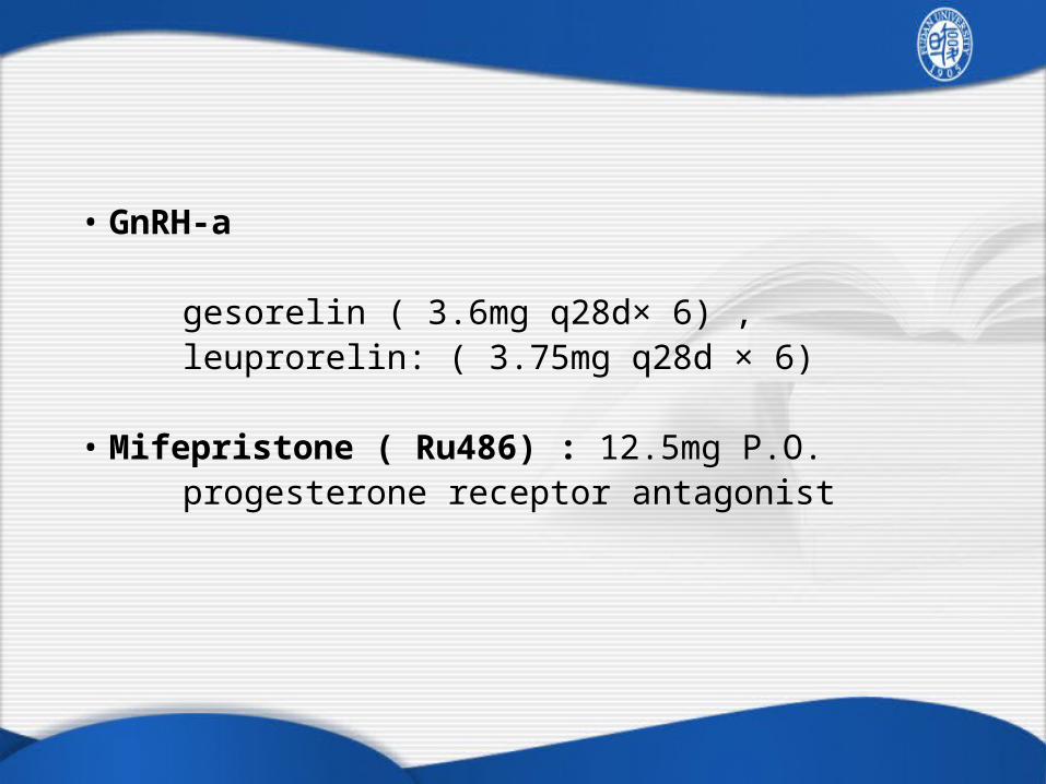
• GnRH-a gesorelin ( 3.6mg q28d× 6) , leuprorelin: ( 3.75mg q28d × 6)
• Mifepristone ( Ru486) : 12.5mg P.O. progesterone receptor antagonist

Indications:
1. Menorrhagia with anemia, resistant to medication
2. Markedly enlarged uterus with compression symptoms
3. Chronic pain, dyspareunia, Acute pain, as in torsion of a pedunculated myoma, or
prolapsing submucosal fibroid
4. Rapid enlargement of uterus-sarcomatous change?
5. Infertility or spontaneous abortion with myoma as the only abnormal finding
Surgery

Surgical procedures
Myomectomy
Hysterectomy
Abdominal / laparoscopic / hysteroscopic or vaginal

• Myomectomy Indications: young patients who desire for childbearing Recurrence risk: as high as 50%, and up to 1/3 requiring repeat surgery

Hysterectomy Indications: no requirement of uterine preservation Note: : Cervical or endometrial cancer must be excluded before
operation

Other treatments:
Uterine artery embolization, UAE
Endometrium ablation by hysteroscopy

Video 1: Laparoscopic myomectomy
Advantages : Minimizes incision, quicker recovery
Disadvantages: Risks of convertion to a laparotomy Immature suture technique: uterine rupture during pregnancy
Video 2: Laparoscopic hysterectomy

Uterine sarcoma

General informationRare tumors of mesodermal origin (myometrium, connective tissue, stroma of endometrium, or secondly to myoma)
2~4% of uterine malignancies
Poor prognosis ( death occurring within 1 to 2 years after diagnosis, except ESS)

Leiomyosarcoma (~45%)
Endometrial stromal sarcoma (ESS) Undifferentiated endometrial sarcoma (15~25%)
Mixed epithelial and mesenchymal tumors Adenosarcoma Carcinosarcoma , or malignant mesodermal mixed tumor, MMMT
Three commonest types

leiomyosarcoma• Age: 45-55 yr, • Usually arise de novo from uterine smooth muscle,
rarely arise in a preexisting myoma • Diagnosis usually is not made before surgery. D&C
are diagnostic only for ~10% of tumors that are submucous.
• Poor prognosis

leiomyosarcoma
mitotic figures>5/10HPF severe cytologic atypia coagulative tumor necrosis

Endometrial stromal sarcoma • Before 2003, low grade ESS, low grade (低度恶性子宫内膜间质肉瘤) Most ESS involve endometrium, infiltrate muscles, sometimes protrude from the OS.
D&C lead to diagnosis (about half).
The only uterine sarcoma related to hormone, ER, PR (+), response to hormone treatment
Behaviour : indolent, late recurrence and metastasis may occur. 5-yr survival >80%

ESS, low grade
ESS with invasive borderOriginated from endometrial stromal cells, similar to proliferative phase

• Undifferentiated endometrial sarcoma ( UES) UES: behave aggressively, with 5-year survival < 40%
UES with severe atypiaMitosis>10/10HPF

Mixed epithelial and mesenchymal tumors
• Adenosarcoma :
Benign epithelial element Malignant mesenchymal element

• CarcinosarcomaMalignant mesodermal mixed tumor, MMMT
Both epithelial and mesenchymal elements are malignant
In FIGO 2009, carcinosarcoma was regarded as type II endometrial carcinoma, because the prognosis is mainly determined by epithelial elements.

• most patients being postmenopausal
• Enlarged or irregular uterus Tumor protrudes through the cervical OS like a polyp (50%)
• Behaviour: aggressive Recurrence rate: 53% 5 year survival 11~35%
Carcinosarcoma

Patterns of spread
• Directly spread (to myometrium, pelvic structures) • pelvic vessels• lymphatics

Symptoms and signs• Uterine Bleeding ( 75%~95%)
• Pelvic pain (33%)
• Pelvic mass
Enlarged uterus ( 15%~50%)
Prolapsed necrotic tissue through cervical OS
• Other :
Compressive symptoms
Discharge

DiagnosisSymptoms ( Uterine bleeding ) and signs
Ultrasound / MRI D & C
Pathological diagnosis

Staging
New staging systems ( FIGO 2009)
Three different staging systems for 1. Leiomyosarcoma 2. ESS and adenosarcoma 3. Carcinosarcoma

Staging FIGO 2009 leiomyosarcoma
• I Tumor limited to uterus IA<5CM
IB ≥ 5CM• II Tumor grows outside of uterus but not outside the pelvis IIA tumor is growing into adnexa IIB tumor is growing to the tissue of pelvis other than adnexa
• III tumor grows into tissue of abdomen ( not just intruding into abdomen) IIIA in one place IIIB in 2 or more places IIIC tumor has spread to pelvic/ para-aortic lymph nodes
• IV The tumorr has spread to the urinary bladder or the rectum, and/or to distant organs, such as the bones or lung
IVA spread to bladder or the rectum IVB distant metastasis

Staging FIGO 2009 ESS and adenosarcoma
• I Tumor limited to uterus IA limited to endometrium
IB <1/2 myometrium IC ≥ 1/2 myometrium
• II Tumor grows outside of uterus but not outside the pelvis IIA tumor is growing into adnexa IIB tumor is growing to the tissue of pelvis other than adnexa
• III tumor grows into tissue of abdomen ( not just intruding into abdomen) IIIA in one place IIIB in 2 or more places IIIC tumor has spread to pelvic/ parpaotic lymphnodes
• IV The tumor has spread to the urinary bladder or the rectum, and/or to distant organs, such as the bones or lung
IVA spread to bladder or the rectum IVB distant metastasis

Carcinosarcoma
the same as FIGO Staging for endometrial cancer

Treatment 1. Surgery: only treatment of proven curative value
Stage I and II : hysterectomy + bilateral oorphorectomy
Pelvic and or para-aortic lymphnectomy: ESS/UES and Carcinosarcoma: required Leiomyosarcoma: not certain

• cytoreductive surgery for advanced stage ( III or IV) patients
2. Adjunvant therapy: Chemotherapy +/- radiotherapy
Radiotherapy improves tumor control in the pelvis without influencing final outcome
chemotherapy : response rate (~20%) Drugs: doxorubicin, cisplatin, ifosfamide, palitaxel

3. Hormone therapy ( only used in ESS, low grade)
progesterone, letrozol GnRH antagonist

Prognosis
Generally poor, 5-year survival 20%~30% Stage is the most important prognostic factor.
Cell type, grade, metastasis, and treatment
• If the leiomyosarcoma arises in a benign myoma, the prognosis is improved
• ESS: 5-yr survival >80%.

Case discussion
History : A 33 year old woman complains heavy bleeding during period for 1 year. The duration of bleeding usually lasts 9 days. Sometimes she has blotting.
Physical examination : shows pale and short of breath. Pelvic examination revealed enlarged uterus with a size of two-month pregnancy.

Case discussion
Ultrasound: A 65/55/50 mm low-echoes mass with clear margin in myometrium was seen by ultrasound. In addition, a 23/20/19mm low echoes mass protrudes from uterus cavity.
Lab test: Hb: 80g/L.

Questions
•What ‘s the diagnosis ? ( give the evidence)
•Which diseases should be excluded?
•What is the suitable treatment?
•Does this treatment affect fertility?

Take home message
About the myoma
•The symptoms are related to the types of location and degenerations. Half of the patients are asymptomatic. The commonest symptom is change of mense.
•Ultrasound is the common and accurate diagnostic tool.

Take home message
About the myoma
•No treatment is required for asymptomatic patients. Medications are suitable for peri-menopausal patients with mild symptoms.
•Surgery is the effective way to treat symptomatic patients or suspicious for sarcomatous change.

Take home message
About the sarcoma
•Rare tumors with poor prognosis
•The commonest symptom is irregular vaginal bleeding with pain. Diagnosis is by pathology results.
•Surgical treatment is the main option. Adjunvant therapy depends on stage and type.