Embed Size (px)
Citation preview

~ 174 ~
Journal of Medicinal Plants Studies 2016; 4(4): 174-188
ISSN 2320-3862 JMPS 2016; 4(4): 174-188 © 2016 JMPS Received: 23-05-2016 Accepted: 24-06-2016
Dr. Abboud Y El Kichaoui
Department of Biology &
Biotechnology, Islamic
University, Gaza, Palestine.
Dr. Basem M Ayesh
Department of Medical
Technology, Al-Aqsa University,
Gaza, Palestine.
Dr. Saeb Aliwaini
Department of Biology &
Biotechnology, Islamic
University, Gaza, Palestine.
Correspondence
Dr. Abboud Y El Kichaoui
Department of Biology &
Biotechnology, Islamic
University, Gaza, Palestine
The use of some plant extracts as an alternative
approaches for treatment of certain malignant
cell lines
Dr. Abboud Y El Kichaoui, Dr. Basem M Ayesh and Dr. Saeb Aliwaini
Abstract In Palestine like in other countries of the world, cancer is one of the most serious health problems that
affect the duration and quality of the individuals’ life. Enormous efforts are invested to cope with this
problem, but unfortunately, limited success has ever been achieved with most of the therapeutic
strategies. These efforts are usually complicated with the need for well experienced surgeons, lack of
specificity and high cost, as well as being usually accompanied with a wide range of side effects.
As the conventional therapeutic strategies fail to fulfill the major requirements for a successful cancer
therapy, the use of naturally developed anticancer agents has evolved as an alternative safe, low-cost and
convenient one. Therefore, the use of plant extracts with potential anticancer therapeutic effects might be
particularly significant, especially in Palestine, which is rich in thousands of plant species known for
their medical uses.
The current study, investigates the effect of crude water extracts from Bottle gourd (Lagenaria siceraria),
Fig (Ficus carica) and Nettle (Urtica pilulifera) on cell lines derived from different human tissue origins
(Hep3b: Hepatocellular carcinoma; Hela: cervical epithelial cancer; and PC-3: prostate cancer).
The results showed a concentration-dependent reduction in the final number of cancer cells in
consequence to treatment with the aforementioned crude extracts. Two kinds of anticancer effects were
evaluated and found to contribute to this reduction: the anti-proliferation effect (decreased number of
metabolically active cells) and cytotoxicity (decreased number of live cells). The three plants examined
possess both of the effects with various degrees. Urtica pilulifera possess the strongest and most
profound effects on the three cell lines, mainly by induction of cell death. On the other hand, Lagenaria
siceraria probably affects the three cell lines by a combination of cytotoxicity and antiproliferation
almost to a similar degree. Ficus carica most probably reduces the final number of metabolically active
cells mainly by its antiproliferative effect.
Both Ficus carica and Lagenaria siceraria are edible plants that were chosen on the bases of being
mentioned in the holy Quran. Therefore, although their effect is lower than that of Urtica pilulifera their
amount in the diet or as a treatment can be safely scaled up when ingested in their native form. On the
other hand, despite its possible toxicity Urtica pilulifera is frequently orally used as a medication in
many conditions by traditional medicine.
Further studies are needed to assess the active ingredients of Ficus carica, Lagenaria siceraria and
Urtica pilulifera, involved in the antiproliferative or cytotoxic effects of these plants.
Keywords: Plant extracts, cell line, hepatocellular carcinoma, cervical epithelial, prostate, cancer
1. Introduction Cancer is one of the most serious health problems worldwide, affecting individuals from
different sexes, ages, and races. It is a group of diseases, characterized by uncontrolled cellular
growth with frequent cancer cells invasion to different body parts and spreading to other
organs, a process referred to as Metastasis. Metastasis is the major cause of cancer related
mortality (W. H. O. 2006) [1]. In 2005, cancer was the second leading cause of death among
both men and women and accounted for 13% of the total 58 million deaths worldwide World
Health Organization, 2006. In 2006, about 10.9 million new cancer cases are expected to be
diagnosed worldwide and more than 7.8 million cancer patients may die (W. H. O. 2006) [1].
According to the latest report of cancer registry unit in Gaza strip, 5500 cases have been
reported over the period from January, 1995 to December, 2003. In addition, 1026 cancer
patients died in 2004 in the Palestinian territories with a mortality rate of 28.2 per 100.000.
Palestine Ministry of Health, 2005.
Cancer is a heterogeneous illness, which can originate from many different organs of the
human body.

~ 175 ~
Journal of Medicinal Plants Studies
However, the most frequent cancer types in the world are lung,
prostate, stomach, colorectal, and esophagus in men; and
breast, lung, stomach, colorectal and cervical in women (W. H.
O. 2006) [1].
Prostate cancer is the most frequently diagnosed and the
second leading cause of cancer death among men, with 234460
new cases estimated to occur in USA during 2006, and 27350
American men will die as a result of this disease (American
Cancer Society. 2006) [3]. In Palestine, the mortality rate of
prostate cancer was 1.4 per 100000 during the period from
January 1995 to December 2002 (PMOH. 2005) [2]. Despite
the fact there are several cell types in the prostate, nearly all of
the prostate cancers are adenocarcinoma, originating in the
gland cells (American Cancer Society. 2006) [3].
Liver cancer ranks as the sixth most common type of cancer
worldwide (American Cancer Society. 2006) [3]. According to
the Palestinian ministry of health, liver cancer mortality rate
was 1.6 per 100000 over the period from January 1995 to
December 2003 (PMOH. 2005) [2]. Many different liver related
tumors are identified depending on the type of cells where they
originate, from these types about 83% are hepatocellular
carcinoma (HCC) that begin in the hepatocytes, the main type
of liver cells.
Cervical cancer is the most common cause of cancer death
among women in developing countries and the second most
common cancer in women worldwide (W. H. O. 2006). It is
caused by a change in the epithelial cells, which line the wall
of the cervix, and the most common risk factor for this type of
cancer is the human Papillomavirus (HPV) (W. H. O. 2006).
In the Palestinian territories, like in other countries of the
world, cancer is becoming one of the most serious problems
that affect the population’s welfare and cause death. This
problem is lately gaining dangerous dimensions and needs a
convenient, efficient and safe medical intervention with
minimal side effects and few expenses. In the last decades,
there were great advances in the diagnosis of cancer as well as
in the field of molecular oncology. However, the cure rate of
most cancers remains low. Several strategies have been used to
cure cancer among which the most common are surgery,
chemotherapy, radiotherapy, and immunotherapy. Other
modern approaches such as hormonal and gene therapy were
proposed by researchers to replace conventional cancer
therapy, with variable degrees of success (American Cancer
Society. 2006, Ayesh et al. 2003) [3, 4]. All of these therapies
have undesired side effects, they are usually not available all
the time and they are expensive. For instance, in surgery the
immune system is compromised due to the large amount of
cortisol released subsequent to the surgery, which increase the
probability of cancer relapse (Rebecca, 2004) [5]. Moreover,
the current use of chemotherapy is accompanied with difficult
side effects. It inhibits bone marrow stem cells proliferation
leading to immune suppression (Rebecca, 2004) [5].
Radiotherapy, which is widely used in the world, is also
accompanied by a great deal of side effects. Lymphocytes are
most readily affected by radiation resulting in prolonged T-cell
suppression (Rebecca, 2004) [5]. Other side effects such as,
bone necrosis, lung fibrosis, skin revascularization, ulceration,
nausea, vomiting, and renal damage are also associated with
all types of conventional therapies.
As the conventional cancer therapies failed to completely
fulfill the criteria for a successful cancer therapy, the use of
naturally developed anticancer agents has evolved as an
alternative safe, low-cost and convenient one. Nontoxic
chemoprevention agents from natural resources were proposed
by researchers for this purpose.
Historically, plants with known therapeutic potential have long
been used to cure a wide range of diseases. An example for
these drugs is Morphine, which is a plant product discovered
in 1861 as an analgesic agent. Later, Quinine the active
component of Cinchona bark was isolated in 1820 as an
effective anti-malaria drug ((Rebecca, 2004) [5], Gary and
Bryn, 2003) [6]. Our Arabic tradition is particularly rich in
medical plants that have been used by pioneer Arabic
physicians to establish the basis for modern therapies. These
were also recommended by our Profit Mohammad ( صلى هللا
and the Holly Quran. Nigella, Garlic, Onion and (عليه وسلم
Fenugreek are famous examples for these plants that were
recently proven to have therapeutic effects on several illnesses.
The use of potentially curative plants might be particularly
significant in the Palestinian territories where the plain and
mountains are covered with more than 2600 plant species of
which more than 700 are known for their uses as medicinal
herbs or as botanical pesticides (Said et al 2002).
The main objective of the current research is to investigate the
effect of Bottle gourd (Lagenaria siceraria), Fig. (Ficus
carica) and Nettle (Urtica pilulifera), extracts on Hep3b from
human Hepatocellular carcinoma; Hela: human cervical
epithelial cancer; and PC-3- human prostate cancer cells in
vitro based on Arabic and Islamic traditional medicine. To
achieve our main objective, the research include the following
specific aims which are the determination of the proliferation
activities of each cell line in response to each plant extract
treatment, the determination of percent viability of each cell
line in response to each plant extracts treatment and the
determination of any morphological changes of each cell line
in each time performing viability testing assay in parallel.
2. Methodology
2.1 Plant collection
Three plants were studied in this thesis: Roman nettle (Urtica
pilulifera), Bottle gourd (Lagenaria siceraria) and Edible fig
(Ficus carica). They were collected between March to June. All parts of the plant (roots, leaves, and fruits) were harvested
by drawing the plant stem. The seeds were isolated form the
fruits, then seeds and leaves were washed under tap water and
dried in shadow places for seventh to ten days. The dried plant
parts were grounded by hand and stored in dry and clean
bottles until the time of experiments
2.2 Preparation of the crude plant extracts
Twenty grams of grounded dry parts of the upper-indicated
parts of each plant were soaked in 80 ml distilled water (20%
dry wt. /v). The extraction was carried out by using a reflux
condenser at boiling temperature for 30 min. The condenser
returns the extract vapor to the boiling flask. The extract was
cooled at room temperature and filtered using a Buchner
funnel with 0.4µm cellulose filter paper. Finally, the extract
was sterilized-filtered using vacuum filter with 0.2µm
cellulose filter paper (Sartorius, UK). About 80% of the extract
volume was collected after filtration, and stored in sterilized
bottles at 4 ˚C (Stock extract). The different working dilutions
were prepared in the cell culture media as indicated
(Dzhambazov et al 2002; Lee et al 2004; Sartippour et al
2001; Nair et al 2005 and Yao et al 2002) [11-15, 19].
2.3 Cell culture
The human cervical carcinoma cell line (Hela), hepatocellular
carcinoma cell line (Hep3b) and Prostate cancer cell line PC-3
were obtained from the Hebrew university of Jerusalem. They
were chosen based on their high proliferation rates and

~ 176 ~
Journal of Medicinal Plants Studies
availability. The cell lines were routinely maintained as a
monolayer in Dulbecco's modified Eagle's medium (DMEM)
Containing 10% fetal calf serum (inactivated at 55 °C for 30
min), 25mM HEPES (pH 7.4), penicillin (180 units/ml),
streptomycin (100μg/ml) and amphotericin-B (0.2
µg/ml)(4,109). The cells were grown to confluence in a
humidified incubator with 5% CO2 in polystyrene culture
flasks. They were subcultured by removing the medium and
adding 4-6 ml of 0.05% trypsin-EDTA solution. The cells
were allowed to detach at (37 ˚C) for 5-10 min. About 1/6 of
the trypsinized Hep3b or 1/4 of the other cells was passed
twice a week to new flasks containing fresh medium.
2.4 Experimental design
In the first (preparatory) experiments, the extracts were
prepared from the desired plant parts. The working extract
concentrations were then determined by testing an array of
extract dilutions on one cell type.
The working extract concentrations were tested for each plant
against each of the three cell lines in terms of cellular
proliferation.
The effects of the same extracts working concentration were
further analyzed by viability assay to determine the type of
cellular effect.
Observation of the morphological changes was carried out in
parallel to the viability assays. The data was statistically
analyzed and comparison of the results from different methods
was done and reported.
2.5 Determination of growth characteristics
Cells from Hela, Hep3b and PC-3 cell lines were seeded in 6-well plats, at a density of 100000 cells/well, as indicated
earlier. Three wells from each cell line were trypsinized and
harvested after 24, 48 and 72 hours and the medium was
changed for the cells continuing to grow at each time point.
The harvested cells were counted on hemocytometer and the
average number of three wells was used for the growth curve.
2.6 Determination of plant extract working concentrations
Plant extract-DMEM preparations were prepared by
incorporation of sterile stock extracts into the DMEM, media
preparation (the plant extract volume was included in final
volume calculation). The highest extract-DMEM plant
concentration achieved this way was 16% dry wt. /v (Stock
extract-DMEM), and the desired extract-DMEM
concentrations were prepared by dilution with the proper
volume of complete DMEM medium.
To determine the concentration with a 100% cytotoxic effect,
200000 cells were seeded onto 25cm2 flasks containing
DMEM media for 24 hours. The medium was then replaced by
8ml of 16, 8, 4 or 0.0% plant extract-DMEM. The flasks were
prepared in triplicates, and the cells were incubated in extract
presence for 24 hours. The viability of cells was determined as
described in the following section.
2.7 Cell Proliferation Assay
A commercially purchased colorimetric kit was used to
determine the proliferation activity of cells (Biological
Industries, Biet Haemic, Israel). The method is based on the
ability of metabolically active cells to reduce the tetrazolium
salt XTT to orange-colored compound of formazon. The
formed dye is water-soluble and the dye optical intensity can
be determined at 490 nm. The intensity of the dye is
proportional to the number of metabolically active cells. The
test procedure includes cultivation of cells in 96-well plates,
addition of the XTT reagent and incubation for 2-24 hours,
during which an orange color is formed. The grater the number
of metabolically active cells in the well, the grater the activity
of mitochondria enzymes and the higher the concentration of
the dye formed, which can then be measured and quantified
(Ofek et al 2003) [22].
The Hela and PC-3 cells were seeded in 96-well plats at a
density of 7000 cells/well, whereas the Hep3b cells were
seeded at density 5000 cells/well. The cells were maintained at
(37 ˚C) for 24 hours in the presence of 100 l DMEM. The
medium was replaced with 8, 4, 2, 1, 0.5 or 0.25% plant
extract-DMEM concentration in triplicates. Twenty four hours
later, 50 µl of the XTT reaction solution were added to each
well and the plates were incubated at 37 ˚C for 3 hours. The
absorbance was measured with ELISA reader at a wave length
of 490 nm. The reference absorbance (nonspecific background
reading) was measured at 630nm. Negative control cells were
incubated with no extract in the medium.
The cells proliferative activity was estimated by calculating
the ratio of remaining viable cells in each well in comparison
to the control and expressed as (% of control). Each assay was
repeated for two additional times and the mean and standard
error was calculated for each extract concentration (Chung et
al 2004; Frahm et al 2004; Ohyama et al 2003 and Ofek et al
2003) [19-22].
2.8 Trypan-blue dye exclusion Viability assay
The trypan-blue dye exclusion assay was used to determine the
plant extract- mediated cell death (Konard et al 2000; A so K.
et al 2004; Fimognari et al 2002; Fagundes et al 2002.) [10, 16-
18]. 200000 cells/well were seeded in 6-well plates and grown
as previously described. After 24 hours the medium was
replaced with different plant extract-DMEM concentration (8,
4, 2, 1, 0.5 and 0.25%) in triplicates. After 48 hours incubation
the medium was discarded and the cells were harvested by
trypsinization and washed twice with (PBS). A volume of
0.4% trypan-blue stain that is equal to the residual PBS was
then added. After 5 min incubation, the cells were counted
with a haemocytometer by compound light microscope. The
unstained (viable) cells and the blue-stained (dead) cells were
counted separately.
Negative control cells were incubated with DMEM media
without any extract and treated the same way. Positive control
cells were incubated for 10 min with 0.5mM H2O2 before
being harvested and counted as described.
The percentage cell viability was calculated using the
following equation:
% cell viability = total viable cells (unstained) X 100 / total
cells (stained + unstained)
Each experiment was carried out in triplicate and repeated for
one more time and the average of 6 wells was considered for
each extract concentration.
2.9 Determination of morphological changes of the cells in
culture
The different cell lines normally grown in 6-well plates or
incubated with the desired extract concentration for the
purpose of viability testing were monitored by an inverted
microscope in 24 hours intervals. Any morphological changes
in the cells shape, level of adhesion and any other alterations
were observed and documented by photography before end of
experiment (Dzhambazov et al 2002; Frahm et al 2004; Wang
et al 1999; Cao et al 2005) [11, 20, 23, 24].

~ 177 ~
Journal of Medicinal Plants Studies
2.10 Statistical analysis
All values expressed as mean ± standard error of the mean by
Microsoft Excel. The data were statistically analyzed by SPSS
by performing the correlation, regression and one-way
ANOVA tests.
For each experiment, the data obtained was blotted against
extract concentration and the obtained curve equation and R2
value were calculated by the Microsoft Excel software. The
extract concentration that gives 50% or 100% reduction in the
number of metabolically active cells or in viability of cells
(IC50 and IC100 respectively), was determined by substitution
in the obtained equation (Konard et al 2000; Wang et al 1999) [10, 23].
3. Results
3.1 Determination of the cell lines growth characteristics:
Figure1 shows the growth curve of each of the hepatocellular
carcinoma Hep3B, cervical epithelial Hela and Prostate PC-3
cell lines in normal DMEM culture media. The three cell lines
maintained exponential growth characteristics until the end of
experiments (96 hours). The Hep3b cells grew faster than the
other two cell lines. However, no cell line growth curve
reached a plateau at the end of the experiments (four days). It
should be emphasized that the time of all of the following
experiments did not exceeded this time.
Hepatocellular carcinoma Hep3b, Cervical epithelial Hela and
Prostate PC-3 Cells were seeded at a density of 100000
cells/well. The wells were prepared in triplicates for each time
point and were incubated at 37 ˚C in 5% CO2. Three wells
from each cell line were harvested and counted at each time
point and the rest of wells allowed continuing growing. The
numbers of cells were blotted against the growth duration.
3.2 Determination of the different plant extracts working
concentrations: Plant extracts-media were prepared by
introducing sterile plant extracts into the DMEM culture media
(the plant extract volume was included in final volume
calculation). The plant extract sterilization by a 0.2µl filter was
difficult to perform as the filter was blocked by the fine pieces
of extract. Thus the highest plant concentration achieved
(stock concentration) was 20% (wt/v). The examined extract
concentrations were 0, 4, 8 and 16% for each plant type. The
effect of such concentrations from Urtica pilulifera, Lagenaria
siceraria and Ficus carica on the viability of Hep3b cells is
illustrated in figure 6. Urtica pilulifera concentrations less
than 4% (wt. /v) showed a profound effect on the viability of
cells, while 4% and higher concentrations gave maximal effect
of 100% cell mortality. According to these result the gap
between 0% and 4% is critical and a wider range of
concentrations in this interval are necessary. Lagenaria
siceraria gave weak reduction of Hep3b cells viability and the
testing concentrations did not give more than 70% effect.
Ficus carica had lower but considerable effect on the viability
of Hep3b cells, and the highest concentrations did not reach
the 50% inhibition.
From figure 2 it’s noticed that concentrations lower than 4%
are needed to be introduced into the experiment to assess cell
death before reaching maximal values. Moreover, it seems
futile to examine concentrations higher than 8% as all of the
extract effects reach plateaus at those concentrations.
Therefore the concentrations (0.25, 0.5, 1, 2, 4 and 8%) were
used of each plant extract to be examined in the rest of the
study. Any effect within this range of concentrations would be
amplified theoretically if we increased the concentrations.
~ 178 ~
Journal of Medicinal Plants Studies
Hep3b cells were seeded onto 25cm2 flasks at a density of
200000 cells/flask. The flasks were prepared in triplicates for
each concentration of the different plants. After incubation at
(37 ˚C) for 48h, the cells were harvested and their viability
was determined by the Trypan-blue test. The percent viability
of cells was calculated as stated previously and plotted against
the plants concentrations.
3.3 Effect of plant extracts on cancer cells proliferation
activity in culture
3.3.1. Hepatocellular carcinoma cell line Hep3b
The Human hepatocellular carcinoma cell line Hep3b was
maintained in the presence of increasing concentrations of
each of the three plant extracts as indicated in each
experiment. The following sections describe the effect of each
plant extract on this cell line in terms of proliferation activity
(figure3).
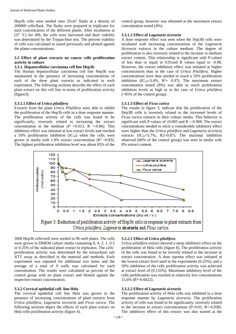
3.3.1.1 Effect of Urtica pilulifera
Extracts from the plant Urtica Pilulifera were able to inhibit
the proliferation of the Hep3b cells in a dose response manner.
The proliferation activity of the cells was found to be
significantly, inversely related to increasing the extract
concentration in the medium (P =0.013, R =-0.86). This
inhibitory effect was initiated at low extract levels and reached
a 50% proliferation inhibition (IC50) when the cells were
grown in media with 1.9% extract concentration (R2 =0.83).
The highest proliferation inhibition level was about 85% of the
control group, however was obtained at the maximum extract
concentration tested (4%).
3.3.1.2 Effect of Lagenaria siceraria
A dose response effect was seen when the Hep3B cells were
incubated with increasing concentrations of the Lagenaria
Siceraria extracts in the culture medium. The degree of
proliferation is also inversely related to the increase in medium
extract content. This relationship is significant with P-values
of less than or equal to 0.01and R values equal to -0.98.
However, the extract inhibitory effect was initiated at higher
concentrations than in the case of Urtica Pilulifera. Higher
concentrations were thus needed to reach a 50% proliferation
inhibition (IC50=3.4%, R2= 0.97). The maximum extract
concentration tested (8%) was able to reach proliferation
inhibition levels as high as in the case of Urtica pilulifera
(>85% of the control group).
3.3.1.3 Effect of Ficus carica
The results in figure 3, indicate that the proliferation of the
Hep3B cells is inversely related to the increased levels of
Ficus carica extracts in their culture media. This behavior is
significant with P-values of ≤0.005 and R =-0.908. The extract
concentrations needed to elicit a considerable inhibitory effect
were higher than the Urtica pilulifera and Lagenaria siceraria
extracts (IC50=5.7%, R2=0.87). The maximal inhibition
observed (60% of the control group) was seen in media with
8% extract content.
5000 Hep3b cells/well were seeded in 96-well plates. The cells
were grown in DMEM culture media containing 8, 4, 2, 1, 0.5
or 0.25% of the indicated plant extract in triplicates. The cells’
proliferation activity was determined by the tetrazolium salt
XTT assay as described in the material and methods. Each
experiment was repeated for additional two times and the
average of a total of 9 wells was calculated for each
concentration. The results were calculated as percent of the
control group with no plant extract and blotted against the
respective extract concentration.
3.3.2 Cervical epithelial cell- line Hela
The cervical epithelial cell line Hela was grown in the
presence of increasing concentrations of plant extracts from
Urtica pilulifera, Lagenaria siceraria and Ficus carica. The
following sections depict the effects of each plant extract on
Hela cells proliferation activity (figure 4).
3.3.2.1 Effect of Urtica pilulifera
Urtica pilulifera extract showed a steep inhibitory effect on the
proliferation of Hela cells (figure 8). The proliferation activity
of the cells was found to be inversly related to the increase in
extract concentration. A dose rsponse effect was initiated at
the lowest extract level used in the experiments (0.25%), and a
50% inhibition of the cells proliferation activity was achevied
at extract level of (0.132%). Maximum inhibitory level of the
cells proliferation was reached at relatively low concentrations
15.6% (R2=0.6022).
3.3.2.2 Effect of Lagenaria siceraria
The proliferation activity of Hela cells was inhibited in a dose
response manner by Lagenaria siceraria. The proliferation
activity of cells was found to be significantly, inversely related
to the increase in extract concentrations (P<0.01, R=-0.938).
The inhibitory effect of this extract was also started at the

~ 179 ~
Journal of Medicinal Plants Studies
lowest extract concentration used (0.25%) however higher
concentrations were needed (3.1%) to reach a 50% inhibition,
than in the case of urtica pliulifera. Higher concentration also
were needed to reach the maximum levels of inhibition than in
the case of urtica pilulifera.
3.3.2.3 Effect of Ficus carica
The results indicate that Ficus carica extract is also capable of
reducing the Hela cells proliferation in a concentration
dependent manner. The proliferation activity is also inversely
related to the increase in extract concentration. This
relationship is significant with P ≤ 0.05 and R= to -0.87.
Although a considerable inhibitory effect was induced by
lower concentrations, IC50 was reached at 3.1% extract
concentration. The effect of Ficus carica is similar to that of
Lagenaria siceraria, but with minor differences.
7000 Hela cells/well were seeded in 96-well plates. The
experiment conditions were as in figure 3.
3.3.3 Prostate cell line PC-3
The prostate cells PC-3 were incubated in the presence of plant
extracts from Urtica pilulifera, Lagenaria siceraria of
different concentrations. The inhibition of proliferation activity
of PC-3 is illustrated in the following sections (figure 5).
3.3.3.1 Effect of Urtica pilulifera Urtica pilulifera extract showed a strong inhibition of cell
proliferation in a dose response manner. The proliferation
activity is inversely related to the increase in extract
concentration. This relationship is significant with P=0.004
and R= -0.916. The 50% proliferation inhibition (IC50) was
achieved at 2.3% extract content in the medium. 4% extract
concentration was able to reduce the proliferation activity to
reach about 10% only.
3.3.3.2 Effect of Lagenaria siceraria
The results indicate that the proliferation activity of PC-3 cells
is inversely related to the increased levels of Lagenaria
siceraria extracts concentration. Although this proliferation
inhibition was weaker than Urtica pilulifera effect, a
significant dose response manner was shown (P=0.00, R=-
0.995). A relatively high extract concentration 7.9% was
necessary to reach 50% proliferation inhibition.
3.3.3.3 Effect of Ficus carica
Ficus carica extract showed a significant inhibition of PC-3
proliferation activity P= 0.001, R= -0.954. The lowest
concentrations of this extract were able to induce inhibitory
effects more than the other extracts; despite of this noticeable
effect, IC50 was 5.5% extract concentration.

~ 180 ~
Journal of Medicinal Plants Studies
4. Effect of plant extracts on cancer cell lines viability in
culture
4.1.1 Hepatocellular carcinoma cell line Hep3B
Hep3b cells viability was determined in the presence of
increasing concentrations of plant extracts from Urtica
pilulifera, Lagenaria siceraria and Ficus carica. Figure 10,
depicts the percent viability of Hep3b cells after incubation for
48 hours with different extracts at different concentrations.
Hep3b cells which were maintained in DMEM without any
plant extract have a percent viability ranging from 92 to 94.5%
according to the three experiments.
4.1.1.1 Effect of Urtica pilulifera
The percent viability of Hep3b cells was found to be
significantly, inversely related to the increase in Urtica
pilulifera extract concentration (P=0.03, R=-0.787). The
lowest concentration 0.25% of this extract was capable to
reduce the percent viability of the cells by slightly less than
30%. The 50% viability reduction was obtained by 2.1% plant
extract concentration. At 4% extract, concentration 100%
reduction in cell viability was already obtained.
4.1.1.2 Effect of Lagenaria siceraria
The percent viability of Hep3b cells was reduced in a
significant, dose response manner due to treatment with
Lagenaria siceraria (P=0.002, R=-0.937). Although there is
no noticeable inhibitory effect at the lowest concentration, the
highest concentration examined (8%) was able to elicit
viability reduction by 60%. In contrast with Urtica pilulifera
behavior Lagenaria siceraria effect is weaker, and IC50 was
obtained at 7.1% extract concentration.
4.1.1.3 Effect of Ficus carica
The results show that increasing the levels of Ficus carica in
the DMEM media is inversely related to the percent viability
of Hep3b cells. This behavior is significant with P= 0.001 and
R=-0.94. Despite of that both Ficus carica and Lagenaria
siceraria behave similarly at most of the concentrations
examined, higher Ficus extract concentration (18.1%) are
estimated to be needed for reaching the 50% reduction in cell
viability, than Lagenaria siceraria extract (6.7%). The
difference between both extracts arrows at high concentration
(8%).
20000 Hep3b cells/well were seeded in 6-well plates. The cells
were grown in DMEM culture media containing 8, 4, 2, 1, 0.5
or 0.25% of the indicated plant extract in triplicates. The
percent viability was determined by the Trypan-blue test as
described in the materials and methods. Each experiment was
repeated for one additional time and the average ± Standard
error of the mean, of a total of 6-wells was calculated for each
concentration. The results were calculated as percent of the
control group with no plant extract and blotted against the
respective extract concentration.
4.1.2. Cervical epithelial cells Hela
The percent viability of Hela cells was determined in the
presence of increasing concentrations of Urtica pilulifera,
Lagenaria siceraria and Ficus carica plant extracts. Figure11
shows the effect of each plant extract on this cell line in terms
of percent viability. In normal conditions without any, extract
treatment, the percent viability ranges from 90 to 95%
according to the results of the three experiments.\
4.1.2.1 Effect of Urtica pilulifera
Urtica pilulifera extract shows a steep and strong reduction of
cell viability in a dose response manner. This effect is
significant with P=0.004, R=-0.911. This effect was noticeable
at 1% extract concentration, whereas to reach 50% viability
reduction, 2.8% extract concentration was required. A
complete elimination of viable cells was obtained at 4%
extract concentration, and maintained at higher concentrations.
4.1.2.2 Effect of Lagenaria siceraria
The results illustrated in figure 7 indicate that Lagenaria
siceraria extract causes a significant viability reduction with
P=0.00 and R=-0.982. This inhibitory effect is weaker than the
effect of Urtica pilulifera with IC50 equals to 14.1% Lagenaria
siceraria extract concentration.
4.1.2.3 Effect of Ficus carica
Ficus carica behaved similar to Lagenaria siceraria extract in
causing a considerable reduction in percent viability of Hela
cells. A dose response effect was observed and significant with
P= 0.003, R=-0.921. The fifty percent inhibition of cell
viability was at 15.7%. No cell viability reduction was
achieved as much as in the case of Urtica pilulifera even with
the highest concentration (8%) of both Lagenaria siceraria
and Ficus carica.

~ 181 ~
Journal of Medicinal Plants Studies
Fig 7: Reduction of Hela cells viability in response to extracts from Urttica a pilulifera, Lagenria siceraria and Ficus caria. The experiments
condition as figure 6
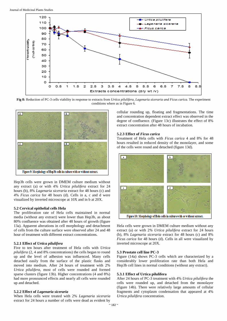
4.1.3 Prostate cell line PC-3
Figure 8 shows the percent viability of PC-3 cells grown in the
presence of increasing concentrations of plant extracts from
Urtica pilulifera, Lagenaria siceraria and Ficus carica. The
following sections describe the effect of each plant extract on
this cell line in terms of percent viability. In normal conditions
without any extract addition, PC-3 cells showed a lower
percent viability than Hep3b and Hela cells. According to the
three different experiments, the percent viability of PC-3 at
normal DMEM was ranging between 85.5 and 90.5%.
4.1.3.1 Effect of Urtica pilulifera
Percent viability of PC-3 affected by Urtica pilulifera extract
was significantly inhibited in a dose response manner (P= 0.00
and R= -0.992). A smooth and a steep relationship was
initiated at the lowest extract concentration. The 50% viability
was obtained by 3.6% extract concentration, while 8% extract
concentration resulted in 100% elimination of any viable cells.
4.1.3.2 Effect of Lagenaria siceraria
The percent cell viability was found to be significantly,
inversely related to the increase in extract concentrations (P=
0.001, R= -0.955). Despite of that this effect is weaker than the
previous extract effect, IC50 of Lagenaria siceraria was
achieved at 9.4% extract concentration.
4.1.3.3 Effect of Ficus carica
A dose response effect was also seen when PC-3 was grown in
increasing concentrations of Ficus carica. Percent viability
was significantly, inversely related to the increasing
concentrations of this extract (P=0.00, R =-0.974). Higher
concentrations were needed to reach a 50% viability inhibition
(IC50 =13.8% extract concentration).
5. Determination of morphological changes of cell lines in
response to treatment of different plant extracts
The morphological characteristics of the Hep3b, Hela and PC-
3 cell lines were evaluated in parallel to each time performing
viability testing assay. In each viability testing experiment the
6-well plates were examined for any morphological changes in
response to treatment with the three plant extracts (Urtica
pilulifera, Lagenaria siceraria and Ficus carica) compared to
the normally maintained cells (no treatment). Representative
wells were pictured and shown in this study (figure 9, 10 and
11). A few cell culture flasks were frequently monitored at
short time intervals.
5.1 Hepatocellular carcinoma cell line Hep3B
The normally maintained Hep3b cells (no extract) were
proliferating with high rate, and formed a monolayer growth
with no less than 90% confluence within 48 hours (figure 9a).
5.1.1 Effect of Urtica pilulifera
Urtica pilulifera showed noticeable morphological alterations
with weaker adhesion level at different concentrations as
evident by the cells floating. After 48 hours, the 2% extract
concentration showed a potent activity. It induced Hep3b cells
to round and form a grape-shaped cluster. At 24 hours after
treatment of hep3b with 4 and 8%, extract most of cells were
detached and aggregated in clusters (figure 12b)
5.1.2 Effect of Lagenaria siceraria
Hep3b cells treated with Lagenaria siceraria 4 and 8%
concentrations for 24 hours suffered from noticeable
alterations such as shrinkage, sparse, and spindle shape
formation (figure12c). At the highest concentration (8%) the
level of adhesion was reduced and many cells were detached.
5.1.3 Effect of Ficus carica
Ficus carica treated cells showed a high proliferation rat at
low extract concentrations (0.25 and 0.5%). While at 4%
extract concentration their growth was inhibited and slight cell
shrinkage was observed (figure 12d). On the other hand 8%
Ficus carica extract concentration induced obvious alterations
to the cells morphology such as, the rounding up and
detachment of many cells.
~ 182 ~
Journal of Medicinal Plants Studies
Fig 8: Reduction of PC-3 cells viability in response to extracts from Urtica pilulifera, Lagenaria siceraria and Ficus carica. The experiment
conditions where as in Figure 6.
Hep3b cells were grown in DMEM culture medium without
any extract (a) or with 4% Urtica pilulifera extract for 24
hours (b), 8% Lagenaria siceraria extract for 48 hours (c) and
4% Ficus carica for 48 hours (d). Cells in a, c and d were
visualized by inverted microscope at 10X and in b at 20X.
5.2 Cervical epithelial cells Hela
The proliferation rate of Hela cells maintained in normal
media (without any extract) were lower than Hep3b, as about
80% confluence was obtained after 48 hours of growth (figure
13a). Apparent alterations in cell morphology and detachment
of cells from the culture surface were observed after 24 and 48
hour of treatment with different extract concentrations.
5.2.1 Effect of Urtica pilulifera
Five to ten hours after treatment of Hela cells with Urtica
pilulifera (2, 4 and 8% concentrations) the cells began to round
up and the level of adhesion was influenced. Many cells
detached easily from the surface of the plastic flasks and
moved into medium. After 24 hours of treatment with 2%
Urtica pilulifera, most of cells were rounded and formed
sparse clusters (figure 13b). Higher concentrations (4 and 8%)
had more pronounced effects and nearly all cells were rounded
up and detached.
5.2.2 Effect of Lagenaria siceraria
When Hela cells were treated with 2% Lagenaria siceraria
extract for 24 hours a number of cells were dead as evident by
cellular rounding up, floating and fragmentations. The time
and concentration dependent extract effect was observed in the
degree of confluence. (Figure 13c) illustrates the effect of 8%
extract concentration after 48 hours of incubation.
5.2.3 Effect of Ficus carica
Treatment of Hela cells with Ficus carica 4 and 8% for 48
hours resulted in reduced density of the monolayer, and some
of the cells were round and detached (figure 13d).
Hela cells were grown in DMEM culture medium without any
extract (a) or with 2% Urtica pilulifera extract for 24 hours
(b), 8% Lagenaria siceraria extract for 48 hours (c) and 8%
Ficus carica for 48 hours (d). Cells in all were visualized by
inverted microscope at 20X.
5.3 Prostate cell line PC-3
Figure (14a) shows PC-3 cells which are characterized by a
considerably lower proliferation rate than both Hela and
Hep3b cell lines in normal conditions (without any extract).
5.3.1 Effect of Urtica pilulifera
After 24 hours of PC-3 treatment with 4% Urtica pilulifera the
cells were rounded up, and detached from the monolayer
(figure 14b). There were relatively large amounts of cellular
fragments and cytoplasm condensation that appeared at 4%
Urtica pilulifera concentration.

~ 183 ~
Journal of Medicinal Plants Studies
5.3.2.1 Effect of Lagenaria siceraria
PC-3 cells treated with 4 and 8% Lagenaria siceraria showed
a noticeable decrease in confluency after 48 hours of
treatment, but no evident morphological changes (figure 14c).
5.3.2.2 Effect of Ficus carica
The reduction of confluence degree was visible in PC-3 cells
when incubated for 48 hours in the highest extract
concentration used, but also with no noticeable morphological
changes (figure 14d).
PC-3 cells were grown in DMEM culture medium without any
extract (a) or with 4% Urtica pilulifera extract for 24 hours
(b), 8% Lagenaria siceraria extract for 48 hours (c) and 8%
Ficus carica for 48 hours (d). Cells in all were visualized by
inverted microscope at 20X.
6. Discussion
Cancer is a term describing conditions characterized by
unscheduled and uncontrolled cellular proliferation (Ponder.
2001). It is a very common disease, and its incidence is
increasing at an average annual rate of 1.2% (Ponder. 2001).
Lately, there has been improvement in the treatment strategies
of cancer, which has resulted in prolonged survival of patients
with chronic cancer disease. However, there is a growing need
for additional means of cancer therapy, in the form of both
palliative and curative treatments. The strategies available
today, are sophisticated, and are only able to affect 50 to 60%
of cancer patients, while the others will eventually die from
their disease (Verweij et al 2003; Talback et al 2003 and
Socialstyrlesn 2005) [27-29 ].
Chemotherapy has been used for cancer treatment for more
than 50 years; sometimes in combination with or parallel to
surgery and radiotherapy. After surgical ablation of
progressive cancer, metastasized tumor cells continue to
progress and this is one of the faultiest associated with surgery.
One the other hand, radioactive rays and most anticancer
chemotherapeutic agents damage DNA or suppress DNA
duplication to kill the rapidly growing tumor cells. At the same
time, they also affect normal cells causing serious adverse
effects, such as bone marrow function inhibition, bone
necrosis, lung fibrosis, skin devascularization, ulceration,
nausea, vomiting, renal damage and alopecia (Cassady et al
1981) [30]. Thus it is evident that a wide array of selective and
potent components is needed to match the growing problems
associated with cancer.
Plants and natural products play an important role in medicine
and provide important prototypes for the development of novel
drugs (Cragg 1998). They offer a valuable source of
compounds with a wide variety of biological activities and
chemical structures. Many anti-cancer agents have been
derived from natural sources; directly as pure native
compounds, or as semi-synthetic analogues (Pezzuto 1997;
Schwartsmann 2000 and Lee 1999) [32-34].
Our Arabic and Islamic tradition is particularly rich in medical
plants that have been used by pioneer Arabic physicians to
establish the basis for modern therapies. But a few of these
plants have been examined scientifically. In this extent we
studied the potential effect of three Arabic and Islamic
traditional plants as anticancer agents. These were Fig (Ficus
carica) and Bottle gourd (Lagenaria siceraria) that were
mentioned in the holy Quran in more than one occasion and
Roman nettle (Urtica pilulifera) which is traditionally used as
a popular medication (Shtayeh et al, 2000) [8]. To fulfill this
aim, the proliferation, viability and morphological
characteristics of three cell lines (Hep3b, Hela and PC-3) were
studied in response to treatment with extracts from the
aforementioned plants.
The cellular proliferation activity was tested by a colorimetric
method which is based on the ability of metabolically active
cells to reduce the tetrazolium salt XTT to orange-colored
compounds of formazon. The intensity of the formed water
soluble dye is proportional to the number of metabolically
active cells. The proliferation activity of the treated cells was
normalized to that of normally growing cells from the same
type with no treatment. This normalized value was expressed
as percent of the control group. Theoretically, any reduction in
the number of metabolically active proliferating cells might
mean that the proliferation pathway itself was halted (cell
cycle arrest), or that a fraction of the cells went through a
death pathway. Therefore, a viability test was necessary to
determine which of these two options may play a role in this
study. Therefore, the trypan-blue dye exclusion test was used
to determine the plant extract-mediated cell death. The
unstained cells (viable) and the blue stained (dead) were
counted separately, and the percent cell viability was
calculated as previously described. The viability assay was
performed with the same cell-extract combinations as in the
proliferation assay. The percent viability of normally grown
cells from the same types was determined for comparison with
extract-treated cells. If we consider the results of the
proliferation and viability assays in parallel, then we would be
able to say whether reduction of the number of the
metabolically active cell has resulted from death of part of
them or due to any other reason. Any conclusion drawn from
such comparison may be confirmed by morphologically
following up the treated cells in comparison with normally
maintained control cells. In this study a combination of the
three different assays was performed in parallel for each plant
extract and each cell line.
In order to assess the degree of proliferation inhibition and
viability reduction obtained by each extract in comparison to
the others, the IC50 values were summarized in tables 3 and 4.
The findings of this study indicated that the proliferation
activity of Hep3b cells was inhibited by all of the three plant
extracts (Urtica pilulifera, Lagenaria siceraria and Ficus
carica). A dose response effect was obtained with all of these
extracts, but the degree of this effect varied from one extract to
the other. The results indicated that Urtica pilulifera is the
most potent extract, even at low concentrations with IC50 of
(1.9%) followed by Lagenaria siceraria with IC50 of (3.4%)
and Ficus carica with of IC50 of (5.7%). At the highest, extract
concentration used in the study (8%) both Urtica pilulifera and
Lagenaria siceraria reached a similar proliferation inhibition
level of about (90%). However, this degree of inhibition was

~ 184 ~
Journal of Medicinal Plants Studies
reached at lower concentration of Urtica pilulifera extract than
of Lagenaria siceraria extract (figure7). Ficus carica on the
other hand did not reach the same degrees of proliferation
inhibition obtained by both Urtica pilulifera and Lagenaria
siceraria even at the highest concentration. When considering
the results of Heb3b cells viability assay, the three plant
extracts showed various degrees of Heb3b cells viability
reduction. Urtica pilulifera showed a steep reduction of the
cells viability with IC50 of 2.1% extract concentration, which
equals the IC50 of the same extract in cell proliferation assay.
These compatible results (Figure 16) suggest that reduction in
the metabolically active Hep3b cells in consequence to Urtica
pilulifera extract treatment is due to cell death. However such
compatibility was not evident in the case of Lagenaria
siceraria (Figure 17) and Ficus carica (Figure 18). Lagenaria
siceraria has a less potent cytotoxic effect against Hep3b cells
(IC50 = 7.1%). This might indicate that this extract counteracts
the proliferation activity of Hep3b cells mostly by mechanisms
other than induction of their death. This deviation between the
cytotoxic effect and proliferation effect was more profound in
the case of Ficus carica with IC50 of 18.1% in case of
cytotoxicity compared to 5.7% in the case of proliferation.
Table 1: Summary of calculated concentrations of the three plant
extracts Urtica pilulifera, Lagenaria siceraria and Ficus carica that
give 50% (IC50) reduction in proliferation activity of Hep3b, Hela and
PC-3 cancer cell lines. The R2 value for each curve equation is
illustrated
Plant Hep3b cells Hela cells PC-3 cells
Urtica pilulifera IC50 1.98 0.132 2.31
R2 0.837 0.6022 0.8563
Lagenaria siceraria IC50 3.4 3.1 7.9
R2 0.97 0.9761 99.82
Ficus carica IC50 5.7 3.1 5.5
R2 0.8675 0.9059 0.9583
Table 2: Summary of calculated concentrations of the three plant
extracts Urtica pilulifera, Lagenaria siceraria and Ficus carica that
give 50% (IC50) reduction in the % viability of Hep3b, Hela and PC-3
cancer cell lines. The R2 value for each curve equation is illustrated.
Plant Hep3b cells Hela cells PC-3 cells
Urtica pilulifera IC50 2.1 2.8 3.6
R2 0.7056 0.8305 0.9843
Lagenaria siceraria IC50 7.16 14.18 9.4
R2 0.8496 0.9585 0.9367
Ficus carica IC50 18.12 15.7 13.8
R2 0.8886 0.852 0.9442

~ 185 ~
Journal of Medicinal Plants Studies
These data were confirmed by the inspection of morphological
alterations of Hep3b cells due to treatment with the three plant
extracts (figure13). As shown in figure 13b Urtica pilulifera
had the most potent effect compared to the control (figure
13a). Most of cells were rounded up and formed a grape
shaped cluster, which is one of the prominent morphological
features of cellular death in culture. Such features were also
noticeable to lesser extent when the cells were incubated with
Lagenaria siceraria, and could hardly be detected in the case
of Ficus carica. Moreover, the confluence of Heb3b cells was
reduced in response to treatment with the three plant extracts
and in accordance to the previously discussed results of
proliferation inhibition.
Similarly Hela cells proliferative activity was inhibited with
various levels due to their treatment with the three plant
extracts. As shown in figure 8, Urtica pilulifera has a strong
inhibitory effect on the proliferation activity in extract
concentration dependent manner with IC50 of 0.13% extract
concentration. Both Ficus carica and Lagenaria siceraria had a
similar but weaker effect on proliferation of Hela cells, and the
IC50 for both extracts was 3.1%. The percent viability of Hela
cells was also reduced in a dose response manner by all of
these plant extracts. Urtica pilulifera extract was the most
effective with IC50 of 2.8%, while the Lagenaria siceraria and
Ficus carica effect on cells viability were similarly less potent
with IC50 of 15.7% for Ficus carica and 14.1% for Lagenaria
siceraria. Being the most potent plant, Urtica pilulifera mostly
exerts its effects by inducing cell death although other effect
can be present (IC50 for proliferation=0.13% and for
cytotoxicity =2.8%). The difference between the IC50 of both
Ficus carica and Lagenaria siceraria in proliferation inhibition
and the IC50 of cell viability reduction indicates that these
extracts exert their effect by cell cycle arrest with low
cytotoxicity. The morphological observations of Hela cells
maintained in increasing concentrations of the three plant
extracts show that Urtica pilulifera had the most rapid and
potent effect of these extracts. Figure 14b shows that most of
the cells were rounded up and formed sparse clusters due to
treatment with 2% Urtica pilulifera. These results suggest that
most of Hela cells were dead in the case of Urtica pilulifera. A
reduction of Hela cells confluence was the most prominent
observation when these cells were incubated with Ficus carica
or Lagenaria siceraria, but cell death indications were almost
not seen. These observations indicate an antiproliferative
effect of both Ficus carica and Lagenaria siceraria but less
likely cytotoxicity.
The proliferation activity of the prostate cell line PC-3 was
inversely related to increasing the levels of the three plant
extracts. The degree of proliferation reduction varied from one
plant extract to the other. As shown in figure 9, Urtica
pilulifera was again the most potent extract with IC50 of 2.3%,
whereas Ficus carica comes second with IC50 of 5.5%, and
Lagenaria siceraria comes last with IC50 of 7.9%. When
comparing these results with results from PC-3 cells viability
assay, we find that Urtica pilulifera also has the strongest
effect with IC50 of 3.6%. These results are supported by the
morphological changes shown in figure15b including cells
rounding up and detachment from the monolayer indicating
that cell death might has occurred.
Lagenaria siceraria induced a reduction of PC-3 cell viability
with IC50 of 9.4% extract concentration similar to IC50 from
the same extract in the proliferation assay. This fact suggests
that this extract also caused PC-3 cell death. The reduction of
the cells confluence due to Lagenaria siceraria treatment
observed in figure 14c gives evidence of the effect of this
extract. A Higher concentration of Ficus carica was needed to
reach a 50% viability inhibition (IC50 =13.8%) than that
needed to reach 50% inhibition in cell proliferation
(IC50=5.5%). This again suggests that the effect of this extract
did not occur only through cell death.
Based on the previous results, Urtica pilulifera have a potent
antiproliferative effect on the three tested cancer cell lines.
While there is no previous studies about the role of Urtica
pilulifera extract as anticancer agent, many studies
investigated the role of its genus member Urtica dioica. Urtica
pilulifera and Urtica dioica are similar in many chemical and

~ 186 ~
Journal of Medicinal Plants Studies
morphological aspects (Shtayeh et al 2000 and Maitai et al
1980) [8, 9]. Our results on Urtica pilulifera are in agreement
with previous studies on Urtica dioica. For example an
aqueous extract of Urtica dioica roots was shown to directly
inhibit the proliferation of Hela cells and block its binding by
epidermal growth factor (EGF) (Wagener 1994) [35]. In the
same study, a polysaccharide mixture from an aqueous root
extract was shown to exert an anti-inflammatory activity in a
rat Paw oedema test. Investigation of the effect of a 20%
methanol extract of Urtica dioica roots on prostate cell line
(LNCap) resulted in a selective and significant concentration-
dependent proliferation reducing effect on prostate cells (89).
In the same study, the cell proliferation activity was
determined by a colorimetric assay and the cytotoxicity was
examined by Trypan-blue test. Our results are also comparable
with data obtained from an in vitro study that aimed to
investigate the effect of Urtica dioica leaves aqueous extract
on the enzyme activity of prostate cancer tissue (Durak et al
2004) [36]. The results of the study indicated that the extract
caused a significant inhibition of adenosine deaminase activity
(ADA) of these tissues in a dose dependent manner. These
data might be of importance because ADA is a key enzyme in
the nucleotide metabolism and DNA turn over. Extracts of
Urtica dioica were also used in the treatment of adult mouse
with bingeing prostate hyperplasia (Lichius and Muth 1997) [37]. Five differently prepared root extracts were tested on these
rats. The 20% methanol extract was the most effective with
51.4% inhibition of induced growth.
Despite all of these data, the mechanism(s) of action of Urtica
dioica extract as an antiproliferative agent has yet to be
established. Different modes of actions are proposed in this
regard. For example, it has been observed that some sterols
and hydroxyl-fatty acids, given their low concentrations in
Urtica dioica, can inhibit aromatase, which is a key enzyme in
steroid hormone metabolism mediating the conversion of
androgens into estrogens (Gansser and Spiteller 1995) [38].
Another mechanism involves a dose dependent inhibition of
the binding of sex hormone binding gluobin (SHBG) to its
receptor in response to Urtica dioica extract (Hryb et al 1995).
Some lignans present in Urtica dioica were shown to interfere
with the binding of androgens to SHBG, thereby reducing the
transport capacity of androgens (Gansser and Spiteller 1995) [38]. Based on the similarity between Urtica pilulifera and
Urtica dioica we would suggest that Urtica pilulifera might
exerts its antiproliferative effect via a similar fashion.
The results of this study strongly indicate a possible cytotoxic
effect of Urtica pilulifera against cancer cell lines. This was
evident from the results of Trypan-blue viability assay as well
as from the inspection of cells morphology in culture. Only
one study was found to deal with this issue in the scientific
debate. In this study, the cytotoxicity of fixed and volatile oils
extracted from Urtica pilulifera leaves and seeds were tested
on Swiss albino mice (Qzbek et al 2004) [40]. The results
indicated that both oils of Urtica pilulifera are completely
nontoxic even at doses reaching 12.8 ml/kg. However, this
study involved only the oil fractions of Urtica pilulifera
extract. Other components of the extract may play a role in the
observed toxicity in our study. Moreover, the aforementioned
study was performed in vivo on mice and the only cytotoxicity
parameter considered was the mice death. No other mice
toxicity indicators were analyzed such as, histopathological
alterations of the different mice tissues and organs.
According to results discussed earlier the inhibitory role of
Ficus carica on the proliferation of the three cell lines was
more profound than its cytotoxic effect on the same cell lines.
This conclusion is in accordance with the results of previous
studies. For instance, the proliferation of different cell lines
was inhibited by components of Ficus carica (Rubnov et al
2001) [41]. Such proliferation inhibition was also obtained
when cow teat papillomatosis skin surface benign tumors,
were treated by Ficus carica latex (Hemmatzadeh; Fatemi and
Amini 2003) [42]. Furthermore, Ficus carica was found to have
an in vivo antioxidant effect after being consumed by human
(Vinson et al 2005) [43]. Accordingly, the dried fruits of this
plant should be eaten more frequently as they are rich in
phenol antioxidants and fibers. Compared with vitamins C and
E, the well-known antioxidants, the dried fruits of Ficus carica
were found to be a superior one.
Lagenaria siceraria was found to possess both
antiproliferative effects as well as a cytotoxic effect to a
considerable degree. Unfortunately no literature was found
regarding the anticancer activities of this plant. However,
members of its family (Cucurbitaceae), were shown to have a
class of biologically active compounds (Cucurbitacins) with
well-established anticancer cytotoxic activity (Rehm et al
1960; Guha and Sen 1975 And Miro1995) [44-46]. For example,
Cucurbitacins were shown to have strong cytotoxic effect on
KB cell line, derived from human nasopharyngeal carcinoma
and on Hela cells by different proposed mechanisms
(Miro1995 and Witkowski; Woynarowska and Konopa 1984) [46, 47]. Moreover, (Cucurbitacins) were shown to have a
proliferative inhibitory activity in vivo against several
carcinoma, sarcoma, and leukemia models (Gitter et al 1961;
Gallily et al 1962 and Reddy et al 1988) [48-50].
The level of these compounds and/or any of their derivatives
in Lagenaria siceraria is unknown. However the results of this
thesis indicate both antiproliferative and cytotoxic activity of
Lagenaria siceraria. Therefore we expect Lagenaria siceraria
extracts to behave in a similar manner like other members of
its family via (Cucurbitacins) involvement.
7. Conclusion
In conclusion, all of the three plants examined in this study
possess varying levels of anticancer activity in vitro. This is
evident by the concentration dependent manner reduction in
the final number of cancer cells as a consequence to treatment.
Two kinds of anticancer effects were examined and found to
take part in this study. The first is anti-cell proliferation effect
(decreased number of metabolically active cells) and the
second is cytotoxicity (decreased number of live cells). The
three plants examined possess both of the effects with various
degrees. Urtica pilulifera possess the strongest and most
profound effects on the three cell lines, probably by
cytotoxicity mainly. On the other hand Lagenaria siceraria
probably affects the three cell lines by combination of
cytotoxicity and anti-proliferation almost to a similar degree.
Ficus carica most probably reduces the final number of
metabolically active cells mainly by its antiproliferative effect,
although cytotoxicity likely contributes to viability reduction
at high concentrations.
Both Ficus carica and Lagenaria siceraria are edible plants
that were chosen on the bases of being mentioned in the holy
Quran. Therefore, although their effect is lower than that of
Urtica pilulifera their amount in the diet or as a treatment can
be safely scaled up when ingested in their native form. On the
other hand, despite its possible toxicity Urtica pilulifera is
frequently orally used as a medication in many conditions by
traditional medicine.

~ 187 ~
Journal of Medicinal Plants Studies
8. References
1. World Health Organization (W.H.O, 2006) Fact sheet,
2006, 297.
2. Ministry of Health. Health Status in Palestine Ministry of
Health Annual Report 2004 Palestine Health Information
Center, 2005. (PMOH, 2005).
3. American Cancer Society. Cancer Facts and Figures.
American Cancer Society Inc, GA, U.S.A, 2006,
4. Ayesh B, Matouk I, Ohana P, Sughayer MA, Birman T et
al., Inhibition of tumor growth by DT A expressed under
the control of IGF2 P3 and P4 promoter sequences
Molecular Therapy 2003; 7(4):535-41.
5. Rebecca CG. -Isolation of Natural Products from Casearia
Nigrescens. Virginia Polytechnic Institute and State
University, Master thesis in chemistry science, 2004, 4-20.
6. Gary S, Bryn D. Bioprospecting for Microbial Endophytes
and Their Natural Products American Society for
Microbiology, 2003, 491-502.
7. Said O, Khalil K, Fulder S, Azaizeh H. Ethnopharma-
cological survey of medicinal herbs in Israel, the Golan
Heights and the West bank region, Journal of
Ethnopharmacology. 2002; 83:251-265.
8. Shtayeh A, Yaniv Z, Mahajna J. Ethnobotanical survey in
the Palestinian area: a classification of the healing
potential of medicinal plants, Journal of
Ethnopharmacology. 2000; 73:221-32.
9. Maitai CK, Talalaj S, Njoroge D, Wamugunda R. -Effect
of extract of hairs from the herb Urtica massaica, on
smooth muscle- Toxicon, Clinical Journal of medicine.
1980; 18(2):225-9.
10. Konard L, Muller H, Lenz C, Laubinger H, Aumuller G.
Antiproliferative effect on Human prostate cancer cells by
Stinging Nettle root (Urtica dioica) extract Planta Midica
2000; 66:44-47.
11. Dzhambazov B, Daskalova S, Monteva A, Popov N. In
vitro screening for antitumor activity of Clinopodium
vulgare L. (Lamiaceae) extract. Biological and
Pharmacological Bulletin 2002; 25(4)499-504.
12. Lee BC, Doo HK, Lee HJ, Jin SY, Jung JH et al., The
inhibitory effects of Aqueous extract of Magnolia
officinalis on Human Mesangial Cell proliferation by
regulation of Platelet-Derived Growth Factor BB
transforming Growth Factor β1 expression, Journal of
pharmacological science. 2004; 94:81-85.
13. Sartippour MR, Liu C, Shao ZH, Go VL, Heber D et al.,
Livistone extract inhibits angiogenesis and cancer growth
Oncology Reports 2001; 8:1355-1357.
14. Nair R, Kalariya T, Chanda S. Antibacterial activity of
some selected Indian medicinal flora, Turkish Journal of
biology. 2005; 29:41-47.
15. Yao XX, Tang YW, Yao DM, Xiu HM. Effects of Yigan
Decoction on proliferation of hepatic stellate cells, World
Journal Gastroenterology. 2002; 15-8(3):511-514.
16. A so K, Kanno SI, Tadano T, Satoh S, Ishikawa M.
Inhibitory effects of Propolis on the Growth of Human
Leukemia U937, Biological and Pharmacological Bulletin
2004; 27(5):727-730.
17. Fimognari C, Nusse M, Cesari R, Iori R, Forti GC et al.
Growth inhibition, cell-cycle arrest and apoptosis in
human T-cell leukemia by the isothiocyanate sulforaphane
Carcinogenesis 2002; 23(4):581-586.
18. Fagundes EM, Queiroz AB, Filho OAM, Gazzinnelli G,
Oliveira RC et al. screening of fractionation of plant
extract with antiproliferative activity on Human peripheral
Blood Mononuclear cells Memories de institute Oswaldo
Cruz 2002; 97(8):1207-1212.
19. Chung TW, Moon SK, Chang YC, Ko JH, Lee YC. Novel
therapeutic effect of caffiec acid and caffiec acid phenyl
ester on hepatocarcinoma cells: complete regression of
hepatoma growth and metastasis by dual mechanism, The
Faseb Journal. 2004; 18:1670-1681.
20. Frahm S, Kurtz A, Kluwe L, Farassti F, Friedrich RE et
al. Sulindac derivatives inhibit cell growth and induce
apoptosis in primary cells from malignant peripheral
nerve sheath tumors of NFI patients Cancer international
2004; 4:4.
21. Ohyama K, Akaike T, Hiriboe C, Yamakawa T.
Cytotoxicity and apoptotic inducibility of Vitex agnus-
castus Fruit extract in cultred Human normal and cancer
cells and effect on growth Biological and Pharmacological
Bulletin 2003; 26(1):10-18.
22. Ofek P, Meir DB, Inbal ZK, Oren M, Lavi S. Cell cycle
regulation and P53 activation by protein phosphatase 2Cα,
Journal of Biological Chemistry. 2003; 278(16):14299-
1305.
23. Wang Y, Blandino G, Givol D. Induced P21waf
expression in H1299 cell line promotes a cell senescence
and protects against cytotoxic effect of radiation and
doxorubicin Carcinogenesis 1999; 18:2643-2649.
24. Cao QZ, Niu G, Tan HR. In vitro growth inhibition of
human colonic tumor cells by verapamil, World Journal
Gastroenterology. 2005; 11(15):2255-2259.
25. Rajkapoor B, Jayakar B, Murugesh N. Antitumor activity
of Indigofera aspalathoides on Ehrlich ascites carcinoma
in mice, Indian Journal of Pharmacology. 2004; 36:38-40.
26. Ponder BAJ. Cancer genetics. Nature, 2001; 411:336-341.
27. Verweij J, De Jong MJA. Achievements and future of
chemotherapy European Journal of cancer. 2003; 36:1479-
1487.
28. Talback M, Stenbeck M, Rosen M, Barlow L, Glimelius
B. Cancer survival in Sweden (1960-1998) developments
across four decades Acta oncology 2003; 42:637-659.
29. Socialstyrlesn 2004 cancer incidence in Sweden 2003.
Statistics- health and disease, 10 online March 2005.
www.sos.se/sos/statisti.htm.
30. Cassady JM, Chang CJ, Mclaughlin JL. Recent advances
in the isolation of structural elucidation of antineoplastic
agents of higher plants. Natural products of a medicinal
agents, 1981; 93-124.
31. Cragg GM. A Success story with valuable lessens for
natural products drug discovery and development.
Medicinal Research Reviews, 1998; 18:315-331.
32. Pezzuto JM. Plant-derived anticancer agents
Biochemichal Pharmacology 1997; 53:121-133.
33. Schwartsmann G. Marine organisms and other novel
natural sources of new cancer drugs Annals of Oncology
2000; 11:235-243.
34. Lee KH. Novel antitumor agents from higher plants
Medicinal Research Review 1999; 19:569-596.
35. Wagener H, Willer F, Samtleben R, Boos G. Search for
the antiprostatic principle of stinging nettle (Urtica dioica)
roots. Phytomedicine 1994; 1:213-24.
36. Durak I, Biri H, Devrim E, Sozen S, Avci A. - Aqueous
extract of Urtica dioica makes significant inhibition on
adenosine deaminase activity in prostate tissue from
patients with prostate cancer Cancer Biology and Therapy
2004; 3(9):855-7.
37. Lichius JJ, Muth C. The inhibiting effects of urtica dioica
root extracts on experimentally induced Prostatic
hyperplasia in the mouse Planta Medica 1997; 63(4):307-

~ 188 ~
Journal of Medicinal Plants Studies
10.
38. Gansser D, Spiteller G. Aromatase from Urtica dioica
roots. Planta Medica 1995; 61:138-40.
39. Hryb DJ, Khan MS, Romas NA, Rosner W. The effects of
extracts of the roots of stinging nettles (Urtica dioica) on
the interaction of SHBG with its receptor on human
Prostatic membranes Planta Medica 1995; 61:31-2.
40. Qzbek H, Qzturk M, Qzturk A, Ceylan E, YenerZ. -
Determination of lethal doses of volatile and fixed oils of
several plants, Eastern Journal of medicine. 2004; 9(1):4-6.
41. Rubnov S, Kashman Y, Rbinowitz R, Schlesinger M,
Mechoulam R. Suppressors of cancer cell proliferation
from fig (Ficus carica) Resin: isolation and structure
elucidation Natural Products 2001; 64(7):993-6.
42. Hemmatzadeh F, Fatemi A, Amini F. Therapeutic effects
of fig tree latex on bovine Papillomatosis Vet Med Infect
Dis Vet public health 2003; 50(10):473-6.
43. Vinson JA, Zubic L, Bose P, Samman N, Proch J. Dried
fruits: excellent in vitro and in vivo antioxidant American
College of Nutrition 2005; 24(1):44-50.
44. Rehm S. Die Bitterstoffe der Cucurbbitaceen Ergebnisse
der biology 1960; 22:108-136.
45. Guha J, Sen P. The Cucrbitacins a review plant
Biochemistry 1975; 2:12-28.
46. Miro M. Cucurbitacins and their pharmacological effects.
Pythotherapy research 1995; 9:159-168.
47. Witkowski A, Woynarowska B, Konopa J. Inhibition of
the biosynthesis of deoxyribonucleic acid, ribonucleic
acid, and protein in Hela S3 cells by Cucrbitacins,
glucocorticoid-like cytotoxic triterpenes Biochemichal
Pharmacology 1984; 33:995-1004.
48. Gitter S, Gallily R, Shohat B, Lavie D. Studies of the
antitumor effect of Cucurbitacins, Cancer research 1961;
21:517-521.
49. Gallily B, Shohat B, Kalish J, Lavie D. Further studies of
the antitumor effects of Cucrbitacins Cancer research
1962; 22:1038-1045.
50. Reddy KS, Amonkar AJ, McCloud TG, Change C,
Cassady JM. Sapinosides A and B. Two cytotoxic
Cucurbitacin glycosides from Desfontainia spinosa
Phytochemistry, 1988; 27:3781-3785.


![DIPLOMARBEIT - univie.ac.atothes.univie.ac.at/19272/1/2012-03-26_0442113.pdf · Cinchona sp. in the treatment of malaria were ... Afterwards this cortex was called China-bark [1]](https://img.dokumen.tips/doc/110x75/5ad001d57f8b9a56098de2ac/diplomarbeit-sp-in-the-treatment-of-malaria-were-afterwards-this-cortex-was.jpg)


![Antimalarial Activity of Stem Bark of Periploca ...downloads.hindawi.com/journals/ecam/2018/4169397.pdf · quinine from cinchona bark [] and artemisinins from ... prior to the beginning](https://img.dokumen.tips/doc/110x75/5af47b917f8b9a190c8d23c9/antimalarial-activity-of-stem-bark-of-periploca-from-cinchona-bark-and-artemisinins.jpg)


