Embed Size (px)
Citation preview

RESEARCH Open Access
Use of gluteus maximus adipomuscular slidingflaps in the reconstruction of sacral defects aftertumor resectionYao Weitao*, Cai Qiqing, Gao Songtao and Wang Jiaqiang
Abstract
Background: While performing sacrectomy from a posterior approach enables the en bloc resection of sacraltumors, it can result in deep posterior peritoneal defects and postoperative complications. We investigated whetherdefect reconstruction with gluteus maximus (GLM) adipomuscular sliding flaps would improve patient outcomes.
Methods: Between February 2007 and February 2012, 48 sacrectomies were performed at He Nan Cancer Hospital,Zhengzhou City, China. We retrospectively examined the medical records of each patient to obtain the followinginformation: demographic characteristics, tumor location and pathology, oncological resection, postoperativedrainage and complications. Based on the date of the operation, patients were assigned to two groups on thebasis of closure type: simple midline closure (group 1) or GLM adipomuscular sliding reconstruction (group 2).
Results: We assessed 21 patients in group 1 and 27 in group 2. They did not differ with regards to gender, age,tumor location, pathology or size, or fixation methods. The mean time to last drainage was significantly longer ingroup 1 compared to group 2 (28.41 days (range 17–43 days) vs. 16.82 days (range 13–21 days, P < 0.05)) and themean amount of fluid drained was higher (2,370 mL (range 2,000–4,000 mL) vs. 1,733 mL (range 1,500–2,800 mL)).The overall wound infection rate (eight (38.10%) vs. four (14.81%), P < 0.05) and dehiscence rate (four (19.05%)] vs.three (11.11%), P < 0.05) were significantly higher in group 1 than in group 2. The rate of wound margin necrosiswas lower in group 1 than in group 2 (two (9.82%) vs. three (11.11%), P < 0.05).
Conclusions: The use of GLM adipomuscular sliding flaps for reconstruction after posterior sacrectomy cansignificantly reduce the risk of infection and improve outcomes.
Keywords: Sacral tumors, Surgery, Gluteus maximus adipomuscular sliding flaps
BackgroundSacral tumors include benign subtypes, such as giant-cell tumors [1] and aneurysmal bone cysts [2], as well asmalignant subtypes, including chordomas, multiple mye-lomas and metastatic tumors [3]. These tumors are oftenasymptomatic or involve vague signs and symptoms [4],such as backache with or without numbness, leg weaknessor bowel and/or bladder dysfunction. Therefore, diagnosisis frequently delayed by several months to up to 6 years[5]. During the interim, the tumors can become very largeand may destroy most segments of the sacrum.
Treatment ranges from intralesional curettage [6] toablation to en bloc excision [7]. Surgical resection canresult in large defects that extend to the rectum ventrallyor to the sacroiliac joints laterally. Defects can disruptthe posterior pelvic wall and often present a reconstruct-ive challenge to surgeons [8]. The musculature over thesacrum can become weakened or of insufficient volumeto fill large defects. Simple midline closures usually fail,which can lead to infection [9], wound breakdown,parasacral herniation or a combination of these compli-cations, which may delay adjuvant treatment [10].Good results have been reported with the use of verti-
cal rectus abdominis myocutaneous flaps [11]. Othermethods have involved meshes [12], omental mobilization[13] and free flaps [14]; however, some of these methods
* Correspondence: [email protected] and soft tumor department, He Nan Cancer Hospital, The AffiliatedHospital of Zheng Zhou University, 127 Dong Ming Road, Zheng Zhou City450000, China
WORLD JOURNAL OF SURGICAL ONCOLOGY
© 2013 Weitao et al.; licensee BioMed Central Ltd. This is an Open Access article distributed under the terms of the CreativeCommons Attribution License (http://creativecommons.org/licenses/by/2.0), which permits unrestricted use, distribution, andreproduction in any medium, provided the original work is properly cited.
Weitao et al. World Journal of Surgical Oncology 2013, 11:110http://www.wjso.com/content/11/1/110
have been associated with additional injuries to patientsand/or dangerous complications, such as necrosis [15].We performed a retrospective study to assess whether re-construction with gluteus maximus (GLM) adipomuscularsliding flaps would improve outcomes after sacral tumorexcision.
MethodsWe assessed patients who had undergone sacrectomybecause of sacral tumors between February 2007 andFebruary 2012. All patients, or guardians when appropri-ate, gave informed consent before surgery. The authorsobtained approval from the ethics committee of our hos-pital before doing the study.During sacrectomy, complete spondylectomies to the
appropriate levels and sectioning of the bilateral piriformmuscles and of the bilateral sacrospinous and sacro-tuberous ligaments were carried out. The sacral nerve
roots (above sacral vertebra S3) of involved segments onboth sides were separated and protected (Figure 1a). Iftotal sacrectomy was performed, complex lumbopelvic re-construction and arthrodesis were undertaken with au-tologous iliac crest bone grafts and instrumentation (forexample, bilateral lumbar vertebrae L4 and L5 pediclescrews and iliac screws). Blood loss was controlled by tem-porary abdominal aorta block during sacrectomy of S1.Based on the date of the operation, from February
2007 to December 2010, patients who underwent simplemidline closure by two-layer suture of the fascia andskin comprised group 1. From January 2011 to January2013, patients were chosen for GLM adipomuscular slid-ing flaps reconstruction if they had undergone resectionof a sacral tumor via an exclusively posterior approach,had no tumor invasion on either side of the GLMmuscle on MRI before surgery (Figure 2a), had adequateblood supply to the GLM flap and had not undergone
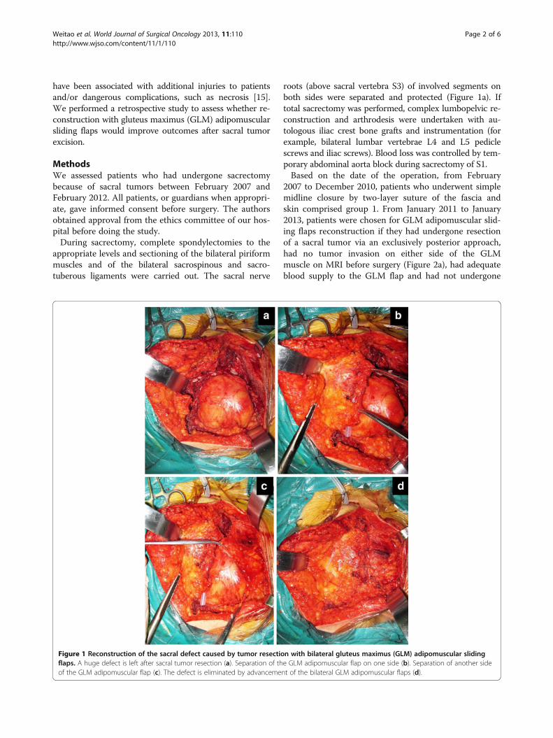
Figure 1 Reconstruction of the sacral defect caused by tumor resection with bilateral gluteus maximus (GLM) adipomuscular slidingflaps. A huge defect is left after sacral tumor resection (a). Separation of the GLM adipomuscular flap on one side (b). Separation of another sideof the GLM adipomuscular flap (c). The defect is eliminated by advancement of the bilateral GLM adipomuscular flaps (d).
Weitao et al. World Journal of Surgical Oncology 2013, 11:110 Page 2 of 6http://www.wjso.com/content/11/1/110

previous sacral radiotherapy. These patients comprisedgroup 2.One or both sides of the GLM adipomuscular sliding
flaps were used to fill the dead space at the posteriorperitoneum defect depending on the invasion and resec-tion of GLM (see Figure 2a). In cases where both sidesof the GLM were intact with no resection during the op-eration, bilateral flaps were used to fill the defect. Whenthe GLM was invaded by tumors and wide resection wasundertaken, the other side of the muscle flap was usedto slide into the defect.The first step was elevating the gluteal skin flap, in-
cluding the superficial fascia, based on the lumbarvascular perforation. The deep adipose tissue beneaththe superficial fascia was left on the GLM muscle (seeFigure 1b and c). The separation area included the fol-lowing: the upper boundary of the flap reaching the iliaccrest, the low boundary of the buttock line according tothe area of the posterior peritoneal defect, and the 6 to10 cm of adipomuscle that lay in between. Then, theunilateral or bilateral adipomuscular flaps were slid intothe retroperitoneal space and sutured together in themidline to obliterate the dead space (see Figure 1d).Wound drainages were placed into the bone defect andthe subcutaneous cave of all patients after surgery.All patients ate a normal diet after surgery. Early am-
bulation was allowed in patients whose procedures didnot involve instrumentation. By 2 weeks post-surgery,patients without instrumentation were allowed to getout of bed supervised by a physiotherapist. If patientshad undergone lumbopelvic reconstruction and arthrod-esis, weight-bearing activity was delayed until new boneformation was seen at the sites of bone grafting. Gaitwas evaluated with the following grades: normal, slightlimp and severe limp. Movement of hip joints was mea-sured and recorded.Wound drainage was continued in all patients for at
least 2 weeks. Drains were removed when the output
was 5 mL or less for two consecutive days. The numberof days to final drainage and the amount of fluid drainedwere both recorded. Chemotherapy or radiotherapy wereused to treat multiple myelomas or metastatic tumorsafter wound healing.Postoperatively, all patients were discharged from the
hospital when the wound healed and the drains were re-moved. They were followed up on a monthly basis for 3months, and were then seen every 3 months for 2 years,and every 6 months for years 3 to 4. At each visit, pa-tients underwent radiography of the sacral area andphysical examination. Magnetic resonance imaging(MRI) was used to assess local tumor collapse 12 to 48months post-operation in selected patients who hadgiant-cell tumors, chordomas, multiple myelomas andmetastatic tumors.We recorded the sex, age, sacral segment, pathology,
size of tumor and resection and fixation methods in acoded spreadsheet. Statistical analysis was conductedwith the SPSS statistical software package (version 11.5;Chicago, IL, USA). Sex, age, sacral segment, pathology,size of the defect, the type of resection methods and re-construction, postoperative complications (infection, flapnecrosis, wound dehiscence and parasacral hernia) andfunctional outcomes (such as gait) were compared be-tween groups 1 and 2 with a nonparametric independent-samples t-test. The movement of hip joints, mean time tolast drainage and the mean total amount of fluid drainedwere compared with an independent-samples t-test. A dif-ference with P <0.05 was considered to be statisticallysignificant.
ResultsThe patient population consisted of 32 men and 16women, with a mean age of 56.82 years and a range of23 to 83 years. The histological diagnoses and sacral seg-ments removed are shown in Table 1. Group 1 com-prised 21 patients and group 2 comprised 27 patients.
Figure 2 Magnetic resonance imaging of the tumor and flap. Sacral tumor and intact gluteus maximus (GLM) (arrow) before surgery (a).Well-maintained GLM adipomuscular flap (arrow) behind the rectum (b) 6 months after surgery.
Weitao et al. World Journal of Surgical Oncology 2013, 11:110 Page 3 of 6http://www.wjso.com/content/11/1/110

The groups did not differ significantly with regards todemographic or pathological characteristics, size oftumor or fixation (Table 1). The mean diameter of sacraldefects was 17.4 cm (range 6.5 to 35.8 cm). In group 2,bilateral GLM adipomuscular sliding flaps were used in23 patients, while unilateral GLM adipomuscular slidingflaps were used in 4 patients. The flaps enabled thesuccessful reconstruction of the posterior peritoneal de-fect, and the volume of the flap was well maintainedbehind the rectum on postoperative MRI in all cases(Figure 2b).Four patients died as a result of the primary disease
within 9 to 24 months, and seven patients had a relapseat the site of the operation during the 10 to 32 month
follow up period in the two groups. The mean time tofinal drainage and the mean amount of fluid drained dif-fered significantly between the two groups, with betteroutcomes seen in group 2 (Table 2). Eight patients ingroup 1 and four patients in group 2 developed woundinfections 2 to 4 weeks after surgery, and this differencewas statistically significant (Table 2). The rate of wounddehiscence was lower in group 2 than in group 1 (three(11.11%) versus four (19.05%), P <0.05). However, mar-gin necrosis was more frequent in group 2 than in group1 (three (11.11%) versus two (9.82%), P <0.05). Gait andhip joint movement were preserved in all cases, irre-spective of closure method.
DiscussionAs sacral tumors are often asymptomatic or the symp-toms are vague (low-back pain with or without numb-ness, leg weakness or bowel and/or bladder dysfunction),diagnosis is frequently delayed by several months to sev-eral years, depending on the rate of tumor growth [5].Thus, at the time of diagnosis, the sacral roots might beinvolved or even have been destroyed. Tumors can be-come very large and extend into the surrounding softtissue, including the GLM muscles, the presacral spaceand beyond the sacroiliac joints to the ileum.The first-line treatment for sacral tumors is surgery,
and is often radical, or may involve marginal surgical ex-cision. Extensive resection, however, always leaves a largedefect, usually with a diameter of 6.5 to 35.8 cm that ex-tends laterally from the sagittal lumbar spine to thesacroiliac joints. In these cases, simple midline closuresgenerally fail and have a high risk of wound infection ordehiscence. Development of successful techniques forreconstruction of these defects is, therefore, importantto lower the risk of complications.Many techniques have been used for reconstruction
after sacrectomy, including myocutaneous flaps, freeflaps and mesh. The most commonly used is trans-pelvicvertical rectus abdominis myocutaneous flaps, which in-volves a circumferential approach. This method has sev-eral advantages, including the ease of the procedure,providing suitable bulk, a long pedicle, adequate bloodsupply and re-creation of the pelvic floor [16]. Use ofthis method in conjunction with a laparotomy or
Table 1 Characteristics of patients grouped by closuremethod
Group 1 Group 2 P-value
Gender
Male 13 (61.90%) 19 (70.37%) 0.85
Female 8 (38.10%) 8 (29.63%)
Age, years
20 to 40 3 (14.29%) 5 (18.52%) 0.06
40 to 60 11 (52.38%) 16 (59.26%)
60 to 80 7 (33.33%) 6 (22.22%)
Segment of sacrum
S4 to S5 1 (4.76%) 1 (3.70%) 0.06
S4 to S3 2 (9.82%) 4 (14.81%)
S2 to S3 6 (28.57%) 7 (25.93%)
S1 to S2 7 (33.33%) 10 (37.04%)
L5 to S1 5 (23.81%) 5 (18.52%)
Pathology
Chordomas 12 (57.14%) 14 (51.85%) 0.05
Multiple myelomas 2 (9.52%) 3 (11.11%)
Metastatic tumors 2 (9.52%) 5 (18.52%)
Giant-cell tumor 4 (19.05%) 3 (11.11%)
Others 1 (4.67%) 1 (3.70%)
Size of tumor, diameter
<10 cm 11 (52.38%) 6 (22.22%) 0.11
10 to 20 cm 7 (33.33%) 14 (51.85%)
>20 cm 3 (14.29%) 7 (25.93%)
Resection methods
Radical resection 10 (47.62%) 14 (51.85%) 0.02
Marginal resection 6 (28.57%) 9 (33.33%)
Intradural curettage 5 (23.81%) 4 (14.81%)
Fixation
Yes 10 (47.62%) 14 (51.85%) 0.84
No 11 (52.38%) 13 (48.15%)
Data are number (%).
Table 2 Surgical outcomes according to treatment group
Drainage and complications Group 1 Group 2 P-value
Time to last drainage, days 28.41 ± 11.05 16.82 ± 7.38 0.02
Amount of fluid drained, mL 2370 ± 284 1733 ± 326 0.00
Wound infection 8 (38.10%) 4 (14.81%) 0.00
Wound margin necrosis 2 (9.82%) 3 (11.11%) 0.00
Wound dehiscence 4 (19.05%) 3 (11.11%) 0.00
Data are mean ± SD or number (%).
Weitao et al. World Journal of Surgical Oncology 2013, 11:110 Page 4 of 6http://www.wjso.com/content/11/1/110
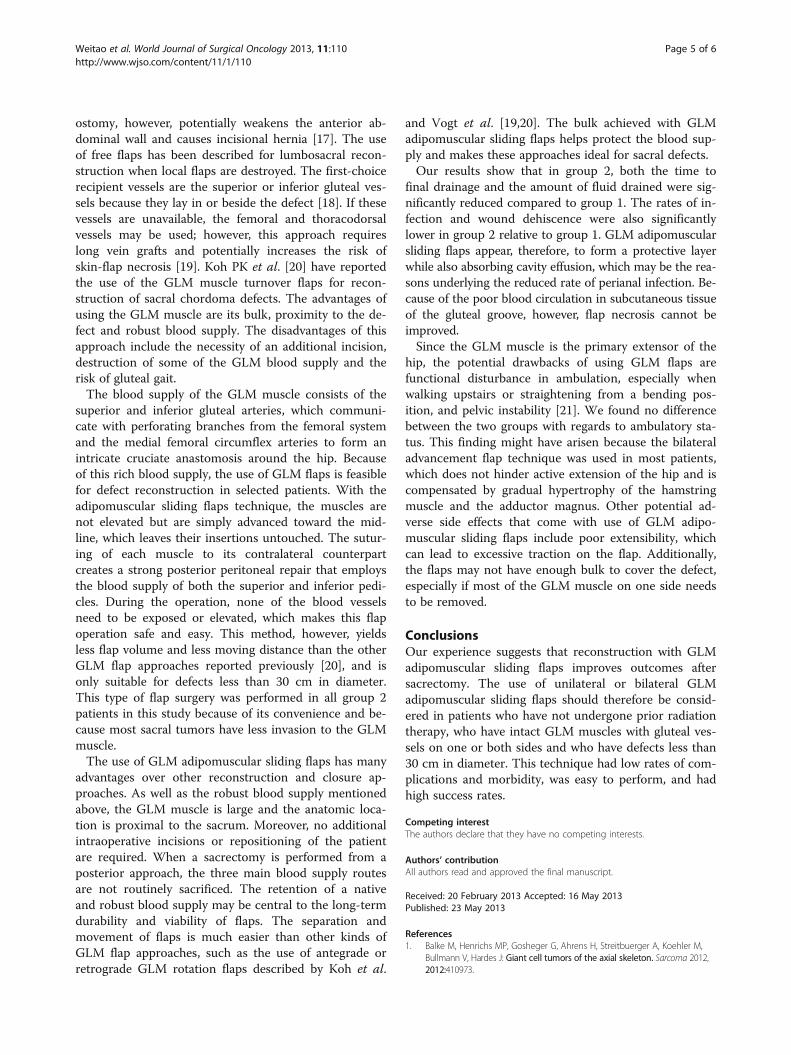
ostomy, however, potentially weakens the anterior ab-dominal wall and causes incisional hernia [17]. The useof free flaps has been described for lumbosacral recon-struction when local flaps are destroyed. The first-choicerecipient vessels are the superior or inferior gluteal ves-sels because they lay in or beside the defect [18]. If thesevessels are unavailable, the femoral and thoracodorsalvessels may be used; however, this approach requireslong vein grafts and potentially increases the risk ofskin-flap necrosis [19]. Koh PK et al. [20] have reportedthe use of the GLM muscle turnover flaps for recon-struction of sacral chordoma defects. The advantages ofusing the GLM muscle are its bulk, proximity to the de-fect and robust blood supply. The disadvantages of thisapproach include the necessity of an additional incision,destruction of some of the GLM blood supply and therisk of gluteal gait.The blood supply of the GLM muscle consists of the
superior and inferior gluteal arteries, which communi-cate with perforating branches from the femoral systemand the medial femoral circumflex arteries to form anintricate cruciate anastomosis around the hip. Becauseof this rich blood supply, the use of GLM flaps is feasiblefor defect reconstruction in selected patients. With theadipomuscular sliding flaps technique, the muscles arenot elevated but are simply advanced toward the mid-line, which leaves their insertions untouched. The sutur-ing of each muscle to its contralateral counterpartcreates a strong posterior peritoneal repair that employsthe blood supply of both the superior and inferior pedi-cles. During the operation, none of the blood vesselsneed to be exposed or elevated, which makes this flapoperation safe and easy. This method, however, yieldsless flap volume and less moving distance than the otherGLM flap approaches reported previously [20], and isonly suitable for defects less than 30 cm in diameter.This type of flap surgery was performed in all group 2patients in this study because of its convenience and be-cause most sacral tumors have less invasion to the GLMmuscle.The use of GLM adipomuscular sliding flaps has many
advantages over other reconstruction and closure ap-proaches. As well as the robust blood supply mentionedabove, the GLM muscle is large and the anatomic loca-tion is proximal to the sacrum. Moreover, no additionalintraoperative incisions or repositioning of the patientare required. When a sacrectomy is performed from aposterior approach, the three main blood supply routesare not routinely sacrificed. The retention of a nativeand robust blood supply may be central to the long-termdurability and viability of flaps. The separation andmovement of flaps is much easier than other kinds ofGLM flap approaches, such as the use of antegrade orretrograde GLM rotation flaps described by Koh et al.
and Vogt et al. [19,20]. The bulk achieved with GLMadipomuscular sliding flaps helps protect the blood sup-ply and makes these approaches ideal for sacral defects.Our results show that in group 2, both the time to
final drainage and the amount of fluid drained were sig-nificantly reduced compared to group 1. The rates of in-fection and wound dehiscence were also significantlylower in group 2 relative to group 1. GLM adipomuscularsliding flaps appear, therefore, to form a protective layerwhile also absorbing cavity effusion, which may be the rea-sons underlying the reduced rate of perianal infection. Be-cause of the poor blood circulation in subcutaneous tissueof the gluteal groove, however, flap necrosis cannot beimproved.Since the GLM muscle is the primary extensor of the
hip, the potential drawbacks of using GLM flaps arefunctional disturbance in ambulation, especially whenwalking upstairs or straightening from a bending pos-ition, and pelvic instability [21]. We found no differencebetween the two groups with regards to ambulatory sta-tus. This finding might have arisen because the bilateraladvancement flap technique was used in most patients,which does not hinder active extension of the hip and iscompensated by gradual hypertrophy of the hamstringmuscle and the adductor magnus. Other potential ad-verse side effects that come with use of GLM adipo-muscular sliding flaps include poor extensibility, whichcan lead to excessive traction on the flap. Additionally,the flaps may not have enough bulk to cover the defect,especially if most of the GLM muscle on one side needsto be removed.
ConclusionsOur experience suggests that reconstruction with GLMadipomuscular sliding flaps improves outcomes aftersacrectomy. The use of unilateral or bilateral GLMadipomuscular sliding flaps should therefore be consid-ered in patients who have not undergone prior radiationtherapy, who have intact GLM muscles with gluteal ves-sels on one or both sides and who have defects less than30 cm in diameter. This technique had low rates of com-plications and morbidity, was easy to perform, and hadhigh success rates.
Competing interestThe authors declare that they have no competing interests.
Authors’ contributionAll authors read and approved the final manuscript.
Received: 20 February 2013 Accepted: 16 May 2013Published: 23 May 2013
References1. Balke M, Henrichs MP, Gosheger G, Ahrens H, Streitbuerger A, Koehler M,
Bullmann V, Hardes J: Giant cell tumors of the axial skeleton. Sarcoma 2012,2012:410973.
Weitao et al. World Journal of Surgical Oncology 2013, 11:110 Page 5 of 6http://www.wjso.com/content/11/1/110
2. Donati D, Frisoni T, Dozza B, DeGroot H, Albisinni U, Giannini S: Advance inthe treatment of aneurysmal bone cyst of the sacrum. Skeletal Radiol2011, 40:1461–1466.
3. Syed R, Bishop JA, Ali SZ: Sacral and presacral lesions: cytopathologicanalysis and clinical correlates. Diagn Cytopathol 2012, 40:7–13.
4. Ferraresi V, Nuzzo C, Zoccali C, Marandino F, Vidiri A, Salducca N, Zeuli M,Giannarelli D, Cognetti F, Biagini R: Chordoma: clinical characteristics,management and prognosis of a case series of 25 patients. BMC Cancer2010, 10:22.
5. Lee J, Bhatia NN, Hoang BH, Ziogas A, Zell JA: Analysis of prognosticfactors for patients with chordoma with use of the California CancerRegistry. J Bone Joint Surg Am 2012, 94:356–363.
6. Quraishi NA, Giannoulis KE, Edwards KL, Boszczyk BM: Management ofmetastatic sacral tumours. Eur Spine J 2012, 21:1984–1993.
7. Clarke MJ, Dasenbrock H, Bydon A, Sciubba DM, McGirt MJ, Hsieh PC, Yassari R,Gokaslan ZL, Wolinsky JP: Posterior-only approach for en bloc sacrectomy:clinical outcomes in 36 consecutive patients. Neurosurgery 2012, 71:357–364.
8. Garvey PB, Rhines LD, Feng L, Gu X, Butler CE: Reconstructive strategies forpartial sacrectomy defects based on surgical outcomes. Plast ReconstrSurg 2011, 127:190–199.
9. Ruggieri P, Angelini A, Pala E, Mercuri M: Infections in surgery of primarytumors of the sacrum. Spine (Phila Pa 1976) 2012, 3:420–428. 1.
10. Asavamongkolkul A, Waikakul S: Wide resection of sacral chordoma via aposterior approach. Int Orthop 2012, 36:607–612.
11. Horch RE, D'Hoore A, Holm T, Kneser U, Hohenberger W, Arkudas A:Laparoscopic abdominoperineal resection with open posterior cylindricalexcision and primary transpelvic VRAM flap. Ann Surg Oncol 2012, 19:502–503.
12. Dasenbrock HH, Clarke MJ, Bydon A, Witham TF, Sciubba DM, Simmons OP,Gokaslan ZL, Wolinsky JP: Reconstruction of extensive defects fromposterior en bloc resection of sacral tumors with human acellular dermalmatrix and gluteus maximus myocutaneous flaps. Neurosurgery 2011,69:1240–1247.
13. Unal C, Eren GG, Isil E, Alponat A, Sarlak A: Utility of the omentum in sacralreconstruction following total sacrectomy due to recurrent and irradiatedgiant cell tumour of the spine. Indian J Plast Surg 2012, 45:140–143.
14. Stechl NM, Baumeister S, Grimm K, Kraus TW, Bockhorn H, Exner KE:Microsurgical reconstruction of the pelvic floor after pelvic exenteration.Reduced morbidity and improved quality of life by an interdisciplinaryconcept. Chirurg 2011, 82:625–630.
15. Diaz J, McDonald WS, Armstrong M, Eismont F, Hellinger M, Thaller S:Reconstruction after extirpation of sacral malignancies. Ann Plast Surg2003, 51:126–129.
16. Glatt BS, Disa JJ, Mehrara BJ, Pusic AL, Boland P, Cordeiro PG:Reconstruction of extensive partial or total sacrectomy defects with atransabdominal vertical rectus abdominis myocutaneous flap. Ann PlastSurg 2006, 56:526–531.
17. Daigeler A, Simidjiiska-Belyaeva M, Drücke D, Goertz O, Hirsch T, Soimaru C,Lehnhardt M, Steinau HU: The versatility of the pedicled vertical rectusabdominis myocutaneous flap in oncologic patients. Langenbecks ArchSurg 2011, 396:1271–1279.
18. Park S: Muscle-splitting approach to superior and inferior gluteal vessels:versatile source of recipient vessels for free-tissue transfer to sacral,gluteal, and ischial regions. Plast Reconstr Surg 2000, 106:81–86.
19. Vogt PM, Kall S, Lahoda LU, Spies M, Muehlberger T: The free "muttonchop" flap: a fascio-musculocutaneous flap for the reconstruction of theentire sacral and perineal area. Plast Reconstr Surg 2004, 114:1220–1224.
20. Koh PK, Tan BK, Hong SW, Tan MH, Tay AG, Song C, Tan KC: The gluteusmaximus muscle flap for reconstruction of sacral chordoma defects.Ann Plast Surg 2004, 53:44–49.
21. Haapamäki MM, Pihlgren V, Lundberg O, Sandzén B, Rutegård J: Physicalperformance and quality of life after extended abdominoperinealexcision of rectum and reconstruction of the pelvic floor with gluteusmaximus flap. Dis Colon Rectum 2011, 54:101–106.
doi:10.1186/1477-7819-11-110Cite this article as: Weitao et al.: Use of gluteus maximus adipomuscularsliding flaps in the reconstruction of sacral defects after tumorresection. World Journal of Surgical Oncology 2013 11:110.
Submit your next manuscript to BioMed Centraland take full advantage of:
• Convenient online submission
• Thorough peer review
• No space constraints or color figure charges
• Immediate publication on acceptance
• Inclusion in PubMed, CAS, Scopus and Google Scholar
• Research which is freely available for redistribution
Submit your manuscript at www.biomedcentral.com/submit
Weitao et al. World Journal of Surgical Oncology 2013, 11:110 Page 6 of 6http://www.wjso.com/content/11/1/110